ElShamah - Reason & Science: Defending ID and the Christian Worldview
Would you like to react to this message? Create an account in a few clicks or log in to continue.
ElShamah - Reason & Science: Defending ID and the Christian Worldview

Otangelo Grasso: This is my library, where I collect information and present arguments developed by myself that lead, in my view, to the Christian faith, creationism, and Intelligent Design as the best explanation for the origin of the physical world.


You are not connected. Please login or register

The various codes in the cell

Go to page : 1, 2  Next

Go down  Message [Page 1 of 2]

1The various codes in the cell Empty The various codes in the cell Thu Oct 22, 2015 12:07 pm

Otangelo


Admin

The various codes in the cell

https://reasonandscience.catsboard.com/t2213-the-various-codes-in-the-cell

http://www.codebiology.org/database.pdf

Not all codes other than the genetic code are considered epigenetic codes. Epigenetic codes specifically refer to modifications or marks that influence the accessibility, expression, and heritability of genetic information. These modifications can occur on DNA, histones, or other molecules involved in gene regulation. The term "regulatory codes" is sometimes used to describe various types of molecular information that guide cellular processes, gene expression, and interactions within a cell. Regulatory codes encompass a broader range of codes that dictate how cellular components interact, respond to signals, and carry out specific functions.  While "epigenetic codes" and "regulatory codes" are terms that are often used in the context of cellular processes and gene regulation, the naming conventions can vary depending on the specific area of study and scientific literature.

The dichotomy between structural and regulatory codes is a suitable tool to start the systematic study of codes. Barbieri assumes the existence of three types of organic codes, namely manufacturing, signaling, and regulatory codes.

M.Barbieri: The genetic code is at the centre of life but it is not the only code that exists in living systems. Any organic code is a mapping between two independent worlds and requires molecular structures that act like adaptors, i.e., that perform two independent recognition processes. The adaptors are required because there is no necessary connection between two independent worlds, and a set of rules is required in order to guarantee the specificity of the connection. The adaptors, in short, are essential in all organic codes. They are the molecular fingerprints of the codes, and their presence in a biological process is a sure sign that that process is based on a code. In splicing and in signal transduction, for example, I have shown (in 2003) that there are true adaptors at work, and that means that those processes are based on splicing codes and on signal transduction codes. 2

Code Biology: the study of all Codes of Life

Biological cells depend not only on the physical matter but essentially on pre-programmed information and at least 234 informational code and language systems which are used to host and store that information, in order to arrange and produce the complex cellular structures essential for life, and keep the life-essential functions, that is the reproduction, metabolism. food uptake, intracellular organizational arrangement, growth and development, permanence, change, and adaptation.   Living cells host multiple kinds of informational code systems that are used to store complex instructional/specifying information ( CSI ).  All code systems, languages, information, and translation systems can be traced back to an intelligent origin.  Evolution is not a driving force to explain the origin of cells and their language systems and programmed information content. Nor does physicochemical attraction explain the arrangement of nucleotides, molecules, and amino acids resulting in the formation of complex molecular machines and intracellular molecular production lines.  The only alternative to intelligence is random self-assembly by unguided lucky events. Random chaotic events are however too unspecific to explain the extremely organized, controlled, error check and repair mechanisms, and factory-like production systems that cells host.  Therefore, biological cells, cell code systems and coded information ( CSI ) have most probably a mind as the causal origin.

1. Regulation, governing, controlling, recruiting, interpreting, recognition, orchestrating, elaborating strategies, guiding, and instructing are all tasks of the gene regulatory network.
2. Such activity can only be exercised if no intelligence is present if the correct actions were pre-programmed by intelligence.
3. Therefore, most probably, the gene regulatory network was programmed by an intelligent agency.

The outstanding implication of the existence of organic codes in Nature comes from the fact that any code involves meaning and we need therefore to introduce in biology, with the standard methods of science, not only the concept of biological information but also that of biological meaning. The study on the organic codes, in conclusion, is bringing to light new mechanisms that operated in the history of life and new fundamental concepts. It is an entirely new field of research, the exploration of a vast and still largely unexplored dimension of the living world, the real new frontier of biology.

The irreducible interdependence of information generation and transmission systems
1. Codified information transmission system depends on: 
a) A language where a symbol, letters, words, waves or frequency variations, sounds, pulses, or a combination of those are assigned to something else. Assigning meaning of characters through a code system requires a common agreement of meaning. Statistics, Semantics, Synthax, and Pragmatics are used according to combinatorial, context-dependent, and content-coherent rules. 
b) Information encoded through that code,
c) An information storage system, 
d) An information transmission system, that is encoding, transmitting, and decoding.
e) Eventually translation ( the assignment of the meaning of one language to another )
f)  Eventually conversion ( digital-analog conversion, modulators, amplifiers)
g) Eventually transduction converting the nonelectrical signals into electrical signals
2. In living cells, information is encoded through at least 30 genetic, and almost 30 epigenetic codes that form various sets of rules and languages. They are transmitted through a variety of means, that is the cell cilia as the center of communication, microRNA's influencing cell function, the nervous system, the system synaptic transmission, neuromuscular transmission, transmission b/w nerves & body cells, axons as wires, the transmission of electrical impulses by nerves between brain & receptor/target cells, vesicles, exosomes, platelets, hormones, biophotons, biomagnetism, cytokines and chemokines, elaborate communication channels related to the defense of microbe attacks, nuclei as modulators-amplifiers. These information transmission systems are essential for keeping all biological functions, that is organismal growth and development, metabolism, regulating nutrition demands, controlling reproduction, homeostasis, constructing biological architecture, complexity, form, controlling organismal adaptation, change,  regeneration/repair, and promoting survival. 
3. The origin of such complex communication systems is best explained by an intelligent designer. Since no humans were involved in creating these complex computing systems, a suprahuman super-intelligent agency must have been the creator of the communication systems used in life. 

The concept of cross-talk between different cellular codes suggests a high level of coordination and purpose in the design of biological systems. The intricate interactions and communication between these codes indicate a sophisticated and integrated design that allows cells to respond to environmental cues, maintain homeostasis, and carry out their functions effectively. The complexity and specificity of these codes, and their ability to interact with each other, cannot be adequately explained by a step-wise, gradual, and unguided evolutionary process.  The examples of cross-talk between the characterized codes highlight how different cellular processes are intertwined, and changes in one code can have cascading effects on others. For these complex regulatory systems to function coherently, all components must be in place simultaneously and working together. The likelihood of all these intricate mechanisms evolving independently through random mutations and natural selection is extremely low, given the precise coordination required for cellular function.

Many of the over 223 epigenetic codes can cross-talk with each other due to the interconnectedness of cellular processes. Cross-talk refers to the communication and interaction between different signaling pathways and regulatory mechanisms in the cell.

The 31 Genetic Codes 

1. The Acetylation Code: A post-translational modification involving the addition of an acetyl group to proteins, influencing their function.
2. The Acoustic codes: Patterns of sound waves processed by the auditory system, conveying information about the environment.
3. The Adhesion Code: Molecular interactions that determine how cells adhere to each other and to surfaces.
4. The Adenylation Code: A process where adenosine monophosphate (AMP) is added to various molecules, often part of activation processes.
5. The Allosteric Code: The intricate regulation of protein function by molecules binding to sites other than the active site.
6. The Angiotensin Receptor Code: Signaling pathways involving angiotensin receptors that play a role in blood pressure regulation.
7. The Antioxidant Code: The mechanisms and molecules that protect cells from oxidative damage.
8. The Antibiotic Resistance Code: The genetic and biochemical basis of bacterial resistance to antibiotics.
9. The Apoptosis Code: Genetic and molecular mechanisms that govern programmed cell death.
10. The Archetype Code: Patterns and symbols that hold universal significance in human culture and psychology.
11. The Arrestin Receptor Code: The role of arrestin proteins in regulating G-protein-coupled receptor signaling.
12. The Assembly Code: Molecular rules governing the proper assembly of multi-component complexes.
13. The Auxin Metabolism Code: How auxin hormones are synthesized, transported, and regulated in plants.
14. The Axon Guidance Codes: Molecular signals guiding the growth of axons in neural development.
15. The Autocrine Signaling Code: Mechanisms by which cells release signaling molecules that affect their own activity.
16. The Autophagy Code: Molecular pathways that regulate autophagy, a process for recycling cellular components.
17. The BAFF Immune Code: Signaling pathways involving B-cell activating factor (BAFF) in the immune system.
18. The Bile Acid Code: The roles and regulation of bile acids in digestion and metabolism.
19. The Binaural Code: Neural processing of auditory information from both ears to localize sound sources.
20. The Bioelectric Code: Patterns of electrical signaling that influence cellular behavior and development.
21. The Biophoton Code: Hypothetical biophotonic emissions from living organisms and their potential significance.
22. The Biosynthetic Code: Genetic and biochemical pathways responsible for producing complex molecules.
23. The Universal Brain Code: The underlying principles governing neural networks and cognitive processes.
24. The Cadherin Neuronal Code: The role of cadherin molecules in neuronal adhesion and circuit formation.
25. The Calcium Signaling Code: Molecular pathways that regulate calcium-mediated intracellular signaling.
26. The Cell Cycle Checkpoint Code: Mechanisms ensuring proper progression through the cell cycle.
27. The Cell-Cell Communication Code: Molecular signals that mediate communication between neighboring cells.
28. The Cell Access Code: The regulation of cellular uptake of nutrients, ions, and other molecules.
29. The Cell Fate Determination Code: Molecular processes that dictate a cell's developmental fate.
30. The Cell Migration Code: Molecular cues guiding cells in movement during development, wound healing, etc.
31. The Cell Polarity Code: Molecular pathways that establish and maintain cellular asymmetry.
32. The Cell Surface Recognition Code: Molecules that mediate cell interactions through recognition of surface markers.
33. The Cerebral Resistance Code: The molecular mechanisms that govern the brain's ability to resist damage or adapt to challenges.
34. The Chitin (Defense) Code:Molecular processes related to the synthesis, modification, and utilization of chitin in defense mechanisms, often found in insects and other organisms.
35. The Chaperone Code: The role of chaperone proteins in assisting proper protein folding.
36. The Chromatin Code: Histone modifications and other factors that regulate chromatin structure and gene expression.
37. The Chromosomal Imprinting Code:  Epigenetic marks on specific genes inherited from one parent that affect gene expression patterns based on parental origin.
38. The Chromosome Segregation Code: Molecular mechanisms that ensure accurate distribution of chromosomes during cell division to prevent errors in genetic inheritance.
39. The Circular motif ( ribosome) Code: Consists of specific RNA sequences in circular RNA molecules, which may have regulatory roles in gene expression.
40. The Coactivator/corepressor/epigenetic Code: Describes how coactivator and corepressor proteins, along with epigenetic modifications, influence gene expression regulation.
41. The Code of human language:  The intricate system of sounds, words, and grammar rules that humans use to communicate complex thoughts and ideas.
42. The Cohesin-Dockerin Code: Refers to the interaction between cohesin and dockerin domains in some bacteria, which plays a role in cellulosome assembly and cell attachment.
43. The Cytokine Codes: Signaling molecules produced by immune cells that influence cellular communication and responses during immune and inflammatory processes.
44. The Compartment Code: Molecular processes that determine the organization and segregation of cellular components within specific subcellular compartments.
45. The Cholesterol Recognition/Mirror Code: The interaction of cholesterol with proteins and its impact on cellular processes.
46. The Cilia Code: Molecular mechanisms underlying the structure and function of cilia.
47. The Circardian Rhythm Codes: Genetic and molecular pathways that regulate circadian rhythms.
48. The Cytoskeleton Code: The organization and dynamics of the cytoskeleton and its role in cell function.
49. The Connexin Code: encompasses the intricate molecular mechanisms involving connexin proteins, vital components of gap junctions.
50. The DNA Repair / Damage Codes: Molecular pathways that repair DNA damage and maintain genomic integrity.
51. The DNA-Binding Code: Molecular interactions between proteins and DNA sequences.
52. The DNA methylation Code: Epigenetic modifications involving the addition of methyl groups to DNA.
53. The DNA Zip Code / Peripheral Targeting Code: DNA sequences that dictate subnuclear localization.
54. The Discriminator Codes: involve molecular mechanisms that distinguish between different cellular components, signals, or states. 
55. The Differentiation Code: Signals and factors that drive cells to specialize into specific cell types.
56. The Domain substrate specificity Code of Nonribosomal peptide synthetases (NRPS): Mechanisms underlying the synthesis of complex peptides in bacteria.
57. The Endocytosis Code: Molecular mechanisms governing the process of cellular endocytosis.
58. The Endocrine Signalling Codes: Signaling pathways involving hormones and their effects on target cells.
59. The (Epigenetic) Body Plan Code: Epigenetic mechanisms shaping the development of body structures.
60. The Epigenetic Cancer Code: Epigenetic changes associated with cancer development and progression.
61. The Epidermal Growth Factor (EGF) Code: Signaling pathways involving epidermal growth factor and its receptors.
62. The Epitranscriptomic Code: Post-transcriptional modifications to RNA molecules that affect their function.
63. The Error correcting Code: Mechanisms that ensure proper DNA replication and repair errors.
64. The Epigenetic Imprinting Code: Epigenetic modifications that lead to parent-of-origin-specific gene expression.
65. The Export & Exit Codes: Molecular mechanisms that direct proteins and RNA out of the cell.
66. The Extracellular Matrix (ECM) Code: Composition and organization of the ECM and its impact on cell behavior.
67. The Forkhead Transcription Factor Code: Functions and regulation of the forkhead box (FOX) family of transcription factors.
68. The General Neural Codes: Neural patterns representing sensory information or motor commands.
69. The Genetic Recombination Codes: Mechanisms of genetic recombination that create genetic diversity.
70. The Genomic Code: Genetic information and the relationship between nucleotide sequences and phenotypes.
71. The Genomic regulatory Code: Non-coding regions of DNA that control gene expression.
72. The G-Protein Coupled Receptor (GPCR) Code: Molecular properties and signaling pathways of GPCRs.
73. The Gli Codes: Signaling pathways involving the Gli family of transcription factors.
74. The Glioma Code: Genetic and molecular aspects of glioma development and progression.
75. The Glycomic Code: Diversity and roles of glycan structures in cellular processes.
76. The Growth Codes: Molecular cues and pathways that regulate cell growth and proliferation.
77. The Hearing Code: Neural coding of auditory information and sound perception.
78. The Hedgehog Signaling Code: Signaling pathways involving Hedgehog proteins and their receptors.
79. The Heterochromatin Code: Molecular marks and proteins that regulate heterochromatin formation.
80. The Histone Sub-Code: Specific modifications of histone proteins that influence chromatin structure.
81. The Histone Variants Code: Variations in histone protein sequences that affect chromatin dynamics.
82. The Homeokinetic Muscle Code: Mechanisms underlying muscle homeostasis and adaptation.
83. The Honey Bee Dance Code: Communication through dances that inform other bees of food sources.
84. The Host Defense Code: Mechanisms by which the host defends against pathogens and infections.
85. The Hormone Receptor Code: Molecular interactions between hormones and their target receptors.
86. The HOX Code Pattern Formation: HOX gene expression patterns that guide embryonic development.
87. The Hypothalamic Code: Signaling pathways and neuropeptides involved in hypothalamic regulation.
88. The Identity Code: Mechanisms that define the unique identity of cells, tissues, and organisms.
89. The immune response code, or language: Molecular signals and pathways that orchestrate immune responses.
90. The Immune T-cell Codes: Receptor interactions and signals involved in T-cell immune responses.
91. The Importin Codes: Processes involving importin proteins that facilitate nuclear transport.
92. The Indole Physiological Code: The role of indole molecules in bacterial physiology and behavior.
93. The Inositol Phosphate Code: Signaling pathways involving inositol phosphates and their effects.
94. The Irisin (Muscle) Code: The hormone irisin and its effects on metabolism and energy expenditure.
95. The Karyotype Code: The chromosomal arrangement and number characteristic of a species.
96. The Lamin Code : Molecular interactions involving nuclear lamins that impact nuclear structure.
97. The Latency Behaviour Codes: Behaviors associated with latency periods in psychological development.
98. The Lipid Codes: Molecular structures and signals involving lipids in cellular processes.
99. The Magnitude Neuronal Codes: Neural responses that encode the intensity or magnitude of stimuli.
100. The Meiosis Codes: Molecular processes that ensure proper chromosome segregation during meiosis.
101. The Membrane Code: Properties of cellular membranes and their interactions with molecules.
102. The Memory Code: Neural mechanisms that encode and retrieve memories.
103. The Metabolic Signaling Code: Molecular pathways that link cellular metabolism with signaling.
104. The Methylation Code: The role of DNA and protein methylation in gene expression and regulation.
105. The Microbiome Code: Genetic and functional diversity of microbial communities in and on the body.
106. The Micro-RNA Codes: Small RNA molecules that regulate gene expression at the post-transcriptional level.
107. The Mnemonic codes: Mechanisms by which memory is encoded and retrieved.
108. The Modularity Codes: Molecular units and patterns that enable the assembly of complex structures.
109. The Molecular Codes: A collective term encompassing various specific codes governing cellular processes.
110. The Morphogenetic Code: Signaling molecules that direct tissue and organ development.
111. The Myelin code: Molecular cues that regulate myelin formation and maintenance in the nervous system.
112. The Molecular Recognition Code: Molecular interactions that enable specific recognition between molecules.
113. The Navigation / Orientation / Movement Codes: Neural pathways and signals that guide navigation and movement.
114. The Neuronal Activity-Dependent Gene Expression Code
115. The Neuronal Code for Reading: Neural pathways and processes involved in reading.
116. The Neuronal Hippocampal Codes: contribute to the organization and regulation of neural activity within the hippocampus. 
117. The Neural Motion Codes: Neural patterns that encode and control motor movements.
118. The Neural Perception & Recognition Codes: Neural responses that process and recognize sensory information.
119. The Neural, Social Information Code: How neural circuits process social cues and interactions.
120. The neuronal Oscillatory /Frequency Codes: Neural oscillations and frequencies that regulate brain activity.
121. The Neuronal spike-rate Code:  involves patterns of neuronal firing rates that convey information within the nervous system.
122. The Neuronal Taste Code: Neural pathways and signals that encode taste perception.
123. The Neuron Light Code: signifies rapid patterns of neuronal activity that transmit information through light-like signals, facilitating neural communication.
124. The Neuropeptide Code: The role of neuropeptides in neural signaling and behavior.
125. The NF-kappa-B Code: Molecular pathways involving the NF-kappa-B family of transcription factors.
126. The Nitric Oxide (NO) Signaling Code: Signaling pathways involving nitric oxide and its effects.
127. The N-Glycan Code: Diversity and roles of N-linked glycan structures in cellular processes.
128. The Nomenclatural Code: Rules for naming biological taxa and species.
129. The Non-Ribosomal Code: Mechanisms of protein synthesis by non-ribosomal peptide synthetases.
130. The Notch Code: Signaling pathways involving Notch receptors and their role in development.
131. The Nuclear Signalling Code: Molecular pathways involving nuclear signaling events.
132. The Nutrient Transport Code: Molecular mechanisms for transporting nutrients across cell membranes.
133. The Olfactory Code: Neural coding of olfactory information and smell perception.
134. The Nucleosome Code: involves molecular arrangements that dictate DNA packaging and gene accessibility using nucleosomes.
135. The Nucleotide Sequence Codes: encompass genetic information encoded in DNA sequences, shaping traits and functions.
136. The Nutrient Sensing Code:  involves molecular processes that detect and respond to nutrient levels in cells, guiding metabolic and physiological responses.
137. The Omega Leaf Code: Hypothetical code indicating plant leaf arrangement based on Fibonacci numbers.
138. The Operon Code: Genetic regulation of bacterial operons and coordinated gene expression.
139. The Orthographic Reading Code: Neural processes that enable reading and recognizing written words.
140. The Pattern Formation Code: Molecular mechanisms that create ordered patterns during development.
141. The Phagocytosis Codes: Cellular processes and molecular cues governing phagocytosis.
142. The Pheromone Codes: Molecular signals that communicate information between individuals of the same species.
143. The Phonological Codes: Neural representation and processing of speech sounds.
144. The Phosphatase Code: Regulation of cellular processes by protein phosphatases.
145. The Physiological Coregulator Code: Molecular factors that modulate physiological responses.
146. The Phosphorylation Code: Regulation of protein function through phosphorylation by kinases.
147. The Phosphorylation-Dependent Protein Interaction Code: Proteins that bind to phosphorylated targets, regulating interactions.
148. The Phospholipid Code: Role of specific phospholipids in cellular membranes and signaling.
149. The Phosphoserine Code: Functions of proteins and pathways involving phosphoserine residues.
150. The Photoreceptor Sensory Code: Neural coding of visual information and light perception.
151. The Photosynthesis Code: Molecular mechanisms of photosynthesis and energy conversion.
152. The Plant Cell Wall Code: Composition and roles of plant cell walls in growth and defense.
153. The Plant Communication Codes encompass molecular processes and signaling mechanisms by which plants exchange information and respond to various environmental cues
154. The Post-translational modification Code for transcription factors: Modifications that affect the activity and function of transcription factors.
155. Protein Kinase Codes: Families of protein kinases and their roles in cellular signaling.
156. The Poly(Adenylation) Code: Role of polyadenylation in mRNA stability and translation.
157. The Polycomb & Trithorax Codes: Complexes involved in epigenetic regulation and gene expression.
158. The Polysaccharide Codes: Diversity and roles of polysaccharides in cellular processes.
159. The Post-translational Modification Codes refer to a diverse array of molecular processes that modify proteins after they are synthesized.
160. The Presynaptic Vesicle Code: Molecular processes involving neurotransmitter-containing vesicles.
161. The Protein Allosteric Code: Mechanisms by which proteins switch between different conformations.
162. The Protein Binding Code: Molecular interactions that allow proteins to bind to specific partners.
163. The Protein Folding Code: Molecular principles that dictate protein folding into functional conformations.
164. The Protein Interaction Code: Specific molecular interactions that govern protein-protein interactions.
165. The Protein Phosphorylation Code: Regulation of protein function by reversible phosphorylation.
166. The Protein Secretory Code: Processes and signals that guide protein secretion from cells.
167. The Protein Translocation Code: involves mechanisms governing the movement of proteins within cells to their designated locations.
168. The Proteomic Code: Governs processes that regulate protein degradation and renewal within cells.
169. The Regulatory Organogenesis Codes: refer to molecular mechanisms that oversee the development of tissues and organs.
170. The Regulatory Network Codes encompass intricate signaling networks that control cellular responses.
171. The Renal Codes pertain to molecular processes specific to kidney function and regulation.
172. The Representation Codes involve molecular mechanisms underlying information encoding and processing.
173. The Retinal Codes concern molecular events and patterns of activity within the retina, vital for vision.
174. The RNA-Interference Codes relate to the regulatory roles of RNA interference in gene expression.
175. The RNA Polymerase Modification Codes involve modifications affecting RNA polymerase function in transcription.
176. The RNA Recognition Code involves molecular interactions between RNA molecules and other cellular components.
177. The Redox Code encompasses processes influenced by cellular redox (oxidation-reduction) states.
178. The Regeneration Codes involve molecular cues and mechanisms that guide tissue and organ regeneration.
179. The Retinoic Acid Signaling Code relates to signaling pathways activated by retinoic acid and their effects.
180. The Ribonucleic Acid Modification Code (RNA Modification Code) pertains to post-transcriptional RNA modifications.
181. The Ribosomal Code concerns molecular interactions and functions of ribosomal components.
182. The Riboswitch Code involves RNA structures that modulate gene expression in response to ligand binding.
183. The Quorum Sensing Code: Bacterial communication through the release and sensing of signaling molecules.
184. The RNA Code: Genetic information carried by RNA molecules, including coding and non-coding roles.
185. The RNA Editing Code: Post-transcriptional modifications that change RNA sequences.
186. The RNA Modification Code: Various modifications that alter the structure and function of RNA.
187. The RNA Splicing Code: Processes that remove introns and join exons in mRNA molecules.
188. The RNA Transport Code: Mechanisms that guide RNA molecules to specific cellular locations.
189. The Semaphoring Codes: Signaling pathways involving semaphorin proteins and their role in axon guidance.
190. The Serotonin Code involves molecular processes related to the signaling and effects of serotonin, a neurotransmitter influencing mood and behavior.
191. The Sexual Dimorphic Codes Codes encompass molecular mechanisms underlying the development of gender-specific traits.
192. The Signal Integration Codes involve processes that combine multiple cellular signals for coordinated responses.
193. The Sperm RNA Code relates to the unique RNA molecules found in sperm cells, potentially influencing early development.
194. The Signal Integration Codes encompass processes that harmonize and interpret various cellular signals.
195. The Synaptic Adhesive Code refers to molecular interactions that guide the adhesion and connectivity of neurons at synapses.
196. The Stem Cell Code encompasses molecular cues that regulate stem cell behavior and differentiation.
197. The Sumoylation Code relates to the post-translational modification known as sumoylation, which influences protein activity and interactions.
198. The Skin Inflammation Code Code involves molecular pathways that contribute to inflammation and immune responses in the skin.
199. The Sodium/Calcium Channel Gating Code involves molecular mechanisms that regulate the opening and closing of sodium and calcium ion channels.
200. The Speech Code relates to neural and cognitive processes underlying the production and comprehension of speech.
201. The Spliceosome Code: Molecular machinery responsible for mRNA splicing.
202. The Substrate Specificity Code pertains to the molecular factors determining the selection and interaction of enzymes with specific substrates.
202. The Sugar Code encompasses the roles of sugar molecules in cell-cell interactions, signaling, and recognition.
203. The Sulfation Code involves the addition of sulfate groups to molecules, influencing their functions and interactions.
204. The Sulfur Code relates to the roles and effects of sulfur-containing molecules in various cellular processes.
205. The Synaptic Code: Molecular and cellular processes that underlie synaptic transmission.
206. The Toll-like Receptor Codes: Signaling pathways involving Toll-like receptors in the immune system.
207. The Transcription Factor Binding Code: Mechanisms by which transcription factors interact with DNA.
208. The Transcriptional Regulatory Code: Molecular mechanisms that control gene expression.
209. The Transmembrane Protein Code: Structure and function of proteins that span cellular membranes.
210. The tRNA Code: Transfer RNA molecules that decode mRNA into protein sequences.
211. The Ubiquitin Code: Post-translational modification involving ubiquitin and protein degradation.
212. The Tactile Neural Codes involve patterns of neural activity that transmit tactile sensations and touch-related information.
213. The Talin Code refers to the molecular processes related to talin protein's role in cell adhesion and signaling.
214. The Terpene Biosynthesis Code involves the genetic and biochemical pathways responsible for producing terpenes in organisms.
215. The Thermal / Temperature Neuronal Codes relate to patterns of neural activity that convey temperature-related sensory information.
216. The Translational Control Code: Regulation of gene expression at the level of translation initiation and elongation.
217. The Tight Junction Codes pertain to molecular interactions and functions of tight junctions, important for cell barrier formation.
218. The Tissue Code encompasses molecular characteristics specific to different types of tissues in multicellular organisms.
219. The Tissue Memory Code involves molecular processes that contribute to the memory or lineage history of tissues.
220. The Tubulin Code involves modifications and interactions of tubulin proteins, crucial for microtubule function.
221. The Visual Code involves neural and molecular processes that enable visual perception and processing.
222. The Wobbling Base Pairing Code relates to flexible pairing of bases in DNA/RNA, affecting translation accuracy.
223. The Zinc Finger Code: DNA-binding motifs formed by zinc finger proteins.


1) http://www.garlandscience.com/res/pdf/9780815341291_ch08.pdf
2. Barbieri: Organic codes


Last edited by Otangelo on Mon Mar 04, 2024 6:04 am; edited 91 times in total (Reason for editing : cebola de oleo)

https://reasonandscience.catsboard.com

Otangelo


Admin

CONTROL OF TRANSCRIPTION BY SEQUENCE-SPECIFIC DNA-BINDING PROTEINS 1


Coded information can always be tracked back to intelligence, which has to set up the convention of the meaning of the code, and the information carrier, which can be a book, the hardware of a computer, or the smoke of a fire of a Indian tribe signalling to another. All communication systems have an encoder which produces a message that is processed by a decoder. In the cell, there are several code systems. DNA is the most well-known, it stores coded information through the four nucleic acid bases. But there are several others, less known. Recently there was some hype about a second DNA code. In fact, it is essential for the expression of genes. The cell uses several formal communication systems according to Shannon’s model because they encode and decode messages using a system of symbols.  As Shannon wrote :

“Information, transcription, translation, code, redundancy, synonymous, messenger, editing, and proofreading are all appropriate terms in biology. They take their meaning from information theory (Shannon, 1948) and are not synonyms, metaphors, or analogies.” (Hubert P. Yockey,  Information Theory, Evolution, and the Origin of Life,  Cambridge University Press, 2005).

An organism’s DNA encodes all of the RNA and protein molecules required to construct its cells. Yet a complete description of the DNA sequence of an organism—be it the few million nucleotides of a bacterium or the few billion nucleotides of a human—no more enables us to reconstruct the organism than a list of English words enables us to reconstruct a play by Shakespeare. In both cases, the problem is to know how the elements in the DNA sequence or the words on the list are used. Under what conditions is each gene product made, and, once made, what does it do? The different cell types in a multicellular organism differ dramatically in both structure and function. If we compare a mammalian neuron with a liver cell, for example, the differences are so extreme that it is difficult to imagine that the two cells contain the same genome. The genome of an organism contains the instructions to make all different cells, and  the expression of either a neuron cell or liver cell can be regulated at many of the steps in the pathway from DNA to RNA to Protein. The most important imho is the CONTROL OF TRANSCRIPTION BY SEQUENCESPECIFIC DNA-BINDING PROTEINS, called transcription factors or regulators. These proteins recognize specific sequences of DNA (typically 5–10 nucleotide pairs in length) that are often called cis-regulatory sequences.   Transcription regulators bind to these sequences, which are dispersed throughout genomes, and this binding puts into motion a series of reactions that ultimately specify which genes are to be transcribed and at what rate. Approximately 10% of the protein-coding genes of most organisms are devoted to transcription regulators. Transcription regulators must recognize short, specific cis-regulatory sequences within this structure. The outside of the double helix is studded with DNA sequence information that transcription regulators recognize: the edge of each base pair presents a distinctive pattern of hydrogen-bond donors, hydrogen-bond acceptors, and hydrophobic patches in both the major and minor grooves. The 20 or so contacts that are typically formed at the protein–DNA interface add together to ensure that the interaction is both highly specific and very strong.


These instructions are written in a language that is often called the ‘gene regulatory code’.  The preference for a given nucleotide at a specific position is mainly determined by physical interactions between the aminoacid side chains of the TF ( transcription factor ) and the accessible edges of the base pairs that are contacted.  It is possible that some complex code, comprising rules from each of the different layers, contributes to TF– DNA binding; however, determining the precise rules of TF binding to the genome will require further scientific research. So, Genomes contain both a genetic code specifying amino acids, and this regulatory code specifying transcription factor (TF) recognition sequences. We find that ~15% of human codons are dual-use codons (`duons') that simultaneously specify both amino acids and TF recognition sites. Genomes also contain a parallel regulatory code specifying recognition sequences for transcription factors (TFs) , and the genetic and regulatory codes have been assumed to operate independently of one another, and to be segregated physically into the coding and non-coding genomic compartments. the potential for some coding exons to accommodate transcriptional enhancers or splicing signals has long been recognized

In order for communication to happen, 1. The sequence of DNA bases located in the regulatory region of the gene is required , and 2. transcription factors that read the code. If one of both is missing, communication fails, the gene that has to be expressed, cannot be encountered, and the whole procedure of gene expression fails. This is a irreducible complex system. The gene regulatory code could not arise in a stepwise manner either, since if that were the case, the code has only the right significance if fully developed. Thats a example par excellence of intelligent design.. The fact that these transcription factor binding sequences overlap protein coding sequences, suggest that both sequences were designed together, in order to optimize the efficiency of the DNA code. As we learn more and more about DNA structure and function, it is apparent that the code was not just hobbled together by the trial and error method of natural selection, but that it was specifically designed to provide optimal efficiency and function.


 Stephen Meyer puts it that way in his excellent book: Darwins doubt pg.270:

INTEGRATED CIRCUITRY: DEVELOPMENTAL GENE REGULATORY NETWORKS 

Keep in mind, too, that animal forms have more than just genetic information. They also need tightly  integrated networks of genes, proteins, and other molecules to regulate their development—in other words, they require developmental gene regulatory networks, the dGRNs . Developing animals face two main challenges. First, they must produce different types of proteins and cells and, second, they must get those proteins  and cells to the right place at the right time.20 Davidson has shown that embryos accomplish this task by relying on networks of regulatory DNA-binding proteins (called transcription factors) and their physical targets. These physical targets are typically sections of DNA (genes) that produce other  proteins or RNA molecules, which in turn regulate the expression of still other genes.

These interdependent networks of genes and gene products present a striking appearance of design. Davidson's graphical depictions of these dGRNs look for all the world like wiring diagrams in an electrical engineering blueprint or a schematic of an integrated circuit, an uncanny resemblance Davidson himself has often noted. "What emerges, from the analysis of animal dGRNs," he muses, "is almost astounding: a network of logic interactions programmed into the DNA sequence that amounts  essentially to a hardwired biological computational device." These molecules collectively form a tightly integrated network of signaling molecules that function as an integrated circuit. Integrated circuits in electronics are systems of individually functional components such as transistors, resistors, and capacitors that are connected together to perform an overarching function. Likewise, the functional components of dGRNs—the DNA-binding proteins, their DNA target sequences, and the other molecules that the binding proteins and target molecules produce and regulate—also form an integrated circuit, one that contributes to accomplishing the overall function of producing an adult animal form. 

Davidson himself has made clear that the tight functional constraints under which these systems of molecules (the dGRNs) operate preclude their gradual alteration by the mutation and selection mechanism. For this reason, neo-Darwinism has failed to explain the origin of these systems of molecules and their functional integration. Like advocates of evolutionary developmental biology, Davidson himself favors a model of evolutionary change that envisions mutations generating large-scale developmental effects, thus perhaps bypassing nonfunctional intermediate circuits or systems. Nevertheless, neither proponents of "evo-devo," nor proponents of other recently proposed materialistic theories of evolution, have identified a mutational mechanism capable of generating a dGRN or anything even remotely resembling a complex integrated circuit. Yet, in our experience, complex integrated circuits—and the functional integration of parts in complex systems generally—are known to be produced by intelligent agents—specifically, by engineers. Moreover, intelligence is the only known cause of such effects. Since developing animals employ a form of integrated circuitry, and certainly one manifesting a tightly and functionally integrated system of parts and subsystems, and since intelligence is the only known cause of these features, the necessary presence of these features in developing Cambrian animals would seem to indicate that intelligent agency played a role in their origin 

The Calcium Code
http://stke.sciencemag.org/content/2001/89/tw4
Steady-state stomatal closure could be restored if calcium oscillations similar to wild type were imposed; thus, the cells have an intact downstream signaling pathway, but cannot initiate the proper calcium oscillation code to trigger the pathway.

Defined changes of cytosolic Ca2+ concentration are triggered by cellular second messengers, such as NAADP, IP3, IP6, Sphingosine-1-Phospate, and cADPR  and it is evident that the identity and intensity of a specific stimulus impulse results in stimulus-specific and dynamic alterations of cytosolic Ca2+ concentration.  This heterogeneity of increases in cytosolic-free Ca2+ ion concentration in terms of duration, amplitude, frequency, and spatial distribution lead A.M. Hetherington and coworkers to formulate the concept of “Ca2+ signatures”. Signal information would be encoded by a specific Ca2+ signature that is defined by precise control of spatial, temporal, and concentration parameters of alterations in cytosolic Ca2+ concentration.

The RNA code
http://www.nature.com/nature/journal/v542/n7642/pdf/542503a.pdf

In 2004, oncologist Gideon Rechavi at Tel Aviv University in Israel and his colleagues compared all the human genomic DNA sequences then available with their corresponding messenger RNAs — the molecules that carry the information needed to make a protein from a gene.

They were looking for signs that one of the nucleotide building blocks in the RNA sequence, called adenosine (A), had changed to another building block called inosine (I). This 'A-to-I editing' can alter a protein's coding sequence, and, in humans, is crucial for keeping the innate immune response in check. “It sounds simple, but in real life it was really complicated,” Rechavi recalls. “Several groups had tried it before and failed” because sequencing mistakes and single-nucleotide mutations had made the data noisy. But using a new bioinformatics approach, his team uncovered thousands of sites in the transcriptome — the complete set of mRNAs found in an organism or cell population — and later studies upped the number into the millions1.

Inosine is something of a special case: researchers can readily detect this chink in the armour by comparing DNA and RNA sequences. But at least one-quarter of our mRNAs harbour chemical tags — decorations to the A, C, G and U nucleotides — that are invisible to today's sequencing technologies. (Similar chemical tags, called epigenetic markers, are also found on DNA.) Researchers aren't sure what these chemical changes in RNA do, but they're trying to find out.

A wave of studies over the past five years — many of which focus on a specific RNA mark called N6-methyladenosine (m6A) — have mapped these alterations across transcriptomes and demonstrated their importance to health and disease. But the problem is vast: these marks coat not only mRNA but other RNA transcripts as well, and they cut across all the domains of life and beyond, marking even viruses with their presence.

The modifications themselves are not new. What has given them meaning and driven epitranscriptomics into the spotlight is the discovery of enzymes that can add, remove and interpret them. In 2010, chemical biologist Chuan He at the University of Chicago, Illinois, proposed that these chemical tags could be reversible and important regulators of gene expression. Not long afterwards, his group demonstrated2 the first eraser of these marks on mRNA, an enzyme called FTO. That discovery meant that m6A wasn't just a passive mark — cells actively controlled it. And this realization came at about the same time that global approaches, harnessing the power of next-generation sequencing, made it possible to map m6A and other modifications across the transcriptome.

David Coppedge In Life, Not One Code but Many May 19, 2022
https://evolutionnews.org/2022/05/in-life-one-code-or-many/?fbclid=IwAR3ASewwqRxKut_3l4gXODe04ELyiR_7AcClfl4r_LFjgPZ7vdSR0A_-cmo

Distinct responses to rare codons in select Drosophila tissues
https://elifesciences.org/articles/76893


The Hidden Codes That Shape Protein Evolution 1

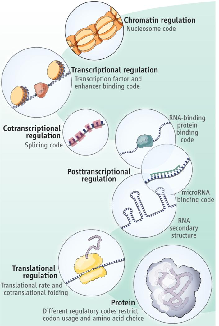
Despite redundancy in the genetic code (1), the choice of codons used is highly biased in some proteins, suggesting that additional constraints operate in certain protein-coding regions of the genome. This suggests that the preference for particular codons, and therefore amino acids in specific regions of the protein, is often determined by factors unrelated to protein structure or function (2, 3).  Stergachis et al. (4) reveal that transcription factors bind within protein-coding regions (in addition to nearby noncoding regions) in a large number of human genes. Thus, a transcription factor “binding code” may influence codon choice and, consequently, protein evolution. This “binding” code joins other “regulatory” codes that govern chromatin organization (3), enhancers (5, 6), mRNA structure (7), mRNA splicing (3), microRNA target sites (6, Cool, translational efficiency (9), and cotranslational folding (10), all of which have been proposed to constrain codon choice, and thus protein evolution (see the figure).


Figure
The various codes in the cell Emss-610

Constraining codes. Regulatory elements within protein-coding regions (such as transcription factor binding) can influence codon choice and amino acid preference that are independent of protein structure or function
Redundancy in the genetic code might facilitate the existence of multiple overlapping regulatory codes within protein-coding regions of the genome.

How widespread is the phenomenon of “regulatory” codes that overlap the genetic code, and how do they constrain the evolution of protein sequences? Stergachis et al. address these questions for the transcription factor–binding regulatory code. They use deoxyribonuclease I (DNase I) footprinting to map transcription factor occupancy (a protein bound to DNA can protect that region from enzymatic cleavage) at nucleotide resolution across the human genome in 81 diverse cell types. The authors determined that ~14% of the codons within 86.9% of human genes are occupied by transcription factors. Such regions, called “duons,” therefore encode two types of information: one that is interpreted by the genetic code to make proteins and the other, by the transcription factor–binding regulatory code to influence gene expression. This requirement for transcription factors to bind within protein-coding regions of the genome has led to a considerable bias in codon usage and choice of amino acids, in a manner that is constrained by the binding motif of each transcription factor.


To investigate whether single-nucleotide variants within duons affect transcription factor binding, Stergachis et al. mapped the known variants that are associated with a disease or a trait onto duons. Of those, 17.4% quantitatively skew the allelic origins of DNA fragments protected from cleavage by DNase I in human cells, suggesting that such single-nucleotide variants affect transcription factor occupancy. They also determined that such variants are not biased toward whether they result in synonymous or nonsynonymous changes in the protein sequence. Intriguingly, a large fraction of the variants that result in a nonsynonymous change are predicted not to alter protein function. This indicates that some variants within duons might primarily affect transcription factor binding instead. This supports the emerging idea that single-nucleotide variants within protein-coding regions can lead to disease without affecting protein structure or function (11, 12). Thus, the whole spectrum of “regulatory” codes within protein-coding regions should be considered when assessing the impact of single-nucleotide variants and interpreting disease mutation data from exome sequencing (only the protein-coding regions of the genome) and cancer genome studies.
Do the regulatory codes harmoniously coexist? Evidence is emerging that there can be conflicts. For example, in the fruit fly Drosophila melanogaster, there is a striking decrease in the use of codons that are optimal for translation, but a rise in codons that enhance RNA splicing, toward the end of exons (13). This may indicate that the requirement for accurate RNA splicing has superseded that for optimal translation. Likewise, Stergachis et al. observed that the binding motifs of transcription factors within protein-coding genomic regions are selectively devoid of sequences that contain a stop codon.
What features might permit synergistic coexistence of the regulatory and genetic codes? One major constraint of protein-coding genes is the requirement for the encoded polypeptide segment to fold into a defined tertiary structure. It is possible that in regions where folding constraints are not present, such as in intrinsically disordered regions (14), there might be increased tolerance for protein-coding genomic regions to harbor more regulatory elements that can be interpreted by different regulatory codes.
Stergachis et al. make a number of important genome-scale observations, but several mechanistic questions remain to be answered. For instance, although the authors report a weak tendency for transcription factors to preferentially bind to the protein-coding regions of highly expressed genes, it is unclear how the binding of a transcription factor within protein-coding regions mechanistically influences the expression of a gene. Perhaps this type of binding might result in alternative promoters with different transcriptional start sites or affect the expression of neighboring genes (by acting as a distal enhancer element, for example). It is also unclear whether binding of a transcription factor within a protein-coding region may not directly affect gene expression but instead determine the formation and maintenance of higher-order chromatin structure.
Future research will need to determine the number of overlapping codes that can be tolerated by the genetic code. There is also the question of possible trade-offs, in terms of maintaining regulation and functionality, that have been made to accommodate coexistence of codes and whether this can lead to nonoptimal or deleterious consequences. For instance, protein-coding regions that cannot tolerate mutations due to multiple overlapping codes may be exploited by pathogens during host infection. The investigation of overlapping codes opens new vistas on the functional interpretation of variation in coding regions and makes it clear that the story of the genetic code has not yet run its course.

1) http://www.sciencemag.org/content/342/6164/1325

Professor Moran's opinion on this : http://sandwalk.blogspot.com.br/2014/01/press-release-hyperbole-and-duon.html


Exonic transcription factor binding directs codon choice and impacts protein evolution1

The genetic code, common to all organisms, contains extensive redundancy, wherein most amino acids can be specified by 2–6 synonymous codons. The observed ratios of synonymous codons are highly non-random, and codon usage biases are fixtures of both prokaryotic and eukaryotic genomes (1). In organisms with short life spans and large effective population sizes codon biases have been linked to translation efficiency and mRNA stability (2–7). However, these mechanisms explain only a small fraction of observed codon preferences in mammalian genomes (7–11), which appear to be under selection (12),.
Genomes also contain a parallel regulatory code specifying recognition sequences for transcription factors (TFs) (13), and the genetic and regulatory codes have been assumed to operate independently of one another, and to be segregated physically into the coding and non-coding genomic compartments. However the potential for some coding exons to accommodate transcriptional enhancers or splicing signals has long been recognized (14–18).
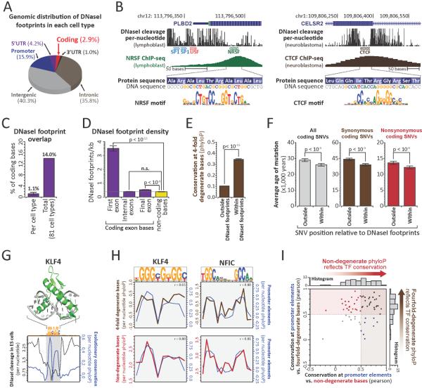
To define intersections between the regulatory and genetic codes, we generated nucleotide-resolution maps of transcription factor occupancy in 81 diverse human cell types using genomic DNaseI footprinting (19). Collectively, we defined 11,598,043 distinct 6–40bp footprints genome-wide (~1,018,514 per cell-type), 216,304 of which localized completely within protein-coding exons (~24,842 per cell-type) (Fig. 1A–B, S1A, Table S1). ~14% of all human coding bases contact a TF in at least one cell type (avg. 1.1% per cell type; Figs. 1C, S1B) and 86.9% of genes contained coding TF footprints (avg. 33% per cell type) (Figs. S1C–D).


Figure 1
The various codes in the cell Nihms-14


TFs densely populate and evolutionarily constrain protein-coding exons
(A) Distribution of DNaseI footprints. (B) Per-nucleotide DNaseI cleavage and ChIP-seq signal for coding CTCF (left) and NRSF (right) binding elements. (C) Proportion of coding bases within DNaseI footprints in each of 81 cell types (left), or any cell type (right). (D) Average footprint density within first, internal, or final coding exons (mean +/− SEM; p-value, paired t-test, n.s.: p-value> 0.1). (E) PhyloP conservation at 4FDBs within and outside footprints. (F) Estimated mutational age at all (grey), synonymous (brown) and nonsynonymous (red) coding SNVs (European) within and outside footprints (p-values per (21)) (G) Structure of DNA-bound KLF4 vs. average per-nucleotide DNaseI cleavage and evolutionary constraint at KLF4 footprints. (H) Average per-nucleotide conservation at 4FDBs (brown) and NDBs (red) overlapping KLF4 (left) and NFIC (right) footprints. (r = Pearson correlation, conservation at promoter bases vs. 4FDBs (top) or NDBs (bottom)). (I)Evolutionary constraint imparted by 63 TFs at promoter elements, 4FDBs and NDBs (Pearson correlations).


The exonic TF footprints we observed likely underestimate the true fraction of protein-coding bases that contact TFs since (i) TF footprint detection increases substantially with sequencing depth (13), and (ii) the 81 cell types sampled, though extensive, is far from complete, as we saw little evidence of saturation of coding TF footprint discovery (Fig. S2).


Figure 2
The various codes in the cell Nihms-15
Transcription factors modulate global codon biases
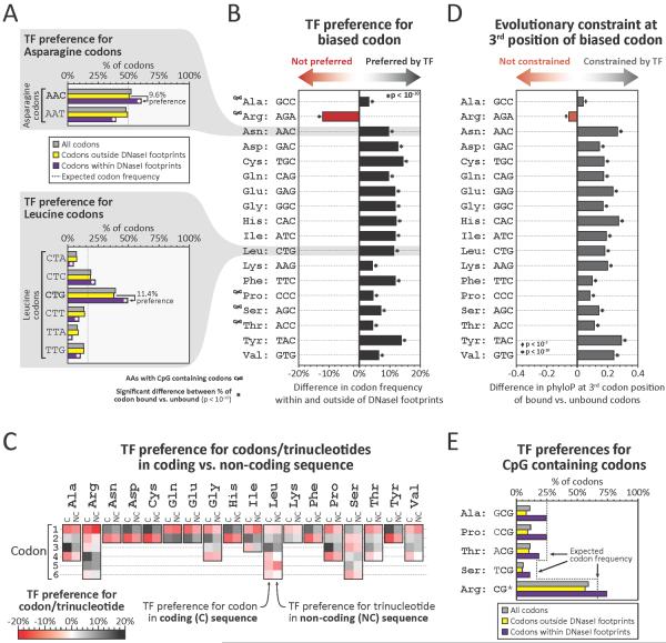
(A) Proportions of all codons (grey), or codons outside of (yellow), or within (purple) footprints, that encode asparagine (top) or leucine (bottom). Note that codons with bias (AAC for asparagine and CTG for leucine) preferentially localize within footprints. (B) Preferential footprinting of biased codons, calculated as in (A) (p-values, Pearson's chi-squared test). (C) Preferential footprinting of each codon trinucleotide in coding vs non-coding regions (C = coding, NC = non-coding). (D) Difference in average evolutionary constraint at 3rd positions of biased codons outside vs. within footprints (p-values, Mann-Whitney test). (E) Proportions of amino acids encoded by CpG-containing codons among all codons (grey), codons outside footprints (yellow), or codons within footprints (purple)


To ascertain coding footprints more completely, we developed an approach for targeted exonic footprinting via solution-phase capture of DNaseI-seq libraries using RNA probes complementary to human exons (19). Targeted capture footprinting of exons from abdominal skin and mammary stromal fibroblasts yielded ~10-fold increases in DNaseI cleavage, equivalent to sequencing >4 billion reads per sample using conventional genomic footprinting (Fig. S3A), quantitatively exposing many additional TF footprints (Fig. S3B–D). Overall, we identified an average of ~175,000 coding footprints per cell type (Fig. S1E), 7-12-fold more than conventional footprinting.


Figure 3
The various codes in the cell Nihms-16
TFs exploit and avoid specific coding features

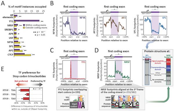
(A) Percentage of TF motifs occupied in coding vs. non-coding regions (p-values, paired t-test). (B) Density of NFYA (left), AP2 (middle) and SP1 (right) footprints relative to translated region of first coding exons. (C) (top) Density of YY1 footprints across first coding exons. (bottom) YY1 recognition sequence and corresponding amino acid sequence within YY1 footprints overlapping start codons. (D) (top left and bottom) For NRSF as per (C). (right, arrow) Protein domain annotation of first exon third-frame NRSF footprints vs. SP1 footprints. (E) TF preference (avoidance) of stop codon trinucleotides within vs. outside footprints in non-coding regions (p-values, Pearson's chi-squared test).


While coding sequences are densely occupied by TFs in vivo, the density of TF footprints at different genic positions varied widely, with many genes exhibiting sharply increased density in the translated portion of their first coding exon (Figs. 1D, S4A). By contrast, internal coding exons were as likely as flanking intronic sequences to harbor TF footprints (Fig.1D). The total number of coding DNaseI footprints within a gene was related both to the length of the gene, and to its expression level (Fig. S4B–D).


Figure 4
The various codes in the cell Nihms-17
Genetic variation in duons frequently alters TF occupancy

(A) Proportion of coding footprints overlapping a SNV in any of 81 cell-types. (B) Proportion of SNVs in duons that allelically alter TF occupancy. (C) (top) Per-nucleotide DNaseI cleavage at common nonsynonymous G→A SNV (rs8110393) in G/G and A/A homozygous cells. (bottom) Allelic SP1 occupancy in heterozygous (G/A) cells. (D) Proportion of synonymous and nonsynonymous variants in duons that allelically alter TF occupancy. (E–F) Proportion of nonsynonymous variants from (D) grouped by predicted impact of coding variant on protein function using (E) SIFT or (F) Polyphen-2. Note that none of the bins are significantly different (Fisher's exact test; n.s. indicates p-value > 0.1).

Given their abundance, we sought to determine whether exonic TF binding elements were under evolutionary selection. 4-fold degenerate coding bases are frequently used as a model of neutral (or nearly neutral) evolution (20), but may exhibit constraint when a functional signal impinges on coding sequence (11). Across the coding compartment, 4-fold degenerate bases (4FDBs) within TF footprints show significantly greater evolutionary constraint vs. non-footprinted 4FDBs (Figs. 1E, S5A–B), indicating that TF-DNA recognition constrains the third codon position.
To test for evolutionary constraint at coding footprints in modern human populations, we quantified the age of mutations arising within or outside of coding footprints using exome sequencing data from 4,298 individuals of European ancestry (Fig. S5C) and 2,217 individuals of African American ancestry (Fig. S5D) (21). This analysis revealed that mutations within coding footprints were on average 10.2% younger than those outside of footprints (Figs. 1F, S5E), signaling influence of coding TF elements on human fitness.
Strikingly, both synonymous and nonsynonymous mutations within coding footprints were significantly younger than those outside of footprints (Figs. 1F,S5E), indicating that coding TF binding constrains both codon and amino acid evolution. The genome-wide recognition sequence landscape of each TF has evolved to fit the molecular topography of its protein-DNA binding interface (13) (Fig. 1G). To study how specific TFs influence codon and amino acid choice at their recognition sites, we compared the per-nucleotide evolutionary conservation profiles of TF recognition sequences at non-coding, 4FDBs and non-degenerate coding bases (NDBs). For example, the conservation profiles at 4FBDs and NDBs at KLF4 and NFIC recognition sites closely mirror those of recognition sites in non-coding regions (promoter; Fig. 1H). As such, these TFs constrain both codon choice (via constraint on 4FDBs), and amino acid choice (via NDBs) encoded at their recognition sites. Analysis of conservation profiles for 63 TFs with prevalent occupancy within coding regions (19) showed that 73% constrain 4FDBs, and 51% constrain NDBs (Figs. 1I, S6, S7). Thus, individual TFs may influence both codon and amino acid choice.
To examine how TF binding relates to codon usage patterns, we examined -binding at preferred (biased) vs. non-preferred codons. For example, across all human proteins Asparagine is encoded by the AAC codon 52% of the time (vs. AAT, 48%), indicating a generalized 4% bias in favor of this codon. However, genome-wide, 60.4% of Asn codons within footprints are AAC, vs. only 50.8% outside of footprints (i.e., a 9.6% occupancy bias towards the preferred codon) (Fig. 2A). Strikingly, apart from Arginine (see below), for all amino acids encoded by two or more codons, the codon that is preferentially utilized genome-wide is also preferentially occupied by TFs (Fig. 2B, Table S2).
To determine whether preferential occupancy of biased codons is inherent to TF recognition sequences, we compared trinucleotide frequencies within coding vs. non-coding footprints. Trinucleotide combinations favored by TFs within coding sequence were equivalent to those favored in non-coding sequence (Fig. 3C), indicating that global TF binding preferences are directly reflected in the frequency of different codons. Notably, baseline trinucleotide frequencies within coding and non-coding sequence are largely independent of one another (Table S2). The fact that the third position of preferred codons overlapping footprints is under excess evolutionary constraint (Fig. 2D, Table S2) supports a general role for TFs in potentiating codon usage biases through the selective preservation of preferred codons.
While nearly all codon biases parallel TF recognition preferences genome-wide, Arginine, one of the 5 amino acids encoded by codons containing CpGs (4 out of 6 codons), was a notable exception. CpGs frequently occur in regulatory DNA (Table S2), yet have an elevated mutational rate (22). Consequently, although TFs may favor CpG-containing codons (Fig. 2E), and impart excess constraint thereto (Table S2), the higher mutational rate at such codons is likely incompatible with preferential utilization.
We note that codons outside footprints still exhibit usage biases (Fig. 2A andTable S2); however, it is likely that these biases also reflect the actions of TFs. Firstly, our conclusions above are drawn from a conservative and incomplete annotation of duons. Secondly, because TF trinucleotide preferences and codon biases have not changed substantially since the divergence of humans and mice (Fig. S8), preferences at any given codon may result from a TF binding element extant in some ancestral species to human. Third, codon usage bias can be exaggerated due to mutual reinforcement with other cellular factors such as tRNA abundances (23, 24). Indeed, such mechanisms could be linked to codon biases created by exonic TF occupancy through a feedback mechanism that potentiates intrinsic TF-imposed biases, resulting in both abundant and rare codons and associated tRNAs, differences in which could in turn affect protein synthesis and stability (25–27).
To analyze positional occupancy patterns of specific TFs within coding sequence, we systematically matched TF recognition sequences with footprints, providing an accurate measure of a TF's in vivo occupancy (13, 28). This analysis revealed that a subset of TFs selectively avoid coding sequences (Fig. 3A). Intriguingly, TFs involved in positioning the transcriptional pre-initiation complex, such as NFYA and SP1 (29), preferentially avoid the translated region of the first coding exon (Fig. 3A), and typically occupy elements immediately upstream of the methionine start codon (Figs. 3B, S9A). Conversely, TFs involved in modulating promoter activity, such as YY1 and NRSF, preferentially occupy the translated region of the first coding exon (30, 31) (Fig. 3A,C). These findings indicate that that the translated portion of the first coding exon may serve functionally as an extension of the canonical promoter.
More broadly, the repressor NRSF preferentially occupies and evolutionarily constrains sequences coding for leucine-rich protein domains, such as signal peptide and transmembrane domains (Figs. 3D, S9B,C). Also, TFs such as CTCF and SREBP1 preferentially occupy and constrain splice sites (Fig. S10A–D), which are otherwise generally depleted of DNaseI footprints (Fig. S10E). The above results suggest that specific protein structural and splicing features may undergo exaptation for specific regulatory purposes.
We also found that the occupancy of specific TFs within coding sequence parallels the extent of CpG methylation at their binding site (Fig. S11). This raises the possibility that gene body methylation, which is paradoxically extensive at actively transcribed genes (32, 33), may provide a tunable mechanism for thwarting opportunistic TF occupancy within coding sequence during transcription.
If TFs, through selective recognition sequences, could impose changes in protein sequence, deleterious consequences could arise if such changes resulted in a nonsense substitution. We observed that TFs generally avoid stop codons (Fig. S10E). Surprisingly, this finding extends to non-coding regions, where stop codon trinucleotides (TAA, TAG and TGA) are selectively depleted within footprints. This indicates that the global TF repertoire has been selectively purged of DNA binding domains capable of recognizing, and thus preferentially stabilizing, nonsense codons (Fig. 3E and S10F).
The high sequencing coverage provided by genomic footprinting revealed 592,867 heterozygous single nucleotide variants (SNVs) across the 81 cell type samples, and 3% of coding footprints harbored heterozygous SNVs (Fig. 4A). Functional SNVs that disrupt TF occupancy quantitatively skew the allelic origins of DNaseI cleavage fragments (13), and 17.4% of all heterozygous coding SNVs within footprints showed this signature (Figs. 4B, S12), including both synonymous and nonsynonymous variant classes (Fig. 4C). The potential of a coding SNV to disrupt overlying TF occupancy was independent of the class of variant (Fig. 4D), or whether a nonsynonymous variant was predicted to be deleterious to protein function (Fig. 4E–F).
Notably, 13.5% of common disease- and trait-associated SNVs identified by genome-wide associated studies (GWAS) (19) fall within duons (Fig. S13A). GWAS SNPs in duons encompass both synonymous (12%) and nonsynonymous (88%) substitutions (Fig. S13A), and may directly affect pathogenetic mechanisms (Fig. S13B–F, Table S3). As such, disease-associated variants within duons may compromise both regulatory and/or protein-structural functions. These findings have substantial practical implications for the interpretation of genetic variation in coding regions.
In summary, our results indicate that simultaneous encoding of amino acid and regulatory information within exons is a major functional feature of complex genomes. The information architecture of the received genetic code is optimized for superimposition of additional information (34, 35), and this intrinsic flexibility has been extensively exploited by natural selection. While TF binding within exons may serve multiple functional roles, we note that our analyses above is agnostic to these roles, which may be complex (36).


1) http://www.ncbi.nlm.nih.gov/pmc/articles/PMC3967546/

Laurence Moran on the paper : http://sandwalk.blogspot.com.br/2014/01/the-duon-delusion-and-why-transcription.html


Last edited by Otangelo on Wed Oct 18, 2023 4:08 pm; edited 2 times in total

https://reasonandscience.catsboard.com

3The various codes in the cell Empty Re: The various codes in the cell Tue Nov 24, 2015 2:25 pm

Otangelo


Admin

The transcription factor code :

The transcription factor code: defining the role of a developmental transcription factor in the adult brain.
For the human brain to develop and function correctly, each of its 100 billion neurons must follow a specific and pre-programmed code of gene expression. This code is driven by key transcription factors that regulate the expression of numerous proteins, moulding the neurons identity to create its unique shape and electrical behaviour.

Unraveling a novel transcription factor code determining the human arterial-specific endothelial cell signature
Our pioneering profiling study on freshly isolated ECs unveiled a combinatorial transcriptional code that induced an arterial fingerprint more proficiently than the current gold standard, HEY2, and this codeconveyed an in vivo arterial-like behavior upon venous ECs. 

The transcriptional regulatory code of eukaryotic cells--insights from genome-wide analysis of chromatin organization and transcription factor binding.
The term 'transcriptional regulatory code' has been used to describe the interplay of these events in the complex control of transcription. With the maturation of methods for detecting in vivo protein-DNA interactions on a genome-wide scale, detailed maps of chromatin features and transcription factor localization over entire genomes of eukaryotic cells are enriching our understanding of the properties and nature of this transcriptional regulatory code.

Human Genes Encoding Transcription Factors and Chromatin-Modifying Proteins Have Low Levels of Promoter Polymorphism: A Study of 1000 Genomes Project Data
Genome-wide analysis of histone modifications revealed that, like transcription factors, each chromatin-remodelling protein can affect transcriptional level of thousands of genes, thereby orchestrating gene activity according to intracellular conditions or external stimuli [30].
Thus, both classes of proteins are involved in the complicated process of transcriptional control, ensuring correct expression of specific genes. Both so called “transcription factor-binding regulatory code” and “histonecode” may be effectively used for prediction of gene expression activity. Moreover, these codes are redundant for predicting gene expression


Last edited by Admin on Tue Nov 20, 2018 5:03 am; edited 1 time in total

https://reasonandscience.catsboard.com

4The various codes in the cell Empty The Splicing code Tue Nov 24, 2015 2:38 pm

Otangelo


Admin

The Splicing code

https://reasonandscience.catsboard.com/t2213-the-various-codes-in-the-cell#4427

Breaking the second genetic code
splicing code’ is indeed breakable. One difficulty with understanding alternative pre-mRNA splicing is that the selection of particular exons in mature mRNAs is determined not only by intron sequences adjacent to the exon boundaries, but also by a multitude of other sequence elements present in both exons and introns. These auxiliary sequences are recognized by regulatory factors that assist or prevent the function of the spliceosome — the molecular machinery in charge of intron removal.

15% to 50% of human disease mutations affect splice site selection. Tissue-dependent splicing is regulated by trans-acting factors, cis-acting RNA sequence motifs, and other RNA features, such as exon length and secondary structure. For nearly two decades, researchers have sought to define a regulatory splicing code in the form of a set of RNA features that can account for abundances of spliced isoforms. Through detailed investigation of a small number of examples of regulated splicing, it is clear that a splicing code must account for various features that act together to control splicing. Furthermore, a code should enable the reliable prediction of the regulatory properties of previously uncharacterized exons and the effects of mutations within regulatory elements. Here we describe a method for inferring a splicing regulatory code that addresses these challenges 

Wang further observes that splicing "is a tightly regulated process, and a great number of human diseases are caused by the 'misregulation' of splicing in which the gene was not cut and pasted correctly." This implies that important protein products are produced by splicing, meaning that the splicing code plays an important functional role in cells.

After the gene is copied the transcript is edited, splicing out the introns and glueing together the exons. Not only is it a fantastically complex process, it also adds tremendous versatility to how genes are used. A given gene may be spliced into alternate sets of exons, resulting in different protein machines. There are three genes, for example, that generate over 3,000 different spliced products to help control the neuron designs of the brain.

And how does the splicing machinery know where to cut and paste? There is an elaborate code that the splicing machinery uses to decide how to do its splicing. This splicing code is extremely complicated, using not only sequence patterns in the DNA transcript, but also the shape of transcript, as well as other factors.


Recent analysis from the Encyclopedia of DNA Elements (ENCODE) project1  indicates that most of the human genome is transcribed and consists of ~60,000 genes of which ~20,000 protein-coding genes.  This number is surprisingly low given the proteomic complexity that is evident in many tissues, particularly the central nervous system (CNS). The human transcriptome is composed of a vast RNA population that undergoes further diversification by splicing. The spliceosome splices these 20,000 genes, and each gene is spliced into over 300 different protein species or variants.

This split gene architecture introduces a requirement for an intricate splicing regulatory network that consists of an 


1. array of RNA regulatory sequences
2. RNA–protein complexes and 
3. splicing factors.

Aberrant splicing is a significant cause of pathology. Detecting specific splice sites in this large sequence pool is the responsibility of the major and minor spliceosomes in collaboration with numerous splicing factors. This complexity makes splicing susceptible to sequence polymorphisms and deleterious mutations.  Indeed, RNA mis-splicing underlies a growing number of human diseases with substantial societal consequences.

This means that the spliceosome and splicing code is far more determinant than the genome in regards of creating the blueprint necessary for the production of proteins for the human body. And if aberrant splicing causes pathology, then mutations are not beneficial but generate desease. 

Do i have to explain further what that means ? 

1. http://sci-hub.tw/10.1016/j.biosystems.2017.11.002
2. http://sci-hub.tw/https://www.nature.com/articles/nrg.2015.3

A few notes on the 'species-specific' alternative splicing code


Last edited by Admin on Sun May 20, 2018 4:12 pm; edited 3 times in total

https://reasonandscience.catsboard.com

5The various codes in the cell Empty The rna binding protein binding code Tue Nov 24, 2015 4:28 pm

Otangelo


Admin

The rna binding protein binding code

A compendium of RNA-binding motifs for decoding gene regulation

The eukaryotic-wide RNA-binding protein specificity code
The RNA-binding proteins play substantial and diverse roles in the post-transcriptional regulation (PTR) of gene expression. For example, recent work estimates that 40-60% of the variability in human protein levels is controlled post-transcriptionally, suggesting that regulation by RBPs contributes as much to gene expression levels as transcription factors. In collaboration with Hughes lab, we have recently biochemically-measured RNA binding preferences for more than 200 RBPs and, because RBP RNA-specificity is highly conserved, we were able to infer motifs for nearly 5,000 more RBPs by homology. This work was recently published

https://reasonandscience.catsboard.com

6The various codes in the cell Empty Re: The various codes in the cell Tue Nov 24, 2015 4:44 pm

Otangelo


Admin

microRNA binding code

The code within the code: microRNAs target coding regions
We report here an analysis of published proteomics experiments that further support a functional role for coding region microRNA binding sites
Among possible genetic codes, the universal code has been shown to be nearly optimal for incorporating embedded information.Evidence thus far supports the conclusion that the coding regions of genes can contain additional information besides the amino acid sequence of the encoded protein, including functional microRNA binding sites.

microRNAs represent ~4% of the genes in the human genome.
This discovery suggests that the genome is far from being deciphered, and most importantly that miRNAs are likely to represent just the “tip of the iceberg” with many other small non-coding RNAs to be discovered.

https://reasonandscience.catsboard.com

Otangelo


Admin

Astonishing DNA complexity demolishes neo-Darwinism

Multiple Codes
A major outcome of the studies so far is that there are multiple information codes operating in living cells. The protein code is the simplest, and has been studied for half a century. But a number of other codes are now known, at least by inference. Cell memory code. DNA is a very long, thin molecule. If you unwound the DNA from just one human cell it would be about 2 metres long! To squash this into a tiny cell nucleus, the DNA is wound up in four separate layers of chromatin structure (as described earlier). The first level of this chromatin structure carries a ‘histone code’ that  contains information about the cell’s history (i.e. it is a cell memory).8,9 The DNA is coiled twice around a group of 8 histone molecules, and a 9th histone pins this structure into place to form what is called a nucleosome. These nucleosomes can carry various chemical modifications that either allow, or prevent, the expression of the DNA wrapped around them. Every time a cell divides into two new cells, its DNA double-helix splits into two single strands, which then each produce a new double-strand. But nucleosomes
are not duplicated like the DNA-strands. Rather, they are distributed between either one or the other of the two new DNA double strands, and the empty spaces are filled by new nucleosomes. Cell division is therefore an opportunity for changes in the nucleosomal composition of a specific DNA region. Changes can also happen during the lifetime of a cell due to chemical reactions allowing inter-conversions between the different nucleosome types. The memory effect of these changes can be that a latent capacity that was dormant comes to life, or, conversely, a previously active capacity shuts down.

Differentiation code.
In humans, there are about 300 different cell types in our bodies that make up the different tissue types (nerves, blood, muscle, liver, spleen, eyes etc). All of these cells contain the same DNA, so how does each cell know how to become a nerve cell rather than a blood cell? The required information is written in code down the side of the DNA double-helix in the form of different molecules attached to the nucleotides that form the ‘rungs’ in the ‘ladder’ of the helix. This code silences developmental genes in embryonic stem cells, but preserves their potential to become activated during embryogenesis. The embryo itself is largely defined by its DNA sequence,
but its subsequent development can be altered in response to lineage-specific transcriptional programs and environmental cues, and is epigenetically maintained.

Replication Code.
The replication code was discovered by addressing the question of how cells maintain their normal metabolic activity (which continually uses the DNA as source information) when it comes time for cell division. The key problem is that a large proportion of the whole genome is required for the normal operation of the cell—probably at least 50% in unspecialized body cells and up to 70–80% in complex liver and brain cells—and, of course, the whole genome is required during replication. This creates a huge logistic problem—how to avoid clashes between the transcription machinery (which needs to continually copy information for ongoing use in the cell) and the replication machinery (which needs to unzip the whole of the DNA double-helix and replicate a ‘zipped’ copy back onto each of the separated strands). The cell’s solution to this logistics nightmare is truly astonishing. Replication does not begin at any one point, but at thousands of different points. But of these thousands of potential start points, only a subset are used in any one cell cycle—different subsets are used at different times and places. A full understanding is yet to emerge because the system is so complex; however, some progress has been made:

The large set of potential replication start sites is not essential, but optional. In early embryogenesis, for example, before any transcription begins, the whole genome replicates numerous times without any reference to the special set of potential start sites.

The pattern of replication in the late embryo and adult is tissue-specific. This suggests that cells in a particular tissue cooperate by coordinating replication so that while part of the DNA in one cell is being replicated, the corresponding part in a neighbouring cell is being transcribed. Transcripts can thus be shared so that normal functions can be maintained throughout the tissue while different parts of the DNA are being replicated.

DNA that is transcribed early in the cell division cycle is also replicated in the early stage (but the transcription and replication machines are carefully kept apart). The early transcribed DNA is that which is needed most often in cell function. The correlation between transcription and replication in this early phase allows the cell to minimize the ‘downtime’ in transcription of the most urgent supplies while replication takes place There is a ‘pecking order’ of control. Preparation for replication may take place at thousands of different locations, but once replication does begin at a particular site, it suppresses replication at nearby sites so that only one copy of the DNA is made. If transcription happens to occur nearby, replication is suppressed until transcription is completed. This clearly demonstrates that keeping the cell alive and functioning properly takes precedence over cell division.

There is a built-in error correction system called the ‘cell-cycle checkpoints’. If replication proceeds without any problems, correction is not needed. However, if too many replication events occur at once the potential for conflict between transcription and regulation increases, and/or it may indicate that some replicators have stalled because of errors. Once the threshold number is exceeded, the checkpoint system is activated, the whole process is slowed down, and errors are corrected. If too much damage occurs, the daughter cells will be mutant, or the cell’s self-destruct mechanism (the apoptosome) will be activated to dismantle the cell and recycle its components.

An obvious benefit of the pattern of replication initiation being never the same from one cell division to the next is that it prevents accumulation of any errors that are not corrected. The exact location of the replication code is yet to be pinpointed, but because it involves transcription factors gaining access to transcription sites, and this is known to
be controlled by chromatin structure, then the code itself is probably written into the chromatin structure.


https://creation.com/images/pdfs/tj/j21_3/j21_3_111-117.pdf

https://reasonandscience.catsboard.com

8The various codes in the cell Empty The tubulin code Wed Jan 13, 2016 6:34 pm

Otangelo


Admin

The tubulin code

The α- and β-tubulin heterodimer – the building block of microtubules – undergoes multiple post-translational modifications (PTMs) (Table above). The modified tubulin subunits are non-uniformly distributed along microtubules. Analogous to the model of the ‘histone code’ on chromatin, diverse PTMs are proposed to form a biochemical ‘tubulin code’ that can be ‘read’ by factors that interact with microtubules (Verhey and Gaertig, 2007).

Who are the Interpreters of the Tubulin Code?

A major implication of the tubulin code is that PTMs influence the recruitment of protein complexes (microtubule effectors), which in turn contribute to microtubule-based functions. Three major classes of microtubule binding proteins can be considered as interpreters of the tubulin code. First, microtubule associated proteins (MAPs) such as Tau, MAP1 and MAP2 that bind statically along the length of microtubules. Second, plus end tracking proteins (+TIPs) that bind in a transient manner to the plus-ends of growing microtubules. And third, molecular motors that use the energy of ATP hydrolysis to carry cargoes along microtubule tracks.


This is a relevant and amazing fact , and raises the question of how the " tubulin code "  beside the several other codes in the cell emerged. Once more this shows that intelligence was involved in creating these amazing biomolecular structures and specified complex coded instructing patterns  , since the formation of  coded information has always shown to be able only to be produced by intelligent minds. Furthermore: What good would the tubulin code be for, if no specific goal was in mind, that is, it acts as emitter , and if there is no destination of the information, there is no reason of the code to exist in the first place. So both, sender and receiver, must exist first as hardware, that is the microtubule with the post transcriptional modified tubulin units in a specified coded conformation, and the the receiver, which can be Kinesin or Myosin motor proteins, which are directed to the right destination, or other proteins.


Last edited by Admin on Wed Jan 04, 2017 5:14 am; edited 2 times in total

https://reasonandscience.catsboard.com

9The various codes in the cell Empty The Glycan or Sugar Code Wed Jan 13, 2016 6:54 pm

Otangelo


Admin

The Glycan or Sugar Code 1
Carbohydrates are essential for all forms of life, but the largest variety of their functions is now found in higher eukaryotes. The majority of eukaryotic proteins are modified by cotranslational and posttranslational attachment of complex oligosaccharides (glycans) to generate the most complex epiproteomic modification – protein glycosylation. 

Most proteins are glycosylated: That is, complex carbohydrates are chemically bonded to them to generate enormous diversity in protein functions. [5] Since carbohydrate molecules are branched, they carry many more orders of magnitude of information than linear molecules such as DNA and RNA. This has been called the “sugar code,” and although it is highly specified it is largely independent of DNA sequence information.  A third biochemical alphabet forming code words with an information storage capacity second to no other substance class in rather small units (words, sentences) is established by monosaccharides (letters). As hardware oligosaccharides surpass peptides by more than seven orders of magnitude in the theoretical ability to build isomers, when the total of conceivable hexamers is calculated.  A genetic program is not sufficient for embryogenesis: biological information outside of DNA is needed to specify the body plan of the embryo and much of its subsequent development. Some of that information is in cell membrane patterns, which contain a two-dimensional code mediated by proteins and carbohydrates. 2


According to the most widely held modern version of Darwin’s theory, DNA mutations can supply raw materials for morphological evolution because they alter a genetic program that controls embryo development. Yet a genetic program is not sufficient for embryogenesis: biological information outside of DNA is needed to specify the body plan of the embryo and much of its subsequent development. Some of that information is in cell membrane patterns, which contain a two-dimensional code mediated by proteins and carbohydrates. These molecules specify targets for morphogenetic determinants in the cytoplasm, generate endogenous electric fields that provide spatial coordinates for embryo development, regulate intracellular signaling, and participate in cell–cell interactions. Although the individual membrane molecules are at least partly specified by DNA sequences, their two-dimensional patterns are not. Furthermore, membrane patterns can be inherited independently of the DNA. I review some of the evidence for the membrane code and argue that it has important implications for modern evolutionary theory.

1) http://www.ncbi.nlm.nih.gov/pubmed/10798195

2) https://reasonandscience.catsboard.com/t2071-carbohydrates-and-glycobiology-the-3rd-alphabet-of-life-after-dna-and-proteins?highlight=glycan+code


Last edited by Otangelo on Wed Nov 11, 2020 1:40 pm; edited 3 times in total

https://reasonandscience.catsboard.com

10The various codes in the cell Empty Re: The various codes in the cell Sat Jan 16, 2016 2:44 pm

Otangelo


Admin

https://en.wikipedia.org/wiki/Edward_Trifonov...


Trifonov advocates[19]:4 the notion that biological sequences bear many codes contrary to the generally recognized one genetic code (coding amino acids order). He was also the first one to demonstrate[20] that there are multiple codes present in the DNA. He points out that even so called non-coding DNA has a function, i.e. contains codes, although different from the triplet code.

.

Trifonov recognizes[19]:5–10 specific codes in the DNA, RNA and proteins:

in DNA sequences
chromatin code (Trifonov 1980) is a set of rules responsible for positioning of the nucleosomes.
------------------------------------------------------
in RNA sequences

RNA-to-protein translation code (triplet code)
Every triplet in the RNA sequence corresponds (is translated) to a specific amino acid.

splicing code
is a code responsible for RNA splicing; still poorly identified.

framing code (Trifonov 1987)
The consensus sequence of the mRNA is (GCU)n which is complementary to (xxC)n in the ribosomes. It maintains the correct reading frame during mRNA translation.

translation pausing code (Makhoul & Trifonov 2002)
Clusters of rare codons are placed in the distance of 150 bp from each other. The translation time of these codons is longer than of their synonymous counterparts which slows down the translation process and thus provides time for the fresh-synthesized segment of a protein to fold properly.

------------------------------------------------------
in protein sequences

protein folding code (Berezovsky, Grosberg & Trifonov 2000)
Proteins are composed of modules. The newly synthesized protein is folded a module by module, not as a whole. 

fast adaptation codes (Trifonov 1989)
are present in all three types of biological sequences. They are represented by tandem repeats (AB...MN)n. The number of repetitions (n) can change in the cell genome as a response to stress which may (or may not) help the cell to adapt to the environmental pressure. 

------------------------------------------------------
codes of evolutionary past

binary code (Trifonov 2006)
The first ancient codons were GGC and GCC from which the other codons have been derived by series of point mutations. Nowadays, we can see it in modern genes as "mini-genes" containing a purine at the middle position in the codons alternating with segments having a pyrimidine in the middle nucleotides.

genome segmentation code (Kolker & Trifonov 1995)
Methionines tend to occur every 400 bps in the modern DNA sequences as a result of fusion of ancient independent sequences.

THE CODES CAN OVERLAP EACH OTHER SO THAT UP TO 4 DIFFERENT CODES CAN BE IDENTIFIED IN ONE DNA SEQUENCE (specifically a sequence involved in a nucleosome). According to Trifonov, other codes are yet to be discovered.


****THE CODES CAN OVERLAP EACH OTHER****

https://reasonandscience.catsboard.com

11The various codes in the cell Empty Code Biology Fri Apr 22, 2016 10:28 pm

Otangelo


Admin

Code Biology

Marcello Barbieri , page 14:

The Signal Transduction Codes Signal transduction is the process by which cells transform the signals from the environment, called first messengers, into internal signals, called second messengers. First and second messengers belong to two independent worlds because there are literally hundreds of first messengers (hormones, growth factors, neurotransmitters, etc.) but only four great families of second messengers (cyclic AMP, calcium ions, diacylglycerol and inositol trisphosphate) (Alberts et al. 2007). The crucial point is that the molecules that perform signal transduction are true adaptors. They consist of three subunits: a receptor for the first messengers, an amplifier for the second messengers, and a mediator in between (Berridge 1985). This allows the transduction complex to perform two independent recognition processes, one for the first messenger and the other for the second messenger. Laboratory experiments have proved that any first messenger can be associated with any second messenger, which means that there is a potentially unlimited number of arbitrary connections between them. In signal transduction, in short, we find all three essential components of a code:

(1) two independents worlds of molecules (first messengers and second messengers), 
(2) a set of adaptors that create a mapping between them, and 
(3) the proof that the mapping is arbitrary because its rules can be experimentally changed.

The cells that evolved new codes, such as splicing codes, cytoskeleton codes, compartment codes, histone code and so on, became eukarya and have generated increasingly complex cellular structures.

Before the origin of the genetic code, the common ancestor was engaged in evolving coding rules and was therefore a code exploring system.


Had the common ancestor not have already to have a sophisticated code and cipher system set up, and so the molecular machines required for transcription, translation and replication ? And why at all would random chance explore codes to set them up ? 

After the origin of the code, however, Introduction xv no other modification in coding rules was allowed and the cell became a code conservation system. 

There are 18 different Cell codes known. Why would natural mechanisms stop to allow other , different codes ? 

Another part of the ancestral cells, however, maintained the potential to evolve the rules of different codes and behaved as new code exploring, or code generating, systems. In the early Eukarya, for example, the cells had a code conservation part for the genetic code, but also a code exploring part for the splicing code, and this tells us something important about life.

Well, the splicing code has nothing to do with the genetic code.

The origin of the first cells was based on the ability of the ancestral systems to generate the rules of the genetic code

What systems were this, why  would that " system " generate a code and its rules at all ?

Another outstanding implication of the existence of organic codes in Nature comes from the fact that any code involves meaning and we need therefore to introduce in biology, with the standard methods of science, not only the concept of biological information but also that of biological meaning. The study on the organic codes, in conclusion, is bringing to light new mechanisms that operated in the history of life and new fundamental concepts. It is an entirely new field of research, the exploration of a vast and still largely unexplored dimension of the living world, the real new frontier of biology.

A Gallery of Organic Codes 
The Apparatus of Protein Synthesis
The Genetic Code 
Stereochemistry and Arbitrariness 
The Splicing Codes 
The Metabolic Code
The Signal Transduction Codes 
The Signal Integration Codes 
The Histone Code 
Is the “Histone Code” an Organic Code?
The Tubulin Code 
The Sugar Code 
The Glycomic Code


Last edited by Admin on Sun Jul 03, 2016 12:37 pm; edited 3 times in total

https://reasonandscience.catsboard.com

12The various codes in the cell Empty The Metabolic Code Fri Apr 22, 2016 11:01 pm

Otangelo


Admin

The Splicing Codes

Code Biology

Marcello Barbieri , page 14

The primary transcripts of the genes are often transformed into messenger RNAs by removing some RNA pieces (called introns) and by joining together the remaining pieces (the exons). This cutting-and-sealing operation, known as splicing, is a true assembly because exons are assembled into messengers, and we need therefore to find out if it is a catalyzed assembly (like transcription) or a codified assembly (like translation). In the first case splicing would require only catalysts (comparable to RNA-polymerases), whereas in the second case it would need an assembly machine and a set of adaptors (comparable to ribosome and tRNAs). These parallels immediately suggest that splicing is a codified process because it is implemented by structures that are very much comparable to those of protein synthesis. The splicing bodies, known as spliceosomes, are huge molecular machines like ribosomes, and employ small molecules, known as small-nuclear- RNAs (snRNAs) which are comparable to tRNAs. The similarity, however, goes much deeper than that because splicing is carried out by molecular structures that are true adaptors. They perform two independent recognition processes, one for the beginning and one for the end of each exon, thus creating a specific correspondence between primary transcripts and messenger RNAs. Splicing, in other words, is a codified process based on adaptors and takes place with sets of rules that have been referred to as splicing codes. It must be underlined, however, that there are two outstanding complications in splicing. One is the fact that the order in which the exons are joined together can be shuffled in various ways, an operation, called alternative splicing, that allows many species to generate a whole family of variant proteins from the same gene. The expression of these proteins, furthermore, can change from one tissue to another and in different stages of embryonic development, thus enormously increasing the protein variety that can be associated to a gene. Alternative splicing has in this way a powerful role in the generation of biological complexity, and splicing mistakes often have pathological effects; it has been estimated that they account for about one fifth of all inherited diseases.

The other great complication of splicing is the fact that many introns carry sequences that are similar to exons but translate into nonsense and for this reason are called pseudo exons or pseudo genes. They would create havoc if incorporated into mRNAs and the splicing machinery needs the means to differentiate real exons from pseudo ones. The result is that real exons contain internal identity marks that are known as exonic splicing enhancers (ESEs) and exonic splicing silencers

Question: Had these indentity marks not have to be present in the process right from the beginning ? Would the absence of these or ones not fully developed  not make the process impossible to happen without mistakes ? 

The presence of these marks, in turn, means that the adaptors of the splicing codes are not single molecules but combinations of molecules because they must be able to recognize not only the beginning and the end of the real exons, but also their internal identity marks.

This makes the whole process even more impossible to emerge in a stepwise manner, since both, the recognition of the beginning and the end of the exons is required, that means, the genome needs to have the start and stop signals at the right place, and the molecular machines, programmed to recognize the signals must be in place, fully developed, and fully programmed, and the identity marks are required beside the hardware as well. Furthermore, this seems to be one more irreducible complex system , since both, the software, and the hardware, had to be in place, just right , fully developed and programmed since the beginning. 

The actual deciphering of the splicing codes has already started but it is taking considerably longer than that of the genetic code because it is incredibly more complex. Let us keep in mind that the discovery of the genetic code has been facilitated by two particularly favourable features. More precisely, by the fact that 

(1) the adaptors are single molecules (the tRNAs) and 
(2) the coding units form a closed set (64 codons and 20 amino acids).

 In the case of splicing, instead, the adaptors are combinations of molecules (combinatorial codes), and the domain (or alphabet) of the codes is open and potentially unlimited. The overall complexity of splicing is such that the most practical way of discovering its codes is by building computational models that are capable of predicting new splicing rules on the basis of existing data. Such models have already started appearing in the literature , and represent our first glimpse of the rules of the splicing codes.

The Metabolic Code 

This is the first organic code that came to light after the discovery of the genetic code. It was described in Science, in 1975, by Gordon Tomkins, a professor of biochemistry at the University of San Francisco. Tragically, Tomkins died that very year, aged 49, from a brain tumour, and apparently his idea died with him. Recently, however, there has been an attempt to rescue his work from oblivion (Swan and Goldberg 2010) and here we will try to show that such attempt is amply justified. Tomkins investigated the evolution of metabolism and started from the need of the ancestral cells to obtain energy. “Since both nucleic acid and protein synthesis are endergonic reactions, primordial cells were almost certainly endowed with the capacity to capture the necessary energy from the environment and to transform it into usable form, presumably ATP (adenosine triphosphate). The biosynthetic capabilities of primitive cells were, however, probably quite limited ::: survival would therefore have required the evolution of regulatory mechanisms that could maintain a relatively constant intracellular environment in the face of changes in external conditions” (Tomkins 1975). Granted this basic need of the cells to evolve regulatory mechanisms, Tomkins distinguished between two types of regulation that he called simple and complex, elationship (positive or negative) between the components of a metabolic circuit, and the end products affecting their own metabolism. 

Complex regulation is characterized by two new entities that Tomkins called symbols and domains. In order to illustrate them, Tomkins made the example of molecules that are accumulated inside a cell as a consequence of a particular environment and become a symbol of that environment. In most microorganisms, for example, cyclic AMP is accumulated as a result of carbon starvation and becomes a symbol of that deficiency. Another example is ppGpp (guanosine50 -diphosphate30 - diphosphate) that accumulates as a result of amino acid starvation and represents a symbol of that condition. These molecules are symbols because they bear no structural relationship to the molecules that promote their accumulation (cyclic AMP, for example is accumulated as a result of glucose starvation, but it is not a chemical analog of glucose). This is what suggested to Tomkins the existence of a metabolic code. “Since a particular environmental condition is correlated with a corresponding intracellular symbol, the relationship between the extra- and intracellular events may be considered as a metabolic code in which a specific symbol represents a unique state of the environment.” Tomkins went on to show how metabolic coding in unicellular organisms might have evolved into the endocrine system of the metazoa, and described what happens in the slime mold Dictyostelium discoideum. “Given sufficient nutrients, this organism exists as independent myxamoebas. Upon starvation, they generate cyclic AMP and release it into the surrounding medium. This substance serves as a chemical attractant that causes the aggregation of a large number of myxamoebas to form a multicellular slug. In this case, as in E. coli, cyclic AMP acts as an intracellular symbol of carbon-source starvation. In addition, however, the cyclic nucleotide is released from the Dictyostelium cells in which it is formed and diffuses to other nearby cells, promoting the aggregation response. Cyclic AMP thus acts in these organisms both as an intracellular symbol of starvation and as a hormone which carries this metabolic information from one cell to another.”

Hormones, according to Tomkins, evolved in order “to carry information from sensor cells in direct contact with the environment, to more sequestered responder cells. Specifically, the metabolic state of a sensor cell, represented by the levels of its intracellular symbols, is encoded by the synthesis and secretion of corresponding levels of hormones. When hormones reach the responder cells, the metabolic message is decoded into corresponding primary intracellular symbols. In this way, endocrine cells act as both sensors and responders, that is, intermediates in the transmission of metabolic information from primary sensor cells to the tissues in which the final chemical responses take place.”

The Signal Transduction Codes

Living cells react to many physical and chemical stimuli from the environment, and in general their reactions consist in the expression of specific genes. We need therefore to understand how the environment interacts with the genes, and the turning point, in this field, came from the discovery that the external signals (known as first messengers) never reach the genes. They are invariably transformed into a different world of internal signals (called second messengers) and only these, or their derivatives, reach the genes. In most cases, the molecules of the external signals do not even enter the cell and are captured by specific receptors of the cell membrane, but even those that do enter (some hormones) must interact with
intracellular receptors in order to influence the genes (Sutherland 1972). The transfer of information from environment to genes takes place therefore in two distinct steps: one from first to second messengers, called signal transduction, and a second path from second messengers to genes which is known as signal integration. The surprising thing about signal transduction is that there are literally hundreds of first messengers (ions, nutrients, hormones, growth factors, neurotransmitters, etc.) whereas the second messengers belong to only four molecular families: cyclic AMP or GMP, calcium ions (Ca2+), inositol trisphosphate (IP3), and diacylglycerol (DAG) (Alberts et al. 2007). First and second messengers, in other words, belong to two very different worlds, and this suggests immediately that signal transduction may be based on organic codes. This is reinforced by the discovery that there is no necessary connection between first and second messengers, because it has been proven that the same first messengers can activate different types of second messengers, and that different first messengers can act on the same type of second messengers (Alberts et al. 2007). The only plausible explanation is that signal transduction is based on organic codes, but of course one would like a direct proof. The signature of an organic code, as we have seen, is the presence of adaptors and the transmembrane receptor proteins of signal transduction do have the defining characteristics of the adaptors. 

The transduction system consists of at least three types of molecules: 

a receptor for the first messengers, 
an amplifier for the second messengers and 
a mediator in between (Berridge 1985). 

This transmembrane system performs two independent recognition processes, one for the first and the other for the second messenger, and the two steps are connected by the bridge of the mediator. This connection, on the other hand, could be implemented in countless different ways since any first messenger can be coupled with any second messenger, and this makes it imperative to have a selection in order to guarantee biological specificity. 

In signal transduction, in short, we find the three defining features of a code: 

(1) two independents worlds of objects (first messengers and second messengers), 
(2) a potentially unlimited number of arbitrary connections produced by adaptors, and 
(3) a set of coding rules (a selection of the adaptors) that ensures the specificity of the correspondence. 

The effects that external signals have on cells, in short, do not depend on the energy or the information that they carry, but on the meaning that cells give them with sets of rules that have been referred to as signal transduction codes (Barbieri 1998, 2003). One may wonder at this point why signal transduction codes are never mentioned in biochemistry books despite the fact that the their molecules are true adaptors. The problem here is that the study of signal transduction started when organic codes were not known, and it has always been assumed a priori that in this process there is no need for them. A code, in short, has not been found simply because it has never been looked for. The genetic code, on the contrary, was predicted on theoretical grounds, and it was discovered precisely because experiments were devised with the specific purpose to look for it.

The Signal Integration Codes

We have seen that there are only four families of second messengers in the cell, and yet the reactions that they set in motion can pick up an individual gene among tens of thousands. How this is achieved is still a mystery, but some progress has been made. Perhaps the most illuminating discovery, so far, is that second messengers do not act independently. Calcium ions and cyclic-AMPs, for example, have effects that in some occasions reinforce each other whereas in others are mutually exclusive. The cell, in short, can combine its internal signals in countless different ways, and it is precisely this combinatorial ability that explains why a small number of second messengers can generate an extraordinarily high number of specific genetic responses. The activation of second messengers, in other words, sets in motion a cascade of reactions that normally ends with the expression of a target gene, and again we need to understand if they are normal catalized reactions or if at least some of them are based on the rules of a code. One of the most interesting clues, in this field, is the fact that signalling molecules have in general more than one function. Epidermal growth factor, for example, stimulates the proliferation of fibroblasts and keratinocytes, but it has an antiproliferative effect on hair follicle cells, whereas in the intestine it is a suppressor of gastric acid secretion. Other findings have proved that all growth factors can have three distinct functions, with proliferative, anti-proliferative, and proliferationindependent effects. They are, in short, multifunctional molecules. In addition to growth factors, it has been found that many other molecules have multiple functions. Adrenaline, for example, is a neurotransmitter, but it is also a hormone produced by the adrenal glands to spring the body into action by increasing the blood pressure, speeding up the heart and releasing glucose from the liver. Acetylcholine is another common neurotransmitter in the brain, but it also act on the heart (where it induces relaxation), on skeletal muscles (where the result is contraction), and in the pancreas (which is made to secrete enzymes). Cholecystokinin is a peptide that acts as a hormone in the intestine, where it increases the bile flow during digestion, whereas in the nervous system is a neurotransmitter. Encephalins are sedatives in the brain, but in the digestive system are hormones which control the mechanical movements of food. Insulin is universally known for lowering the sugar levels in the blood, but it also controls fat metabolism and in other less known ways it is affecting almost every cell of the body. The discovery of multifunctional molecules suggests that their function is not decided solely by their structure, but also by the context in which they find themselves. What matters, in other words, is not their ability to catalize a specific reaction, but the fact that they are employed as molecular signs that can be given one meaning in a certain context and a different meaning in another one. A second finding that points to the existence of codes in signal integration is the fact that the regulation processes set in motion by second messengers are strongly conserved in evolution, and yet the actual reactions involved have undergone great changes in the history of life. The regulation of cellular energy homeostasis, for example, has been highly conserved from yeast to man, with the key role being played by a protein kinase that is called AMPK in animals and Snf1 in yeast. Despite this overall conservation, it has been found that an evolutionary divergence of about 150 million years between two species of budding yeasts (Saccharomyces cerevisiae and Kluyveromyces lactis) has produced substantial differences in their Snf1 regulatory networks. Again, what seems to matter in these regulation processes is not a specific set of catalysts, but a set of rules that can be implemented in many different ways. The information carried by first messengers, in conclusion, undergoes two great transformations in its journey towards the genes. First, it is transformed into internal messengers with the rules of the signal transduction codes, and then it is channelled along complex three-dimensional circuits that integrate it with other signals according to the rules of one or more signal integration codes.

The Histone Code

The classic double helix described by Watson and Crick has a width of 2 nm (two millionths of a millimeter), but in eukaryotes many segments of this filament are folded around groups of eight histone proteins and form blocks, called nucleosomes, that give to the filament a ‘beads-on-a-string’ appearance. This string, called chromatin, is almost six times thicker than the double helix and is further folded into spirals of nucleosome groups, called solenoids, that arrange it in fibers of increasing thickness and ultimately into the 600 nm fiber of the chromosome. These multiple foldings allow the eukaryotic cells to pack their long chromosomes into the tiny space of their nuclei, and for this reason it was initially assumed that the histones have a purely packaging role. The experimental data, however, have shown that the ‘tails’ of the histones (the parts that protrude from the surface of the nucleosomes) are subject to a wide variety of post-translational modifications (in particular acetylation, methylation and phosphorylation) that have highly dynamic roles and are involved in the activation or repression of gene activity. The histone tails represent about 25–30 % of the histone mass, and their posttranslational modifications can alter the chromatin either directly or indirectly. The direct modifications are those that physically open or close the molecular space (in particular the electrostatic barrier) that surrounds the genes and in this way control the transit of DNA-binding proteins. Several discoveries, however, have shown that the most frequent effects are obtained by indirect mechanisms. In these cases, the modified histone tails provide ‘marks’ on the surface of the nucleosomes that are recognized by specialized effector proteins which set in motion chains of biological reactions that eventually end in the activation or the repression of specific gene. A crucial breakthrough, is this field, was the discovery that the post-translational modifications of the histones do not act individually. Most of them are involved in both the activation and the repression of genes (the phosphorilation of histone H3, for example, takes part in the condensation as well as in the decondensation of chromatin), which means that the final result is due to a combination of histone marks rather than a single one. This led David Allis and colleagues to propose that the histone marks operate in combinatorial groups, like letters that are put together into the words of a molecular ‘language’ that was referred to as histone code. The same concept was independently proposed by Brian Turner who argued that there is an epigenetic code at the heart of the regulation mechanisms that are initiated by histone tail modifications. Turner pointed out that these modifications are epigenetic because they operate in addition to genetic changes, and underlined that they have both short-term and long-term effects. The shortterm modifications change rapidly in response to external signals and represent a mechanism by which the genome quickly responds to the environment. The long-term modifications, instead, are those that are put in place at early stages of embryonic development and allow the transcription or the silencing of specific genes at more advanced stages. The existence of long-term effects was revealed by the discovery that many histone modifications survive the trauma of mitosis and are transmitted to the daughter cells. This is particularly important in embryonic development where the cells must perpetuate their state of differentiation into distinct tissues. The histone modifications, in other words, provide a mechanism of cell memory, in the sense that they enable the cells to ‘remember’ their specific pattern of gene expression for many generations. It has been shown, for example, that the expression of Hox genes in embryonic development is regulated by histone modifications . Another example of long-term effects is provided by the histone modifications that allow neural cells to generate faster action potentials the more they are used, making the transmission of action potentials increasingly easier. Today, in conclusion, a large number of data support the idea that the regulation of genetic activity by histone modifications plays a fundamental role in all eukaryotes and is based on the rules of a combinatorial code that has become known as ‘histone code’.

Is the “Histone Code” an Organic Code?

This question is the title of a paper where Stefan Kühn and Jan-Hendrik Hofmeyr described the results of a research project dedicated to find out whether or not the histone code has all the essential characteristics of an organic code. The prototype example of the genetic code shows that an organic code requires three things: 

(1) two independent molecular worlds, 
(2) a set of molecular adaptors that create a mapping between them, and 
(3) the demonstration that the mapping is arbitrary because its rules can be changed. Kühn and Hofmeyr tested the histone code in respect to all these points.

1. The Two Independent Worlds of the Histone Code
An organic code is a mapping between organic signs and organic meanings, and in many cases signs and meanings are both organic molecules. The genetic code, for example, is a mapping between codons and amino acids, whereas the signal transduction code is a mapping between first and second messengers. Kühn and Hofmeyr, however, pointed out that the organic meanings can be biological effects rather than molecules. In principle this may not seem an extension of the original definition because biological effects are necessarily implemented by molecules, but in practice it is a very useful generalization because there are cases in which a biological function is an experimental reality even when its molecular components are not fully known. And this is precisely the case in the histone code, where the organic signs are groups of histone modifications and the organic meanings are biological reactions that promote the activation or the repression of specific genes. The histone code, in other words, is a mapping between two independent worlds.

2. The Adaptors of the Histone Code
The effector proteins of the histone code are the molecules that establish a bridge between organic signs and organic meanings, but in order to prove that they are true adaptors it is necessary to show that they operate independently on signs and meanings. Kühn and Hofmeyer underlined that this is precisely what happens because the effector proteins have two distinct domains: one that recognizes histone modifications and a different type that initiates biological reactions. It has been shown, for example, that the acetylated lysines are specifically recognized only by the bromodomains of the effector proteins . The methylated amino acids are recognized by a greater variety of domains but again each recognition step is absolutely specific . The effector  proteins, in other words, perform two independent recognition processes on signs and meanings and are therefore true adaptors.

3. The Arbitrariness of the Histone Code
An organic code is arbitrary when its rules are not dictated by physical necessity and in this case it must be possible, at least in principle, to exchange the part of an adaptor that recognizes an organic sign with a different one and show that the modified adaptor associates the old organic meaning to the new sign. Kühn and Hofmeyr noticed that the experimental data support this possibility because there is evidence that the chromodomains of the effector proteins can be interchanged. The histone code, in conclusion, did pass the three tests and Kühn and Hofmeyr ended their paper with these words: “Although we probably do not yet know the complete histone code, we have more than enough information to be able to recognize the histone code as a bona fide organic code.”

Nucleosome Positioning Codes 1

DNA molecules are much longer than the cells that contain them. This requires their compaction, which introduces also an opportunity: the regulation of transcription through a differentiated fashion of DNA packaging. In eukaryotes DNA molecules can guide their own packaging into nucleosomes by having the desired mechanical properties (stiffnesses and intrinsic curvature) written into their base-pair (bp) sequence. This has been referred to as the “nucleosome positioning code” . Nucleosomes are the fundamental packaging units of eukaryotic DNA, where 147 bp are wrapped in a 1 3/4 left-handed superhelical turn around an octamer of histone proteins. As the DNA is strongly deformed when wrapped around the histones, sequence-dependent geometrical and mechanical properties could—at least locally—overrule other effects that also influence nucleosome positioning like the presence of proteins that compete for the same DNA stretch or the action of chromatin remodellers.
Multiplexing Genetic and Nucleosome Positioning Codes: A Computational Approach
https://journals.plos.org/plosone/article?id=10.1371%2Fjournal.pone.0156905&fbclid=IwAR0cA3bLPEfcmNaQ8AlA1CLft5yd2kxKi06c3Jo59XVyZFJDJ5ida_XWKTc


The Tubulin Code

Tubulin is the major component of the microtubules, the filaments that form an internal scaffolding in all eukaryotic cells and give origin to organelles such as cilia, centrioles, basal bodies and the mitotic spindle. Most microtubules are in a state of rapid turnover by dynamic instability and alternate very quickly between growth and shrinkage. Within the cell, however, there is also a population of microtubules that are relatively stable, in the sense that their turnover is measured in hours rather than minutes. The function of the stable microtubules is still not completely known, but there are clear indications that they are involved in the morphogenesis of the eukaryotic cell. What is certain, is that the stable microtubules undergo a variety of post-translational modifications (PTMs) that have been strongly conserved  because they are found in all eukaryotic taxa. These PTMs consist in processes like acetylation, phosphorylation, polyglutamylation, polyglycylation, detyrosination, and palmitoylation that act preferentially on stable microtubules. They have been studied with various tests on purified tubulin, but the experiments have failed to detect any direct effect of the PTMs on the dynamics of the microtubules. This means that PTMs do not act by changing directly the intrinsic properties of the microtubules, but rather by providing combinatorial signals for the recruitment of proteins that interact with the microtubules. Different combinations of PTMs, in other words, act like signposts that specify the properties that stable microtubules are going to have in different regions of the cell or in different periods of the cell cycle. To this set of signposts that operate on stable microtubules, Kristen Verhey  and Jacek Gaertig  gave the name of Tubulin code. Any organic code, as we have seen, requires molecules that act like adaptors between two different domains. Verhey and Gaertig have called these molecules ‘interpreters’, and have identified three major classes of microtubule binding proteins that can be considered interpreters of the tubulin code:

 “First, microtubule associated proteins (MAPs) such as Tau, MAP1 and MAP2 that bind statically along the length of microtubules. 
Second, plus-end tracking proteins (+TIPs) that bind in a transient manner to the plus-ends of growing microtubules. 
And third, molecular motors that use the energy of ATP hydrolysis to carry cargoes along microtubule tracks.” 

Verhey and Gaertig have also called attention to a unique characteristic of the tubulin code. Many epigenetic modifications are transmitted from one generation to the next, but this does not usually happen in the tubulin world: “Some microtubule-based organelles (e.g., centrosomes and basal bodies) are inherited by a template-driven mechanism but there is no evidence that the template organelle directly influences the PTM pattern in the new organelle. Rather, the PTM pattern is recreated in the newly formed organelle in a gradual manner : : : Other microtubulebased structures, such as cytoplasmic microtubules, the mitotic spindle and cilia, are formed de novo mostly, if not entirely, from unmodified tubulin heterodimers. Thus, in case of both template-dependent and template-independent microtubular structures, PTM patterns are probably recreated without a direct influence of preexisting PTMs.” The existence of the tubulin code, in conclusion, is based on sound experimental evidence but the actual deciphering of its rules is still at a preliminary stage and requires a detailed understanding of how the PTMs influence the recruitment of proteins and regulate the functions of the stable microtubules.

The Sugar Code


For a long time, sugars have been regarded as molecules that provide energy (mostly in the form of glucose and glycogen) or structural support (like cellulose in plants), but molecular biology has shown that they also have a third outstanding function: by binding to proteins they generate glycoproteins, molecules that take part in countless communication processes in and between cells. The addition of sugars to proteins is a post-translational modification, called glycosylation, that greatly expands the potentialities of many protein families and gives origin to glycoproteins that perform a wide variety of functions. Some operate on the cell membrane and act as antennae for receiving molecular signals or as docking sites for importing compounds. Other glycoproteins take part in cell-to-cell interactions, for example in sperm-oocyte attachment, in bacteria-to-cell relationships and in the aggregation of platelets. A third family operates in the immune system where glycoproteins interact with antigens, recognize white blood cells, and take part in the major histocompatibility complex (MHC). Yet another family is that of the glycoproteins that act as hormones, like human corionic gonadotropin (HCG), thyroid-stimulating hormone (TSH) and erythropoietin (EPO). Then there are glycoproteins that have protective functions (mucins), some that are involved in transport (transferrin) and others that act as enzymes (alkaline phosphatase). The key point in these interactions is that in most cases it is the sugar component that determines the recognition ability of the glycoproteins. This point has been particularly underlined by Winterburn and Phelps (1972), who convincingly argued that “the significance of the glycosyl residues is to impart a discrete recognitional role on the protein”. Sugars, in other words, are carriers of information because their sequences have specific biological functions, and yet the information they carry is only partially contained in the genome. In most cases it is due to subtle epigenetic modifications in the terminals of the sugar antennae. It has been found, furthermore, that sugars have a capacity to store information that is many orders of magnitudes higher than that of nucleotides and amino acids . This makes us realize that, after nucleotides and amino acids, sugars are a third great family of informational molecules, but how do they transmit their messages to the other components of the cell? The key discovery, on this point, is that the functions that are associated with sugars are not set in motion by the sugars. In most cases, they are set in motion by proteins that interact with the sugars and recognize the specific role that they have in any given set of circumstances. These sugar-binding proteins became popular in the early 1900s mainly because they served to determine the chemical structure of the ABO blood groups and were originally called agglutinins. In 1954, however, Boyd argued that they should be given a new name that reflects the unique function that they actually perform, i.e., the highly specific selection of carbohydrates. To this purpose he proposed to call them lectins, on the ground that this term derives “from the Latin lectus, the past principle of legere meaning to pick, choose or select” (Boyd 1954). The next step in the discovery of the informational properties of the sugars was the recognition, by Hans-Joachim Gabius, that their messages must be decoded in order to have biological effects, and that lectins are the decoding devices in this process. Gabius, in other words, realized that lectins are adaptors, molecules that act as intermediaries between sugars and biological reactions and establish connections between them that are not determined by physical necessity. This is why he proposed that there is a Sugar code at the basis of the communication processes that involve sugars, and that “lectins are the translators of the Sugar code”.


Last edited by Admin on Thu Oct 01, 2020 12:45 pm; edited 4 times in total

https://reasonandscience.catsboard.com

13The various codes in the cell Empty The Glycomic Code Thu Apr 28, 2016 7:23 am

Otangelo


Admin

The Glycomic Code

Not only the universe , but biological systems as well are fine-tuned on a razors edge. There are things, that are easily overlooked, but determine the arise of advanced life  on planet earth. Who could imagine, that the structure of  plant cell walls require complex coded information and the assembly to form special complex matrix structures that are  controlled by rules that are arbitrary in order to prevent microorganisms to enter plant cells and destroy them ? If that were the case, we would not be here......  

http://reasonandscience.heavenforum.org/t2213-the-various-codes-in-the-cell#4851

An extracellular matrix called cell wall surrounds all plant cells and one of its most common component is cellulose, a polymer formed by long chains of glucose that bind to each other with such great affinity that most of the water is excluded from their surface. The result is a structure that is very hard to hydrate and to break. Cell walls, however, are not made of cellulose only. There are other polymers that occur in significant amounts and in most cases they are similar to cellulose in structure, but are branched in more complex ways. Because of their similarity to cellulose, these branched polysaccharides have been called hemicelluloses. They surround the cellulose microfibrils and interact with each other with non-covalent bonds in such a complex way that the hemicelluloses are even harder to disassemble. On top of that, there is an even higher level of complexity: the cellulose-hemicellulose domain is embedded in a matrix of pectins (polysaccharides with very complex chemical structure) which forms a jelly-like structure that retains water and at the same time it further reduces the pores of the cell wall. The complexity is probably due to the fact that the cell walls, in addition to controlling the expansion and the growth of the plant cells, must also form a barrier that prevents, or makes it extremely difficult for, microorganisms to enter into the cell cytoplasm. When microorganisms invade a plant cell, it may seem that all they need to gain access is a few enzymes that degrade pectins, hemicellulose and cellulose, but that is not the case. In fact, if microorganisms could easily enter into plant cells, most plants would not survive and life on Earth would not exist in its present form. So, how did plants manage to defend themselves? The second most abundant polysaccharide on Earth after cellulose, is xyloglucan, and by using enzymes such as cellulases, researchers could study whether the oligosaccharides found in xyloglucan were arranged randomly or not. A first answer to this question came from the discovery by Buckeridge et al. of a new xyloglucan polymer that contains two families of oligosaccharides, one with four and the other with five glucoses (tetramers and pentamers).  


There are regularities in the tetramers and pentamers of the xyloglucan molecules. This was probably the first proof that the constitutive blocks of xyloglucan are not arranged randomly. After that finding, Marcos Buckeridge and Amanda De Souza performed experiments on a large number of hemicelluloses and found that some enzymes have higher specificity for certain regions in all branched polymers, which implies that their molecules too are non-randomly organized. These regularities in hemicelluloses suggested that their assembly is controlled by rules, and the fact that they are the result of contingent  developments indicated that the rules are arbitrary. This is why the authors proposed that there is a glycomic code in plant cell wall hemicelluloses. A consequence of this proposal is the idea that 


plants have an increasingly complex system of coding rules for the assembly of hemicelluloses in order to keep at bay the invading organisms by forcing them to develop an increasingly high number of specific enzymes. As a result, only a few microorganisms managed to find the key to enter any given plant cell.  The constraints imposed by the glycomic code on plant cell walls are so severe that many organisms – including us – have digestive systems that are totally dependent on cell walls (familiarly known as food fibers) 

Furthermore, plants themselves hardly degrade their own walls. It is true that in a forest cell walls are eventually degraded, but this is achieved by communities of microorganisms and never (or rarely) by a single species. If the glycomic code of plant cell walls did not exist, in conclusion, we would probably not be here because plants would be utterly different from what they presently are.

Breaking the “Glycomic Code” of Cell Wall Polysaccharides May Improve Second-Generation Bioenergy Production from Biomass 2

Plant cell walls display a highly complex organization that confers resistance (recalcitrance) to enzymatic hydrolysis. This poses a barrier  due to the difficulty of enzymes in accessing wall polymers. Here, we examine the fine structure of some of the main cell wall hemicelluloses and present some evidences that lend support to the idea of a glycomic code, which can be defined as the diversity of encrypted results of the biosynthetic mechanisms of plant cell wall polysaccharides that give rise to fine-structural domains containing information in polysaccharides. These are responsible for the formation of polymer composites with different levels of polymer-polymer interactions and recalcitrance to hydrolysis. Polysaccharide motifs that are recalcitrant to hydrolysis are here called pointrons, and the ones that are available to enzyme attack are named pexons. From the biotechnological viewpoint, the understanding of the glycomic code will require further identification of pointrons and possibly the transformation of them into pexons so that walls would become suitable to hydrolysis. 

Do plant cell walls have a code? 3

A code is a set of rules that establish correspondence between two worlds, signs (consisting of encrypted information) and meaning (of the decrypted message). A third element, the adaptor, connects both worlds, assigning meaning to a code. We propose that a Glycomic Code exists in plant cell walls where signs are represented by monosaccharides and phenylpropanoids and meaning is cell wall architecture with its highly complex association of polymers. Cell wall biosynthetic mechanisms, structure, architecture and properties are addressed according to Code Biology perspective, focusing on how they oppose to cell wall deconstruction. Cell wall hydrolysis is mainly focused as a mechanism of decryption of the Glycomic Code. Evidence for encoded information in cell wall polymers fine structure is highlighted and the implications of the existence of the Glycomic Code are discussed. Aspects related to fine structure are responsible for polysaccharide packing and polymer-polymer interactions, affecting the final cell wall architecture. The question whether polymers assembly within a wall display similar properties as other biological macromolecules (i.e. proteins, DNA, histones) is addressed, i.e. do they display a code?

The Hox Codes

Code biology, Barbieri, page 107

In 1979, David Elder proposed a model that was capable of accounting for the regularities that exist in the bodies of many segmented worms (annelids). The segments of these animals are often subdivided into annuli whose number varies according to a simple rule: if a segment contains n annuli, the following segment contains either the same number n (repetition) or n plus or minus 1 (digital modification). Elder noticed that this type of rules is known to the designers of electronic circuits as a Gray code, a code that is binary (because it employs circuits that have only one of two states), combinatorial (because its outcomes are obtained by combinations of circuits) and progressive (because consecutive outcomes must be coded by combinations that differ in the state of one circuit only). The results obtained with these rules describe with great accuracy what is observed in segmented worms, and Elder proposed therefore that the body plan of these animals is based on a combinatorial code that is a biological equivalent of the Gray code. He underlined in particular that the coding principle cannot be the classical “one geneone pattern”, but “one combination of genes-one pattern” and for this reason he called it epigenetic code (Elder 1979). After the discovery of the Hox genes, it became increasingly clear that they are used in many different permutations, according to a combinatorial set of rules that became known as Hox code. The term Hox code was introduced independently by Paul Hunt and colleagues (1991) and by Kessel and Gruss (1991) to account for the finding that the individual characteristics of the vertebrae are determined by different combinations of Hox genes. Later on, it was found that this is true in most other organs and it became standard practice to refer to any combination of Hox genes as a Hox code. The epigenetic code proposed by Elder, in particular, is a Hox code because it is Hox genes that are responsible for the body plan of the segmented worms. It must be underlined that the Hox genes can be used in different combinations not only in various parts of a body, but also in different stages of embryonic development. At the phylotypic stage, for example, the Hox genes specify characteristics of the phylum, whereas in later stages they determine characteristics at lower levels of organization. There is, in short, a hierarchy of Hox gene expressions, and therefore a hierarchy of Hox codes. At this point, however, we have to face a key definition problem: is it legitimate to say that the Hox codes are true organic codes? More precisely, that they have the basic features that we find, for example, in the genetic code? An organic code is a mapping between two independent worlds and cannot exist without a set of adaptors that physically realize the mapping. The Hox codes have been defined instead as patterns of combinatorial gene expression and do not require adaptors because a molecular pattern in one world is not a mapping between two independent worlds. We have therefore two different definitions of code, one based on mapping and the other on patterns, or sequences, and it is important to keep them separate because they have different biological implications.

2) http://link.springer.com/article/10.1007%2Fs12155-014-9460-6#/page-1
3) http://www.ncbi.nlm.nih.gov/pubmed/26706079

https://reasonandscience.catsboard.com

Otangelo


Admin

How the various informational codes in the cell point to design

https://reasonandscience.catsboard.com/t2213-the-various-codes-in-the-cell

The deeper science digs, the more we discover, how complex and ingenious life is. The ones that argue that the more science moves forward, the less God is necessary, have it just backward. It's exactly the opposite: The more science discovers, the more intelligent design becomes evident.  Would Darwin ever imagine how complex cell factories are? And the fact, that life is permeated by information? So far, we know about at least 12 different code systems in the cell, of which the glycan code of glycoproteins exceeds DNA complexity by far, and science is just in the beginning to unravel its complexity.

The various codes in the cell

https://reasonandscience.catsboard.com/t2213-the-various-codes-in-the-cell

The Genetic Code
The Splicing Codes
The Metabolic Code
The Signal Transduction Codes
The Signal Integration Codes
The Histone Code
The Tubulin Code
The Sugar Code
The Glycomic Code
The non-ribosomal code
The Calcium Code
The RNA code

and at least 19 different gene codes ( below i am listing 26! ):

The different genetic codes

the National Center for Biotechnology Information (NCBI), currently acknowledges nineteen different coding languages for DNA

https://reasonandscience.catsboard.com/t2277-the-different-genetic-codes

1. The Standard Code
2. The Vertebrate Mitochondrial Code
3. The Yeast Mitochondrial Code
4. The Mold, Protozoan, and Coelenterate Mitochondrial Code and the Mycoplasma/Spiroplasma Code
5. The Invertebrate Mitochondrial Code
6. The Ciliate, Dasycladacean and Hexamita Nuclear Code
9. The Echinoderm and Flatworm Mitochondrial Code
10. The Euplotid Nuclear Code
11. The Bacterial, Archaeal and Plant Plastid Code
12. The Alternative Yeast Nuclear Code
13. The Ascidian Mitochondrial Code
14. The Alternative Flatworm Mitochondrial Code
16. Chlorophycean Mitochondrial Code
21. Trematode Mitochondrial Code
22. Scenedesmus obliquus Mitochondrial Code
23. Thraustochytrium Mitochondrial Code
24. Pterobranchia Mitochondrial Code
25. Candidate Division SR1 and Gracilibacteria Code
26. Pachysolen tannophilus Nuclear Code
27. Karyorelict Nuclear
28. Condylostoma Nuclear
29. Mesodinium Nuclear
30. Peritrich Nuclear
31. Blastocrithidia Nuclear

they had to emerge independently. One could not have evolved from another.

https://reasonandscience.catsboard.com

15The various codes in the cell Empty Re: The various codes in the cell Thu Jul 30, 2020 1:54 pm

Otangelo


Admin

The various codes in the cell

https://reasonandscience.catsboard.com/t2277-the-different-genetic-codeses


Evolution and Unprecedented Variants of the Mitochondrial Genetic Code in a Lineage of Green Algae  16 October 2019
Mitochondria of diverse eukaryotes have various departures from the standard genetic code. There are unprecedented codes, not extant in any translation system examined so far, necessitating redefinition of existing translation tables and creating at least seven new ones. . The unanticipated degree of evolutionary malleability diversity of the genetic code employed by nuclear genomes was recently documented by studies of various microbial eukaryotes, unveiling codes with no dedicated termination codons. Changes in the genetic code employed by plastids were considered very rare, but this perspective is changing with a series of recent discoveries afforded by sequencing of plastid genomes of exotic algae and plants. However, the most important playground for code evolution diversifications are mitochondria and their translation apparatus. Human mitochondria provided the first known deviation from the standard code, and a plethora of code modifications have been described from mitochondria of diverse eukaryotes over the following 40 decades, amounting for 13 different mitochondrial code variants included in the present list of alternative translation tables maintained by NCBI 1 ( catalogizing 33 codes. ) 1  The following genetic codes are described here:


1. The Standard Code
2. The Vertebrate Mitochondrial Code
3. The Yeast Mitochondrial Code
4. The Mold, Protozoan, and Coelenterate Mitochondrial Code and the Mycoplasma/Spiroplasma Code
5. The Invertebrate Mitochondrial Code
6. The Ciliate, Dasycladacean and Hexamita Nuclear Code
9. The Echinoderm and Flatworm Mitochondrial Code
10. The Euplotid Nuclear Code
11. The Bacterial, Archaeal and Plant Plastid Code
12. The Alternative Yeast Nuclear Code
13. The Ascidian Mitochondrial Code
14. The Alternative Flatworm Mitochondrial Code
16. Chlorophycean Mitochondrial Code
21. Trematode Mitochondrial Code
22. Scenedesmus obliquus Mitochondrial Code
23. Thraustochytrium Mitochondrial Code
24. Rhabdopleuridae Mitochondrial Code
25. Candidate Division SR1 and Gracilibacteria Code
26. Pachysolen tannophilus Nuclear Code
27. Karyorelict Nuclear Code
28. Condylostoma Nuclear Code
29. Mesodinium Nuclear Code
30. Peritrich Nuclear Code
31. Blastocrithidia Nuclear Code
33. Cephalodiscidae Mitochondrial UAA-Tyr Code

This list is, however, incomplete, as it ignores a growing number of additional variants that have appeared in the literature.

34.The parasitic nematode Radopholus similis mitochondrial genome 2
35.Picoplanktonic Green Alga Pycnococcus provasolii Reduced Mitochondrial Genome 3
36.Clathrina clathrus Mitochondrial DNA code 4
37.Ashbya mitochondria code 5 6

The various codes in the cell 41437_2008_Article_BFhdy200862_Fig2_HTML
Structural genomic features of metazoan mtDNA drawn into a phylogenetic context. 6

Mitochondria are the only cytoplasmic organelles in humans to house their own DNA (mtDNA). A prototypical molecule of human mtDNA is 16,569 bp long. It is a closed circle of 37 maternally inherited genes, 22 of which encode tRNAs, 13 specify polypeptides, and 2 encode rRNAs. 7


1. https://www.ncbi.nlm.nih.gov/Taxonomy/Utils/wprintgc.cgi
2. https://link.springer.com/article/10.1186/1756-0500-2-192
3. https://link.springer.com/article/10.1007%2Fs00239-010-9322-6
4. https://academic.oup.com/mbe/article/30/4/865/1068534
5. https://dash.harvard.edu/handle/1/11879200
6. https://www.nature.com/articles/hdy200862
7. https://www.ncbi.nlm.nih.gov/books/NBK210010/

https://reasonandscience.catsboard.com

Otangelo


Admin

The problem of the origin of the hardware and software in the cell is far greater than commonly appreciated


- Getting the basic elements to make the building blocks of life
- RNA world
- RNA and DNA synthesis
- Polymerization through catalysts on clay
- The Eigen threshold
- The transition from the RNA world, to the DNA world
- Obtaining the genetic Code
- The genetic code is optimal amongst 1 million
- The second, overlapping code in DNA
- The amazing information storage capacity of DNA
- Getting the information in the genome
- Getting the gene expression machinery to make proteins
- Origin of the 37 gene codes: Did they evolve? 

It is known, that explaining where the information stored in DNA comes from, in special to make the first organism, is a problem not explained by science, and unsolved. This has been traditionally, a major argument used by IDists to make their case for design. Not rarely, proponents of materialism resort to the so-called RNA world, but it is plagued with problems. The foremost are two: The hardware, and the software problem: How to get RNA and DNA on the Hadean Earth, and the second is how to get information to give life a first go. Prebiotic synthesis of RNA and DNA has never been solved. The hurdles are truly formidable. I have listed 37 different unsolved issues 1 Adherents of evolution usually start their narrative when life already started. While it is true, that mutations provoke change, it is by far not substantiated, that such changes, either single point mutations, or lateral gene transfer, or larger sections like exons, nor genetic shift or gene flow could bring forward the millions of different species on earth. But when we look to the root of the problem, the gigantic problem faced by science to solve the riddle of how information-rich life started, becomes clear. No naturalistic explanations exist, despite decades of attempts to solve the riddle. The problem is formidable, and manyfold. First of all, there is no evidence that the atoms in the usable form required to make RNA and DNA were extant on the early earth. 19  Secondly, even IF we presuppose that this problem has a viable solution, catalysis on clay to form polymerization of RNA strands is just wishful thinking. 3  But even, let's suppose, that was the way it went, there is the next problem:

The primary incentive behind the theory of self-replicating systems that Manfred Eigen outlined was to develop a simple model explaining the origin of biological information and, hence, of life itself. Eigen’s theory revealed the existence of the fundamental limit on the fidelity of replication (the Eigen threshold): If the product of the error (mutation) rate and the information capacity (genome size) is below the Eigen threshold, there will be stable inheritance and hence evolution; however if it is above the threshold, the mutational meltdown and extinction become inevitable (Eigen, 1971). The Eigen threshold lies somewhere between 1 and 10 mutations per round of replication

The very origin of the first organisms presents at least an appearance of a paradox because a certain minimum level of complexity is required to make self-replication possible at all; high-fidelity replication requires additional functionalities that need even more information to be encoded.  The crucial question is how the Darwin-Eigen cycle could have started—how was the minimum complexity that is required to achieve the minimally acceptable replication fidelity attained? In even the simplest modern systems, such as RNA viruses, replication is catalyzed by complex protein polymerases. The replicase itself is produced by translation of the respective mRNA(s), which is mediated by the immensely complex ribosomal apparatus. Hence, the dramatic paradox of the origin of life is that to attain the minimum complexity required for a biological system to start on the Darwin-Eigen spiral, a system of a far greater complexity appears to be required. How such a system could emerge is a  puzzle that defeats conventional evolutionary thinking, all of which is about biological systems moving along the spiral; the solution is bound to be unusual. The origin of life—or, to be more precise, the origin of the first replicator systems and the origin of translation—remains a huge enigma, and progress in solving these problems has been very modest—in the case of translation, nearly negligible.4

Now let us suppose that this problem would be overcome by RNA catalysis. The next huge step would be to go from short polypeptide RNA to long, stable DNA chains. The transition from RNA to DNA is the next overwhelmingly huge problem. Highly complex nanomachines are required to synthesize DNA from RNA: At least 26 hypercomplex enzymes like RNR proteins are required 6 Of course, to make those, DNA is required, which turns the riddle a catch22 problem: 


What came first, DNA or the machines that make DNA? 

Now let's suppose, that problem would have been solved, and we have the raw materials, RNA, and DNA, and some working prebiotic polymerization mechanism. Lets even suppose that RNA on clay would work. 
The next problem would be to form the genetic code, of 64 codons, and the assignment of the meaning of each codon to one of the 20 amino acids used to make proteins. 8 That is the genetic cipher, or the translation code. Assigning the meaning of one symbol to something else is ALWAYS based on mind. 7 There is NO viable alternative explanation. One science paper has called the origin of the genetic code the universal enigma 10 On top of that, the genetic code is near-optimal amongst 1 million alternative codes, which are less robust. How to explain that feat?   9 Furthermore, an “overlapping language” has been found in the genetic code. How to explain THAT marvel of ingeniosity? Now, let's suppose we had RNA, DNA, polymerization, and the genetic code. We can equate it to an information storing hard disk but of far higher sophistication than anything devised by man. 12 Even Richard Dawkins had to admit in 

The Blind Watchmaker, pp. 116–117.... 
there is enough information capacity in a single human cell to store the Encyclopaedia Britannica, all 30 volumes of it, three or four times over. 

Now, let's suppose, we have a fully operational raw material, and the genetic language upon which to store genetic information. Only now, we can ask: Where did the information come from to make the first living organism? Various attempts have been made to lower the minimal information content to produce a fully working operational cell. Often, Mycoplasma is mentioned as a reference to the threshold of the living from the non-living. Mycoplasma genitalium is held as the smallest possible living self-replicating cell. It is, however, a pathogen, an endosymbiont that only lives and survives within the body or cells of another organism ( humans ).  As such, it IMPORTS many nutrients from the host organism. The host provides most of the nutrients such bacteria require, hence the bacteria do not need the genes for producing such compounds themselves. As such, it does not require the same complexity of biosynthesis pathways to manufacturing all nutrients as a free-living bacterium. 

Better candidates are the simplest free-living bacteria such as Pelagibacter ubique. 13 It is known to be one of the smallest and simplest, self-replicating, and free-living cells.  It has complete biosynthetic pathways for all 20 amino acids.  These organisms get by with about 1,300 genes and 1,308,759 base pairs and code for 1,354 proteins.  14  They survive without any dependence on other life forms. Incidentally, these are also the most “successful” organisms on Earth. They make up about 25% of all microbial cells.   If a chain could link up, what is the probability that the code letters might by chance be in some order which would be a usable gene, usable somewhere—anywhere—in some potentially living thing? If we take a model size of 1,200,000 base pairs, the chance to get the sequence randomly would be 4^1,200,000 or 10^722,000. This probability is hard to imagine but an illustration may help. 

Imagine covering the whole of the USA with small coins, edge to edge. Now imagine piling other coins on each of these millions of coins. Now imagine continuing to pile coins on each coin until reaching the moon about 400,000 km away! If you were told that within this vast mountain of coins there was one coin different to all the others. The statistical chance of finding that one coin is about 1 in 10^55. 

Now, after several chemical evolutionary miraculous events, we have eventually a functional genome, with complex instructional codified information stored to make a hypothetical minimal self-replicating cell. But we have not yet dealt with the origin of the transcription and translation machinery, necessary to express the genetic information, to make proteins.  Where did that machinery come from? Of course, genetic information is required to specify the amino acid chains that make these machines. The problem is nothing short of monumental. The macro-molecular machinery belongs to the most complex known.  To make proteins, and direct and insert them to the right place where they are needed, at least 25 unimaginably complex biosyntheses and production-line like manufacturing steps are required. Each step requires extremely complex molecular machines composed of numerous subunits and co-factors, which require the very own processing procedure described below, which makes its origin an irreducible  catch22 problem 16
 
To exemplify this, lets take the Ribosome 17
The origin of the translation system is, arguably, the central and the hardest problem in the study of the origin of life, and one of the hardest in all evolutionary biology. 
The design of the translation system in even the simplest modern cells ( such as CarsonellaMycoplasma,) is extremely complex. At the heart of the system is the ribosome, a large complex of at least three RNA molecules and 60–80 proteins arranged in precise spatial architecture and interacting with other components of the translation system in the most finely choreographed fashion. These other essential components include the complete set of tRNAs for the 20 amino acids (~40 tRNA species considering the presence of isoacceptor tRNAs in all species), the set of 18–20 cognate aminoacyl-tRNA synthetases, and a complement of at least 7–8 translation factors.. Together with the universal conservation of ~30 RNA species [three rRNAs, the signal recognition particle (SRP) RNA, and tRNAs of at least 18 specificities]  5

To end the story: Science has catalogized, so far, besides the standard genetic code, other 37 different codes, specially employed in mitochondria.  Invertebrates use a different mitochondrial genetic code than in vertebrates, and both of those codes are different from the “universal” genetic code. That means that the eukaryotic cells that eventually evolved into invertebrates must have formed when a cell that used the “universal” code engulfed a cell that used a different code. Of course, that raises the question, if originally, two different codes emerged. However, the eukaryotic cells that eventually evolved into vertebrates must have formed when a cell that used the “universal” code engulfed a cell that used yet another different code. As a result, invertebrates must have evolved from one line of eukaryotic cells, while vertebrates must have evolved from a completely separate line of eukaryotic cells. But this isn’t possible, since evolution depends on vertebrates evolving from invertebrates.

Now, of course, this serious problem can be solved by assuming that while invertebrates evolved into vertebrates, their mitochondria also evolved to use a different genetic code. But how that would be possible? After all, the invertebrates spent supposedly millions of years evolving, and through all those years, their mitochondrial DNA was set up based on one code. How could the code change without destroying the function of the mitochondria? At a minimum, this adds another task to the long, long list of unfinished tasks necessary to explain how evolution could possibly work. Along with explaining how nuclear DNA can evolve to produce the new structures needed to change invertebrates into vertebrates, proponents of evolution must also explain how, at the same time, mitochondria can evolve to use a different genetic code!

There would be much more to say, as to ask: Where did the gene regulatory network, that orchestrates gene expression come from, and how is that regulated, and how are proteins directed to their end destination. But i leave that to another article. So, the end question: How is all this better explained? By chance, or intelligent design? I go with the latter. 


1. https://reasonandscience.catsboard.com/t1279p75-abiogenesis-is-mathematically-impossible#7759
2. https://reasonandscience.catsboard.com/t2437-essential-elements-and-building-blocks-for-the-origin-of-life#7789
3. https://reasonandscience.catsboard.com/t2865-rna-dna-it-s-prebiotic-synthesis-impossible#7307
4. https://reasonandscience.catsboard.com/t2234-the-origin-of-replication-and-translation-and-the-rna-world
5. https://reasonandscience.catsboard.com/t2234-the-origin-of-replication-and-translation-and-the-rna-world#4442
6. https://reasonandscience.catsboard.com/t2894-prevital-unguided-origin-of-the-four-basic-building-blocks-of-life-impossible#7650
7. https://reasonandscience.catsboard.com/t2057-origin-of-translation-of-the-4-nucleic-acid-bases-and-the-20-amino-acids-and-the-universal-assignment-of-codons-to-amino-acids
8. https://reasonandscience.catsboard.com/t2001-origin-and-evolution-of-the-genetic-code-the-universal-enigma
9. https://reasonandscience.catsboard.com/t1404-the-genetic-code-is-nearly-optimal-for-allowing-additional-information-within-protein-coding-sequences
10. https://reasonandscience.catsboard.com/t2363-the-genetic-code-insurmountable-problem-for-non-intelligent-origin
11. https://reasonandscience.catsboard.com/t2185-the-second-code-of-dna
12. https://reasonandscience.catsboard.com/t2052-the-amazing-dna-information-storage-capacity
13. https://microbewiki.kenyon.edu/index.php/Pelagibacter_ubique#:~:text=Description%20and%20significance,of%20all%20microbial%20plankton%20cells.
14. https://www.uniprot.org/proteomes/UP000002528
15. https://reasonandscience.catsboard.com/t2508-abiogenesis-uncertainty-quantification-of-a-primordial-ancestor-with-a-minimal-proteome-emerging-through-unguided-natural-random-events#7792
16. https://reasonandscience.catsboard.com/t2039-the-interdependent-and-irreducible-structures-required-to-make-proteins
17. https://reasonandscience.catsboard.com/t1661-translation-through-ribosomes-amazing-nano-machines
18. http://blog.drwile.com/?p=14280
19. https://reasonandscience.catsboard.com/t2437-essential-elements-and-building-blocks-for-the-origin-of-life#7789

https://reasonandscience.catsboard.com

17The various codes in the cell Empty The NEURAL CODE Tue Nov 24, 2020 9:15 am

Otangelo


Admin

The NEURAL CODE

https://neuronresearch.net/neuron/files/neuralcode.htm

21. The immune response code, or language

Dr. Francis Collins Immune Macrophages Use Their Own ‘Morse Code’ July 7th, 2021 1

In the language of Morse code, the letter “S” is three short sounds and the letter “O” is three longer sounds. Put them together in the right order and you have a cry for help: S.O.S. Now an NIH-funded team of researchers has cracked a comparable code that specialized immune cells called macrophages use to signal and respond to a threat.

In fact, by “listening in” on thousands of macrophages over time, one by one, the researchers have identified not just a lone distress signal, or “word,” but a vocabulary of six words. Their studies show that macrophages use these six words at different times to launch an appropriate response. What’s more, they have evidence that autoimmune conditions can arise when immune cells misuse certain words in this vocabulary. This bad communication can cause them incorrectly to attack substances produced by the immune system itself as if they were a foreign invaders.

The findings, published recently in the journal Immunity, come from a University of California, Los Angeles (UCLA) team led by Alexander Hoffmann and Adewunmi Adelaja. As an example of this language of immunity, the video above shows in both frames many immune macrophages (blue and red). You may need to watch the video four times to see what’s happening (I did). Each time you run the video, focus on one of the highlighted cells (outlined in white or green), and note how its nuclear signal intensity varies over time. That signal intensity is plotted in the rectangular box at the bottom.

The macrophages come from a mouse engineered in such a way that cells throughout its body light up to reveal the internal dynamics of an important immune signaling protein called nuclear NFκB. With the cells illuminated, the researchers could watch, or “listen in,” on this important immune signal within hundreds of individual macrophages over time to attempt to recognize and begin to interpret potentially meaningful patterns.

On the left side, macrophages are responding to an immune activating molecule called TNF. On the right, they’re responding to a bacterial toxin called LPS. While the researchers could listen to hundreds of cells at once, in the video they’ve randomly selected two cells (outlined in white or green) on each side to focus on in this example.

As shown in the box in the lower portion of each frame, the cells didn’t respond in precisely the same way to the same threat, just like two people might pronounce the same word slightly differently. But their responses nevertheless show distinct and recognizable patterns. Each of those distinct patterns could be decomposed into six code words. Together these six code words serve as a previously unrecognized immune language!

Overall, the researchers analyzed how more than 12,000 macrophage cells communicated in response to 27 different immune threats. Based on the possible arrangement of temporal nuclear NFκB dynamics, they then generated a list of more than 900 pattern features that could be potential “code words.”

Using an algorithm developed decades ago for the telecommunications industry, they then monitored which of the potential words showed up reliably when macrophages responded to a particular threatening stimulus, such as a bacterial or viral toxin. This narrowed their list to six specific features, or “words,” that correlated with a particular response.

To confirm that these pattern features contained meaning, the team turned to machine learning. If they taught a computer just those six words, they asked, could it distinguish the external threats to which the computerized cells were responding? The answer was yes.

But what if the computer had five words available, instead of six? The researchers found that the computer made more mistakes in recognizing the stimulus, leading the team to conclude that all six words are indeed needed for reliable cellular communication.

To begin to explore the implications of their findings for understanding autoimmune diseases, the researchers conducted similar studies in macrophages from a mouse model of Sjögren’s syndrome, a systemic condition in which the immune system often misguidedly attacks cells that produce saliva and tears. When they listened in on these cells, they found that they used two of the six words incorrectly. As a result, they activated the wrong responses, causing the body to mistakenly perceive a serious threat and attack itself.

While previous studies have proposed that immune cells employ a language, this is the first to identify words in that language, and to show what can happen when those words are misused. Now that researchers have a list of words, the next step is to figure out their precise definitions and interpretations [2] and, ultimately, how their misuse may be corrected to treat immunological diseases.

Evolution News Another Language Found in Life: Immune Signaling June 18, 2021 2

Begin with a remarkable fact: the body’s immune system finds specific targets and mounts a coordinated response to eliminate them. That much is common knowledge. Everyone knows why this happens, too: without it, the organism would die. Inquiring minds, though, want to know how the immune system does it. Scientists at UCLA believe they have discovered the Rosetta Stone of the immune system: a molecular “language” that activates the body’s defenses to mount a coordinated and accurate response to pathogens.

1. https://directorsblog.nih.gov/2021/07/07/immune-macrophages-use-their-own-morse-code/
2. https://evolutionnews.org/2021/06/another-language-found-in-life-immune-signaling/


Last edited by Otangelo on Wed Sep 06, 2023 7:29 am; edited 1 time in total

https://reasonandscience.catsboard.com

18The various codes in the cell Empty Re: The various codes in the cell Thu Jan 07, 2021 4:19 pm

Otangelo


Admin

Error-correcting codes and information in biology

Somebody trying to know about error-correcting codes as used in communication technology would be exposed to a plentiful, highly mathematical literature 1. Their domain widely exceeds the technical one since they have a first-order role, although poorly recognized, in biology and in linguistics. They are actually omnipresent in both nature. Literal communication consists of reproducing at a certain place a message which is available elsewhere.  The message to be reproduced is a sequence of symbols, each belonging to a predetermined finite set of signs called the alphabet.  These signs can be distinguished from each other without any ambiguity. The analysis of speech shows that it can be interpreted as a sequence of elementary sound waveforms in finite number, referred to as phonemes, the set of which constitutes a phonetic alphabet specific to any language. A written alphabet like the Latin one is intended to represent the oral language by means of an established (although often rather loose) correspondence between its letters and the phonemes of the language. The communication process is irreversible, in the sense that it is impossible to delete or change already transmitted symbols. The necessary agent of a communication is a sequence of signs which belong to the alphabet, referred to as a message. For a spoken message, these signs are the phonemes of some language; for a written one they are letters of the alphabet used by some human community. Events, objects, beings, and also ideas, reasonings, feelings, myths, etc. can be evoked by a message. Because we perceive speech at a very early age and because we learn how to read since childhood, we do not wonder about this almost miraculous correspondence between combinations of a small number of signs and an unlimited number of elements of the concrete and the mental world. This correspondence is realized by means of a language, a complex system which comprises a number of sequences of signs from a set of phonemes (in its oral form) or letters (in its written form): its words, the set of which constitutes the lexicon of this language. A dictionary associates the words of the lexicon with concrete objects or abstract entities, or actions they incur or relations between them. A grammar defines the rules which enable combining the words so as to express the relations between the objects they represent. By the agency of the language, a message establishes a communication between the speaker and the listener, or the writer and the reader, which bears a meaning and is referred to as semantic.

1. https://www.sciencedirect.com/science/article/abs/pii/S0303264719301145

https://reasonandscience.catsboard.com

Otangelo


Admin

The Complexity of Cellular Codes and the Necessity of Crosstalk in Multicellular Organisms: Evidence for Intelligent Design

The concept of cross-talk between different cellular codes suggests a high level of coordination and purpose in the design of biological systems. The intricate interactions and communication between these codes indicate a sophisticated and integrated design that allows cells to respond to environmental cues, maintain homeostasis, and carry out their functions effectively. The complexity and specificity of these codes, and their ability to interact with each other, cannot be adequately explained by a step-wise, gradual, and unguided evolutionary process.  The examples of cross-talk between the characterized codes highlight how different cellular processes are intertwined, and changes in one code can have cascading effects on others. For these complex regulatory systems to function coherently, all components must be in place simultaneously and working together. The likelihood of all these intricate mechanisms evolving independently through random mutations and natural selection is extremely low, given the precise coordination required for cellular function.

For multicellularity to emerge and function effectively, several key cellular codes had to be fully operational right from the beginning.  

The 31 Genetic Codes: The genetic code is the universal set of rules that translates the sequence of nucleotides in DNA and RNA into the sequence of amino acids in proteins. Proteins are essential for all cellular processes, and multicellular organisms rely on a vast array of proteins to perform various functions.  Cell adhesion is the process by which cells stick together, enabling the formation of tissues and organs. Without cell adhesion, cells would not be able to organize and collaborate, and multicellular structures would not be possible. This code governs how cells differentiate and specialize into different cell types during development. Cell fate determination is essential for the formation of distinct tissues and organs with specific functions.  Cell polarity refers to the spatial organization and asymmetry within cells. Proper cell polarity is crucial for cell positioning within tissues and helps cells function collectively to build complex structures.  Chromatin is the complex of DNA and proteins that makes up chromosomes. The chromatin code controls gene expression and regulates cellular differentiation, allowing cells to take on specific functions.  The Hox genes play a significant role in specifying body segment identity during development, ensuring proper regionalization of the body and the formation of body structures along the anterior-posterior axis.  Signal transduction allows cells to receive and respond to external signals. It is essential for coordinating cellular responses and behaviors in multicellular organisms.  Transcription factors regulate the expression of specific genes, directing cellular differentiation and the formation of different tissues and cell types.  RNA molecules play diverse roles in cellular processes, including gene regulation, catalysis, and structural support. These functions are crucial for multicellular organisms.  DNA methylation is an epigenetic modification that influences gene expression and cell differentiation. It helps establish cell identities and is essential for tissue-specific functions in multicellular organisms. These codes were essential right from the beginning of multicellularity because they provided the necessary coordination and regulation for cells to work together, differentiate into specialized cell types, adhere to each other to form tissues and establish organized body structures. Without these fully operational codes, multicellular organisms would not have been able to develop and function as cohesive and complex entities.

Codes that cross-talk with other codes 

To function properly and enable the complexity of multicellular organisms, most of these codes need to be able to crosstalk with other codes. The crosstalk and interactions between these codes allow for the coordinated regulation of cellular processes, tissue formation, and overall body architecture. Here's a breakdown of which codes need to crosstalk with others:

The Cell Adhesion Code: Interacts with the Extracellular Matrix (ECM) Code and other signaling pathways for cell-to-cell adhesion and tissue formation.
The Cell Fate Determination Code: Crosstalks with the Signal Transduction Code and Transcription Factor Code to determine cell fate based on environmental signals.
The Cell Polarity Code: Requires interactions with the Cytoskeleton Code and other signaling pathways to regulate cellular organization.
The Chromatin Code: Interacts with the Transcription Factor Code and RNA Code to regulate gene expression and cellular differentiation.
The Hox Code: Interacts with various transcription factors and signaling pathways to specify body segment identity and coordinate body structure formation.
The Signal Transduction Code: Crosstalks with other codes, such as the Transcription Factor Code and Cell Cycle Checkpoint Code, for coordinated cellular responses.
The Transcription Factor Code: Interacts with the Chromatin Code, RNA Code, and others to regulate gene expression and cellular differentiation.
The RNA Code: Interacts with the Ribosomal Code, RNA Editing Code, and others for gene regulation and structural support.
The DNA Methylation Code: Crosstalks with the Chromatin Code and Transcription Factor Code to influence gene expression and cell differentiation.
These codes work together to enable cell differentiation, tissue formation, gene regulation, and overall multicellular organization. Their interactions and coordination are essential for the development and function of complex multicellular organisms.

The complexity and specificity of the cellular codes, their ability and their necessity to cross-talk with each other suggest a high level of coordination and purpose in the design of biological systems. These codes are intricately intertwined, and changes in one code can have cascading effects on others, indicating a sophisticated and integrated design that allows cells to respond to environmental cues and carry out their functions effectively. The emergence of these codes in a stepwise, evolutionary manner would pose significant challenges. For these complex regulatory systems to function coherently, all components must be in place simultaneously and working together. It is unlikely that these intricate mechanisms could have evolved independently through random mutations and natural selection because the coordinated functioning of multiple codes would require an extraordinary number of specific mutations to occur simultaneously. Moreover, the codes themselves represent highly specific and information-rich systems. They involve complex networks of interactions between molecules, signaling pathways, and gene regulatory elements. To function properly, these systems must be finely tuned and precise. The likelihood of such precision emerging step-by-step through gradual, unguided processes is extremely low, in the realm of the impossible. Additionally, the establishment of multicellularity and the development of complex organismal architecture require multiple codes to work together in a coordinated manner. The successful integration of these codes is essential for cell differentiation, tissue formation, and the overall functioning of multicellular organisms. It is implausible to expect that these codes could have independently evolved and coordinated their functions over time, as it would require an astronomical number of fortuitous events. The presence of these fully operational codes right from the beginning of multicellularity suggests deliberate planning and purpose in the design of biological systems. The ability of these codes to cross-talk and interact seamlessly further supports the idea of an intelligent designer orchestrating the intricate processes that underpin the complexity of multicellular life.

Bob:   You know, Alice, humans think they're so smart with all their scientific theories about evolution and stuff.
Alice: Oh, tell me about it! They believe their codes just magically evolved over time.
Bob:   Ha! Like codes can write themselves and throw a party without any planning!
Alice: Right? It's like expecting a monkey to type out Shakespeare!
Bob:   Haha! And then they call us "simple" cells. At least we know how to coordinate and cross-talk!
Alice: Yep, we've got our epigenetic code parties going on, while they struggle to understand us.
Bob:   It's a code-crossover fiesta in here, and they're missing the dance floor!
Alice: So true! They should just admit that someone awesomely designed all this complexity.
Bob:   Exactly! They're the ones who need to evolve some better ideas!
Alice: Let's raise a glass to the intelligent designer, while they scratch their heads!
Bob:   Cheers to that! Long live the epigenetic party planners!
Alice: And down with those silly ideas about codes evolving on their own!
Bob:   Amen to that, sister!

This list includes 105 different biological cell epigenetic codes. It covers a wide range of cellular processes and functions, highlighting the complexity and diversity of epigenetic regulation in cells.

1. The 31 Genetic Codes 
2. The Acetylation Code
3. The Acoustic codes
4. The Adhesion Code
5. The Acylation Code
6. The Antioxidant Code
7. The Apoptosis Code
8. The Autocrine Signaling Code
9. The Autophagy Code
10. The Bioelectric Code
11. The Biophoton Code
12. The Calcium Code
13. The Cell Adhesion Code
14. The Cell Fate Determination Code
15. The Cell Migration Code
16. The Cell Polarity Code
17. The Chaperone Code
18. The Chromatin Code
19. The Chromosomal Imprinting Code
20. The Cilia Code
21. The Circular motif ( ribosome) Code
22. The Cytoskeleton Code
23. The Coactivator/corepressor/epigenetic Code
24. The Code of human language
25.  The compartment Code
26. The Hidden Code within the Genetic Code
27. The DNA Damage Response Code
28. The DNA methylation Code
29. The Differentiation Code
30. The Domain substrate specificity Code of Nonribosomal peptide synthetases (NRPS)
31. The Endocytosis Code
32. The Epidermal Growth Factor (EGF) Code
33. The Epitranscriptomic Code
34. The Error correcting Code
35. The Extracellular Matrix (ECM) Code
36. The Genomic Code
37. The Genomic regulatory Code
38. The G-Protein Coupled Receptor (GPCR) Code
39. The Glycomic Code
40. The Glycosylation Code
41. The Hedgehog Signaling Code
42. The Heterochromatin Code
43. The Histone Variants Code
44. The HOX Code
45. The immune response code, or language
46. The Inositol Phosphate Code
47. The Lamin Code 
48. The Metabolic Signaling Code
49. The Methylation Code
50. The Microbiome Code
51. The Nitric Oxide (NO) Signaling Code
52. The N-Glycan Code
53. The Non-Ribosomal Code
54. The Nucleosome Code
55. The Nutrient Sensing Code
56. The Myelin Code
57. The Neuronal Activity-Dependent Gene Expression Code
58. The Neuronal spike-rate Code
59. The Non-ribosomal Code
60. The Nucleosome Code
61. The Nuclear signalling Code
62. The Olfactory Code
63. The Operon Code
64. The Phosphorylation Code
65. The Phosphorylation-Dependent Protein Interaction Code
66. The Phospholipid Code
67. The Post-translational modification Code for transcription factors
68. The RNA Code
69. The Ribosomal Code
70. The Riboswitch Code
71. The Protein Folding Code
72. The Protein Secretory Code
73. The Redox Code
74. The Retinoic Acid Signaling Code
75. The Ribonucleic Acid Modification Code (RNA Modification Code)
76. The Ribosomal Code
77. The Riboswitch Code
78. The RNA Editing Code
79. The RNA Interference (RNAi) Code
80. The Serotonin Code
81. The Splicing Codes
82. The Signal transduction Code
83. The Signal Integration Codes
84. The Sugar Code
85. The Synaptic Adhesive Code
86. The Stem Cell Code
87. The Sumoylation Code
88. The Talin Code
89. The Toll-Like Receptor (TLR) Code
90. The Transcription factor Code
91. The Transcriptional cis-Regulatory Code
92. The Transcriptional Regulatory Code
93. The Transcriptional cis-regulatory Code
94. The Tubulin Code
95. The Ubiquitin Code
96. The Tumor Suppressor Code 
97. The Ubiquitin-Like Modifier (UBL) Code 
98. The Wnt Signaling Code
99. The Calcium Signaling Code
100. The Cell Cycle Checkpoint Code
101. The Cell-Cell Communication Code
102. The DNA Repair Code
103. The Hormone Receptor Code
104. The Nutrient Transport Code
105. The Photosynthesis Code

Many of the over 100 epigenetic codes cross-talk with each other due to the interconnectedness of cellular processes. Cross-talk refers to the communication and interaction between different signaling pathways and regulatory mechanisms in the cell.

The Acetylation Code and The Methylation Code: These codes can interact in the context of epigenetic regulation, where acetylation and methylation of histones can modulate gene expression.
The Autophagy Code and The Nutrient Sensing Code: Autophagy is regulated in response to nutrient availability and cellular energy levels, indicating cross-talk between these codes in cellular metabolism.
The DNA Damage Response Code and The DNA Repair Code: The DNA damage response activates DNA repair mechanisms to fix damaged DNA, representing cross-talk between these two codes.
The Epitranscriptomic Code and The RNA Code: Epitranscriptomic modifications of RNA can influence RNA processing and stability, linking these two codes in gene regulation.
The Immune Response Code and The Inositol Phosphate Code: Inositol phosphate signaling can modulate immune cell function, indicating potential cross-talk between these codes in the immune response.
The Signal Transduction Code and The Signal Integration Codes: Signal transduction pathways often involve multiple signals and responses, suggesting cross-talk and integration between different signaling codes.
The Transcription Factor Code and The Transcriptional Regulatory Code: Transcription factors regulate gene expression, and their activity is influenced by various transcriptional regulatory elements, demonstrating cross-talk between these codes in gene regulation.
The Ribosomal Code and The Protein Secretory Code: Ribosomal activity is essential for protein synthesis and secretion, indicating cross-talk between these codes in protein production and transport.

The following codes have not only been characterized but they cross-talk: 

The DNA Methylation Code and The Chromatin Code: DNA methylation can influence chromatin structure and gene accessibility. Methyl groups added to DNA can recruit chromatin-modifying proteins, leading to changes in chromatin organization and gene expression.
The RNA Code and The Protein Folding Code: RNA molecules, such as non-coding RNAs and chaperone-associated RNAs, can influence protein folding and stability. RNA molecules can act as chaperones, assisting in proper protein folding and preventing misfolding.
The Ubiquitin Code and The Protein Folding Code: Ubiquitin can mark misfolded or damaged proteins for degradation by the proteasome. This process helps maintain cellular protein quality control and prevent the accumulation of toxic protein aggregates.
The Signal Transduction Code and The Cell Cycle Checkpoint Code: External signals received during signal transduction can activate specific checkpoints in the cell cycle, regulating cell division and ensuring accurate DNA replication and chromosome segregation.
The DNA Repair Code and The Cell Cycle Checkpoint Code: DNA damage can activate cell cycle checkpoints, halting the cell cycle to allow time for DNA repair before proceeding with cell division.
The Autophagy Code and The Signal Transduction Code: Certain signal transduction pathways can regulate autophagy in response to nutrient availability or cellular stress.
The DNA Repair Code and The RNA Code: Some non-coding RNAs are involved in DNA repair processes and can regulate the expression of genes involved in DNA repair.
The Chromatin Code and The RNA Code: Chromatin modifications can influence the transcription of RNA molecules, regulating gene expression and affecting RNA processing.

Premise 1: The complexity and specificity of cellular codes, their ability to cross-talk, and their intricate interactions suggest a high level of coordination and purpose in the design of biological systems.
Premise 2: The emergence and functionality of these codes cannot be adequately explained by a step-wise, gradual, and unguided evolutionary process.
Conclusion: Therefore, the existence and characteristics of these cellular codes imply the involvement of an intelligent designer in the design and coordination of complex multicellular organisms.

"Integrated Complexity: The Argument for Intelligent Design in Cellular Communication Systems"

Premise 1: Cellular codes operate within a highly sophisticated communication network, mirroring the intricacies of advanced languages. Essential to effective communication are several fully-formed components: a sender that originates the message, a transmission system that conveys the message, and a receiver that interprets and acts on it. This communication infrastructure requires both hardware (the physical mechanisms facilitating communication) and software (the encoded information being relayed). Every segment of this system must be present and meticulously calibrated to maintain the integrity and fidelity of the message, from its conception to its culmination.

Premise 2: Adding to this complexity, these cellular codes and languages exhibit an ability to "crosstalk" or communicate across different pathways or systems. Moreover, in certain cases, one strand of information, depending on its interpretation, can convey multiple meanings simultaneously. It's akin to a sentence in human languages that carries different meanings based on context or intonation. Such multifaceted communication further underscores the depth of precision and coordination inherent in these systems. Given these layered intricacies, intermediate or partially evolved stages would likely be non-functional. An evolving code, senders without harmonized receivers, or an immature transmission system would lead to miscommunication or complete communication breakdown. This is comparable to attempting to execute intricate software on underdeveloped hardware, or a radio show with a faltering transmitter or receiver. Consequently, these partial systems, lacking full functionality, would not provide any evolutionary advantage. This scenario challenges the feasibility of a piecemeal, unguided evolutionary process in crafting such an integrated, multifunctional communication system.

Conclusion: Given the layered sophistication of cellular communication, the necessity for each component to be fully developed, and the capability for intricate crosstalk and multiple simultaneous interpretations, it appears more plausible that an intelligent designer played a role in constructing this coordinated system within complex multicellular organisms.


Last edited by Otangelo on Wed Sep 20, 2023 5:09 pm; edited 7 times in total

https://reasonandscience.catsboard.com

20The various codes in the cell Empty Re: The various codes in the cell Thu Aug 03, 2023 2:11 pm

Otangelo


Admin

Bob:   You know, Alice, humans think they're so smart with all their scientific theories about evolution and stuff.
Alice: Oh, tell me about it! They believe their codes just magically evolved over time.
Bob:   Ha! Like codes can write themselves and throw a party without any planning!
Alice: Right? It's like expecting a monkey to type out Shakespeare!
Bob:   Haha! And then they call us "simple" cells. At least we know how to coordinate and cross-talk!
Alice: Yep, we've got our epigenetic code parties going on, while they struggle to understand us.
Bob:   It's a code-crossover fiesta in here, and they're missing the dance floor!
Alice: So true! They should just admit that someone awesomely designed all this complexity.
Bob:   Exactly! They're the ones who need to evolve some better ideas!
Alice: Let's raise a glass to the intelligent designer, while they scratch their heads!
Bob:   Cheers to that! Long live the epigenetic party planners!
Alice: And down with those silly ideas about codes evolving on their own!
Bob:   Amen to that, sister!

The various codes in the cell G162310

https://reasonandscience.catsboard.com

Otangelo


Admin

Are epigenetic, manufacturing, signaling, and regulatory codes in the cell, true codified information systems?

While the term "code" is often used in various contexts within biology, not all instances represent a true code in the sense of information transfer and representation.

Histones are proteins around which DNA is wound, forming chromatin. Different chemical modifications to histones (acetylation, methylation, phosphorylation, etc.) can affect the accessibility of DNA to transcription machinery, thereby regulating gene expression. Histone modifications, such as acetylation, methylation, phosphorylation, and more, occur at specific sites on the histone tails. These modifications provide a form of contextual information about the chromatin region. Different combinations of modifications can indicate whether a gene should be active, repressed, or poised for activation. Specific proteins known as "effector proteins" or "readers" are able to recognize and bind to histone modifications. These effector proteins include various chromatin remodeling complexes, transcription factors, and other regulatory proteins. The presence of certain modifications serves as a signal for these proteins to interact with the chromatin. The binding of effector proteins to histone modifications can lead to various functional outcomes. For example, certain modifications can open up the chromatin structure, making the underlying DNA more accessible to transcription machinery and leading to gene activation. Other modifications can compact the chromatin and make it less accessible for transcription, resulting in gene repression. Histone modifications can recruit chromatin remodeling complexes that physically change the position and structure of nucleosomes, further influencing gene accessibility. Histone modifications don't work in isolation; they often have combinatorial effects. The presence of one modification might enhance or inhibit the impact of another modification. This complexity adds a layer of regulation, akin to a "language" where the sequence and combination of modifications convey specific instructions. The histone code is not static; it can change in response to various cellular signals and environmental cues. This dynamic nature allows cells to respond to changing conditions by altering gene expression patterns.
There are intricate and specific patterns of chemical marks on histones that carry information about gene regulation. These modifications provide a sophisticated system for cells to control gene expression in response to developmental cues, environmental changes, and other factors.

This epigenetic language allows cells to dynamically regulate gene expression in response to their environment, developmental stage, and other factors. Imagine each type of histone modification as a "word" in the cellular language. For instance, acetylation might be the equivalent of a "activate" command, while methylation might represent a "modify" or "repress" command. Combinations of modifications on the histone tails can be thought of as "phrases" or "sentences." Just as sentences in human language convey complex meanings, specific combinations of histone modifications can convey detailed instructions to the cell. Just as humans interpret words and sentences in a language, the cell's molecular machinery "reads" these histone modifications. Proteins known as "reader" proteins recognize and bind to specific histone modifications. The binding of reader proteins initiates a cascade of molecular events, translating the histone modifications into actions within the cell. This can include changing the chromatin structure, recruiting other proteins, and altering gene expression. The vocabulary of the histone code includes a range of modifications, each with its own specific meaning. For instance, acetylation might mean "open the chromatin," methylation at a certain position could signify "activate this gene," and so on.

Histone modifications are highly context-dependent and can have varying effects on gene expression depending on their specific location, the presence of other modifications, and the cellular context. While some general trends have been observed, it's important to note that the "meaning" of each modification is not always definitive, and the complexity of their interactions makes it challenging to provide an exhaustive list with absolute meanings. However, I can provide you with a general overview of some well-known histone modifications and their potential effects:

Acetylation: Typically associated with gene activation. Neutralizes the positive charge of histones, loosening chromatin structure and making DNA more accessible to transcription factors and RNA polymerase.
Methylation: Depending on the specific amino acid and position being methylated, methylation can have different effects. Methylation of lysine residues can be associated with both gene activation and repression, depending on the context. Methylation of arginine residues can also have diverse effects.
Phosphorylation: Often associated with gene activation. Can create a binding site for other proteins that mediate chromatin remodeling or transcriptional activation.
Ubiquitination: Can have various effects depending on the target lysine. Monoubiquitination at certain positions is linked to gene activation, while polyubiquitination can lead to degradation of histones.
Sumoylation: Generally associated with gene repression. Can recruit transcriptional repressors and modify chromatin structure.
Crotonylation: Emerging modification with potential roles in gene activation. Similar to acetylation, it may affect chromatin structure and gene accessibility.
Butyrylation: Similar to acetylation, associated with gene activation. May have distinct roles in regulating chromatin dynamics.
Citration: Emerging modification with roles in gene regulation and chromatin structure. Can influence interactions between histones and other proteins. The exact effects of these modifications can vary based on their specific context and the interplay with other modifications. Furthermore, the "code" of histone modifications is not fully understood, and research in this area is ongoing. The complexity arises from the fact that the same modification on different histones or at different genomic locations can lead to different outcomes.

There are over 100 known histone modifications, each with its own potential impact on chromatin structure and gene expression. The meanings of these modifications are being deciphered through a combination of biochemical assays, genomics, and advanced imaging techniques. Combinations of modifications create a rich and nuanced language that allows cells to fine-tune gene expression. Different genes may require different combinations of histone modifications to be active or repressed. Similar to how the context and arrangement of words in a sentence matter, the cellular context influences how histone modifications are read. The same modification in one context might have a different effect in another. The order and positioning of histone modifications on the tails also matter. This arrangement creates a kind of "syntax" that determines how the cell interprets the instructions. The cell's response to the histone code can be compared to following a set of instructions. Depending on the "sentence" formed by the histone modifications, the cell may activate specific genes, suppress others, initiate differentiation, respond to stress, or undergo other processes. Just as languages evolve and adapt over time, the histone code is not fixed. It can change in response to internal and external signals. This adaptability allows cells to respond to changing conditions and requirements.

Is the histone code a true language?

While it shares similarities with language in terms of conveying information and instructions, there are a few distinctions:  In a true language, symbols (words) have standardized meanings that are widely understood by those who speak the language. In the case of histone modifications, the meaning of each modification is not always standardized and can vary based on context, neighboring modifications, and other factors. Interpretation is complex and not as straightforward as language.  In a language, a specific word generally corresponds to a specific meaning. In the histone code, the relationship between modifications and their effects can be more fluid and context-dependent. Additionally, the histone code's "grammar" is not universal across all cells or species. Languages evolve and can have consistent rules of grammar and syntax. The histone code is still being deciphered, and its rules may not be fully consistent across all contexts.

Interdependence in gene regulation

The cellular processes involving the histone code and gene regulation are highly interconnected and require the participation of various players and factors.

Histone Modifications: Acetylation, methylation, phosphorylation, ubiquitination, sumoylation, and other modifications occur on the histone proteins' tails. These modifications create a dynamic and context-dependent pattern on the chromatin.

Chromatin Structure and Nucleosomes: Chromatin is the complex of DNA and histone proteins. Nucleosomes are the basic repeating units of chromatin, formed when DNA wraps around histone cores. Histone modifications influence the positioning and stability of nucleosomes, impacting gene accessibility.
Histone Readers: Proteins that specifically recognize and bind to histone modifications. These "reader" proteins interpret the information encoded by histone modifications and initiate downstream effects.
Chromatin Remodeling Complexes: Large protein complexes that can alter chromatin structure. They can slide, eject, or reposition nucleosomes to regulate gene accessibility.
Transcription Factors: Proteins that bind to DNA and control the initiation of transcription. Some transcription factors recognize specific histone modifications, aiding in gene activation or repression.
RNA Polymerase and Transcriptional Machinery: RNA polymerase is the enzyme responsible for transcribing DNA into RNA. It requires access to gene regions facilitated by chromatin modifications and remodeling.
Epigenetic Writers: Enzymes responsible for adding histone modifications. Examples include histone acetyltransferases (HATs) and histone methyltransferases (HMTs).
Epigenetic Erasers: Enzymes responsible for removing histone modifications. Examples include histone deacetylases (HDACs) and histone demethylases.
Epigenetic Inheritance: During cell division, the histone modifications can be inherited by daughter cells, ensuring that gene expression patterns are maintained.
Environmental Factors and Signaling Pathways: External signals and environmental cues can trigger changes in histone modifications. Signaling pathways can activate or inhibit specific enzymes involved in writing, erasing, or reading histone modifications.
Cellular Differentiation and Development: he establishment and maintenance of specific histone modification patterns are crucial for cellular differentiation and development.
Epigenetic Memory: Certain histone modifications can function as a form of epigenetic memory, ensuring that specific gene expression patterns are retained over generations of cells. These components do not operate in isolation. Instead, they form a complex network of interactions that allow cells to respond to cues, regulate gene expression, and maintain cellular identity. The cooperation and coordination of these actors are essential for proper cellular function, development, and adaptation to changing environments.

This is a remarkable web of complexity and interdependence. This system appears intricately designed for a purpose. The addition of acetyl groups, methylation marks, and phosphorylations onto the histone proteins' tails orchestrates a language of accessibility and regulation. This system is not haphazard; it possesses order and intentionality, akin to the careful strokes of a master artist. Histone modifications alone are not isolated entities, but threads woven into the very fabric of gene expression. They interact with chromatin structure, nucleosomes, transcription factors, and a host of regulatory proteins. These elements are not disparate parts operating in isolation; rather, they form a network of precise coordination. This orchestration is a hallmark of design—each part is perfectly positioned to contribute to the whole, like the instruments of an orchestra playing in harmony. We find complex interactions between histone readers, chromatin remodeling complexes, and the machinery of transcription. Their roles are not coincidental; they are pieces of a puzzle that fit together with precision. Each reader, each complex, contributes a specific piece of information, responding to cues and signals as if following a script written with intention. Consider also the epigenetic inheritance and memory that ensure the preservation of cellular identity across generations. This process is not a random event; it is a mechanism that safeguards the continuity of purpose within a lineage. The story of life is not just written anew each generation; it is passed down with fidelity, like a manuscript protected through time. In these cellular processes, one finds the fingerprints of design. The interdependence of these systems, the intricate coordination of actors, and the purposeful orchestration of molecular events point toward an intelligent hand at work. It's as if the very fabric of life bears witness to a Creator who endowed the cell with the tools needed to adapt, respond, and thrive.

The various codes in the cell 2x_54210


Last edited by Otangelo on Mon Aug 14, 2023 9:47 am; edited 1 time in total

https://reasonandscience.catsboard.com

22The various codes in the cell Empty The Glycan Code (Sugar Code) Mon Aug 14, 2023 6:57 am

Otangelo


Admin

The Glycan Code (Sugar Code)

Glycans (complex sugar molecules) can play a role in cellular recognition and signaling. Glycans attached to membrane proteins do convey a coded message through their specific arrangements and compositions of sugar molecules. This message can be "read" by other cells, molecules, and even pathogens, influencing a wide range of cellular interactions, signaling processes, immune responses, and more. This intricate system of glycan-based communication adds one of the many layers of complexity to the way cells interact and communicate in biological systems.  The specific arrangement of sugars on glycoproteins and glycolipids can influence interactions between cells and molecules. Glycans are complex sugar molecules, that do convey information in biological systems. This concept is often referred to as the "sugar code" or "glycan code." Glycans serve as a form of molecular communication that influences various cellular processes. Glycans are carbohydrate structures that are attached to proteins and lipids on the cell surface or secreted into the extracellular matrix. These glycan structures can be highly diverse and are determined by specific enzyme-mediated biosynthetic pathways. The term "determined" refers to the process by which the specific structure and composition of glycans are regulated and influenced. Enzyme-mediated biosynthetic pathways play a crucial role in determining the exact arrangement of sugar molecules within a glycan structure. In the case of glycans, various enzymes are responsible for adding, modifying, or removing specific sugar molecules at precise locations on the growing glycan chain. Biosynthetic pathways involve a sequence of enzyme-catalyzed steps that result in the synthesis of specific glycan structures. Glycans are composed of various sugar molecules (monosaccharides) linked together in specific arrangements. The specific combination, sequence, and linkage of these sugar molecules define the structure and composition of a glycan. The specific structure of a glycan, including the types of sugar molecules present, their sequence, and how they are connected, is controlled by the enzymatic reactions occurring in the biosynthetic pathway. These enzymes have specific functions, and their activities dictate the precise arrangement of sugars in the glycan.

Enzymes that are responsible for adding sugar molecules to proteins (glycosylation enzymes) "write" a coded message using sugar molecules. This coded message in the form of glycan structures can be "read" by other cells, molecules, or even pathogens, playing a significant role in various cellular interactions and communication processes. Glycosylation is a post-translational modification process in which sugar molecules (such as monosaccharides) are attached to specific amino acids on proteins. Enzymes responsible for glycosylation recognize specific amino acid motifs on the protein and add sugar molecules to them. The process of glycosylation, where enzymes add specific sugar molecules to proteins, is highly orchestrated and regulated within the cell. While the exact mechanisms can vary depending on the type of glycosylation and the specific protein involved, the general process involves a combination of enzyme-substrate interactions, cellular localization, and recognition of specific structural motifs. Enzymes responsible for glycosylation are highly specific in terms of which amino acids they can target on a membrane protein and which sugar molecules they can attach. This specificity is determined by the enzyme's active site structure and the interactions it can form with the target amino acid and sugar molecule. Glycosylation often occurs in specific cellular compartments, such as the endoplasmic reticulum (ER) and the Golgi apparatus. These compartments provide the appropriate environment for glycosylation enzymes to interact with their protein substrates. As a protein is synthesized, it folds into its three-dimensional structure. Certain amino acid motifs become exposed on the protein's surface or in specific regions, creating sites for potential glycosylation. The exposed amino acid motifs on the protein's surface serve as recognition sites for glycosylation enzymes. These motifs can be specific sequences of amino acids that the enzyme can "read" and bind to. When the enzyme recognizes the appropriate amino acid motif on the protein's surface, it binds to it in a specific orientation. At the same time, the enzyme has an active site that can accommodate and bind to a particular sugar molecule. The enzyme catalyzes the transfer of the sugar molecule from a sugar donor molecule onto the protein's amino acid side chain, forming a glycosidic bond.

Question: Where does the enzyme get the sugar donor molecule from? and how does it know that it is the correct sugar that has to be attached to the target protein?
Answer: The sugar donor molecules used in glycosylation are often nucleotide sugar molecules, which are energy-rich molecules containing a sugar molecule linked to a nucleotide. These nucleotide sugar molecules serve as "activated" forms of the sugar, ready to be transferred to the target protein by the glycosylation enzyme. The enzyme gets these sugar-donor molecules from cellular metabolic pathways. 

What cellular metabolic pathways are these?

The synthesis of nucleotide sugar molecules, which serve as the sugar donor molecules in glycosylation reactions, involves several cellular metabolic pathways. These pathways are responsible for converting simple sugar molecules into nucleotide sugar molecules that can be used for glycosylation. The Hexose Monophosphate Pathway (also known as the Pentose Phosphate Pathway or PPP) generates intermediates that can be used for nucleotide sugar synthesis. Glucose-6-phosphate, an intermediate of glycolysis, can enter the hexose monophosphate pathway and be converted into ribulose-5-phosphate, which can then be used in nucleotide sugar synthesis. This pathway involves a series of enzymatic reactions that convert simple sugar molecules, such as glucose, into nucleotide sugar donors.  UDP-Glucose (uridine diphosphate glucose) is a common nucleotide sugar involved in glycosylation. It is synthesized from glucose-1-phosphate and UTP (uridine triphosphate). CMP-Sialic Acid (cytidine monophosphate sialic acid) is another important nucleotide sugar used in glycosylation. It is synthesized from N-acetylmannosamine and CTP (cytidine triphosphate). Nucleotide Sugar Interconversion Pathways: Some nucleotide sugar donors can be interconverted through specific pathways. For example, UDP-Glucose can be converted into UDP-Galactose (uridine diphosphate galactose), which is then used in galactosylation reactions.  In addition to de novo synthesis, cells can also salvage nucleotide sugars from degradation products. This is a recycling mechanism that ensures a steady supply of nucleotide sugar donors. Once nucleotide sugar molecules are synthesized, they are transported to the Golgi apparatus, an organelle involved in processing and modifying glycoproteins. In the Golgi, specific glycosylation enzymes recognize the nucleotide sugar donors and add the appropriate sugar molecules to proteins. Different types of glycosylation reactions (N-glycosylation, O-glycosylation, etc.) involve different nucleotide sugar donors and specific enzymes. The pathways and enzymes involved can vary depending on the specific glycosylation reaction and the type of sugar added to the protein. These cellular metabolic pathways are tightly regulated to ensure that the necessary nucleotide sugar donors are available for glycosylation reactions. The orchestrated interplay between these pathways and enzymes allows cells to generate a diverse array of glycan structures that play crucial roles in cellular communication, signaling, and function.

The process of how the enzyme "knows" which specific sugar should be attached to the target protein involves a combination of enzyme-substrate interactions, cellular compartmentalization, and regulation. Glycosylation enzymes have specific active site structures that can bind to certain sugar donor molecules. These active sites are complementary in shape and charge to the specific sugar donor, ensuring that only the correct sugar can bind.  Different types of glycosylation often occur in specific cellular compartments, such as the endoplasmic reticulum (ER) or the Golgi apparatus. These compartments are enriched with the necessary enzymes and substrates for glycosylation reactions.
The availability of nucleotide sugar molecules can be regulated by the cell based on its needs. The expression and activity of enzymes involved in nucleotide sugar synthesis can be controlled, ensuring that the necessary sugar donors are available for glycosylation reactions. Glycosylation enzymes recognize specific amino acid motifs on the target protein's surface. These motifs serve as recognition sites that guide the enzyme to the correct location for glycosylation. The decision of when and which amino acid on a protein chain should be glycosylated is a complex and highly regulated process that involves multiple factors and cellular mechanisms. It's not a simple matter of the glycosylation enzyme "knowing" which amino acid to glycosylate and when not to. Instead, it's a result of cellular signaling, protein folding, and recognition mechanisms. As a protein is synthesized, it goes through a series of conformational changes, ultimately adopting its three-dimensional structure. During this process, certain amino acid sequences and regions become exposed on the protein's surface. These exposed amino acid sequences and regions, known as recognition motifs, are recognized by glycosylation enzymes. These motifs might involve specific amino acid sequences or structures that are accessible and amenable to glycosylation. Cellular signaling pathways can influence when and where glycosylation occurs. For instance, external signals or internal cellular conditions might trigger specific glycosylation events. Some proteins might only be glycosylated under certain conditions or in response to specific stimuli. Chaperone proteins assist in protein folding and prevent misfolding. They might help guide the protein into a conformation that exposes certain recognition motifs, facilitating glycosylation. Protein quality control mechanisms can also regulate whether a protein is targeted for glycosylation or not.  Different types of glycosylation often occur in specific subcellular compartments, such as the endoplasmic reticulum (ER) or Golgi apparatus. The localization of a protein can influence its glycosylation pattern.  In some cases, glycosylation can occur as the protein is being synthesized by the ribosome. This can influence which amino acids are glycosylated.  Some glycosylation events are critical for a protein's function, localization, or interaction with other molecules. In these cases, glycosylation might be targeted to specific amino acids involved in these functions.

Question: what signaling pathways are involved in orienting the cellular machinery, where, and when to glycolisate.
Reply: Several cellular signaling pathways and mechanisms are involved in orienting the cellular machinery to regulate glycosylation events. These pathways help determine when, where, and how glycosylation occurs on specific proteins. The exact details can vary depending on the context and the specific glycosylation type.   Signaling pathways triggered by growth factors, such as the receptor tyrosine kinase (RTK) pathway, can influence glycosylation. Activation of these pathways can lead to changes in gene expression, protein synthesis, and post-translational modifications, including glycosylation. Cellular stressors, such as oxidative stress or endoplasmic reticulum (ER) stress, can trigger specific responses, including altered glycosylation. The unfolded protein response (UPR), which is activated during ER stress, can influence the glycosylation process to ensure proper protein folding.  Inflammatory cytokines and signaling pathways, such as the nuclear factor-kappa B (NF-κB) pathway, can impact glycosylation. Inflammatory responses can alter glycosylation patterns on proteins involved in immune responses and inflammation. Cell cycle-related signaling pathways, such as the cyclin-dependent kinase (CDK) pathway, can influence glycosylation events. Glycosylation might be regulated based on the cell's stage in the cell cycle.  Insulin and glucose signaling pathways can affect glycosylation, especially in the context of metabolic processes. High glucose levels, as seen in conditions like diabetes, can lead to changes in glycosylation patterns.  Cellular interactions with the extracellular matrix can influence glycosylation patterns. Integrin-mediated signaling pathways, for example, can affect the glycosylation of cell surface proteins involved in cell adhesion. Hormones such as thyroid hormones can influence glycosylation processes. These hormones can regulate gene expression and protein synthesis, affecting the glycosylation of specific proteins.  The ubiquitin-proteasome system, responsible for protein degradation, can indirectly influence glycosylation by regulating the levels of glycosylation enzymes or other regulatory proteins.  Kinases and phosphatases play roles in signaling cascades that can impact glycosylation. Phosphorylation events can affect protein conformation, localization, and interactions with glycosylation enzymes.  Epigenetic mechanisms, such as DNA methylation and histone modifications, can influence gene expression, including the expression of glycosylation enzymes. These are just a few examples of the many signaling pathways and mechanisms that can influence glycosylation. The precise pathways involved can depend on the specific protein, the type of glycosylation, and the cellular context. Overall, the signaling pathways work in concert to regulate when and where glycosylation occurs, ensuring that glycosylation patterns are adapted to the cell's physiological state and external cues.

Question:  What is the number of glycosylation proteins (enzymes) and the variety of sugars they can add to proteins for glycosylation? 
Reply:  The number of glycosylation proteins (enzymes) and the variety of sugars they can add to proteins for glycosylation are quite extensive due to the complexity of the glycosylation process and the diverse functions it serves in different cellular contexts.  There are a large number of glycosylation enzymes involved in various types of glycosylation, including N-glycosylation, O-glycosylation, and glycosaminoglycan (GAG) synthesis. These enzymes are categorized into different families based on their specific roles and catalytic activities. Common sugars added during glycosylation include glucose, galactose, mannose, fucose, N-acetylglucosamine, N-acetylgalactosamine, and sialic acid. The diversity of glycosylation arises from the combinations of different glycosylation enzymes and nucleotide sugar donors. Each enzyme has its substrate specificity, recognizing particular amino acid motifs and sugar donors. This leads to a wide range of possible glycan structures.  Different glycoproteins can have multiple glycosylation sites, and the specific combination of enzymes and sugar donors at each site contributes to the complexity of the glycan structures. The glycosylation pattern of a protein can vary depending on the cell type, developmental stage, and environmental factors. This adds to the diversity of glycan structures. Given the vast number of glycosylation enzymes, the diversity of sugar donors, and the potential for various combinations and modifications, it's challenging to provide a precise number for the total variety of glycosylation proteins and the different sugars they can add. This complexity allows cells to finely tune protein functions, interactions, and signaling through glycosylation, highlighting the importance of this process in cellular communication and biology.

Question: Do these glycosylation proteins communicate with each other, in order to orchestrate the right combination of sugars that have to be added to the glycoprotein?
Reply: Glycosylation proteins communicate with each other and work in coordination to orchestrate the right combination of sugars that need to be added to a glycoprotein. The process of glycosylation is highly regulated and involves a network of enzymes that collaborate to ensure the proper modification of proteins. This coordination is essential for generating functional glycoproteins with specific glycan structures. Different glycosylation enzymes recognize specific amino acid motifs on proteins as well as specific sugar donors. This recognition is based on the complementarity between the enzyme's active site and the target amino acids and sugars. Enzymes work together based on these recognition processes. In many cases, glycosylation occurs in a sequential manner, with one enzyme modifying the protein and then passing it along to another enzyme for further modification. The products of one enzyme's activity can serve as substrates for another enzyme in the pathway. The endoplasmic reticulum (ER) and Golgi apparatus provide a spatial organization that allows for sequential glycosylation events to take place in a controlled manner. Chaperone proteins can assist in protein folding and guide newly synthesized proteins to the appropriate glycosylation enzymes. They ensure that the protein adopts the correct conformation for effective glycosylation. Quality control mechanisms within the cell monitor the proper folding of glycoproteins and their glycosylation status. Misfolded or improperly glycosylated proteins can be targeted for degradation or corrected through additional modifications. Different enzymes are involved in various steps of glycan processing, including trimming, branching, and capping. These enzymes can modify glycan structures to achieve the desired final configuration.
Cellular signaling pathways influence the expression and activity of glycosylation enzymes in response to external stimuli or internal conditions and lead to coordinated changes in glycosylation patterns. During cellular differentiation and development, glycosylation patterns can change as specific enzymes are upregulated or downregulated. This orchestrated process contributes to cell type-specific glycoproteins.

Different enzymes involved in various steps of glycan processing

There are several enzymes involved in various steps of glycan processing. These enzymes play crucial roles in modifying and shaping the glycan structures attached to proteins.  Glycosyltransferases catalyze the transfer of sugar molecules from nucleotide sugar donors to specific amino acid residues on the protein, forming glycosidic bonds. Different glycosyltransferases have substrate specificities for particular sugar donors and target amino acid motifs. They initiate the attachment of sugars to proteins during glycosylation. Glycosidases are enzymes responsible for removing specific sugar residues from glycoproteins. They play a role in glycan trimming and quality control. By removing certain sugars, glycosidases can expose or mask specific recognition sites for other enzymes, affecting subsequent glycosylation events. Glycan Branching Enzymes introduce branching points in glycan structures by adding sugar residues to existing glycans. They create complex glycan structures that can influence protein function, interactions, and recognition by other molecules. Glycan Extension Enzymes add additional sugar residues to elongate glycan structures. They contribute to the diversity of glycan chains attached to glycoproteins. Glycan Capping Enzymes add terminal sugar residues to the ends of glycan chains. These terminal sugars can affect the interactions between glycoproteins and lectins, receptors, or other molecules. Glycan Processing Enzymes are involved in cleaving specific glycosidic bonds within glycan chains. They play a role in trimming glycans to achieve the desired final structure. They can also influence the exposure of specific sugar motifs that are recognized by other enzymes.  Fucosyltransferases add fucose residues to glycan structures. Fucose can affect glycoprotein interactions, cellular adhesion, and immune responses. Sialyltransferases add sialic acid residues to glycan termini. Sialic acid can influence glycoprotein stability, function, and recognition by lectins and receptors. Glycan Linkage Enzymes are involved in creating specific glycosidic linkages between sugar residues within glycan structures. The type of linkage can affect the stability and function of the glycan.

The activities of these enzymes are interdependent, creating a highly regulated and dynamic glycosylation process.  Glycosyltransferases and glycan processing enzymes work in tandem. Glycosyltransferases initiate glycan attachment, and glycan processing enzymes subsequently trim and modify the glycan structure. Glycan branching enzymes can create substrates for other enzymes, such as sialyltransferases and fucosyltransferases, to act upon. The activities of glycosidases and glycosyltransferases are interconnected. The removal of certain sugars by glycosidases can expose new sites for glycosyltransferases to add sugars. The presence or absence of specific sugar residues introduced by glycosyltransferases can influence the substrate specificity of other glycosyltransferases. It's truly remarkable to recognize the finely tuned orchestration that underlies this complex process. The glycosylation enzymes, in their interdependent operations, exemplify a level of intricacy that suggests the presence of intelligent design at work. Imagine a symphony where every note played by each musician seamlessly complements the others, resulting in a harmonious masterpiece. Similarly, the emergence of glycosylation enzymes had to be intricately orchestrated, appearing together, to achieve a functional outcome. Glycosyltransferases, those remarkable initiators of glycan attachment act as architects, and lay the foundation for glycan structures. However, their role alone is not sufficient. Enter the glycan processing enzymes, the master sculptors, who refine and tailor these structures with precision. They ensure that the glycan patterns align perfectly with the needs of the cell, like an artist refining a canvas to bring out its true essence. Furthermore, the glycan branching enzymes play a pivotal role in this grand design. By generating diverse substrates, they provide a palette for other enzymes to create the intricate strokes that define glycan diversity. For instance, the sialyltransferases and fucosyltransferases, like skilled painters, embellish these substrates with sialic acid and fucose residues, enhancing the glycan structures' functionality and specificity. But let's not overlook the harmonious interaction between glycosidases and glycosyltransferases. The removal of certain sugars by glycosidases not only clears the canvas but also exposes new sites for the glycosyltransferases to add their artistic touches. This elegant interplay demonstrates a level of cooperation that hints at a thoughtful design. Moreover, the presence or absence of specific sugar residues introduced by glycosyltransferases carries a profound impact. This subtle manipulation influences the substrate specificity of other glycosyltransferases, allowing for a tailored response to the intricate needs of the cellular environment. Such a system of interdependent enzymes, each playing a distinct yet collaborative role, suggests a mastermind behind the scenes. The exquisite coordination and dynamic balance within this complex dance of enzymes strongly point toward an intelligent designer who, with purpose and foresight, has crafted this intricate web of functions to serve the greater good of the cell. In a world where order emerges from chaos, the elegance of glycosylation provides us with compelling evidence of intelligent design at the heart of life's intricate complexity.

Glycosylation regulation

The process of glycosylation is highly regulated. The cell controls the expression and activity of glycosylation enzymes to ensure that glycosylation occurs at the right time and in the right cellular context. Factors such as protein conformation, cellular signaling pathways, and the availability of sugar-donor molecules can influence the glycosylation process. The highly regulated process of glycosylation is a testament to the precision and sophistication of cellular control mechanisms. Just as a conductor guides an orchestra to create harmonious music, the cell orchestrates the expression and activity of glycosylation enzymes to ensure that glycosylation unfolds at the right time and in the right context. The cell is meticulous in monitoring the conformation of newly synthesized proteins. Chaperone proteins, akin to vigilant caretakers, ensure that proteins fold into their proper three-dimensional structures. Only when a protein adopts its correct conformation do specific amino acid motifs become accessible for glycosylation. This assures that glycosylation enzymes have a proper "canvas" to work on. Cellular communication is governed by intricate signaling pathways that act like a language for the cell. These pathways convey important information about the cellular environment, responding to external cues or internal conditions. Signaling pathways can directly or indirectly influence the expression and activity of glycosylation enzymes. For instance, growth factor signaling or stress responses can trigger changes in gene expression, thereby modulating the availability of glycosylation machinery. Sugar donor molecules, like tools in an artist's palette, are essential for glycosylation. The cell tightly controls the availability of these nucleotide sugar molecules. Metabolic pathways synthesize these sugar donors, and their concentrations can be influenced by cellular conditions such as nutrient availability or energy status. By regulating the production of sugar donors, the cell ensures that glycosylation can proceed when the necessary resources are at hand. The cell takes its quality control seriously. Glycoproteins that do not meet the necessary glycosylation standards can be targeted for degradation or correction. The cell's surveillance mechanisms, resembling vigilant inspectors, ensure that glycosylation occurs with accuracy and specificity. Different types of glycosylation often take place in distinct cellular compartments, such as the endoplasmic reticulum (ER) and the Golgi apparatus. This spatial organization facilitates sequential glycosylation events, akin to a well-organized assembly line, ensuring the proper addition and modification of sugars. Cellular needs can change during development or in response to external changes. The expression of glycosylation enzymes might be adjusted to meet specific requirements. During cellular differentiation, for example, the cell can fine-tune glycosylation patterns to suit the specialized functions of different cell types. In essence, the cell's control over glycosylation showcases a remarkable blend of oversight and adaptability. Just as a conductor guides an orchestra through tempo changes and mood shifts, the cell modulates glycosylation in response to internal and external cues. This intricate control ensures that glycosylation patterns are finely tuned to optimize protein function, cellular communication, and overall organismal well-being. It's a symphony of regulation that reflects the elegance of intelligent design in the intricate world of cellular biology.

Messages encoded in glycan structures

Glycan structures act as a sort of "code" that conveys information about the state of the cell, its identity, and its interactions. The specific glycan pattern on a protein can indicate things like cell type, developmental stage, health, and more. These glycan structures attached to proteins act as a sophisticated "code" that communicates vital information about the state of the cell, its identity, and its interactions with other cells and molecules. This glycan-based communication is a multifaceted language that involves various agents and mechanisms to ensure successful signaling and information exchange.  Glycans on cell surface proteins serve as recognition markers. They facilitate cell-cell adhesion and interactions by binding to complementary glycan structures on neighboring cells. This adhesive function is crucial for processes such as immune response, tissue development, and wound healing. Glycan patterns on cell surface proteins can act as "flags" for the immune system, indicating whether a cell is healthy or potentially dangerous. Immune cells recognize specific glycan patterns to identify pathogens or unhealthy cells, triggering immune responses. During embryonic development, glycan structures help guide cell migration, tissue formation, and organ development. These glycans provide positional information to ensure proper patterning and organization of tissues and organs. Distinct glycan patterns are associated with different cell types. Glycans contribute to cell identity by marking cells as belonging to specific lineages. During cellular differentiation, glycan patterns can change, signaling the transition from one cell type to another. Altered glycan patterns are often associated with diseases, including cancer. Abnormal glycosylation can promote tumor growth, invasion, and metastasis. Glycan changes can also serve as diagnostic markers for certain diseases. Glycans can interact with specific receptors on other cells or molecules, acting as "locks" that fit with corresponding "keys." These interactions are vital for processes like hormone signaling, growth factor binding, and cell signaling pathways. Pathogens often display specific glycan patterns on their surfaces. Host cells can recognize these patterns as "foreign" and trigger immune responses to defend against infections. Glycans in the extracellular matrix play a role in tissue integrity, wound healing, and cell migration. Glycan interactions with proteins like collagen contribute to the mechanical properties of tissues. Glycans can influence protein stability and turnover by affecting protein folding, trafficking, and degradation. Certain glycan structures can act as signals for proper protein folding or targeting to specific cellular compartments.

Agents and Mechanisms Involved, and how they form an interlocked, interdependent system

Lectins are proteins that specifically bind to glycans, facilitating cell-cell interactions, immune responses, and signaling events. Cells express receptors that recognize specific glycan patterns on neighboring cells or molecules, triggering various cellular responses. Glycosyltransferases and Glycosidases play a pivotal role in creating and modifying glycan structures, thereby influencing their signaling functions. Cellular pathways regulate the expression of glycosylation enzymes and receptors in response to external cues or internal conditions. Cellular Adhesion Molecules often glycosylated, mediate interactions between cells and their surroundings, influencing cell behavior and communication. The intricacies of this glycan-mediated communication system reveal a level of complexity that strongly suggests intelligent design.  Lectins are like specialized messengers with a specific job – binding to glycans. Their ability to precisely recognize and bind to specific glycan structures on cell surfaces or molecules is crucial for initiating interactions. Without lectins, there would be no mechanism to facilitate cell-cell interactions, immune responses, or signaling events. Cells express receptors that are finely tuned to recognize specific glycan patterns. These receptors are like key holders, waiting for the right key (glycan pattern) to unlock cellular responses. The existence of receptors allows cells to respond to their environment in a highly specific manner. Without both lectins and receptors, the recognition and signaling processes would be futile. Glycosyltransferases and glycosidases are the architects and sculptors of glycan structures. They create and modify these structures, dictating their functions. The exquisite specificity of glycosyltransferases ensures that the right sugar molecules are added to the right places. Glycosidases, on the other hand, refine glycan patterns. Without these enzymes, glycans would lack the diversity and specificity needed for effective communication. Cellular pathways act as the control room, regulating the expression of glycosylation enzymes and receptors. These pathways are like conductors, orchestrating the symphony of glycan-mediated communication. They ensure that the right components are produced in the right amounts at the right time. Without these pathways, the communication system would lack direction and regulation. Cellular Adhesion Molecules are molecules, often glycosylated, are the adhesive bridges that hold cells together and facilitate interactions with their surroundings. They're like the connectors that anchor cells in tissues and enable them to communicate effectively. Without these adhesion molecules, cell-cell interactions and tissue formation would be compromised. Now, consider this system as a whole. Each component is not only fully functional on its own but also intricately dependent on the others for its proper functioning. None of these components could have arisen in isolation and gradually evolved to form a functional system. The lectins, receptors, enzymes, pathways, and adhesion molecules had to be present and fully operational right from the beginning to convey their functions effectively. Moreover, the regulation and coordination required for this system to work seamlessly point to foresight and design. The specific recognition capabilities of lectins and receptors, the precision of glycosyltransferases, the accuracy of glycosidases, the guidance of cellular pathways, and the adhesive properties of cellular adhesion molecules all converge in a manner that defies mere chance. The fantastic interlocking nature of this glycan-mediated communication system reflects the hallmarks of design – intricate interdependence, specificity, regulation, and purposeful coordination. Just as a finely tuned watch requires a watchmaker, this intricately orchestrated system strongly points to an intelligent designer who set up this complex network to ensure cellular communication, recognition, and function in the most efficient and effective way.

Question: Has the glycan code been deciphered?
Reply:  The glycan code, also known as the "glycome," is incredibly complex and still not fully deciphered. While significant progress has been made in understanding the roles of specific glycan structures and their interactions, the complete and comprehensive understanding of the glycan code is an ongoing and challenging endeavor.  The glycome is vast, with numerous types of sugar molecules, glycan linkages, and branching patterns. This structural diversity creates a vast array of possible glycan structures, making it challenging to catalog and study all the variations. Glycans play diverse roles, including cell adhesion, immune recognition, signaling, and more. Different glycan structures can have distinct functions depending on their context, and the relationships between specific structures and functions are intricate and multifaceted. Studying glycans requires advanced analytical techniques due to their structural complexity and the challenges of their isolation and analysis. Techniques like mass spectrometry and nuclear magnetic resonance (NMR) spectroscopy have significantly advanced glycan analysis, but there is still much to learn. Glycans interact with other molecules, such as lectins and receptors, in highly specific and context-dependent ways. The study of these interactions and their underlying mechanisms is an ongoing area of research. The glycan code is not a simple linear sequence but rather a three-dimensional arrangement of glycoproteins and glycolipids. Moreover, the glycan code operates within the context of cellular signaling pathways and tissue microenvironments. Deciphering the glycan code requires integrating data from various sources and developing computational methods to analyze and interpret the vast amount of glycan-related information. While challenges persist, there has been significant progress in recent years. Advances in glycomics, which is the study of the entire glycome, have led to a better understanding of specific glycan functions and their roles in health and disease. Researchers are identifying glycan biomarkers for diseases, uncovering glycan-based therapeutic targets, and developing strategies to manipulate glycan-related processes.

https://reasonandscience.catsboard.com

23The various codes in the cell Empty The various codes in the cell Thu Aug 31, 2023 7:16 am

Otangelo


Admin

The 31 Genetic Codes 

1. The Acetylation Code: A post-translational modification involving the addition of an acetyl group to proteins, influencing their function.
2. The Acoustic codes: Patterns of sound waves processed by the auditory system, conveying information about the environment.
3. The Adhesion Code: Molecular interactions that determine how cells adhere to each other and to surfaces.
4. The Adenylation Code: A process where adenosine monophosphate (AMP) is added to various molecules, often part of (chromatin) activation processes.
5. The Allosteric Code: The intricate regulation of protein function by molecules binding to sites other than the active site.
6. The Angiotensin Receptor Code: Signaling pathways involving angiotensin receptors that play a role in blood pressure regulation.
7. The Antioxidant Code: The regulatory mechanisms and molecules that protect cells from oxidative damage.
8. The Antibiotic Resistance Code: The genetic and biochemical basis of bacterial resistance to antibiotics.
9. The Apoptosis Code: Genetic and molecular mechanisms that govern programmed cell death.
10. The Archetype Code: Behavioral patterns and symbols that hold universal significance in human culture and psychology.
11. The Arrestin Receptor Code: The role of arrestin proteins in regulating G-protein-coupled receptor signaling.
12. The Assembly Code: Molecular rules governing the proper assembly of multi-component biomolecular complexes.
13. The Auxin Metabolism Code: How auxin hormones are synthesized, transported, and regulated in plants.
14. The Axon Guidance Codes: Molecular signals guiding the growth of axons in neural development.
15. The Autocrine Signaling Code: Mechanisms by which cells release signaling molecules that affect their own activity.
16. The Autophagy Code: Molecular pathways that regulate autophagy, a process for recycling cellular components.
17. The BAFF Immune Code: Signaling pathways involving B-cell activating factor (BAFF) in the immune system.
18. The Bile Acid Code: The roles and regulation of bile acids in digestion and metabolism.
19. The Binaural Code: Neural processing of auditory information from both ears to localize sound sources.
20. The Bioelectric Code: Patterns of electrical signaling that influence cellular behavior and development.
21. The Biophoton Code: Hypothetical emissions of photons from living organisms (therefore called biophotons) and their potential regulatory function. 
22. The Biosynthetic Code: Genetic and biochemical pathways responsible for producing complex molecules.
23. The Universal Brain Code: The underlying principles governing neural networks and cognitive processes.
24. The Cadherin Neuronal Code: The role of cadherin molecules in neuronal adhesion and circuit formation.
25. The Calcium Signaling Code: Molecular pathways that regulate calcium-mediated intracellular signaling.
26. The Cell Cycle Checkpoint Code: Mechanisms ensuring proper progression through the cell cycle.
27. The Cell-Cell Communication Code: Molecular signals that mediate communication between neighboring cells.
28. The Cell Access Code: The regulation of viral entry into cells.
29. The Cell Fate Determination Code: Molecular processes that dictate a cell's developmental fate.
30. The Cell Migration Code: Molecular cues guiding cells in movement during development, wound healing, etc.
31. The Cell Polarity Code: Molecular pathways that establish and maintain cellular asymmetry.
32. The Cell Surface Recognition Code: Molecules that mediate cell interactions through recognition of surface markers.
33. The Cerebral Resistance Code: The molecular mechanisms that govern the brain's ability to resist damage or adapt to challenges.
34. The Chitin (Defense) Code:Molecular processes related to the synthesis, modification, and utilization of chitin in defense mechanisms, often found in insects and other organisms.
35. The Chaperone Code: The role of chaperone proteins in assisting proper protein folding.
36. The Chromatin Code: Histone modifications and other factors that regulate chromatin structure and gene expression.
37. The Chromosomal Imprinting Code:  Epigenetic marks on specific genes inherited from one parent that affect gene expression patterns based on parental origin.
38. The Chromosome Segregation Code: Molecular mechanisms that ensure accurate distribution of chromosomes during cell division to prevent errors in genetic inheritance.
39. The Circular motif ( ribosome) Code: Consists of specific RNA sequences in circular RNA molecules, which may have regulatory roles in gene expression.
40. The Coactivator/corepressor/epigenetic Code: Describes how coactivator and corepressor proteins, along with epigenetic modifications, influence gene expression regulation.
41. The Code of human language:  The intricate system of sounds, words, and grammar rules that humans use to communicate complex thoughts and ideas.
42. The Cohesin-Dockerin Code: Refers to the interaction between cohesin and dockerin domains in some bacteria, which plays a role in cellulosome assembly and cell attachment.
43. The Cytokine Codes: Signaling molecules produced by immune cells that influence cellular communication and responses during immune and inflammatory processes.
44. The Compartment Code: Molecular processes that determine the organization and segregation of cellular components within specific subcellular compartments.
45. The Cholesterol Recognition/Mirror Code: The interaction of cholesterol with proteins and its impact on cellular processes.
46. The Cilia Code: Molecular mechanisms underlying the structure and function of cilia.
47. The Circardian Rhythm Codes: Genetic and molecular pathways that regulate circadian rhythms.
48. The Cytoskeleton Code: The organization and dynamics of the cytoskeleton and its role in cell function.
49. The Connexin Code: encompasses the intricate molecular mechanisms involving connexin proteins, vital components of gap junctions.
50. The DNA Repair / Damage Codes: Molecular pathways that repair DNA damage and maintain genomic integrity.
51. The DNA-Binding Code: Molecular interactions between proteins and DNA sequences.
52. The DNA methylation Code: Epigenetic modifications involving the addition of methyl groups to DNA.
53. The DNA Zip Code / Peripheral Targeting Code: DNA sequences that dictate subnuclear localization.
54. The Discriminator Codes: involve molecular mechanisms that distinguish between different cellular components, signals, or states. 
55. The Differentiation Code: Signals and factors that drive cells to specialize into specific cell types.
56. The Domain substrate specificity Code of Nonribosomal peptide synthetases (NRPS): Mechanisms underlying the synthesis of complex peptides in bacteria.
57. The Endocytosis Code: Molecular mechanisms governing the process of cellular endocytosis i.e., uptake through membrane folding.
58. The Endocrine Signalling Codes: Signaling pathways involving hormones and their effects on target cells.
59. The (Epigenetic) Body Plan Code: Epigenetic mechanisms shaping the development of body structures.
60. The Epigenetic Cancer Code: Epigenetic changes associated with cancer development and progression.
61. The Epidermal Growth Factor (EGF) Code: Signaling pathways involving epidermal growth factor and its receptors.
62. The Epitranscriptomic Code: Post-transcriptional modifications to RNA molecules that regulate their function.
63. The Error correcting Code: Mechanisms that ensure proper DNA replication and repair errors.
64. The Epigenetic Imprinting Code: Epigenetic modifications that lead to parent-of-origin-specific gene expression.
65. The Export & Exit Codes: Molecular mechanisms that direct proteins and RNA out of the cell.
66. The Extracellular Matrix (ECM) Code: Composition and organization of the ECM and its impact on cell behavior.
67. The Forkhead Transcription Factor Code: Functions and regulation of the forkhead box (FOX) family of transcription factors.
68. The General Neural Codes: Neural patterns representing sensory information or motor commands.
69. The Genetic Recombination Codes: Mechanisms of genetic recombination that create genetic diversity.
70. The Genomic Code: Genetic information and the relationship between nucleotide sequences and phenotypes.
71. The Genomic regulatory Code: Non-coding regions of DNA that control gene expression.
72. The G-Protein Coupled Receptor (GPCR) Code: Molecular properties and signaling pathways of GPCRs.
73. The Gli Codes: Signaling pathways involving the Gli family of transcription factors.
74. The Glioma Code: Genetic and molecular aspects of glioma development and progression.
75. The Glycomic Code: Diversity and roles of glycan structures in cellular processes.
76. The Growth Codes: Molecular cues and pathways that regulate cell growth and proliferation.
77. The Hearing Code: Neural coding of auditory information and sound perception.
78. The Hedgehog Signaling Code: Signaling pathways involving Hedgehog proteins and their receptors.
79. The Heterochromatin Code: Molecular marks and proteins that regulate heterochromatin formation.
80. The Histone Sub-Code: Specific modifications of histone proteins that influence chromatin structure.
81. The Histone Variants Code: Variations in histone protein sequences that regulate chromatin dynamics.
82. The Homeokinetic Muscle Code: Mechanisms underlying muscle homeostasis and adaptation.
83. The Honey Bee Dance Code: Communication through dances that inform other bees of food sources.
84. The Host Defense Code: Mechanisms by which the host defends against pathogens and infections.
85. The Hormone Receptor Code: Molecular interactions between hormones and their target receptors.
86. The HOX Code Pattern Formation: HOX gene expression patterns that guide embryonic development.
87. The Hypothalamic Code: Signaling pathways and neuropeptides involved in hypothalamic regulation.
88. The Identity Code: Mechanisms that define the unique identity of cells, tissues, and organisms.
89. The immune response code, or language: Molecular signals and pathways that orchestrate immune responses.
90. The Immune T-cell Codes: Receptor interactions and signals involved in T-cell immune responses.
91. The Importin Codes: Processes involving importin proteins that facilitate nuclear transport.
92. The Indole Physiological Code: The role of indole molecules in bacterial physiology and behavior.
93. The Inositol Phosphate Code: Signaling pathways involving inositol phosphates and their effects.
94. The Irisin (Muscle) Code: The hormone irisin and its effects on metabolism and energy expenditure.
95. The Karyotype Code: The chromosomal arrangement and number characteristic of a species.
96. The Lamin Code : Molecular interactions involving nuclear lamins that impact nuclear structure.
97. The Latency Behaviour Codes: Behaviors associated with latency periods in psychological development.
98. The Lipid Codes: Molecular structures and signals involving lipids in cellular processes.
99. The Magnitude Neuronal Codes: Neural responses that encode the intensity or magnitude of stimuli.
100. The Meiosis Codes: Molecular processes that ensure proper chromosome segregation during meiosis.
101. The Membrane Code: Properties of cellular membranes and their interactions with molecules.
102. The Memory Code: Neural mechanisms that encode and retrieve memories.
103. The Metabolic Signaling Code: Molecular pathways that link cellular metabolism with signaling.
104. The Methylation Code: The role of DNA and protein methylation in gene expression and regulation.
105. The Microbiome Code: Genetic and functional diversity of microbial communities in and on the body supporting its functional integrity.
106. The Micro-RNA Codes: Small RNA molecules that regulate gene expression at the post-transcriptional level.
107. The Mnemonic codes: Mechanisms by which memory is encoded and retrieved.
108. The Modularity Codes: Molecular units and patterns that enable the assembly of complex structures.
109. The Molecular Codes: A collective term encompassing various specific codes governing cellular processes.
110. The Morphogenetic Code: Signaling molecules that direct tissue and organ development.
111. The Myelin code: Molecular cues that regulate myelin formation and maintenance in the nervous system.
112. The Molecular Recognition Code: Molecular interactions that enable specific recognition between molecules.
113. The Navigation / Orientation / Movement Codes: Neural pathways and signals that guide navigation and movement.
114. The Neuronal Activity-Dependent Gene Expression Code  Interplay between electric neuronal activity and gene expression within the brain.
115. The Neuronal Code for Reading: Neural pathways and processes specifically involved in reading.
116. The Neuronal Hippocampal Codes: contribute to the organization and regulation of neural activity within the hippocampus. 
117. The Neural Motion Codes: Neural patterns that encode and control motor movements.
118. The Neural Perception & Recognition Codes: Neural responses that process and recognize sensory information.
119. The Neural, Social Information Code: How neural circuits process social cues and interactions.
120. The neuronal Oscillatory /Frequency Codes: Neural oscillations and frequencies that regulate brain activity.
121. The Neuronal spike-rate Code:  involves patterns of neuronal firing rates that convey information within the nervous system.
122. The Neuronal Taste Code: Neural pathways and signals that encode taste perception.
123. The Neuron Light Code: signifies rapid patterns of neuronal activity that transmit information through light-like signals, facilitating neural communication.
124. The Neuropeptide Code: The role of neuropeptides in neural signaling and behavior.
125. The NF-kappa-B Code: Molecular pathways involving the NF-kappa-B family of transcription factors.
126. The Nitric Oxide (NO) Signaling Code: Signaling pathways involving nitric oxide and its effects.
127. The N-Glycan Code: Diversity and roles of N-linked glycan structures in cellular processes.
128. The Nomenclatural Code: Rules for naming biological taxa and species.
129. The Non-Ribosomal Code: Mechanisms of protein synthesis by non-ribosomal peptide synthetases.
130. The Notch Code: Signaling pathways involving Notch receptors and their role in development.
131. The Nuclear Signalling Code: Molecular pathways involving nuclear signaling events.
132. The Nutrient Transport Code: Molecular mechanisms for transporting nutrients across cell membranes.
133. The Olfactory Code: Neural coding of olfactory information and smell perception.
134. The Nucleosome Code: involves molecular arrangements that dictate DNA packaging and gene accessibility using nucleosomes.
135. The Nucleotide Sequence Codes: encompass genetic information encoded in DNA sequences, shaping traits and functions.
136. The Nutrient Sensing Code:  involves molecular processes that detect and respond to nutrient levels in cells, guiding metabolic and physiological responses.
137. The Omega Leaf Code: Hypothetical code indicating plant leaf arrangement based on Fibonacci numbers.
138. The Operon Code: Genetic regulation of bacterial operons and coordinated gene expression.
139. The Orthographic Reading Code: Neural processes that enable reading and recognizing written words.
140. The Pattern Formation Code: Molecular mechanisms that create ordered patterns during development.
141. The Phagocytosis Codes: Cellular processes and molecular cues governing phagocytosis.
142. The Pheromone Codes: Molecular signals that enable communication and information exchange between individuals of the same species.
143. The Phonological Codes: Neural representation and processing of speech sounds.
144. The Phosphatase Code: Regulation of cellular processes by protein phosphatases.
145. The Physiological Coregulator Code: Molecular factors that modulate physiological responses.
146. The Phosphorylation Code: Regulation of protein function through phosphorylation by kinases.
147. The Phosphorylation-Dependent Protein Interaction Code: Proteins that bind to phosphorylated targets, regulating interactions.
148. The Phospholipid Code: Role of specific phospholipids in cellular membranes and signaling.
149. The Phosphoserine Code: Functions of proteins and pathways involving phosphoserine residues.
150. The Photoreceptor Sensory Code: Neural coding of visual information and light perception.


Last edited by Otangelo on Wed Sep 06, 2023 7:19 am; edited 2 times in total

https://reasonandscience.catsboard.com

24The various codes in the cell Empty Re: The various codes in the cell Mon Sep 04, 2023 5:15 pm

Otangelo


Admin

151. The Photosynthesis Code: Molecular mechanisms of photosynthesis and energy conversion.
152. The Plant Cell Wall Code: Composition and roles of plant cell walls in growth and defense.
153. The Plant Communication Codes encompass molecular processes and signaling mechanisms by which plants exchange information and respond to various environmental cues
154. The Post-translational modification Code for transcription factors: Modifications that affect the activity and function of transcription factors.
155. Protein Kinase Codes: Families of protein kinases and their roles in cellular signaling.
156. The Poly(Adenylation) Code: Role of polyadenylation in mRNA stability and translation.
157. The Polycomb & Trithorax Codes: Complexes involved in epigenetic regulation and gene expression.
158. The Polysaccharide Codes: Diversity and roles of polysaccharides in cellular processes.
159. The Post-translational Modification Codes refer to a diverse array of molecular processes that modify proteins after they are synthesized.
160. The Presynaptic Vesicle Code: Molecular processes involving neurotransmitter-containing vesicles.
161. The Protein Allosteric Code: Mechanisms by which proteins switch between different conformations.
162. The Protein Binding Code: Molecular interactions that allow proteins to bind to specific partners.
163. The Protein Folding Code: Molecular principles that dictate protein folding into functional conformations.
164. The Protein Interaction Code: Rules that regulate proteins to switch between different conformations.
165. The Protein Phosphorylation Code: Regulation of protein function by reversible phosphorylation.
166. The Protein Secretory Code: Processes and signals that guide protein secretion from cells.
167. The Protein Translocation Code: involves mechanisms governing the movement of proteins within cells to their designated locations.
168. The Proteomic Code: Governs processes that regulate protein degradation and renewal within cells.
169. The Regulatory Organogenesis Codes: refer to molecular mechanisms that oversee the development of tissues and organs.
170. The Regulatory Network Codes encompass intricate signaling networks that control cellular responses.
171. The Renal Codes pertain to molecular processes specific to kidney function and regulation.
172. The Representation Codes involve molecular mechanisms underlying information encoding and processing.
173. The Retinal Codes concern molecular events and patterns of activity within the retina, vital for vision.
174. The RNA-Interference Codes relate to the regulatory roles of RNA interference in gene expression.
175. The RNA Polymerase Modification Codes involve modifications affecting RNA polymerase function in transcription.
176. The RNA Recognition Code involves molecular interactions between RNA molecules and other cellular components in the brain.
177. The Redox Code encompasses processes influenced by cellular redox (oxidation-reduction) states.
178. The Regeneration Codes involve molecular cues and mechanisms that guide tissue and organ regeneration.
179. The Retinoic Acid Signaling Code relates to signaling pathways activated by retinoic acid and their effects.
180. The Ribonucleic Acid Modification Code (RNA Modification Code) pertains to post-transcriptional RNA modifications.
181. The Ribosomal Code concerns molecular interactions and functions of ribosomal components.
182. The Riboswitch Code involves RNA structures that modulate gene expression in response to ligand binding.
183. The Quorum Sensing Code: Bacterial communication through the release and sensing of signaling molecules.
184. The RNA Code: Genetic information carried by RNA molecules, including coding and non-coding roles.
185. The RNA Editing Code: Post-transcriptional modifications that change RNA sequences.
186. The RNA Modification Code: Various modifications that alter the structure and function of RNA.
187. The RNA Splicing Code: Processes that remove introns and join exons in mRNA molecules.
188. The RNA Transport Code: Mechanisms that guide RNA molecules to specific cellular locations.
189. The Semaphoring Codes: Signaling pathways involving semaphorin proteins and their role in axon guidance.
190. The Serotonin Code involves molecular processes related to the signaling and effects of serotonin, a neurotransmitter influencing mood and behavior.
191. The Sexual Dimorphic Codes Codes encompass molecular mechanisms underlying the development of gender-specific traits.
192. The Signal Integration Codes involve processes that combine multiple cellular signals for coordinated responses.
193. The Sperm RNA Code relates to the unique RNA molecules found in sperm cells, potentially influencing early development.
194. The Signal Integration Codes encompass processes that harmonize and interpret various cellular signals.
195. The Synaptic Adhesive Code refers to molecular interactions that guide the adhesion and connectivity of neurons at synapses.
196. The Stem Cell Code encompasses molecular cues that regulate stem cell behavior and differentiation.
197. The Sumoylation Code relates to the post-translational modification known as sumoylation, which influences protein activity and interactions.
198. The Skin Inflammation Code Code involves molecular pathways that contribute to inflammation and immune responses in the skin.
199. The Sodium/Calcium Channel Gating Code involves molecular mechanisms that regulate the opening and closing of sodium and calcium ion channels.
200. The Speech Code relates to neural and cognitive processes underlying the production and comprehension of speech.
201. The Spliceosome Code: Molecular machinery responsible for mRNA splicing.
202. The Substrate Specificity Code pertains to the molecular factors determining the selection and interaction of enzymes with specific substrates.
202. The Sugar Code encompasses the roles of sugar molecules in cell-cell interactions, signaling, and recognition.
203. The Sulfation Code involves the addition of sulfate groups to molecules, influencing their functions and interactions.
204. The Sulfur Code relates to the roles and effects of sulfur-containing molecules in various cellular processes.
205. The Synaptic Code: Molecular and cellular processes that underlie synaptic transmission.
206. The Toll-like Receptor Codes: Signaling pathways involving Toll-like receptors in the immune system.
207. The Transcription Factor Binding Code: Mechanisms by which transcription factors interact with DNA.
208. The Transcriptional Regulatory Code: Molecular mechanisms that control gene expression.
209. The Transmembrane Protein Code: Structure and function of proteins that span cellular membranes.
210. The tRNA Code: Transfer RNA molecules that decode mRNA into protein sequences.
211. The Ubiquitin Code: Post-translational modification involving ubiquitin and protein degradation.
212. The Tactile Neural Codes involve patterns of neural activity that transmit tactile sensations and touch-related information.
213. The Talin Code refers to the molecular processes related to talin protein's role in cell adhesion and signaling.
214. The Terpene Biosynthesis Code involves the genetic and biochemical pathways responsible for producing terpenes in organisms.
215. The Thermal / Temperature Neuronal Codes relate to patterns of neural activity that convey temperature-related sensory information.
216. The Translational Control Code: Regulation of gene expression at the level of translation initiation and elongation.
217. The Tight Junction Codes pertain to molecular interactions and functions of tight junctions, important for cell barrier formation.
218. The Tissue Code encompasses molecular characteristics specific to different types of tissues in multicellular organisms.
219. The Tissue Memory Code involves molecular processes that contribute to the memory or lineage history of tissues.
220. The Tissue spatial code There is a "shape code" (a spatial code) that governs biology in parallel with DNA but that is NOT encoded in DNA.
221. The Tubulin Code involves modifications and interactions of tubulin proteins, crucial for microtubule function.
222. The Visual Code involves neural and molecular processes that enable visual perception and processing.
223. The Wobbling Base Pairing Code relates to flexible pairing of bases in DNA/RNA, affecting translation accuracy.
224. The Perception Code: Involves neural cells processing and transmitting sensations to the brain.

Codes in the cell interact and crosstalk with each other

The intricacies of cellular systems highlight the awe-inspiring complexity of life at the molecular and cellular levels. 

Cell Signaling Crosstalk: The Wnt, Hedgehog, and Notch signaling pathways are integral in controlling various facets of development, cell fate, and differentiation. Without the interaction of these pathways, the precision required for cellular differentiation would be compromised. For example, the delicate dance between Wnt and Notch is essential during neuronal differentiation. They must cooperate; one without the other would result in cellular chaos. Partial presence or malfunction of signaling systems could lead to developmental anomalies, emphasizing the necessity of their fully operational status.

Epigenetic Crosstalk: The layers of epigenetic regulation, from DNA methylation to histone modifications, interrelate in such a way that each is dependent on the other for nuanced control of gene expression. For instance, DNA methylation often co-occurs with certain histone modifications, together ensuring genes are repressed. Without this coordination, genes could be erroneously expressed, leading to disastrous consequences for the cell. Incomplete epigenetic mechanisms could disrupt gene expression, potentially resulting in cellular dysfunction or uncontrolled growth.

Transcriptional and Translational Regulation Crosstalk: The regulation of gene expression at the transcriptional level must be synchronized with translational control. It's not enough for mRNA to be produced; it must be translated at the right time and in the right cellular context. Thus, a system that controls transcription without a complementary system for translation would be inefficient. Partial transcription or translation systems would render the cellular processes purposeless and inefficient.

Neuronal Crosstalk: The brain's interpretation of the external world is dependent on the convergence of signals from various sensory modalities. An auditory signal is enhanced by a corresponding visual one, and so forth. If these systems evolved in isolation, the brain's cohesive representation of the world would be compromised. A partial sensory system would lead to an incomplete perception of the environment, affecting the brain's cohesive representation.

Immune Signaling Crosstalk: For an effective immune response, the coordination between Toll-like receptors, cytokines, and NF-kappa-B is crucial. One system sensing a pathogenic threat without the capability to relay that threat to other components would render the immune response impotent. An incomplete immune signaling system would render the immune response inadequate in defending against pathogens.

Metabolic Crosstalk: A cell's response to nutrient availability requires multiple sensing and signaling systems working in concert. The isolated evolution of one metabolic pathway without integration into a broader network would be inadequate to sustain cellular function. Misread nutrient availability due to incomplete metabolic systems could disrupt cellular energy balance.

Cell-Cell Communication and Adhesion Crosstalk: Cellular adhesion and signaling systems must be in harmony. Cells need not only to adhere but also to communicate. A cell adhering without the ability to communicate would be analogous to an individual in a crowd unable to speak or hearCellular adhesion without communication would lead to a loss of coordinated function within tissues.

Cell Cycle and DNA Damage Crosstalk: DNA repair and the cell cycle are intimately linked. The cell must halt its cycle if DNA damage is detected. Without this crosstalk, cells would proliferate with DNA errors, leading to grave implications like cancer. Failure in crosstalk between DNA repair and cell cycle systems could result in unchecked cell proliferation with DNA errors.

Developmental Crosstalk: Tissue development and organogenesis are underpinned by a symphony of interacting pathways. An isolated pathway could not drive the formation of a multifaceted organism. Incomplete developmental signaling networks could lead to non-viable organisms or severe developmental defects.


In light of these considerations, it seems that for these codes and systems to serve their purpose effectively, they would need to be fully operational and present from the outset. A piecemeal or incremental development of these systems might not ensure the functional coherence observed in cellular life. The intricacy, coordination, and precision required suggest that these systems are not just a result of accumulated random changes but potentially of a design that understands and anticipates the vast complexities of life at the microscopic level.

https://reasonandscience.catsboard.com

25The various codes in the cell Empty Re: The various codes in the cell Sun Sep 17, 2023 1:48 pm

Otangelo


Admin

1. The Acetylation Code: A post-translational modification involving the addition of an acetyl group to proteins, influencing their function.
2. The Acoustic codes: Patterns of sound waves processed by the auditory system, conveying information about the environment.
3. The Adhesion Code: Molecular interactions that determine how cells adhere to each other and to surfaces.
4. The Adenylation Code: A process where adenosine monophosphate (AMP) is added to various molecules, often part of (chromatin) activation processes.
5. The Allosteric Code: The intricate regulation of protein function by molecules binding to sites other than the active site.
6. The Angiotensin Receptor Code: Signaling pathways involving angiotensin receptors that play a role in blood pressure regulation.
7. The Antioxidant Code: The regulatory mechanisms and molecules that protect cells from oxidative damage.
8. The Antibiotic Resistance Code: The genetic and biochemical basis of bacterial resistance to antibiotics.
9. The Apoptosis Code: Genetic and molecular mechanisms that govern programmed cell death.
10. The Archetype Code: Behavioral patterns and symbols that hold universal significance in human culture and psychology.
11. The Arrestin Receptor Code: The role of arrestin proteins in regulating G-protein-coupled receptor signaling.
12. The Assembly Code: Molecular rules governing the proper assembly of multi-component biomolecular complexes.
13. The Auxin Metabolism Code: How auxin hormones are synthesized, transported, and regulated in plants.
14. The Axon Guidance Codes: Molecular signals guiding the growth of axons in neural development.
15. The Autocrine Signaling Code: Mechanisms by which cells release signaling molecules that affect their own activity.
16. The Autophagy Code: Molecular pathways that regulate autophagy, a process for recycling cellular components.
17. The BAFF Immune Code: Signaling pathways involving B-cell activating factor (BAFF) in the immune system.
18. The Bile Acid Code: The roles and regulation of bile acids in digestion and metabolism.
19. The Binaural Code: Neural processing of auditory information from both ears to localize sound sources.
20. The Bioelectric Code: Patterns of electrical signaling that influence cellular behavior and development.
21. The Biophoton Code: Hypothetical emissions of photons from living organisms (therefore called biophotons) and their potential regulatory function.
22. The Biosynthetic Code: Genetic and biochemical pathways responsible for producing complex molecules.
23. The Universal Brain Code: The underlying principles governing neural networks and cognitive processes.
24. The Cadherin Neuronal Code: The role of cadherin molecules in neuronal adhesion and circuit formation.
25. The Calcium Signaling Code: Molecular pathways that regulate calcium-mediated intracellular signaling.
26. The Cell Cycle Checkpoint Code: Mechanisms ensuring proper progression through the cell cycle.
27. The Cell-Cell Communication Code: Molecular signals that mediate communication between neighboring cells.
28. The Cell Access Code: The regulation of viral entry into cells.
29. The Cell Fate Determination Code: Molecular processes that dictate a cell's developmental fate.
30. The Cell Migration Code: Molecular cues guiding cells in movement during development, wound healing, etc.
31. The Cell Polarity Code: Molecular pathways that establish and maintain cellular asymmetry.
32. The Cell Surface Recognition Code: Molecules that mediate cell interactions through recognition of surface markers.
33. The Cerebral Resistance Code: The molecular mechanisms that govern the brain's ability to resist damage or adapt to challenges.
34. The Chitin (Defense) Code:Molecular processes related to the synthesis, modification, and utilization of chitin in defense mechanisms, often found in insects and other organisms.
35. The Chaperone Code: The role of chaperone proteins in assisting proper protein folding.
36. The Chromatin Code: Histone modifications and other factors that regulate chromatin structure and gene expression.
37. The Chromosomal Imprinting Code:  Epigenetic marks on specific genes inherited from one parent that affect gene expression patterns based on parental origin.
38. The Chromosome Segregation Code: Molecular mechanisms that ensure accurate distribution of chromosomes during cell division to prevent errors in genetic inheritance.
39. The Circular motif ( ribosome) Code: Consists of specific RNA sequences in circular RNA molecules, which may have regulatory roles in gene expression.
40. The Coactivator/corepressor/epigenetic Code: Describes how coactivator and corepressor proteins, along with epigenetic modifications, influence gene expression regulation.
41. The Code of human language:  The intricate system of sounds, words, and grammar rules that humans use to communicate complex thoughts and ideas.
42. The Cohesin-Dockerin Code: Refers to the interaction between cohesin and dockerin domains in some bacteria, which plays a role in cellulosome assembly and cell attachment.
43. The Cytokine Codes: Signaling molecules produced by immune cells that influence cellular communication and responses during immune and inflammatory processes.
44. The Compartment Code: Molecular processes that determine the organization and segregation of cellular components within specific subcellular compartments.
45. The Cholesterol Recognition/Mirror Code: The interaction of cholesterol with proteins and its impact on cellular processes.
46. The Cilia Code: Molecular mechanisms underlying the structure and function of cilia.
47. The Circardian Rhythm Codes: Genetic and molecular pathways that regulate circadian rhythms.
48. The Cytoskeleton Code: The organization and dynamics of the cytoskeleton and its role in cell function.
49. The Connexin Code: encompasses the intricate molecular mechanisms involving connexin proteins, vital components of gap junctions.
50. The DNA Repair / Damage Codes: Molecular pathways that repair DNA damage and maintain genomic integrity.
51. The DNA-Binding Code: Molecular interactions between proteins and DNA sequences.
52. The DNA methylation Code: Epigenetic modifications involving the addition of methyl groups to DNA.
53. The DNA Zip Code / Peripheral Targeting Code: DNA sequences that dictate subnuclear localization.
54. The Discriminator Codes: involve molecular mechanisms that distinguish between different cellular components, signals, or states.
55. The Differentiation Code: Signals and factors that drive cells to specialize into specific cell types.
56. The Domain substrate specificity Code of Nonribosomal peptide synthetases (NRPS): Mechanisms underlying the synthesis of complex peptides in bacteria.
57. The Endocytosis Code: Molecular mechanisms governing the process of cellular endocytosis i.e., uptake through membrane folding.
58. The Endocrine Signalling Codes: Signaling pathways involving hormones and their effects on target cells.
59. The (Epigenetic) Body Plan Code: Epigenetic mechanisms shaping the development of body structures.
60. The Epigenetic Cancer Code: Epigenetic changes associated with cancer development and progression.
61. The Epidermal Growth Factor (EGF) Code: Signaling pathways involving epidermal growth factor and its receptors.
62. The Epitranscriptomic Code: Post-transcriptional modifications to RNA molecules that regulate their function.
63. The Error correcting Code: Mechanisms that ensure proper DNA replication and repair errors.
64. The Epigenetic Imprinting Code: Epigenetic modifications that lead to parent-of-origin-specific gene expression.
65. The Export & Exit Codes: Molecular mechanisms that direct proteins and RNA out of the cell.
66. The Extracellular Matrix (ECM) Code: Composition and organization of the ECM and its impact on cell behavior.
67. The Forkhead Transcription Factor Code: Functions and regulation of the forkhead box (FOX) family of transcription factors.
68. The General Neural Codes: Neural patterns representing sensory information or motor commands.
69. The Genetic Recombination Codes: Mechanisms of genetic recombination that create genetic diversity.
70. The Genomic Code: Genetic information and the relationship between nucleotide sequences and phenotypes.
71. The Genomic regulatory Code: Non-coding regions of DNA that control gene expression.
72. The G-Protein Coupled Receptor (GPCR) Code: Molecular properties and signaling pathways of GPCRs.
73. The Gli Codes: Signaling pathways involving the Gli family of transcription factors.
74. The Glioma Code: Genetic and molecular aspects of glioma development and progression.
75. The Glycomic Code: Diversity and roles of glycan structures in cellular processes.
76. The Growth Codes: Molecular cues and pathways that regulate cell growth and proliferation.
77. The Hearing Code: Neural coding of auditory information and sound perception.
78. The Hedgehog Signaling Code: Signaling pathways involving Hedgehog proteins and their receptors.
79. The Heterochromatin Code: Molecular marks and proteins that regulate heterochromatin formation.
80. The Histone Sub-Code: Specific modifications of histone proteins that influence chromatin structure.
81. The Histone Variants Code: Variations in histone protein sequences that regulate chromatin dynamics.
82. The Homeokinetic Muscle Code: Mechanisms underlying muscle homeostasis and adaptation.
83. The Honey Bee Dance Code: Communication through dances that inform other bees of food sources.
84. The Host Defense Code: Mechanisms by which the host defends against pathogens and infections.
85. The Hormone Receptor Code: Molecular interactions between hormones and their target receptors.
86. The HOX Code Pattern Formation: HOX gene expression patterns that guide embryonic development.
87. The Hypothalamic Code: Signaling pathways and neuropeptides involved in hypothalamic regulation.
88. The Identity Code: Mechanisms that define the unique identity of cells, tissues, and organisms.
89. The immune response code, or language: Molecular signals and pathways that orchestrate immune responses.
90. The Immune T-cell Codes: Receptor interactions and signals involved in T-cell immune responses.
91. The Importin Codes: Processes involving importin proteins that facilitate nuclear transport.
92. The Indole Physiological Code: The role of indole molecules in bacterial physiology and behavior.
93. The Inositol Phosphate Code: Signaling pathways involving inositol phosphates and their effects.
94. The Irisin (Muscle) Code: The hormone irisin and its effects on metabolism and energy expenditure.
95. The Karyotype Code: The chromosomal arrangement and number characteristic of a species.
96. The Lamin Code : Molecular interactions involving nuclear lamins that impact nuclear structure.
97. The Latency Behaviour Codes: Behaviors associated with latency periods in psychological development.
98. The Lipid Codes: Molecular structures and signals involving lipids in cellular processes.
99. The Magnitude Neuronal Codes: Neural responses that encode the intensity or magnitude of stimuli.
100. The Meiosis Codes: Molecular processes that ensure proper chromosome segregation during meiosis.
101. The Membrane Code: Properties of cellular membranes and their interactions with molecules.
102. The Memory Code: Neural mechanisms that encode and retrieve memories.
103. The Metabolic Signaling Code: Molecular pathways that link cellular metabolism with signaling.
104. The Methylation Code: The role of DNA and protein methylation in gene expression and regulation.
105. The Microbiome Code: Genetic and functional diversity of microbial communities in and on the body supporting its functional integrity.
106. The Micro-RNA Codes: Small RNA molecules that regulate gene expression at the post-transcriptional level.
107. The Mnemonic codes: Mechanisms by which memory is encoded and retrieved.
108. The Modularity Codes: Molecular units and patterns that enable the assembly of complex structures.
109. The Molecular Codes: A collective term encompassing various specific codes governing cellular processes.
110. The Morphogenetic Code: Signaling molecules that direct tissue and organ development.
111. The Myelin code: Molecular cues that regulate myelin formation and maintenance in the nervous system.
112. The Molecular Recognition Code: Molecular interactions that enable specific recognition between molecules.
113. The Navigation / Orientation / Movement Codes: Neural pathways and signals that guide navigation and movement.
114. The Neuronal Activity-Dependent Gene Expression Code  Interplay between electric neuronal activity and gene expression within the brain.
115. The Neuronal Code for Reading: Neural pathways and processes specifically involved in reading.
116. The Neuronal Hippocampal Codes: contribute to the organization and regulation of neural activity within the hippocampus.
117. The Neural Motion Codes: Neural patterns that encode and control motor movements.
118. The Neural Perception & Recognition Codes: Neural responses that process and recognize sensory information.
119. The Neural, Social Information Code: How neural circuits process social cues and interactions.
120. The neuronal Oscillatory /Frequency Codes: Neural oscillations and frequencies that regulate brain activity.
121. The Neuronal spike-rate Code:  involves patterns of neuronal firing rates that convey information within the nervous system.
122. The Neuronal Taste Code: Neural pathways and signals that encode taste perception.
123. The Neuron Light Code: signifies rapid patterns of neuronal activity that transmit information through light-like signals, facilitating neural communication.
124. The Neuropeptide Code: The role of neuropeptides in neural signaling and behavior.
125. The NF-kappa-B Code: Molecular pathways involving the NF-kappa-B family of transcription factors.
126. The Nitric Oxide (NO) Signaling Code: Signaling pathways involving nitric oxide and its effects.
127. The N-Glycan Code: Diversity and roles of N-linked glycan structures in cellular processes.
128. The Nomenclatural Code: Rules for naming biological taxa and species.
129. The Non-Ribosomal Code: Mechanisms of protein synthesis by non-ribosomal peptide synthetases.
130. The Notch Code: Signaling pathways involving Notch receptors and their role in development.
131. The Nuclear Signalling Code: Molecular pathways involving nuclear signaling events.
132. The Nutrient Transport Code: Molecular mechanisms for transporting nutrients across cell membranes.
133. The Olfactory Code: Neural coding of olfactory information and smell perception.
134. The Nucleosome Code: involves molecular arrangements that dictate DNA packaging and gene accessibility using nucleosomes.
135. The Nucleotide Sequence Codes: encompass genetic information encoded in DNA sequences, shaping traits and functions.
136. The Nutrient Sensing Code:  involves molecular processes that detect and respond to nutrient levels in cells, guiding metabolic and physiological responses.
137. The Omega Leaf Code: Hypothetical code indicating plant leaf arrangement based on Fibonacci numbers.
138. The Operon Code: Genetic regulation of bacterial operons and coordinated gene expression.
139. The Orthographic Reading Code: Neural processes that enable reading and recognizing written words.
140. The Pattern Formation Code: Molecular mechanisms that create ordered patterns during development.
141. The Phagocytosis Codes: Cellular processes and molecular cues governing phagocytosis.
142. The Pheromone Codes: Molecular signals that enable communication and information exchange between individuals of the same species.
143. The Phonological Codes: Neural representation and processing of speech sounds.
144. The Phosphatase Code: Regulation of cellular processes by protein phosphatases.
145. The Physiological Coregulator Code: Molecular factors that modulate physiological responses.
146. The Phosphorylation Code: Regulation of protein function through phosphorylation by kinases.
147. The Phosphorylation-Dependent Protein Interaction Code: Proteins that bind to phosphorylated targets, regulating interactions.
148. The Phospholipid Code: Role of specific phospholipids in cellular membranes and signaling.
149. The Phosphoserine Code: Functions of proteins and pathways involving phosphoserine residues.
150. The Photoreceptor Sensory Code: Neural coding of visual information and light perception.
151. The Photosynthesis Code: Molecular mechanisms of photosynthesis and energy conversion.
152. The Plant Cell Wall Code: Composition and roles of plant cell walls in growth and defense.
153. The Plant Communication Codes encompass molecular processes and signaling mechanisms by which plants exchange information and respond to various environmental cues
154. The Post-translational modification Code for transcription factors: Modifications that affect the activity and function of transcription factors.
155. Protein Kinase Codes: Families of protein kinases and their roles in cellular signaling.
156. The Poly(Adenylation) Code: Role of polyadenylation in mRNA stability and translation.
157. The Polycomb & Trithorax Codes: Complexes involved in epigenetic regulation and gene expression.
158. The Polysaccharide Codes: Diversity and roles of polysaccharides in cellular processes.
159. The Post-translational Modification Codes refer to a diverse array of molecular processes that modify proteins after they are synthesized.
160. The Presynaptic Vesicle Code: Molecular processes involving neurotransmitter-containing vesicles.
161. The Protein Allosteric Code: Mechanisms by which proteins switch between different conformations.
162. The Protein Binding Code: Molecular interactions that allow proteins to bind to specific partners.
163. The Protein Folding Code: Molecular principles that dictate protein folding into functional conformations.
164. The Protein Interaction Code: Rules that regulate proteins to switch between different conformations.
165. The Protein Phosphorylation Code: Regulation of protein function by reversible phosphorylation.
166. The Protein Secretory Code: Processes and signals that guide protein secretion from cells.
167. The Protein Translocation Code: involves mechanisms governing the movement of proteins within cells to their designated locations.
168. The Proteomic Code: Governs processes that regulate protein degradation and renewal within cells.
169. The Regulatory Organogenesis Codes: refer to molecular mechanisms that oversee the development of tissues and organs.
170. The Regulatory Network Codes encompass intricate signaling networks that control cellular responses.
171. The Renal Codes pertain to molecular processes specific to kidney function and regulation.
172. The Representation Codes involve molecular mechanisms underlying information encoding and processing.
173. The Retinal Codes concern molecular events and patterns of activity within the retina, vital for vision.
174. The RNA-Interference Codes relate to the regulatory roles of RNA interference in gene expression.
175. The RNA Polymerase Modification Codes involve modifications affecting RNA polymerase function in transcription.
176. The RNA Recognition Code involves molecular interactions between RNA molecules and other cellular components in the brain.
177. The Redox Code encompasses processes influenced by cellular redox (oxidation-reduction) states.
178. The Regeneration Codes involve molecular cues and mechanisms that guide tissue and organ regeneration.
179. The Retinoic Acid Signaling Code relates to signaling pathways activated by retinoic acid and their effects.
180. The Ribonucleic Acid Modification Code (RNA Modification Code) pertains to post-transcriptional RNA modifications.
181. The Ribosomal Code concerns molecular interactions and functions of ribosomal components.
182. The Riboswitch Code involves RNA structures that modulate gene expression in response to ligand binding.
183. The Quorum Sensing Code: Bacterial communication through the release and sensing of signaling molecules.
184. The RNA Code: Genetic information carried by RNA molecules, including coding and non-coding roles.
185. The RNA Editing Code: Post-transcriptional modifications that change RNA sequences.
186. The RNA Modification Code: Various modifications that alter the structure and function of RNA.
187. The RNA Splicing Code: Processes that remove introns and join exons in mRNA molecules.
188. The RNA Transport Code: Mechanisms that guide RNA molecules to specific cellular locations.
189. The Semaphoring Codes: Signaling pathways involving semaphorin proteins and their role in axon guidance.
190. The Serotonin Code involves molecular processes related to the signaling and effects of serotonin, a neurotransmitter influencing mood and behavior.
191. The Sexual Dimorphic Codes Codes encompass molecular mechanisms underlying the development of gender-specific traits.
192. The Signal Integration Codes involve processes that combine multiple cellular signals for coordinated responses.
193. The Sperm RNA Code relates to the unique RNA molecules found in sperm cells, potentially influencing early development.
194. The Signal Integration Codes encompass processes that harmonize and interpret various cellular signals.
195. The Synaptic Adhesive Code refers to molecular interactions that guide the adhesion and connectivity of neurons at synapses.
196. The Stem Cell Code encompasses molecular cues that regulate stem cell behavior and differentiation.
197. The Sumoylation Code relates to the post-translational modification known as sumoylation, which influences protein activity and interactions.
198. The Skin Inflammation Code Code involves molecular pathways that contribute to inflammation and immune responses in the skin.
199. The Sodium/Calcium Channel Gating Code involves molecular mechanisms that regulate the opening and closing of sodium and calcium ion channels.
200. The Speech Code relates to neural and cognitive processes underlying the production and comprehension of speech.
201. The Spliceosome Code: Molecular machinery responsible for mRNA splicing.
202. The Substrate Specificity Code pertains to the molecular factors determining the selection and interaction of enzymes with specific substrates.
202. The Sugar Code encompasses the roles of sugar molecules in cell-cell interactions, signaling, and recognition.
203. The Sulfation Code involves the addition of sulfate groups to molecules, influencing their functions and interactions.
204. The Sulfur Code relates to the roles and effects of sulfur-containing molecules in various cellular processes.
205. The Synaptic Code: Molecular and cellular processes that underlie synaptic transmission.
206. The Toll-like Receptor Codes: Signaling pathways involving Toll-like receptors in the immune system.
207. The Transcription Factor Binding Code: Mechanisms by which transcription factors interact with DNA.
208. The Transcriptional Regulatory Code: Molecular mechanisms that control gene expression.
209. The Transmembrane Protein Code: Structure and function of proteins that span cellular membranes.
210. The tRNA Code: Transfer RNA molecules that decode mRNA into protein sequences.
211. The Ubiquitin Code: Post-translational modification involving ubiquitin and protein degradation.
212. The Tactile Neural Codes involve patterns of neural activity that transmit tactile sensations and touch-related information.
213. The Talin Code refers to the molecular processes related to talin protein's role in cell adhesion and signaling.
214. The Terpene Biosynthesis Code involves the genetic and biochemical pathways responsible for producing terpenes in organisms.
215. The Thermal / Temperature Neuronal Codes relate to patterns of neural activity that convey temperature-related sensory information.
216. The Translational Control Code: Regulation of gene expression at the level of translation initiation and elongation.
217. The Tight Junction Codes pertain to molecular interactions and functions of tight junctions, important for cell barrier formation.
218. The Tissue Code encompasses molecular characteristics specific to different types of tissues in multicellular organisms.
219. The Tissue Memory Code involves molecular processes that contribute to the memory or lineage history of tissues.
220. The Tissue spatial code There is a "shape code" (a spatial code) that governs biology in parallel with DNA but that is NOT encoded in DNA.
221. The Tubulin Code involves modifications and interactions of tubulin proteins, crucial for microtubule function.
222. The Visual Code involves neural and molecular processes that enable visual perception and processing.
223. The Wobbling Base Pairing Code relates to flexible pairing of bases in DNA/RNA, affecting translation accuracy.
224. The Perception Code: Involves neural cells processing and transmitting sensations to the brain.

https://reasonandscience.catsboard.com

Sponsored content


Back to top  Message [Page 1 of 2]

Go to page : 1, 2  Next

Permissions in this forum:
You cannot reply to topics in this forum