ElShamah - Reason & Science: Defending ID and the Christian Worldview
Would you like to react to this message? Create an account in a few clicks or log in to continue.
ElShamah - Reason & Science: Defending ID and the Christian Worldview

Welcome to my library—a curated collection of research and original arguments exploring why I believe Christianity, creationism, and Intelligent Design offer the most compelling explanations for our origins. Otangelo Grasso


You are not connected. Please login or register

The amazing design of the T4 bacteriophage and its DNA packaging motor

Go down  Message [Page 1 of 1]

Otangelo


Admin

The amazing design of the T4 bacteriophage and its DNA packaging motor

https://reasonandscience.catsboard.com/t2134-the-amazing-design-of-bacteriophage-viruses-and-its-dna-packaging-motor

The assembly Pathway of bacteriophages
The amazing bacteriophage DNA packaging motor
The bacteriophage DNA injection machine, and cell-puncturing device
INFECTION PROCESS OF BACTERIOPHAGE T4 BASED ON THE STRUCTURE
Proteins of the bacteriophage T4
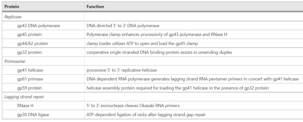
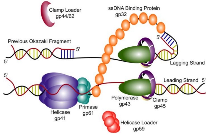
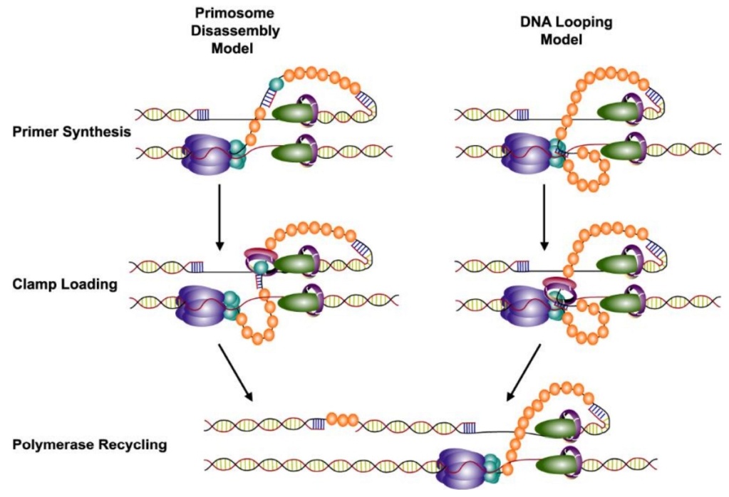
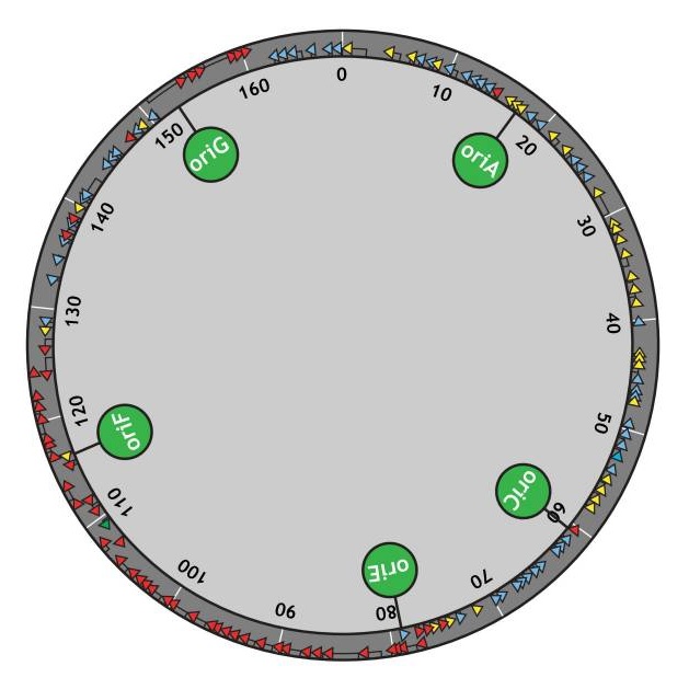
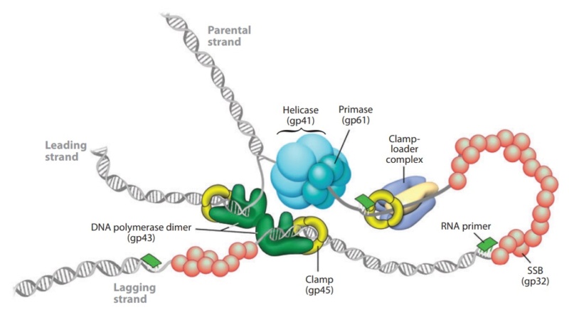
DNA replication by the bacteriophage T4 replisome

Michael Rossmann, Purdue - T4 Bacteriophage Assembly
https://vimeo.com/10700469

Michael Rossman, Purdue - T4 Bacteriophage Infection
https://vimeo.com/10701736

Fly Through Bacteriophage T4
https://vimeo.com/167273755

Introduction

The T4 phage, acts like a spring-loaded syringe and looks like something out of an industrial parts catalog. It can stick to a bacterium, punch a hole, and inject viral DNA (yes, even bacteria suffer infections). Like a conqueror seizing factories to build more tanks, this DNA then directs the cell’s machines to build more viral DNA and syringes. Like all organisms, these viruses exist because they are fairly stable and are good at getting copies of themselves made. Whether in cells or not, nanomachines obey the universal laws of nature. Ordinary chemical bonds hold their atoms together, and ordinary chemical reactions (guided by other nanomachines) assemble them. Protein molecules can even join to form machines without special help, driven only by thermal agitation and chemical forces. By mixing viral proteins (and the DNA they serve) in a test tube, molecular biologists have assembled working T4 viruses. This ability is surprising: imagine putting automotive parts in a large box, shaking it, and finding an assembled car when you look inside! Yet the T4 virus is but one of many self-assembling structures .

Comment: Imagine what it would take for protein engineers to produce nanomachines that would need no nano arms and nano hands to assemble complex nanomachines, but parts of these machines that would be able to assemble on their own just by shaking them, like motors, bearings, and moving parts coming together randomly, and then self-assemble into a fully operational nano-machine. The engineers would need to know the single individual forces and how they would interact with other forces from the other parts. The problem becomes even more apparent when we consider that one of the forces that influence proteins are for example Van der Waals forces which operate based on quantum mechanical principles.   R. W. Newberry (2019): The dominant contributors to protein folding include the hydrophobic effect and conventional hydrogen bonding, along with Coulombic interactions and van der Waals interactions. What a feat would THAT be! 

Frederick William Twort FRS was an English bacteriologist and was the original discoverer in 1915 of bacteriophages. 40% of all bacteria in the oceans are killed by bacteriophages, every single day.

M. YANAGIDA (1984): The virus particle contains a double-stranded DNA molecule of 170 X 10^3 base pairs and more than 3,000 protein subunits of some 30 polypeptide species. As a virus, phage T4 has two fundamental attributes in common with cells or higher forms of life: a definite architecture and the ability to replicate that architecture according to the genetic instructions encoded in molecules of DNA. Experimental results may give new insight into design principles underlying the large and complex bacteriophage T4 head. 27

F.Arisaka (2005): Bacteriophage is an elaborate molecular machine that carries its genomic DNA and efficiently injects it into bacteria. It has a complicated assembly mechanism, where proteins as scaffolding proteins and cleavage of polypeptide bonds in some cases are involved. T4 phage belongs to a family, Caudovirales, which designates a group of phages that has a tail. More than 95% of phages have tails, and possession of the tail is unique to bacteriophages.  Bacteria as single cell organisms have a much more strongly constructed membrane structure than eukaryotic cells. For example, E. coli, a gram-negative bacterium, has triple-layered cell membranes; namely, outer membrane, peptidoglycan layer, and inner membrane. Phages have such a complex structure as a tail to invade the tough barrier of the host cells. 24
The amazing design of the T4 bacteriophage and its DNA packaging motor Bacter17

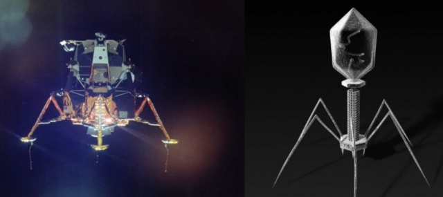
Vergote (2018): The virus bacteriophage T4 resembles the Lunar Lander that was used in the 70’s by the Apollo space program. It has a landing system, duplicates of one protein in the head and a tail used to pass that DNA to infect bacteria. If you are looking for the best design, nature is the perfect place to start. 20 
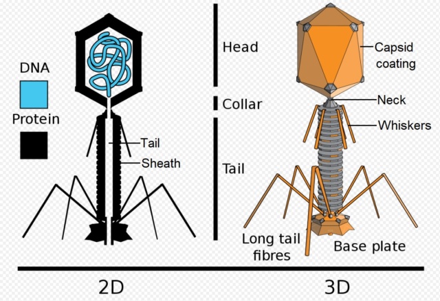
Is it a coincidence? The Bacteriophage T4 consists of a capsid shell, a head where it stores and protects its genome and a syringe-like structure used to insert the DNA into a host. The tail terminates with a multiprotein baseplate that changes its conformation from a “high-energy” dome-shaped to a “low-energy” star-shaped structure during infection. It also has an ultrafast DNA packaging motor to translocate or pack, long stretches of the virus's genetic material into its capsid shell.

Bacteriophages are molecular machines – created for one reason – to kill bacteria – to control bacterial species population. 
Eric S Miller (2003): T4 bacteriophages constitute a beautifully integrated system of biological machines and networks 13

Eric S Miller (2010): Phage T4 is one of the most extensively investigated viruses and has been the central focus of several monographs and reviews over the last 25 years. The T4 biological system is amenable to investigation by genetic, phylogenetic, biochemical, biophysical, structural, computational, and other tools. 12

A. Roberts (2015): Phage help maintain microbial diversity and balance within Earth’s biosphere. Phage are thought to turn over 20–50 percent of the biomass in Earth’s oceans daily! In the absence of these microbial predators it is hard to imagine how our planet would ever sustain life beyond mere microbes. The planet would be covered with microbial competition specialists, sequestering all of Earth’s resources necessary for advanced life. If not for bacterial predation via phage, bacteria would certainly dominate life to the exclusion of advanced organisms. 2

Vincent R. Racaniello (2015): There are more than 10^30 bacteriophage particles in the world’s oceans, enough to extend out into space for 200 million light-years if arranged head to tail. The average human body contains approximately 10^13 cells, but these are outnumbered 10-fold by bacteria and as much as 100-fold by virus particles. 14

Nicola Twilley (2015): There are an estimated 10^31—ten million trillion trillion—phages on Earth, more than every other organism, including bacteria, put together. According to researchers in Vancouver, these tiny viruses cause a collective trillion trillion successful infections per second, in the process destroying up to forty percent of all bacterial cells in the ocean every single day. Following their deaths at the hands of phages, those carbon-containing microorganisms sink down into the marine sediment, effectively removing greenhouse gases from circulation. Anything that bacteria do, from breaking down the carcasses of dead animals to converting atmospheric nitrogen into plant food, is at the mercy of the phages that infect, kill, or otherwise transform them. Phages are the puppet masters; they insure that essential biochemical processes run smoothly. 11

J.Sarfati (2008): Viruses are particles so tiny that they can’t be seen by an ordinary light microscope, but only under an electron microscope. Viruses come in many different sizes, shapes and designs, and they operate in diverse ways. They are composed of DNA (or RNA in the case of RNA viruses, including retroviruses) and protein. They are not living organisms because they cannot carry out the necessary internal metabolism to sustain life, nor can they reproduce themselves. They are biologically inert until they enter into host cells. Then they start to propagate using host cellular resources. The infected cell produces multiple copies of the virus, then often bursts to release the new viruses so the cycle can repeat. One of the most common types is the bacteriophage (or simply ‘phage’) which infects bacteria. It consists of an infectious tailpiece made of protein, and a head capsule (capsid) made of protein and containing DNA packaged at such high pressure that when released, the pressure forces the DNA into the infected host cell. How does the virus manage to assemble this long information molecule at high pressure inside such a small package, especially when the negatively charged phosphate groups repel each other? It has a special packaging motor, more powerful than any molecular motor yet discovered, even those in muscles.The genome is about 1,000 times longer than the diameter of the virus. It is the equivalent of reeling in and packing 100 yards of fishing line into a coffee cup, but the virus is able to package its DNA in under five minutes.This motor exerts a force twice as powerful as a car engine. So the motor, a terminase enzyme complex, ‘can capture and begin packaging a target DNA molecule within a few seconds.’ Such a powerful motor must use a lot of energy, and in one second, this one goes through over 300 units of life’s energy currency ATP.  The virus has a complementary motor-enzyme, ATPase, built into its packaging engine, to release the energy of the ATP.  And not only is the packing motor powerful, it can change its speed as if it had gears. The researchers say that this is important, because the DNA fed to it from the cell is likely not a straightforward untangled thread. Just as it is good for a car to have brakes and gears, rather than only being able to go 60 miles per hour, the DNA-packaging motor may need to slow down, or stop and wait if it encounters an obstruction. It may permit DNA repair, transcription or recombination— the swapping of bits of DNA to enhance genetic diversity—to take place before the genetic material is packaged within the viral capsid. 4

Joseph W. Francis (2003): Microbes and viruses perform essential roles in all ecosystems of the biosphere. Microbes and viruses perform many beneficial activities in ecosystems and in symbiotic partnerships with all biological organisms. I propose that microbes were created as an organosubstrate; a link between macro-organisms and a chemically rich but inert physical environment, to provide a substrate upon which multicellular creatures can thrive and persist in intricately designed ecosystems. Viewed in this context microbes and viruses could also be thought of as a single, complex, massive, multicellular, multitaxon organism with incredible and powerful life supporting properties. Many microbes live on and within living organisms. It is estimated that the number of microbes living on the human body far exceeds the 70 trillion human cells that comprise it. The discipline of microbial ecology is increasingly revealing that microbial and viral symbionts play vitally important roles within organisms and ecosystems. In fact, axenic (germ-free life) probably does not exist in nature; all animal species with the exception of prenatal life are thought to live with microbial symbionts. A tremendous number of symbiotic relationships are being discovered. Many of these relationships involve complex lifestyles and anatomies that appear to be designed to foster the symbiotic lifestyle. A general survey of symbiotic relationships also shows that the most common functions provided by symbionts involve nutritional support, protection and reproduction/population control.

Structure of the bacteriophage T4
The amazing design of the T4 bacteriophage and its DNA packaging motor Bacter12
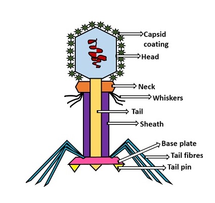
Head: It is elongated and hexagonal in shape. Possesses a prismoid structure. It is surrounded by an envelope called a capsid.
Capsid: It is produced by identical protein subunits called capsomeres. It contains around 2000 capsomeres.
Genetic material: It is 50 nm long and can be either DNA or RNA. The structure of genetic material can be linear or circular. It is tightly packed inside the head.
Neck: It is also called a collar, which connects the head and tail. It possesses a circular plate-like structure.
Tail: It resembles a hollow tube. A tail is surrounded by a protein sheath.
Sheath: It is composed of around 144 protein subunits. The sheath of the bacteriophage is highly contractile. It contains 24 rings.
Base plate: It is hexagonal in shape. The base plate is present at a distal end.
Tail fibers: These are attached to the base plate. It appears long and thread-like filaments. Tail fibers induce host specificity, or they are host-specific. They are generally found 6 in number. Size: 130x2nm
Spikes: It is also called a tail pin. Spikes recognize the receptor sites of the host cell.

Michael Rossmann, Purdue - T4 Bacteriophage Assembly
https://vimeo.com/10700469

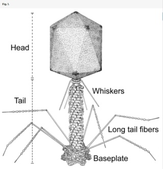
Bo Hu (2015): The T4 virion comprises a capsid containing a 170-kb dsDNA genome, a collar region that displays short whiskers called whisker antigen control (Wac) (also known as fibritin), a contractile tail with a complex baseplate that harbors short tail fibers (STFs), and a set of side or long tail fibers (LTFs) (Fig. 1D). After recognition of a host, the tail transmits a signal to the head for genome ejection and provides the channel through which the DNA moves. 28
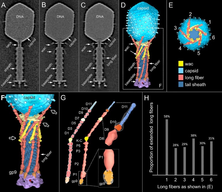
The amazing design of the T4 bacteriophage and its DNA packaging motor Compar10
Comparative structural analysis of infective T4 virions.
Asymmetric 3D reconstructions are shown as central slices of Wac-minus 
(A), fiberless (X4E mutant) 
(B), and WT 
(C) virions. In the absence of Wac, LTFs are seen only as short stubs emanating from the baseplate due to their flexibility when extended. A 3D reconstruction of WT T4 
(D), enlarged in F, highlighting the six (orange) LTFs attached (open arrows) to the whiskers and collar of the sixfold symmetrical Wac (yellow) and the fivefold symmetrical capsid vertex. 
(G) Domain structure of an LTF (4) aligned with our cryo-ET reconstruction. For clarity, not all subdomains are named on the figure. Subdomains P1–P5 correspond to a trimer of gp34, K-C to gp35, and part of gp36, and subdomains D1–D11 in the distal half-fiber are comprised of trimeric gp36 and gp37. Crystal structures of gp9 (PDB ID code 1S2E) and the gp37 fiber tip (PDB ID code 2XGF) are fitted into the density map. Classification of each LTF reveals the symmetry mismatch viewed along the tail sheath axis, where fiber 1 is set to interact with a capsid edge 
(H) Classification was also used to evaluate the density of each LTF and thus to estimate the distribution of extended fibers on a WT virion. Also, see Figs. S1–S3.

Hari charan (2020): This overall structure is necessary the way the phages deliver their payload of genetic material into bacteria. Once on the surface of a bacterium, the tube portion contracts, and the phage acts like microscopic hypodermic needle, literally injecting the genetic material into the bacterium.

P. G. Leiman (2003): Bacteriophage T4 is one of the most complex viruses. More than 40 different proteins form the mature virion, which consists of a protein shell encapsidating a 172-kbp double-stranded genomic DNA, a ‘tail,’ and fibers, attached to the distal end of the tail. The fibers and the tail carry the host cell recognition sensors and are required for attachment of the phage to the cell surface. The tail also serves as a channel for delivery of the phage DNA from the head into the host cell cytoplasm. The tail is attached to the unique ‘portal’ vertex of the head through which the phage DNA is packaged during head assembly. Similar to other phages, and also herpes viruses, the unique vertex is occupied by a dodecameric portal protein, which is involved in DNA packaging. Bacteriophage T4 is a double-stranded DNA (dsDNA) tailed virus that infects Escherichia coli (figure below).

The amazing design of the T4 bacteriophage and its DNA packaging motor The_t410
Structure of bacteriophage T4. The proteins comprising the virion are labeled with their corresponding gene number or name. 
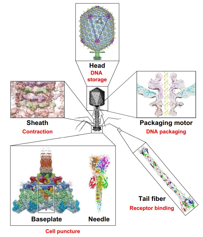
The amazing design of the T4 bacteriophage and its DNA packaging motor Struct21
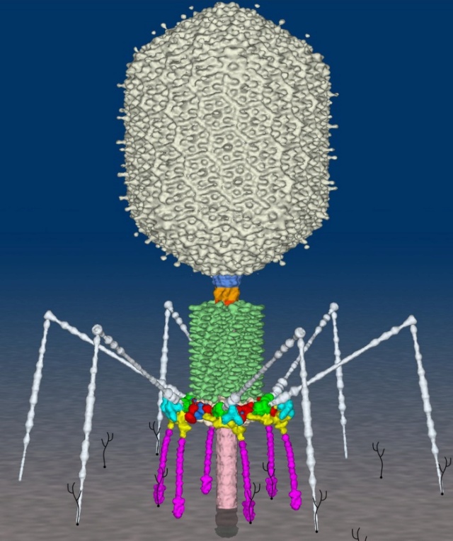
Bacteriophage T4 with detailed structures of the head, the packaging motor, the sheath, the tail fiber and the baseplate with the needle

It is one of the most complex viruses, with a genome that contains 274 open reading frames out of which more than 40 encode structural proteins. The mature virus, or ‘virion,’ consists of a prolate head with hemiicosahedral ends  encapsidating the genomic DNA; a cocylindrical contractile tail, terminated with a baseplate; and six fibers attached to the baseplate. The head, tail, and fibers assemble via independent ordered pathways and join together to form a mature virus particle. Unlike animal viruses, infection of host cells by tailed bacteriophages is highly efficient – only one bacteriophage T4 particle is required, in general, to infect a host cell. Upon infection, the phage shuts down host-specific nucleic acid and protein syntheses, thus ensuring production of only its own components in amounts sufficient to assemble up to 200 progeny virus particles per infected cell. The efficiency of the infection process and the large genome of bacteriophage T4, in which only half of the genes are necessary for proliferation on E. coli, contribute to the diversity of the phages from the T4-like family, a subgroup of Myoviridae. These phages propagate on a wide range of bacterial hosts that grow in diverse environments. 

The amazing bacteriophage DNA packaging motor

The bacteriophage DNA injection machine

Bacteriophage T4 head structure
 
M.YANAGIDA (1984): There are three classes of lattice proteins, namely, the major coat protein (gp23), soc, and hoe. Six molecules of gp23 and one molecule of hoe produce a hexamer and a center unit, respectively, Six molecules of soc form six bridges (as ellipsoidal particles in Fig. 2). Thus the repeating unit of the head lattice consists of "hexamer+a center unit+six bridges," or type "(6+ 1)+6". This model contains 840 subunits for hexamers, 60 subunits for pentamers (at vertices), 900 bridges, and 140 center units, that is, 900 gp23, 900 soc, and 140 hoc molecules. About 1,000 gp23, 1,000 soc, and 150 hoc were reported to be present in the capsid from chemical analysis, in agreement with the ultrastructural analysis. 27

The head of bacteriophage T4 is composed of more than 3000 polypeptide chains of at least 12 kinds of protein and a 172-kbp dsDNA chromosome, which comprises 102% of the unique region of about 169 kbp.. The shell has icosahedral ends and a cylindrical equatorial midsection with a unique portal vertex where the phage tail is attached. 9

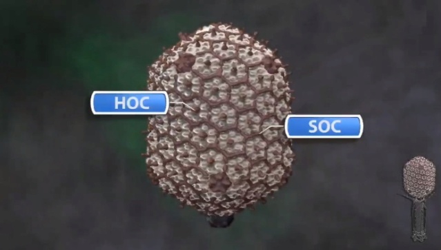
Moh Lan Yap (2015): The mature head encapsidates 172 kbp dsDNA. The head is first assembled as an empty capsid that is subsequently packaged with DNA by an ATP-dependent packaging machine. This machine binds to the same special pentameric vertex that is later occupied by the phage tail. The head is prolate, meaning that it has two icosahedral ends and a cylindrical mid-section. The geometrical organization (expressed as triangulation numbers) of the ends and mid-section are based on planar hexagonal grids. The capsid is composed of 930 post-translationally modified monomers, or 155 hexamers of the major protein, gene product 23 (gp23*, where the * signifies post-translational cleavage). The presence of proteins, homologous to the major capsid protein, which form pentamers as opposed to hexamers is a frequent solution to the formation of the pentameric vertices in icosahedral viruses. The portal protein has multiple roles. It initiates head assembly, genome packaging and serves as the genome gatekeeper to prevent leakage of the packaged DNA. Two accessory proteins, Hoc (highly antigenic outer capsid protein) and Soc (small outer capsid protein) attach to the capsid surface (Figure below). 
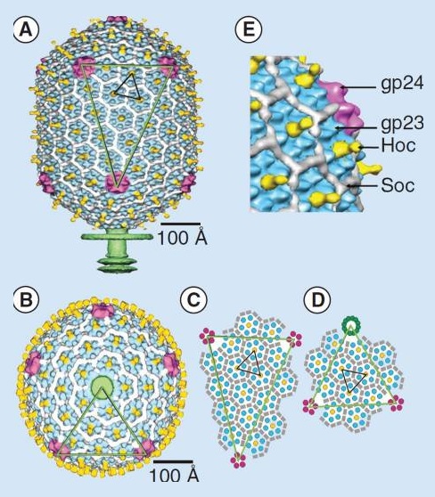
The amazing design of the T4 bacteriophage and its DNA packaging motor T4_bac11
The structure of the bacteriophage T4 head
The symmetry of the gp23* major capsid protein shell is characterized by triangulation numbers Tend = 13 laevo and Tmid = 20. The facet triangles are shown in green and the basic triangles are shown in black.
(A) Shaded surface representation of the cryo-electron microscopy reconstruction viewed perpendicular to the fivefold axis. gp23* is shown in blue, gp24* is in magenta, Soc is in white, Hoc is in yellow and the tail is in green.
(B) View of the reconstruction along the fivefold axis with the portal vertex toward the observer; the tail has been cut away at the level shown by the black arrow in (A). Proteins are colored as described for (A).
(C) Schematic representation of the distribution of proteins in the elongated midsection facet.
(D) Schematic representation of an end-cap facet. Proteins are colored as described for (A) except the Soc molecules are shown as gray rectangles. (E) A closer look of the distribution of proteins on the head.

The rod-shaped Soc binds between two gp23* hexamers, thus forming a continuous mesh surrounding the hexameric gp23* on the capsid. Soc maintains the stability of the head under extreme environments. Hoc is an elongated molecule protruding from the center of gp23* hexamers. Its Ig-like domains, exposed on the outer surface of the head, may provide survival advantages to the phage. 21

V. V. Mesyanzhinov (2004): The head of phage T4, or capsid, is a prolate icosahedron elongated along a fivefold axis and is composed of more than 1500 protein subunits encoded by at least twelve genes (Table 1).In total, the mature T4 capsid contains 930 subunits of gp23* (* indicates a protein proteolytically processed during capsid maturation) and 55 subunits of gp24*. Pentamers of gp24* occupy eleven vertices of the icosahedron, and gp20 forms a unique portal vertex required for DNA packaging and subsequent attachment of the tail. The T4 capsid shell is decorated on the outside with gphoc (highly antigenic outer capsid protein) and gpsoc (small outer capsid protein). The latter two proteins enhance head stability. 23


The amazing design of the T4 bacteriophage and its DNA packaging motor Protei10


The Molecular Architecture of the Bacteriophage T4 Neck

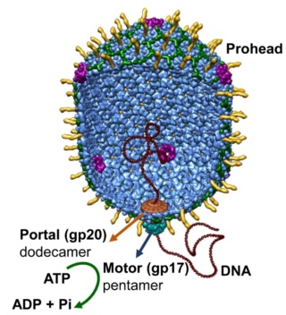
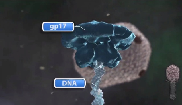
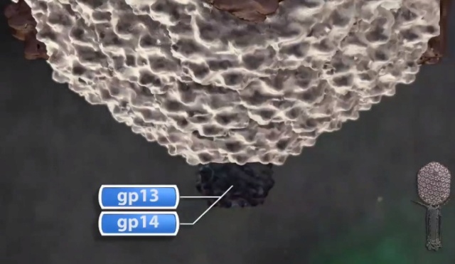
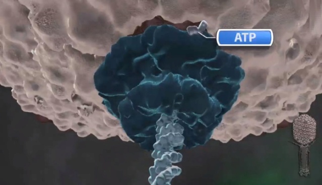
Andrei Fokine et.al. (2013):  The T4 head and tail are assembled via independent pathways. Assembly of the T4 head is a complex process that includes a number of intermediate stages. The head assembly is initiated by the dodecamer (A dodecamer (protein) is a protein complex with 12 protein subunits.) of the portal protein, gp20 (gp, gene product). First, a head precursor, called the prohead, is assembled, which is subsequently processed by a scaffold-associated protease. Then the phage genomic DNA is packaged into the capsid through the portal vertex by an ATP-driven motor composed of five gp17 molecules. Upon completion of the DNA packaging, the head assembly is finalized by attachment of several copies of the gp13 and gp14 proteins to the portal vertex. Monomers of gp13 and gp14 have a size of 309 and 256 amino acid (aa) residues, respectively. The gp13–gp14 complex seals the portal vertex and creates a site for attachment of the independently assembled tail. Mutant phages lacking these proteins produce heads that are unable to bind tails and lose their DNA.

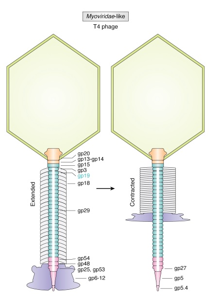
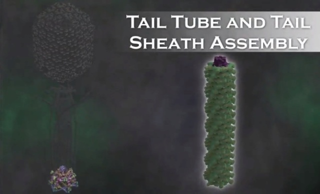
The T4 tail assembly begins with the baseplate formation and proceeds with polymerization of the tail tube and the contractile sheath. The tail tube is formed by gp19 molecules (163 aa residues). The length of the tube is controlled by a mechanism involving the “tape-measure protein”, gp29. The elongation of the tail tube is terminated by attachment of the hexamer of the 175-residue tail tube terminator protein, gp3, which binds to the last row of gp19 subunits (probably also to gp29) and stabilizes the tail tube. The T4 tail tube is used as a scaffold for the polymerization of the contractile sheath. The gp18 sheath molecules (659 aa residues) assemble around the tube in the form of a six-start helix. The T4 tail assembly is completed by the hexamer of the tail terminator protein, gp15 (the monomer is 272 aa residues long), which binds to the top† of the tail. Contraction of the tail during infection is associated with a substantial rearrangement of the gp18 subunits and results in shortening of the sheath to less than one-half of its original length. 22

The amazing design of the T4 bacteriophage and its DNA packaging motor 1-s2_027

Coat, scaffolding, and portal proteins are encoded by P22 genes 5, 8, and 1, respectively. In the absence of scaffolding protein, P22 coat protein assembles into TZ4 and TZ7 icosahedral shells as well as “spiral” structures, and all of these lack the essential portal protein and at least one protein required for DNA injection. 10

Lei Sun et al., (2015):The portal structure probably dates back to a time when self-replicating microorganisms were being established on Earth. 11

Amy D. Migliori (2014): Recent structural studies of the bacteriophage T4 packaging motor have led to a proposed mechanism wherein the gp17 motor protein translocates DNA by transitioning between extended and compact states, orchestrated by electrostatic interactions between complimentarily charged residues across the interface between the N- and C-terminal subdomains.  2

They are the most numerous biological entity on earth, with an estimated number of 10^31 tailed phages in the biosphere. They are arguably very ancient as a group, with some estimates placing their ancestors before the divergence of the Bacteria from the Archaea and Eukarya

The amazing design of the T4 bacteriophage and its DNA packaging motor Ncomms12

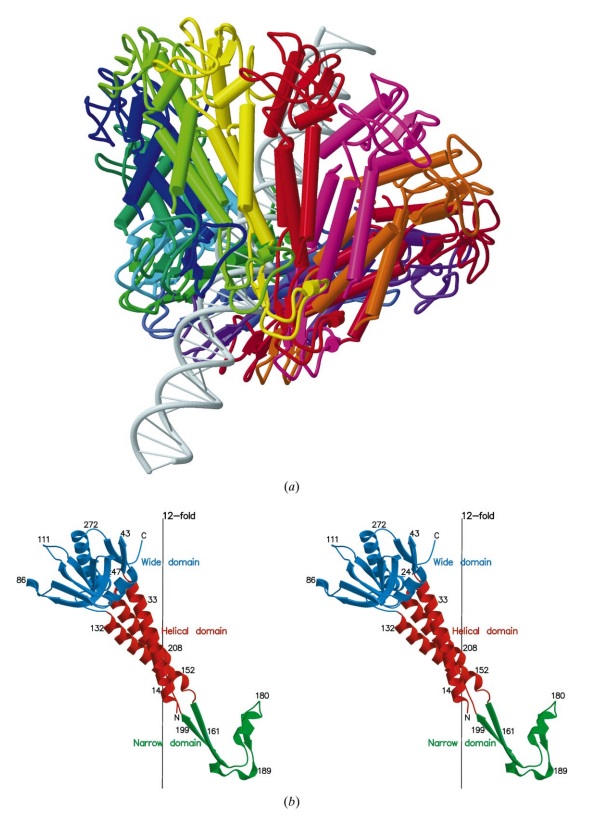
(a) 3D density map of T4 portal protein assembly at 3.6 Å resolution with each subunit color-coded. Shown is the top view (left)
 and side view (right). 
(b) Ribbon diagram of the gp20 atomic model with each subunit color-coded. Shown is the top view (left)
 and side view (right).

The amazing design of the T4 bacteriophage and its DNA packaging motor Ncomms13
  
(a) Charge distribution on the outer surface of dodecameric gp20. Blue and red colours correspond to 10 kT e− positive and negative potential,
respectively. 
(b) Charge distribution on the inner surface of dodecameric gp20. (c) Ribbon drawing of the gp20 monomer structure with each
domain colour-coded.

The amazing design of the T4 bacteriophage and its DNA packaging motor Ncomms14

(a,b) Cryo-EM density map of the T4 prolate head (gp23: cyan; gp24:magenta; Soc: pink; Hoc: yellow).
(c) Bottom view of the prolate head, showing the gap between gp20 and the capsid. (d) Fit of the gp20 and gp23
structures into the cryo-EM map of the T4 prolate head. (e) A model of the T4 head assembly. A dodecameric portal is
assembled on the inner membrane of E. coli with the assistance of the phage-coded chaperone gp40 and the E. coli chaperone YidC58.
The portal assembly acts as an initiator for head assembly, leading to co-polymerization of the major capsid protein gp23
and scaffolding proteins.

The amazing design of the T4 bacteriophage and its DNA packaging motor Ncomms16

(a) Fitting of the T4 portal protein (purple) and gp17 (tan) into the 35 Å cryo-EM reconstruction of the procapsid+gp17
(EMD-1572 accession number).
(b) Residues involved in the interaction between gp20 (purple) and gp17 (tan) are shown
as sticks. 
(c) The surface charge of gp20 and gp17 around the interface area showing electrostatic interactions. The view
orientation is the same as in panel (b).

The amazing design of the T4 bacteriophage and its DNA packaging motor Ncomms17

The different portal protein subunits with their wing, stem, clip and crown domains are coloured green, blue, purple and orange, respectively.

Phyiscsworld ( 2014): The molecular motor that folds and packs DNA into a virus is at its most efficient when the DNA shows some self-repulsion. That is the surprising finding of researchers based in the US – it was previously thought that such repulsion would act as an obstacle in the packing process. The team also found that pausing the motor and allowing it to relax increased the rate of the whole packaging process. In addition to providing new insights into how viruses function, the work could benefit biotechnologies that enclose long polymers into nanoscale devices. 6

After invading its host cell, a virus reprogrammes the cell's nucleus to duplicate it.

Question: How was the virus programmed to re-program the cell's nucleus? trial and error? Had the function of reprogramming not have to be fully operating since the beginning, otherwise, the virus would not be able to replicate. 

As it replicates, a strand of DNA is pulled from an infected host cell and squeezed into a protein shell – known as a prohead – which then carries the DNA to infect other cells. In some species, the prohead is produced first, leaving only a small hole at one end through which a powerful molecular motor pushes the DNA in and then packs it at very high densities.

Question: How did it emerge the function to pack the DNA at very high densities? trial and error?

The motor has to overcome three forces: the electrostatic self-resistance that comes into play because DNA is negatively charged; the mechanical resistance of DNA to bending; and the entropic resistance of DNA to be crowded on itself.

Question: How did the motor emerge this function of overcoming the three forces? trial and error?

The tail structure

Phys.Org ( 2016):  To infect bacteria, most bacteriophages employ a 'tail' that stabs and pierces the bacterium's membrane to allow the virus's genetic material to pass through. The most sophisticated tails consist of a contractile sheath surrounding a tube akin to a stretched coil spring at the nanoscale. When the virus attaches to the bacterial surface, the sheath contracts and drives the tube through it. All this is controlled by a million-atom baseplate structure at the end of the tail. Phages are widely distributed on the planet. They accompany bacteria everywhere - in the soil, water, hot springs, algal bloom, animal intestines etc - and have a dramatic impact on the diversity of bacterial populations, including for example, the microbiome of the human gut. 18

Petr G.Leiman (2006): Bacteriophage T4 has one of the most complex tails of all studied phages. The T4 tail is composed of ∼400 polypeptide chains that form the tube, the contractile sheath around the tube, and the baseplate that terminates both of them. 26

Petr G Leiman (2006): Bacteriophage T4 has one of the most complex tails of all studied phages. It is composed of w400 polypeptide chains that form the tube, the contractile sheath around the tube, and the baseplate that terminates both of them. https://pubmed.ncbi.nlm.nih.gov/16554069/

The tail, fibers, and infection process  
Phages from the Myoviridae family have exceptionally complex, contractile tails. Bacteriophage T4 devotes 25 kbp of its genome to tail assembly, which is comparable with the size of the entire adenovirus genome (36 kbp). Products of at least 22 genes are involved in tail assembly (table 3), 

The amazing design of the T4 bacteriophage and its DNA packaging motor T4_bas11
which include a phage-encoded chaperone that participates in folding of the long and short tail fibers (table 2). 

The amazing design of the T4 bacteriophage and its DNA packaging motor T4-enc11

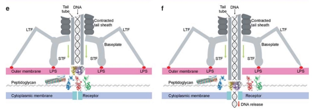
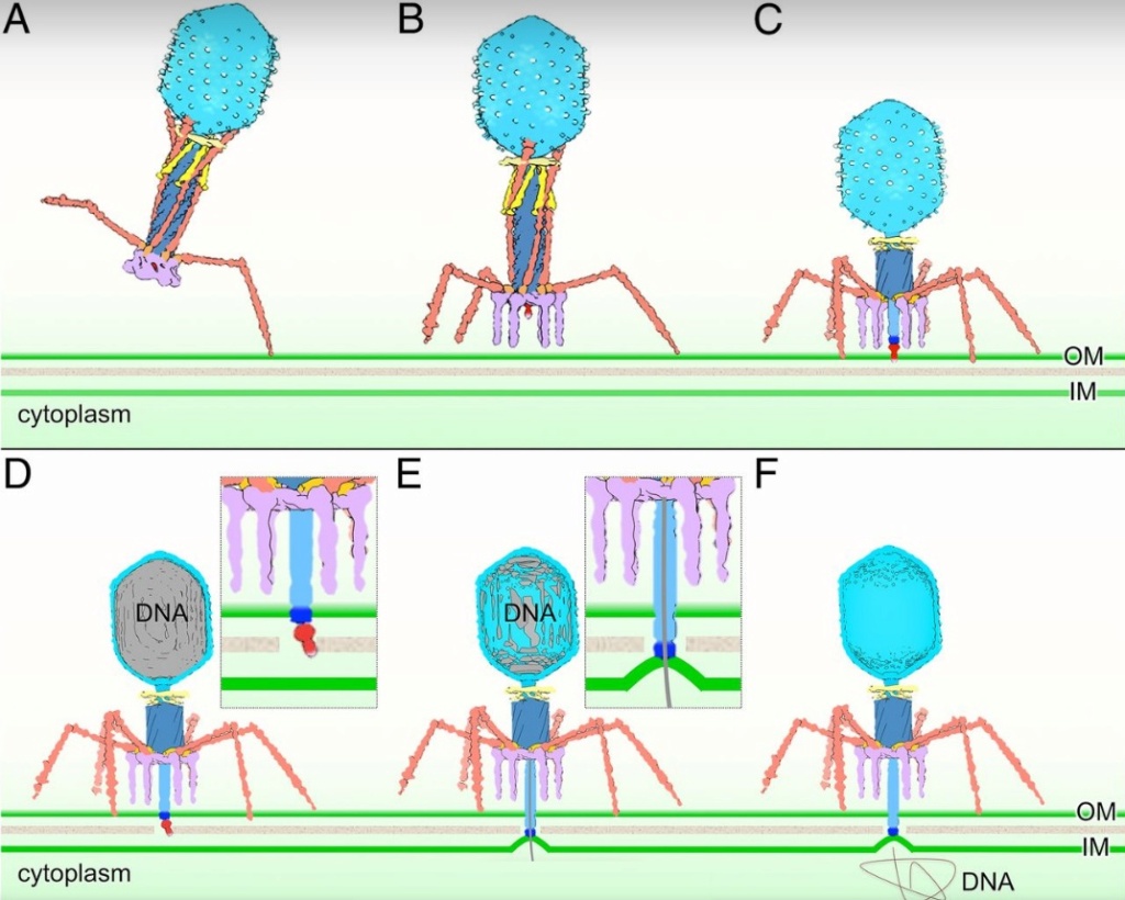
The bacteriophage T4 tail is composed of two concentric protein cylinders, at one end of which is the baseplate and fibers. The inner cylinder, called the tail tube, is built of 144 copies of gp19. The tail tube has a 40 Å-diameter channel for DNA passage from the head to the infected cell. The outer cylinder, called the tail sheath, tightly envelopes the 90 Å-diameter tail tube and has a width of about 210 Å. It is composed of 144 copies of gp18. The subunits comprising each cylinder form a six-start helix with a pitch of 41 Å and a righthanded twist angle of 17°. The helix has a length of 984 Å and contains 24 repeats. During infection, the phage recognizes an E. coli bacterium using its long tail fibers (LTFs) connected to the baseplate. The phage then anchors the baseplate to the lipopolysaccharide cell surface receptors using the short tail fibers (STF), which are initially assembled under the baseplate. This event triggers a hexagon-to-star conformational change in the baseplate and causes an irreversible contraction of the tail sheath, releasing about 25 kcal/ mol of energy per gp18 monomer. During this process, the gp18 hexamers flatten, rotate, and expand radially, resulting in a decrease of their thickness by 26 Å and an increase of the twist angle by 15°. The contracted tail sheath has a length of only 360 Å and a width of 270 Å. The tail tube does not change its length during sheath contraction. As a result, almost half of the tube protrudes out of the contracted tail sheath and the baseplate.

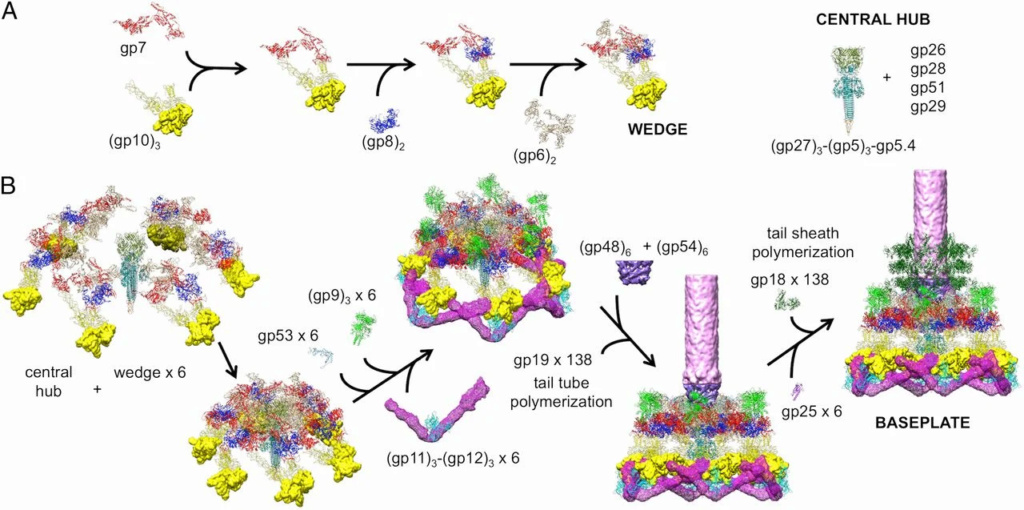
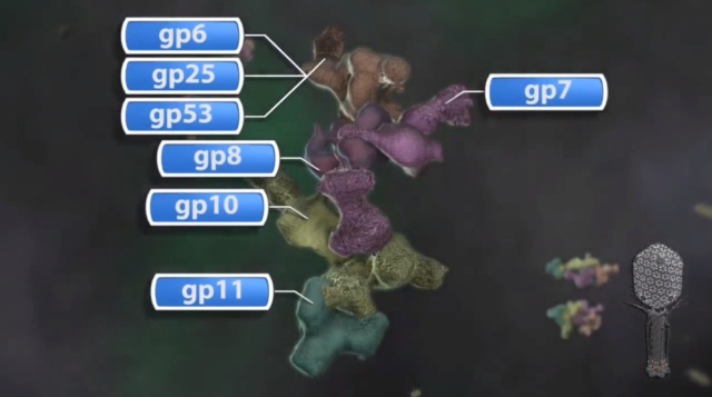
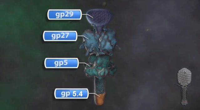
The sheath can be caused to contract by exposing the phage to 3 M urea. Nevertheless, the DNA is not released until the tail tube tip binds to a cytoplasmic membrane receptor common to enteric bacteria, suggesting that tail contraction does not cause the release of DNA. The interaction of the tail tube tip with the cytoplasmic membrane involves creation of a channel for DNA passage. During DNA transfer from the capsid into the cell, the membrane remains virtually undamaged since the transfer requires a proton motive force across the membrane. The assembly pathway of the bacteriophage T4 tail is regulated by ordered sequential interactions of proteins rather than sequential gene expression. The baseplate, a remarkably complex multiprotein structure, is assembled first. It is composed of about 150 subunits of at least 16 different gene products, many of which are oligomeric (table 3). These proteins form six independently assembled wedges that join together around the central hub with the help of the trimeric proteins (gp9) and (gp12). Each wedge is assembled by sequential interactions of the seven protein oligomers: (gp11), (gp10), (gp7), (gp8), (gp6), gp53, and gp25. The baseplate hub is formed by (gp5), (gp27), gp29 and, probably, gp28. The assembly of the baseplate is completed with the attachment of six copies of gp48 and six copies of gp54 to the external interface between the wedges and the hub. The latter proteins serve as a starting point for polymerization of gp19 to form the tail tube, which is terminated with gp3. The tail tube serves as a scaffold for polymerization of the tail sheath around it. During this process, gp18 stores energy in its conformation (possibly by ATP hydrolysis), making the non-contracted T4 tail a stretched spring. The length of the tail tube is controlled by the ruler protein, gp29, which also participates in assembly of the central part of the baseplate. The length of the tail sheath is determined by the length of the tube. The assembly of the tail is completed by attachment of a gp15 hexamer to the last ring of the tail sheath. The baseplate is a dome-shaped object.  The hollow tail tube stems from the center of the baseplate. 15

The amazing design of the T4 bacteriophage and its DNA packaging motor Protei11

V. V. Mesyanzhinov (2004): Products of at least 22 genes are involved in assembly of the T4 phage tail (Table above) that uses the energy of the sheath contraction for DNA ejection into the host cell. The assembly pathway of the tail is based on strictly ordered sequential interactions of proteins. The baseplate is a remarkably complex multiprotein structure of the tail that serves as a control unit of virus infection. The baseplate is composed of ~150 subunits of at least 16 different gene products, many of which are oligomeric, and assembled from six identical wedges that surround a central hub. The T4 gp11 (the short tail fiber connecting protein), gp10, gp7, gp8, gp6, gp53, and gp25 combine sequentially to built up a wedge. The central hub is formed by gp5, gp27, and gp29 and probably gp26 and gp28. Assembly of the baseplate is completed by attaching gp9 and gp12 forming the short tail fibers, and also gp48 and gp54 that are required to initiate polymerization of the tail tube, a channel for DNA ejection that is constructed of 138 copies of gp19. The length of the tail tube is probably determined by the “ruler protein” or template, gp29. The tail tube serves as a template for assembly of 138 copies of gp18 that form the contractile tail sheath. In the absence of the tail tube, gp18 assembles into long polysheaths with a structure similar in several aspects to the contracted state. Both the tail tube and the tail sheath have helical symmetry. Assembled tail sheath represents a metastable supramolecular structure, and sheath contraction is an irreversible process. During contraction the length of tail sheath decreases from 980 to 360 Å and its outer diameter increases from 210 to 270 Å. The assembly of the tail is completed by a gp15 hexamer that binds to the last gp18 ring of the tail sheath. The assembled tail associates with the head after DNA packaging. Then six gpwac (fibritin) molecules attach to the neck of the virion forming a ring embracing it (“collar”) and thin filaments protruding from the collar (“whiskers”) that help with attachment of the phage particle to other fibrous proteins, the long tail fibers. 23

The amazing design of the T4 bacteriophage and its DNA packaging motor Bacter26
Stereo diagram of bacteriophage T4 showing the extended tail, the LTFs, the neck and a small part of the capsid. NA indicates that the density has not been assigned to a specific gene product.

The cell-puncturing device of bacteriophage T4

ScienceDaily (2016): To infect bacteria, most bacteriophages employ a 'tail' that stabs and pierces the bacterium's membrane to allow the virus's genetic material to pass through. The most sophisticated tails consist of a contractile sheath surrounding a tube akin to a stretched coil spring at the nanoscale. When the virus attaches to the bacterial surface, the sheath contracts and drives the tube through it. All this is controlled by a million-atom baseplate structure at the end of the tail. EPFL scientists have now shown, in atomic detail, how the baseplate coordinates the virus's attachment to a bacterium with the contraction of the tail's sheath. 

ScienceDaily (2002): The viral machine works as follows: The virus uses its long-tail fibers to recognize its host and to send a signal back to the baseplate. Once the signal is received, the short-tail fibers help anchor the baseplate into the cell surface receptors. As the virus sinks down onto the surface, the baseplate undergoes a change — shifting from a hexagon to a star-shaped structure. At this time, the whole tail structure shrinks and widens, bringing the internal pin-like tube in contact with the outer membrane of the E. coli cell. As the tail tube punctures the outer and inner membranes of the E. coli cell, the virus' DNA is injected through the tail tube into the host cell. 17

P. G. Leiman (2003): Phages are widely distributed on the planet. They accompany bacteria everywhere -- in the soil, water, hot springs, algal bloom, animal intestines etc -- and have a dramatic impact on the diversity of bacterial populations, including for example, the microbiome of the human gut.  The entire baseplate-tail-tube complex consists of one million atoms, making up 145 chains of 15 different proteins. The scientists were also able to identify a minimal set of molecular components in the baseplate that work together like miniature gears to control the activity of the virus's tail. These components, and the underlying functional mechanism, are the same across many viruses and even bacteria that use similar tail-like structures to inject toxins into neighboring cells. 15

One of the most remarkable features of the baseplate is the spike, or needle, along the axis of the dome. The crystal structure of the gp5-gp27 complex (fig. below) can be fitted into the baseplate map so that the needle density is occupied by the C-terminal domain of gp5. The gp27 trimer forms a channel suitable for the passage of a dsDNA and serves as an extension of the tail tube (figs below).

The amazing design of the T4 bacteriophage and its DNA packaging motor Struct22
a Baseplate of bacteriophage T4 
b gp5–gp27–gp5.4
The amazing design of the T4 bacteriophage and its DNA packaging motor Struct20
Structure of the gp5-gp27 complex. 
(a) Ribbon stereo diagram. The three gp5 monomers are colored red, green, and blue. The three gp27 monomers are colored yellow, gray, and purple. The K+ ion within gp5C is shown in pink. The (PO4)– is hidden behind the lysozyme domain. 
(b) The structure of the gp27 monomer with its four domains colored cyan, pink, light green, and gold along the polypeptide chain. 
(c) Top view of the gp27 cylinder shows that the cyan and green domains form a hexagonal torus

Gp5 consists of three domains: an N-terminal oligosaccharide binding-fold domain, the middle lysozyme domain, and the C-terminal triple b-helix domain (fig.above). The gp5 lysozyme domain has 43% sequence identity and a closely similar structure to the T4 lysozyme encoded by gene e (T4L). 

Structure of the T4 baseplate

ScienceDaily (2002): The baseplate is the "nerve center" of the virus. When the long and short fibers attach to E. coli, the baseplate transmits this message to the tail, which contracts like a muscle. The baseplate both controls the needlepoint of the tail and the cutting enzyme that make a tiny, nanometer-sized hole through the cell wall of the E. coli. The viral DNA is then squeezed through the tail into the host. The E. coli, thus infected, starts to make only new phage particles and ultimately dies. 16

Moh Lan Yap (2016): T4 has a complex baseplate that is essential for assuring a highly efficient infection mechanism 25

M. I. Taylor (2016): Bacteriophages (viruses of bacteria) use a specialized organelle called a tail to deliver their genetic material and proteins across the cell envelope during infection. In phages with the most complex contractile tails, attachment to the host cell is accompanied by a substantial transformation of the tail structure: the external tail sheath contracts and drives a spike-tipped , rigid tube through the host cell membrane. Other macromolecular complexes, such as the type VI secretion system (T6SS), metamorphosis-associated contractile (MAC) arrays, R-type pyocins, Serratia antifeeding prophage, Photorhabdus virulence cassette and rhapidosomes, use a similar contractile sheath–rigid tube mechanism to breach the bacterial or eukaryotic cell envelope. The most complex part of these ‘contractile injection systems is the baseplate, which is responsible for coordinating host recognition or other environmental signals with sheath contraction. The T4 baseplate is currently thought to contain at least 15 different proteins with copy numbers ranging from 1 to 18. Assembly of the T4 baseplate involves two large independent intermediates: a hub and a wedge. Several phage and possibly host cell chaperones mediate the joining of six wedges to the hub, which is then followed by the attachment of receptor-binding fibres to this structure. The nascent baseplate initiates tube assembly and subsequent polymerization of the sheath in the extended high-energy state. The remarkable structure and transformation of the T4 tail and other contractile injection systems have received considerable attention. This is a 440,000-atom (not counting hydrogens) structure. 

Overall structure of the T4 baseplate:  The final atomic model is around 96% complete and contains 56,082 amino acid residues (9,886 unique). These amino acids belong to 145 polypeptide chains of 15 different proteins (gene products, gps) that comprise the baseplate (gp5, gp5.4, gp6, gp7, gp8, gp9, gp10, gp11, gp12, gp25, gp27, and gp53) and the proximal region of the tail tube (gp19, gp48 and gp54, although gp48 and gp54 could also be considered to be baseplate components). 19 

The amazing design of the T4 bacteriophage and its DNA packaging motor T4_bas13
The amazing design of the T4 bacteriophage and its DNA packaging motor T4_bas12
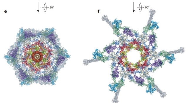
Maps and atomic models of T4 baseplate in pre- and post-attachment states. 
a, b, Cryo-EM reconstructions of the pre- and post-attachment T4 baseplate, respectively. c–f, Atomic models of the two states with component proteins shown as ribbon diagrams. In d and f, the STFs (gp12 trimers) are displayed semi-transparently to indicate that they are not present in the refined model of the post-attachment baseplate.

The amazing design of the T4 bacteriophage and its DNA packaging motor Confor11
Conformational change of the T4 baseplate upon host cell attachment. 
The left and right panels show the pre-and post-attachment structures that are derived from cryo-EM data. The middle image is a model of an intermediate with assumptions described in the text. The insets show close-up views of the central part of the baseplate demonstrating a release of the tail tube–central spike complex, whose position is kept unchanged throughout the transformation.

The amazing design of the T4 bacteriophage and its DNA packaging motor Model_12
The amazing design of the T4 bacteriophage and its DNA packaging motor Pseudo10
Model for baseplate-induced sheath contraction. 
a, b, Pseudoatomic model of the complete T4 tail in the extended (pre-attachment) and contracted (post-attachment) conformations. The insets show a close-up view (labelled with a black box) of the position of the gp18 subunit on the baseplate in the extended and contracted conformations of the sheath. The white geometrical shapes label the same regions of the sheath subunit in both conformations. 
c, Interaction of the two conserved domains of the gp18 sheath protein with the conserved components of the T4 baseplate wedge. Coloured lines indicate the putative topology of the N- and C-terminal gp18 extensions, as well as the gp25 C-terminal strand. 
d, The same view as in c, but the external domain is now not shown for clarity to demonstrate the interaction of gp25-like sheath domains with each other and with gp25. 
e, f, The same as c and d but in the contracted state. 
g, h, Two diagrams demonstrating the motion of baseplate components that results in sheath contraction.

V. V. Mesyanzhinov (2004): Practically every assembled T4 particle is able to infect an E. coli host cell. The baseplate is the control center of the viral infectivity, and understanding of the baseplate structure, a multiprotein machine, is a challenging problem. Below we represent the data about baseplate proteins with known atomic structure.

The amazing design of the T4 bacteriophage and its DNA packaging motor Struct26
Structure of the conserved inner baseplate. 
a, The minimal composition of a contractile injection system is derived from the T4 tail structure: the central spike complex (gp5, gp5.4, gp27), the conserved part of the wedge (gp6, gp7, gp25, gp53), the tail tube (gp19, gp48, gp54) and the conserved part of the sheath (gp18), some of which is modelled using the pyocin sheath14. 
b, A close-up of the structure of the conserved region of the wedge consisting of the (gp6)2–gp7 heterotrimer, gp25 and gp53. The EPR motif of gp25 and the LysM motif of gp53 are highlighted with semi-transparent grey.

The tail tube & tail sheath terminators

Moh Lan Yap (2015): The polymerized tail tube and sheath are capped by the terminator proteins gp3 and gp15, respectively, to prevent depolymerization before the tail attaches to the head. Both gp3 and gp15 form hexameric rings that interact with the last row of gp19 and gp18 molecules. The central pore and the side surface of gp15 are negatively charged, whereas the top and the bottom surfaces are positively charged. The top and bottom surfaces interact with gp14 and gp3 proteins, respectively. The interaction between gp15 and gp18 is different in the extended and contracted (postinfection) conformations. In the contracted tail, the negatively charged side surface of the gp15 hexamer interacts with positively charged surfaces of the C-terminal domains of the gp18 molecules. These interactions help to maintain the integrity of the tail in its contracted form. The gp15 hexamer may have undergone a conformational change during the infection process, which might be propagated through gp14 and gp13, to the portal assembly to allow the release of the genomic DNA. 21

The amazing design of the T4 bacteriophage and its DNA packaging motor Crysta14
Crystal structures of gp18 and gp15
(A) Ribbon diagrams of gp18M and gp15 monomers. The three domains of gp18M are shown in blue (domain I), olive green (domain II) and orange red (domain III); the β-hairpin (residues 454–470) and the last 14 C-terminal residues are shown in cyan. Gp15 represented in rainbow color running from N terminus (blue) to C terminus (red). The broken lines indicate protein regions that are disordered in the crystals.
(B) Relative positions of the gp15 and gp18 molecules in the extended and contracted T4 tails, viewed from the side (upper) and from the top (lower) of the phage. A model of the entire gp18 molecule was created based on the crystal structure of gp18M and the prophage tail sheath protein LIN1278. The models of gp18 molecules belonging to the topmost ring of the contractile sheath are shown in green. The gp15 hexamer is shown in red.
(C) The gp15 and gp18 molecules are fitted into the cryo-electron microscopy reconstructions of the extended and contracted tails.
(D) A helical strand of gp18 in the extended (green) and contracted (brown) tail. The hexagonal baseplate, tail tube, whiskers and collar are shown in gray-blue.

The amazing design of the T4 bacteriophage and its DNA packaging motor Struct25
Structure of the contracted T4 tail 
(side view (a), with an inclination (b), cross-section (c), from the bottom (d)). Each protein or complex is labeled with their respective gene number and indicated by color: spring green, gp5; red, putative gp7; dark blue, gp8; green, gp9; yellow, putative gp10; cyan, gp11; magenta, gp12; salmon, gp19; sky blue, gp27; pink, putative gp48 or gp54; beige, gp6 + gp25 + gp53; orange, putative gp26. 23

1. http://www.pnas.org/content/111/42/15096.short
2. Anjeanette Roberts Celebrating 3.8 Billion Years of Bacteriophage October 22, 2015
3. http://www.ncbi.nlm.nih.gov/pubmed/22297528
4. https://creation.com/images/pdfs/tj/j22_1/j22_1_15-16.pdf
6. http://creation.com/did-god-make-pathogenic-viruses
7. Joseph W. Francis: The Organosubstrate of Life: A Creationist Perspective of Microbes and Viruses 2003 
8.   http://teaguesterling.com/dna/motor-protein.pdf
9.   G. Leiman: Structure and morphogenesis of bacteriophage T4 P.  9 May 2003 
10. http://www.ncbi.nlm.nih.gov/pmc/articles/PMC3109452/
11. Nicola Twilley: Inside the World of Viral Dark Matter February 6, 2015
12. Eric S Miller Bacteriophage T4 and its relatives 28 October 2010
13. Eric S Miller Bacteriophage T4 genome 2003 Mar;6
14. Vincent R. Racaniello: Principles of Virology, Volume 1: Molecular Biology 18 agosto 2015
15. Science Daily: How viruses infect bacteria: A tale of a tail May 18, 2016
16. Science Daily: New Understanding Of Complex Virus Nano-Machine For Cell Puncturing And DNA Delivery February 4, 2002
17. Science Daily: Study Reveals New Information On How Viruses Enter Cells February 7, 2002
18. Phys.Org: How viruses infect bacteria: A tale of a tail MAY 18, 2016
19. M. I. Taylor et.al. Structure of the T4 baseplate and its function in triggering sheath contraction  18 May 2016
20. Jaap Vergote Design and nature Jun 24, 2018
21. Moh Lan Yap: Structure and function of bacteriophage T4  2015 Aug 1
22. Andrei Fokine: The Molecular Architecture of the Bacteriophage T4 Neck 2013 Feb 19
23. V. V. Mesyanzhinov: Molecular Architecture of Bacteriophage T4 July 9, 2004
24. Fumio Arisaka:[url=https://aip.scitation.org/doi
25. Petr G. Leiman: Evolution of Bacteriophage Tails: Structure of T4 Gene Product 10 2006 May 5
26. Petr G.Leiman: Evolution of Bacteriophage Tails: Structure of T4 Gene Product 10 5 May 2006
27. MITSUHIRO YANAGIDA: MOLECULAR ORGANIZATION OF THE HEAD OF BACTERIOPHAGE Teven: UNDERLYING DESIGN PRINCIPLES 1984
28. Bo Hu: Structural remodeling of bacteriophage T4 and host membranes during infection initiation August 17, 2015


Last edited by Otangelo on Sat Oct 29, 2022 9:17 am; edited 117 times in total

https://reasonandscience.catsboard.com

2The amazing design of the T4 bacteriophage and its DNA packaging motor Empty The assembly Pathway of bacteriophages Sun Aug 02, 2015 2:24 pm

Otangelo


Admin

Long tail fibers collar & whiskers

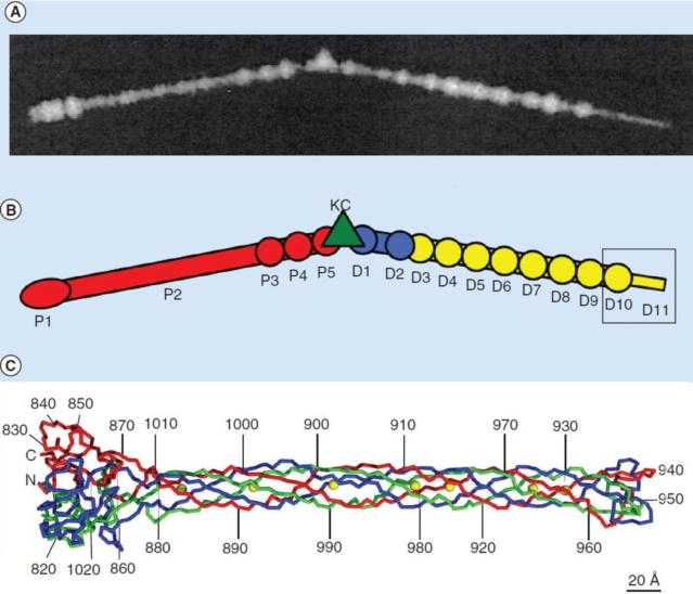
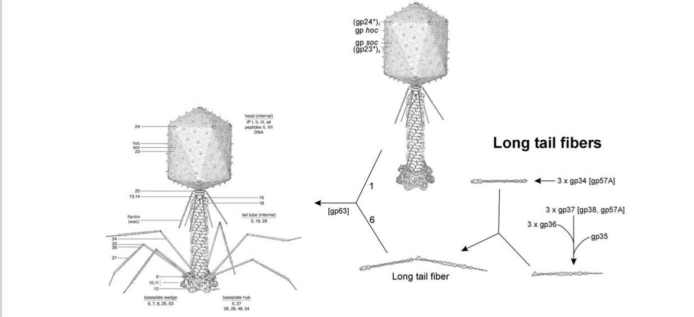
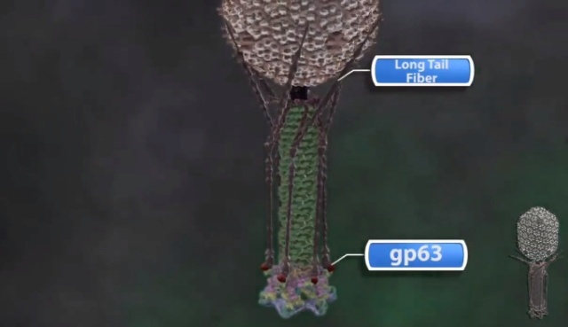
Moh Lan Yap (2015): The long tail fibers (LTFs) consist of four different proteins, namely gp34, gp35, gp36 and gp37 (Figure A & B). 

The amazing design of the T4 bacteriophage and its DNA packaging motor Bacter19
T4 long tail fiber
(A) Image of a long tail fiber.
(B) Domain organization of the long tail fiber. Domains of the proximal tail fiber are named P1–5 and of the distal half D1–11; gp35, or the KC is represented as a green triangle. Crystal structure of D10 and D11 (box) has been determined.
(C) Structure of gp37 (residues 785–1026). Three chains in the trimeric protein are colored red, green and blue, respectively, whereas iron ions are shown in yellow. The N- and C-termini and every 10th residue of chain A are labeled.

The chaperon protein gp57A is required for the trimerization of gp34 and gp37, whereas the chaperon protein gp38 is required for proper folding of gp37. The proximal half of the fiber is formed by gp34, which interacts with the adaptor protein gp9 on the baseplate. The monomeric gp35 forms the hinge or knee connecting the proximal and distal parts of the LTFs. The proximal and distal half-fibers assemble independently. Subsequently, the C-terminal part of gp36 binds to the N-terminal region of gp37. The distal part of the fiber is a trimer that can be divided by visual inspection of EM images into 11 domains (D1–D11) (Figure B). Domains D1 and D2 are a part of gp36 and D3–D11 are a part of gp37. The crystal structure of what had been assigned by EM as domains D10 and D11 of gp37 (residues 811–1026) has been determined (Figure C). This structure suggested that a more accurate description of this part of gp37 was in terms of a collar, needle and head domain. The head domain sits at the tip of the distal end and thus should be the T4 component that recognizes the receptor-binding site on the host cell. Since gp37 is known to bind to lipopolysaccharide, and as protein saccharide interactions usually involve aromatic side chains, a series of Tyr and Trp residues at the tip of gp37 might be important for host recognition. Sequence analysis shows that the collar and needle domains are conserved among other phages, whereas the head domain has diverged, suggesting that the host range specificity is determined by the head domain, consistent with the head domain having the receptor recognition function. 21

The amazing design of the T4 bacteriophage and its DNA packaging motor Struct24
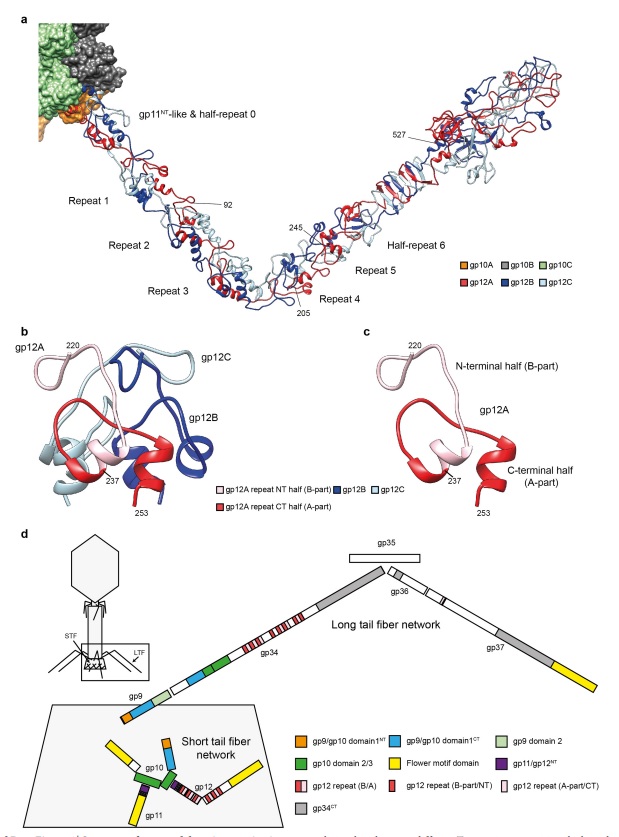
Structure of gp12 and domain organization of T4 fibres. 
a, Ribbon diagram of the gp12 trimer, anchored to gp10 domain 2 (shown in surface representation). The N-terminal part of the fibre (residues 2–245) was built de novo. 
b, Structure of the gp12 repeat. 
c, Fold of the polypeptide chain making up the repeat. 
d, Evolutionary relationships between different T4 proteins comprising the baseplate’s periphery and fibres. The size of each bar is proportional to the amino acid sequence length. The gp12 repeat shown in b and c constitutes a major part of the proximal LTF protein gp34

V. V. Mesyanzhinov (2004): THE STRUCTURES OF PHAGE FIBERS: Certain viruses, like adeno- and reoviruses, as well as many bacteriophages use fibrous proteins to recognize their host receptors. T4 has three types of fibrous proteins: the long tail fibers, the short tail fibers, and whiskers. The long tail fibers, which are ~1450 Å long and only ~40 Å in diameter, are primary reversible adsorption devices. Each fiber consists of the rigid proximal halves, encoded by gene 34, and the distal ones, encoded by genes 36 and 37. These halves are connected by gp35 that forms a hinge region and interacts with gp34 and gp36. The proteins that form the long tail fibers are homotrimers, except for gp35 that assembles as a monomer. The N-terminus of gp34 forms the baseplate-binding bulge, and the C-terminus of gp37 binds to a cell lipopolysaccharide (LPS) receptor. Two phage-encoded chaperones, gp57A and gp38, are required for assembly of both long tail fiber proximal and distal parts. Gp38 is a structural component for closely related T2 phage distal part of long tail fiber; it binds to the tip of gp37 and is responsible for receptor recognition. Gp57A is also required for assembly of short tail fibers. Another two assembly-assisted proteins, gp63 and gpwac, participate in the attachment of the long tail fibers to the baseplate. 


Structure of short tail fibers. 
The short tail fiber is a club-shaped molecule ~340 Å long consisting of a parallel, in-register assembled trimer of gp12 of 527 residues per polypeptide chain. Short tail fibers are attached to baseplate by the N-terminal thin part, while the globular C-terminus binds to the host cell LPS receptors. The ordered residues, 246-289, revealed a new folding motif, which is composed of intertwined strands. Residues 290-327 form a central righthanded triple β-helix. . X-Ray crystallography of this fragment at 1.5 Å resolution reveals the structure of the C-terminal part of the molecule that has a novel “knitted” fold, consisting of three extensively intertwined gp12 monomers, and interacts with LPS. The intertwining of the receptor binding domain represents a case of a 3D “domain swapping” phenomena found in several proteins. 

Fibritin structure (Whiskers) 
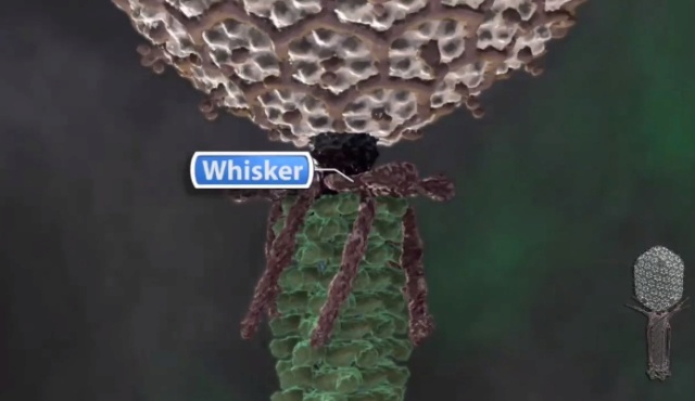
T4 fibrous protein, gpwac (whisker antigen control) or fibritin, is attached to the neck formed by gp13 and gp14, the collar and whiskers. Fibritin is a homotrimer assembled in parallel and attached to the T4 neck by the N-terminal domain. The C-terminal domain is a “foldon”, a unit required for fibritin folding and initiation of protein assembly. It was shown that the foldon provides correct alignment of three polypeptide chains [82, 83]. The foldon is a protein unit, which forms on the initial steps of folding and often remains intact after it is transferred into other proteins. Fibritin belongs to a specific class of accessory proteins acting in the phage assembly as a bi-complementary template accelerating the connection of the distal parts of long tail fibers to their proximal parts. Being a structural component of the mature phage particle, fibritin also works as a primitive molecular sensor. Under conditions unfavorable for phage growth (low temperature), fibritin holds the long fibers in a fixed position, raised to the tail and capsid, keeping virus particles noninfectious. 23


The amazing design of the T4 bacteriophage and its DNA packaging motor Bacter10
 
The amazing design of the T4 bacteriophage and its DNA packaging motor Virus_11


Φ29 Phage Molecular Motor DNA Packaging Mechanism - 3D version
https://www.youtube.com/watch?v=H0xdDaWcrdk


How DNA got into the bacteriophage
https://www.youtube.com/watch?v=RbL3BZCGPA4

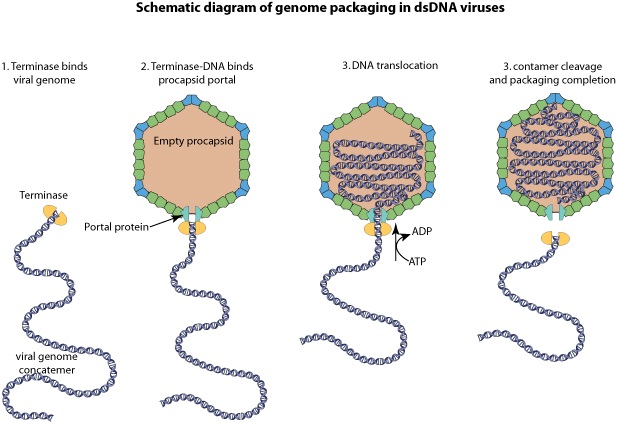
In a bacterium infected by T4 new bacteriophages are assembled in a stepwise process. The shaft builds up. DNA is replicated and the pro head assembles as an empty shell. But how does the DNA get into the prohead? The initiation of DNA import is not entirely clear yet but once DNA the packaging motor and pro head interact the DNA is rapidly threaded through a pore in the circular motor at the speed of about 2000 base pairs a second. Once the head is full, the packaging motor cuts the DNA, and the motor complex falls away. The shaft and long tail fibers are attached to complete the infectious particle. Within one hour more than 100 new phages are released from a single infected bacteria which makes the t4 phage one of the most efficient but also a fascinating killing machine


The argument from the DNA’s molecular motor
1. There is a “very fast and powerful molecular motor” that crams the viral DNA tightly into the capsid with the help of five moving parts.
2. The parts of the motor move in sequence like the pistons in a car's engine, progressively drawing the genetic material into the virus's head, or capsid.
3. The motor is needed to insert DNA into the capsid of the T4 virus, which is called a bacteriophage because it infects bacteria.
4. The T4 molecular motor is the strongest yet discovered in viruses and proportionately twice as powerful as an automotive engine. The motors generate 20 times the force produced by the protein myosin, one of the two proteins responsible for the contraction and strength of muscles.
5. Even viruses, which are not even alive by the scientific definition of being able to reproduce independently, show incredible design.
6. If design is what we observe, then there must be a designer.
7. God most probably, exists.

The assembly Pathway of bacteriophages

F.Arisaka (2005): Assembly of bacteriophage generally proceeds along a well-ordered pathway.  About 45 genes or gene products GP are involved in the assembly of the virion.  In order to assemble such a complex structure as T4, a number of intriguing “molecular devices” are employed. These “molecular devices” are considered to make the assembly efficient and free from errors

Scaffold processing and expansion
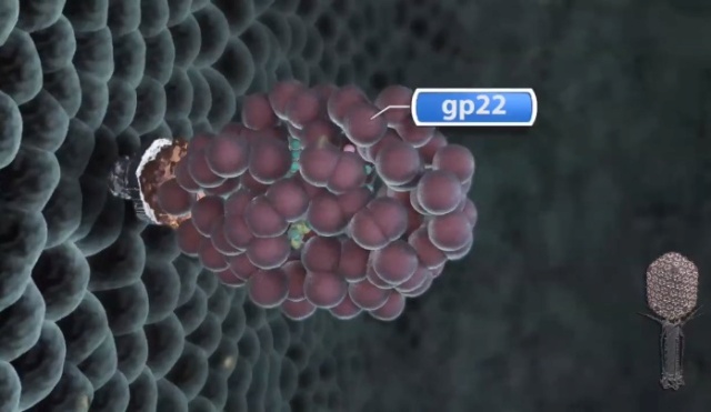
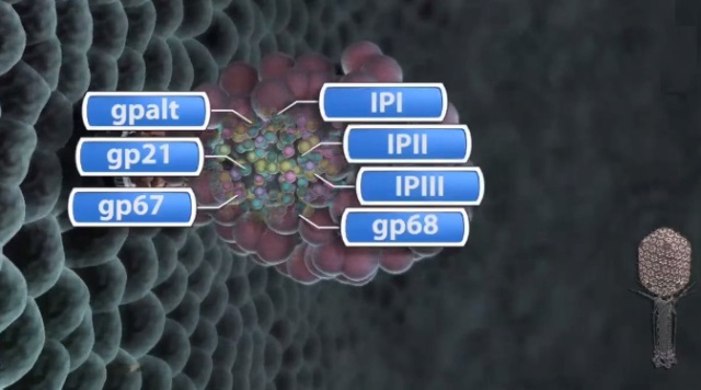
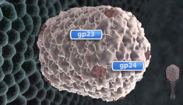
Head formation takes place on the inner membrane of the host E. coli. The initiation complex, made of twelve subunits of gp20, forms part of a neck or knob structure on which a scaffold of the head is formed. The main component of the scaffold is gp22, which is assumed to form a helix-rich ellipsoid of revolution together with minor components such as inner proteins I, II, III, and prohead protease gp21 or T4PPase. The major capsid protein gp23 forms the capsid, which is an icosahedron and elongated along the fivefold axis with T= 13 and Q= 20,6 where T is the triangulation number, which denotes the number of unit triangles in the p6 net, and Q is similar to the T number, but is related to the length of the elongated icosahedron. How the length of the head is determined is not yet fully understood, but the capsid formation starts as the scaffold formation is initiated and it is assumed to be determined by their interaction together with its intrinsic curvature. After the prohead is formed, most of the head proteins except for gp20 are processed or cleaved by T4PPase. T4PPase is specific for Glu residue, but appears to recognize higher order structure as well. The major capsid protein gp23 is cleaved between residue E65 and A66. The scaffold is cleaved into small peptides and is taken out of the prohead, making enough space for DNA packaging. As the processing of the prohead is completed, the prohead leaves the inner membrane and expansion takes place. It expands about 15%, accompanying the increase of the inner space by 50%. The expanded shell of the capsid is thinner but more rigid. The head is further strengthened by the accessory proteins, gp hoc highly immunogenic outer capsid and gp soc small outer capsid. Recently, the three-dimensional structure of gp24 was determined. Gp24 has a high homology to gp23 and forms pentamers at the vertices of the icosahedron, whereas gp23 forms hexamers on the surface. As the fold of gp23 would be similar, we can now predict the atomic structure of the whole head. Tail proteins are not processed with one exception, i.e., gp5 or tail lysozyme, where the peptide bond between Ser351 and Ala352 is cleaved by E. coli protease. This cleavage makes it possible for the lysozyme domain to be released from the rest of protein molecule during penetration of the tail tube 

HOST FACTORS ESSENTIAL FOR THE PHAGE ASSEMBLY 
As a virus by definition, phage T4 utilizes all the necessary bacterial organelles or protein complexes for its growth. Smaller phages are more dependent on their host functions than are larger phages. T4 phage is one of the largest phages and many functions are self-supplied, but it still requires a number of host factors. Such factors include the inner membrane of E. coli, where the initiation complex of the head is formed, RNA polymerase, ribosome, GroEL, etc. RNA polymerase is not encoded in T4, but the E. coli RNA polymerase undergoes some modifications by T4 proteins. Aside from the ADP-ribosylation of the  subunit of the holoenzyme, E. coli omega factor, 70, is replaced by T4-encoded omega factor gp55 to transcribe the late genes, which has a special consensus sequence of TATAAATA. In other words, the switch of transcription from early genes to late genes is accomplished by the replacement of the omega factor of RNA polymerase; from omega 70 to gp55. Most of the structural proteins for virion assembly are encoded in late genes. 19

Moh Lan Yap (2015): Bacteriophage T4 is classified as a member in the Myoviridae family of the Caudovirales order because it has a contractile tail. The head, the tail and the long tail fibers (LTFs) of T4 are assembled independently before they are joined together to produce a mature phage.The 168 kbp dsDNA genome of T4 is encapsidated in its head. A contractile tail is attached to a special portal vertex at one end of the head. A hexagonal baseplate is attached to the distal end of the contractile tail. Six long tail fibers  (LTFs) are attached to the periphery of the hexagonal baseplate. The LTFs are the sensors that can recognize receptor molecules on the host. There are six short tail fibers (STFs) folded beneath the baseplate that unfold upon host recognition. After unfolding these STFs bind irreversibly to the host cell, thereby increasing the efficiency of infection. The contractile tail improves the efficiency of infection by making it possible for the tail tube to penetrate the outer host cell membrane prior to the delivery of phage DNA into the host cell.

The amazing design of the T4 bacteriophage and its DNA packaging motor Bacter18
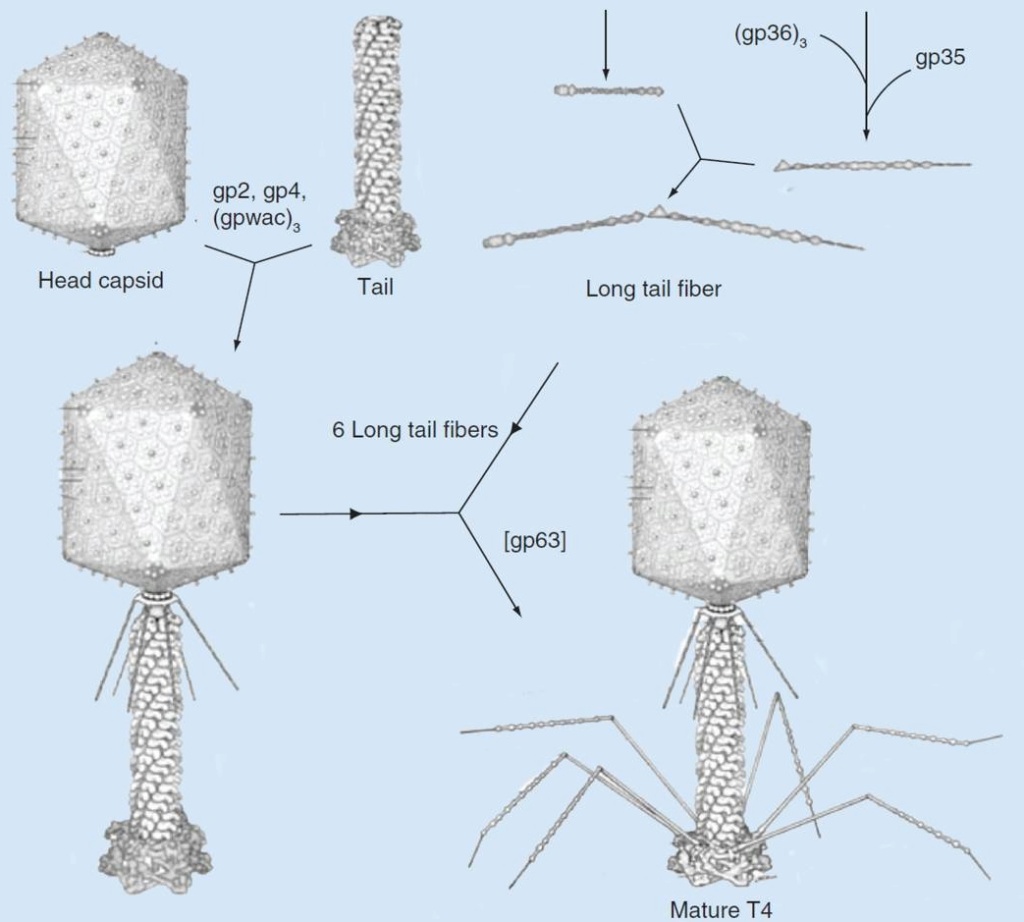
Assembly pathway of bacteriophage T4
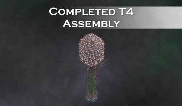
The assembly of T4 can be divided into three independent subassemblies: the head, the tail and the long tail fibers. The tail binds to the head followed by attachment of the fibritin protein at the neck region. Six long tail fibers then attach to form a viable T4 virion. 18

The amazing design of the T4 bacteriophage and its DNA packaging motor The_t414
The amazing design of the T4 bacteriophage and its DNA packaging motor The_t413
Morphogenesis of the bacteriophage T4 virion.
The overall assembly pathway can be divided into three independent stages: head, tail, and long tail fiber assembly. The chaperonines and catalytic proteins are indicated in brackets near the protein, or assembly step, that
requires the chaperonine. Known protein stoichiometries are given as subscripts. Crystal structures of structural proteins are shown as ribbon drawings. 16

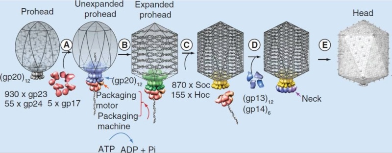
Zhihong Zhang (2011):  Complex viruses are assembled from simple protein subunits by sequential and irreversible assembly. During genome packaging in bacteriophages, a powerful molecular motor assembles at the special portal vertex of an empty prohead to initiate packaging. The capsid expands after about 10%–25% of the genome is packaged. When the head is full, the motor cuts the concatemeric DNA and dissociates from the head. These viruses encode powerful machines to package their genomes tightly inside an icosahedral-shaped capsid “head.” Packaging requires precise orchestration of a series of steps: assembly of an empty prohead, concatemer cutting and attachment of the motor-DNA complex to the portal vertex, ATP-fueled DNA translocation until the head is full, DNA cutting to terminate packaging, detachment of the motor, and sealing of the packaged head by “neck” assembly. The virion consists of a head into which the genome is packaged, and a tail that delivers the genome into the bacterial cell. A capsid of precise dimensions is first assembled, often with a single type of protein subunit polymerizing around a protein scaffold

A capsid of precise dimensions is first assembled, often with a single type of protein subunit polymerizing around a protein scaffold. A cone-shaped dodecameric portal initiates assembly and remains at the special five-fold vertex of the isometric capsid (prehead), facilitating all subsequent transactions: DNA entry, tail attachment, and DNA ejection. The scaffold is removed, creating an empty space inside the capsid (prohead or procapsid) for encapsidating the viral genome.

The amazing design of the T4 bacteriophage and its DNA packaging motor Sdffsd10
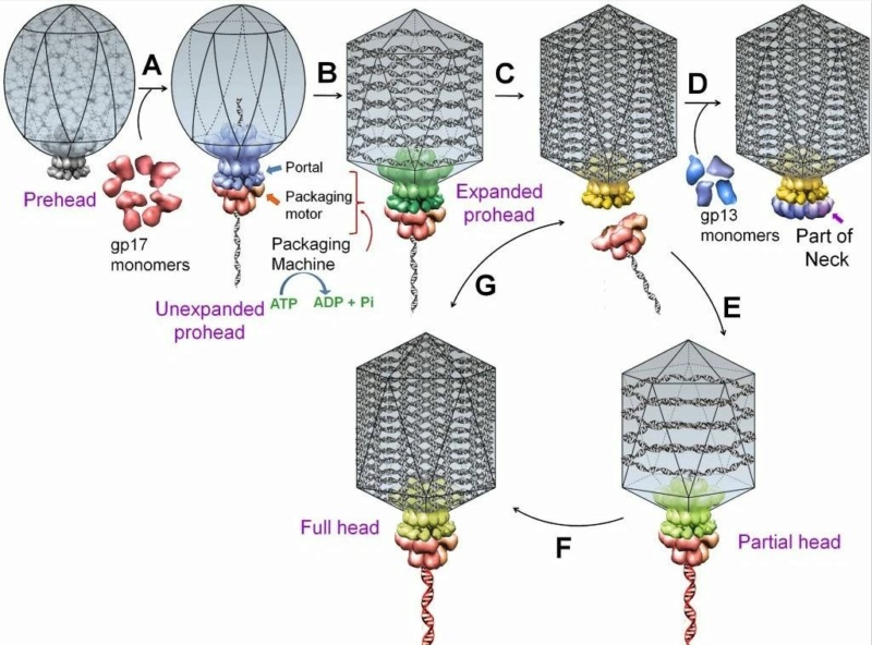
A schematic of DNA packaging by sequential assembly and promiscuous assembly.
The major capsid protein assembles around a scaffolding core into a prehead. The core is removed by proteolysis to produce an empty unexpanded prohead 
(A). The unexpanded prohead normally has a round shape, but in phage T4 it has angular geometry. The packaging motor–DNA complex docks on portal and initiates packaging. The prohead expands after about 10%–25% of the DNA is packaged 
(B). After headful packaging, the motor cuts the concatemeric DNA and dissociates from the DNA-full head 
(C). The neck proteins (gp13, gp14, and gp15) assemble on portal to seal off the DNA-full head and provide a platform for tail assembly 
(D). The various colors of portal represent different conformational states. In promiscuous assembly, the packaging motor assembles on a partial head produced by ejection of packaged DNA 
(E)  or a full head 
(G), and refills the head with new fragments of DNA 
([F] and [G]; new DNA fragments shown in red).

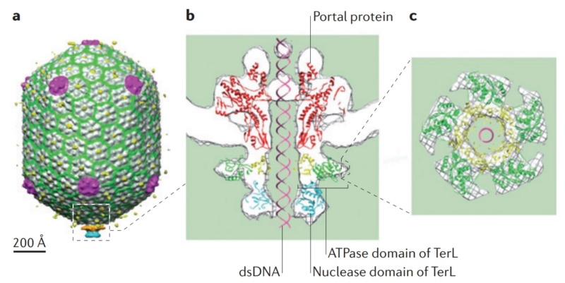
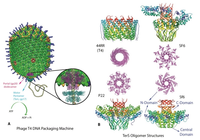
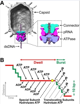
A packaging ATPase motor, also known as the “terminase,” recognizes and cuts the concatemeric viral DNA and docks at the narrow protruding end of the prohead portal, inserting the DNA end into the portal channel. The packaging machine thus assembled drives DNA translocation utilizing the free energy of ATP hydrolysis (Figure B above).

After filling the head (“headful” packaging), the motor cuts the DNA and dissociates from the DNA-full head (Figure C above). The neck and tail proteins assemble on the portal, completing the infectious virus assembly (Figure D above).

A fundamental feature of virus assembly is “sequential assembly” in which “simple” components assemble in a strict sequence to generate a complex nanomachine with unique biological properties. Each assembly step generates a new site or conformational state to which the next component binds with exquisite specificity, essentially irreversibly. A series of such steps, as documented by elegant studies in phage T4  and numerous other viruses, leads to rapid and high-fidelity assembly of a complex infectious virion. In phage T4, this process assembles virions approaching a theoretical infection efficiency of 1. 

The sequence of steps in the head morphogenesis of phage T4, as well as in other phages and dsDNA viruses (e.g., herpes viruses), is as follows:

(i) assembly of the packaging motor on a nascent (unexpanded) empty prohead (Figure A)
(ii) expansion of the capsid after about 10%–25% of the genome is packaged (Figure B)
(iii) packaging until the head is full
(iv) cutting of DNA and dissociation of the motor (Figure C)
(v) assembly of neck proteins to seal the packaged heads (Figure D)

Conformational changes in the portal are reported to drive these sequential irreversible transitions (Figure above; different colors of portal represent different conformational states) 12

Overview of Major Steps in T4 Head Assembly

M. YANAGIDA (1984): A fundamental question in the phage T4 assembly is how the capsid structure is formed. Clearly the processes must include a number of steps, but there is a distinct structural intermediate called the prehead. Intensive studies have been carried out to characterize the prehead. It is much less stable and smaller in size than the capsid and contains a protein core but has no DNA. The prehead is often associated with the inner side of the cell membranes of infected E. coll. A striking change in protein composition has been observed during the transformation of prehead to capsid; all but one of the prehead proteins are cleaved with the morphogenetic protease. The major core protein of the prehead is degraded and lost during the transformation. At least 12 polypeptide species are present in the head. It should be mentioned here that many of the head gene products are not incorporated into the mature head. For example, the product of gene 31 acts as a catalytic factor to prevent the random aggregation of coat protein. 24

The amazing design of the T4 bacteriophage and its DNA packaging motor Propos10
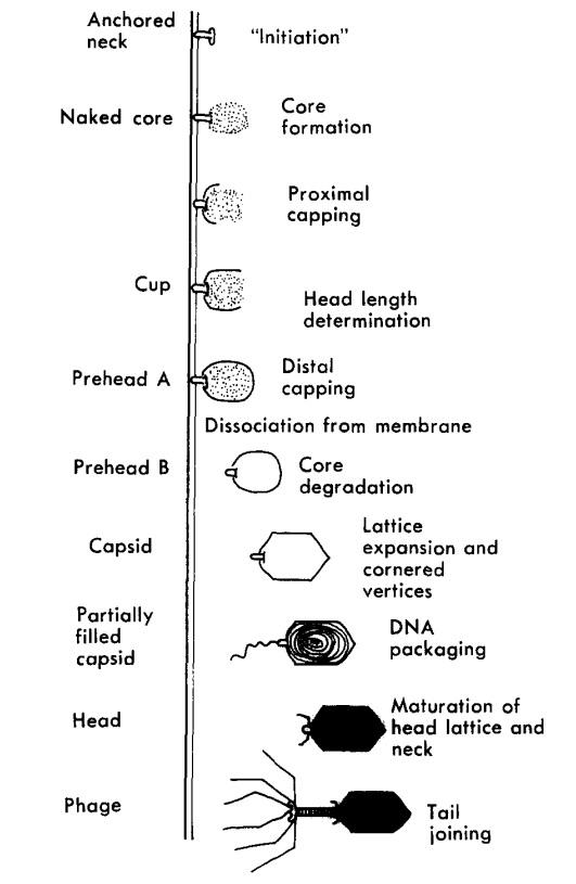
Proposed assembly pathway of a T4 head. 
The prehead assembly is directional, starting from the neck and ending at the distal cap (see text). All the head related structures except the anchored neck have been observed under two experimental conditions reported in this article (i.e., infection of wild type at 19°C and with gene 23 cs mutants at a restrictive temperature), and were combined with electron microscopy. 

Andreas Kuhn (2022): T4 head assembly initiates at the cytoplasmic surface of the inner membrane of E. coli by the interaction of the gp20 portal protein with the membrane insertase YidC (Figure 1). 


The amazing design of the T4 bacteriophage and its DNA packaging motor Overvi15

Figure 1. Overview of the major steps in T4 phage head assembly. 
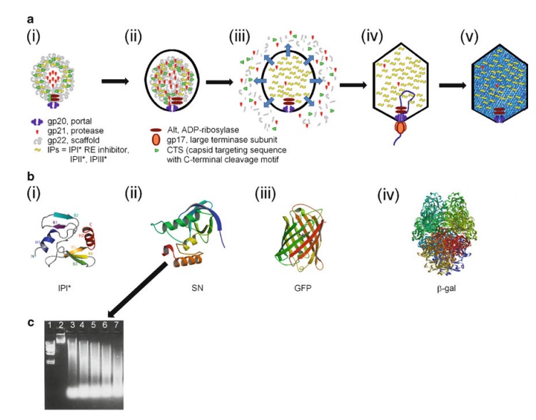
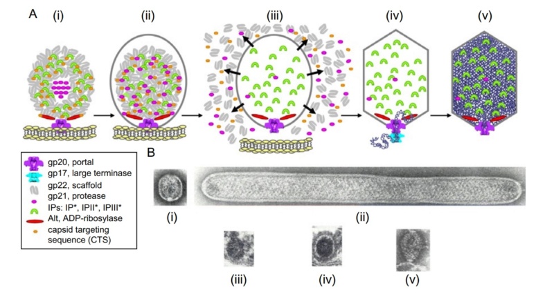
(A,B) Assembly initiates via the formation of a protein core anchored to the E. coli inner membrane via the portal, and around which the major capsid protein concomitantly assembles. Only in the absence of gp23 can naked cores be observed. 
(A) Electron microscopy of proheads produced by 21- mutants in vivo (upper, thin section) and in vitro (lower). White arrow indicates a central hole in proheads assembled in the absence of the prohead protease (adapted from van Driel, Traub and Showe [6]). (C,D) Proteolytic maturation involves cleavage and removal of scaffold/core proteins as well as the propeptides of the internal and shell proteins, release of the prohead from the inner membrane and semi-expansion of the shell. 
(E,F) Packaging of the genome into the prohead occurs via the action of the main packaging proteins, TerS (gp16) and TerL (gp17). 
(C,E). Electron microscopy of thin sections of wild-type T4-infected E. coli (reproduced from Black and Thomas [7]). 
(F) Scheme of how DNA packaging in vivo is integrated with late transcription, and DNA replication (reproduced from Black and Peng [8]). 
(G) Recombinant Δhoc phage particles after immuno-gold labelling with an anti-Hoc antibody (inset WT phage particle). The visualization of the “gold necklace” provided evidence that the portal structure contained fusion proteins (gp20-Hoc). Confirming the recombinant phenotype was important, as these particles had a central role in refuting the rotary portal packaging model (reproduced from Baumann, Mullaney and Black [9]). 
(H) Cryo-electron micrograph of a T4 Alt mutant imaged after the eighth exposure of a dose series of 16.5 el/Å2 per exposure). The bubbles are generated from the internal proteins, which are inferred to be randomly positioned within the DNA, but excluded from a zone of about 100–110 Å directly under the outer shell (reproduced from Wu et al. [10]).

The resulting dodecameric ring structure forms the foundation onto which a protein-only structure, the “core”, forms and acts as a scaffold to direct the correct assembly of the prolate exterior shell. Concomitantly with core formation, or immediately after, hexagons of the major shell protein gp23 and pentagons of gp24 assemble around the core, leading to the formation of a “prohead”. The core contains the proteins gp21, gp22, gp67 and gp68, together with the internal proteins Alt, IP1, IPII and IPIII. Proheads undergo proteolytic maturation by gp21, detach from the membrane, and the capsid shell expands to its final symmetry. DNA is then packaged into the capsid via interactions of the terminase with the portal protein. Once packaging is completed, a neck structure composed of gp13 and gp14 assembles onto the portal, allowing the addition of the tail and tail fibers. These steps in T4 assembly are typically referred to individually (e.g., assembly, maturation, packaging), but it is important to recognize that each is interlinked. Hence, each step is somewhat dependent on the former and/or concomitant steps, but also part of the overall process. 20

Assembly of the capsid shell

Moh Lan Yap (2015): There are several stages in the assembly of the head (Figure below): prohead formation, prohead proteolysis, DNA packaging, expansion of the prolate head and binding of the Hoc and Soc accessory proteins. The T4 prohead starts to assemble with the formation of the membrane-bound initiation complex, which comprises the portal protein gp20 mediated by the chaperone protein gp40. The initiation complex first attaches to the inner side of the host cytoplasmic membrane followed by binding of the prohead core (scaffold) proteins gp21, gp22, gp67, gp68, the initiation proteins IPI, IPII, IPIII and gpalt. Subsequently, the capsid proteins gp23 and gp24 start to form a shell around the core proteins to assemble into a prohead. Both the core and shell assemble concurrently. The major capsid protein gp23 requires two chaperone proteins, the host-encoded GroEL and the phage-encoded gp31, for proper folding. A total of 930 copies of the gp23 and 55 copies of gp24 are needed for the assembly of the prohead. The volume of the prohead is approximately 15–20% smaller than that of the mature head. The gp21 is then self-cleaved and activated to become a protease (T4PPase) in the prohead. The amino terminal peptides of gp23, gp24, IPI, IPII, IPIII and gpalt are cleaved, whereas gp21, gp22, gp67 and gp68 are extensively digested into small peptide fragments. All digested peptide fragments except for peptide II and IV of gp67 and gp22, respectively, are then expelled from the prohead. The cleaved head is released from the membrane into the cytosol and the gp23* and probably gp24* proteins undergo a large conformational rearrangement resulting in the expansion of the prohead. During this process, the capsid’s facets are flattened and the wall of the capsid becomes thinner. The processed prohead is then released from the membrane for DNA packaging. 18


The amazing design of the T4 bacteriophage and its DNA packaging motor T4_bac12
T4 head assembly
The dodecameric portal vertex acts as an initiator of head assembly. The major capsid protein assembles around a scaffolding core into a prohead. The core is removed by proteolysis to produce an empty unexpanded prohead.
(A) The packaging machine-DNA complex docks on portal and initiates packaging.
(B) The prohead expands after approximately 10–25% of the DNA is packaged.
(C) When the head is full, the packaging machine cuts the DNA and dissociates from the head.
(D) The neck proteins, gp13 and gp14, assemble on the portal to seal the DNA-full head.
(E) The accessory proteins, Hoc and Soc, bind to the head. The head is ready to be joined to the tail.

CrevInfo: Virus: Like DNA in a Hard Plastic Shell (2004):  A European team of biophysicists studied the mechanical properties of a virus and found the shell, made of protein, to act like hard plastic.  Writing in PNAS,1 they described the coat of a bacteriophage they studied:

I. L. Ivanovska (2004): The protective proteinaceous shells (capsids) of viruses are striking examples of biological materials engineering.  These highly regular, self-assembled, nanometer-sized containers are minimalistic in design, but they combine complex passive and active functions.  Besides chemical protection, they are involved in the selective packing and the injection of viral genetic material.
The capsids look like oblong, geometric shapes with pointy ends.  The DNA is packed inside under pressure, and the coat can withstand indentations of 30%.  “The measured Young’s modulus,” they found, “is comparable with that of hard plastic.”  They seemed to admire the little cases: the bacteriophage capsid is remarkably dynamic yet resilient and tough enough to easily withstand the known packing pressure of DNA (~60 atmospheres).  These capsids, thus, not only provide a chemical shield but also significant mechanical protection for their genetic contents.  Viral shells are a remarkable example of nature’s solution to a challenging materials engineering problem: they self-assemble to form strong shells of precisely defined geometry by using a minimum amount of different proteins. 10


The team is looking at these miniaturized packages for inspiration in the burgeoning field of nanotechnology.
 Here is observational evidence that leads to interesting questions.  It shows that living things need to overcome the same kinds of physics problems that engineers face.  Yet viruses are not, by definition, alive; they rely on a host for replication.  How could such precision bio-nanotechnology evolve?  Why do viruses exist?  Did they ever have a beneficial role, considering that the vast majority are harmless?  We may never be able to explain such things completely, but we can marvel at the biophysics capabilities found in nature, and deduce that such things don’t just happen.

The amazing design of the T4 bacteriophage and its DNA packaging motor Sem_tz40
The amazing design of the T4 bacteriophage and its DNA packaging motor Sem_tz41

The amazing design of the T4 bacteriophage and its DNA packaging motor Sem_tz42
(A) Assembly of T4 prohead and head.  (i) Membrane-bound portal initiates scaffolding core assembly; (ii) full prohead assembled on membrane; (iii) gp21 processing of core and procapsid components followed by detachment from membrane; (iv) DNA packaging into processed prohead; and (v) expanded fully packaged head. 
(B) Assembly intermediates: (i) portal bound scaffolding core with procapsid; (ii) giant procapsid and core; (iii) in vivo-assembled naked core on membrane is precursor to (iv) full prohead; and (v) portal initiator assembles size-determining scaffolding core in vitro. 21

Julie A. Thomas (2017):  In T4, proteolytic processing by gp21 is an essential step in head assembly and results in a vast remodeling of the capsid architecture, setting the stage for genome packaging (Black et al., 1994; Miller et al., 2003). Briefly, T4 heads assemble via nucleation of the portal ring on the inner host membrane. Upon this ring a spherical mass of protein assembles, referred to as the core structure, which consists of the scaffold protein, gp22 (576 copies), internal proteins (~1,000 copies), Alt (40 copies), gp67 (341 copies), gp68 (240 copies), and gp21 (100 copies) (Black et al., 1994). All of the T4 internal head proteins associate with the core structure via a capsid-targeting-sequence or CTS (Mullaney and Black, 1996). Upon completion of the core complex the shell then assembles around. The shell is composed of two proteins, the MCP, gp23 (960 copies) and gp24 (55 copies), which is located at the capsid vertices and is a paralog of gp24 22


https://www.youtube.com/watch?v=dMvU6Eh4nlw


M.G. Rossmann (2011) How proteins and nucleic acids assemble, often spontaneously, into structurally well-defined three-dimensional objects is an intriguing question. The limited size of the phage genome and the multi-component composition of bacteriophages make them well suited for assembly investigations. Genetic manipulation of phages has made it easy to observe the effects of gene inactivation on protein-protein association, providing information on the sequence of assembly processes.

The amazing design of the T4 bacteriophage and its DNA packaging motor Capsid11
Capsid assembly: refers to the formation of the capsid shell.
Packaging: refers to the viral genome placement inside a capsid or an envelope

Most viruses capsid spontaneously self-assembles around the viral genome in the cytoplasm, thus linking the assembly and packaging process.

Helical capsids are assembled around genomic RNA or DNA, and rely on self and nucleic acid interactions to assemble. Assembly and packaging are linked.
-<u>Negatives stranded RNA viruses genome is concomitantly encapsidated during replication. The packaging of these viruses occurs prior budding at the plasma membrane.

Icosahedric capsids usually assemble by affinity around the viral genome.
Complex capsids need the help of scaffolding proteins to assemble into empty procapsids. The scaffolding proteins are removed from the empty capsid by maturation events before packaging.
Nucleo-Cytoplasmic Large DNA viruses (NCLDV) contain an internal membrane, consequently they have a complex and regulated assembly mechanism. Poxviridae capsid-like protein is removed before virion maturation and serves as a scaffolding protein. 
"Reoviridae" and Totiviridae have their capsid assembled around messenger RNAs that are later replicated into genomic dsRNA, thereby hiding the dsRNA from cellular antiviral sensors.

The amazing design of the T4 bacteriophage and its DNA packaging motor Large_10

Large dsDNA bacteriophages, herpesviruses, adenoviruses and microviruses encode a powerful DNA-translocating machinenery that encapsidates a viral genome into a preassembled capsid or procapsid. The packaging machine is often composed of a portal structure, which provides a gate for DNA entry, and an ATP-driven motor. This motor is composed of the large subunit whose ATPase activity fuels DNA translocation, and most frequently, a small subunit that binds to the viral packaging site. DNA cleavage can be coupled to genome packaging.

Dong-Hua Chen (2010): Molecular Mechanism for Capsid Assembly, Scaffolding Protein Release, and Capsid Maturation:  To form a functional P22 procapsid, at least four types of proteins are required: coat, scaffolding, ejection, and portal proteins, in which the portal proteins form a unique 12-fold portal complex at a fivefold vertex. The lack of scaffolding proteins results in the failure to incorporate the portal and can lead to incomplete particles.
Based on our density maps, we propose that the formation of a unique portal complex with the requisite scaffolding and coat proteins is likely the key for initiating proper procapsid assembly


The amazing design of the T4 bacteriophage and its DNA packaging motor Pathwa11
Pathway for capsid assembly, scaffolding protein release and capsid maturation. 
(A and B) The portal (gray) associates with scaffolding (red) and coat (cyan) proteins to initiate procapsid assembly. The assembly continues with the addition of scaffolding and coat subunits to the growing shell 
(C) until the full procapsid is assembled 
(D). DNA is then packaged into the procapsid shell through the channel of the portal by the terminase motor. The scaffolding proteins are released by the electrostatic forces from DNA being packaged and exit through the central large openings of hexamers 
(E). During the release of scaffolding proteins, conformational changes associated with the maturation transition occur. After capsid expansion and DNA packaging, the tail hub, needle, and tail spikes are attached to the portal to form an infectious virion (magenta) 
(F). In B and C, the insets show the side views. In D and F, the insets show one hexamer and the adjacent pentamer rotated from the side view to the end-on view.

Scaffolding proteins may play a critical role during the capsid assembly nucleation because the portal would not be incorporated into the procapsid when the scaffolding proteins are absent. Once nucleated, procapsid assembly proceeds by the addition of scaffolding and coat subunits to the growing shell until the full procapsid is assembled with the proper size and shape as directed by the scaffolding proteins (Fig. 5 C and D). Coat proteins are likely added as monomers or dimers, with the mediation of scaffolding proteins, because our structures show that each scaffolding protein’s C-terminal helix-loop-helix motif interacts with the N arm of the corresponding coat protein. Though the exact timing is not known, ejection proteins are also incorporated and possibly interact with the scaffolding and portal proteins before procapsid assembly is completed. Once the capsid shell forms (Fig. 5D), DNA packaging can begin. In the early stages of DNA packaging, the terminase complex (gp2 and gp3) docks against the portal and hydrolyzes ATP to drive DNA into the procapsid shell through the portal.

Siyu Li ( 2018): More than 50 y ago, Caspar and Klug made the striking observation that the capsids of most spherical viruses display icosahedral order (IO), defined by 12 five-coordinated units (disclinations or pentamers) occupying the vertices of an icosahedron surrounded by hexameric units.  A nonspecific template not only selects the radius of the capsid, but also leads to the error-free assembly of protein subunits into capsids  with universal icosahedral order(IO). Under many circumstances, small icosahedral capsids assemble spontaneously around their genetic material. Larg double-stranded (ds) RNA or DNA viruses require what we generically denote as the template: scaffolding proteins (SPs) or an inner core. A “generic” template provides a robust path to self-assembly of large shells with IO. For large shells successful assembly into IO requires a nonspecific attractive interaction between protein subunits and a template. 8

Viruses: An Intelligent Design Perspective
“Our study shows that if a messy shell forms because of the high protein concentration or strong attractive interaction, then, as the shell grows larger, the cost of elastic energy becomes so high that several bonds can get broken, resulting in the disassembly and subsequent reassembly of a symmetric shell.”

The paper by Zandi’s team, published in ACS Nano, describes how the shells, even if disordered, can break apart and reassemble into symmetrical forms by physical forces like elastic energy. As the proteins attract,
the key for the disorder–order transition in both en masse and nucleation and growth pathways lies in the strength of elastic energy compared to the other forces in the system including protein–protein interactions and the chemical potential of free subunits. Our findings explain, at least in part, why perfect virions with icosahedral order form under different conditions including physiological ones.

Peter E. Prevelige (2011): Scaffolding proteins mediate, catalyze, and promote proper virus assembly. For many smaller viruses, all the information required for high fidelity assembly can be encoded, or self-contained, entirely in the coat protein subunits. However, larger viruses or small viruses frequently require additional proteins to insure robust assembly. Among those proteins are the “scaffolding” proteins, a class of auxiliary proteins that are present transiently during assembly and are not part of the final structure. Although common, scaffolding proteins are not ubiquitous. As coat proteins  (the capsid are called capsid proteins or viral coat proteins (VCP).) perform additional functions, efficient assembly may have become compromised. In some systems, best represented by the parvoviruses, maximal infectivity and/or fitness most likely requires constructing capsids with two or three-coat protein variants. The P = 3 picornavirus capsids represent a more complex example: capsids contain three unique coat proteins.  Most coat proteins will form aberrant capsid-like structures if left to their own devices. Therefore, a mechanism to ensure morphogenetic fidelity, vis-à-vis proper capsid size and shape formation is required, and this mechanism includes scaffolding proteins. There is a need to rapidly assemble capsids before cell death and/or programmed cell lysis. This contribution of this factor is most apparent with the microviruses, ostensibly simple viruses that accomplish an almost unimaginable fast replication cycle by employing two-scaffolding proteins.

Phage-encoded molecular chaperones 
F.Arisaka (2005): A molecular chaperone is a group of proteins which facilitate protein folding in the cell. GroE of E. coli was originally discovered as an essential host factor for the growth of phages. The name “GroE” stems from the fact that the growth of phage lambda requires a host factor and that the factor is required for the proper folding of the major capsid protein gpE. It was subsequently found that phage T4 also requires GroE for its growth and that groE consists of two genes: groEL and groES. Later, it was reported that they are heat shock proteins and molecular chaperones. GroEL, which is now known as Hsp60, together with GroES or Hsp10, is called chaperonin.  T4 phage requires GroEL for the folding of its major capsid protein gp23. The fact that T4 does not require GroES had been a mystery, but recent investigations have established that gp31 of phage T4 functions as GroES in the T4-infected cells. Gp31 forms a heptameric ring, as does GroES. The high-resolution structure of gp31 and GroES complexes revealed that their three-dimensional shapes are very similar, except that the former has an extra loop that made the “Anfinsen cage” of the GroEL/gp31 complex larger than that of GroEL/GroES. “Anfinsen cage” denotes a hole inside the chaperonin complex, where the intermediate folding protein is eventually completely folded. The reason that GroEL/gp31 is required but not the Gro EL/GroES system for gp23 folding is unknown, but recent investigation indicated that the GroEL/gp31 complex, but not GroEL/GroES, has the affinity to the folding intermediate of gp23, the major capsid protein. There are a number of other phage-encoded chaperone or chaperone-like proteins. Gp40 is known to be essential for head formation and is a membrane protein, but its precise function is not known. Gp57A is essential for the fiber formation of both long and short tail fibers. This is a small protein with 79 amino acid residues. It is a helix-rich fibrous protein and hexamer appears to be the main oligomer species, which reversibly dissociates into trimers and then monomers. The mechanism of chaperone function is not known. Gp38 is also essential for the fiber formation of the distal half-fibers. Gp51 has long been known to function catalytically for the formation of the hub of the baseplate. On the other hand, gp63 is known to facilitate the binding of tail fibers to the baseplate. In the absence of gp63, the tail fiber attachment is much slower, but the mechanism is not known. Gp63 has an RNA ligase activity, but that activity is not related to the tail attachment function.

E. Fibritin „whisker… and foldon, an internal molecular chaperone 
Another protein that also facilitates the binding of long tail fibers to the baseplate is a structural protein called “whiskers” or “fibritin.” They are trimeric fibrous protein with coiled-coil structure, and six of them stick out from the neck. They are not essential, but without them, the tail fiber attachment takes a much longer time. The distal end of the fibritin, the C-terminal domain, binds to the kink region of the long tail fiber i.e., the junction between the distal and proximal tail fibers and orients the fibers so that the proximal end of the tail fiber approaches the baseplate, which facilitates the binding of the tail fibers to the baseplate. The C-terminal domain, consisting of 27 amino acid residues, is essential for trimerization of the fibritin. It is called “foldon.” When the foldon part was cloned and overexpressed, it spontaneously formed a trimer.  Apparently, the foldon domain facilitates the alignment of the other part of the three polypeptide chains and functions as an internal chaperonin. The foldon has been fused to the C-termini of several other proteins including collagen peptides and has been shown to successfully make the homogeneous trimeric collagen helix. 19

Assembly of the baseplate

Nicholas M. I. Taylor (2018): More than 24 structural proteins and chaperones participate in the assembly of T4 baseplate. The above-mentioned baseplate centerpiece gp48 and the central spike complex proteins gp5, gp5.4 and gp27 – form a continuation of the tube and the other four – gp6, gp7, gp25 and gp53 – create the bona fide ‘plate’.  Gp6, gp7, gp25 and gp53 are part of a large assembly intermediate that has been historically called the ‘wedge’ although the shape of this complex is more complicated than that of a simple wedge 23

Moh Lan Yap (2016):  Bacteriophage T4 consists of a head for protecting its genome and a sheathed tail for inserting its genome into a host. The tail terminates with a multiprotein baseplate that changes its conformation from a “high-energy” dome-shaped to a “low-energy” star-shaped structure during infection. Although these two structures represent different minima in the total energy landscape of the baseplate assembly, as the dome-shaped structure readily changes to the star-shaped structure when the virus infects a host bacterium, the dome-shaped structure must have more energy than the star-shaped structure. This structure, together with other genetic and structural data, shows why the high-energy baseplate is formed in the presence of the central hub and how the baseplate changes to the low-energy structure, via two steps during infection. Thus, the presence of the central hub is required to initiate the assembly of metastable, high-energy structures. If the high-energy structure is formed and stabilized faster than the low-energy structure, there will be insufficient components to assemble the low-energy structure. Most bacteriophages have a tail. At the distal end of the tail there is usually a baseplate that is decorated by some fibers. The baseplate initiates infection when the tail fibers bind to a host cell. Signals are transmitted from the tail fibers via the baseplate to the tail that then trigger the ejection of the phage genome from the head into the host cell through the tail tube. Two evolutionary-related structures, of pyocin and of the type VI secretion system, are found in bacteria as defense systems to kill competing bacteria. These structures are remarkably similar to the tail baseplate structure of bacteriophages, suggesting that tail baseplate-like structures are effective organelles for infecting bacteria. T4 is a member of the Myoviridae family of bacteriophages. These phages have a sheath around the tail tube that contracts during infection (Figure below).

The amazing design of the T4 bacteriophage and its DNA packaging motor Schema16
Schematic diagram of bacteriophage T4.
Bacteriophage T4 has a contractile tail and a complex baseplate. Six long-tail fibers are attached to the upper part of the baseplate and six short-tail fibers are folded under the baseplate before infection.

T4 has a complex baseplate that is essential for assuring a highly efficient infection mechanism. After recognition of an Escherichia coli host cell by some of the six long-tail fibers (LTF), the short-tail fibers (STF) that are a part of the baseplate, bind irreversibly to the cell. This process is accompanied by a large conformational change in the baseplate from a “high-energy” dome- to a “low-energy” star-shaped structure, although each of these structures represent an energy minimum in the energy landscape of the baseplate assembly. This change triggers contraction of the tail sheath, driving the tail tube into the outer host cell membrane and further across the periplasmic space to the inner membrane. The genomic DNA is then ejected into the host’s cytoplasm. Hence, the baseplate serves as the nerve center for transmitting signals from the tail fibers to the head for the release of DNA into the host.

The hexagonal dome-shaped T4 baseplate assembles from six wedges surrounding a central hub. A total of 134 protein subunits from 15 different proteins form the ∼6.5-MDa baseplate. The assembly of a wedge had been shown to follow a strictly ordered sequence. First, an initial complex is formed by a monomer of gp7 and a trimer of gp10, followed sequentially by binding of a dimer of gp8 and a dimer of gp6 to the complex. In the absence of a central hub, at least five proteins (gp7, gp10, gp8, gp6, and gp53) are required for assembly of wedges in vitro into a star-shaped, low-energy baseplate-like structure. Assembly of the high-energy, dome-shaped structure requires the presence of the central hub. However, how the sequential wedge assembly events are regulated remained unknown. In particular, the question remained how the high-energy dome-shaped baseplate could assemble.

How is it possible to assemble the high-energy dome-shaped baseplate initially as opposed to the low-energy star-shaped baseplate? After the individual wedges have assembled, six wedges assemble around the central hub to form the dome-shaped baseplate (Fig. 4B and Movie S2). The present structure has shown the complete structure of gp6, which when fitted into the cryo-EM density of the dome-shaped baseplate shows that gp6 binds tightly around the central hub protein gp27 (Fig. 5A).

The amazing design of the T4 bacteriophage and its DNA packaging motor Pnas_w11
Assembly of a baseplate based on present and earlier results.
(A) Wedge assembly. Gp10, gp8, and gp6 bind sequentially to the gp7 backbone protein. The central hub of the baseplate is assembled independently.
(B) Baseplate and tail assembly. Six wedges assemble around the central hub to form a baseplate. Gp53 binds adjacent wedges together. Subsequently, gp9 and the gp11–gp12 complex bind to the baseplate, further stabilizing the dome-shaped configuration. Then, gp48 and gp54 bind to the top of the central hub and initiate polymerization of the tail tube. Gp25 attaches to the gp48–gp54 complex, initiating polymerization of the tail sheath. For clarity, only three rings of the tail sheath are shown.

1. http://www.nature.com/ncomms/2015/150706/ncomms8548/full/ncomms8548.html
full text pdf: http://www.nature.com/ncomms/2015/150706/ncomms8548/pdf/ncomms8548.pdf
2. Amy D. Migliori: Evidence for an electrostatic mechanism of force generation by the bacteriophage T4 DNA packaging motor 17 June 2014
3. http://mmbr.asm.org/content/75/3/423.full.pdf
4. http://www.ncbi.nlm.nih.gov/pmc/articles/PMC1208326/
5. http://www.uncommondescent.com/intelligent-design/in-other-words-phylogenetic-reconstruction-is-sheer-fantasy/
6. http://physicsworld.com/cws/article/news/2014/jun/25/relaxation-and-repulsion-helps-viruses-pack-dna
7. Dong-Hua Chen: Structural basis for scaffolding-mediated assembly and maturation of a dsDNA virus October 22, 2010
8.Siyu Li: Why large icosahedral viruses need scaffolding proteins October 9, 2018
9. Peter E. Prevelige: Building the Machines: Scaffolding Protein Functions During Bacteriophage Morphogenesis  01 January 2011
10. I. L. Ivanovska: Bacteriophage capsids: Tough nanoshells with complex elastic properties May 7, 2004
11. Lei Sun et al.,: Cryo-EM structure of the bacteriophage T4 portal protein assembly at near-atomic resolution 06 July 2015
12. Zhihong Zhang: A Promiscuous DNA Packaging Machine from Bacteriophage T4 2011 Feb 15
13. John E. Johnson:  The Structure of an Infectious P22 Virion Shows the Signal for Headful DNA Packaging (2006)
14. Peter E. Prevelige: Building the Machines: Scaffolding Protein Functions During Bacteriophage Morphogenesis  01 January 2011
15. Moh Lan Yap: Role of bacteriophage T4 baseplate in regulating assembly and infection February 29, 2016
16. M. G. Rossmann et.al: Structure and morphogenesis of bacteriophage T4 November 2003
17. M. G. Rossmann et.al: Morphogenesis of the T4 tail and tail fibers 03 December 2010
18. Moh Lan Yap: Structure and function of bacteriophage T4  2015 Aug 1
19. Fumio Arisaka: Assembly and infection process of bacteriophage T4 03 November 2005
20. Andreas Kuhn: The Beauty of Bacteriophage T4 Research: Lindsay W. Black and the T4 Head Assembly 28 March 2022
21. Lindsay W. Black Structure, Assembly, and DNA Packaging of the Bacteriophage T4 Head 2012
22. Julie A. Thomas To Be or Not To Be T4: Evidence of a Complex Evolutionary Pathway of Head Structure and Assembly in Giant Salmonella Virus SPN3US 15 November 2017
23. Nicholas M. I. Taylor: Contractile injection systems of bacteriophages and related systems 05 February 2018
24. MITSUHIRO YANAGIDA: MOLECULAR ORGANIZATION OF THE HEAD OF BACTERIOPHAGE Teven: UNDERLYING DESIGN PRINCIPLES 1984


Last edited by Otangelo on Sat Oct 29, 2022 9:18 am; edited 73 times in total

https://reasonandscience.catsboard.com

Otangelo


Admin

Thus, the nucleation of the baseplate assembly from individual wedges is dependent on the interaction of gp6 with gp27. However, in the star-shaped baseplate structure the tight ring of six gp6 dimers around gp27 has expanded and lost contacts with gp27. Thus, the initiation of baseplate assembly is the association of gp6 with gp27, from wedges that are probably assembled as in the dome-shaped baseplate. In the absence of gp27, as was the case for the in vitro-assembled baseplate reported here, gp6 can assemble only as a star-shaped baseplate. Thus, the route to the low-energy star-shaped wedge is via the initially assembled dome-shaped wedge.

Infection Mechanism.
Phage T4 uses the LTFs to recognize a LPS and/or OmpC receptor on the surface of the host cell. Because phages have been observed that have baseplates, which have changed to star-shaped structure, but that still have extended sheaths, presumably, the initial step of infection is the change of the dome-shaped structure to an intermediate structure in which the STFs are released. Subsequently, attachment of the STFs to cell-surface molecules triggers the baseplate to change its conformation to the final star shape, which in turn causes the sheath to contract and the tail tube to puncture the outer cell membrane. Thus, it is likely that the conformational changes of the baseplate occur in two steps—namely, first from the dome-shaped structure to an intermediate star-shaped structure and then from the intermediate structure to the final star-shaped structure.

The LTFs are attached to the baseplate via an adaptor protein gp9. The fit of the gp7 and gp9 structures into the 16-Å resolution cryo-EM density of the dome-shaped baseplate shows that domain III of gp7 interacts with density that represents the N-terminal part of gp9 (Movie S2). Thus, a flexible region of gp9 contacts gp7, allowing the LTFs to have multiple conformations in solution. When the LTFs attach to the cell, the Brownian motion of the phage particle may cause these flexible regions (31) to pull on domain III of gp7. This domain has extensive interactions with the gp8 dimer in the adjacent wedge, and with the C-terminal domain IV of gp10 in the same wedge. The rigid gp8 dimer in turn interacts extensively with the C-terminal domain VI of gp7, which is attached to the N-terminal domain I of gp10. Therefore, the interaction of gp9 with domain III of gp7 is likely to disturb interactions among gp7, gp8, and gp10 at the vertices of the dome-shaped baseplate and trigger the periphery of the baseplate to change to a star-shaped conformation (Movie S3). As a result, the STFs attached to gp10 might unfold from the baseplate and point toward the cell surface with their C-terminal domain, whereas the N-terminal part of the STFs remain attached to domain II of gp10, after release of gp11 (Fig. 6). During the conformational change from dome-shaped to a star-shaped intermediate structure, the gp11 molecules, attached to domain III of the gp10 molecules, move closer to the phage head and push on the end of the LTFs close to the baseplate. The interaction of gp11 with the LTFs probably causes the parts of the LTFs closest to the baseplate to rotate with respect to the baseplate as seen in the star-shaped baseplate. Thus, LTFs attached to the cell surface are used as levers to bring the phage closer to the cell surface. On completion of these movements, the baseplate will have the structure of the intermediate state that has a star-shaped periphery (gp7, gp8, gp9, gp10, gp11, and gp12), although the inner ring of gp6-gp53-gp25 molecules probably remains attached to the hub (Fig. 6).

Assembly of the tail

The amazing design of the T4 bacteriophage and its DNA packaging motor Assemb13
Assembly of the tail. Rows A, B and C show the assembly of the wedge; the baseplate and the tail tube with the sheath, respectively.

M. G. Rossmann et.al (2010): The fully assembled baseplate is a prerequisite for the assembly of the tail tube and the sheath both of which polymerize into the extended structure using the baseplate as the assembly nucleus (Figure 2). The baseplate is comprised of about 140 polypeptide chains of at least 16 proteins. Two gene products, gp51 and gp57A, are required for assembly, but are not present in the final particle. The baseplate has sixfold symmetry and is assembled from 6 wedges and the central hub. The only known enzyme associated with the phage particle, the T4 tail lysozyme, is a baseplate component. It is encoded by gene 5 (gp5). 17

The amazing bacteriophage DNA packaging motor

M. YANAGIDA (1984): Inside the head, a 50micrometer long DNA molecule (500 times the head size) is packed together with internal protein, peptides, and polyamines. Upon osmotic shock, DNA and the internal components are released, and the remaining protein shell is called a capsid. 22

Purdue (2220): In december 2000, Scientists solved the three-dimensional structure of the central component of a biological "motor" that powers the DNA packaging system in a virus, providing scientists with their first glimpse of such a motor system. The study describes atom-by-atom how the core of the tiny motor, just millionths of a millimeter in size, is constructed and suggests how it works to translocate, or pack, long stretches of the virus' genetic material into its outer shell during the process of viral replication. "Though other motor systems have been studied in biology, this is the first motor known to translocate genetic material."Viruses are essentially a simple parasite consisting only of an envelope that contains the genetic material ready for transportation from one host to another. They can reproduce only after infecting a host cell. Once inside a cell, the virus manipulates the cell's machinery to produce all the necessary components, including genetic material, to assemble new viruses. It is here that the biological motor is needed to fill newly assembled envelopes with their genetic material The new viruses are then released from the host cell and are free to infect other cells. The DNA packaging motor is comprised of three primary parts: an elongated prohead that serves as the virus shell a doughnut-shaped connector that is positioned at the entrance to the virus shell and feeds DNA into the shell a novel ribonucleic acid (RNA)-enzyme complex that converts chemical energy to mechanical energy needed for packaging. The connector is made up of 12 protein subunits that may serve as "cylinders" in the motor system to pull long chains of DNA through the center of the doughnut-shaped system. Five identical enzymes, called ATPases, are positioned around the connector, just outside the opening in the virus shell. The enzymes break down the cell's chemical fuel, called ATP, to produce the energy needed to power the motor. Successive chemical reactions produced by the ATP cause the phi29 connector to oscillate and rotate, pulling the DNA into the shell two base pairs at a time."Our results suggest that the prohead and connector comprise a rotary motor, with the head and ATPase complex acting as a stator and the DNA acting as a spindle.  5

CrevInfo: Handy Motor Found in Virus (2008): Your job today is to stuff a delicate chain into a barrel without breaking it and make it wrap neatly inside.  A tiny virus does this with helping hands.  A research team uncovered the mechanism of a “powerful molecular motor” that crams the viral DNA tightly into the capsid with the help of five moving parts. These are not real hands, of course; the captioned artist rendering whimsically shows five gp17 protein structures drawn to look like hands. These structures are protein subdomains embedded in the ring-shaped motor mechanism.  The capsid, or viral container, acts like a hard plastic shell, protecting the DNA inside.  Two rings on the opening hold the motor in place.  The gp17 subdomains take turns grasping the DNA and shoving it in.  Another analogy is that they work like pistons operating in sequence.  Using ATP energy pellets, they take advantage of electrostatic forces to gently but firmly transfer the DNA strand into the interior, where it coils in an orderly fashion.  The mechanism generates 20 times the force used by myosin, the motor in muscle.  The article claims that proportional to its size, the motor is twice as powerful as an automotive engine. The virus they studied is a bacteriophage – a virus that infects and destroys bacteria.  The cutaway diagram of the capsid shows the DNA wound neatly into a fabric-like pattern. 

Even viruses, which are not even alive by the definition of being able to reproduce independently, show incredible design.  They are too well designed to be accidents.  Why do so many viruses cause disease and death?  Actually, only a small fraction are harmful; most cause no harm and some are beneficial. Some creationists speculate that they all had a beneficial function originally: keeping bacteria in check or delivering genetic instructions to animals encountering a new environment.  After the Fall, they either were allowed to mutate into machines gone wild, turning on their customers or were recast as agents of judgment on a world sentenced to death and hardship.  Can they prove this?  No; but what is the alternative?  First, they have to believe in miracles – that super-efficient, compact, powerful motors like this just appeared, arose or emerged (favorite Darwinian miracle-words) from nowhere.  Second, they have to deny that anything is evil or out of order.  In Darwin’s world, whatever is, is right.  A logical consequence is that it is vain to seek cures for disease.  So what if millions of humans die in a pandemic?  It just shows that viruses are more fit. If miracles and apathy don’t motivate you to swallow the evolutionary line, then look at the mechanism from a design perspective and figure out what it’s there for.  Basic research can reveal the mechanism.  Philosophy and theology can elucidate their purpose.  Engineering can look for applications.  Who needs Darwin, the guy who sits around telling miracle stories? The Purdue team obviously didn’t act like “nothing in biology makes sense except in the light of evolution.”  They had no need for that hypothesis.  The E-word failed to materialize in the press release or any of the writeups on other sites.  “Viruses, start your engines!” EurekAlert began its version.  “Researchers find what drives one of nature’s powerful, nanoscale motors.” If design is what you observe, then design will lead to the right explanation, which may lie outside the capabilities of science. 3

The amazing design of the T4 bacteriophage and its DNA packaging motor Virus_10
This motor is made of two ringlike structures, and both of these discs contain five segments made of a protein called gp17. The image shows a cross-section of the virus head, or capsid, and an artist's interpretation of the motor as it packages DNA into the virus. The hands represent the five segments of the ringlike structures. Each hand takes a turn grabbing the DNA and moving it into the head until the head is full.

Purdue (2008):  Researchers have discovered the atomic structure of a powerful "molecular motor" that packages DNA into the head segment of some viruses during their assembly, an essential step in their ability to multiply and infect new host organisms. Researchers, from Purdue University and The Catholic University of America, also have proposed a mechanism for how the motor works. Parts of the motor move in sequence like the pistons in a car's engine, progressively drawing the genetic material into the virus's head or capsid, said Michael Rossmann, Purdue's Hanley Distinguished Professor of Biological Sciences. The motor is needed to insert DNA into the capsid of the T4 virus, which is called a bacteriophage because it infects bacteria. The same kind of motor, however, also is likely present in other viruses, including the human herpes virus. This particular motor is very fast and powerful. The T4 molecular motor is the strongest yet discovered in viruses and proportionately twice as powerful as an automotive engine. The motors generate 20 times the force produced by the protein myosin, one of the two proteins responsible for the contraction and strength of muscles. The virus consists of a head and tail portion. The DNA-packaging motor is located in the same place where the tail eventually connects to the head. Most of the motor falls off after the packaging step is completed, allowing the tail to attach to the capsid. The DNA is a complete record of a virus's properties, and the capsid protects this record from damage and ensures that the virus can reproduce by infecting a host organism. The packaging motor is made of two ringlike structures, and both of these discs contain five segments made of a protein called gp17, for gene product 17.  One disc sits on top of the other, and each of the five segments of the top disc shares a gp17 protein with a corresponding segment in the bottom disc. The gp17 proteins have two segments, or domains, one segment in the lower disc and the other segment in the upper disc. The lower disc first attaches to the DNA and is then drawn upward by the upper disc, pushing the DNA into the virus's capsid in the process. The top disc of the motor pulls the lower disc upward using electrostatic forces generated between oppositely charged objects. The motor is dynamic and apparently exists in two states: relaxed and tensed, the latter likely occurring when the top disk has attracted the lower disc. 6

M. G. Rossmann (2008): Nucleases are essential for DNA processing, such as replication and repair. Incorrect or ill-timed cutting can lead to genome instability and loss of viability of virus, cell or organism. In most tailed bacteriophages and herpes viruses, the product of viral genome replication is a concatemer, a branched head to tail polymer of the viral genome. Phage ‘terminase’, a hetero-oligomeric complex of small and large terminase subunits, recognizes the viral genome, makes an endonucleolytic cut, and links the end of the genome to an empty prohead by docking at the special portal vertex.

The amazing design of the T4 bacteriophage and its DNA packaging motor Mmi_6344_f1
The gp17 C-terminal domain has nuclease active site.
A. Schematic of phage T4 DNA packaging motor showing the large terminase protein, gp17, bound to the special dodecameric portal vertex (gp20, violet) of the prohead. gp17 consists of an N-terminal ATPase domain (aa 1–360, blue) and a C-terminal endonuclease domain (aa 361–610, yellow).
B. The nuclease domain of 16 T4-family phage large terminase proteins contains a highly conserved Asp (purple) previously shown to be essential for in vivo DNA cleavage. Combinatorial mutagenesis identified that E404 (blue), G405 (green) and D409 (orange) are critical for function, and also conserved among the T4-family terminases.
C. The in vivo nuclease of gp17 mutants. The plasmid DNA was isolated at 0, 60 and 120 min after IPTG induction. Lane M shows λ-HindIII size standards. See Experimental procedures for more details.

The packaging motor, powered by an ATPase present in the large terminase protein, translocates DNA into the prohead. When the head has been filled with one (cos phages) or slightly more than one (pac phages) genome, the DNA is cut again and the filled head is disengaged from the terminase. The mechanism by which the ‘headful’ nuclease catalyses DNA cleavage is poorly understood for any phage system. 8

Purdue (2007): The ATPase enzyme provides energy to run the motor needed to insert DNA into the capsid, or head, of the T4 virus. "The virus first assembles the protein shell of the head and then packages the DNA into this empty capsid," Rossmann said. "This process could be likened to building a house and then filling it with furniture." The DNA is a complete record of a virus' properties, and the capsid protects this record from damage and ensures that the virus can reproduce by infecting a host organism. Energy to run the packaging motor is produced when the ATPase enzyme uses ATP. This release of energy is used to run the molecular motor in the T4 virus. The DNA-packaging motor is located in the same place where the tail eventually connects to the head. The motor falls off after the packaging step is completed, allowing the tail to attach to the capsid. DNA is made of four different kinds of "nucleotides" identified by a specific "base." The bases are paired together to form the rungs of a ladderlike, double-stranded helical structure. Because there is a negative charge associated with each nucleotide, they repel each other when compressed together, creating a pressure inside the confining space of the capsid. A motor is needed to counteract this pressure, in effect pumping the DNA into the head. The authors of the research paper have proposed a mechanism for how the motor works by comparing its structure to those of other, similar enzymes called helicases. The helicases are needed to separate double-stranded DNA into single strands during gene replication. Helicases alternatively bind to and release their grip on DNA during replication, progressively moving along the helix to separate the strands in a motion similar to an inchworm's movement. The authors proposed that the motor uses a similar inchworm mechanism to package the DNA into the virus. "While the helicases use the mechanism to unwind double-stranded DNA, this ATPase uses the mechanism to pump genetic material into the virus capsid," Sun said. 8

CrevInfo: We Are Filled with Viruses (2011):  Viruses have a bad connotation.  We immediately think of the ones that cause disease: “I’ve got a virus,” you say when feeling under the weather.  Actually, you have trillions of them all the time, even in the best of health.  A single gram of stool sample can have 10 billion of them! What does that mean?  Scientists are only beginning to find out. In the past decade, scientists have come to appreciate the vast bacterial world inside the human body.  They have learned that it plays a role in regulating the energy we take in from food, primes the immune system, and performs a variety of other functions that help maintain our health.  Now, researchers are gaining similar respect for the viruses we carry around. Bacteria have been easier to count than the tiny viruses.  Many of our internal viruses are bacteriophages that invade and kill bacteria.  This suggests they play a role in keeping the brakes on bacterial infections. For every bacterium in our body, there are probably 100 phages.  The number of virus species identified in stool samples of healthy adults varied from 52 to 2773.  But people who eat the same foods tend to converge on virus profiles.   We are full of viruses, in other words, but we don’t know what they all do.  This is “a true frontier” of research, with much to learn. “Ultimately, those viruses are incredibly important in driving what’s going on. It’s always been intriguing that viruses look incredibly well designed. Some bacteriophages look like lunar landing capsules, legs and all. 2


Zhihong Zhang (2011): The phage T4 packaging motor is the fastest and most powerful reported to date. It generates ∼60 pN of force and packages at a rate of up to ∼2,000 bp/s. The motor is composed of a large terminase protein, gp17, and a small terminase protein, gp16. gp17 contains all the enzymatic activities necessary for DNA packaging: ATPase, nuclease, and translocase. Five molecules of gp17 assemble on the portal, forming a pentameric motor with a central translocation channel that is continuous with the portal channel. gp16, a putative 11-mer, regulates gp17′s activities. Structural and biochemical studies suggest that packaging is driven by the electrostatic force generated by the motor alternating between relaxed and tensed conformational states. 

gp17 contains all the enzymatic activities necessary for DNA packaging: ATPase, nuclease, and translocase 

The large packaging subunit gp17 but not the small subunit gp16 exhibited an ATPase activity. 2 Although gp16 lacked ATPase activity, it enhanced the gp17-associated ATPase activity by >50-fold. The gp16 enhancement was specific and was due to an increased catalytic rate for ATP hydrolysis. A phosphorylated gp17 was demonstrated under conditions of low catalytic rates but not under high catalytic rates in the presence of gp16. The data are consistent with the hypothesis that a weak ATPase is transformed into a translocating ATPase of high catalytic capacity after the assembly of the packaging machine. The nonstructural terminase complex, constituted by one small subunit and one large subunit, is a key component of the DNA-packaging machine 12

So both subunits are required for the proper functioning of the molecular motor. These subunits do not have any use unless duly embedded in the nanomotor. An irreducibly complex system must have at least two subunits, that could not have emerged through evolutionary steps. This seems to be the case in this nanomotor as well. Further evidence is the fact that no protein homology exists between different Phages, which is another indication that they are designed and created separately. 

The genome-packaging machine

The amazing design of the T4 bacteriophage and its DNA packaging motor Dna_pa10
DNA-packaging motor of phage T4. 
a | A three-dimensional cryo-electron microscopy reconstruction of the phage T4 procapsid with bound subunits of large terminase subunit (TerL). The coat protein (gp23) is shown in white; vertex protein (gp24) is magenta; small outer capsid protein (Soc) is green; highly immunogenic outer capsid protein (Hoc) is yellow; and the bound TerL appears as two rings coloured orange and blue. 
b | A magnification of the portal vertex from part a. The white areas represent electron density, in which ribbon diagrams for TerL and the portal protein are modelled. The ribbon diagrams are: red, the phage SPP1 portal protein gp6 (which is used here as a model for the T4 portal protein, the structure of which has not yet been elucidated); blue, the carboxy‑terminal nuclease domain of T4 TerL; and yellow and green, subdomains I and II of the amino‑terminal ATPase portion of T4 TerL (subdomain I contains the ATP-binding site). 
c | The indicated ring of electron density in panel b is reoriented 90° to show a view along the central channel. In parts b and c, a scaled double-stranded DNA (dsDNA) molecule is shown. 21


Moh Lan Yap (2015): The genome-packaging machine consists of three components: the dodecameric portal protein gp20, the pentameric large terminase gp17 and the small terminase gp16 (Figure below).

The amazing design of the T4 bacteriophage and its DNA packaging motor Struct23
Structure of the phage T4 DNA-packaging machine
(A) A model of B-form polyA-polyT DNA molecule is shown bound to the T4 gp17 large terminase in the packaging mode. The N-terminal subdomain I is shown in green, the subdomain II is shown in yellow and the C-terminal domain is shown in cyan. The dashed box indicates potential interactions between the gp17 molecule and the DNA.
(B) Structure of the dodecameric SPP1 portal (red) (based on the crystal structure of the 13-mer) and crystal structure of T4 gp17 were fitted into the T4 procapsid-gp17 complex cryo-electron microscopy density.

The external shape of the dodecameric portal protein assembly has a cylindrically shaped structure with the wider end inside the capsid, whereas the narrower end protrudes out of the capsid creating an attachment platform for the packaging motor (Figure B). The gp20 may act as a valve to stop the packaged DNA from escaping the head during successive strokes of the packaging motor and also serves as a sensor when a headful of genome is packaged. The atomic structures of the portal proteins of phages ϕ29, SPP1 and P22 have been determined. These portal proteins have less than 20% sequence identity, but are similar in their overall shape and structure.

The large terminase gp17 consists of two functional domains, the N-terminal ATPase domain and the C-terminal nuclease domain, connected by a flexible linker (Figure A). The ATPase domain consists of the classic nucleotide binding fold, the functional motifs such as Walker A, Walker B and catalytic carboxylate found in most ATP-driven macromolecular motors. There are two DNA-binding grooves on opposite sides of the C-terminal nuclease domain. One of these sites functions to cleave the concatenated DNA molecule to create an end for initiating packaging and again after packaging has been completed. The other binding site is most probably used to bind the DNA during DNA translocation into the head. A flexible peptide linker between the N-terminal ATPase domain and the C-terminal nuclease domain is essential for DNA translocation. Five copies of gp17 assemble into a packaging motor on the protruding ‘stalk’ of the dodecameric portal, thus creating a symmetry mismatch between the portal and the motor.

The small terminase, gp16, is involved in initiating genome packaging and regulating the gp17 functions. In phages such as lambda and SPP1, the small terminase binds to a specific sequence (cos and pac sites, respectively) and brings it to the large terminase for initial cleavage to start the packaging mechanism. However, there are no unique cos and pac sequences in the T4 genome. Thus, gp16 may bind weakly to nonspecific DNA sequences to initiate DNA packaging. The functional oligomeric state of T4 gp16 is uncertain as the crystal structures of the small terminases vary from eight to 12 among three different phages (Sf6, SF6 and 44RR). Nevertheless, the domain organization of the small terminases is conserved and consists of an N-terminal DNA-binding domain, a central oligomerization domain, and a C-terminal large terminase-binding domain. The central oligomerization domain forms a ring-shaped structure. The N-terminal domains fold into a helix-turn-helix structure located around the periphery of the ring, whereas the C-terminal domains form a crown over the end of the ring assembly. Crystal structures, biochemical analyses and mutational studies suggest that the DNA wraps around the small terminase assembly. 18

Derek N. Fuller (2007): A complex is formed between the empty prohead and the large terminase protein (gp17) that can capture and begin packaging a target DNA molecule within a few seconds, thus demonstrating a distinct viral assembly pathway. The motor generates forces >60 pN, similar to those measured with phage φ29, suggesting that high force generation is a common property of viral DNA packaging motors. However, the DNA translocation rate for T4 was strikingly higher than that for φ29, averaging ≈700 bp/s and ranging up to ≈2,000 bp/s, consistent with packaging by phage T4 of an enormous, 171-kb genome in <10 min during viral infection and implying high ATP turnover rates of >300 s−1. The motor velocity decreased with applied load but averaged 320 bp/s at 45 pN, indicating very high power generation. Interestingly, the motor also exhibited large dynamic changes in velocity, suggesting that it can assume multiple active conformational states gearing different translocation rates. This capability, in addition to the reversible pausing and slipping capabilities that were observed, may allow phage T4 to coordinate DNA packaging with other ongoing processes, including viral DNA transcription, recombination, and repair.

A critical step in the assembly of many viruses is the packaging of the viral genome into a preassembled prohead shell by the action of an ATP-powered molecular motor. Viral DNA packaging complexes constitute a wide and potentially diverse family of molecular motors that are considerably understudied compared with cellular molecular motors such as myosins, kinesins, and helicases.

In a typical phage assembly pathway, a prohead shell of precise dimensions co-assembles with a scaffolding core. One of the vertices of the prohead is unique, containing a dodecameric portal ring structure. When the scaffolding leaves, a defined space is created inside the capsid. A packaging ATPase complex then docks onto the outer end of the portal, inserting one end of the viral genome into the 3.5- to 4-nm channel, and translocates the DNA by using ATP hydrolysis energy. After genome packaging, the ATPase dissociates, leaving the portal with the head, the outer surface of which provides a platform for the assembly of tail components. When the virus infects a cell, the densely packed DNA exits rapidly through the portal channel and tail tube into the host. 13

Venigalla B. Rao (2008): During large dsDNA virus assembly, viral DNA is translocated into preformed protein shells. The DNA packaging process compacts the highly negatively charged DNA to a density similar to that of crystalline DNA. DNA packaging is not spontaneous; rather, the DNA is driven into the shell by a translocating motor powered by ATP hydrolysis (Figure 1).

The amazing design of the T4 bacteriophage and its DNA packaging motor Virus_12
Components of the Phage DNA packaging machine. 
(a) Left: Prohead I of HK97. Right: A pentameric model of the T4 gp17 translocation ATPase domain. ATP is shown in red. Center: Schematic showing the packaging complex of a prohead (blue) with terminase ( green) docked on the portal ( yellow). DNA: red lines. 
(b) Left: CryoEM image of the ϕ29 portal, side view. Right: Functional map of the small (gpNu1) and large (gpA) subunits of phage λ terminase. gpNu1: HTH and W indicate the winged helix-turn-helix motif, and the segment marked gpA is the functional domain for interaction with the N terminus of gpA. gpNu1 segments involved in dimerization and oligomerization are indicated. Not shown is a low-affinity ATPase center in gpNu1 near the wing motif. gpA: The N-terminal 60% of the protein contains the translocation ATPase. ATPase motifs: YQ, adenine binding motif; A, B, C, the Walker A and B sequences and the coupling motif, respectively. At the N and C termini are functional domains for interacting with gpNu1 and the portal protein, respectively. The C-terminal domain contains the cohesive end-generating endonuclease, which includes a putative Walker A segment (A) and a basic leucine zipper (bZip).

The preformed empty shell is an icosahedron formed by many copies of the major capsid protein. One of the shell’s 12 fivefold vertices is a special portal vertex formed by the dodecameric portal protein. During translocation, a viral enzyme, called terminase, is docked on the portal and the DNA is translocated through the portal channel. 

DNA PACKAGING AND VIRUS ASSEMBLY: AN OVERVIEW 
Chromosome The virion DNAs of the tailed bacteriophages, adeno-, herpes-, and pox-viruses are linear dsDNA molecules. The chromosomes of many tailed phages, e.g., the λ-like and P2/P4-like phages, have complementary cohesive ends, which anneal to cyclize the DNA upon injection into a host cell. The DNA packaging recognition site, cos, includes the cohesive end sequence. Virion genomes are generated from concatemers during packaging when terminase introduces staggered nicks at cos sites to regenerate the cohesive ends (Figure 2a).

The amazing design of the T4 bacteriophage and its DNA packaging motor Dna_pr10
DNA processing by tailed dsDNA phages. 
(a) Top: Packaging-dependent concatemer processing by cos-containing phages (e.g., λ-like phages). Middle: Concatemer processing by pac-containing phages (e.g., P22, SPP1). Bottom: Protein-primed monomeric DNA replication and packaging by a strand displacement mechanism (ϕ29-like phages, adenoviruses) 
(b) i: Front and top views of SPP1 13-fold and ϕ29 12-fold portal structures. ii: A single subunit of the SPP1 and ϕ29 portal rings is colored green and magenta, respectively. ii: Left: A single SPP1 portal subunit showing subdomains and the conserved core structure ( green). Right: A single ϕ29 portal subunit showing the conserved structure (magenta). Center: Superimposition of the core structures shows striking structural conservation despite negligible sequence identity.  
(c) Top: CryoEM image of phage P22–cross section. Shell, blue; the portal protein, gp1, red; gp4, mauve; and DNA, green. The remaining structures are internal proteins and tail components. Bottom: P22 virion cryoEM image with head shell in blue and tail components in yellow.

The chromosomes of other tailed phages are terminally redundant, permitting cyclization by homologous recombination (e.g., phages P22 and SPP1), or end-to-end recombination to form concatemers (e.g., phages T4 and T7). Processing is initiated when terminase binds to the pacrecognition site on the concatemer and makes the initiating cut at a nearby sequence. Terminase remains bound to the newly created chromosome end, captures a prohead, and translocation of DNA into the prohead ensues. Head filling triggers terminase to make a second, nonspecific cut, which produces a terminally redundant virion DNA. The latter is due to a strict linkage between capsid size and genome length, the capsid volume accommodating little over a unit length genome (102%–110%). Packaging is processive so that the next chromosomes cut from the concatemer are likewise terminally redundant and circularly permuted (Figure 2a). Phage T4 DNA processing is similar to that of P22 and SPP1, except that the initial cuts are not necessarily made near to or at a unique pac site. 

Terminase 
Phage terminases are DNA packaging enzymes that contain the ATPase activity that powers DNA translocation. Most terminases also contain the endonuclease that, during DNA packaging, cuts concatemeric DNA into genome lengths. Terminases must also recognize viral DNA in a pool that may also include host DNA. Terminases generally are hetero-oligomers of a small protein involved in DNA recognition, and a large protein containing the translocation ATPase, the endonuclease, and a motif for docking at the portal vertex. Phage ϕ29 is unusual in several respects. First, DNA replication is protein-primed by gp3, which is covalently joined to the viral DNA. Second, gp3 is a necessary component of the DNA packaging machinery and is analogous to the small terminase protein of other phages. The large terminase protein equivalent is gp16, which possesses the packaging ATPase activity. Third, ϕ29’s prohead contains a small 174-nt packaging RNA (pRNA). 

DNA Packaging 
To initiate DNA packaging, the small terminase subunit binds specifically to concatemeric DNA. The binding site is near the site of DNA cutting; for example, the P22 small subunit, gp3, binds to a 17-bp pac site located within the 3 gene, and the gp2 large subunit cuts the DNA at various sites in a 120-bp segment near pac. Following the initial DNA cut, terminase remains bound to the DNA end in a gp2-gp3-DNA complex that then docks on the prohead’s portal vertex.  Major structural changes are expected to occur during these transitions. For example, cohesive ends like those of λ must be separated after being created through the introduction of staggered nicks by terminase. Separating the cohesive ends may also involve the driving apart of terminase protomers. Similarly, docking of the terminase-DNA complex onto the portal protein requires structural changes to activate the translocation ATPase.

Rotary Motors 
Nut and bolt model. In 1978 it was proposed that the portal is not merely a passive conduit for DNA entry and exit but an active packaging machine, a rotary motor that transports DNA into the capsid (67) (Figure 5a). The basic features of the model are: 

(i ) The symmetry mismatch between fivefold icosahedral capsid and sixfold portal vertex allows portal rotation imposing minimal energy barriers; 
(ii ) the portal channel must be threaded to match the DNA structure, analogous to a nut that rotates on a bolt; 
(iii ) directional rotary motion of portal (nut) causes linear motion of DNA (bolt) into the capsid; and (iv) ATP hydrolysis powers directional rotation; 30 equivalent positions are expected where a rotating portal subunit comes into registry with a capsid subunit, triggering ATP hydrolysis. 

The free energy of hydrolysis drives portal rotation through one to several of these equivalent positions, coupling it to translocation of a proportional number of base pairs. This could be tailored, or regulated, to generate different gearing ratios.

The amazing design of the T4 bacteriophage and its DNA packaging motor Dna_mo10
Portal rotation models.

DNA is metabolically dynamic, as the center of transcription, recombination, replication, repair, partition, translocation, and so on. These processes require motor proteins, including polymerases, nucleases, helicases, and translocases. Translocases that transport DNA include ATPases that move DNA in the cells, like FtsK and SpoIIIE. Viral DNA translocases move viral genomes into shells during virus assembly. Viral dsDNA translocation is coordinated with the processing of concatemeric DNA to produce unit-length virion chromosomes. Efficient viral assembly requires that the DNA packaging motor be very fast and very powerful. The viral DNA packaging machine is an ancient invention that is found in all kingdoms, and consists of the terminase with a translocation ATPase and an endonuclease, and the icosahedral shell with its portal protein. Furthermore, how the translocation complex is assembled/disassembled, how the packaging ATPase is stimulated, and how the endonuclease and translocase activities are coordinated to orchestrate DNA processing and packaging are issues about which we know little. Understanding the biochemical and structural basis for force generation, and the dynamics of DNA compaction as well as the precise measurement of viral genome are sure to elicit some surprises. 

Force:  A surprising finding from single-molecule studies is that the phage packaging motor generates enormous force in order to package DNA. Forces as high as ∼60 pN were measured in phages ϕ29, λ, and T4, thus making the packaging motor one of the strongest force-generating biological motors reported to date.  The force is 20–25 times that of myosin, 10 times that of kinesin, or >2 times that of RNA polymerase. Such high forces seem to be essential to pack the viral DNA against the enormous electrostatic repulsive forces (and bending and entropic energies) to confine a highly negatively charged DNA polymer within a limited volume of the capsid 8
Velocity: The phage packaging motors show high rates of packaging as well as high processivity. The T4 motor can achieve rates as high as ∼2000 bp/sec, the highest recorded to date. 
Power:  Phage packaging motors generate enormous power, with the T4 motor being the fastest and the most powerful. Even with a high external load force of 40 pN, the T4 motor can translocate DNA at a remarkable speed of ∼380 bp/sec. This is equivalent to a power of 15,200 pN/bp/s, or 5.2 × 10⁻¹⁸ W. Scaling up the nanoscale T4 packaging motor to a macromotor, the motor power density is approximately twice that of a typical automobile engine

SUMMARY POINTS 
1. The DNA packaging machine utilizes energy from ATP hydrolysis to translocate DNA into a preformed empty shell. In the packaging machine, the packaging enzyme terminase docks on the special portal vertex of the icosahedral shell. The portal vertex is occupied by the dodecameric portal protein. The translocation ATPase and the concatemer processing endonuclease reside in the large terminase subunit. 
2. The packaging motor is an extraordinarily powerful biological motor, generating forces of about 60 pN. Translocation proceeds against a force that rises sharply as the shell is filled, resulting from extensive DNA bending and charge repulsion. The internal pressure is sufficient to power injection of much of the viral DNA during an infection. 
3. Packaging models focus on terminase and/or portal protein as the mechanical center of the motor. Models suggest that conformational changes brought about by the ATP hydrolysis cycle cause domains of the terminase and/or the portal protein to translocate DNA into the shell. 
4. The translocation ATPase center has a classic nucleotide binding fold and an ATPase catalytic pocket similar to that found in RecA and other ATPases. Structure and sequence alignments show the closest similarity to the ATPase domain of monomeric helicases. 
5. For the first time, in recent years, researchers have been able to design and execute hypothesis-driven experiments testing the predictions of translocation models. For example, genetic and biophysical experiments indicate that the portal protein does not rotate relative to the capsid shell during translocation, challenging models that invoke portal rotation during translocation. 10

Reza Vafabakhs (2014):DNA packaging into a viral capsid is a complex process consisting of initiation, elongation, and termination. It involves orchestrated coordination and sequential action of multiple proteins 11

Song Gao (2016): Genome packaging, a key step in the assembly of these viruses constitutes a significant portion of biological energy transactions occurring on the planet. These phages employ powerful molecular machines to forcibly translocate DNA into a preformed empty capsid known as procapsid or prohead. In the myoviridae phage T4, ∼171 kb genomic DNA is packaged into a  capsid. 

The packaging machine consists of three essential components:
i) TerS or the small ‘terminase’ (gp16), which recognizes the newly replicated viral genome, a head-tail concatemer that in T4 is extensively branched; 
ii) TerL or the large terminase (gp17), which forms a complex with TerS (holo-terminase) and makes a cut in the genomic DNA to initiate genome packaging; TerL also contains an ATPase activity that provides energy for DNA packaging; and 
iii) the dodecameric portal assembly (gp20), which is located at the special 5-fold vertex of the icosahedral capsid. It provides a channel through which DNA is transported into the capsid as well as a platform for assembly of gp17 into an oligomeric molecular motor. The phage T4 machine packaging at a rate of up to ∼2000 bp/sec is the fastest and most powerful machine reported to date.

The structures of all three packaging components have been determined from different viruses. They revealed highly conserved structural features even though there is no significant sequence similarity. For instance, the dodecameric portal is a cone-shaped structure consisting of crown and wing domains at the wider mouth located inside the capsid, stem domain that forms a channel, and clip domain that protrudes out at the vertex. TerL consists of two domains, an N-terminal ATPase domain and a C-terminal nuclease/translocase domain linked by a flexible hinge. The ATPase domain contains two subdomains; subdomain I having all the canonical signatures such as Walker A, Walker B, and catalytic carboxylate, and subdomain II having the regulatory sites. Cryo-EM structure of the prohead–gp17 complex showed a pentameric motor with five gp17 molecules assembled on the portal's clip domain. An electrostatic force dependent DNA packaging mechanism was proposed in which the C-domain bound to DNA, powered by ATP hydrolysis by the N-domain, moves in a piston-like fashion translocating 2-bp of DNA at a time.


The amazing design of the T4 bacteriophage and its DNA packaging motor The_as14
Components of the bacteriophage T4 DNA packaging machine
(A) Structural model of the minimal phage T4 DNA packaging machine. It consists of the pentameric motor assembled at the dodecameric portal vertex of the capsid. 
(B) X-ray structures of TerS oligomers from different phages (T4-related phage 44RR, PDB code: 3TXQ (25); P22, PDB code: 3P9A; Sf6, PDB code: 3HEF). The SF6 structure is a model built using the X-ray structures of full-length TerS (trimer in the asymmetric unit containing two N-domains at different spatial positions; and truncated TerS lacking the N-terminal domains (amino acids 65–141; nanomer). The structures are shown in rainbow colours ranging from blue at the N-terminus to red at the C-terminus. The top views of the structures are shown in the center (magenta). The subdomains are labeled in the Sf6 TerS structure.

The first identifiable structural intermediate is a “procapsid” composed of an outer shell of 415 molecules of the coat protein (the product of gene 5), arranged with T = 7 symmetry. The procapsid does not contain nucleic acid. Instead, it contains a core composed of ~300 molecules of the scaffolding protein (encoded by gene 8 ). Biochemical and genetic studies demonstrated that in addition to scaffolding protein, the procapsid contains approximately 12 copies of the portal protein (the product of gene 1) and 12–20 copies of each of the pilot and ejection proteins (the products of genes 7, 16, and 20). All of these proteins are required for productive infection. In addition to promoting the fidelity of coat protein assembly, the results of genetic studies implicate the P22 scaffolding protein in the incorporation of these minor capsid proteins. Scaffolding-dependent minor capsid protein incorporation is observed in many assembly systems. One fivefold symmetrical vertex of the icosahedron is differentiated from the other 11 by the presence of a dodecameric portal protein complex. Structural studies indicate that the core of the portal protein is conserved among phages P22, Phi29, and SPP1. This conservation appears to extend even to the herpesviruses. DNA is packaged through this portal vertex. A terminase complex composed of multiple copies of two proteins is responsible for recognizing a “pac” sequence on the DNA, delivering the DNA to the portal vertex, and driving chemomechanical translocation through ATP hydrolysis. DNA packaging results in an approximately 10% expansion of the T = 7 lattice, a pronounced increase in stability, and the egress of the scaffolding protein. In P22 and the Bacillus subtilis phage Phi29, the scaffolding protein exits intact and can be recycled in further rounds of assembly. In most other dsDNA-containing bacteriophage and in herpesviruses, cleavage of the scaffolding protein by a virally encoded protease facilitates its removal. 9

Comment: In many biological systems, the assembly of a protein is assisted by chaperone proteins. They promote the right folding of a protein. In other cases, chaperones prevent the folding of an amino acid strand, or an RNA polymer strand too early, which has to fold into a 3D protein shape at a later stage. Chaperones are so-called helper proteins. In other cases, proteins aid during the folding process of RNAs. Larger bacteriophage capsid shells would never form, unless the scaffold proteins were readily synthesized from the get to, with the right sizes, fits, and able to join in a functional way to serve as a scaffold. 

We, humans, build a scaffold only with a purpose. For example, to build a house. To do so, several steps must proceed: The first steps involve knowing the size of the building, in order to know the size of the scaffold. Then, designing the project, the blueprint, or drawing the single scaffold units, and how they have to be assembled. Then, gathering the materials and tools needed to construct the project. Then, one needs to build the individual scaffold units with the right sizes and fits. Then, choose the location, then it's time to start setting up and assembling the scaffolding. The structure has in the end to be stabilized. Each of these individual steps requires foresight and knowledge of how to achieve the task. Several engineering challenges require a solution, foresight, and foreplanning is a must.

The capsid construction of bacteriophages like P22, which requires scaffold proteins, is IMHO a far more sophisticated process than human-built scaffolds. While human interventions is required all along building the scaffolds, bacteriophage capsid assembly is a fully autonomous, pre-programmed process.  

Procapsid morphogenesis is a nucleation process. The pathway of assembly is well directed. Approximately 120 molecules of scaffolding protein are required for procapsid assembly. Scaffolding protein dimers are the dominant active form in assembly. However, monomers are required for completion of assembly. Thus, scaffolding is required not just to nucleate assembly but throughout the assembly process. In the absence of monomeric scaffolding protein, assembly appears to become kinetically trapped leading to the production of partially formed shells. Full elongation can be achieved by the subsequent addition of monomeric scaffolding protein. Kinetic trapping can also be achieved by decreasing the ionic strength which favors the electrostatic coat/scaffolding interaction. In this case, completion can be achieved by increasing the salt concentration. Collectively, these experimental results fit nicely with the observation that the scaffolding protein is a weak monomer–dimer–tetramer association system and suggest that the proper balance between nucleation and growth is maintained through the distribution of scaffolding oligomers.

Altering the speed of a DNA packaging motor from bacteriophage T4
The speed at which a molecular motor operates is critically important for the survival of a virus or an organism but very little is known about the underlying mechanisms. Tailed bacteriophage T4 employs one of the fastest and most powerful packaging motors, a pentamer of gp17 that translocates DNA at a rate of up to ∼2000 bits per second bp/s. We hypothesize, guided by structural and genetic analyses, that a unique hydrophobic environment in the catalytic space of gp17-adenosine triphosphatase (ATPase) determines the rate at which the ‘lytic water’ molecule is activated and OH− nucleophile is generated, in turn determining the speed of the motor. We tested this hypothesis by identifying two hydrophobic amino acids, M195 and F259, in the catalytic space of gp17-ATPase that are in a position to modulate motor speed. Combinatorial mutagenesis demonstrated that hydrophobic substitutions were tolerated but polar or charged substitutions resulted in null or cold-sensitive/small-plaque phenotypes. Quantitative biochemical and single-molecule analyses showed that the mutant motors exhibited 1.8- to 2.5-fold lower rate of ATP hydrolysis, 2.5- to 4.5-fold lower DNA packaging velocity, and required an activator protein, gp16 for rapid firing of ATPases. These studies uncover a speed control mechanism that might allow selection of motors with optimal performance for organisms’ survival.

Sherry Seethaler (2007): The T4 DNA-packaging motor was able to speed up and slow down as if it had gears. The researchers report that this is the first discovery of a molecular motor exhibiting widely variable speed, and they propose that the feature may have an important biological function. It may permit DNA repair, transcription or recombination-the swapping of bits of DNA to enhance genetic diversity-to take place before the genetic material is packaged within the viral capsid. "The dynamic variability of packaging rate makes sense because, in the infected cell, the DNA is not fed to the motor as a free molecule," explained Rao. "It is very likely a complex and highly metabolically active structure. Thus the motor needs to adjust the packaging rate to accommodate other processes." "Just as it is good for a car to have brakes and gears, rather than only being able to go 60 miles per hour, the DNA-packaging motor may need to slow down, or stop and wait if it encounters an obstruction," added Smith. 19

The tailed bacteriophage T4 belongs to the family of Myoviridae. It is a relatively large phage and an important model in molecular biology to elucidate basic mechanisms. During assembly, its 120 × 86 nm icosahedral head (capsid) is packaged with ∼171-kb, 56 μm-long DNA to near crystalline density. An oligomeric motor containing five subunits of gp17 ‘large terminase’ (TerL) docks on the special dodecameric portal (gp20) vertex of the capsid (Figure below)

The amazing design of the T4 bacteriophage and its DNA packaging motor Overvi14
A structural model of the phage T4 DNA packaging machine.
It consists of the pentameric motor (gp17) assembled at the dodecameric portal vertex (gp20) of the prohead (11). The model is based on the cryo-EM structure of the prohead–motor complex and functional studies

gp17 consists of an N-terminal ATPase domain that provides energy for packaging and a C-terminal nuclease/translocase domain that generates an end and translocates the genome. The ATPase domain has two subdomains; subdomain I (Nsub I) that contains all the canonical signatures including Walker A, Walker B and catalytic carboxylate, and a smaller subdomain II (Nsub II) containing sites that regulate ATP hydrolysis. The ATPase and translocase domains are linked by a flexible hinge and several charge pairs at the interface. The motor subunits are proposed to alternate between two conformational states, Extended (or Relaxed) and Compact (or Tensed), generating electrostatic force that powers translocation DNA, 2 bp at a time.

In addition to motor and portal, the phage packaging machine consists of a regulator, the 11- or 12-meric gp16 ‘small terminase’ (TerS) that interacts with gp17 to form a holo-terminase complex and regulates gp17 functions. TerS is essential for recognition and cutting of concatemeric viral genome in vivo, although the TerL motor alone is sufficient to package an already-cut DNA in vitro starting from an end. In addition, gp16 stimulates gp17-ATPase which is thought to be important for rapid firing of motor subunits when the newly created end is inserted into the motor channel for packaging initiation.

The phage T4 DNA packaging motor is the fastest and most powerful reported to date. It can package up to 2000 bp/s generating a power density of 5000 kW/m3, twice that of an automobile engine. A fast motor enables packaging of phage T4’s 171-kb genome in the same amount of time in one infection cycle as other phages that package shorter genomes. Single-molecule analyses determined that the packaging velocity of phage motors is roughly proportional to the size of the genome the motor packages; 2,000-bp/sec for T4 that packages 171-kb genome, 800-bp/s for λ that packages 48.5 kb genome, and 200-bp/s for phi29 that packages 20-kb genome.  The speed of the motor may be scaled to the size of the genome it packages, in order to optimize the motor’s performance for phage survival.

The speed at which a motor performs its task is critical for optimal functioning of a metabolic process and ultimately for the survival of an organism. The speed of different phage packaging motors may be optimized to accommodate the size of the genome the virus packages. Otherwise, a phage such as T4 that packages a large genome may not be able to effectively compete with another phage packaging much smaller genome if both phages infected the same cell. In addition, packaging sequesters the newly replicated genome inside the capsid, protecting it from degradation by nonspecific nucleases. 


1. Sheng Cao: Insights into the Structure and Assembly of the Bacteriophage ϕ29 Double-Stranded DNA Packaging Motor 2014
2. CrevInfo: [url= https://web.archive.org/web/20111104111446/http://creationsafaris.com/crev201103.htm#20110326b]We Are Filled with Viruses[/url] 03/26/2011
3. CrevInfo: Handy Motor Found in Virus 2008
4.http://pubs.acs.org/doi/full/10.1021/nn4002775
5. Purdue: Study reveals structure of DNA packaging motor in virus December 2000
6. Purdue: Biologists learn structure, mechanism of powerful 'molecular motor' in virus December 24, 2008
7. M.G. Rossmann The headful packaging nuclease of bacteriophage T4 05 August 2008
8. Purdue: Biologists learn structure of enzyme needed to power 'molecular motor' March 22, 2007
9. Song Gao: Exclusion of small terminase mediated DNA threading models for genome packaging in bacteriophage T4 2016 May 19
10. Venigalla B. Rao: The Bacteriophage DNA Packaging Motor August 7, 2008
11. Reza Vafabakhs: Single-molecule packaging initiation in real time by a viral DNA packaging machine from bacteriophage T4  2014 Oct 6
12. Zhihong Zhang: A Promiscuous DNA Packaging Machine from Bacteriophage T4[/size] 2011 Feb 15
13. Derek N. Fuller: Single phage T4 DNA packaging motors exhibit large force generation, high velocity, and dynamic variability 2007 Oct 23
14. G. Leiman: Structure and morphogenesis of bacteriophage T4 P.  9 May 2003 
15. 
Eric S Miller Bacteriophage T4 genome 2003 Mar;6
16. Siying Lin: Altering the speed of a DNA packaging motor from bacteriophage T4 13 September 2017
17. Shixin Liu: A Viral Packaging Motor Varies Its DNA Rotation and Step Size to Preserve Subunit Coordination as the Capsid Fills  (2015)
18. Moh Lan Yap: Structure and function of bacteriophage T4  2015 Aug 1
19. Sherry Seethaler: Powerful Molecular Motor Permits Speedy Assembly of Viruses October 29, 2007
20. Siyang Sun: The Structure of the Phage T4 DNA Packaging Motor Suggests a Mechanism Dependent on Electrostatic Forces  December 26, 2008
21. Sherwood R. Casjens: The DNA-packaging nanomotor of tailed bacteriophages 12 August 2011
22. MITSUHIRO YANAGIDA: MOLECULAR ORGANIZATION OF THE HEAD OF BACTERIOPHAGE Teven: UNDERLYING DESIGN PRINCIPLES 1984


Last edited by Otangelo on Wed Oct 05, 2022 11:59 am; edited 62 times in total

https://reasonandscience.catsboard.com

Otangelo


Admin

Our genetic and structural analyses revealed that the hydrophobic microenvironment in the catalytic space encompassing Walker B catalytic glutamate and γ-phosphate of ATP might be a critical determinant of motor speed. Bulk solvent is excluded from this space, except for, generally, two to four water molecules of which one is the lytic water. Polarization of lytic water molecule and acceptance of a proton by the glutamate base creates an OH− nucleophile that then attacks the γ-phosphate. This architecture provides a large window to modulate hydrophobicity by introducing combinations of sidechains into this space, thereby controlling the rate at which the nucleophile is generated. The rate of ATP hydrolysis and the speed of the motor could be manipulated in this way, which allows for selection of an optimally performing motor.

Our structural analyses identified two hydrophobic residues, M195 and F259, oriented at the distance of 5.3 and 6.4 Å respectively from catalytic glutamate, which we predicted would contribute to the hydrophobicity. These residues are strictly conserved in the T4 related large terminases and notably, in the slow-packaging phage phi29 motor, the equivalently positioned residues are polar and/or charged (19). Combinatorial mutagenesis of these residues showed that, at both the positions, hydrophobic substitutions were tolerated whereas polar or charged substitutions resulted in null or partially functional phenotypes. 16

Siyang Sun (2008): Electrostatic interactions between complimentarily charged ions on two globular protein domains, such as proposed here to occur in the T4 packaging motor, would be capable of generating such a strong force. The driving force of the motor being electrostatic is consistent with the observation that high NaCl concentration interferes with in vitro packaging (V.B.R., unpublished data). Furthermore, in the mechanism for the T4 and presumably f29 dsDNA packaging motors, the synchrony of ATP hydrolysis is determined by the structure of dsDNA whereas, for hexameric ATPases, as found in f12, transfer of information would not be easily possible through a structure as flexible as ssRNA. Instead, the synchrony is presumably determined by the arginine finger of one subunit interacting directly with the ATPase active center of the neighboring subunit. In any of these packaging motors, the subunits hydrolyze ATPs in succession. Thus, blocking one motor subunit by a nonhydrolyzable ATP analog would stall the motor, as was observed in the f29 DNA packaging motor 20

DNA packaging

P. G. Leiman (2003): DNA packaging The proteolytically processed proheads are released from the cytoplasmic membrane for DNA packaging. The T4 DNA substrate is a long, branched concatemer created by multiple rounds of homologous recombination between the ends of several genomic DNA molecules synthesized immediately after the infection has occurred. Upon re-combination, a branch in the concatemer can contain up to 20 phage genomes along its length. The T4 endonuclease VII, encoded by gene 49, cleaves the dsDNA concatemer at the branch points, thus creating small regions of single-stranded DNA (ssDNA). These regions are recognized by gp17, which is the large subunit of the gp16-gp17 hetero-oligomeric complex called the terminase. Gp17 digests these ssDNA regions, but remains associated with the rest of the DNA. The terminase complex bound to DNA is then transported to the portal vertex of the prohead where the gp17 ATPase interacts with the gp20 portal protein. The packaging proceeds until the head is full, after which the terminase cuts the capsid free from the rest of the DNA substrate. The process of packaging requires ATPase activity of gp17, which is greatly facilitated by gp16. The packaging apparatus has been proposed to represent a rotary motor powered by a virally encoded ATPase via ATP hydrolysis. The DNA is a movable central spindle of the motor, surrounded by a dodecameric portal protein serving as a ball race between the DNA spindle and the fivefold symmetric capsid vertex, to which the ATPase complex is attached. During packaging, the ATPase powers rotation of the portal protein, which causes translocation of the helical DNA molecule into the capsid. Although the sequence homology between the connector proteins from different tailed phages is low (less than 20%), they all have a similar cone-shaped dodecameric structure, and function similarly (figures below).
 
The amazing design of the T4 bacteriophage and its DNA packaging motor Cross-10
(a) The connector structure, including a model of DNA passing through its center.
Cross-section of the cryo-EM prohead density (red) with the Ca backbone of the connector (yellow) and the cryoEM pRNA difference map (green). Shown also is a DNA molecule placed through the central channel of the connector. The prohead capsid, one of the contacts between the pRNA with the capsid, and the partially disordered residues 229 to 246 and 287 to 309 in the connector are indicated. Each monomer is represented by a different color. Cross-section of the cryoEM prohead density (gray mesh) fitted with the Ca backbone of the f29 portal protein, called the head-tail connector (solid lighter gray), and the difference cryoEM map of the structural prohead RNA (pRNA) surrounding the portal vertex (light-gray mesh). Shown also is a DNA molecule passing through the central channel of the connector. The viral ATPase, which powers the DNA-packaging process, binds to the pRNA during packaging.

The amazing design of the T4 bacteriophage and its DNA packaging motor Struct19
(b) Stereo diagram of a single monomer seen with respect to the central 12-fold axis. Selected amino-acid residue numbers are shown. The three domains are colored differently. Structure of the f29 connector, which is homologous to the T4 portal protein, gp20. Helices are shown as cylinders. The 12 monomers and the DNA, passing through the central pore, are colored different.

Furthermore, the connectors from phages f29 (19.3-kbp genome) and l (48.5-kbp genome) are interchangeable. Similar to other dsDNA tailed phages, such as l, HK97, T3, T7, and P22, initiation of DNA packaging in T4 causes prohead expansion. Upon initiation of DNA packaging, the hexagonal lattice of the T4 prohead expands from a 117-Å to a 140-Å repeat distance between the gp23* hexamer centers, increasing the capsid volume by 50%. The expansion involves rotation of the gp23* hexamers and their partial refolding. This transformation stabilizes the capsid structure and creates binding sites for gp hoc and gp soc. Mutants with altered lengths of the capsid cylindrical midsection are capable of DNA packaging. The length of the packaged DNA is proportional to the volume of the head: the isometric heads contain only about 70% of the genome, whereas the giant heads can contain more than a dozen of the genomic sequences repeated along a single dsDNA molecule. The density of packaged DNA is similar in all head types and, therefore, the termination of packaging is regulated by the stress applied during DNA packaging on the portal complex and/or the entire shell. The packaged DNA is probably wound in a tight toroidal bundle whose axis is perpendicular to the head length. Upon completion of DNA packaging, the gp16-gp17 terminase complex dissociates from the head. The head maturation is finalized by attachment of gp13, gp14, gp2, and gp4 to the portal vertex. These proteins are necessary for subsequent attachment of the independently assembled tail and fibers to produce the infectious virus particles. Since the tails do not attach to the empty but otherwise mature heads, a DNA-mediated interaction is involved in the joining process. 

Eric S Miller (2003): Packaging of DNA is initiated from double-stranded ends. For intracellular concatemers, the terminase complex initiates packaging by first generating ends. This complex contains a small subunit, gp16, and large subunits, gp17. The nuclease activity of terminase resides in the products of gene 17. The terminase-DNA complex is translocated from the cytosol to the portal protein gp20 at the vertex of the head to form the “packasome”, which uses the energy of ATP hydrolysis to translocate DNA into the head. Expansion of the head is coupled to entry of DNA. There is a symmetry mismatch between the neck initiator complex, which has 12-fold symmetry, and the head, which has 5-fold symmetry. It has been suggested that the neck rotates during DNA packaging. The packaging mechanism cuts the DNA when the head is filled, and it appears that EndoVII trims branches of DNA even after packaging has been initiated. The head full of DNA is about 3% longer than the genome size, accounting for the circular permutation of T4 genomes, with terminal redundancy of each genome; this circular permutation is the basis for the circular T4 genetic map. Shorter or longer phage heads are occasionally formed, due to assembly errors that are increased by specific mutations in some head genes or by incorporation of arginine analogues. The amount of DNA packaged into the head is altered accordingly. While short-headed phages cannot infect singly, they can complement each other to give a normal infection. After packaging, gp13, gp14, and six trimers of gp wac (whisker or fibritin) bind to the portal vertex to complete the head, which then binds to the tail. These can then be assembled in vitro with tails and tail fibers to form infectious or transducing particles. Nearly all the genes for virion structural proteins, the assembly catalysts, and the scaffold appear to be present in the genomes of T4-like phages examined to date. Two T4 head proteins— encoded by soc and hoc—are nonessential. The unusual locations of their genes, their absence in some T4-related phages, and the fact that they are added only after head expansion during assembly are consistent with their being a later acquisition. They are possibly retained because they enhance particle stabilization. The dispensable nature of Soc and Hoc has provided a rationale for a T4 phage display system capable of presenting large polypeptides on the capsid surface. 1

Shixin Liu (2015): To compact the stiff, highly-charged dsDNA to near-crystalline densities inside the capsid, the packaging motor needs to perform large amounts of mechanical work. Our earlier single-molecule studies on the φ29 motor have revealed a unique packaging mechanism in which each packaging cycle is composed of a dwell phase and a burst phase (Figure 1B).


The amazing design of the T4 bacteriophage and its DNA packaging motor Overvi13

Overview of the φ29 Packaging Motor
(A) Cryo-EM reconstruction of the φ29 capsid (gray), connector (cyan), pRNA (magenta), and gp16 ATPase (blue). Reproduced from (Morais et al., 2008), with permission from Elsevier.
(B) Mechanochemical model of the dwell-burst packaging cycle at low capsid filling.

During the dwell phase, all five gp16 subunits release ADP and load ATP in an interlaced fashion. During the burst phase, sequential ATP hydrolysis and inorganic phosphate (Pi) release by four gp16 subunits result in the translocation of 10 bp of DNA in four 2.5-bp steps. 17

https://www.youtube.com/watch?v=9p16GER8dEA


The bacteriophage DNA injection machine, and cell-puncturing device


Moh Lan Yap (2015): The cell-puncturing device terminates the distal end of the tail tube and is therefore surrounded by the dome of the infectious virus. This device acts not only as a mechanical way of inserting the phage tail tube into the periplasmic space, but also contains three lysozyme domains for digesting the peptidoglycan layer within the periplasmic space. The cell-puncturing device consists of three proteins, gp5, gp27 and gp5.4. Both gp5 and gp27 are trimers forming a threefold symmetric prism-shaped structure when viewed from the top and resembles a flash light when viewed from the side (Figure 9H). The gp27 consists of four domains. Domain 1 (residues 2–111) and domain 3 (residues 297–239 and 307–368) have homologous structures that form a pseudo-sixfold symmetric trimeric torus. This structure binds to the inside of the dome-shaped base plate and forms a link between the sixfold symmetric dome and threefold symmetric puncturing device.

The gp5 component of the cell-puncturing device consists of three domains (Figure 9H). The N-terminal region forms an oligosaccharide-binding domain (residues 1–129), which interacts with gp27 to form the cylindrical ‘head’ of the flash light structure. The middle domain (residues 174–339) has lysozyme activity and a structure closely similar to hen egg white lysozyme. The function of this domain is to lyse the host’s periplasmic cell wall during penetration of the bacterial cell wall barrier. Three middle domains, linked by two long peptide linkers, surround the upper part of the trimeric β-helix cylinder of the C-terminal domain (residues 389–575). The gp5 lysozyme domain has the conserved residues Glu184, Asp193, Thr199 at the active site that correspond to the ‘T4 lysozyme’ (Glu11, Asp20, Thr26), required for phage exit, as well as hen egg white lysozyme (Glu35, Asp52). The C-terminal triple β-helix domain of gp5 contains 11 VXGXXXXX repeats that form a cylindrical hollow tube with an internal diameter of 28 Å and a length of 110 Å. The gp5 molecules undergo a maturation cleavage between residues Ser351 and Ala 352 to activate the lysozyme. Both cleavage parts (gp5* and gp5C) remain attached by the C-terminal linker. This process increases the enzymatic activity by a factor of 10. At the distal end of the prism, there is bound an additional, needle-shaped protein that is probably gp5.4. Based on its sequence homology to a protein in bacterial type VI secretion (T6SS), this protein is a monomer, containing a Zn atom at its end, coordinated by three histidines and one cysteine. The T4 phage can therefore be likened to a drill, with gp5.4 at the drill head that bores a hole into the outer cell membrane. 11

Michael G Rossmann (2004): A vast majority of phages use a special organelle, called a ‘tail’, for host recognition, attachment and genome delivery into the cell. The tail is attached to the capsid (or head), containing the phage genome, which is packaged in a process that requires energy derived from ATP hydrolysis. The order of tailed bacteriophages, Caudovirales, contains three families: Myoviridae, Siphoviridae and Podoviridae. Phages belonging to these three families have contractile, long non-contractile and short non-contractile tails, respectively. Although the tails from all three families are complex macromolecular assemblies, the Myoviridae contractile tails are especially elaborate (Figure 1). 

The amazing design of the T4 bacteriophage and its DNA packaging motor Charac12
Characteristics of the Myoviridae viral family. 
(a) Cryo-EM micrograph of phage T4. 
(b) Schematic of the major structural components of a Myoviridae phage. The black triangle in the center of the baseplate represents the cell-puncturing device. The short tail fibers are shown as bent arrow-like objects around the periphery of the baseplate.

For example, more than 20 proteins, each present in multiple copies, comprise the tail of the Myoviridae phage T4. During infection, the baseplate of the tail attaches the phage particle to the cell surface and undergoes a global conformational change from the ‘hexagonal’ to the ‘star’ conformation. This initiates contraction of the sheath, which drives the tail tube through the cell envelope. Subsequently, the phage genome is passed through the tail tube into the host cytoplasm. The phage T4 baseplate is a dome-shaped object, composed primarily of fibrous proteins. The tail lysozyme, encoded by gene 5, which is responsible for digesting the intermembrane peptidoglycan layer during infection. Assembly of the T4 tail begins with formation of the baseplate, and proceeds with polymerization of the tail tube and the tail sheath. The baseplate is required for initiation of tube assembly. Both the baseplate and the tube are essential for the sheath to adopt the extended conformation. The extended sheath is a stretched spring for which the free energy is higher than in the contracted conformation. Similarly, because purified baseplates often switch to the ‘star’ conformation when stored for a prolonged time, the native ‘hexagonal’ conformation of the baseplate, found in conjunction with the extended sheath, has a higher free energy than the ‘star’ conformation, which is associated with the contracted sheath. Sheath contraction releases approximately 6000 kcal per mol of tails. This energy is used to create an opening in the outer cell membrane so that the tail tube can be inserted into the periplasmic space. The baseplate, composed of about 130 protein subunits of at least 14 different proteins, is assembled from six identical wedges, which join around a central threefold-symmetric cylindrical structure, called the ‘hub’. The hub consists of three proteins — gp5, gp27 and gp29. Gp27 forms a torus-like trimer, which serves as a symmetry adjustor between the six wedges and the threefold-symmetric hub. Two b-barrel domains of gp27 in the trimer are related by quasi-sixfold and exact threefold rotation. They are similarly hydrophobic and have similar charge surface properties, despite having very low sequence similarity (4% sequence identity). Gp5, or the tail lysozyme, is the only baseplate protein that undergoes processing by proteolysis and has enzymatic activity. This protein is responsible for digesting the intermembrane peptidoglycan layer of the cell wall during infection.

Infection mechanism of a Myoviridae phage T4
Infection is initiated when the long tail fibers interact with the cell surface receptors [lipopolysaccharide molecules or OmpC (surface antigen) proteins]. When a minimum of three long tail fibers have bound to the host cell receptors, the fibers change their conformation, thereby signaling to the baseplate through gp9 that binding has been successful. Concomitantly, the baseplate is brought into proximity with the cell surface and the short tail fibers interact with their host cell receptors, presumably unlocking the garland, which holds the baseplate pins and secures the hexagonal conformation of the baseplate. The baseplate switches from the hexagonal to the star conformation and initiates contraction of the tail sheath, which then drives the rigid tail tube through the outer cell membrane using the pointed needle that is formed by the gp5 C-terminal b-helix, situated at the tip of the tube extension (formed by the baseplate hub). The b-helix dissociates when it comes into contact with the periplasmic peptidoglycan layer, thus activating the three lysozyme domains of gp5. These digest the peptidoglycan layer and create an opening through which the tail tube can reach the cytoplasmic membrane of the host cell. The contact of the tail tube with the cytoplasmic membrane initiates release of the phage DNA into the host through the tail tube. The multiprotein baseplate, comparable in size and complexity to an average-size icosahedral virus, can undergo large, concerted conformational changes, which coordinate several steps of the phage infection process. 

Ameneh Maghsoodi (2017):  Bacteriophage T4 is one of the most common and complex of the tailed viruses from the family Myoviridae that infects Escherichia coli using a highly efficient contractile tail.  The structure of T4 consists of a large icosahedral multiprotein capsid containing the 172-kilobase pairs double-stranded DNA genome and a long contractile tail that transmits genomic DNA from the capsid to a bacterial host. Phage T4 infects E. coli through a four-stage process, which includes 

1) recognizing, 
2) binding, and 
3) piercing the host cell membrane, followed by 
4) translocating genomic DNA into the host cytoplasm. 7

Evolutionews (2019):: From biggest to smallest: a virus, though not a free-living organism, also possesses exceptional design machinery. The T4 bacteriophage has been studied for years. It looks for all the world like a lunar lander. It lands on legs and injects DNA into E. coli. The DNA makes copies of the phage and then kills the host. A paper in PNAS explores “How the phage T4 injection machinery works including energetics, forces, and dynamic pathway.”  Ameneh Maghsoodi et al. started thinking about how engineers might borrow the nanotechnology they witnessed.

Ameneh Maghsoodi (2019): The virus bacteriophage T4 infects the bacterium Escherichia coli using an intriguing nanoscale injection machinery that employs a contractile tail. The injection machinery is responsible for recognizing and puncturing the bacterial host and transferring the viral genome into the host during infection. Fundamental questions remain concerning how this injection process unfolds in real time, a process that presently defies direct experimental observation. Using a combination of atomistic and continuum representations, this study contributes a system-level model of the entire bacteriophage T4 interacting with a host cell, and in doing so, it exposes the energetics, forces, and dynamical pathway associated with the injection process. The results have further implications for future nanotechnology devices for DNA transfection and experimental phage therapies.

Figure 1 from the paper shows how a sheath below the capsid, where the DNA is housed, contracts to inject the DNA into the host. First, though, protein fibers (the “legs” of the lander) attach to the host membrane. Long fibers “land” to orient the machinery. Then, a baseplate under the sheath changes shape. Short fibers extend from the baseplate and penetrate the membrane, then rotate 90 degrees to anchor them into position. The sheath goes into action!  Made up of six interacting protein strands arranged as a spiral, the sheath twists and contracts, bringing the capsid, shaped like a geodesic dome, close to the host. The capsid and sheath rotate nearly a full circle in the operation. This allows the needle-like tip to penetrate the membrane with its hard peptidoglycan shell, and insert the viral DNA into the cytoplasm. 
Each component of this “injection machinery” is more complicated than summarized here, but the rotation action is shown beautifully in a short color animation of the machine in action. This is an energetic process. It takes mechanical work to rotate the device and penetrate the host. The authors measured the forces and energy costs of the machine.  Functional action like this does not just happen by chance. Multiple parts of the machinery have to work together. The authors speak of “intricate machinery” in the paper, using the word machinery 42 times, but avoiding the word evolution entirely. “Studying the structure, function, and dynamics of these nanoinjection machineries,” the scientists conclude, “has important implications for future bionanotechnologies.” 5

Maghsoodi : The virus bacteriophage T4, from the family Myoviridae, employs an intriguing contractile injection machine to inject its genome into the bacterium Escherichia coli. Bacteriophage T4 from family Myoviridae is one of the most complex tailed viruses that infects Escherichia coli (E. coli) by injecting its genome into the host cell using a highly efficient contractile injection machinery. As illustrated in Figure below, phage T4 possesses a 1,195-Å-long and 860-Å-wide prolate capsid containing the 172-kg base pairs genomic DNA. 

The amazing design of the T4 bacteriophage and its DNA packaging motor Struct18
Introduction to structure and function of the bacteriophage T4.
(A) Major structural components of bacteriophage T4. 
(B) A schematic of the phage T4 infection process: 
(I) Phage T4 recognizes the host cell and binds to the cell membrane using the long tail fibers. 
(II) The baseplate undergoes a large conformational change from a dome-shaped to a star-shaped structure and the short fibers attach to the cell irreversibly. 
(III) The sheath contracts from the extended state to the contracted state. 
(IV) During sheath contraction, the rigid tail tube pierces the cell host outer membrane and then initiates translocation of DNA into the host.

The capsid connects to a tail assembly. The tail assembly consists of a (rigid) tail tube (composed of gp19 protein monomers). The tail tube is surrounded by a contractile sheath composed of 6 interacting helical strands composed of 23 (gp18) protein subunits. The sheath strands connect to the neck at the upper end and to the baseplate at the lower end. The strands also couple laterally in forming 23 hexameric rings. Fig.B illustrates how the injection process for phage T4 is believed to occur. The process begins with the receptor binding proteins at the tip of the long tail fibers recognizing and interacting reversibly with the cell surface (Fig.B). This stimulates T4 to advance toward the cell such that the baseplate approaches within 100–200 Å from the cell surface (Fig. B, I). Next, the baseplate undergoes a large conformational change from a high-energy dome-shaped structure to a low-energy star-shaped structure Subsequently, the short tail fibers rotate downward about 90° and anchor irreversibly to the cell surface (Fig. B, II). This conformational change of the baseplate triggers the contraction of the sheath by releasing the tip of the tail tube(Fig.B, II and III). During contraction, the sheath undergoes a large conformational change from a high-energy extended state (Fig. B, II) to a low-energy contracted state (Fig. 1 B, IV). This conformational change derives from the relative rotation and translation of the gp18 subunits that form the 6 interacting helical sheath strands. The rapid rotation and translation of the tail assembly during sheath contraction provides the required motion for the needlelike tip of the tail tube to penetrate the cell membrane. The tail tube pierces the cell membrane in 3 major steps. First, the needle tip of the tail tube mechanically pierces the outer membrane. Next, the tube penetrates through the periplasmic space and the lysozomic activity of the needle tip degrades the stiffer layer of the cell wall (peptidoglycan). Finally, the cytoplasmic membrane bulges locally outward (Fig. 1, IV) to fuse with the tail tube and to complete the conduit for translocating DNA into the cytoplasm. During penetration, the tip of the tail tube dissociates from the remainder of the tube.

There may be a “contraction wave” pathway for sheath contraction. The sheath contraction mechanism is likely displacive with contraction initiating at the baseplate and propagating dynamically toward the neck; see Fig. 2A. In this mechanism, sheath subunits are represented by (white) knobs pinned to circumferential grooves machined in a central rod (representing the tail tube). The sequential release of each ring of knobs from the circumferential grooves allows the mechanism to contract in discrete, static steps. While useful in visualizing Moody’s hypothesis, this static mechanism does not of course mimic the underlying dynamics of a propagating contraction wave. 7

The amazing design of the T4 bacteriophage and its DNA packaging motor Pnas_110
The intermediate structures of the sheath reveal the contraction wave propagating upward from the baseplate toward the neck.
(A) Micrographs of phage T4 sheath in the extended, partially contracted, and fully contracted conformations reported by Moody.  
(B) Steps of phage T4 contraction.
(C) The partially contracted intermediate structure of phage A511. The arrows refer to the contracted regions during sheath contraction.

The amazing design of the T4 bacteriophage and its DNA packaging motor Distin10
Distinct virion conformations during infection initiation. 
Pictured are 3D tomograms, shown as central slices, of individual virions after 30 s (A and F), 1 min (B and G), 3 min (C and H), 5 min (D and I), and 10 min (E and J) of infection. Boxed areas in A–E are enlarged in panels F–J and also rendered in 3D in K–O; the outer and inner membranes (green) were segmented manually. The baseplate (purple) changes conformation from hexagonal (A, F, and K) to star (B–E, G–J, and L–O), releasing the STF. The capsid is in cyan, tail sheath in blue, Wac in yellow, and LTF in orange. DNA remaining in the capsid in N is in gray.

Structure of the extended sheath and the tube
The sheath is assembled onto the baseplate and terminates with an elaborate 'neck' structure at the other end (Figure below). The 138 copies of the sheath protein, gp18, form 23 rings of six subunits each stacked onto one another. The sheath surrounds the tail tube. The area of contact between the adjacent gp18 subunits with the neighboring gp18 subunit in the ring above is significantly greater than that between neighboring subunits within a ring. Thus, the sheath is a six-fold-symmetric, six-start helix. 10


The amazing design of the T4 bacteriophage and its DNA packaging motor Connec10
Connectivity of the sheath subunits in the extended (A) and contracted (B) tail sheath
The cryoEM map of the entire tail is shown on the far left. Immediately next to it, the three adjacent helices (in pink, blue and green) are shown to permit a better view of the internal arrangement. The successive hexameric discs are numbered 1, 2, 3, 4 and 5 with disc number 1 being closest to the baseplate. In the middle panels are the three helices formed by domains I, II and III. On the right is the arrangement of domain IV, for which the crystal structure is unknown. This domain retains the connectivity between neighboring subunits within each helix in both conformations of the sheath. C, One sixth of the gp18 helix - one strand - is shown for the extended (green) and contracted (golden brown) sheath conformations.

Wikipedia: Synthesis of viral proteins and nucleic acid
Within minutes, bacterial ribosomes start translating viral mRNA into protein. For RNA-based phages, RNA replicase is synthesized early in the process. Proteins modify the bacterial RNA polymerase so it preferentially transcribes viral mRNA. The host's normal synthesis of proteins and nucleic acids is disrupted, and it is forced to manufacture viral products instead. These products go on to become part of new virions within the cell, helper proteins that contribute to the assemblage of new virions, or proteins involved in cell lysis.  Some dsDNA bacteriophages encode ribosomal proteins, which are thought to modulate protein translation during phage infection.


https://www.youtube.com/watch?v=41aqxcxsX2w


The Infection process

The amazing design of the T4 bacteriophage and its DNA packaging motor Infect11
The amazing design of the T4 bacteriophage and its DNA packaging motor Infect10
Infection process. 
(a) T4 phage recognizes the E. coli LPS molecules using the LTFs. 
(b) The phage attaches the baseplate to the cell surface initiating contraction of the tail sheath. 
(c) Tail contraction causes the gp5 needle to puncture the outer cell membrane. 
(d) Gp5C dissociates from the tail tube, thus activating the three lysozyme domains. 
(e) The lysozyme domains create an opening in the peptidoglycan layer. 
(f) Gp27 associates with a receptor on the inner membrane, which initiates release of DNA into the cytoplasm. 

Michael G Rossmann (2003): The T4 phage initiates infection of an E. coli bacterium by recognizing the lipopolysaccharide cell surface receptors with the distal ends of its LTFs. The recognition signal is then transmitted through the LTFs to the baseplate attachment protein, gp9, and then to the baseplate itself. Subsequently, the STFs unravel from underneath the baseplate and bind irreversibly to the lipopolysaccharide cell surface receptors, thus securely anchoring the baseplate to the cell membrane. The baseplate changes its conformation from hexagonal to star-shaped, causing contraction of the tail sheath. The contracted tail sheath drives the head closer to the cell surface and, therefore, exerts a force onto the tail tube directed toward the cell membrane. This force is transmitted through the gp27 cylinder and the N-terminal domain of gp5 to the b-helix needle, causing the latter to puncture the outer membrane of the cell. As the tail sheath contraction progresses, the b-helix needle spans the entire 40 Å width of the outer membrane, thereby enlarging the pore in the membrane. Subsequently, when the b-helix needle comes into contact with the periplasmic peptidoglycan layer, it dissociates from the tip of the tube, thus activating the lysozyme domain of gp5. The latter digests the cell wall, allowing penetration of the tail tube to the inner membrane. The gp27 trimer, forming the tip of the tail tube, probably interacts with a specific receptor molecule on the cytoplasmic membrane to initiate release of DNA from the phage head through the tail tube into the host cell. 12

Bo Hu (2015): The bacteriophage T4 tail is a complex nanomachine that undergoes a succession of structural changes as it infects a bacterium. Sheath contraction drives the tail tube only into the periplasm where, unexpectedly, the cytoplasmic membrane bulges outwards to fuse with the tail tube. Fusion does not require the proton motive force, which only becomes necessary for genome translocation. The first stages of productive bacteriophage infections of bacterial host cells require efficient adsorption to the cell surface followed by ejection of phage DNA into the host cytoplasm. To achieve this goal, a phage virion must undergo significant structural remodeling. For phage T4, the most obvious change is the contraction of its tail. Most long tail fibers are folded back against the tail sheath until irreversible adsorption, a feature compatible with the virion randomly walking across the cell surface to find an optimal site for infection. Our data confirm that tail contraction is triggered by structural changes in the baseplate. After contraction, the tail tube penetrates the host cell periplasm, pausing while it degrades the peptidoglycan layer. Penetration into the host cytoplasm is accompanied by a dramatic local outward curvature of the cytoplasmic membrane as it fuses with the phage tail tip. The baseplate hub protein gp27 and/or the ejected tape measure protein gp29 likely form the transmembrane channel for viral DNA passage into the cell cytoplasm. 18

The amazing design of the T4 bacteriophage and its DNA packaging motor Bacter28
A schematic model of T4 infection initiation. 
When the phage particle is free in solution, an extended LTF (brown) does not have a fixed orientation. A subset, probably one to three, of the LTFs binds to host receptors 
(A). Strain in LTF–gp9 baseplate junctions can trigger conformational changes in the baseplate (purple) that release a subset of STF 
(B). The baseplate of this transient intermediate rapidly transitions into its star configuration, triggering contraction of the tail and a conformational change in the Wac (yellow) collar, which releases the remaining LTF 
(C). Tail contraction pushes the needle through the outer membrane (OM), after which gp5* and gp5C3-gp5.4 (red) dissociate 
(D). Gp27 (dark blue), now the distal end of the ejection machinery, contacts the inner membrane (IM), which is bulged out from its normal plane. Bulging may be caused by the tape measure protein gp29 that must exit the tail tube before DNA can leave the capsid 
(E). Phage DNA is fully released into the cytoplasm 
(F). See Discussion for a more detailed description.


Bacteriophage T4 genome

M. YANAGIDA (1984): The number of essential genes are 67 and among those, 17 are essential for the head assembly. Essential head genes known are 2, 4, 13, 14, 16, 17, 20, 21, 22, 23, 24, 31, 40, 50, 64, and 65. Non-essential head genes are alt, ipI, ipII, iplII, wac, soc, and hoc. In addition, an essential gene seems to be present between gene 21 and 22  17

The double-stranded DNA genome of T4 is about 169 kbp in length, encoding about 289 proteins and bearing three eukaryotic-like introns 13

Eric S Miller (2003): T4 phages have been major model systems in the development of modern genetics and molecular biology since the 1940s; Bacteriophages T2 and T4 were instrumental in the first formulations of many fundamental biological concepts. These include the unambiguous recognition of nucleic acids as the genetic material; the definition of the gene by fine-structure mutational, recombinational, and functional analyses; the demonstration that the genetic code is triplet; the discovery of mRNA; the importance of recombination in DNA replication; light-dependent and light-independent DNA repair mechanisms; restriction and modification of DNA; self-splicing introns in prokaryotes; translational bypassing; and others. The advantages of T4 as a model system stemmed in part from the virus’s total inhibition of host gene expression, which allows investigators to differentiate between host and phage macromolecular syntheses. Analysis of the assembly of the intricate T4 capsid and of the functioning of its nucleotide-synthesizing complex, its replisome, and its recombination complexes has led to important insights into macromolecular interactions, substrate channeling, and cooperation between phage and host proteins within such complexes. Indeed, the current view of biological “molecular machines” has its beginnings in T4 biology; the T4 replisome, late gene transcription complex and capsid assembly are paradigms of molecular machines. The redundancies of protein functions and of pathways of DNA transactions probably allow T4 phages to exploit a broad range of potential hosts and environments while conferring substantial resistance against a wide range of antiviral mechanisms imposed by the host. Many would argue that to know T4 is to know the foundations of molecular biology and the essential paradigms of genetics and gene expression. There was a price to pay for all of the benefits provided by this highly tractable genetic system. Early efforts to clone T4 genes were largely thwarted by the glucosylated hydroxymethyl cytosine (HMC) DNA (which is central to the high expression and replication of the phage genome, the concurrent total inhibition of host transcription, and the eventual degradation of the host DNA). Most of the available restriction endonucleases failed to digest T4 DNA, delaying the gene-by-gene cloning analysis that rapidly advanced in other model organisms.  

T4 genome appears to have a lengthy, common history. Interestingly, it is the capsid proteins that have the lowest A-T contents, and these are the most widely conserved in the T4-related phages and presumably among the earliest to have arisen. Gene 23, encoding the major head protein, is the lowest, at 55% A-T. It also uses the highest proportion of codons that are translationally optimal for the host (65%), in keeping with its very high level of expression; about 1,000 copies of the protein are needed per phage particle synthesized. 9

The amazing design of the T4 bacteriophage and its DNA packaging motor Bacter22

The bacteriophage T4 has 54,574 codons, coding for 274 genes.

The amazing design of the T4 bacteriophage and its DNA packaging motor Bacter23
Genomic and functional map of bacteriophage T4:
This map, tying various aspects of phage production to the relevant genes. The mainly-structural late genes, beginning with gene 3, are almost all transcribed in the clockwise direction on this map, while all of the genes expressed early and in the middle mode are transcribed in the counter-clockwise direction, including all of the genes in the big deletable regions that are indicated in color here. 

Nucleotide Synthesis and DNA Replication
T4 encodes almost all of the enzymes involved in the synthesis of its deoxyribonucleotides, and they have some unique properties. T4’s have unique enzymes, deoxycytidine monophosphate (dCMP) hydroxymethylase. These enzymes are mainly produced between 3 and 8 min after infection, along with the DNA polymerase and its complex of auxiliary proteins.  They have the unique property of functioning together as a tight production-line complex (Figure 4), rather than floating individually in the cytoplasm, and these complexes are in turn linked to the active DNA polymerase complex. 

The amazing design of the T4 bacteriophage and its DNA packaging motor Bacter24
T4 DNA Replication Complex: The tight T4 complex of the enzymes responsible for nucleotide biosynthesis, DNA replication and late gene transcription. As host transcription is shut off, most of the ribonucleoside diphosphates are quickly channeled into producing a set of DNA precursor pools, in the process making hydroxymethyl deoxycytidine triphosphate (HMdCTP) rather than deoxycytidine triphosphate (dCTP). They flow in tightly linked fashion through the complex to the DNA polymerase, sustaining T4’s extremely rapid and efficient DNA replication.

A major unique feature of the complex is that it synthesizes dATP and deoxy-thymidine triphosphate (dTTP) in a 2:1 ratio to deoxyguanosine monophosphate (dGMP) and dCMP, reflecting the ratio in T4 DNA. This happens even when DNA synthesis is mutationally blocked, so this is not just the result of some sort of feedback mechanism. In sharp contrast, the four bases all occur in a 1:1 ratio in E. coli DNA, and no multienzyme complex is involved in their production in E. coli, or anywhere else that we know of beyond the T4-like phages. Most of the enzymes involved in the complex are T4-encoded, but the particularly abundant E. coli NDP kinase and deoxyadenosine monophosphate (dAMP) kinase are incorporated into the complex rather than T4 producing new enzymes of its own for those key steps. 14

No common ancestor for bacteriophages

Darwin’s idea of common ancestry does not work with viruses. 17

David Veesler (2011): The dramatic divergence of bacteriophage genomes is an obstacle that frequently prevents the detection of homology between proteins and, thus, the determination of phylogenetic links between phages. For instance, sequence similarity between Siphoviridae major tail proteins (MTPs), which have been experimentally demonstrated to form the phage tail tube, is often not detectable  Bacteriophages belonging to the order Caudovirales possess a tail acting as a molecular nanomachine used during infection to recognize the host cell wall, attach to it, pierce it, and ensure the high-efficiency delivery of the genomic DNA to the host cytoplasm. Bacterial viruses (phages or bacteriophages) are very efficient nanomachines designed to infect their hosts with exquisite specificity and efficacy. The vast majority of them belong to the order Caudovirales and possess a double-stranded DNA (dsDNA) genome enclosed in a polyhedral head, being most frequently icosahedral, to which a tail is attached. They are the most numerous biological entity on earth, with an estimated number of 10^31 tailed phages in the biosphere. They are arguably very ancient as a group, with some estimates placing their ancestors before the divergence of the Bacteria from the Archaea and Eukarya. The bacteriophage tail is a molecular machine used during infection to recognize the host and ensure efficient genome delivery to the cell cytoplasm. Its morphology serves as a basis for the classification of Caudovirales phages into three distinct families (Fig. 1A): the Myoviridae, possessing a complex contractile tail; the Podoviridae, bearing a short noncontractile tail; and the Siphoviridae, characterized by their long noncontractile tail.

The amazing design of the T4 bacteriophage and its DNA packaging motor Bacter10
(A) The three Caudovirales families. From left to right are the Myoviridae (T4), the Podoviridae (P22), and the Siphoviridae (p2).
(B) Schematic representation of the typical genome organization within the Siphoviridae tail morphogenesis module (this organization is also observed for several myophages with some adaptations). Trp, tail terminator; MTP, major tail protein; C and C*, tail chaperones; TMP, tape measure protein; Dit, distal tail protein; gp27-like/Tal (tail-associated lysozyme or tail fiber), the presence of a C-terminal domain depends on the phage considered; P1 and P2, baseplate/tip peripheral proteins (their number varies among phages).

No common ancestor for Caudovirales bacteriophages

The dramatic divergence of bacteriophage genomes is an obstacle that frequently prevents the detection of homology between proteins and, thus, the determination of phylogenetic links between phages. For instance, sequence similarity between Siphoviridae major tail proteins (MTPs), which form the phage tail tube, is often not detectable. However, the high degree of conservation of function-associated gene orders in regions encoding morphogenesis modules is striking when numerous phage genomes belonging to the Siphoviridae, Podoviridae, and Myoviridae families are compared. This feature is especially interesting because it allows the accurate identification of protein functions in totally unknown phages, provided that genomes are sequenced, even in the absence of detectable sequence similarity with characterized genes. A comparison of the P22 (Podoviridae) and λ (Siphoviridae) genetic maps achieved 3 decades ago demonstrated a similar organization, in which regions without sequence similarity are interspersed with regions exhibiting similarity. It is noteworthy that the conserved segments are often found in regions without a known function, as if regions being relatively free to diverge were constrained to be flanked by conserved ones. The conservation appears to extend beyond the bacterial kingdom, as some observed folds are shared with eukaryotic viruses, which place them at the origin of life 

The receptor-binding/receptor-blocking module observed for several members of the Siphoviridae bacteria has indeed no sequence similarity, at the nucleotidic level.
Portal proteins from different phages do not have detectable sequence similarity and show large variations in their subunit molecular masses
The middle ring of the phage connector has been reported to be constituted of very different proteins in terms of both sequence and structure.
Therefore, each of the three components encountered in many phage connectors (i.e., the portal and the two head completion proteins) appears to exhibit a distant respective ancestor. 2

J.S. Andrade-Martínez (2018): It is imperative to reassess the current family classification of the Caudovirales, since the three traditional families do not seem to be evolutionary consistent according to the VDOG and kmer analysis performed here. 4

Elizabeth Kutter (1995): The origin of most phage genes is unclear, and only 42 genes in T4 have significant similarities to anything currently included in GenBank. The DNA polymerases of different groups, for example, are related to quite different DNA polymerases. T4's DNA polymerase, topoisomerase, and ligase most resemble those from eukaryotes and eukaryotic viruses, while its recombination and methyltransferase proteins resemble prokaryotic proteins. The host range of the family is clearly substantially broader than is generally assumed, with none of the data to date pointing to a specific original source. 3

Phylogenetic reconstruction using the complete genome sequence not only failed to recover the correct evolutionary history because of these convergent changes, but the true history was rejected as being a significantly inferior fit to the data. 8

Comment: There are several remarkable points. Lacking homology, this phage family has no common ancestor, but the same function, which is unexpected under the evolutionary paradigm. 

Long-tailed bacteriophages require a tight regulation of the tail tube length, and this is achieved through the use of a ruler protein, i.e., the TMP. All Siphoviridae and Myoviridae genomes thus bear a large gene (>2 kbp) encoding such a protein.

Claim: Michael G Rossmann: Structural similarities among phages as well as of T4 components with bacterial proteins, demonstrate common evolutionary ancestry or co-evolution with bacterial hosts. For example, the ATP-binding fold in the gp17 large terminase [137] is found in numerous situations that require an ATP-driven motor [76], or the HK97 fold [138] forms the basic unit to construct virus capsids of different sizes found among most tailed phages [31,139] and herpes viruses [140]. The T4 tail [111] is also found to resemble the bacterial type-VI secretion system [122] in its structural organization and function. Thus, the study of T4 and bacteriophages in general has provided basic biological and mechanistic information because of the generally highly conserved structural and functional themes over long evolutionary time spans. 15
Response:  Co-evolution, according to britannica.com, is the process of reciprocal evolutionary change that occurs between pairs of species or among groups of species as they interact with one another. The activity of each species that participates in the interaction applies selection pressure on the others. Terminase is a key component of this highly dynamic packaging process. The phage T4 terminase is composed of the small terminase protein, gene product 16 (gp16), and the large terminase protein, gp17. 16 That means, that the ATP-binding fold is essential in the gp17 large terminase. And so , the terminase itself. Without it, the T4 could not translocate and pack DNA into its capsid, which is an essential function. That means, coevolution is off the table. The same is the case with the other examples. A better explanation is common design, where the intelligent designer used similar principles in different organisms. 

1. Ameneh Maghsoodi: How the phage T4 injection machinery works including energetics, forces, and dynamic pathway  November 25, 2019
2. David Veesler: A Common Evolutionary Origin for Tailed-Bacteriophage Functional Modules and Bacterial Machineries 2011 Sep; 7
3. Elizabeth Kutter: Evolution of T4-related phages June 1995
4. Juan S. Andrade-Martínez: Exploring the Caudovirales: Evaluation of their Internal Classification and Potential Relationships with the Tectiviridae  2018
5. Evolution News: Whales, Bees, and Viruses: Intelligent Design from Biggest to Smallest December 9, 2019
6. Michael G Rossmann: The bacteriophage T4 DNA injection machine 2004 Apr
7. A. Maghsoodi: Dynamic Model Exposes the Energetics and Dynamics of the Injection Machinery for Bacteriophage T4 11 July 2017
8. William Dembski: In Other Words, Phylogenetic Reconstruction Is Sheer Fantasy … April 11, 2009
9. Eric S Miller Bacteriophage T4 genome 2003 Mar;6
10. Michael G Rossmann: Morphogenesis of the T4 tail and tail fibers 03 December 2010
11. Moh Lan Yap: Structure and function of bacteriophage T4  2015 Aug 1
12. M. G. Rossmann: Structure and morphogenesis of bacteriophage T4 November 2003
13. Kaining Zhang: Systemic Expression, Purification, and Initial Structural Characterization of Bacteriophage T4 Proteins Without Known Structure Homologs 13 April 2021
14. Elizabeth Kutter: From Host to Phage Metabolism: Hot Tales of Phage T4’s Takeover of E. coli 21 July 2018
15. Michael G Rossmann: Structure and function of bacteriophage T4 2015 Aug 1
16. B.Rao: The ATPase Domain of the Large Terminase Protein, gp17, from Bacteriophage T4 Binds DNA: Implications to the DNA Packaging Mechanism 7 March 2008
17. MITSUHIRO YANAGIDA: MOLECULAR ORGANIZATION OF THE HEAD OF BACTERIOPHAGE Teven: UNDERLYING DESIGN PRINCIPLES 1984
18. Bo Hu: Structural remodeling of bacteriophage T4 and host membranes during infection initiation August 17, 2015


Last edited by Otangelo on Wed Oct 05, 2022 12:12 pm; edited 34 times in total

https://reasonandscience.catsboard.com

5The amazing design of the T4 bacteriophage and its DNA packaging motor Empty The bacteriophage lytic cycle Fri Sep 16, 2022 11:24 am

Otangelo


Admin

The bacteriophage lytic cycle


The amazing design of the T4 bacteriophage and its DNA packaging motor Bacter31

The lysogenic cycle, sometimes referred to as temperate or non-virulent infection, does not kill the host cell, instead using it as a refuge where it exists in a dormant state. Following the injection of the phage DNA into the host cell, it integrates itself into the host genome, with the help of phage-encoded integrases, where it is then termed a prophage. The prophage genome is then replicated passively along with the host genome as the host cell divides for as long as it remains there and does not form the proteins required to produce progeny. As the phage genome is generally comparatively small, the bacterial hosts are normally relatively unharmed by this process. 2

The amazing design of the T4 bacteriophage and its DNA packaging motor Bacter30

Transition from lysogenic to lytic
If a bacterium containing prophage is exposed to stressors, such as UV light, low nutrient conditions, or chemicals like mitomycin C, prophage may spontaneously extract themselves from the host genome and enter the lytic cycle in a process called induction. This process, however, is not perfect and prophage may sometimes leave portions of their DNA behind or take portions of host DNA with them when they re-circularize. If they then infect a new host cell, they may transport bacterial genes from one strain to another in a process called transduction. This is one method by which antibiotic resistance genes, toxin and superantigen-encoding genes and other virulence traits may spread through a bacterial population. Transition between lytic and lysogenic infection is also dependent on the abundance of phage in an area as they are able to produce and sense small peptides in a process akin to quorum sensing.

https://www.youtube.com/watch?v=hFwA0aBX5bE&t=37s


INFECTION PROCESS OF BACTERIOPHAGE T4 BASED ON THE STRUCTURE
 
Adsorption of T4 phage to the outer membrane of E. coli takes place in two steps Fig. 3. 

The amazing design of the T4 bacteriophage and its DNA packaging motor Infect12
Infection process of phage T4
Infection proceeds in two steps a →b and b→c. Recognition of the host bacterium by long tail fibers involves glucose residues of the core saccharide chain of lipopolysaccharide LPS (a). The baseplate changes its conformation from the “hexagon” to “star” upon landing on the outer membrane, which triggers the sheath contraction b→c. At the tip of the tail tube, the tail lysozyme complex sits and functions as a needle to puncture the cell and locally digests the peptidoglycan layer by its lysozyme domain (d).

First, the tips of the long tail fibers or the C-terminus of gp37 bind to the glucose residue of the core lipopolysaccharide LPS. This step is reversible. When the phage particle is oriented normally to the outer membrane, the signal of attachment of the baseplate to the outer membrane is presumed to be transmitted from the tail fiber to gp9 of the baseplate and triggers the conformational change from “hexagon” to “star.” This conformational change releases the short tail fibers, gp12, which had been accommodated beneath the baseplate stick out and the tip of gp12 or the distal end binds tightly to the heptose part of the core of LPS. In the absence of gp12, the sheath contraction still takes place, but the phage particles tend to be detached from the bacterium presumably due to the reaction of the sheath contraction. The conformational change of the baseplate triggers the contraction of the sheath. It is very likely that the conformational change induces the rearrangement of the first layer of the sheath annulus, or the hexameric ring of gp18, which is in direct contact with the baseplate. The sheath transforms from the “extended conformation” to the “contracted conformation,” and the transformation is transmitted from the base-plate side toward the head. The sheath contraction is exothermic. The energy barrier for the conformational change of the baseplate must be reasonably high to prevent the abortive contraction, but low enough to be triggered by phage adsorption. 

Observation: This demonstrates that it is a finely adjusted process. 

The precise triggering mechanism is not known, but it is generally thought that the signal of adsorption, probably related to the angle between the proximal and distal tail fibers, to the surface of E. coli would be transmitted to gp9, the tail fiber socket protein of the baseplate. The extended conformation of the tail sheath before infection is considered to be metastable, which spontaneously contracts without energy. 


Bacteriophage lambda replication cycle

Replication through its tail fibers
The baseplate of the virion attaches to the entry receptor.
Ejection of the viral DNA into host cell cytoplasm by long flexible tail ejection system.
Transcription and translation of early genes.
Replication of genomic DNA by theta replication.
Replication of genomic DNA by rolling circle.
Transcription and translation of late genes.
Assembly of empty procapsids and viral genome packaging.
Mature virions are released from the cell by lysis. 1

The amazing design of the T4 bacteriophage and its DNA packaging motor Lambda11

1. http://viralzone.expasy.org/all_by_species/512.html
2. https://www.technologynetworks.com/immunology/articles/lytic-vs-lysogenic-understanding-bacteriophage-life-cycles-308094


Last edited by Otangelo on Thu Oct 06, 2022 10:09 am; edited 8 times in total

https://reasonandscience.catsboard.com

Otangelo


Admin

In a bacterium infected by T4 new bacteriophages are assembled in a stepwise process. The shaft builds up. DNA is replicated and the pro head assembles as an empty shell. But how does the DNA get into the prohead? The initiation of DNA import is not entirely clear yet but once DNA the packaging motor and pro head interact the DNA is rapidly threaded through a pore in the circular motor at the speed of about 2000 base pairs a second. Once the head is full, the packaging motor cuts the DNA, and the motor complex falls away. The shaft and long tail fibers are attached to complete the infectious particle. Within one hour more than 100 new phages are released from a single infected bacteria which makes the t4 phage one of the most efficient but also a fascinating killing machine

According to the United Nations, 2015 is the International Year of Light as well as the International Year of Soils. But, for the marine microbial ecologist Forest Rohwer, 2015 is also the Year of the Phage.  Phages, more formally known as bacteriophages, are viruses that infect bacteria. They are easily as ubiquitous, universal, and essential to life on Earth as light and soil, and yet they are largely unknown.

“The thing that even most biologists don’t get—let alone most of the rest of the world—is that phages are the most diverse things on the planet, and there are more of them than anything else, and we really don’t have a clue” Phages possess a wide array of forms and functions. They are all incredibly small; at just a few nanometres across, they lie on the border of measurability between quantum and classical physics, all but impossible to see without a scanning electron microscope.

Viruses look incredibly well designed. Some bacteriophages look like lunar landing capsules, legs and all.

Viruses are tiny particles that can’t reproduce on their own, but hijack the machinery of truly living cells. But they still have genetic material, long strands of DNA (or sometimes RNA) enclosed in a protein sheath. They are biologically inert until they enter into host cells. Then they start to propagate using host cellular resources. The infected cell produces multiple copies of the virus, then often bursts to release the new viruses so the cycle can repeat. One of the most common types is the bacteriophage (or simply ‘phage’) which infects bacteria. It consists of an infectious tailpiece made of protein, and a head capsule (capsid) made of protein and containing DNA packaged at such high pressure that when released, the pressure forces the DNA into the infected host cell.

How does the virus manage to assemble this long information molecule at high pressure inside such a small package, especially when the negatively charged phosphate groups repel each other? It has a special packaging motor, more powerful than any molecular motor yet discovered, even those in muscles.  ‘The genome is about 1,000 times longer than the diameter of the virus. It is the equivalent of reeling in and packing 100 mts of fishing line into a coffee cup, but the virus is able to package its DNA in under five minutes.
Force
A surprising finding is that the phage packaging motor generates enormous force in order to package DNA. Forces as high as ∼60 pN were measured in phages ϕ29, λ, and T4, thus making the packaging motor one of the strongest force-generating biological motors reported to date.  The force is 20–25 times that of myosin, 10 times that of kinesin, or >2 times that of RNA polymerase. Such high forces seem to be essential to pack the viral DNA against the enormous electrostatic repulsive forces (and bending and entropic energies) to confine a highly negatively charged DNA polymer within a limited volume of the capsid

Velocity
The phage packaging motors show high rates of packaging as well as high processivity. The T4 motor can achieve rates as high as ∼2000 bp/sec, the highest recorded to date.

Power
Phage packaging motors generate enormous power, with the T4 motor being the fastest and the most powerful. Even with a high external load force of 40 pN, the T4 motor can translocate DNA at a remarkable speed of ∼380 bp/sec. This is equivalent to a power of 15,200 pN/bp/s, or 5.2 × 10⁻¹⁸ W. Scaling up the nanoscale T4 packaging motor to a macro motor, the motor power density is approximately twice that of a typical automobile engine

The sequence of steps in the head morphogenesis  is as follows:

(i) assembly of the packaging motor on a nascent (unexpanded) empty prohead (Figure A)
(ii) expansion of the capsid after about 10%–25% of the genome is packaged (Figure B)
(iii) packaging until the head is full
(iv) cutting of DNA and dissociation of the motor (Figure C)
(v) assembly of neck proteins to seal the packaged heads (Figure D)

Question : How could natural forces and chemical reactions have come up with such a elaborated mechanism ?

In a specially interesting scientific paper from last year scientists report that  The 30° tilt of the subunits matches perfectly with the 30° transitions that the dsDNA helix exhibits during revolution (360° ÷ 12 = 30°).

Question : how did this precise and finely tuned arrangement emerge ? trial and error ?

In each step of revolution that moves the dsDNA to the next subunit, the dsDNA physically moves to a second point on the channel wall, keeping a 30° angle between the two segments of the DNA strand . This structural arrangement enables the dsDNA to touch each of the 12 connector subunits in 12 discrete steps of 30° transitions for each helical pitch . Nature has created and evolved  a clever machine   that advances dsDNA in a single direction while avoiding the difficulties associated with rotation, such as DNA supercoiling, as seen in many other processes.

Question : how did this precise and finely tuned arrangement emerge ? trial and error ? since when can  be clever be assigned to something that is not intelligent ?? Should the author of the article not rather honor the inventor of this amazing nano machinery, namely the creator ??

The dramatic divergence of bacteriophage genomes is an obstacle that frequently prevents the detection of homology between proteins and, thus, the determination of phylogenetic links between phages.

Phylogenetic reconstruction using the complete genome sequence not only failed to recover the correct evolutionary history because of these convergent changes, but the true history was rejected as being a significantly inferior fit to the data.

Convergence, of course,is a common feature of design. It’s also precisely the opposite of “divergence”, which is supposed to be a hallmark of evolution.

Even viruses, which are not even alive by the definition of being able to reproduce independently, show incredible design.  They are too well designed to be accidents.

Proponents of naturalism have to believe in miracles – that super-efficient, compact, powerful motors like this just appeared, arose or emerged (favorite Darwinian miracle-words) from nowhere.

The large packaging subunit gp17 but not the small subunit gp16 exhibited an ATPase activity. 2 Although gp16 lacked ATPase activity, it enhanced the gp17-associated ATPase activity by >50-fold. The gp16 enhancement was specific and was due to an increased catalytic rate for ATP hydrolysis. A phosphorylated gp17 was demonstrated under conditions of low catalytic rates but not under high catalytic rates in the presence of gp16. The data are consistent with the hypothesis that a weak ATPase is transformed into a translocating ATPase of high catalytic capacity after assembly of the packaging machine. The nonstructural terminase complex, constituted by one small subunit and one large subunit, is a key component of the DNA-packaging machine

So both subunits are required for proper functioning of the molecular motor. These subunits do not have any use unless duly embedded in this nano motor. A irreducible complex system must have at least two subunits, who could not have emerged through evolutionary steps. This seems to be the case in this amazing molecular machine as well. Further evidence is the fact that no protein homology exists between different Phages, which is another indication that they are designed and created separately.

Did God make pathogenic viruses?  6 
 
bacteria are at the basis of our life-support system. They supply our fertile soil and atmospheric gases. They cleanse our water supply, play a role in stabilising the atmospheric nitrogen concentration, regulate the acidity or alkalinity of the soil environment, and thus generally ensure that our world is liveable.

The view now emerging of the normal relationship between viruses and genes is not so much a host/invader relationship, but a relationship more akin to bees carrying pollen from flower to flower, thus causing cross-fertilisation. Viruses carry not only their own genes, but also those of other creatures as well, especially those of bacteria.21 Although bacteria pass genetic information to each other using several processes such as pili transfer (see below), viral transfer is now known to be critically important. viruses convert all bacteria into one giant, global ‘superorganism’, and that viruses ‘possess a remarkable mechanism for the creation and exchange of genetic material’. The traditional understanding that viruses are alien invaders competing against humans in a life or death struggle for the cell’s manufacturing facility is now understood to be oversimplified, if not incorrect. It is usually not expedient for a virus to kill its host, since this may cause the death of the virus. Viruses must have a reservoir of host species in which they can live permanently otherwise they would soon go extinct.
The dramatic divergence of bacteriophage genomes is an obstacle that frequently prevents the detection of homology between proteins and, thus, the determination of phylogenetic links between phages. 1      common descent bye bye ?


Last edited by Otangelo on Mon Sep 19, 2022 2:15 pm; edited 1 time in total

https://reasonandscience.catsboard.com

7The amazing design of the T4 bacteriophage and its DNA packaging motor Empty The phi29 DNA packaging motor Mon Sep 19, 2022 7:02 am

Otangelo


Admin

The phi29 DNA packaging motor

Sheng Cao (2014):dsDNA phages ϕ29 is assembled via a well-defined morphogenetic pathway that includes the formation of the prohead, the assembly of a packaging motor complex on the head, ATP-driven translocation, and motor disassembly at the completion of packaging. Unlike the other well-studied dsDNA phages, ϕ29 has an additional essential motor component, an oligomeric ring of RNA (termed pRNA), that binds to proheads and bridges the connector and ATPase components 1

The amazing design of the T4 bacteriophage and its DNA packaging motor F1_lar14
Assembly pathway of bacteriophage ϕ29.


The amazing design of the T4 bacteriophage and its DNA packaging motor Nanomo11

Peixuan Guo (2013): The bacteriophage phi29 DNA translocation motor contains three coaxial rings: a dodecamer channel, a hexameric ATPase ring, and a hexameric pRNA ring. The viral DNA packaging motor has been believed to be a rotational machine. However, we discovered a revolution mechanism without rotation. 2


The amazing design of the T4 bacteriophage and its DNA packaging motor Nn-20110
One-way traffic of dsDNA translocation is facilitated by five factors:

(1) ATPase changes its conformation to revolve dsDNA within a hexameric channel in one direction;
(2) the 30° tilt of the channel subunits causes an antiparallel arrangement between two helices of dsDNA and channel wall to advance one-way translocation;
(3) unidirectional flow property of the internal channel loops serves as a ratchet valve to prevent reversal;
(4) 5′–3′ single-direction movements of one DNA strand along the channel wall ensures single direction; and
(5) four electropositive layers interact with one strand of the electronegative dsDNA phosphate backbone, resulting in four relaying transitional pauses during translocation.

The discovery of a riding system along one strand provides a motion nanosystem for cargo transportation and a tool for studying force generation without coiling, friction, and torque. The revolution of dsDNA among 12 subunits offers a series of recognition sites on the DNA backbone to provide additional spatial variables for nucleotide discrimination for sensing applications.


The amazing design of the T4 bacteriophage and its DNA packaging motor Nn-20111

Illustration of the phi29 DNA packaging motor structure. Side view (A) and bottom view (B). The 30° tilt of the helix of the connector subunit and its antiparallelism with the dsDNA helix is depicted (A). The three coaxial rings: pRNA hexamer, ATPase hexamer, and connector dodecamer in the phi29 DNA packaging motor are depicted (B).

The amazing design of the T4 bacteriophage and its DNA packaging motor Nn-20112

Illustration showing the antiparallel configuration between connector subunit and DNA helix. [/size]
External view (A) and internal view (B) of the antiparallel configuration of connector and DNA as dsDNA revolves through the connector. One-twelfth of a dsDNA helix is 30° (C), which is the angle dsDNA revolves to advance between two adjacent connector subunits (D). The contact at every 30° for twelve 30° transitions resulted in translocation of one helical turn of the dsDNA through the connector (B).


The 30° Tilting of Channel Subunits Causes an Antiparallel Arrangement between Two Helices Resulting in Revolution in a Single Direction
A cone-shaped central channel is encircled by 12 copies of the protein connector subunit gp10 and serves as a pathway for dsDNA translocation. The wider C-terminal end, 13.6 nm in diameter, is buried inside the procapsid. The narrower N-terminal end is 3.6 nm in diameter and allows dsDNA to enter. The connector is a one-way valve that only allows dsDNA to move into the procapsid unidirectionally All 12 gp10 subunits are tilted at a 30° angle and encircle the channel in a configuration that runs antiparallel to the dsDNA helix residing in the channel. The antiparallel arrangement between the two helices of the connector subunit, and the helix of the dsDNA, can be visualized in an external view (Figure A), with dsDNA potentially making contact at each connector subunit (Figure above). The antiparallelism exhibited by the helices argues against a bolt and screw rotation model since a screw thread and the corresponding whorl should match. The 30° tilt of the subunits matches perfectly with the 30° transitions that the dsDNA helix exhibits during revolution (360° ÷ 12 = 30°).  

Question: how did this precise and finely tuned arrangement emerge? trial and error? 

In each step of revolution that moves the dsDNA to the next subunit, the dsDNA physically moves to a second point on the channel wall, keeping a 30° angle between the two segments of the DNA strand (Figure above). This structural arrangement enables the dsDNA to touch each of the 12 connector subunits in 12 discrete steps of 30° transitions for each helical pitch . 

Nature Nature, or the creator of the natural world ?? 

has created and evolved evolved ??!!  a clever machine since when can to be clever be assigned to something that is not intelligent ?? Should the author of the article not rather honor the inventor of this amazing nano machinery, namely the creator ??  

that advances dsDNA in a single direction while avoiding the difficulties associated with rotation, such as DNA supercoiling, as seen in many other processes. For reference, the Earth rotates around its own axis every day, but revolves around the sun every 365 days.

The amazing design of the T4 bacteriophage and its DNA packaging motor Nn-20113

Structure of the phi29 DNA packaging motor, showing the four lysine rings scattered inside the inner wall of the connector. 
Side view (A) and top view (B) of the connector, showing K200 (magenta) and K209 (yellow). The 229 (cyan) with 246 (red) show the boundary of the connector inner flexible loops that harbor the other two lysines. Due to the flexibility of the loop, the crystal structure of this loop is not available, and the known boundary of the loop was used to show the location. Side (C) and top views (D) of the detailed scheme of DNA revolution through the connector are shown. In this figure, the related position of the dsDNA and the connector subunit are displayed as three-dimensional and viewed at different angles; the position of the dsDNA is different between two channel subunits, even though the DNA itself does not rotate.


1. Sheng Cao: Insights into the Structure and Assembly of the Bacteriophage ϕ29 Double-Stranded DNA Packaging Motor 2014
2. Peixuan Guo: Mechanism of One-Way Traffic of Hexameric Phi29 DNA Packaging Motor with Four Electropositive Relaying Layers Facilitating Antiparallel Revolution March 20, 2013


Last edited by Otangelo on Mon Sep 19, 2022 7:36 am; edited 4 times in total

https://reasonandscience.catsboard.com

Otangelo


Admin

Fazale Rana (2011): Researchers have taken long-term interest in the T4 virus, particularly because of the way the DNA double helix is packed extremely tightly within the viral head. As the DNA presses against the capsid walls, it generates high pressure (about ten times that of a bottle of champagne). This high pressure serves a functional purpose by driving the viral DNA into the host cell during the injection process. 1

1. Fazale Rana A Cornucopia of Evidence for Intelligent Design: DNA Packaging of the T4 Virus May 4, 2011

https://reasonandscience.catsboard.com

Otangelo


Admin

The assembly of bacteriophage capsids: evidence of design

https://reasonandscience.catsboard.com/t2134-the-amazing-design-of-the-bacteriophage-and-its-dna-packaging-motor#9534

https://www.youtube.com/watch?v=dMvU6Eh4nlw

In many biological systems, the assembly of a protein is assisted by chaperone proteins. They promote the right folding of a protein. In other cases, chaperones prevent the folding of an amino acid strand, or an RNA polymer strand too early, which has to fold into a 3D protein shape at a later stage. Chaperones are so-called helper proteins. In other cases, proteins aid during the folding process of RNAs. Larger bacteriophage capsid shells would never form, unless the scaffold proteins were readily synthesized from the get to, with the right sizes, fits, and able to join in a functional way to serve as a scaffold. 

We, humans, build a scaffold only with a purpose. For example, to build a house. To do so, several steps must proceed: The first steps involve knowing the size of the building, in order to know the size of the scaffold. Then, designing the project, the blueprint, or drawing the single scaffold units, and how they have to be assembled. Then, gathering the materials and tools needed to construct the project. Then, one needs to build the individual scaffold units with the right sizes and fits. Then, choose the location, then it's time to start setting up and assembling the scaffolding. The structure has in the end to be stabilized. Each of these individual steps requires foresight and knowledge of how to achieve the task. Several engineering challenges require a solution, foresight, and foreplanning is a must.

The capsid construction of bacteriophages like P22, which requires scaffold proteins, is IMHO a far more sophisticated process than human-built scaffolds. While human interventions are required all along building the scaffolds, bacteriophage capsid assembly is a fully autonomous, pre-programmed process.

More than 50 y ago, Caspar and Klug made the striking observation that the capsids of most spherical viruses display icosahedral order (IO), defined by 12 five-coordinated units (disclinations or pentamers) occupying the vertices of an icosahedron surrounded by hexameric units.  A nonspecific template not only selects the radius of the capsid, but also leads to the error-free assembly of protein subunits into capsids  with universal icosahedral order(IO). Under many circumstances, small icosahedral capsids assemble spontaneously around their genetic material. Larg double-stranded (ds) RNA or DNA viruses require what we generically denote as the template: scaffolding proteins (SPs) or an inner core. A “generic” template provides a robust path to self-assembly of large shells with IO. For large shells successful assembly into IO requires a nonspecific attractive interaction between protein subunits and a template. 

How proteins and nucleic acids assemble, often spontaneously, into structurally well-defined three-dimensional objects is an intriguing question. Most viruses capsid spontaneously self-assembles around the viral genome in the cytoplasm, thus linking the assembly and packaging process. Complex capsids need the help of scaffolding proteins to assemble into empty procapsids. The scaffolding proteins are removed from the empty capsid by maturation events before packaging. Nucleo-Cytoplasmic Large DNA viruses (NCLDV) contain an internal membrane, consequently they have a complex and regulated assembly mechanism. Poxviridae capsid-like protein is removed before virion maturation and serves as a scaffolding protein.  Large dsDNA bacteriophages encode a powerful DNA-translocating machinenery that encapsidates a viral genome into a preassembled capsid or procapsid. The packaging machine is often composed of a portal structure, which provides a gate for DNA entry, and an ATP-driven motor. This motor is composed of the large subunit whose ATPase activity fuels DNA translocation, and most frequently, a small subunit that binds to the viral packaging site. DNA cleavage can be coupled to genome packaging.

To form a functional P22 procapsid, at least four types of proteins are required: coat, scaffolding, ejection, and portal proteins, in which the portal proteins form a unique 12-fold portal complex at a fivefold vertex. The lack of scaffolding proteins results in the failure to incorporate the portal and can lead to incomplete particles. Scaffolding proteins may play a critical role during the capsid assembly nucleation because the portal would not be incorporated into the procapsid when the scaffolding proteins are absent. 

Viruses: An Intelligent Design Perspective
“Our study shows that if a messy shell forms because of the high protein concentration or strong attractive interaction, then, as the shell grows larger, the cost of elastic energy becomes so high that several bonds can get broken, resulting in the disassembly and subsequent reassembly of a symmetric shell.”
The paper by Zandi’s team, published in ACS Nano, describes how the shells, even if disordered, can break apart and reassemble into symmetrical forms by physical forces like elastic energy. As the proteins attract,
the key for the disorder–order transition in both en masse and nucleation and growth pathways lies in the strength of elastic energy compared to the other forces in the system including protein–protein interactions and the chemical potential of free subunits. Our findings explain, at least in part, why perfect virions with icosahedral order form under different conditions including physiological ones.

Scaffolding proteins mediate, catalyze, and promote proper virus assembly. For many smaller viruses, all the information required for high fidelity assembly can be encoded, or self-contained, entirely in the coat protein subunits. However, larger viruses or small viruses frequently require additional proteins to insure robust assembly. Among those proteins are the “scaffolding” proteins, a class of auxiliary proteins that are present transiently during assembly and are not part of the final structure. Although common, scaffolding proteins are not ubiquitous. As coat proteins  (the capsid are called capsid proteins or viral coat proteins (VCP).) perform additional functions, efficient assembly may have become compromised. In some systems, best represented by the parvoviruses, maximal infectivity and/or fitness most likely requires constructing capsids with two or three-coat protein variants. The P = 3 picornavirus capsids represent a more complex example: capsids contain three unique coat proteins.  Most coat proteins will form aberrant capsid-like structures if left to their own devices. Therefore, a mechanism to ensure morphogenetic fidelity, vis-à-vis proper capsid size and shape formation is required, and this mechanism includes scaffolding proteins. There is a need to rapidly assemble capsids before cell death and/or programmed cell lysis. This contribution of this factor is most apparent with the microviruses, ostensibly simple viruses that accomplish an almost unimaginable fast replication cycle by employing two-scaffolding proteins.

The assembly pathway of bacteriophage P22. 
The initial structure formed, the procapsid, is assembled by the copolymerization of 415 molecules of the coat protein with approximately 300 molecules of the scaffolding protein. The portal and minor proteins are incorporated at this stage. The DNA is replicated as a concatemer. The terminase proteins (gp2 and gp3) bind to the DNA to deliver it to the portal vertex ( seem image below). The DNA is packaged by a headful mechanism in an ATP-dependent process. DNA packaging results in scaffolding protein exit. The released scaffolding protein can function in additional rounds of assembly. Packaging results in an expansion of the capsid and an increase in angularity. The portal vertex is closed by the binding of the products of gene 4, 10, and 26. Finally, attachment of the tailspike trimers renders the phage infectious

The first identifiable structural intermediate is a “procapsid” composed of an outer shell of 415 molecules of the coat protein (the product of gene 5), arranged with T = 7 symmetry. The procapsid does not contain nucleic acid. Instead, it contains a core composed of ~300 molecules of the scaffolding protein (encoded by gene 8 ). Biochemical and genetic studies demonstrated that in addition to scaffolding protein, the procapsid contains approximately 12 copies of the portal protein (the product of gene 1) and 12–20 copies of each of the pilot and ejection proteins (the products of genes 7, 16, and 20). All of these proteins are required for productive infection. In addition to promoting the fidelity of coat protein assembly, the results of genetic studies implicate the P22 scaffolding protein in the incorporation of these minor capsid proteins. Scaffolding-dependent minor capsid protein incorporation is observed in many assembly systems. One fivefold symmetrical vertex of the icosahedron is differentiated from the other 11 by the presence of a dodecameric portal protein complex. Structural studies indicate that the core of the portal protein is conserved among phages P22, Phi29, and SPP1. This conservation appears to extend even to the herpesviruses. DNA is packaged through this portal vertex. A terminase complex composed of multiple copies of two proteins is responsible for recognizing a “pac” sequence on the DNA, delivering the DNA to the portal vertex, and driving chemomechanical translocation through ATP hydrolysis. DNA packaging results in an approximately 10% expansion of the T = 7 lattice, a pronounced increase in stability, and the egress of the scaffolding protein. In P22 and the Bacillus subtilis phage Phi29, the scaffolding protein exits intact and can be recycled in further rounds of assembly. In most other dsDNA-containing bacteriophage and in herpesviruses, cleavage of the scaffolding protein by a virally encoded protease facilitates its removal.

Procapsid morphogenesis is a nucleation process. The pathway of assembly is well directed. Approximately 120 molecules of scaffolding protein are required for procapsid assembly. Scaffolding protein dimers are the dominant active form in assembly. However, monomers are required for completion of assembly. Thus, scaffolding is required not just to nucleate assembly but throughout the assembly process. In the absence of monomeric scaffolding protein, assembly appears to become kinetically trapped leading to the production of partially formed shells. Full elongation can be achieved by the subsequent addition of monomeric scaffolding protein. Kinetic trapping can
also be achieved by decreasing the ionic strength which favors the electrostatic coat/scaffolding interaction. In this case, completion can be achieved by increasing the salt concentration. Collectively, these experimental results fit nicely with the observation that the scaffolding protein is a weak monomer–dimer–tetramer association system and suggest that the proper balance between nucleation and growth is maintained through the distribution of scaffolding oligomers.

Coat, scaffolding, and portal proteins are encoded by P22 genes 5, 8, and 1, respectively. In the absence of scaffolding protein, P22 coat protein assembles into TZ4 and TZ7 icosahedral shells as well as “spiral” structures, and all of these lack the essential portal protein and at least one protein required for DNA injection. 10

https://reasonandscience.catsboard.com

Otangelo


Admin

Coat, scaffolding, and portal proteins are encoded by P22 genes 5, 8, and 1, respectively. In the absence of scaffolding protein, P22 coat protein assembles into TZ4 and TZ7 icosahedral shells as well as “spiral” structures, and all of these lack the essential portal protein and at least one protein required for DNA injection. 10

Lei Sun et al., (2015):The portal structure probably dates back to a time when self-replicating microorganisms were being established on Earth. 11

Amy D. Migliori (2014): Recent structural studies of the bacteriophage T4 packaging motor have led to a proposed mechanism wherein the gp17 motor protein translocates DNA by transitioning between extended and compact states, orchestrated by electrostatic interactions between complimentarily charged residues across the interface between the N- and C-terminal subdomains.  2

They are the most numerous biological entity on earth, with an estimated number of 10^31 tailed phages in the biosphere. They are arguably very ancient as a group, with some estimates placing their ancestors before the divergence of the Bacteria from the Archaea and Eukarya


The amazing design of the T4 bacteriophage and its DNA packaging motor Ncomms12

(a) 3D density map of T4 portal protein assembly at 3.6 Å resolution with each subunit color-coded. Shown is the top view (left)
 and side view (right). 
(b) Ribbon diagram of the gp20 atomic model with each subunit color-coded. Shown is the top view (left)
 and side view (right).


The amazing design of the T4 bacteriophage and its DNA packaging motor Ncomms13
  
(a) Charge distribution on the outer surface of dodecameric gp20. Blue and red colours correspond to 10 kT e− positive and negative potential,
respectively. 
(b) Charge distribution on the inner surface of dodecameric gp20. (c) Ribbon drawing of the gp20 monomer structure with each
domain colour-coded.


The amazing design of the T4 bacteriophage and its DNA packaging motor Ncomms14

(a,b) Cryo-EM density map of the T4 prolate head (gp23: cyan; gp24:magenta; Soc: pink; Hoc: yellow).
(c) Bottom view of the prolate head, showing the gap between gp20 and the capsid. (d) Fit of the gp20 and gp23
structures into the cryo-EM map of the T4 prolate head. (e) A model of the T4 head assembly. A dodecameric portal is
assembled on the inner membrane of E. coli with the assistance of the phage-coded chaperone gp40 and the E. coli chaperone YidC58.
The portal assembly acts as an initiator for head assembly, leading to co-polymerization of the major capsid protein gp23
and scaffolding proteins.


The amazing design of the T4 bacteriophage and its DNA packaging motor Ncomms16

(a) Fitting of the T4 portal protein (purple) and gp17 (tan) into the 35 Å cryo-EM reconstruction of the procapsid+gp17
(EMD-1572 accession number).
(b) Residues involved in the interaction between gp20 (purple) and gp17 (tan) are shown
as sticks. 
(c) The surface charge of gp20 and gp17 around the interface area showing electrostatic interactions. The view
orientation is the same as in panel (b).


The amazing design of the T4 bacteriophage and its DNA packaging motor Ncomms17

The different portal protein subunits with their wing, stem, clip and crown domains are coloured green, blue, purple and orange, respectively.


Phyiscsworld ( 2014): The molecular motor that folds and packs DNA into a virus is at its most efficient when the DNA shows some self-repulsion. That is the surprising finding of researchers based in the US – it was previously thought that such repulsion would act as an obstacle in the packing process. The team also found that pausing the motor and allowing it to relax increased the rate of the whole packaging process. In addition to providing new insights into how viruses function, the work could benefit biotechnologies that enclose long polymers into nanoscale devices. 6

After invading its host cell, a virus reprogrammes the cell's nucleus to duplicate it.

Question: How was the virus programmed to re-program the cell's nucleus? trial and error? Had the function of reprogramming not have to be fully operating since the beginning, otherwise, the virus would not be able to replicate. 

As it replicates, a strand of DNA is pulled from an infected host cell and squeezed into a protein shell – known as a prohead – which then carries the DNA to infect other cells. In some species, the prohead is produced first, leaving only a small hole at one end through which a powerful molecular motor pushes the DNA in and then packs it at very high densities.

Question: How did it emerge the function to pack the DNA at very high densities? trial and error?

The motor has to overcome three forces: the electrostatic self-resistance that comes into play because DNA is negatively charged; the mechanical resistance of DNA to bending; and the entropic resistance of DNA to be crowded on itself.

Question: How did the motor emerge this function of overcoming the three forces? trial and error? 

https://reasonandscience.catsboard.com

Otangelo


Admin

The bacteriophage DNA injection machine: How did it get its high-energy state? 

https://reasonandscience.catsboard.com/t2134-the-amazing-design-of-the-bacteriophage-and-its-dna-packaging-motor#9542

The virus bacteriophage T4, from the family Myoviridae, employs an intriguing contractile injection machine to inject its genome into the bacterium Escherichia coli. Bacteriophage T4 from family Myoviridae is one of the most complex tailed viruses that infects Escherichia coli (E. coli) by injecting its genome into the host cell using a highly efficient contractile injection machinery. 

During contraction, the sheath undergoes a large conformational change from a high-energy extended state  to a low-energy contracted state. This conformational change derives from the relative rotation and translation of the gp18 subunits that form the 6 interacting helical sheath strands. The rapid rotation and translation of the tail assembly during sheath contraction provides the required motion for the needlelike tip of the tail tube to penetrate the cell membrane. The tail tube pierces the cell membrane in 3 major steps. First, the needle tip of the tail tube mechanically pierces the outer membrane. Next, the tube penetrates through the periplasmic space and the lysozomic activity of the needle tip degrades the stiffer layer of the cell wall (peptidoglycan). Finally, the cytoplasmic membrane bulges locally outward  to fuse with the tail tube and to complete the conduit for translocating DNA into the cytoplasm. During penetration, the tip of the tail tube dissociates from the remainder of the tube.

The baseplate switches from the hexagonal to the star conformation and initiates contraction of the tail sheath, which then drives the rigid tail tube through the outer cell membrane using the pointed needle that is formed by the gp5 C-terminal b-helix, situated at the tip of the tube extension (formed by the baseplate hub). The b-helix dissociates when it comes into contact with the periplasmic peptidoglycan layer, thus activating the three lysozyme domains of gp5. These digest the peptidoglycan layer and create an opening through which the tail tube can reach the cytoplasmic membrane of the host cell. The contact of the tail tube with the cytoplasmic membrane initiates release of the phage DNA into the host through the tail tube. The multiprotein baseplate, comparable in size and complexity to an average-size icosahedral virus, can undergo large, concerted conformational changes, which coordinate several steps of the phage infection process. 

Phages have contractile tails, which are complex macromolecular assemblies, which are very elaborate. For example, more than 20 proteins, each present in multiple copies, comprise the tail of the Myoviridae phage T4. During infection, the baseplate of the tail attaches the phage particle to the cell surface and undergoes a global conformational change from the ‘hexagonal’ to the ‘star’ conformation. This initiates contraction of the sheath, which drives the tail tube through the cell envelope. Subsequently, the phage genome is passed through the tail tube into the host cytoplasm. The phage T4 baseplate is a dome-shaped object, composed primarily of fibrous proteins. The tail lysozyme, encoded by gene 5, which is responsible for digesting the intermembrane peptidoglycan layer during infection. The extended sheath is a stretched spring for which the free energy is higher than in the contracted conformation.


Question:   In biochemistry, force generation and making it available where needed is required to drive many biochemical systems to convey precise functions. The natural tendency is for systems to be in resting state, where the least amount of energy is required, and to remain so.  To be in energetical equilibrium. That is the case when the sheath spring is "close coiled"  But the rest state of the sheath is when it is opened and extended and is energetically demanding.  How would evolutionary mechanisms have achieved the arrangement and engineering feat to have the spring in an open conformation, and use its energy when the phage recognizes: It's time to inject the DNA into the host? - and then unleashing it to close, and with the movement, injecting the DNA?    

How could such a system ever "get off the ground"? SPONTANEOUSLY recruiting Gibbs free energy in order to reduce its own entropy? That is tantamount to a rock recruiting the wand to roll it up the hill, or a rusty nail "figuring out" how to spontaneously rust and add layers of galvanizing zinc on itself to fight corrosion. How would evolutionary mechanisms arrange instructions to produce these parts that would become a spring out of energy equilibrium,  self-organize into a high-energy extended state, and keep it, until a signal unleashes the entire process of 3 sequential steps?  This entire process is clearly purpose-driven, since depending on several orchestrated steps.

https://www.youtube.com/watch?v=Ya7GSbfzZO8

https://reasonandscience.catsboard.com

Otangelo


Admin

https://www.t4lab.net/Key%20Publications%20(since%202007).htm

https://reasonandscience.catsboard.com

Otangelo


Admin

The Assembly Pathway of Bacteriophage P22
 
John E. Johnson (2006): Since its discovery in 1952, the bacteriophage P22, which infects Salmonella enterica, has been an intensely studied model for virus assembly. The assembly pathway of P22, illustrating the role of the portal protein (gp1) as a conduit for DNA packaging, is shown in Fig. 1. 

The amazing design of the T4 bacteriophage and its DNA packaging motor Procap10
The bacteriophage P22 assembly pathway. 
P22 assembles a protein precursor particle called a procapsid, which is the receptacle into which its 43.5 thousand base pairs (kbp) DNA chromosome is packaged. P22 procapsid shells are built from two major components, 415 molecules of coat protein (gp5, the product of gene 5) arranged in a T 0 7l icosahedral shell and roughly 250 molecules of scaffolding protein (gp8) within the coat protein shell. In addition, smaller numbers of four other proteins are present in the procapsid. A dodecamer of 84-kD proteins (gp1) is present at a single unique icosahedral vertex. Six to 20 intravirion molecules of the products of genes 7, 16, and 20 are required for successful DNA injection into susceptible cells and are released from the virion during the injection process. As DNA is packaged, the thick procapsid shell expands from a radius of about 55 nm to a thinner, more angular shell 65 nm in diameter. Despite having a genome 41.7 kbp in length, P22 packages DNA until the capacity of the capsid is reached, È43.5 kbp, a strategy referred to as ‘‘headful’’ DNA packaging. Termination of packaging by cleavage of the concatemeric DNA is initiated not by sequence, but when the chromosome is at a defined packing density that is sensed by portal protein. After DNA is packaged, the tail assembly is constructed by the sequential addition of multiple copies of four gene products (gp4, gp10, gp26, and gp9) to the vertex occupied by the portal ring.

The asymmetric reconstruction of the P22 virion electron density exhibited the expected T 0 7l coat protein lattice (a left-handed lattice containing seven subunits in an icosahedral assymetric unit) observed in the icosahedrally averaged reconstruction, with a hexameric tail assembly replacing one pentamer vertex (Fig. below).

The amazing design of the T4 bacteriophage and its DNA packaging motor Surfac10
Surface volume representation of the P22 bacteriophage infectious virion at 17 A˚ resolution. . A three-dimensional reconstruction of the P22 virion resulting from the superposition of 26442 particles is shown with the same coloring scheme as in Fig. 1. The T 0 7l organization (indicated by the yellow lattice cage) of the coat proteins (blue) is clearly visible in the reconstruction without the imposition of icosahedral symmetry. The tail machinery, which exhibits 6- and 12-fold symmetry at different distances from the virion center, is situated at a single five-fold vertex of the capsid and replaces five coat subunits there.

As in many other dsDNA viruses, a sensor that detects chromosome density within the capsid independently of DNA sequence controls termination of genome packaging. Genetic studies showed that the portal plays this role, but how events occurring within the particle are detected and signaled to exterior packaging machinery remains unclear. Here we show a three-dimensional reconstruction of infectious P22 particles determined without applying icosahedral symmetry, revealing the portal switch in an activated state, presumably triggered by close contact with spooled dsDNA. Comparison of this structure of the isolated P22 portal with the portal in the virion reconstruction revealed a significant reorganization during assembly and maturation of the virion (Fig. C). 

The amazing design of the T4 bacteriophage and its DNA packaging motor P22_vi10
P22 virion substructures. 
(A) Docking of tail-spike crystal structures. The crystal structure of the tail spike was solved as two separate parts, the virion-binding domain and the receptor-binding domain, and they fit into the six-fold averaged reconstruction density with exceptional accuracy. The reconstruction depicts the head-binding domain of the tail spike at an angle of 20- relative to the receptor-binding domain, indicating that there is a hinge between the two domains. This fitting also shows that the spike is in contact with gp4 and gp10 proteins in the tail tube at two distinct locations, one with the head-binding domain and a smaller one with the receptor-binding domain. 
(B) The coat-portal symmetry mismatch. The symmetry mismatch between the 12-fold portal (red) and the 5-fold opening in the T 0 7l coat protein surface lattice is shown with each hexamer of the capsid colored uniquely to emphasize the different units that form the 5-fold symmetric vertex. 
(C) Two P22 portal protein states. On the left is the 20 A˚ reconstruction of the free portal ring in solution (built from assembly of naı¨ve gp1 subunits in vitro), and on the right is the portal ring that was virtually extracted from the asymmetric reconstruction of the P22 virion. The portal rings are colored radially such that density closest to the central axis is red, intermediate distance density is green, and the density farthest away is blue. Surrounding the intravirion portal is the first ring of dsDNA seen in the reconstruction (yellow) with a short segment of B-form dsDNA (PDB ID:1bna) arbitrarily docked into the density.

Part of this apparent change in conformation may be due to the missing 102 amino acids at the C terminus of the isolated protein; however, the extensive differences between the two structures imply that portions of the portal adopt a different conformation in the virion than when it is isolated in solution. Termination of DNA packaging when the P22 head is full implies a pressure sensor that conveys a signal from within the particle to the exterior, which initiates a program of cutting the DNA (by the gp2/gp3 terminase complex), release of gp2 and gp3, and attachment of the other components of the tail machine (gp4, 10, 9, and 26) to the portal (Fig. 1). The reconstruction shows that the portal ring extends from the capsid interior (where it makes direct contact with packaged DNA) to the outside (where it must make direct contact with the DNA gp2/gp3 packaging-terminase complex, during the DNA filling process). The structural change of the portal from a Blowpressure[ free state to a Bhigh-pressure [ assembled state is consistent with the portal as the signal transducer of a full head of DNA. Indeed, Casjens et al. proposed a role for the portal in headful sensing when they found that two different single–amino acid changes (near the N terminus and the middle of the protein) each caused 2000 extra base pairs to be encapsidated before the packaged DNA was cleaved from the remaining concatemeric DNA. Examination of the intravirion portal shows a tightly wound ring of dsDNA (resulting from averaging many particles with different start points for the duplex spiral into the next ring) surrounding a region of the portal that has undergone a conformational change (Fig. 4C) relative to the free form. We suggest, therefore, that the portal is in the isolated form within the procapsid as packaging of the DNA commences. As the DNA continues to enter and is spooled into the capsid, the resulting increase in pressure forces DNA to tighten around the portal. At a critical point in the packaging process, when the capsid has fully expanded and with the chromosome at the headful density, the surrounding ring of DNA exerts such a force on the portal that it changes conformation, signaling the packaging motor to cease and the packaging-terminase complex to cut the packaged chromosome from the remaining concatemeric DNA. The new portal conformation can bind the remaining gene products that form the tail machinery required for infection. Although it was not discussed in detail, a similar ring of apparent DNA density was seen in the asymmetric reconstructions of T7 and epsilon 15 virions, suggesting that such a portal-DNA interaction may be a general feature of the tailed-phage virions. Further support for this hypothesis is evidenced by a comparison of the P22 portal to the crystal structure of the phage f29 portal. Although it does not use the headful packaging mechanism, f29 uses a DNA translocase that is similar to other tailed phages . Docking of the f29 portal crystal structure into the P22 portal density reveals an exceptionally good fit to the lower stalk region that extends outside of the particle and to the lower portion of the wing region that makes contact with the capsid protein. However, the model of P22 does not extend to the density that is in the upper part of the wing and above the wing in the high-pressure form (fig. S4). This comparison suggests that the additional 416 residues per subunit in the P22 portal are required for pressure sensing, while a structural core of similar size to the f29 portal (307 residues) participates in DNA packaging.

In addition to the tail machine and packaging proteins, multiple molecules of the products of genes 7, 16, and 20 are also present in the infectious virion. They are not essential for assembly of virion-like particles that contain DNA, but are ejected from the virion during DNA injection and are required for successful DNA injection into the host cell. One or more of these ejection proteins are likely to form the tubular set of electron densities above the portal and around the central axis of the tail machine. This density is not dsDNA (Fig. 3B), because of its size and morphology, as well as the fact that the inner channel is occupied with density that is probably dsDNA aligned with the tail-machine tube. 

The amazing design of the T4 bacteriophage and its DNA packaging motor P22_vi11
The interior features of the P22 virion. 
(A) The locations, deduced from many previous molecular biological studies, of the assembled gene products within a cutaway view of the reconstructed density of the P22 virion. The same coloring scheme is used here as in Fig. 1. Gene products 1, 4, 9, 10, and 26 make up the tail machine. Layers of dsDNA (green) are clearly visible as concentric shells within the capsid; they break into distinct rings of density near the portal vertex. Density (green) in the center of the channel formed by the ejection proteins (purple) could be the end of the P22 chromosome; however, density on this axis within the portal protein ring (red) does not appear to be consistent with DNA. 
(B) A cutaway view of the internal portion of the asymmetrically reconstructed particle contoured at 3 s, showing the 12-fold symmetry of the portal (red), the putative ejection proteins (purple), and individual strands of dsDNA (green). 
(C) Close-up view of the packaged interior upon 12-fold averaging along the tail tube axis. Although the E-proteins (purple) themselves in reality may or may not exhibit 12-fold symmetry, this view demonstrates the channel-like nature of the structure they form in the virion, as well as the dsDNA (green) that may be seated within their channel. Three concentric shells of spooled DNA are clearly visible.

Another candidate for an ejection protein is density within the portal and tail machine that is clearly not dsDNA, but rather a plug-like density (colored gray in Fig. C above) that probably aids in maintaining the highly pressurized DNA within the capsid. The virion structure reported here is valuable for understanding the P22 assembly program as well as the mechanism of injection of DNA into susceptible cells. All of the major components of P22 were recognized in the reconstruction (Fig. 3A), and candidates for minor components were identified, and DNA is clearly recognizable in the particle_s interior. There are no proteins in tailed-phage virions that hold the DNA in place, so the internal rings of density must be the packaged DNA. The observed dsDNA density is consistent with DNA spooling about the central axis of the particle (defined by extending the 12- fold axis of the portal to the coat pentamer on the opposite side of the particle), initially laying down a layer of coaxial rings adjacent to the protein capsid. This spool orientation is similar to that seen in T7 and epsilon 15 and so may be general, at least among the Podoviridae . It is impossible to discern from the structure, however, whether the first DNA rings deposited during packaging are adjacent to the portal or at the vertex opposite the portal. The structure is clearly a coaxial spool of DNA in contrast to concentric spool, Bball of twine[ or folded DNA models. The icosahedrally averaged structure of P22 could not distinguish among these models, but the asymmetric reconstruction does. 13

Peter E. Prevelige (2011): The assembly pathway of the Salmonella typhimurium bacteriophage P22 is typical of the double-stranded (ds) DNA bacteriophages. 

The amazing design of the T4 bacteriophage and its DNA packaging motor The_as12
The assembly pathway of bacteriophage P22. 
The initial structure formed, the procapsid, is assembled by the copolymerization of 415 molecules of the coat protein with approximately 300 molecules of the scaffolding protein. The portal and minor proteins are incorporated at this stage. The DNA is replicated as a concatemer. The terminase proteins (gp2 and gp3) bind to the DNA to deliver it to the portal vertex ( seem image below). The DNA is packaged by a headful mechanism in an ATP-dependent process. DNA packaging results in scaffolding protein exit. The released scaffolding protein can function in additional rounds of assembly. Packaging results in an expansion of the capsid and an increase in angularity. The portal vertex is closed by the binding of the products of gene 4, 10, and 26. Finally, attachment of the tailspike trimers renders the phage infectious 14

https://reasonandscience.catsboard.com

Otangelo


Admin

Zinke M (2021): T4 phage belongs to the Myoviridae family and is, thus, part of the Myoviridae-like tail-morphotype group. The dodecameric portal protein gp20 forms a membrane-spanning initiation complex with gp40, which recruits 11 scaffolding proteins initiating procapsid assembly. The capsid shell consists of the major capsid protein gp23 and vertex protein gp24 which form the fivefold vertices of the prohead. The scaffolding and capsid proteins are subsequently matured by proteolytic cleavage, which releases the procapsids from the membrane and provides space for the DNA. The DNA is translocated through the portal complex by the ATP-dependent terminase, which expands the procapsid into the final capsid. This expansion creates binding sites for the small outer capsid protein (gp soc) and the highly antigenic outer capsid protein (gp hoc) that decorate the surface of the capsid. The architecture of the T4 phage capsid resembles a prolate icosahedron, that is, icosahedral ends and cylindrical equatorial middle sections. The portal complex is sealed off by the binding of the gp13–gp14 neck complex completing the head. Tail assembly commences by the association of the baseplate that consists of six wedges joined around a central tube. The baseplate is appended at the proximal end by hexameric rings of gp48 and gp54, which act as hub for tail tube polymerization, and by gp27, gp5, and gp5.4 at the distal end forming the tail tip. The trimeric protein gp27 acts as a conduit for DNA passage, whereas the trimeric proteins gp5 and gp5.4 are involved in host membrane puncturing and host peptidoglycan hydrolysis. The MTP gp19 polymerizes into 24 hexameric rings around the tape measure protein gp29—that is anchored within the baseplate—creating the tail tube, which is tapered at the proximal end by the hexameric tail tube terminator protein gp3. Subsequently, the sheath protein gp18 polymerizes helically around the tail tube. Binding of the hexameric tail completion protein gp15 onto gp3 and the last ring of the tail sheath completes the tail. The full virion is completed by joining of the tail and the head-to-tail connector via gp15–(gp13–gp14) interaction. In addition, T4 phage possesses short and long tail fibers at the baseplate and a head whisker at the head-to-tail connector. The binding of the long tail fibers to LPSs and OmpC of the outer membrane of E. coli triggers the short tail fibers to unwind from beneath the baseplate and irreversibly bind to LPS anchoring the baseplate onto the outer membrane. Rearrangements in the baseplate lead to the contraction of the tail sheath driving the tail tip through the outer membrane enabling digestion of the peptidoglycan layer and subsequent translocation of the tape measure protein gp29 and the viral DNA through the tail tube. Possibly, the tape measure protein gp29 and/or gp27 form a pore in the inner membrane allowing for DNA conduit into the host cytoplasm. https://europepmc.org/article/MED/34890646

The amazing design of the T4 bacteriophage and its DNA packaging motor Bacter27
Structural proteins of bacteriophage T4. More than 30 species of polypeptides are present in the phage particle.

The amazing design of the T4 bacteriophage and its DNA packaging motor Bacter20
Typical architectures of Myoviridae-like phage virions are visualized by cross sections through T4 phages. 

The amazing design of the T4 bacteriophage and its DNA packaging motor Struct27
Structural organization of a contractile injection system.

A. A schematic showing the composition and architecture of T4 tail. The color code is in the Inset. For simplicity, the multicomponent tail fiber network of T4 is shown as two proteins (Tail fiber 1 and 2) that are attached to the N- and C-terminus of gp7. Several features of the T4 tail (all of which are discussed in the text) are not universally conserved such as, for example, the presence of two tube initiator proteins (gp48 and gp54), the tube terminator gp3 and the interaction of tail fibers with the N-terminus of gp7-like protein. These functions are performed by other universally conserved proteins in other contractile injection systems (e.g., the same protein forms the body of the tube and the terminator). The universally conserved core bundle and the trifurcation units are labeled. The two non-equivalent copies of gp6 are in yellow and red, and gp7 is in blue. The dashed line indicates the position of the cross-section shown in panel B.

B. A schematic representation of the circularization and fiber (receptor-binding protein) attachment mechanisms in a contractile injection system baseplate. The (gp6)2–gp7 trifurcation unit and the gp6 C-terminal domain dimer are labeled with magenta and cyan lines respectively. The largest assembly precursor wedge complex is shown with a semitransparent gray shape. The schematic corresponds to the cross-section of the baseplate indicated in panel A with dashed lines. The protein color code is as in panel A. 5

The amazing design of the T4 bacteriophage and its DNA packaging motor 1abcde23

The amazing design of the T4 bacteriophage and its DNA packaging motor 1a10


The amazing design of the T4 bacteriophage and its DNA packaging motor 1abc10

The Structure of the Hinge-Pin of the Baseplate

The baseplate of bacteriophage T4 is a multiprotein molecular machine that controls host cell recognition, attachment, tail sheath contraction, and viral DNA ejection.

G. Rossmann (2009): Bacteriophage T4 virion consists of a prolate head and a contractile tail. The tail is a molecular machine that undergoes a large conformational change during viral infection and facilitates high infection efficiency. It consists of a baseplate with tail fibers and a cylindrical part, composed of an inner tube and an outer sheath. The bacteriophage baseplate is a complex that consists of about 150 subunits of 16 different proteins. All components of the assembly attach in a specific order that is regulated by protein-protein interactions, i.e., a protein from the assembly pathway can bind only after the attachment of the previous component. The first intermediate of baseplate assembly is a 15S wedge. It is assembled starting from the attachment of the proteins that are located on the periphery of the baseplate structure in the following order: gp10, gp11, gp7, gp8, gp6, gp53, and gp25. Subsequently, six wedges polymerize around a central hub composed of gp5, gp27, and gp29. Binding of gp12, gp9, gp48, and gp54 completes the baseplate assembly, which is followed by polymerization of the tail tube and tail sheath. Sequential assembly processes are common in the formation of most viruses and many cellular multiprotein complexes. 1

Wedge assembly is initiated by association of gp10 and gp11, followed by addition of gp7, gp8, gp6, gp53 and gp25, in that order (except that gp11 can be added later, along with gp12). 

Tail complex of E. coli phage T4

gp6   Base plate wedge component: Essential.  Employed in Inter wedge interaction and wedge-to-hub interaction
gp7   Base plate wedge component DNA: Essential. binding to gp7 may have a role in mediating the structural transition from prohead to mature virus and scaffold release.
gp8   Base plate wedge component: Essential. is a baseplate structural protein
gp9   Base plate wedge component, tail fiber socket, trigger for tail sheath contraction: Essential.gp9 is involved in the final stages of the baseplate assembly
gp10 Base plate wedge component, tail pin: Essential. functions as a molecular lever that rotates and extends the hinged short tail fibers to facilitate cell attachment.
gp11 Base plate wedge component, tail pin, interface with short tail fibers, gp12: Essential.    connects the short tail fibers to the baseplate
gp12 Short tail fibers: Essential.  attaches to the baseplate via their N termini, while the C-terminal globular domain binds to the bacterial host cell
gp25 Base plate wedge subunit: Essential. is a sheath polymerization initiator
gp48 The tail tube is a smooth continuation of the spike complex, and gp48 and gp54 form its first two annuli. 
gp53 is a host cell-binding protein. It is also involved in tail assembly. Gp53 performs a critical function in the baseplate, and we have postulated earlier that it is a conserved wedge protein
gp54 In the complete tail, gp54 interacts with the first layer of sheath subunits, suggesting that gp54 is involved in the initiation of sheath assembly. 

gp6 appears to represent the interwedge “glue,” which ties the entire baseplate together and is critical for maintaining its stability during the conformational change.
https://doi.org/10.1016/j.str.2009.04.005

Removal of the gp7–gp10 covalent linkage had a profound effect on infectivity, confirming that this bond is important for maintaining the structure of the baseplate during the conformational switch.
https://www.nature.com/articles/nature17971

During baseplate assembly, binding of gp8 is required before gp6 can attach to the wedge. Additionally, gp8 is essential for gp6 folding
https://doi.org/10.1016/j.str.2009.04.005

The biologically active form of gp9 is a trimer. The protein contains flexible interdomain hinges, which are presumably required to facilitate signal transmission between the long tail fibers and the baseplate.
https://doi.org/10.1016/S0969-2126(00)80055-6

In the baseplate assembly pathway, the binding of gp11 is a prerequisite for the attachment of gp12 to the baseplate. 
https://www.nature.com/articles/nsb970

In the assembled structure, gp25 is a seamless continuation of the lattice formed by gp25-like tube interacting domains of the sheath. To achieve this, gp25 must accept the long N- and C-terminal arms of the first sheath subunit, thus firmly attaching the sheath to the baseplate. 
https://pubmed.ncbi.nlm.nih.gov/27193680/

Upon joining of the wedges around the hub, gp48 and gp54 create a platform on top of the hub that initiates oligomerization of gp19 subunits into the tail tube 
https://www.sciencedirect.com/science/article/abs/pii/S0959440X04000326?via%3Dihub

gp53 is required for gp25 binding. Gp53 performs a critical function in the baseplate, and we have postulated earlier that it is a conserved wedge protein. Gp25 interacts with the tip of the core bundle, and gp53 clamps the bundle roughly in the middle
https://virologyj.biomedcentral.com/articles/10.1186/1743-422X-7-355

gp54 is a tail tube-associated protein that interacts structurally. In bacteriophage T4 and related viruses, the virion is composed of head and tail components; the capsid tail consists of a contractile outer sheath, a inner tube, and a baseplate; gp48 and gp54 create a platform on top of the hub that initiates oligomerization of gp19 subunits into the tail tube.
https://www.sciencedirect.com/science/article/abs/pii/S0959440X04000326?via%3Dihub

B.Alberts (1998): The entire cell can be viewed as a factory that contains an elaborate network of interlocking assembly lines, each of which is composed of a set of large protein machines.… Why do we call the large protein assemblies that underlie cell function protein machines? Precisely because, like machines invented by humans to deal efficiently with the macroscopic world, these protein assemblies contain highly coordinated moving parts. 2

Comment: The making of a complex assembly line requires as well the correct assembly of the assembly line itself. Assembling complex machines, where each part is positioned in the right way at the right place requires intelligence.

For a working biological system to be built, the five following conditions would all have to be met:
1. Availability. Among the parts available for recruitment to form the system, there would need to be ones capable of performing the highly specialized tasks of individual parts, even though all of these items serve some other function or no function.
2. Synchronization. The availability of these parts would have to be synchronized so that at some point, either individually or in combination, they are all available at the same time.
3. Localization. The selected parts must all be made available at the same ‘construction site,’ perhaps not simultaneously but certainly at the time, they are needed.
4. Coordination. The parts must be coordinated in just the right way: even if all of the parts of a system are available at the right time, it is clear that the majority of ways of assembling them will be non-functional or irrelevant.
5. Interface compatibility. The parts must be mutually compatible, that is, ‘well-matched’ and capable of properly ‘interacting’: even if subsystems or parts are put together in the right order, they also need to interface correctly. 3

The amazing design of the T4 bacteriophage and its DNA packaging motor Sem_tz39

Gp5    Base plate lysozyme; hub component Essential The hub has lysozyme activity attributed to the lysozyme domain of gp5, which is required to digest the cellular intermembrane peptidoglycan layer during phage tail contraction
Gp5.4 is required to penetrate the outer cell membrane of E. coli and to disrupt the intermembrane peptidoglycan layer, promoting the subsequent entry of the phage DNA into the host
Gp27  Base plate hub subunit Essential gp5 forms a stable complex with gp27 in equimolar quantities and this complex falls apart in low pH conditions.
Gp29  is involved in initiating wedge and hub assembly, respectively, and gp29 later becomes the tail-length calibrator.

These domains of gp27 form a pseudo-sixfold-symmetric torus in the trimer, which serves as the symmetry adjuster between the trimeric gp5-gp27 complex and the sixfold-symmetric baseplate.
https://virologyj.biomedcentral.com/articles/10.1186/1743-422X-7-355

The hub is completed by binding of gp29 to the gp5-gp27 complex. It appears that some structural modification of gp29 is necessary before associating with the gp5-gp27 complex.
https://www.ncbi.nlm.nih.gov/pmc/articles/PMC150520/


The amazing design of the T4 bacteriophage and its DNA packaging motor 1abcde24

The amazing design of the T4 bacteriophage and its DNA packaging motor 1abcd10

GP3    Head-proximal tip of tail tube Essential gp3 is an integral part of the tail tube and has a role in preventing aberrant elongation of tail tubes.
Gp15  Attaches to the top of the phage tail stabilizing the contractile sheath and forming the interface for binding of the independently assembled head.
Gp18  The sheath consists of 138 copies of the tail sheath protein, gene product (gp) 18, which surrounds the central non-contractile tail tube. The contraction of the sheath drives the tail tube through the outer membrane, creating a channel for the viral genome delivery.
Gp19  The inner cylinder, called the tail tube or simply tube, consists of 144 subunits of gp19 arranged in 24 stacked hexameric rings. 

The gp15 hexamer interacts with the gp3 hexamer and, presumably, with the C-terminal domains of gp18 molecules located in the last ring of the contractile sheath (Fig. 1c). Mutant tails lacking gp3 also lack gp15 and sheaths of the gp15-lacking tails are unstable.
https://www.sciencedirect.com/science/article/abs/pii/S0022283613000983?via%3Dihub

The amazing design of the T4 bacteriophage and its DNA packaging motor 1abcde25

The amazing design of the T4 bacteriophage and its DNA packaging motor 1abcde14

Gp22 The main component of the core is the scaffolding protein, gp22 with ∼580 copies

Scaffold protein, gp22 (MW 32,000--~ degraded to peptide) A major protein of the core. Alternatively called the major core protein. It is degraded during the head assembly. Peptides cleaved off remain in the mature head. Mutations in gene 22 produce poly heads, multilayered poly heads, and aberrant head structures.


The amazing design of the T4 bacteriophage and its DNA packaging motor 1abcde11

GpALT  The GpALT protein is processed during phage head maturation and enters the host cell in the process of infection together with the phage DNA. It ADP-ribosylates host RNA polymerase, presumably at only one of the two α-subunits. It participates in the regulation of the T4 replication cycle, together with ModA, and ModB
Gp21  After completion of prohead assembly, the gp21 protease is converted to an active enzyme that cleaves most of the molecules of the core proteins gp22, gp67, and gp68 into small peptides. These peptides escape from the capsid, probably through holes in the major capsid protein capsomers, thus liberating space for the phage DNA genome.

After digestion of the core proteins, the gp21 protease finally cuts itself into small fragments which then leave the capsid. After destruction of the inner core, phage DNA is packaged into the head through the portal by the ATP-driven packaging motor, composed of gp17
https://doi.org/10.1016/j.str.2016.08.013

Comment: Once human-made scaffolds are not required any further in the construction process ( of a house, for example), the scaffold is disassembled by intelligent people, with the intent to leave only the construction that was intended from the beginning intact. And then these human disassembly workers leave the construction site as well. In biology, such a process has to occur fully autonomously, without direct intervention. That is only possible if the process is preprogrammed. That is what is being observed in the case of gp21. They first disassemble the scaffold proteins, called chaperones, and they self-destruct themselves (the equivalent of the workers leaving the building construction site). While evolution is about the survival of the fittest, what we see here, is purposeful disassembly of a scaffold, and self-destruction, after the intended job of the protein is concluded. That is evidence of an intelligently planned, and implemented setup based on pre-programming.

Gp67 (340 copies) Purified cores contain seven major proteins, three of which have been shown to be essential for T4 growth (gp21, gp22 and gp67) and three to be non-essential (IPI, IPII and IPIII). 
Gp68 (240 copies) Three core proteins have now been shown to be involved in T4 head size determination, the products of genes 22, 67 and 68. In the absence of gp68 (the 17K protein) isometric phages and phages with two tails are produced as well as morphologically normal phages.

Internal proteins I, II, and III ipI, ipII,  ipIII, These three basic proteins are present in the head. The precursors are cleaved during the head assembly. Non-essential for phage growth. Interacts with DNA.  
IPI   Phage T4 protects its DNA from the two-gene-encoded gmrS/gmrD (glucose-modified hydroxymethylcytosine restriction endonuclease) of pathogenic Escherichia coli, CT596, by injecting several hundred copies of the 76-amino-acid-residue nuclease inhibitor, IPI, into the infected host.
IPII   Internal protein II Auxiliary
IPIII Internal protein II Auxiliary

Prohead formation is initiated by the portal protein, gp20, which nucleates the assembly of a scaffolding core. The main component of the core is the scaffolding protein, gp22 with ∼580 copies. The other proteins constituting the core are gp67 (∼340 copies), gp68 ( ∼240 copies), gpalt (∼40 copies), IPI (∼360 copies), IPII (∼360 copies), and IPIII (∼370 copies). In addition, the inner core contains the prohead maturation protease, gp21 ( ∼72 copies) which is initially inactive
https://doi.org/10.1016/j.str.2016.08.013

Proteins required for the head completion 
gp2, gp50, gp64, and gp65 are required for the head completion. Mutations in these genes produce the filled inactive heads and the empty capsids. gp2 may function to protect phage DNA following injection. gpl3 and gpl4 are required for the head completion. These two proteins are involved in the neck assembly, gpl3 is probably structural.

Seven head proteins are so-called non-essential; they are four internal components, ipI, ipII, iplII, and alt and three capsid proteins, soc, hoe, and wac. Their amounts are rather high; almost 60% of the total number of head protein molecules belong to these proteins. 

The amazing design of the T4 bacteriophage and its DNA packaging motor 1abcde12

Gp21 degrades most scaffolding proteins into small peptides, which are expelled from the head structure.

Protease, gp21 (MW 25,000 ~ degraded) Located in the core of the prehead. An enzyme that cleaves all but one structural protein of the prehead. The enzyme is degraded during the head assembly and not present in the mature head. Mutations  

The amazing design of the T4 bacteriophage and its DNA packaging motor 1abcde13

Gp23 Precursor of major head subunit: Essential. During procapsid assembly, gp23, gp24, and gp20 form a shell around the core structure composed primarily of the scaffolding protein gp22 and assembly protease gp21
Gp24 Forms the pentameric vertices of the capsid.

The N-terminal regions of gp23 interact extensively with four neighboring subunits belonging to two different capsomers. These stabilizing interactions allow the capsid to withstand the internal pressure from the tightly packed DNA. The architecture of T4 capsid illustrates how an extremely stable virus capsid can be constructed, yet with considerable plasticity to alter the size of the capsid.
https://www.pnas.org/doi/10.1073/pnas.1708483114

Gp23: phage-encoded gene product 31 (gp31) acts during the early stages of the T4 life cycle and is required for the proper folding of the major capsid protein, gp23 
https://pubmed.ncbi.nlm.nih.gov/2311934/

Major coat protein, gp23 A major component of the head. About 1,000 copies are present in the head. The precursor protein is cleaved during the head assembly. A number of mutants in gene 23 have been isolated. The am mutants produce no head-related structures while ts and cs mutants produce head-related particles.

Gp24: The protein shells of viral capsids are remarkably stable, yet dynamic structures. They have to protect the genome during its transfer between hosts, withstand the high pressure of the condensed nucleic acid, and be able to release the genome once a susceptible host has been recognized. To reconcile both stability and dynamic requirements, assembled procapsids of many viruses undergo large conformational changes during genome packaging and maturation
https://www.pnas.org/doi/full/10.1073/pnas.0502164102

Vertex protein, gp24: A minor head component located at vertices of the capsid. About 60 copies are present in a head shell. It might form pentamers situated at the vertices. The gene function can be bypassed in certain mutants in gene 23. Mutations in gene 24 cause polyhead, preheads, or giant phages.  

The amazing design of the T4 bacteriophage and its DNA packaging motor 1abcde15

Gp16 and Gp17 Terminase subunit with nuclease and ATPase activity; binds single-stranded DNA, gp16 and gp20 Essential: In T4, the gp16 (small subunit) and gp17 (large subunit) terminase proteins are essential proteins for DNA packaging.  The gp16 small subunit interaction is most important for conversion to a highly active and catalytic form, but other protein interactions may be necessary to lock the protein into specific functional conformations for packaging.

gp16 and gp17 are required for DNA packaging into the capsid. Mutations in these two genes produce empty heads. 

Gp20 ( Not in the image) Portal vertex ( Neck) protein of the head: Essential:  The head is assembled on the initiator complex, which is a 12-mer of gp20 arranged in a ring.  The neck is essential for the core formation but may not interact directly with the coat protein. However, the possibility that the neck also acts as a guiding structure for closing the shell cannot be ruled out.   

The portal assembly serves at least three functions in the life cycle of most of the tailed phages: 
(i) it initiates head assembly; 
(ii) it provides a platform for the packaging motor during DNA packaging into the prohead; and 
(iii) it is required for tail attachment

The portal assembly provides the interface between the capsid and DNA during DNA packaging or ejection. The portal assembly must, therefore, adapt itself on the one hand to the different structural properties of the various phage capsids and on the other to the different requirements during DNA packaging and ejection.

The T4 portal has an early essential function in assembling an empty prohead and then a late essential function in packaging coupled to the terminase-ATPase motor

gp20 A minor head component; about 15 copies are localized at one apex of the head forming a hollow cylinder. Mutations in gene 20 produce either tubes or capsids. The neck is thought to be essential for the initial steps of the head assembly. Neck protein appears to be hydrophobic. It interacts with the cell membrane and also with the scaffold protein. 

Interaction between gp20 and gp22 is essential for the initiation of the core assembly. 


The amazing design of the T4 bacteriophage and its DNA packaging motor 1abcde20

Gp13 and Gp14: Head completion: Essential: After packaging of DNA into the head of bacteriophage T4 is completed, a neck is formed at the portal vertex of the head to be ready for the tail attachment. The main components of the neck are gp13 and gp14 (gp: gene product), which consist of 309 and 256 amino acid residues, respectively. Binding experiments show that the gp13 and gp14 proteins attach in vitro to T4 heads with approximately twelve and six copies per head, respectively

The amazing design of the T4 bacteriophage and its DNA packaging motor 1abcde19

The amazing design of the T4 bacteriophage and its DNA packaging motor 1abcde18

Hoc and Soc: Large outer capsid proteins Auxiliary soc and hoc are dispensable structural proteins in the T4 head. These proteins may become essential under certain special circumstances.

Small outer capsid protein, gpsoc:  A major capsid protein. About 1,000 copies are present in the head. It stabilizes the shell structure. Non-essential for phage growth. 
Highly antigenic outer capsid protein, gphoc: A minor capsid protein. About 200 copies are present and it regulates the surface charge of the capsid. Non-essential for phage growth


The amazing design of the T4 bacteriophage and its DNA packaging motor 1abcde26

The amazing design of the T4 bacteriophage and its DNA packaging motor 1abcde21

The long tail fibers (LTFs) consist of four different proteins, namely gp34, gp35, gp36 and gp37. It is assembled from ten polypeptide chains of four different gene products. The gp35–gp36–gp37 complex form the complete LTF, which then can join to the tail with the aid of the gene 63 product. The long tail fibres are connected to the baseplate via gp9   The gp12, gp34 and gp37 trimers need the chaperone gp57A to help them fold correctly. 

Gp34 Proximal tail fiber subunit Essential
Gp35 Tail fiber hinge Essential:  The knee-cap protein, gp35, is a 372-aa protein
Gp36 Small distal tail fiber subunit Essential Structural component of the distal-half of the long-tail fiber.
Gp37 Large distal tail fiber subunit Essential  The central region of gp37 probably has a more structural role, like that of the central region of gp34. The “business end” of gp37 is the C-terminal region, which forms a collar domain (D10) and a needle region, at the end of which a small intertwined receptor-binding domain is located.
Gp38, a phage chaperone protein necessary for folding of gp37, is thought to act on an α-helical coiled-coil region in gp37
Gp63 Fibritin and gp63 (RNA ligase) promote attachment of the LTFs to the phage. Mutational studies showed that fibritin is also important in sensing the optimum environment for phage infection. RNA ligase is the product of gene 63 (is gp63). This gene codes for the tail fiber attachment (TFA) protein, which promotes the noncovalent joining of tail fibers to the phage baseplate. Tail fiber attachment normally is the last step in 'T4 morphogenesis.

The amazing design of the T4 bacteriophage and its DNA packaging motor 1abcde22

Whisker antigen control, gpwac: A minor capsid protein; forms the whisker short fibrils hanging from the neck. The whiskers control the process of tail fiber attachment and the tail fiber configuration. Non-essential for phage growth

Wedge assembly is initiated by association of gp10 and gp11, followed by addition of gp7, gp8, gp6, gp53 and gp25, in that order (except that gp11 can be added later, along with gp12).
https://www.ncbi.nlm.nih.gov/pmc/articles/PMC150520/

The baseplate consists of a hub surrounded by six wedges, which are assembled independently. Hub assembly is fairly complex. The six products of genes 5, 27, 29, 26, 28, and 51 have been reported to be involved in the assembly.
https://www.ncbi.nlm.nih.gov/pmc/articles/PMC150520/

Gp28  gp28 forms a complex with an another baseplate structural components: gp27. This complex was located in the membrane fraction. Its affinity to the inner membrane indicates that the identified complex may function as an initiator of the central hub assembly.

Solubility (kinetic) factor, gp31 A factor controlling an early step of the head assembly. It interacts with the major coat protein for the correct conformation. It also interacts with a host component (product ofgroE gene). The number of molecules per infected cell is about 4 × 10⁴. Mutations in gene 31 cause the disordered aggregation (so-called lumps) of the major coat protein.  

Early factor, gp40 Required in the early step of the head assembly. This factor is involved in the processes of entrapment of the neck protein in the membranes. The factor may provide a membrane-binding site for the proper assembly of the neck protein.  


Perhaps gp57, the chaperone necessary for folding of gp12, plays an important role in keeping apart unfolded chains before trimerisation. In vitro folding assays of gp12 showed that addition of gp57 increases the rate of refolding threefold and nearly doubles the yield of native protein. 
https://pubmed.ncbi.nlm.nih.gov/11743729/

1. Michael G. Rossmann: The Structure of Gene Product 6 of Bacteriophage T4, the Hinge-Pin of the Baseplate JUNE 10, 2009
2. Bruce Alberts: The Cell as a Collection of Protein Machines: Preparing the Next Generation of Molecular Biologists FEBRUARY 06, 1998
3. Angus Menuge: Agents Under Fire: Materialism and the Rationality of Science 1 July 2004
4. Zinke M: Major tail proteins of bacteriophages of the order Caudovirales. 08 Dec 2021
5. Nicholas M. I. Taylor: Contractile injection systems of bacteriophages and related systems 05 February 2018


Last edited by Otangelo on Wed Oct 05, 2022 10:13 am; edited 3 times in total

https://reasonandscience.catsboard.com

Otangelo


Admin

Why large icosahedral viruses need scaffolding proteins

While small single-stranded viral shells encapsidate their genome spontaneously, many large viruses, such as the herpes simplex virus or infectious bursal disease virus (IBDV), typically require a template, consisting of either scaffolding proteins or an inner core. Despite the proliferation of large viruses in nature, the mechanisms by which hundreds or thousands of proteins assemble to form structures with icosahedral order (IO) is completely unknown. Using continuum elasticity theory, we study the growth of large viral shells (capsids) and show that a nonspecific template not only selects the radius of the capsid, but also leads to the error-free assembly of protein subunits into capsids with universal IO. We prove that as a spherical cap grows, there is a deep potential well at the locations of disclinations that later in the assembly process will become the vertices of an icosahedron. Furthermore, we introduce a minimal model and simulate the assembly of a viral shell around a template under nonequilibrium conditions and find a perfect match between the results of continuum elasticity theory and the numerical simulations. Besides explaining available experimental results, we provide a number of predictions.

More than 50 y ago, Caspar and Klug made the striking observation that the capsids of most spherical viruses display icosahedral order (IO), defined by 12 five-coordinated units (disclinations or pentamers) occupying the vertices of an icosahedron surrounded by hexameric units (Fig. 1). While many studies have shown that this universal IO is favored under mechanical equilibrium, the mechanism by which these shells grow, circumventing many possible activation barriers and leading to the perfect IO, remains mainly unknown.

The amazing design of the T4 bacteriophage and its DNA packaging motor Rom_le10
(A) From Left to Right: bacteriophage P22 (28), bacteriophage N4 (29), rotavirus (30), herpes simplex virus (31)
M12 (32), and pseudoalteromonas virus PM2 (33). The triangulation number of each virus is shown below it. The scaffolding proteins and hydrogenases inside the capsid of bacteriophage P22 and the inner shell of rotavirus are shown. To form structures with IO, all viruses shown need scaffolding proteins as illustrated for bacteriophage P22. Only rotavirus requires a preformed scaffolding layer. Rotavirus belongs to the Reoviridae virus family, and these viruses all form  T=13T=13 and have multishell structures. Bacteriophage P22 reprinted with permission from ref. 28, Springer Nature: Nature Chemistry, copyright (1995). Rotavirus reprinted from ref. 30. Copyright (2003), with permission from Elsevier. Herpes simplex virus from ref. 31.. Pseudoalteromonas virus republished with permission of Microbiology Society, from ref. 33; permission conveyed through Copyright Clearance Center, Inc. Bacteriophage N4 and phage ΦΦ M12 are rendered in UCSF Chimera, based on Electron Microscopy Data Bank accession nos. EMD-1472 and EMD-5718. 
(B) Capsids obtained in the simulations from Left to Right:  T=7T=7T=9T=9T=13T=13, T=16T=16T=19T=19, and T=21T=21.

Under many circumstances, small icosahedral capsids assemble spontaneously around their genetic material, often a single-stranded viral RNA. However, larger double-stranded (ds) RNA or DNA viruses require what we generically denote as the template: scaffolding proteins (SPs) or an inner core. The focus of this paper is on these large viruses that require a template for successful assembly. The major difficulty in understanding the pathway toward IO is apparent from the results of the generalized Thomson problem, consisting of finding the minimum configuration for interacting MM-point particles constrained to be on the surface of a sphere. Simulation studies show that the number of metastable states increases exponentially with MM, and only with the help of sophisticated optimization algorithms at relatively small values of MM is it possible to obtain IO ground states. These situations, typical of spherical crystals, become even more difficult when considering the assembly of large capsids, in which once protein subunits are attached and a few bonds are made, it becomes energetically impossible for them to rearrange: Should a single pentamer appear in an incorrect location, IO assembly would fail. The combined effect of irreversibility and the inherent exponentially large number of metastable states typical of curved crystals puts many drastic constraints on IO growth. The complexity of the problem may be visualized by the various viral shells illustrated in Fig. 1, characterized by a structural index, the T number (1, 20–22) T=h2+k2+hkT=h2+k2+hk, with hh and kk arbitrary integers, such that the crystal includes 60T60T monomers or 10(T−1)10(T−1) hexamers and 12 pentamers (disclinations).

A possible mechanism to successfully self-assemble a desirable structure might consist of protein subunits with chemical specificity, very much like in DNA origami where structures with complex symmetries are routinely assembled. In viruses, however, capsids are built either from one or from a few different types of proteins, so specificity cannot be the driving mechanism leading to IO

https://www.pnas.org/doi/10.1073/pnas.1807706115#fig04


Last edited by Otangelo on Thu Oct 06, 2022 8:33 am; edited 1 time in total

https://reasonandscience.catsboard.com

Otangelo


Admin

DNA replication by the bacteriophage T4 replisome

The T4 Genome and Its Modes of Transcription and Replication 
JIM D. KARAM (2000): The T4 developmental program has been described in terms of three stages: early, middle, and late. Each stage corresponds to a different state of modification of host RNA polymerase, which is utilized for transcription of the phage genome at all times after phage infection. Transcription during the early and middle stages leads to biosynthesis of the T4 DNA replication proteins and other factors necessary for activating replication of the phage genome and for transcription of genes that are expressed during the late stage. There are overlaps between the three stages that vary in degree depending on physiological conditions. For the most part, however, middle-mode transcription requires a DNA-binding protein (T4 MotA protein), which is made via early-mode transcription, and late-mode transcription requires RNA polymerase-binding proteins (T4 gp33 and the sigma factor T4 gp55) that are made during the early and middle stages. Other phage-induced proteins are also involved in transcriptional control of phage development. In addition, late-mode transcription requires concurrent DNA replication and direct participation of at least some of the phage replication proteins. The T4 replication apparatus allows facile sharing of its proteins with other multienzyme complexes that operate on the phage genome, such as the transcriptional apparatus of the infected host cell. 

The chromosome of a wild-type T4 phage particle is linear and double-stranded and encompasses -172,000 base pairs (bp) of DNA, including a -3000-bp sequence redundancy at the molecule's end. One T4 genome equivalent is contained in - 169,000 bp of the chromosome and can be represented by a circular genetic map because the gene order is circularly permuted among the population of linear DNA molecules of a T4 phage stock. Circular permutation and terminal redundancy of the T4 chromosome reflect the nature of the relationship between its modes of replication and mechanism of packaging into phage heads. Initiation of phage T4 DNA replication occurs by two types of mechanisms (or "modes") that are largely under temporal control, but can operate concurrently in the infected cell. One mode, the first to be used after infection, involves the recognition of specific internal "origins" on the linear DNA and requires initiation of DNA synthesis on RNA primers produced by host RNA polymerase. There are at least seven such origins per T4 genome, the utilization of some of which requires MotA-dependent (middle-mode) transcription. The second mode of initiation of T4 DNA replication depends on the availability of phage-induced recombination enzymes and the use of the redundant ends of linear phage chromosomes to invade homologous sequences at internal locations of other chromosomes in the circularly permuted DNA pool. The invading ends serve as the primers for leading-strand DNA synthesis. Recombination-dependent initiations, which predominate during the later stages of T4 development, ultimately lead to the accumulation of a large pool of concatenated and branched DNA that is used by the network of phage maturation and packaging enzymes and structural proteins for the ordered cleavage of ~102% genomic equivalents and their insertion into phage heads. Thus, the mechanism of phage DNA packaging leads to a pool of circularly permuted and terminally redundant phage DNA molecules.

T4 DNA replication, whether initiated by the origin-dependent or recombination-dependent mode, requires a phage-induced DNA primase (T4 gp61) to synthesize RNA primers for lagging-strand DNA synthesis, a ring-shaped hexameric DNA helicase (T4 gp41) to unwind DNA ahead of the replication fork, and a number of other phage-induced proteins whose coordinated activities result in a transition from initiation to formation of the replication fork. Specific transcription factors (e.g., T4 MotA protein) and DNA modifications may exert some effects on differential origin utilization in T4; however, additional "initiator" or "origin-recognition" proteins (or other substances) may be needed and are yet to be discovered in this phage system. For example, no T4 counterparts have so far been identified for the nucleotide-sequence specific E. coli DnaA protein, lambda O protein, and SV40 T antigen, or the eukaryotic "origin-recognition complex". Phage mutations that affect recognition at only one or even few origins may not be detectable in the burst size due to availability of alternative origins and the recombinational mode of initiation, which can occur at any nucleotide sequence in the circularly permuted phage genome. 5

Stephen J. Benkovic (2017): Enterobacteria phage T4 infects Escherichia coli bacteria. Its genome is 170 kb long and encodes 289 proteins. The DNA genome within an icosahedral head of a virus whose tail is hollow passes into the bacterial cell for infection. The rate of DNA replication in the cell is 400–700 nucleotides per second with a mutation rate per base pair of only 7 × 100,000,000. 

In vitro reconstitution of a T4 replication system capable of leading- and lagging-strand synthesis on a duplex DNA substrate requires a minimum of seven proteins 1

1. DNA polymerase (gp43); 
2. ssDNA2-binding protein (gp32); 
3. clamp loader (gp44/62); 
4. clamp (gp45); 
5. helicase (gp41); 
6. primase (gp61). 
7. helicase loader (gp59)  

The amazing design of the T4 bacteriophage and its DNA packaging motor Asdfaf10
The molecular models, rendered to scale, of a DNA replication fork. Structures of four of ten T4 proteins are known; the RNase H (tan), the gp59 helicase loading protein (rose), the gp45 clamp (magenta), and the gp32 ssb (orange). Two additional structures from RB69, a T4 related phage, have also been completed; the RB69 gp43 polymerase (light blue) and the gp45 clamp (not shown). The E. coli clamp loader (γ complex) (pink) is used here in place of the T4 gp44/62 clamp loader, and two proteins from bacteriophage T7, T7 ligase (green) and T7 gene 4 helicase-primase (blue/salmon) are used instead of T4 ligase, and gp41/gp61, respectively. 8

The amazing design of the T4 bacteriophage and its DNA packaging motor 11111115

Bacteriophage T4 DNA Replication
Timothy C Mueser (2010): The process of DNA replication is conserved in all life forms. The parental anti-parallel DNA strands are separated and copied following hydrogen bonding rules for the keto form of each base as proposed by Watson and Crick. Progeny cells therefore inherit one parental strand and one newly synthesized strand comprising a new duplex DNA genome. Protection of the integrity of genomic DNA is vital to the survival of all organisms. In a masterful dichotomy, the genome encodes proteins that are also the caretakers of the genome. RNA can be viewed as the center of this juxtaposition of DNA and protein. Simply defined, viruses are encapsulated genomic information.  

DNA replication initiation is best exemplified by interaction of the E. coli DnaA protein with the OriC sequence which promotes DNA unwinding and the subsequent bi-directional loading of DnaB, the replicative helicase. Assembly of the replication complex and synthesis of an RNA primer by DnaG initiates the synthesis of complimentary DNA polymers, comprising the elongation phase. The bacteriophage T4 encodes all of the proteins essential for its DNA replication. Table above lists these proteins, their functions and corresponding T4 genes. Initiation of phage DNA replication within the T4-infected cell is more complicated than for the E. coli chromosome, as the multiple circularly permuted linear copies of the phage genome appear as concatemers with homologous recombination events initiating strand synthesis during middle and late stages of infection. 

The bacteriophage T4 replisome can be subdivided into two components, the DNA replicase and the primosome. The DNA replicase is composed of the gene 43-encoded DNA polymerase (gp43), the gene 45 sliding clamp (gp45), the gene 44 and 62 encoded ATP-dependent clamp loader complex (gp44/62), and the gene 32 encoded single-stranded DNA binding protein (gp32). The gp45 protein is a trimeric, circular molecular clamp that is equivalent to the eukaryotic processivity factor, proliferating cell nuclear antigen (PCNA). The gp44/62 protein is an accessory protein required for gp45 loading onto DNA. The gp32 protein assists in the unwinding of DNA and the gp43 DNA polymerase extends the invading strand primer into the next genome, likely co-opting the E. coli gyrase (topo II) to reduced positive supercoiling ahead of the polymerase. The early stages of elongation involves replication of the leading strand template in which gp43 DNA polymerase can continuously synthesize a daughter strand in a 5' to 3' direction. The lagging strand requires segmental synthesis of Okazaki fragments which are initiated by the second component of the replication complex, the primosome. This T4 replicative complex is composed of the gp41 helicase and the gp61 primase, a DNA directed RNA polymerase. The gp41 helicase is a homohexameric protein that encompasses the lagging strand and traverses in the 5' to 3' direction, hydrolyzing ATP as it unwinds the duplex in front of the replisome. gp41 helicase cannot load onto replication forks protected by the gp32 protein single-stranded DNA binding protein. The T4 gp59 protein is a helicase-loading protein comparable to E. coli DnaC and is required for the loading of gp41 helicase if DNA is preincubated with the gp32 single-stranded DNA binding protein. We have shown that the gp59 protein preferentially recognizes branched DNA and Holliday junction architectures and can recruit gp32 single-strand DNA binding protein to the 5' arm of a short fork of DNA. The gp59 helicase loading protein also delays progression of the leading strand polymerase, allowing for the assembly and coordination of lagging strand synthesis. Once gp41 helicase is assembled onto the replication fork by gp59 protein, the gp61 primase synthesizes an RNA pentaprimer to initiate lagging strand Okazaki fragment synthesis. It is unlikely that the short RNA primer, in an A-form hybrid duplex with template DNA, would remain annealed in the absence of protein, so a hand-off from primase to either gp32 protein or gp43 polymerase is probably necessary.

Both the leading and lagging strands of DNA are synthesized by the gp43 DNA polymerase simultaneously, similar to most prokaryotes. Okazaki fragments are initiated stochastically every few thousand bases in prokaryotes (eukaryotes have slower pace polymerases with primase activity every few hundred bases). The lagging strand gp43 DNA polymerase is physically coupled to the leading strand gp43 DNA polymerase. This juxtaposition coordinates synthesis while limiting the generation of single-stranded DNA. As synthesis progresses, the lagging strand duplex extrude from the complex creating a loop, or as Alberts proposed, a trombone shape (Figure 1).

The amazing design of the T4 bacteriophage and its DNA packaging motor 12985_10
A cartoon model of leading and lagging strand DNA synthesis by the Bacteriophage T4 Replisome.
The replicase proteins include the gp43 DNA polymerase, responsible for leading and lagging strand synthesis, the gp45 clamp, the ring-shaped processivity factor involved in polymerase fidelity, and gp44/62 clamp loader, an AAA + ATPase responsible for opening gp45 for placement and removal on duplex DNA. The primosomal proteins include the gp41 helicase, a hexameric 5' to 3' ATP dependent DNA helicase, the gp61 primase, a DNA dependent RNA polymerase responsible for synthesis of primers for lagging strand synthesis, the gp32 single stranded DNA binding protein, responsible for protection of single stranded DNA created by gp41 helicase activity, and the gp59 helicase loading protein, responsible for the loading of gp41 helicase onto gp32 protected ssDNA. Repair of Okazaki fragments is accomplished by the RNase H, a 5' to 3' exonuclease, and gp30 ligase, the ATP dependent DNA ligase. Leading and lagging strand synthesis is coordinated by the replisome. Lagging strand primer extension and helicase progression lead to the formation of a loop of DNA extending from the replisome as proposed in the "trombone" model

Upon arrival at the previous Okazaki primer, the lagging strand gp43 DNA polymerase halts, releases the newly synthesized duplex, and rebinds to a new gp61 generated primer. The RNA primers are removed from the lagging strands by the T4 rnh gene encoded RNase H, assisted by gp32 single-strand binding protein if the polymerase has yet to arrive or by gp45 clamp protein if gp43 DNA polymerase has reached the primer prior to processing. For this latter circumstance, the gap created by RNase H can be filled either by reloading of gp43 DNA polymerase or by E. coli Pol I. The rnh- phage are viable indicating that E. coli Pol I 5' to 3' exonuclease activity can substitute for RNase H. Repair of the gap leaves a single-strand nick with a 3' OH and a 5' monophosphate, repaired by the gp30 ATP-dependent DNA ligase; better known as T4 ligase. Coordination of each step involves molecular interactions between both DNA and the proteins discussed above. Elucidation of the structures of DNA replication proteins reveals the protein folds and active sites as well as insight into molecular recognition between the various proteins as they mediate transient interactions. 8

Michael A. Trakselis (2001): The bacteriophage T4 DNA replisome is a complex dynamic system employing a variety of proteins to orchestrate the synthesis of DNA on both the leading and lagging strands. Assembly of the protein complexes responsible for DNA synthesis and priming requires the coordination of transient biomolecular interactions.

DNA replication is a process requiring the interactions of multiple proteins to form a functional machine, with many of these proteins possessing structures and functions conserved throughout evolution. One of the more elementary replication systems is that of bacteriophage T4, which involves eight proteins in the formation and propagation of the replication fork. The core of this replication complex is the DNA polymerase (gp43), which forms the holoenzyme in conjunction with interacting accessory proteins. Gp43 catalyzes the incorporation of nucleotides in the 5′→3′ direction, and maintains replication fidelity through a 3′-exonuclease activity. The accessory proteins are the sliding clamp [gp45; a ring-shaped homotrimeric processivity factor with an internal diameter large enough to encircle DNA] and the clamp loader (gp44/62; a 4:1 complex of the gp44 and gp62 proteins, respectively). Gp45 is responsible for the processivity of the holoenzyme, and gp44/62 acts catalytically to load the clamp onto DNA and then chaperone the polymerase to the gp45–DNA complex in the formation of the holoenzyme. 

In addition to the leading- and lagging-strand holoenzymes, other proteins are required for complete DNA synthesis in bacteriophage T4. The discontinuous replication of the lagging strand is initiated through primer synthesis by the primase–helicase complex (gp61 and gp41, respectively) on DNA. Gp61 is responsible for the production of the ribopentamer needed for the initiation of the Okazaki fragments, and gp41 unwinds the dsDNA ahead of the replication fork in an ATP- or GTP-dependent manner. Loading of gp41 onto ssDNA requires the helicase accessory factor, gp59, which displaces the single-strand binding protein, gp32, from the DNA. Thus, DNA replication is a very dynamic process involving many proteins. 3

Erin Noble (2015): Bacteriophages encode proteins that carry out many of the same basic processes that are found in eukaryotic cells. The T4 bacteriophage, which infects Escherichia coli, is one of the best-studied viruses in this group. Its double-stranded DNA genome encodes all of the proteins necessary to carry out viral DNA replication in the infected cell. This system has been well characterized as a model for DNA replication at a fork. The T4 replisome consists of eight proteins, which together catalyze coordinated leading and lagging strand synthesis (Figure 1). 

The amazing design of the T4 bacteriophage and its DNA packaging motor Coordi10
Figure 1. A model of the T4 bacteriophage DNA replisome. 
Replication of T4 genomic DNA is accomplished by a replication complex composed of eight proteins. The helicase (gp41) and primase (gp61) interact to form the primosome with the assistance of the helicase loader (gp59). The primosome complex encircles the lagging strand DNA, unwinding duplex DNA while synthesizing RNA primers for use by the lagging strand polymerase (gp43). DNA synthesis on both strands is catalyzed by a holoenzyme complex formed by a polymerase (gp43) and a trimeric processivity clamp (gp45). The clamp is loaded onto the DNA by the clamp loader complex (gp44/62). The leading and lagging strand holoenzymes interact to form a dimer. Single-stranded DNA formed by the helicase is coated with single-stranded DNA-binding protein

These proteins are similar in structure and function to their eukaryotic homologues. Studies on the T4 system have contributed greatly to the understanding of DNA replication and paved the way for current studies on human and yeast DNA replication. 

T4 Replication Fork Components 
T4 replication can be initiated via several different pathways. Two specialized structures, R-loops and D-loops, have been shown to be important. R-loops form at T4 origin sites where an RNA primer is synthesized. D-loops are formed by the recombination machinery and are used to initiate origin-independent DNA synthesis. Synthesis of the T4 genomic DNA is accomplished by a holoenzyme complex composed of the gp43 polymerase and the gp45 sliding clamp. On the leading strand, DNA synthesis is carried out continuously by one holoenzyme complex. On the lagging strand, DNA is synthesized in the opposite direction of the progression of the replication fork. Multiple priming events allow a second holoenzyme complex to carry out DNA synthesis discontinuously in 1 to 2 kb fragments known as Okazaki fragments. While there is no available crystal structure for the T4 gp43, the structure for the RB69 bacteriophage gp43 has been solved alone and as part of a binary and ternary complex. The two proteins are 62% identical and 74% similar and thus, the proteins are likely very similar in topology. The RB69 structure reveals five conserved domains in a configuration similar to that of the eukaryotic B family polymerases. The N-terminus contains a 30 to 50 exonuclease active site. This truncated exonuclease domain from T4 gp43 has been isolated and the structure solved. The catalytic activity of this domain is independent from the rest of the polymerase. The C-terminus of RB69 gp43 is organized into conserved finger, palm, and thumb domains, which catalyze DNA polymerization 50 to 30. The T4 sliding clamp, gp45, is a ring-shaped, trimeric protein that serves as a processivity factor for the polymerase. The inner diameter of the ring is about 35 Å, which is large enough to accommodate duplex DNA. Unlike clamps in other systems, the T4 clamp exists in solution as a partially open ring with one of the three subunit interfaces disrupted. Once loaded onto DNA, the interior of the clamp interacts with the DNA phosphate backbone through a number of basic residues and anchors the polymerase to the DNA. gp43 has a C-terminal PIP box domain that mediates the interaction of the polymerase and the sliding clamp. 

The circular gp45 clamp is loaded onto the DNA by a clamp loader complex. In T4, four gp44 subunits associate with one gp62 subunit forming the gp44/62 clamp loader. Each gp44 subunit binds ATP and the complex has a strong DNA-dependent ATPase activity. The clamp loader is a member of the AAA+ family of ATPases, but unlike other enzymes of this type, clamp loaders are pentameric rather than hexameric. This asymmetry results in a gap that allows the clamp loader to specifically recognize the primer-template junction when loading a clamp. The T4 helicase, gp41, forms a hexamer upon binding GTP or ATP. This active form of the helicase hydrolyzes GTP/ATP to move along single-stranded DNA. There are two forms of the hexameric gp41, a symmetric ring and a gapped asymmetric ring. The “open” ring is important for the loading of the helicase onto DNA. As part of the replication fork, gp41 unwinds the double stranded DNA by traveling 50 to 30 , encircling the lagging strand while excluding the leading strand. The preferred substrate for the helicase is a forked DNA with both 50 and 30 single-stranded DNA regions, suggesting the protein interacts with both the leading and lagging strands. T4 also encodes two other helicases, UvsW and Dda. Both accessory helicases have been suggested to have roles in replication initiation, recombination, and repair. Priming on the lagging strand is catalyzed by the gp61 primase, which interacts with gp41 to form the primosome. This primosome synthesizes pentaribonucleotides from 50 -GTT-30 priming sites. The 30 -T is necessary for priming but is not used to template the primer; the resulting primers have the sequence 50 -pppACNNN-30. Exposed single-stranded DNA is bound by gp32, which is necessary for DNA replication in vivo. It has many functions including preventing the formation of DNA secondary structure, protecting DNA from nuclease digestion, and stimulation of the gp43 synthesis rate and processivity. A crystal structure of gp32 in complex with DNA reveals three domains. The N-terminus binds other gp32 monomers allowing for oligomerization, the C-terminus mediates interactions with other proteins such as the T4 polymerase, and the core domain binds single-stranded DNA. In vivo a helicase loader, gp59, is required for origin-dependent initiation of replication. In the presence of gp32, the helicase cannot efficiently load onto the DNA fork without the addition of gp59. gp59 interacts with gp41 stoichiometrically and helps to displace gp32, allowing the helicase to load. gp59 mediates loading by inducing a conformational change in gp41 that promotes DNA binding. It is unclear if gp59 dissociates or remains as part of the replication complex. 

Holoenzyme Formation 
The gp43 polymerase alone can only copy short stretches of single-stranded DNA without dissociating. The gp45 sliding clamp is a homotrimeric ring that allows gp43 to catalyze processive DNA synthesis. It is loaded onto DNA by gp44/62 with the clamp loader specifically recognizing the free 30 end of the primer-template junction. As the clamp is partially open in solution, the function of the T4 clamp loader is to stabilize the open clamp and direct it onto DNA in the correct orientation. Crystal structures of the clamp/clamp loader complex, both with and without DNA, have provided detailed insight into how loading occurs. The clamp loader has a low affinity for the clamp until the binding of ATP through an AAA+ module in each of the gp44 subunits. ATP binding causes the clamp loader subunits to adopt a spiral conformation that can bind to the clamp and open it further, allowing it to be loaded onto DNA. The opening of the clamp occurs in two planes. Movement of ∼9 Å in the plane of the ring allows single-stranded DNA to pass through the gap, while an out-of-plane shift of ∼23 Å results in a twisted conformation of the clamp, aligning it with the helical structure of the DNA. DNA binding stimulates the ATPase activity of the clamp loader and the hydrolysis of ATP in each of the four gp44 subunits. This hydrolysis triggers a change in the conformation of the clamp loader, which closes the clamp around the DNA. Once the clamp is closed around the DNA, it must be bound by the polymerase to form the holoenzyme.  The clamp and clamp loader complex rapidly bind to the DNA after ATP binding. In the absence of the gp43 polymerase, the clamp and clamp loader remain as a complex and dissociate from the DNA together. In the presence of the polymerase, a functional holoenzyme forms in three kinetically distinct steps. The first corresponds to the hydrolysis of ATP and the dissociation of the clamp loader. The subsequent two steps involve slower conformational changes leading to the formation of a stable complex. The dissociation of the clamp in the presence of the polymerase is significantly slower than the clamp alone. This stable holoenzyme complex is then able to efficiently carry out processive DNA synthesis on the leading strand and discontinuous DNA synthesis on the lagging strand. 

Holoenzyme Processivity 
The holoenzyme on the leading strand synthesizes DNA in the same direction as the movement of the replication fork. In vivo, the T4 genome can be synthesized within 15 min. The half-life of the holoenzyme complex has been measured as 11 min as part of a moving fork and about 6 min on a small, defined DNA fork structure. Given the half-life of the holoenzyme and the speed of synthesis, it is possible that the entire T4 genome could be synthesized by a single holoenzyme on the leading strand. While this highly processive holoenzyme would be advantageous on the leading strand, the lagging strand is synthesized discontinuously and the holoenzyme must repeatedly dissociate and rebind for synthesis of each Okazaki fragment. A more recent study probing the processivity of the T4 holoenzyme confirmed the long half-life during replication using a standard dilution experiment.  The C-terminus of gp43 is essential for polymerase binding to the clamp, but its deletion does not affect DNA polymerization. When polymerase containing this deletion was used as a trap, it could no longer displace the replicating polymerase. As the clamp is trimeric, it is hypothesized that multiple polymerases could bind and facilitate the exchange. This “toolbelt” model for the clamp has been suggested in other systems as well, with numerous proteins involved in DNA replication and repair also containing clamp binding domains. In the T7 system, where there is no sliding clamp, the exchange process has been shown to be mediated by an interaction between the polymerase and the helicase. It is thought that the helicase can bind multiple polymerases facilitating exchange on the leading strand and recycling on the lagging strand.

Coupling of Helicase and Polymerase for Leading Strand Synthesis 
While both gp41 helicase and gp43/gp45 holoenzyme can function independently in vitro to unwind duplex DNA, the two enzymes work best when their activities are combined. The helicase alone is significantly slower and less processive than the replication fork, and the holoenzyme is very inefficient at strand displacement synthesis. Together, the helicase and holoenzyme are able to efficiently carry out leading strand synthesis. In the presence of a macromolecular crowding reagent, only gp43 and gp41 are needed, indicating the clamp does not play a role. While the functional coupling between the two proteins has been clearly demonstrated, there is no evidence of a physical interaction between gp43 and gp41. One study also found that the T4 polymerase could be replaced with another processive polymerase and still carry out strand displacement synthesis, but could not be replaced with a low processivity polymerase. This suggests that each enzyme is stabilized on the DNA replication fork by the activity of the other, with the helicase providing single-stranded DNA that the polymerase then traps. In the T7 system, it was reported that nucleotide incorporation by the polymerase provided the driving force to stimulate helicase activity, but a detailed mechanism for helicase-polymerase coupling was not described. A more recent single-molecule study of the coupling in the T4 system used magnetic tweezers to monitor both coupled and uncoupled activity. A DNA hairpin was tethered to a glass slide with a magnetic bead on the other end. Force was applied to destabilize the duplex and assist enzymes in opening the hairpin. At low force, where the duplex of the hairpin is stable, the helicase moved at 6 times slower than its maximal translocation rate and showed sequence dependent pausing. As higher force was applied, the rate of helicase activity increased dramatically. Additionally, at low helicase concentrations, significant helicase slippage was observed involving the reannealing of tens to hundreds of base pairs. This fits with the passive model of helicase activity previously demonstrated, in which the helicase is not efficient in destabilizing duplex DNA and relies on transient fraying of base pairs to move forward. The T4 holoenzyme was found to have very low strand displacement activity at low force and mainly exhibited exonuclease activity. When higher forces destabilized the duplex, the holoenzyme was able to replicate the hairpin at maximal speeds. At moderate forces, the holoenzyme exhibited pausing and stalling. The proportion of holoenzymes observed synthesizing DNA, pausing, or degrading DNA was highly dependent on the force used. This indicates that at higher forces the holoenzyme is able to stay in the polymerization mode, while lower forces shift the holoenzyme to the exonuclease mode. When pausing and exonuclease events were excluded from analysis, the holoenzyme activity fits with a model of a strongly active motor. The basis for collaborative coupling then emerges in a model where the helicase provides the single-stranded DNA for the holoenzyme, but also prevents the fork regression pressure from switching the polymerase into the exonuclease mode. As the holoenzyme is kept in its highly processive polymerization mode, it stimulates the activity of the helicase and prevents slippage backwards.

Coordination of Helicase and Priming on the Lagging Strand 
The leading and lagging strands are thought to be synthesized at the same net rate, despite the need for repeated priming and extension events on the lagging strand. Priming is catalyzed by a gp61-gp41 complex known as the primosome. Both priming and DNA unwinding activity are stimulated when both proteins are present. There is strong biochemical evidence for the interaction of the hexameric gp41 helicase and oligomeric gp61 primase. Importantly, a gp61-gp41 fusion protein has been shown to have close to wild-type priming and helicase activity and can successfully catalyze coordinated leading and lagging strand synthesis. 

This tight coordination of activity is clear, despite the fact that the helicase travels 50 to 30 unwinding duplex DNA while the primase synthesizes primers 30 to 50 on the same strand. There are three models for how this coupling can occur. The first model suggests that the helicase, and possibly the whole replisome, pauses while the primers are being synthesized. In the second model, primase subunits dissociate from the helicase and are left behind to synthesize primers. In the third model, coupling is accomplished by the formation of priming loops wherein the lagging strand folds back allowing for priming. The loop is then released after the primer is synthesized. By observing helicase and priming activity on DNA hairpins using magnetic tweezers, the role of the three models in the T4 primosome could be directly observed. In the T7 system, both pausing of the primosome and priming loops have been reported. The T4 study yielded no evidence of pausing of the T4 primosome. However, clear evidence of both primase disassembly and looping were seen in these experiments, indicating that there are two different mechanisms used by T4 to couple the helicase and primase (Figure 2).

The amazing design of the T4 bacteriophage and its DNA packaging motor 2_the_10
The two models of primosome activity used by T4 to initiate lagging strand synthesis. 
The helicase (gp41) and primase (gp61) interact as stacked rings encircling the lagging strand. This complex unwinds duplex DNA while synthesizing pentaribonucleotide RNA primers for use by the lagging strand polymerase (gp43). Primer synthesis occurs while the helicase continues to unwind DNA in the opposite direction. Two models have been proposed to accommodate these coupled activities. In the primosome disassembly model (shown left), one of the primase subunits dissociates from the primosome complex and remains with the newly synthesized primer. In the DNA looping model (shown right), the excess DNA unwound by the helicase during primer synthesis loops out allowing the primase to stay intact. In both models, the clamp loader (gp44/62) loads a clamp (gp45) onto the newly synthesized primer. The lagging strand polymerase is then signaled to release and recycle to the new primer. 

While primase disassembly was the predominant mode, in the case where the primase and helicase were fused only the looping mechanism was seen. 

Recycling of the Lagging Strand Polymerase 
The trombone model was proposed to explain the coordination of leading and lagging strand synthesis with the two polymerases synthesizing in opposite directions. In this model, the lagging strand DNA loops out during the formation of each Okazaki fragment. These loops have been visualized in electron micrographs of T4 replication products. The lagging strand polymerase is retained as part of the replisome after completing synthesis of each Okazaki fragment. It dissociates from the DNA, but then rapidly binds the next primer to continue synthesis. This recycling of the lagging strand polymerase is supported by numerous studies. While the clamp, clamp loader, primase, and gp32 have all been shown to exchange with proteins in solution during replication, the polymerase is resistant to dilution. The size of the Okazaki fragments is also independent of polymerase concentration. Importantly, the leading and lagging strand polymerases interact in the presence of DNA, which provides a mechanism for tethering the lagging strand polymerase to the replisome. While the holoenzyme on the leading strand is highly processive, on the lagging strand it must repeatedly dissociate. The trigger for the dissociation of the lagging strand polymerase has not clearly been defined despite a number of studies. Several models have been proposed with two gaining the most support and evidence suggests that both play a role during replication. The collision model proposes that the lagging strand polymerase dissociates after colliding with the end of the previous Okazaki fragment, and this stimulates the primase to synthesize a new primer. However, it has been also shown that dissociation of the lagging strand polymerase can occur before reaching the previous Okazaki fragment leaving single-stranded DNA gaps. To account for this observation, the signaling model has been proposed where recycling is triggered by the synthesis of a new primer and the timing controlled by gp61. Recently, additional signals have been proposed to regulate this recycling in other replication systems such E. coli and T7. These new triggers include tension induced dissociation of the polymerase, primer availability, and a third polymerase. While it has been shown that a third T4 polymerase does not seem to play a role in Okazaki fragment synthesis, the nature of the signal for recycling is still unknown. 2

Initiation of bacteriophage T4 DNA replication
Kenneth N Kreuzer (2010):At least 5 origins of replication are active early during infection, oriA, oriC, oriE, oriF, and oriG.

The amazing design of the T4 bacteriophage and its DNA packaging motor T4_bac13
Location of the T4 origins of replication.
The linear 169 kb T4 genome is circularly permuted and has no defined telomeres, so it is depicted in this diagram as a circle. The positions of major T4 origins are indicated with green lollypops. The positions of major T4 open reading frames (>100 amino acids) are indicated with arrows and are color coded to indicate the timing of transcription: blue, early; yellow, middle; and red, late transcripts. Three relevant smaller open reading frames are also included: soc near oriA; rI.-1 near oriC; and repEA near oriE.

Molecular mechanism of origin initiation
Though few obvious sequence characteristics are shared between them, all of the T4 origins are thought to facilitate formation of RNA primers used to initiate leading strand DNA synthesis. Most of what is known about the detailed mechanism of T4 replication initiation comes from studies of the two origins (oriF and oriG) that support autonomous replication of plasmids in T4-infected cells.  A minimal sequence of about 100 bp from each origin was shown to be necessary for autonomous replication, and though there is little homology between oriF and oriG, both minimal sequences include a middle-mode promoter and an A + T-rich downstream unwinding element (DUE) 4

The amazing design of the T4 bacteriophage and its DNA packaging motor Sem_tz43
Model of the replication fork of phage T4. 
The gene 41 helicase (gp41) unwinds the DNA. The DNA polymerase (gp43) is present as a homodimer resulting from cross-linking via a disulfide bond between two cysteines. A trimeric clamp processivity factor (gp45) encircles the DNA and binds to gp43. The dimeric polymerase is anchored at the fork via its interaction with the helicase. The clamp-loader complex is a pentameric complex of 4:1 of gp44 and gp62. Up to six monomers of the primase (gp61) interact with the helicase and catalyze primer synthesis. The ssDNA is coated with gene 32 (ssDNA-binding protein). 9

Samir M. Hamdan (2009) :The T4 DNA polymerase (gp43) has both polymerase and 3–5 exonuclease activity. The processivity clamp (gp45) is a trimeric protein. As in E. coli, there is also a clamp-loading complex consisting of four molecules of the gene 44 protein (gp44) and one molecule of the gene 62 protein (gp62). T4 DNA polymerase is monomeric, but unlike T7 DNA polymerase, it forms a dimer through a disulfide bond when bound to a primer-template (Figure above). The loading of the sliding clamp and its use by T4 DNA polymerase are parts of a dynamic process that is mediated by ATP hydrolysis. However, in contrast to E. coli, where ATP binding is important for loading the clamp and ATP hydrolysis is important for dissociation of the clamp loader, in T4 ATP hydrolysis is important for both events. In contrast to the E. coli β-clamp, the gp45 clamp is partially open in solution and is bound by the clamp loader. Hydrolysis of ATP by the clamp loader opens the clamp further to bind the duplex DNA. The binding to DNA stimulates ATP hydrolysis, which in turn leads to the clamp closing around the DNA, but the clamp is not closed completely. The closure of the clamp and dissociation of the clamp loadertake place after the binding of the polymerase at the face of the partially open clamp. Other possible pathways of assembling gp43 and gp45 on DNA have been proposed. T4 DNA polymerase uses its C-terminal region to interact with the trimeric clamp gp45 at the subunit interface. In contrast to E. coli, the dissociated clamp loader does not remain bound to the polymerase-clamp and primer-template complexes. The binding of the processivity factors to the polymerases in the T7, T4, and E. coli systems has dramatic effect on their processivity. A direct comparison between the processivity of T7 DNA polymerase and E. coli DNA Pol III holoenzyme using single-molecule techniques shows that the two have comparable processivity in the range of 1000 not per single DNA-binding event. Surprisingly the complex process of clamp loading of ring type processivity factors does not confer greater processivity than that obtained in the case of the less-complex phage T7 system. 9

DNA polymerase (gp43)
Vasiliy M Petrov (2002): DNA polymerase (gp43) of phage T4 plays two biological roles, one as an essential DNA binding replication enzyme and the other as an mRNA-specific autogenous translational repressor. 7

Jingsong Yang (2004): Bacteriophage T4 DNA polymerase is responsible for DNA synthesis on both leading and lagging strands. This enzyme is the gene 43 product (gp43), which, along with seven other T4 replication proteins, constitutes the T4 replisome that carries out coordinated DNA synthesis. Among these seven proteins, the clamp loader (gp44) and the clamp protein (gp45) are polymerase accessory factors that significantly increase the processivity of gp43 during replication by forming the holoenzyme complex. The current working model of T4 DNA replication involves two such holoenzyme complexes acting on leading and lagging strands. Moving ahead of the holoenzyme complexes is the T4 primosome generated from the helicase (gp41), the primase (gp61), and the helicase accessory protein (gp59). This unit is required to rapidly unwind the double-stranded DNA in front of the moving fork and to synthesize the pentaribonucleotide primers for Okazaki fragment synthesis. The last replisome component is the singlestranded DNA (ssDNA) binding protein (gp32) which is involved in the stabilization of the ssDNA loop structure generated during lagging strand synthesis and in the organization of the replisome. The entire 172-kb T4 genome is fully duplicated within 15 min. As a result, the high processivity of the polymerase is crucial for efficient DNA replication. The holoenzyme stability has been measured on a short, defined DNA fork to give a half-life of 6 min. This value is within the same range as the 11-min half-life for the T4 helicase on a moving fork determined by Alberts and coworkers. Dissociation half-lives of this magnitude suggest that both the helicase and the polymerase within the replisome could potentially finish replicating the entire genome before dissociation. However, several points are worthy of emphasis. First, the replisome cannot be regarded as a rigid body during replication, a process during which every component stays in position. The T4 replisome is subject to continuous remodeling during lagging strand synthesis; namely, the clamp, the clamp loader, and the primase repetitively dissociate from the replisome to be recruited from solution during each round of Okazaki fragment synthesis. Second, during in vivo replication, the replisome may potentially interact with protein components from other pathways, such as those involved in transcription and lesion bypass processes. In all these cases, changes in the replisome may occur to modulate those interactions. An intriguing question is how the replisome achieves the remodeling flexibility required under these circumstances while maintaining high processivity during normal replication. gp43 possesses high processivity on both strands during replication with a dissociation half-life of 9 min, consistent with the previously reported data. Consequently with considerable interest is our discovery that the replicating polymerase can be replaced rapidly (1 min) by an inactive mutant gp43 (D408N), implying that an active polymerase exchange process takes place during normal replication. Taken together, these results define a ‘‘dynamic processivity’’ of the polymerase.

The polymerase exchange during replication enables us to view the polymerase processivity from a new perspective, namely, the polymerase possesses a ‘‘dynamic’’ rather than a ‘‘static’’ processivity during replication. This dynamic processivity reflects the fluidity of the T4 replisome that confers both high processivity during normal DNA synthesis and structural flexibility needed for the interaction between the replisome and other cellular components. 6

Biological Properties of T4 DNA Polymerase 
JIM D. KARAM (2000): The genetics of T4 gp43 is the most extensively studied among the known DNA polymerases. It was on the basis of a genetic analysis that this phage enzyme provided the first evidence implicating a replicative DNA polymerase in the control of replication fidelity.  It is well established that the gene product is absolutely essential for phage DNA replication and that it is the major determinant of the high accuracy of T4 DNA synthesis. Also, this polymerase is an RNA-binding autogenous translational repressor that determines its own concentration in the phage-infected cell. Genetic, biochemical, and direct structural analyses have all indicated that the multifunctional gp43 single chain is modularly organized into discreet, but interacting, domains. Additional studies have focused on the determinants of function within these domains and their importance for communications between the polymerase and other proteins of the phage DNA replication complex. 

Fidelity Functions of T4 gp43 T4 DNA polymerase determines replication fidelity through two catalytic activities, one for DNA synthesis (polymerase function) and the other for  DNA depolymerization (3'-exonuclease function). Integral to the Pol function is the ability to select the appropriate deoxyribonucleotides (dNTPs) for addition to the growing 3' end at a primer-template (PT) junction. The Exo function plays a proofreading (editing) role by removing inaccurately incorporated, and therefore mispaired, nucleotides at the PT junction. Between them, the Pol and Exo functions of T4 gp43 maintain replication errors at about 2 × 10-s per base per genome replication. Elimination of the gp43 Exo activity, although not lethal for T4 phage propagation, results in a 400- to 700-fold increase in mutation frequency. Presumably, the Exo function contributes to the long-term stability of the genome. In E. coli and eukaryotic cells, where spontaneous error frequencies are in the order of ~ 10-lo per base per genome replication, methyl-directed mismatch repair pathways add to fidelity control by removing most polymerase misincorporations that are missed by the editing functions of the cellular replicative DNA polymerases. Apparently, the E. coli enzymes for mismatch repair do not operate on the T4 genome in the T4-infected cell and T4 does not encode such an error-correcting system of its own.

T4 gene 43 missense mutations that affect gp43 fidelity tend to cluster in the two sectors of the gene that specify the Exo and Pol functions, and usually affect the PohExo activity ratio of the enzyme; however, this is not always the case. Most of the mutations that have been used in such studies were initially isolated as ts conditional lethals, and some may affect fidelity of the gene product indirectly by interfering with other aspects of gp43 function. In addition to effects on protein folding and stability, ts gene 43 mutations may affect gp43 coupling to the pool of nucleotide precursors, the amount of gp43 available for replication, and gp43 interactions with other proteins of the DNA replication complex. Among the phenotypes that have been observed for ts T4 gp43 mutants (during growth at permissive temperatures) are (1) overproduction of the mutant protein due to loss of autogenous translational repression, (2) suppression of gene 42 (dCMP hydroxymethylase) mutants, and (3) diminished phage growth on dGTPase-defective (OptA mutant) E. coli hosts. 5

Gene 43 DNA Polymerase
The T4 gp43 DNA polymerase, an 898 amino acid residue protein related to the Pol B family, is used in both leading and lagging strand DNA synthesis. The Pol B family includes eukaryotic pol α, δ, and ε. The full length T4 enzyme and the exo- mutant (D219A) have been cloned, expressed and purified. While the structure of the T4 gp43 DNA polymerase has yet to be solved, the enzyme from the RB69 bacteriophage has been solved individually (PDB 1waj) and in complex with a primer-template DNA duplex (PDB 1ig9, Figure 3A). The primary sequence alignment reveals that the T4 gp43 DNA polymerase is 62% identical and 74% similar to RB69 gp43 DNA polymerase, a 903 residue protein.


1. Stephen J. Benkovic: Understanding DNA replication by the bacteriophage T4 replisome 2017 Nov 10
2. Erin Noble Coordinated DNA Replication by the Bacteriophage T4 Replisome  19 June 2015
3. Michael A. Trakselis: Dynamic protein interactions in the bacteriophage T4 replisome 2001 Sep;26
4. Kenneth N Kreuzer: Initiation of bacteriophage T4 DNA replication and replication fork dynamics: a review in the Virology Journal series on bacteriophage T4 and its relatives 2010 Dec 3
5. Kivie Moldave: Progress in Nucleic Acid Research and Molecular Biology 1st Edition - February 9, 2000
6. Jingsong Yang: The dynamic processivity of the T4 DNA polymerase during replication April 13, 2004
7. Vasiliy M Petrov: Protein determinants of RNA binding by DNA polymerase of the T4-related bacteriophage RB69 2002 Sep 6
8. Timothy C Mueser: Structural analysis of bacteriophage T4 DNA replication: a review in the Virology Journal series on bacteriophage T4 and its relatives 03 December 2010
9. Samir M. Hamdan: Motors, Switches, and Contacts in the Replisome 06/03/09.


Last edited by Otangelo on Thu Oct 13, 2022 2:27 pm; edited 14 times in total

https://reasonandscience.catsboard.com

Otangelo


Admin

The arms race of living cells and viruses: By evolution, or design?

https://reasonandscience.catsboard.com/t2134-the-amazing-design-of-the-t4-bacteriophage-and-its-dna-packaging-motor#9574

Atheists ask constantly for proof. I can prove, that intelligence can instantiate hardware, software, information transmission systems, translation systems, and codes, and upon the employment of those things, the making, construction, and operation, of machines, robotic assembly lines, and complex integrated factories that produce intended products. We, human beings with intelligence, can engineer all those things.  I have never seen any of my opponents able to demonstrate that non-intelligent agents/ causal mechanisms are capable to instantiate the same manufacturing processes. From digital information to matching analog 3D devices.
It should be quite obvious to anyone that the unguided, random, non-intelligent mechanisms are too unspecific to bring forth complex, specific, orderly outcomes conveying (purposeful) function based on instructional, and integrated complexity. Each of the things mentioned above incorporates a myriad of difficulties to be instantiated/implemented on their own. Even more, integrate them in a meaningful manner.

Cells have a codified description of themselves in digital form stored in genes and have the machinery to transform that blueprint through information transfer from genotype to phenotype, into an identical representation in analog 3D form, the physical 'reality' of that description.

Smart invaders know about the precious manufacturing park, and intend to hijack it and use its machines for their own purposes, to manufacture something else than the factory usually does. In order to do so, they need to enter the building of the factory, hijack the machinery and employ it for their intended aim. They need special tools to enter the building and reprogram the machine software, so it starts to produce the intended different product. So it shuts down the software program from the machines of the factory, destroys it,  and replaces it and inserts its own. And the machines start operating on the basis of that new instruction program and produce these other goods. Once done, they need other tools to remove the made product from inside the factory building. Once the invaders have achieved their goal, they implode the factory and give way to remove the products. Each of these steps requires specialized smartly implemented solutions.

The owners of the factory, on the other hand, know that there are multiple hijackers and thieves out there, just looking for an opportunity to invade the factory. What do they do? They think about a way to install prevention and defensive mechanisms, to protect the factory from an invasion to occur. They know that one key issue is the software, the operating program from the invader, which has to be brought and installed in the computer of the machines in the factory. They also know how to ways to distinguish the invader software from the internal software. So they invent a genius idea. A monitoring robot. He makes constantly rounds inside the factory, and whenever he encounters a mailman carrying a package of a hard disk with a software program, on its way somewhere, it checks if the software program has certain code sequences, that identify it as coming from an invader. What does he do? He cuts a part of the software sequence and stores it in a data bank, and then destroys the package with the invading software. In the next step, he copies that software sequence from the databank and installs it in the monitoring robot. That way, the robot now, every time he encounters a new mailman with a package of software inside the factory, compares the software sequence with the sequence in his memory, and if it matches, he knows the software is from an invader and destroys it. That's all he does. Doing so, the factory has a strategic defense system, that constantly monitors and destroys invading packages of software, that he knows is there to do harm.

The invaders, on the other hand, know about these sophisticated monitoring systems and invent ways to protect themselves from these robots, in order for their software package not to be detected. The factory on its side tries to be always on top of the current technology and adapts as well to these new defense mechanisms from the invaders. A literal arms race takes place, where constantly new technology is introduced on both sides.

What I described, is what happens between Viruses, and Cells. Viruses are invaders. They have specialized highly sophisticated machinery to invade the cell, like the bacteriophage's needle:

The bacteriophage DNA injection machine, and cell-puncturing device
https://reasonandscience.catsboard.com/t2134-the-amazing-design-of-the-t4-bacteriophage-and-its-dna-packaging-motor#9528

that destroy the genome of the cell, replace it with their own, hijack the cell machinery, express their genome in the cell's ribosome, and produce the parts of the virus. That includes replicating their genome using their own replication machinery:

DNA replication by the bacteriophage T4 replisome
https://reasonandscience.catsboard.com/t2134-the-amazing-design-of-the-t4-bacteriophage-and-its-dna-packaging-motor#9573

Assemble "baby" viruses:

The assembly Pathway of bacteriophages
https://reasonandscience.catsboard.com/t2134-the-amazing-design-of-the-t4-bacteriophage-and-its-dna-packaging-motor#3811

The capsid, or viral container, acts like a hard plastic shell. Once the virus capsid shell is made, a DNA motor is attached, and it translocates the newly synthesized DNA into the virus head. DNA packaging into a viral capsid is a complex process consisting of initiation, elongation, and termination. It involves orchestrated coordination and sequential action of multiple proteins. The 50micrometer long DNA molecule (500 times the head size) is packed together with internal protein, peptides, and polyamines. The bacteriophage T4 is a marvel of molecular engineering. The motor can achieve rates as high as ∼2000 bp/sec, the highest recorded to date, being the fastest and the most powerful. It is one of the strongest force-generating biological motors reported to date. The motor is twice as powerful as an automotive engine The mechanism generates 20 times the force used by myosin, the motor in muscle.  It moves in a piston-like fashion translocating 2-bp of DNA at a time. The connector is made up of 12 protein subunits that may serve as "cylinders" in the motor system to pull long chains of DNA through the center of the doughnut-shaped system. Interestingly, the motor also exhibited large dynamic changes in velocity, suggesting that it can assume multiple active conformational states gearing different translocation rates. This capability, in addition to the reversible pausing and slipping capabilities that were observed, may allow phage T4 to coordinate DNA packaging with other ongoing processes, including viral DNA transcription, recombination, and repair.

https://reasonandscience.catsboard.com/t2134-the-amazing-design-of-the-t4-bacteriophage-and-its-dna-packaging-motor#3813

and once the cell is full of baby viruses, the cell explodes, and the viruses are released into the environment, to continue the circle.

The bacteriophage lytic cycle
https://reasonandscience.catsboard.com/t2134-the-amazing-design-of-the-t4-bacteriophage-and-its-dna-packaging-motor#9534

Cells on the other hand have sophisticated defense systems to protect themselves from invaders. One is CRISPR-Cas. It does what I described above.

Pascale Cossart (2016): In nature, bacteria need to defend themselves constantly, particularly against bacteriophages (or phages), the viruses that specifically attack bacteria. A phage generally attaches itself to a bacterium, injects its DNA into it, and subverts the bacterium’s mechanisms of replication, transcription, and translation in order to replicate itself. The phage DNA reproduces its own DNA, transcribes it into RNA, and produces phage proteins that accumulate to generate new phages and eventually cause the bacterial cell to explode (or lyse), releasing hundreds of new bacteriophages. Phages continually infect bacteria everywhere—in soil, in water, and even in our own intestinal microbiota.To begin their attack, bacteriophages need a site of attachment, a particular component on the surface of a bacterium. This site of attachment is specific for each virus and the bacteria that it can infect. Bacteria have an immune system called CRISPR.  CRISPR regions in the chromosomes allow bacteria to recognize predators, particularly previously encountered phages, and to destroy them. CRISPR regions protect and essentially “vaccinate” bacteria against bacteriophages. In fact, it has been shown that bacteria can be artificially vaccinated! When a population of bacteria is inoculated with a phage, a small number survive and are able to integrate a fragment of the phage DNA into their genome, in the region called the CRISPR locus. This allows the bacteria, if the phage ever attacks again, to recognize the phage DNA and degrade it. This ingenious phenomenon, known as interference, occurs due to the structure of the CRISPR region and to cas genes (CRISPR-associated genes) located near this region. The CRISPR locus is a region of the chromosome composed of repeated sequences of around 50 nucleotides, interspersed with sequences known as spacers that are similar to those of bacteriophages. Some bacteria have several CRISPR loci with different sequence repetitions. CRISPRs have two functions: acquisition and interference. Acquisition, also called adaptation, is the process of acquiring fragments of DNA from a phage, and interference is the immunization process by Cas proteins encoded by cas genes.

Bacteria have numerous proteins with various complementary and synergistic functions in the process of adaptation and interference. They permit the addition of DNA fragments into the CRISPR locus, but their main purpose is to react to invading phages. The CRISPR locus is transcribed into a long CRISPR RNA, which is then split into small RNAs called crRNAs, each containing a spacer and a part of the repeated sequence. When a phage injects its DNA into the bacterium, the crRNA recognizes and binds to it. An enzyme then recognizes the hybrid and cleaves the phage DNA at the point where the crRNA has paired. Replication of the phage DNA is inactivated, and the infection is stopped. Genome editing or modification is the identification of the proteins involved in the cleavage of the hybrid DNA. This process is performed by a complex of proteins containing the protein Cas1 and sometimes by a single protein called Cas9. Cas9 is unique in that it can attach itself to a DNA strand and, due to the two distinct domains of its structure, cut this DNA on each of its two strands. This protein is the basis of the CRISPR/Cas9 technology, which enables a variety of genome modifications and mutations in mammals, plants, insects, and fish in addition to bacteria. This system works due to the Cas9 protein and also a guide RNA hybrid that is made from one RNA similar to the region to be mutated and a second RNA called tracrRNA, or trans-activating crRNA. tracrRNA was discovered next to the CRISPR locus in Streptococcus pyogenes and was shown to be homologous to the repeated regions of the locus, enabling it to guide the Cas9 protein and the crRNA toward the target. In summary, by expressing the Cas9 protein with a composite RNA made up of an identical sequence to the target region, a tracrRNA, and a complementary fragment to the tracrRNA, one can now introduce a mutation or deletion into a target genome of any origin.

Hugh Ross (2020): Without viruses, bacteria would multiply and, within a relatively short time period, occupy every niche and cranny on Earth’s surface. The planet would become a giant bacterial slime ball. Those sextillions of bacteria would consume all the resources essential for life and die. Viruses keep Earth’s bacterial population in check. They break up and kill bacteria at the just-right rates and in the just-right locations so as to maintain a population and diversity of bacteria that is optimal for both the bacteria and for all the other life-forms. It is important to note that all multicellular life depends on bacteria being present at the optimal population level and optimal diversity. We wouldn’t be here without viruses! Viruses also play a crucial role in Earth’s carbon cycle. They and the bacterial fragments they create are carbonaceous substances. Through their role in precipitation, they collect as vast carbonaceous sheets on the surfaces of the world’s oceans. These sheets or mats of viruses and bacterial fragments sink slowly and eventually land on the ocean floors. As they are sinking they provide important nutrients for deep-sea and benthic (bottom-dwelling) life. Plate tectonics drive much of the viral and bacterial fragments into Earth’s crust and mantle where some of that carbonaceous material is returned to the atmosphere through volcanic eruptions.

Ecological cycles depend on the interplay of viruses and bacteria. If all the molecular technology described above was not there, we would not be here. How is the origin of all this best explained ?

The origin of viruses is another mystery besides  the origin of life
https://reasonandscience.catsboard.com/t1433-viruses-another-mistery-of-origin-of-life-scenarios

Origin of CRISPR-Cas molecular complexes of prokaryotes
https://reasonandscience.catsboard.com/t3243-origin-of-crispr-cas-molecular-complexes-of-prokaryotes

The amazing design of the T4 bacteriophage and its DNA packaging motor
https://reasonandscience.catsboard.com/t2134-the-amazing-design-of-bacteriophage-viruses-and-its-dna-packaging-motor

Energy cycles, how did they "take off" ?
https://reasonandscience.catsboard.com/t2660-energy-cycles-how-did-they-take-off

The amazing design of the T4 bacteriophage and its DNA packaging motor Bacter31

https://reasonandscience.catsboard.com

Otangelo


Admin

The amazing design of the T4 bacteriophage and its DNA packaging motor

Introduction

Wikipedia: Frederick William Twort was an English bacteriologist and was the original discoverer 1915 of bacteriophages. 40% of all bacteria in the oceans are killed by bacteriophages, every single day. Bacteriophages are molecular machines – created for one reason – to kill bacteria – to control bacterial species populations.

CrevInfo: We Are Filled with Viruses (2011):  Viruses have a bad connotation.  We immediately think of the ones that cause disease: “I’ve got a virus,” you say when feeling under the weather.  Actually, you have trillions of them all the time, even in the best of health.  A single gram of stool sample can have 10 billion of them! In the past decade, scientists have come to appreciate the vast bacterial world inside the human body. They have learned that it plays a role in regulating the energy we take in from food, primes the immune system, and performs a variety of other functions that help maintain our health.  Now, researchers are gaining similar respect for the viruses we carry around. Bacteria have been easier to count than the tiny viruses.  Many of our internal viruses are bacteriophages that invade and kill bacteria.  This suggests they play a role in keeping the brakes on bacterial infections. For every bacterium in our body, there are probably 100 phages.  The number of virus species identified in stool samples of healthy adults varied from 52 to 2773.  But people who eat the same foods tend to converge on virus profiles.   We are full of viruses, in other words, but we don’t know what they all do.  This is “a true frontier” of research, with much to learn. “Ultimately, those viruses are incredibly important in driving what’s going on. It’s always been intriguing that viruses look incredibly well designed. Some bacteriophages look like lunar landing capsules, legs and all. 2

M. YANAGIDA (1984): The bacteriophage T4 contains a double-stranded DNA molecule of 170,000 base pairs and more than 3,000 protein subunits of some 30 polypeptide species. As a virus, phage T4 has two fundamental attributes in common with cells or higher forms of life: a definite architecture and the ability to replicate that architecture according to the genetic instructions encoded in molecules of DNA. Experimental results may give new insight into design principles underlying the large and complex bacteriophage T4 head. 1

F.Arisaka (2005): Bacteriophage is an elaborate molecular machine that carries its genomic DNA and efficiently injects it into bacteria. It has a complicated assembly mechanism, where proteins as scaffolding proteins and cleavage of polypeptide bonds in some cases are involved. T4 phage belongs to a family, Caudovirales, which designates a group of phages that has a tail. More than 95% of phages have tails, and possession of the tail is unique to bacteriophages.  Bacteria as single-cell organisms have a much more strongly constructed membrane structure than eukaryotic cells. For example, E. coli, a gram-negative bacterium, has triple-layered cell membranes; namely, outer membrane, peptidoglycan layer, and inner membrane. Phages have such a complex structure as a tail to invade the tough barrier of the host cells. 2

The amazing design of the T4 bacteriophage and its DNA packaging motor Bacter17

Vergote (2018): The virus bacteriophage T4 resembles the Lunar Lander that was used in the ’70s by the Apollo space program. It has a landing system, duplicates of one protein in the head, and a tail used to pass that DNA to infect bacteria. If you are looking for the best design, nature is the perfect place to start.  Is it a coincidence? The Bacteriophage T4 consists of a capsid shell, a head where it stores and protects its genome, and a syringe-like structure used to insert the DNA into a host. The tail terminates with a multiprotein baseplate that changes its conformation from a “high-energy” dome-shaped to a “low-energy” star-shaped structure during infection. It also has an ultrafast DNA packaging motor to translocate or pack, long stretches of the virus's genetic material into its capsid shell. 3

Eric S Miller (2003): T4 bacteriophages constitute a beautifully integrated system of biological machines and networks 4  Eric S Miller (2010): Phage T4 is one of the most extensively investigated viruses and has been the central focus of several monographs and reviews over the last 25 years. The T4 biological system is amenable to investigation by genetic, phylogenetic, biochemical, biophysical, structural, computational, and other tools. 5 

A. Roberts (2015): Phage help maintain microbial diversity and balance within Earth’s biosphere. Phages are thought to turn over 20–50 percent of the biomass in Earth’s oceans daily! In the absence of these microbial predators it is hard to imagine how our planet would ever sustain life beyond mere microbes. The planet would be covered with microbial competition specialists, sequestering all of Earth’s resources necessary for advanced life. If not for bacterial predation via phage, bacteria would certainly dominate life to the exclusion of advanced organisms. 6

Vincent R. Racaniello (2015): There are more than 10^30 bacteriophage particles in the world’s oceans, enough to extend out into space for 200 million light-years if arranged head to tail. The average human body contains approximately 10^13 cells, but these are outnumbered 10-fold by bacteria and as much as 100-fold by virus particles. 7

Nicola Twilley (2015): There are an estimated 10^31—ten million trillion trillion—phages on Earth, more than every other organism, including bacteria, put together. According to researchers in Vancouver, these tiny viruses cause a collective trillion trillion successful infections per second, in the process destroying up to forty percent of all bacterial cells in the ocean every single day. Following their deaths at the hands of phages, those carbon-containing microorganisms sink down into the marine sediment, effectively removing greenhouse gases from circulation. Anything that bacteria do, from breaking down the carcasses of dead animals to converting atmospheric nitrogen into plant food, is at the mercy of the phages that infect, kill, or otherwise transform them. Phages are the puppet masters; they insure that essential biochemical processes run smoothly. 8

J.Sarfati (2008): Viruses are particles so tiny that they can’t be seen by an ordinary light microscope, but only under an electron microscope. Viruses come in many different sizes, shapes and designs, and they operate in diverse ways. They are composed of DNA (or RNA in the case of RNA viruses, including retroviruses) and protein. They are not living organisms because they cannot carry out the necessary internal metabolism to sustain life, nor can they reproduce themselves. They are biologically inert until they enter into host cells. Then they start to propagate using host cellular resources. The infected cell produces multiple copies of the virus, then often bursts to release the new viruses so the cycle can repeat. One of the most common types is the bacteriophage (or simply ‘phage’) which infects bacteria. It consists of an infectious tailpiece made of protein, and a head capsule (capsid) made of protein and containing DNA packaged at such high pressure that when released, the pressure forces the DNA into the infected host cell. How does the virus manage to assemble this long information molecule at high pressure inside such a small package, especially when the negatively charged phosphate groups repel each other? It has a special packaging motor, more powerful than any molecular motor yet discovered, even those in muscles.The genome is about 1,000 times longer than the diameter of the virus. It is the equivalent of reeling in and packing 100 yards of fishing line into a coffee cup, but the virus is able to package its DNA in under five minutes.This motor exerts a force twice as powerful as a car engine. So the motor, a terminase enzyme complex, ‘can capture and begin packaging a target DNA molecule within a few seconds.’ Such a powerful motor must use a lot of energy, and in one second, this one goes through over 300 units of life’s energy currency ATP.  The virus has a complementary motor-enzyme, ATPase, built into its packaging engine, to release the energy of the ATP.  And not only is the packing motor powerful, it can change its speed as if it had gears. The researchers say that this is important, because the DNA fed to it from the cell is likely not a straightforward untangled thread. Just as it is good for a car to have brakes and gears, rather than only being able to go 60 miles per hour, the DNA-packaging motor may need to slow down, or stop and wait if it encounters an obstruction. It may permit DNA repair, transcription or recombination— the swapping of bits of DNA to enhance genetic diversity—to take place before the genetic material is packaged within the viral capsid. 9

Joseph W. Francis (2003): Microbes and viruses perform essential roles in all ecosystems of the biosphere. Microbes and viruses perform many beneficial activities in ecosystems and in symbiotic partnerships with all biological organisms. I propose that microbes were created as an organosubstrate; a link between macro-organisms and a chemically rich but inert physical environment, to provide a substrate upon which multicellular creatures can thrive and persist in intricately designed ecosystems. Viewed in this context microbes and viruses could also be thought of as a single, complex, massive, multicellular, multitaxon organism with incredible and powerful life-supporting properties. Many microbes live on and within living organisms. It is estimated that the number of microbes living on the human body far exceeds the 70 trillion human cells that comprise it. The discipline of microbial ecology is increasingly revealing that microbial and viral symbionts play vitally important roles within organisms and ecosystems. In fact, axenic (germ-free life) probably does not exist in nature; all animal species with the exception of prenatal life are thought to live with microbial symbionts. A tremendous number of symbiotic relationships are being discovered. Many of these relationships involve complex lifestyles and anatomies that appear to be designed to foster the symbiotic lifestyle. A general survey of symbiotic relationships also shows that the most common functions provided by symbionts involve nutritional support, protection and reproduction/population control. 10

Structure of the bacteriophage T4

The amazing design of the T4 bacteriophage and its DNA packaging motor Bacter32

Head: It is elongated and hexagonal in shape. Possesses a prismoid structure. It is surrounded by an envelope called a capsid.
Capsid: It is produced by identical protein subunits called capsomeres. It contains around 2000 capsomeres.
Genetic material: It is 50 nm long and can be either DNA or RNA. The structure of genetic material can be linear or circular. It is tightly packed inside the head.
Neck: It is also called a collar, which connects the head and tail. It possesses a circular plate-like structure.
Tail: It resembles a hollow tube. A tail is surrounded by a protein sheath.
Sheath: It is composed of around 144 protein subunits. The sheath of the bacteriophage is highly contractile. It contains 24 rings.
Base plate: It is hexagonal in shape. The base plate is present at a distal end.
Tail fibers: These are attached to the base plate. It appears long and thread-like filaments. Tail fibers induce host specificity, or they are host-specific. They are generally found 6 in number. Size: 130x2nm
Spikes: It is also called a tail pin. Spikes recognize the receptor sites of the host cell. 11


Hari charan (2020): This overall structure is necessary the way the phages deliver their payload of genetic material into bacteria. Once on the surface of a bacterium, the tube portion contracts, and the phage acts like microscopic hypodermic needle, literally injecting the genetic material into the bacterium. 12

P. G. Leiman (2003): Bacteriophage T4 is one of the most complex viruses. More than 40 different proteins form the mature virion, which consists of a protein shell encapsidating a 172-kbp double-stranded genomic DNA, a ‘tail,’ and fibers, attached to the distal end of the tail. The fibers and the tail carry the host cell recognition sensors and are required for attachment of the phage to the cell surface. The tail also serves as a channel for the delivery of the phage DNA from the head into the host cell cytoplasm. The tail is attached to the unique ‘portal’ vertex of the head through which the phage DNA is packaged during head assembly. Similar to other phages, and also herpes viruses, the unique vertex is occupied by a dodecameric portal protein, which is involved in DNA packaging. Bacteriophage T4 is a double-stranded DNA (dsDNA) tailed virus that infects Escherichia coli. It is one of the most complex viruses, with a genome that contains 274 open reading frames out of which more than 40 encode structural proteins. The mature virus, or ‘virion,’ consists of a prolate head with hemiicosahedral ends  encapsidating the genomic DNA; a cocylindrical contractile tail, terminated with a baseplate; and six fibers attached to the baseplate. The head, tail, and fibers assemble via independent ordered pathways and join together to form a mature virus particle. Unlike animal viruses, infection of host cells by tailed bacteriophages is highly efficient – only one bacteriophage T4 particle is required, in general, to infect a host cell. Upon infection, the phage shuts down host-specific nucleic acid and protein syntheses, thus ensuring production of only its own components in amounts sufficient to assemble up to 200 progeny virus particles per infected cell. The efficiency of the infection process and the large genome of bacteriophage T4, in which only half of the genes are necessary for proliferation on E. coli, contribute to the diversity of the phages from the T4-like family, a subgroup of Myoviridae. These phages propagate on a wide range of bacterial hosts that grow in diverse environments. 13


1. MITSUHIRO YANAGIDA: MOLECULAR ORGANIZATION OF THE HEAD OF BACTERIOPHAGE Teven: UNDERLYING DESIGN PRINCIPLES 1984
2. Fumio Arisaka:  Control of Bacteriophage T4 Tail Lysozyme Activity During the Infection Process 4 March 2005
3. Jaap Vergote Design and nature Jun 24, 2018
4. Eric S Miller Bacteriophage T4 genome 2003 Mar;6
5. Eric S Miller Bacteriophage T4 and its relatives 28 October 2010
6. Anjeanette Roberts: Celebrating 3.8 Billion Years of Bacteriophage October 22, 2015
7. Vincent R. Racaniello: Principles of Virology, Volume 1: Molecular Biology 18 agosto 2015
8. Nicola Twilley: Inside the World of Viral Dark Matter February 6, 2015
9. Jonathan Sarfati: Virus has powerful mini-motor to pack up its DNA 2008 
10. Joseph W. Francis: The Organosubstrate of Life: A Creationist Perspective of Microbes and Viruses 2003
11. Biology reader: What is a Bacteriophage
12. Quora: How would you explain the structure of T4 bacteriophage?
13. G. Leiman: Structure and morphogenesis of bacteriophage T4 P.  9 May 2003 


Last edited by Otangelo on Wed Oct 19, 2022 4:28 pm; edited 5 times in total

https://reasonandscience.catsboard.com

Otangelo


Admin

The amazing bacteriophage DNA packaging motor

Reza Vafabakhs (2014): DNA packaging into a viral capsid is a complex process consisting of initiation, elongation, and termination. It involves orchestrated coordination and sequential action of multiple proteins 11

M. YANAGIDA (1984): Inside the bacteriophage's head, a 50micrometer long DNA molecule (500 times the head size) is packed together with internal protein, peptides, and polyamines. Upon osmotic shock, DNA and the internal components are released, and the remaining protein shell is called a capsid. 22

Purdue (2220): In december 2000, Scientists solved the three-dimensional structure of the central component of a biological "motor" that powers the DNA packaging system in a virus, providing scientists with their first glimpse of such a motor system. The study describes atom-by-atom how the core of the tiny motor, just millionths of a millimeter in size, is constructed and suggests how it works to translocate, or pack, long stretches of the virus' genetic material into its outer shell during the process of viral replication. "Though other motor systems have been studied in biology, this is the first motor known to translocate genetic material."Viruses are essentially a simple parasite consisting only of an envelope that contains the genetic material ready for transportation from one host to another. They can reproduce only after infecting a host cell. Once inside a cell, the virus manipulates the cell's machinery to produce all the necessary components, including genetic material, to assemble new viruses. It is here that the biological motor is needed to fill newly assembled envelopes with their genetic material The new viruses are then released from the host cell and are free to infect other cells. The DNA packaging motor is comprised of three primary parts: an elongated prohead that serves as the virus shell a doughnut-shaped connector that is positioned at the entrance to the virus shell and feeds DNA into the shell a novel ribonucleic acid (RNA)-enzyme complex that converts chemical energy to mechanical energy needed for packaging. The connector is made up of 12 protein subunits that may serve as "cylinders" in the motor system to pull long chains of DNA through the center of the doughnut-shaped system. Five identical enzymes, called ATPases, are positioned around the connector, just outside the opening in the virus shell. The enzymes break down the cell's chemical fuel, called ATP, to produce the energy needed to power the motor. Successive chemical reactions produced by the ATP cause the phi29 connector to oscillate and rotate, pulling the DNA into the shell two base pairs at a time."Our results suggest that the prohead and connector comprise a rotary motor, with the head and ATPase complex acting as a stator and the DNA acting as a spindle.  5

CrevInfo: Handy Motor Found in Virus (2008): Your job today is to stuff a delicate chain into a barrel without breaking it and make it wrap neatly inside.  A tiny virus does this with helping hands.  A research team uncovered the mechanism of a “powerful molecular motor” that crams the viral DNA tightly into the capsid with the help of five moving parts. These are not real hands, of course; the captioned artist rendering whimsically shows five gp17 protein structures drawn to look like hands. These structures are protein subdomains embedded in the ring-shaped motor mechanism.  The capsid, or viral container, acts like a hard plastic shell, protecting the DNA inside.  Two rings on the opening hold the motor in place.  The gp17 subdomains take turns grasping the DNA and shoving it in.  Another analogy is that they work like pistons operating in sequence.  Using ATP energy pellets, they take advantage of electrostatic forces to gently but firmly transfer the DNA strand into the interior, where it coils in an orderly fashion.  The mechanism generates 20 times the force used by myosin, the motor in muscle.  The article claims that proportional to its size, the motor is twice as powerful as an automotive engine. The virus they studied is a bacteriophage – a virus that infects and destroys bacteria.  The cutaway diagram of the capsid shows the DNA wound neatly into a fabric-like pattern. 

Even viruses, which are not even alive by the definition of being able to reproduce independently, show incredible design.  They are too well designed to be accidents.  Why do so many viruses cause disease and death?  Actually, only a small fraction are harmful; most cause no harm and some are beneficial. Some creationists speculate that they all had a beneficial function originally: keeping bacteria in check or delivering genetic instructions to animals encountering a new environment.  After the Fall, they either were allowed to mutate into machines gone wild, turning on their customers or were recast as agents of judgment on a world sentenced to death and hardship.  Can they prove this?  No; but what is the alternative?  First, they have to believe in miracles – that super-efficient, compact, powerful motors like this just appeared, arose or emerged (favorite Darwinian miracle-words) from nowhere.  Second, they have to deny that anything is evil or out of order.  In Darwin’s world, whatever is, is right.  A logical consequence is that it is vain to seek cures for disease.  So what if millions of humans die in a pandemic?  It just shows that viruses are more fit. If miracles and apathy don’t motivate you to swallow the evolutionary line, then look at the mechanism from a design perspective and figure out what it’s there for.  Basic research can reveal the mechanism.  Philosophy and theology can elucidate their purpose.  Engineering can look for applications.  Who needs Darwin, the guy who sits around telling miracle stories? The Purdue team obviously didn’t act like “nothing in biology makes sense except in the light of evolution.”  They had no need for that hypothesis.  The E-word failed to materialize in the press release or any of the writeups on other sites.  “Viruses, start your engines!” EurekAlert began its version.  “Researchers find what drives one of nature’s powerful, nanoscale motors.” If design is what you observe, then design will lead to the right explanation, which may lie outside the capabilities of science. 3

The amazing design of the T4 bacteriophage and its DNA packaging motor Virus_10
This motor is made of two ringlike structures, and both of these discs contain five segments made of a protein called gp17. The image shows a cross-section of the virus head, or capsid, and an artist's interpretation of the motor as it packages DNA into the virus. The hands represent the five segments of the ringlike structures. Each hand takes a turn grabbing the DNA and moving it into the head until the head is full.

Purdue (2008):  Researchers have discovered the atomic structure of a powerful "molecular motor" that packages DNA into the head segment of some viruses during their assembly, an essential step in their ability to multiply and infect new host organisms. Researchers, from Purdue University and The Catholic University of America, also have proposed a mechanism for how the motor works. Parts of the motor move in sequence like the pistons in a car's engine, progressively drawing the genetic material into the virus's head or capsid. The motor is needed to insert DNA into the capsid of the T4 virus. This particular motor is very fast and powerful. The T4 molecular motor is the strongest yet discovered in viruses and proportionately twice as powerful as an automotive engine. The motors generate 20 times the force produced by the protein myosin, one of the two proteins responsible for the contraction and strength of muscles. The virus consists of a head and tail portion. The DNA-packaging motor is located in the same place where the tail eventually connects to the head. Most of the motor falls off after the packaging step is completed, allowing the tail to attach to the capsid. The DNA is a complete record of a virus's properties, and the capsid protects this record from damage and ensures that the virus can reproduce by infecting a host organism. 6

M. G. Rossmann (2008): Nucleases are essential for DNA processing, such as replication and repair. Incorrect or ill-timed cutting can lead to genome instability and loss of viability of virus, cell, or organism. Phage ‘terminase’, a hetero-oligomeric complex of small and large terminase subunits, recognizes the viral genome, makes an endonucleolytic cut, and links the end of the genome to an empty prohead by docking at the special portal vertex. The packaging motor, powered by an ATPase present in the large terminase protein, translocates DNA into the prohead. When the head has been filled with one (cos phages) or slightly more than one (pac phages) genome, the DNA is cut again and the filled head is disengaged from the terminase. 8

Purdue (2007): The ATPase enzyme provides energy to run the motor needed to insert DNA into the capsid, or head, of the T4 virus. "The virus first assembles the protein shell of the head and then packages the DNA into this empty capsid. This process could be likened to building a house and then filling it with furniture. The DNA is a complete record of a virus' properties, and the capsid protects this record from damage and ensures that the virus can reproduce by infecting a host organism. Energy to run the packaging motor is produced when the ATPase enzyme uses ATP.  Because there is a negative charge associated with each nucleotide, they repel each other when compressed together, creating a pressure inside the confining space of the capsid. A motor is needed to counteract this pressure, in effect pumping the DNA into the head. The authors of the research paper have proposed a mechanism for how the motor works by comparing its structure to those of other, similar enzymes called helicases. The helicases are needed to separate double-stranded DNA into single strands during gene replication. Helicases alternatively bind to and release their grip on DNA during replication, progressively moving along the helix to separate the strands in a motion similar to an inchworm's movement. The authors proposed that the motor uses a similar inchworm mechanism to package the DNA into the virus. "While the helicases use the mechanism to unwind double-stranded DNA, this ATPase uses the mechanism to pump genetic material into the virus capsid," Sun said. 8


Zhihong Zhang (2011): The phage T4 packaging motor is the fastest and most powerful reported to date. It generates ∼60 pN of force and packages at a rate of up to ∼2,000 bp/s. The motor is composed of a large terminase protein, gp17, and a small terminase protein, gp16. gp17 contains all the enzymatic activities necessary for DNA packaging: ATPase, nuclease, and translocase. Five molecules of gp17 assemble on the portal, forming a pentameric motor with a central translocation channel that is continuous with the portal channel. gp16, a putative 11-mer, regulates gp17′s activities. Structural and biochemical studies suggest that packaging is driven by the electrostatic force generated by the motor alternating between relaxed and tensed conformational states.  gp17 contains all the enzymatic activities necessary for DNA packaging: ATPase, nuclease, and translocase  The large packaging subunit gp17 but not the small subunit gp16 exhibited an ATPase activity. 2 

Although gp16 lacked ATPase activity, it enhanced the gp17-associated ATPase activity by >50-fold. The gp16 enhancement was specific and was due to an increased catalytic rate for ATP hydrolysis. A phosphorylated gp17 was demonstrated under conditions of low catalytic rates but not under high catalytic rates in the presence of gp16. The data are consistent with the hypothesis that a weak ATPase is transformed into a translocating ATPase of high catalytic capacity after the assembly of the packaging machine. The nonstructural terminase complex, constituted by one small subunit and one large subunit, is a key component of the DNA-packaging machine 12

Comment: So both subunits are required for the proper functioning of the molecular motor. These subunits do not have any use unless duly embedded in the nanomotor. An irreducibly complex system must have at least two subunits, that could not have emerged through evolutionary steps. This seems to be the case in this nanomotor as well. Further evidence is the fact that no protein homology exists between different Phages, which is another indication that they are designed and created separately. 

Derek N. Fuller (2007): A complex is formed between the empty prohead and the large terminase protein (gp17) that can capture and begin packaging a target DNA molecule within a few seconds, thus demonstrating a distinct viral assembly pathway. The motor generates forces >60 pN, similar to those measured with phage φ29, suggesting that high force generation is a common property of viral DNA packaging motors. However, the DNA translocation rate for T4 was strikingly higher than that for φ29, averaging ≈700 bp/s and ranging up to ≈2,000 bp/s, consistent with packaging by phage T4 of an enormous, 171-kb genome in <10 min during viral infection and implying high ATP turnover rates of >300 s−1. The motor velocity decreased with applied load but averaged 320 bp/s at 45 pN, indicating very high power generation. Interestingly, the motor also exhibited large dynamic changes in velocity, suggesting that it can assume multiple active conformational states gearing different translocation rates. This capability, in addition to the reversible pausing and slipping capabilities that were observed, may allow phage T4 to coordinate DNA packaging with other ongoing processes, including viral DNA transcription, recombination, and repair. A critical step in the assembly of many viruses is the packaging of the viral genome into a preassembled prohead shell by the action of an ATP-powered molecular motor. Viral DNA packaging complexes constitute a wide and potentially diverse family of molecular motors that are considerably understudied compared with cellular molecular motors such as myosins, kinesins, and helicases. In a typical phage assembly pathway, a prohead shell of precise dimensions co-assembles with a scaffolding core. One of the vertices of the prohead is unique, containing a dodecameric portal ring structure. When the scaffolding leaves, a defined space is created inside the capsid. A packaging ATPase complex then docks onto the outer end of the portal, inserting one end of the viral genome into the 3.5- to 4-nm channel, and translocates the DNA by using ATP hydrolysis energy. After genome packaging, the ATPase dissociates, leaving the portal with the head, the outer surface of which provides a platform for the assembly of tail components. When the virus infects a cell, the densely packed DNA exits rapidly through the portal channel and tail tube into the host. 13

Venigalla B. Rao (2008): The DNA packaging process compacts the highly negatively charged DNA to a density similar to that of crystalline DNA. DNA packaging is not spontaneous; rather, the DNA is driven into the shell by a translocating motor powered by ATP hydrolysis. The preformed empty shell is an icosahedron formed by many copies of the major capsid protein. One of the shell’s 12 fivefold vertices is a special portal vertex formed by the dodecameric portal protein. During translocation, a viral enzyme, called terminase, is docked on the portal and the DNA is translocated through the portal channel. 

Terminase
 
Phage terminases are DNA packaging enzymes that contain the ATPase activity that powers DNA translocation. Most terminases also contain the endonuclease that, during DNA packaging, cuts concatemeric DNA into genome lengths. Terminases must also recognize viral DNA in a pool that may also include host DNA. Terminases generally are hetero-oligomers of a small protein involved in DNA recognition, and a large protein containing the translocation ATPase, the endonuclease, and a motif for docking at the portal vertex. Phage ϕ29 is unusual in several respects. First, DNA replication is protein-primed by gp3, which is covalently joined to the viral DNA. Second, gp3 is a necessary component of the DNA packaging machinery and is analogous to the small terminase protein of other phages. The large terminase protein equivalent is gp16, which possesses the packaging ATPase activity. Third, ϕ29’s prohead contains a small 174-nt packaging RNA (pRNA). 

Rotary Motors 

DNA is metabolically dynamic, as the center of transcription, recombination, replication, repair, partition, translocation, and so on. These processes require motor proteins, including polymerases, nucleases, helicases, and translocases. Viral DNA translocases move viral genomes into shells during virus assembly. Viral dsDNA translocation is coordinated with the processing of concatemeric DNA to produce unit-length virion chromosomes. Efficient viral assembly requires that the DNA packaging motor be very fast and very powerful. The viral DNA packaging machine is an ancient invention that is found in all kingdoms, and consists of the terminase with a translocation ATPase and an endonuclease, and the icosahedral shell with its portal protein. Furthermore, how the translocation complex is assembled/disassembled, how the packaging ATPase is stimulated, and how the endonuclease and translocase activities are coordinated to orchestrate DNA processing and packaging are issues about which we know little. 

Force:  A surprising finding from single-molecule studies is that the phage packaging motor generates enormous force in order to package DNA. Forces as high as ∼60 pN were measured in phages ϕ29, λ, and T4, thus making the packaging motor one of the strongest force-generating biological motors reported to date.  The force is 20–25 times that of myosin, 10 times that of kinesin, or >2 times that of RNA polymerase. Such high forces seem to be essential to pack the viral DNA against the enormous electrostatic repulsive forces (and bending and entropic energies) to confine a highly negatively charged DNA polymer within a limited volume of the capsid 8

Velocity: The phage packaging motors show high rates of packaging as well as high processivity. The T4 motor can achieve rates as high as ∼2000 bp/sec, the highest recorded to date. 

Power:  Phage packaging motors generate enormous power, with the T4 motor being the fastest and the most powerful. Even with a high external load force of 40 pN, the T4 motor can translocate DNA at a remarkable speed of ∼380 bp/sec. This is equivalent to a power of 15,200 pN/bp/s, or 5.2 × 10⁻¹⁸ W. Scaling up the nanoscale T4 packaging motor to a macromotor, the motor power density is approximately twice that of a typical automobile engine

SUMMARY POINTS 
1. The DNA packaging machine utilizes energy from ATP hydrolysis to translocate DNA into a preformed empty shell. In the packaging machine, the packaging enzyme terminase docks on the special portal vertex of the icosahedral shell. The portal vertex is occupied by the dodecameric portal protein. The translocation ATPase and the concatemer processing endonuclease reside in the large terminase subunit. 

2. The packaging motor is an extraordinarily powerful biological motor, generating forces of about 60 pN. Translocation proceeds against a force that rises sharply as the shell is filled, resulting from extensive DNA bending and charge repulsion. The internal pressure is sufficient to power injection of much of the viral DNA during an infection. 

3. Packaging models focus on terminase and/or portal protein as the mechanical center of the motor. Models suggest that conformational changes brought about by the ATP hydrolysis cycle cause domains of the terminase and/or the portal protein to translocate DNA into the shell. 

4. The translocation ATPase center has a classic nucleotide binding fold and an ATPase catalytic pocket similar to that found in RecA and other ATPases. Structure and sequence alignments show the closest similarity to the ATPase domain of monomeric helicases. 

5. For the first time, in recent years, researchers have been able to design and execute hypothesis-driven experiments testing the predictions of translocation models. For example, genetic and biophysical experiments indicate that the portal protein does not rotate relative to the capsid shell during translocation, challenging models that invoke portal rotation during translocation. 10

Song Gao (2016): Genome packaging, a key step in the assembly of these viruses constitutes a significant portion of biological energy transactions occurring on the planet. These phages employ powerful molecular machines to forcibly translocate DNA into a preformed empty capsid known as procapsid or prohead. In the myoviridae phage T4, ∼171 kb genomic DNA is packaged into a  capsid. 

The packaging machine consists of three essential components:
1. TerS or the small ‘terminase’ (gp16), which recognizes the newly replicated viral genome, a head-tail concatemer that in T4 is extensively branched; 
2. TerL or the large terminase (gp17), which forms a complex with TerS (holo-terminase) and makes a cut in the genomic DNA to initiate genome packaging; TerL also contains an ATPase activity that provides energy for DNA packaging; and 
3. the dodecameric portal assembly (gp20), which is located at the special 5-fold vertex of the icosahedral capsid. It provides a channel through which DNA is transported into the capsid as well as a platform for assembly of gp17 into an oligomeric molecular motor. The phage T4 machine packaging at a rate of up to ∼2000 bp/sec is the fastest and most powerful machine reported to date.

The structures of all three packaging components have been determined from different viruses. They revealed highly conserved structural features even though there is no significant sequence similarity. An electrostatic force-dependent DNA packaging mechanism was proposed in which the C-domain bound to DNA, powered by ATP hydrolysis by the N-domain, moves in a piston-like fashion translocating 2-bp of DNA at a time. The first identifiable structural intermediate is a “procapsid” composed of an outer shell of 415 molecules of the coat protein (the product of gene 5), arranged with T = 7 symmetry. The procapsid does not contain nucleic acid. Instead, it contains a core composed of ~300 molecules of the scaffolding protein (encoded by gene 8 ). Biochemical and genetic studies demonstrated that in addition to scaffolding protein, the procapsid contains approximately 12 copies of the portal protein (the product of gene 1) and 12–20 copies of each of the pilot and ejection proteins (the products of genes 7, 16, and 20). All of these proteins are required for productive infection. In addition to promoting the fidelity of coat protein assembly, the results of genetic studies implicate the P22 scaffolding protein in the incorporation of these minor capsid proteins. Scaffolding-dependent minor capsid protein incorporation is observed in many assembly systems. One fivefold symmetrical vertex of the icosahedron is differentiated from the other 11 by the presence of a dodecameric portal protein complex. Structural studies indicate that the core of the portal protein is conserved among phages P22, Phi29, and SPP1. This conservation appears to extend even to the herpesviruses. DNA is packaged through this portal vertex. A terminase complex composed of multiple copies of two proteins is responsible for recognizing a “pac” sequence on the DNA, delivering the DNA to the portal vertex, and driving chemomechanical translocation through ATP hydrolysis. DNA packaging results in an approximately 10% expansion of the T = 7 lattice, a pronounced increase in stability, and the egress of the scaffolding protein. In P22 and the Bacillus subtilis phage Phi29, the scaffolding protein exits intact and can be recycled in further rounds of assembly. In most other dsDNA-containing bacteriophage and in herpesviruses, cleavage of the scaffolding protein by a virally encoded protease facilitates its removal. 9

Comment: In many biological systems, the assembly of a protein is assisted by chaperone proteins. They promote the right folding of a protein. In other cases, chaperones prevent the folding of an amino acid strand, or an RNA polymer strand too early, which has to fold into a 3D protein shape at a later stage. Chaperones are so-called helper proteins. In other cases, proteins aid during the folding process of RNAs. Larger bacteriophage capsid shells would never form, unless the scaffold proteins were readily synthesized from the get to, with the right sizes, fits, and able to join in a functional way to serve as a scaffold. 

We, humans, build a scaffold only with a purpose. For example, to build a house. To do so, several steps must proceed: The first steps involve knowing the size of the building, in order to know the size of the scaffold. Then, designing the project, the blueprint, or drawing the single scaffold units, and how they have to be assembled. Then, gathering the materials and tools needed to construct the project. Then, one needs to build the individual scaffold units with the right sizes and fits. Then, choose the location, then it's time to start setting up and assembling the scaffolding. The structure has in the end to be stabilized. Each of these individual steps requires foresight and knowledge of how to achieve the task. Several engineering challenges require a solution, foresight, and foreplanning is a must.

The capsid construction of bacteriophages like P22, which requires scaffold proteins, is IMHO a far more sophisticated process than human-built scaffolds. While human interventions is required all along building the scaffolds, bacteriophage capsid assembly is a fully autonomous, pre-programmed process.  

Procapsid morphogenesis is a nucleation process. The pathway of assembly is well directed. Approximately 120 molecules of scaffolding protein are required for procapsid assembly. Scaffolding protein dimers are the dominant active form in assembly. However, monomers are required for completion of assembly. Thus, scaffolding is required not just to nucleate assembly but throughout the assembly process. In the absence of monomeric scaffolding protein, assembly appears to become kinetically trapped leading to the production of partially formed shells. Full elongation can be achieved by the subsequent addition of monomeric scaffolding protein. Kinetic trapping can also be achieved by decreasing the ionic strength which favors the electrostatic coat/scaffolding interaction. In this case, completion can be achieved by increasing the salt concentration. Collectively, these experimental results fit nicely with the observation that the scaffolding protein is a weak monomer–dimer–tetramer association system and suggest that the proper balance between nucleation and growth is maintained through the distribution of scaffolding oligomers.

Altering the speed of a DNA packaging motor from bacteriophage T4

The speed at which a molecular motor operates is critically important for the survival of a virus or an organism but very little is known about the underlying mechanisms. Tailed bacteriophage T4 employs one of the fastest and most powerful packaging motors, a pentamer of gp17 that translocates DNA at a rate of up to ∼2000 bits per second bp/s. We hypothesize, guided by structural and genetic analyses, that a unique hydrophobic environment in the catalytic space of gp17-adenosine triphosphatase (ATPase) determines the rate at which the ‘lytic water’ molecule is activated and OH− nucleophile is generated, in turn determining the speed of the motor. We tested this hypothesis by identifying two hydrophobic amino acids, M195 and F259, in the catalytic space of gp17-ATPase that are in a position to modulate motor speed. Combinatorial mutagenesis demonstrated that hydrophobic substitutions were tolerated but polar or charged substitutions resulted in null or cold-sensitive/small-plaque phenotypes. Quantitative biochemical and single-molecule analyses showed that the mutant motors exhibited 1.8- to 2.5-fold lower rate of ATP hydrolysis, 2.5- to 4.5-fold lower DNA packaging velocity, and required an activator protein, gp16 for rapid firing of ATPases. These studies uncover a speed control mechanism that might allow selection of motors with optimal performance for organisms’ survival.

Sherry Seethaler (2007): The T4 DNA-packaging motor was able to speed up and slow down as if it had gears. The researchers report that this is the first discovery of a molecular motor exhibiting widely variable speed, and they propose that the feature may have an important biological function. It may permit DNA repair, transcription or recombination-the swapping of bits of DNA to enhance genetic diversity-to take place before the genetic material is packaged within the viral capsid. "The dynamic variability of packaging rate makes sense because, in the infected cell, the DNA is not fed to the motor as a free molecule," explained Rao. "It is very likely a complex and highly metabolically active structure. Thus the motor needs to adjust the packaging rate to accommodate other processes." "Just as it is good for a car to have brakes and gears, rather than only being able to go 60 miles per hour, the DNA-packaging motor may need to slow down, or stop and wait if it encounters an obstruction," added Smith. 19

The tailed bacteriophage T4 belongs to the family of Myoviridae. It is a relatively large phage and an important model in molecular biology to elucidate basic mechanisms. During assembly, its 120 × 86 nm icosahedral head (capsid) is packaged with ∼171-kb, 56 μm-long DNA to near crystalline density. An oligomeric motor containing five subunits of gp17 ‘large terminase’ (TerL) docks on the special dodecameric portal (gp20) vertex of the capsid.

gp17 consists of an N-terminal ATPase domain that provides energy for packaging and a C-terminal nuclease/translocase domain that generates an end and translocates the genome. The ATPase domain has two subdomains; subdomain I (Nsub I) that contains all the canonical signatures including Walker A, Walker B and catalytic carboxylate, and a smaller subdomain II (Nsub II) containing sites that regulate ATP hydrolysis. The ATPase and translocase domains are linked by a flexible hinge and several charge pairs at the interface. The motor subunits are proposed to alternate between two conformational states, Extended (or Relaxed) and Compact (or Tensed), generating electrostatic force that powers translocation DNA, 2 bp at a time.

In addition to motor and portal, the phage packaging machine consists of a regulator, the 11- or 12-meric gp16 ‘small terminase’ (TerS) that interacts with gp17 to form a holo-terminase complex and regulates gp17 functions. TerS is essential for recognition and cutting of concatemeric viral genome in vivo, although the TerL motor alone is sufficient to package an already-cut DNA in vitro starting from an end. In addition, gp16 stimulates gp17-ATPase which is thought to be important for rapid firing of motor subunits when the newly created end is inserted into the motor channel for packaging initiation.

The phage T4 DNA packaging motor is the fastest and most powerful reported to date. It can package up to 2000 bp/s generating a power density of 5000 kW/m3, twice that of an automobile engine. A fast motor enables packaging of phage T4’s 171-kb genome in the same amount of time in one infection cycle as other phages that package shorter genomes. Single-molecule analyses determined that the packaging velocity of phage motors is roughly proportional to the size of the genome the motor packages; 2,000-bp/sec for T4 that packages 171-kb genome, 800-bp/s for λ that packages 48.5 kb genome, and 200-bp/s for phi29 that packages 20-kb genome.  The speed of the motor may be scaled to the size of the genome it packages, in order to optimize the motor’s performance for phage survival.

The speed at which a motor performs its task is critical for optimal functioning of a metabolic process and ultimately for the survival of an organism. The speed of different phage packaging motors may be optimized to accommodate the size of the genome the virus packages. Otherwise, a phage such as T4 that packages a large genome may not be able to effectively compete with another phage packaging much smaller genome if both phages infected the same cell. In addition, packaging sequesters the newly replicated genome inside the capsid, protecting it from degradation by nonspecific nucleases. 


1. Sheng Cao: Insights into the Structure and Assembly of the Bacteriophage ϕ29 Double-Stranded DNA Packaging Motor 2014
2. CrevInfo: [url= We]https://web.archive.org/web/20111104111446/http://creationsafaris.com/crev201103.htm#20110326b]We Are Filled with Viruses[/url] 03/26/2011
3. CrevInfo: Handy Motor Found in Virus 2008
4. http://pubs.acs.org/doi/full/10.1021/nn4002775
5. Purdue: Study reveals structure of DNA packaging motor in virus December 2000
6. Purdue: Biologists learn structure, mechanism of powerful 'molecular motor' in virus December 24, 2008
7. M.G. Rossmann The headful packaging nuclease of bacteriophage T4 05 August 2008
8. Purdue: Biologists learn structure of enzyme needed to power 'molecular motor' March 22, 2007
9. Song Gao: Exclusion of small terminase mediated DNA threading models for genome packaging in bacteriophage T4 2016 May 19
10. Venigalla B. Rao: The Bacteriophage DNA Packaging Motor August 7, 2008
11. Reza Vafabakhs: Single-molecule packaging initiation in real time by a viral DNA packaging machine from bacteriophage T4  2014 Oct 6
12. Zhihong Zhang: A Promiscuous DNA Packaging Machine from Bacteriophage T4[/size] 2011 Feb 15
13. Derek N. Fuller: Single phage T4 DNA packaging motors exhibit large force generation, high velocity, and dynamic variability 2007 Oct 23
14. G. Leiman: Structure and morphogenesis of bacteriophage T4 P.  9 May 2003 
15. 
Eric S Miller Bacteriophage T4 genome 2003 Mar;6
16. Siying Lin: Altering the speed of a DNA packaging motor from bacteriophage T4 13 September 2017
17. Shixin Liu: A Viral Packaging Motor Varies Its DNA Rotation and Step Size to Preserve Subunit Coordination as the Capsid Fills  (2015)
18. Moh Lan Yap: Structure and function of bacteriophage T4  2015 Aug 1
19. Sherry Seethaler: Powerful Molecular Motor Permits Speedy Assembly of Viruses October 29, 2007
20. Siyang Sun: The Structure of the Phage T4 DNA Packaging Motor Suggests a Mechanism Dependent on Electrostatic Forces  December 26, 2008
21. Sherwood R. Casjens: The DNA-packaging nanomotor of tailed bacteriophages 12 August 2011
22. MITSUHIRO YANAGIDA: MOLECULAR ORGANIZATION OF THE HEAD OF BACTERIOPHAGE Teven: UNDERLYING DESIGN PRINCIPLES 1984


Last edited by Otangelo on Thu Oct 20, 2022 2:58 pm; edited 6 times in total

https://reasonandscience.catsboard.com

Otangelo


Admin

The bacteriophage DNA injection machine

Bacteriophage T4 head structure
 
M.YANAGIDA (1984): There are three classes of lattice proteins, namely, the major coat protein (gp23), soc, and hoe. Six molecules of gp23 and one molecule of hoe produce a hexamer and a center unit, respectively, Six molecules of soc form six bridges (as ellipsoidal particles in Fig. 2). Thus the repeating unit of the head lattice consists of "hexamer+a center unit+six bridges," or type "(6+ 1)+6". This model contains 840 subunits for hexamers, 60 subunits for pentamers (at vertices), 900 bridges, and 140 center units, that is, 900 gp23, 900 soc, and 140 hoc molecules. About 1,000 gp23, 1,000 soc, and 150 hoc were reported to be present in the capsid from chemical analysis, in agreement with the ultrastructural analysis. 27

The head of bacteriophage T4 is composed of more than 3000 polypeptide chains of at least 12 kinds of protein and a 172-kbp dsDNA chromosome, which comprises 102% of the unique region of about 169 kbp.. The shell has icosahedral ends and a cylindrical equatorial midsection with a unique portal vertex where the phage tail is attached. 9

Moh Lan Yap (2015): The mature head encapsidates 172 kbp dsDNA. The head is first assembled as an empty capsid that is subsequently packaged with DNA by an ATP-dependent packaging machine. This machine binds to the same special pentameric vertex that is later occupied by the phage tail. The head is prolate, meaning that it has two icosahedral ends and a cylindrical mid-section. The geometrical organization (expressed as triangulation numbers) of the ends and mid-section are based on planar hexagonal grids. The capsid is composed of 930 post-translationally modified monomers, or 155 hexamers of the major protein, gene product 23 (gp23*, where the * signifies post-translational cleavage). The presence of proteins, homologous to the major capsid protein, which form pentamers as opposed to hexamers is a frequent solution to the formation of the pentameric vertices in icosahedral viruses. The portal protein has multiple roles. It initiates head assembly, genome packaging and serves as the genome gatekeeper to prevent leakage of the packaged DNA. Two accessory proteins, Hoc (highly antigenic outer capsid protein) and Soc (small outer capsid protein) attach to the capsid surface (Figure below). 

The amazing design of the T4 bacteriophage and its DNA packaging motor T4_bac11
The structure of the bacteriophage T4 head
The symmetry of the gp23* major capsid protein shell is characterized by triangulation numbers Tend = 13 laevo and Tmid = 20. The facet triangles are shown in green and the basic triangles are shown in black.
(A) Shaded surface representation of the cryo-electron microscopy reconstruction viewed perpendicular to the fivefold axis. gp23* is shown in blue, gp24* is in magenta, Soc is in white, Hoc is in yellow and the tail is in green.
(B) View of the reconstruction along the fivefold axis with the portal vertex toward the observer; the tail has been cut away at the level shown by the black arrow in (A). Proteins are colored as described for (A).
(C) Schematic representation of the distribution of proteins in the elongated midsection facet.
(D) Schematic representation of an end-cap facet. Proteins are colored as described for (A) except the Soc molecules are shown as gray rectangles. (E) A closer look of the distribution of proteins on the head.

The rod-shaped Soc binds between two gp23* hexamers, thus forming a continuous mesh surrounding the hexameric gp23* on the capsid. Soc maintains the stability of the head under extreme environments. Hoc is an elongated molecule protruding from the center of gp23* hexamers. Its Ig-like domains, exposed on the outer surface of the head, may provide survival advantages to the phage. 21

V. V. Mesyanzhinov (2004): The head of phage T4, or capsid, is a prolate icosahedron elongated along a fivefold axis and is composed of more than 1500 protein subunits encoded by at least twelve genes (Table 1).In total, the mature T4 capsid contains 930 subunits of gp23* (* indicates a protein proteolytically processed during capsid maturation) and 55 subunits of gp24*. Pentamers of gp24* occupy eleven vertices of the icosahedron, and gp20 forms a unique portal vertex required for DNA packaging and subsequent attachment of the tail. The T4 capsid shell is decorated on the outside with gphoc (highly antigenic outer capsid protein) and gpsoc (small outer capsid protein). The latter two proteins enhance head stability. 23
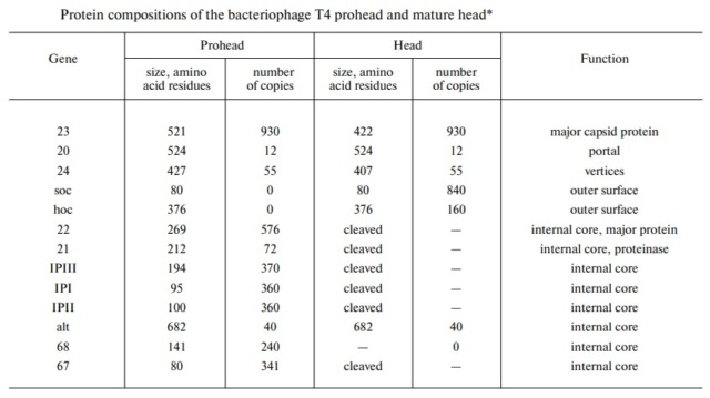

The amazing design of the T4 bacteriophage and its DNA packaging motor Protei10


The Molecular Architecture of the Bacteriophage T4 Neck

Andrei Fokine et.al. (2013):  The T4 head and tail are assembled via independent pathways. Assembly of the T4 head is a complex process that includes a number of intermediate stages. The head assembly is initiated by the dodecamer (A dodecamer (protein) is a protein complex with 12 protein subunits.) of the portal protein, gp20 (gp, gene product). First, a head precursor, called the prohead, is assembled, which is subsequently processed by a scaffold-associated protease. Then the phage genomic DNA is packaged into the capsid through the portal vertex by an ATP-driven motor composed of five gp17 molecules. Upon completion of the DNA packaging, the head assembly is finalized by attachment of several copies of the gp13 and gp14 proteins to the portal vertex. Monomers of gp13 and gp14 have a size of 309 and 256 amino acid (aa) residues, respectively. The gp13–gp14 complex seals the portal vertex and creates a site for attachment of the independently assembled tail. Mutant phages lacking these proteins produce heads that are unable to bind tails and lose their DNA.

The T4 tail assembly begins with the baseplate formation and proceeds with polymerization of the tail tube and the contractile sheath. The tail tube is formed by gp19 molecules (163 aa residues). The length of the tube is controlled by a mechanism involving the “tape-measure protein”, gp29. The elongation of the tail tube is terminated by attachment of the hexamer of the 175-residue tail tube terminator protein, gp3, which binds to the last row of gp19 subunits (probably also to gp29) and stabilizes the tail tube. The T4 tail tube is used as a scaffold for the polymerization of the contractile sheath. The gp18 sheath molecules (659 aa residues) assemble around the tube in the form of a six-start helix. The T4 tail assembly is completed by the hexamer of the tail terminator protein, gp15 (the monomer is 272 aa residues long), which binds to the top† of the tail. Contraction of the tail during infection is associated with a substantial rearrangement of the gp18 subunits and results in shortening of the sheath to less than one-half of its original length. 22
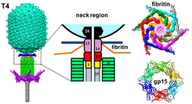
The amazing design of the T4 bacteriophage and its DNA packaging motor 1-s2_027

Coat, scaffolding, and portal proteins are encoded by P22 genes 5, 8, and 1, respectively. In the absence of scaffolding protein, P22 coat protein assembles into TZ4 and TZ7 icosahedral shells as well as “spiral” structures, and all of these lack the essential portal protein and at least one protein required for DNA injection. 10

Lei Sun et al., (2015):The portal structure probably dates back to a time when self-replicating microorganisms were being established on Earth. 11

Amy D. Migliori (2014): Recent structural studies of the bacteriophage T4 packaging motor have led to a proposed mechanism wherein the gp17 motor protein translocates DNA by transitioning between extended and compact states, orchestrated by electrostatic interactions between complimentarily charged residues across the interface between the N- and C-terminal subdomains.  2

They are the most numerous biological entity on earth, with an estimated number of 10^31 tailed phages in the biosphere. They are arguably very ancient as a group, with some estimates placing their ancestors before the divergence of the Bacteria from the Archaea and Eukarya

The amazing design of the T4 bacteriophage and its DNA packaging motor Ncomms12

(a) 3D density map of T4 portal protein assembly at 3.6 Å resolution with each subunit color-coded. Shown is the top view (left)
 and side view (right). 
(b) Ribbon diagram of the gp20 atomic model with each subunit color-coded. Shown is the top view (left)
 and side view (right).
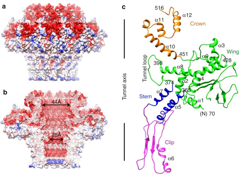
The amazing design of the T4 bacteriophage and its DNA packaging motor Ncomms13
  
(a) Charge distribution on the outer surface of dodecameric gp20. Blue and red colours correspond to 10 kT e− positive and negative potential,
respectively.
(b) Charge distribution on the inner surface of dodecameric gp20. (c) Ribbon drawing of the gp20 monomer structure with each
domain colour-coded.
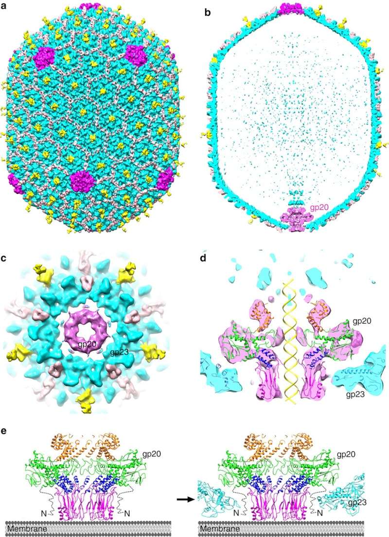
The amazing design of the T4 bacteriophage and its DNA packaging motor Ncomms14

(a,b) Cryo-EM density map of the T4 prolate head (gp23: cyan; gp24:magenta; Soc: pink; Hoc: yellow).
(c) Bottom view of the prolate head, showing the gap between gp20 and the capsid. (d) Fit of the gp20 and gp23
structures into the cryo-EM map of the T4 prolate head. (e) A model of the T4 head assembly. A dodecameric portal is
assembled on the inner membrane of E. coli with the assistance of the phage-coded chaperone gp40 and the E. coli chaperone YidC58.
The portal assembly acts as an initiator for head assembly, leading to co-polymerization of the major capsid protein gp23
and scaffolding proteins.
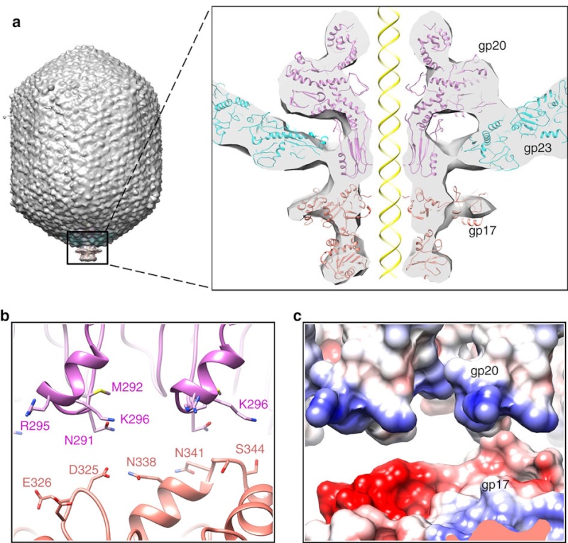
The amazing design of the T4 bacteriophage and its DNA packaging motor Ncomms16

(a) Fitting of the T4 portal protein (purple) and gp17 (tan) into the 35 Å cryo-EM reconstruction of the procapsid+gp17
(EMD-1572 accession number).
(b) Residues involved in the interaction between gp20 (purple) and gp17 (tan) are shown
as sticks. 
(c) The surface charge of gp20 and gp17 around the interface area showing electrostatic interactions. The view
orientation is the same as in panel (b).

The amazing design of the T4 bacteriophage and its DNA packaging motor Ncomms17

The different portal protein subunits with their wing, stem, clip and crown domains are coloured green, blue, purple and orange, respectively.

Phyiscsworld ( 2014): The molecular motor that folds and packs DNA into a virus is at its most efficient when the DNA shows some self-repulsion. That is the surprising finding of researchers based in the US – it was previously thought that such repulsion would act as an obstacle in the packing process. The team also found that pausing the motor and allowing it to relax increased the rate of the whole packaging process. In addition to providing new insights into how viruses function, the work could benefit biotechnologies that enclose long polymers into nanoscale devices. 6

After invading its host cell, a virus reprogrammes the cell's nucleus to duplicate it.

Question: How was the virus programmed to re-program the cell's nucleus? trial and error? Had the function of reprogramming not have to be fully operating since the beginning, otherwise, the virus would not be able to replicate. 

As it replicates, a strand of DNA is pulled from an infected host cell and squeezed into a protein shell – known as a prohead – which then carries the DNA to infect other cells. In some species, the prohead is produced first, leaving only a small hole at one end through which a powerful molecular motor pushes the DNA in and then packs it at very high densities.

Question: How did it emerge the function to pack the DNA at very high densities? trial and error?

The motor has to overcome three forces: the electrostatic self-resistance that comes into play because DNA is negatively charged; the mechanical resistance of DNA to bending; and the entropic resistance of DNA to be crowded on itself.

Question: How did the motor emerge this function of overcoming the three forces? trial and error?

The tail structure

Phys.Org ( 2016):  To infect bacteria, most bacteriophages employ a 'tail' that stabs and pierces the bacterium's membrane to allow the virus's genetic material to pass through. The most sophisticated tails consist of a contractile sheath surrounding a tube akin to a stretched coil spring at the nanoscale. When the virus attaches to the bacterial surface, the sheath contracts and drives the tube through it. All this is controlled by a million-atom baseplate structure at the end of the tail. Phages are widely distributed on the planet. They accompany bacteria everywhere - in the soil, water, hot springs, algal bloom, animal intestines etc - and have a dramatic impact on the diversity of bacterial populations, including for example, the microbiome of the human gut. 18

Petr G.Leiman (2006): Bacteriophage T4 has one of the most complex tails of all studied phages. The T4 tail is composed of ∼400 polypeptide chains that form the tube, the contractile sheath around the tube, and the baseplate that terminates both of them. 26

Petr G Leiman (2006): Bacteriophage T4 has one of the most complex tails of all studied phages. It is composed of w400 polypeptide chains that form the tube, the contractile sheath around the tube, and the baseplate that terminates both of them. https://pubmed.ncbi.nlm.nih.gov/16554069/

The tail, fibers, and infection process  
Phages from the Myoviridae family have exceptionally complex, contractile tails. Bacteriophage T4 devotes 25 kbp of its genome to tail assembly, which is comparable with the size of the entire adenovirus genome (36 kbp). Products of at least 22 genes are involved in tail assembly (table 3), 
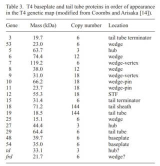
The amazing design of the T4 bacteriophage and its DNA packaging motor T4_bas11
which include a phage-encoded chaperone that participates in folding of the long and short tail fibers (table 2). 
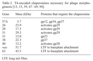
The amazing design of the T4 bacteriophage and its DNA packaging motor T4-enc11

The bacteriophage T4 tail is composed of two concentric protein cylinders, at one end of which is the baseplate and fibers. The inner cylinder, called the tail tube, is built of 144 copies of gp19. The tail tube has a 40 Å-diameter channel for DNA passage from the head to the infected cell. The outer cylinder, called the tail sheath, tightly envelopes the 90 Å-diameter tail tube and has a width of about 210 Å. It is composed of 144 copies of gp18. The subunits comprising each cylinder form a six-start helix with a pitch of 41 Å and a righthanded twist angle of 17°. The helix has a length of 984 Å and contains 24 repeats. During infection, the phage recognizes an E. coli bacterium using its long tail fibers (LTFs) connected to the baseplate. The phage then anchors the baseplate to the lipopolysaccharide cell surface receptors using the short tail fibers (STF), which are initially assembled under the baseplate. This event triggers a hexagon-to-star conformational change in the baseplate and causes an irreversible contraction of the tail sheath, releasing about 25 kcal/ mol of energy per gp18 monomer. During this process, the gp18 hexamers flatten, rotate, and expand radially, resulting in a decrease of their thickness by 26 Å and an increase of the twist angle by 15°. The contracted tail sheath has a length of only 360 Å and a width of 270 Å. The tail tube does not change its length during sheath contraction. As a result, almost half of the tube protrudes out of the contracted tail sheath and the baseplate.

The sheath can be caused to contract by exposing the phage to 3 M urea. Nevertheless, the DNA is not released until the tail tube tip binds to a cytoplasmic membrane receptor common to enteric bacteria, suggesting that tail contraction does not cause the release of DNA. The interaction of the tail tube tip with the cytoplasmic membrane involves creation of a channel for DNA passage. During DNA transfer from the capsid into the cell, the membrane remains virtually undamaged since the transfer requires a proton motive force across the membrane. The assembly pathway of the bacteriophage T4 tail is regulated by ordered sequential interactions of proteins rather than sequential gene expression. The baseplate, a remarkably complex multiprotein structure, is assembled first. It is composed of about 150 subunits of at least 16 different gene products, many of which are oligomeric (table 3). These proteins form six independently assembled wedges that join together around the central hub with the help of the trimeric proteins (gp9) and (gp12). Each wedge is assembled by sequential interactions of the seven protein oligomers: (gp11), (gp10), (gp7), (gp8), (gp6), gp53, and gp25. The baseplate hub is formed by (gp5), (gp27), gp29 and, probably, gp28. The assembly of the baseplate is completed with the attachment of six copies of gp48 and six copies of gp54 to the external interface between the wedges and the hub. The latter proteins serve as a starting point for polymerization of gp19 to form the tail tube, which is terminated with gp3. The tail tube serves as a scaffold for polymerization of the tail sheath around it. During this process, gp18 stores energy in its conformation (possibly by ATP hydrolysis), making the non-contracted T4 tail a stretched spring. The length of the tail tube is controlled by the ruler protein, gp29, which also participates in assembly of the central part of the baseplate. The length of the tail sheath is determined by the length of the tube. The assembly of the tail is completed by attachment of a gp15 hexamer to the last ring of the tail sheath. The baseplate is a dome-shaped object.  The hollow tail tube stems from the center of the baseplate. 15

The amazing design of the T4 bacteriophage and its DNA packaging motor Protei11

V. V. Mesyanzhinov (2004): Products of at least 22 genes are involved in assembly of the T4 phage tail (Table above) that uses the energy of the sheath contraction for DNA ejection into the host cell. The assembly pathway of the tail is based on strictly ordered sequential interactions of proteins. The baseplate is a remarkably complex multiprotein structure of the tail that serves as a control unit of virus infection. The baseplate is composed of ~150 subunits of at least 16 different gene products, many of which are oligomeric, and assembled from six identical wedges that surround a central hub. The T4 gp11 (the short tail fiber connecting protein), gp10, gp7, gp8, gp6, gp53, and gp25 combine sequentially to built up a wedge. The central hub is formed by gp5, gp27, and gp29 and probably gp26 and gp28. Assembly of the baseplate is completed by attaching gp9 and gp12 forming the short tail fibers, and also gp48 and gp54 that are required to initiate polymerization of the tail tube, a channel for DNA ejection that is constructed of 138 copies of gp19. The length of the tail tube is probably determined by the “ruler protein” or template, gp29. The tail tube serves as a template for assembly of 138 copies of gp18 that form the contractile tail sheath. In the absence of the tail tube, gp18 assembles into long polysheaths with a structure similar in several aspects to the contracted state. Both the tail tube and the tail sheath have helical symmetry. Assembled tail sheath represents a metastable supramolecular structure, and sheath contraction is an irreversible process. During contraction the length of tail sheath decreases from 980 to 360 Å and its outer diameter increases from 210 to 270 Å. The assembly of the tail is completed by a gp15 hexamer that binds to the last gp18 ring of the tail sheath. The assembled tail associates with the head after DNA packaging. Then six gpwac (fibritin) molecules attach to the neck of the virion forming a ring embracing it (“collar”) and thin filaments protruding from the collar (“whiskers”) that help with attachment of the phage particle to other fibrous proteins, the long tail fibers. 23
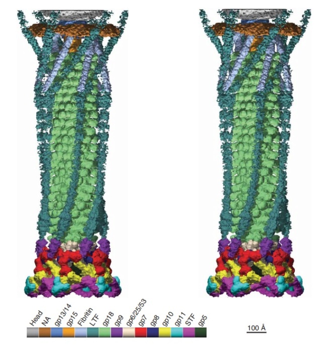
The amazing design of the T4 bacteriophage and its DNA packaging motor Bacter26
Stereo diagram of bacteriophage T4 showing the extended tail, the LTFs, the neck and a small part of the capsid. NA indicates that the density has not been assigned to a specific gene product.

The cell-puncturing device of bacteriophage T4

ScienceDaily (2016): To infect bacteria, most bacteriophages employ a 'tail' that stabs and pierces the bacterium's membrane to allow the virus's genetic material to pass through. The most sophisticated tails consist of a contractile sheath surrounding a tube akin to a stretched coil spring at the nanoscale. When the virus attaches to the bacterial surface, the sheath contracts and drives the tube through it. All this is controlled by a million-atom baseplate structure at the end of the tail. EPFL scientists have now shown, in atomic detail, how the baseplate coordinates the virus's attachment to a bacterium with the contraction of the tail's sheath. 

ScienceDaily (2002): The viral machine works as follows: The virus uses its long-tail fibers to recognize its host and to send a signal back to the baseplate. Once the signal is received, the short-tail fibers help anchor the baseplate into the cell surface receptors. As the virus sinks down onto the surface, the baseplate undergoes a change — shifting from a hexagon to a star-shaped structure. At this time, the whole tail structure shrinks and widens, bringing the internal pin-like tube in contact with the outer membrane of the E. coli cell. As the tail tube punctures the outer and inner membranes of the E. coli cell, the virus' DNA is injected through the tail tube into the host cell. 17

P. G. Leiman (2003): Phages are widely distributed on the planet. They accompany bacteria everywhere -- in the soil, water, hot springs, algal bloom, animal intestines etc -- and have a dramatic impact on the diversity of bacterial populations, including for example, the microbiome of the human gut.  The entire baseplate-tail-tube complex consists of one million atoms, making up 145 chains of 15 different proteins. The scientists were also able to identify a minimal set of molecular components in the baseplate that work together like miniature gears to control the activity of the virus's tail. These components, and the underlying functional mechanism, are the same across many viruses and even bacteria that use similar tail-like structures to inject toxins into neighboring cells. 15

One of the most remarkable features of the baseplate is the spike, or needle, along the axis of the dome. The crystal structure of the gp5-gp27 complex (fig. below) can be fitted into the baseplate map so that the needle density is occupied by the C-terminal domain of gp5. The gp27 trimer forms a channel suitable for the passage of a dsDNA and serves as an extension of the tail tube (figs below).
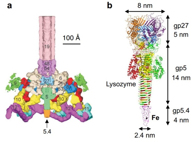
The amazing design of the T4 bacteriophage and its DNA packaging motor Struct22
a Baseplate of bacteriophage T4
b gp5–gp27–gp5.4
The amazing design of the T4 bacteriophage and its DNA packaging motor Struct20
Structure of the gp5-gp27 complex. 
(a) Ribbon stereo diagram. The three gp5 monomers are colored red, green, and blue. The three gp27 monomers are colored yellow, gray, and purple. The K+ ion within gp5C is shown in pink. The (PO4)– is hidden behind the lysozyme domain. 
(b) The structure of the gp27 monomer with its four domains colored cyan, pink, light green, and gold along the polypeptide chain. 
(c) Top view of the gp27 cylinder shows that the cyan and green domains form a hexagonal torus

Gp5 consists of three domains: an N-terminal oligosaccharide binding-fold domain, the middle lysozyme domain, and the C-terminal triple b-helix domain (fig.above). The gp5 lysozyme domain has 43% sequence identity and a closely similar structure to the T4 lysozyme encoded by gene e (T4L). 

Structure of the T4 baseplate

ScienceDaily (2002): The baseplate is the "nerve center" of the virus. When the long and short fibers attach to E. coli, the baseplate transmits this message to the tail, which contracts like a muscle. The baseplate both controls the needlepoint of the tail and the cutting enzyme that make a tiny, nanometer-sized hole through the cell wall of the E. coli. The viral DNA is then squeezed through the tail into the host. The E. coli, thus infected, starts to make only new phage particles and ultimately dies. 16

Moh Lan Yap (2016): T4 has a complex baseplate that is essential for assuring a highly efficient infection mechanism 25

M. I. Taylor (2016): Bacteriophages (viruses of bacteria) use a specialized organelle called a tail to deliver their genetic material and proteins across the cell envelope during infection. In phages with the most complex contractile tails, attachment to the host cell is accompanied by a substantial transformation of the tail structure: the external tail sheath contracts and drives a spike-tipped , rigid tube through the host cell membrane. Other macromolecular complexes, such as the type VI secretion system (T6SS), metamorphosis-associated contractile (MAC) arrays, R-type pyocins, Serratia antifeeding prophage, Photorhabdus virulence cassette and rhapidosomes, use a similar contractile sheath–rigid tube mechanism to breach the bacterial or eukaryotic cell envelope. The most complex part of these ‘contractile injection systems is the baseplate, which is responsible for coordinating host recognition or other environmental signals with sheath contraction. The T4 baseplate is currently thought to contain at least 15 different proteins with copy numbers ranging from 1 to 18. Assembly of the T4 baseplate involves two large independent intermediates: a hub and a wedge. Several phage and possibly host cell chaperones mediate the joining of six wedges to the hub, which is then followed by the attachment of receptor-binding fibres to this structure. The nascent baseplate initiates tube assembly and subsequent polymerization of the sheath in the extended high-energy state. The remarkable structure and transformation of the T4 tail and other contractile injection systems have received considerable attention. This is a 440,000-atom (not counting hydrogens) structure. 

Overall structure of the T4 baseplate:  The final atomic model is around 96% complete and contains 56,082 amino acid residues (9,886 unique). These amino acids belong to 145 polypeptide chains of 15 different proteins (gene products, gps) that comprise the baseplate (gp5, gp5.4, gp6, gp7, gp8, gp9, gp10, gp11, gp12, gp25, gp27, and gp53) and the proximal region of the tail tube (gp19, gp48 and gp54, although gp48 and gp54 could also be considered to be baseplate components). 19 
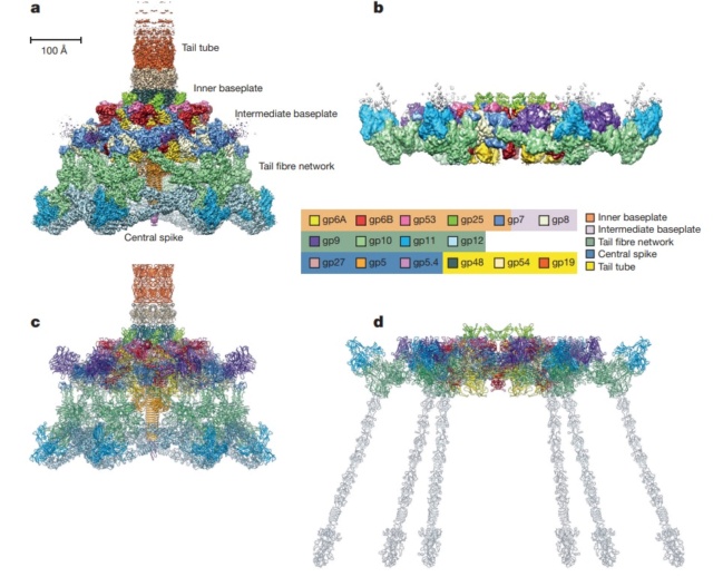
The amazing design of the T4 bacteriophage and its DNA packaging motor T4_bas13
The amazing design of the T4 bacteriophage and its DNA packaging motor T4_bas12
Maps and atomic models of T4 baseplate in pre- and post-attachment states. 
a, b, Cryo-EM reconstructions of the pre- and post-attachment T4 baseplate, respectively. c–f, Atomic models of the two states with component proteins shown as ribbon diagrams. In d and f, the STFs (gp12 trimers) are displayed semi-transparently to indicate that they are not present in the refined model of the post-attachment baseplate.
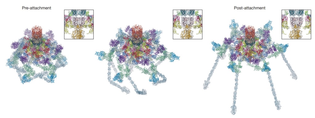
The amazing design of the T4 bacteriophage and its DNA packaging motor Confor11
Conformational change of the T4 baseplate upon host cell attachment. 
The left and right panels show the pre-and post-attachment structures that are derived from cryo-EM data. The middle image is a model of an intermediate with assumptions described in the text. The insets show close-up views of the central part of the baseplate demonstrating a release of the tail tube–central spike complex, whose position is kept unchanged throughout the transformation.
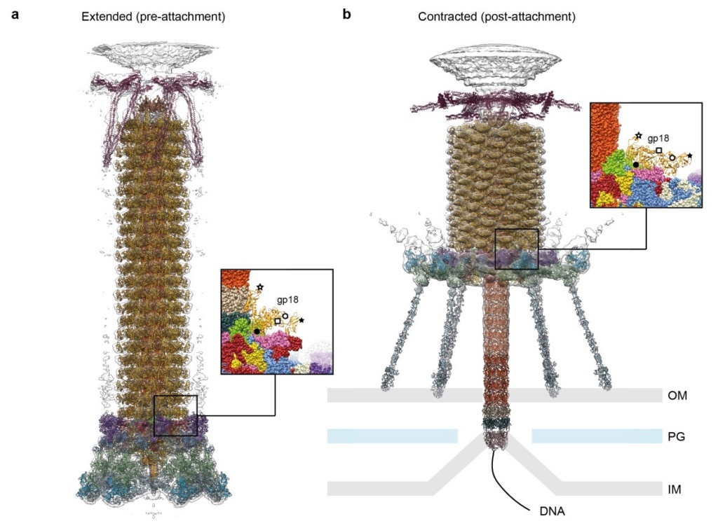
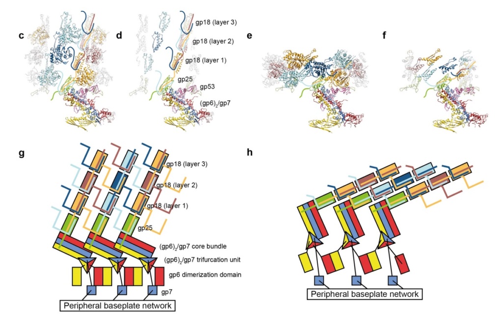
The amazing design of the T4 bacteriophage and its DNA packaging motor Model_12
The amazing design of the T4 bacteriophage and its DNA packaging motor Pseudo10
Model for baseplate-induced sheath contraction. 
a, b, Pseudoatomic model of the complete T4 tail in the extended (pre-attachment) and contracted (post-attachment) conformations. The insets show a close-up view (labelled with a black box) of the position of the gp18 subunit on the baseplate in the extended and contracted conformations of the sheath. The white geometrical shapes label the same regions of the sheath subunit in both conformations. 
c, Interaction of the two conserved domains of the gp18 sheath protein with the conserved components of the T4 baseplate wedge. Coloured lines indicate the putative topology of the N- and C-terminal gp18 extensions, as well as the gp25 C-terminal strand. 
d, The same view as in c, but the external domain is now not shown for clarity to demonstrate the interaction of gp25-like sheath domains with each other and with gp25. 
e, f, The same as c and d but in the contracted state. 
g, h, Two diagrams demonstrating the motion of baseplate components that results in sheath contraction.

V. V. Mesyanzhinov (2004): Practically every assembled T4 particle is able to infect an E. coli host cell. The baseplate is the control center of the viral infectivity, and understanding of the baseplate structure, a multiprotein machine, is a challenging problem. Below we represent the data about baseplate proteins with known atomic structure.
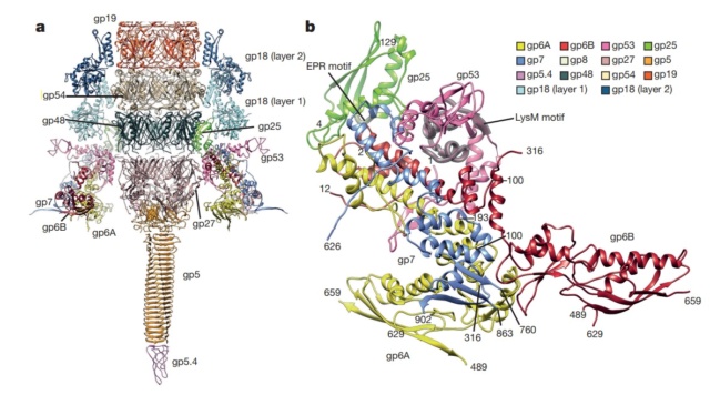
The amazing design of the T4 bacteriophage and its DNA packaging motor Struct26
Structure of the conserved inner baseplate. 
a, The minimal composition of a contractile injection system is derived from the T4 tail structure: the central spike complex (gp5, gp5.4, gp27), the conserved part of the wedge (gp6, gp7, gp25, gp53), the tail tube (gp19, gp48, gp54) and the conserved part of the sheath (gp18), some of which is modelled using the pyocin sheath14. 
b, A close-up of the structure of the conserved region of the wedge consisting of the (gp6)2–gp7 heterotrimer, gp25 and gp53. The EPR motif of gp25 and the LysM motif of gp53 are highlighted with semi-transparent grey.

The tail tube & tail sheath terminators

Moh Lan Yap (2015): The polymerized tail tube and sheath are capped by the terminator proteins gp3 and gp15, respectively, to prevent depolymerization before the tail attaches to the head. Both gp3 and gp15 form hexameric rings that interact with the last row of gp19 and gp18 molecules. The central pore and the side surface of gp15 are negatively charged, whereas the top and the bottom surfaces are positively charged. The top and bottom surfaces interact with gp14 and gp3 proteins, respectively. The interaction between gp15 and gp18 is different in the extended and contracted (postinfection) conformations. In the contracted tail, the negatively charged side surface of the gp15 hexamer interacts with positively charged surfaces of the C-terminal domains of the gp18 molecules. These interactions help to maintain the integrity of the tail in its contracted form. The gp15 hexamer may have undergone a conformational change during the infection process, which might be propagated through gp14 and gp13, to the portal assembly to allow the release of the genomic DNA. 21
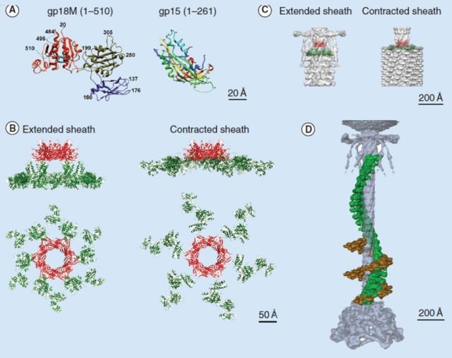
The amazing design of the T4 bacteriophage and its DNA packaging motor Crysta14
Crystal structures of gp18 and gp15
(A) Ribbon diagrams of gp18M and gp15 monomers. The three domains of gp18M are shown in blue (domain I), olive green (domain II) and orange red (domain III); the β-hairpin (residues 454–470) and the last 14 C-terminal residues are shown in cyan. Gp15 represented in rainbow color running from N terminus (blue) to C terminus (red). The broken lines indicate protein regions that are disordered in the crystals.
(B) Relative positions of the gp15 and gp18 molecules in the extended and contracted T4 tails, viewed from the side (upper) and from the top (lower) of the phage. A model of the entire gp18 molecule was created based on the crystal structure of gp18M and the prophage tail sheath protein LIN1278. The models of gp18 molecules belonging to the topmost ring of the contractile sheath are shown in green. The gp15 hexamer is shown in red.
(C) The gp15 and gp18 molecules are fitted into the cryo-electron microscopy reconstructions of the extended and contracted tails.
(D) A helical strand of gp18 in the extended (green) and contracted (brown) tail. The hexagonal baseplate, tail tube, whiskers and collar are shown in gray-blue.

The amazing design of the T4 bacteriophage and its DNA packaging motor Struct25
Structure of the contracted T4 tail 
(side view (a), with an inclination (b), cross-section (c), from the bottom (d)). Each protein or complex is labeled with their respective gene number and indicated by color: spring green, gp5; red, putative gp7; dark blue, gp8; green, gp9; yellow, putative gp10; cyan, gp11; magenta, gp12; salmon, gp19; sky blue, gp27; pink, putative gp48 or gp54; beige, gp6 + gp25 + gp53; orange, putative gp26. 23


Long tail fibers collar & whiskers

Moh Lan Yap (2015): The long tail fibers (LTFs) consist of four different proteins, namely gp34, gp35, gp36 and gp37 (Figure A & B). 

The amazing design of the T4 bacteriophage and its DNA packaging motor Bacter19
T4 long tail fiber
(A) Image of a long tail fiber.
(B) Domain organization of the long tail fiber. Domains of the proximal tail fiber are named P1–5 and of the distal half D1–11; gp35, or the KC is represented as a green triangle. Crystal structure of D10 and D11 (box) has been determined.
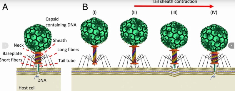
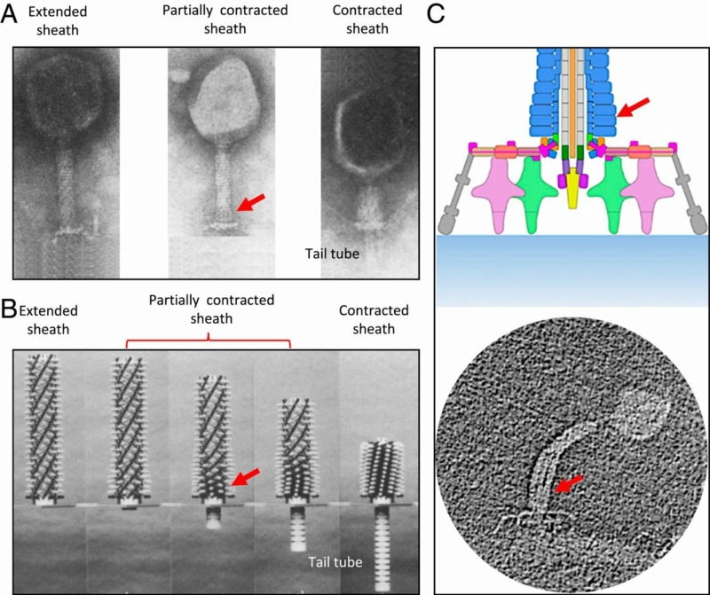
(C) Structure of gp37 (residues 785–1026). Three chains in the trimeric protein are colored red, green and blue, respectively, whereas iron ions are shown in yellow. The N- and C-termini and every 10th residue of chain A are labeled.

1. http://www.pnas.org/content/111/42/15096.short
2. Anjeanette Roberts Celebrating 3.8 Billion Years of Bacteriophage October 22, 2015
3. http://www.ncbi.nlm.nih.gov/pubmed/22297528
4. https://creation.com/images/pdfs/tj/j22_1/j22_1_15-16.pdf
6. http://creation.com/did-god-make-pathogenic-viruses
7. Joseph W. Francis: The Organosubstrate of Life: A Creationist Perspective of Microbes and Viruses 2003 
8.   http://teaguesterling.com/dna/motor-protein.pdf
9.   G. Leiman: Structure and morphogenesis of bacteriophage T4 P.  9 May 2003 
10. http://www.ncbi.nlm.nih.gov/pmc/articles/PMC3109452/
11. Nicola Twilley: Inside the World of Viral Dark Matter February 6, 2015
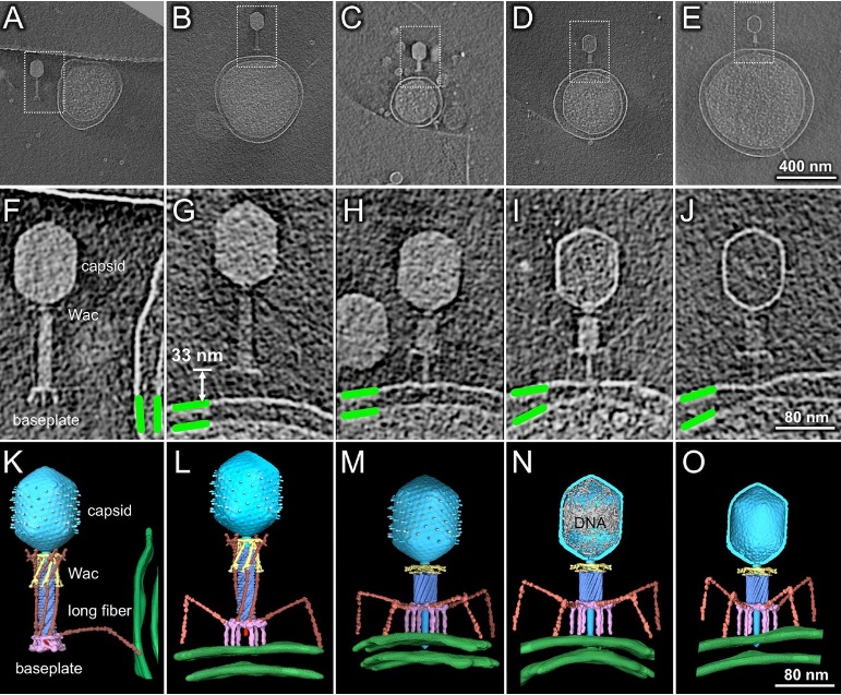
12. Eric S Miller Bacteriophage T4 and its relatives 28 October 2010
13. Eric S Miller Bacteriophage T4 genome 2003 Mar;6
14. Vincent R. Racaniello: Principles of Virology, Volume 1: Molecular Biology 18 agosto 2015
15. Science Daily: How viruses infect bacteria: A tale of a tail May 18, 2016
16. Science Daily: New Understanding Of Complex Virus Nano-Machine For Cell Puncturing And DNA Delivery February 4, 2002
17. Science Daily: Study Reveals New Information On How Viruses Enter Cells February 7, 2002
18. Phys.Org: How viruses infect bacteria: A tale of a tail MAY 18, 2016
19. M. I. Taylor et.al. Structure of the T4 baseplate and its function in triggering sheath contraction  18 May 2016
20. Jaap Vergote Design and nature Jun 24, 2018
21. Moh Lan Yap: Structure and function of bacteriophage T4  2015 Aug 1
22. Andrei Fokine: The Molecular Architecture of the Bacteriophage T4 Neck 2013 Feb 19
23. V. V. Mesyanzhinov: Molecular Architecture of Bacteriophage T4 July 9, 2004
24. Fumio Arisaka:[url=https://aip.scitation.org/doi
25. Petr G. Leiman: Evolution of Bacteriophage Tails: Structure of T4 Gene Product 10 2006 May 5
26. Petr G.Leiman: Evolution of Bacteriophage Tails: Structure of T4 Gene Product 10 5 May 2006
27. MITSUHIRO YANAGIDA: MOLECULAR ORGANIZATION OF THE HEAD OF BACTERIOPHAGE Teven: UNDERLYING DESIGN PRINCIPLES 1984
28. Bo Hu: Structural remodeling of bacteriophage T4 and host membranes during infection initiation August 17, 2015
29. Biology reader: What is a Bacteriophage
30. Quora: How would you explain the structure of T4 bacteriophage?


1. http://www.pnas.org/content/111/42/15096.short
2. Anjeanette Roberts Celebrating 3.8 Billion Years of Bacteriophage October 22, 2015
3. http://www.ncbi.nlm.nih.gov/pubmed/22297528
4. https://creation.com/images/pdfs/tj/j22_1/j22_1_15-16.pdf
6. http://creation.com/did-god-make-pathogenic-viruses
 
8.   http://teaguesterling.com/dna/motor-protein.pdf
10. http://www.ncbi.nlm.nih.gov/pmc/articles/PMC3109452/
15. Science Daily: How viruses infect bacteria: A tale of a tail May 18, 2016
16. Science Daily: New Understanding Of Complex Virus Nano-Machine For Cell Puncturing And DNA Delivery February 4, 2002
17. Science Daily: Study Reveals New Information On How Viruses Enter Cells February 7, 2002
18. Phys.Org: How viruses infect bacteria: A tale of a tail MAY 18, 2016
19. M. I. Taylor et.al. Structure of the T4 baseplate and its function in triggering sheath contraction  18 May 2016

21. Moh Lan Yap: Structure and function of bacteriophage T4  2015 Aug 1
22. Andrei Fokine: The Molecular Architecture of the Bacteriophage T4 Neck 2013 Feb 19
23. V. V. Mesyanzhinov: Molecular Architecture of Bacteriophage T4 July 9, 2004

25. Petr G. Leiman: Evolution of Bacteriophage Tails: Structure of T4 Gene Product 10 2006 May 5
26. Petr G.Leiman: Evolution of Bacteriophage Tails: Structure of T4 Gene Product 10 5 May 2006
28. Bo Hu: Structural remodeling of bacteriophage T4 and host membranes during infection initiation August 17, 2015

https://reasonandscience.catsboard.com

Sponsored content


Back to top  Message [Page 1 of 1]

Permissions in this forum:
You cannot reply to topics in this forum