ElShamah - Reason & Science: Defending ID and the Christian Worldview
Would you like to react to this message? Create an account in a few clicks or log in to continue.
ElShamah - Reason & Science: Defending ID and the Christian Worldview

Otangelo Grasso: This is my personal virtual library, where i collect information, which leads in my view to the Christian faith, creationism, and Intelligent Design as the best explanation of the origin of the physical Universe, life, biodiversity


You are not connected. Please login or register

Proteins: how they provide striking evidence of design

Go down  Message [Page 1 of 1]

Otangelo


Admin

Proteins: how they provide striking evidence of design

https://reasonandscience.catsboard.com/t2062-proteins-how-they-provide-striking-evidence-of-design

How ancestral proteins attained functionality, even though they were most likely markedly smaller than their contemporary descendants, remains a major, unresolved question in the origin of life. 10

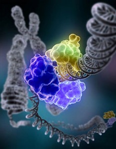
Artur Gora (2013): On one hand, proteins need to have well-defined and organized structures in order to maintain stable functionality in the intracellular environment. On the other hand, some degree of flexibility is often required for catalytic activity. Molecular dynamics simulations have provided key insights into the importance of protein dynamics in catalysis, such as the observation of substrate access and product exit pathways that cannot be identified by inspecting crystal structures.
https://www.ncbi.nlm.nih.gov/pmc/articles/PMC3744840/

Proteins are evidence of intelligent design par excellence. Instructional/specified complex information is required to get the right amino acid sequence which is essential to get functionality in vast sequence space ( amongst trillions os possible sequences, rare are the ones that provide function ), and every protein is irreducibly complex in the sense, that a minimal number of amino acids are required for each protein to get function. This constitutes a unsurmountable hurdle for the origin of life scenarios based on naturalistic hypotheses since unguided random events are too unspecific to get functional sequences in a viable timespan. Another true smack-down is the fact that single proteins or enzymes by themselves confer no advantage of survival at all, and have by their own no function. There is no reason why random RNA strands would become self-replicating. And even IF that were the case, so what? There would be no utility for them unless at least 50 different precisely arranged and correctly interlinked enzymes and proteins, each with its specific function, would interlink in a complex, just right metabolic network, and be encapsulated in a complex membrane with gates and pores, and in a precisely finely tuned and balanced homeostatic ambiance. Energy production and supply to each protein would also have to be fully set up right from the start...... hard to swallow. But if your wish of naturalism to be true is strong enough, just shut your reason up, and believe this. Blindly.

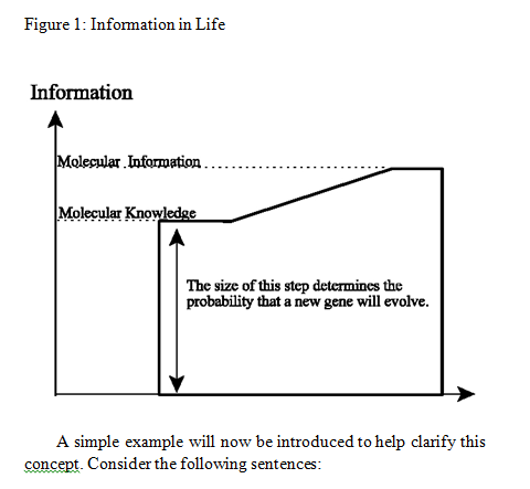
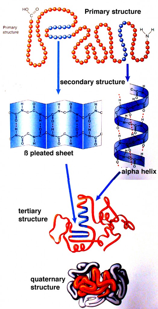
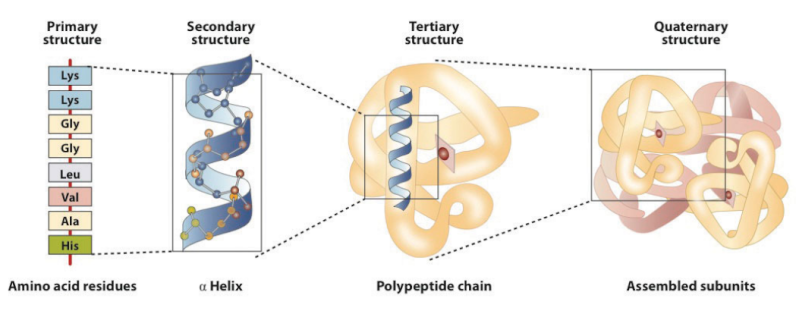
Proteins can be considered as information-enriched polymers. In fact, the instruction for the 3-D structure of a protein is embedded in the sequences of amino acids and the electrochemical attractive forces among them.


It is not surprising that various studies on evolving proteins have failed to show a viable mechanism. One study concluded that 10^63 attempts would be required to evolve a relatively short protein.

Functionally Acceptable Substitutions in Two a-Helical Regions of X Repressor
"The estimated number of sequences capable of adopting the h repressor fold is still 10^63 an exceedingly small fraction, about one in of the total number of possible 92-residue sequences."
http://sci-hub.tw/https://onlinelibrary.wiley.com/doi/pdf/10.1002/prot.340070403

And a similar result (10^65 attempts required) was obtained by comparing protein sequences. Another study found that 10^64 to 10^77 attempts are required, and another study concluded that 10^70 attempts would be required. So something like 10^70 attempts are required yet evolutionists estimate that only 10^43 attempts are possible. In other words, there is a shortfall of 27 orders of magnitude. But it gets worse. The estimate that 10^43 attempts are possible is utterly unrealistic. For it assumes billions of years are available, and that for that entire time the Earth is covered with bacteria, constantly churning out mutations and new protein experiments. Aside from the fact that these assumptions are entirely unrealistic, the estimate also suffers from the rather inconvenient fact that those bacteria are, err, full of proteins. In other words, for evolution to evolve proteins, they must already exist in the first place. This is absurd. And yet, even with these overly optimistic assumptions, evolution falls short by 27 orders of magnitude. The numbers don’t add up. Proteins reveal scientific problems for evolution. What is interesting is how evolutionists react to these problems.
https://darwins-god.blogspot.com.br/2017/04/new-book-new-proteins-evolve-very-easily.html

The protein that enables a firefly to glow, and also reproduce (as its illuminated abdomen also serves as a visible mating call), is a protein made up of a chain of 1,000 amino acids. The full range of possible proteins that can be coded with such a chain is 17 times the number of atoms in the visible universe. This number also represents the odds against the RANDOM coding of such a protein. Yet, DNA effortlessly assembles that protein, in the exactly correct, and absolutely necessary sequence and number of amino acids for the humble firefly. What are we to say of the 25,000 individual, highly specialized, absolutely necessary, and exactly correctly coded proteins in the human body? King David, perhaps, said it best: "We are fearfully and wonderfully made" (Psalms 139:14). "Time and Chance," as an explanation (read: cover story) for Life without a Creator, has all the scientific merit of the phrase, "Once Upon A Time."

A short protein molecule of 150 amino acids, the probability of building a 150 amino acids chain in which all linkages are peptide linkages would be roughly 1 chance in 10^45. 

Paul Davies once said;
How did stupid atoms spontaneously write their own software … ? Nobody knows …… there is no known law of physics able to create information from nothing.

Dembsky : We also know from broad and repeated experience that intelligent agents can and do produce information-rich systems: we have positive experience-based knowledge of a cause that is sufficient to generate new instructing complex information, namely, intelligence.  the design inference does not constitute an argument from ignorance. Instead, it constitutes an "inference to the best explanation" based upon our best available knowledge.  It asserts the superior explanatory power of a proposed cause based upon its proven—its known—causal adequacy and based upon a lack of demonstrated efficacy among the competing proposed causes.  The problem is that nature has too many options and without design couldn’t sort them all out. Natural mechanisms are too unspecific to determine any particular outcome. Mutation and natural selection or luck/chance/probability could theoretically form a new complex morphological feature like a  leg or a limb with the right size and form , and arrange to find out the right body location to grow them, but it could also produce all kinds of other new body forms, and grow and attach them anywhere on the body, most of which have no biological advantage or are most probably deleterious to the organism. Natural mechanisms have no constraints, they could produce any kind of novelty. Its however that kind of freedom that makes it extremely unlikely that mere natural developments provide new specific evolutionary arrangements that are advantageous to the organism.  Nature would have to arrange almost an infinite number of trials and errors until getting a new positive arrangement. Since that would become a highly unlikely event, design is a better explanation. 

Even the simplest of these substances [proteins] represent extremely complex compounds, containing many thousands of atoms of carbon, hydrogen, oxygen, and nitrogen arranged in absolutely definite patterns, which are specific for each separate substance.  To the student of protein structure the spontaneous formation of such an atomic arrangement in the protein molecule would seem as im- probable as would the accidental origin of the text of irgil’s “Aeneid” from scattered letter type.1
– A. I. Oparin

The argument of the proteins specified complexity
1. The number and sequence of amino acids in proteins, such as enzymes, are crucial. 
2. Only specially-shaped forms (left-handed configurations) of each amino acid are used to form proteins.
3. Amino acids can be joined only by peptide bonds to form proteins.
4. To link together, each amino acid first must be activated by a specific enzyme.
5. Multiple special enzymes are required to bind messenger RNA to ribosomes before protein synthesis can begin or end.
6. Out of many details even these few have specified complexity without which the proteins could not exist. Not even half of the functional proteins could survive without important function.
7. If two amino acids are located within the peptidyl transferase center, they will easily form a peptide bond. But as soon as you do that in the absence of the ribosome, the ends of the amino acids come together, forming a cyclic structure. Polymers cannot form. But if the ends are kept apart, by a theoretical primitive ribosome, a chain of peptide bonds could grow into a polymer.
8. An irreducibly complex system cannot be produced gradually by slight, successive modifications of a precursor system, since any precursor to an irreducibly complex system is by definition nonfunctional. Since natural selection requires a function to select, an irreducibly complex biological system, if there is such a thing, would have to arise as an integrated unit for natural selection to have anything to act on. It is almost universally conceded that such a sudden event would be irreconcilable with the gradualism Darwin envisioned.
9. This is creation by an intelligent designer, and this is the dictionary meaning of the word God.

The individual macromolecules are complex
But the complex interaction of biological macromolecules is only one aspect of the problem facing the origin of life. What compounds the enigma is that the individual macromolecular components are themselves complex, in the sense that their sequences - of ribonucleotides in the case of RNA, or amino acids for proteins - are very specific. The linear amino acid sequence of a protein is specific because it must (a) be able to fold into a discrete 3-dimensional structure, and (b) have the right amino acids in the right positions in the linear sequence so that, when folded, they are in exactly the right positions in relation to each other to form the active site(s) of the protein. (And similar considerations apply to RNAs.) Sequences which meet these criteria are exceedingly rare compared with the astronomical number of possible sequences of a suitable length. For example Douglas Axe has estimated that only 1 in about 10^74 possible sequences will have biological function (Axe). So it is totally unrealistic to think that such sequences could have arisen by chance. How much less a suite of mutually dependent macromolecules? If the components themselves were not so improbable then it might be realistic to think that a complex combination of components could arise by chance; but the extreme improbability of the individual components is such that they are very unlikely to arise individually, and hence there is no chance whatever of an interdependent system. 5

Where even just two macromolecules are required to perform a function, then it would be necessary for both components to arise together: Because natural selection does not have foresight: if one component arises alone it will not be retained for potential future usefulness (when the second component is available), but will almost certainly degrade by mutation. And, it should be noted, if the probability of getting one component is 1 in 10^74 then the probability of getting two together is 1 in 10^148 (not 1 in 2x10^74); and so on for multi-component systems. This is why the obligatory mutual dependence of many macromolecules in even basic biological systems completely defies any hope of an evolutionary origin. So, in summary, the crux of the problem is that even a basic biological replicating system requires (a) several macromolecules with complementary functions with (b) each having a highly improbable sequence. And this combination of complexities presents an insurmountable challenge to a naturalistic origin of life. 


Proteins need to interact together. They need interface compatibility. So this adds to the unsurmountable problem ot making them by chance. Lets say our good old friend chance , after 10^78 trial and errors , got a functional protein made. That function has imho no value, if it cannot interact properly with other proteins. Then our same old friend made another 10^78 trial and errors attempts, and got the second protein made. How many attempts would be required to make them compatible to be able to interact in a functional way ? So lets suppose for a moment, that they got the right interface compatibility, and interact properly . So what ? This interaction alone has a long way to go, to get all 20 protein complexes needed to get DNA replication done, and making them interact all together in a functional way. Its the same as to make a piston by trial and error. At the end you have eventually a device that is piston like. If you do not know imho exactly the size the piston has to have to fit into the motor block, nothing done. Nothing will go. So FORSIGHT and INTELLIGENCE in order to PROJECT the whole device is ESSENTIAL.


The proteins in living cells are made of just certain kinds of amino acids, those that are “alpha” (short) and “left-handed.” Miller’s “primordial soup” contained many long (beta, gamma, delta) amino acids and equal numbers of both right-and left-handed forms. Problem: just one long or right-handed amino acid inserted into a chain of short, left-handed amino acids would prevent the coiling and folding necessary for proper protein function. What Miller actually produced was a seething brew of potent poisons that would absolutely destroy any hope for the chemical evolution of life. 1

Paper Reports that Amino Acids Used by Life Are Finely Tuned to Explore "Chemistry Space" 4

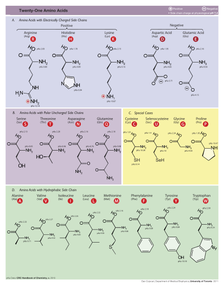
A recent paper in Nature's journal Scientific Reports, "Extraordinarily Adaptive Properties of the Genetically Encoded Amino Acids," 3 has found that the twenty amino acids used by life are finely tuned to explore "chemistry space" and allow for maximal chemical reactions. Considering that this is a technical paper, they give an uncommonly lucid and concise explanation of what they did:

We drew 108 random sets of 20 amino acids from our library of 1913 structures and compared their coverage of three chemical properties: size, charge, and hydrophobicity, to the standard amino acid alphabet. We measured how often the random sets demonstrated better coverage of chemistry space in one or more, two or more, or all three properties. In doing so, we found that better sets were extremely rare. In fact, when examining all three properties simultaneously, we detected only six sets with better coverage out of the 108 possibilities tested. That's quite striking: out of 100 million different sets of twenty amino acids that they measured, only six are better able to explore "chemistry space" than the twenty amino acids that life uses. That suggests that life's set of amino acids is finely tuned to one part in 16 million. Of course they only looked at three factors -- size, charge, and hydrophobicity. When we consider other properties of amino acids, perhaps our set will turn out to be the best:

While these three dimensions of property space are sufficient to demonstrate the adaptive advantage of the encoded amino acids, they are necessarily reductive and cannot capture all of the structural and energetic information contained in the 'better coverage' sets. They attribute this fine-tuning to natural selection, as their approach is to compare chance and selection as possible explanations of life's set of amino acids: This is consistent with the hypothesis that natural selection influenced the composition of the encoded amino acid alphabet, contributing one more clue to the much deeper and wider debate regarding the roles of chance versus predictability in the evolution of life.

But selection just means it is optimized and not random. They are only comparing two possible models -- selection and chance. They don't consider the fact that intelligent design is another cause that's capable of optimizing features. The question is: Which cause -- natural selection or intelligent design -- optimized this trait?

To do so, you'd have to consider the complexity required to incorporate a new amino acid into life's genetic code. That in turn would require lots of steps: a new codon to encode that amino acid, and new enzymes and RNAs to help process that amino acid during translation. In other words, incorporating a new amino acid into life's genetic code is a multimutation feature.

The biochemical language of the genetic code uses short strings of three nucleotides (called codons) to symbolize commands -- including start commands, stop commands, and codons that signify each of the 20 amino acids used in life. After the information in DNA is transcribed into mRNA, a series of codons in the mRNA molecule instructs the ribosome which amino acids are to be strung in which order to build a protein. Translation works by using another type of RNA molecule called transfer RNA (tRNA). During translation, tRNA molecules ferry needed amino acids to the ribosome so the protein chain can be assembled.

Each tRNA molecule is linked to a single amino acid on one end, and at the other end exposes three nucleotides (called an anti-codon). At the ribosome, small free-floating pieces of tRNA bind to the mRNA. When the anti-codon on a tRNA molecule binds to matching codons on the mRNA molecule at the ribosome, the amino acids are broken off the tRNA and linked up to build a protein.

For the genetic code to be translated properly, each tRNA molecule must be attached to the proper amino acid that corresponds to its anticodon as specified by the genetic code. If this critical step does not occur, then the language of the genetic code breaks down, and there is no way to convert the information in DNA into properly ordered proteins. So how do tRNA molecules become attached to the right amino acid?

Cells use special proteins called aminoacyl tRNA synthetase (aaRS) enzymes to attach tRNA molecules to the "proper" amino acid under thelanguage of the genetic code. Most cells use 20 different aaRS enzymes, one for each amino acid used in life. These aaRS enzymes are key to ensuring that the genetic code is correctly interpreted in the cell.

Yet these aaRS enzymes themselves are encoded by the genes in the DNA. This forms the essence of a "chicken-egg problem": aaRS enzymes themselves are necessary to perform the very task that constructs them.

How could such an integrated, language-based system arise in a step-by-step fashion? If any component is missing, the genetic information cannot be converted into proteins, and the message is lost. The RNA world is unsatisfactory because it provides no explanation for how the key step of the genetic code -- linking amino acids to the correct tRNA -- could have arisen.

Few of the many  possible polypeptide chains will be useful to Cells 
Paul Davies puts it more graphically: ‘Making a protein simply by injecting energy is rather like exploding a stick of dynamite under a pile of bricks and expecting it to form a house. You may liberate enough energy to raise the bricks, but without coupling the energy to the bricks in a controlled and ordered way, there is little hope of producing anything other than a chaotic mess.’ It is one thing to produce bricks; it is an entirely different thing to organize the building of a house or factory. If you had to, you could build a house using stones that you found lying around, in all the shapes and sizes in which they came due to natural causes. However, the organization of the building requires something that is not contained in the stones. It requires the intelligence of the architect and the skill of the builder. It is the same with the building blocks of life. Blind chance just will not do the job of putting them together in a specific way. Organic chemist and molecular biologist A.G. Cairns-Smith puts it this way: ‘Blind chance… is very limited… he can produce exceedingly easily the equivalent of letters and small words, but he becomes very quickly incompetent as the amount of organization increases. Very soon indeed long waiting periods and massive material resources become irrelevant.’

Bruce Alberts writes in Molecular biology of the cell :

Since each of the 20 amino acids is chemically distinct and each can, in principle, occur at any position in a protein chain, there are 20 x 20 x 20 x 20 = 160,000 different possible polypeptide chains four amino acids long, or 20n different possible polypeptide chains n amino acids long. For a typical protein length of about 300 amino acids, a cell could theoretically make more than 10^390  different pollpeptide chains. This is such an enormous number that to produce just one molecule of each kind would require many more atoms than exist in the universe. Only a very small fraction of this vast set of conceivable polypeptide chains would adopt a single, stable three-dimensional conformation-by some estimates, less than one in a billion. And yet the vast majority of proteins present in cells adopt unique and stable conformations. How is this possible?

The complexity of living organisms is staggering, and it is quite sobering to note that we currently lack even the tiniest hint of what the function might be for more than 10,000 of the proteins that have thus far been identified in the human genome. There are certainly enormous challenges ahead for the next generation of cell biologists, with no shortage of fascinating mysteries to solve.

Now comes Alberts  striking explanation of how the right sequence arised :

The answer Iies in natural selection. A protein with an unpredictably variable structure and biochemical activity is unlikely to help the survival of a cell that contains it. Such
proteins would therefore have been eliminated by natural selection through the enormously long trial-and-error process that underlies biological evolution. Because evolution has selected for protein function in living organisms, the amino acid sequence of most present-day proteins is such that a single conformation is extremely stable. In addition, this conformation has its chemical properties finely tuned to enable the protein to perform a particular catalltic or structural function in the cell. Proteins are so precisely built that the change of even a few atoms in one amino acid can sometimes disrupt the structure of the whole molecule so severelv that all function is lost.

Proteins are not rigid lumps of material. They often have precisely engineered moving parts whose mechanical actions are coupled to chemical events. It is this coupling of chemistry and movement that gives proteins the extraordinary capabilities that underlie the dynamic processes in living cells

Now think for a moment . It seems that natural selection is the key answer to any phenomena in biology, where there is no scientific evidence to make an empirical claim. Much has been written about the fact that natural selection cannot produce coded information. Alberts's short explanation is a prima facie example of how mainstream scientists make without hesitation " just so "  claims without being able to provide a shred of evidence, just in order to maintain a paradigm on which the scientific establishment relies, where evolution is THE answer to almost every biochemical phenomena. The fact is that precision, coded information, stability, interdependence and irreducible complexity, etc. are products of intelligent minds. The author seems also to forget that natural selection cannot occur before the first living cell replicates. Several hundred proteins had to be already in place and fully operating in order to make even the simplest life possible  

Proteins: how they provide striking evidence of design Protei10

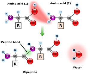
Amino acids link together when the amino group of one amino acid bonds to the carboxyl group of another. Notice that water is a by-product of the reaction (called a condensation reaction).

Stephen Meyer writes  in Signature of the cell:

According to neo-Darwinian theory, new genetic information arises first as random mutations occur in the DNA of existing organisms. When mutations arise that confer a survival advantage on the organisms that possess them, the resulting genetic changes are passed on by natural selection to the next generation. As these changes accumulate, the features of a population begin to change over time. Nevertheless, natural selection can "select" only what random mutations first produce. And for the evolutionary process to produce new forms of life, random mutations must first have produced new genetic information for building novel proteins. That, for the
mathematicians, physicists, and engineers at Wistar, was the problem. Why?

The skeptics at Wistar argued that it is extremely difficult to assemble a new gene or protein by chance because of the sheer number of possible base or amino-acid sequences. For every combination of amino acids that produces a functional protein there exists a vast number of other possible combinations that do not. And as the length of the required protein grows, the number of possible amino-acid sequence combinations of that length grows exponentially, so that the odds of finding a functional sequence—that is, a working protein—diminish precipitously.

To see this, consider the following. Whereas there are four ways to combine the letters A and B to make a two-letter combination (AB, BA, AA, and BB), there are eight ways to make three-letter combinations (AAA, AAB, ABB, ABA, BAA, BBA, BAB, BBB), and sixteen ways to make four-letter combinations, and so on. The number of combinations grows geometrically, 22, 23, 24, and so on. And this growth becomes more pronounced when the set of letters is larger. For protein chains, there are 202, or 400, ways to make a two-amino-acid combination, since each position could be any one of 20 different alphabetic characters. Similarly, there are 203, or 8,000, ways to make a three-amino-acid sequence, and 204, or 160,000, ways to make a sequence four amino acids long, and so on. As the number of possible combinations rises, the odds of finding a correct sequence diminishes correspondingly. But most functional proteins are made of hundreds of amino acids. Therefore, even a relatively short protein of, say, 150 amino acids represents one sequence among an astronomically large number of other possible sequence combinations (approximately 10^195).

Consider the way this combinatorial problem might play itself out in the case of proteins in a hypothetical prebiotic soup. To construct even one short protein molecule of 150 amino acids by chance within the prebiotic soup there are several combinatorial problems—probabilistic hurdles—to overcome. First, all amino acids must form a chemical bond known as a peptide bond when joining with other amino acids in the protein chain

Consider the way this combinatorial problem might play itself out in the case of proteins in a hypothetical prebiotic soup. To construct even one short protein molecule of 150 amino acids by chance within the prebiotic soup there are several combinatorial problems—probabilistic hurdles—to overcome. First, all amino acids must form a chemical bond known as a peptide bond when joining with other amino acids in the protein chain (see Fig. 9.1). If the amino acids do not link up with one another via a peptide bond, the resulting molecule will not fold into a protein. In nature many other types of chemical bonds are possible between amino acids. In fact, when amino-acid mixtures are allowed to react in a test tube, they form peptide and nonpeptide bonds with roughly equal probability. Thus, with each amino-acid addition, the probability of it forming a peptide bond is roughly 1/2. Once four amino acids have become linked, the likelyhood that they are joined exclusively by peptide bonds is roughly 1/2 × 1/2 × 1/2 ×
1/2 = 1/16, or (1/2)4. The probability of building a chain of 150 amino acids in which all linkages are peptide linkages is (1/2)149, or roughly 1 chance in 10^45.

Second, in nature every amino acid found in proteins (with one exception) has a distinct mirror image of itself; there is one left-handed version, or L-form, and one right-handed version, or D-form. These mirror-image forms are called optical isomers (see Fig. 9.2). Functioning proteins tolerate only left-handed amino acids, yet in abiotic amino-acid production the right-handed and left-handed isomers are produced with roughly equal frequency. Taking this into consideration further compounds the improbability of attaining a biologically functioning protein. The probability of attaining, at random, only L-amino acids in a hypothetical peptide chain 150 amino acids long is (1/2)150, or again roughly 1 chance in 1045. Starting from mixtures of D-forms and L-forms, the probability of building a 150-amino-acid chain at random in which all bonds are peptide bonds and all amino acids are L-form is, therefore, roughly 1 chance in 1090.

Second, in nature every amino acid found in proteins (with one exception) has a distinct mirror image of itself; there is one left-handed version, or L-form, and one right-handed version, or D-form. These mirror-image forms are called optical isomers . Functioning proteins tolerate only left-handed amino acids, yet in abiotic amino-acid production the right-handed and left-handed isomers are produced with roughly equal frequency. Taking this into consideration further compounds the improbability of attaining a biologically functioning protein. The probability of attaining, at random, only L-amino acids in a hypothetical peptide chain 150 amino acids long is (1/2)150, or again roughly 1 chance in 10^45. Starting from mixtures of D-forms and L-forms, the probability of building a 150-amino-acid chain at random in which all bonds are peptide bonds and all amino acids are L-form is, therefore, roughly 1 chance in 10^90.

Functioning proteins have a third independent requirement, the most important of all: their amino acids, like letters in a meaningful sentence, must link up in functionally specified sequential arrangements. In some cases, changing even one amino acid at a given site results in the loss of protein function. Moreover, because there are 20 biologically occurring amino acids, the probability of getting a specific amino acid at a given site is small—1/20. (Actually the probability is even lower because, in nature, there are also many nonprotein-forming amino acids.) On the assumption that each site in a protein chain requires a particular amino acid, the probability of attaining a particular protein 150 amino acids long would be (1/20)150, or roughly 1 chance in 10^195.

How rare, or common, are the functional sequences of amino acids  among all the possible sequences of amino acids in a chain of any given length?

Douglas Axe answered this question in 2004 3 , and  Axe was able to make a careful estimate of the ratio of (a) the number of 150-amino-acid sequences that can perform that particular function to (b) the whole set of possible amino-acid sequences of this length. Axe estimated this ratio to be 1 to 10^77.

This was a staggering number, and it suggested that a random process would have great difficulty generating a protein with that particular function by chance. But I didn't want to know just the likelihood of finding a protein with a particular function within a space of combinatorial possibilities. I wanted to know the odds of finding any functional protein whatsoever within such a space. That number would make it possible to evaluate chance-based origin-of-life scenarios, to assess the probability that a single protein—any working protein—would have arisen by chance on the early earth.

Fortunately, Axe's work provided this number as well.17 Axe knew that in nature  proteins perform many specific functions. He also knew that in order to perform these functions their amino-acid chains must first fold into stable three-dimensional structures. Thus, before he estimated the frequency of sequences performing a specific (beta-lactamase) function, he first performed experiments that enabled him to estimate the frequency of sequences that will produce stable folds. On the basis of his experimental results, he calculated the ratio of (a) the number of 150-amino-acid sequences capable of folding into stable "function-ready" structures to (b) the whole set of possible amino-acid sequences of that length. He determined that ratio to be 1 to 10^74.

In other words, a random process producing amino-acid chains of this length would stumble onto a functional protein only about once in every 10^74 attempts.

When one considers that Robert Sauer was working on a shorter protein of 100 amino acids, Axe's number might seem a bit less prohibitively improbable. Nevertheless, it still represents a startlingly small probability. In conversations with me, Axe has compared the odds of producing a functional protein sequence of modest (150-amino-acid) length at random to the odds of finding a single marked atom out of all the atoms in our galaxy via a blind and undirected search. Believe it or not, the odds of finding the marked atom in our galaxy are markedly better (about a billion times better) than those of finding a functional protein among all the sequences of corresponding length.
Problems with Making Mutation the Basis for Macroevolution

Dembsi, the design of life, general notes, page 11: 
If the proportion of gene sequences that are biologically useful were large, there might be reason to think that point or chromosome mutations could be helpful in achieving the novel biological structures required by macroevolution. But all the evidence points to biologically useful gene sequences being exceedingly rare. It’s therefore highly unlikely that point and chromosome mutations can transform a duplicated gene into a novel functional gene. Genetic sequence space (i.e., the set of all possible genetic sequences) is functionally sparse (i.e., the overwhelming majority of genetic sequences don’t, and indeed can’t, do anything biologically useful or significant). As a consequence, navigating genetic sequence space by undirected means is no help getting from one island of functionality (i.e., one region of biologically useful or significant genetic sequences) to the next.

For instance, there is no evidence that conventional evolutionary mechanisms, such as natural selection, can evolve a gene in one region of genetic sequence space with one set of functions into a gene in a far distant region of genetic sequence space with another set of functions (distance here being measured in terms of sequence similarity). In the language of mathematical biology, genetic sequence space gives no indication of being highly interconnected by functional pathways that continuously connect genes with one function to genes with another (which would be required if natural selection, say, were to assist a duplicated gene in transforming into a novel gene). But there are still more problems with trying to make mutation the basis for macroevolution. For point and chromosome mutations to account for macroevolutionary change, it is not enough for individual genes to be transformed into novel genes that exhibit novel functions. Rather it is required that a whole suite of novel genes be produced through the coordinated transformation of existing genes. This is because for new biological structures to evolve (as required by macroevolution), many genes will have to change.

But it has not been demonstrated that mutations can produce the highly coordinated protein parts required for many biological structures. These are the structures that macroevolution would need to produce. Till now, however, there is no evidence for the coordinated macromutations required for macroevolution. The closest evidence cited in
textbooks includes increased immunity to malaria associated with the mutation for sickle-cell anemia and the resistance to antibiotics by mutant strains of bacteria.
In no such case, however, do we see a coordinated set of mutations that lead to complex novel structures. Sickle-cell anemia, for instance, is induced by a single point mutation that leads to single change in an amino acid (a valine is substituted for a glutamic acid in a hemoglobin molecule). Point mutations like this might enable organisms to stabilize and maintain themselves in the face of severe environmental pressure. In most instances, however, novel traits induced by such mutations do not continue to benefit the organisms when the environmental pressure is removed. Apart from environmental pressure, such mutations can even be deleterious. For instance, when an individual is heterozygous for sickle-cell anemia, the mutation provides an advantage for surviving the threat of malaria. It does so, however, at the expense of inflicting on homozygous individuals an anemia that impairs the transport of oxygen to the body’s cells. Indeed, sickle-cell anemia is often lethal.

To observe the origin of new species, one would expect to find it most readily in bacteria. That’s because bacteria can be easily mutated with chemicals and radiation in the laboratory, they take up very little space, and they have very short generation times. Indeed, thousands of mutations, billions of organisms, and thousands of generations can be studied by a single scientist. Yet, bacteriologists have never witnessed the origin of a new species. (Some new plant species have been observed to originate through hybridization, but the combining of two species to make a third is the opposite of the Darwinian process of splitting one species into two.) For mutations to contribute to evolution, they must benefit the organism. If a mutation harms the organism, it will tend to be eliminated, rather than favored, by natural selection. The only beneficial mutations that have ever been observed, in bacteria or in any other kind of organism, have been biochemical. That is, they affect only single molecules (such as the target molecule for streptomycin). There are no known beneficial mutations affecting morphology, or shape. All known morphological mutations are either neutral (i.e., they don’t have any noticeable effect on the organism’s fitness), or they are harmful—and the bigger their effect the more harmful they are. Yet, Darwinian evolution (i.e., the origin of new species, new organs, and new body plans) clearly requires changes in shape. So, there is no evidence for (and indeed a lot of evidence against) a role for mutations as providing raw materials for Darwinian evolution.

The specific genetic changes that give rise to the evolutionary origins of novel protein-protein interactions have rarely been documented in detail
Although numerous investigators assume that the global features of genetic networks are moulded by natural selection, there has been no formal demonstration of the adaptive origin of any genetic network 7  The mechanisms by which genetic networks become established evolutionarily are far from clear. 

Many physicists, engineers and computer scientists, and some cell and developmental biologists, are convinced that biological networks exhibit properties that could only be products of natural selection; however, the matter has rarely been examined in the context of well-established evolutionary principles.

Alon states that it is “…wondrous that the solutions found by evolution have much in common with good engineering,”

Biological designs require a great deal more than the specification of a single DNA residue. Proteins, for instance, require a great many DNA residues to be specified. Even a single, typical protein requires more than 10^100 (a one followed by one hundred zeros) evolutionary experiments to find. This is an astronomical problem that is well beyond evolution’s capabilities. See here, here, here and here for more details. [url=10. http://darwins-god.blogspot.com.br/2011/06/vision-cascade-is-initiated-not-by.html]8[/url]


1) https://answersingenesis.org/origin-of-life/the-origin-of-life-dna-and-protein/
2) B.Alberts  Molecular biology of the cell.
3) http://www.ncbi.nlm.nih.gov/pubmed/15321723
4) http://www.evolutionnews.org/2015/06/paper_reports_t096581.html
5) https://www.c4id.org.uk/index.php?option=com_content&view=article&id=211:the-problem-of-the-origin-of-life&catid=50:genetics&Itemid=43
6. http://journals.plos.org/plosone/article?id=10.1371/journal.pone.0001054
7. https://sci-hub.bz/http://www.nature.com/nrg/journal/v8/n10/abs/nrg2192.html
8. http://darwins-god.blogspot.com.br/2011/06/vision-cascade-is-initiated-not-by.html
9. Molecular Mechanisms of Autonomy in Biological Systems Relativity of Code, Energy and Mass, page 34
10. https://www.nasa.gov/sites/default/files/atoms/files/pohorille_wilson_shannon_2017_1.pdf

More readings:
Comprehensive experimental fitness landscape and evolutionary network for small RNA
Our study suggests that replaying the “tape of life” at the very origin of life might lead to quite different results.


Last edited by Otangelo on Wed Nov 09, 2022 6:28 am; edited 42 times in total

https://reasonandscience.catsboard.com

Otangelo


Admin

Orchestration and timing of cellular processes require life-essential, precise timing, cross-regulation, coordination, the right sequence of processes, the right speed, at the right rate, there are checkpoint mechanisms, error checking, and repair at various stages.

Question: What emerged first: protein synthesis, or the right, precise coordination of the whole process, and its respective proteins and signaling processes doing the job?

Protein recipe requires precise timing 4
March 25, 2016
The activation of genes is a complicated biochemical endeavor akin to cooking a meal, and a new Yale study details just how precisely choreographed those steps need to be. Do cells cook the meat entrée before the vegetable? Or are these dishes cooked simultaneously? The Yale team led by Karla Neugebauer examined the intricacies of RNA splicing—the removal of RNA segments (known as introns) encoded by DNA that do not contain instructions for making proteins. The splicing machinery stitches the RNA segments together into accurate copies of mature messenger RNA that in turn sets in motion the cell's protein-making machinery. It was previously thought that splicing takes place well after transcription of the RNA segments. The Yale researchers found this crucial splicing step occurs as soon as the RNA is transcribed from the DNA template and that the timing of both transcription and splicing processes are precisely coordinated. "It's like cooking Thanksgiving dinner: The turkey and mashed potatoes need to be completely cooked and hot when the feast is served," Neugebauer said.

Splicing of Nascent RNA Coincides with Intron Exit from RNA Polymerase II 5
March 24, 2016
Here, we present two single-molecule nascent RNA sequencing methods that directly determine the progress of splicing catalysis as a function of Pol II position. Perturbations that slow the rate of spliceosome assembly or speed up the rate of transcription caused splicing delays, showing that regulation of both processes determines in vivo splicing profiles.


The evidence of the protein origin
1. On Protein Origins, Getting to the Root of Our Disagreement with James Shapiro – Doug Axe – January 2012.
I know of many processes that people talk about as though they can do the job of inventing new proteins (and of many papers that have resulted from such talk), but when these ideas are pushed to the point of demonstration, they all seem to retreat into the realm of the theoretical.
2. Shapiro admits he has no ‘real time’ empirical evidence for the origin of novel protein domains and/or genes by Darwinian processes (so as to be able to have the ‘protein domains’ to shuffle around in the first place) but must rely, as do neo-Darwinists, on the DNA/protein sequence similarity/dissimilarity data to try to make his case that novel protein domains were created in the distant past so that ‘natural genetic engineering’ can presently create all the diversity we see in life on earth today.
3. The primary problem is never addressed! i.e. Can the novel functional information we see in protein domains and/or genes ever be generated in a ‘bottom up’ fashion by the unguided material processes of neo-Darwinism? The answer to that question, as far as empirical evidence is concerned, is a resounding NO.
4. “Now Evolution Must Have Evolved Different Functions Simultaneously in the Same Protein” – Cornelius Hunter – Dec. 1, 2012
In one study naturalists estimated the number of attempts that evolution could possibly have to construct a new protein. Their upper limit was 10^43. The lower limit was 10^21.
These estimates are optimistic for several reasons, but in any case they fall short of the various estimates of how many attempts would be required to find a small protein. One study concluded that 10^63 attempts would be required for a relatively short protein.
And a similar result (10^65 attempts required) was obtained by comparing protein sequences.
Another study found that 10^64 to 10^77 attempts are required.
And another study concluded that 10^70 attempts would be required. In that case the protein was only a part of a larger protein which otherwise was intact, thus making the search easier.
These estimates are roughly in the same ballpark, and compared to the first study giving the number of attempts possible, you have a deficit ranging from 20 to 56 orders of magnitude. Of course it gets much worse for longer proteins.
5. “Why Proteins Aren’t Easily Recombined, Part 2″ – Ann Gauger – May 2012.
Excerpt: “So we have context-dependent effects on protein function at the level of primary sequence, secondary structure, and tertiary (domain-level) structure. This does not bode well for successful, random recombination of bits of sequence into functional, stable protein folds, or even for domain-level recombinations where significant interaction is required.”
6. The Law of Physicodynamic Incompleteness – David L. Abel – August 2011.
Summary: “The Law of Physicodynamic Incompleteness” states that inanimate physicodynamics is completely inadequate to generate, or even explain, the mathematical nature of physical interactions (the purely formal laws of physics and chemistry). The Law further states that physicodynamic factors cannot cause formal processes and procedures leading to sophisticated function. Chance and necessity alone cannot steer, program or optimize algorithmic/computational success to provide desired non-trivial utility.
7. From all this it is seen that research has advanced to the point of falsifying neo-Darwinism and Darwinism.
8. Intelligent design and its greatest intelligent designer God was a must to create DNA, RNA, proteins etc.
9. God  exists.

Frank Salisbury, an evolutionary biologist, discusses the difficulty surrounding the chance development of DNA as follows: 3

Salisbury, Frank B., "Doubts About the Modern Synthetic Theory of Evolution," American Biology Teacher, September 1971, pp. 336-338.
"Now we know that the cell itself is far more complex than we had imagined. It includes thousands of functioning enzymes, each one of them a complex machine itself. Furthermore, each enzyme comes into being in response to a gene, a strand of DNA. The information content of the gene (its complexity) must be as great as that of the enzyme it controls. "A medium protein might include about 300 amino acids. The DNA gene controlling this would have about 1,000 nucleotides in its chain, one consisting of 1,000 links could exist in 41.000 different forms. Using a little algebra (logarithms) we can see that 4^1.000 = 10^60° . Ten multiplied by itself 600 times gives the figure "1 " followed by 600 zeros! This number is completely beyond our comprehension." 2

A further complication involved with the original synthesis and subsequent replication of the DNA molecule is that the DNA molecule can only be replicated with the assistance of specific enzymes which, in turn, can only be produced by the controlling DNA molecule. Each is absolutely necessary for the other and both must be present for replication to occur. Dr. Haskins, president of the Carnegie Institute of Washington, has commented on this difficulty:

"Did the code and the means of translating it appear simultaneously in evolution? It seems almost incredible that any such coincidence could have occurred, given the extraordinary complexities of both sides and the requirement that they be coordinated accurately for survival. By a pre-Darwinian (or a skeptic of evolution after Darwin) this puzzle would surely have been interpreted as the most powerful sort of evidence for special creation."

http://www.discovery.org/scripts/viewDB/filesDB-download.php?command=download&id=10271

Protein scientist Douglas Axe suspected Robert Sauer’s research underestimated the rarity of functional protein sequences because it failed to consider simultaneous changes to multiple amino acids. Axe conducted more stringent mutagenesis experiments on enzymes to determine the rarity of amino acid sequences that yield stable protein folds—the smallest, most fundamental unit of structural innovation possible, key to generating macroevolutionary change. Axe’s research found that only 1 in 10^77 sequences of 150 amino acids in length can yield a stable protein fold. Richard Dawkins compares Darwinian evolution to climbing a mountain peak, but Axe’s work suggests functional amino acid sequences are so rare in sequence space that random mutation could never successfully find a peak, and once on a peak, could never leave one peak and successfully find another. Indeed, since only 10^40 organisms have ever existed on Earth, random mutations could never find even one functional protein fold over life’s entire history, much less in the timespan of the Cambrian explosion

Paul Davies puts it more graphically: ‘Making a protein simply by injecting energy is rather like exploding a stick of dynamite under a pile of bricks and expecting it to form a house. You may liberate enough energy to raise the bricks, but without coupling the energy to the bricks in a controlled and ordered way, there is little hope of producing anything other than a chaotic mess.’ It is one thing to produce bricks; it is an entirely different thing to organize the building of a house or factory. If you had to, you could build a house using stones that you found lying around, in all the shapes and sizes in which they came due to natural causes. However, the organization of the building requires something that is not contained in the stones. It requires the intelligence of the architect and the skill of the builder. It is the same with the building blocks of life. Blind chance just will not do the job of putting them together in a specific way. Organic chemist and molecular biologist A.G. Cairns-Smith puts it this way: ‘Blind chance… is very limited… he can produce exceedingly easily the equivalent of letters and small words, but he becomes very quickly incompetent as the amount of organization increases. Very soon indeed long waiting periods and massive material resources become irrelevant.’

Protein Evolution 1

We think that domains arose from the fusion of shorter, non-folding peptides, which evolved as cofactors supporting a primitive, RNA-based life form (the 'RNA world')

the building blocks to make the proteins, amino acids, are made by  COMPLEX biosynthesis pathways :

http://reasonandscience.heavenforum.org/t1740-the-biosynthesis-pathway-for-the-20-standard-amino-acids

, the enzymes that catalyze the reactions to make amino acids are by themself made of proteins.


1. http://www.biologicinstitute.org/post/57103144912/protein-evolution-a-guide-for-the-perplexed
2. Scott M. Huse, "The Collapse of Evolution", Baker Book House: Grand Rapids (Michigan), 1983 p:92
3. Salisbury, Frank B., "Doubts About the Modern Synthetic Theory of Evolution," American Biology Teacher, September 1971, pp. 336-338.
4. https://phys.org/news/2016-03-protein-recipe-requires-precise.html
5. http://www.cell.com/cell/fulltext/S0092-8674(16)30195-7


Last edited by Admin on Sat Dec 30, 2017 3:39 am; edited 8 times in total

https://reasonandscience.catsboard.com

Otangelo


Admin

Why the Origin of Life and the Evolution  of Molecular Knowledge Imply Design 1

The Evolution of Molecular Knowledge

Because of natural selection, information cannot be used to calculate the probability that a gene will evolve. Information is useful because it has a precise mathematical definition not because it can answer questions concerning whether or not the evolution of a new gene is possible. Chance is not in control if natural selection is guiding which mutations survive. Therefore, relating the amount of information in a gene to a probability that it can evolve is not a valid mathematical analysis.  Molecular knowledge is the minimum amount of useful information required for a gene to have any function. If a gene does not contain molecular knowledge, then it has no function, it confers no selective advantage, and it is not a gene. Thus, before a region of DNA contains the requisite molecular knowledge, natural selection plays no role in guiding its evolution. Chance controls which mutations survive. Thus, molecular knowledge can be related to a probability of  evolution.

Proteins: how they provide striking evidence of design Inform10

Notice that the first step that creates the required molecular knowledge is vertical, and the subsequent step that creates molecular information is sloped. This difference is important in that it is meant to show that natural selection can help guide the last transition, but it plays no role in the first. Thus, it is the size of the first step that determines whether or not a gene can evolve. A simple example will now be introduced to help clarify this
concept. Consider the following sentences:

I have a 13 year old black lab who likes to fetch a tennis ball. His name is Bubba.


My 13 year old black lab, Bubba, likes to fetch a tennis ball.


mi 13 yr od blk lab, buba, like fetch tenis bal.

Each sentence represents a gene. The first sentence uses the most letters; therefore, by definition, it contains the most information. The second sentence uses fewer letters, communicates the same points and is still grammatically correct. The last sentence uses the fewest letters. f these sentences are composed by randomly selecting letters from  the alphabet, then the probability of spelling the last sentence is much better than the first. So only the last sentence and all sentences similar to it are useful for assigning a probability to the evolution of a gene. In this example, the first sentence represents molecular information and the last one represents molecular knowledge. If some concepts are not required, the last sentence may be simplified further. For example, the sentence fragment, mi black lab, still communicates some knowledge. Thus, finding the precise threshold of molecular knowledge can never be exact. It relies on both math and human insight.  At least one biologist, Richard Dawkins, has suggested that sentences like: "hh n swd dwqdoe ffnfnriiq jddk" still confer a selective advantage; therefore, given time these sentences will evolve into something useful under the guidance of natural selection.1 Dawkins ran many computer simulations with sentences like the one above, and they all evolved into the desired result. But his programming and logic are both flawed because natural selection cannot preserve or optimize a gene that offers no selective advantage (the nonsense sentence above represents a gene that confers no selective advantage). A gene must contain some useful information before natural selection activates. Thus, chance and chance alone must create the initial knowledge. Because the probability that chance can accomplish this goal is proportional to the step height in figure 1, the size of this step completely determines whether or not a new gene will evolve. It is the goal of this book to characterize the size of this initial step. If it is small then naturalistic laws explain the evolution of knowledge. On the other hand, as the step size increases, the probability that chance will create the required molecular knowledge approaches zero, and at some critical threshold, the design inference becomes valid.  Large steps are associated with the evolution of genes that are completely different from all other existing genes. For these genes, the probability of chance finding an appropriate solution (even given 50,000 billion years) is very close to zero. The origin of these genes imply design.


Science does not have a plausible explanation for how the first genes and proteins evolved. 2Instead of sweeping this problem under the rug, they label any scientist who conducts experiments or develops mathematical and computer  models  to look at this issue a Creationist.  This label destroys the scientific credibility of both the scientist and his (or her) ideas and findings.

   This behavior may at first seem odd. But there is a good explanation for it.  Natural selection and chance (even given 100 trillion  years to operate) do not seem to be able to explain the evolution of the first genes and proteins. This observation threatens the very nature of science because it calls attention to the fact that the naturalistic axiom might just be a faulty assumption.  The truth is no longer  important because the  scientific establishment must preserve the axioms on which it is based. Thus, scientists who do not have faith in the naturalistic axiom are labeled  Creationists and their theories about evolution  are dismissed as cleverly disguised forms of religion.

Developers of computer simulations like Ev and Avida claim they can generate new information through sophisticated evolutionary algorithms. The problem with all these algorithms is no matter how sophisticated they need some kind of “forward looking memory”78 Natural selection in nature lacks foresight. It does not know where it is going. Selection cannot occur before new functional sequences arise. In simulation algorithms, they all use strategies to ensure the program will generate an information-rich sequence. For example Ev is provided with a target sequence (sequence of nucleotide bases) that functions as a binding site. A program is devised that allows Ev to eventually converge on the target sequence. It makes use of information that gives the process a goal-directed foresight, that is not like natural selection, but rather is like human selection. “Ev exhibits the genius of its designer.” 2

Willing to look behind the  curtain? A science teacher questions the party line…


http://followthelambwhereverhegoes.zohosites.com/books1.html

Popcorn strings, trains and balsa wood planes
The first core problem is one of information. I spent many years in a different career as a structural inspector, working closely with engineers, architects and builders to ensure that structures were being built according to the plans. My role was to ensure that the contractors were actually executing what was in the blue prints, down to minute details. If corners were cut, materials incorrect or craftsmanship sub-par there could be disastrous consequences for the future occupants because it would not be built as it was intended to be. You can probably make the connection with life, with DNA (or its transcript RNA) serving as the blue prints, ribosomes and transfer RNA being like the contractors, amino acids as the building materials and so forth. All that is well and good, and cells do what they are supposed to when the plans are executed properly. If not, you have disease, deformation or death. But who wrote the plans?!

Darwinian evolution cannot possibly explain the final outcome that we call life. Though natural selection is a really important mechanism that keeps life going strong, it could not have generated all the code that exists. It does a wonderful job of ensuring that life can respond to pressures, changing the allele frequencies within a population so that it can adapt and continue on. However, the Darwinian theory relies solely on one thing for the creation of new code for natural selection to act on, namely, mutation. Unfortunately, mutations make a mess of the code and are not credible as a source of creative power, regardless of what the textbooks say. In short, they (mutations) almost always make you either less fit or dead outright! The rare instances that they are “neutral,” or even more rarely “helpful” cannot account for the vast addition of well-oiled and fine-tuned instructions that it takes to make a mammal as compared to a bacteria. In fact, even bacteria are too intricate and sophisticated for me to stomach the notion that they could be happenstance. More importantly, mutations are random accidents that occur to an already existing set of instructions, and the question of where the blueprints for even the simplest life forms came from is not answered by believing that it could be amended successfully in a gigantic way.

Maybe it would be good to spend a moment examining how the code works in order to gain an appreciation for the insurmountable task at hand which mutation would have to account for.  You are probably aware that nucleic acids are code for protein synthesis, but accidental assemblies of these large and complex molecules cannot explain the reverse engineering that would be necessary in order to accomplish the creation of even the simplest functional cell. The most basic bacteria are incredibly complicated when you consider their structure and function. It’s a really large “pill to swallow” that the working proteins which cells are built out of could randomly occur and accomplish anything meaningful. Allow me to explain what I mean. Let’s grant the existence of ribosomes and the other machinery such as transcription factors, spliceosomes and tRNA which are necessary to translate nucleic acids into proteins (how they could arise spontaneously is another story). If all this is in place in a functional cell, the main issue at stake in the genetic code is the sequence of the instructions.

Each nucleotide subunit of the DNA (or RNA if you believe it came first) specifies which amino acid comes next in the strand of protein which is being built. Three letters of the code specifies one amino acid for protein synthesis. These three letter units are called a codon. In some ways it’s a bit like the sequence of box cars on a train, where some are red, some yellow, some blue, some carry coal or autos and so forth, and yet it’s not like that, because the locomotive will pull the train to its destination regardless of the sequence of the cars. But what if the locomotive couldn’t move unless the order was right? That would be weird, but in proteins the sequence of the amino acids, and hence the three letter codons in the DNA, really does matter intensely. 

In order to make a functional protein there has to be an ultimate shape that it ends up forming. This shape determines what the protein is able to do, and in fact, what it is intended to do within the cell, or outside the cell sometimes in the case of multicellular organisms. But what does this final shape have to do with the sequence of the amino acids? Well, as it turns out, the protein won’t hold its shape after being folded, twisted and bunched up unless the right amino acids end up being across from each other and can make a bridge-like bond which keeps it from unfolding and unraveling. Some amino acids have a sulfur atom in a region of the molecule called the “R group” which is the part that distinguishes between them. There are 20 different amino acid building blocks, and most lack the sulfur atom. Only when these amino acids are placed in the long chain of amino acids in the right position do you maintain the final shape.  The problem is, these particular amino acids have to be sometimes hundreds of units away from each other in the single file line that the ribosomes chug out as they read and translate the code. If they are in the wrong position in the sequence (called the primary structure), then the twisting and folding (such as alpha helixes and beta pleats called secondary structures) won’t be able to give rise to three-dimensional shapes (tertiary structures) which will “stay” when they themselves fold up. It would be a bit like the rubber band engine on a balsa wood airplane (you know, the kind with a plastic propeller). When you turn the prop it twists the rubber band into thicker and thicker wads that take on a three- dimensional lumpy shape, but there is nothing keeping it in that shape when you let go, so it just unwinds again.
Another analogy may help make this clear. Imagine a child threading popcorn on a needle and string. If some of the pieces were Velcro and the child randomly sequenced them hundreds of pieces long, what would the odds be that a specific shape required for final structure and function would be achieved if it could be maintained only when certain positions had a Velcro piece? In fact, this would have to be true many times down the length of the string, where if a Velcro piece was missing from any of the right spots, the ultimate shape could not be preserved once it was attained. Not only that, the child would have to make thousands if not tens or even hundreds of thousands of other strings with just the right sequence in order to make a working cell. Remember, the ones that can make a sulfur bridge (disulfide bond) have to end up opposite each other once all the folding and twisting is done or the protein will not work properly. So, you have to know what the final three-dimensional shape has to be in order to program the code to achieve it. Moreover, you have to know how many proteins are needed and what their jobs will be in order to pull it all off! So the question is not just one of whether or not the complex molecules of RNA and DNA could arise by random chance, but more fundamentally, it is a question of information, foreknowledge, planning and intent.

To grasp the magnitude of this accomplishment, let’s consider the size/length of the code for a generic, rod-shaped bacterium like E. coli. If you were to type the nitrogenous base sequence in 12 point font, it would fill the entire collection of one of those old-fashioned sets of encyclopedias! If just one of the letters in the code was goofed up it might seriously harm the bacteria. Take a guess how long the code is in OUR genome?  Would you believe we have 3 billion base pairs?! When you realize that there are debilitating diseases such as Tay-Sachs that result from a single point mutation (just one letter of the code being wrong) it staggers the mind! Granted, some mutations are more “survivable” than others (like a letter being switched out for a different letter) because of the redundancy that’s built into the code to ward off these outcomes, but others are horribly destructive. Omissions (deletions) or additions wreak havoc, because they shift all the instructions “downstream” one spot. As an illustration, try reading this sentence if you take out a letter e in the first word the, and shift everything back a spot (remember, codons are read in groups of three): the dog ate her bags.  It would read “thd oga teh erb ags”. Gibberish. When this happens to the DNA, the sequence of amino acids will probably be wrong, a deformed protein is likely and its function is subsequently lost. And there is another kind of mutation called a translocation that is especially devastating. This is when portions of the code get misplaced to some other location in the DNA and the coordination/control that previously existed is shattered.

Organisms fight these alterations tenaciously with attempts at repair, and the maintenance systems which perform this invaluable service are absolutely mind bending. The genetic code is jealously guarded for a reason. Suffice it to say for now that DNA isn’t called “the miracle of life” (mind you, by professed atheists) for no reason. To me, it seems more reasonable right at the outset to conclude that the genetic code, just as with all codes containing information, has a writer. I see enormous evidence that an author has “written” it for a specific purpose, and reject the proposition that it arose randomly, with accidental serendipity creating delicate and incredibly precise structures, complete with a host of specific functions which must work in a coordinated fashion in order to create even the simplest living cell. You are free to conclude that is the case if you want, and I’m not trying to insult you if you do, honestly. I’m just calling it like I see it, that’s all! There are actually more serious and convincing reasons to reject the materialistic world view, but let’s save that till the end of the discussion. For now, trying to explain how this level of complexity, harmony and order could arise from chaos is only one of the huge holes in the theories for the origin of life from non-life.


1) http://www.idnet.com.au/files/pdf/Molecular%20Evolution.pdf
2) http://www.orvosiangol.com/Texts4/Medical_Subjects/Bioethics/Evolution2.htm


Last edited by Admin on Wed Mar 29, 2017 1:13 pm; edited 4 times in total

https://reasonandscience.catsboard.com

Otangelo


Admin

Test of Functional Sequence Space Shows Less Tolerance for Mutations than Expected 1

When you change a tire, you expect the holes on the wheel to line up with the lug bolts on the axle. But manufacturing defects can occur. The bolts might be slightly offset, or slightly longer or shorter than spec. A bolt or two might be missing. How many defects, and how large, will still permit you to continue down the road without being stranded?
Protein interactions are a more sophisticated instance of much the same problem. Proteins must interact with other proteins, often fitting like a lock and key. As with the tire example, there's a bit of wiggle room that proteins can tolerate and still function. A new study shows that this wiggle room, in practice, is less than theoretically conceivable.
Proteins are made up of chains of amino acids, sequenced in such a way that the chain folds into a three-dimensional shape with "functional information" (the ability to do work or provide structure for the cell). One or more parts of that shape have to "fit" with other proteins. There's a certain amount of error that can allow substitutions of amino acids (mutations) to be tolerated; that's called "neutral evolution" or "neutral variation." It might be compared to typos in a paragraph that don't prevent the reader from understanding the message. The reader might gloss over the typo "tfe" in a sentence like "Functional space is a subset of tfe sequence space," and still understand what was intended. Other substitutions, though, may render the meaning unintelligible, like gubsetfor subset, or spade for space.
Like our simple sentence says, the functional space of a chain of amino acids is a small subset of possible sequences -- a very small subset, in fact. Take a chain of amino acid residues. Since there are 20 amino acids in proteins, there are 20N possible combinations. For a relatively small protein of 100 amino acids, that's 20100 combinations, or 20 thousand trillion trillion trillion trillion trillion trillion trillion trillion possibilities. Of those, only a tiny fraction of those are functional -- if they fold at all.
Homologous proteins from different organisms often show substantial variation yet are still functional. In proteins, contact sites that interact with partners are typically less forgiving than other sites. The protein might still interact if a hydrophobic amino acid residue at the active site is substituted for another hydrophobic residue; the function can break, however, if a very different amino acid were substituted.
"Degeneracy" is a measure of the tolerance for substitution in a sequence. Consider Morse code as an example of a non-degenerate code: each letter has one "and only one" sequence of dots and dashes for each letter of the alphabet. The genetic code, however, is degenerate: some amino acids can be coded by multiple triplet codons in the DNA. For example, the codons GGA, GGC, GGG, and GGU all code for glycine. There are functional reasons for this apparent laxness in the code, but that's another story.
Now to the crux of the issue: How much tolerance for error is there in proteins? How degenerate is the protein code? As seen from our 20100 example, it would be practically impossible to search sequence space for functional chains of amino acids. The only way to broach the question is by breaking it down into a tractable question testable by experiment. Take one specific protein interaction: How much substitution can it tolerate and remain functional?
Two researchers at MIT, Podgornaia and Laub, decided to tackle that question. Their work is published in Science. A summary by Guy Riddihough, "Exploring the limits of protein sequence space," in the same issue of Science, gives the main lesson learned:
Exploring the variability of individual functional proteins iscomplicated by the vast number of combinations of possible amino acid sequences. Podgornaia and Laub take on this challenge by analyzing four amino acids critical for the interaction between two signaling proteins in Escherichia coli. They build all the possible 160,000 variants of one of the two proteins and find thatover 1650 are functional. Even though there can be very high variability in the composition of the interface between the two proteins, there are nonetheless strong context-dependent constraints for some amino acids, which suggests why many functional variants are not seen in nature. (Emphasis added.)
As stated, the researchers performed a very scaled-down experiment on two interacting proteins named PhoP and PhoQ. We don't need to concern ourselves with what the two proteins do. We just need to ask how much substitution they can tolerate and still interact. Now that we know what "degeneracy" means, we need to add another term to our working vocabulary: epistasis. This refers to something like "unintended consequences" of making changes. Sure, you might substitute another hydrophobic residue from the wild type, but will that change the spacing of the other nearby amino acids? Will it affect the folding of the protein? Will it break the function? A permissible substitution may not be practical in context.
The researchers basically found that these two properties, degeneracy and epistasis, work against each other. Their paper's title is, "Pervasive degeneracy and epistasis in a protein-protein interface." Degeneracy allows for a fairly large amount of variation (if you can call 1% "large"). Sure enough, when they considered all the possible substitutions for "four key residues" in PhoQ (160,000 of them), they found 1659 that still worked. However, epistasis cut that number way down.
Our results reveal extensive degeneracy in the PhoQ-PhoP interfaceand epistasis, with the effect of individual substitutions often highly dependent on context. Together, epistasis and the genetic code create a pattern of connectivity of functional variants in sequence space that likely constrains PhoQ evolution.Consequently, the diversity of PhoQ orthologs is substantially lower than that of functional PhoQ variants.
In theory, therefore, you can find a lot of varieties (orthologs) that are functional. In practice, epistasis constrains that number substantially. That's why only a small fraction of their 1659 functional varieties were actually detected in living E. coli.
One reason for the constraint is the mutational path to some of the functional orthologs. We need to recall that Darwinian evolution is blind to future goals; every substitution has to produce a functional advantage right now. The researchers found that getting to some of their theoretical functional orthologs by random mutation would require traversing through two or more non-functional intermediates. Those non-functional intermediates would likely be eliminated by selection before the next step or steps could be reached. The only way Darwinian evolution could "select" those variants would be for the two mutations to appear simultaneously. This approaches "the edge of evolution" that Michael Behe discusses at length in his book of that name: the probability for simultaneous random mutations rapidly drops off the edge of a cliff.
How many functional variants remain out of 1659 when epistasis is considered? To answer that question, we need to add another term to our working vocabulary: "Hamming distance." In information theory, the Hamming distance is the number of substitutions required to transform one string into another of equal length. The Hamming distance to transform 1011 to 1001 is 1. The Hamming distance to transform "cartop" into "carpet" is 3. You might be able to transform one string into another via a series of meaningful words, like gateinto name via the series, gate, game, name. Darwinian evolution, however, cannot tolerate non-functional intermediates. The "path length" of functional intermediates cannot exceed the Hamming distance.
Now that we have reviewed these concepts, what do the MIT researchers tell us about the actual subset of functional orthologs of PhoQ out of the theoretical possibilities, after considering epistasis, Hamming distance and path length?
Shortest path lengths now exceeded Hamming distances for >97% of all connected variant pairs (fig. S8B). Together, the genetic code and epistasis severely constrain mutational paths in sequence space for PhoQ....

In general, the natural diversity in PhoQ orthologs (Fig. 4H and fig. S9C), even those with divergent PhoP partners, is much more limited than the diversity in our selected, functional variants[i.e., the 1659 orthologs]....
Collectively, our results suggest greater functional degeneracy for PhoQ than would be expected by site-saturation mutagenesis.However, the interconnectivity of functional variants, which results from epistasis and the structure of the genetic code, haslikely limited nature's exploration of sequence space, as reflected in the limited diversity of PhoQ orthologs (Fig. 4H).
Theoretically, then, one could imagine a fairly large number of variations in the four key residues of PhoQ that do not break functionality. Practically speaking, however, you can't get to most of them naturally. For one thing, nature doesn't even explore large areas of theoretical sequence space, because functional variants are interconnected. For another, epistasis, with its unintended consequences, interferes. Finally, natural selection can't even get to most of the possible functional forms, because "shortest path lengths now exceeded Hamming distances for > 97% of all connected variant pairs." But then, out of the 57 or so (3%) they considered practical, they only found 13 could actually compete with the wild type.
Knowing the results, let's look again at what they set out to test:
Protein-protein interactions drive the operation and function of cells. These interactions involve a molecular interface formed by a subset of amino acids from each protein. Interfacial residues often vary between orthologs, indicating some mutational tolerance or degeneracy, but such natural variability may not capture the full plasticity of interfaces. Thus, it remains unclear how many combinations of interface residues will support a given interactionand how these combinations are distributed and connected in sequence space (3) (fig. S1A). It is also unknown whether all functional variants can be reached through a series of mutations that retain function, or whether evolution is fundamentally constrained, limiting the natural diversity in orthologous proteins.
The test results are clear. We've dropped from 20N variants in sequence space to 160,000 possible sequences for just four key residues, to 1659 functional variants of those, to 57 that are accessible to unguided natural processes, to 13 that could actually compete in the wild. In summary, functional space is a tiny, tiny fraction of sequence space. And "practical" functional space is another tiny fraction of theoretical functional space. We might put it this way: nature abhors the edge of evolution.
Seeing that the "wiggle room" for our spare tire on the axle is so small, we might return to the old, tried-and-true explanation that the car was designed after all.

1) http://www.evolutionnews.org/2015/02/test_of_functio093631.html

https://reasonandscience.catsboard.com

Otangelo


Admin

How Proteins Evolved

1

Proteins: how they provide striking evidence of design Hemoglobin
Fifty years ago molecular biologists began to uncover the inner workings of the cell and one of their profound discoveries was that genetic information, stored in the double helix DNA molecule, was translated according to a code to produce a string of amino acids which, after being hitched to each other like train cars, folded up to produce a protein that did something useful in the cell. Interestingly, a given protein’s amino acid sequence was found to have some degree of flexibility. Hemoglobin proteins, for instance, across different species revealed quite a few changes to the sequence while still functioning as a hemoglobin.

Two discoveries that later followed seemed to bode well for evolution. First, protein sequences fit the evolutionary pattern when compared across different species. For instance, a given protein such as hemoglobin has a very similar sequence of amino acids when compared between two similar species, and a much more different sequence when compared between distant species. Second, the degree of flexibility of a given protein’s amino acid sequence was substantial. Hemoglobin sequences in distant species had practically no resemblance. This suggested that a relatively small amount of information was actually needed to code for a given protein. Proteins have hundreds of amino acids, but perhaps only tens of them are important in determining the structure and function of the folded protein structure. This would make evolution’s task of constructing proteins less daunting.

These discoveries are a bit technical and so not always easy to convey to non scientific audiences. But proponents of evolution  have tried, for these discoveries definitely gave them added confidence their theory was right. What proponents of evolution have been less vocal about are the discoveries that have been made more recently. We now know that for both these discoveries, the story is more complicated than it originally appeared.

The evolutionary pattern

First, the observation that protein sequences fit the evolutionary pattern is not particularly surprising. If we discovered a new bone or tissue, would we not generally expect that it would be more similar between species that otherwise are similar, and more different in more distant species? After all, it functions within a context. So it is no shocker that cellular components such as proteins generally follow the pattern.

But this is not always the case. This pattern that evolution predicts is sometimes violated. Biology reveals some very big differences between otherwise similar species, and some striking similarities between otherwise distant species. This is true for visible features as well as at the molecular level. The evolutionary pattern has now been falsified many times over. Nonetheless, the textbooks continue to proclaim the good news that proteins fit the predicted pattern, and curiously omit any mention of the more recent failures.

The flexibility of a protein sequence

Second, we now understand that the degree of flexibility in a protein sequence is far less than once imagined. One way to think of this is with an analogy to English. Consider these two sentences:

Evolutionary theory has produced a vast number of falsified predictions.

and,

Most of the expectations generated by evolution have turned out false.

These two sentences share the the same meaning yet if you align and compare them letter-by-letter, you would find most of the letters are different. They seem to be unrelated at that level. Could it be that there are only a few key letters that determine the entire meaning of the sentence?

Of course not. We all know that while very different text messages can share the same meaning, this does not imply that most of the letters carry no effect and can be mutated without little or no effect. In fact, even for such short messages there is a large number of possible letter sequences. Each letter has 26 possibilities and in the above example there are about 60 letters. This means there about 10^85 (a one followed by 85 zeros) different letter sequences that are theoretically possible. Of that astronomical set of possibilities, only a relatively tiny set of selections is grammatically correct. The set of selections that makes any sense is even smaller, and the set that carries the same meaning as those above is tiny.

Likewise, it is now clear that while a given protein such as hemoglobin may come in very different sequences, only few changes to that sequence can be sustained. There are many ways to code for the hemoglobin machine, and they may appear to be very different, but that does not mean there is a relatively large number of sequences that can produce a hemoglobin. Here is how one recent paper explained the new findings:

The accepted paradigm that proteins can tolerate nearly any amino acid substitution has been replaced by the view that the deleterious effects of mutations, and especially their tendency to undermine the thermodynamic and kinetic stability of protein, is a major constraint on protein evolvability—the ability of proteins to acquire changes in sequence and function.

Another recent paper explained that only a few percent of a protein’s amino acids can tolerate change at any given point in time.

We are a long way from having hard numbers, but even conservative estimates of the number of protein sequences that are viable, for a given type of protein, are tiny. Any given residue in a protein may not be required to have a specific type of amino acid in order to form a hemoglobin, but in the context of the remainder of the sequence there is a tight requirement. And the number of such contexts is relatively small.

Years ago creationists argued that evolution was improbable because a protein sequence is a long shot. A typical globin sequence has about 140 amino acids. This means the sequence represents a one in 10^182 chance (a one followed by 182 zeros).

It was an imprecise argument, for there is far more than just one sequence that can do the job. But in this case, being off by orders of magnitude hardly matters. A viable globin sequence may be a mere one in 10^150 chance. Who knows, perhaps it is even a one in 10^120 chance. The fact is we do not know, but today’s science is telling us that a viable globin sequence makes finding a needle in a haystack seem easy. And the hemoglobin protein is a relatively small one. Many proteins are several times longer.

How do proteins evolve?

Contrary to early notions that protein sequences were extremely flexible, science is now telling us the opposite. This indication that viable protein sequences occupy a tiny sliver of sequence space suggests that they are difficult to evolve.

If you ask a proponent of evolution  how a protein evolved, you will likely hear the standard answer: via gene duplication and subsequent divergence. In other words, the protein arose from a different type of protein that was pre existing. The gene for that protein duplicated, and then mutated until landing on a new protein that was helpful. And of course this story must have repeated itself thousands of times to create the many different proteins in biology.

It is an unlikely, just-so, story, for viable protein sequences are hard to find. If the different types of proteins each have their own tiny slivers of sequence space as science is suggesting, then gene duplication and divergence, alone, doesn’t stand a chance.

What would be needed are long trails of intermediate, functional, proteins connecting the different types of proteins. These proteins would not only need to be functional, their particular function would have to be useful at the time.

And why would the known proteins just happen to be fortuitously connected by these trails? Science gives us no reason to think such a lucky circumstance is built into the protein world. So either there are no such trails, which means evolution has a problem, or there are such trails which means someone has monkeyed with the fundamentals of protein chemistry.

And we have not yet even addressed the problem of how the first proteins evolved. Remember the proponents of evolutions standard explanation for how proteins evolved is by gene duplication and subsequent divergence. But that requires the pre existence of other types of proteins. In other words, the question of how proteins evolve in the first place has been swept under the rug.

The problem of how evolution could create a new type of protein from an existing protein, via gene duplication and subsequent divergence, as difficult as it is, pales in comparison to how evolution was supposed to have created new proteins from scratch. Proponents of evolution speak of an initial world where RNA molecules do the work of proteins. But even this heroic story doesn’t magically make the problem of protein evolution go away. Whether there were RNA precursors or not, there is a substantial difficulty in explaining how the first proteins could have evolved.

Stepping stone: ATP binding

A few years ago an experiment showed that randomly constructed short proteins have a one in 10^12 shot (a million million) of having function. The function, in this case, was the binding of ATP, the cell’s unit of energy. While that is an interesting experiment, the results do little to help evolution. Most obviously, that function alone is quite minor. Yes, proteins bind to ATP or other chemicals, but that binding is in a complex, tight coordination with other functions. Comparing ATP binding with the incredible feats of hemoglobin, for example, is like comparing a tricycle with a jet airplane.


And even the one in 10^12 shot, though it pales in comparison to the odds of constructing a more useful protein machine, is no small barrier. If that is what is required to even achieve simple ATP binding, then evolution would need to be incessantly running unsuccessful trials. The machinery to construct, use and benefit from a potential protein product would have to be in place, while failure after failure results. Evolution would make Thomas Edison appear lazy, running millions of trials after millions of trials before finding even the tiniest of function. Why would this machinery be in place? Why would it continue to construct and test? Why would evolution maintain such an incompetent test bench?


Evolutionary answers to such questions are like all their stories. It did it because it did it. And if it seems unlikely, then remember there are many planets revolving about many stars, in many galaxies. And beyond that there are many universes. And in any case, a different sort of life—or no life at all—could have evolved. This is what evolution has done to science. Religion drives science and it matters.

1) http://darwins-god.blogspot.com.br/2010/12/how-proteins-evolved.html


Last edited by Admin on Fri Dec 25, 2015 10:46 am; edited 2 times in total

https://reasonandscience.catsboard.com

Otangelo


Admin

Computing the "Best Case" Probability of Proteins from Actual Data, and Falsifying a Prediction of Darwinism


Biological life requires thousands of different protein families, about 70 percent of which are "globular" proteins, with a three-dimensional shape that is unique to each family of proteins. An illustration is shown in the picture at the top of this post. This 3D shape is necessary for a particular biological function and is determined by the sequence of the different amino acids that make up that protein. In other words, it is not biology that determines the shape, but physics. Sequences that produce stable, functional 3D structures are so rare that scientists today do not attempt to find them using random sequence libraries. Instead, they use information they have obtained from reverse-engineering biological proteins to intelligently design artificial proteins.

Indeed, our 21st-century supercomputers are not powerful enough to crunch the variables and locate novel 3D structures. Nonetheless, a foundational prediction of neo-Darwinian theory is that a ploddingly slow evolutionary process consisting of genetic drift, mutations, insertions, and deletions must be able to "find" not just one, but thousands of sequences pre-determined by physics that will have different stable, functional 3D structures. So how does this falsifiable prediction hold up when tested against real data? As ought to be the case in science, I have made my program available so that you can run your own data and verify for yourself the kinds of probabilities these protein families represent.
This program can compute an upper limit for the probability of obtaining a protein family from a wealth of actual data contained in the Pfam database. The first step computes the lower limit for the functional complexity or functional information required to code for a particular protein family, using amethod published by Durston et al. This value for I(Ex) can then be plugged into an equation published by Hazen et al. in order to solve the probability M(Ex)/N of "finding" a functional sequence in a single trial.


I downloaded 3,751 aligned sequences for the Ribosomal S7 domain, part of a universal protein essential for all life. When the data was run through the program, it revealed that the lower limit for the amount of functional information required to code for this domain is 332 Fits (Functional Bits). The extreme upper limit for the number of sequences that might be functional for this domain is around 10^92. In a single trial, the probability of obtaining a sequence that would be functional for the Ribosomal S7 domain is 1 chance in 10^100 ... and this is only for a 148 amino acid structural domain, much smaller than an average protein.


For another example, I downloaded 4,986 aligned sequences for the ABC-3 family of proteins and ran it through the program. The results indicate that the probability of obtaining, in a single trial, a functional ABC-3 sequence is around 1 chance in 10^128. This method ignores pairwise and higher order relationships within the sequence that would vastly limit the number of functional sequences by many orders of magnitude, reducing the probability even further by many orders of magnitude -- so this gives us a best-case estimate.
What are the implications of these results, obtained from actual data, for the fundamental prediction of neo-Darwinian theory mentioned above? If we assume 10^30 life forms with a fast replication rate of 30 minutes and a huge genome with a very high mutation rate over a period of 10 billion years, an extreme upper limit for the total number of mutations for all of life's history would be around 10^43. Unfortunately, a protein domain such as Ribosomal S7 would require a minimum average of 10^100 trials. In other words, the sum total of mutational events for the entire theoretical history of life falls short by at least 57 orders of magnitude from what would have a reasonable expectation of "finding" any RS7 sequence -- and this is only for one domain. Forget about "finding" an average sized protein, not to mention thousands.


As we all know from probabilities, you can get lucky once, but not thousands of times. This definitively falsifies the fundamental prediction of Darwinian theory that evolutionary processes can "find" functional protein families. A theory that has an essential prediction thoroughly falsified by the data should have no place in science.
Could natural selection come to the rescue? As we know from genetic algorithms, an evolutionary "search" will only work for hill-climbing problems, not for "needle in a haystack" problems. There are small proteins that require such low levels of functional information to perform simple binding tasks that they form a nice hill-climbing problem that can be easily located in a search. This is not the case, however, for the vast majority of protein families. As real data shows, the probability of finding a functional sequence for one average protein family is so low, there is virtually zero chance of obtaining it anywhere in this universe over its entire history -- never mind finding thousands of protein families.
What are the implications for intelligent design science? A testable, falsifiable hypothesis of intelligent design can be stated as follows:


A unique attribute of an intelligent mind is the ability to produce effects requiring a statistically significant level of functional information.

Given the above testable hypothesis, if we observe an effect that requires a statistically significant level of functional information, we can conclude there is an intelligent mind behind the effect. The average protein family requires a statistically significant level of functional, or prescriptive, information. Therefore, the genomes of life have the fingerprints of an intelligent source all over them.


1) http://www.evolutionnews.org/2015/07/computing_the_b098101.html


Last edited by Admin on Wed Mar 29, 2017 1:14 pm; edited 2 times in total

https://reasonandscience.catsboard.com

Otangelo


Admin

Proteins and Their Exquisite Design


Proteins are the building blocks of life. They are the structural parts that give cells shape, the enzymes that build or break down the molecules of life, the motors that transport things, the agents that send signals and regulate the activity of other proteins and genes, and the morphogens that help determine the development of the organism. What determines a protein's activity and properties? Its shape. And what determines its shape? The way its one-dimensional string of amino acids folds together. This is a complex process involving many interactions, so complex that we cannot reliably predict a protein's structure based on its sequence. To get an idea of the problem, take a look at the picture above, the figure on the left. This is an illustration of a single protein called porin, whose structure has been determined experimentally. This protein has around 300 amino acids. What you see here is the arrangement of all its covalent bonds between atoms, shown as sticks. Making sense of that tangle of bonds is difficult. So scientists often depict proteins in a simplified cartoon form that shows the secondary structure of the protein fold. These secondary structures are motifs within the protein that form either alpha helical coils or flat beta sheets, and are therefore drawn as coils or flat arrows in cartoon illustrations. The middle picture above shows porin, in the same orientation and size as the picture on the left, but now drawn in cartoon form. Porin is composed mostly of antiparallel beta sheets, arrayed in a barrel-like shape, with an opening in the center. But neither of these pictures illustrates porin as it would look if we could take a snapshot. The figure above on the right is a surface view of porin, showing its many knobs and hollows and a hole in the middle. Those knobs and hollows allow porin to assemble in its final functional form in the membrane, a trimer composed of three porin chains tightly coupled together. The image below is of trimeric sucrose-specific porin found in E coli. The artist has rendered it so that we can see the secondary structure within the assembled trimer, and it is colored as a rainbow according to the threading of the amino acid chains, from first (blue) to last (red).

What does porin do? It serves as a pore to let specific molecules enter the periplasmic space of bacteria. The pore has to have a specific shape and polarity to allow the right chemicals admittance, but exclude others, and the outside of the protein has to be able to interact stably either with other porins or with the membrane. That all depends on the information contained in porin's primary sequence. Research indicates that sequences that fold into a particular functional shape are rare. Only about 1 in 10^77 possible sequences will adopt a functional fold 150 amino acids in length. How rare is porin as a functional sequence? Based on its functional constraints, it is probably as rare as the enzymatic fold already tested. My point? Proteins exhibit exquisite design, with extraordinary specified complexity embedded in their sequences. Too much to be the result of random processes.


1) http://www.evolutionnews.org/2012/05/proteins_and_th059161.html


Last edited by Admin on Wed Mar 29, 2017 1:14 pm; edited 3 times in total

https://reasonandscience.catsboard.com

8Proteins: how they provide striking evidence of design Empty Evolution Dissected By Fredric P. Nelson Tue Nov 24, 2015 11:58 am

Otangelo


Admin

Evolution Dissected By Fredric P. Nelson 1

Proteins: how they provide striking evidence of design Protei11
Proteins: how they provide striking evidence of design Protei14
Proteins: how they provide striking evidence of design Protei12
Proteins: how they provide striking evidence of design Protei15

https://books.google.com.br/books?id=DqVvc70qLrgC&pg=PA39&lpg=PA39&dq=Functional+proteins+from+a+random-sequence+library&source=bl&ots=Xrtlxzfk8z&sig=59KBNgvQgSr9Q9bb3eW94yI-GD4&hl=en&sa=X&ved=0ahUKEwjA-sil3KnJAhVRnZAKHUHVCPgQ6AEIRzAI#v=onepage&q&f=false

https://reasonandscience.catsboard.com

Otangelo


Admin

The Szostak lab found functional proteins (four different ones) in a pool of about 10^11 random sequence proteins (sequences that were 80 amino acids in length). And they only tested for one function (bind ATP), they could have tested for thousands of additional functions, such as chemical catalysts and binding to countless other molcules. They also only tested at one temperarure. Who’s to say what else could be found in that pool of random sequence proteins if they had tested at other temperatures, at different ion/salt concentrations or tested for binding to other molecules or even for the presence of weak chemical catalysts?

It is trivial and easy to find functiona proteins in random sequence space. The Szostak lab proved this experimentally back in the late 90’s and early 2000’s and showed it to be true both for proteins and RNA’s:

Functional proteins from a random-sequence library

https://molbio.mgh.harvard.edu/szostakweb/publications/Szostak_pdfs/Keefe_Szostak_Nature_01.pdf

Anthony D. Keefe & Jack W. Szostak
“Functional primordial proteins presumably originated from random sequences, but it is not known how frequently functional, or even folded, proteins occur in collections of random sequences. Here we have used in vitro selection of messenger RNA displayed proteins, in which each protein is covalently linked through its carboxy terminus to the 39 end of its encoding mRNA1 , to sample a large number of distinct random sequences. Starting from a library of 6 x 10^12 proteins each containing 80 contiguous random amino acids, we selected functional proteins by enriching for those that bind to ATP. This selection yielded four new ATPbinding proteins that appear to be unrelated to each other or to anything found in the current databases of biological proteins. The frequency of occurrence of functional proteins in random sequence libraries appears to be similar to that observed for equivalent RNA libraries2,3.”

80 random amino acids strung together into a protein. Generate 6×10^12 different, random copies, test them all for a single (and extremely biologically important) function: Bind ATP.

Among that starting pool of random proteins 80 amino acids in length, there were four (4) different, unrelated proteins found that could do it. That gives about 1 in every 10^11 proteins capable of binding ATP. Which strongly indicates that as an absolute minimum there is at least one biologically relevant function in every 10^11 80-amino-acid long proteins. (I could stop here already, this is enough to render all of creationism bunk).

Notice how only a single function was tested for for that pool of random proteins. They could have tested millions of different functions (bind other biologically important molecules, tested for catalysis of thousands of different chemical reaction, stabilize phospholipid membranes etc. etc.) – but they only tested for one and found it already to begin with.

The simple fact is that Sean Pitman, the creationist liar for doctrine from which you probably copy-pasted this religiously motivated drivel, is talking out of his religiously biased ass, based on a couple of studies where he wildly extrapolates the results into areas the data don’t support. For example, the Discovery Institute paid their liar propaganda laboratory to mutate a functional protein until it stopped working (at what it was doing), then they tried to derive a general rule for the rarity of function in protein sequence space on this stupid experiment. It’s true, it only required relatively few mutations to destroy the function of the protein in question, and as a result they computed that functional proteins are supposed to exist at a rate of approximately 1 in every 10^77 proteins. Which if true, would entail that functional proteins were, as you go on to copy-paste, exceptionally rare. But does their experiment really warrant that kind of conclusion? They mutated a protein until it stopped working (again, at what it was doing). Even then, that is still not any guarantee that the protein in question is entirely nonfunctional. It is entirely possible that you can mutate a specific protein fold that, say, catalyzes some chemical reaction until it stops catalyzing that chemical reaction. But who’s to say that protein can’t do something else now? It might be able to catalyze a different but related chemical reaction now. You actually have to test for that, you can’t just declare it nonfunctional and then extrapolate from a test of your single fold into every function for every protein in every environment ever. Obviously.


Last edited by Admin on Wed Mar 29, 2017 1:15 pm; edited 1 time in total

https://reasonandscience.catsboard.com

Otangelo


Admin

But now we hit a snag. The second step on the road to life, or at least the road to proteins, is for amino acids to link together to form molecules known as peptides. A protein is a long peptide chain, or a polypeptide. Whereas the spontaneous formation of amino acids from an inorganic chemical mixture is an allowed downhill process, coupling amino acids together to form peptides is an uphill process. It therefore heads in the wrong direction, thermodynamically speaking. Each peptide bond that is forged requires a water molecule to be plucked from the chain. In a watery medium like a primordial soup, this is thermodynamically unfavorable. Consequently, it will not happen spontaneously: work has to be done to force the newly extracted water molecule into the watersaturated medium. Obviously peptide formation is not impossible, because it happens inside living organisms. But there the uphill reaction is driven along by the use of customized molecules that are pre-energized to supply the necessary work. In a simple chemical soup, no such specialized
molecules would be on hand to give the reactions the boost they need. So a watery soup is a recipe for molecular disassembly, not self-assembly.

There is a more fundamental reason why the random self-assembly of proteins seems a nonstarter. This has to do not with the formation of the chemical bonds as such, but with the particular order in which the amino acids link together. Proteins do not consist of any old peptide chains; they are very specific amino-acid sequences that have specialized chemical properties needed for life. However, the number of alternative permutations available to a mixture of amino acids is superastronomical. A small protein may typically contain a hundred amino acids of twenty varieties. There are about 10 (which is one followed by 130 zeros) different arrangements of the amino acids in a molecule of this length. Hitting the right one by accident would be no mean feat

Getting a useful configuration of amino acids from the squillions of useless combinations on offer can be thought of as a mammoth information-retrieval problem, like trying to track down a site on the Internet without a search engine. The difficulty can be expressed in thermodynamic terms by recalling the connection between information and entropy explained in chapter 2. The highly special information content of a protein represented by its very specific amino-acid sequence implies a big decrease in entropy when the molecule forms. Again, the mere uncontrolled injection of energy won’t accomplish the ordered result needed. To return to the bricklaying analogy, making a protein simply by injecting energy is rather like exploding a stick of dynamite under a pile of bricks and expecting it to form a house. You may liberate enough energy to raise the bricks, but, without coupling the energy to the bricks in a controlled and ordered way, there is little hope of producing anything other than a chaotic mess. So making proteins by randomly shaking amino acids runs into double trouble, thermodynamically. Not only must the molecules be shaken “uphill,” they have to be shaken into a configuration that is an infinitesimal fraction of the total number of possible combinations

This is a good question, and it marks the point where we encounter the truly subtle and mysterious nature of life in the starkest manner. Fact one: the vast majority of possible sequences in a nucleicacid molecule are random sequences. Fact two: not all random sequences are potential genomes. Far from it. In fact, only a tiny, tiny fraction of all possible random sequences would be even remotely biologically functional. A functioning genome is a random sequence, but it is not just any random sequence. It belongs to a very, very special subset of random sequences—namely, those that encode biologically relevant information. All random sequences of the same length encode about the same
amount of information, but the quality of that information is crucial: in the vast majority of cases it would be, biologically speaking, complete gobbledygook.

https://reasonandscience.catsboard.com

Otangelo


Admin

Protein Folding in the Neuron


Proteins: how they provide striking evidence of design Feature-Protein-folding--300x265


The shape is what determines the functions of proteins, either as a structural element in the neuron, or as an enzyme in reactions. To perform as an enzyme, the protein must have very exact structures, allowing specific molecules to interact with it while encouraging chemical reactions.
Somehow the regulatory network of the cell knows how the protein will fold from the linear code of amino acids. How the code is translated into a three-dimensional structure is unknown. In fact, there are an almost infinite number of different ways the amino acids can fold incorrectly and disastrously for the cell. 
For most proteins there appears to be only one or two correct shapes. Many use two shapes for different actions, such as hemoglobin with one shape for oxygen and another for carbon dioxide. Recently a few rare loosely folded proteins have been observed to function.

Protein Shape Determines Function

Shape determines everything in the cell.


Proteins: how they provide striking evidence of design Antibody_21-150x150


Special proteins are manufactured by the immune system called Antibodies that are shaped to latch onto the surface of microbes. Antibodies are made when special blood cells self edit their DNA to create uniquely shaped proteins.


Proteins: how they provide striking evidence of design Building-muscle-the-breakdown-150x150


Contractile proteins are shaped to slide over one another for movement such as in muscles, and microbe cilia.
Enzymes are complex proteins that facilitate chemical reactions


 Proteins: how they provide striking evidence of design Enzyme2-150x150


by specially binding to the two chemicals, creating a unique space to speed up the reaction. These reactions usually would not have the energy characteristics to occur without the enzyme. Another enzyme version breaks down other molecules, such as food particles in the gut.


Proteins: how they provide striking evidence of design Growth-hormone--150x150


Hormones are proteins that signal complex functions throughout the body
Receptors in membranes are proteins that are triggered when a specifically shaped molecule forms a lock and key.


Proteins: how they provide striking evidence of design Membrane-Receptors--150x150


Proteins that form embrane Channels allow electricity to flow in the axon (see post on Electricity in the Brain).
Structural Proteins like microtubules build the highways of cells, where material is transported (see post The Remarkable Microtubule). They are also stringy and fibrous building cellular structures.


Proteins: how they provide striking evidence of design Deoxy_6-150x150


Storage Protein provides amino acid for food, such as egg whites.
Transport proteins, like hemoglobin, carry important molecules throughout the body. In photosynthesis, they allow the transfer to electrons to make energy from light.

Protein Folding


Protein folding, when abnormal, causes many important diseases. Theprion, an abnormally folded protein, causes other proteins in its path to alter their shape, eventually damaging brains in the process. The folding principles of the prion disease Mad Cow (known also as Creutzfeldt-Jakob disease) are unknown. Recently, there is evidence that other major degenerative neurological diseases may also be related to protein folding, including Alzheimer’s, Parkinson’s, Huntington’s, and amyotrophic lateral sclerosis.
In a previous post on jumping genes, it was noted that entire sequences of DNA representing the shape of sections of proteins are copied in jumping genes, as well as viruses. This commerce in protein sections may be the major driver of evolution.


Proteins: how they provide striking evidence of design 256px-DNA_Repair-233x300

Another post noted the unusual fact that cells self edit their own DNA and are able to understand and fix errors in the DNA copying process. A similar editing processoccurs when RNA revises the material it takes from the genes before using to manufacture a protein. Another similar editing process occurs with jumping genes, which edit themselves in and out of locations in the gene.


All of these elaborate and deliberate processes appear to involve cellular cognitive processes.  Another layer of this cognitive and coding puzzle is the immensely complicated determination of how the chain of amino acids translates into the very specific all-important three-dimensional shape of the protein. The folding seems to occur immediately, which adds to the mystery.
To date, we do not know enough to translate a sequence of amino acids, which are determined by the DNA sequence, into the all important shape of the protein.

Protein Structure


Proteins: how they provide striking evidence of design 400px-Peptidformationball-300x246
Amino acids are small molecules that consist of a carbon, a nitrogen, an oxygen and unique side chain. When amino acids combine end to end they form a structure with the carbon and nitrogen bond forming a spine like structure that can attach hundreds or thousands of amino acids in a row.


There are twenty different amino acids used in proteins, each with a different side chain. The shape and chemical properties of the 20 side chains are distinct and determine the chemical properties of the specific amino acid (A.A.) and the folding of the long chain.  There are actually 150 different A.A.s found in a cell but only 20 are used for proteins.
Proteins: how they provide striking evidence of design 463px-Amino_Acids1


The average human protein is under 400 A.A.s long, but they can be considerably longer with the sarcomere muscle made of 27,000 A.A. in a row. Below is a picture from the Protein Data Base of many proteins to see that there is great range of relative sizes. This can be downloaded for free. 


Proteins: how they provide striking evidence of design Goodsell-poster
.


A.A.s are strung together by what is called a peptide bond, where a carboxyl group, a carbon and oxygen on one end, combines with the amino group, the nitrogen, of another and forms the peptide bond.  (see picture).  When all the amino acids are strung together forming the protein one end of the entire protein is a carboxyl group and the other end is nitrogen. A small string of A.A.s is called a polypeptide.

Four Levels of Folding


Primary Structure:- The sequence of A.A.s is called the primary structure, directly based upon the sequence of DNA, then RNA that produces the code to string together the specific set of A.A.s.

Proteins: how they provide striking evidence of design Protein_structure1-524x1024
Secondary Structure – When small local sections of the amino acids start to fold they can form specific local substructures called the secondary structure.


Two common secondary structures are the Alpha Helix and Beta Sheets. The alpha helix is the most common, the most predictable from the sequence, and the most regularly shaped structure.
The beta sheet is another regular structure, slightly less common than the alpha helix. These beta sheets can form aggregates called amyloid, a primary finding in Alzheimer’s disorder and other diseases.


Tertiary Structure – This is the three-dimensional structure after folding is complete.  The structure is locked into place with specific chemical interactions between A.A.s on the side chains that are located near each other in the final fold.


Quaternary Sructure – Large proteins are often the subunits of a much larger structure made of multiple pieces. (Dimer if two subunits, trimer for three, tetramer for four) Examples of these large structures include many of the membrane channels, the histone molecules, the postsynaptic cleft and many other structures.  The same chemical forces stabilize these as the tertiary structure.

Forces Determine Folding – Final Shape

There are a variety of forces operating on the folding protein that determine small local attractions between side chains. These forces do not create the strong co-valent bond that is the basis of atoms forming molecules, but rather are weaker forces that attract and repel loose sections of the molecule.  (The peptide bond that links A.A.s in a chain is a strong co-valent bond.)

Proteins: how they provide striking evidence of design 740px-Hydrogen_Bond_Quadruple_AngewChemIntEd_1998_v37_p75-300x243
Hydrogen bonds – This bond occurs when there is electromagnetic attraction of a positively charged hydrogen atom and a negatively charged atom, such as nitrogen, oxygen, or fluorine. This can occur in many molecules large and small, such as water and DNA. Since all cellular molecules are surrounded by water, these hydrogen bonds are very frequent.


Van der Waals interactions – These are close range reactions from a variety of sources that influence solids and liquids and affect proteins when they are tightly packed.
Proteins: how they provide striking evidence of design Backbone-angles-21-150x150


Backbone angle preferences – These are preferred angles of bonding along the spine of the protein.
Electrostatic interactions – Amino acids can have other positive and negative charges and attract and repel each other.
Hydrophobic Interactions – Hydrophobic means “water fearing.”  Molecules can be polar meaning they have slightly positive and negative charges on either side, or non-polar where there is no charge.  Since water is a polar molecule and all of the cellular reactions occur in water, the non-polar molecules, such as fat or oil, will accumulate in a clump and will not mix with the water. This is seen when you attempt to mix oil and water.

Proteins: how they provide striking evidence of design Oil-and-water-1b90guo1-198x300
Likewise, sections of the proteins that are hydrophobic tend for form a clump on the inside of the protein, and the charged polar molecules are on the surface interacting with the water that surrounds it.


Chain Entropy – This is a concept from thermodynamics where a protein will go to its lowest energy state, which is the final stable folding shape.
When proteins fold correctly, they end up in a state with low energy that is a very stable state. If they start folding in a wrong way first, the entire folding process can be disturbed. These intermediate partially folded states each have an energy state.  Scientifically this is visualized as a funnel energy graph where the high energies are partial or incorrectly folded proteins and down at the bottom of the funnel is the only way the chain of amino acids can really be at rest. Theoretically this funnel could be drawn for each protein, but in reality this information is not known for most proteins because it is too complex.


The Folding Pathway

When you look at a relatively small protein of 100 Amino acids, the folding can occur in an almost infinite number of ways. Starting with the very first twist, it can fold in many directions. Each further twist adds an enormous number of possibilities.
To understand this number of possibilities, it must be considered that with only a 100 amino acids the number of possible folds is an extremely large number. If 100 billion fold attempts were done each second, then the number of possibilities would be greater than the age of the universe, approximately 10 billion years. These possibilities are impossible for the largest computers to consider.


In fact, the average human protein is 370 A.A.s, a much larger number of possibilities.
Proteins first appear to fold in the secondary structure. As the folding proceeds it becomes more stable. First local structures occur such as helixes and specific turns, then


Proteins: how they provide striking evidence of design F3.large_-300x252
larger structures occur. Studies have shown that there are many possible folds of a protein that have high energy because they are not stable. In other words they will eventually fold into the more permanent shape, which has a stable low energy.


This landscape of energy is shown in the funnel diagram (above.) At the bottom of the funnel is the resting state with the lowest energy, and the most permanent shape.  Higher in the funnel are shapes that can still change and therefore have more energy.  The picture shows that only one, or at least very few, possible folds out of an infinite number are stable.

Some Results for Small Proteins

Extensive research for fifty years has lead to some results, but only in some very small proteins.
Small molecules can be calculated to some degree but not their thermodynamic or energy characteristics. Recently, a group was able to make observations of specific alpha and beta structures, and several rules were deduced. Thus in a small group of structures involving alpha and beta sheets, new structures were recently created with these rules.


Proteins: how they provide striking evidence of design F2.medium-300x239
The picture to the right shows a calculated shape of several small proteins (red), and the actual observed shape (blue).


Anything larger cannot be calculated. At first it was thought that certain amino acid structures would fold in certain ways, but that has been shown to not be true.

Large Scale Calculation of Protein Folding

The protein data bank currently has analyzed 80,000 structures with the details of each atom. These represent 4,000 different structural families of proteins with 1200 folds. The downloadable free picture shown above previouslyis from the data bank and shows the extremely varied sizes and shapes of proteins.
Even knowing these exact final shapes from Xray observation the folding cannot be deduced. Some progress has occurred with less than 100 amino acids, but not consistently.
There are a number of attempts to find the folding patterns of proteins with known shape. These use unique networks of a large number of individual computers working together. One such attempt is a yearly public competition to describe an algorithm for a particular known protein.  This competition has occurred for eight years.
One project called Folding@home has more than a million registered users where local computers are used with an average of a quarter million computers working at any moment on calculating folding. Another computer game Foldit, computer gamers compete to calculate folding.
Both computing power and knowledge of force fields are increasing, but still the numbers are overwhelming.
While a huge amount of research continues, there is still little known. A recent lead summary article in Science summarized the state of knowledge about protein folding.  Science currently CANNOT:
Predict the shape of an amino acid sequence
Describe the energy landscapes of folding
Observe the details of folding
Devise algorithms to predict binding to proteins
Understand how such dense molecules can stay intact in cells
Understand the folding diseases

How Can They Fold So Fast: Chaperones


Proteins: how they provide striking evidence of design Peptide-300x163


One thing we do know is that are large proteins called chaperones assisting other large molecules in proper folding, including other proteins. When the first chains of peptides (strings of amino acids – see picture at left) are formed they might have a tendency to fold in a way that will not help the total protein folding.  In the crowded cell it would be easy for side chains to stick to other side chains. One of the major functions of chaperones is stopping small polypeptides from folding prematurely. To perform this function they must notbind too tightly or too loosely, so the protein is protected but not bound.


Proteins: how they provide striking evidence of design Chaperone-1-300x296


Some chaperones help the histones that surround and protect DNA to fold and unfold correctly. Many chaperones are part of a group of proteins called Heat Shock proteins. These are molecules that are manufactured when there is stress, often increased temperature, to help molecules not be influenced by the increased heat to fold incorrectly. Incorrect damaging folding, called denaturing can be observed when we cook an egg and see the proteins become the “whites” of the egg.

How Can They Fold So Fast: Quantum


Folding occurs in microseconds. The assumption in the field of protein folding, as with all molecular science, is that the process is random and blind, and that the folding gropes through random attempts to follow the energy states of each fold. But, this could not explain the speed at which proteins fold. And biological reactions often go to a higher energy state in order to reach a lower state eventually.
A quantum answer would not be random and blind. It could find the best, lowest energy, by quantum superposition (see post Quantum Effects in Life).

Proteins: how they provide striking evidence of design Photosythesis-molecules-picture--300x193
A previous post on quantum effects in biology showed that the electron in photosynthesis instantly finds the most efficient exact path to the center. In the same way, the protein strand of amino acids seems to know which of the almost infinite possible folds it should do instantly.


Is protein folding an even larger example of quantum effects in biology? Recent research seems to lead in this direction.
There are also other very unusual findings. While it is known that proteins have a very specific shape, or several different shapes, it fact there are some proteins recently discovered that have a much looser shape, or a vibrating shape and they also function. These could be other forms of quantum superposition.

Mind Works Through Regulatory Network


Posts on ENCODE have shown that cells self edit their own DNA, and the RNA’s edit the transcript, directed by millions of regulatory factors.Discussion of jumping genes showed that the most probable mechanism of evolution is not point mutations, but rather the copying of entire strands (shapes) of DNA, which are then modified and used for innovations. A question was raised about cellular cognitive function in self-editing, and in the innovations of jumping genes.

Proteins: how they provide striking evidence of design Protein-molecule-for-Complex-Neuron-Machinery--300x225
The effects of editing DNA or RNA strands changes the shape of the protein, this shape known only after full folding of the protein. This folding of the protein is not currently predictable but it must be based upon the amino acid sequence. Somehow the editing of the genetic sequence is translated into the 3-D spatial characteristics of the many different proteins.


Somehow, the cell knows the shape that will come from the sequence.


Or maybe the better explanation is that the CREATOR has forsight, and knows beforehand what shape is necessary, and programs the information into the gene in order to produce the right amino acid chain, which will fold correctly. Seems a far more reasonable explanation to me. The cell by its own has no knowledge. 


The most advanced computers can’t do it, but the cell is able to self-edit.
The mind works somehow through this genetic regulatory maze in neurons to determine the specific code, translated by unknown measures, into the three dimensional structures and functions in neurons.
Next week we will continue with a look at some of the very specific spatial characteristics of proteins in neurons and how this might be activated by the mind.


http://jonlieffmd.com/blog/protein-folding-and-the-mind

https://reasonandscience.catsboard.com

Otangelo


Admin

Proteins in the Neuron Shape is Function

Despite great difficulties in understanding folding, for proteins in the neuron shape is function.
At a billion tries per second, it takes ten billion years to test all protein folding possibilities in an average sized protein. (see post)
Improbably, the protein accurately folds in milliseconds. (see post)
The final shapes of the proteins determine the functions throughout the neuron.
Mental events flow through the interlocked protein machines.

The Neuron is a Very Large Complex Cell


The neuronal signal is sent through the protein structures. Some think it is just an electrical signal, but there is, in reality an entire elaborate structure, a veritable city built in proteins, which mediates the changes that occur in the neuron. These alterations in the neuron are known as plasticity and the signaling of information related to mental events.

Proteins, discussed in the previous post, have very specific and accurate shapes to provide load bearing, force generation, chemical reaction enhancing, infection protection, and information signaling. A previous post described the remarkable microtubules, a set of LEGO pieces that provide a language of function in the neuron. As dendrites and spines bud, as axons grow, the microtubules grow and contract to provide the necessary structure. As structures are built in the neuron, the microtubule becomes a highway with motors providing transit for energy producing mitochondria, and all types of material for construction, repair, and signaling.

To provide the necessary generation of forces in the neuron, proteins expand, contract, bend, turn, twist, and flex to meet these varied needs. The protein’s shape, and therefore the folding of the protein, is the critical element in all of these processes. This shape is somehow determined by code that is regulated from millions of genetic factors and edited multiple times in the process. The code evolves over time, often through copying and altering entire sections of DNA that represent protein shapes (see post).

Physical Forces in Cells


Mechanical forces acting on the cells influence all of the major functions of neurons.

These functions include cell division, migration and adhesion, electric flow through ion channels in the membrane, and transportation of neurotransmitters in vesicles.
Specific mechanical events in the neuron include the change in shape of the membranes as the electrical signal travels through. The unique merging (without breaks) of the vesicles carrying neurotransmitters with the membrane is a mechanical event that requires elaborate, but flexible and changing, scaffolding to connect and disconnect the vesicle and membrane.

Proteins: how they provide striking evidence of design Spines-communication-crop-actin--300x233
A previous post discussed how new synapses related to new mental events are often in close proximity to other synapses of related events. The spines  that grow from dendrite buds forming synapses appear to “twitch.” This mechanical movement is now thought to be critical to sending messages between adjacent dendrites. Cells sense mechanical stimuli, transduce this energy, and then respond, all through the shapes of the proteins. This is a form of neuroplasticity.

Brain Mechanics


The brain is extremely soft and elastic, one of the softest materials in the body. Surprisingly, the stiffness of the brain varies between regions, and these properties change in disease, all related to the shape of the proteins.  Stiffness is related to many different factors, each with their interlocking shapes, related to the membrane, cytoskeleton, extra cellular matrix, surface cell adhesion proteins, and membrane embedded ion channels. It is the interlocking shapes that direct the forces and information flowing through the neuron.

Plasma Membranes

Membranes are dynamic structures that change moment to moment and respond to forces with changes in shape. The membrane is not sensitive to compression, but is very susceptible to bending.
Proteins: how they provide striking evidence of design CellMembraneDrawing1
The bending properties enable merging with vesicles releasing neurotransmitters, and reabsorbing them back into the neuron after the signal. The bending can also be beneficial while growing new axons, dendrites, spines and synapses. The membrane, meanwhile, is hardened by skeletal elements such as actin protein structure.

The membrane is a layer of fatty material, which is hydrophobic (water fearing) having no water in the middle. With this separation of watery and fatty regions, watery inside the cell and outside in the extra cellular space, fatty in the membrane itself, it keeps a definite demarcation between internal events and extracellular matrix events.

Proteins: how they provide striking evidence of design Receptor-molecule-embedded-in-membrane--300x225
There are many large protein molecules embedded in the membrane communicating information and forces between the outside and inside of the neuron. Some of these large embedded proteins are receptors that trigger mechanisms on the other side. Some are the channels that let ions and other molecules flow in and out, creating the electric current and supplying materials.

Cytoskeleton


The substance on the inner part of the cell membrane is called the protoplasm. Its structure is determined by protein shapes formed by molecules like actin and microtubules. 


Proteins: how they provide striking evidence of design Large-cell-structure-crop-
The actin structure gives stability to the membrane when it is merging with vesicles to deliver neurotransmitters. Actin gives strength to the bending and shape changing of the membrane when a new dendrite or axon is growing.  Actin accumulates in a region at first in the form of small particles called G actin. When there is sufficient density of particles it forms long connected molecules polymers, called F-actin, which is very strong. Branches between these fibers make it even stronger.


Proteins: how they provide striking evidence of design Actin-in-depolizataion


The leading edge of the axon, the growth cone, is filled with skeletal structural molecules, as it travels to find its mate in a synapse. The actin grows by polymerization at the leading edge on the inside of the membrane, while on the outside of the neuron adhesions form with elements of the extracellular matrix through adhesion molecules, which are involved in guiding the axon on its very lengthy journey toward other neurons, perhaps in totally different regions of the brain. In fact, growth cones are quite soft, which makes them very sensitive to both the actin on the inside and the adhesion molecules on the outside.
Meanwhile on the dendrite side of the synapse in the other neuron, actin is also building the budding dendrite skeleton. It is not as clear how the forces operate in the spines as in the axons.

Another molecule, the spectrins, are important in the axon and dendrite skeletons.  These proteins have a spring function and combine with actin to form important scaffolding for the dendrite. Spectrins are needed to form dendrites and spines in Purkinje cells, and in hippocampus.  These spectrins are critical also to stabilize nodes of Ranvier in myelinated neurons, and potassium channels in the axon.

Actin and Spectrin in Axons and Dendrites

Recently, the skeletal structures in the axons and dendrites have been imaged more successfully. These studies showed that actin formed rings around the circumferences of the axon at regular intervals to hold the shape of the axon. These rings have the appearance of a ladder. The rings are evenly spaced and then reinforced with other scaffolding molecules. Two other important proteins add to the structure. Adducin caps the actin fibers and spectrin alternates with the actin and strengthenes it.
The complex protein sodium channels fit neatly into the structure created by these other proteins.
The dendrites do not have periodic rings, but rather long filaments along the shafts and spines.  
Neuroplasticity occurs by an altered synapse structure, using these structural fibers.

Microtubules and Neurofilaments


A previous post discussed the importance of microtubules for the functioning of the neuron. Microtubules are the essential highways of transport for any function, including materials, mitochondria, and sacs. A recent study showed that when traffic jams occur during the transport of essential products, some of the important cargo use multiple motors, so they can switch tracks in a jam.


Microtubules are forceful engines in some cellular processes, being the source of spindles in dividing cells. Construction forces are determined by the addition of longer microtubules or the collapse of a microtubule. They can bend but also they can break.
re are many other fibers that strengthen the microtubule track to withstand movement of multiple cargoes. Tau, an important molecule in Alzheimer’s disease, is one of the proteins strengthening microtubules. ATP energy is used to propel the kinesin motors along the microtubule.
Recently, it has been shown that microtubules do more than transport cargoes and support structures. They now appear to be involved in the regulation of spines and the production of synaptic plasticity. Previous posts have discussed how important forms of neuronal plasticity are accomplished by changes in the structures of the synapses.

Extracellular Matrix (ECM)


Outside of the membrane, protein structures hold the neuron in shape and in space. Adhesion molecules on the outside surface of the neuron connect with the matrix of the extracellular space and glia cells. A previous postshowed that some of these adhesion molecules are related to immune globulins.


Reelin is a protein in the ECM that helps maintaining the synapse structure as well as the plasticity changes in the synapse, including helping ion channels, reuptake of neurotransmitters, and creating dendritic spines. There are many other proteins that support the structures – collagens, laminins, and fibronectins. While neglected in some accounts, in fact the ECM appears to be important in many diseases including seizures, and psychiatric illness.

Proteins: how they provide striking evidence of design ECM-INTEGRIN--300x288
Integrins are important adhesion molecules, implicated in psychiatric disease, that connect to the ECM. Integrins are critical to forming synapses and contribute to signals from the glia and ECM to form and maintain synapses.


Cadherins are another adhesion molecule connecting neurons to other neurons. These connections stabilize dendrite spines and other synapse structures.
 

Ion Channels

It is well known that mechanical pressure and tension can activate sensory systems such
as in touch and hearing.Plasma membranes curve and move and respond to electrical charge (see electrical post) and these mechanical effects can stimulate ion channels.Many potassium, sodium and calcium channels are activated by mechanical change in the cell. These changes can be the addition of more protein channels embedded in the membrane. Also, when very active signaling occurs, increased merging of vesicles with the membrane when neurotransmitters are released or picked up can bend the membranes.

The Synapse – New Mechanisms


Proteins: how they provide striking evidence of design ECM-3-300x222
Synapses are extremely complex structures with many different elements connected in three dimensions – the skeletons of each neuron, the thousands of proteins in the post synaptic density, special adhesion molecules of the surface of each neuron connecting to the large extracellular matrix proteins.


Because all of these are connected, a force on one can influence the others. For example, the spines on dendrites might be sending forces across the structures, such as contraction of the spine, that influence the release of neurotransmitters in the pre synaptic neuron. (See picture below.) In some hippocampal cells a contraction of a spine leads to expansion of the presynaptic neuron sending neurotransmitters. Integrins become critical factors in the life of the synapse (integrins may be significant in psychiatric illness).

Proteins: how they provide striking evidence of design Spine-communication-microtubule-crop-2-300x254
Neuroplasticity can occur through changes in these structures. Spines can communicate with secretion of substances, but also with mechanical forces. For example, the microtubule might span the distance of five to ten spines and can influence communication between them. The microtubule can also grow into the spine. Pushing and pulling of the microtubules and actin molecules can signal between adjacent spines.

The postsynaptic density is so complex, its exact function is not totally clear. The large number of interlocking proteins forms a disc and are integrally related to the region of the postsynaptic cell where the dendritic spine accepts messages from the synapse. Different brain regions have very different compositions in the PSD.

The Language of Shapes and Mental Events

Proteins’ shapes produce all of the neuron’s functions. The shape is determined by the correct folding, demonstrated by the low energy of a stable molecule. The interlocking shapes determine the large complex civilization of the neuron.
Shapes, however, are determined by, the extremely complex genetic process described in the posts on ENCODE (encyclopedia of DNA elements). Only 1.5% of the human DNA are in the form of genes that make proteins, whereas 30 times more (another 20%, and possibly 40%) create millions of small RNA particles that regulate the process of editing and translating the code into proteins. (see post of genetic elements)
Protein shapes are gradually altered in evolution producing innovations. These changes occur, most probably, by utilizing the 50% of the human DNA that are copies of jumping genes and viruses (see post on jumping genes). It is the commerce in pieces of DNA, each of which represents a shape of a protein, which probably allows the positive evolutionary changes that create new capabilities. Previous posts have shown that the self editing by the cell shows cellular cognitive processes – editing of DNA copying errors, editing of RNA transcripts in “alternative splicing,” and possibly editing of the new jumping gene shapes.

These shapes are the language of the cell and determine the vast array of proteins in the neuron. The coded information flows through regulatory mechanisms, protein structures and shapes, and allows the mind to use the neuron for mental events.
The commerce of evolution is shape. The language of the mind in the neuron is shape.


http://jonlieffmd.com/blog/proteins-in-the-neuron-shape-is-function


Last edited by Admin on Tue Mar 29, 2016 3:24 pm; edited 1 time in total

https://reasonandscience.catsboard.com

Otangelo


Admin

A Few Hundred Thousand Computers vs A Single Protein Molecule


It has been estimated that a supercomputer applying plausible rules for protein folding would need 10^127 years to find the final folded form for even a very short sequence consisting of just 100 amino acids. (The universe is 13.7 x 10^9 years old). In fact, in 1993 Aviezri S. Fraenkel of the University of Pennsylvania showed that the mathematical formulation of the protein-folding problem is computationally “hard” in the same way that the traveling-salesman problem 

https://www.youtube.com/watch?v=lHqi3ih0GrI


http://www.cs.virginia.edu/~robins/Confronting_Sciences_Logical_Limits.pdf
http://www.uncommondescent.com/intelligent-design/mystery-at-the-heart-of-life/

https://reasonandscience.catsboard.com

Otangelo


Admin

Evidence Pointing Away from Darwinian Evolution 1

What are Proteins? They are the crucial biological machines that keep our bodies running. For example, Hemoglobin is the protein that allows oxygen to be carried thru your bloodstream. The protein is basically a precise, complex chain of many different amino acids. Proteins are constructed within cells – the protein has a distinctive 3 dimensional shape that determines its function. The instructions for assembling proteins from amino acids are found in our DNA, which lives in the nucleus of our cells. Since Charles Darwin, it has long been proposed that the evolution of life occurs one step at a time. Zooming down to the cellular level, it is proposed that protein evolution works something like this. An event causes the protein – which is composed of many amino acids – to randomly flip one of its amino acids in its chain. Assuming the newly mutated protein is still functionally viable – natural selection ensures that the system continues creating more and more proteins like the new mutated one for a while longer. Until the next new protein mutation occurs – and so on.

Biochemist Michael Behe does not believe that the incredibly intricate biological machinery in the cell has developed step by step, with natural selection acting on each viable mutation. Instead – he has contended that much cellular machinery is – like a mousetrap – irreducibly complex. In other words – if you take away the base or the spring or the lever or the trigger…the system fails to function as a mousetrap altogether. While the mousetrap example is hotly debated amongst Biologists – it is just a simple example. The real issue – is the incredible complex co-dependent mechanisms that we find, working away in the billions of cells that operate within our bodies.

John Maynard Smith, Evolutionary Biologist, describes the process using a word game.

The object of the game is to get from one meaningful word to another meaningful word while only changing one letter at a time. Remember – each intermediate step must also be a meaningful word.

For example, to get from the word “WORD” to the word “GENE” takes 4 steps  –

WORD -> WORE -> GORE -> GONE -> GENE

This is the essential understanding of how protein evolution would work – one step at a time. Whenever an amino acid flips causing the intermediate to be non-meaningful (e.g. WORD -> WORQ) then natural selection ensures that that this latest protein does not continue to reproduce. This protein dies out. This model makes sense – and it fits inside the wider cultural understanding of evolution in the West. Namely – day by day…step by step…we are getting better and better all the time. So – what is the big deal around the latest experiments that perhaps point towards Design rather than evolution?

Behe has proposed in his book “The Edge of Evolution” that – for many functioning proteins, one could never ever arrive at its function by moving just one a step at a time toward it. Why? Because in order for the protein to survive – and maintain its meaningful status – more than one amino acid has to flip state simultaneously. In other words…two letters or more have to flip SIMULTANEOUSLY. Further, the nature of the protein is such that – were you to try to get to certain functions one step at a time – you would fail. The protein would die, it would become non-functional during the intermediate steps. UNLESS two or more specific amino acids flipped to the appropriate setting at the same time. Behe was predicting that for the process of evolution to actually produce the complex protein mechanisms we find in life forms today – highly complex changes must happen in one evolutionary step. This is a massive problem for the theory of evolution – because science doesn’t believe the Universe is old enough to accommodate all the probabilities and random letter flipping involved. Darwin only works if we can get there one step at a time! As you can imagine, many Darwinian Evolutionary Biologists have rejected Behe’s proposition. They choose instead to believe in a step by step approach to protein formation.

https://respondblogs.wordpress.com/2014/07/19/respondblog-new-empirical-evidence-pointing-away-from-darwinian-evolution/

https://reasonandscience.catsboard.com

Otangelo


Admin

Evolution's new wrinkle: Proteins with cruise control provide new perspective 1

A team of Princeton University scientists has discovered that chains of proteins found in most living organisms act like adaptive machines, possessing the ability to control their own evolution.

The research, which appears to offer evidence of a hidden mechanism guiding the way biological organisms respond to the forces of natural selection, provides a new perspective on evolution, the scientists said.

The researchers -- Raj Chakrabarti, Herschel Rabitz, Stacey Springs and George McLendon -- made the discovery while carrying out experiments on proteins constituting the electron transport chain (ETC), a biochemical network essential for metabolism. A mathematical analysis of the experiments showed that the proteins themselves acted to correct any imbalance imposed on them through artificial mutations and restored the chain to working order.

"The discovery answers an age-old question that has puzzled biologists since the time of Darwin: How can organisms be so exquisitely complex, if evolution is completely random, operating like a 'blind watchmaker'?" said Chakrabarti, an associate research scholar in the Department of Chemistry at Princeton. "Our new theory extends Darwin's model, demonstrating how organisms can subtly direct aspects of their own evolution to create order out of randomness."

The work also confirms an idea first floated in an 1858 essay by Alfred Wallace, who along with Charles Darwin co-discovered the theory of evolution. Wallace had suspected that certain systems undergoing natural selection can adjust their evolutionary course in a manner "exactly like that of the centrifugal governor of the steam engine, which checks and corrects any irregularities almost before they become evident." In Wallace's time, the steam engine operating with a centrifugal governor was one of the only examples of what is now referred to as feedback control. Examples abound, however, in modern technology, including cruise control in autos and thermostats in homes and offices.

The research, published in a recent edition of Physical Review Letters, provides corroborating data, Rabitz said, for Wallace's idea. "What we have found is that certain kinds of biological structures exist that are able to steer the process of evolution toward improved fitness," said Rabitz, the Charles Phelps Smyth '16 Professor of Chemistry. "The data just jumps off the page and implies we all have this wonderful piece of machinery inside that's responding optimally to evolutionary pressure."

The authors sought to identify the underlying cause for this self-correcting behavior in the observed protein chains. Standard evolutionary theory offered no clues. Applying the concepts of control theory, a body of knowledge that deals with the behavior of dynamical systems, the researchers concluded that this self-correcting behavior could only be possible if, during the early stages of evolution, the proteins had developed a self-regulating mechanism, analogous to a car's cruise control or a home's thermostat, allowing them to fine-tune and control their subsequent evolution. The scientists are working on formulating a new general theory based on this finding they are calling "evolutionary control."

The work is likely to provoke a considerable amount of thinking, according to Charles Smith, a historian of science at Western Kentucky University. "Systems thinking in evolutionary studies perhaps began with Alfred Wallace's likening of the action of natural selection to the governor on a steam engine -- that is, as a mechanism for removing the unfit and thereby keeping populations 'up to snuff' as environmental actors," Smith said. "Wallace never really came to grips with the positive feedback part of the cycle, however, and it is instructive that through optimal control theory Chakrabarti et al. can now suggest a coupling of causalities at the molecular level that extends Wallace's systems-oriented approach to this arena."

Evolution, the central theory of modern biology, is regarded as a gradual change in the genetic makeup of a population over time. It is a continuing process of change, forced by what Wallace and Darwin, his more famous colleague, called "natural selection." In this process, species evolve because of random mutations and selection by environmental stresses. Unlike Darwin, Wallace conjectured that species themselves may develop the capacity to respond optimally to evolutionary stresses. Until this work, evidence for the conjecture was lacking.

The experiments, conducted in Princeton's Frick Laboratory, focused on a complex of proteins located in the mitochondria, the powerhouses of the cell. A chain of proteins, forming a type of bucket brigade, ferries high-energy electrons across the mitrochondrial membrane. This metabolic process creates ATP, the energy currency of life.

Various researchers working over the past decade, including some at Princeton like George McClendon, now at Duke University, and Stacey Springs, now at the Massachusetts Institute of Technology, fleshed out the workings of these proteins, finding that they were often turned on to the "maximum" position, operating at full tilt, or at the lowest possible energy level.

Chakrabarti and Rabitz analyzed these observations of the proteins' behavior from a mathematical standpoint, concluding that it would be statistically impossible for this self-correcting behavior to be random, and demonstrating that the observed result is precisely that predicted by the equations of control theory. By operating only at extremes, referred to in control theory as "bang-bang extremization," the proteins were exhibiting behavior consistent with a system managing itself optimally under evolution.

"In this paper, we present what is ostensibly the first quantitative experimental evidence, since Wallace's original proposal, that nature employs evolutionary control strategies to maximize the fitness of biological networks," Chakrabarti said. "Control theory offers a direct explanation for an otherwise perplexing observation and indicates that evolution is operating according to principles that every engineer knows."

The scientists do not know how the cellular machinery guiding this process may have originated, but they emphatically said it does not buttress the case for intelligent design, a controversial notion that posits the existence of a creator responsible for complexity in nature.

Chakrabarti said that one of the aims of modern evolutionary theory is to identify principles of self-organization that can accelerate the generation of complex biological structures. "Such principles are fully consistent with the principles of natural selection. Biological change is always driven by random mutation and selection, but at certain pivotal junctures in evolutionary history, such random processes can create structures capable of steering subsequent evolution toward greater sophistication and complexity."

The researchers are continuing their analysis, looking for parallel situations in other biological systems.

The research was funded by the National Science Foundation.

1. http://www.princeton.edu/main/news/archive/S22/60/95O56/index.xml?section=topstories

https://reasonandscience.catsboard.com

Otangelo


Admin

Enzymes: The Cell's Miniature Factories 1

The importance of the three-dimensional structure of proteins can best be illustrated by the function of enzymes. Virtually all of the complex chemical reactions in living cells involve special proteins called enzymes. Enzymes act to speed up (catalyze) chemical reactions in biological systems. Enzymes are employed in the production of DNA, RNA, proteins, and nearly every chemical reaction in the cell. Digestion, thought, sight, and the function of nerve and muscles all require the use of enzymes. In fact, these activities would be impossible without them.Enzymatic reactions occur like "lock and key" mechanisms. An enzyme (the lock) has a highly specific three-dimensional shape which will only allow chemicals with the correct three-dimensional fit (the key) to bind and result in a chemical reaction. (Figure 3).


Figure 3
Proteins: how they provide striking evidence of design Dna3
In this illustration the enzyme breaks the bond that holds two sugar molecules together releasing two unbonded sugars.


The three-dimensional structure of these protein enzymes (which is determined by the sequence of pure l-amino acids) must be preserved within a narrow range or these "lock and key" chemical reactions cannot occur. Consequently, a primordial soup consisting of equal portions of left and right-handed amino acids, which will only result in proteins containing equal portions of left and right-handed amino acids, is incapable of forming enzymes with the correct three-dimensional shapes and precise "lock and key" mechanisms. Therefore, a primordial soup of left and right-handed building blocks is completely incapable of forming life. Since all spark and soup experiments produce a 50/50 mix of right and left-handed amino acids, chemists have tried to decipher how only left-handed amino acids became integrated into the proteins of living systems. For decades chemists have attempted to separate out a pure mixture of left-handed amino acids from a racemic mix by chance chemistry alone. Chance, or un-directed chemistry has, however, consistently proven to be an inadequate mechanism for the separation of the right and left-handed amino acid forms.31 So, how did it happen? Mathematically, random-chance would never select such an unlikely pure molecule out of a racemic primordial soup. The solution is simple, yet it has profound implications. To separate the two amino acid forms requires the introduction of biochemical expertise or know-how, which is the very antithesis of chance! However, biochemical expertise or know-how comes only from a mind. Without such know-how or intelligent guidance, the right and left-handed building blocks of life will never separate. Consequently, enzymes, with their lock and key mechanisms, and ultimately, life, are impossible!32
However, the existence of a mind or a Creator involved in the creation of life is anathema to the atheist's scenario. But the volume of biochemical knowledge supports this fact: To produce pure mixtures of left-handed amino acids and right-handed nucleotides, requires intelligent guidance. And since no human chemists were around before the origin of life on earth, the source of this intelligent guidance must have been extraterrestrial!

1. http://xwalk.ca/origin.html#fn15

https://reasonandscience.catsboard.com

Otangelo


Admin

The Defining Concept of Biochemistry Is “Molecular Recognition Through Structural Complementarity”

https://reasonandscience.catsboard.com/t2062-proteins-how-they-provide-striking-evidence-of-design#5818

Biochemistry, 6th edition :
Structural complementarity is the means of recognition in biomolecular interactions. The complicated and highly organized patterns of life depend on the ability of biomolecules to recognize and interact with one another in very specific ways. Such interactions are fundamental to metabolism, growth, replication, and other vital processes. The interaction of one molecule with another, a protein with a metabolite, for example, can be most precise if the structure of one is complementary to the structure of the other, as in two connecting pieces of a puzzle or, in the more popular analogy for macromolecules and their b ligands, a lock and its key. This principle of structural complementarity is the very essence of biomolecular recognition. Structural complementarity is the significant clue to understanding the functional properties of biological systems. Biological systems, from the macromolecular level to the cellular level, operate via specific molecular recognition mechanisms based on structural complementarity: A protein recognizes its specific metabolite, an antibody recognizes its antigen, a strand of DNA recognizes its complementary strand, sperm recognize an egg. All these interactions involve structural complementarity between molecules.

Proteins: how they provide striking evidence of design 1twvI4K
Structural complementarity: the pieces of a puzzle, the lock and its key, a biological macromolecule and its ligand—an antigen–antibody complex. 
The antigen on the right (gold) is a small protein, lysozyme, from hen egg white. The antibody molecule (IgG) (left) has a pocket that is structurally complementary to a surface feature (red) on the antigen.


Last edited by Admin on Sat Mar 02, 2019 8:26 am; edited 1 time in total

https://reasonandscience.catsboard.com

Otangelo


Admin

Proteins with knots - the most complex folds known in biology and chemistry

https://reasonandscience.catsboard.com/t2062-proteins-how-they-provide-striking-evidence-of-design#6367

Human ubiquitin hydrolase is a protein with the most complicated knot ever seen in a protein. It is linked to its protein's function as a rescue agent for proteins marked for destruction. "In proteins, the three-dimensional structure is very important to the function. 4 Knots are rare in proteins - less than 1 percent of all proteins have any knots. When a protein in a cell needs to be destroyed, it gets labeled with another protein called ubiquitin. "It's a death mark for the protein". Once the "death mark" is applied, proteins are shuttled to a cell structure called a proteasome, which pulls the protein in and chops it into pieces. However, if ubiquitin hydrolase intervenes and removes the ubiquitin, the protein is saved. The complicated knot found in ubiquitin hydrolase may prevent it from getting sucked into the proteasome as it works. Proteins with complex knots can't be pulled into the proteasome as easily, and the knots may make it harder for the protein to unfold, which is necessary for degradation. 

in fact, a paper in Nature reports
Topologically knotted deubiquitinases exhibit unprecedented mechanostability to withstand the proteolysis by an AAA+ protease
Knotted proteins are often enzymes that play key biological functions in all kingdoms of life. Over the years, a series of systematic experimental analyses on the folding dynamics and kinetics of a broad range of knotted proteins have been reported

If you use ties, you probably know, how easy it is to become confused to tie a tie. A full Windsor takes 8 steps to have its knot done. But in order to do the right steps and moves and turns, you need to know in advance how the end result has to be. And of course, you use your hands to lead the tie to make the complex turns and movements. Amazingly,  there are proteins with knots as well. And they have to fold without the aid of external hands. Just using hydrophobic interactions, disulfide bonds,  Charge-charge interactions on the surface of proteins which are electrostatic interactions,  pure ionic interactions, van der Waals forces, but also the surroundings of proteins, and their milieu. A recurrent feature is the presence of highly populated folding intermediates, indicating rugged free energy landscapes associated with the folding knotted proteins.

The question is: How would evolution or natural forces in a huge sequence space be able to discover the way to fold an amino acids sting and making six complex knots, where only the final 3D form would provide function?  Protein knots, mostly regarded as intriguing oddities, are gradually being recognized as significant structural motifs. Seven distinctly knotted folds have already been identified. It is by and large unclear how these exceptional structures actually fold, and only recently, experiments and simulations have begun to shed some light on this issue.

In recent years, however, a new class of proteins with knotted topologies emerged that broadened the scope of possible folding landscapes. DehI is a member of a large family of dehalogenases, microbial enzymes that catalyze the breakdown of organic pollutants. The homodimer DehI shares no sequence or structural similarity to other dehalogenases and has a novel fold. The protein reveals six crossings belonging to a so-called Stevedore knot – a type of stopper knot used by stevedores to prevent large blocks from running through the line while raising cargo. It is difficult to imagine how proteins can actually fold into topologically elaborate structures like the 6 fold knot. Many reports have shown that the knotted regions of knotted proteins play crucial roles in enzymatic activities and ligand binding.

How do these knots form belongs to the major questions that remain unanswered in biology.  The complexity of these problems makes this hard, even for today’s most powerful computers, so better folding simulations will be an important area for future work. That suggests that the knot shape forms a crucial part of the lock and key shape. This may explain their presence—knots may allow a protein to form shapes that are otherwise difficult or impossible to achieve.

Proteins: how they provide striking evidence of design VcWsqPn
Common knot types with up to six crossings denoted by the Alexander–Briggs notation. 

DehI is the most complex Stevedore’s 6 knot known to date

The Stevedore knot

Proteins: how they provide striking evidence of design RaLzetk

Proteins: how they provide striking evidence of design KRfJX3Y
Stevedore knot before tightening

Proteins: how they provide striking evidence of design VoTgOYu
Stevedore knot, Double figure eight 1

Proteins: how they provide striking evidence of design IuG47lv
Ribbon diagram of a stevedore (61) knotted α-haloacid dehalogenase protein

Proteins: how they provide striking evidence of design 0hwwrMI
(A) Cartoon representation of the crystal structure of DehI. The N- and C-terminal domains of one subunit are colored in cyan and pink, respectively, and the connecting linker in yellow. The other subunit is colored in grey with the three tryptophan residues labeled with their respective sequence numbers. The two-fold symmetry axes of the homodimer and two symmetric domains are indicated by arrows.
(B) Simplified backbone representation of a DehI monomer. The ribbon diagram is color-ramped from blue to red from the N- to C-termini.
(C) Reduced backbone topology of DehI generated by the pKNOT web server using the Taylor smoothing algorithm7. The positions of the three tryptophan residues are indicated.
(D) The simplified 61 knot topology of DehI with the positions of the six crossings indicated.
2


1. https://en.wikipedia.org/wiki/Stevedore_knot
2. https://www.nature.com/articles/srep31514
3. http://iopscience.iop.org/article/10.1088/0953-8984/27/35/354101/pdf
4. https://thinking101canada.files.wordpress.com/2014/08/mit-finds-most-complex-protein-knot-ever-seen-sciencedaily.pdf


Last edited by Otangelo on Thu Feb 18, 2021 7:29 pm; edited 3 times in total

https://reasonandscience.catsboard.com

Otangelo


Admin

Franklin M. Harold in The Way of the Cell , on proteins:

Enzymes derive meaning from being parts of a larger whole, the metabolic web. How enzymes perform their catalytic feats, greater by many orders of magnitude than those of inorganic catalysts, has long been one of the central questions in biochemistry. The heart of the matter is the specific, intimate, and tight binding of the substrate (or substrates) to the enzyme. Proteins (and virtually all enzymes are proteins) are not shapeless blobs, but sculptured objects, equipped with crannies and cavities that admit particular molecules, while excluding others. Binding commonly entails changes in the configuration of both substrate and enzyme, inducing stresses and strains that contribute to the mechanism of catalysis. Besides, the catalytic site supplies chemically active groups in the form of amino acid side-chains that actually participate in the reaction. The catalytic site is tailored, as it were, to its particular task, linking its structure to its function.

The genome of E. coli encodes approximately 4,000 proteins, that of yeast 6,000; it takes 3.000,000 proteins or more to make a man. What do they all do? Many proteins are enzymes but by no means all. Some proteins serve as the building blocks of structural scaffolding. Some make tracks for the movement of organelles, itself mediated by motor proteins. Proteins act as receptors for signals from within the cell or from the outer world; they transport nutrients, waste products and viruses across membranes. Proteins also commonly modulate the activities of other proteins, or of genes. The general principle is that, except for the storage and transmission of genetic information and the construction of compartments, almost all that cells do is done by proteins. The explanation for the functional versatility of proteins is not chemical so much as physical. Amino acid chains can fold into a variety of shapes, globular and fibrous, each determined by the sequence of the amino acids that make up the protein in question. As they fold, each generates a unique contour with its own pattern of structural features: rods and hinges, platforms and channels, holes and crevices. Moreover, proteins are flexible and dynamic constructs that commonly change shape when they interact with ligands or with each other. The range of stable configurations that amino acid chains can assume is wider than that of other classes of macromolecules, nucleic acids in particular; and their flexibility permits all sorts of mechanical actions demanded of molecular machines.

Proteins, as catalysts and structural elements, are part of biochemical tradition; more recently we have come to see many of them as mechanical devices that rely on energized motion to perform their tasks. Even enzymes can be profitably looked at from this point of view: with the growing catalogue of enzyme structures has come the recognition that active sites and their elements commonly undergo rearrangement as part of the catalytic cycle and its regulation. Other proteins are there to bring about overt movement, either of molecules or of larger objects. Transport carriers reorient the binding site from one membrane surface to the other, and back again; sometimes the mechanical cycle is coupled to an energy source, turning the carrier into a pump. Students of eukaryotic cells are finding ever more motor proteins that translocate vesicles, chromosomes, or elements of the cytoskeleton from one place to another. The most familiar example is myosin, whose cyclic change of conformations underlies muscle contraction and some instances of cell motility. And bear in mind ribosomes and the polymerases that transcribe and replicate genetic information: energized movements are central to their operations. As we unravel the molecular workings of life, the cell presents itself as an assemblage of tiny machines; mundane mechanical engineering looms as large as the subtle flow of energy and information.

Franklin M. Harold in The Way of the Cell (Oxford: Oxford University Press, c. 2001, 205.)

https://reasonandscience.catsboard.com

Otangelo


Admin

"Without catalysts, there would be no life at all, from microbes to humans," 1

Without catalysts, there would be no life at all, from microbes to humans. It makes you wonder how natural selection operated in such a way as to produce a protein that got off the ground as a primitive catalyst for such an extraordinarily slow reaction.’ 2


1. https://www.sciencedaily.com/releases/2003/05/030506073321.htm
2. https://creation.com/world-record-enzymes-richard-wolfenden

https://reasonandscience.catsboard.com

Otangelo


Admin

Biological polymers tend to break apart not form.

Peptide bonding of amino acids to form proteins and its origins
https://reasonandscience.catsboard.com/t2130-peptide-bonding-of-amino-acids-to-form-proteins-and-its-origins

https://reasonandscience.catsboard.com

Otangelo


Admin

Elaborated Tunnel Architectures in Enzyme Systems point to a designed setup

https://reasonandscience.catsboard.com/t2062-proteins-how-they-provide-striking-evidence-of-design#9672

RNA and DNA belong to the four basic building blocks of life. They are complex macromolecules made of three constituents: the base, the backbone, which is the ribose five-carbon sugar, and phosphate, the moiety which permits DNA polymerization and catenation of monomers, to become polymers. The nucleobases are divided into pyrimidine and purines. These bases must be made in complex biosynthesis pathways in the cell, requiring several molecular machines, and enzymes, that perform the gradual, stepwise operations to yield the nucleobases, which, in the end, are handed over for further processing. Pyrimidines, one of the two classes, require 7 enzymes, of which Carbamoyl phosphate synthase II is the first in the production line. 

In bacteria, a single enzyme supplies carbamoyl phosphate for the synthesis of arginine and pyrimidines. The bacterial enzyme has three separate active sites, spaced along a tunnel nearly 100 Å long. Bacterial carbamoyl phosphate synthetase provides a vivid illustration of the channeling of unstable reaction intermediates between active sitesThis reaction consumes two molecules of ATP: One provides a phosphate group and the other energizes the reaction.  The need for this channel exists to efficiently translocate reactive gaseous molecules that can either be toxic to the cell or are reactive intermediates that need to be delivered to complete a coupled reaction.

Comment: Consider that no lifeform exists that does not use DNA and RNA. Therefore, the synthesis of these molecules is a prerequisite for life. The origin of this metabolic pathway can therefore not be explained through evolution. Either it was design or random nonguided fortunate events.  

Tunnel Architectures in Enzyme Systems that Transport Gaseous Substrates

Derinkuyu Underground City in Cappadocia, Turkey, is one of the deepest and most fascinating multilevel subterranean cities, excavated in tunnel systems. Specifically constructed, elaborated Air ducts ensure fresh oxygen supply, and the oxygen ratio inside never changes no matter at what level one is in. Such systems are always engineering marvels, and must be precisely calculated, and constructed. Remarkably, some proteins act similarly and exist in molecular biological systems.  

Ruchi Anand (2021): Tunnels connect the protein surface to the active site or one active site with the others and serve as conduits for the convenient delivery of molecules. Tunnels transferring small molecules such as N2, CH4, C2H6, O2, CO, NH3, H2, C2H2, NO, and CO2 are termed gaseous tunnels. Conduits that have a surface-accessible connection and can accept gases from the surroundings are named external gaseous (EG) tunnels. Whereas, buried gaseous tunnels that do not emerge to the surface are named internal gaseous (IG) tunnels. In some cases, the tunnels can be performed, permanently visible within the protein structure such that the natural breathing motions in proteins do not alter the tunnel dimensions to the extent that the radius of the gaseous tunnel falls below the minimum threshold diameter, e.g., carbamoyl phosphate synthetase (CPS) has a preformed tunnel. In contrast, it can be transient such that the tunnel diameter is not sufficiently wide enough to allow the incoming molecule to pass through it or certain constrictions in the tunnel block its delivery. This could be either to control the frequency of molecules traveling across or to coordinate and facilitate coupled reaction rates. Another possible scenario of transient tunnel formation is one in which the tunnel is nonexistent in the apo state, and only upon significant conformational change, under appropriate cues, is the tunnel formed. In several cases transient tunnels require intermediate/substrate-induced conformational changes in the tunnel residues to open up for the transport of the incoming molecule, within the respective enzyme. These tunnels undergo enormous fluctuations and switch between open and close states. It is remarkable that the presence of these conduits, which are as long as 20−30 Å and even longer like 96 Å in CPS,6a run inside the protein body, forming pores that serve as highways for transport of these gaseous molecules. In several cases, an added level of tuning into the tunnel architecture is introduced by incorporating gating mechanisms into the EG and IG tunnel architectures.

Gates serve as checkpoints and vary from system to system; some are as simple as an amino acid blocking the path which moves out upon receiving appropriate cues such as the swinging door type in cytidine triphosphate synthase (CTP) and in others more complex arrangement of amino acids come together to form control units such as aperture gates, drawbridge, and shell type gates. These tunnels and their gates are connected via an active communication network that spans between distal centers and hence introduces both conformation and dynamic allostery into the protein systems. It is not uncommon to observe long-distance allosteric networks that can be dynamic in nature and transiently formed via the motion of loop elements, secondary structural rearrangements, or of entire domains.

EXTERNAL GASEOUS (EG) TUNNEL ARCHITECTURES 
EG tunnels connect the bulk solvent with the active site of an enzyme. These tunnels are found in several enzymes that accept gaseous substrates to facilitate their delivery to the buried active site. A class of predominant gaseous substrates are alkanes such as methane and ethane gases that are oxidized aerobically or via anaerobic pathways. Recently,   the crystal structure of the enzyme that anaerobically oxidizes ethane to ethylCoM from Candidatus Ethanoperedens thermophilum was determined, and named it ethylCoM reductase. The enzyme belongs to the broad methylCoM reductase superfamily, which oxidizes methane. The ethylCoM reductase has a 33 Å tunnel that runs across the length of the protein. Interestingly, the EG tunnel present in ethylCoM reductase has some very unique features. At the end of the tunnel, near the Ni-cofactor F430 active site, there are several residues that are post-translationally modified. Methylated amino acids, such as S-methylcysteine, 3-methylisoleucine, 2(S)-methylglutamine, and N2 -methylhistidine line the tunnel. It is likely that these residues tune the enzyme to select for ethane by creating a very hydrophobic environment and prevent similar-sized hydrophilic molecules such as methanol from reaching the active center. The larger hydrophobic alkanes are selected out via optimization of the tunnel diameter, which is fit to accommodate ethane. Another example of an alkane transporting tunnel exists in soluble methane monooxygenase (sMMO) that performs C− H functionalization by breaking the strongest C−H bond, among saturated hydrocarbons, in methane and aerobically oxidizes it to form methanol. In methanotrophs, these enzymes are tightly regulated, and the complex formation between the two proteins, hydroxylase MMOH and regulatory protein MMOB, is required for function. The EG tunnel formed in this system is very hydrophobic, and the diameter is such that it only allows for smaller gases such as methane and O2 to percolate into the di-Fe cluster harboring active site. In Methylosinus trichosporium OB3b, half of the tunnel is at the interface of the MMOH/MMOB complex, and another half of the tunnel is buried within MMOH, where the oxidation reaction is catalyzed. As an added control feature, the complex has multiple gates to regulate its function. Residues W308 and P215 guard the entrance of the substrate molecules and block the formation of the EG tunnel in the absence of the complex between MMOH and MMOB.

Comment: This demonstrates and exemplifies how in many cases, single monomers have important functions, and changing them through mutations can remove the function of the entirety of the enzyme.  

Upon complexation, a conformational change is triggered, and these residues move out of the path, opening the passage for the entire tunnel. When the upper gating residues move upon MMOB/MMOH complex formation, another residue F282 right near the active site also concomitantly undergoes a shift, allowing methane and oxygen to access the di-Fe center. MMOH also has an alternative secondary hydrophilic passage, accessible only when MMOB/MMOH complex dissociates which allows the polar methanol product to be released through it. The gating residues, F282 in the hydrophobic EG tunnel and E240 in the hydrophilic passage, switch between open and close states alternately upon binding/unbinding of MMOB and hence opens one of the two tunnels at a time. This regulates the flow of substrates and products and avoids overoxidation of methanol by releasing it through the hydrophilic passage prior to the entry of substrates in the active site via the hydrophobic EG tunnel.

One of the most common gaseous substrates for which several examples of tunneling enzymes exist is oxygen (O2). It is used in several important oxidation reactions for the generation of essential pathway intermediates and also is a key transport gas in cells. Interestingly in several cases, oxygen is transported to the desired site via molecular tunnels, perhaps to modulate its flow. There are two types of tunnel architectures that are prevalent: first, where there is a main tunnel connected to several subsidiary tunnels, and second, those with fewer tunnels but with stringent gating controls. For instance, soybean lipoxygenase-1 is an example of a multitunnel system that has eight EG tunnels, out of which the one that is formed by hydrophobic residues, such as L496, I553, I547, and V564, has the highest throughput and is identified as the main gaseous tunnel for delivering O2 to the reaction center. It catalyzes the stereospecific peroxidation of linoleic acid via forming a pentadienyl radical intermediate. Under oxygen-deficient conditions, the intermediate escapes from the active site to the bulk and forms four products, i.e., 13S-, 13R-, 9S-, and 9R-hydroperoxy-octadecadienoic acid, in equal distributions. However, under ambient O2 conditions, the EG tunnel delivers O2 efficiently into the active site which has a properly positioned and oriented radical intermediate. Here, O2 is delivered by the EG tunnel such that it stereo- and regiospecifically attacks the radical intermediate to yield 13S-hydroperoxy-octadecadienoic acid as a major product with ∼90% yield. It has also been shown that when the EG tunnel residue L496 is mutated to a bulky tryptophan, it opens up a new gaseous tunnel for O2 delivery, where it attacks at the different side of the pentadienyl intermediate, preferring the formation of 9S- and 9Rproducts. This example showed the importance of the gaseous tunnel in determining the stereo- and regiospecificity for product formation

INTERNAL GASEOUS (IG) TUNNEL ARCHITECTURES 

While the EG tunnels transport gases and have pores that are accessible to the surface, there is another class of tunnels formed within the core of the enzyme system, buried in the body of the protein, called the IG tunnels. 

Question: How could these tunnels be the product of evolutionary pressures, requiring long periods of time, if, in case the tunnel that protects the toxic intermediates is not instantiated from the beginning, the products would leak, and eventually kill the cell? This is an all-or-nothing business, where these tunnels had to be created right from the start, fully set up and developed. 

These systems generally have the tunnel connecting two reactive centers, and the product of one reaction is transported to the second active site. In some cases, an IG tunnel network, instead of leading to another active site, can also lead to the lipid membrane so as to directly access the active site of membrane-bound enzymes. The substrate is generated within one of the active centers and is in the limiting amount as well as it could be toxic or unstable in the presented environment. Therefore, to ensure it reaches the destination reaction center, nature has devised strategies by constructing IG tunnels which, in several instances, are transient tunnels that only form upon entry of substates and have much more controlled and complex gating architectures. 57

Comment: This is truly fascinating evidence of intended design for important functions: To direct gases to where they are needed to perform a reaction.

Image description: The structure of carbamoyl phosphate synthetase 
The small subunit that contains the active site for the hydrolysis of glutamine is shown in green. The N-terminal domain of the large subunit that contains the active site for the synthesis of carboxy phosphate and carbamate is shown in red. The C-terminal domain of the large subunit that contains the active site for the synthesis of carbamoyl phosphate is shown in blue. The two molecular tunnels for the translocation of ammonia and carbamate are shown in yellow dotted lines 56

Nucleotide metabolism: By evolution? 

G. Caetano-Anollés (2013): The origin of metabolism has been linked to abiotic chemistries that existed in our planet at the beginning of life. While plausible chemical pathways have been proposed, including the synthesis of nucleobases, ribose and ribonucleotides, the cooption of these reactions by modern enzymes remains shrouded in mystery. Pathways of nucleotide biosynthesis, catabolism, and salvage originated ∼300 million years later by concerted enzymatic recruitments and gradual replacement of abiotic chemistries. The simultaneous appearance of purine biosynthesis and the ribosome probably fulfilled the expanding matter-energy and processing needs of genomic information. 59

Comment: These are assertions, clearly not based on scientific data and observations, but ad-hoc conclusions that lack evidence. 

Proteins: how they provide striking evidence of design F_carb10

https://reasonandscience.catsboard.com

Sponsored content


Back to top  Message [Page 1 of 1]

Permissions in this forum:
You cannot reply to topics in this forum