ElShamah - Reason & Science: Defending ID and the Christian Worldview
Would you like to react to this message? Create an account in a few clicks or log in to continue.
ElShamah - Reason & Science: Defending ID and the Christian Worldview

Otangelo Grasso: This is my library, where I collect information and present arguments developed by myself that lead, in my view, to the Christian faith, creationism, and Intelligent Design as the best explanation for the origin of the physical world.


You are not connected. Please login or register

X-ray of Life: Mapping the First Cell and the Challenges of Origins

Go to page : Previous  1, 2, 3, 4  Next

Go down  Message [Page 2 of 4]

Otangelo


Admin

VII. Formation of Early Cellular Life


9. Lipid Synthesis 

Membranes always come from membranes

Every new cell originates from a pre-existing cell through a process of cell division. This idea is part of the Cell Theory, one of the fundamental principles of biology. When a cell divides, its plasma membrane pinches in and eventually splits to form two daughter cells, each with its own enclosing membrane. The membrane of the daughter cells arises directly from the membrane of the parent cell. As cells grow, they need to increase the surface area of their membranes. This is achieved by adding new lipid molecules (phospholipids, cholesterol, etc.) and proteins to the existing membrane. The new lipids and proteins are synthesized within the cell and then transported to the membrane, where they are incorporated.  The creation of lipid asymmetry and lipid transport mechanisms is a complex topic, and much of what we understand comes from piecing together bioinformatics data, comparative biology, and structural biology. P-type ATPases, including those that function as flippases, are ancient and diverse proteins found across all domains of life: Bacteria, Archaea, and Eukarya. Given their widespread distribution and essential roles in maintaining membrane asymmetry, it's conceivable that a primitive form of flippase was present in LUCA. The phospholipid translocating flippases, especially those of the P4-ATPase family (like ATP8A1 and ATP8B1 you mentioned), are particularly interesting because they have been identified in both eukaryotes and some bacterial lineages. ATP-binding cassette (ABC) transporters, like the floppases you mentioned, are also ancient and ubiquitous, found across all three domains of life. Their primary roles often involve the transport of various substrates across cellular membranes. Given their broad distribution and diversity, it's plausible that a primitive form of ABC transporter, perhaps with floppase-like activity, existed in LUCA.

A key aspect of membrane biology is the asymmetric distribution of lipids between the inner and outer leaflets of the lipid bilayer. This asymmetry is not a static feature but is actively maintained by various proteins that facilitate the movement of lipids across the membrane. In this paper, we will explore two major classes of lipid transporters: flippases and floppases, as well as touch upon ion transport proteins. These molecular machines work in concert to establish and maintain the unique lipid compositions of membrane leaflets, which is essential for numerous cellular processes and likely played a critical role in the emergence of life itself.

Roy Yaniv (2023): In a recent paper (Kahana, A, Lancet, D, 2021), the researchers point out that it is the modest nanoscopic micelles that had numerous advantages as early protocells, despite the fact that they did not have an inner water volume (Figure 1). Within these tiny protocellular structures, networks of molecules can collaboratively function, akin to a team, because all molecules are crowded in a miniscule volume, initiating a critical step towards the emergence of life. Scientists are now exploring how simple lipid molecules, copiously present in ancient oceans, could have autonomously come together. Importantly, these lipid micelles are far from random assemblies; they possess an innate capacity for self-organisation. However, this organisation is not in terms of spatial position or order of amino acids as in a protein. Instead, the organisation is expressed in terms of composition. In a simplified example, imagine an environment in which all types of lipids have the same concentration. Upon micelle growth driven by molecule accretion, the network dynamics are capable of biasing the inner composition, with some being in high amounts and others being small or rejected entirely. This behaviour is analogous to highly specific membrane transport mechanisms controlling the content of present-day cells. Figure 1: Nanoscopic micelles: Seeking early protocellular simplicity and efficacy (Kahana, A, Lancet, D, 2021). The truly surprising aspect is that not only do lipid micelles have capacity to self-organise, but they can also maintain a constant composition upon growth. This means that these micelles have a built-in system to ensure that their lipid composition would remain stable as they get bigger. This is called ‘homeostatic growth’, another capability of reproducing living cells. When these entities split into two, the offspring are very similar to each other, just like when living cells reproduce. One of the most important findings of the research is that the catalytic networks within lipid micelles (a team of molecules working together, where certain molecules speed up the entry of some others) might have enabled self-reproduction, meaning micelles could reproduce themselves by a mechanism analogous to metabolism in living cells (Figure 2) (Lancet, D, Zidovetzki, R, Markovitch, O, 2018). 1

X-ray of Life: Mapping the First Cell and the Challenges of Origins - Page 2 1oooo10
Nanoscopic micelles: Seeking early protocellular simplicity and efficacy (Kahana, A, Lancet, D, 2021). Link

Unresolved Challenges in Early Micelle-Based Protocellular Structures

1. Self-Organisation Without Spatial Order  
The self-organisation observed in micelle-based protocells is expressed in their composition, not in a spatial or structural sense like in modern cells. While the micelles' lipid composition adjusts dynamically, it is unclear how such sophisticated compositional control could emerge unguided.

Conceptual problem: Lack of Spatial Order in Organization  
- No mechanism to explain how molecular networks can function cooperatively without spatial coordination  
- Difficulty explaining how compositional biases emerge in the absence of external regulation or enzymatic catalysis.

2. Homeostatic Growth in Primitive Micelles  
The ability of lipid micelles to maintain a constant composition during growth, termed 'homeostatic growth,' is a trait usually associated with living cells. This phenomenon requires a robust system that can stabilize and monitor internal lipid content during size expansion, a process not clearly understood in prebiotic conditions.

Conceptual problem: Spontaneous Emergence of Homeostatic Control  
- No known prebiotic mechanism to explain how primitive micelles can regulate and maintain stable compositions during growth  
- Homeostatic growth typically requires feedback systems absent in early environments.

3. Catalytic Networks in Lipid Micelles  
Lipid micelles appear capable of forming catalytic networks where certain molecules assist in the transport or catalysis of others, mimicking metabolic activities. This coordinated network suggests a high degree of functional complexity, difficult to explain without guided interactions.

Conceptual problem: Emergence of Catalytic Complexity  
- No natural unguided pathway explains how molecules could spontaneously form highly organized catalytic networks  
- Without proteins or ribozymes, there is no clear method for efficient catalytic activity within micelles.

4. Spontaneous Formation of Amphipathic Lipids  
Lipid micelles depend on amphipathic molecules (lipids with hydrophilic heads and hydrophobic tails) for their structural integrity. The synthesis of such molecules is a multi-step process, traditionally reliant on enzymatic catalysis. In prebiotic environments, where no enzymes existed, it is unclear how these molecules could form.

Conceptual problem: Prebiotic Synthesis of Lipids  
- The multi-step process of lipid formation lacks plausible prebiotic catalysts  
- Environmental conditions necessary for spontaneous lipid formation remain speculative, with no direct evidence of sustained favorable conditions.

5. Absence of Selective Permeability in Micelles  
Selective permeability is a key feature of living cells, enabling them to control the flow of substances in and out. However, early micelle structures would have lacked proteins such as transporters or channels, raising the question of how these micelles could support basic proto-cellular functions without these crucial mechanisms.

Conceptual problem: Lack of Permeability Control  
- Primitive membranes likely lacked the selectivity required to differentiate between nutrient intake and waste removal  
- No known primitive mechanism explains how micelles could develop selective permeability without proteins.

6. Energy Requirements for Micelle Stability and Growth  
In modern cells, processes such as membrane growth and lipid synthesis are energy-intensive and depend on molecules like ATP. The lack of prebiotic energy equivalents challenges the possibility of maintaining micelle stability and supporting growth mechanisms.

Conceptual problem: Energy Source for Lipid Dynamics  
- Lack of ATP or similar high-energy molecules in early Earth environments complicates explanations for the energy-intensive processes involved in micelle growth  
- Without external energy sources, the stability and persistence of lipid micelles are difficult to justify.

7. Environmental Instability and Lipid Degradation  
Lipid micelles are vulnerable to environmental degradation, particularly from UV radiation and oxidation, which would have been prevalent in early Earth conditions. The absence of protective mechanisms in these primitive structures further exacerbates the problem of maintaining lipid integrity long enough for them to participate in protocellular processes.

Conceptual problem: Stability of Lipids in Harsh Environments  
- Early Earth’s conditions, such as radiation and fluctuating temperatures, would likely degrade lipids before they could contribute to protocell formation  
- No protective systems existed in early micelles to shield lipids from environmental degradation.

8. Self-Reproduction in Micelles without Prebiotic Machinery  
The Kahana and Lancet (2021) paper suggests that lipid micelles may have had the ability to self-reproduce, which would require the coordination of complex molecular networks similar to metabolic systems in living cells. However, the mechanisms driving this self-reproduction in the absence of biological machinery remain unknown.

Conceptual problem: Reproduction Without Metabolic Networks  
- Reproduction of micelles in a manner analogous to cellular metabolism lacks a clear, unguided pathway  
- Without enzymes or ribozymes, it is unclear how molecular interactions could replicate the complexity of metabolic processes necessary for self-reproduction.

9. Prebiotic Bias Towards Specific Lipid Compositions  
The concept of lipid micelles developing compositional biases through accretion mechanisms akin to modern membrane transport systems poses a significant challenge. Prebiotic environments likely had a uniform distribution of lipid types, making it difficult to explain how specific lipids could have been favored in the absence of a selective mechanism.

Conceptual problem: Emergence of Lipid Compositional Bias  
- The bias in lipid composition suggests a level of selectivity typically seen in cellular transport systems, which would not have been available prebiotically  
- No clear mechanism exists to explain how micelles could have developed compositional diversity spontaneously.

10. Interdependence of Lipid Networks and Other Biochemical Systems  
For micelles to function as protocells, they would need to interact with genetic material or other biomolecules, such as peptides or sugars, to establish the cooperative networks necessary for life. The simultaneous emergence of these interdependent systems presents a formidable challenge without invoking guided or designed processes.

Conceptual problem: Co-Emergence of Lipids and Biochemical Networks  
- Lipid micelles alone cannot explain the full complexity required for life without the concurrent emergence of other biomolecules  
- No natural process has been identified that could account for the coordinated emergence of lipid and other biomolecular systems.

11. Prebiotic Membrane Chirality Selection  
Modern membranes exhibit chirality, which is essential for their function. However, prebiotic synthesis of lipids would likely produce racemic mixtures, meaning an equal proportion of right- and left-handed molecules, which would compromise membrane function.

Conceptual problem: Lack of Mechanism for Chirality Selection  
- Prebiotic environments would not naturally select for one chiral form over another, yet functional membranes require specific chirality  
- No known mechanism explains how primitive micelles could have developed the necessary chiral purity for functional membranes.

12. Integration with Other Molecular Systems  
Even if lipid micelles could form under early Earth conditions, their integration with other systems, such as genetic material and proteins, is required for the full development of proto-cellular life. The simultaneous emergence of these diverse systems presents an unresolved problem, as no known natural mechanism can explain their coemergence.

Conceptual problem: Lack of Mechanism for Integrated Systems  
- The integration of lipid micelles with other molecular systems would require simultaneous, coordinated development, which remains unexplained  
- Without genetic material or primitive proteins, it is unclear how lipid micelles alone could have achieved the complexity necessary for life.


9.1. Fatty acid synthesis

The synthesis of fatty acids and phospholipids is a fundamental process that underpins the very existence of cellular life as we know it. This complex biochemical pathway not only provides essential components for cell membranes but also plays essential roles in energy storage, signaling, and maintaining cellular homeostasis. The importance of these molecules cannot be overstated, as they form the structural backbone of all living cells and enable the compartmentalization necessary for complex biological functions. At the heart of this process lies acetyl-CoA, a versatile molecule derived from glucose metabolism or other carbon sources. Acetyl-CoA serves as the primary building block for fatty acid synthesis, highlighting the interconnectedness of cellular metabolic pathways. The ability to generate and utilize acetyl-CoA would have been essential for any early form of life, as it bridges central carbon metabolism with lipid biosynthesis. The synthesis of fatty acids is a highly coordinated and energy-intensive process, requiring a suite of specialized enzymes working in concert. The fatty acid synthase complex, a marvel of molecular engineering, efficiently catalyzes a series of reactions that elongate the growing fatty acid chain two carbons at a time. This process involves multiple steps, including condensation, reduction, dehydration, and another reduction, each catalyzed by a specific enzyme or enzyme domain. The initiation of fatty acid synthesis begins with the carboxylation of acetyl-CoA to form malonyl-CoA, catalyzed by acetyl-CoA carboxylase. This step is often considered the committed step in fatty acid biosynthesis and is subject to tight regulation. The subsequent transfer of the malonyl group to the acyl carrier protein sets the stage for the cyclical process of chain elongation.

As the fatty acid chain grows, it undergoes a series of modifications that determine its final structure and properties. The introduction of double bonds by desaturases, for instance, produces unsaturated fatty acids, which are critical for maintaining membrane fluidity and function across a range of temperatures. The synthesis of phospholipids builds upon the fatty acid synthesis pathway, incorporating these hydrophobic tails into more complex molecules that form the bilayer structure of cell membranes. This process involves the addition of polar head groups to diacylglycerol, creating amphipathic molecules capable of self-assembling into the lipid bilayers that define cellular boundaries. The intricate nature of fatty acid and phospholipid synthesis, with its multiple steps and regulatory mechanisms, raises profound questions about the origin and evolution of these pathways. The complexity and interdependence of the enzymes involved challenge simplistic explanations for their emergence. Each enzyme in the pathway must function with remarkable specificity and efficiency, and the entire process must be tightly coordinated to produce fatty acids of the correct length and degree of saturation. Moreover, the fatty acid synthase complex itself, with its multiple functional domains working in a coordinated fashion, represents a level of molecular sophistication that defies easy explanation through gradual, stepwise acquisition of function. The precise arrangement of these domains is crucial for the efficiency of the overall process, suggesting a need for an all-or-nothing emergence of this complex. The biosynthesis of fatty acids and phospholipids exemplifies the principle of irreducible complexity in biological systems. Each component of the pathway is necessary for the production of functional lipids, and the removal of any single enzyme would render the entire process inoperative. This interdependence extends beyond the immediate pathway to encompass the broader metabolic network of the cell, including the generation of precursors and cofactors essential for lipid synthesis. The essential nature of these pathways for all cellular life, combined with their complexity and interdependence, invites deeper consideration of the mechanisms underlying the origin and diversification of biological systems.


Lipids can be distinguished between mono - or diacyl glycerols (“incomplete lipids”, ILs) or phospholipids (“complete lipids”, CLs). 28 

X-ray of Life: Mapping the First Cell and the Challenges of Origins - Page 2 Osc_mi11
All unstructured text is available under the Creative Commons Attribution-ShareAlike License;

Acetyl-CoA, derived from glucose metabolism or other carbon sources, serves as the basic building block for fatty acid synthesis. The glycolytic pathway or a variant of it would have been essential for LUCA to produce Acetyl-CoA.

To form a complete list that encompasses the synthesis of fatty acids through the Fatty Acid Synthase Complex and complements the earlier list you provided, we can follow a logical order from initiation to elongation. Here's a comprehensive, ordered list:

9.1.1. Initiation of Fatty Acid Synthesis

Fatty acid synthesis is a fundamental metabolic process that produces fatty acids from acetyl-CoA and malonyl-CoA precursors. The initiation phase of this pathway is crucial as it sets the stage for the subsequent elongation cycle. This process is essential for membrane lipid biosynthesis, energy storage, and various cellular functions involving lipids.

Key enzymes involved in the initiation of fatty acid synthesis:

Acetyl-CoA Carboxylase (ACC) (EC 6.4.1.2): Smallest known: 2,346 amino acids (Homo sapiens)
Catalyzes the ATP-dependent carboxylation of acetyl-CoA to form malonyl-CoA. This is the first committed and rate-limiting step in fatty acid synthesis. ACC plays a crucial role in regulating the balance between fatty acid synthesis and oxidation. The enzyme exists in two isoforms in mammals: ACC1 (primarily involved in fatty acid synthesis) and ACC2 (involved in regulating fatty acid oxidation). ACC is a key target for regulation of lipid metabolism and is subject to both allosteric and covalent modifications.
Malonyl-CoA-Acyl Carrier Protein Transacylase (MCAT) (EC 2.3.1.39): Smallest known: 290 amino acids (Escherichia coli)
Catalyzes the transfer of the malonyl group from malonyl-CoA to the acyl carrier protein (ACP), forming malonyl-ACP. This reaction is crucial for providing the two-carbon units needed for fatty acid chain elongation. MCAT is part of the fatty acid synthase complex in bacteria and plants, while in animals, it's a domain of the multifunctional fatty acid synthase enzyme. The malonyl-ACP produced by this enzyme serves as the primary extender unit in the fatty acid synthesis cycle.
Fatty Acid Synthase (FAS) (EC 2.3.1.85): Smallest known: 2,511 amino acids (Homo sapiens)
While not explicitly mentioned in your initial list, Fatty Acid Synthase is crucial to include in the initiation of fatty acid synthesis. In animals, FAS is a large, multifunctional enzyme that carries out all the reactions of fatty acid synthesis, including the functions of MCAT. It contains seven catalytic domains and an acyl carrier protein domain. The initiation step involves the transfer of an acetyl group from acetyl-CoA to the ACP domain, setting the stage for elongation.

The initiation of fatty acid synthesis enzyme group consists of 3 enzymes. The total number of amino acids for the smallest known versions of these enzymes is 5,147.

Proteins with metal clusters or cofactors:
Acetyl-CoA Carboxylase (ACC) (EC 6.4.1.2): Requires biotin as a covalently bound cofactor. Also needs ATP, Mg2+ or Mn2+, and HCO3- for catalysis. Some forms are activated by citrate.
Malonyl-CoA-Acyl Carrier Protein Transacylase (MCAT) (EC 2.3.1.39): Does not require metal ions or additional cofactors for catalysis. However, it interacts with the 4'-phosphopantetheine prosthetic group of the acyl carrier protein.
Fatty Acid Synthase (FAS) (EC 2.3.1.85): Contains multiple cofactors across its various domains:
- Requires NADPH as a reducing agent
- Contains a 4'-phosphopantetheine prosthetic group on its ACP domain
- The ketoacyl synthase domain requires a catalytic cysteine residue
- The dehydratase domain uses a histidine-aspartate catalytic dyad

This overview highlights the complexity and importance of the initiation phase of fatty acid synthesis. These enzymes work together to begin the process of fatty acid production, which is critical for numerous cellular functions. The regulation of these enzymes, particularly ACC, is crucial for controlling lipid metabolism in response to cellular energy status and hormonal signals. Understanding these enzymes and their regulation is important for research into metabolic disorders, obesity, and potential therapeutic interventions targeting lipid metabolism.

9.1.2. Elongation through Fatty Acid Synthase Complex

Fatty acid synthesis is a cyclical process that extends a growing fatty acid chain by two carbons in each round. In eukaryotes, this process is carried out by a large, multifunctional enzyme complex called Fatty Acid Synthase (FAS). Each domain of FAS catalyzes a specific step in the synthesis cycle. In prokaryotes, these activities are typically performed by separate enzymes.

Key enzyme domains involved in the fatty acid synthesis cycle:

Fatty Acid Synthase - Malonyl/Acetyltransferase (MAT) (EC 2.3.1.39): Smallest known: 290 amino acids (Escherichia coli, as a separate enzyme)
This domain is responsible for loading malonyl groups from malonyl-CoA onto the acyl carrier protein (ACP) domain of FAS. It also loads the initial acetyl group to start the fatty acid chain. This step is crucial for providing the two-carbon units needed for chain elongation in each cycle.
Fatty Acid Synthase - 3-ketoacyl-ACP synthase (KS) (EC 2.3.1.41): Smallest known: 412 amino acids (Escherichia coli, as a separate enzyme)
Catalyzes the condensation reaction between the growing acyl-ACP and malonyl-ACP, extending the fatty acid chain by two carbons. This is the first step in each cycle of fatty acid elongation and results in the release of CO2 from the malonyl group.
Fatty Acid Synthase - 3-ketoacyl-ACP reductase (KR) (EC 1.1.1.100): Smallest known: 244 amino acids (Escherichia coli, as a separate enzyme)
Reduces the 3-keto group formed by the KS reaction to a 3-hydroxy group, using NADPH as the reducing agent. This is the first of two reduction steps in the fatty acid synthesis cycle.
Fatty Acid Synthase - 3-hydroxyacyl-ACP dehydratase (DH) (EC 4.2.1.59): Smallest known: 171 amino acids (Escherichia coli, as a separate enzyme)
Catalyzes the dehydration of the 3-hydroxyacyl-ACP to form a trans-2-enoyl-ACP. This reaction eliminates a water molecule, creating a double bond in the fatty acid chain.
Fatty Acid Synthase - Enoyl-ACP reductase (ER) (EC 1.3.1.9): Smallest known: 262 amino acids (Escherichia coli, as a separate enzyme)
Reduces the double bond created by the DH reaction, using NADPH as the reducing agent. This final step in the cycle produces a saturated acyl-ACP, which is then ready for another round of elongation.

The fatty acid synthesis cycle enzyme group consists of 5 enzyme domains. The total number of amino acids for the smallest known versions of these enzymes (as separate entities in E. coli) is 1,379.

Proteins with metal clusters or cofactors:
Fatty Acid Synthase - Malonyl/Acetyltransferase (MAT) (EC 2.3.1.39): Does not require metal ions or additional cofactors for catalysis. However, it interacts with the 4'-phosphopantetheine prosthetic group of the ACP.
Fatty Acid Synthase - 3-ketoacyl-ACP synthase (KS) (EC 2.3.1.41): Requires a catalytic cysteine residue for its condensation reaction. No metal ions or additional cofactors are needed.
Fatty Acid Synthase - 3-ketoacyl-ACP reductase (KR) (EC 1.1.1.100): Requires NADPH as a cofactor for the reduction reaction.
Fatty Acid Synthase - 3-hydroxyacyl-ACP dehydratase (DH) (EC 4.2.1.59): Does not require metal ions or additional cofactors. It uses a histidine-aspartate catalytic dyad for its dehydration reaction.
Fatty Acid Synthase - Enoyl-ACP reductase (ER) (EC 1.3.1.9): Requires NADPH as a cofactor for the reduction reaction. Some bacterial forms may use NADH instead.

This overview highlights the complexity and efficiency of the fatty acid synthesis cycle. In eukaryotes, these enzyme activities are combined into a single, large, multifunctional FAS enzyme, which enhances the efficiency of the process by keeping intermediates bound to the enzyme complex. The cycle repeats until the fatty acid reaches the desired length, typically 16 or 18 carbons in most organisms. Understanding this process is crucial for research into lipid metabolism, metabolic disorders, and the development of antibiotics targeting bacterial fatty acid synthesis.

9.1.3. Termination and Modification

The termination and modification of fatty acids are crucial steps that determine the final products of fatty acid synthesis. These processes involve the release of the completed fatty acid from the synthesis machinery and subsequent modifications to produce various types of fatty acids needed for cellular functions.

Key enzymes involved in the termination and modification of fatty acid synthesis:

Fatty Acid Synthase (FAS) (EC 2.3.1.86): Smallest known: 2,511 amino acids (Homo sapiens)
FAS is a large, multifunctional enzyme complex that catalyzes all steps of fatty acid synthesis, including termination. In mammals, it's responsible for synthesizing palmitate (16:0) as the primary product. The thioesterase domain of FAS, which is not always included in the EC number 2.3.1.86, is crucial for termination:
Thioesterase domain: This domain hydrolyzes the thioester bond between the completed fatty acid (usually palmitate) and the acyl carrier protein (ACP), releasing the free fatty acid. This step terminates the fatty acid synthesis cycle. FAS integrates multiple catalytic activities, including acetyl transferase, malonyl transferase, ketoacyl synthase, ketoacyl reductase, dehydratase, enoyl reductase, and thioesterase. Its complex structure allows for efficient synthesis of long-chain saturated fatty acids.
Stearoyl-CoA Desaturase (SCD) (EC 1.14.19.1): Smallest known: 355 amino acids (Mycobacterium tuberculosis)
Catalyzes the introduction of the first double bond at the Δ9 position of saturated fatty acyl-CoAs. This enzyme is crucial for the production of monounsaturated fatty acids, primarily oleic acid (18:1) from stearic acid (18:0). Key features include:
1. Substrate specificity: Primarily acts on palmitoyl-CoA and stearoyl-CoA.
2. Reaction mechanism: Introduces a cis-double bond between carbons 9 and 10, counting from the carboxyl end.
3. Importance: Balances the ratio of saturated to unsaturated fatty acids, which is critical for membrane fluidity and various cellular processes.
Fatty Acyl-CoA Elongase (ELOVL) (EC 2.3.1.199): Smallest known: 267 amino acids (Homo sapiens, ELOVL3)
While not mentioned in your initial list, Fatty Acyl-CoA Elongases are crucial for the production of very long-chain fatty acids (VLCFAs). They extend the fatty acid chain beyond the 16-18 carbon atoms produced by FAS. There are seven ELOVL enzymes (ELOVL1-7) in mammals, each with different substrate specificities and tissue distributions.

The termination and modification of fatty acid synthesis enzyme group consists of 3 enzymes. The total number of amino acids for the smallest known versions of these enzymes is 3,133.

Proteins with metal clusters or cofactors:
Fatty Acid Synthase (FAS) (EC 2.3.1.86):
- Requires NADPH as a reducing agent
- Contains a 4'-phosphopantetheine prosthetic group on its ACP domain
- The ketoacyl synthase domain requires a catalytic cysteine residue
- The dehydratase domain uses a histidine-aspartate catalytic dyad
Stearoyl-CoA Desaturase (SCD) (EC 1.14.19.1):
- Contains a di-iron center in its active site
- Requires molecular oxygen and electrons from cytochrome b5 for catalysis
- Uses NADH or NADPH as the ultimate electron donor
Fatty Acyl-CoA Elongase (ELOVL) (EC 2.3.1.199):
- Does not require metal ions or additional cofactors for catalysis
- Works in conjunction with other enzymes of the elongation complex, which use NADPH and malonyl-CoA

This overview highlights the complexity of fatty acid termination and modification processes. These enzymes work together to produce a diverse array of fatty acids essential for various cellular functions:

1. FAS terminates the synthesis of long-chain saturated fatty acids.
2. SCD introduces double bonds, creating monounsaturated fatty acids.
3. ELOVLs extend fatty acids to produce very long-chain fatty acids.

Understanding these enzymes and their regulation is crucial for research into lipid metabolism, metabolic disorders, and the development of therapies targeting lipid-related diseases. The balance and diversity of fatty acids produced by these enzymes are critical for membrane structure, energy storage, and signaling molecules in cells.

9.1.4. Fatty Acid Elongation (if needed)


The term elongation in this context refers specifically to the extension of already synthesized fatty acid chains (usually palmitate, a 16-carbon chain) to produce long-chain fatty acids. This process also involves elongation but happens after the initial fatty acid has been synthesized. Fatty Acid Elongation is a crucial process in lipid metabolism that extends the carbon chain of fatty acids. This pathway is essential for producing long-chain fatty acids, which are vital components of cellular membranes, energy storage molecules, and signaling lipids. The elongation process typically occurs in the endoplasmic reticulum and involves a series of enzymatic reactions that add two-carbon units to the growing fatty acid chain.

Key enzyme involved:


Enoyl-ACP reductase (EC 1.3.1.9): Smallest known: 262 amino acids (Mycobacterium tuberculosis)
Catalyzes the final step in each cycle of fatty acid elongation by reducing enoyl-CoA (or enoyl-ACP) to acyl-CoA (or acyl-ACP). This enzyme is crucial for the completion of each elongation cycle and plays a key role in determining the final length of fatty acids. It's essential for maintaining the proper balance of fatty acid species in cells.

The Fatty Acid Elongation enzyme group consists of 1 enzyme domain. The total number of amino acids for the smallest known version of this enzyme is 262.

Information on metal clusters or cofactors:
Enoyl-ACP reductase (EC 1.3.1.9): Requires NADH or NADPH as a cofactor for the reduction reaction. Some variants of this enzyme, particularly in plants and bacteria, contain a [4Fe-4S] iron-sulfur cluster that is crucial for its catalytic activity. In certain organisms, like Mycobacterium tuberculosis, the enzyme uses NADH and contains no metal cofactors.

The Fatty Acid Elongation pathway, of which Enoyl-ACP reductase is a part, typically involves four main steps that are repeated cyclically:

1. Condensation: Addition of a two-carbon unit to the growing fatty acid chain.
2. Reduction: Conversion of 3-ketoacyl-CoA to 3-hydroxyacyl-CoA.
3. Dehydration: Removal of water to form enoyl-CoA.
4. Reduction: Catalyzed by Enoyl-ACP reductase, converting enoyl-CoA to acyl-CoA.

Enoyl-ACP reductase is particularly important because it catalyzes the rate-limiting step in many fatty acid elongation systems. Its activity can significantly influence the overall rate of fatty acid synthesis and the distribution of fatty acid chain lengths in the cell. The enzyme's role in fatty acid elongation makes it a target for antibacterial and antifungal drugs, as inhibiting this enzyme can disrupt the organism's ability to synthesize essential fatty acids. For example, the antibiotic isoniazid targets the enoyl-ACP reductase in Mycobacterium tuberculosis as part of its mechanism of action against tuberculosis. In addition to its role in primary metabolism, the fatty acid elongation pathway, including the action of enoyl-ACP reductase, is crucial for the production of specialized lipids such as waxes in plants and very-long-chain fatty acids in mammals. These products have diverse functions, including energy storage, water resistance in plant cuticles, and components of skin lipids in animals. Understanding the function and regulation of enoyl-ACP reductase and the fatty acid elongation pathway is crucial for various fields, including metabolic engineering for biofuel production, development of new antibiotics, and research into lipid-related disorders in humans.

Unresolved Challenges in Fatty Acid Synthesis

1. Enzyme Complexity and Specificity
The fatty acid synthesis pathway involves highly specific enzymes, each catalyzing a distinct reaction. The challenge lies in explaining the origin of such complex, specialized enzymes without invoking a guided process. For instance, acetyl-CoA carboxylase (EC 6.4.1.2) requires a sophisticated active site to catalyze the carboxylation of acetyl-CoA to form malonyl-CoA. The precision required for this catalysis raises questions about how such a specific enzyme could have arisen spontaneously.

Conceptual problem: Spontaneous Complexity
- No known mechanism for generating highly specific, complex enzymes without guidance
- Difficulty explaining the origin of precise active sites and substrate specificity

2. Multi-Domain Enzyme Complexity
The fatty acid synthase complex (EC 2.3.1.86) is a multi-domain enzyme responsible for catalyzing the synthesis of long-chain saturated fatty acids. The challenge lies in explaining how such a sophisticated multi-functional enzyme could have emerged without a guided process. Each domain must function precisely and in coordination with others for the complex to work effectively.

Conceptual problem: Coordinated Multi-functionality
- No known mechanism for the spontaneous emergence of multi-domain enzymes
- Difficulty in explaining the origin of coordinated functions within a single enzyme complex

3. Pathway Interdependence
The fatty acid synthesis pathway exhibits a high degree of interdependence among its constituent enzymes. Each step in the pathway relies on the product of the previous reaction as its substrate. This sequential dependency poses a significant challenge to explanations of gradual, step-wise origin. For example, malonyl-CoA-acyl carrier protein transacylase (EC 2.3.1.39) requires malonyl-CoA (produced by acetyl-CoA carboxylase) as its substrate.

Conceptual problem: Simultaneous Emergence
- Challenge in accounting for the concurrent appearance of interdependent components
- Lack of explanation for the coordinated development of multiple, specific molecules

4. Cofactor Requirements
Several enzymes in the fatty acid synthesis pathway require specific cofactors for their function. For instance, 3-ketoacyl-ACP reductase (EC 1.1.1.100) requires NADPH as a cofactor. The challenge lies in explaining the origin of these cofactors and their specific interactions with enzymes without invoking a guided process.

Conceptual problem: Cofactor-Enzyme Coordination
- Difficulty in explaining the simultaneous emergence of enzymes and their specific cofactors
- Lack of a mechanism for the coordinated development of enzyme active sites and cofactor binding regions

5. Regulatory Mechanisms
The fatty acid synthesis pathway is subject to complex regulatory mechanisms to ensure appropriate production levels. For example, acetyl-CoA carboxylase is regulated by both allosteric regulation and covalent modification. The challenge lies in explaining the emergence of these sophisticated regulatory mechanisms without invoking a guided process.

Conceptual problem: Regulatory Complexity
- Difficulty in accounting for the emergence of complex regulatory mechanisms
- Lack of explanation for the coordinated development of enzymes and their regulatory systems

6. Substrate Availability
The pathway requires specific substrates, such as acetyl-CoA and malonyl-CoA, which must be available in sufficient quantities. The challenge lies in explaining how early cellular systems could have maintained a steady supply of these substrates without a fully developed metabolic network.

Conceptual problem: Substrate Accessibility
- Difficulty in accounting for the availability of specific substrates in early cellular systems
- Lack of explanation for the coordinated emergence of substrate production and utilization pathways

7. Energy Requirements
Several reactions in the pathway, such as the one catalyzed by acetyl-CoA carboxylase, require ATP. The challenge lies in explaining how early cellular systems could have met these energy requirements without a fully developed energy metabolism.

Conceptual problem: Energy Availability
- Difficulty in accounting for the availability of high-energy molecules in early cellular systems
- Lack of explanation for the coordinated emergence of energy-producing and energy-consuming pathways

8. Structural Complexity
The enzymes involved in fatty acid synthesis exhibit complex three-dimensional structures essential for their function. For instance, fatty acid synthase forms a large, multi-subunit complex. The challenge lies in explaining the emergence of such sophisticated protein structures without invoking a guided process.

Conceptual problem: Spontaneous Structural Organization
- No known mechanism for the spontaneous formation of complex protein structures
- Difficulty in explaining the origin of specific subunit organizations and quaternary structures

9. Chirality
Fatty acid synthesis involves chiral molecules and stereospecific reactions. For example, 3-hydroxyacyl-ACP dehydratase (EC 4.2.1.59) catalyzes a stereospecific dehydration reaction. The challenge lies in explaining the emergence of such stereospecificity without a guided process.

Conceptual problem: Spontaneous Stereospecificity
- No known mechanism for the spontaneous emergence of stereospecific reactions
- Difficulty in explaining the origin of enzymes capable of distinguishing between stereoisomers

10. Metabolic Integration
The fatty acid synthesis pathway is deeply integrated with other metabolic processes, including the citric acid cycle and glycolysis. The challenge lies in explaining how such intricate metabolic integration could have emerged without a guided process.

Conceptual problem: Metabolic Interconnectivity
- No known mechanism for the spontaneous emergence of integrated metabolic networks
- Difficulty in explaining the origin of pathway interconnections and metabolic flexibility

These unresolved challenges highlight the complexity of the fatty acid synthesis pathway and the significant conceptual problems faced when attempting to explain its origin through unguided processes. The high degree of specificity, interdependence, and complexity observed in these enzymes and their interactions pose substantial questions that current naturalistic explanations struggle to address adequately.

Phospholipid Synthesis

9.2. Phospholipid synthesis

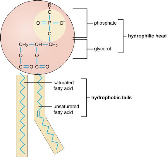
The synthesis of phospholipids represents a fundamental process underpinning the essence of cellular existence. These complex molecules form the structural backbone of all biological membranes, enabling the compartmentalization that defines life at the cellular level. The ability to produce phospholipids would have been an absolute necessity for the first living organisms on Earth. At its core, phospholipid synthesis is a process of enzymatic reactions, beginning with simple precursors and forming sophisticated amphipathic ( a molecule that has both hydrophilic (water-attracting) and hydrophobic (water-repelling) parts) molecules capable of self-assembling into bilayers. This process bridges the gap between basic metabolic pathways and the complex architecture of cellular membranes, highlighting the interconnectedness of biochemical systems. The pathway begins with glycerol-3-phosphate (G3P), a pivotal molecule that serves as the backbone for phospholipid construction. The formation of G3P itself is tied to central carbon metabolism, illustrating how lipid synthesis is integrated with other essential cellular processes. From this foundation, a series of carefully orchestrated enzymatic steps attach fatty acids and diverse head groups, ultimately producing the variety of phospholipids necessary for membrane function and cellular homeostasis. The complexity of phospholipid synthesis extends beyond the mere addition of molecular components. Each step requires exquisite specificity and regulation to ensure the production of lipids with the correct composition and properties. To achieve the desired outcome, the enzymes involved must work in concert, with precise timing and spatial organization. This level of coordination raises pertinent questions about the origins of such a sophisticated system. Moreover, the diversity of phospholipids produced through these pathways is critical for the proper functioning of cellular membranes across a wide range of environments and physiological conditions. The ability to modulate membrane composition in response to environmental cues is a hallmark of cellular adaptability, further underscoring the importance of a flexible and responsive lipid synthesis machinery.  The precision required at each step, from the initial formation of fatty acids to the final assembly of complex phospholipids, speaks to a level of biochemical sophistication that challenges simplistic explanations for its emergence. This introduction sets the stage for a deeper exploration of the enzymatic processes involved in phospholipid synthesis, the potential pathways, and the implications of this essential biochemistry for our understanding of cellular life's origins and fundamental nature.

Glycerol-3-phosphate (G3P) formation: G3P is a central molecule in phospholipid synthesis. The first life forms might have obtained G3P either through glycolysis or from dihydroxyacetone phosphate (DHAP), a glycolytic intermediate. 


9.2.1. Attachment of two fatty acyl groups to glycerol-3-phosphate (G3P)

Attachment of Fatty Acids to G3P: Two fatty acyl groups, usually derived from acyl-CoA molecules, are esterified to the G3P at the sn-1 and sn-2 positions to produce phosphatidic acid. For the synthesis of phosphatidic acid through the attachment of two fatty acyl groups to glycerol-3-phosphate (G3P), the enzymatic steps are as follows:

Phosphatidic acid biosynthesis is a critical initial step in glycerophospholipid metabolism. This pathway is essential for the production of phospholipids, which are fundamental components of cellular membranes and play crucial roles in cellular signaling and energy storage. The process involves the sequential attachment of two fatty acyl groups to glycerol-3-phosphate (G3P), resulting in the formation of phosphatidic acid, a key intermediate in lipid biosynthesis.

Key enzymes involved:

Glycerol-3-phosphate O-acyltransferase (GPAT) (EC 2.3.1.15): Smallest known: 306 amino acids (Mycobacterium tuberculosis)
Catalyzes the initial and rate-limiting step in de novo glycerophospholipid biosynthesis. GPAT transfers an acyl group from acyl-CoA to the sn-1 position of glycerol-3-phosphate, forming lysophosphatidic acid (LPA). This enzyme is crucial for regulating the flux of fatty acids into the glycerophospholipid biosynthetic pathway and plays a significant role in triglyceride biosynthesis.
Lysophosphatidic acid acyltransferase (LPAAT) (EC 2.3.1.51): Smallest known: 257 amino acids (Chlamydia trachomatis)
Catalyzes the second acylation step in phosphatidic acid biosynthesis. LPAAT transfers an acyl group from acyl-CoA to the sn-2 position of lysophosphatidic acid, producing phosphatidic acid. This enzyme is critical for determining the fatty acid composition of membrane phospholipids and thus influences membrane fluidity and cellular function.

The phospholipid biosynthesis enzyme group consists of 2 enzymes. The total number of amino acids for the smallest known versions of these enzymes is 563.

Information on metal clusters or cofactors:
Glycerol-3-phosphate O-acyltransferase (GPAT) (EC 2.3.1.15): Requires Mg²⁺ as a cofactor for optimal activity. The magnesium ion is essential for the catalytic mechanism, facilitating the transfer of the acyl group from acyl-CoA to glycerol-3-phosphate.
Lysophosphatidic acid acyltransferase (LPAAT) (EC 2.3.1.51): Does not require metal ions or cofactors for its catalytic activity. However, the enzyme's activity can be modulated by various lipids and proteins in the cellular environment.

These two enzymes work in concert to produce phosphatidic acid, a critical metabolite in lipid biosynthesis. GPAT, as the initial and rate-limiting enzyme, plays a crucial role in regulating the flux of fatty acids into glycerolipid synthesis. LPAAT, by catalyzing the second acylation step, is key in determining the molecular species of phospholipids produced. Together, they form the foundation of the glycerophospholipid biosynthetic pathway, which is essential for membrane biogenesis, lipid signaling, and energy storage in cells across various organisms.

Formation of the Phospholipid Head Group: Various head groups can be added to phosphatidic acid to form different phospholipids. The CDP-diacylglycerol pathway is one way to achieve this. For instance, in the synthesis of phosphatidylethanolamine and phosphatidylserine, the head groups ethanolamine and serine would be activated and subsequently attached.

9.2.2. Formation of phospholipid head groups

The formation of phospholipid head groups via the CDP-diacylglycerol pathway entails several enzymatic steps. Here are the primary enzymatic reactions involved:

The CDP-diacylglycerol pathway is a critical metabolic route for the biosynthesis of various phospholipids, including phosphatidylinositol, phosphatidylglycerol, and cardiolipin. This pathway is essential for membrane biogenesis and cellular signaling. The initial step in this pathway involves the conversion of phosphatidic acid to CDP-diacylglycerol, which serves as a key intermediate for subsequent phospholipid synthesis.

Key enzyme involved:

Phosphatidate cytidylyltransferase (CDS) (EC 2.7.7.41): Smallest known: 243 amino acids (Synechocystis sp.)
Catalyzes the formation of CDP-diacylglycerol from phosphatidic acid and CTP. This enzyme plays a crucial role in channeling phosphatidic acid into the CDP-diacylglycerol pathway, thus regulating the synthesis of phosphatidylinositol, phosphatidylglycerol, and cardiolipin. CDS is essential for maintaining the appropriate balance of these phospholipids in cellular membranes and is particularly important in tissues with high energy demands, such as the heart, due to its role in cardiolipin synthesis.

The CDP-diacylglycerol synthesis enzyme group consists of 1 enzyme. The total number of amino acids for the smallest known version of this enzyme is 243.


Information on metal clusters or cofactors:
Phosphatidate cytidylyltransferase (CDS) (EC 2.7.7.41): Requires divalent metal ions, typically Mg²⁺ or Mn²⁺, for catalytic activity. These metal ions play a crucial role in the enzyme's mechanism by:
1. Facilitating the binding of the CTP substrate
2. Stabilizing the transition state during the reaction
3. Promoting the release of the pyrophosphate byproduct

The metal ions coordinate with the phosphate groups of CTP and the phosphatidic acid substrate, bringing them into the correct orientation for the nucleophilic attack that forms the CDP-diacylglycerol product.

Phosphatidate cytidylyltransferase is a pivotal enzyme in phospholipid biosynthesis, acting as a metabolic branch point that directs the flow of lipid precursors into specific phospholipid classes. Its activity is tightly regulated in response to cellular lipid levels and metabolic demands. The enzyme's importance is underscored by its conservation across diverse organisms, from bacteria to humans, reflecting its fundamental role in membrane biogenesis and cellular homeostasis.

The CDP-diacylglycerol formed by this enzyme serves as a versatile precursor for the synthesis of several phospholipids:
1. Phosphatidylinositol, crucial for cell signaling and membrane trafficking
2. Phosphatidylglycerol, important for bacterial membranes and as a precursor to cardiolipin
3. Cardiolipin, essential for mitochondrial function and energy metabolism

By regulating the availability of CDP-diacylglycerol, phosphatidate cytidylyltransferase indirectly influences numerous cellular processes, including signal transduction, membrane dynamics, and energy production. This makes it a potential target for therapeutic interventions in disorders involving lipid metabolism or mitochondrial dysfunction.

9.2.3. Phosphatidylethanolamine (PE) synthesis

The biosynthesis of phosphatidylethanolamine (PE) and phosphatidylserine (PS) is crucial for maintaining cellular membrane structure and function. These phospholipids play essential roles in various cellular processes, including membrane fusion, cell signaling, and apoptosis. The CDP-diacylglycerol pathway and related enzymes are key to the synthesis of these important phospholipids.

Key enzymes involved:

Ethanolaminephosphate cytidylyltransferase (ECT) (EC 2.7.7.14): Smallest known: 367 amino acids (Saccharomyces cerevisiae)
Catalyzes the rate-limiting step in the CDP-ethanolamine pathway for PE synthesis. ECT converts phosphoethanolamine to CDP-ethanolamine, which is a crucial intermediate in PE biosynthesis. This enzyme is essential for maintaining proper PE levels in cellular membranes and is particularly important in rapidly dividing cells.
CDP-diacylglycerol—ethanolamine O-phosphatidyltransferase (EPT) (EC 2.7.8.1): Smallest known: 389 amino acids (Saccharomyces cerevisiae)
Catalyzes the final step in PE synthesis via the CDP-ethanolamine pathway. EPT transfers the phosphoethanolamine group from CDP-ethanolamine to diacylglycerol, forming PE. This enzyme is crucial for regulating the balance between PE and other phospholipids in cellular membranes.
CDP-diacylglycerol—serine O-phosphatidyltransferase (PSS) (EC 2.7.8.8 ): Smallest known: 473 amino acids (Saccharomyces cerevisiae)
Catalyzes the formation of PS by transferring a phosphatidyl group from CDP-diacylglycerol to L-serine. This enzyme is essential for PS biosynthesis and plays a crucial role in maintaining PS levels in cellular membranes, particularly in eukaryotic cells.
Phosphatidylserine decarboxylase (PSD) (EC 4.1.1.65): Smallest known: 353 amino acids (Escherichia coli)
Catalyzes the decarboxylation of PS to form PE. This enzyme provides an alternative route for PE synthesis and is particularly important in prokaryotes and in the mitochondria of eukaryotes. PSD plays a crucial role in maintaining the proper balance between PS and PE in cellular membranes.

The phosphatidylethanolamine and phosphatidylserine biosynthesis enzyme group consists of 4 enzymes. The total number of amino acids for the smallest known versions of these enzymes is 1,582.

Information on metal clusters or cofactors:
Ethanolaminephosphate cytidylyltransferase (ECT) (EC 2.7.7.14): Requires Mg²⁺ as a cofactor for catalytic activity. The magnesium ion helps coordinate the CTP substrate and stabilize the transition state during the reaction.
CDP-diacylglycerol—ethanolamine O-phosphatidyltransferase (EPT) (EC 2.7.8.1): Does not require metal ions or cofactors for its catalytic activity. However, its activity can be modulated by the lipid composition of the membrane environment.
CDP-diacylglycerol—serine O-phosphatidyltransferase (PSS) (EC 2.7.8.8 ): Does not require metal ions or cofactors for its catalytic activity. Like EPT, its activity can be influenced by the surrounding lipid environment.
Phosphatidylserine decarboxylase (PSD) (EC 4.1.1.65): Contains a covalently bound pyruvoyl group as a prosthetic group, which is essential for its catalytic activity. This pyruvoyl group is formed through a post-translational modification and serves as the electron sink during the decarboxylation reaction.

These enzymes work together to regulate the synthesis and interconversion of PE and PS, which are critical components of cellular membranes. The pathway provides flexibility in phospholipid synthesis, allowing cells to adjust their membrane composition in response to various physiological conditions and metabolic demands. The diverse catalytic mechanisms and regulatory properties of these enzymes highlight the complexity of phospholipid metabolism and its importance in cellular homeostasis.

9.2.4. Formation of Phospholipids

As previously discussed, two fatty acid molecules (usually in the form of acyl-CoA) are attached to a glycerol-3-phosphate (G3P) molecule through esterification reactions, resulting in the formation of phosphatidic acid (PA).
The phospholipid head group is then attached to the phosphatidic acid. In the CDP-diacylglycerol pathway, for example, the activated head group displaces the cytidyl group from CDP-diacylglycerol, leading to the formation of the final phospholipid. The nature of the head group determines the specific type of phospholipid (e.g., phosphatidylethanolamine, phosphatidylcholine, phosphatidylserine, etc.).

9.2.5. CDP-diacylglycerol pathway

Phospholipid biosynthesis is a fundamental process in all living organisms, crucial for membrane formation, cellular signaling, and energy storage. The initial steps involve the formation of phosphatidic acid from glycerol-3-phosphate and its subsequent conversion to CDP-diacylglycerol, which serves as a key intermediate for various phospholipid species.

Key enzymes involved:

Glycerol-3-phosphate O-acyltransferase (GPAT) (EC 2.3.1.15): Smallest known: 306 amino acids (Mycobacterium tuberculosis)
Catalyzes the initial and rate-limiting step in de novo glycerophospholipid biosynthesis. GPAT transfers an acyl group from acyl-CoA to the sn-1 position of glycerol-3-phosphate, forming lysophosphatidic acid (LPA). This enzyme is crucial for regulating the flux of fatty acids into the glycerophospholipid biosynthetic pathway and plays a significant role in triglyceride biosynthesis.

1-acylglycerol-3-phosphate O-acyltransferase (AGPAT) (EC 2.3.1.51): Smallest known: 257 amino acids (Chlamydia trachomatis)
Catalyzes the second acylation step in phosphatidic acid biosynthesis. AGPAT transfers an acyl group from acyl-CoA to the sn-2 position of lysophosphatidic acid, producing phosphatidic acid. This enzyme is critical for determining the fatty acid composition of membrane phospholipids and thus influences membrane fluidity and cellular function.

Phosphatidate cytidylyltransferase (CDS) (EC 2.7.7.41): Smallest known: 243 amino acids (Synechocystis sp.)
Catalyzes the formation of CDP-diacylglycerol from phosphatidic acid and CTP. This enzyme plays a crucial role in channeling phosphatidic acid into the CDP-diacylglycerol pathway, thus regulating the synthesis of phosphatidylinositol, phosphatidylglycerol, and cardiolipin. CDS is essential for maintaining the appropriate balance of these phospholipids in cellular membranes.

The glycerophospholipid biosynthesis enzyme group consists of 3 enzymes. The total number of amino acids for the smallest known versions of these enzymes is 806.

Information on metal clusters or cofactors:
Glycerol-3-phosphate O-acyltransferase (GPAT) (EC 2.3.1.15): Requires Mg²⁺ as a cofactor for optimal activity. The magnesium ion is essential for the catalytic mechanism, facilitating the transfer of the acyl group from acyl-CoA to glycerol-3-phosphate.
1-acylglycerol-3-phosphate O-acyltransferase (AGPAT) (EC 2.3.1.51): Does not require metal ions or cofactors for its catalytic activity. However, the enzyme's activity can be modulated by various lipids and proteins in the cellular environment.
Phosphatidate cytidylyltransferase (CDS) (EC 2.7.7.41): Requires divalent metal ions, typically Mg²⁺ or Mn²⁺, for catalytic activity. These metal ions play a crucial role in the enzyme's mechanism by facilitating the binding of the CTP substrate, stabilizing the transition state during the reaction, and promoting the release of the pyrophosphate byproduct.

These three enzymes work in concert to produce CDP-diacylglycerol, a critical metabolite in lipid biosynthesis. GPAT and AGPAT collaborate to form phosphatidic acid, which is then converted to CDP-diacylglycerol by CDS. This pathway is tightly regulated and plays a central role in membrane biogenesis and lipid signaling.

The sequential action of these enzymes highlights the complexity and precision of phospholipid biosynthesis:

1. GPAT initiates the pathway by attaching the first fatty acid to glycerol-3-phosphate.
2. AGPAT completes the formation of phosphatidic acid by adding the second fatty acid.
3. CDS then converts phosphatidic acid to CDP-diacylglycerol, creating a versatile precursor for various phospholipids.

This pathway is critical for maintaining the proper composition of cellular membranes and for producing lipid-based signaling molecules. The regulation of these enzymes allows cells to adjust their membrane composition in response to various physiological conditions and metabolic demands, underscoring their importance in cellular homeostasis and adaptation.

9.2.6. Enzymes involved in Phospholipid Synthesis from CDP-diacylglycerol

The synthesis of specific phospholipids from CDP-diacylglycerol is a crucial process in cellular membrane biogenesis and lipid metabolism. These enzymes catalyze the final steps in the formation of various phospholipids, each with unique functions in cellular processes.


Last edited by Otangelo on Sun Sep 22, 2024 2:50 pm; edited 15 times in total

https://reasonandscience.catsboard.com

Otangelo


Admin

Key enzymes involved:

Phosphatidylglycerophosphate synthase (PGPS) (EC 2.7.8.5): Smallest known: 182 amino acids (Bacillus subtilis)
Catalyzes the formation of phosphatidylglycerophosphate from CDP-diacylglycerol and glycerol-3-phosphate. This enzyme is crucial for the biosynthesis of phosphatidylglycerol and cardiolipin, which are important components of bacterial membranes and mitochondrial membranes in eukaryotes. PGPS plays a vital role in maintaining membrane integrity and function, particularly in energy-transducing membranes.
Phosphatidylserine synthase (PSS) (EC 2.7.8.8 ): Smallest known: 473 amino acids (Saccharomyces cerevisiae)
Catalyzes the formation of phosphatidylserine by transferring a phosphatidyl group from CDP-diacylglycerol to L-serine. This enzyme is essential for PS biosynthesis and plays a crucial role in maintaining PS levels in cellular membranes, particularly in eukaryotic cells. Phosphatidylserine is important for cell signaling, apoptosis, and maintaining the asymmetry of plasma membranes.
Phosphatidylethanolamine synthase (PES) (EC 2.7.8.1): Smallest known: 389 amino acids (Saccharomyces cerevisiae)
Catalyzes the formation of phosphatidylethanolamine by transferring the phosphatidyl group from CDP-diacylglycerol to ethanolamine. This enzyme is crucial for the synthesis of phosphatidylethanolamine, a major component of cellular membranes. PE is involved in membrane fusion, cell division, and various cellular signaling processes.

The glycerophospholipid biosynthesis enzyme group consists of 3 enzymes. The total number of amino acids for the smallest known versions of these enzymes is 1,044.

Information on metal clusters or cofactors:
Phosphatidylglycerophosphate synthase (PGPS) (EC 2.7.8.5): Requires Mg²⁺ for catalytic activity. The magnesium ion helps coordinate the substrates and stabilize the transition state during the reaction.
Phosphatidylserine synthase (PSS) (EC 2.7.8.8 ): Does not require metal ions or cofactors for its catalytic activity. However, its activity can be modulated by the lipid composition of the membrane environment and various regulatory proteins.
Phosphatidylethanolamine synthase (PES) (EC 2.7.8.1): Does not require metal ions or cofactors for its catalytic activity. Like PSS, its activity can be influenced by the surrounding lipid environment and cellular regulatory mechanisms.

These enzymes play crucial roles in the final steps of phospholipid biosynthesis, each producing a specific type of phospholipid from the common precursor CDP-diacylglycerol. Their activities are tightly regulated to maintain the proper balance of different phospholipids in cellular membranes:

1. PGPS initiates the pathway for phosphatidylglycerol and cardiolipin synthesis, which are especially important for bacterial and mitochondrial membranes.
2. PSS produces phosphatidylserine, a key signaling molecule and an important component of the inner leaflet of plasma membranes.
3. PES synthesizes phosphatidylethanolamine, a major structural component of cellular membranes that also plays roles in membrane fusion and cell division.

The diversity of these enzymes reflects the complexity of cellular membranes and the need for various phospholipid species to maintain proper membrane function. Each enzyme contributes to the unique lipid composition of different cellular membranes and organelles, allowing for specialized functions and responses to cellular needs. The regulation of these enzymes is critical for maintaining membrane homeostasis and adapting to changing cellular conditions. Their activities can be modulated by various factors, including substrate availability, product feedback inhibition, and cellular signaling pathways. This allows cells to fine-tune their membrane composition in response to environmental stresses, metabolic demands, and developmental stages. Understanding the functions and regulations of these enzymes is crucial for comprehending cellular membrane dynamics and has implications for various fields, including cell biology, microbiology, and biomedical research.


Unresolved Challenges in Phospholipid Biosynthesis

1. Enzyme Complexity and Specificity
The phospholipid biosynthesis pathway involves highly specific enzymes, each catalyzing a distinct reaction. The challenge lies in explaining the origin of such complex, specialized enzymes without invoking a guided process. For instance, glycerol-3-phosphate O-acyltransferase (EC 2.3.1.15) requires a sophisticated active site to catalyze the esterification of a fatty acid from an acyl-CoA to the sn-1 position of glycerol-3-phosphate. The precision required for this catalysis raises questions about how such a specific enzyme could have arisen spontaneously.

Conceptual problem: Spontaneous Complexity
- No known mechanism for generating highly specific, complex enzymes without guidance
- Difficulty explaining the origin of precise active sites and substrate specificity

2. Pathway Interdependence
The phospholipid biosynthesis pathway exhibits a high degree of interdependence among its constituent enzymes. Each step in the pathway relies on the product of the previous reaction as its substrate. This sequential dependency poses a significant challenge to explanations of gradual, step-wise origin. For example, 1-acylglycerol-3-phosphate O-acyltransferase (EC 2.3.1.51) requires the product of glycerol-3-phosphate O-acyltransferase as its substrate.

Conceptual problem: Simultaneous Emergence
- Challenge in accounting for the concurrent appearance of interdependent components
- Lack of explanation for the coordinated development of multiple, specific molecules

3. Stereospecificity
Many enzymes in the phospholipid biosynthesis pathway exhibit stereospecificity. For instance, glycerol-3-phosphate O-acyltransferase specifically acylates the sn-1 position of glycerol-3-phosphate. This stereospecificity is crucial for the formation of functional phospholipids but poses a significant challenge in explaining its origin without a guided process.

Conceptual problem: Spontaneous Stereospecificity
- No known mechanism for the spontaneous emergence of stereospecific reactions
- Difficulty in explaining the origin of enzymes capable of distinguishing between stereoisomers

4. Cofactor Requirements
Several enzymes in the phospholipid biosynthesis pathway require specific cofactors for their function. For instance, phosphatidate cytidylyltransferase (EC 2.7.7.41) requires CTP as a cofactor. The challenge lies in explaining the origin of these cofactors and their specific interactions with enzymes without invoking a guided process.

Conceptual problem: Cofactor-Enzyme Coordination
- Difficulty in explaining the simultaneous emergence of enzymes and their specific cofactors
- Lack of a mechanism for the coordinated development of enzyme active sites and cofactor binding regions

5. Membrane Integration
Many enzymes involved in phospholipid biosynthesis are integral membrane proteins. The challenge lies in explaining how these enzymes could have emerged and integrated into membranes without pre-existing functional membranes.

Conceptual problem: Membrane-Enzyme Chicken-and-Egg
- Difficulty in accounting for the emergence of membrane-integrated enzymes without pre-existing membranes
- Lack of explanation for the coordinated development of membrane structure and membrane-associated enzymes

6. Substrate Availability

The pathway requires specific substrates, such as glycerol-3-phosphate and fatty acyl-CoA, which must be available in sufficient quantities. The challenge lies in explaining how early cellular systems could have maintained a steady supply of these substrates without a fully developed metabolic network.

Conceptual problem: Substrate Accessibility
- Difficulty in accounting for the availability of specific substrates in early cellular systems
- Lack of explanation for the coordinated emergence of substrate production and utilization pathways

7. Energy Requirements
Several reactions in the pathway, such as those catalyzed by phosphatidate cytidylyltransferase, require high-energy molecules like CTP. The challenge lies in explaining how early cellular systems could have met these energy requirements without a fully developed energy metabolism.

Conceptual problem: Energy Availability
- Difficulty in accounting for the availability of high-energy molecules in early cellular systems
- Lack of explanation for the coordinated emergence of energy-producing and energy-consuming pathways

8. Regulatory Mechanisms
The phospholipid biosynthesis pathway is subject to complex regulatory mechanisms to ensure appropriate production levels. The challenge lies in explaining the emergence of these sophisticated regulatory mechanisms without invoking a guided process.

Conceptual problem: Regulatory Complexity
- Difficulty in accounting for the emergence of complex regulatory mechanisms
- Lack of explanation for the coordinated development of enzymes and their regulatory systems

9. Diversity of Phospholipids
The pathway produces a variety of phospholipids with different head groups (e.g., phosphatidylethanolamine, phosphatidylserine). The challenge lies in explaining how this diversity emerged without a guided process, given that each type of phospholipid requires specific enzymes for its synthesis.

Conceptual problem: Functional Diversity
- No known mechanism for the spontaneous emergence of diverse, yet functionally related, enzymatic pathways
- Difficulty in explaining the origin of enzymes with different specificities for various head groups

10. Membrane Assembly
The final step in phospholipid biosynthesis involves the assembly of these molecules into functional membranes. This process requires specific orientation and organization of phospholipids. The challenge lies in explaining how this complex assembly process could have emerged without guidance.

Conceptual problem: Spontaneous Organization
- No known mechanism for the spontaneous assembly of complex, functional membranes
- Difficulty in explaining the origin of the specific orientation and organization of phospholipids in membranes

These unresolved challenges highlight the complexity of the phospholipid biosynthesis pathway and the significant conceptual problems faced when attempting to explain its origin through unguided processes. The high degree of specificity, interdependence, and complexity observed in these enzymes and their interactions pose substantial questions that current naturalistic explanations struggle to address adequately.


9.3. Membrane Asymmetry

9.3.1. Flippases (P-type ATPases)

Flippases are ATP-dependent enzymes that belong to the P-type ATPase family. Their primary function is to translocate specific phospholipids from the extracellular (or luminal) side of the membrane to the cytoplasmic leaflet. This unidirectional transport is crucial for establishing and maintaining lipid asymmetry in cellular membranes, a feature that is fundamental to life. One well-characterized flippase is ATP8A1, a member of the P4-ATPase subfamily. This enzyme specifically transports phosphatidylserine (PS) and phosphatidylethanolamine (PE) from the outer to the inner leaflet of the plasma membrane. The concentration of these lipids on the cytoplasmic face of the membrane is critical for various cellular functions, including cell signaling cascades and the recognition of apoptotic cells by the immune system. Another important flippase is ATP8B1, which also belongs to the P4-ATPase family. This protein facilitates the inward movement of phosphatidylserine and phosphatidylcholine. The activity of ATP8B1 is particularly important in maintaining the integrity of the cell membrane and in specialized tissues such as the liver, where it plays a role in bile secretion. The presence of flippases or flippase-like proteins at the origin of life is highly probable. The ability to create and maintain lipid asymmetry would have been crucial for early protocells to establish the necessary chemical gradients for energy production and to regulate their internal environment. This fundamental process likely predates many other cellular functions and may have been one of the earliest forms of active transport in primitive biological systems.

Key enzymes involved:

ATP8A1 (ATPase phospholipid transporting 8A1) (EC 7.6.2.1): Smallest known: 1,138 amino acids (Homo sapiens)
ATP8A1 is a P4-ATPase that primarily translocates phosphatidylserine (PS) and phosphatidylethanolamine (PE) from the outer to the inner leaflet of cellular membranes. This enzyme plays a crucial role in maintaining the asymmetric distribution of these phospholipids, which is essential for various cellular functions including:
1. Cell signaling: PS exposure on the outer leaflet serves as an "eat-me" signal for apoptotic cells.
2. Blood coagulation: PS asymmetry is important for the proper function of platelets.
3. Vesicle budding and membrane trafficking: The specific distribution of PS and PE influences membrane curvature.
ATP8B1 (ATPase phospholipid transporting 8B1) (EC 7.6.2.1): Smallest known: 1,251 amino acids (Homo sapiens)
ATP8B1 is another P4-ATPase family member that translocates phosphatidylserine (PS) and phosphatidylcholine (PC) to the cytoplasmic leaflet of cellular membranes. This enzyme is particularly important for:
1. Maintaining lipid asymmetry in the canalicular membrane of hepatocytes, which is crucial for bile secretion.
2. Protecting cells from bile salt-induced damage in the liver and pancreas.
3. Contributing to the structural integrity and function of the plasma membrane in various cell types.

The ATP8A1 and ATP8B1 enzyme group consists of 2 enzymes. The total number of amino acids for the smallest known versions of these enzymes (in Homo sapiens) is 2,389.


Information on metal clusters or cofactors:
ATP8A1 (EC 7.6.2.1):
- Requires Mg²⁺ for ATP hydrolysis and phospholipid translocation.
- Contains a phosphorylation site that undergoes cycles of phosphorylation and dephosphorylation during the transport process.
- Interacts with CDC50 proteins (particularly CDC50A) which act as beta-subunits and are essential for the proper folding, trafficking, and activity of ATP8A1.
ATP8B1 (EC 7.6.2.1):
- Also requires Mg²⁺ for ATP hydrolysis and phospholipid translocation.
- Contains a similar phosphorylation site involved in the transport mechanism.
- Interacts with CDC50 proteins (particularly CDC50A and CDC50B) which are crucial for its function and localization to the plasma membrane.

These P4-ATPases are critical for maintaining the proper distribution of phospholipids across the bilayer of cellular membranes. Their functions highlight the importance of lipid asymmetry in various cellular processes:

1. Membrane Structure and Function: By maintaining the asymmetric distribution of phospholipids, these enzymes contribute to the overall structure and function of cellular membranes, including membrane curvature and fluidity.
2. Cell Signaling: The exposure of PS on the outer leaflet, typically maintained on the inner leaflet by these enzymes, serves as a signal for various cellular processes, including apoptosis and platelet activation.
3. Vesicle Trafficking: The specific distribution of phospholipids influences membrane curvature, which is crucial for vesicle budding and membrane trafficking events.
4. Organ-specific Functions: ATP8B1, in particular, plays a crucial role in bile secretion in the liver and protecting cells from bile salt-induced damage.

The activity of these enzymes is tightly regulated to maintain proper membrane asymmetry while allowing for dynamic changes when necessary (e.g., during apoptosis or cell activation). Their importance is underscored by the fact that mutations in these genes can lead to severe disorders, such as intrahepatic cholestasis in the case of ATP8B1 deficiency. Understanding the function and regulation of these flippases is crucial for comprehending membrane dynamics, cell signaling, and various physiological processes. This knowledge has implications for numerous fields, including cell biology, neuroscience, and medical research, particularly in areas related to liver diseases, neurodegenerative disorders, and cancer.

Unresolved Challenges in Flippase-Mediated Membrane Asymmetry

1. Structural Complexity of Flippases
Flippases like ATP8A1 and ATP8B1 are large, complex proteins with over 1,000 amino acids. They contain multiple domains for ATP binding, phospholipid recognition, and membrane spanning.

Conceptual problems:
- No known mechanism for spontaneous generation of such large, multi-domain proteins
- Difficulty explaining the origin of specific substrate binding sites and catalytic domains without guided processes

2. Energy Coupling Mechanism
Flippases use ATP hydrolysis to power phospholipid translocation, involving a sophisticated phosphorylation-dephosphorylation cycle.

Conceptual problems:
- Lack of explanation for the emergence of ATP-dependent transport systems
- No known mechanism for spontaneous development of energy coupling without pre-existing energy metabolism

3. Substrate Specificity
ATP8A1 and ATP8B1 exhibit high specificity for certain phospholipids (PS, PE, PC) but not others.

Conceptual problems:
- Difficulty accounting for the origin of precise substrate recognition
- No known mechanism for spontaneous development of specific binding pockets

4. Protein-Protein Interactions
Flippases require interaction with CDC50 proteins for proper folding, trafficking, and activity.

Conceptual problems:
- Lack of explanation for co-emergence of interacting protein partners
- No known mechanism for spontaneous development of specific protein-protein interaction interfaces

5. Membrane Integration
Flippases must be correctly integrated into the membrane to function, involving complex protein folding and insertion mechanisms.

Conceptual problems:
- Difficulty explaining spontaneous membrane insertion of large, multi-domain proteins
- Lack of mechanism for proper orientation and folding of transmembrane segments

6. Regulatory Mechanisms
Flippase activity is tightly regulated to maintain appropriate membrane asymmetry while allowing dynamic changes when necessary.

Conceptual problems:
- No known mechanism for spontaneous development of sophisticated regulatory networks
- Difficulty explaining the origin of allosteric regulation and signal transduction pathways

7. Cofactor Dependencies
Flippases require specific cofactors like Mg2+ for function.

Conceptual problems:
- Lack of explanation for co-emergence of proteins and their required cofactors
- No known mechanism for spontaneous development of specific metal ion binding sites

8. Phosphorylation Site Specificity
Flippases contain specific phosphorylation sites crucial for their catalytic cycle.

Conceptual problems:
- Difficulty explaining the origin of precise phosphorylation sites without guided processes
- No known mechanism for spontaneous development of phosphorylation-dependent conformational changes

9. Membrane Asymmetry Paradox
Flippases are necessary to establish membrane asymmetry, yet their proper function requires pre-existing asymmetry.

Conceptual problems:
- Chicken-and-egg dilemma: How could asymmetry-maintaining proteins emerge without pre-existing membrane asymmetry?
- Lack of explanation for initial establishment of lipid asymmetry in primordial membranes

10. System-Level Coordination
Membrane asymmetry requires coordinated action of flippases, floppases, and scramblases.

Conceptual problems:
- No known mechanism for simultaneous emergence of multiple, interdependent components
- Difficulty explaining the origin of system-level coordination without invoking guided processes

11. Evolutionary Irreducibility
The flippase-mediated membrane asymmetry system appears to be irreducibly complex, with each component necessary for overall function.

Conceptual problems:
- Lack of explanation for the simultaneous emergence of all required components
- No known mechanism for gradual development of the system without loss of function at intermediate stages

These unresolved challenges highlight the significant conceptual hurdles faced by naturalistic explanations for the origin of flippase-mediated membrane asymmetry. The intricate specificity, energy coupling, regulatory mechanisms, and system-level requirements pose formidable obstacles to unguided origin scenarios, necessitating careful consideration of alternative explanations.


9.4. The Essential Nature of Phospholipid Recycling in Early Life

Phospholipid recycling is a fundamental process that likely played a crucial role in the earliest forms of life. This system of lipid metabolism and membrane remodeling is not just a sophisticated feature of modern cells but may have been an essential component of the first life forms.  The enzymes involved in phospholipid degradation, such as phospholipases, play a vital role in this process. These enzymes, found across different domains of life, suggest an ancient origin. Their presence in early life forms would have allowed for:

- Membrane fluidity adjustment
- Removal of damaged lipids
- Generation of signaling molecules
- Energy production through lipid breakdown

In the nutrient-limited environment of early Earth, the ability to recycle and reuse cellular components would have been a significant advantage. Phospholipid recycling allows cells to conserve resources by:

- Reusing lipid components rather than synthesizing them de novo
- Adapting membrane composition without complete replacement
- Generating energy from lipid breakdown when other sources are scarce

Enzymes like glycerophosphodiester phosphodiesterase (GlpQ) would have been crucial in this process, enabling the breakdown and reuse of lipid components.

Cellular Homeostasis and Adaptation: The dynamic nature of phospholipid metabolism allows cells to maintain homeostasis and adapt to changing conditions. This would have been essential for early life forms facing fluctuating environments. Key processes include:

- Adjusting membrane composition in response to temperature changes
- Modifying lipid ratios to alter membrane permeability
- Generating signaling molecules for rudimentary cellular responses

Enzymes like diacylglycerol kinase (DGK) and phosphatidate phosphatase (PAP) would have played crucial roles in these adaptive processes.

Cellular Division and Growth: For the first life forms to propagate, they needed mechanisms for growth and division. Phospholipid recycling and synthesis would have been essential for:

- Expanding membrane surface area during growth
- Generating new membrane material for daughter cells
- Facilitating membrane fission during primitive cell division

The interconversion of lipids, facilitated by enzymes like CDP-diacylglycerol-serine O-phosphatidyltransferase (PSS), would have been critical in these processes.

Emergence of Cellular Complexity: The ability to recycle and remodel phospholipids may have been a stepping stone towards increased cellular complexity. This process could have allowed for:

- Development of specialized membrane domains
- Formation of primitive organelles or compartments
- Evolution of more complex signaling pathways

The diverse functions of phospholipases and other lipid-modifying enzymes suggest their potential role in driving cellular evolution. The intricate system of phospholipid recycling and metabolism we observe in modern cells is likely not a recent evolutionary development, but rather a fundamental feature that was present in the earliest life forms. The ubiquity of these processes across all domains of life, their essential role in cellular function, and their potential to drive adaptation and complexity all point to their ancient origins. The interdependence of these enzymatic processes, their precise specificity, and the complex regulation required for their function suggest a level of organization that is difficult to account for through random processes alone. This complexity, present at the very foundations of life, invites us to consider alternative explanations for the origin and early development of living systems.

Some enzymes related to phospholipid turnover, like phospholipases, are found across different domains of life, suggesting that they might have been present in the first life form. The exact mechanisms and specificities might have diverged over time, but the general capability for phospholipid remodeling could have been extant in the first life form. The remodeling of phospholipids through deacylation and reacylation (Lands' Cycle) is considered a fundamental process in lipid metabolism.

9.4.1. Enzymes involved in Phospholipid Degradation

Precursors: Phospholipid degradation is a crucial process in lipid metabolism, membrane remodeling, and cell signaling. This pathway involves the hydrolysis of various bonds in phospholipid molecules, resulting in the production of bioactive lipid mediators and the recycling of membrane components. The precursor molecules for this pathway are intact phospholipids, which are primarily found in cellular membranes. The degradation process is carried out by a group of enzymes known as phospholipases, each targeting specific bonds within the phospholipid structure. Below is an overview of key enzymes involved in phospholipid degradation:

Phospholipase A1 (PlaA) (EC 3.1.1.32): Smallest known: 269 amino acids (Mycobacterium tuberculosis)
Hydrolyzes the sn-1 ester linkage of phospholipids, releasing a fatty acid and a lysophospholipid. PlaA plays a crucial role in lipid metabolism, membrane remodeling, and the production of lipid signaling molecules.
Phospholipase A2 (PlaB) (EC 3.1.1.4): Smallest known: 124 amino acids (Elapid snakes)
Catalyzes the hydrolysis of the sn-2 ester bond in phospholipids, producing a free fatty acid (often arachidonic acid) and a lysophospholipid. PlaB is critical for the generation of eicosanoids and other lipid mediators involved in inflammation and cell signaling.
Phospholipase C (Plc) (EC 3.1.4.3): Smallest known: 245 amino acids (Bacillus cereus)
Cleaves the phosphodiester bond of glycerophospholipids, releasing diacylglycerol and a phosphorylated head group. Plc plays a vital role in signal transduction pathways, particularly in the phosphatidylinositol cycle, influencing cell proliferation and differentiation.
Phospholipase D (Pld) (EC 3.1.4.4): Smallest known: 502 amino acids (Streptomyces sp.)
Hydrolyzes the terminal phosphodiester bond of glycerophospholipids, primarily phosphatidylcholine, producing phosphatidic acid and a free head group (e.g., choline). Pld is involved in lipid signaling, membrane trafficking, and cytoskeletal reorganization.

The phospholipid degradation enzyme group consists of 4 key enzymes. The total number of amino acids for the smallest known versions of these enzymes is 1,140.

Proteins with metal clusters or cofactors:
Phospholipase A1 (PlaA) (EC 3.1.1.32): Requires Ca²⁺ for optimal activity. Some PlaA enzymes may also contain a zinc-binding domain that is important for their catalytic function.
Phospholipase A2 (PlaB) (EC 3.1.1.4): Most PlaB enzymes require Ca²⁺ as a cofactor for catalytic activity. Some forms also contain disulfide bonds that are crucial for maintaining their tertiary structure.
Phospholipase C (Plc) (EC 3.1.4.3): Many Plc enzymes require Ca²⁺ for both membrane binding and catalytic activity. Some bacterial Plc enzymes contain zinc in their active sites, which is essential for catalysis.
Phospholipase D (Pld) (EC 3.1.4.4): Pld enzymes typically require Ca²⁺ for optimal activity. Some Pld enzymes also contain conserved HKD motifs that are crucial for their catalytic function.

These phospholipases play critical roles in various cellular processes:
1. Membrane Remodeling: By selectively hydrolyzing phospholipids, these enzymes contribute to the dynamic nature of cellular membranes, allowing for rapid changes in membrane composition and properties.
2. Lipid Signaling: The products of phospholipid hydrolysis, such as diacylglycerol, phosphatidic acid, and lysophospholipids, serve as important second messengers in various signaling pathways.
3. Inflammatory Response: Particularly, PlaB's release of arachidonic acid is a key step in the production of eicosanoids, which are crucial mediators of inflammation.
4. Cell Growth and Differentiation: The signaling lipids produced by these enzymes, especially those generated by Plc and Pld, are involved in pathways regulating cell proliferation and differentiation.
5. Membrane Trafficking: Pld-generated phosphatidic acid plays a role in vesicle formation and fusion, important for intracellular membrane trafficking.

The study of these phospholipases continues to be a significant area of research, with implications for understanding various physiological and pathological processes, including inflammation, cancer, and neurodegenerative diseases. Their diverse functions highlight the complex roles of lipids in cellular biology beyond their structural contributions to membranes.


9.4.2. Lipid Reuse and Recycling 

Enzymes involved in Lipid Reuse and Recycling

Precursors: Lipid reuse and recycling is a critical process in cellular metabolism, allowing organisms to efficiently utilize and conserve lipid resources. This pathway involves the breakdown of complex lipids into simpler components that can be reused for the synthesis of new lipids or other cellular processes. The primary precursor for this pathway is glycerophosphodiesters, which are products of phospholipid degradation. These molecules are further broken down to yield valuable metabolic intermediates. Given the importance of lipid conservation in cellular energy management and membrane homeostasis, it's likely that rudimentary forms of these recycling mechanisms existed in early life forms. Below is an overview of a key enzyme involved in lipid reuse and recycling:

Glycerophosphodiester phosphodiesterase (GlpQ) (EC 3.1.4.2): Smallest known: 247 amino acids (Escherichia coli)
Catalyzes the hydrolysis of glycerophosphodiesters, cleaving the phosphodiester bond to yield glycerol-3-phosphate and the corresponding alcohol (such as choline or ethanolamine). This enzyme plays a crucial role in lipid recycling by:
1. Facilitating the reuse of glycerol backbones in lipid synthesis
2. Releasing head groups that can be utilized in various cellular processes
3. Contributing to phosphate homeostasis by liberating inorganic phosphate

The lipid reuse and recycling enzyme group consists of 1 key enzyme. The total number of amino acids for the smallest known version of this enzyme is 247.

Proteins with metal clusters or cofactors:
Glycerophosphodiester phosphodiesterase (GlpQ) (EC 3.1.4.2): Requires divalent metal ions, typically Ca²⁺ or Mg²⁺, for catalytic activity. Some forms of GlpQ may also contain a binuclear metal center, often featuring two Zn²⁺ ions, which is crucial for their catalytic mechanism.

The GlpQ enzyme plays a critical role in cellular metabolism and lipid homeostasis:
1. Lipid Recycling: By breaking down glycerophosphodiesters, GlpQ enables the cell to recycle valuable lipid components, particularly the glycerol backbone and phosphate groups.
2. Membrane Homeostasis: The enzyme contributes to membrane remodeling by facilitating the breakdown of lipid degradation products.
3. Nutrient Acquisition: In some microorganisms, GlpQ is involved in the utilization of exogenous glycerophosphodiesters as sources of carbon, energy, and phosphate.
4. Phosphate Homeostasis: By releasing inorganic phosphate from glycerophosphodiesters, GlpQ contributes to cellular phosphate balance.
5. Metabolic Flexibility: The enzyme's activity allows cells to adapt to changing nutrient conditions by enabling the use of lipid breakdown products as metabolic intermediates.

The study of GlpQ and related enzymes is significant for understanding cellular lipid metabolism, membrane dynamics, and microbial physiology. In particular, the enzyme's role in bacterial systems has implications for understanding pathogen-host interactions and developing novel antimicrobial strategies.

Challenges and Unresolved Questions:

1. Enzyme Complexity and Specificity
GlpQ exhibits remarkable substrate specificity and catalytic efficiency, which poses challenges to explanations of its origin through unguided processes.

Conceptual problems:
- No known mechanism for spontaneous generation of highly specific enzyme active sites
- Difficulty explaining the origin of precise substrate recognition without invoking guided processes

2. Metal Ion Dependency
The requirement for specific metal ions (Ca²⁺, Mg²⁺, or Zn²⁺) in GlpQ's catalytic mechanism raises questions about the co-emergence of the protein and its cofactor requirements.

Conceptual problems:
- Lack of explanation for the coordinated emergence of metal-binding sites and catalytic residues
- No known mechanism for spontaneous development of metal ion selectivity

3. Catalytic Mechanism Complexity
GlpQ's catalytic mechanism involves precise positioning of substrates, metal ions, and catalytic residues.

Conceptual problems:
- Difficulty accounting for the origin of a sophisticated catalytic mechanism without a step-wise, guided process
- No known explanation for the spontaneous emergence of cooperative interactions between enzyme components

4. Integration with Metabolic Networks
GlpQ's function is intricately connected to broader lipid metabolism and phosphate homeostasis pathways.

Conceptual problems:
- Lack of explanation for the integration of GlpQ into complex metabolic networks without pre-existing regulatory systems
- Difficulty accounting for the coordinated emergence of interdependent metabolic pathways

5. Structural Complexity
Even the smallest known GlpQ (247 amino acids) represents a significant level of structural complexity.

Conceptual problems:
- No known mechanism for spontaneous generation of folded proteins with specific tertiary structures
- Difficulty explaining the origin of long, functional polypeptide sequences without guided synthesis

These unresolved challenges highlight the significant conceptual hurdles faced by naturalistic explanations for the origin of lipid reuse and recycling systems. The intricate specificity, metal ion dependencies, and metabolic integration of enzymes like GlpQ pose formidable obstacles to unguided origin scenarios, necessitating careful consideration of alternative explanations.


9.4.3. Conversion and Recycling of Head Groups

Enzymes involved in Conversion and Recycling of Head Groups in Phospholipid Metabolism

The Conversion and Recycling of Head Groups is a critical subprocess within phospholipid metabolism. This pathway is essential for maintaining the proper balance of various phospholipid species in cellular membranes and plays a crucial role in lipid-mediated signaling. The interconversion of different phospholipids allows cells to rapidly respond to changing environmental conditions and cellular needs.

Key enzymes involved:

CDP-diacylglycerol-serine O-phosphatidyltransferase (PSS) (EC 2.7.7.15): Smallest known: 186 amino acids (Staphylococcus aureus)
Forms phosphatidylserine from CDP-diacylglycerol and serine. This enzyme is crucial for the synthesis of phosphatidylserine, an important phospholipid in cell membranes that plays a role in cell signaling, apoptosis, and membrane asymmetry maintenance.
Phosphatidate phosphatase (PAP) (EC 3.1.3.4): Smallest known: 263 amino acids (Saccharomyces cerevisiae)
Converts phosphatidic acid to diacylglycerol, a key step in lipid metabolism. This enzyme acts as a critical regulator of the balance between phosphatidic acid and diacylglycerol, influencing both lipid biosynthesis and lipid-mediated signaling pathways.
Diacylglycerol kinase (DGK) (EC 2.7.1.137): Smallest known: 124 amino acids (Bacillus anthracis)
Phosphorylates diacylglycerol to form phosphatidic acid. This enzyme plays a crucial role in lipid-mediated signal transduction by regulating the levels of diacylglycerol and phosphatidic acid, both of which are important second messengers in various cellular processes.

The CDP-diacylglycerol-serine O-phosphatidyltransferase, phosphatidate phosphatase, and diacylglycerol kinase enzyme group consists of 3 enzymes. The total number of amino acids for the smallest known versions of these enzymes is 573.


Information on metal clusters or cofactors:
CDP-diacylglycerol-serine O-phosphatidyltransferase (PSS) (EC 2.7.7.15): Requires Mg2+ or Mn2+ as a cofactor for optimal activity. These metal ions are essential for the catalytic mechanism of the enzyme.
Phosphatidate phosphatase (PAP) (EC 3.1.3.4): Some isoforms require Mg2+ for catalytic activity. The metal ion plays a role in stabilizing the enzyme-substrate complex and facilitating the phosphate hydrolysis reaction.
Diacylglycerol kinase (DGK) (EC 2.7.1.137): Requires Mg2+ or Mn2+ as a cofactor for phosphoryl transfer. These metal ions are crucial for coordinating the ATP molecule and facilitating the transfer of the phosphate group to diacylglycerol.

Key metabolites in this pathway:
1. Diacylglycerol: A crucial lipid second messenger and a precursor for various phospholipids.
2. Phosphatidic acid: An important signaling lipid and precursor for phospholipid synthesis.
3. Glycerol-3-phosphate: A key intermediate in glycerolipid metabolism.
4. CDP-diacylglycerol: An activated form of phosphatidic acid used in the synthesis of various phospholipids.

These enzymes and metabolites work together to maintain the dynamic balance of phospholipids in cellular membranes and regulate lipid-mediated signaling pathways. The interconversion between different phospholipid species allows cells to rapidly adapt to changing environmental conditions and cellular needs, making this pathway essential for proper cellular function and homeostasis.

Challenges in Understanding the Origin of Phospholipid Transport and Recycling Systems

1. Complexity of Transport Systems:
The intricate nature of phospholipid precursor transport systems presents significant challenges in explaining their origin:

- How did highly specific transporters like GlpT for glycerol-3-phosphate or the Pst system for phosphate evolve?
- What mechanisms could account for the development of complex ABC transporters with multiple subunits and specific substrate recognition?
- How did cells acquire the ability to regulate these transporters in response to cellular needs and environmental conditions?

2. Specificity of Phospholipases:
The existence of various phospholipases with precise specificities raises questions:

- How did enzymes like phospholipase A1, A2, C, and D develop their specific cleavage sites on phospholipids?
- What processes could explain the evolution of enzymes that can distinguish between closely related lipid substrates?
- How did cells acquire the ability to regulate these enzymes to prevent uncontrolled membrane degradation?

3. Interdependence of Lipid Metabolism Pathways:
The interconnected nature of lipid synthesis, degradation, and recycling pathways presents challenges:

- How could such intricate metabolic networks arise, given that many components seem to depend on the pre-existence of others?
- What mechanisms could explain the development of feedback loops and regulatory systems in lipid metabolism?
- How did cells acquire the ability to balance lipid synthesis and degradation to maintain membrane integrity?

4. Origin of Lipid Signaling Systems:
The dual role of lipids in membrane structure and cellular signaling raises questions:

- How did cells develop the ability to use lipid breakdown products as signaling molecules?
- What processes could explain the evolution of receptors and downstream effectors that respond to lipid-derived signals?
- How did cells acquire the ability to regulate lipid-based signaling pathways without disrupting membrane integrity?

5. Emergence of Lipid Asymmetry:
The asymmetric distribution of lipids in cellular membranes is crucial for many cellular processes:

- How did cells develop mechanisms to establish and maintain lipid asymmetry?
- What processes could explain the evolution of flippases, floppases, and scramblases that regulate lipid distribution?
- How did cells acquire the ability to use lipid asymmetry for cellular functions while maintaining membrane stability?

6. Adaptation to Diverse Environments:
The ability of cells to modify their membrane composition in response to environmental changes presents challenges:

- How did cells develop the capacity to adjust their lipid composition in response to temperature, pH, or osmotic stress?
- What mechanisms could explain the evolution of sensors that detect environmental changes and trigger lipid modifications?
- How did cells acquire the ability to maintain membrane function while undergoing significant compositional changes?

7. Origin of Lipid Droplets and Lipid Storage:
The ability of cells to store excess lipids in specialized structures raises questions:

- How did cells develop the ability to form lipid droplets without disrupting cellular functions?
- What processes could explain the evolution of proteins that regulate lipid droplet formation and breakdown?
- How did cells acquire the ability to mobilize stored lipids in response to cellular needs?

8. Methodological Challenges:
Researchers face significant obstacles in studying the origin of lipid metabolism systems:

- Limited fossil evidence of early cellular lipid compositions
- Difficulties in recreating early Earth conditions to test lipid metabolism hypotheses
- Challenges in developing model systems that accurately represent primitive lipid metabolic pathways

These challenges highlight the complexity involved in understanding the origin of phospholipid transport and recycling systems. They underscore the need for innovative research approaches to address these fundamental questions about the origins of cellular lipid metabolism. The nature of these systems, their interdependence, and the precise regulation required for their function suggest a level of complexity that is difficult to account for through undirected processes alone. This complexity, present at the very foundations of cellular life, invites consideration of alternative explanations for the origin and early development of lipid metabolic systems.


1. December 14, 2023 From lipids to life: Cracking the puzzle about the origin of life Link


Last edited by Otangelo on Fri Sep 20, 2024 7:43 am; edited 11 times in total

https://reasonandscience.catsboard.com

Otangelo


Admin

10. Cofactors

Cofactors represent a diverse group of essential molecules that play indispensable roles in cellular metabolism and biochemical processes. These non-protein chemical compounds work in concert with enzymes to facilitate a wide array of reactions necessary for life. The intricate structures and specific functions of cofactors, ranging from simple metal ions to complex organic molecules, underscore their fundamental importance in the origin and maintenance of living systems.  The existence and function of cofactors present a significant challenge to our understanding of life's beginnings. Their complex molecular structures and precise interactions with enzymes suggest a level of biochemical sophistication that is difficult to account for through unguided processes alone. The interdependence between cofactors and their associated enzymes raises questions about how such intricate systems could have emerged simultaneously. Notably, many cofactors exhibit no clear structural or functional homology across different organisms or pathways, suggesting independent origins. This lack of common ancestry among cofactor systems challenges the notion of a single, universal common ancestor and points towards a polyphyletic origin of life. The diversity and specificity of cofactors, coupled with their essential roles in metabolism, highlight the complexity inherent in even the most basic life processes. Understanding the origin and function of cofactors is essential for unraveling the mysteries of early biochemistry and the fundamental processes that gave rise to life. The sophisticated nature of these molecules and their interactions presents a significant hurdle for explanations relying solely on unguided, naturalistic events.

Energy Transfer and Metabolism
1. ATP (Adenosine Triphosphate): Essential energy currency for cellular processes, driving numerous biochemical reactions.
2. Guanosine Triphosphate (GTP): Crucial for energy transfer, signal transduction, and protein synthesis.
3. Cytidine Triphosphate (CTP): Essential for nucleic acid synthesis, particularly in RNA production.

Electron Carriers
4. NAD+ (Nicotinamide Adenine Dinucleotide): Crucial electron carrier in metabolic redox reactions, central to energy production.
5. NADP+ (Nicotinamide Adenine Dinucleotide Phosphate): Key electron donor in anabolic reactions, essential for biosynthetic pathways.
6. FAD (Flavin Adenine Dinucleotide): Important electron carrier in various metabolic pathways, particularly in the electron transport chain.
7. FMN (Flavin Mononucleotide): Electron acceptor in numerous oxidation-reduction reactions, vital for energy metabolism.
8. Ubiquinone (Coenzyme Q10): Crucial component of the electron transport chain, essential for cellular energy production.
9. Pyrroloquinoline Quinone (PQQ): Redox cofactor involved in various physiological processes, including cellular growth and development.

Fatty Acid and Energy Metabolism
10. Coenzyme A: Central to fatty acid metabolism and the citric acid cycle, crucial for energy production.
11. Pantothenic Acid (Vitamin B5): Crucial component of Coenzyme A, essential for fatty acid metabolism and energy production.
12. Lipoic Acid: Key component in several multi-enzyme complexes, important in energy metabolism.

Carbohydrate and Amino Acid Metabolism
13. Thiamine Pyrophosphate (TPP): Essential for carbohydrate metabolism, particularly in decarboxylation reactions.
14. Pyridoxal Phosphate (PLP): Vital for amino acid metabolism, including transamination, decarboxylation, and racemization reactions.

One-Carbon Metabolism and Methylation
15. Tetrahydrofolate (THF): Essential for one-carbon transfer reactions, critical in nucleotide synthesis and amino acid metabolism.
16. Cobalamin (Vitamin B12): Critical for DNA synthesis and fatty acid metabolism, particularly in methyl transfer reactions.
17. S-Adenosyl Methionine (SAM): Primary methyl group donor in various biological reactions, crucial for epigenetic regulation.

Antioxidants and Redox Regulators
18. Ascorbic Acid (Vitamin C): Important antioxidant and enzyme cofactor, essential for collagen synthesis and immune function.
19. Glutathione: Key antioxidant and detoxification agent, essential for cellular redox balance.

Metallic Cofactors
20. Heme: Essential component of cytochromes and hemoglobin, crucial for electron transport and oxygen transport.
21. Iron-Sulfur Clusters: Critical in electron transfer and metabolic reactions, particularly in the electron transport chain.
22. Magnesium Ions (Mg2+): Essential for many enzymatic reactions, particularly those involving phosphate group transfers.
23. Zinc Ions (Zn2+): Important structural and catalytic component in many enzymes, crucial for protein folding and gene expression.
24. Copper Ions (Cu2+): Essential for electron transfer in key enzymes and oxygen transport proteins.
25. Manganese Ions (Mn2+): Critical for various enzymatic reactions, particularly in photosynthesis and antioxidant defense.
26. Molybdenum Cofactor: Necessary for certain oxidation-reduction reactions in carbon, sulfur, and nitrogen metabolism.

Specialized Cofactors
27. Biotin: Crucial for carboxylation reactions in fatty acid synthesis and gluconeogenesis.
28. Menaquinone (Vitamin K2): Essential for blood clotting and bone metabolism, involved in electron transfer in certain bacteria.
29. Retinal: Light-sensitive cofactor essential for vision in animals and energy production in certain bacteria.
30. Tetrahydrobiopterin (BH4): Critical for aromatic amino acid hydroxylation and nitric oxide synthesis.

Each of these cofactors plays a unique and indispensable role in the complex network of biochemical reactions that sustain life, making them essential for the emergence and continuation of living systems.

10.1.  Energy Transfer and Metabolism

Nucleoside triphosphates (NTPs) - ATP, GTP, and CTP - are fundamental molecules in cellular energy transfer and metabolism. These high-energy compounds play crucial roles in various biochemical processes, from energy provision to biosynthesis and cellular signaling. Their ubiquity and importance across all known life forms underscore their critical role in the most basic cellular operations.

1. Energy Currency and Transfer: ATP stands out as the primary energy currency of the cell, driving numerous energy-requiring processes through its hydrolysis. GTP and CTP, while less prominent in this role, also serve as high-energy compounds in specific biochemical reactions. The ability of these molecules to store and transfer energy through their phosphate bonds is fundamental to cellular energetics.
2. Nucleic Acid Synthesis: All three NTPs are essential precursors for nucleic acid synthesis. ATP and GTP provide the adenine and guanine bases for both DNA and RNA, while CTP is crucial for RNA synthesis, providing the cytosine component. The precision required for incorporating these NTPs into nucleic acid molecules highlights the exquisite specificity of the enzymes involved in these processes.
3. Protein Synthesis: GTP plays a unique role in protein synthesis, powering the elongation and termination steps of translation. Its involvement in this fundamental cellular process underscores the diverse functions of NTPs beyond simple energy provision.
4. Signal Transduction: GTP is particularly important in cellular signaling pathways, acting as a molecular switch in G-protein coupled receptor signaling. This role in signal transduction demonstrates how NTPs are integrated into complex cellular communication networks.
5. Metabolic Regulation: The levels of these NTPs in the cell can act as regulatory signals, influencing various metabolic pathways. This regulatory role highlights the intricate interconnectedness of cellular metabolism and the importance of maintaining proper nucleotide balance.
6. Biosynthetic Processes: Beyond their roles in nucleic acid synthesis, these NTPs are involved in various biosynthetic pathways. For instance, CTP is crucial in phospholipid biosynthesis, particularly in the formation of cell membrane components.

1. ATP (Adenosine Triphosphate): Essential energy currency for cellular processes, driving numerous biochemical reactions.
2. Guanosine Triphosphate (GTP): Crucial for energy transfer, signal transduction, and protein synthesis.
3. Cytidine Triphosphate (CTP): Essential for nucleic acid synthesis, particularly in RNA production.

The multifunctionality and universality of ATP, GTP, and CTP across all known life forms raise profound questions about the nature of life's biochemical foundations. The intricate processes involving these NTPs challenge simplistic explanations of life's origins and point to the sophisticated nature of even the most fundamental cellular operations.

Unresolved Challenges in NTP Biosynthesis and Function

1. Enzyme Complexity and Specificity
The biosynthesis of ATP, GTP, and CTP involves highly specific enzymes, each catalyzing distinct reactions. For instance, ATP synthase, a molecular machine that produces ATP, is incredibly complex with multiple subunits working in concert. The challenge lies in explaining the origin of such complex, specialized enzymes without invoking a guided process.

Conceptual problems:
- No known mechanism for generating highly specific, complex enzymes without guidance
- Difficulty explaining the origin of precise active sites and substrate specificity
- The need for multiple, interdependent enzymes in single pathways compounds the problem

2. Pathway Interdependence
The biosynthesis and utilization of these NTPs are intricately linked with numerous other metabolic processes. This interdependence raises questions about how these interconnected systems could have emerged simultaneously.

Conceptual problems:
- The requirement for multiple, coordinated biochemical pathways
- Difficulty in explaining the emergence of interconnected systems without pre-existing cellular infrastructure
- The need for regulatory mechanisms to coordinate these pathways

3. Energy Requirements
The synthesis of these high-energy compounds is itself an energy-intensive process. How were these energetic requirements met in prebiotic conditions lacking sophisticated energy-generating systems?

Conceptual problems:
- Lack of known prebiotic energy sources capable of driving unfavorable reactions
- The need for specific conditions to overcome thermodynamic barriers
- Difficulty in maintaining these conditions over extended periods

4. Molecular Stability
NTPs and their precursors are relatively unstable molecules. How were these compounds preserved in a prebiotic environment lacking sophisticated cellular machinery?

Conceptual problems:
- Rapid degradation of complex organic molecules under prebiotic conditions
- The need for protective mechanisms or environments to preserve unstable intermediates
- The challenge of accumulating sufficient concentrations of precursors for effective reactions

5. Functional Integration
The diverse roles of ATP, GTP, and CTP in cellular processes require their integration into multiple metabolic pathways. How did this integration occur in the absence of pre-existing cellular systems?

Conceptual problems:
- The need for membrane structures to contain and concentrate reactants
- The requirement for transport mechanisms to move NTPs to various cellular compartments
- The necessity of regulatory systems to control NTP levels and utilization

These unresolved challenges highlight the significant conceptual hurdles in explaining the origin and function of NTPs through unguided processes. The complexity, specificity, and interdependence observed in NTP-related systems suggest that our current understanding of prebiotic chemistry and the origin of life may be incomplete or flawed. Further research and alternative explanations may be necessary to fully comprehend the emergence of these fundamental biochemical processes.


10.2. Electron Carriers

Electron carriers play a crucial role in cellular metabolism, particularly in energy production and redox reactions. These molecules facilitate the transfer of electrons in various biochemical processes, enabling the flow of energy within cells. Their diverse structures and functions highlight the complexity and efficiency of cellular energetics.

1. Redox Reactions and Energy Production: Electron carriers are central to redox reactions, which are fundamental to cellular metabolism. They shuttle electrons between molecules, enabling the stepwise release of energy from nutrients. This process is critical for ATP synthesis, especially in pathways like cellular respiration.
2. Diversity of Carriers: The variety of electron carriers, each with specific properties and roles, allows for fine-tuned control of electron flow in different cellular compartments and pathways. This diversity contributes to the efficiency and versatility of cellular metabolism.
3. Regeneration and Cycling: Most electron carriers function in cycles, alternating between oxidized and reduced forms. This cyclic nature allows them to continuously participate in metabolic processes, enhancing the overall efficiency of cellular energy production.
4. Involvement in Biosynthesis: Beyond energy production, some electron carriers play crucial roles in anabolic processes, providing reducing power for the synthesis of complex molecules.
5. Cellular Compartmentalization: The distribution and function of different electron carriers in various cellular compartments reflect the specialized metabolic roles of these compartments, such as mitochondria and chloroplasts.
6. Evolutionary Conservation: The ubiquity of these electron carriers across diverse life forms suggests their early evolutionary origin and fundamental importance to life processes.


NAD+ (Nicotinamide Adenine Dinucleotide): Crucial electron carrier in metabolic redox reactions, central to energy production.
NADP+ (Nicotinamide Adenine Dinucleotide Phosphate): Key electron donor in anabolic reactions, essential for biosynthetic pathways.
FAD (Flavin Adenine Dinucleotide): Important electron carrier in various metabolic pathways, particularly in the electron transport chain.
FMN (Flavin Mononucleotide): Electron acceptor in numerous oxidation-reduction reactions, vital for energy metabolism.
Ubiquinone (Coenzyme Q10): Crucial component of the electron transport chain, essential for cellular energy production.
Pyrroloquinoline Quinone (PQQ): Redox cofactor involved in various physiological processes, including cellular growth and development.

The  functions and universal presence of these electron carriers across living organisms raise profound questions about the nature of life's biochemical foundations. The complexity of these molecules and their roles in cellular metabolism challenge simplistic explanations of life's origins and point to the sophisticated nature of even the most fundamental cellular operations.

Unresolved Challenges in Electron Carrier Biosynthesis and Function

1. Structural Complexity
Electron carriers like NAD+, FAD, and Ubiquinone have intricate molecular structures. The challenge lies in explaining the origin of such complex molecules without invoking a guided process.

Conceptual problems:
- No known mechanism for generating complex organic molecules spontaneously
- Difficulty explaining the precise arrangement of functional groups necessary for electron transfer
- The need for multiple, coordinated synthetic steps compounds the problem

2. Cofactor Integration
Many electron carriers function as cofactors, requiring specific enzymes for their integration and utilization. This interdependence raises questions about how these systems could have emerged simultaneously.

Conceptual problems:
- The requirement for coordinated evolution of carrier molecules and their associated enzymes
- Difficulty in explaining the emergence of specific binding sites without pre-existing cellular machinery
- The need for regulatory mechanisms to control carrier synthesis and utilization

3. Redox Potential Specificity
Each electron carrier has a specific redox potential, crucial for its function in particular metabolic pathways. How did this specificity arise in prebiotic conditions?

Conceptual problems:
- Lack of known prebiotic mechanisms for fine-tuning molecular redox properties
- The need for precise electrochemical environments to maintain carrier function
- Difficulty in explaining the emergence of a diverse set of carriers with complementary redox potentials

4. Compartmentalization and Transport
Many electron carriers function in specific cellular compartments or need to be transported across membranes. How did these localization and transport systems evolve?

Conceptual problems:
- The need for sophisticated membrane structures and transport proteins
- Difficulty in explaining the emergence of carrier-specific transport mechanisms
- The challenge of maintaining appropriate concentrations of carriers in different cellular compartments

5. Regeneration Cycles
The cyclic nature of electron carrier function requires complex regeneration systems. How did these regeneration pathways emerge alongside the carriers themselves?

Conceptual problems:
- The need for multiple, coordinated enzymatic steps in regeneration pathways
- Difficulty in explaining the emergence of feedback mechanisms controlling regeneration
- The challenge of balancing carrier oxidation and reduction in early metabolic systems

These unresolved challenges highlight the significant conceptual hurdles in explaining the origin and function of electron carriers through unguided processes. The complexity, specificity, and interdependence observed in these systems suggest that our current understanding of prebiotic chemistry and the origin of life may be incomplete or flawed. Further research and alternative explanations may be necessary to fully comprehend the emergence of these fundamental biochemical processes.


10.3. Fatty Acid and Energy Metabolism

Fatty acid and energy metabolism are fundamental processes in cellular function, involving complex pathways and specialized molecules. These systems are critical for energy storage, utilization, and overall cellular homeostasis. The intricate nature of these metabolic processes highlights the sophistication of cellular biochemistry and raises important questions about their origins and evolution.

1. Energy Storage and Utilization: Fatty acids serve as an efficient form of energy storage, containing more energy per gram than carbohydrates. The controlled breakdown and synthesis of fatty acids are crucial for maintaining energy balance in organisms.
2. Metabolic Flexibility: The ability to switch between different energy sources, including fatty acids, carbohydrates, and proteins, allows organisms to adapt to various nutritional states and environmental conditions.
3. Cellular Signaling: Beyond their role in energy metabolism, fatty acids and their derivatives function as signaling molecules, influencing various cellular processes and gene expression.
4. Membrane Structure: Fatty acids are essential components of cellular membranes, affecting membrane fluidity and function. This dual role in energy metabolism and cellular structure underscores their importance.
5. Cofactor Dependency: The metabolism of fatty acids relies heavily on specific cofactors, highlighting the interdependence of various biochemical systems within the cell.
6. Regulatory Complexity: The pathways involved in fatty acid metabolism are subject to complex regulatory mechanisms, ensuring proper energy balance and metabolic health.


Coenzyme A: Central to fatty acid metabolism and the citric acid cycle, crucial for energy production.
Pantothenic Acid (Vitamin B5): Crucial component of Coenzyme A, essential for fatty acid metabolism and energy production.
Lipoic Acid: Key component in several multi-enzyme complexes, important in energy metabolism.

The complexity and efficiency of fatty acid and energy metabolism systems challenge simplistic explanations of their origins. The intricate interplay between various molecules, enzymes, and pathways points to a level of biochemical sophistication that raises profound questions about the nature of life's fundamental processes.

Unresolved Challenges in Fatty Acid and Energy Metabolism

1. Cofactor Complexity
Molecules like Coenzyme A, pantothenic acid, and lipoic acid have intricate structures and specific functions. The challenge lies in explaining the origin of such complex molecules and their precise roles without invoking a guided process.

Conceptual problems:
- No known mechanism for spontaneously generating these complex organic molecules
- Difficulty explaining the precise arrangement of functional groups necessary for their specific roles
- The need for multiple, coordinated synthetic steps compounds the problem

2. Pathway Integration
Fatty acid metabolism is intricately linked with other metabolic pathways, such as the citric acid cycle. This interdependence raises questions about how these interconnected systems could have emerged simultaneously.

Conceptual problems:
- The requirement for multiple, coordinated biochemical pathways
- Difficulty in explaining the emergence of interconnected systems without pre-existing cellular infrastructure
- The need for regulatory mechanisms to coordinate these pathways

3. Enzymatic Specificity
The enzymes involved in fatty acid metabolism exhibit high specificity for their substrates and cofactors. How did this specificity arise in prebiotic conditions?

Conceptual problems:
- Lack of known prebiotic mechanisms for generating highly specific enzymes
- The need for precise active sites and substrate recognition
- Difficulty in explaining the emergence of enzyme-cofactor specificity

4. Compartmentalization
Many processes in fatty acid metabolism occur in specific cellular compartments, such as mitochondria. How did these localization systems evolve?

Conceptual problems:
- The need for sophisticated membrane structures and transport systems
- Difficulty in explaining the emergence of organelle-specific metabolic processes
- The challenge of coordinating reactions across different cellular compartments

5. Regulatory Systems
Fatty acid and energy metabolism are subject to complex regulatory mechanisms. How did these control systems emerge alongside the metabolic pathways?

Conceptual problems:
- The need for sophisticated feedback mechanisms and signaling pathways
- Difficulty in explaining the emergence of transcriptional and post-translational regulation
- The challenge of balancing energy storage and utilization in early metabolic systems

6. Thermodynamic Considerations
The synthesis and breakdown of fatty acids involve complex thermodynamic considerations. How were these energetic requirements met in prebiotic conditions?

Conceptual problems:
- Lack of known prebiotic energy sources capable of driving unfavorable reactions
- The need for specific conditions to overcome thermodynamic barriers
- Difficulty in maintaining these conditions over extended periods

These unresolved challenges highlight the significant conceptual hurdles in explaining the origin and function of fatty acid and energy metabolism through unguided processes. The complexity, specificity, and interdependence observed in these systems suggest that our current understanding of prebiotic chemistry and the origin of life may be incomplete or flawed. Further research and alternative explanations may be necessary to fully comprehend the emergence of these fundamental biochemical processes.


10.4. Carbohydrate and Amino Acid Metabolism

Carbohydrate and amino acid metabolism are fundamental processes in cellular biochemistry, playing crucial roles in energy production, biosynthesis, and cellular homeostasis. These metabolic pathways involve intricate networks of reactions, catalyzed by highly specific enzymes and dependent on various cofactors. The complexity and efficiency of these systems underscore the sophistication of cellular metabolism and raise important questions about their origins and evolution.

1. Energy Production and Storage: Carbohydrate metabolism is central to cellular energy production, providing a rapid source of ATP through glycolysis and the citric acid cycle. The ability to store excess glucose as glycogen demonstrates the metabolic flexibility of organisms.
2. Biosynthetic Precursors: Both carbohydrates and amino acids serve as precursors for the synthesis of various biomolecules, including nucleotides, lipids, and other amino acids. This interconnectedness highlights the integrated nature of cellular metabolism.
3. Protein Synthesis and Function: Amino acid metabolism is crucial for protein synthesis, which underpins virtually all cellular processes. The precise control of amino acid levels and their incorporation into proteins is essential for cellular function.
4. Metabolic Regulation: These pathways are subject to complex regulatory mechanisms, ensuring appropriate energy utilization and maintaining cellular homeostasis. The intricate control systems involve allosteric regulation, hormonal control, and gene expression modulation.
5. Cofactor Dependency: The reliance of these metabolic pathways on specific cofactors, such as TPP and PLP, demonstrates the interdependence of various biochemical systems within the cell.
6. Evolutionary Conservation: The high degree of conservation of these metabolic pathways across diverse life forms suggests their fundamental importance and early evolutionary origin.


Thiamine Pyrophosphate (TPP): Essential for carbohydrate metabolism, particularly in decarboxylation reactions.
Pyridoxal Phosphate (PLP): Vital for amino acid metabolism, including transamination, decarboxylation, and racemization reactions.

The intricate nature of carbohydrate and amino acid metabolism, along with the specific roles of cofactors like TPP and PLP, presents significant challenges to our understanding of how these systems could have arisen through unguided processes. The complexity, specificity, and interdependence observed in these metabolic pathways raise profound questions about the origins of life's fundamental biochemical processes.

Unresolved Challenges in Carbohydrate and Amino Acid Metabolism

1. Cofactor Complexity
TPP and PLP have intricate molecular structures with specific functional groups essential for their roles in metabolism. The challenge lies in explaining the origin of such complex molecules without invoking a guided process.

Conceptual problems:
- No known prebiotic mechanism for spontaneously generating these complex organic molecules
- Difficulty explaining the precise arrangement of functional groups necessary for their catalytic roles
- The need for multiple, coordinated synthetic steps compounds the problem

2. Enzyme Specificity
The enzymes involved in carbohydrate and amino acid metabolism exhibit remarkable specificity for their substrates and cofactors. How did this specificity arise in prebiotic conditions?

Conceptual problems:
- Lack of known mechanisms for generating highly specific enzymes without guidance
- The need for precise active sites and substrate recognition
- Difficulty in explaining the emergence of enzyme-cofactor specificity

3. Pathway Integration
Carbohydrate and amino acid metabolic pathways are intricately linked with each other and with other cellular processes. This interdependence raises questions about how these interconnected systems could have emerged simultaneously.

Conceptual problems:
- The requirement for multiple, coordinated biochemical pathways
- Difficulty in explaining the emergence of interconnected systems without pre-existing cellular infrastructure
- The need for regulatory mechanisms to coordinate these pathways

4. Stereochemical Precision
Many reactions in these pathways, particularly those involving PLP, require precise stereochemical control. How did this stereochemical specificity evolve?

Conceptual problems:
- Lack of known prebiotic mechanisms for achieving stereochemical specificity
- The need for chiral environments or catalysts in prebiotic conditions
- Difficulty in explaining the emergence of enantioselective enzymes

5. Regulatory Complexity
Carbohydrate and amino acid metabolism are subject to sophisticated regulatory mechanisms. How did these control systems emerge alongside the metabolic pathways?

Conceptual problems:
- The need for complex feedback mechanisms and allosteric regulation
- Difficulty in explaining the emergence of transcriptional and post-translational control
- The challenge of coordinating multiple metabolic pathways in early cellular systems

6. Thermodynamic Considerations
Many reactions in these pathways are thermodynamically unfavorable and require energy input. How were these energetic requirements met in prebiotic conditions?

Conceptual problems:
- Lack of known prebiotic energy sources capable of driving unfavorable reactions
- The need for specific conditions to overcome thermodynamic barriers
- Difficulty in maintaining these conditions over extended periods

These unresolved challenges highlight the significant conceptual hurdles in explaining the origin and function of carbohydrate and amino acid metabolism through unguided processes. The complexity, specificity, and interdependence observed in these systems suggest that our current understanding of prebiotic chemistry and the origin of life may be incomplete or flawed. Further research and alternative explanations may be necessary to fully comprehend the emergence of these fundamental biochemical processes.


10.5. One-Carbon Metabolism and Methylation

One-carbon metabolism and methylation processes are fundamental to numerous cellular functions, playing crucial roles in nucleotide synthesis, amino acid metabolism, epigenetic regulation, and various other biochemical pathways. These intricate systems involve complex molecules and highly specific reactions, highlighting the sophistication of cellular biochemistry. The interdependence and precision of these processes raise important questions about their origins and evolution.

1. Nucleotide Synthesis: One-carbon metabolism is essential for the synthesis of purines and thymidine, critical components of DNA and RNA. This underscores its fundamental importance in genetic information storage and transmission.
2. Amino Acid Metabolism: These pathways are involved in the metabolism of several amino acids, including glycine, serine, and methionine, demonstrating their integration with broader metabolic networks.
3. Epigenetic Regulation: Methylation reactions play a crucial role in epigenetic modifications, influencing gene expression without altering the DNA sequence. This adds another layer of complexity to cellular regulation and adaptation.
4. Redox Balance: One-carbon metabolism is intimately linked with cellular redox status, influencing antioxidant defense mechanisms and overall cellular health.
5. Cofactor Dependency: The reliance on specific cofactors like THF, B12, and SAM highlights the interdependence of various biochemical systems within the cell.
6. Metabolic Integration: These pathways interact with numerous other metabolic processes, including the citric acid cycle and fatty acid metabolism, demonstrating the interconnected nature of cellular biochemistry.


Tetrahydrofolate (THF): Essential for one-carbon transfer reactions, critical in nucleotide synthesis and amino acid metabolism.
Cobalamin (Vitamin B12): Critical for DNA synthesis and fatty acid metabolism, particularly in methyl transfer reactions.
S-Adenosyl Methionine (SAM): Primary methyl group donor in various biological reactions, crucial for epigenetic regulation.

The complexity and precision of one-carbon metabolism and methylation processes, along with the specific roles of cofactors like THF, B12, and SAM, present significant challenges to our understanding of how these systems could have arisen through unguided processes. The intricate nature of these pathways and their profound importance in cellular function raise fundamental questions about the origins of life's biochemical processes.

Unresolved Challenges in One-Carbon Metabolism and Methylation

1. Cofactor Complexity
THF, B12, and SAM are highly complex molecules with specific structures essential for their roles in metabolism. Explaining the origin of such intricate molecules without invoking a guided process presents a significant challenge.

Conceptual problems:
- No known prebiotic mechanism for spontaneously generating these complex organic molecules
- Difficulty explaining the precise arrangement of functional groups necessary for their specific roles
- The need for multiple, coordinated synthetic steps compounds the problem

2. Reaction Specificity
The reactions involved in one-carbon metabolism and methylation are highly specific, often requiring precise stereochemistry and regioselectivity. How did this specificity arise in prebiotic conditions?

Conceptual problems:
- Lack of known mechanisms for achieving high reaction specificity without enzymatic catalysis
- The need for precise spatial orientation of reactants and cofactors
- Difficulty in explaining the emergence of stereo- and regioselective reactions

3. Pathway Integration
One-carbon metabolism and methylation processes are intricately linked with numerous other cellular pathways. This interdependence raises questions about how these interconnected systems could have emerged simultaneously.

Conceptual problems:
- The requirement for multiple, coordinated biochemical pathways
- Difficulty in explaining the emergence of interconnected systems without pre-existing cellular infrastructure
- The need for regulatory mechanisms to coordinate these pathways

4. Epigenetic Complexity
The role of methylation in epigenetic regulation adds another layer of complexity to these systems. How did such sophisticated regulatory mechanisms evolve?

Conceptual problems:
- The need for precise targeting of methylation sites on DNA and histones
- Difficulty in explaining the emergence of the machinery for reading and interpreting epigenetic marks
- The challenge of coordinating epigenetic modifications with gene expression

5. Enzyme Evolution
The enzymes involved in one-carbon metabolism and methylation exhibit remarkable specificity for their substrates and cofactors. How did this specificity arise?

Conceptual problems:
- Lack of known prebiotic mechanisms for generating highly specific enzymes
- The need for precise active sites and cofactor binding domains
- Difficulty in explaining the emergence of enzyme-cofactor specificity

6. Thermodynamic Considerations
Many reactions in these pathways are thermodynamically unfavorable and require energy input. How were these energetic requirements met in prebiotic conditions?

Conceptual problems:
- Lack of known prebiotic energy sources capable of driving unfavorable reactions
- The need for specific conditions to overcome thermodynamic barriers
- Difficulty in maintaining these conditions over extended periods

These unresolved challenges highlight the significant conceptual hurdles in explaining the origin and function of one-carbon metabolism and methylation processes through unguided processes. The complexity, specificity, and interdependence observed in these systems suggest that our current understanding of prebiotic chemistry and the origin of life may be incomplete or flawed. Further research and alternative explanations may be necessary to fully comprehend the emergence of these fundamental biochemical processes.


10.6. Antioxidants and Redox Regulators

Antioxidants and redox regulators play crucial roles in maintaining cellular homeostasis, protecting against oxidative stress, and modulating various cellular processes. These molecules and systems are fundamental to cell survival and function, highlighting the sophistication of cellular biochemistry. The complexity and efficiency of these antioxidant systems raise important questions about their origins and evolution.

1. Oxidative Stress Protection: Antioxidants serve as a defense mechanism against reactive oxygen species (ROS) and other free radicals, preventing damage to cellular components such as DNA, proteins, and lipids.
2. Redox Signaling: Beyond their protective roles, many antioxidants and redox regulators are involved in cellular signaling pathways, influencing gene expression and cellular processes.
3. Metabolic Integration: Antioxidant systems are closely integrated with various metabolic pathways, including energy production and nutrient metabolism, demonstrating the interconnected nature of cellular biochemistry.
4. Enzymatic and Non-enzymatic Systems: Cellular antioxidant defense involves both enzymatic systems (e.g., superoxide dismutase, catalase) and non-enzymatic molecules (e.g., ascorbic acid, glutathione), highlighting the multi-faceted nature of redox regulation.
5. Regeneration and Recycling: Many antioxidant systems include mechanisms for regenerating or recycling oxidized molecules, enhancing their efficiency and reducing the need for constant synthesis.
6. Evolutionary Conservation: The presence of antioxidant systems across diverse life forms suggests their fundamental importance and early evolutionary origin.


Ascorbic Acid (Vitamin C): Important antioxidant and enzyme cofactor, essential for collagen synthesis and immune function.
Glutathione: Key antioxidant and detoxification agent, essential for cellular redox balance.

The intricate nature of antioxidant and redox regulation systems, including the specific roles of molecules like ascorbic acid and glutathione, presents significant challenges to our understanding of how these systems could have arisen through unguided processes. The complexity, specificity, and interdependence observed in these biochemical systems raise profound questions about the origins of life's fundamental processes.

Unresolved Challenges in Antioxidant and Redox Regulation Systems

1. Molecular Complexity
Molecules like ascorbic acid and glutathione have specific structures essential for their antioxidant functions. Explaining the origin of such molecules without invoking a guided process presents a significant challenge.

Conceptual problems:
- No known prebiotic mechanism for spontaneously generating these complex organic molecules
- Difficulty explaining the precise arrangement of functional groups necessary for their antioxidant roles
- The need for multiple, coordinated synthetic steps compounds the problem

2. Functional Specificity
Antioxidants and redox regulators exhibit specific functions and interactions within cellular systems. How did this specificity arise in prebiotic conditions?

Conceptual problems:
- Lack of known mechanisms for achieving high functional specificity without cellular context
- The need for precise interactions with cellular components and other molecules
- Difficulty in explaining the emergence of molecule-specific antioxidant properties

3. System Integration
Antioxidant and redox regulation systems are intricately linked with numerous other cellular processes. This interdependence raises questions about how these interconnected systems could have emerged simultaneously.

Conceptual problems:
- The requirement for multiple, coordinated biochemical pathways
- Difficulty in explaining the emergence of interconnected systems without pre-existing cellular infrastructure
- The need for regulatory mechanisms to coordinate antioxidant systems with other cellular processes

4. Enzymatic Complexity
Many antioxidant systems rely on complex enzymatic processes. How did these specialized enzymes evolve?

Conceptual problems:
- Lack of known prebiotic mechanisms for generating highly specific enzymes
- The need for precise active sites and substrate recognition
- Difficulty in explaining the emergence of enzyme-substrate specificity in antioxidant systems

5. Redox Balance Regulation
Maintaining cellular redox balance requires sophisticated regulatory mechanisms. How did these control systems emerge alongside the antioxidant molecules?

Conceptual problems:
- The need for complex feedback mechanisms and sensing systems
- Difficulty in explaining the emergence of redox-sensitive transcriptional regulation
- The challenge of coordinating multiple redox systems in early cellular environments

6. Regeneration Systems
Many antioxidants, including ascorbic acid and glutathione, have specific regeneration pathways. How did these recycling systems evolve?

Conceptual problems:
- The need for coordinated enzymatic systems for antioxidant regeneration
- Difficulty in explaining the emergence of specific electron transfer pathways
- The challenge of integrating regeneration systems with broader cellular metabolism

These unresolved challenges highlight the significant conceptual hurdles in explaining the origin and function of antioxidant and redox regulation systems through unguided processes. The complexity, specificity, and interdependence observed in these systems suggest that our current understanding of prebiotic chemistry and the origin of life may be incomplete or flawed. Further research and alternative explanations may be necessary to fully comprehend the emergence of these fundamental biochemical processes.


10.7. Metallic Cofactors

Metallic cofactors are essential components of numerous biological processes, playing crucial roles in enzyme catalysis, electron transfer, oxygen transport, and structural stabilization of proteins. These inorganic elements and complexes are fundamental to the function of many proteins and enzymes, highlighting the intricate interplay between organic and inorganic chemistry in biological systems. The diversity and specificity of metallic cofactors raise important questions about their incorporation into biological systems and their evolutionary history.

1. Catalytic Versatility: Metallic cofactors enable a wide range of chemical reactions, often facilitating processes that would be thermodynamically unfavorable or kinetically slow without their presence.
2. Electron Transfer: Many metallic cofactors are crucial for electron transfer processes, particularly in energy metabolism and photosynthesis.
3. Structural Roles: Some metal ions play important structural roles in proteins, influencing protein folding and maintaining tertiary and quaternary structures.
4. Oxygen Transport and Storage: Metallic cofactors like heme are essential for oxygen transport and storage in organisms.
5. Redox Chemistry: The ability of many metal ions to exist in multiple oxidation states makes them ideal for participation in redox reactions.
6. Enzyme Activation: Some metallic cofactors serve as enzyme activators, modulating enzymatic activity in response to cellular needs.


Heme: Essential component of cytochromes and hemoglobin, crucial for electron transport and oxygen transport.
Iron-Sulfur Clusters: Critical in electron transfer and metabolic reactions, particularly in the electron transport chain.
Magnesium Ions (Mg2+): Essential for many enzymatic reactions, particularly those involving phosphate group transfers.
Zinc Ions (Zn2+): Important structural and catalytic component in many enzymes, crucial for protein folding and gene expression.
Copper Ions (Cu2+): Essential for electron transfer in key enzymes and oxygen transport proteins.
Manganese Ions (Mn2+): Critical for various enzymatic reactions, particularly in photosynthesis and antioxidant defense.
Molybdenum Cofactor: Necessary for certain oxidation-reduction reactions in carbon, sulfur, and nitrogen metabolism.

The complexity and specificity of metallic cofactors, along with their diverse roles in biological systems, present significant challenges to our understanding of how these systems could have arisen through unguided processes. The intricate nature of metal-protein interactions and the precise requirements for metal incorporation raise fundamental questions about the origins of life's biochemical processes.

Unresolved Challenges in Metallic Cofactor Systems

1. Cofactor Specificity
Different proteins and enzymes require specific metallic cofactors for their function. How did this specificity arise in prebiotic conditions?

Conceptual problems:
- Lack of known mechanisms for achieving metal-protein specificity without guided processes
- The need for precise metal binding sites in proteins
- Difficulty in explaining the emergence of metal selectivity in primitive systems

2. Complex Structures
Some metallic cofactors, like heme and iron-sulfur clusters, have complex structures. Explaining the origin of such intricate molecules without invoking a guided process presents a significant challenge.

Conceptual problems:
- No known prebiotic mechanism for spontaneously generating these complex metal-organic structures
- Difficulty explaining the precise arrangement of atoms necessary for their specific functions
- The need for multiple, coordinated synthetic steps compounds the problem

3. Incorporation Mechanisms
The incorporation of metallic cofactors into proteins often requires specific cellular machinery. How did these incorporation mechanisms evolve?

Conceptual problems:
- The need for coordinated systems for metal uptake, transport, and incorporation
- Difficulty in explaining the emergence of metal chaperones and incorporation proteins
- The challenge of maintaining metal homeostasis in primitive cellular systems

4. Redox Chemistry
Many metallic cofactors participate in redox reactions. How did cells develop mechanisms to control and utilize these redox properties?

Conceptual problems:
- The need for precise control over metal oxidation states
- Difficulty in explaining the emergence of electron transfer chains
- The challenge of preventing unwanted redox reactions in early cellular environments

5. Evolutionary Trade-offs
While essential for many processes, some metals can also be toxic at high concentrations. How did cells evolve to balance the benefits and risks of metal utilization?

Conceptual problems:
- The need for sophisticated metal homeostasis systems
- Difficulty in explaining the emergence of metal detoxification mechanisms
- The challenge of optimizing metal utilization while minimizing toxicity

6. Coevolution with Proteins
The function of metallic cofactors is intimately tied to the structure of their associated proteins. How did these metal-protein systems coevolve?

Conceptual problems:
- The need for coordinated evolution of metal binding sites and protein function
- Difficulty in explaining the emergence of allosteric regulation involving metals
- The challenge of optimizing protein structures for metal binding and catalysis

These unresolved challenges highlight the significant conceptual hurdles in explaining the origin and function of metallic cofactor systems through unguided processes. The complexity, specificity, and interdependence observed in these systems suggest that our current understanding of prebiotic chemistry and the origin of life may be incomplete or flawed. Further research and alternative explanations may be necessary to fully comprehend the emergence of these fundamental biochemical processes.


10.8. Specialized Cofactors

Specialized cofactors are essential components in various biochemical processes, often playing unique and highly specific roles in metabolism, sensory systems, and regulatory pathways. These diverse molecules highlight the intricate and specialized nature of biological systems, demonstrating the remarkable complexity and efficiency of cellular biochemistry. The specificity and complexity of these cofactors raise important questions about their origins and evolution within biological systems.

1. Metabolic Diversity: Specialized cofactors enable a wide range of specific metabolic reactions, often facilitating processes that would be impossible without their presence.
2. Regulatory Functions: Many specialized cofactors play crucial roles in regulatory processes, influencing gene expression, enzyme activity, and cellular signaling.
3. Sensory Transduction: Some cofactors, like retinal, are essential for sensory processes, enabling organisms to interact with and respond to their environment.
4. Interdependence: Many specialized cofactors function in concert with specific proteins or enzymes, highlighting the interdependence of various cellular components.
5. Biosynthetic Complexity: The synthesis of these cofactors often involves complex, multi-step pathways, further emphasizing the sophistication of cellular biochemistry.


Biotin: Crucial for carboxylation reactions in fatty acid synthesis and gluconeogenesis.
Menaquinone (Vitamin K2): Essential for blood clotting and bone metabolism, involved in electron transfer in certain bacteria.
Retinal: Light-sensitive cofactor essential for vision in animals and energy production in certain bacteria.
Tetrahydrobiopterin (BH4): Critical for aromatic amino acid hydroxylation and nitric oxide synthesis.

The complexity and specificity of specialized cofactors, along with their diverse roles in biological systems, present significant challenges to our understanding of how these systems could have arisen through unguided processes. The intricate nature of cofactor-protein interactions and the precise requirements for their function raise fundamental questions about the origins of life's biochemical processes.

Unresolved Challenges in Specialized Cofactor Systems

1. Structural Complexity
Specialized cofactors often have complex molecular structures. Explaining the origin of such intricate molecules without invoking a guided process presents a significant challenge.

Conceptual problems:
- No known prebiotic mechanism for spontaneously generating these complex organic molecules
- Difficulty explaining the precise arrangement of functional groups necessary for their specific roles
- The need for multiple, coordinated synthetic steps compounds the problem

2. Functional Specificity
Each specialized cofactor has a unique and often highly specific function. How did this specificity arise in prebiotic conditions?

Conceptual problems:
- Lack of known mechanisms for achieving high functional specificity without cellular context
- The need for precise interactions with specific proteins or substrates
- Difficulty in explaining the emergence of cofactor-specific biochemical pathways

3. Biosynthetic Pathways
The synthesis of specialized cofactors often involves complex, multi-step pathways. How did these intricate biosynthetic processes evolve?

Conceptual problems:
- The requirement for multiple, coordinated enzymatic steps
- Difficulty in explaining the emergence of complex biosynthetic pathways without pre-existing cellular infrastructure
- The need for regulatory mechanisms to control cofactor synthesis

4. Coevolution with Proteins
Specialized cofactors often function in concert with specific proteins. How did these cofactor-protein systems coevolve?

Conceptual problems:
- The need for coordinated evolution of cofactor structures and protein binding sites
- Difficulty in explaining the emergence of allosteric regulation involving cofactors
- The challenge of optimizing protein structures for cofactor binding and utilization

5. Metabolic Integration
Specialized cofactors are often integrated into complex metabolic networks. How did these interconnected systems emerge?

Conceptual problems:
- The requirement for multiple, coordinated biochemical pathways
- Difficulty in explaining the emergence of interconnected systems without pre-existing cellular infrastructure
- The need for regulatory mechanisms to coordinate cofactor-dependent processes

6. Evolutionary Diversity
Different organisms utilize specialized cofactors in diverse ways. How did this diversity arise?

Conceptual problems:
- The need to explain the emergence of diverse cofactor systems across different lineages
- Difficulty in accounting for the evolution of alternative cofactor utilization strategies
- The challenge of explaining the conservation of some cofactor systems alongside the diversification of others

These unresolved challenges highlight the significant conceptual hurdles in explaining the origin and function of specialized cofactor systems through unguided processes. The complexity, specificity, and interdependence observed in these systems suggest that our current understanding of prebiotic chemistry and the origin of life may be incomplete or flawed. Further research and alternative explanations may be necessary to fully comprehend the emergence of these fundamental biochemical processes.


Last edited by Otangelo on Tue Sep 17, 2024 7:20 am; edited 3 times in total

https://reasonandscience.catsboard.com

Otangelo


Admin

10.9. Key Cofactors in C1 Metabolism of Chemolithoautotrophs

Chemolithoautotrophs, which obtain energy from inorganic substances and carbon from CO2, are studied separately as potential models for early life forms due to their unique metabolic pathways and ability to thrive in extreme conditions. Their specialized enzymes, such as carbon monoxide dehydrogenase/acetyl-CoA synthase and hydrogenases, offer insights into primitive biochemical processes that could have existed in early Earth environments like hydrothermal vents. The study of one-carbon metabolism in both widespread and chemolithoautotrophic pathways provides a comprehensive approach to understanding the possible origins of life. In chemolithoautotrophs, organisms that obtain energy from the oxidation of inorganic substances and carbon from CO2, the one-carbon (C1) metabolism is central to their existence. They have unique pathways to assimilate C1 compounds. Chemolithoautotrophs are microorganisms that derive energy from the oxidation of inorganic compounds and use CO2 as their sole carbon source. Many of these enzymes and pathways are present in chemolithoautotrophic organisms that inhabit hydrothermal vents, where inorganic substances are abundant and can be utilized for energy.

Carbon Monoxide Dehydrogenase/Acetyl-CoA Synthase (CODH/ACS): Relevance to Vent Organisms: Many vent-dwelling bacteria utilize the CODH/ACS complex for carbon fixation by reducing CO2 to CO and synthesizing acetyl-CoA. This pathway is part of the reductive acetyl-CoA pathway, which is used by many thermophilic organisms in hydrothermal vents.
Hydrogenases: Relevance to Vent Organisms: Hydrothermal vent environments are rich in hydrogen, and vent-dwelling microorganisms often use hydrogenases to oxidize hydrogen, generating reducing power for C1 compound reduction.
Formate Dehydrogenase: Relevance to Vent Organisms: Formate dehydrogenase is crucial for many vent-dwelling microorganisms in oxidizing formate to CO2.
Methanogens and Methanotrophs: Relevance to Vent Organisms: Methanogens are common in anaerobic hydrothermal vent environments, where they produce methane from CO2 and other C1 compounds. Methanotrophs in vents can oxidize this methane, converting it back to CO2 or incorporating it into biomass.
Serine Pathway: Some vent-dwelling microorganisms use the serine pathway for C1 assimilation.
Reductive Acetyl-CoA Pathway: This is a significant pathway for CO2 fixation in many thermophilic organisms found in hydrothermal vents.
3-Hydroxypropionate/4-Hydroxybutyrate Cycle: Used by some archaea in hydrothermal vent environments for carbon fixation.

While many of the molecules and enzymes you've listed (SAM, Biotin, Cobalamin, and Folate) are also crucial for one-carbon metabolism in chemolithoautotrophs, these organisms have unique and additional pathways due to their specialized ecological niches and metabolic needs.

10.10. Folate Metabolism: A Complex and Essential Cellular Process

The synthesis of folate involves a series of complex enzymatic reactions. Dihydropteroate synthase (DHPS) catalyzes a key step in this pathway, forming 7,8-dihydropteroate from p-aminobenzoate and 6-hydroxymethyl-7,8-dihydropteroate. This reaction links two distinct branches of the folate biosynthesis pathway, demonstrating the interconnected nature of these biochemical processes. Folylpolyglutamate synthase (FPGS) then adds glutamate residues to folates, a critical modification that enhances folate retention within cells and increases their affinity for folate-dependent enzymes. The conversion of dihydrofolate (DHF) to tetrahydrofolate (THF) by dihydrofolate reductase represents another crucial step, maintaining the pool of active folate coenzymes essential for numerous cellular processes.

10.10.1.Folate-Dependent Processes

Folate and its derivatives are integral to several vital cellular functions. In DNA synthesis, folate-dependent enzymes play key roles in the production of purines and thymidylate, essential building blocks of genetic material. Amino acid metabolism heavily relies on folate-mediated one-carbon transfers, particularly in the synthesis of methionine, glycine, and serine. The methylation cycle, crucial for epigenetic regulation and numerous other cellular processes, depends on S-adenosylmethionine (SAM), which is produced through a folate-dependent pathway. These interconnected processes highlight the central role of folate metabolism in maintaining cellular health and function. The intricacy of folate metabolism is evident in the precise structure-function relationships of its enzymes. Each enzyme in the pathway possesses a highly specific active site, tailored to recognize and process particular substrates with remarkable accuracy. This specificity extends to cofactor requirements, reaction mechanisms, and regulatory controls. The interdependence of these enzymes creates a sophisticated network where the product of one reaction becomes the substrate for another, forming a tightly regulated and efficient system. Moreover, the folate cycle demonstrates an impressive level of metabolic plasticity. It can adapt to varying cellular needs, shifting between different one-carbon-carrying forms of folate as required for diverse biochemical reactions. This adaptability is crucial for maintaining cellular homeostasis under varying conditions and metabolic demands.

10.10.2. Utilization of Tetrahydrofolate (THF) Derivatives

Tetrahydrofolate (THF) derivatives are essential for various metabolic processes, including nucleotide synthesis, amino acid metabolism, and methylation reactions. The proper conversion and utilization of these derivatives are crucial for cellular function and growth. The following enzymes play key roles in these transformations:

Methenyltetrahydrofolate cyclohydrolase (EC 3.5.4.9): Smallest known: 182 amino acids (*Aquifex aeolicus*): Catalyzes the conversion of 5,10-methenyltetrahydrofolate to 10-formyltetrahydrofolate. This enzyme is critical for the formation of 10-formyltetrahydrofolate, a key intermediate in purine biosynthesis.
Methylenetetrahydrofolate reductase (EC 1.7.99.5): Smallest known: 187 amino acids (*Thermotoga maritima*): Converts 5,10-methylenetetrahydrofolate to 5-methyltetrahydrofolate. This enzyme is vital for maintaining appropriate levels of 5-methyltetrahydrofolate, which is essential for homocysteine remethylation and methionine synthesis.
Methenyltetrahydrofolate synthetase (EC 6.3.4.3): Smallest known: 222 amino acids (*Aquifex aeolicus*): Converts 5,10-methylenetetrahydrofolate to 5,10-methenyltetrahydrofolate. This enzyme is involved in the interconversion of THF derivatives, facilitating their availability for various metabolic reactions.
5,10-Methenyltetrahydrofolate cyclohydrolase (EC 3.5.4.9): Smallest known: 182 amino acids (*Aquifex aeolicus*): Converts 5,10-methenyltetrahydrofolate to 5,10-methylenetetrahydrofolate. This enzyme is essential for maintaining the balance of methylene and methenyl THF derivatives in the cell.

The THF derivative-related essential enzyme group consists of 4 enzymes. The total number of amino acids for the smallest known versions of these enzymes is 793.

Information on metal clusters or cofactors:
Methenyltetrahydrofolate cyclohydrolase (EC 3.5.4.9): Requires Zn²⁺ as a cofactor for its activity. The zinc ion is crucial for stabilizing the enzyme's structure and facilitating the catalytic reaction.
Methylenetetrahydrofolate reductase (EC 1.7.99.5): Requires FAD as a cofactor for its activity. FAD is essential for the enzyme's function in the reduction of 5,10-methylenetetrahydrofolate to 5-methyltetrahydrofolate.
Methenyltetrahydrofolate synthetase (EC 6.3.4.3): Does not require metal ions or additional cofactors for its catalytic activity.
5,10-Methenyltetrahydrofolate cyclohydrolase (EC 3.5.4.9): Requires Zn²⁺ as a cofactor. The zinc ion aids in the enzyme's catalytic process and stabilization.

10.10.3. Other Related Enzymes in Folate Metabolism

5,10-Methenyltetrahydrofolate cyclohydrolase / 5,10-methylenetetrahydrofolate dehydrogenase.
Glycinamide ribonucleotide formyltransferase (GARFT): Converts glycinamide ribonucleotide (GAR) to formylglycinamide ribonucleotide (FGAR).
10-formyltetrahydrofolate dehydrogenase: Converts 10-formyltetrahydrofolate to CO2, THF, and NADP+.
Methylene tetrahydrofolate dehydrogenase (NADP+).

Challenges in Understanding the Origins of Folate Metabolism

1. Enzyme Complexity and Specificity: Folate metabolism involves highly specialized enzymes with precise structures and functions, raising several questions:
- How did enzymes like dihydropteroate synthase (DHPS) and dihydrofolate reductase (DHFR) acquire their complex structures?
- What mechanisms could account for the development of such high substrate specificity?
- How did these enzymes evolve to catalyze reactions with such remarkable efficiency?

2. Pathway Interdependence: The folate metabolism pathway exhibits a high degree of interdependence among its components, presenting significant challenges:
- How could such an interconnected network of reactions have originated?
- What intermediate forms, if any, could have existed that were functional?
- How did the precise coordination between different enzymes in the pathway develop?

3. Chemical Instability of Folates: The inherent instability of folate molecules presents unique challenges in understanding their role in early biological systems:
- How could these unstable molecules have persisted in early environments?
- What mechanisms could have protected folates from degradation in primitive cells?
- How did the requirement for continuous folate synthesis or intake arise?

4. Dual Nature of Folate Metabolism: The dual role of folate metabolism in one-carbon transfer and redox regulation presents additional challenges:
- How did a single pathway evolve to serve these two distinct cellular functions?
- What mechanisms led to the integration of folate metabolism with cellular redox status?
- How did the complex regulatory systems controlling this dual function originate?

5. Integration with Other Metabolic Pathways: The deep integration of folate metabolism with numerous other cellular processes presents further challenges:
- How did folate metabolism become so linked with other essential pathways?
- What mechanisms coordinated the development of these interconnected systems?
- How can we explain the origin of the complex regulatory mechanisms controlling metabolic flux?

10.11. S-Adenosylmethionine (SAM) Metabolism

S-Adenosylmethionine (SAM) metabolism represents one of the most complex and essential biochemical processes in living organisms. This system, involving numerous enzymes and interconnected pathways, presents significant challenges to our understanding of its origins and development.  The synthesis of SAM involves a series of highly specific enzymatic reactions. Methionine adenosyltransferase (MAT) catalyzes the formation of SAM from methionine and ATP. This reaction requires precise molecular recognition and positioning of substrates. The enzyme must overcome significant energetic barriers to form the high-energy sulfonium compound. This molecule contains a positively charged sulfur atom (hence "sulfonium") bonded to three carbon atoms, which makes it energetically unstable and highly reactive, allowing it to readily donate its methyl group in various biochemical reactions. 

10.11.1.The SAM-Dependent Methylation Cycle

SAM serves as the primary methyl donor in numerous cellular reactions. Methyltransferases use SAM to methylate DNA, proteins, lipids, and small molecules. This process generates S-adenosylhomocysteine (SAH), which must be efficiently removed to prevent product inhibition. The SAH hydrolase then converts SAH to homocysteine, completing the cycle. The interdependence of these reactions presents a significant challenge to naturalistic explanations. Each step relies on the products of the previous reaction and influences the next, creating a closed loop of metabolic processes. The question arises: how could such a system have emerged gradually when each component depends on the others for functionality?

10.11.2. Regeneration of Methionine

The regeneration of methionine from homocysteine is crucial for maintaining the SAM cycle. This process involves either methionine synthase, which requires vitamin B12 and folate, or betaine-homocysteine methyltransferase. These enzymes exhibit remarkable substrate specificity and catalytic efficiency. The complexity of methionine regeneration, particularly the involvement of cofactors like vitamin B12, adds another layer of complexity to the system. The precise coordination required between these enzymes and their cofactors challenges the notion of a gradual, step-wise development of this metabolic pathway.

10.11.3. Regulation of SAM Metabolism

SAM metabolism is tightly regulated at multiple levels. Allosteric regulation of key enzymes, transcriptional control, and post-translational modifications all play crucial roles in maintaining appropriate SAM levels. This multi-layered regulatory system ensures that SAM concentrations are kept within a narrow range, critical for cellular function. The existence of such sophisticated regulatory mechanisms poses a significant challenge to naturalistic explanations. How could a system with multiple levels of control, each fine-tuned to respond to specific cellular conditions, have arisen through undirected processes?

10.11.4. Integration with Other Metabolic Pathways

SAM metabolism is deeply integrated with numerous other cellular processes, including the folate cycle, transsulfuration pathway, and polyamine synthesis. This network of interdependent reactions raises questions about the origin and development of such interconnected systems. The challenge lies in explaining how these diverse pathways could have become linked without a guiding principle. The precise coordination required between these various metabolic routes suggests a level of complexity that is difficult to account for through random chemical events.

10.11.5. Synthesis of S-Adenosylmethionine (SAM)

S-Adenosylmethionine (SAM) is a vital methyl donor in numerous biological methylation reactions. It is synthesized from methionine and ATP and plays a crucial role in various metabolic processes, including the regulation of gene expression, neurotransmitter synthesis, and lipid metabolism. The pathway for SAM synthesis involves several key enzymes that convert precursors into SAM and other related compounds. Understanding these enzymes and their functions helps elucidate the complexity of SAM metabolism and its biological significance.

Methionine adenosyltransferase (MAT) (EC 2.5.1.6): Smallest known: 228 amino acids (*Escherichia coli*): Catalyzes the conversion of methionine and ATP to S-adenosylmethionine (SAM). This enzyme initiates the SAM synthesis pathway, making it fundamental for the production of this critical methyl donor.
Methylenetetrahydrofolate reductase (MTHFR) (EC 1.5.1.20): Smallest known: 275 amino acids (*Escherichia coli*): Converts 5,10-methylenetetrahydrofolate to 5-methyltetrahydrofolate, which donates a methyl group to homocysteine in the synthesis of methionine. This enzyme is essential for regenerating methionine from homocysteine, indirectly supporting SAM synthesis.
Betaine-homocysteine methyltransferase (BHMT) (EC 2.1.1.5): Smallest known: 360 amino acids (*Escherichia coli*): Utilizes betaine as a methyl donor to convert homocysteine to methionine. This enzyme contributes to the methylation cycle and supports methionine and SAM levels.
Cystathionine β-synthase (CBS) (EC 4.2.1.22): Smallest known: 298 amino acids (*Escherichia coli*): Converts homocysteine to cystathionine as part of the transsulfuration pathway. This enzyme is involved in the metabolism of homocysteine, affecting its availability for SAM synthesis.

The SAM synthesis enzyme group consists of 4 enzymes. The total number of amino acids for the smallest known versions of these enzymes is 1,161.

Information on metal clusters or cofactors:
Methionine adenosyltransferase (MAT) (EC 2.5.1.6): Requires Mg²⁺ as a cofactor for the synthesis of SAM. Magnesium ions are crucial for stabilizing the ATP molecule and facilitating the transfer of the adenosyl group to methionine.
Methylenetetrahydrofolate reductase (MTHFR) (EC 1.5.1.20): Requires FAD (flavin adenine dinucleotide) as a cofactor. FAD is essential for the enzyme's activity in reducing 5,10-methylenetetrahydrofolate to 5-methyltetrahydrofolate.
Betaine-homocysteine methyltransferase (BHMT) (EC 2.1.1.5): Requires Zn²⁺ (zinc ion) as a cofactor. Zinc plays a key role in the enzyme's catalytic activity and stabilization of the active site.
Cystathionine β-synthase (CBS) (EC 4.2.1.22): Requires PLP (pyridoxal phosphate) as a cofactor. PLP is vital for the enzyme's activity in converting homocysteine to cystathionine.

10.11.6. Recycling and Conversion of Tetrahydrofolate (THF)

Tetrahydrofolate (THF) and its derivatives play crucial roles in one-carbon metabolism, which is essential for the synthesis of nucleotides and amino acids. The recycling and conversion of THF are facilitated by several key enzymes, each contributing to the maintenance and utilization of THF derivatives. Here is an overview of the key enzymes involved in this process:

Dihydrofolate reductase (DHFR) (EC 1.5.1.3): Smallest known: 159 amino acids (*Escherichia coli*): Converts dihydrofolate (DHF) to tetrahydrofolate (THF). This enzyme is essential for the regeneration of THF from DHF, ensuring a continuous supply of THF for various metabolic processes.
Serine hydroxymethyltransferase (SHMT) (EC 2.1.2.1): Smallest known: 214 amino acids (*Escherichia coli*): Catalyzes the conversion of serine and THF to glycine and 5,10-methylenetetrahydrofolate. This enzyme is crucial for the transfer of one-carbon units and the production of key THF derivatives involved in nucleotide synthesis.
Folylpolyglutamate synthase (FPGS) (EC 2.5.1.12): Smallest known: 307 amino acids (*Escherichia coli*): Adds glutamate residues to folates to form polyglutamated folates. This enzyme enhances the retention of folates within the cell and increases their effectiveness in metabolic reactions.
Methylenetetrahydrofolate reductase (MTHFR) (EC 1.5.1.20): Smallest known: 275 amino acids (*Escherichia coli*): Converts 5,10-methylenetetrahydrofolate to 5-methyltetrahydrofolate. This enzyme plays a critical role in the methylation cycle, converting THF derivatives to forms needed for methyl group transfer and amino acid metabolism.
Methylene tetrahydrofolate dehydrogenase (MTHFD) (EC 1.5.1.5): Smallest known: 252 amino acids (*Escherichia coli*): Catalyzes the interconversion of various forms of THF. This enzyme is involved in maintaining the balance of THF derivatives required for different metabolic processes.

The THF recycling and conversion enzyme group consists of 5 enzymes. The total number of amino acids for the smallest known versions of these enzymes is 1,447.

Information on metal clusters or cofactors:
Dihydrofolate reductase (DHFR) (EC 1.5.1.3): Requires NADPH as a cofactor for the reduction of dihydrofolate to tetrahydrofolate. NADPH provides the reducing power needed for this reaction.
Serine hydroxymethyltransferase (SHMT) (EC 2.1.2.1): Requires pyridoxal phosphate (PLP) as a cofactor. PLP is crucial for the enzyme's transamination and decarboxylation activities.
Folylpolyglutamate synthase (FPGS) (EC 2.5.1.12): Requires ATP and Mg²⁺ as cofactors. ATP drives the glutamylation reaction, while magnesium ions stabilize the ATP molecule.
Methylenetetrahydrofolate reductase (MTHFR) (EC 1.5.1.20): Requires FAD (flavin adenine dinucleotide) as a cofactor. FAD is essential for the enzyme's reductive activity in the conversion of 5,10-methylenetetrahydrofolate to 5-methyltetrahydrofolate.
Methylene tetrahydrofolate dehydrogenase (MTHFD) (EC 1.5.1.5): Requires NAD⁺ or NADP⁺ as cofactors. These cofactors are necessary for the enzyme's oxidative reactions involving THF derivatives.

10.11.7. Central enzymes and transporters related to the methionine cycle and SAM/SAH metabolism

The methionine cycle and the metabolism of S-adenosylmethionine (SAM) and S-adenosylhomocysteine (SAH) are crucial for cellular methylation processes and the regulation of homocysteine levels. Several key enzymes are involved in these processes:

Methionine adenosyltransferase (MAT) (EC 2.5.1.6): Smallest known: 285 amino acids (*Escherichia coli*): Converts methionine and ATP to S-adenosylmethionine (SAM). This enzyme is central to the methionine cycle, providing SAM, a critical methyl donor for various methylation reactions.
S-adenosylhomocysteine hydrolase (SAHH) (EC 3.3.1.1): Smallest known: 316 amino acids (*Escherichia coli*): Hydrolyzes S-adenosylhomocysteine (SAH) to adenosine and homocysteine. This enzyme is essential for regulating the levels of SAM and SAH, thus controlling methylation reactions and homocysteine metabolism.
Methionine synthase (MS) (EC 2.1.1.13): Smallest known: 755 amino acids (*Bacillus subtilis*): Uses a methyl group from 5-methyltetrahydrofolate to convert homocysteine to methionine. This enzyme is crucial for regenerating methionine, which is essential for maintaining SAM levels and overall methylation balance.

The methionine cycle and SAM/SAH metabolism enzyme group consists of 3 enzymes. The total number of amino acids for the smallest known versions of these enzymes is 1,356.

Information on metal clusters or cofactors:
Methionine adenosyltransferase (MAT) (EC 2.5.1.6): Requires Mg²⁺ as a cofactor. Magnesium ions are essential for the ATP-dependent activation of methionine.
S-adenosylhomocysteine hydrolase (SAHH) (EC 3.3.1.1): Requires Mg²⁺ or Mn²⁺ as cofactors. These metal ions are necessary for the enzyme’s hydrolytic activity on SAH.
Methionine synthase (MS) (EC 2.1.1.13): Requires vitamin B12 (cobalamin) as a cofactor. Vitamin B12 is essential for the transfer of the methyl group from 5-methyltetrahydrofolate to homocysteine, completing the methionine synthesis.

10.11.8. Methyl transfer with S-adenosylmethionine (SAM)

S-adenosylmethionine (SAM) is a pivotal methyl donor involved in various methylation reactions within the cell, influencing gene expression, protein function, and cellular metabolism. Here is an overview of key components and enzymes involved in methyl transfer with SAM:

S-adenosylmethionine (SAM): Smallest known: Not applicable (SAM is a metabolite rather than a protein): Serves as the principal methyl donor in the cell. SAM provides a methyl group for methylation reactions, which are critical for modifying nucleic acids, proteins, and lipids. The availability of SAM directly affects cellular methylation processes and overall metabolism.
S-adenosylhomocysteine hydrolase (SAHH) (EC 3.3.1.1): Smallest known: 316 amino acids (*Escherichia coli*): Regenerates homocysteine and adenosine from S-adenosylhomocysteine (SAH). This enzyme is essential for maintaining the balance of SAM and SAH, which are crucial for the regulation of methylation reactions and overall cellular methylation status.

The methyl transfer and SAM-related enzyme group consists of 2 components. The total number of amino acids for the smallest known versions of these enzymes is 316 for SAHH. SAM itself is not a protein and does not have an amino acid count.

Information on metal clusters or cofactors:
S-adenosylhomocysteine hydrolase (SAHH) (EC 3.3.1.1): Requires Mg²⁺ or Mn²⁺ as cofactors. These metal ions are essential for the enzyme’s hydrolytic activity on SAH, facilitating the regeneration of homocysteine and adenosine.

This overview highlights the critical role of SAM in methylation and the essential enzyme SAHH in regulating methylation balance by managing SAH levels.

Challenges to Naturalistic Explanations of S-Adenosylmethionine (SAM) Metabolism

1. Complex Enzymatic Reactions and Molecular Recognition: The synthesis and utilization of SAM involve highly specific enzymatic reactions that present significant challenges:

- How did enzymes like methionine adenosyltransferase (MAT) develop the ability to catalyze the formation of the high-energy sulfonium compound SAM?
- What intermediate forms, if any, could have existed that were functional in SAM synthesis?
- How did these enzymes acquire the precise molecular recognition capabilities required for substrate binding and catalysis?

2. Interdependence of Reactions in the SAM-Dependent Methylation Cycle: The cycle of SAM-dependent methylation presents a chicken-and-egg problem:

- How could the cycle have emerged when each step depends on the products of the previous reactions?
- What intermediate forms of this cycle, if any, could have been functional?
- How did the precise coordination between methyltransferases, SAH hydrolase, and methionine regeneration enzymes develop?

3. Cofactor Dependence and Methionine Regeneration: The regeneration of methionine involves complex enzymes and cofactors:

- How did the dependence on vitamin B12 and folate in methionine synthase develop?
- What were the intermediate steps, if any, in the emergence of betaine-homocysteine methyltransferase?
- How did these diverse cofactor requirements become integrated into a single metabolic pathway?

4. Multi-layered Regulation of SAM Metabolism: The tight regulation of SAM metabolism at multiple levels poses significant questions:

- How did such sophisticated regulatory mechanisms develop?
- What intermediate forms of regulation, if any, could have been functional?
- How did the various levels of control (allosteric, transcriptional, post-translational) become integrated?

5. Integration with Other Metabolic Pathways: The deep integration of SAM metabolism with numerous other cellular processes presents challenges:

- How did SAM metabolism become so intricately linked with the folate cycle, transsulfuration pathway, and polyamine synthesis?
- What intermediate stages, if any, could have existed in the development of these interconnections?
- How did the precise coordination required between these pathways arise?

6. Enzyme Specificity and Catalytic Efficiency: The enzymes involved in SAM metabolism display remarkable specificity and efficiency:

- How did these enzymes acquire their high degree of substrate specificity?
- What mechanisms allowed for the development of such catalytic efficiency?
- How do these enzymes maintain their function in the presence of structurally similar molecules?

7. Folate Metabolism and One-Carbon Transfer: The intricate folate metabolism pathway, crucial for SAM synthesis, presents its own set of challenges:

- How did the complex network of enzymes involved in folate metabolism arise?
- What intermediate forms, if any, could have existed in the development of one-carbon transfer reactions?
- How did the precise coordination between folate metabolism and SAM synthesis develop?

8. Compartmentalization and Transport of SAM Metabolites: The cellular compartmentalization of SAM metabolism components poses additional questions:

- How did the specific localization of SAM metabolism enzymes in different cellular compartments develop?
- What mechanisms allowed for the emergence of specific transporters for SAM and related metabolites?
- How did the precise coordination between compartmentalized reactions arise?

These challenges to naturalistic explanations of SAM metabolism highlight the need for further research and careful consideration of alternative hypotheses. The intricate nature of this system, its essential role in cellular function, and the complexity of its components raise significant questions about its origin and development.

10.12. Biotin Biosynthesis

Biotin biosynthesis represents a remarkable feat of biochemical engineering. This process involves a series of highly specific enzymatic reactions, each catalyzed by a unique enzyme with precise substrate recognition capabilities. The pathway begins with pimeloyl-CoA and progresses through several intermediates before culminating in the formation of biotin. The first step in this process involves the condensation of pimeloyl-CoA with L-alanine, catalyzed by 8-amino-7-oxononanoate synthase. This reaction requires precise molecular recognition and positioning of both substrates. The enzyme must overcome significant energetic barriers to form the carbon-nitrogen bond, a process that demands exquisite catalytic prowess. Subsequent steps in the pathway involve equally complex reactions. The conversion of 8-amino-7-oxononanoate to 7,8-diaminononanoate, catalyzed by 8-amino-7-oxononanoate aminotransferase, requires the transfer of an amino group from a donor molecule. This reaction demands not only substrate specificity but also the ability to facilitate the transfer of chemical groups between molecules. The formation of dethiobiotin from 7,8-diaminononanoate, catalyzed by dethiobiotin synthetase, involves the ATP-dependent closure of a ureido ring. This step represents a significant increase in molecular complexity, requiring precise control over the reaction trajectory to ensure the correct product is formed. The final step, the conversion of dethiobiotin to biotin by biotin synthase, is perhaps the most remarkable. This reaction involves the insertion of a sulfur atom into an unactivated carbon-hydrogen bond, a feat that pushes the boundaries of known biochemistry. The enzyme employs a complex iron-sulfur cluster and S-adenosyl methionine as a radical initiator to accomplish this challenging transformation.

10.12.1. Enzyme Specificity and Catalytic Efficiency

The enzymes involved in biotin biosynthesis exhibit remarkable substrate specificity and catalytic efficiency. Each enzyme in the pathway must recognize its specific substrate among a sea of structurally similar molecules within the cell. This level of discrimination requires precisely shaped binding pockets and intricate networks of chemical interactions between the enzyme and its substrate. Moreover, these enzymes catalyze their respective reactions with extraordinary efficiency. They accelerate reaction rates by factors of millions or even billions, allowing the cell to produce biotin on biologically relevant timescales. This catalytic prowess is achieved through complex mechanisms involving precisely positioned catalytic residues, controlled micro-environments within the active site, and dynamic conformational changes during the catalytic cycle.

10.12.2. Pathway Integration and Regulation

The biotin biosynthesis pathway does not exist in isolation but is intimately connected with other cellular processes. The pathway's starting material, pimeloyl-CoA, intersects with fatty acid metabolism. The pathway also connects with amino acid metabolism through the use of L-alanine and the aminotransferase reaction. Furthermore, the final step requires S-adenosyl methionine, linking biotin synthesis to one-carbon metabolism. This integration demands precise regulation to ensure that biotin production matches cellular needs without depleting resources required for other essential processes. The pathway is subject to complex regulatory mechanisms, including feedback inhibition and transcriptional control. These regulatory systems must have developed in concert with the biosynthetic pathway itself, adding another layer of complexity to the system.

10.12.3. Cofactor Dependence

Several steps in the biotin biosynthesis pathway require specific cofactors. The aminotransferase reaction depends on pyridoxal phosphate, while the final sulfur insertion step requires both an iron-sulfur cluster and S-adenosyl methionine. The dependence on these cofactors raises additional questions about the origin of the pathway. How did these enzymes develop their ability to bind and utilize these complex cofactors? How did the cell ensure the availability of these cofactors in concert with the development of the biotin synthesis pathway?

Biotin biosynthesis is a crucial metabolic pathway that produces biotin (vitamin B7), an essential cofactor for carboxylase enzymes involved in fatty acid synthesis, gluconeogenesis, and amino acid metabolism. This pathway is present in many bacteria, fungi, and plants, but most animals, including humans, lack the ability to synthesize biotin and must obtain it from their diet. The biotin biosynthesis pathway is of significant interest due to its potential as a target for antimicrobial drugs and its importance in industrial biotechnology.

Key enzymes involved:

Lysine 6-aminotransferase (EC 2.6.1.36): Smallest known: 405 amino acids (Thermus thermophilus). This enzyme catalyzes the first step in biotin biosynthesis, converting L-lysine to L-2,6-diaminopimelate. It plays a crucial role in initiating the pathway and is essential for organisms that synthesize biotin de novo.
7,8-Diaminononanoate synthase (EC 6.3.1.25): Smallest known: 384 amino acids (Aquifex aeolicus). This enzyme catalyzes the synthesis of 7,8-diaminononanoate from 7-keto-8-aminopelargonic acid and S-adenosyl methionine. It is critical for the formation of the carbon skeleton of biotin.
Dethiobiotin synthetase (EC 6.3.3.3): Smallest known: 224 amino acids (Helicobacter pylori). This enzyme catalyzes the formation of dethiobiotin from 7,8-diaminononanoate. It is essential for creating the ureido ring structure characteristic of biotin.
Biotin synthase (EC 2.8.1.6): Smallest known: 316 amino acids (Bacillus subtilis). This enzyme catalyzes the final step in biotin biosynthesis, converting dethiobiotin to biotin. It is crucial for completing the biotin structure and is often considered the rate-limiting step in the pathway.

The biotin biosynthesis essential enzyme group consists of 4 enzymes. The total number of amino acids for the smallest known versions of these enzymes is 1,329.

Information on metal clusters or cofactors:
Lysine 6-aminotransferase (EC 2.6.1.36): Requires pyridoxal 5'-phosphate (PLP) as a cofactor. PLP is covalently bound to a lysine residue in the active site and is essential for the transamination reaction.
7,8-Diaminononanoate synthase (EC 6.3.1.25): Requires ATP and Mg²⁺ for its catalytic activity. The magnesium ion acts as a cofactor, facilitating the ATP-dependent reaction.
Dethiobiotin synthetase (EC 6.3.3.3): Requires ATP and Mg²⁺ for its catalytic activity. The magnesium ion is essential for ATP binding and the subsequent reaction.
Biotin synthase (EC 2.8.1.6): Contains an iron-sulfur cluster ([4Fe-4S]) and requires S-adenosyl methionine (SAM) as a cofactor. The iron-sulfur cluster is crucial for the radical SAM mechanism used to insert the sulfur atom into dethiobiotin.

This overview highlights the complexity and importance of the biotin biosynthesis pathway, emphasizing the unique roles of each enzyme and their cofactor requirements. The pathway's absence in most animals makes it an attractive target for antimicrobial drug development, while its presence in certain microorganisms is leveraged for industrial biotin production.

Utilization of Biotin

Acetyl-CoA carboxylase: EC: 6.4.1.2 - Utilizes biotin to carboxylate acetyl-CoA to malonyl-CoA.
Recycling and Conversion of Biotin

Biotinidase: EC: 3.5.1.76 Hydrolyzes biocytin to release biotin for recycling.
Biotinidase: EC: 3.5.1.76 - Hydrolyzes biocytin to release biotin for recycling.

Challenges in Understanding Biotinidase Function and Regulation

Biotinidase exhibits remarkable specificity in recognizing and hydrolyzing biocytin. This presents several challenging questions:

- How did biotinidase develop its precise active site configuration to accommodate biocytin?
- What intermediate forms, if any, could have existed that were functional in biotin recycling?
- How does biotinidase distinguish biocytin from structurally similar molecules in the cellular milieu?

1. Catalytic Mechanism and Efficiency: The catalytic mechanism of biotinidase involves complex proton transfers and nucleophilic attack. This raises several questions:
- How did the precise arrangement of catalytic residues in biotinidase's active site arise?
- What is the exact sequence of chemical events during catalysis, and how is it coordinated?
- How does biotinidase achieve its high catalytic efficiency?

2. Regulation of Biotinidase Activity: The regulation of biotinidase activity is crucial for maintaining proper biotin levels. This presents several challenges:

- How is biotinidase activity coordinated with biotin synthesis and utilization?
- What mechanisms control biotinidase expression and activity in response to cellular biotin levels?
- How did these regulatory mechanisms develop in concert with biotinidase itself?

Studies by Pindolia et al. (2011) have shown that biotinidase expression is regulated by biotin status, but the molecular details of this regulation remain unclear.

4. Multifunctionality of Biotinidase: Biotinidase has been found to have functions beyond biotin recycling, including a potential role in processing biotinylated histones. This raises several questions:

- How did biotinidase acquire these additional functions?
- What is the relationship between biotinidase's different functions?
- How does the cell regulate these diverse activities?

5. Biotinidase Deficiency and Genetic Variations: Biotinidase deficiency is a genetic disorder with varying degrees of severity. This presents several challenges:

- How do different mutations in the biotinidase gene affect enzyme function?
- What is the relationship between enzyme structure and the various clinical presentations of biotinidase deficiency?
- How has biotinidase maintained its function despite genetic variations across populations?

The study of biotinidase presents numerous challenges that defy simple explanations. The enzyme's structural complexity, catalytic sophistication, regulatory mechanisms, and multifunctionality raise profound questions about its origins and development. Current research continues to uncover new aspects of biotinidase function, but many fundamental questions remain unanswered. Understanding these aspects fully will require interdisciplinary approaches and novel experimental techniques.

10.13. Carbon Monoxide Dehydrogenase (CODH): A Marvel of Biochemical Engineering

Carbon Monoxide Dehydrogenase (CODH) represents a remarkable feat of biochemical engineering, playing a crucial role in carbon cycling and autotrophic growth in certain microorganisms. 

CODH is named for its primary function:

1. "Carbon Monoxide" refers to its substrate, CO.
2. "Dehydrogenase" indicates its role in removing hydrogen (in this case, as part of oxidizing CO to CO2).

The name reflects the enzyme's ability to catalyze the oxidation of carbon monoxide (CO) to carbon dioxide (CO2), effectively removing hydrogen from the substrate. While some CODHs can also catalyze the reverse reaction, the name emphasizes its historically first-discovered and most prominent function. This enzyme catalyzes the interconversion of carbon monoxide (CO) and carbon dioxide (CO2), a reaction central to the Wood-Ljungdahl carbon fixation pathway. The complexity and efficiency of CODH raises questions about its origin and function, challenging our understanding of biochemical systems. CODH exists in two main forms: the monofunctional CODH (EC: 1.2.99.2) and the bifunctional CODH/Acetyl-CoA Synthase (CODH/ACS) complex (EC: 1.2.7.4). The monofunctional CODH primarily oxidizes CO to CO2, while the bifunctional complex not only catalyzes this reaction but also participates in the synthesis of acetyl-CoA from CO2, CO, and a methyl group. These enzymes demonstrate extraordinary catalytic prowess, operating at the thermodynamic limit with minimal overpotential.

10.13.1. Catalytic Efficiency

CODHs are among the most efficient enzymes known, operating near the thermodynamic limit of the CO/CO2 interconversion reaction. This means they catalyze the reaction with minimal energy loss, achieving maximum possible efficiency. The turnover rates (kcat) of CODHs are impressively high:

- For CO oxidation: up to 40,000 s⁻¹
- For CO2 reduction: up to 12 s⁻¹

These rates are among the highest observed for metalloenzymes, especially considering the complexity of the reaction. CODHs operate with an overpotential of only about 90 mV for CO oxidation. This is remarkably low, especially when compared to synthetic catalysts which typically require overpotentials of 400-600 mV for similar reactions. Imagine you're trying to push a heavy box up a small hill. The hill represents the energy barrier that needs to be overcome for a chemical reaction to occur. In an ideal world, you'd only need to exert exactly enough energy to get the box to the top of the hill. This "ideal" amount of energy is like the theoretical minimum energy needed for a chemical reaction. Now, in reality, you might need to push a bit harder than this ideal minimum to get the box moving and overcome friction. This extra push is similar to what we call "overpotential" in chemistry. In the context of Carbon Monoxide Dehydrogenase (CODH) enzymes:

The "hill" is the energy barrier for converting CO to CO2.
The "ideal push" is the theoretical minimum energy (or voltage) needed to make this conversion happen.
The "extra push" (overpotential) is the additional energy the enzyme actually needs to use above this theoretical minimum.

When we say CODHs operate with an overpotential of only about 90 mV for CO oxidation, it means:

- These enzymes need only a tiny bit of extra energy (90 millivolts) beyond the theoretical minimum to catalyze the reaction.
- This is remarkably low - like needing only a small extra push to get that heavy box over the hill.
- Most artificial catalysts we've created for similar reactions need a much bigger "extra push" - often 4-6 times more (400-600 mV).

To put it in everyday terms, it's like CODH enzymes are incredibly efficient cars that can climb a hill using just a touch more gas than the absolute minimum required. In contrast, many of our artificial catalysts are like less efficient vehicles that need to rev their engines much harder to climb the same hill. This extremely low overpotential is one of the reasons why CODHs are considered so remarkably efficient. They're doing a complex chemical job with very little wasted energy.

10.13.2. Mechanisms of Efficiency

1. Optimized Active Site Structure: The [NiFe4S4] C-cluster is precisely arranged to facilitate electron transfer and substrate binding. The asymmetric position of the nickel ion allows for optimal interaction with CO and CO2. The [NiFe4S4] C-cluster at the heart of Carbon Monoxide Dehydrogenase (CODH) is indeed a marvel of biochemical engineering, showcasing an extraordinary level of complexity and precision. Let's break down the intricacies of this structure and its assembly:

X-ray of Life: Mapping the First Cell and the Challenges of Origins - Page 2 Dehytr10
a Crystal structure of ACS/CODH complex (Darnault et al. 2003). ACS forms the bifunctional enzyme with carbon monoxide dehydrogenase (CODH), which converts carbon dioxide into carbon monoxide, AcetylCoA Synthase/Carbon Monoxide Dehydrogenase (ACS/CODH). The structure of the CODH/ACS enzyme consists of the CODH enzyme as a dimer at the center with two ACS subunits on each side (Ragsdale 2004). b Structure of A-cluster (Svetlitchnyi et al. 2004). c Structure of C-cluster (Dobbek et al. 2001) 1


Last edited by Otangelo on Tue Sep 17, 2024 2:38 pm; edited 20 times in total

https://reasonandscience.catsboard.com

Otangelo


Admin

2. Complexity of the C-cluster: The C-cluster is a unique metallocenter, unlike any found in synthetic chemistry. It consists of:

- 4 iron atoms
- 4 sulfur atoms
- 1 nickel atom
- Additional sulfur ligands

This cluster must be assembled with atomic precision. The positions of each metal ion and sulfur atom are crucial for the cluster's function. The C-cluster must be positioned exactly right within the protein scaffold of CODH. This positioning is critical for several reasons:

Substrate access: Channels in the protein must align perfectly to allow CO and CO2 to reach the active site.
Electron transfer: The cluster must be oriented correctly relative to other metallocenters in the enzyme to facilitate efficient electron transfer.
Proton transfer: Specific amino acid residues must be positioned precisely to mediate proton transfer during catalysis.

3. Atomic-level precision: The function of CODH depends on atomic-level precision in several ways:

Nickel positioning: The nickel ion is positioned asymmetrically within the cluster. This specific position is crucial for its interaction with CO and CO2.
Fe-S bond lengths: The distances between iron and sulfur atoms in the cluster are finely tuned to optimize electron transfer properties.
Ni-Fe distance: The distance between the nickel and the closest iron atom is critical for the enzyme's function.
Ligand orientation: The orientation of additional ligands, such as cysteine residues from the protein, must be precise to maintain the cluster's structure and reactivity.

Fine-tuned angles: The angles between atoms in the C-cluster are also critical:

S-Ni-S angles: These angles affect the electronic properties of the nickel ion.
Fe-S-Fe angles: These angles influence the magnetic and electronic properties of the iron-sulfur core.
Ni-C-O angle: When CO binds, the angle it makes with the nickel is important for activating the CO molecule.

4. Likelihood of random assembly: The probability of such a complex structure arising randomly is vanishingly small:

Specific atomic composition: The exact ratio of Ni:Fe:S atoms is crucial.
Precise spatial arrangement: Each atom must be in its exact position, with correct distances and angles.
Protein scaffold: The surrounding protein must provide the exact chemical environment to support and stabilize the cluster.
Assembly process: Specific cellular machinery is required to assemble and insert the cluster correctly.
Functional integration: The cluster must work in concert with the rest of the enzyme and cellular metabolism.

Essential nature: CODH is essential for microorganisms that rely on the Wood-Ljungdahl pathway for carbon fixation. This pathway is thought to be one of the most ancient metabolic pathways, potentially playing a role in the origin of life. Given the complexity of CODH, its essential role, and the precision required for its function, it's challenging to explain its origin through random processes.  The complex structure of CODH, with its precisely positioned C-cluster, exemplifies the remarkable complexity found in biological systems. Its existence raises profound questions about the origin of such finely-tuned molecular machines, especially considering their fundamental role in the metabolic processes of earliest life forms.

Proton-Coupled Electron Transfer (PCET): CODHs use PCET mechanisms to couple proton and electron movement, reducing the energy barriers for catalysis.
Substrate Channeling: In bifunctional CODH/ACS complexes, a hydrophobic tunnel efficiently transports CO from CODH to ACS, minimizing diffusion losses.

5. Fine-Tuned Redox Potentials: The redox potentials of the various metal clusters in CODH are carefully tuned to facilitate efficient electron transfer.
Illustrative Example: Consider the CODH from Carboxydothermus hydrogenoformans: This enzyme catalyzes CO oxidation at a rate of 31,000 s⁻¹ at 70°C. To put this in perspective:
If this enzyme were the size of a typical factory (say, 100m x 100m), it would process CO molecules at a rate equivalent to filling an Olympic-sized swimming pool (2,500,000 liters) in about 1.3 seconds.
In terms of CO2 reduction, while slower at about 10 s⁻¹, this is still remarkably fast given the challenging nature of CO2 activation. It would be like this factory-sized enzyme filling a large tanker truck (30,000 liters) with liquid CO in about 50 minutes, starting from just CO2 and electrons. 
The enzyme's efficiency in CO oxidation is so high that it approaches the diffusion limit - meaning it processes CO nearly as fast as the molecules can reach its active site.
This level of efficiency and speed, combined with the ability to operate bidirectionally and with minimal energy loss, showcases the remarkable catalytic prowess of CODHs. Their performance far exceeds current synthetic catalysts for CO/CO2 interconversion, making them subjects of intense study for potential applicat

10.14. Thiamine Biosynthesis

Thiamine, also known as vitamin B1, is an essential cofactor for various enzymatic reactions in the metabolism of carbohydrates and amino acids. Its biosynthesis involves several key enzymes that convert precursors into the active form of thiamine, thiamine diphosphate. The following enzymes are critical in this biosynthetic pathway:

Phosphomethylpyrimidine synthase (ThiC) (EC 4.1.99.17): Smallest known: 457 amino acids (*Escherichia coli*): Catalyzes the formation of hydroxymethylpyrimidine phosphate from aminoimidazole ribotide. This reaction is a crucial step in the thiamine biosynthesis pathway, leading to the production of one of the precursors needed for thiamine synthesis.
Phosphomethylpyrimidine kinase (ThiD) (EC 2.7.1.49): Smallest known: 253 amino acids (*Escherichia coli*): Phosphorylates hydroxymethylpyrimidine phosphate to produce hydroxymethylpyrimidine diphosphate. This enzyme is important for activating the hydroxymethylpyrimidine intermediate, preparing it for the next step in thiamine biosynthesis.
Thiamine-phosphate pyrophosphorylase (ThiE) (EC 2.5.1.3): Smallest known: 369 amino acids (*Escherichia coli*): Combines hydroxymethylpyrimidine diphosphate and thiazole phosphate to produce thiamine phosphate. This enzyme plays a pivotal role in the final steps of thiamine biosynthesis, facilitating the formation of thiamine phosphate.
Thiamine-monophosphate kinase (ThiL) (EC 2.7.4.16): Smallest known: 338 amino acids (*Escherichia coli*): Phosphorylates thiamine monophosphate to produce thiamine diphosphate. This enzyme converts thiamine monophosphate to its active form, thiamine diphosphate, which is crucial for its biological functions.

The thiamine biosynthesis enzyme group consists of 4 enzymes. The total number of amino acids for the smallest known versions of these enzymes is 1,417.

Information on metal clusters or cofactors:
Phosphomethylpyrimidine synthase (ThiC) (EC 4.1.99.17): Requires a cofactor in the form of a metal ion for its activity, though the exact metal ion can vary depending on the organism.
Phosphomethylpyrimidine kinase (ThiD) (EC 2.7.1.49): Requires Mg²⁺ as a cofactor for phosphorylation reactions.
Thiamine-phosphate pyrophosphorylase (ThiE) (EC 2.5.1.3): Requires Mg²⁺ as a cofactor to facilitate the pyrophosphorylation reaction.
Thiamine-monophosphate kinase (ThiL) (EC 2.7.4.16): Requires Mg²⁺ as a cofactor for the phosphorylation of thiamine monophosphate to thiamine diphosphate.

Unresolved Challenges in Thiamine Biosynthesis

1. Enzyme Complexity and Specificity
Thiamine biosynthesis involves several key enzymes, each responsible for a critical step in converting precursors into the active form of thiamine. The complexity of these enzymes and their specific functions presents significant challenges when considering their origin without a guided process. 

Conceptual Problem: Spontaneous Complexity
- The precise active sites and specific cofactor requirements of these enzymes challenge the notion of their spontaneous emergence. The exactitude required for each enzymatic function implies a high level of specificity that is difficult to reconcile with unguided processes.
- Mechanisms for generating such highly specialized and complex enzymes without any form of directed process remain unexplained. The coordination required for the precise binding and transformation of substrates into products poses a significant problem for spontaneous formation.

2. Coordination of Multiple Enzymes
Thiamine biosynthesis involves the sequential action of multiple enzymes, each performing a distinct biochemical transformation. The pathway's reliance on the coordinated function of these enzymes raises questions about how such a complex system could have emerged naturally.

- The pathway requires that each enzyme works in concert with the others, with each step being dependent on the product of the previous one. The integration of these enzymes into a functional pathway necessitates an organized system of interactions.

Conceptual Problem: Pathway Integration
- Understanding how multiple, functionally interdependent enzymes could coemerge and establish a coherent biosynthetic pathway is challenging. The coordination needed for each step and the timing of enzyme activity are difficult to explain without a guided mechanism.
- The interdependence of these enzymes suggests a level of organization and specificity that is challenging to attribute to random processes or unguided emergence.

3. Cofactor Requirements and Specificity
Several enzymes involved in thiamine biosynthesis require metal ions or other cofactors to function correctly. For example, magnesium ions are essential for the activity of Phosphomethylpyrimidine kinase (ThiD), Thiamine-phosphate pyrophosphorylase (ThiE), and Thiamine-monophosphate kinase (ThiL).

- The precise coordination of these cofactors with the enzyme active sites is critical for their function. The requirement for specific metal ions and the exact nature of these interactions adds another layer of complexity to the biosynthetic process.

Conceptual Problem: Cofactor Integration
- The integration of cofactors into the enzyme structure and their role in catalysis presents challenges in explaining how such specificity and coordination could occur spontaneously. The exact binding and function of these cofactors are crucial for enzyme activity, raising questions about their natural emergence.
- The need for specific metal ions and cofactors for enzymatic function implies a high degree of biochemical precision that is difficult to attribute to unguided processes.

4. Recent Scientific Findings and Open Questions
Recent research continues to explore the intricacies of thiamine biosynthesis and enzyme function. Studies have highlighted the precise molecular interactions required for enzyme activity and the complex mechanisms involved in cofactor binding. However, key questions remain unresolved:

- How did the highly specific and complex active sites of these enzymes develop without a guided mechanism?
- What are the mechanisms through which multiple interdependent enzymes coemerged to form a coherent biosynthetic pathway?
- How did the precise cofactor requirements of these enzymes emerge and become integrated into their catalytic mechanisms?

Ongoing Research:
- Investigations into the structural biology of thiamine biosynthesis enzymes continue to reveal insights into their function and specificity. Understanding the evolution of enzyme mechanisms and interactions remains a critical area of research.
- Experimental studies focusing on enzyme kinetics, cofactor binding, and pathway integration are crucial for addressing these unresolved questions.

In summary, the challenges in understanding thiamine biosynthesis revolve around explaining the origin of complex enzyme systems, their coordination, and their cofactor requirements without assuming a guided process. Each of these aspects poses significant conceptual problems that continue to be the focus of scientific inquiry.

10.13.3. Structural Complexity 
of Carbon Monoxide Dehydrogenase (CODH)

The structural and functional complexities of CODH present numerous challenges to our understanding of enzyme function and origin. From its unique metal clusters to its ability to catalyze challenging chemical transformations, CODH embodies a level of biochemical sophistication that demands rigorous scientific inquiry.  The active site of CODH contains a unique [NiFe4S4] cluster, known as the C-cluster, which is essential for its catalytic activity.  The C-cluster's structure is highly specific, with the nickel ion positioned asymmetrically within the iron-sulfur cubane. This arrangement is crucial for the enzyme's function, allowing it to bind and activate CO.   In the bifunctional CODH/ACS complex, CO produced by CODH must be efficiently transferred to the ACS active site.

X-ray of Life: Mapping the First Cell and the Challenges of Origins - Page 2 Dehytr12

Crystal structures of [FeFe]- and [NiFe]- hydrogenases and their active centers. Hydrogenases catalyze one of the simplest molecular reactions, the conversion of dihydrogen into protons and electrons and the reverse reaction. They can be classified according to the metal ion composition of their active sites in [NiFe]-, [FeFe]-, and [Fe]- hydrogenases 1
The substrate channel in CODH/ACS is a remarkable feature, spanning a distance of about 70 Å between active sites. This channel not only transports CO but also protects it from the cellular environment. The development of such a specific and efficient transport system poses significant questions about its origin.

10.13.4. Oxygen Sensitivity and Protection Mechanisms[/size]

Many CODHs are highly sensitive to oxygen, which can irreversibly damage their metal clusters. This raises several questions:

- How do oxygen-tolerant CODHs protect their active sites from oxidative damage?
- What structural features contribute to oxygen sensitivity or tolerance?
- How did these protection mechanisms develop in concert with the enzyme's catalytic function?

Some CODHs have remarkable oxygen tolerance, maintaining activity even under aerobic conditions. This tolerance involves complex structural features and electron transfer pathways that protect the active site. The coexistence of oxygen-sensitive and oxygen-tolerant CODHs presents a puzzle regarding their development and adaptation.

The study of Carbon Monoxide Dehydrogenase reveals a level of biochemical complexity that challenges simplistic explanations.  The precision required in metal cluster assembly, the efficiency of catalysis, the complexity of substrate channeling, and the integration with cellular metabolism all point to a level of engineering that exceeds what can be reasonably attributed to chance events or gradual, undirected modifications. The challenges presented by CODH – its structural complexity, catalytic prowess, and metabolic integration – demand a deeper explanation than what naturalistic, unguided events can provide. The enzyme's features suggest a level of foresight and planning that is inconsistent with purely random processes. As our understanding of CODH deepens, it continues to reveal layers of complexity that underscore the inadequacy of explanations relying solely on undirected natural processes.

10.13.5. Enzymes employed in the Wood-Ljungdahl Pathway

The Wood-Ljungdahl pathway, also known as the reductive acetyl-CoA pathway, is a metabolic pathway of critical importance in carbon fixation and energy conservation. This pathway is found in various anaerobic bacteria and archaea, allowing these organisms to grow autotrophically by using carbon dioxide (CO₂) or carbon monoxide (CO) as their sole carbon source. The pathway is named after Harland G. Wood and Lars G. Ljungdahl, who made significant contributions to its discovery and characterization. The Wood-Ljungdahl pathway is of particular interest due to its role in the global carbon cycle, its potential applications in biofuel production, and its possible relevance to early metabolic processes on Earth.

Key enzymes involved:

Carbon Monoxide Dehydrogenase/Acetyl-CoA Synthase (CODH/ACS) (EC 1.2.7.4): Smallest known: 729 amino acids (Moorella thermoacetica). This bifunctional enzyme complex is central to the Wood-Ljungdahl pathway. It catalyzes the reduction of CO₂ to CO and the subsequent synthesis of acetyl-CoA from CO, a methyl group, and coenzyme A. This enzyme is crucial for autotrophic growth and carbon fixation in acetogenic bacteria and methanogenic archaea.
Carbon Monoxide Dehydrogenase (CODH) (EC 1.2.99.2): Smallest known: 623 amino acids (Rhodospirillum rubrum). This enzyme catalyzes the reversible oxidation of CO to CO₂. It plays a significant role in carbon cycling and is essential for organisms that can grow on CO as their sole carbon and energy source. In the context of the Wood-Ljungdahl pathway, CODH provides the CO substrate for the CODH/ACS complex.

The Wood-Ljungdahl pathway essential enzyme group consists of 2 enzymes. The total number of amino acids for the smallest known versions of these enzymes is 1,352.

Information on metal clusters or cofactors:
Carbon Monoxide Dehydrogenase/Acetyl-CoA Synthase (CODH/ACS) (EC 1.2.7.4): This complex enzyme contains multiple metal clusters:
1. The CODH component contains a [Ni-4Fe-4S] cluster (C-cluster) for CO₂ reduction.
2. The ACS component contains a [4Fe-4S] cluster (A-cluster) and a [Ni-Ni-4Fe-4S] cluster for acetyl-CoA synthesis.
3. Additional [4Fe-4S] clusters (B- and D-clusters) facilitate electron transfer.
These metal clusters are essential for the enzyme's catalytic activity and electron transfer processes.
Carbon Monoxide Dehydrogenase (CODH) (EC 1.2.99.2): This enzyme contains several metal clusters:
1. A [Ni-4Fe-4S] cluster (C-cluster) at the active site, which is responsible for CO oxidation.
2. A [4Fe-4S] cluster (B-cluster) involved in electron transfer.
3. A [4Fe-4S] cluster (D-cluster) also involved in electron transfer.
These metal clusters are crucial for the enzyme's ability to catalyze the reversible oxidation of CO to CO₂.

Challenges to Naturalistic Explanations of CODH Structure and Function

1. Complexity of the C-cluster Active Site
The [NiFe4S4] C-cluster in CODH is a highly complex metallocenter, unprecedented in synthetic chemistry. Its structure demands intricate precision for carbon monoxide oxidation and carbon dioxide reduction, posing several conceptual challenges:

- How could such an intricate metallocenter emerge without guidance, given that no synthetic analogs exist?
- The precise positioning of metal ions, including the asymmetric nickel site, defies random or spontaneous assembly.
- What intermediate states, if any, would provide functional activity in CO/CO₂ interconversion?

Conceptual Problem: Directed Complexity
- Lack of natural processes capable of forming such metallocenters without external influence.

2. Atomic-Level Precision in Structure
CODH function relies on atomic-level precision in the bond lengths and angles within the Fe-S and Ni-Fe clusters, which are critical for efficient electron transfer:

- How did these bond lengths, essential for function, emerge with the necessary specificity?
- The protein scaffold supporting the C-cluster must provide an exact chemical environment—what could account for this fine-tuned construction?

Conceptual Problem: Structural Optimization
- No known natural mechanisms can account for the atomic-level fine-tuning required for CODH functionality.

3. Extraordinary Catalytic Efficiency
CODH achieves turnover rates up to 40,000 s⁻¹ for CO oxidation, operating near the thermodynamic limit with minimal overpotential:

- How could such extreme catalytic efficiency emerge spontaneously?
- What viable intermediate forms of CODH would provide both catalytic function and selective advantage, given the high level of precision required?

Conceptual Problem: Thermodynamic Boundaries
- Natural processes do not typically produce catalysts that function with such minimal overpotential and extreme efficiency without directed influence.

4. Proton-Coupled Electron Transfer (PCET) Mechanisms
CODH uses proton-coupled electron transfer (PCET) mechanisms to lower energy barriers for catalysis:

- The coordination of proton and electron movement is highly sophisticated—what natural processes could explain the emergence of these mechanisms?
- Any intermediate form of PCET would require functionality, but how could such coordination emerge in a stepwise manner?

Conceptual Problem: Functional Integration
- There is no explanation for how proton and electron movements could coemerge with the necessary synchrony and functionality.

5. Substrate Channeling in Bifunctional CODH/ACS
CODH contains a 70 Å hydrophobic channel for CO transport, protecting it during transfer to Acetyl-CoA Synthase (ACS):

- How did such a highly specific tunnel for CO transport emerge spontaneously?
- What intermediate stages, if any, could have provided functional substrate channeling?

Conceptual Problem: Structural Specificity
- The formation of such a long, precise tunnel requires explanations beyond naturalistic frameworks.

6. Oxygen Sensitivity and Protection Mechanisms
Many CODHs are highly oxygen-sensitive, but some have developed mechanisms for oxygen tolerance, protecting their metal clusters:

- How did mechanisms for oxygen protection emerge, while others remained vulnerable to irreversible damage from oxygen?
- The challenge is explaining how these protective mechanisms could coemerge with CODH's catalytic function.

Conceptual Problem: Dual Requirements
- Explaining the coexistence of oxygen protection and catalytic function under natural conditions presents unresolved challenges.

7. Integration with Cellular Metabolism
CODH plays a pivotal role in pathways such as the Wood-Ljungdahl pathway for carbon fixation:

- How did CODH emerge in such a finely coordinated way with other enzymes in these metabolic pathways?
- What intermediate forms of metabolic integration could sustain functional activity?

Conceptual Problem: Metabolic Synchrony
- Integration into complex metabolic pathways requires fine-tuned coordination that challenges naturalistic assumptions.

8. Fine-Tuned Redox Potentials
The metal clusters in CODH have precisely tuned redox potentials that enable efficient electron transfer:

- How did these finely tuned redox potentials emerge in a natural setting?
- The coordination of multiple redox centers within the enzyme presents significant barriers to unguided origins.

Conceptual Problem: Redox Coordination
- There is no known natural process that can fine-tune redox potentials with such precision.

The structural and functional complexity of CODH presents formidable challenges to naturalistic explanations. Its remarkable catalytic efficiency, precise structural arrangement, and metabolic integration suggest an intricate biochemical system that resists explanations rooted in undirected processes. Each layer of complexity demands rigorous scientific inquiry, raising fundamental questions about the origin of such systems.

10.14. Folate-Mediated One-Carbon Metabolism Pathway


Folate: This is the primary carrier molecule in the one-carbon metabolism pathway. Folate and its derivatives (like tetrahydrofolate) are essential cofactors that carry and transfer one-carbon units in various biochemical reactions.
Formate: This is one of the one-carbon units that can be transferred in this pathway. Formate can be incorporated into the folate cycle through the action of formate--tetrahydrofolate ligase (EC 6.3.4.3). The pathway is correctly called the folate-mediated one-carbon metabolism pathway, but formate is an important substrate in this pathway.

Formate plays crucial roles in various metabolic pathways and cellular processes. This simple yet versatile compound serves as a linchpin in the complex machinery of life, participating in essential reactions that support the very foundations of biological systems. Formate, a one-carbon molecule, emerges as a key player in a myriad of biochemical reactions, from energy production to biosynthesis. Its significance extends far beyond its modest structure, as it serves as a building block for more complex molecules and acts as a vital intermediate in numerous metabolic pathways. The enzymes involved in formate metabolism showcase the remarkable precision and efficiency of cellular machinery, each fulfilling a specific role in molecular interactions. At the heart of formate's utility lies its involvement in one-carbon metabolism, a process fundamental to life itself. This pathway is critical for the synthesis of purines, essential components of DNA and RNA, as well as for the production of certain amino acids. The enzymes catalyzing these reactions, such as formate--tetrahydrofolate ligase and methenyltetrahydrofolate cyclohydrolase, demonstrate the exquisite specificity required for these life-sustaining processes. Furthermore, formate plays a dual role in cellular energetics. Through the action of formate dehydrogenase, it can be oxidized to carbon dioxide, coupling this process with the reduction of electron acceptors and contributing to the cell's energy currency. Conversely, in certain anaerobic conditions, the same enzyme can catalyze the reverse reaction, reducing carbon dioxide to formate and showcasing the adaptability of cellular metabolism. The study of formate metabolism not only illuminates the intricacies of cellular biochemistry but also raises questions about the origin of such finely tuned systems. 

Key enzymes involved in one-carbon metabolism and formate oxidation:

Formate--tetrahydrofolate ligase (EC 6.3.4.3): Smallest known: 557 amino acids (Thermococcus kodakarensis)
Catalyzes the reversible conversion of formate and tetrahydrofolate to 10-formyltetrahydrofolate. This enzyme is crucial for initiating the one-carbon cycle and providing essential intermediates for purine biosynthesis.
Methenyltetrahydrofolate cyclohydrolase (EC 3.5.4.9): Smallest known: 288 amino acids (Methanocaldococcus jannaschii)
Involved in the biosynthesis of 5,10-methylenetetrahydrofolate, a critical coenzyme in various one-carbon transfer reactions. This enzyme plays a key role in interconverting folate derivatives and maintaining the flux of one-carbon units.
Methylenetetrahydrofolate dehydrogenase (NADP+) (EC 1.5.1.5): Smallest known: 288 amino acids (Methanocaldococcus jannaschii)
Catalyzes the interconversion of 5,10-methylenetetrahydrofolate and 5,10-methenyltetrahydrofolate. This enzyme is essential for maintaining the balance of different folate species in the cell.
Formate dehydrogenase (EC 1.2.1.2): Smallest known: 340 amino acids (Moorella thermoacetica)
Catalyzes the oxidation of formate to carbon dioxide and couples it with the reduction of an electron acceptor (e.g., NAD+). This enzyme is crucial for formate metabolism and energy production in anaerobic conditions.

The one-carbon metabolism and formate oxidation pathway enzyme group consists of 4 enzymes. The total number of amino acids for the smallest known versions of these enzymes is 1,473.

Information on metal clusters or cofactors:
Formate--tetrahydrofolate ligase (EC 6.3.4.3): Requires ATP and Mg²⁺ as cofactors. The magnesium ion is essential for the enzyme's catalytic activity, facilitating the ATP-dependent reaction.
Methenyltetrahydrofolate cyclohydrolase (EC 3.5.4.9): Does not require specific metal clusters or cofactors, but its activity is pH-dependent.
Methylenetetrahydrofolate dehydrogenase (NADP+) (EC 1.5.1.5): Requires NADP+ as a cofactor for its oxidoreductase activity. Some versions of this enzyme may also use NAD+ as a cofactor.
Formate dehydrogenase (EC 1.2.1.2): Contains metal cofactors, typically molybdenum or tungsten, often in conjunction with iron-sulfur clusters. These metal centers are crucial for the enzyme's electron transfer capabilities and catalytic activity.

The folate-mediated one-carbon metabolism pathway is a testament to the ingenuity of early life forms in developing efficient systems for managing critical cellular processes. These enzymes, with their diverse catalytic activities and cofactor requirements, demonstrate the complexity and adaptability of early metabolic networks. The pathway's central role in nucleotide biosynthesis, amino acid metabolism, and methylation reactions underscores its importance in the emergence and evolution of life on Earth. The relatively small sizes of these enzymes in early life forms suggest an efficient and streamlined approach to essential biochemical processes, which likely contributed to the success and diversification of primitive organisms.

Challenges in Understanding Formate Metabolism

1. Complexity of Formate-Dependent Enzymatic Reactions: The intricate network of formate metabolism presents several unanswered questions:

- How did enzymes like formate--tetrahydrofolate ligase (EC: 6.3.4.3) develop the ability to catalyze the precise conversion of formate and tetrahydrofolate to 10-formyltetrahydrofolate?
- What intermediate forms, if any, could have existed that were functional in formate metabolism?
- How did these enzymes acquire the specific molecular recognition capabilities required for substrate binding and catalysis in formate-dependent reactions?

2. Interdependence in One-Carbon Metabolism: The intricate network of one-carbon metabolism poses significant challenges:

- How could the complex pathway of one-carbon metabolism have emerged when each step depends on the products of previous reactions?
- What intermediate forms of this pathway, if any, could have been functional?
- How did the precise coordination between enzymes like methenyltetrahydrofolate cyclohydrolase (EC: 3.5.4.9) and methenyltetrahydrofolate synthetase (EC: 6.3.4.3) develop?

3. Dual Functionality of Formate Dehydrogenase: The bidirectional capability of formate dehydrogenase (EC: 1.2.1.2) raises intriguing questions:

- How did a single enzyme develop the ability to catalyze both the oxidation of formate to CO2 and the reduction of CO2 to formate?
- What were the intermediate stages, if any, in the development of this dual functionality?
- How did the enzyme acquire the ability to couple with different electron acceptors in varying cellular conditions?

4. Integration of Formate Metabolism with Purine Biosynthesis: The crucial role of formate in purine biosynthesis presents complex challenges:

- How did the intricate connection between formate metabolism and purine biosynthesis develop?
- What were the intermediate stages, if any, in establishing this critical metabolic link?
- How did enzymes like 10-formyltetrahydrofolate synthetase (EC: 6.3.4.3) evolve to play a pivotal role in both pathways?

5. Regulation and Control Mechanisms: The precise regulation of formate metabolism raises several questions:

- How did the intricate regulatory mechanisms controlling formate metabolism develop?
- What were the intermediate stages, if any, in the evolution of these control systems?
- How did cells develop the ability to balance formate utilization between energy production and biosynthetic processes?

6. Origin of Cofactor Dependence: The reliance on complex cofactors in formate metabolism poses significant challenges:

- How did the dependence on tetrahydrofolate and its derivatives in formate metabolism originate?
- What were the intermediate forms, if any, of these cofactor-dependent reactions?
- How did the precise structural complementarity between enzymes and their cofactors develop?

These challenges highlight the complexity and interconnectedness of formate metabolism, raising profound questions about the origin and development of such sophisticated biological systems. The precision and efficiency observed in these processes present significant hurdles for purely naturalistic explanations, inviting deeper investigation into the fundamental nature of life's biochemical foundations.

10.15. Vitamin B12 (cobalamin) A Molecular Masterpiece Essential for Life

Cobalamin, commonly known as vitamin B12, this largest and most structurally sophisticated vitamin plays a pivotal role in the biochemical processes of countless organisms, from the simplest prokaryotes to complex animals. Its unique structure and function underscore its indispensable nature. At the heart of cobalamin's structure lies a modified tetrapyrrole called a corrin ring, which cradles a centrally chelated cobalt atom. This architectural marvel comes in two primary biologically active forms: methylcobalamin and adenosylcobalamin. The precision of this molecular design is not merely coincidental; it is essential for the vitamin's diverse and critical functions within living systems.

X-ray of Life: Mapping the First Cell and the Challenges of Origins - Page 2 Molecu10

The importance of cobalamin becomes particularly evident when we consider its role across different domains of life. While plants and fungi have alternative pathways, the majority of prokaryotes and all animals rely on cobalamin-dependent enzymes. These enzymes serve as crucial cogs in the complex machinery of cellular metabolism, facilitating reactions that are fundamental to life processes. In the realm of extreme environments, such as hydrothermal vents, the significance of cobalamin is magnified. Here, chemolithoautotrophs - organisms that derive energy from inorganic compounds - have adapted to harsh conditions that would be lethal to most life forms. These microorganisms depend on cobalamin to sustain their unique metabolic pathways, showcasing the molecule's versatility and its critical role in enabling life in even the most challenging circumstances. The biosynthesis of cobalamin is a testament to the precision of biological processes. This pathway involves a choreographed series of enzymatic reactions, each step demonstrating a level of specificity that challenges our understanding of molecular interactions. From the initial stages catalyzed by enzymes like cobyrinic acid a,c-diamide synthase to the final modifications made by adenosylcobinamide-GDP ribazoletransferase, the synthesis of cobalamin represents a pinnacle of biochemical engineering. The essentiality of cobalamin for life is further underscored by its involvement in critical metabolic processes:

1. DNA Synthesis: Cobalamin is crucial for the production of DNA precursors, making it indispensable for cellular replication and repair.
2. Fatty Acid Metabolism: It plays a key role in the metabolism of odd-chain fatty acids and certain amino acids, essential for energy production and cellular structure.
3. Methyl Group Transfer: As a cofactor in methyltransferase reactions, cobalamin is vital for numerous biochemical processes, including gene expression regulation.
4. Nervous System Function: In higher organisms, cobalamin is essential for maintaining the myelin sheath of nerve cells, crucial for proper nervous system function.
5. Red Blood Cell Formation: It plays a critical role in the maturation of red blood cells, preventing anemia and ensuring efficient oxygen transport.

The utilization of cobalamin in extreme environments further emphasizes its fundamental importance to life. In these harsh conditions, where traditional energy sources are scarce, cobalamin-dependent enzymes enable unique metabolic pathways that allow organisms to thrive where others cannot survive. The complexity and specificity of cobalamin's structure and biosynthesis pathway point to a level of biochemical sophistication that is remarkable. Its essential nature across diverse life forms, from deep-sea microbes to humans, underscores its fundamental role in the chemistry of life. The fact that such a complex molecule is so crucial for life processes in varied environments challenges simplistic explanations for its origin and ubiquity.

10.15.1. Cobalamin Synthesis: A Marvel of Biochemical Engineering

The synthesis of vitamin B12 stands as a testament to the incredible complexity and precision of biochemical processes. This vital molecule, essential for various metabolic functions in organisms is produced through an extraordinarily complex pathway involving a multitude of enzymes and intermediate compounds. The sheer complexity of this biosynthetic process challenges our understanding of how such sophisticated molecular machinery could arise. At its core, cobalamin synthesis is a masterpiece of chemical engineering, involving over 30 distinct enzymatic steps. Each of these steps is carefully orchestrated, with specific enzymes catalyzing precise reactions to modify and build upon precursor molecules. The pathway progresses through a series of intermediate compounds, each more complex than the last, ultimately culminating in the formation of the final cobalamin molecule. One of the most striking aspects of this process is the diversity of chemical reactions involved. From methylations and reductions to hydrolysis, phosphorylations, and adenylations, the cobalamin synthesis pathway showcases a broad spectrum of biochemical transformations. This diversity underscores the chemical sophistication required to construct such a complex molecule.

X-ray of Life: Mapping the First Cell and the Challenges of Origins - Page 2 Vitami12

[size=13]Biosynthetic pathways of tetrapyrrole compounds.
ALA is synthesized by either the C4 or the C5 pathway. Adenosylcobalamin is synthesized via the de novo or via salvage pathways. The enzymes shown in the adenosylcobalamin biosynthetic pathway originate from P. denitrificans or S. typhimurium, which either use the aerobic pathway or the anaerobic pathway, respectively


A crucial step in the synthesis is the incorporation of cobalt into the molecular structure. This process, catalyzed by enzymes such as cobaltochelatase, represents a remarkable feat of metalloprotein engineering. The precise insertion of cobalt into the corrin ring structure is essential for the biological activity of cobalamin, highlighting the importance of metal coordination in biochemical processes.

10.15.2. The Remarkable Journey of Cobalt: From Earth to Essential Biomolecule

The path of cobalt from its environmental sources to its incorporation into vitamin B12 (cobalamin) within living cells is a fascinating journey that showcases the interplay between geochemistry and biochemistry. This process illuminates the sophisticated mechanisms that organisms have to acquire and utilize this essential metal. Cobalt, a transition metal, is relatively rare in the Earth's crust, typically found in various mineral forms. The most common sources are cobaltite, erythrite, and smaltite. Weathering and erosion of these minerals release cobalt into soil and water systems. In aquatic environments, cobalt can exist in various forms, including free ions and complexes with organic and inorganic ligands. The first challenge for organisms is the acquisition of cobalt from the environment. Many microorganisms, particularly those that synthesize vitamin B12, have specialized uptake systems for cobalt. These systems often involve specific transmembrane proteins that can recognize and transport cobalt ions across cell membranes. Some bacteria use siderophore-like molecules, termed cobalophores, which bind cobalt with high affinity and specificity. Once inside the cell, cobalt must be processed and directed to the appropriate biosynthetic pathways. This involves a delicate balance, as cobalt can be toxic in high concentrations. Cells employ various strategies to manage intracellular cobalt levels, including sequestration by metallothioneins and other metal-binding proteins. For the synthesis of vitamin B12, cobalt must be inserted into the corrin ring structure. This process is catalyzed by enzymes known as chelatases, specifically cobaltochelatase in the case of B12 synthesis. The insertion of cobalt is a critical step, occurring relatively late in the biosynthetic pathway. Prior to this, the cell constructs the complex corrin ring structure through a series of enzymatic reactions.

The cobalt insertion process is highly specific and regulated. The cobaltochelatase enzyme must distinguish cobalt from other similar metals, ensuring that only the correct metal is incorporated into the B12 precursor. This specificity is crucial, as the incorporation of an incorrect metal would render the final molecule biologically inactive. Following cobalt insertion, the molecule undergoes further modifications, including the addition of upper and lower axial ligands. The upper ligand is typically a methyl group, while the lower ligand can vary, leading to different forms of vitamin B12. The final stages of B12 synthesis involve the assembly of these components into the complete cobalamin molecule. This process requires additional enzymes and cofactors, culminating in the formation of the biologically active vitamin. The journey of cobalt from environmental mineral to essential biomolecule highlights several key aspects of cellular biochemistry:

1. The ability of cells to acquire specific, rare elements from the environment.
2. The sophisticated transport and storage mechanisms for potentially toxic metals.
3. The precise control over metal insertion into complex organic structures.
4. The intricate enzymatic pathways that can construct large, complex molecules.

This trajectory, from environmental cobalt to cellular B12, represents a remarkable feat of biological engineering. The level of specificity, regulation, and chemical sophistication involved in this process is extraordinary. It demonstrates the cell's ability to interface with its environment, acquiring and transforming raw materials into essential biological components.  
The pathway also demonstrates an impressive degree of specificity and control. Many of the enzymes involved are highly specific, catalyzing reactions on particular intermediate compounds. This specificity ensures that each step in the pathway proceeds correctly, preventing the formation of unwanted byproducts that could interfere with the synthesis. Furthermore, the energy investment required for cobalamin synthesis is substantial. Many steps in the pathway involve energy-consuming reactions, such as phosphorylations and adenylations.  The complexity of cobalamin synthesis also has important implications for our understanding of cellular metabolism and regulation. Given the complex nature of this pathway, it's likely subject to sophisticated regulatory mechanisms to ensure it's only activated when necessary. This level of control adds another layer of complexity to an already intricate process.

10.15.3. Enzymes involved in Cobalamin (Vitamin B12) Biosynthesis

Cobalamin (Vitamin B12) biosynthesis is a complex metabolic pathway crucial for the production of this essential cofactor. Cobalamin is vital for various cellular processes, including DNA synthesis and methylation reactions. The pathway is particularly significant in prokaryotes and some eukaryotes, as humans and many animals must obtain this vitamin through their diet.

Key enzymes involved:

Cobyrinic acid a,c-diamide adenosyltransferase (EC 2.5.1.17): Smallest known: 178 amino acids (Methanocaldococcus jannaschii): Catalyzes the adenylation of cobyrinic acid a,c-diamide, a crucial step in cobalamin biosynthesis.
Cobyrinic acid a,c-diamide synthase (EC 6.3.5.10): Smallest known: 483 amino acids (Methanocaldococcus jannaschii): Forms cobyrinic acid a,c-diamide, an essential precursor in the cobalamin biosynthetic pathway.
Cob(II)yrinate a,c-diamide reductase (EC 1.3.7.17): Smallest known: 309 amino acids (Methanocaldococcus jannaschii): Reduces Cob(II)yrinate a,c-diamide, an intermediate step crucial for cobalamin synthesis.
Adenosylcobyrinate a,c-diamide amidohydrolase (EC 3.5.1.90): Smallest known: 226 amino acids (Methanocaldococcus jannaschii): Catalyzes the amidohydrolysis of adenosylcobyrinate a,c-diamide, contributing to the modification of the cobalamin structure.
Adenosylcobinamide kinase (EC 2.7.1.156): Smallest known: 196 amino acids (Methanocaldococcus jannaschii): Phosphorylates adenosylcobinamide, a key reaction in the later stages of cobalamin biosynthesis.
Adenosylcobinamide phosphate guanylyltransferase (EC 2.7.7.62): Smallest known: 201 amino acids (Methanocaldococcus jannaschii): Catalyzes adenosylcobinamide-phosphate guanylylation, vital for completing the nucleotide loop of cobalamin.
Cobalamin biosynthetic protein CobS: Smallest known: 247 amino acids (Methanocaldococcus jannaschii): Part of the cobalamin biosynthetic complex, likely involved in the assembly or modification of the corrin ring structure.
Adenosylcobinamide-GDP ribazoletransferase (EC 2.7.8.26): Smallest known: 359 amino acids (Methanocaldococcus jannaschii): Transfers ribazole from GDP-ribazole to adenosylcobinamide.
Adenosylcobinamide-phosphate synthase (EC 2.7.8.25): Smallest known: 247 amino acids (Methanocaldococcus jannaschii): Forms adenosylcobinamide-phosphate.
Cobaltochelatase (EC 4.99.1.3): Smallest known: 310 amino acids (Methanocaldococcus jannaschii): Inserts cobalt into the corrin ring.
Cobalt-factor III methyltransferase (EC 2.1.1.272): Smallest known: 245 amino acids (Methanocaldococcus jannaschii): Methylates cobalt-factor III.
Cobalt-precorrin-4 methyltransferase (EC 2.1.1.271): Smallest known: 238 amino acids (Methanocaldococcus jannaschii): Methylates cobalt-precorrin-4.
Cobalt-precorrin-5A hydrolase (EC 3.7.1.12): Smallest known: 201 amino acids (Methanocaldococcus jannaschii): Hydrolyzes cobalt-precorrin-5A.
Cobalt-precorrin-5B methyltransferase (EC 2.1.1.195): Smallest known: 243 amino acids (Methanocaldococcus jannaschii): Methylates cobalt-precorrin-5B.
Cobalt-precorrin-6A reductase (EC 1.3.1.54): Smallest known: 276 amino acids (Methanocaldococcus jannaschii): Reduces cobalt-precorrin-6A.
Cobalt-precorrin-6B methyltransferase (EC 2.1.1.210): Smallest known: 229 amino acids (Methanocaldococcus jannaschii): Methylates cobalt-precorrin-6B.
Cobalt-precorrin-6X reductase (EC 1.3.1.76): Smallest known: 280 amino acids (Methanocaldococcus jannaschii): Reduces cobalt-precorrin-6X.
CobU protein: Smallest known: 182 amino acids (Methanocaldococcus jannaschii): Involved in cobalamin biosynthesis, specific function may vary among organisms.
CobT protein: Smallest known: 366 amino acids (Methanocaldococcus jannaschii): Involved in cobalamin biosynthesis, specific function may vary among organisms.
CobO protein: Smallest known: 195 amino acids (Methanocaldococcus jannaschii): Involved in cobalamin biosynthesis, specific function may vary among organisms.
Cobalt-precorrin-7 (C15)-methyltransferase (EC 2.1.1.211): Smallest known: 244 amino acids (Methanocaldococcus jannaschii): Methylates cobalt-precorrin-7 at the C15 position.
Cobalt-precorrin-8 methyltransferase (EC 2.1.1.271): Smallest known: 238 amino acids (Methanocaldococcus jannaschii): Methylates cobalt-precorrin-8.
Cobalt-precorrin-8X methylmutase: Smallest known: 218 amino acids (Methanocaldococcus jannaschii): Involved in the methylation of cobalt-precorrin-8X.
Hydrogenobyrinic acid a,c-diamide synthase (EC 6.3.5.10): Smallest known: 483 amino acids (Methanocaldococcus jannaschii): Synthesizes hydrogenobyrinic acid a,c-diamide.
Hydrogenobyrinic acid a,c-diamide corrinoid adenosyltransferase: Smallest known: 178 amino acids (Methanocaldococcus jannaschii): Involved in the adenylation of hydrogenobyrinic acid a,c-diamide.
Hydrogenobyrinic acid-binding periplasmic protein: Smallest known: 207 amino acids (Methanocaldococcus jannaschii): Binds to hydrogenobyrinic acid in the periplasmic space.
Precorrin-2 dehydrogenase (EC 1.3.1.76): Smallest known: 280 amino acids (Methanocaldococcus jannaschii): Catalyzes the dehydrogenation of precorrin-2.
Precorrin-3B synthase (EC 1.14.13.83): Smallest known: 228 amino acids (Methanocaldococcus jannaschii): Catalyzes the formation of precorrin-3B.
Precorrin-6Y methyltransferase (EC 2.1.1.131): Smallest known: 256 amino acids (Methanocaldococcus jannaschii): Methylates precorrin-6Y.
Precorrin-6B synthase (EC 1.14.13.83): Smallest known: 228 amino acids (Methanocaldococcus jannaschii): Catalyzes the formation of precorrin-6B.
[/size]


Last edited by Otangelo on Wed Oct 02, 2024 3:39 pm; edited 20 times in total

https://reasonandscience.catsboard.com

Otangelo


Admin

The cobalamin biosynthesis enzyme group consists of 30 enzymes. The total number of amino acids for the smallest known versions of these enzymes is 7,720.

Information on metal clusters or cofactors in cobalamin biosynthesis:

Cob(II)yrinate a,c-diamide reductase (EC 1.3.7.17): Requires an iron-sulfur cluster for electron transfer during the reduction of Cob(II)yrinate a,c-diamide.
Cobaltochelatase (EC 4.99.1.3): Requires cobalt as a cofactor for the insertion of cobalt into the corrin ring.
Cobalt-precorrin-4 methyltransferase (EC 2.1.1.271): Utilizes cobalt as a substrate for the methylation of cobalt-precorrin-4.
Cobalt-precorrin-6A reductase (EC 1.3.1.54): Requires an iron-sulfur cluster to facilitate electron transfer during the reduction of cobalt-precorrin-6A.
Cobalt-precorrin-6X reductase (EC 1.3.1.76): Requires an iron-sulfur cluster for the reduction of cobalt-precorrin-6X.

10.15.4. Cobalamin recycling

This is a complex process that involves multiple players to ensure the efficient usage and conservation of this essential cofactor. Specifically, during intracellular recycling, cobalamin is released from proteins and then reattached as needed. Some of the steps include:

The removal of the upper ligand from cobalamin when it is attached to a protein.
The conversion of one form of cobalamin to another (e.g., conversion of methylcobalamin to adenosylcobalamin).
The reattachment of cobalamin to proteins.
The proteins and enzymes involved in these steps, as found in various organisms, are:

Key enzymes involved:

Cob(I)alamin adenosyltransferase (EC 2.5.1.17): Smallest known: 178 amino acids (Methanocaldococcus jannaschii): Catalyzes the conversion of cob(I)alamin to adenosylcobalamin, a crucial step in generating the active form of the cofactor.
Cobalamin reductase (EC 1.16.1.3): Smallest known: 309 amino acids (Methanocaldococcus jannaschii): Converts cob(II)alamin to cob(I)alamin, which is essential for the activation of cobalamin and its subsequent use in various metabolic processes.
Methylcobalamin--homocysteine methyltransferase (EC 2.1.1.13): Smallest known: 1,227 amino acids (Thermotoga maritima): Uses methylcobalamin as a cofactor to convert homocysteine to methionine, releasing cob(I)alamin in the process. This enzyme plays a crucial role in both cobalamin recycling and methionine metabolism.
Ribonucleotide triphosphate reductase (EC 1.17.4.1): Smallest known: 698 amino acids (Thermotoga maritima): Uses adenosylcobalamin as a cofactor and is involved in the cobalamin recycling process. This enzyme is essential for DNA synthesis, catalyzing the formation of deoxyribonucleotides from ribonucleotides.

The cobalamin recycling enzyme group consists of 4 enzymes. The total number of amino acids for the smallest known versions of these enzymes is 2,412.

Information on metal clusters or cofactors:
Cob(I)alamin adenosyltransferase (EC 2.5.1.17): Requires ATP and magnesium ions (Mg²⁺) as cofactors. The enzyme uses ATP to adenosylate cob(I)alamin, with Mg²⁺ playing a crucial role in ATP binding and catalysis.
Cobalamin reductase (EC 1.16.1.3): Utilizes NADPH as a cofactor for the reduction of cob(II)alamin to cob(I)alamin. Some forms of the enzyme may also contain iron-sulfur clusters, which participate in electron transfer during the reduction process.
Methylcobalamin--homocysteine methyltransferase (EC 2.1.1.13): Requires methylcobalamin as a cofactor and zinc ions (Zn²⁺) for structural stability. The enzyme also uses S-adenosylmethionine (SAM) for the reactivation of its vitamin B12 cofactor.
Ribonucleotide triphosphate reductase (EC 1.17.4.1): Contains a di-iron center in its R2 subunit and requires adenosylcobalamin as a cofactor. The enzyme also utilizes thioredoxin or glutaredoxin as electron donors in its catalytic cycle.

This group of enzymes plays a critical role in maintaining the cellular pool of active cobalamin, which is essential for various metabolic processes, including DNA synthesis and methionine metabolism. The recycling process ensures efficient use of this vital cofactor, particularly important given the complex biosynthesis of cobalamin and its limited availability in many environments.

Challenges in Understanding Cobalamin Biosynthesis, Utilization, and Recycling

1. Complexity of Cobalamin Structure and Biosynthesis:
The complex structure of cobalamin and its complex biosynthetic pathway present significant challenges to our understanding:
- How did the sophisticated corrin ring structure, with its precise arrangement of side chains, emerge?
- What intermediate forms, if any, could have existed that were functional in early metabolic systems?
- How did the highly specific enzymes involved in cobalamin biosynthesis, such as cobyrinic acid a,c-diamide synthase (EC: 6.3.5.10), develop their precise catalytic functions?

2. Cobalt Incorporation and Specificity:
The insertion of cobalt into the corrin ring is a critical and highly specific process:
- How did cobaltochelatase (EC: 4.99.1.3) originate to specifically recognize and insert cobalt, distinguishing it from other similar metals?
- What mechanisms ensure the precise timing of cobalt insertion in the biosynthetic pathway?
- How do cells maintain the delicate balance between acquiring sufficient cobalt for B12 synthesis while avoiding toxicity?

3. Coenzyme Forms and Interconversion:
The existence of multiple coenzyme forms of cobalamin raises questions about their origins and interrelationships:
- How did distinct forms like methylcobalamin and adenosylcobalamin emerge with their specific functions?
- What is the significance of the various cobalamin forms in different organisms and metabolic pathways?
- How do enzymes like Cob(I)alamin adenosyltransferase achieve the precise conversion between these forms?

4. Cobalamin-Dependent Enzymes:
The existence of cobalamin-dependent enzymes across various species presents intriguing questions:
- How did these enzymes develop their dependence on such a complex cofactor?
- What are the structural and functional relationships between different cobalamin-dependent enzymes?
- Why do some organisms (like plants and fungi) lack cobalamin-dependent enzymes, while others critically depend on them?

5. Cobalamin Transport and Cellular Uptake:
The mechanisms of cobalamin transport and cellular uptake are highly sophisticated:
- How did the intricate system of transport proteins, such as transcobalamin and intrinsic factor, develop?
- What is the origin of the specific cellular receptors for cobalamin-protein complexes?
- How do cells regulate the uptake of cobalamin to meet their metabolic needs?

6. Cobalamin Recycling and Conservation:
The efficient recycling of cobalamin within cells is crucial for its continued function:
- How did the complex recycling mechanisms, involving multiple enzymes like cobalamin reductase, originate?
- What are the molecular mechanisms that allow for the efficient removal and reattachment of upper ligands in cobalamin recycling?
- How do cells balance the processes of de novo synthesis, uptake, and recycling of cobalamin?

7. Cobalamin in Extreme Environments:
The presence of cobalamin-dependent organisms in extreme environments poses additional questions:
- How have cobalamin-dependent pathways adapted to function in extreme conditions, such as those found in hydrothermal vents?
- What modifications, if any, exist in the cobalamin structure or associated enzymes in extremophiles?
- How do these organisms maintain cobalamin stability and function under extreme temperature, pressure, or pH conditions?

8. Interdependence of Cobalamin Metabolism:
The cobalamin metabolic network exhibits a high degree of interdependence:
- How could such an interconnected system of biosynthesis, utilization, and recycling have emerged?
- What minimal set of components would be necessary for a functional cobalamin-based metabolism?
- How do organisms coordinate the various aspects of cobalamin metabolism to maintain homeostasis?

9. Implications of Cobalamin Dependency:
The distribution of cobalamin-dependent pathways across life forms raises fundamental questions:
- Why do some organisms require cobalamin while others have developed alternative pathways?
- What are the implications of cobalamin dependency for our understanding of early life and metabolism?
- How do we account for the complexity of cobalamin metabolism in the context of early life forms?

10. Methodological Challenges in Studying Cobalamin:
Research into cobalamin metabolism faces several technical hurdles:
- How can we accurately model the complex interactions involved in cobalamin biosynthesis and utilization?
- What techniques can be developed to study the dynamic processes of cobalamin metabolism in living cells?
- How can we better understand the role of cobalamin in ancient metabolic systems?

These challenges highlight the remarkable complexity of cobalamin biochemistry and the many open questions that remain in our understanding of this essential molecule. The highly complex nature of cobalamin metabolism, from its biosynthesis to its diverse roles in cellular functions, presents a formidable challenge to explanations based solely on undirected processes. The precision and interdependence observed in these systems suggest a level of biochemical sophistication that warrants careful consideration of alternative explanatory frameworks.

Objection:  B12 is not required for all organisms
Reply: While B12 is crucial for many organisms, including humans and many animals, not all life forms require it. Some microorganisms, particularly certain bacteria and archaea, have alternative pathways that don't rely on B12.  However, the B12 pathway is widely distributed across diverse domains of life, including bacteria, archaea, and eukaryotes. This widespread presence suggests an ancient origin. The high degree of conservation in the B12 biosynthetic pathway across different organisms further supports its early emergence. B12-dependent enzymes are involved in core metabolic processes that are fundamental to life, such as DNA synthesis, fatty acid metabolism, and amino acid synthesis. These central roles suggest that B12 was likely crucial from the early stages of life's emergence. B12-dependent enzymes often catalyze reactions with remarkable efficiency, sometimes approaching the diffusion limit. This high catalytic power would have provided a significant advantage to early life forms, making it more likely to be retained and spread. The cobalt-carbon bond in B12 enables chemical reactions that are difficult or impossible for other cofactors. This unique capability suggests that B12 filled a crucial niche in early metabolic systems that could not be easily replaced. Despite its complexity, some components of B12 (like the corrin ring) share structural similarities with porphyrins, which have been synthesized under prebiotic conditions. This suggests a potential prebiotic origin for B12 precursors. Once established, the B12 pathway would have been difficult to replace due to its integration with multiple metabolic processes. The cost of developing entirely new pathways would have been prohibitively high. While the existence of B12-independent pathways in some organisms is noteworthy, the preponderance of evidence suggests that the B12 pathway was likely present in very early life forms. Its fundamental role, widespread distribution, and unique chemical properties make it more probable that it was a primary feature of early metabolism rather than a later innovation.

Many organisms that require B12 obtain it through their diet or from symbiotic relationships, rather than synthesizing it themselves. This includes humans and many animals. However, this nutritional dependence is only possible in a developed biosphere where B12-producing organisms already exist. The B12 cofactor is considered to be an ancient molecule, likely present in the first life forms. Its widespread use across diverse domains of life, particularly in bacteria and archaea, suggests its fundamental role in early cellular metabolism. B12's unique cobalt-carbon bond and its ability to facilitate radical reactions make it exceptionally versatile in biochemical processes. This versatility would have been crucial in the limited chemical landscape of early life. B12-dependent methyl transfers are highly efficient, which would have been a significant advantage in the resource-limited environment of early Earth. The alternatives found in plants and fungi, while functional, are generally less efficient. B12's involvement in the synthesis of DNA precursors suggests its importance in the origin of life. B12-dependent enzymes often function well in anaerobic conditions, which aligns with the oxygen-poor environment of early Earth.  B12 plays a role in central metabolic processes like the TCA cycle (in some organisms) and amino acid metabolism, suggesting its early integration into core cellular functions.  The B12-independent pathways found in some organisms, including plants and fungi represent specialized solutions to specific environmental pressures rather than primordial metabolic strategies. The catalytic efficiency of B12-dependent enzymes is often orders of magnitude higher than their B12-independent counterparts, suggesting that B12-dependent pathways would have provided a significant advantage. The existence of B12-independent pathways in more complex organisms like plants and fungi doesn't negate B12's likely primordial role. The fundamental nature of B12 in core metabolic processes, its chemical uniqueness, and its widespread distribution in the most ancient lineages of life all point to its critical role in early cellular biochemistry. While the complexity of B12 does present challenges in explaining its prebiotic origins, its central position in so many fundamental cellular processes makes it the most likely candidate for a crucial cofactor in the earliest forms of life.

Objection: B12 is utilized as a cofactor. As such it functions by improving catalytic effectiveness, not the it stops the reaction if it is not present.
Reply: These biochemical reactions need to occur at specific rates to be functional within the cellular context. It's not just about whether a reaction can happen, but whether it happens at the right speed and under the right conditions to be useful for the cell. Enzymes and their cofactors are part of complex, integrated systems that must be able to respond to cellular signals and adjust their activity accordingly. This ability to modulate production rates based on cellular needs is crucial and must be present from the start for the system to be functional. These are not isolated reactions but part of integrated cellular processes. These processes are interconnected and interdependent, requiring a level of complexity and coordination that must be present from the beginning to be functional. The idea of these systems developing gradually is problematic. A partially developed system would likely not be functional or provide any advantage, making a step-by-step evolution of these pathways implausible. The B12 biosynthesis pathway is an example of irreducible complexity. A partially formed B12 molecule would convey no function, making it difficult to explain how this pathway could have evolved gradually. The interdependent nature of these pathways and processes presents a significant challenge to explanations relying on gradual, step-by-step evolution. These systems may need to have emerged in a more complete form, rather than through a series of small, incremental changes.

Objection: Your assumption is false as you are looking at current system requirements, not taking into fact the earlier system requirements would be far more simplistic.
The simple factor is that you are basing your objection of abiogenesis based on observed requirements of biological systems in evolved organisms by billions of years instead of simply looking at it as the rates, the catalyst requirements for primordial systems would be radically different. Even the standard environmental conditions would be different.
That is why I asked about your understanding of chemistry, you are looking at a complex system without understanding the precursors or even if the system is universal. Reaction rates are different between organisms, because the metabolic rates of the biochemical reactions are different.
Reply:  Vitamin B12, or cobalamin, its origin presents a significant challenge to our understanding of how the first life forms could have emerged through unguided processes. At its core, Vitamin B12 is an organometallic compound with a cobalt ion at its center, surrounded by a corrin ring and various side chains. This structure is remarkably complex, involving precise three-dimensional arrangements that are crucial for its function. The molecule contains a unique carbon-cobalt bond, rarely found in nature, which is essential for its catalytic activities. The biosynthesis of B12 in modern organisms involves a series of about 30 enzymatic steps. This pathway is one of the most complex known in nature, requiring numerous specific enzymes and cofactors.  B12 plays critical roles in various metabolic processes, including DNA synthesis and fatty acid metabolism. These functions are fundamental to life as we know it. The idea that early life could have existed without B12 or a similarly complex molecule performing its roles is difficult to substantiate. The prebiotic synthesis of a molecule as complex as B12 faces significant hurdles. The precise arrangement of atoms, the specific chirality, and the unique carbon-cobalt bond all present challenges to undirected chemical processes. Experiments attempting to synthesize B12-like molecules under presumed early Earth conditions have not yielded results that bridge the gap between simple organic compounds and this sophisticated cofactor. B12's functions are intimately tied to other complex biological systems. For instance, its role in DNA synthesis connects it to the broader machinery of genetic replication and protein synthesis. This interdependence suggests that B12 (or a functional equivalent) would need to have emerged in concert with these other systems, further complicating scenarios for its origin. The universality of B12 across many domains of life suggests its presence in very early life forms. However, its complexity seems at odds with the notion of a gradual, step-wise development from simpler precursors. This presents a chicken-and-egg problem: how could such a complex molecule have arisen without the sophisticated biological machinery that it itself is part of? The origin of Vitamin B12 presents a formidable challenge to naturalistic explanations for the origin of life. Its structural complexity, intricate biosynthesis pathway, and critical functional roles in fundamental life processes all point to a level of sophistication that seems incompatible with undirected chemical evolution.

Objection: The standard conditions (as defined in chemistry) were different during primordial earth. The atmosphere was reducing and O2 was not abundant. This completely changes the conditions for the primordial reactions to occur.
Reply: While it's true that early Earth's atmosphere was different, likely more reducing and lacking abundant oxygen, this fact actually supports rather than negates the presence of B12 in early life. B12 is ancient and widespread across life's domains, suggesting its presence in early common ancestors. Its complex structure and unique carbon-cobalt bond make it particularly suited for anaerobic environments, which were prevalent on early Earth. Many B12-dependent enzymes function optimally under anaerobic conditions, aligning with the reducing atmosphere of primordial Earth. The biosynthesis of B12 is an anaerobic process in many organisms. The pathway doesn't require oxygen; in fact, some steps are inhibited by its presence. This anaerobic nature of B12 production fits perfectly with the conditions of early Earth, making it more likely, not less, that B12 was present in early life forms. The argument also overlooks the fundamental roles B12 plays in core metabolic processes, such as DNA synthesis and methyl group transfers. These functions are so central to life that it's difficult to envision early organisms thriving without B12 or a similarly complex molecule fulfilling its roles. While some modern organisms have B12-independent pathways, these are generally viewed as adaptations to B12 scarcity, not primitive traits. The complexity and efficiency of B12-dependent processes suggest they are original rather than later innovations.


Objection: b12 was not required for early life as methylation of DNA simply was not as much of an issue for simple organisms, the organisms metabolisms would have been significantly reduced due to a lack of biomolecule avaliablity. That is the point, it is needed now in most organisms, not all, because of metabolic complexity. Primordial life would have been very simplistic simply due to availability of molecules to drive chemical reactions. Perhaps you may want to review archaea to understand that primordial life that would draw energy sources from inorganic sources absolutely have different metabolic pathways.
Reply: We don't know whether early life forms had simpler metabolic processes or how B12-dependent pathways emerged. The assumption of a gradual increase in complexity is not supported by evidence.
The diversity of metabolic pathways we observe today, including B12-dependent processes and the inorganic energy sources used by some archaea, presents a significant challenge to linear evolutionary models. There is no known evolutionary pathway to transition between these fundamentally different metabolic strategies. The polyphyletic nature of viruses provides a precedent for considering that life itself might have started polyphyletically. This perspective suggests that rather than a single lineage gradually developing complexity, multiple distinct forms of life with different metabolic strategies may have emerged independently. Given the lack of a feasible evolutionary pathway to transition between these diverse metabolic mechanisms, a polyphyletic origin of life from the onset becomes a compelling explanation. In this scenario, B12-dependent pathways could have been fully instantiated from their inception, rather than gradually evolving. This view aligns with the observation that B12 is a highly complex molecule that functions as part of complex metabolic systems. The idea of it emerging fully formed is more consistent with its current structure and function than a gradual evolutionary development. 


1. Yoshiya, K., Sato, T., Omori, S., & Maruyama, S. (2019). The Birthplace of Proto-Life: Role of Secondary Minerals in Forming Metallo-Proteins through Water-Rock Interaction of Hadean Rocks. Origins of Life and Evolution of Biospheres. doi:10.1007/s11084-019-09571-y Link (This paper explores the potential role of secondary minerals formed through water-rock interactions in Hadean rocks in the formation of early metallo-proteins, proposing a mechanism for the emergence of proto-life.)

11. The Complex Web of Central ( Oxaloacetate) Metabolism

The enzymes involved in central metabolism, particularly those in the citric acid cycle and pantothenate/CoA biosynthesis, are fundamental to life. These molecular machines orchestrate the carbon and energy flow, enabling cells to extract energy from nutrients and synthesize essential biomolecules. The citric acid cycle, with its key players like ATP citrate lyase, aconitase, and succinyl-CoA ligase, forms the hub of cellular respiration. Meanwhile, the pantothenate and CoA biosynthesis pathway, featuring enzymes such as ketopantoate reductase, phosphopantothenoylcysteine decarboxylase, and phosphopantothenate-cysteine ligase, ensures the production of CoA, a critical cofactor in numerous metabolic reactions. The origin of these complex interdependent metabolic networks poses significant challenges to naturalistic explanations of life's beginnings. Each enzyme in these pathways exhibits remarkable specificity and efficiency, catalyzing reactions with precision that seems improbable to have arisen through unguided processes.  Consider the citric acid cycle: aconitase catalyzes the isomerization of citrate to isocitrate, a step that is crucial for the cycle's progression. However, this step alone is meaningless without the subsequent enzymes to process isocitrate. Similarly, in CoA biosynthesis, the actions of ketopantoate reductase would be futile without the downstream enzymes to complete the pathway. This interdependence raises doubts about how such systems could have emerged gradually. Moreover, the existence of alternative pathways for similar metabolic outcomes in different organisms presents another layer of complexity. If multiple solutions exist for the same metabolic challenge, how can we account for the specific pathways observed in nature through unguided processes? This diversity suggests a level of sophistication in metabolic organization that is difficult to reconcile with scenarios of chance-based origin. The fine-tuning observed in these enzymes also presents a significant hurdle for naturalistic explanations. Many of these enzymes require specific cofactors or prosthetic groups to function. For instance, aconitase requires an iron-sulfur cluster for its catalytic activity. The simultaneous availability of these cofactors and their precise incorporation into enzyme structures in early Earth conditions remains unexplained. Furthermore, the energy requirements of these pathways pose additional challenges. Many reactions in central metabolism are energetically unfavorable and require coupling to energy-rich molecules like ATP. The origin of such energy-coupling mechanisms in primitive conditions lacks a plausible explanation in the context of unguided processes. The complexity and specificity observed in central metabolic pathways, coupled with their essential nature for life, present significant challenges to naturalistic origin scenarios. The intricate interdependencies, the need for precise regulation, and the existence of alternative pathways all point to a level of sophistication that seems to transcend explanations based solely on unguided chemical processes. As our understanding of these systems deepens, the inadequacy of purely naturalistic explanations becomes increasingly apparent, prompting a reevaluation of our assumptions about the origin of life's fundamental metabolic processes.

X-ray of Life: Mapping the First Cell and the Challenges of Origins - Page 2 Ece38910

Ancestral enzyme functions, as determined from consensus LUCA (Last Universal Common Ancestor) clusters, have been mapped onto a universal metabolic network. This mapping reveals 169 distinct enzyme functions, represented by their corresponding Enzyme Commission codes. These enzymes are distributed across various metabolic pathways, providing insight into the fundamental biochemical processes that likely existed in the earliest forms of life. The universal metabolic network used for this mapping encompasses a wide range of metabolic categories, each represented by a distinct color code. These categories include carbohydrate metabolism, energy metabolism, lipid metabolism, nucleotide metabolism, amino acid metabolism, and the metabolism of cofactors and vitamins. Additionally, the network includes a category for the metabolism of non-proteinogenic amino acids, which are amino acids not typically incorporated into proteins, such as D-amino acids. This comprehensive mapping allows researchers to visualize the distribution and interconnectedness of these ancestral enzyme functions across different metabolic pathways. It provides a glimpse into the core metabolic capabilities that may have been present in LUCA, shedding light on the fundamental biochemical processes that were likely essential for early life. By understanding these ancestral enzyme functions and their roles in various metabolic pathways, scientists can gain insights into the evolution of metabolism and the minimal set of biochemical reactions necessary for life. ( Source Link ) 
These enzymes play pivotal roles in central metabolism, allowing for the efficient processing of oxaloacetate and related intermediates, as well as the integration of energy production, carbon flow, and biosynthesis.

11.0. Pantothenate and CoA Biosynthesis

Pantothenate (vitamin B5) and Coenzyme A (CoA) biosynthesis is a crucial metabolic pathway that produces essential cofactors for numerous cellular processes. Pantothenate is a precursor for CoA, which plays a vital role in fatty acid metabolism, the citric acid cycle, and various other metabolic pathways. This biosynthetic pathway is found in many organisms, including bacteria, fungi, and plants, while animals typically obtain pantothenate from their diet.

Key enzymes involved:

Ketopantoate reductase (EC 1.1.1.169): Smallest known: 292 amino acids (Thermus thermophilus): Catalyzes the NADPH-dependent reduction of 2-dehydropantoate to D-pantoate, a crucial step in pantothenate biosynthesis. This enzyme is essential for the production of the pantoate moiety of pantothenate.
Phosphopantothenoylcysteine decarboxylase (EC 4.1.1.36): Smallest known: 198 amino acids (Thermotoga maritima): Converts 4'-phospho-N-pantothenoyl-L-cysteine to 4'-phosphopantetheine by decarboxylating the cysteine moiety. This is a key step in CoA biosynthesis, producing an important intermediate in the pathway.
Phosphopantothenate-cysteine ligase (EC 6.3.2.5): Smallest known: 280 amino acids (Thermotoga maritima): Catalyzes the ATP-dependent ligation of cysteine to 4'-phosphopantothenate, forming 4'-phospho-N-pantothenoyl-L-cysteine. This enzyme is crucial for incorporating the cysteine moiety into the CoA structure.

The pantothenate and CoA biosynthesis enzyme group consists of 3 enzymes. The total number of amino acids for the smallest known versions of these enzymes is 770.

Information on metal clusters or cofactors:
Ketopantoate reductase (EC 1.1.1.169): Requires NADPH as a cofactor for the reduction reaction. Some versions of the enzyme may also use NADH, albeit less efficiently. The enzyme does not typically require metal ions for its catalytic activity.
Phosphopantothenoylcysteine decarboxylase (EC 4.1.1.36): Does not require metal ions or additional cofactors for its catalytic activity. However, the enzyme uses a covalently bound pyruvoyl group as its catalytic center, which is formed through a post-translational modification of a serine residue.
Phosphopantothenate-cysteine ligase (EC 6.3.2.5): Requires ATP as a cofactor and magnesium ions (Mg²⁺) for its catalytic activity. The Mg²⁺ ions are essential for coordinating the ATP molecule and facilitating the ligase reaction.

These enzymes play crucial roles in the biosynthesis of pantothenate and CoA, which are essential for numerous metabolic processes. The pathway is particularly important in organisms that cannot obtain pantothenate from their diet and must synthesize it de novo. The production of CoA, facilitated by these enzymes, is vital for energy metabolism, fatty acid synthesis and oxidation, and various other cellular functions across all domains of life. Pantothenate (Vitamin B5) is a precursor for the synthesis of coenzyme A (CoA), a vital coenzyme in cellular metabolism that plays a central role in energy production, as well as the synthesis and breakdown of fatty acids. The aforementioned enzymes are critical for the conversion of pantothenate into CoA, ensuring the cell's metabolic processes function smoothly.

Unresolved Challenges in Pantothenate and CoA Biosynthesis

1. Enzyme Complexity and Specificity
The enzymes involved in pantothenate and CoA biosynthesis exhibit remarkable complexity and specificity. For instance, ketopantoate reductase (EC: 1.1.1.169) catalyzes a highly specific NADPH-dependent reduction of 2-dehydropantoate to D-pantoate. The precision required for this catalysis, including the exact positioning of the substrate and cofactor, poses a significant challenge to explanations of spontaneous origin.

Conceptual problem: Spontaneous Enzyme Formation
- No known mechanism for generating highly specific enzymes without guidance
- Difficulty explaining the origin of precise active sites and cofactor requirements
- Challenge in accounting for the specific folding patterns necessary for enzyme function

2. Pathway Interdependence
The pantothenate and CoA biosynthesis pathway exhibits a high degree of interdependence among its constituent enzymes. Each step relies on the product of the previous reaction as its substrate. For example, phosphopantothenate-cysteine ligase (EC: 6.3.2.5) requires the product of ketopantoate reductase as a precursor. This sequential dependency poses a significant challenge to explanations of a gradual, step-wise origin.

Conceptual problem: Simultaneous Emergence
- Challenge in accounting for the concurrent appearance of interdependent components
- Lack of explanation for the coordinated development of multiple, specific molecules
- Difficulty in explaining the origin of a functional pathway without all components present

3. Cofactor Requirements
Many enzymes in this pathway require specific cofactors for their function. For instance, ketopantoate reductase requires NADPH, while phosphopantothenate-cysteine ligase requires ATP. The simultaneous availability of these cofactors and the enzymes that use them presents a significant challenge to naturalistic explanations.

Conceptual problem: Cofactor-Enzyme Coupling
- Difficulty explaining the concurrent origin of enzymes and their specific cofactors
- Challenge in accounting for the precise structural complementarity between enzymes and cofactors
- Lack of explanation for the origin of the cofactor biosynthesis pathways themselves

4. Thermodynamic Considerations
The biosynthesis of pantothenate and CoA involves several energetically unfavorable reactions. For example, the ATP-dependent ligation catalyzed by phosphopantothenate-cysteine ligase is thermodynamically unfavorable. Naturalistic explanations struggle to account for how these reactions could have proceeded in prebiotic conditions without the sophisticated enzymatic machinery present in modern cells.

Conceptual problem: Overcoming Energy Barriers
- Lack of explanation for how energetically unfavorable reactions could occur spontaneously
- Difficulty accounting for the origin of energy coupling mechanisms
- Challenge in explaining how the overall pathway could have been thermodynamically favorable without enzymes

5. Chirality and Stereochemistry
The enzymes in this pathway often work with chiral molecules and produce stereospecific products. For instance, ketopantoate reductase specifically produces D-pantoate. The origin of this stereochemical specificity in a prebiotic setting is a significant challenge for naturalistic explanations.

Conceptual problem: Spontaneous Chirality
- Difficulty explaining the origin of homochirality in biological molecules
- Lack of mechanism for spontaneous generation of stereospecific catalysts
- Challenge in accounting for the maintenance of stereochemical purity in prebiotic conditions

6. Regulatory Mechanisms
The pantothenate and CoA biosynthesis pathway is tightly regulated to maintain appropriate levels of these essential molecules. The origin of these sophisticated regulatory mechanisms, including feedback inhibition and transcriptional regulation, poses a significant challenge to naturalistic explanations.

Conceptual problem: Spontaneous Regulation
- Lack of explanation for the origin of complex regulatory networks
- Difficulty accounting for the coordinated regulation of multiple pathway components
- Challenge in explaining how pathway flux could be controlled without sophisticated regulatory mechanisms

11.1. Energy Metabolism

The first life forms are hypothesized to have had an intricate and resilient metabolic network capable of adeptly managing carbon, nitrogen, and energy, hinting at the early evolutionary advancements in life on Earth. The presence of sophisticated metabolic pathways such as the Pentose Phosphate Pathway (PPP) and Gluconeogenesis in present-day organisms lends credence to the belief in the metabolic versatility and complexity of these early life forms. The PPP plays a pivotal role by generating essential reducing equivalents like NADPH, which is instrumental in the biosynthesis of vital molecules and providing defense against oxidative stress. This pathway would have been crucial for early life forms to adeptly manage their redox state, a fundamental aspect for the survival and proliferation of life, especially in the diverse and fluctuating environmental conditions of early Earth. Additionally, the process of Gluconeogenesis underpins the conversion of non-carbohydrate precursors to glucose and other sugars, underscoring another layer of metabolic adaptability. This pathway would have ensured survival in environments with diverse nutrient availability, enabling the efficient utilization of various substrates for energy production and the synthesis of essential macromolecules. In essence, these pathways highlight the ability of the first life forms to efficiently harness and utilize available resources, adapt to the varying environmental conditions of early Earth, and lay the foundation for the metabolic complexity observed in contemporary life forms. Chorismate metabolism is part of central carbon metabolism because chorismate is a crucial compound that serves as a precursor for the synthesis of various essential biomolecules in organisms. It is a key intermediate in the shikimate pathway, which is a seven-step metabolic route used by bacteria, archaea, fungi, algae, and plants for the biosynthesis of folates, ubiquinones, and aromatic amino acids (phenylalanine, tyrosine, and tryptophan). In the context of hydrothermal vent prokaryotes, they may also utilize other metabolic pathways for energy production, such as sulfur oxidation, methanogenesis, or the Calvin cycle for carbon fixation, each involving their specific sets of enzymes.  The listed enzymes are involved in the most common pathway of methanogenesis, the reduction of carbon dioxide with hydrogen. This pathway is known as the methanogenesis pathway or methanogenic pathway, which is a form of microbial metabolism that generates methane as the end product. Specifically, the series of reactions you listed is a portion of the pathway known as hydrogenotrophic methanogenesis, wherein carbon dioxide is reduced to methane using hydrogen as an electron donor.

11.2. Methanogenesis Pathway

11.2.1. CO₂ Reduction Pathway (Hydrogenotrophic Methanogenesis)

The CO₂ reduction pathway, also known as hydrogenotrophic methanogenesis, is a fundamental biochemical process that has garnered significant attention in the study of life's origins on Earth. This series of enzymatic reactions plays a crucial role in carbon fixation and energy production, potentially providing insights into the earliest metabolic systems that could have supported primordial life forms. The pathway involves a sequence of six key enzymes, each catalyzing a specific step in the conversion of CO₂ to methane, utilizing hydrogen as an electron donor. This process is not only essential for modern methanogens but may have been critical in the early Earth's atmosphere and biochemistry. The CO₂ reduction pathway is particularly intriguing because it addresses one of the fundamental requirements for life: the ability to fix carbon and generate energy. In the oxygen-poor environment of early Earth, this pathway could have provided a means for primitive organisms to produce organic compounds and ATP, the universal energy currency of life. However, the complexity of this pathway poses significant questions about its origin. Each enzyme in the sequence is a sophisticated molecular machine, finely tuned to perform its specific function. The interdependence of these enzymes presents a challenge to explaining how such a system could have arisen through unguided processes. Furthermore, research has revealed alternative carbon fixation pathways in other organisms, such as the Calvin cycle in plants and the reverse tricarboxylic acid cycle in some bacteria. Intriguingly, these pathways show no significant homology to the CO₂ reduction pathway or to each other. This lack of shared ancestry among different carbon fixation mechanisms suggests independent origins, pointing towards polyphyletic development rather than a single, universal common ancestor. The existence of multiple, unrelated carbon fixation pathways raises questions about the nature of life's origins. If these essential metabolic processes arose independently, it challenges the notion of a single tree of life with a common root. Instead, it suggests a scenario where life may have originated multiple times or through multiple distinct chemical pathways. This polyphyletic view of carbon fixation mechanisms has significant implications for our understanding of early biochemical origins. It suggests that the capacity for carbon fixation—a cornerstone of life as we know it—may have emerged through different chemical routes in response to varied environmental conditions on early Earth. The CO₂ reduction pathway, with its complex enzyme cascade, exemplifies the challenges faced in explaining the origin of complex biochemical systems through unguided processes.

The CO₂ reduction pathway, also known as the Wood-Ljungdahl pathway or the reductive acetyl-CoA pathway, is a crucial metabolic process used by various anaerobic microorganisms, including methanogens and acetogens. This pathway allows these organisms to fix carbon dioxide and use it as a carbon source, playing a vital role in the global carbon cycle and potentially in biotechnological applications for carbon capture and utilization.

Key enzymes involved:
Formate dehydrogenase (EC 1.2.1.2): Smallest known: 715 amino acids (Methanococcus maripaludis): Catalyzes the conversion of CO₂ to formate, initiating the hydrogenotrophic methanogenesis process. This enzyme is crucial for carbon fixation in methanogens and other CO₂-reducing organisms.
Formylmethanofuran dehydrogenase (EC 1.2.99.5): Smallest known: 592 amino acids (Methanocaldococcus jannaschii): Converts formate to formylmethanofuran, a critical step in the pathway. This enzyme links the initial CO₂ reduction to the subsequent steps of the methanogenesis pathway.
Formylmethanofuran:tetrahydromethanopterin formyltransferase (EC 2.3.1.101): Smallest known: 285 amino acids (Methanocaldococcus jannaschii): Transfers the formyl group from formylmethanofuran to tetrahydromethanopterin. This enzyme is essential for channeling the fixed carbon into the methanogenesis pathway.
Methenyltetrahydromethanopterin cyclohydrolase (EC 3.5.4.27): Smallest known: 210 amino acids (Methanopyrus kandleri): Catalyzes the conversion of formylmethanopterin to methenyltetrahydromethanopterin. This enzyme facilitates the progression of the carbon through the methanogenesis pathway.
Methylene tetrahydromethanopterin dehydrogenase (EC 1.5.98.2): Smallest known: 312 amino acids (Methanocaldococcus jannaschii): Converts methenyltetrahydromethanopterin to methylene-tetrahydromethanopterin. This enzyme is crucial for the reduction of the carbon unit in the pathway.
Methylene tetrahydromethanopterin reductase (EC 1.5.99.11): Smallest known: 289 amino acids (Methanocaldococcus jannaschii): Converts methylene-tetrahydromethanopterin to methyl-tetrahydromethanopterin, a key intermediate in the final stages of methanogenesis. This enzyme is essential for the final reduction steps leading to methane formation.

The CO₂ reduction pathway enzyme group consists of 6 enzymes. The total number of amino acids for the smallest known versions of these enzymes is 2,403.

Information on metal clusters or cofactors:
Formate dehydrogenase (EC 1.2.1.2): Contains molybdenum or tungsten cofactor, iron-sulfur clusters, and requires NAD⁺ or NADP⁺ as electron acceptors.
Formylmethanofuran dehydrogenase (EC 1.2.99.5): Contains molybdenum or tungsten cofactor, iron-sulfur clusters, and uses ferredoxin as an electron acceptor.
Formylmethanofuran:tetrahydromethanopterin formyltransferase (EC 2.3.1.101): Does not require metal cofactors but uses tetrahydromethanopterin as a cofactor.
Methenyltetrahydromethanopterin cyclohydrolase (EC 3.5.4.27): Does not require metal cofactors but uses tetrahydromethanopterin as a substrate.
Methylene tetrahydromethanopterin dehydrogenase (EC 1.5.98.2): Uses F420 (a deazaflavin derivative) as a cofactor.
Methylene tetrahydromethanopterin reductase (EC 1.5.99.11): Contains iron-sulfur clusters and uses F420 as a cofactor.

These enzymes collectively enable the fixation of CO₂ and its reduction to methane or acetate, depending on the organism. This pathway is not only crucial for the carbon metabolism of these microorganisms but also plays a significant role in global carbon cycling and has potential applications in biotechnology for carbon capture and utilization.

Unresolved Challenges in CO₂ Reduction Pathway (Hydrogenotrophic Methanogenesis)

1. Enzyme Complexity and Specificity
The CO₂ reduction pathway relies on a series of highly specific enzymes, each performing distinct catalytic functions in the conversion of CO₂ to methane. The complexity of these enzymes, such as formate dehydrogenase (EC: 1.2.1.2), which catalyzes the first step in the pathway, raises significant challenges regarding their origins. These enzymes possess intricate active sites, precise substrate specificity, and require complex cofactors, all of which need to be finely tuned to carry out their specific roles. The spontaneous emergence of such sophisticated molecular machinery in the absence of any guiding process poses a significant conceptual hurdle.

Conceptual problem: Spontaneous Enzyme Complexity
- No known natural process accounts for the emergence of highly specific enzymes with precise active sites and cofactor dependencies.
- Current understanding of prebiotic chemistry does not offer a viable explanation for the origin of complex catalytic functions observed in these enzymes.

2. Pathway Interdependence and Sequential Dependency
The hydrogenotrophic methanogenesis pathway exhibits a sequential dependency, where each enzymatic step relies on the product of the previous reaction. For example, formylmethanofuran dehydrogenase (EC: 1.2.99.5) catalyzes the conversion of formate to formylmethanofuran, which is subsequently utilized by formylmethanofuran:tetrahydromethanopterin formyltransferase (EC: 2.3.1.101). This interdependence suggests that the pathway must function as an integrated system for effective carbon fixation and energy production. The simultaneous emergence of these interdependent enzymes in a coordinated manner is difficult to explain through natural, unguided processes.

Conceptual problem: Coordinated Emergence of Interdependent Components
- There is a lack of plausible explanations for the simultaneous availability and interaction of specific enzymes and substrates required for the pathway.
- The necessity of all components being present and functional from the outset challenges models of gradual or step-wise assembly.

3. Energy Utilization and Thermodynamic Constraints
The pathway's reliance on hydrogen as an electron donor and the conversion of CO₂ to methane involves multiple redox reactions that are tightly regulated. The energy dynamics and thermodynamic constraints associated with these reactions require finely tuned enzymatic control. For instance, methylene tetrahydromethanopterin dehydrogenase (EC: 1.5.98.2) plays a critical role in maintaining the electron flow necessary for methane production. Explaining how such precise energy management systems could have emerged without a guiding process remains an unresolved challenge.

Conceptual problem: Thermodynamic Control and Energy Efficiency
- The precise control of redox reactions and the efficient use of hydrogen as an electron donor are essential for the pathway's function.
- Current models do not account for how early life forms could have developed the necessary energy regulation mechanisms in a prebiotic environment.

4. Alternative Carbon Fixation Pathways and Lack of Homology
The existence of multiple, distinct carbon fixation pathways, such as the Calvin cycle and the reverse tricarboxylic acid cycle, which show no significant homology to the CO₂ reduction pathway, suggests independent origins of these metabolic systems. This diversity raises critical questions about the emergence of carbon fixation mechanisms. The CO₂ reduction pathway's unique enzyme set and lack of similarity to other known pathways challenge the idea of a single origin of life's metabolic processes, suggesting that these pathways may have emerged through different routes.

Conceptual problem: Independent Origins and Lack of Pathway Homology
- The independent emergence of distinct carbon fixation pathways points towards multiple origins of complex biochemical systems.
- The absence of shared ancestry among pathways complicates naturalistic explanations and highlights the need for exploring alternative models of pathway emergence.

5. Prebiotic Plausibility and Environmental Conditions
The CO₂ reduction pathway's dependence on specific environmental conditions, such as the availability of hydrogen and specific cofactors, poses questions about the plausibility of its emergence under prebiotic conditions. The early Earth environment would need to support not only the existence of these conditions but also the stability and functionality of the enzymes involved. This requirement for highly specific conditions casts doubt on the natural emergence of the pathway without a guiding influence.

Conceptual problem: Environmental Specificity and Prebiotic Constraints
- The specific environmental prerequisites for pathway functionality are unlikely to have been consistently met in early Earth scenarios.
- Stability and catalytic efficiency of the pathway's enzymes under varying prebiotic conditions remain unresolved, further complicating naturalistic models.

Conclusion
The CO₂ reduction pathway's complexity, interdependence, and specificity underscore significant challenges to natural, unguided origins. The spontaneous emergence of such an intricate system, complete with highly specialized enzymes and precise energy regulation, lacks sufficient explanatory models within current scientific understanding. These unresolved challenges highlight the need for further investigation into the origins of life's foundational metabolic processes, questioning assumptions of unguided emergence and exploring alternative hypotheses that may better account for the observed complexity.


11.3. Acetate Conversion to Methane (Acetoclastic methanogenesis)

Acetoclastic methanogenesis, a crucial metabolic pathway in the global carbon cycle, involves the conversion of acetate to methane. This process, catalyzed by key enzymes such as acetyl-CoA synthetase and carbon monoxide dehydrogenase/acetyl-CoA synthase, plays a vital role in anaerobic environments and may have been essential for the emergence of life on early Earth. The acetoclastic methanogenesis pathway represents a sophisticated biochemical process that allows certain microorganisms to derive energy from acetate, a simple organic compound. This pathway's significance extends beyond its current role in modern ecosystems; it may have been pivotal in the early stages of life on our planet. Acetate is the negatively charged form of acetic acid, consisting of a methyl group bonded to a carboxylate group. This simple organic compound plays a crucial role in various biological and chemical processes. In nature, acetate arises from diverse sources, including microbial fermentation, the breakdown of complex organic molecules, and as a metabolic byproduct in many organisms. Interestingly, acetate can also form abiotically in environments like hydrothermal vents and through atmospheric chemical reactions. Its potential presence in early Earth conditions makes it significant in origin-of-life theories. Acetate's ability to serve as both a carbon and energy source for primitive metabolic processes, such as acetoclastic methanogenesis, underscores its importance in biochemical evolution. The widespread occurrence of acetate in various environments, coupled with its versatility in biological systems, positions it as a key player in the carbon cycle and energy flow, particularly in anaerobic settings. This ubiquity and functionality make acetate a subject of interest in studying early biochemical processes and the emergence of life on Earth. The enzymes involved in this pathway, particularly acetyl-CoA synthetase and carbon monoxide dehydrogenase/acetyl-CoA synthase, exhibit remarkable catalytic capabilities. These proteins facilitate complex chemical transformations under anaerobic conditions, which were likely prevalent in Earth's primordial atmosphere. The ability to metabolize acetate, a potential early organic molecule, could have provided a crucial energy source for primitive life forms. However, acetoclastic methanogenesis is not the only pathway proposed for early metabolic processes. Alternative routes, such as hydrogenotrophic methanogenesis and methylotrophic methanogenesis, have also been suggested as potential primordial metabolic pathways. Intriguingly, these different methanogenic pathways often show little to no homology in their enzymatic machinery. The lack of homology between these pathways presents a fascinating conundrum. If life arose from a single common ancestor, one might expect to see more similarities in these fundamental metabolic processes. Instead, the distinct nature of these pathways suggests the possibility of multiple, independent origins of life. The acetoclastic methanogenesis pathway, along with its alternatives, showcases the diverse nature of early biochemical processes.  


Last edited by Otangelo on Tue Sep 17, 2024 7:51 am; edited 12 times in total

https://reasonandscience.catsboard.com

Otangelo


Admin

Enzymes employed in Acetyl-CoA metabolism

Acetyl-CoA synthetase (EC 6.2.1.1): Smallest known: 540 amino acids (Methanothermobacter thermautotrophicus): Catalyzes the formation of acetyl-CoA from acetate and coenzyme A, using ATP. This enzyme is crucial for activating acetate for use in various metabolic pathways, including energy production and biosynthesis of fatty acids and cholesterol.
Carbon monoxide dehydrogenase/acetyl-CoA synthase (EC 2.3.1.169): Smallest known: 729 amino acids (Moorella thermoacetica): This bifunctional enzyme catalyzes the reversible reduction of CO2 to CO and the synthesis of acetyl-CoA from CO, a methyl group, and CoA. It plays a central role in the Wood-Ljungdahl pathway of carbon fixation in acetogenic and methanogenic microorganisms.

The acetyl-CoA-related essential enzyme group consists of 2 enzymes. The total number of amino acids for the smallest known versions of these enzymes is 1,269.

Information on metal clusters or cofactors:
Acetyl-CoA synthetase (EC 6.2.1.1): Requires Mg²⁺ or Mn²⁺ as a cofactor for optimal activity. These metal ions are essential for ATP binding and catalysis. The enzyme also uses coenzyme A (CoA) as a substrate, which contains a pantothenic acid moiety.
Carbon monoxide dehydrogenase/acetyl-CoA synthase (EC 2.3.1.169): Contains multiple metal clusters, including a [4Fe-4S] cluster and a unique Ni-Fe-S cluster called the C-cluster in the CO dehydrogenase active site. The acetyl-CoA synthase active site contains an A-cluster, which is a Ni-Ni-[4Fe-4S] center. These metal clusters are crucial for the enzyme's ability to catalyze CO2 reduction and C-C bond formation.[/size]

11.4. Methylamine Reduction Pathway (Methylotrophic methanogenesis)

The methylamine reduction pathway, also known as methylotrophic methanogenesis enables certain microorganisms to derive energy from methylated compounds, and presents a fascinating enigma when considering the origin of life on Earth. The pathway's complexity and specificity raise questions about the emergence of such elaborate biochemical systems in the early stages of our planet's history. At the heart of this pathway lies a series of highly specialized enzymes, each tailored to perform specific chemical transformations. For instance, methylamine methyltransferase, a key player in this metabolic dance, exhibits remarkable substrate specificity in its ability to process monomethylamine. The precision required for these enzymatic reactions challenges our understanding of how such finely tuned molecular machines could have arisen in the primordial soup of early Earth. The significance of the methylamine reduction pathway extends beyond its current role in modern ecosystems. Its potential as an early energy-harvesting mechanism makes it a subject of intense scrutiny in origin-of-life studies. The ability to metabolize simple organic compounds like methylamines could have provided a crucial stepping stone for nascent life forms, offering a means to capture and utilize energy in the harsh conditions of ancient Earth. However, the plot thickens when we consider the existence of alternative methanogenic pathways, such as acetoclastic and hydrogenotrophic methanogenesis. These distinct metabolic strategies, each with its own suite of specialized enzymes, present a perplexing scenario. Intriguingly, these pathways often show little to no homology in their enzymatic machinery, a fact that challenges simplistic notions of biochemical evolution.

If life arose from a single common ancestor, one might expect to see more similarities in these foundational metabolic processes. Instead, the distinct nature of these pathways suggests the possibility of multiple, independent origins of core biochemical systems. This diversity in methanogenic strategies, coupled with their lack of homology, presents a significant challenge to the concept of universal common ancestry. The polyphyletic nature of these pathways - that is, their apparent independent origins - stands in stark contrast to the monophyletic view often associated with the idea of a single tree of life.
The spontaneous emergence of such complex, interdependent biochemical systems through unguided processes stretches the boundaries of probability. [/size]

Enzymes employed in Methylamine Reduction Pathway (Methylotrophic methanogenesis)

The Methylamine Reduction Pathway, also known as methylotrophic methanogenesis, is a crucial metabolic process in certain methanogenic archaea. This pathway allows these organisms to utilize methylated compounds, particularly methylamines, as both carbon and energy sources, converting them to methane. This process is significant in anaerobic environments and contributes substantially to the global methane cycle. The pathway involves several key enzymes that transfer methyl groups and ultimately lead to methane production, playing a vital role in the ecology of anaerobic habitats and potentially in early Earth's biosphere.

Methylamine methyltransferase (EC 2.1.1.248): Smallest known: 419 amino acids (Methanosarcina mazei): Catalyzes the transfer of methyl groups from methylamines to coenzyme M. This enzyme is crucial for the initial step of methylamine utilization in methanogenesis, enabling the organism to use methylamines as a substrate.
Methyl-coenzyme M reductase (EC 2.8.4.1): Smallest known: 593 amino acids (Methanothermobacter marburgensis): Catalyzes the final step in methanogenesis, reducing methyl-coenzyme M to methane. This enzyme is essential in all methanogenic pathways and represents the key step in methane formation.
Tetrahydromethanopterin S-methyltransferase (EC 2.1.1.86): Smallest known: 446 amino acids (Methanocaldococcus jannaschii): Transfers methyl groups from tetrahydromethanopterin to coenzyme M. This enzyme is critical in the central carbon metabolism of methanogens, linking the C1 metabolism to the final steps of methanogenesis.
Heterodisulfide reductase (EC 1.8.98.1): Smallest known: 304 amino acids (Methanocaldococcus jannaschii): Reduces the heterodisulfide bond formed between coenzyme M and coenzyme B during methanogenesis. This enzyme is crucial for regenerating the coenzymes needed for continued methanogenesis and energy conservation.
F420-reducing hydrogenase (EC 1.12.98.1): Smallest known: 395 amino acids (Methanocaldococcus jannaschii): Reduces coenzyme F420, an important electron carrier in methanogenesis. This enzyme plays a key role in providing reducing equivalents for various steps in the methanogenic pathway.

The methylamine reduction pathway-related essential enzyme group consists of 5 enzymes. The total number of amino acids for the smallest known versions of these enzymes is 2,157.

Information on metal clusters or cofactors:
Methylamine methyltransferase (EC 2.1.1.248): Contains a corrinoid prosthetic group, typically a cobalt-containing corrinoid similar to vitamin B12. This cofactor is essential for methyl group transfer.
Methyl-coenzyme M reductase (EC 2.8.4.1): Contains a unique nickel-containing cofactor called coenzyme F430. This cofactor is crucial for the enzyme's catalytic activity, facilitating the reduction of the methyl group to methane.
Tetrahydromethanopterin S-methyltransferase (EC 2.1.1.86): Requires cobalamin (vitamin B12) as a cofactor for methyl transfer. It also contains iron-sulfur clusters that are important for its catalytic activity.
Heterodisulfide reductase (EC 1.8.98.1): Contains multiple iron-sulfur clusters and a unique [4Fe-4S] cluster coordinated by a special cysteine-rich sequence. These metal clusters are essential for electron transfer during the reduction of the heterodisulfide.
F420-reducing hydrogenase (EC 1.12.98.1): Contains multiple iron-sulfur clusters and a nickel-iron active site. The metal clusters are crucial for electron transfer from hydrogen to coenzyme F420.

Unresolved Challenges in the Origin of the Methylamine Reduction Pathway

1. Enzyme Complexity and Specificity

The methylamine reduction pathway involves highly specific enzymes, each catalyzing a distinct reaction. The challenge lies in explaining the origin of such complex, specialized enzymes without invoking a guided process. For instance, methylamine methyltransferase exhibits remarkable substrate specificity for monomethylamine. The precision required for this catalysis raises questions about how such a specific enzyme could have arisen spontaneously in early Earth conditions.

Conceptual problem: Spontaneous Complexity
- No known mechanism for generating highly specific, complex enzymes without guidance
- Difficulty explaining the origin of precise active sites and cofactor requirements

2. Pathway Interdependence
The methylamine reduction pathway exhibits a high degree of interdependence among its constituent enzymes. Each step in the pathway relies on the product of the previous reaction as its substrate. This sequential dependency poses a significant challenge to explanations of gradual, step-wise origin. The simultaneous availability of specific substrates and cofactors in early Earth conditions is difficult to account for without invoking a coordinated system.

Conceptual problem: Simultaneous Emergence
- Challenge in accounting for the concurrent appearance of interdependent components
- Lack of explanation for the coordinated development of multiple, specific molecules

3. Thermodynamic Constraints

The methylamine reduction pathway operates under strict thermodynamic constraints. Each step must be energetically favorable or coupled to energy-yielding reactions. Explaining how these thermodynamic requirements were met in the prebiotic environment, without the complex cellular machinery that exists today, presents a significant challenge.

Conceptual problem: Energy Management
- Difficulty in explaining how early systems maintained favorable energetics
- Lack of known prebiotic mechanisms for coupling unfavorable reactions to energy sources

4. Cofactor Biosynthesis
Many enzymes in the methylamine reduction pathway require specific cofactors, such as coenzyme M and coenzyme B. The biosynthesis of these cofactors involves complex pathways themselves. Explaining the origin of both the enzymes and their required cofactors simultaneously presents a chicken-and-egg problem.

Conceptual problem: Cofactor-Enzyme Interdependence
- No clear explanation for the simultaneous origin of enzymes and their specific cofactors
- Challenge in accounting for the prebiotic synthesis of complex organic cofactors

5. Membrane Association
Some steps in the methylamine reduction pathway are associated with membrane-bound complexes. The origin of functional, selective membranes and their integration with specific enzymes presents another layer of complexity that is difficult to explain through undirected processes.

Conceptual problem: Membrane-Protein Coordination
- Lack of explanation for the spontaneous association of specific proteins with membranes
- Difficulty in accounting for the development of selective membrane permeability

6. Pathway Regulation
The methylamine reduction pathway requires precise regulation to function efficiently and prevent the accumulation of toxic intermediates. The origin of such regulatory mechanisms in a prebiotic context is challenging to explain without invoking complex, pre-existing systems.

Conceptual problem: Spontaneous Regulation
- No known mechanism for the emergence of complex regulatory systems without guidance
- Difficulty in explaining how early metabolic pathways avoided toxic intermediate accumulation

7. Alternative Pathways
The existence of alternative methanogenic pathways, such as acetoclastic and hydrogenotrophic methanogenesis, which show little to no homology with the methylamine reduction pathway, raises questions about their independent origins. This lack of homology challenges explanations based on a single, common ancestral pathway.

Conceptual problem: Multiple Independent Origins
- Difficulty in explaining the emergence of multiple, complex pathways with similar functions
- Challenge to the concept of a single origin for core metabolic processes

8. Prebiotic Availability of Substrates
The methylamine reduction pathway requires specific methylated compounds as substrates. The prebiotic availability and concentration of these compounds in early Earth environments remains a subject of debate and uncertainty.

Conceptual problem: Substrate Scarcity
- Lack of clear evidence for sufficient concentrations of methylated compounds in prebiotic environments
- Difficulty in explaining how early systems could have utilized dilute or scarce substrates efficiently

These challenges collectively highlight the significant hurdles faced by naturalistic explanations for the origin of the methylamine reduction pathway. The complexity, specificity, and interdependence observed in this system stretch the boundaries of what can be reasonably attributed to undirected processes, necessitating a critical re-evaluation of current hypotheses regarding the emergence of core biochemical systems

11.4.1. Final Step in Methane Production (common to all pathways) : Methyl-Coenzyme M Reductase

At the heart of methanogenesis lies an enzyme of unparalleled importance and complexity: methyl-coenzyme M reductase. This remarkable molecular machine catalyzes the final step in methane production, a reaction common to all methanogenic pathways. The enzyme's ability to convert methyl-coenzyme M and coenzyme B into methane and a heterodisulfide represents a biochemical feat of astounding precision and efficiency. The significance of methyl-coenzyme M reductase extends far beyond its role in modern ecosystems. Its presence at the confluence of various methanogenic pathways positions it as a potential linchpin in the emergence of early life on Earth. The enzyme's ability to facilitate energy production from simple carbon compounds could have provided a crucial metabolic foundation for nascent life forms in the harsh conditions of primordial Earth. However, the very sophistication that makes methyl-coenzyme M reductase so essential also presents a profound conundrum when considering life's origins. The enzyme's intricate structure, including its unique nickel-containing cofactor F430, raises perplexing questions about how such a complex molecular system could have arisen in the absence of pre-existing biological machinery. Adding layers to this mystery is the existence of alternative methanogenic pathways, each culminating in this shared final step. These diverse routes to methane production, including methylotrophic, acetoclastic, and hydrogenotrophic methanogenesis, often display little to no homology in their preceding enzymatic steps. This lack of shared ancestry among pathways that converge on a common endpoint challenges simplistic notions of biochemical evolution. The absence of clear homology between these fundamental metabolic strategies suggests the possibility of multiple, independent origins for core biochemical systems. This scenario of polyphyletic origins stands in stark contrast to the concept of universal common ancestry, a cornerstone of traditional evolutionary theory. The diversity and distinctiveness of methanogenic pathways, all converging on methyl-coenzyme M reductase, paint a picture of life's beginnings that is far more complex and multifaceted than previously envisioned.

X-ray of Life: Mapping the First Cell and the Challenges of Origins - Page 2 Methyl10
Structure of methyl coenzyme-M reductase from Methanosarcina barkeri (PDB accession number: 1e6y). The α and α0 subunits are colored in shades of pink as indicated; the β and β0 subunits are colored in yellow and orange, respectively; the γ and γ0 subunits are colored in shades of blue as indicated. The N-terminus of the γ and γ0 subunits is highlighted to show the loop where the tandem affinity purification (TAP) tag is inserted. Note: the amino-acid identity of the α, β, and γ subunits between M. barkeri and M. acetivorans is 90% ( Source Link ) 

The Methyl-Coenzyme M Reductase (MCR), central to methanogenesis and anaerobic methane oxidation, exhibits a level of sophistication that challenges our understanding of biochemical origins. MCR's heterohexameric structure, composed of α2β2γ2 subunits, represents a feat of molecular architecture. Each subunit must fold precisely and assemble in a specific configuration to form the functional enzyme. At the core of MCR's catalytic prowess lies Factor F430, a nickel-containing tetrapyrrole cofactor. This cofactor, one of the most complex metal-containing biological molecules known, is essential for the enzyme's unique reactivity. The catalytic mechanism of MCR involves the formation of a highly reactive Ni(I) species, capable of cleaving the strong C-S bond in methyl-coenzyme M. This reaction, unparalleled in enzymatic chemistry, requires exquisite positioning of substrates and cofactors within the active site. The precision required for this mechanism raises questions about its origin and development. MCR's extreme sensitivity to oxygen necessitates a strictly anaerobic environment for its function. This requirement for specific conditions adds complexity to explanations of its origin and early function. Furthermore, several amino acids in MCR undergo unique post-translational modifications, including thioamide formation and methylation of specific residues. These modifications, crucial for enzyme activity and stability, imply the existence of additional specialized enzymes responsible for these alterations. The high conservation of MCR among all methanogenic archaea suggests its presence in their last common ancestor. This conservation indicates that the enzyme was already highly optimized at the time of organismal divergence, challenging gradual development scenarios. MCR functions as an integral part of complex metabolic pathways, serving as the terminal enzyme in methanogenesis and the initiating enzyme in anaerobic methane oxidation. This interdependence with other enzymes presents challenges to explanations of its independent emergence. Remarkably, MCR can catalyze both forward (methanogenesis) and reverse (methane oxidation) reactions. This reversibility requires a delicate balance of thermodynamic and kinetic parameters, suggesting a high degree of refinement from its inception. Evidence also points to MCR's involvement in substrate channeling, where reaction products are directly transferred between enzymes without diffusion into the bulk solution. This level of metabolic organization adds another layer of complexity to the system. The MCR pathway and associated enzymes likely played a crucial role in the establishment of early life on Earth. 

Methanogenesis, catalyzed by MCR, represents one of the most ancient metabolic processes. It allows for energy production in anaerobic environments, which were prevalent on early Earth. This ability to generate energy without oxygen would have been essential for the survival and proliferation of early life forms. However, it's important to note that science is not certain which metabolic pathway was the first to emerge. Alternative pathways, such as acetogenesis or sulfur reduction, have also been proposed as potential candidates for early life processes. Interestingly, these pathways often share no homology with each other at the molecular level. The lack of homology between these fundamental metabolic pathways presents a significant challenge to the concept of universal common ancestry. If these essential life processes emerged independently, without a common molecular ancestor, it suggests a polyphyletic origin of life rather than a monophyletic one. This observation contradicts the expectation of a single, universal common ancestor for all life forms. The structural intricacy, catalytic sophistication, and metabolic integration of Methyl-Coenzyme M Reductase present a formidable challenge to explanations relying solely on undirected, naturalistic processes. The level of complexity observed in MCR, from its unique cofactor to its reversible catalytic mechanism and metabolic interdependencies, suggests a degree of refinement that seems incongruous with gradual, step-wise development. Moreover, the existence of multiple, non-homologous pathways essential for early life processes raises questions about the origin and early diversification of life. The apparent independent emergence of these crucial metabolic systems challenges simple, linear models of biochemical evolution. These observations underscore the need for a critical reexamination of prevailing theories about the origin and early evolution of life. The remarkable sophistication observed in even the most ancient and fundamental biochemical systems, exemplified by MCR, invites us to consider whether current naturalistic models are sufficient to explain the origin of such complex biological systems, or if other factors might have played a role in the emergence of life's fundamental processes.


Enzymes employed in Methanogenesis

Methanogenesis is a crucial metabolic pathway in archaeal methanogens, playing a significant role in global carbon cycling and anaerobic environments. This process involves the production of methane as a metabolic byproduct, which is important both ecologically and as a potential energy source. The final step in methanogenesis is catalyzed by the enzyme methyl-coenzyme M reductase, which is unique to methanogenic archaea and some anaerobic methanotrophic archaea.

Methyl-coenzyme M reductase (EC 2.8.4.1): Smallest known: 593 amino acids (Methanothermobacter marburgensis): Catalyzes the terminal step in methanogenesis, converting methyl-coenzyme M (CH3-S-CoM) and coenzyme B (HS-CoB) into methane (CH4) and a heterodisulfide (CoM-S-S-CoB). This enzyme is essential for energy conservation in methanogenic archaea and plays a key role in the global methane cycle.

The methanogenesis-related essential enzyme group consists of 1 enzyme. The total number of amino acids for the smallest known version of this enzyme is 593.

Information on metal clusters or cofactors:
Methyl-coenzyme M reductase (EC 2.8.4.1): Contains a unique nickel-containing cofactor called coenzyme F430. This cofactor is a tetrapyrrole ring structure with nickel at its center, similar to the heme group but with nickel instead of iron. Coenzyme F430 is crucial for the enzyme's catalytic activity, facilitating the reduction of the methyl group to methane. The enzyme also requires the presence of coenzyme M (2-mercaptoethanesulfonate) and coenzyme B (7-mercaptoheptanoylthreonine phosphate) as substrates. The large subunit of the enzyme contains the active site with the F430 cofactor, while the small subunits are involved in substrate binding and overall structural stability.

Unresolved Challenges in Methyl-Coenzyme M Reductase (MCR) Biochemistry

1. Enzyme Complexity and Catalytic Mechanism
Methyl-coenzyme M reductase (MCR) is a highly specialized enzyme responsible for the final step in the methane production process in methanogenic archaea. MCR catalyzes the reduction of methyl-coenzyme M (CH₃-S-CoM) with coenzyme B (HS-CoB) to form methane and a heterodisulfide (CoM-S-S-CoB). The enzyme's active site contains a nickel-containing cofactor, known as F430, which plays a crucial role in the catalytic mechanism. The structural and functional complexity of MCR, including its precise coordination of the nickel ion and the intricate electron transfer process, presents a significant challenge to naturalistic models of enzyme origin.

Conceptual problem: Spontaneous Emergence of Catalytic Precision
- No current naturalistic model adequately explains how the highly specific catalytic mechanism of MCR, including the precise placement and role of the nickel cofactor, could have emerged without a guided process.
- The enzyme's requirement for complex cofactors like F430, whose own biosynthesis involves multiple steps and specific enzymes, adds layers of complexity that are difficult to account for in a prebiotic context.

2. Cofactor Biosynthesis and Functional Integration
The F430 cofactor, essential for MCR function, is a unique tetrapyrrole that requires an elaborate biosynthetic pathway involving several enzymes. The biosynthesis of F430 itself relies on a series of precursor modifications, metal incorporation, and final assembly that necessitate tightly regulated enzymatic activity. This intricate cofactor biosynthesis must not only produce F430 but also ensure its correct integration into the MCR active site to facilitate methane production. The simultaneous emergence of both the cofactor biosynthesis pathway and its incorporation into MCR poses a formidable challenge.

Conceptual problem: Concurrent Emergence of Cofactor and Enzyme Functionality
- The interdependence between the cofactor biosynthesis and its functional role in MCR raises significant issues regarding the spontaneous coemergence of these components.
- Without all elements being fully operational, the pathway's functionality collapses, highlighting the difficulty of accounting for the emergence of both complex cofactor synthesis and enzyme integration simultaneously.

3. Active Site Specificity and Substrate Channeling
MCR's active site is highly specific, not only in terms of substrate binding but also in facilitating the correct chemical environment for the reduction of methyl-coenzyme M and coenzyme B. The enzyme’s structure ensures precise substrate channeling, guiding the reactants to the active site while maintaining the necessary conditions for catalysis. The intricate arrangement of amino acids, cofactors, and the protein scaffold all contribute to the enzyme's catalytic efficiency, which is critical for methane production.

Conceptual problem: Origin of Specificity and Substrate Channeling
- The requirement for precise spatial and chemical arrangements in MCR's active site makes it difficult to conceive how such specificity could arise naturally without guided assembly.
- The necessity of correct substrate orientation and the maintenance of a unique electrochemical environment suggest an intricate design that seems implausible to occur through unguided processes.

4. Thermodynamic and Kinetic Constraints
The reaction catalyzed by MCR is energetically challenging, as it involves overcoming significant activation barriers to achieve the reduction of methyl-coenzyme M to methane. The enzyme must effectively manage these thermodynamic and kinetic constraints to sustain methanogenesis. MCR achieves this by coupling the exergonic and endergonic steps through a finely tuned mechanism, which includes the cycling of redox states and careful control of electron flow. Explaining the natural origin of such a sophisticated thermodynamic management system remains unresolved.

Conceptual problem: Spontaneous Achievement of Energetic Efficiency
- The precise control of electron flow and energy coupling in MCR’s mechanism is critical for its function and lacks a clear, unguided origin.
- The complexity of balancing the energetic landscape of MCR’s catalytic steps suggests an advanced level of biochemical regulation that is difficult to attribute to spontaneous processes.

5. Pathway Interdependence and Enzyme Coordination
MCR does not function in isolation; it is part of a broader metabolic network that includes multiple enzymes and pathways essential for the overall methanogenic process. Each enzyme in the pathway relies on the presence and activity of others, with MCR catalyzing the final and crucial step. This interdependence implies a highly coordinated system where the failure of one component can disrupt the entire methane production pathway. The concurrent emergence of all necessary enzymes and their integration into a functional network poses substantial conceptual difficulties.

Conceptual problem: Coordination and Integration of Metabolic Pathways
- The requirement for a fully integrated and coordinated set of enzymes challenges naturalistic explanations, as it necessitates simultaneous and precise interactions among multiple components.
- The absence of a clear mechanism for the spontaneous assembly and coordination of such a complex metabolic network underscores the improbability of its unguided origin.

Conclusion
The biochemistry of MCR presents numerous unresolved challenges that question the sufficiency of naturalistic explanations for its origin. From the complexity of its catalytic mechanism and cofactor requirements to the intricate interdependence of metabolic pathways, MCR exemplifies the difficulties faced when attributing such systems to unguided processes. The absence of viable naturalistic models for the simultaneous emergence and integration of these biochemical components necessitates a reconsideration of current hypotheses and invites exploration of alternative explanations that can account for the observed complexity in a coherent manner.


11.5. Pyruvate Metabolism

Pyruvate metabolism represents a fundamental set of biochemical reactions that are essential for life as we know it. These pathways are considered to be among the most ancient metabolic processes, potentially dating back to the earliest forms of life on Earth. The enzymes involved in pyruvate metabolism play crucial roles in energy production, biosynthesis, and maintaining cellular redox balance, particularly in anaerobic environments that likely characterized early Earth. The significance of pyruvate metabolism in the origin of life lies in its versatility and adaptability to various environmental conditions. For instance, pyruvate kinase catalyzes the final step of glycolysis, a pathway that can function in both aerobic and anaerobic conditions. Similarly, lactate dehydrogenase provides a vital alternative pathway during oxygen deficiency, allowing for continued energy production in anaerobic environments. Interestingly, while pyruvate metabolism is ubiquitous in modern organisms, there are alternative pathways for glucose metabolism and energy production that share little to no homology with these enzymes. The Entner-Doudoroff pathway and the phosphoketolase pathway, for example, perform similar functions but utilize different enzymes and reaction mechanisms. This lack of homology among these pathways presents a significant challenge to the concept of a single, universal origin of metabolism. The existence of multiple, functionally similar but structurally distinct pathways for core metabolic processes raises the possibility of polyphyletic origins of life. This hypothesis suggests that life may have emerged independently multiple times, each instance developing its own unique set of metabolic pathways. Such a scenario would be difficult to reconcile with the idea of universal common ancestry. The complexity and specificity of the enzymes involved in pyruvate metabolism further complicate naturalistic explanations for their origin. Each enzyme requires a precisely structured active site to catalyze its specific reaction. For instance, pyruvate ferredoxin oxidoreductase (EC 1.2.7.1) not only catalyzes the oxidative decarboxylation of pyruvate but also transfers electrons to ferredoxin, a process requiring intricate molecular interactions. Moreover, the interdependence of these enzymes within metabolic pathways presents a significant challenge to step-wise, unguided origin scenarios. The product of one enzyme often serves as the substrate for another, creating a complex network of reactions that would need to emerge simultaneously to be functional. While pyruvate metabolism is undoubtedly essential for life as we know it, the diversity of glucose metabolism pathways and the complexity of the enzymes involved raise profound questions about the adequacy of naturalistic, unguided processes to account for their origin. The existence of alternative pathways with no apparent homology suggests that the emergence of these fundamental metabolic processes may be more complex than previously thought, challenging simplistic narratives of metabolic evolution and highlighting the need for more comprehensive explanations of life's origins.

Enzymes employed in Pyruvate Metabolism

Pyruvate metabolism is a central hub in cellular energy production and biosynthesis, playing a crucial role in both aerobic and anaerobic processes. This pathway is fundamental to life, connecting glycolysis, the citric acid cycle, and various fermentation pathways. The versatility of pyruvate allows organisms to adapt to different environmental conditions and energy demands. The enzymes involved in pyruvate metabolism are highly conserved across species, suggesting their ancient origins and essential roles in early life forms.

Pyruvate kinase (EC 2.7.1.40): Smallest known: 340 amino acids (Thermococcus kodakarensis): Catalyzes the final step of glycolysis, converting phosphoenolpyruvate to pyruvate while generating ATP. This enzyme is crucial for energy production in both aerobic and anaerobic organisms, regulating the flux between glycolysis and pyruvate metabolism.
Lactate dehydrogenase (EC 1.1.1.27): Smallest known: 316 amino acids (Thermotoga maritima): Converts pyruvate to lactate under anaerobic conditions, providing a vital pathway during oxygen deficiency. This enzyme is essential for maintaining redox balance and continuing glycolysis in anaerobic environments, a critical adaptation for early life forms.
Pyruvate decarboxylase (EC 4.1.1.1): Smallest known: 552 amino acids (Zymomonas mobilis): Decarboxylates pyruvate to produce acetaldehyde in fermentation pathways, important for ethanol fermentation in microorganisms. While less common in prokaryotes, this enzyme plays a crucial role in anaerobic energy metabolism in certain microorganisms.
Pyruvate, phosphate dikinase (EC 2.7.9.1): Smallest known: 874 amino acids (Clostridium symbiosum): Involved in the interconversion of pyruvate and PEP, a critical enzyme in some anaerobic bacteria and archaea. While primarily known for its role in C4 and CAM plants, its presence in prokaryotes suggests an ancient origin and importance in early metabolic pathways.
Phosphoenolpyruvate carboxylase (EC 4.1.1.31): Smallest known: 883 amino acids (Corynebacterium glutamicum): Catalyzes the irreversible carboxylation of phosphoenolpyruvate (PEP) to produce oxaloacetate. While central in gluconeogenesis and C4 photosynthesis in higher organisms, its presence in bacteria suggests an early role in anaplerotic reactions and carbon fixation.
Pyruvate ferredoxin oxidoreductase (EC 1.2.7.1): Smallest known: 1170 amino acids (Moorella thermoacetica): Catalyzes the oxidative decarboxylation of pyruvate, transferring electrons to ferredoxin. This enzyme is crucial in anaerobic bacteria and archaea, playing a key role in carbon fixation and energy metabolism in early anaerobic environments.

The pyruvate metabolism-related essential enzyme group consists of 6 enzymes. The total number of amino acids for the smallest known versions of these enzymes is 4,135.

Information on metal clusters or cofactors:
Pyruvate kinase (EC 2.7.1.40): Requires monovalent (K+) and divalent (Mg2+ or Mn2+) cations for catalytic activity. These metal ions are essential for substrate binding and stabilization of the transition state.
Lactate dehydrogenase (EC 1.1.1.27): Requires NADH as a cofactor for the reduction of pyruvate to lactate. The enzyme does not contain metal clusters, but the NADH binding site is crucial for its function.
Pyruvate decarboxylase (EC 4.1.1.1): Requires thiamine pyrophosphate (TPP) as a cofactor, which is essential for the decarboxylation reaction. Mg2+ ions are also required for the binding of TPP to the enzyme.
Pyruvate, phosphate dikinase (EC 2.7.9.1): Requires Mg2+ or Mn2+ ions for catalytic activity. The enzyme also uses ATP as a cofactor in the phosphoryl transfer reaction.
Phosphoenolpyruvate carboxylase (EC 4.1.1.31): Requires Mg2+ or Mn2+ ions for catalytic activity. Some forms of the enzyme also require acetyl-CoA as an allosteric activator.
Pyruvate ferredoxin oxidoreductase (EC 1.2.7.1): Contains multiple iron-sulfur clusters, typically three [4Fe-4S] clusters, and requires thiamine pyrophosphate (TPP) as a cofactor. The iron-sulfur clusters are crucial for electron transfer, while TPP is essential for the decarboxylation of pyruvate.

Unresolved Challenges in Pyruvate Metabolism

1. Enzyme Complexity and Specificity
Pyruvate metabolism involves highly specialized enzymes, each with a unique and complex structure. For instance, pyruvate kinase (EC 2.7.1.40) requires a precisely structured active site to catalyze the conversion of phosphoenolpyruvate to pyruvate. The origin of such intricate molecular machines through unguided processes remains unexplained. Similarly, pyruvate ferredoxin oxidoreductase (EC 1.2.7.1) catalyzes a complex reaction involving electron transfer, raising questions about how such a sophisticated mechanism could arise spontaneously.

Conceptual problem: Spontaneous Complexity
- No known mechanism for generating highly specific, complex enzymes without guidance
- Difficulty explaining the origin of precise active sites and cofactor requirements

2. Pathway Interdependence
The pyruvate metabolism pathway exhibits a high degree of interdependence among its constituent enzymes. Each step relies on the product of the previous reaction as its substrate. This sequential dependency poses a significant challenge to explanations of gradual, step-wise origin. For example, lactate dehydrogenase (EC 1.1.1.27) requires pyruvate as its substrate, which is produced by pyruvate kinase. The simultaneous availability of these specific molecules in early Earth conditions is difficult to account for without invoking a coordinated system.

Conceptual problem: Simultaneous Emergence
- Challenge in accounting for the concurrent appearance of interdependent components
- Lack of explanation for the coordinated development of multiple, specific molecules

3. Alternative Pathways and Lack of Homology
The existence of alternative glucose metabolism pathways, such as the Entner-Doudoroff and phosphoketolase pathways, which perform similar functions but use different enzymes, presents a significant challenge. These pathways share little to no homology with pyruvate metabolism enzymes, suggesting independent origins. This lack of homology is difficult to reconcile with the concept of a single, universal origin of metabolism.

Conceptual problem: Multiple Independent Origins
- Difficulty explaining the emergence of multiple, functionally similar but structurally distinct pathways
- Challenge to the idea of a single, universal metabolic ancestor

4. Thermodynamic Constraints
The reactions in pyruvate metabolism must overcome significant thermodynamic barriers. For instance, the conversion of pyruvate to phosphoenolpyruvate by pyruvate, phosphate dikinase (EC 2.7.9.1) is thermodynamically unfavorable. Naturalistic explanations struggle to account for how these reactions could have been driven forward in prebiotic conditions without the complex regulatory mechanisms found in modern cells.

Conceptual problem: Energy Coupling
- Lack of explanation for how unfavorable reactions were initially driven forward
- Difficulty in accounting for the origin of sophisticated energy coupling mechanisms

5. Cofactor Requirements
Many enzymes in pyruvate metabolism require specific cofactors for their function. For example, pyruvate ferredoxin oxidoreductase requires iron-sulfur clusters and thiamine pyrophosphate. The simultaneous availability of these cofactors and the enzymes that use them in early Earth conditions presents a significant challenge to naturalistic explanations.

Conceptual problem: Cofactor-Enzyme Interdependence
- Difficulty explaining the concurrent emergence of enzymes and their specific cofactors
- Challenge in accounting for the precise matching of cofactors to enzyme active sites

6. Regulatory Mechanisms
Pyruvate metabolism is tightly regulated in modern organisms to maintain metabolic balance. The origin of these sophisticated regulatory mechanisms, such as allosteric regulation of pyruvate kinase, remains unexplained by naturalistic processes. The development of such intricate control systems without guidance is a significant conceptual hurdle.

Conceptual problem: Spontaneous Regulation
- No known mechanism for the spontaneous emergence of complex regulatory systems
- Difficulty explaining the origin of allosteric sites and their specific interactions with effector molecules

7. Chirality and Stereochemistry
The enzymes involved in pyruvate metabolism exhibit strict stereochemical requirements. For instance, lactate dehydrogenase specifically produces L-lactate. The origin of this stereochemical specificity in a prebiotic environment, where racemic mixtures would be expected, remains a significant challenge for naturalistic explanations.

Conceptual problem: Spontaneous Chiral Selection
- Lack of explanation for the emergence of stereospecific enzymes in a racemic prebiotic environment
- Difficulty accounting for the consistent chirality across multiple enzymes and substrates

These unresolved challenges highlight the significant conceptual problems faced by naturalistic explanations for the origin of pyruvate metabolism. The complexity, specificity, and interdependence observed in this fundamental metabolic pathway raise profound questions about the adequacy of unguided processes to account for their emergence.

11.6. Electron Transport Chain in Prokaryotes (General)

The electron transport chain (ETC) involves a series of protein complexes embedded in cellular membranes, is responsible for generating the proton gradient necessary for ATP synthesis, the universal energy currency of life. The complexity and diversity of electron transport chains across different organisms present a significant challenge to our understanding of life's origins. The ETC's importance in early life cannot be overstated. It provides a mechanism for harvesting energy from various food sources, allowing organisms to thrive in diverse environments. However, the existence of multiple, apparently unrelated ETC systems raises profound questions about the nature of life's beginnings. In bacteria, we observe a wide array of electron transport chains, utilizing different electron donors (such as NADH, formate, or hydrogen) and acceptors (like oxygen, nitrate, or fumarate). These systems often show little to no homology with each other, suggesting independent origins rather than divergence from a common ancestor. For instance, the nitrate reductase complex in denitrifying bacteria bears little structural similarity to the cytochrome oxidase complex in aerobic organisms, despite both serving as terminal electron acceptors in their respective chains. Moreover, the existence of entirely different energy production pathways, such as the Wood-Ljungdahl pathway in acetogens and methanogens, which operate without a traditional electron transport chain, further complicates the picture. These alternative pathways share no apparent homology with the more common ETC systems, suggesting they may have emerged independently. This diversity and lack of clear evolutionary relationships between different energy production systems pose a significant challenge to the concept of universal common ancestry. If all life descended from a single common ancestor, we would expect to see clear homologies and evolutionary links between these various systems. Instead, the evidence points towards multiple, independent origins of energy production mechanisms - a polyphyletic rather than monophyletic origin of life.

11.6.1. Complexity and Precision of Protein Complexes

The electron transport chain comprises several large protein complexes, each consisting of multiple subunits that must be precisely arranged to function effectively. For example, Complex I (NADH-Q Oxidoreductase) in bacteria contains at least 14 core subunits, while in mammals it has expanded to 45 subunits. The assembly of these complexes requires not only the correct synthesis of individual proteins but also their proper folding and integration into the membrane. The precision required for electron transfer within these complexes is astounding. Electron tunneling, a quantum mechanical process crucial for the ETC, occurs over distances of only 14 Å or less. A mere 1 Å increase in distance between electron carriers can result in a tenfold decrease in electron transfer rate. This level of precision necessitates exquisite control over protein structure and complex assembly. Imagine you're trying to pass a small ball through a narrow tube. In our everyday world, if the ball is bigger than the tube, it won't go through. But in the microscopic world of atoms and electrons, something extraordinary happens - a phenomenon called electron tunneling. In the electron transport chain, electrons need to move from one protein to another. These proteins act like stepping stones for the electrons. However, the electrons don't just jump from one stone to the next. Instead, they do something that seems impossible - they "tunnel" through the space between the proteins. Now, here's where precision becomes crucial. For this tunneling to work efficiently, the proteins need to be incredibly close to each other - no more than 14 angstroms apart. An angstrom is unimaginably small - about one ten-billionth of a meter. To put this in perspective, if an angstrom were the size of a penny, an actual penny would be about as large as the Earth! The precision required is astounding. If the distance between proteins increases by just one angstrom - remember, that's smaller than a single atom - the rate at which electrons can tunnel drops by a factor of ten. It's like trying to whisper to a friend; move just a tiny bit further away, and suddenly they can barely hear you at all. This level of precision is like trying to build a tower of playing cards in a gentle breeze. The slightest mistake or disturbance, and the whole system falls apart. That's why the assembly and maintenance of these protein complexes require exquisite control and precision.


11.6.2. Diversity of Electron Donors and Acceptors

The variety of electron donors and acceptors used in different organisms presents another layer of complexity. While some bacteria use NADH as the primary electron donor, others can utilize formate, hydrogen, or various organic compounds. Similarly, the terminal electron acceptor can range from oxygen in aerobic organisms to nitrate, sulfate, or even metal ions in anaerobic bacteria. Each of these alternative electron donors and acceptors requires specific enzymes and protein complexes for their utilization. For instance, the formate dehydrogenase complex in formate-utilizing bacteria shares little structural similarity with the NADH dehydrogenase of the canonical ETC. The diversity of these systems, coupled with their apparent lack of homology, suggests independent origins rather than divergence from a common ancestral system.

11.6.3. Quinone Diversity and Specificity

Quinones play a crucial role in the ETC as mobile electron carriers, but their diversity across different organisms is striking. While ubiquinone is common in many aerobic organisms, some bacteria use menaquinone or plastoquinone. These quinones differ not only in their chemical structure but also in their redox potentials and interactions with protein complexes. The specificity of protein-quinone interactions is critical for proper ETC function. For example, the binding site for ubiquinone in Complex III (cytochrome bc1 complex) is highly specific, with precise amino acid residues positioned to facilitate electron transfer. The existence of different quinones with their corresponding specific protein interactions in various organisms points to independent paths of origins.

11.6.4. Proton Pumping Mechanisms

The coupling of electron transfer to proton pumping is a fundamental aspect of the ETC, but the mechanisms vary across different complexes and organisms. In Complex I, for example, the mechanism involves a long-range conformational change that couples electron transfer in the hydrophilic domain to proton pumping in the membrane domain. This process requires a series of precisely positioned amino acid residues to form a proton translocation pathway. The diversity of proton pumping mechanisms observed in different ETC complexes and organisms suggests that these systems may have evolved independently. For instance, the proton pumping mechanism in bacterial cytochrome c oxidase differs significantly from that in the structurally distinct cytochrome bd oxidase found in some bacteria and archaea.

11.6.5. Regulatory Mechanisms and Energy Conservation

The ETC is subject to sophisticated regulatory mechanisms that optimize energy production and prevent damage from reactive oxygen species. These mechanisms include allosteric regulation, post-translational modifications, and dynamic supercomplex formation. For example, the formation of supercomplexes, where multiple ETC complexes associate into larger structures, has been observed in many organisms. These supercomplexes are thought to enhance electron transfer efficiency and reduce reactive oxygen species production. The diversity of supercomplex compositions and their regulatory mechanisms across different species suggests multiple, independent evolutionary paths.

11.6.6. Alternative Electron Transport Chains

Some organisms possess alternative electron transport chains that operate alongside or instead of the canonical ETC. For instance, many plants and fungi have alternative oxidases that bypass parts of the standard ETC. These alternative pathways often show no clear homology to the main ETC components, suggesting independent origins. The existence of these alternative pathways, each with its own unique set of proteins and electron carriers, further complicates the picture of ETC evolution. 


11.6.7. Challenges to Naturalistic Explanations

The extraordinary complexity, precision, and diversity of electron transport chains pose significant challenges to purely naturalistic explanations of their origin. The level of sophistication observed in these systems, from the quantum-mechanical precision of electron transfers to the intricate regulatory mechanisms, seems to defy explanation by unguided processes. The apparent lack of homology between different ETC systems and the existence of alternative energy production pathways suggest multiple, independent origins rather than divergence from a single ancestral system.


X-ray of Life: Mapping the First Cell and the Challenges of Origins - Page 2 Hsa00110

Source Link

11.7. Complex I: NADH-quinone oxidoreductase (NADH dehydrogenase)

The NADH-quinone oxidoreductase, also known as Complex I, is a cornerstone of cellular energetics, playing a crucial role in the electron transport chain and cellular respiration. This complex molecular machine, composed of multiple subunits working in concert, represents one of the most fundamental and ubiquitous energy-generating systems in living organisms. Complex I's structure and function are remarkably sophisticated. Its significance in the context of life's origin and maintenance on Earth cannot be overstated. The complex couples electron transfer from NADH to ubiquinone with proton translocation across a membrane, a critical process in energy production that drives ATP synthesis, the universal energy currency of life. The structure of Complex I is precisely organized:

- It has an L-shaped structure, with one arm inserted in the membrane and the other projecting into the mitochondrial matrix or bacterial cytoplasm.
- In mammals, it consists of 44 different subunits, while in bacteria like Thermus thermophilus, it has at least 16 subunits with a combined molecular weight of 536 kDa.
- It contains multiple iron-sulfur clusters and a bound flavin mononucleotide (FMN) as co-factors, forming an electron transfer "wire" about 90 Å long.
- The complex includes parts that move like pistons, with a coupling rod similar to those found in steam engines, transforming electrical energy into mechanical energy to drive proton pumping.


Last edited by Otangelo on Tue Sep 17, 2024 2:45 pm; edited 12 times in total

https://reasonandscience.catsboard.com

Otangelo


Admin

The function of Complex I is equally complex:

- It oxidizes NADH to pump four protons across the membrane for every two electrons transferred.
- The electron transfer process is carefully constructed to prevent the formation of dangerous reactive oxygen species.
- It contains four potential proton-pumping channels within the membrane, each likely pumping a single proton.
- The complex exhibits remarkable long-distance communication, with the NADH binding site being 85 Å away from the quinone binding site, and the most distant proton channel being 140 Å from the quinone site.

X-ray of Life: Mapping the First Cell and the Challenges of Origins - Page 2 Ghjjgh10
Structure and Function of Respiratory Complex I: The image depicts the structure of respiratory Complex I, a crucial component of the electron transport chain in cellular respiration. The structure combines data from two bacterial species: The membrane domain from Escherichia coli is shown in color. The hydrophilic domain from Thermus thermophilus is depicted in grey.

Antiporter-like subunits: Three subunits involved in proton translocation are highlighted. Each contains two symmetry-related inverted domains, shown in different shades of green.
Connecting elements: Two important structural components that link different parts of the complex are highlighted:
   - A helix (HL) shown in yellow
   - A beta-hairpin-helix element shown in blue

Function and mechanism: Complex I functions as a molecular proton pump, coupling electron transfer to the movement of protons across a membrane. The structure suggests a mechanism involving conformational changes:

- The connecting elements (yellow helix and blue beta-hairpin-helix) likely act as coupling rods.
- These coupling rods drive movement in the symmetry-related domains (shown in green).
- This coordinated movement facilitates the pumping of protons across the membrane.

The structure of Complex I, with its precisely arranged subunits and connecting elements, allows for the efficient coupling of electron transfer to proton pumping. This process is fundamental to energy production in cells, contributing to the generation of ATP through oxidative phosphorylation. 
( Image Link ) 

As a side note: The human body's mitochondrial membranes, spread across countless cells, collectively cover an area of about 14 square meters - comparable to the floor space of a small bedroom or the surface of three ping-pong tables. This extensive surface area is due to the numerous mitochondria in our cells and their highly folded inner membranes, known as cristae, which maximize the space available for ATP production. What's truly astonishing is the daily ATP production facilitated by these membranes. The human body produces approximately 65 kilograms of ATP every day, a quantity nearly equivalent to the average adult's body weight. However, this doesn't mean we're creating 65 kg of new ATP molecules daily. Instead, this figure represents the total turnover of ATP, highlighting the constant cycle of ATP use and regeneration occurring within our bodies. At any given moment, the human body contains only about 250 grams of ATP. This means the ATP in our system is being used and regenerated at an incredibly rapid rate, cycling through approximately 260 times per day. This high turnover rate demonstrates the remarkable efficiency of our cellular energy systems, constantly powering various cellular processes from muscle contraction to nerve signaling. The fact that our daily ATP production nearly equals our body weight underscores the enormous energy demands of the human body. It illustrates the substantial energy required to maintain our bodily functions, even when we're not consciously expending energy. This ATP production occurs continuously, even during sleep, highlighting our bodies' constant energy needs for basic life-sustaining processes. This level of ATP production showcases the exceptional efficiency of mitochondria and the electron transport chain, including Complex I. It explains why mitochondrial dysfunction can have such severe impacts on health and energy levels. 

Interestingly, while Complex I is widespread in nature, there are alternative systems for NADH oxidation and electron transfer to quinones. Some bacteria possess a single-subunit NADH dehydrogenase (NDH-2) that performs a similar function but lacks the proton-pumping ability. Archaea often use different electron donors and have distinct electron transport chains. These alternative systems share little to no homology with the multi-subunit Complex I, suggesting independent origins.

The existence of these functionally similar but structurally distinct systems raises questions about the concept of a universal common ancestor. If these diverse electron transport systems emerged independently, it could support the idea of polyphyletic origins of life, challenging the notion of universal common ancestry.

The structural and functional complexity of Complex I, combined with the existence of alternative systems, presents significant challenges to naturalistic explanations of its origin:

1. Subunit Interdependence: The function of Complex I relies on the precise interaction of multiple subunits, each needing to be present and correctly positioned.
2. Electron Transfer Precision: The electron transfer process involves a series of precisely positioned redox centers, crucial for efficient electron transfer.
3. Proton Pumping Mechanism: The coupling of electron transfer to proton pumping requires a specific protein structure.
4. Membrane Integration: The complex is intricately integrated into the cell membrane, raising questions about how such a large, multi-subunit complex could become properly inserted and oriented.
5. Cofactor Incorporation: Complex I requires specific cofactors, such as iron-sulfur clusters, for its function.

The sheer complexity and precision of Complex I, coupled with the existence of alternative systems, highlight the challenges in explaining its origin through unguided, naturalistic processes. The interdependencies, the need for multiple, specific components to be present simultaneously, and the existence of functionally similar but structurally distinct systems in different organisms all point to a level of complexity that some argue defies explanation by chance events or gradual, stepwise development.


Enzymes employed in NADH dehydrogenase Complex I (Electron Transport Chain)

NADH dehydrogenase Complex I, also known as NADH:ubiquinone oxidoreductase, is a crucial component of the electron transport chain in the inner mitochondrial membrane of eukaryotes and the plasma membrane of many prokaryotes. This complex plays a vital role in cellular energy production by coupling electron transfer from NADH to ubiquinone with proton translocation across the membrane, contributing to the proton gradient used for ATP synthesis. The complex's structure and function are highly conserved across species, indicating its ancient origin and fundamental importance in early life forms.

Here's a detailed overview of the NADH dehydrogenase Complex I, following the structure and formatting you requested:

NADH Dehydrogenase Complex I Subunits

Introduction: NADH dehydrogenase Complex I, also known as NADH:ubiquinone oxidoreductase, is a crucial enzyme complex in the electron transport chain of mitochondria and many bacteria. It catalyzes the first step of oxidative phosphorylation, transferring electrons from NADH to ubiquinone while pumping protons across the membrane. This process is fundamental to cellular energy production and has been conserved throughout the evolution of life.

Key subunits involved:

NADH-quinone oxidoreductase subunit A (NuoA) (EC 1.6.5.3): Smallest known: 121 amino acids (Escherichia coli): Involved in the electron transfer from NADH to quinone. This small subunit is crucial for the overall function of the complex.
NADH-quinone oxidoreductase subunit B (NuoB) (EC 1.6.5.3): Smallest known: 180 amino acids (Escherichia coli): Contributes to the formation of the quinone-binding site. Contains iron-sulfur clusters essential for electron transfer.
NADH-quinone oxidoreductase subunit C (NuoC) (EC 1.6.5.3): Smallest known: 266 amino acids (Escherichia coli): Plays a role in quinone binding and electron transfer. Important for the structural integrity of the complex.
NADH-quinone oxidoreductase subunit D (NuoD) (EC 1.6.5.3): Smallest known: 405 amino acids (Escherichia coli): Helps in creating the binding site for NADH. Critical for the initial electron acceptance from NADH.
NADH-quinone oxidoreductase subunit E (NuoE) (EC 1.6.5.3): Smallest known: 166 amino acids (Escherichia coli): Assists in the transfer of electrons to ubiquinone. Contains iron-sulfur clusters important for electron transfer.
NADH-quinone oxidoreductase subunit F (NuoF) (EC 1.6.5.3): Smallest known: 445 amino acids (Escherichia coli): Integral to the formation of the quinone-binding pocket. Contains the FMN cofactor and iron-sulfur clusters.
NADH-quinone oxidoreductase subunit G (NuoG) (EC 1.6.5.3): Smallest known: 908 amino acids (Escherichia coli): Facilitates electron transfer. Contains multiple iron-sulfur clusters forming part of the electron transfer chain.
NADH-quinone oxidoreductase subunit H (NuoH) (EC 1.6.5.3): Smallest known: 325 amino acids (Escherichia coli): Involved in NADH binding and electron transfer. Important for the proton-pumping mechanism.
NADH-quinone oxidoreductase subunit I (NuoI) (EC 1.6.5.3): Smallest known: 180 amino acids (Escherichia coli): Integral for the proton-pumping mechanism. Contains iron-sulfur clusters essential for electron transfer.
NADH-quinone oxidoreductase subunit J (NuoJ) (EC 1.6.5.3): Smallest known: 181 amino acids (Escherichia coli): Important for the structure and function of the complex. Contributes to the proton-pumping mechanism.
NADH-quinone oxidoreductase subunit K (NuoK) (EC 1.6.5.3): Smallest known: 100 amino acids (Escherichia coli): Contributes to the binding of NADH. Involved in the proton-pumping mechanism.
NADH-quinone oxidoreductase subunit L (NuoL) (EC 1.6.5.3): Smallest known: 613 amino acids (Escherichia coli): Crucial for the correct assembly of the complex. Major component of the proton-pumping machinery.
NADH-quinone oxidoreductase subunit M (NuoM) (EC 1.6.5.3): Smallest known: 485 amino acids (Escherichia coli): Involvement in the binding of ubiquinone and electron transfer. Important for proton translocation.
NADH-quinone oxidoreductase subunit N (NuoN) (EC 1.6.5.3): Smallest known: 425 amino acids (Escherichia coli): Critical for the electron transfer process. Plays a role in proton pumping.

The NADH dehydrogenase Complex I-related essential enzyme group consists of 14 subunits. The total number of amino acids for the smallest known versions of these subunits is 4,800.

Information on metal clusters or cofactors:
NADH dehydrogenase Complex I (EC 1.6.5.3): Contains multiple iron-sulfur clusters, including several [4Fe-4S] and [2Fe-2S] clusters, which are crucial for electron transfer. The complex also contains a flavin mononucleotide (FMN) cofactor in the NADH-binding domain. These metal clusters and cofactors are distributed among various subunits:

- NuoB, NuoI, and NuoG contain [4Fe-4S] clusters
- NuoE and NuoF contain [2Fe-2S] clusters
- NuoF also contains the FMN cofactor

The iron-sulfur clusters form an "electron wire" that facilitates the transfer of electrons from NADH to ubiquinone. The FMN cofactor is involved in the initial acceptance of electrons from NADH. Additionally, the complex requires ubiquinone (Coenzyme Q10) as an electron acceptor, although this is not permanently bound to the complex.

The metal clusters and cofactors are essential for the complex's function in electron transfer and energy transduction, highlighting the importance of these inorganic components in the evolution of early metabolic processes. The intricate arrangement of these cofactors allows for efficient electron transfer over long distances within the protein complex, a crucial feature for the energy conservation process in living organisms.


Unresolved Challenges in NADH-Quinone Oxidoreductase (Complex I)

1. Subunit Interdependence and Assembly
Complex I's function hinges on the precise interaction and assembly of its multiple subunits, which form an L-shaped structure embedded in the cell membrane. The bacterial Complex I, for instance, consists of at least 16 subunits, while the human version has 44 subunits. Each subunit plays a specific role, from electron transfer to proton pumping, and all must be correctly positioned and integrated into the membrane for the complex to function. This requirement for simultaneous presence and correct assembly of multiple subunits poses a significant challenge to unguided explanations of the complex’s origin.

Conceptual problem: Simultaneous Assembly
- No known unguided mechanism adequately explains the assembly of large, multi-subunit complexes.
- The interdependence of subunits requires a coordinated assembly process that is unlikely to have arisen spontaneously.

2. Electron Transfer Precision
The electron transfer process in Complex I involves a carefully arranged series of redox centers, including iron-sulfur clusters and a flavin mononucleotide (FMN). The electron transfer pathway spans approximately 90 Å, from the NADH binding site to the quinone binding site, and this precise spatial arrangement is critical to prevent the formation of harmful reactive oxygen species. The specificity and efficiency of electron transfer rely on precise positioning and the correct redox potential of each center, which adds to the complexity of explaining the unguided emergence of such a system.

Conceptual problem: Precision in Redox Center Placement
- Unguided processes do not account for the precise spatial arrangement and tuning of redox centers.
- Difficulty explaining how random processes could achieve the necessary alignment for effective electron transfer.

3. Proton Pumping Mechanism
Complex I couples electron transfer with proton translocation across the membrane, a process essential for generating the proton gradient that drives ATP synthesis. This mechanism involves conformational changes within the protein complex, with elements such as the yellow helix and blue beta-hairpin-helix acting as coupling rods. These rods drive the movement of antiporter-like subunits, which are essential for proton pumping. The intricacies of this coupling process, which involves large-scale conformational changes and coordinated movement across several domains, are difficult to reconcile with unguided origins.

Conceptual problem: Coordination of Proton Pumping
- Challenges in explaining how a system involving coordinated conformational changes and specific structural elements could arise without guidance.
- Lack of naturalistic explanations for the emergence of a highly coordinated proton pumping mechanism.

4. Membrane Integration and Orientation
Complex I's integration into the membrane is essential for its function, as it must correctly span the membrane to facilitate proton translocation. The insertion and orientation of such a large multi-subunit complex into the membrane require precise positioning and alignment, especially given that the hydrophilic and hydrophobic domains need to be correctly oriented relative to the membrane. The spontaneous insertion of such a complex into the membrane, with the correct orientation, poses a significant challenge.

Conceptual problem: Membrane Insertion and Orientation
- Difficulty in explaining how a large, multi-subunit complex could spontaneously integrate into the membrane with the correct orientation.
- No known naturalistic mechanisms that adequately account for the precise membrane integration of complex molecular machines.

5. Cofactor Incorporation and Stability
Complex I relies on specific cofactors, such as iron-sulfur clusters and flavin mononucleotide (FMN), which are critical for its electron transfer function. The incorporation and stabilization of these cofactors within the complex require precise coordination, as improper placement or instability could disrupt electron flow and lead to the formation of harmful reactive oxygen species. The formation of these cofactors themselves involves complex biosynthetic pathways, which adds another layer of complexity to the unguided origin of Complex I.

Conceptual problem: Cofactor Integration
- Difficulty in explaining the spontaneous integration of essential cofactors within Complex I.
- Lack of naturalistic explanations for the precise incorporation and stabilization of redox-active cofactors.

6. Alternative Electron Transport Systems and Independent Origins
The existence of alternative electron transport systems, such as the single-subunit NADH dehydrogenase (NDH-2) in some bacteria and different electron transport chains in archaea, which perform similar functions without proton pumping, presents a significant challenge. These systems lack homology with Complex I, suggesting independent origins. The diversity of these systems complicates the idea of a universal common origin for cellular energy production pathways, pointing instead to multiple, independent emergences of similar functions.

Conceptual problem: Multiple Independent Origins
- Challenges the notion of a single origin of metabolism due to the lack of homology between functionally similar systems.
- Difficult to explain how functionally similar but structurally distinct systems could emerge without guided processes.

7. Energetic Requirements and Efficiency
Complex I plays a crucial role in cellular energy production by contributing to the proton gradient that drives ATP synthesis. The efficiency of this process is remarkably high, with Complex I pumping four protons for every pair of electrons transferred from NADH to ubiquinone. This high efficiency is necessary for cellular survival, especially under energy-limited conditions. The energetic requirements for such efficiency, and the precise tuning of the system to avoid energy waste or harmful by-products, present another hurdle for naturalistic explanations.

Conceptual problem: Energy Efficiency
- Lack of explanations for the emergence of a highly efficient proton-pumping mechanism that meets cellular energy demands.
- Difficulty in reconciling the high efficiency of Complex I with unguided origins, especially in the context of early life conditions.

Overall, the structural and functional complexity of Complex I, combined with the existence of alternative systems and the requirement for precise subunit interaction, cofactor integration, and membrane orientation, presents significant challenges to naturalistic explanations of its origin. The interdependencies, need for simultaneous presence of multiple specific components, and the existence of functionally similar but structurally distinct systems across different organisms all suggest a level of complexity that challenges unguided emergence theories.

11.8. Complex II: Succinate dehydrogenase (SDH)

At the heart of life's biochemical machinery lies Complex II, or succinate dehydrogenase (SDH). This remarkable enzyme complex plays a pivotal role in both the citric acid cycle and the electron transport chain, serving as a crucial link between these two fundamental metabolic pathways. The significance of Complex II in the emergence and sustenance of life on Earth cannot be overstated, as it represents a key component of the cell's efficient energy production systems. Complex II's primary function is to catalyze the oxidation of succinate to fumarate while simultaneously reducing ubiquinone to ubiquinol.
Succinate is a four-carbon dicarboxylic acid that is an intermediate in the citric acid cycle (Krebs cycle). Fumarate is another four-carbon dicarboxylic acid, also an intermediate in the citric acid cycle. It's the oxidized form of succinate. Ubiquinone, also known as Coenzyme Q10, it's a lipid-soluble electron carrier in the electron transport chain. It exists in the oxidized form. Ubiquinol is the reduced form of ubiquinone. It carries electrons in the electron transport chain. In this reaction, succinate is oxidized (loses electrons) to become fumarate, while ubiquinone is reduced (gains electrons) to become ubiquinol.

This dual role in central metabolism and energy transduction highlights its importance in life. The enzyme's structure, comprising four subunits (SdhA, SdhB, SdhC, and SdhD), is a marvel of molecular engineering, each component precisely tailored for its specific task. The SdhA subunit, housing the FAD cofactor, is responsible for the oxidation of succinate. SdhB, with its iron-sulfur clusters, facilitates electron transfer from succinate to ubiquinone. SdhC and SdhD anchor the complex to the membrane and assist in ubiquinone binding and electron transfer. This complex arrangement allows for the seamless integration of the citric acid cycle with the electron transport chain, a critical feature for efficient energy production in aerobic organisms. What's particularly noteworthy is the multifunctionality of Complex II. Its dual role in both the citric acid cycle and electron transport chain represents a level of functional integration that is challenging to explain through unguided, random processes. This multifunctionality, especially when essential for life, requires precise coordination of multiple components and functions, which significantly increases the improbability of its chance of emergence. However, the story of cellular respiration and energy production is not limited to Complex II alone. Alternative pathways, such as those involving hydrogenases (EC: 1.12.1.2), present alternative possibilities for early life forms. These enzymes, capable of reversibly reducing protons to hydrogen gas, offer a glimpse into potential anaerobic respiratory mechanisms that may have coexisted with the more familiar aerobic pathways. The diversity of these energy-producing systems raises doubts about the origins of life on Earth. The existence of multiple, seemingly unrelated pathways for energy production challenges the notion of a single, universal common ancestor. The lack of clear homology between these diverse energy-producing systems is particularly striking. Complex II, hydrogenases, and other alternative respiratory complexes show distinct structural and functional characteristics that are not easily reconciled with a single evolutionary origin. This diversity and apparent independence in design point towards the possibility of separate trajectories of origins for these crucial life-sustaining mechanisms. The implications of this diversity are far-reaching. If these fundamental energy-producing systems did indeed arise independently, it challenges the central tenet of universal common ancestry proposed by Darwin's theory of evolution. The polyphyletic origin of these pathways suggests a more complex and multifaceted story of life's beginnings on Earth. Moreover, the design and precise functionality of these systems, particularly their multifunctionality, raise significant questions about the adequacy of unguided, naturalistic processes in explaining their origin. 

Succinate dehydrogenase (Complex II) and alternative respiratory complexes play crucial roles in cellular energy metabolism. These enzymes are integral to the electron transport chain and anaerobic respiration, facilitating the transfer of electrons and the generation of a proton gradient for ATP synthesis. This pathway is significant for its dual function in the citric acid cycle and the electron transport chain, highlighting the interconnectedness of cellular metabolic processes.

Protein subunits: 

Succinate dehydrogenase Complex II (EC 1.3.5.1): Oxidizes succinate to fumarate, transferring electrons to ubiquinone. This complex functions in both the citric acid cycle and the electron transport chain.
Succinate dehydrogenase subunit A (SdhA) (EC 1.3.5.1): Smallest known: 588 amino acids (Escherichia coli): Binds the FAD cofactor and is responsible for the oxidation of succinate to fumarate. This subunit is crucial for the catalytic activity of the complex.
Succinate dehydrogenase subunit B (SdhB) (EC 1.3.5.1): Smallest known: 238 amino acids (Escherichia coli): Contains iron-sulfur clusters and transfers electrons from succinate to ubiquinone. This subunit is essential for the electron transfer function of the complex.
Succinate dehydrogenase subunit C (SdhC) (EC 1.3.5.1): Smallest known: 129 amino acids (Escherichia coli): Anchors the complex to the inner mitochondrial/cellular membrane and helps in ubiquinone binding. This subunit is critical for the structural integrity and function of the complex.
Succinate dehydrogenase subunit D (SdhD) (EC 1.3.5.1): Smallest known: 115 amino acids (Escherichia coli): Also anchors the complex to the membrane and assists in transferring electrons to ubiquinone. This subunit contributes to the overall stability and function of the complex.
Hydrogenase Alternative Complex (EC 1.12.1.2): Smallest known: 340 amino acids (Thermococcus onnurineus): Involved in the reversible reduction of protons to hydrogen gas, playing a role in anaerobic respiration. This enzyme is crucial for energy conservation in anaerobic environments.

The succinate dehydrogenase and alternative respiratory complexes essential enzyme group consists of 6 enzymes. The total number of amino acids for the smallest known versions of these enzymes is 1,750.

Information on metal clusters or cofactors:
Succinate dehydrogenase Complex II (EC 1.3.5.1): Contains multiple cofactors essential for its function:
- SdhA contains a covalently bound flavin adenine dinucleotide (FAD) cofactor
- SdhB contains three iron-sulfur clusters: [2Fe-2S], [4Fe-4S], and [3Fe-4S]
- SdhC and SdhD coordinate a b-type heme group
These cofactors form an electron transfer chain within the complex, allowing the efficient transfer of electrons from succinate to ubiquinone.
Hydrogenase Alternative Complex (EC 1.12.1.2): Contains various metal clusters depending on the type of hydrogenase:
- [NiFe]-hydrogenases contain a nickel-iron active site and multiple iron-sulfur clusters
- [FeFe]-hydrogenases contain an iron-iron active site (H-cluster) and multiple iron-sulfur clusters
- [Fe]-hydrogenases (or H2-forming methylenetetrahydromethanopterin dehydrogenase) contain a unique iron-guanylylpyridinol cofactor
These metal clusters are crucial for the catalytic activity of hydrogenases, allowing them to reversibly oxidize hydrogen or reduce protons.

The quinone pool, while not an enzyme itself, plays a critical role in this pathway:
Ubiquinone (Coenzyme Q10) serves as an essential electron carrier, accepting electrons from Complex II and transferring them to subsequent complexes in the electron transport chain. Different bacteria may use alternative quinones, such as menaquinone or plastoquinone, adapting to their specific metabolic needs and environmental conditions.

The arrangement of metal clusters and cofactors in these enzymes highlights the importance of inorganic components in the evolution of energy metabolism. These complexes demonstrate the sophisticated electron transfer mechanisms that have been conserved and adapted throughout the evolution of life, enabling organisms to thrive in diverse environments and metabolic conditions.

Unresolved Challenges in Succinate Dehydrogenase Function and Origin

1. Enzyme Complexity and Multifunctionality
Succinate dehydrogenase (Complex II) exhibits remarkable complexity and multifunctionality, participating in both the citric acid cycle and the electron transport chain. The challenge lies in explaining the origin of such a sophisticated enzyme without invoking a guided process. For instance, the SdhA subunit requires a precisely structured active site to accommodate the FAD cofactor and catalyze succinate oxidation. The intricate design necessary for this dual functionality raises questions about how such a specific and multifaceted enzyme could have arisen spontaneously.

Conceptual problem: Spontaneous Multifunctionality
- No known mechanism for generating highly specific, multifunctional enzymes without guidance
- Difficulty explaining the origin of precise active sites and cofactor requirements across multiple subunits

2. Subunit Interdependence and Electron Transfer
The succinate dehydrogenase complex exhibits a high degree of interdependence among its subunits (SdhA, SdhB, SdhC, and SdhD). Each subunit plays a crucial role in the overall function, from succinate oxidation to electron transfer and membrane anchoring. This intricate cooperation poses a significant challenge to explanations of gradual, step-wise origin. For example, the electron transfer pathway from SdhA through SdhB to ubiquinone requires precise positioning of iron-sulfur clusters and interaction with membrane-bound subunits. The simultaneous availability and correct assembly of these specific components in early Earth conditions is difficult to account for without invoking a coordinated system.

Conceptual problem: Simultaneous Emergence and Integration
- Challenge in accounting for the concurrent appearance of interdependent subunits
- Lack of explanation for the coordinated development of multiple, specific electron transfer components and their integration into cellular membranes

3. Cofactor Incorporation and Stability
The incorporation of specific cofactors, such as FAD and iron-sulfur clusters, is essential for the function of succinate dehydrogenase. These cofactors must be synthesized, incorporated into the enzyme structure, and maintained in a stable configuration. The precise mechanisms for cofactor synthesis and incorporation in early life forms remain unclear, presenting a significant challenge to naturalistic explanations of Complex II's origin.

Conceptual problem: Cofactor-Enzyme Co-evolution
- Difficulty in explaining the simultaneous evolution of cofactor synthesis pathways and enzyme structures
- Lack of clear evolutionary precursors for complex cofactor-enzyme interactions

4. Membrane Integration and Orientation
As an integral membrane protein, succinate dehydrogenase must be correctly inserted and oriented within the lipid bilayer. This process requires sophisticated cellular machinery for protein targeting and membrane insertion. The origin of such machinery, along with the development of membrane-specific subunits (SdhC and SdhD), presents a significant challenge to step-wise evolutionary models.

Conceptual problem: Membrane-Protein Co-evolution
- Challenge in explaining the coordinated evolution of membrane structures and integral membrane proteins
- Difficulty accounting for the specific orientation and integration of Complex II without pre-existing cellular machinery

5. Alternative Pathways and Convergent Evolution
The existence of alternative electron transfer pathways, such as those involving hydrogenases, complicates the evolutionary narrative for succinate dehydrogenase. These diverse systems often lack clear homology, suggesting independent origins. This diversity challenges the concept of a single, universal common ancestor and raises questions about the likelihood of convergent evolution producing such complex, yet functionally similar, systems.

Conceptual problem: Multiple Independent Origins
- Difficulty in reconciling the diversity of electron transfer systems with a single evolutionary origin
- Challenge in explaining the apparent convergent evolution of complex, multi-subunit enzymes across diverse life forms

Summary:
The structural and functional complexity of succinate dehydrogenase, combined with its critical role in cellular metabolism, presents significant challenges to naturalistic explanations of its origin. The intricate interplay between subunits, cofactors, and membrane components, along with the existence of alternative pathways, suggests a level of complexity that is difficult to account for through unguided processes alone. These unresolved challenges invite further investigation into the mechanisms of enzyme evolution and the possible roles of design or guided processes in the origin of complex biological systems.


11.9. Complex III: Cytochrome bc1 complex (Ubiquinol-cytochrome c oxidoreductase)

The cytochrome bc1 complex, also known as Complex III or ubiquinol-cytochrome c oxidoreductase, is a pivotal component in the machinery of cellular energy production. This remarkable enzyme complex plays a crucial role in the electron transport chain, a fundamental process that underlies the very essence of life as we know it. The significance of Complex III in the emergence and sustenance of life on Earth cannot be overstated, as it represents a key link in the cell's ability to harness energy efficiently. At its core, Complex III's primary function is to catalyze the transfer of electrons from ubiquinol
( Ubiquinol is the reduced form of ubiquinone (Coenzyme Q10), serving as a crucial electron carrier in the mitochondrial electron transport chain, particularly in the transfer of electrons from Complex I and Complex II to Complex III.)  to cytochrome c while simultaneously contributing to the creation of a proton gradient across the membrane. This process is essential for the generation of ATP, the universal energy currency of cells.  The cytochrome b subunit, with its two b-type heme groups, facilitates electron transfer within the complex. The iron-sulfur protein, containing a crucial 2Fe-2S cluster, plays a central role in the electron transport mechanism. Cytochrome c1, another key component, further aids in the electron transfer process. This sophisticated arrangement allows for the efficient coupling of electron transfer to proton translocation, a critical feature for energy production in living organisms.  What's particularly noteworthy is the existence of alternative pathways for electron transport and energy production in various organisms. For instance, some microorganisms utilize different complexes or even entirely distinct mechanisms for energy generation. The diversity of these energy-producing systems raises doubts about the origins of life on Earth. The lack of clear homology between Complex III and these alternative pathways is especially striking, challenging the notion of a single, universal common ancestor for all life forms. The apparent independence in design and function of these diverse energy-producing systems points towards the possibility of separate trajectories in the origins of these crucial life-sustaining mechanisms. This diversity suggests a more complex and multifaceted story of life's beginnings on our planet, one that may not align with the concept of universal common ancestry.

X-ray of Life: Mapping the First Cell and the Challenges of Origins - Page 2 Cyt_bc10
Structural Comparison of Mitochondrial and Bacterial Cytochrome bc1 Complexes: The cytochrome bc1 complex, also known as Complex III, exhibits notable structural differences between mitochondrial and bacterial forms, reflecting their evolutionary divergence and functional adaptations. ( Image Link ) 

Mitochondrial Cytochrome bc1 (Bovine)
The mitochondrial cytochrome bc1 complex, as exemplified in bovine cells, is a sophisticated structure comprising 11 distinct subunits. This complexity reflects the specialized needs of eukaryotic cellular respiration.

Bacterial Cytochrome bc1 (Rhodobacter sphaeroides)
In contrast, the bacterial cytochrome bc1 complex, such as that found in Rhodobacter sphaeroides, presents a simpler structure with only 4 subunits, of which 3 are typically visible in structural studies.

The Essential Core
Despite these differences, both mitochondrial and bacterial complexes share a fundamental core structure consisting of three key proteins:

1. Cytochrome b
2. Cytochrome c1
3. Rieske iron-sulfur protein (ISP)

This conserved core underscores the fundamental importance of these subunits in the electron transfer function of the cytochrome bc1 complex across diverse organisms. The additional subunits in the mitochondrial complex likely serve regulatory or structural roles to meet the specific needs of eukaryotic cells. The stark contrast between the structural complexity of mitochondrial and bacterial cytochrome bc1 complexes raises questions about the origins of these crucial energy-transducing systems. The preservation of the essential three-subunit core amid significant structural elaboration in mitochondria points to a delicate balance between functional conservation and adaptive complexity in biological electron transfer systems. 


11.9.1. Cytochrome bc1 Complex III: Ubiquinol-Cytochrome c Oxidoreductase

The Cytochrome bc1 complex, also known as Complex III or ubiquinol-cytochrome c oxidoreductase, is a crucial component of the electron transport chain in mitochondria and many bacteria. This complex plays a pivotal role in cellular respiration by transferring electrons from ubiquinol to cytochrome c, while simultaneously pumping protons across the membrane. This process contributes to the proton gradient used for ATP synthesis, making it essential for energy production in aerobic organisms.

Key subunits involved:

Cytochrome b subunit (EC 1.10.2.2): Smallest known: 379 amino acids (Paracoccus denitrificans): Contains two b-type heme groups (bL and bH) and participates in electron transfer. This subunit is crucial for the Q-cycle mechanism, which allows the complex to pump protons across the membrane.
Ubiquinol-cytochrome c reductase iron-sulfur subunit (ISP) (EC 1.10.2.2): Smallest known: 181 amino acids (Rhodobacter sphaeroides): Central in the electron transport chain, containing a 2Fe-2S cluster. This subunit is essential for the initial oxidation of ubiquinol and the transfer of electrons to cytochrome c1.
Cytochrome c1 (EC 1.10.2.2): Smallest known: 240 amino acids (Rhodobacter capsulatus): A component of the cytochrome bc1 complex, involved in the electron transport chain. This subunit receives electrons from the ISP and transfers them to the mobile electron carrier cytochrome c.

The Cytochrome bc1 complex III essential enzyme group consists of 3 subunits. The total number of amino acids for the smallest known versions of these subunits is 800.

Information on metal clusters or cofactors:
Cytochrome bc1 complex III (EC 1.10.2.2): Contains multiple metal-containing prosthetic groups essential for its function:

- Cytochrome b subunit: Contains two b-type heme groups (bL and bH)
 - bL (low potential) heme: Located near the positive side of the membrane
 - bH (high potential) heme: Located near the negative side of the membrane
 These hemes are non-covalently bound and crucial for electron transfer within the subunit.

- Ubiquinol-cytochrome c reductase iron-sulfur subunit (ISP):
 - Contains a [2Fe-2S] cluster
 This iron-sulfur cluster is essential for the initial oxidation of ubiquinol and electron transfer to cytochrome c1.

- Cytochrome c1 subunit:
 - Contains a c-type heme group
 This heme is covalently bound to the protein and is responsible for transferring electrons to the mobile cytochrome c.

The complex also interacts with:
- Ubiquinol (QH2): The electron donor
- Cytochrome c: The electron acceptor

The arrangement of these metal-containing cofactors allows for the efficient transfer of electrons through the complex while facilitating the pumping of protons across the membrane. This process, known as the Q-cycle, effectively couples electron transfer to proton translocation, contributing to the proton motive force used for ATP synthesis. The Cytochrome bc1 complex III demonstrates the intricate evolution of electron transfer mechanisms in biological systems. Its conserved structure across various organisms highlights its fundamental importance in energy metabolism. The complex's ability to perform vectorial chemistry, moving electrons and protons in opposite directions across the membrane, is a prime example of nature's efficiency in energy transduction processes.

Unresolved Challenges in the Cytochrome bc1 Complex

1. Complexity of the Electron Transport Mechanism
The cytochrome bc1 complex exhibits an intricately coordinated electron transport mechanism, involving multiple subunits with highly specific roles. The cytochrome b subunit contains two b-type heme groups that are critical for electron transfer, while the Rieske iron-sulfur protein (ISP) contains a 2Fe-2S cluster, essential for efficient electron transport. The precise alignment of these components, enabling the correct flow of electrons from ubiquinol to cytochrome c, raises significant questions about how such a complex system could have emerged without a guided process. The challenge lies in explaining the origin of this highly coordinated and specific arrangement, particularly given that any misalignment would result in functional failure, leading to an energy production collapse in the cell.

Conceptual problem: Coordinated Emergence of Complexity
- No known mechanism adequately explains the spontaneous emergence of such a highly coordinated electron transfer system.
- The necessity for precise alignment and interaction of multiple components from the onset challenges unguided origin hypotheses.

2. Structural Divergence Between Mitochondrial and Bacterial Complexes
The structural differences between mitochondrial and bacterial cytochrome bc1 complexes present another significant challenge. While both forms share a core of three essential proteins (cytochrome b, cytochrome c1, and ISP), the mitochondrial complex includes several additional subunits absent in bacterial forms. This divergence suggests that while the core structure is conserved, the additional subunits serve specialized roles, potentially related to regulatory functions in eukaryotic cells. The origin of these additional subunits, their integration into the existing complex, and the specific functional roles they play pose significant challenges to the concept of a natural, unguided origin. The question remains: how did such complexity arise independently in different organisms, without clear homology or evolutionary predecessors?

Conceptual problem: Emergence of Structural Complexity
- The independent emergence of additional subunits in eukaryotic cytochrome bc1 complexes is difficult to explain without invoking an external guiding influence.
- Lack of clear homology between these subunits in different organisms challenges the notion of a single, universal origin.

3. Alternative Electron Transport Pathways
The existence of alternative electron transport pathways in various microorganisms further complicates the understanding of the cytochrome bc1 complex's origins. Some microorganisms utilize entirely different complexes or mechanisms for energy production, indicating that multiple, distinct systems have emerged to perform similar functions. The absence of clear homology between these systems and the cytochrome bc1 complex raises fundamental questions about their origins. How could such diverse and functionally equivalent systems arise independently, with no shared ancestry or precursor? The diversity of these energy-producing systems suggests multiple, independent origins, which challenges the idea of a single, universal common ancestor.

Conceptual problem: Independent Emergence of Energy Systems
- The independent emergence of functionally equivalent but structurally distinct energy-producing systems is difficult to reconcile with a single, unguided origin.
- The lack of shared ancestry or homology between these systems points to a more complex, multifaceted origin of life.

4. Functional Conservation Amid Structural Elaboration
Despite the significant structural differences between mitochondrial and bacterial cytochrome bc1 complexes, the core function of electron transfer is conserved. This raises the question of how such a crucial function could remain unchanged while the surrounding structure underwent significant elaboration. The preservation of the core function amid such structural diversity suggests a delicate balance between functional conservation and structural adaptation. However, explaining how this balance was achieved without a guided process poses a major challenge. How did the functional integrity of the cytochrome bc1 complex remain intact while its structure diversified in different organisms?

Conceptual problem: Conservation of Function During Structural Diversification
- The conservation of function amid structural changes challenges the idea of a purely natural, unguided origin.
- The difficulty in explaining how functional integrity was maintained during the diversification of structure in different organisms suggests the need for an external guiding influence.

Conclusion
The cytochrome bc1 complex represents a critical component in cellular energy production, yet its origins and the mechanisms behind its emergence remain unresolved. The complexity of its electron transport mechanism, the structural divergence between mitochondrial and bacterial forms, the existence of alternative pathways, and the conservation of function amid structural elaboration all point to significant challenges in explaining its origin through natural, unguided processes. These issues highlight the need for a more comprehensive understanding of the mechanisms behind the emergence of such complex biological systems, challenging the notion of a universal, unguided origin for all life forms.


Last edited by Otangelo on Mon Sep 16, 2024 11:49 am; edited 9 times in total

https://reasonandscience.catsboard.com

Otangelo


Admin

11.10. Complex IV: Cytochrome c oxidase
https://reasonandscience.catsboard.com/t1439p25-the-irreducibly-complex-atp-synthase-nanomachine-amazing-evidence-of-design#12686

11.15. Reverse Citric Acid Cycle (TCA) and Related

At the heart of the origin of life on Earth lies the question of how the first organisms acquired the ability to fix carbon dioxide (CO2) into organic compounds, a process fundamental to all known life forms. The reverse citric acid cycle (rTCA) and related enzymatic pathways play a crucial role in this carbon fixation process, serving as the biochemical foundation for life's emergence and persistence on our planet. These metabolic pathways, involving a complex array of enzymes such as fumarase, pyruvate kinase, and carbonic anhydrase, are essential for converting inorganic carbon into the organic building blocks of life. The precision and efficiency with which these enzymes operate raise significant questions about their origin. The diversity of carbon fixation pathways observed across different organisms is particularly intriguing. The rTCA cycle, the Calvin-Benson-Bassham cycle, and other alternative pathways each represent distinct solutions to the challenge of carbon fixation. Remarkably, these pathways often show little to no sequence homology, suggesting independent origins rather than divergence from a common ancestral pathway. This lack of homology among carbon fixation pathways presents a significant challenge to the concept of universal common ancestry.

The enzymes involved in these pathways exhibit remarkable specificity and efficiency. For instance, carbonic anhydrase (EC 4.2.1.1) catalyzes the rapid interconversion of CO2 and water to bicarbonate and protons, a reaction crucial for various physiological processes. The structure and function of this enzyme, like many others in carbon fixation pathways, suggest a level of complexity that is difficult to account for through gradual, stepwise modifications. Moreover, the interdependence of these enzymes within their respective pathways poses a significant challenge. Each enzyme must not only perform its specific function but also integrate seamlessly with others in the pathway.  
The existence of multiple, distinct solutions to the problem of carbon fixation, each highly optimized for its specific context, suggests a degree of foresight and planning incompatible with undirected processes. 

Key enzymes (not employed in the standard TCA cycle):

Pyruvate kinase (EC 2.7.1.40): Smallest known: 470 amino acids (Thermococcus kodakarensis)
Catalyzes the transfer of a phosphate group from phosphoenolpyruvate (PEP) to ADP, yielding one molecule of pyruvate and one molecule of ATP. In the rTCA cycle, this enzyme operates in reverse, converting pyruvate to PEP, which is an important step in gluconeogenesis and carbon fixation.
Pyruvate, phosphate dikinase (EC 2.7.9.1): Smallest known: 874 amino acids (Thermoproteus tenax)
Catalyzes the reversible conversion of pyruvate, ATP, and inorganic phosphate to phosphoenolpyruvate, AMP, and pyrophosphate. In the rTCA cycle, it operates in the direction of PEP formation, playing a crucial role in carbon fixation and the regeneration of cycle intermediates.
Phosphoenolpyruvate carboxykinase (EC 4.1.1.32): Smallest known: 540 amino acids (Thermus thermophilus)
Catalyzes the decarboxylation and phosphorylation of oxaloacetate to form phosphoenolpyruvate. This enzyme is key in the rTCA cycle for regenerating PEP from oxaloacetate, facilitating the continuation of the cycle and carbon fixation.
Oxoglutarate:ferredoxin oxidoreductase (EC 1.2.7.3): Smallest known: 590 amino acids (Hydrogenobacter thermophilus)
Catalyzes the reductive carboxylation of succinyl-CoA to α-ketoglutarate, using reduced ferredoxin as an electron donor. This enzyme is crucial for the reductive direction of the rTCA cycle, allowing for the fixation of CO2 into organic compounds.

The rTCA cycle enzyme group (excluding those also in the standard TCA cycle) consists of 4 enzymes. The total number of amino acids for the smallest known versions of these enzymes is 2,474.

Information on metal clusters or cofactors:
Pyruvate kinase (EC 2.7.1.40): Requires K⁺ and Mg²⁺ or Mn²⁺ as cofactors. These metal ions are essential for the enzyme's catalytic activity and structural integrity.
Pyruvate, phosphate dikinase (EC 2.7.9.1): Requires Mg²⁺ as a cofactor. The enzyme undergoes a complex catalytic mechanism involving phosphorylation and dephosphorylation of a histidine residue.
Phosphoenolpyruvate carboxykinase (EC 4.1.1.32): Requires divalent metal ions, typically Mn²⁺ or Mg²⁺, for catalytic activity. Some forms of the enzyme also use GTP or ATP as a phosphoryl donor.
Oxoglutarate:ferredoxin oxidoreductase (EC 1.2.7.3): Contains iron-sulfur clusters and requires thiamine pyrophosphate (TPP) as a cofactor. The iron-sulfur clusters are crucial for electron transfer, while TPP is involved in the decarboxylation step of the reaction.

These enzymes, along with those shared with the standard TCA cycle, enable the rTCA cycle to function as a carbon fixation pathway, allowing certain organisms to grow autotrophically using CO2 as their sole carbon source. This capability is particularly important in extreme environments where organic carbon may be limited.

Carbonic anhydrase is often associated with CO2 fixation pathways, including the rTCA cycle, because it plays a crucial role in facilitating the availability of CO2 for these pathways. While not directly part of the rTCA cycle, it supports the cycle's function by:

1. Increasing the local concentration of CO2 around carboxylating enzymes.
2. Rapidly interconverting CO2 and bicarbonate, which can be important for maintaining pH balance and ensuring a steady supply of the correct form of inorganic carbon for fixation.
3. Potentially aiding in the transport of CO2/bicarbonate across membranes.

Carbonic anhydrase (EC 4.2.1.1): Smallest known: 167 amino acids (Thermovibrio ammonificans)
Catalyzes the rapid interconversion of carbon dioxide and water to bicarbonate and protons (CO2 + H2O ⇌ HCO3- + H+). While not directly part of the rTCA cycle, it plays a crucial supporting role in CO2 fixation by maintaining the local concentration and appropriate form of inorganic carbon for carboxylation reactions.

Carbonic anhydrase (EC 4.2.1.1) consists of 1 enzyme. The total number of amino acids for the smallest known version of this enzyme is 167 (found in Thermovibrio ammonificans).

Information on metal clusters or cofactors:
Carbonic anhydrase (EC 4.2.1.1): Typically requires a zinc ion (Zn2+) in its active site for catalytic activity. The zinc ion is coordinated by three histidine residues and a water molecule. This arrangement is critical for lowering the pKa of the bound water molecule, facilitating its deprotonation to form a zinc-bound hydroxide ion, which then attacks incoming CO2 molecules. Some forms in certain organisms use other metal ions instead of zinc, such as cadmium in marine diatoms or iron in some archaeal species.

By including carbonic anhydrase in the context of CO2 fixation and the rTCA cycle, we acknowledge its important role in supporting and enhancing the efficiency of carbon fixation processes, even though it's not a direct participant in the cycle itself.

Unresolved Challenges in Carbon Fixation Pathways

1. Enzyme Complexity and Specificity
Carbon fixation pathways involve highly specific enzymes, each catalyzing a distinct reaction. The challenge lies in explaining the origin of such complex, specialized enzymes without invoking a guided process. For instance, carbonic anhydrase (EC 4.2.1.1) requires a sophisticated active site to catalyze the rapid interconversion of CO2 and water to bicarbonate and protons. The precision required for this catalysis raises questions about how such a specific enzyme could have arisen spontaneously.

Conceptual problem: Spontaneous Complexity
- No known mechanism for generating highly specific, complex enzymes without guidance
- Difficulty explaining the origin of precise active sites and cofactor requirements

2. Pathway Interdependence
Carbon fixation pathways exhibit a high degree of interdependence among their constituent enzymes. Each step in the pathway relies on the product of the previous reaction as its substrate. This sequential dependency poses a significant challenge to explanations of gradual, step-wise origin. For example, in the reverse TCA cycle, isocitrate dehydrogenase (EC 1.1.1.42) requires isocitrate (produced by aconitase) as its substrate. The simultaneous availability of these specific molecules in early Earth conditions is difficult to account for without invoking a coordinated system.

Conceptual problem: Simultaneous Emergence
- Challenge in accounting for the concurrent appearance of interdependent components
- Lack of explanation for the coordinated development of multiple, specific molecules

3. Pathway Diversity and Lack of Homology
The existence of multiple, distinct carbon fixation pathways (e.g., reverse TCA cycle, Calvin-Benson-Bassham cycle) with little to no sequence homology presents a significant challenge. This diversity suggests independent origins rather than divergence from a common ancestral pathway, contradicting the concept of universal common ancestry.

Conceptual problem: Multiple Independent Origins
- Difficulty explaining the emergence of diverse, complex pathways without common ancestry
- Challenge in accounting for the optimization of each pathway for its specific context

4. Thermodynamic Constraints
Carbon fixation is thermodynamically unfavorable, requiring energy input. The challenge lies in explaining how early life forms could have overcome these thermodynamic barriers without pre-existing energy-generating systems. For instance, pyruvate kinase (EC 2.7.1.40) catalyzes an energy-yielding step, but its function relies on the prior investment of energy in earlier steps of the pathway.

Conceptual problem: Energy Source
- Lack of explanation for the initial energy source to drive unfavorable reactions
- Difficulty accounting for the development of coupled energy-generating and energy-consuming processes

5. Cofactor Requirements
Many enzymes in carbon fixation pathways require specific cofactors for their function. For example, oxoglutarate:ferredoxin oxidoreductase (EC 1.2.7.3) requires iron-sulfur clusters and coenzyme A. The simultaneous availability of these cofactors and the enzymes that use them presents a significant challenge to naturalistic explanations.

Conceptual problem: Cofactor-Enzyme Coordination
- Difficulty explaining the concurrent emergence of enzymes and their specific cofactors
- Challenge in accounting for the precise matching of cofactors to enzyme active sites

6. Reaction Specificity and Side Reactions
The enzymes in carbon fixation pathways exhibit remarkable specificity, catalyzing precise reactions while avoiding potentially detrimental side reactions. For instance, citrate synthase (EC 2.3.3.1) specifically catalyzes the condensation of acetyl-CoA and oxaloacetate, avoiding reactions with similar molecules. Explaining the origin of such specificity without invoking guided processes remains a significant challenge.

Conceptual problem: Precision vs. Promiscuity
- Difficulty accounting for the development of high reaction specificity from potentially promiscuous precursors
- Challenge in explaining the avoidance of detrimental side reactions in early, less specific systems

7. Regulatory Mechanisms
Carbon fixation pathways are tightly regulated to respond to cellular energy states and environmental conditions. The origin of these sophisticated regulatory mechanisms, such as allosteric regulation of phosphoenolpyruvate carboxykinase (EC 4.1.1.32), presents a significant challenge to naturalistic explanations.

Conceptual problem: Coordinated Regulation
- Lack of explanation for the emergence of complex regulatory systems
- Difficulty accounting for the integration of regulatory mechanisms with metabolic pathways

8. Chirality and Stereochemistry
Enzymes in carbon fixation pathways often exhibit strict stereospecificity. For example, fumarase (EC 4.2.1.2) catalyzes the specific addition of water to fumarate to produce L-malate. The origin of such precise stereochemical control in a prebiotic environment remains unexplained by naturalistic processes.

Conceptual problem: Stereochemical Precision
- Challenge in explaining the emergence of stereospecific catalysis without guided processes
- Difficulty accounting for the prevalence of specific chiral forms in biological systems

These unresolved challenges highlight the significant conceptual problems faced by naturalistic explanations for the origin of carbon fixation pathways. The complexity, specificity, and interdependence observed in these systems suggest that a more comprehensive explanation, one that accounts for the apparent design and foresight evident in these pathways, may be necessary to fully understand their origin and function.

11.136. Beta-alanine biosynthesis

Beta-alanine biosynthesis is a crucial metabolic pathway in prokaryotes, playing a vital role in the production of coenzyme A (CoA), an essential cofactor in numerous cellular reactions. This pathway is significant not only for its importance in prokaryotic metabolism but also for its potential insights into the earliest life forms on Earth. The universal presence of CoA across prokaryotes and its role in fundamental metabolic processes suggest that beta-alanine biosynthesis may have been a feature of the first living organisms. Additionally, beta-alanine's involvement in peptidoglycan synthesis in some bacteria further emphasizes its importance in prokaryotic physiology.

Key enzyme:

Aspartate decarboxylase (EC 4.1.1.11): Smallest known: 110 amino acids (Helicobacter pylori)
Catalyzes the direct conversion of aspartate to beta-alanine through decarboxylation. This enzyme is crucial for the de novo synthesis of beta-alanine, which is an essential precursor for coenzyme A and pantothenic acid (vitamin B5) biosynthesis. Its importance in prokaryotic metabolism is underscored by its role in producing these vital cellular components.

The beta-alanine biosynthesis essential enzyme group consists of 1 enzyme. The total number of amino acids for the smallest known version of this enzyme is 110.

Information on metal clusters or cofactors:
Aspartate decarboxylase (EC 4.1.1.11): This enzyme belongs to a unique class of decarboxylases that use a covalently bound pyruvoyl group as a cofactor instead of the more common pyridoxal 5'-phosphate (PLP). The pyruvoyl group is formed through a post-translational modification of a serine residue in the protein. This self-catalyzed modification involves the cleavage of the peptide backbone, creating two subunits (α and β) and generating the pyruvoyl group at the N-terminus of the α subunit. This pyruvoyl group acts as an electron sink, facilitating the decarboxylation reaction.

The use of a covalently bound pyruvoyl group as a cofactor is an interesting feature that sets aspartate decarboxylase apart from many other decarboxylases. This unique cofactor strategy might represent an ancient enzymatic mechanism, potentially providing insights into the enzymatic functions in the first life forms or other early organisms. The small size of the enzyme (110 amino acids in its smallest known form) is noteworthy, as it represents a highly efficient and compact catalytic unit. This efficiency could have been advantageous in early life forms with limited genetic and metabolic complexity. The conservation of this enzyme across diverse prokaryotic species, coupled with its central role in producing a precursor for the universally important coenzyme A, strongly suggests that beta-alanine biosynthesis was likely present in the earliest living organisms. This pathway thus offers a valuable glimpse into the metabolic capabilities of primordial life and the core biochemical processes that have been maintained throughout billions of years of evolution.

Unresolved Challenges in Beta-Alanine Biosynthesis

1. Enzyme Complexity and Specificity
The beta-alanine biosynthesis pathway, particularly the aspartate decarboxylase (EC 4.1.1.11) enzyme, exhibits remarkable specificity and complexity. This enzyme catalyzes the precise conversion of aspartate to beta-alanine, a reaction crucial for coenzyme A synthesis. The challenge lies in explaining the origin of such a specialized enzyme without invoking a guided process. The active site of aspartate decarboxylase requires a specific arrangement of amino acids to facilitate the decarboxylation reaction, raising questions about how such a precise configuration could have arisen spontaneously.

Conceptual problem: Spontaneous Enzyme Sophistication
- No known mechanism for generating highly specific, complex enzymes like aspartate decarboxylase without guidance
- Difficulty explaining the origin of the precise active site configuration required for efficient catalysis

2. Pathway Integration and Interdependence
Beta-alanine biosynthesis is intricately linked with other metabolic pathways, particularly coenzyme A synthesis and anaplerotic reactions. This integration poses a significant challenge to explanations of gradual, step-wise origin. For instance, the product of aspartate decarboxylase (beta-alanine) is essential for coenzyme A synthesis, which in turn is crucial for numerous cellular processes. The simultaneous development of these interdependent pathways is difficult to account for without invoking a coordinated system.

Conceptual problem: Simultaneous Pathway Development
- Challenge in explaining the concurrent emergence of beta-alanine biosynthesis and related pathways
- Lack of explanation for the coordinated integration of multiple metabolic processes

3. Cofactor Requirements and Pyridoxal Phosphate Dependence
Aspartate decarboxylase requires pyridoxal phosphate (PLP) as a cofactor for its catalytic activity. The dependence on this specific cofactor presents a significant challenge to naturalistic explanations. The simultaneous availability of the enzyme and its cofactor, along with the precise binding mechanism between them, is difficult to account for without invoking a pre-existing, coordinated system.

Conceptual problem: Cofactor-Enzyme Coordination
- Difficulty explaining the concurrent emergence of PLP-dependent enzymes and PLP itself
- Challenge in accounting for the specific binding mechanism between aspartate decarboxylase and PLP

4. Stereochemical Precision
The beta-alanine biosynthesis pathway exhibits strict stereochemical control. Aspartate decarboxylase specifically acts on L-aspartate to produce beta-alanine. This stereoselectivity is crucial for the proper functioning of downstream processes. Explaining the origin of such precise stereochemical control in a prebiotic environment remains a significant challenge for naturalistic explanations.

Conceptual problem: Spontaneous Stereospecificity
- Lack of explanation for the emergence of stereospecific catalysis without guided processes
- Difficulty accounting for the prevalence of specific chiral forms in the pathway

5. Regulatory Mechanisms
Beta-alanine biosynthesis is tightly regulated to maintain appropriate cellular levels of this important metabolite. The origin of these sophisticated regulatory mechanisms, such as feedback inhibition of aspartate decarboxylase, presents a significant challenge to naturalistic explanations. The complexity of these regulatory systems suggests a level of foresight incompatible with undirected processes.

Conceptual problem: Coordinated Regulation Development
- Challenge in explaining the emergence of complex regulatory systems for beta-alanine biosynthesis
- Difficulty accounting for the integration of regulatory mechanisms with metabolic pathways

6. Thermodynamic Constraints
The decarboxylation of aspartate to beta-alanine is thermodynamically unfavorable under standard conditions. Aspartate decarboxylase must overcome this thermodynamic barrier to catalyze the reaction efficiently. Explaining how this enzyme evolved to overcome these thermodynamic constraints without invoking guided processes remains a significant challenge.

Conceptual problem: Thermodynamic Barrier Overcoming
- Difficulty explaining the development of mechanisms to overcome unfavorable thermodynamics
- Lack of explanation for the origin of energy coupling mechanisms in the enzyme

7. Substrate Specificity and Side Reaction Avoidance
Aspartate decarboxylase exhibits high substrate specificity, preferentially acting on aspartate while avoiding potentially similar molecules. This specificity is crucial for preventing unwanted side reactions that could produce toxic or non-functional products. Explaining the origin of such precise substrate discrimination without invoking guided processes presents a significant challenge.

Conceptual problem: Spontaneous Specificity Development
- Challenge in accounting for the development of high substrate specificity from potentially promiscuous precursors
- Difficulty explaining the avoidance of detrimental side reactions in early, less specific systems

8. Integration with Cell Wall Biosynthesis
In some bacteria, beta-alanine is incorporated into peptidoglycan, an essential component of the cell wall. This dual role of beta-alanine in both coenzyme A synthesis and cell wall formation suggests a level of metabolic integration that is difficult to explain through gradual, unguided processes.

Conceptual problem: Multifunctional Metabolite Origin
- Difficulty explaining the development of multiple, distinct roles for beta-alanine in cellular metabolism
- Challenge in accounting for the integration of beta-alanine biosynthesis with diverse cellular processes

These unresolved challenges highlight the significant conceptual problems faced by naturalistic explanations for the origin of beta-alanine biosynthesis. The complexity, specificity, and integration observed in this system suggest that a more comprehensive explanation, one that accounts for the apparent design and foresight evident in the pathway, may be necessary to fully understand its origin and function.

11.17. NAD Metabolism

Nicotinamide adenine dinucleotide (NAD) and Flavin adenine dinucleotide (FAD) are ubiquitous cofactors in living systems, playing pivotal roles in numerous metabolic processes. The pathways and enzymes involved in their synthesis and metabolism are not merely important for cellular function; they are fundamental to life itself.  NAD+ synthase, NAD kinase, and Nicotinamide mononucleotide adenylyltransferase are key enzymes in NAD metabolism. These catalysts perform highly specific reactions, exhibiting remarkable efficiency and selectivity.  Different organisms employ various pathways for NAD and FAD metabolism, often with no apparent homology between them.  Furthermore, the cofactors themselves - NAD and FAD - are complex molecules. Their synthesis requires multiple steps, each catalyzed by specific enzymes. The origin of these cofactors presents another layer of complexity to the already challenging question of enzyme origin. The diversity of metabolic pathways across different organisms, combined with the complex nature of the enzymes and cofactors involved, presents a significant challenge to the concept of a single, universal common ancestor. 

Enzymes employed in NAD+ metabolism

NAD+ synthase (EC 6.3.5.1): Smallest known: 275 amino acids (Aquifex aeolicus)
Catalyzes the final step in NAD+ biosynthesis, converting nicotinic acid adenine dinucleotide (NaAD) to NAD+. This enzyme is crucial for completing the de novo NAD+ biosynthesis pathway and the Preiss-Handler pathway.
NAD kinase (EC 2.7.1.23): Smallest known: 254 amino acids (Archaeoglobus fulgidus)
Phosphorylates NAD+ to produce NADP+, playing a pivotal role in maintaining the balance between NAD+ and NADP+ pools in cells. This enzyme is essential for generating NADPH, which is critical for biosynthetic reactions and cellular redox homeostasis.
Nicotinamide mononucleotide adenylyltransferase (NMNAT) (EC 2.7.7.1): Smallest known: 179 amino acids (Methanocaldococcus jannaschii)
Catalyzes the formation of NAD+ from nicotinamide mononucleotide (NMN) and ATP. This enzyme is a key player in both the de novo biosynthesis and salvage pathways of NAD+, making it crucial for maintaining cellular NAD+ levels.
Nicotinamidase (EC 3.5.1.19): Smallest known: 165 amino acids (Pyrococcus horikoshii)
Converts nicotinamide to nicotinic acid, an important step in the NAD+ salvage pathway. This enzyme is particularly important in organisms that lack the ability to synthesize NAD+ de novo and rely on the salvage pathway.
Nicotinic acid phosphoribosyltransferase (NAPRT) (EC 2.4.2.12): Smallest known: 437 amino acids (Thermus thermophilus)
Catalyzes the conversion of nicotinic acid to nicotinic acid mononucleotide (NaMN), a key step in the Preiss-Handler pathway of NAD+ biosynthesis. This enzyme is important for utilizing dietary nicotinic acid for NAD+ production.

The NAD+-related essential enzyme group consists of 5 enzymes. The total number of amino acids for the smallest known versions of these enzymes is 1,310.

Information on metal clusters or cofactors:
NAD+ synthase (EC 6.3.5.1): Requires ATP and magnesium ions (Mg2+) as cofactors. Some forms of the enzyme also use glutamine as an amino group donor, while others use ammonia directly.
NAD kinase (EC 2.7.1.23): Requires ATP and magnesium ions (Mg2+) for its catalytic activity. Some forms of the enzyme can also use other nucleoside triphosphates as phosphate donors.
Nicotinamide mononucleotide adenylyltransferase (NMNAT) (EC 2.7.7.1): Requires magnesium ions (Mg2+) for its catalytic activity. The enzyme uses ATP as a substrate to transfer the adenylyl group to NMN.
Nicotinamidase (EC 3.5.1.19): Some forms of the enzyme contain a catalytic zinc ion (Zn2+) in their active site, which is crucial for the hydrolysis of the amide bond in nicotinamide.
Nicotinic acid phosphoribosyltransferase (NAPRT) (EC 2.4.2.12): Requires magnesium ions (Mg2+) for its catalytic activity. The enzyme uses phosphoribosyl pyrophosphate (PRPP) as a substrate to transfer the phosphoribosyl group to nicotinic acid.

This group of enzymes collectively ensures the proper synthesis, recycling, and utilization of NAD+, which is essential for numerous cellular processes including energy metabolism, signaling, and gene regulation. The pathway's significance is underscored by its conservation across various life forms, from early prokaryotes to complex eukaryotes, highlighting its fundamental role in cellular function and survival.

11.18. FAD Metabolism

Flavin metabolism is a crucial pathway involved in the synthesis and utilization of flavin cofactors, primarily Flavin Mononucleotide (FMN) and Flavin Adenine Dinucleotide (FAD). These cofactors are essential for a wide range of biological processes, including energy metabolism, redox reactions, and various cellular functions. The enzymes in this pathway play vital roles in converting riboflavin (vitamin B2) into its biologically active forms and maintaining the cellular pool of flavin cofactors.

Key enzymes involved:

FAD synthetase (EC 2.7.7.2): Smallest known: 293 amino acids (Methanocaldococcus jannaschii)
Catalyzes the phosphorylation of FMN to form FAD, using ATP as a phosphate donor. This enzyme is crucial for the final step in FAD biosynthesis, producing a cofactor that acts as an electron carrier in numerous biological reactions, including those in the electron transport chain.
Riboflavin kinase (EC 2.7.1.26): Smallest known: 157 amino acids (Methanocaldococcus jannaschii)
Converts riboflavin (vitamin B2) to FMN by phosphorylation. This enzyme is essential for the initial step in flavin cofactor biosynthesis, producing FMN, which is both a cofactor itself and a precursor to FAD.
NADH-flavin oxidoreductase (EC 1.5.1.42): Smallest known: 203 amino acids (Bacillus subtilis)
Catalyzes redox reactions using NADH as an electron donor and various flavins as electron acceptors. This enzyme plays a crucial role in cellular redox reactions and energy production, particularly in anaerobic environments.
NADPH-flavin oxidoreductase (EC 1.5.1.42): Smallest known: 203 amino acids (Bacillus subtilis)
Similar to NADH-flavin oxidoreductase but uses NADPH as the electron donor. This enzyme is essential for maintaining cellular redox balance and participates in various biosynthetic pathways that require reducing power.

The flavin-related essential enzyme group consists of 4 enzymes. The total number of amino acids for the smallest known versions of these enzymes is 856.

Information on metal clusters or cofactors:
FAD synthetase (EC 2.7.7.2): Requires magnesium ions (Mg2+) as a cofactor for its catalytic activity. The enzyme uses ATP as a substrate to transfer the adenylyl group to FMN, forming FAD.
Riboflavin kinase (EC 2.7.1.26): Requires magnesium ions (Mg2+) or other divalent metal ions for its catalytic activity. The enzyme uses ATP as a phosphate donor to phosphorylate riboflavin.
NADH-flavin oxidoreductase (EC 1.5.1.42): Contains a flavin cofactor (usually FMN or FAD) as part of its active site. This flavin cofactor is essential for the enzyme's ability to catalyze redox reactions.
NADPH-flavin oxidoreductase (EC 1.5.1.42): Similar to NADH-flavin oxidoreductase, this enzyme also contains a flavin cofactor (usually FMN or FAD) in its active site, which is crucial for its redox activity.

These enzymes collectively ensure the proper synthesis and utilization of flavin cofactors, which are essential for numerous cellular processes including energy metabolism, redox reactions, and various biosynthetic pathways. The significance of this pathway is underscored by its conservation across diverse life forms, from early prokaryotes to complex eukaryotes, highlighting the fundamental role of flavin cofactors in cellular function and survival. The interplay between these enzymes and their cofactors demonstrates the intricate network of cellular metabolism and the importance of maintaining proper flavin homeostasis for overall cellular health.

Unresolved Challenges in NAD and FAD Metabolism

1. Enzyme Complexity and Specificity
NAD+ synthase, NAD kinase, and Nicotinamide mononucleotide adenylyltransferase are highly specific enzymes, each catalyzing a distinct reaction in NAD metabolism. The challenge lies in explaining the origin of such complex, specialized enzymes without invoking a guided process. For instance, NAD+ synthase (EC: 6.3.5.1) requires a sophisticated active site to catalyze the conversion of NaAD to NAD+. The precision required for this catalysis raises questions about how such a specific enzyme could have arisen spontaneously.

Conceptual problem: Spontaneous Complexity
- No known mechanism for generating highly specific, complex enzymes without guidance
- Difficulty explaining the origin of precise active sites and cofactor requirements

2. Pathway Interdependence
NAD and FAD metabolism pathways exhibit a high degree of interdependence among their constituent enzymes. Each step in the pathway relies on the product of the previous reaction as its substrate. This sequential dependency poses a significant challenge to explanations of gradual, step-wise origin. For example, NAD kinase (EC: 2.7.1.23) requires NAD+ (produced by NAD+ synthase) as its substrate. The simultaneous availability of these specific molecules in early Earth conditions is difficult to account for without invoking a coordinated system.

Conceptual problem: Simultaneous Emergence
- Challenge in accounting for the concurrent appearance of interdependent components
- Lack of explanation for the coordinated development of multiple, specific molecules

3. Cofactor Complexity
NAD and FAD are intricate molecules with specific structures crucial for their function. The synthesis of these cofactors involves multiple steps, each requiring specific enzymes. For instance, the formation of NAD from NMN and ATP, catalyzed by Nicotinamide mononucleotide adenylyltransferase (EC: 2.7.7.1), is a complex process. Explaining the origin of these cofactors alongside the enzymes that synthesize and utilize them presents a significant challenge.

Conceptual problem: Chicken-and-Egg Dilemma
- Difficulty in explaining the origin of cofactors without the enzymes that produce them
- Challenge in accounting for the origin of enzymes that require these cofactors to function

4. Diversity of Metabolic Pathways
Different organisms employ various pathways for NAD and FAD metabolism, often with no apparent homology. This diversity suggests multiple, independent origins of these crucial biochemical systems, challenging the concept of a single, universal common ancestor.

Conceptual problem: Multiple Origins
- Difficulty in explaining the diverse, non-homologous pathways through a single origin event
- Challenge in accounting for the convergence of functionally similar but structurally different enzymes

5. Thermodynamic Considerations
The synthesis of complex molecules like NAD and FAD is thermodynamically unfavorable under prebiotic conditions. The energy required for these reactions and the maintenance of these molecules in a prebiotic environment pose significant challenges to naturalistic explanations.

Conceptual problem: Energy Requirements
- Lack of explanation for the source of energy required for unfavorable reactions in prebiotic settings
- Difficulty in maintaining complex molecules in a high-entropy environment

6. Information Content
The enzymes involved in NAD and FAD metabolism contain significant amounts of specified information in their amino acid sequences. The origin of this information, necessary for the precise folding and function of these enzymes, remains unexplained by naturalistic processes.

Conceptual problem: Information Source
- No known mechanism for generating specified information without intelligent input
- Challenge in explaining the origin of the genetic code necessary to produce these specific proteins

These unresolved challenges highlight the significant hurdles faced by naturalistic explanations for the origin of NAD and FAD metabolism. The complexity, specificity, and interdependence observed in these systems stretch the limits of what unguided processes can plausibly achieve, inviting consideration of alternative explanations that can adequately account for the observed phenomena.


Last edited by Otangelo on Mon Sep 16, 2024 12:31 pm; edited 10 times in total

https://reasonandscience.catsboard.com

Otangelo


Admin

11.19. Nicotinate and Nicotinamide Metabolism

The pathways of nicotinate and nicotinamide metabolism are fundamental to the very existence of living systems. The enzymes involved in these pathways, such as nicotinamidase (EC: 3.5.1.19) and nicotinate phosphoribosyltransferase (EC: 2.4.2.11), exhibit remarkable specificity and efficiency, catalyzing reactions that are essential for the synthesis and recycling of NAD+ and NADP+.  Consider, for instance, the enzyme quinolinate phosphoribosyltransferase (EC: 2.4.2.19), which plays a crucial role in NAD+ production. Its existence presupposes not only the availability of its substrate, quinolinate, but also the presence of sophisticated cellular machinery capable of synthesizing and maintaining this enzyme. Furthermore, the diversity of these pathways across different organisms is striking. Some life forms utilize nicotinamide phosphoribosyltransferase (EC: 2.4.2.12) for NAD+ synthesis, while others rely on nicotinamide riboside kinase (EC: 2.7.1.173). These distinct pathways often share no apparent homology, suggesting independent origins rather than divergence from a common ancestral system.  Each enzyme requires specific conditions, cofactors, and substrates to function effectively. The simultaneous emergence of these interdependent components through unguided processes strains the limits of probabilistic resources available on the early Earth.

Now, let's address why nicotinate and nicotinamide metabolism might be discussed in the context of amino acid synthesis:

NAD and NADP in Amino Acid Synthesis: These coenzymes are crucial for the redox reactions involved in amino acid synthesis. For instance, NADPH provides the reducing power necessary for the synthesis of amino acids.
Tryptophan Metabolism: Nicotinic acid and nicotinamide can be synthesized from tryptophan, an essential amino acid. This interconversion means there's a direct link between amino acid metabolism and niacin metabolism.
Shared Enzymes and Pathways: Some enzymes and metabolic pathways are involved in both niacin metabolism and amino acid synthesis or catabolism. For instance, quinolinate phosphoribosyltransferase is an enzyme involved in the de novo synthesis of NAD from tryptophan, bridging the connection between amino acid and niacin metabolism.
Historical and Pedagogical Reasons: When discussing metabolic pathways, it's often convenient to group them based on shared intermediates or enzymes. While nicotinate and nicotinamide metabolism might not be directly involved in amino acid synthesis, their interconnectedness through shared compounds, intermediates, or cofactors can make it pedagogically practical to discuss them in conjunction.


NAD+ Biosynthesis Pathway Enzymes

Nicotinamide adenine dinucleotide (NAD+) is a crucial coenzyme found in all living cells, playing a vital role in cellular metabolism, energy production, and various signaling pathways. The biosynthesis of NAD+ involves a complex network of enzymes that work together to synthesize this essential molecule from precursor compounds. This pathway is fundamental to life, as NAD+ is involved in hundreds of redox reactions and serves as a substrate for enzymes that regulate critical cellular processes, including DNA repair, gene expression, and cell death.

Key enzymes involved in NAD+ biosynthesis:

Nicotinamidase (EC 3.5.1.19): Smallest known: 165 amino acids (Oceanobacillus iheyensis)
Catalyzes the hydrolysis of nicotinamide to nicotinic acid, an essential step in the salvage pathway of NAD+ biosynthesis. This enzyme is crucial for recycling nicotinamide and maintaining NAD+ levels in many organisms.
Nicotinate phosphoribosyltransferase (EC 2.4.2.11): Smallest known: 437 amino acids (Thermoplasma acidophilum)
Converts nicotinate to nicotinate mononucleotide (NaMN), a key step in the Preiss-Handler pathway of NAD+ synthesis. This enzyme is essential for utilizing dietary niacin for NAD+ production.
Quinolinate phosphoribosyltransferase (EC 2.4.2.19): Smallest known: 253 amino acids (Helicobacter pylori)
Converts quinolinate to nicotinate mononucleotide (NaMN), a critical step in the de novo biosynthesis of NAD+ from tryptophan. This enzyme links the kynurenine pathway to NAD+ production.
Nicotinamide phosphoribosyltransferase (EC 2.4.2.12): Smallest known: 464 amino acids (Thermoplasma acidophilum)
Converts nicotinamide to nicotinamide mononucleotide (NMN), a rate-limiting step in the NAD+ salvage pathway. This enzyme is crucial for maintaining cellular NAD+ levels and has emerged as a target for therapeutic interventions.
Nicotinamide riboside kinase (EC 2.7.1.173): Smallest known: 189 amino acids (Saccharomyces cerevisiae)
Phosphorylates nicotinamide riboside (NR) to form nicotinamide mononucleotide (NMN), participating in an alternative salvage pathway for NAD+ biosynthesis. This enzyme has gained attention for its role in utilizing NR as a precursor for NAD+ boosting.
Nicotinate-nucleotide adenylyltransferase (EC 2.7.7.18): Smallest known: 180 amino acids (Bacillus subtilis)
Catalyzes the conversion of nicotinate mononucleotide (NaMN) to nicotinate adenine dinucleotide (NaAD), a penultimate step in NAD+ biosynthesis. This enzyme is essential in both de novo and salvage pathways.
NAD+ synthase (EC 6.3.5.1): Smallest known: 275 amino acids (Thermotoga maritima)
Converts nicotinate adenine dinucleotide (NaAD) to NAD+, the final step in NAD+ biosynthesis. This enzyme completes both the de novo and Preiss-Handler pathways, making it crucial for overall NAD+ production.

Total 7 enzymes in the pathway. Total amino acid count for the smallest known versions: 1,963

Information on metal clusters or cofactors:
Nicotinamidase (EC 3.5.1.19): Requires a divalent metal ion, typically Zn2+, for catalytic activity. The metal ion is coordinated by conserved residues in the active site and plays a crucial role in the hydrolysis mechanism.
Nicotinate phosphoribosyltransferase (EC 2.4.2.11): Requires Mg2+ as a cofactor. The magnesium ion facilitates the binding of the phosphoribosyl pyrophosphate substrate and stabilizes the transition state during catalysis.
Quinolinate phosphoribosyltransferase (EC 2.4.2.19): Contains a [4Fe-4S] cluster in some organisms, which is essential for catalytic activity. In other organisms, it may require Mg2+ or Mn2+ as cofactors.
Nicotinamide phosphoribosyltransferase (EC 2.4.2.12): Requires Mg2+ or Mn2+ as a cofactor. These metal ions are essential for the enzyme's catalytic activity and substrate binding.
Nicotinamide riboside kinase (EC 2.7.1.173): Requires Mg2+ or Zn2+ as cofactors. These metal ions are involved in ATP binding and the phosphoryl transfer reaction.
Nicotinate-nucleotide adenylyltransferase (EC 2.7.7.18): Requires Mg2+ as a cofactor. The magnesium ion is essential for ATP binding and the adenylyl transfer reaction.
NAD+ synthase (EC 6.3.5.1): Requires Mg2+ as a cofactor. The magnesium ion is crucial for ATP binding and the amidation reaction. Some forms of this enzyme also use glutamine as a nitrogen donor and contain a glutaminase domain.


Unresolved Challenges in Nicotinate and Nicotinamide Metabolism

1. Enzyme Complexity and Specificity
The nicotinate and nicotinamide metabolism pathways involve highly specific enzymes, each catalyzing a distinct reaction. The challenge lies in explaining the origin of such complex, specialized enzymes without invoking a guided process. For instance, quinolinate phosphoribosyltransferase (EC: 2.4.2.19) requires a sophisticated active site to catalyze the conversion of quinolinate to NaMN. The precision required for this catalysis raises questions about how such a specific enzyme could have arisen spontaneously.

Conceptual problem: Spontaneous Complexity
- No known mechanism for generating highly specific, complex enzymes without guidance
- Difficulty explaining the origin of precise active sites and cofactor requirements

2. Pathway Interdependence
The nicotinate and nicotinamide metabolism pathways exhibit a high degree of interdependence among their constituent enzymes. Each step in the pathway relies on the product of the previous reaction as its substrate. This sequential dependency poses a significant challenge to explanations of gradual, step-wise origin. For example, nicotinate-nucleotide adenylyltransferase (EC: 2.7.7.18) requires deamido-NAD+ (produced by previous reactions) as its substrate. The simultaneous availability of these specific molecules in early Earth conditions is difficult to account for without invoking a coordinated system.

Conceptual problem: Simultaneous Emergence
- Challenge in accounting for the concurrent appearance of interdependent components
- Lack of explanation for the coordinated development of multiple, specific molecules

3. Diversity of Pathways
The existence of multiple, distinct pathways for nicotinate and nicotinamide metabolism across different organisms presents a significant challenge. For instance, some organisms use nicotinamide phosphoribosyltransferase (EC: 2.4.2.12) for NAD+ synthesis, while others employ nicotinamide riboside kinase (EC: 2.7.1.173). These pathways often show no apparent homology, suggesting independent origins.

Conceptual problem: Multiple Independent Origins
- Difficulty in explaining the emergence of diverse, functionally equivalent pathways
- Challenge to the concept of a single, universal common ancestor

4. Cofactor Dependency
Many enzymes in these pathways require specific cofactors to function. For example, NAD+ synthase (EC: 6.3.5.1) requires ATP and glutamine. The availability and synthesis of these cofactors in prebiotic conditions pose additional challenges.

Conceptual problem: Cofactor Availability
- Unexplained source of complex cofactors in prebiotic environments
- Circular dependency: cofactors needed for enzymes that produce cofactors

5. Regulatory Mechanisms
The pathways of nicotinate and nicotinamide metabolism are tightly regulated to maintain appropriate cellular NAD+ levels. The origin of these sophisticated regulatory mechanisms, which involve feedback inhibition and allosteric regulation, presents another layer of complexity.

Conceptual problem: Regulatory Complexity
- Difficulty in explaining the emergence of intricate regulatory networks
- Challenge in accounting for the fine-tuning of enzyme activities

6. Integration with Other Metabolic Pathways
Nicotinate and nicotinamide metabolism is intricately linked with other cellular processes, including amino acid synthesis and energy metabolism. The origin of these interconnected systems poses significant challenges to step-wise explanations.

Conceptual problem: System Integration
- Difficulty in explaining the emergence of interconnected metabolic networks
- Challenge in accounting for the coordinated function of multiple pathways

These unresolved challenges highlight the significant hurdles faced by naturalistic explanations for the origin of nicotinate and nicotinamide metabolism. The complexity, specificity, and interdependence observed in these systems suggest a level of sophistication that is difficult to account for through unguided processes. As our understanding of these pathways deepens, so too does the challenge of explaining their origin through purely naturalistic means.

11.17. Nitrogen metabolism

Nitrogen metabolism involves a network of enzymes involved in nitrogen fixation, assimilation, and cycling forms the backbone of protein synthesis, nucleic acid formation, and overall cellular function. Without these pathways, the transition from prebiotic chemistry to living systems would be inconceivable. The complexity and specificity of enzymes such as nitrogenase (EC: 1.18.6.1) and glutamine synthetase (EC: 6.3.1.2) present significant challenges to explanations of their spontaneous origin. These enzymes require precise active sites, specific cofactors, and complex regulatory mechanisms to function effectively. The simultaneous emergence of these interdependent components through unguided processes strains the limits of probabilistic resources available on the early Earth. Moreover, the diversity of nitrogen metabolism pathways across different organisms is striking. Some life forms utilize nitrate reductase (EC: 1.7.99.4) for nitrogen assimilation, while others rely on glutamate dehydrogenase (EC: 1.4.1.2). These distinct pathways often share no apparent homology, suggesting independent origins rather than divergence from a common ancestral system. This observation challenges the notion of a single, universal common ancestor and points towards a polyphyletic origin of life. The integration of nitrogen metabolism with other cellular processes, such as carbon fixation and energy production, adds another layer of complexity. The interdependence of these systems suggests a need for a coordinated network to be in place from the start, further challenging step-wise explanations of origin. The precision and efficiency of enzymes like nitrite reductase (EC: 1.7.2.2) and nitrous oxide reductase (EC: 1.7.99.6) in the denitrification process underscore the sophistication of nitrogen metabolism. These enzymes catalyze reactions that are thermodynamically unfavorable under standard conditions, requiring specific cellular environments and energy input to function. The existence of alternative pathways for similar metabolic outcomes, such as the assimilatory and dissimilatory nitrate reduction pathways, raises questions about the supposed directionality and inevitability of biochemical evolution. If multiple solutions exist for the same metabolic challenge, how can we account for the specific pathways we observe in nature today through unguided processes? The regulatory mechanisms controlling nitrogen metabolism, including feedback inhibition and transcriptional regulation, add yet another dimension of complexity. The origin of these sophisticated control systems, which fine-tune enzyme activities and gene expression in response to environmental cues, poses significant challenges to naturalistic explanations. In light of these considerations, the complexity, specificity, and diversity observed in nitrogen metabolism pathways present substantial hurdles for purely naturalistic explanations of life's origin. The interdependencies, the need for precise catalytic mechanisms, and the existence of alternative pathways with no apparent common ancestor all point to a level of sophistication that is difficult to reconcile with unguided processes. As our understanding of these systems deepens, the challenges they pose to naturalistic origin scenarios become increasingly apparent, prompting a reevaluation of our assumptions about life's beginnings.

Nitrogen Fixation: A Crucial Process for Life

Nitrogen fixation is a fundamental process that converts atmospheric nitrogen (N2) into biologically usable forms such as ammonia (NH3). This transformation is essential for life, as nitrogen is a key component of proteins, nucleic acids, and other vital biomolecules. However, the triple bond in N2 is extremely strong, making it challenging to break and convert into usable forms. The process of nitrogen fixation requires significant energy input and specialized mechanisms to overcome the stability of the N2 molecule. In nature, this process is primarily carried out by certain bacteria and archaea that possess the enzyme nitrogenase. This complex enzyme system consists of two main components: dinitrogenase (composed of NifD and NifK subunits) and dinitrogenase reductase (typically made of NifH subunits).
Nitrogenase catalyzes the reduction of N2 to NH3 through a series of electron transfer steps, requiring substantial energy in the form of ATP. The reaction also demands a strong reducing agent, typically ferredoxin or flavodoxin. The overall reaction can be summarized as:

N2 + 8H+ + 8e- + 16ATP → 2NH3 + H2 + 16ADP + 16Pi

This process is highly sensitive to oxygen, as oxygen can irreversibly damage the nitrogenase enzyme. Consequently, nitrogen-fixing organisms have various strategies to protect the enzyme, such as spatial or temporal separation of nitrogen fixation from oxygen-producing processes. The precise arrangement of metal clusters within the enzyme's active site, particularly the iron-molybdenum cofactor, is crucial for its function. This cofactor's structure is unique in biology and requires specific biosynthetic pathways for its assembly. complex nature of this cofactor and its biosynthesis pathway presents another layer of complexity that must be accounted for in any explanation of the origin of nitrogen fixation. The energy requirements of nitrogen fixation also present a challenge. The process demands a significant amount of ATP and reducing power, which necessitates an already established and efficient energy production system. This creates a "chicken and egg" problem: how could such an energy-intensive process evolve before the establishment of robust energy-generating mechanisms? Furthermore, the oxygen sensitivity of nitrogenase presents an additional complication. In an oxygen-rich atmosphere, the enzyme would be quickly inactivated. This suggests that nitrogen fixation must have either originated in an anaerobic environment or developed alongside sophisticated oxygen protection mechanisms. When considering potential scenarios for the origin of nitrogen fixation, such as at hydrothermal vents, several issues arise. While these environments could provide some necessary components, such as hydrogen as a reducing agent and geothermal energy, they also present significant challenges. The high temperatures at hydrothermal vents could potentially denature the nitrogenase enzyme, and the extreme conditions might not be conducive to the precise molecular interactions required for nitrogen fixation. While nitrogen fixation is undoubtedly a crucial process for life, its origin presents numerous challenges that are difficult to explain through unguided, naturalistic events. The complexity of the nitrogenase enzyme system, the unique structure of its cofactors, the high energy requirements, and the need for oxygen protection mechanisms all point to a level of sophistication that seems to necessitate foresight and planning. These factors collectively suggest that the origin of nitrogen fixation may require explanations beyond what unguided processes can offer.

X-ray of Life: Mapping the First Cell and the Challenges of Origins - Page 2 Zh1uQed
Enzymes and cofactors of the nitrogenase complex.
(a) The holoenzyme consists of two identical dinitrogenase reductase molecules (green), each with a 4Fe-4S redox center and binding sites for two ATP, and two identical dinitrogenase heterodimers (purple and blue), each with a P cluster (Fe-S center) and an FeMo cofactor. In this structure, ADP is bound in the ATP site, to make the crystal more stable. 
(b) The electron-transfer cofactors. A P cluster is shown here in its reduced (top) and oxidized (middle) forms. The FeMo cofactor (bottom) has a Mo atom with three S ligands, a His ligand, and two oxygen ligands from a molecule of homocitrate. In some organisms, the Mo atom is replaced with a vanadium atom. (Fe is shown in orange, S in yellow.) Link


Challenges and Unsolved Questions in Nitrogen Fixation Research

1. Enzyme Complexity and Origin
The nitrogenase enzyme system presents a significant challenge due to its complex structure and function. Key questions include how the complex multi-subunit structure of nitrogenase arose, the origin of the unique iron-molybdenum cofactor (FeMo-co) in the enzyme's active site, and how the precise arrangement of metal clusters crucial for electron transfer originated. These questions are particularly challenging because the nitrogenase system requires multiple components to function effectively. The absence of any single component would render the system non-functional. This irreducible complexity poses a conceptual problem for unguided origin scenarios, as it's unclear how a partially formed system could provide any selective advantage.

2. Energy Requirements and Metabolic Integration
Nitrogen fixation is an extremely energy-intensive process. Unresolved issues include how early life forms generated sufficient ATP to power nitrogen fixation, the source of strong reducing agents (like ferredoxin) necessary for the process, and how the nitrogen fixation process became integrated with other metabolic pathways. The high energy demand of nitrogen fixation presents a paradox: it requires a well-established energy production system, yet it's also crucial for producing essential biomolecules needed for such a system. This chicken-and-egg problem is difficult to resolve through gradual, unguided processes.

3. Oxygen Sensitivity and Protection Mechanisms
Nitrogenase is highly sensitive to oxygen, which poses several questions. How did nitrogen fixation originate in an oxygen-rich atmosphere? What mechanisms developed to protect nitrogenase from oxygen damage? How did organisms balance the need for oxygen in other metabolic processes with the anaerobic requirements of nitrogen fixation? The development of sophisticated oxygen protection mechanisms seems necessary, yet the pathway to their origin remains unclear. The requirement for multiple, coordinated adaptations challenges explanations based on incremental, unguided changes.

4. Environmental Constraints and Early Earth Conditions
The conditions under which nitrogen fixation originated are still debated. What were the nitrogen sources available in early Earth environments? How did early life forms access these nitrogen sources? What role did hydrothermal vents or other extreme environments play in the origin of nitrogen fixation? While hydrothermal vents offer some advantages (like hydrogen availability), they also present challenges such as extreme temperatures that could denature enzymes. The narrow range of conditions suitable for nitrogen fixation raises questions about the probability of its unguided emergence.

5. Genetic and Regulatory Mechanisms
The genetic basis of nitrogen fixation raises several questions. How did the genes encoding nitrogenase components originate? What was the evolutionary path of the nif gene cluster? How did regulatory mechanisms for nitrogen fixation develop? The coordinated expression and regulation of multiple genes necessary for nitrogen fixation present a complex problem in understanding its origin. The need for multiple, interrelated genetic changes challenges explanations based on random events.

6. Alternative Nitrogen Fixation Pathways
Research into alternative nitrogen fixation methods raises additional questions. Are there simpler, non-biological methods of nitrogen fixation that could have preceded enzymatic fixation? What role might metal catalysts or other inorganic processes have played in early nitrogen fixation? How do we explain the transition from potential non-biological fixation methods to the sophisticated biological systems we see today? The lack of plausible intermediate stages between abiotic and enzymatic nitrogen fixation poses difficulties for unguided origin scenarios.

7. Molecular Evolution of Nitrogenase
Understanding the molecular evolution of nitrogenase presents several challenges. How did the protein structure of nitrogenase evolve to optimize its function? What were the intermediate forms of nitrogenase, if any, during its development? How did the enzyme achieve its current level of substrate specificity and catalytic efficiency? These questions are particularly challenging given the lack of intermediate forms of nitrogenase in existing organisms. The absence of a clear evolutionary pathway challenges gradualistic explanations for nitrogenase origin.

8. Biogeochemical Cycling and Ecosystem Impact
The broader impact of nitrogen fixation on early ecosystems is not fully understood. How did the advent of biological nitrogen fixation affect early Earth's biogeochemical cycles? What was the impact of fixed nitrogen availability on early ecosystem development? How did nitrogen fixation influence the diversification of early life forms? The interdependence between nitrogen fixation and ecosystem development presents a causality dilemma that is difficult to resolve through unguided processes.

Nitrogenase Complex and Associated Energy Delivery Proteins

Biological nitrogen fixation, the process of converting atmospheric nitrogen (N2) into biologically accessible ammonia (NH3), is a cornerstone of the global nitrogen cycle and essential for life on Earth. At the heart of this process lies the nitrogenase enzyme complex, a remarkable molecular machine that catalyzes one of the most energetically demanding reactions in nature. This complex system, found in diverse prokaryotic organisms, has played a crucial role in the evolution of life by making nitrogen available for the synthesis of amino acids, nucleotides, and other vital biomolecules. The nitrogenase complex and its associated energy delivery proteins represent a pinnacle of enzymatic efficiency and highlight the intricate relationship between protein structure, metal cofactors, and biological function.

Key enzymes involved in the nitrogenase complex and associated energy delivery system:

Dinitrogenase (EC 1.18.6.1): Smallest known: ~1000 amino acids (combined α and β subunits, exact size varies by organism)
This heterotetramer (α2β2) is the catalytic component of the nitrogenase complex, containing the active site where N2 is reduced to NH3. It's composed of NifD (α) and NifK (β) subunits, each typically around 500 amino acids. The enzyme houses the FeMo-cofactor and P-cluster, which are crucial for its function.
Dinitrogenase reductase (EC 1.18.6.1): Smallest known: 512 amino acids (Methanocaldococcus jannaschii)
This homodimeric protein, also known as the Fe protein, is responsible for transferring electrons to the dinitrogenase component. It couples ATP hydrolysis to electron transfer, undergoing cycles of association and dissociation with dinitrogenase during catalysis.
Pyruvate:ferredoxin oxidoreductase (PFOR) (EC 1.2.7.1): Smallest known: ~1200 amino acids (varies by organism)
While not part of the nitrogenase complex itself, PFOR is crucial for generating reduced ferredoxin, which serves as the ultimate electron donor for nitrogenase in many nitrogen-fixing organisms. It catalyzes the oxidative decarboxylation of pyruvate to acetyl-CoA, coupled to the reduction of ferredoxin.
Electron transfer flavoprotein (ETF) (EC 1.5.5.1): Smallest known: ~550 amino acids (combined α and β subunits)
ETF acts as an intermediate in electron transfer from NADH to ferredoxin, which then reduces nitrogenase. It's an important part of the electron delivery system in some nitrogen-fixing bacteria, consisting of α (~300 amino acids) and β (~250 amino acids) subunits.

The nitrogenase complex and its associated energy delivery proteins consist of 4 distinct enzyme systems. The total number of amino acids for the smallest known versions of these enzymes is approximately 3,262.


Information on metal clusters or cofactors:

Dinitrogenase (EC 1.18.6.1): Contains the FeMo-cofactor ([7Fe-9S-Mo-C-homocitrate]) in the active site, responsible for N2 binding and reduction. Also houses the P-cluster ([8Fe-7S]), which mediates electron transfer from the Fe protein to the FeMo-cofactor.
Dinitrogenase reductase (EC 1.18.6.1): Contains a [4Fe-4S] cluster that mediates electron transfer to dinitrogenase. Also binds ATP/ADP, which is crucial for its function.
Pyruvate:ferredoxin oxidoreductase (PFOR) (EC 1.2.7.1): Contains multiple [4Fe-4S] clusters and uses thiamine pyrophosphate (TPP) as a cofactor. These cofactors are essential for its role in electron generation and transfer.
Electron transfer flavoprotein (ETF) (EC 1.5.5.1): Contains FAD as a cofactor, which is crucial for its electron transfer function. Some ETFs also contain AMP as an additional cofactor.


Unresolved Challenges in Nitrogenase Complex and Associated Energy Delivery Proteins

1. Enzyme Complexity and Specificity
The nitrogenase complex and its associated energy delivery proteins represent an intricate system of highly specialized enzymes, each with unique structures and functions. The challenge lies in explaining the origin of such complex, interrelated enzymes without invoking a guided process.

Conceptual problems:
- Spontaneous emergence of multiple, interdependent enzyme systems
- Difficulty explaining the origin of precise active sites and cofactor requirements
- No known mechanism for generating highly specific, complex enzymes without guidance

For instance, the dinitrogenase enzyme (α2β2 heterotetramer) requires a sophisticated structure to house both the FeMo-cofactor and P-cluster. The precision required for this arrangement raises questions about how such a specific enzyme could have arisen spontaneously.

2. Cofactor Complexity and Assembly
The metal clusters and cofactors associated with the nitrogenase complex are extraordinarily complex and unique in biology. The FeMo-cofactor, in particular, is a highly sophisticated structure containing 7 iron atoms, 9 sulfur atoms, 1 molybdenum atom, 1 carbon atom, and a homocitrate molecule.

Conceptual problems:
- Origin of complex metal clusters without pre-existing biosynthetic pathways
- Spontaneous assembly of intricate cofactor structures
- Incorporation of diverse elements (Fe, S, Mo, C) into a single cofactor

The challenge lies in explaining how these cofactors could have emerged and been incorporated into enzymes without pre-existing biosynthetic machinery or guidance.

3. Energy Coupling and Electron Transfer
The nitrogenase system requires precise coupling of ATP hydrolysis to electron transfer, as well as a sophisticated electron delivery system involving multiple proteins.

Conceptual problems:
- Emergence of ATP-dependent electron transfer without pre-existing energy metabolism
- Coordination of multiple electron transfer steps across different proteins
- Spontaneous development of redox-active proteins with specific reduction potentials

For example, the dinitrogenase reductase couples ATP hydrolysis to electron transfer in a highly specific manner. Explaining the origin of this coupling mechanism without invoking a guided process presents a significant challenge.

4. Substrate Specificity and Catalytic Mechanism
The nitrogenase complex exhibits remarkable substrate specificity, selectively reducing N2 to NH3 under physiological conditions. This specificity is crucial for the enzyme's biological function but difficult to explain through unguided processes.

Conceptual problems:
- Origin of specific substrate binding sites for N2
- Development of a catalytic mechanism capable of breaking the strong N≡N triple bond
- Emergence of proton-coupled electron transfer mechanisms

The challenge lies in explaining how an enzyme could spontaneously develop the ability to catalyze one of the most energetically demanding reactions in biology without guidance or pre-existing templates.

5. Protein-Protein Interactions and Complex Assembly
The nitrogenase system relies on precise protein-protein interactions, particularly between the dinitrogenase and dinitrogenase reductase components. These interactions are critical for electron transfer and catalysis.

Conceptual problems:
- Spontaneous emergence of complementary protein interfaces
- Development of dynamic association/dissociation mechanisms
- Coordination of multiple protein subunits into functional complexes

Explaining how these specific and dynamic interactions could have emerged without guided processes presents a significant challenge.

6. Oxygen Sensitivity and Protection Mechanisms
Nitrogenase is highly sensitive to oxygen, which irreversibly inactivates the enzyme. Nitrogen-fixing organisms have developed sophisticated mechanisms to protect nitrogenase from oxygen damage.

Conceptual problems:
- Origin of oxygen protection mechanisms in parallel with nitrogenase emergence
- Development of regulatory systems to control nitrogenase expression in response to oxygen
- Spontaneous emergence of specialized cellular compartments (e.g., heterocysts in cyanobacteria)

The challenge lies in explaining how these protection mechanisms could have co-emerged with nitrogenase without guided processes or pre-existing templates.

7. Metabolic Integration

The nitrogenase system is tightly integrated with cellular metabolism, requiring significant energy input and coordination with other metabolic pathways.

Conceptual problems:
- Integration of nitrogenase activity with central carbon metabolism
- Development of regulatory mechanisms to balance nitrogen fixation with other cellular processes
- Emergence of specialized energy delivery systems (e.g., PFOR, ETF) in concert with nitrogenase

Explaining the spontaneous integration of such a complex and energy-demanding process into cellular metabolism without guided processes presents a significant challenge.

8. Genetic Organization and Regulation
The genes encoding the nitrogenase complex and associated proteins are typically organized in complex operons with sophisticated regulatory mechanisms.

Conceptual problems:
- Origin of coordinated gene expression for multiple nitrogenase components
- Development of regulatory elements responsive to nitrogen availability and other environmental factors
- Spontaneous emergence of complex genetic organization without pre-existing templates

The challenge lies in explaining how such intricate genetic organization and regulation could have emerged without guided processes or pre-existing regulatory systems.

9. Evolutionary Conundrum
The nitrogenase system presents a conundrum when considering its origin without invoking evolutionary processes.

Conceptual problems:
- Lack of simpler precursor systems that could serve as stepping stones to full nitrogenase function
- Absence of a clear pathway for gradual emergence of nitrogenase activity
- All-or-nothing nature of nitrogen fixation functionality

Explaining the emergence of such a complex system without intermediate forms or evolutionary processes presents a significant challenge to naturalistic origin scenarios.

In conclusion, the nitrogenase complex and its associated energy delivery proteins present numerous challenges to explanations relying solely on unguided, natural processes. The intricate interplay of protein structure, metal cofactors, and enzymatic function in this system raises profound questions about the origin of biological complexity. Further research is needed to address these conceptual problems and provide a comprehensive explanation for the emergence of this remarkable molecular machine.


Nitrogenase and the Integrated Nitrogen Cycle in the Origin of Life

The nitrogen cycle is a fundamental biogeochemical process crucial for life on Earth, involving the conversion of nitrogen between various chemical forms. At the heart of this cycle lies the nitrogenase enzyme complex, which catalyzes the conversion of atmospheric nitrogen (N2) into biologically accessible ammonia (NH3). However, in the context of the origin of life, it's critical to understand that nitrogenase does not and cannot operate in isolation. Instead, it functions as part of an intricate, interdependent system that raises profound questions about how life began and evolved.

Key components of the nitrogen cycle involving nitrogenase:
Nitrogenase (EC 1.18.6.1): Smallest known: 512 amino acids (NifH subunit in Methanocaldococcus jannaschii)
This enzyme complex catalyzes the ATP-dependent reduction of N2 to NH3. However, its function is dependent on a supply of electrons, ATP, and the removal of its product (NH3) to prevent inhibition.
Nitrate reductase (EC 1.7.99.4): Smallest known: 713 amino acids (Thermus thermophilus)
Catalyzes the reduction of nitrate (NO3-) to nitrite (NO2-). This enzyme is crucial for recycling oxidized forms of nitrogen, which would be necessary to maintain a steady supply of reduced nitrogen in early ecosystems.
Glutamine synthetase (EC 6.3.1.2): Smallest known: 431 amino acids (Mycobacterium tuberculosis)
Catalyzes the assimilation of ammonia into glutamine. This enzyme is essential for incorporating the ammonia produced by nitrogenase into organic compounds, preventing the buildup of free ammonia which would inhibit nitrogenase activity.
Glutamate synthase (EC 1.4.1.13): Smallest known: 1472 amino acids (α subunit in Azospirillum brasilense)
Works in concert with glutamine synthetase to assimilate ammonia into amino acids. This enzyme is crucial for distributing fixed nitrogen throughout the metabolic network of early life forms.

The minimal enzyme group for a functional nitrogen fixation and assimilation system consists of 4 enzymes. The total number of amino acids for the smallest known versions of these enzymes is 3,128.


Information on metal clusters or cofactors:
Nitrogenase (EC 1.18.6.1): Contains a complex iron-molybdenum cofactor (FeMoco) and iron-sulfur clusters. These metal clusters are essential for N2 reduction.
Nitrate reductase (EC 1.7.99.4): Requires molybdenum, iron-sulfur clusters, and heme groups for electron transfer and catalysis.
Glutamine synthetase (EC 6.3.1.2): Requires magnesium or manganese ions for ATP binding and catalysis.
Glutamate synthase (EC 1.4.1.13): Contains FAD, FMN, and iron-sulfur clusters for electron transfer.

The interdependence of these enzymes in the nitrogen cycle raises crucial questions about the origin of life:

1. sequence: How did such a complex, interdependent system emerge? Did simpler versions of these enzymes exist, or did the system emerge all at once?
2. Chicken-and-egg problem: Nitrogenase requires fixed nitrogen (in the form of amino acids) to be synthesized, yet it's responsible for fixing nitrogen. How did this cycle initiate?
3. Energy requirements: Nitrogenase is extremely energy-intensive. How did early life forms generate enough ATP to support nitrogen fixation?
4. Metal availability: The dependence on complex metal cofactors raises questions about the availability and incorporation of these metals in early Earth conditions.
5. Alternative pathways: Could other, simpler nitrogen fixation pathways have preceded the nitrogenase-based system? Some hypotheses suggest that nitrogen could have been fixed abiotically in early Earth conditions.
6. Co-emergence: How did the emergence of the nitrogen cycle interact with the emergence of other crucial metabolic pathways, such as carbon fixation and energy generation?

These questions highlight the complexity of the origin of life problem and suggest that the emergence of the nitrogen cycle, including nitrogenase, likely occurred as part of a broader evolution of metabolic networks. This integrated perspective challenges us to consider not just individual enzymes, but entire biochemical systems in our quest to understand life's origins.


Unresolved Challenges in Nitrogen Metabolism

1. Enzyme Complexity and Specificity
Nitrogen metabolism enzymes exhibit remarkable complexity and specificity. For instance, nitrogenase (EC: 1.18.6.1) requires a sophisticated multi-subunit structure with specific metal cofactors to catalyze the energetically demanding reduction of N2 to NH3. The origin of such intricate enzymes through unguided processes remains unexplained.

Conceptual problem: Spontaneous Precision
- No known mechanism for generating highly specific, complex enzymes without guidance
- Difficulty explaining the origin of precise active sites and cofactor requirements

2. Pathway Interdependence
Nitrogen metabolism pathways exhibit high interdependence. For example, the product of nitrogenase (ammonia) serves as a substrate for glutamine synthetase (EC: 6.3.1.2). This sequential dependency challenges explanations of gradual, step-wise origin.

Conceptual problem: Simultaneous Emergence
- Challenge in accounting for the concurrent appearance of interdependent components
- Lack of explanation for the coordinated development of multiple, specific enzymes

3. Energy Requirements
Many nitrogen metabolism reactions are energetically unfavorable. Nitrogenase, for instance, requires 16 ATP molecules to reduce one N2 molecule. The origin of such energy-intensive processes in early Earth conditions remains unexplained.

Conceptual problem: Energetic Hurdles
- Difficulty in explaining the emergence of energy-intensive processes in primitive conditions
- Lack of plausible mechanisms for coupling these reactions to energy sources in early Earth scenarios

4. Regulatory Mechanisms
Nitrogen metabolism is tightly regulated. Glutamine synthetase, for example, is controlled by complex feedback inhibition and transcriptional regulation. The origin of these sophisticated control systems poses significant challenges to naturalistic explanations.

Conceptual problem: Spontaneous Regulation
- No known mechanism for the spontaneous emergence of complex regulatory systems
- Difficulty in explaining the coordination of multiple regulatory mechanisms

5. Alternative Pathways
The existence of alternative pathways for similar metabolic outcomes, such as assimilatory and dissimilatory nitrate reduction, raises questions about the direction and inevitability of biochemical development. If multiple solutions exist, how can we account for the specific pathways observed in nature through unguided processes?

Conceptual problem: Multiple Solutions
- Difficulty in explaining the prevalence of specific pathways when alternatives exist
- Challenge in accounting for the diversity of nitrogen metabolism strategies across different organisms

6. Enzyme Cofactors
Many nitrogen metabolism enzymes require specific cofactors. For instance, nitrogenase requires an iron-molybdenum cofactor. The simultaneous availability of these cofactors and their incorporation into enzyme structures poses significant challenges to naturalistic explanations.

Conceptual problem: Cofactor Coordination
- Difficulty in explaining the concurrent availability of specific cofactors and enzymes
- Lack of plausible mechanisms for the spontaneous incorporation of cofactors into enzyme structures

7. Enzyme Compartmentalization
In eukaryotes, some nitrogen metabolism enzymes are compartmentalized in specific organelles. For example, nitrate reductase is often found in the cytosol while nitrite reductase is in chloroplasts in plants. The origin of this compartmentalization and its coordination with the rest of cellular metabolism remains unexplained.

Conceptual problem: Spatial Organization
- No known mechanism for the spontaneous development of complex cellular compartmentalization
- Difficulty in explaining the coordinated localization of specific enzymes to different cellular compartments

8. Enzyme Diversity
The diversity of nitrogen metabolism enzymes across different organisms is striking. For instance, some organisms use glutamate dehydrogenase (EC: 1.4.1.2) for nitrogen assimilation, while others rely on the glutamine synthetase/glutamate synthase pathway. This diversity challenges the notion of a single, universal common ancestor.

Conceptual problem: Multiple Origins
- Difficulty in explaining the diverse array of nitrogen metabolism strategies through a single origin
- Challenge in accounting for the apparent independence of different nitrogen metabolism pathways

9. Thermodynamic Considerations
Some nitrogen metabolism reactions, such as those catalyzed by nitrite reductase (EC: 1.7.2.2) and nitrous oxide reductase (EC: 1.7.99.6), are thermodynamically unfavorable under standard conditions. The emergence of mechanisms to overcome these thermodynamic barriers in early Earth conditions remains unexplained.

Conceptual problem: Thermodynamic Barriers
- Difficulty in explaining the emergence of mechanisms to overcome unfavorable thermodynamics
- Lack of plausible scenarios for the coupling of these reactions to energy sources in primitive conditions

10. Enzyme Adaptation
Nitrogen metabolism enzymes often show remarkable adaptation to specific environmental conditions. For example, some organisms have nitrogenases adapted to use vanadium instead of molybdenum. The origin of such specific adaptations through unguided processes remains a significant challenge.

Conceptual problem: Specific Adaptations
- No known mechanism for the spontaneous development of highly specific enzyme adaptations
- Difficulty in explaining the fine-tuning of enzymes to particular environmental conditions

These challenges collectively highlight the significant hurdles faced by naturalistic explanations for the origin of nitrogen metabolism. The complexity, specificity, and diversity observed in these systems suggest a level of sophistication that is difficult to reconcile with unguided processes. As our understanding of these systems deepens, the challenges they pose to naturalistic origin scenarios become increasingly apparent, prompting a reevaluation of our assumptions about life's beginnings.

11.18. Phosphonate and Phosphinate Metabolism

Phosphonate and phosphinate metabolism likely played a crucial role in early life forms due to several key factors. The ability to utilize these alternative phosphorus sources would have provided a significant selective advantage in primordial environments where inorganic phosphate might have been scarce or unavailable. The Earth's early oceans and primitive terrestrial environments were likely characterized by diverse chemical compositions, including areas rich in organic phosphorus compounds but poor in inorganic phosphates. The stability of the carbon-phosphorus bond in phosphonates and phosphinates makes these compounds resistant to chemical and enzymatic hydrolysis. This stability could have been particularly advantageous in the harsh conditions of early Earth, allowing these molecules to persist and accumulate in environments where other phosphorus sources might have been quickly degraded. The versatility of phosphonates and phosphinates in biological systems extends beyond their role as phosphorus sources. These compounds can serve as components of cell membranes, participate in cell signaling, and act as antibiotics. Such multifunctionality would have been highly beneficial for early life forms struggling to establish themselves in challenging primordial conditions. Moreover, the presence of this metabolic pathway in a wide range of modern microorganisms, including those found in extreme environments, suggests that this capability may have been an early evolutionary innovation. The ubiquity and diversity of phosphonate and phosphinate metabolism across different microbial lineages point to its fundamental importance in early cellular biochemistry. However, the complexity of the enzymes involved in this pathway, such as L-Serine:3-phosphohydroxy-2-aminopropylphosphonate phospho-L-aminotransferase, presents a significant challenge to explanations of how these metabolic capabilities could have arisen through unguided processes. The intricate structure and precise function of these enzymes require a level of biochemical sophistication that is difficult to account for in early, primitive life forms without invoking a guided or designed process. The existence of this metabolic pathway in early life forms, while potentially advantageous, raises profound questions about the origin of such complex biochemical systems. The combination of specific enzyme structures, unique carbon-phosphorus bonds, and the intricate regulation of these pathways suggests a level of biochemical complexity that defies simple explanations based on chance occurrences or gradual, step-wise development. This sophisticated metabolic capability in what are presumed to be ancient life forms challenges conventional notions about the simplicity of early life and the mechanisms by which complex biological systems arise.


L-Serine:3-phosphohydroxy-2-aminopropylphosphonate phospho-L-aminotransferase (EC 2.6.1.115): Smallest known: 470 amino acids (organism not specified)
This enzyme catalyzes the transamination reaction between L-serine and 3-phosphohydroxy-2-aminopropylphosphonate, producing 3-phosphonooxypyruvate and 3-phosphonooxy-2-aminopropanoate. It plays a crucial role in the metabolic pathways of phosphonates and phosphinates, which are unique organic molecules containing a direct carbon-phosphorus bond. This enzyme's activity is significant in the broader context of nitrogen metabolism, as it involves the transfer of amino groups, a process that intersects with various nitrogen-containing compounds in cellular metabolism. Many aminotransferases require pyridoxal phosphate (PLP) as a cofactor for their catalytic activity. PLP is derived from vitamin B6 and plays a crucial role in the mechanism of transamination reactions.

Unresolved Challenges in Phosphonate and Phosphinate Metabolism

1. Enzyme Complexity and Specificity
The enzyme L-Serine:3-phosphohydroxy-2-aminopropylphosphonate phospho-L-aminotransferase (EC: 2.6.1.115) exhibits remarkable complexity and specificity. Its ability to catalyze the transamination between L-serine and 3-phosphohydroxy-2-aminopropylphosphonate requires a precisely structured active site. The challenge lies in explaining how such a sophisticated enzyme could have originated without a guided process.

Conceptual problem: Spontaneous Enzyme Assembly
- No known mechanism for generating highly specific, complex enzymes spontaneously
- Difficulty explaining the origin of precise active sites and substrate recognition

2. Carbon-Phosphorus Bond Formation
The formation of the carbon-phosphorus bond, a defining feature of phosphonates and phosphinates, presents a significant chemical challenge. This bond is thermodynamically unfavorable to form under standard biological conditions, yet it must have been present for this metabolic pathway to function in early life forms.

Conceptual problem: Thermodynamic Barriers
- Lack of explanation for overcoming energetic barriers in primordial conditions
- Absence of known natural mechanisms for efficient C-P bond formation in early Earth scenarios

3. Pathway Interdependence
The phosphonate and phosphinate metabolic pathway exhibits a high degree of interdependence among its constituent enzymes and substrates. Each step relies on specific precursors and produces intermediates necessary for subsequent reactions. This intricate network of dependencies challenges explanations of a gradual, step-wise origin.

Conceptual problem: Coordinated System Emergence
- Difficulty in accounting for the simultaneous availability of all necessary components
- Lack of explanation for the coordinated development of multiple, specific enzymes and substrates

4. Regulation and Control Mechanisms
The pathway requires sophisticated regulation to function efficiently and avoid wasteful side reactions. This includes feedback inhibition and allosteric regulation of key enzymes. The origin of these control mechanisms presents a significant challenge to naturalistic explanations.

Conceptual problem: Spontaneous Regulatory Systems
- No known mechanism for the spontaneous emergence of complex regulatory networks
- Difficulty explaining the origin of allosteric sites and feedback mechanisms

5. Cofactor Dependence
Many enzymes in this pathway require specific cofactors, such as pyridoxal phosphate for the aminotransferase. The simultaneous availability of these cofactors and their incorporation into enzyme structures poses additional challenges to explanations of unguided origin.

Conceptual problem: Cofactor-Enzyme Coordination
- Lack of explanation for the concurrent emergence of enzymes and their required cofactors
- Difficulty accounting for the specific binding of cofactors to their respective enzymes

6. Membrane Transport Systems
The utilization of phosphonates and phosphinates requires specific membrane transport systems to bring these compounds into cells. The origin of these transport proteins, with their selective permeability and energy coupling mechanisms, presents another layer of complexity.

Conceptual problem: Spontaneous Transporter Evolution
- No known mechanism for the spontaneous generation of complex, selective membrane proteins
- Difficulty explaining the origin of coupled transport mechanisms

7. Integration with Core Metabolism
The phosphonate and phosphinate metabolic pathway must integrate seamlessly with core cellular metabolism to be functional. This integration requires precise coordination with other metabolic pathways, energy generation systems, and cellular biosynthetic processes.

Conceptual problem: Metabolic Integration
- Lack of explanation for the coordinated emergence of compatible metabolic systems
- Difficulty accounting for the fine-tuning required for efficient metabolic flux

These unresolved challenges highlight the significant hurdles faced by naturalistic explanations for the origin of phosphonate and phosphinate metabolism. The complexity, specificity, and interdependence observed in this pathway raise profound questions about the adequacy of unguided processes to account for such sophisticated biochemical systems.


Last edited by Otangelo on Mon Sep 16, 2024 10:50 am; edited 7 times in total

https://reasonandscience.catsboard.com

Otangelo


Admin

11.19. Diaminopimelate Metabolism

Diaminopimelate metabolism is a crucial biochemical pathway found in many bacteria and plants. This process is essential for the biosynthesis of lysine, an indispensable amino acid, and for the production of key components in bacterial cell walls. The enzymes involved in this pathway perform complex chemical transformations, allowing organisms to synthesize molecules necessary for their survival and growth. The diaminopimelate pathway is particularly significant when considering the origin of life on Earth. These enzymes catalyze reactions that would be extremely slow or impossible under prebiotic conditions, raising questions about how early life forms could have emerged without such sophisticated molecular machinery.  Interestingly, variations of the diaminopimelate pathway exist across different organisms, with some using alternative routes to produce the same end products. These distinct pathways often show no clear evolutionary relationship, suggesting independent origins rather than a common ancestral pathway.

Key enzymes involved:

N-Acetylornithine deacetylase (EC 3.5.1.16): Smallest known: 375 amino acids (Thermotoga maritima)
Catalyzes the deacetylation of N-acetyl-L-ornithine to produce L-ornithine, a crucial step in arginine biosynthesis and a branching point for the diaminopimelate pathway. This enzyme's activity is essential for regulating the flux between arginine and lysine biosynthesis.
N-Succinyl-L,L-diaminopimelic acid desuccinylase (EC 3.5.1.18 ): Smallest known: 354 amino acids (Thermus thermophilus)
Converts N-succinyl-L,L-diaminopimelic acid into L,L-diaminopimelic acid, a key step in bacterial peptidoglycan biosynthesis. This enzyme's activity is crucial for cell wall integrity in bacteria, making it an important target for antibiotic development.
Aspartate-semialdehyde dehydrogenase (EC 1.2.1.11): Smallest known: 337 amino acids (Vibrio cholerae)
Produces aspartate semialdehyde, a critical branch point metabolite for both lysine and methionine biosynthesis. This enzyme's activity is essential for balancing the production of these two amino acids, highlighting its importance in cellular metabolism regulation.
4-Hydroxy-tetrahydrodipicolinate reductase (EC 1.17.1.8 ): Smallest known: 241 amino acids (Thermus thermophilus)
Converts 4-hydroxy-tetrahydrodipicolinate into tetrahydrodipicolinate in the lysine biosynthesis pathway. This enzyme catalyzes a key step unique to lysine biosynthesis, making it an attractive target for selective inhibition in antibacterial and herbicide design.
Diaminopimelate epimerase (EC 5.1.1.7): Smallest known: 274 amino acids (Bacillus anthracis)
Interconverts the stereoisomers LL-diaminopimelate and meso-diaminopimelate. This enzyme's activity is crucial for producing the correct stereoisomer required for both lysine biosynthesis and bacterial cell wall formation, highlighting its dual importance in cellular metabolism.
Diaminopimelate decarboxylase (EC 4.1.1.20): Smallest known: 420 amino acids (Methanocaldococcus jannaschii)
Catalyzes the final step in lysine biosynthesis, converting L,L-diaminopimelate into L-lysine. This enzyme's activity is critical for producing lysine, an essential amino acid for protein synthesis and various cellular processes.

The lysine biosynthesis pathway via diaminopimelate involves 6 key enzymes. The total number of amino acids for the smallest known versions of these enzymes is 2,001.

Information on metal clusters or cofactors:
N-Acetylornithine deacetylase (EC 3.5.1.16): Requires a divalent metal ion, typically Zn²⁺, for catalytic activity. The metal ion is crucial for the enzyme's deacetylation mechanism.
N-Succinyl-L,L-diaminopimelic acid desuccinylase (EC 3.5.1.18 ): Contains a binuclear metal center, often Zn²⁺, essential for its catalytic activity.
Aspartate-semialdehyde dehydrogenase (EC 1.2.1.11): Requires NAD⁺ or NADP⁺ as a cofactor for its oxidoreductase activity.
4-Hydroxy-tetrahydrodipicolinate reductase (EC 1.17.1.8 ): Uses NADPH as a cofactor for its reduction reaction.
Diaminopimelate epimerase (EC 5.1.1.7): Does not require metal ions or cofactors, but uses a two-base mechanism involving conserved cysteine residues.
Diaminopimelate decarboxylase (EC 4.1.1.20): Requires pyridoxal 5'-phosphate (PLP) as a cofactor for its decarboxylation activity.

The interplay of these enzymes, their cofactors, and the precise regulation of this pathway highlight the complexity of cellular metabolism. The diversity of cofactors and metal requirements among these enzymes raises questions about their origin and coemergence, particularly in the context of early life forms. The pathway's absence in mammals, coupled with its essentiality in bacteria and plants, underscores its potential as a target for antimicrobial and herbicidal interventions, further emphasizing the pathway's biological and pharmaceutical significance.


Unresolved Challenges in Diaminopimelate Metabolism

1. Enzyme Complexity and Specificity
The diaminopimelate pathway involves highly specific enzymes, each catalyzing a distinct reaction. The challenge lies in explaining the origin of such complex, specialized enzymes without invoking a guided process. For instance, diaminopimelate decarboxylase (EC: 4.1.1.20) requires a sophisticated active site to catalyze the conversion of L,L-diaminopimelate into L-lysine. The precision required for this catalysis raises questions about how such a specific enzyme could have arisen spontaneously.

Conceptual problem: Spontaneous Complexity
- No known mechanism for generating highly specific, complex enzymes without guidance
- Difficulty explaining the origin of precise active sites and cofactor requirements

2. Pathway Interdependence
The diaminopimelate metabolism exhibits a high degree of interdependence among its constituent enzymes. Each step in the pathway relies on the product of the previous reaction as its substrate. This sequential dependency poses a significant challenge to explanations of gradual, step-wise origin. For example, N-succinyl-L,L-diaminopimelic acid desuccinylase (EC: 3.5.1.18) requires the product of earlier steps in the pathway as its substrate. The simultaneous availability of these specific molecules in early Earth conditions is difficult to account for without invoking a coordinated system.

Conceptual problem: Simultaneous Emergence
- Challenge in accounting for the concurrent appearance of interdependent components
- Lack of explanation for the coordinated development of multiple, specific molecules

3. Stereochemical Precision
Enzymes like diaminopimelate epimerase (EC: 5.1.1.7) demonstrate remarkable stereochemical precision, interconverting specific stereoisomers. The origin of such stereochemical control in prebiotic conditions remains unexplained. This precision is crucial for producing biologically active molecules, yet difficult to attribute to undirected chemical processes.

Conceptual problem: Prebiotic Stereoselectivity
- No known prebiotic mechanism for achieving the required stereochemical control
- Challenge in explaining the emergence of stereospecific enzymes without pre-existing templates

4. Metabolic Integration
The diaminopimelate pathway is integrated with other metabolic processes, such as lysine and cell wall biosynthesis. This integration requires a sophisticated regulatory system to coordinate these pathways. The origin of such intricate metabolic networks through undirected processes remains a significant challenge to explain.

Conceptual problem: Coordinated Metabolic Systems
- Difficulty in explaining the emergence of integrated metabolic pathways without foresight
- Lack of plausible mechanisms for the spontaneous development of regulatory systems

5. Alternative Pathways
The existence of alternative diaminopimelate pathways in different organisms, often lacking clear homology, challenges simple explanations of origin. These diverse solutions to the same biochemical problem suggest multiple, independent origins rather than a single, universal ancestral pathway.

Conceptual problem: Multiple Independent Origins
- Difficulty reconciling diverse, non-homologous pathways with a single origin of life
- Challenge in explaining the repeated, independent emergence of complex biochemical solutions

Redox Reactions in Cellular Energetics

Redox reactions stand at the core of life's energy metabolism, playing a pivotal role in the emergence and sustenance of biological systems on Earth. These electron transfer processes, catalyzed by specialized enzymes, form the backbone of essential pathways that drive cellular energetics. The mechanisms of enzymes like Ferredoxin-NADP+ reductase, NADH:quinone oxidoreductase, and Succinate dehydrogenase showcase the complexity and specificity required for life's fundamental processes. The emergence of these redox systems presents a significant challenge to our understanding of life's origins. Each enzyme exhibits a unique structure and function, tailored to its specific role in electron transfer. This specialization raises questions about how such complex and diverse systems could have arisen independently. The lack of clear homology among these enzymes suggests they may have distinct evolutionary histories. This observation aligns with the concept of polyphyly, where similar traits or functions evolve independently in different lineages. Such a scenario challenges the notion of a single common ancestor for all life forms. The intricate nature of these redox systems, their essential role in metabolism, and their apparent independent origins present a formidable puzzle. The probability of such complex, interdependent systems arising through unguided processes remains a subject of intense scientific debate. As our understanding of these fundamental biological mechanisms deepens, it becomes increasingly clear that their origin and diversification require explanations beyond simple, gradual accumulation of random changes.

Key enzymes involved:

Ferredoxin-NADP+ reductase (EC 1.18.1.2): Smallest known: 296 amino acids (Plasmodium falciparum)
Catalyzes the transfer of electrons between NADP+ and ferredoxin during photosynthesis and other metabolic processes. This enzyme plays a crucial role in coupling the light reactions of photosynthesis to the Calvin cycle, enabling the fixation of carbon dioxide into organic compounds.
NADH:quinone oxidoreductase (EC 1.6.5.2): Smallest known: 409 amino acids (Escherichia coli)
Central to the electron transport chain, this enzyme transfers electrons from NADH to quinones. It serves as the entry point for electrons into the respiratory chain, coupling NADH oxidation to proton translocation across the membrane, thus contributing to the proton motive force used for ATP synthesis.
Succinate dehydrogenase (EC 1.3.5.1): Smallest known: 588 amino acids (combined subunits, Escherichia coli)
Participates in both the citric acid cycle and the electron transport chain, catalyzing the oxidation of succinate to fumarate. This dual role makes it a unique enzyme that directly links these two critical metabolic pathways, highlighting the intricate interconnectedness of cellular energetics.

The redox reaction enzyme group consists of 3 key enzymes. The total number of amino acids for the smallest known versions of these enzymes is 1,293.

Information on metal clusters or cofactors:
Ferredoxin-NADP+ reductase (EC 1.18.1.2): Contains a flavin adenine dinucleotide (FAD) cofactor and an iron-sulfur cluster. The FAD is crucial for electron transfer, while the iron-sulfur cluster facilitates interaction with ferredoxin.
NADH:quinone oxidoreductase (EC 1.6.5.2): Contains multiple redox centers including FMN, iron-sulfur clusters, and bound quinone. These cofactors form an electron transfer chain within the enzyme, enabling the coupling of NADH oxidation to quinone reduction.
Succinate dehydrogenase (EC 1.3.5.1): Contains a covalently bound FAD, three iron-sulfur clusters, and a heme group. This complex cofactor arrangement allows the enzyme to perform both its dehydrogenase function in the citric acid cycle and its role in the electron transport chain.


Unresolved Challenges in Redox Reactions

1. Enzyme Complexity and Specificity
Redox enzymes like Ferredoxin-NADP+ reductase, NADH:quinone oxidoreductase, and Succinate dehydrogenase exhibit remarkable complexity and specificity. Each enzyme possesses a unique structure tailored to its function, with intricate active sites and cofactor requirements. The challenge lies in explaining how such sophisticated molecular machines could arise without a guiding process. For instance, Ferredoxin-NADP+ reductase requires precise positioning of its FAD cofactor and specific binding sites for both ferredoxin and NADP+.

Conceptual problem: Spontaneous Emergence of Complexity
- No known mechanism for generating highly specific, complex enzymes without guidance
- Difficulty explaining the origin of precise active sites and cofactor requirements

2. Interdependence of Redox Systems
Redox reactions in biological systems form intricate networks of interdependent processes. For example, NADH:quinone oxidoreductase relies on the availability of NADH, which itself is produced by other metabolic pathways. Similarly, Succinate dehydrogenase functions within both the citric acid cycle and the electron transport chain, requiring a coordinated system of multiple enzymes and substrates. This interconnectedness poses a significant challenge to explanations of gradual, step-wise origin.

Conceptual problem: Simultaneous Emergence of Interdependent Components
- Challenge in accounting for the concurrent appearance of interconnected redox systems
- Lack of explanation for the coordinated development of multiple, specific enzymes and substrates

3. Thermodynamic Considerations
Redox reactions in living systems often proceed against thermodynamic gradients, requiring sophisticated mechanisms to couple energetically unfavorable reactions with favorable ones. The emergence of such systems poses a significant challenge to naturalistic explanations. For instance, the reduction of NADP+ by Ferredoxin-NADP+ reductase in photosynthesis requires the input of energy from light reactions.

Conceptual problem: Overcoming Thermodynamic Barriers
- Difficulty in explaining the origin of mechanisms that couple energetically unfavorable reactions with favorable ones
- Challenge in accounting for the emergence of systems that can harness external energy sources efficiently

4. Cofactor Biosynthesis and Integration
Redox enzymes often require specific cofactors for their function. For example, NADH:quinone oxidoreductase utilizes FMN and iron-sulfur clusters. The biosynthesis of these cofactors involves complex pathways, and their integration into enzymes requires precise molecular recognition. Explaining the origin of these cofactors and their incorporation into enzymes presents a significant challenge.

Conceptual problem: Cofactor-Enzyme Co-evolution
- Difficulty in explaining the simultaneous emergence of cofactors and their corresponding enzymes
- Challenge in accounting for the origin of complex cofactor biosynthesis pathways

5. Redox Potential Fine-tuning
Biological redox systems require precise tuning of redox potentials to ensure efficient electron transfer. This fine-tuning involves subtle structural features of enzymes and their cofactors. For instance, the iron-sulfur clusters in Succinate dehydrogenase have carefully calibrated redox potentials to facilitate electron transfer. Explaining the origin of such finely tuned systems through unguided processes remains a significant challenge.

Conceptual problem: Precision in Redox Potential Calibration
- No known mechanism for spontaneously generating precisely calibrated redox potentials
- Difficulty in explaining the origin of subtle structural features that modulate redox potentials

11.21.Riboflavin Biosynthesis Pathway: 3,4-Dihydroxy 2-butanone 4-phosphate synthase

Riboflavin biosynthesis plays a crucial role in the emergence and maintenance of life on Earth. The enzyme 3,4-Dihydroxy 2-butanone 4-phosphate synthase (EC: 4.1.99.12) exemplifies the machinery required for the formation of essential biomolecules. This enzyme catalyzes a key step in the biosynthesis of riboflavin, a vital cofactor in numerous cellular processes. The pathway's reliance on specific enzymes, each with unique structural and functional properties, challenges simplistic explanations of biological origins. The precision required for these enzymatic reactions suggests a level of sophistication that is difficult to reconcile with undirected processes. Examining the riboflavin biosynthesis pathway reveals no clear homology with other metabolic pathways. This lack of shared ancestry points towards a polyphyletic origin, where similar biochemical functions may have arisen independently in different lineages. Such observations cast doubt on the notion of a single, universal common ancestor for all life forms. The nature of riboflavin biosynthesis, its essential role in metabolism, and the apparent independence of its origin present a formidable puzzle. The probability of such a complex, interdependent system arising through unguided processes remains a subject of intense scientific debate. As our understanding of these fundamental biological mechanisms deepens, it becomes increasingly clear that their origin and diversification require explanations that go beyond simple, gradual accumulation of random changes.

Key enzyme involved:

3,4-Dihydroxy 2-butanone 4-phosphate synthase (EC 4.1.99.12): Smallest known: 217 amino acids (Methanocaldococcus jannaschii)
This enzyme catalyzes a critical step in riboflavin biosynthesis, forming 3,4-dihydroxy-2-butanone 4-phosphate from ribulose 5-phosphate. This reaction is the first committed step in the riboflavin biosynthetic pathway and involves a complex rearrangement of the carbon skeleton. The enzyme's activity is essential for the production of the riboflavin precursor, making it indispensable for all organisms that synthesize this vital cofactor.

The riboflavin biosynthesis precursor formation involves 1 key enzyme. The total number of amino acids for the smallest known version of this enzyme is 217.

Information on metal clusters or cofactors:
3,4-Dihydroxy 2-butanone 4-phosphate synthase (EC 4.1.99.12): Requires divalent metal ions, typically Mg²⁺ or Mn²⁺, for catalytic activity. These metal ions are crucial for the enzyme's complex reaction mechanism, which involves multiple steps including enolization, skeletal rearrangement, and dehydration.

The complexity and specificity of 3,4-Dihydroxy 2-butanone 4-phosphate synthase raise questions about its origin and coemergence with other components of the riboflavin biosynthesis pathway. The enzyme's unique structure and function, tailored to its specific role in riboflavin precursor formation, challenge explanations based on gradual, unguided processes. Several aspects of this enzyme and its role in riboflavin biosynthesis present significant challenges to our understanding of life's origins:

1. Structural Complexity: The enzyme's intricate three-dimensional structure, necessary for its specific catalytic function, is difficult to explain through random processes.
2. Catalytic Precision: The enzyme catalyzes a complex reaction involving carbon skeleton rearrangement with high specificity, a level of precision that is hard to reconcile with undirected chemical processes.
3. Pathway Integration: The enzyme's role as part of a larger biosynthetic pathway requires coordination with other enzymes, raising questions about how such an integrated system could have emerged.
4. Cofactor Dependency: The requirement for specific metal ion cofactors adds another layer of complexity, as the availability and incorporation of these ions must be explained in any origin scenario.
5. Lack of Homology: The absence of clear homology between this enzyme and others in different metabolic pathways suggests a potentially independent origin, challenging the concept of a single, universal common ancestor for all life forms.

The nature of riboflavin biosynthesis, its essential role in metabolism, and the apparent independence of its origin present a formidable puzzle. The probability of such a complex, interdependent system arising through unguided processes remains a subject of intense scientific debate. As our understanding of these fundamental biological mechanisms deepens, it becomes increasingly clear that their origin and diversification require explanations that go beyond simple, gradual accumulation of random changes. The study of 3,4-Dihydroxy 2-butanone 4-phosphate synthase and the riboflavin biosynthesis pathway continues to challenge our understanding of life's origins. The intricate nature of this enzyme's function, its essentiality in producing a vital cofactor, and the questions surrounding its emergence underscore the complexity of life at its most fundamental level. These observations invite further research and may necessitate novel explanations for the origin and diversification of life's core metabolic processes.


11.22. The Essential Role of Riboflavin Biosynthesis in Early Life

Riboflavin, commonly known as vitamin B2, is a fundamental nutrient for all living organisms. It is the precursor to the coenzymes flavin mononucleotide (FMN) and flavin adenine dinucleotide (FAD), which are indispensable in a myriad of biological processes. These processes include vital functions such as electron transport in cellular respiration and redox reactions that are central to metabolism. The enzymes involved in the biosynthesis of riboflavin are therefore crucial for life to sustain itself. Among these enzymes, riboflavin synthase catalyzes the final step in the riboflavin biosynthetic pathway, converting 6,7-dimethyl-8-ribityllumazine into riboflavin. Other essential enzymes in this pathway include 6,7-dimethyl-8-ribityllumazine synthase, which produces the precursor, and FMN adenylyltransferase, which facilitates the conversion of FMN to FAD. Each enzyme in this pathway is indispensable for the successful synthesis of riboflavin and its derivatives, underscoring the complex interdependence of biological processes required for cellular function. The presence of alternative riboflavin biosynthesis pathways in different organisms, which share little to no homology, provides compelling evidence for polyphyly. This suggests that different forms of life may have originated independently, refuting the idea of a universal common ancestor as proposed by Darwin's theory. The diversity of these pathways and their lack of shared ancestry underscore the complexity of life's origins and challenge the adequacy of naturalistic, unguided processes to account for the emergence of such intricate and essential biochemical systems.

Key enzymes involved:

3,4-Dihydroxy 2-butanone 4-phosphate synthase (EC 4.1.99.12): Smallest known: 217 amino acids (Methanocaldococcus jannaschii)
Catalyzes the formation of 3,4-dihydroxy-2-butanone 4-phosphate from ribulose 5-phosphate, a critical step in riboflavin biosynthesis.
Nicotinate-nucleotide adenylyltransferase (EC 2.7.7.18): Smallest known: 178 amino acids (Bacillus subtilis)
Catalyzes the formation of deamido-NAD and AMP from nicotinate mononucleotide, a key step in NAD biosynthesis.
Riboflavin synthase (EC 2.5.1.9): Smallest known: 202 amino acids (Methanocaldococcus jannaschii)
Catalyzes the conversion of two molecules of 6,7-dimethyl-8-ribityllumazine to riboflavin, the final step in riboflavin biosynthesis.
Riboflavin biosynthesis protein RibD (EC 3.1.3.104): Smallest known: 329 amino acids (Bacillus subtilis)
Has both deaminase and reductase activities involved in riboflavin synthesis, demonstrating multifunctionality within a single enzyme.
6,7-dimethyl-8-ribityllumazine synthase (EC 2.5.1.78): Smallest known: 154 amino acids (Escherichia coli)
Catalyzes the formation of 6,7-dimethyl-8-ribityllumazine, a direct precursor to riboflavin.
Riboflavin biosynthesis protein RibE (EC 3.5.4.26): Smallest known: 196 amino acids (Bacillus subtilis)
Converts 5-amino-6-(5-phospho-D-ribitylamino)uracil into 5-amino-6-(5-phospho-D-ribosylamino)uracil, an intermediate step in riboflavin biosynthesis.
FMN adenylyltransferase (EC 2.7.7.2): Smallest known: 293 amino acids (Thermotoga maritima)
Catalyzes the conversion of FMN and ATP to FAD and pyrophosphate, a crucial step in FAD biosynthesis.
Riboflavin biosynthetic protein RibD (EC 2.1.1.156): Smallest known: 367 amino acids (Escherichia coli)
Involved in the synthesis of 5-amino-6-(5-phospho-D-ribitylamino)uracil, another intermediate in riboflavin biosynthesis.

The riboflavin biosynthesis and related pathways involve 9 key enzymes. The total number of amino acids for the smallest known versions of these enzymes is 1,936.

Information on metal clusters or cofactors:
3,4-Dihydroxy 2-butanone 4-phosphate synthase (EC 4.1.99.12): Requires divalent metal ions, typically Mg²⁺ or Mn²⁺, for catalytic activity.
Nicotinate-nucleotide adenylyltransferase (EC 2.7.7.18): Requires Mg²⁺ for catalytic activity.
Riboflavin synthase (EC 2.5.1.9): Does not require metal ions or cofactors, but uses a unique dismutation mechanism.
Riboflavin biosynthesis protein RibD (EC 3.1.3.104): Requires NADPH as a cofactor for its reductase activity.
FMN adenylyltransferase (EC 2.7.7.2): Requires Mg²⁺ for catalytic activity.
Riboflavin biosynthetic protein RibD (EC 2.1.1.156): Requires NADPH as a cofactor.


Unresolved Challenges in Riboflavin Biosynthesis

1. Enzyme Complexity and Specificity
The riboflavin biosynthesis pathway involves a series of highly specific enzymes, each catalyzing a distinct reaction. The challenge lies in explaining the origin of such complex, specialized enzymes without invoking a guided process. For instance, GTP cyclohydrolase II (EC 3.5.4.25) requires a sophisticated active site to catalyze the conversion of GTP to 2,5-diamino-6-(5-phospho-D-ribosylamino)pyrimidin-4(3H)-one. The precision required for this catalysis raises questions about how such a specific enzyme could have arisen spontaneously.

Conceptual problem: Spontaneous Complexity
- No known mechanism for generating highly specific, complex enzymes without guidance
- Difficulty explaining the origin of precise active sites and cofactor requirements

2. Pathway Interdependence
The riboflavin biosynthesis pathway exhibits a high degree of interdependence among its constituent enzymes. Each step in the pathway relies on the product of the previous reaction as its substrate. This sequential dependency poses a significant challenge to explanations of gradual, step-wise origin. For example, 6,7-Dimethyl-8-ribityllumazine synthase (EC 2.5.1.78) requires the products of earlier reactions in the pathway as its substrates. The simultaneous availability of these specific molecules in early Earth conditions is difficult to account for without invoking a coordinated system.

Conceptual problem: Simultaneous Emergence
- Challenge in accounting for the concurrent appearance of interdependent components
- Lack of explanation for the coordinated development of multiple, specific molecules

3. Cofactor Requirements
Several enzymes in the riboflavin biosynthesis pathway require specific cofactors for their function. For instance, 5-Amino-6-(5-phosphoribosylamino)uracil reductase (EC 1.1.1.193) requires NADPH as a cofactor. The presence of these cofactors in the prebiotic environment, along with the enzymes that utilize them, presents a chicken-and-egg problem.

Conceptual problem: Prebiotic Availability
- Difficulty in explaining the simultaneous presence of enzymes and their required cofactors
- Lack of plausible mechanisms for the prebiotic synthesis of complex cofactors

4. Thermodynamic Constraints
The riboflavin biosynthesis pathway involves several energetically unfavorable reactions. For example, the conversion of ribulose 5-phosphate to 3,4-dihydroxy-2-butanone 4-phosphate by 3,4-Dihydroxy-2-butanone 4-phosphate synthase (EC 4.1.99.12) is not thermodynamically favorable. In living systems, these reactions are driven by coupling to energetically favorable processes, but the origin of such coupled systems in a prebiotic context remains unexplained.

Conceptual problem: Energy Coupling
- No clear mechanism for driving unfavorable reactions in prebiotic conditions
- Difficulty in explaining the origin of sophisticated energy coupling systems

5. Structural Complexity of Intermediates
The intermediates in the riboflavin biosynthesis pathway are structurally complex molecules. For instance, 6,7-dimethyl-8-ribityllumazine, the substrate for Riboflavin synthase (EC 2.5.1.9), is a highly specific and complex molecule. The prebiotic synthesis of such intricate structures without enzymatic assistance remains a significant challenge to naturalistic explanations.

Conceptual problem: Prebiotic Synthesis
- Lack of plausible mechanisms for the non-enzymatic synthesis of complex intermediates
- Difficulty in explaining the origin of structural specificity in prebiotic molecules

11.23. Sulfur Metabolism

The sulfur metabolism pathway represents a fundamental aspect of biochemistry that is crucial for the emergence and sustenance of life on Earth. This network of enzymes and reactions plays a pivotal role in numerous cellular processes, from the formation of essential biomolecules to energy production. The enzymes involved in sulfur metabolism, such as (2R)-3-sulfolactate sulfo-lyase (EC 4.2.1.115) and NAD+-dependent 3-sulfolactate dehydrogenase (EC 1.1.1.337), exemplify the sophisticated molecular machinery required for life's basic functions. Each enzyme in the sulfur metabolism pathway catalyzes a unique reaction with remarkable precision, requiring specific substrates and often complex cofactors.  Moreover, the sulfur metabolism pathway exhibits a high degree of interdependence among its components. For instance, the product of one enzymatic reaction often serves as the substrate for the next, creating a tightly integrated system.  Interestingly, sulfur metabolism pathways show significant diversity across different organisms, with little evidence of homology between some of these pathways. This lack of shared ancestry points towards a polyphyletic origin, where similar biochemical functions may have arisen independently in different lineages.  The precise requirements of sulfur metabolism, from the specific structures of enzymes like sulfate adenylate transferase (EC 2.7.7.4) to the intricate regulation of sulfur-containing compounds, suggest a level of organization that is difficult to reconcile with unguided processes.

Key enzymes involved:

(2R)-3-sulfolactate sulfo-lyase (EC 4.2.1.115): Smallest known: 364 amino acids (Chromohalobacter salexigens)
Catalyzes the breakdown of (2R)-3-sulfolactate into pyruvate and sulfite. This enzyme plays a crucial role in the catabolism of sulfoquinovose, a common sulfolipid in photosynthetic organisms.
NAD+-dependent 3-sulfolactate dehydrogenase (EC 1.1.1.337): Smallest known: 253 amino acids (Roseovarius nubinhibens)
Catalyzes the NAD+-dependent dehydrogenation of 3-sulfolactate to 3-sulfopyruvate. This reaction is part of the sulfoquinovose degradation pathway.
Sulfolactate dehydrogenase (EC 1.1.1.310): Smallest known: 291 amino acids (Chromohalobacter salexigens)
Plays a role in the degradation of sulfolactate, catalyzing the reversible conversion of (R)-sulfolactate to 3-sulfopyruvate.
Cysteine desulfurase (EC 2.8.1.7): Smallest known: 386 amino acids (Thermotoga maritima)
Catalyzes the conversion of L-cysteine to L-alanine and contributes to iron-sulfur cluster formation. This enzyme plays a crucial role in sulfur trafficking within cells.
Sulfate adenylate transferase (EC 2.7.7.4): Smallest known: 421 amino acids (Pelobacter carbinolicus)
Involved in the activation of sulfate to adenylyl sulfate (APS), the first step in sulfate assimilation.
Adenylylsulfate kinase (EC 2.7.1.25): Smallest known: 195 amino acids (Arabidopsis thaliana)
Converts APS to 3'-phosphoadenylyl sulfate (PAPS), a key step in sulfate activation for various biosynthetic processes.
Thiosulfate/3-mercaptopyruvate sulfurtransferase (EC 2.8.1.1): Smallest known: 280 amino acids (Escherichia coli)
Plays a role in the formation of thiocyanate or other S-containing molecules, contributing to cellular detoxification processes.

The sulfur metabolism pathway involves 7 key enzymes (excluding the sulfate permease, which is a transporter rather than an enzyme). The total number of amino acids for the smallest known versions of these enzymes is 2,190.

Information on metal clusters or cofactors:
(2R)-3-sulfolactate sulfo-lyase (EC 4.2.1.115): Requires Mg²⁺ as a cofactor for its catalytic activity.
NAD+-dependent 3-sulfolactate dehydrogenase (EC 1.1.1.337): Uses NAD+ as a cofactor for the dehydrogenation reaction.
Sulfolactate dehydrogenase (EC 1.1.1.310): Requires NAD+ or NADP+ as a cofactor.
Cysteine desulfurase (EC 2.8.1.7): Contains a pyridoxal 5'-phosphate (PLP) cofactor and often requires iron for its activity.
Sulfate adenylate transferase (EC 2.7.7.4): Requires Mg²⁺ for its catalytic activity.
Adenylylsulfate kinase (EC 2.7.1.25): Requires Mg²⁺ or Mn²⁺ for its catalytic activity.
Thiosulfate/3-mercaptopyruvate sulfurtransferase (EC 2.8.1.1): Contains a rhodanese domain with a catalytic cysteine residue.

Unresolved Challenges in Sulfur Metabolism

1. Enzyme Complexity and Specificity
The sulfur metabolism pathway involves highly specific enzymes, each catalyzing a distinct reaction. The challenge lies in explaining the origin of such complex, specialized enzymes without invoking a guided process. For instance, (2R)-3-sulfolactate sulfo-lyase (EC 4.2.1.115) requires a sophisticated active site to catalyze the breakdown of (2R)-3-sulfolactate. The precision required for this catalysis raises questions about how such a specific enzyme could have arisen spontaneously.

Conceptual problem: Spontaneous Complexity
- No known mechanism for generating highly specific, complex enzymes without guidance
- Difficulty explaining the origin of precise active sites and cofactor requirements

2. Pathway Interdependence
The sulfur metabolism pathway exhibits a high degree of interdependence among its constituent enzymes. Each step in the pathway often relies on the product of the previous reaction as its substrate. This sequential dependency poses a significant challenge to explanations of gradual, step-wise origin. For example, adenylylsulfate kinase (EC 2.7.1.25) requires APS (produced by sulfate adenylate transferase) as its substrate. The simultaneous availability of these specific molecules in early Earth conditions is difficult to account for without invoking a coordinated system.

Conceptual problem: Simultaneous Emergence
- Challenge in accounting for the concurrent appearance of interdependent components
- Lack of explanation for the coordinated development of multiple, specific molecules

3. Cofactor Requirements
Many enzymes in the sulfur metabolism pathway require specific cofactors for their function. For instance, NAD+-dependent 3-sulfolactate dehydrogenase (EC 1.1.1.337) requires NAD+ as a cofactor. The origin of these cofactors and their precise interactions with enzymes present additional challenges to naturalistic explanations.

Conceptual problem: Cofactor-Enzyme Synergy
- Difficulty explaining the concurrent origin of enzymes and their specific cofactors
- Challenge in accounting for the precise molecular recognition between enzymes and cofactors

4. Polyphyletic Origins
The diversity of sulfur metabolism pathways across different organisms, with little evidence of homology between some of these pathways, suggests independent origins. This polyphyletic pattern is difficult to reconcile with a single, gradual origin of life.

Conceptual problem: Multiple Independent Origins
- Challenge in explaining the independent emergence of similar biochemical functions
- Difficulty reconciling polyphyletic patterns with a single origin of life

5. Regulation and Control Mechanisms
The sulfur metabolism pathway requires sophisticated regulation to maintain cellular homeostasis. The origin of these regulatory mechanisms, such as feedback inhibition and allosteric control, presents additional challenges to naturalistic explanations.

Conceptual problem: Spontaneous Regulation
- No known mechanism for the spontaneous emergence of complex regulatory systems
- Difficulty explaining the origin of precise molecular recognition in regulatory processes

6. Thermodynamic Considerations
The formation of complex, ordered systems like the sulfur metabolism pathway requires a significant decrease in entropy, which is thermodynamically unfavorable. Explaining how this could occur spontaneously in early Earth conditions remains a significant challenge.

Conceptual problem: Entropy Reduction
- Difficulty accounting for the spontaneous formation of ordered biological systems
- Challenge in explaining the origin of energy-coupling mechanisms necessary for entropy reduction

7. Minimal Functional Complexity
The sulfur metabolism pathway requires a minimal set of components to function. The simultaneous presence of these components in early Earth conditions, without a pre-existing biological system, is difficult to explain through unguided processes.

Conceptual problem: Functional Threshold
- No known mechanism for simultaneously generating all components necessary for minimal function
- Challenge in explaining the origin of interdependent components without pre-existing templates

These challenges highlight the significant gaps in our understanding of how complex biochemical systems like the sulfur metabolism pathway could have originated through unguided processes. The intricate interdependencies, specific molecular requirements, and sophisticated regulatory mechanisms inherent in this pathway pose formidable obstacles to naturalistic explanations of life's origin.

11.24. Oxidoreductases in Anaerobic Metabolism and Carbon Fixation

Oxidoreductases represent a class of enzymes that catalyze electron transfer reactions, playing a pivotal role in various metabolic pathways. This group includes several key enzymes such as 2-oxoglutarate ferredoxin oxidoreductase (EC 1.2.7.3), pyruvate ferredoxin oxidoreductase (EC 1.2.7.1), NADH:ferredoxin oxidoreductase (EC 1.18.1.3), ferredoxin:NAD+ oxidoreductase (EC 1.18.1.2), and acetyl-CoA synthase (EC 2.3.1.169). These enzymes are instrumental in energy production and carbon fixation, particularly in anaerobic conditions. The significance of these oxidoreductases extends beyond their immediate biochemical functions. They represent fundamental processes essential for the origin and maintenance of life on Earth. These enzymes facilitate critical reactions in central metabolism, enabling organisms to harness energy from their environment and synthesize complex organic molecules. Interestingly, the diversity of these enzymes and their pathways across different organisms raises questions about life's origins. The existence of alternative pathways for similar metabolic functions in various species suggests the possibility of independent origins of these crucial biochemical processes.  The oxidoreductases involved in these pathways are especially intriguing from an evolutionary perspective. Their presence across diverse organisms, often with variations in structure and function, points to their ancient origins and suggests multiple origins. This diversity not only illuminates the flexibility of metabolic processes but also provides insights into the conditions that may have prevailed during life's early stages on Earth.

Key enzymes:

2-Oxoglutarate ferredoxin oxidoreductase (EC 1.2.7.3): Smallest known: 589 amino acids (Hydrogenobacter thermophilus)
Catalyzes the reversible oxidative decarboxylation of 2-oxoglutarate to succinyl-CoA and CO2, coupled with the reduction of ferredoxin. This enzyme is crucial in anaerobic organisms and plays a key role in the reverse tricarboxylic acid (rTCA) cycle, an important carbon fixation pathway.
Pyruvate ferredoxin oxidoreductase (EC 1.2.7.1): Smallest known: 1174 amino acids (Thermococcus onnurineus)
Catalyzes the reversible oxidative decarboxylation of pyruvate to acetyl-CoA and CO2, coupled with the reduction of ferredoxin. This enzyme is essential in anaerobic metabolism and plays a pivotal role in both catabolic and anabolic processes, including carbon fixation via the rTCA cycle.
NADH:ferredoxin oxidoreductase (EC 1.18.1.3): Smallest known: 308 amino acids (Thermotoga maritima)
Catalyzes the transfer of electrons from NADH to ferredoxin, an ancient electron carrier. This enzyme is crucial for maintaining the redox balance in anaerobic organisms and plays a significant role in energy conservation.
Ferredoxin:NAD+ oxidoreductase (EC 1.18.1.2): Smallest known: 308 amino acids (Thermotoga maritima)
Catalyzes the reverse reaction of NADH:ferredoxin oxidoreductase, transferring electrons from reduced ferredoxin to NAD+. This enzyme is important for regenerating NAD+ in anaerobic conditions and contributes to the overall electron flow in anaerobic metabolism.
Acetyl-CoA synthase (EC 2.3.1.169): Smallest known: 729 amino acids (Moorella thermoacetica)

The oxidoreductase group involved in anaerobic metabolism and carbon fixation consists of 5 enzymes. The total number of amino acids for the smallest known versions of these enzymes is 3,108.

Information on metal clusters or cofactors:
2-Oxoglutarate ferredoxin oxidoreductase (EC 1.2.7.3): Contains iron-sulfur clusters and requires thiamine pyrophosphate (TPP) as a cofactor. The iron-sulfur clusters are crucial for electron transfer, while TPP is essential for the decarboxylation reaction.
Pyruvate ferredoxin oxidoreductase (EC 1.2.7.1): Contains multiple iron-sulfur clusters and requires thiamine pyrophosphate (TPP) and coenzyme A (CoA) as cofactors. The iron-sulfur clusters facilitate electron transfer, while TPP and CoA are involved in the catalytic mechanism.
NADH:ferredoxin oxidoreductase (EC 1.18.1.3): Contains iron-sulfur clusters and flavin adenine dinucleotide (FAD) as prosthetic groups. These cofactors are essential for the enzyme's electron transfer capabilities.
Ferredoxin:NAD+ oxidoreductase (EC 1.18.1.2): Contains iron-sulfur clusters and may also contain flavin adenine dinucleotide (FAD). These cofactors are crucial for the enzyme's electron transfer function.
Acetyl-CoA synthase (EC 2.3.1.169): Contains a complex metal center including nickel, iron, and sulfur atoms. This unique metal cluster, known as the A-cluster, is essential for the enzyme's catalytic activity in CO2 fixation.


Unresolved Challenges in Oxidoreductase Systems

1. Enzyme Complexity and Specificity

Oxidoreductases exhibit remarkable complexity and specificity in their structure and function. For instance, 2-oxoglutarate ferredoxin oxidoreductase (EC 1.2.7.3) requires a precise arrangement of iron-sulfur clusters and specific binding sites for its substrates. The challenge lies in explaining how such intricate molecular machines could arise without a guiding process. The level of complexity observed in these enzymes far exceeds what can be reasonably expected from spontaneous chemical reactions in a prebiotic environment


Conceptual problem: Spontaneous Complexity
- No known mechanism for generating highly specific, complex oxidoreductases without guidance
- Difficulty explaining the origin of precise active sites and cofactor requirements
- Challenge in accounting for the specific arrangement of metal centers crucial for electron transfer

2. Cofactor Dependency

Many oxidoreductases depend on specific cofactors for their function. For example, NADH:ferredoxin oxidoreductase (EC 1.18.1.3) requires both NADH and ferredoxin as electron carriers. The simultaneous availability of these cofactors and the enzymes that utilize them presents a significant challenge to naturalistic explanations. The intricate structures of cofactors like NAD+ and ferredoxin are themselves complex molecules whose origin is difficult to explain without invoking a guided process.

Conceptual problem: Cofactor-Enzyme Interdependence
- Challenge in explaining the concurrent emergence of complex cofactors and their corresponding enzymes
- Difficulty in accounting for the specific binding mechanisms between enzymes and cofactors
- Lack of explanation for the origin of the biosynthetic pathways for these cofactors

3. Thermodynamic Constraints
Oxidoreductases often catalyze thermodynamically unfavorable reactions by coupling them with favorable ones. For instance, acetyl-CoA synthase (EC 2.3.1.169) couples the unfavorable synthesis of acetyl-CoA to the oxidation of carbon monoxide. The precise control of these coupled reactions to overcome thermodynamic barriers poses a significant challenge to naturalistic explanations of their origin.

Conceptual problem: Energy Coupling Mechanisms
- Difficulty in explaining the emergence of sophisticated energy coupling mechanisms
- Challenge in accounting for the precise control of electron flow in these reactions
- Lack of explanation for the origin of mechanisms to overcome thermodynamic barriers

4. Pathway Interdependence
Oxidoreductases function as part of intricate metabolic pathways. For example, pyruvate ferredoxin oxidoreductase (EC 1.2.7.1) is a key component of anaerobic energy metabolism. The interdependence of these enzymes within metabolic networks presents a significant challenge to explanations of their gradual, step-wise origin. Each enzyme relies on the products of other reactions as its substrates, creating a complex web of dependencies.

Conceptual problem: Simultaneous Emergence
- Challenge in accounting for the concurrent appearance of interdependent components
- Lack of explanation for the coordinated development of multiple, specific enzymes in a pathway
- Difficulty in explaining the origin of regulatory mechanisms that control these pathways

5. Oxygen Sensitivity

Many oxidoreductases, particularly those involved in anaerobic metabolism, are highly sensitive to oxygen. For instance, 2-oxoglutarate ferredoxin oxidoreductase is rapidly inactivated in the presence of oxygen. This sensitivity raises questions about how these enzymes could have originated and persisted in an early Earth environment where oxygen levels were fluctuating.

Conceptual problem: Environmental Constraints
- Difficulty in explaining the emergence of oxygen-sensitive enzymes in a potentially oxidizing environment
- Challenge in accounting for the development of protective mechanisms against oxidative stress
- Lack of explanation for the transition from anaerobic to aerobic metabolism

6. Structural Diversity

Oxidoreductases exhibit remarkable structural diversity across different organisms, despite catalyzing similar reactions. For example, NADH:ferredoxin oxidoreductase exists in various forms across different species. This diversity challenges naturalistic explanations, as it suggests multiple, independent origins of these enzymes rather than a single, gradual development.

Conceptual problem: Multiple Origins
- Challenge in explaining the diverse structural solutions for similar enzymatic functions
- Difficulty in accounting for the apparent convergence of function despite structural differences
- Lack of explanation for the origin of species-specific variations in these enzymes

7. Metal Center Complexity

Many oxidoreductases contain complex metal centers crucial for their function. For instance, carbon monoxide dehydrogenase/acetyl-CoA synthase contains a unique Ni-Fe-S cluster. The precise assembly and incorporation of these metal centers into enzymes present significant challenges to naturalistic explanations of their origin.

Conceptual problem: Metal Center Assembly
- Difficulty in explaining the spontaneous formation of complex metal centers
- Challenge in accounting for the specific incorporation of metal centers into protein structures
- Lack of explanation for the origin of the biosynthetic machinery required for metal center assembly


Last edited by Otangelo on Mon Sep 16, 2024 10:48 am; edited 8 times in total

https://reasonandscience.catsboard.com

Otangelo


Admin

11.25. Tetrapyrrole Biosynthesis: Enzymes in Heme and Chlorophyll Synthesis

Tetrapyrrole biosynthesis orchestrates the production of essential molecules like heme and chlorophyll. At the heart of this process lies glutamyl-tRNA reductase (EC 1.2.1.70), an enzyme that catalyzes the first committed step in tetrapyrrole synthesis. This pathway's significance cannot be overstated, as it provides the building blocks for crucial biological processes such as respiration, photosynthesis, and electron transport. The complexity and precision required for tetrapyrrole biosynthesis raise questions about the origins of life on Earth. The enzyme's structure, with its specific active sites and cofactor requirements, presents a formidable challenge to explanations relying solely on unguided processes. Moreover, the existence of alternative pathways for tetrapyrrole synthesis in different organisms, often sharing no apparent homology, points towards multiple independent origins rather than a single, common ancestor. This diversity in biosynthetic routes undermines the notion of universal common descent and suggests a more complex picture of life's emergence. As we examine the intricacies of glutamyl-tRNA reductase and its role in tetrapyrrole biosynthesis, we are confronted with the limitations of naturalistic explanations in accounting for the origin of such sophisticated biochemical systems.

List of heme-dependent enzymes and proteins that are critical for the most basic life processes:

Cytochromes: These are crucial for electron transport in cellular respiration, which is fundamental for energy production in early life forms. The most essential cytochromes include:
Cytochrome c
Cytochrome b
Cytochrome a
Catalase: This enzyme is vital for protecting early cells from oxidative damage by decomposing hydrogen peroxide, which would have been a significant threat in the early oxidizing environment.
Cytochrome c oxidase: As the final enzyme in the electron transport chain, this is essential for cellular respiration and energy production.
Peroxidases: These enzymes help in detoxifying hydrogen peroxide and are crucial for early cellular defense mechanisms.
Nitric oxide synthase: While not strictly necessary for the most primitive life forms, this enzyme became important in early cellular signaling and defense mechanisms.

Key enzymes:

Glutamyl-tRNA reductase (EC 1.2.1.70): Smallest known: 418 amino acids (Methanopyrus kandleri)
Catalyzes the NADPH-dependent reduction of glutamyl-tRNA to glutamate-1-semialdehyde, the first committed step in tetrapyrrole biosynthesis. This enzyme is crucial as it channels glutamate from the general amino acid pool into the specialized tetrapyrrole pathway, representing a key regulatory point in the synthesis of heme, chlorophyll, and other essential tetrapyrroles.
Glutamate-1-semialdehyde 2,1-aminomutase (EC 5.4.3.8 ): Smallest known: 430 amino acids (Methanocaldococcus jannaschii)
Catalyzes the PLP-dependent conversion of glutamate-1-semialdehyde to 5-aminolevulinate, a universal precursor for all tetrapyrroles. This enzyme is essential for channeling the product of glutamyl-tRNA reductase into the main tetrapyrrole synthesis pathway.
Delta-aminolevulinic acid dehydratase (EC 4.2.1.24): Smallest known: 324 amino acids (Chlorobium vibrioforme)
Also known as porphobilinogen synthase, this enzyme catalyzes the condensation of two 5-aminolevulinate molecules to form porphobilinogen, the first pyrrole ring in the pathway. This step is crucial for the formation of the tetrapyrrole structure.
Porphobilinogen deaminase (EC 2.5.1.61): Smallest known: 309 amino acids (Chlorobium tepidum)
Catalyzes the polymerization of four porphobilinogen molecules to form hydroxymethylbilane, a linear tetrapyrrole. This enzyme plays a key role in assembling the basic tetrapyrrole structure.
Uroporphyrinogen III synthase (EC 4.2.1.75): Smallest known: 251 amino acids (Thermus thermophilus)
Catalyzes the cyclization of hydroxymethylbilane to form uroporphyrinogen III, the first cyclic tetrapyrrole in the pathway. This enzyme is crucial for generating the core structure of all tetrapyrroles.

The tetrapyrrole biosynthesis enzyme group consists of 5 enzymes. The total number of amino acids for the smallest known versions of these enzymes is 1,732.

Information on metal clusters or cofactors:
Glutamyl-tRNA reductase (EC 1.2.1.70): Requires NADPH as a cofactor for the reduction reaction. The enzyme does not contain metal clusters, but its activity is sensitive to the presence of certain metal ions.
Glutamate-1-semialdehyde 2,1-aminomutase (EC 5.4.3.8 ): Requires pyridoxal 5'-phosphate (PLP) as a cofactor. PLP is crucial for the enzyme's catalytic activity, participating in the transamination reaction.
Delta-aminolevulinic acid dehydratase (EC 4.2.1.24): Requires zinc as a cofactor in most organisms. The zinc ion is essential for the enzyme's catalytic activity and structural integrity.
Porphobilinogen deaminase (EC 2.5.1.61): Contains a unique dipyrromethane cofactor, which is covalently bound to the enzyme and serves as a primer for the polymerization reaction.
Uroporphyrinogen III synthase (EC 4.2.1.75): Does not require metal ions or organic cofactors for its catalytic activity. However, its function is closely coupled with porphobilinogen deaminase in many organisms.


Unresolved Challenges in Tetrapyrrole Biosynthesis

1. Enzyme Complexity and Specificity
The tetrapyrrole biosynthesis pathway involves a series of highly specific and complex enzymes, each catalyzing a distinct reaction. The challenge lies in explaining the origin of such sophisticated, specialized enzymes without invoking a guided process. For instance, glutamyl-tRNA reductase (EC 1.2.1.70) requires a precise active site to catalyze the NADPH-dependent reduction of glutamyl-tRNA to glutamate-1-semialdehyde. The intricate structure and specificity of this enzyme raise questions about how such a complex catalyst could have arisen spontaneously.

Conceptual problems:
- No known mechanism for generating highly specific, complex enzymes without guidance
- Difficulty explaining the origin of precise active sites and cofactor requirements
- Challenge in accounting for the stereospecificity of enzymatic reactions

2. Pathway Interdependence
The tetrapyrrole biosynthesis pathway exhibits a high degree of interdependence among its constituent enzymes. Each step in the pathway relies on the product of the previous reaction as its substrate. This sequential dependency poses a significant challenge to explanations of gradual, step-wise origin. For example, glutamate-1-semialdehyde 2,1-aminomutase (EC 5.4.3.Cool requires the product of glutamyl-tRNA reductase as its substrate. The simultaneous availability of these specific molecules in early Earth conditions is difficult to account for without invoking a coordinated system.

Conceptual problems:
- Challenge in accounting for the concurrent appearance of interdependent components
- Lack of explanation for the coordinated development of multiple, specific molecules
- Difficulty in proposing a plausible prebiotic scenario for such a complex, interconnected pathway

3. Cofactor Requirements
Many enzymes in the tetrapyrrole biosynthesis pathway require specific cofactors for their function. For instance, glutamyl-tRNA reductase requires NADPH as a cofactor. The origin of these cofactors and their incorporation into enzymatic systems present additional challenges for naturalistic explanations. The precise structural complementarity between enzymes and their cofactors suggests a level of coordination that is difficult to account for through unguided processes.

Conceptual problems:
- Difficulty explaining the origin of complex cofactors like NADPH
- Challenge in accounting for the specific binding of cofactors to enzymes
- Lack of explanation for the coordinated development of enzymes and their required cofactors

4. Regulatory Mechanisms
The tetrapyrrole biosynthesis pathway is tightly regulated to prevent the accumulation of potentially toxic intermediates. This regulation involves sophisticated feedback mechanisms and transcriptional control. For example, the activity of glutamyl-tRNA reductase is regulated by heme, the end product of the pathway. The origin of such complex regulatory systems poses a significant challenge to naturalistic explanations.

Conceptual problems:
- Difficulty in explaining the origin of complex feedback mechanisms
- Challenge in accounting for the coordinated development of regulatory and catalytic functions
- Lack of explanation for the emergence of transcriptional control mechanisms

5. Alternative Pathways and Convergence
Different organisms employ alternative pathways for tetrapyrrole biosynthesis, often using enzymes that share no apparent homology. For instance, some archaea use a distinct glutamyl-tRNA reductase (EC 1.2.1.70) that is structurally different from its bacterial counterpart. This diversity in biosynthetic routes challenges the notion of a single, common ancestral pathway and suggests multiple independent origins.

Conceptual problems:
- Difficulty in explaining the emergence of multiple, functionally equivalent but structurally distinct enzymes
- Challenge in accounting for the convergence of different pathways to produce the same end products
- Lack of explanation for the origin of pathway diversity in the absence of evolutionary mechanisms

6. Chirality and Stereochemistry
The enzymes involved in tetrapyrrole biosynthesis exhibit high stereoselectivity, producing and acting upon specific stereoisomers. For example, glutamate-1-semialdehyde 2,1-aminomutase (EC 5.4.3.Cool catalyzes the conversion of L-glutamate-1-semialdehyde to 5-aminolevulinate with precise stereocontrol. The origin of such stereochemical precision in prebiotic conditions remains unexplained.

Conceptual problems:
- Difficulty in explaining the origin of homochirality in biological systems
- Challenge in accounting for the development of stereospecific enzymes
- Lack of explanation for the preferential formation of specific stereoisomers in prebiotic conditions

7. Energetic Considerations
The tetrapyrrole biosynthesis pathway involves several energetically unfavorable steps that require ATP or other high-energy molecules. For instance, the formation of aminolevulinic acid from glutamate requires ATP. The availability and utilization of such high-energy compounds in prebiotic conditions pose significant challenges to naturalistic explanations of the pathway's origin.

Conceptual problems:
- Difficulty in explaining the origin and accumulation of high-energy molecules in prebiotic conditions
- Challenge in accounting for the coupling of energetically unfavorable reactions with energy sources
- Lack of explanation for the development of sophisticated energy transduction mechanisms

These unresolved challenges in explaining the origin of tetrapyrrole biosynthesis through naturalistic means highlight the complexity of this fundamental biological process. The intricate interplay of highly specific enzymes, cofactors, and regulatory mechanisms, coupled with the diversity of pathways across different organisms, presents significant conceptual hurdles for hypotheses relying solely on unguided processes. Further research and new conceptual frameworks may be necessary to address these challenges and provide a more comprehensive understanding of the origin of this essential biochemical pathway.

11.26. NAD Metabolism

NAD metabolism with its network of reactions and enzymes plays an essential role in energy production, redox balance, and cellular signaling. The pathways involved in NAD metabolism are not merely important; they are fundamental to the very existence of life as we know it. These processes have captivated scientists for decades, prompting deep investigations into their origins and evolution. The complexity and diversity of NAD metabolic pathways across different organisms raise intriguing questions about the emergence of life on Earth and the nature of biological diversity. As we explore the intricacies of NAD metabolism, we uncover a story that challenges our understanding of life's beginnings and the relationships between different forms of life.

Key enzymes:

Quinolinate synthase (EC 2.5.1.72): Smallest known: 293 amino acids (Helicobacter pylori)
Catalyzes the formation of quinolinic acid from aspartate and dihydroxyacetone phosphate. This enzyme is crucial as it represents the entry point into the de novo NAD+ biosynthesis pathway, linking primary metabolism to NAD+ production.
Quinolinate phosphoribosyltransferase (EC 2.4.2.19): Smallest known: 268 amino acids (Mycobacterium tuberculosis)
Converts quinolinic acid to nicotinic acid mononucleotide (NAMN). This enzyme is essential for channeling quinolinic acid into the NAD+ biosynthetic pathway, representing a key step in de novo NAD+ synthesis.
Nicotinate phosphoribosyltransferase (EC 6.3.4.21): Smallest known: 437 amino acids (Thermoplasma acidophilum)
Catalyzes the first step in the Preiss-Handler pathway, converting nicotinic acid to NAMN. This enzyme is crucial for the salvage pathway of NAD+ biosynthesis, allowing organisms to recycle nicotinic acid.
Nicotinamide/nicotinic acid mononucleotide adenylyltransferase (EC 2.7.7.1): Smallest known: 175 amino acids (Bacillus subtilis)
Converts NAMN to nicotinic acid adenine dinucleotide (NAAD). This enzyme represents a convergence point for both de novo and salvage pathways of NAD+ biosynthesis, playing a crucial role in maintaining cellular NAD+ levels.
NAD+ synthase (EC 6.3.1.5): Smallest known: 275 amino acids (Thermotoga maritima)
Catalyzes the final step in NAD+ biosynthesis, converting NAAD to NAD+. This enzyme is essential for completing the NAD+ biosynthetic pathway, producing the active form of the coenzyme used in numerous cellular processes.

The NAD+ biosynthesis enzyme group consists of 5 enzymes. The total number of amino acids for the smallest known versions of these enzymes is 1,448.

Information on metal clusters or cofactors:
Quinolinate synthase (EC 2.5.1.72): Contains an iron-sulfur cluster, typically [4Fe-4S], which is crucial for its catalytic activity. This metal cluster is involved in electron transfer during the formation of quinolinic acid.
Quinolinate phosphoribosyltransferase (EC 2.4.2.19): Requires divalent metal ions, typically Mg²⁺ or Mn²⁺, for its catalytic activity. These metal ions are essential for the phosphoribosyl transfer reaction.
Nicotinate phosphoribosyltransferase (EC 6.3.4.21): Requires ATP and Mg²⁺ for its catalytic activity. The Mg²⁺ ion is crucial for coordinating the ATP molecule and facilitating the phosphoribosyl transfer reaction.
Nicotinamide/nicotinic acid mononucleotide adenylyltransferase (EC 2.7.7.1): Requires Mg²⁺ or Mn²⁺ as a cofactor. These metal ions are essential for coordinating the ATP molecule used in the adenylyl transfer reaction.
NAD+ synthase (EC 6.3.1.5): Requires ATP and Mg²⁺ for its catalytic activity. Some forms of this enzyme also use glutamine as an amino group donor, while others use ammonia directly.


11.26.1. NADP+ Biosynthesis Enzymes

Nicotinamide adenine dinucleotide phosphate (NADP+) biosynthesis is a critical extension of NAD+ metabolism, playing a pivotal role in numerous cellular processes. NADP+ and its reduced form, NADPH, are essential cofactors in various anabolic reactions, including lipid and nucleotide biosynthesis, and serve as crucial components in cellular antioxidant defense mechanisms. The interconversion between NAD+ and NADP+ represents a key regulatory point in cellular metabolism, balancing the cell's needs for energy production and biosynthetic processes. The NADP+ biosynthesis pathway, while seemingly simple with only two primary enzymes, is of paramount importance in cellular function. The enzymes involved in this pathway demonstrate remarkable specificity and efficiency, raising intriguing questions about their evolutionary origin and the development of such finely tuned metabolic control mechanisms. The universal presence of this pathway across all domains of life underscores its fundamental role in the emergence and maintenance of complex biological systems.

Key enzymes:

NAD+ kinase (EC 2.7.1.23): Smallest known: 237 amino acids (Archaeoglobus fulgidus)
Phosphorylates NAD+ to form NADP+, serving as the primary enzyme responsible for NADP+ biosynthesis. This enzyme plays a crucial role in regulating the balance between NAD+ and NADP+ pools in the cell, thereby influencing the distribution of reducing power between catabolic and anabolic processes.
NADP+ phosphatase (EC 3.1.3.100): Smallest known: 248 amino acids (Saccharomyces cerevisiae)
Dephosphorylates NADP+ to NAD+, acting as a counterbalance to NAD+ kinase and further regulating the balance between NAD+ and NADP+. This enzyme provides an additional layer of control over cellular NADP+ levels, allowing for fine-tuning of the cell's redox state and biosynthetic capacity.

The NADP+ biosynthesis enzyme group consists of 2 enzymes. The total number of amino acids for the smallest known versions of these enzymes is 485.

Information on metal clusters or cofactors:
NAD+ kinase (EC 2.7.1.23): Requires ATP and Mg²⁺ or Mn²⁺ as cofactors. The metal ions are essential for coordinating the ATP molecule and facilitating the phosphoryl transfer reaction. Some forms of this enzyme can also use other phosphoryl donors such as inorganic polyphosphate.
NADP+ phosphatase (EC 3.1.3.100): Typically requires Mg²⁺ or Mn²⁺ as a cofactor. These metal ions are crucial for the enzyme's catalytic activity, likely involved in coordinating the phosphate group during the dephosphorylation reaction.

Unresolved Challenges in NAD Metabolism

1. Enzyme Complexity and Specificity  
NAD metabolism is governed by a network of highly specific enzymes, each fulfilling a distinct role in the synthesis and regulation of NAD+ and NADP+. The complexity and precision of these enzymes, such as NAD+ synthase (EC: 6.3.1.5) which catalyzes the final step in NAD+ biosynthesis, presents a significant conceptual challenge. Each enzyme’s active site is fine-tuned for a specific reaction, requiring substrates like NAAD or quinolinic acid in highly regulated processes. The origin of such complex and precise molecular machinery without a guided process remains an unsolved problem.

Conceptual problem: Spontaneous Complexity  
- No natural mechanism is known to explain the spontaneous formation of such highly specific enzymes with precise catalytic properties.  
- The emergence of the intricate active sites and the correct substrate-binding affinities raises serious questions. How did such fine-tuned systems arise without external guidance?  

2. Pathway Interdependence  
The pathways involved in NAD and NADP metabolism demonstrate a high degree of interdependence, where each reaction depends on the prior production of substrates. For example, NAD+ synthase relies on the product of nicotinamide mononucleotide adenylyltransferase to proceed. This sequential nature raises concerns about how such an interdependent system could have emerged simultaneously. In a primordial environment, the concurrent existence of quinolinic acid, nicotinic acid mononucleotide, and the specific enzymes required to catalyze their conversions is difficult to account for without invoking a pre-existing coordinated system.

Conceptual problem: Simultaneous Emergence  
- There is no explanation for how such a system of enzymes and metabolites could have arisen concurrently in early Earth conditions without external intervention.  
- The reliance on multiple, highly specific substrates at various steps suggests a pre-arranged system that challenges naturalistic models of life's origins.  

3. Energetic Constraints  
The synthesis of NAD+ and NADP+ requires energy inputs, typically provided by ATP, as seen in the reactions catalyzed by enzymes like nicotinamide phosphoribosyltransferase (EC: 6.3.4.21). However, how energy-demanding processes were sustained in the absence of sophisticated energy-generation mechanisms is a critical issue. The availability of ATP or similar high-energy molecules in a prebiotic environment is far from certain. Without a clear source of energy, the formation of NAD+ and NADP+ under early Earth conditions remains highly speculative.

Conceptual problem: Lack of Energy Sources  
- The processes leading to NAD+ biosynthesis require substantial energy input, but there is no clear explanation for how such energy-demanding processes could have been supported in the primordial Earth environment.  
- This raises significant questions about how energy was harnessed and channeled into such intricate biosynthetic pathways.  

4. Chemical Stability and Prebiotic Synthesis  
The chemical intermediates in NAD metabolism, such as quinolinic acid and nicotinic acid mononucleotide, must be both synthesized and stabilized in early Earth environments. The spontaneous formation of these intermediates under prebiotic conditions presents another challenge. Additionally, these intermediates are often chemically unstable and would degrade without highly controlled conditions. This instability poses a problem for naturalistic explanations of their emergence and survival long enough to participate in metabolic pathways.

Conceptual problem: Instability of Intermediates  
- The spontaneous formation and stabilization of quinolinic acid and other NAD intermediates in a prebiotic environment are not well-explained by known natural processes.  
- Without the controlled conditions found in cellular environments, it is unclear how these molecules would have remained stable or participated in metabolic reactions.  

5. Redox Balance and Cellular Signaling  
NAD+ plays a critical role in redox reactions and cellular signaling, particularly through its role in oxidation-reduction reactions essential for energy metabolism. However, redox balance requires a highly controlled system to prevent oxidative damage and ensure the appropriate flow of electrons. The emergence of such a finely tuned redox system raises profound questions about how early life managed oxidative stress and maintained energy balance in the absence of pre-existing regulatory mechanisms.

Conceptual problem: Lack of Regulatory Mechanisms  
- Redox balance is a highly regulated process, and there is no clear explanation for how such control could have spontaneously emerged.  
- How did early life forms maintain oxidative balance without the sophisticated regulatory networks seen in modern organisms?

6. Coemergence of NAD and NADP+ Pathways  
The parallel pathways for the biosynthesis of NAD+ and NADP+ add an additional layer of complexity. Both molecules are essential for different cellular processes, yet they share common intermediates and enzymes, such as NAD+ kinase (EC: 2.7.1.23), which converts NAD+ to NADP+. This raises the question of how both NAD and NADP+ emerged concurrently, with distinct yet overlapping functions. The coemergence of these two essential pathways under natural conditions without coordination remains a major unresolved issue.

Conceptual problem: Concurrent Development of Dual Pathways  
- The simultaneous emergence of NAD and NADP+ pathways, with their distinct regulatory roles and shared intermediates, is difficult to explain without invoking a guided process.  
- The overlap in enzymes and intermediates between the two pathways further complicates naturalistic explanations of their origin.


This in-depth analysis underscores the numerous open questions surrounding NAD metabolism. The intricate enzyme complexity, pathway interdependence, energetic constraints, and coemergence of parallel biosynthetic routes all point to unresolved challenges when relying on natural, unguided processes alone.

11.26.2. NAD+ Salvage Pathway

The NAD+ salvage pathway represents an essential metabolic process for maintaining cellular energy balance and homeostasis. This sophisticated system of enzymes plays a pivotal role in recycling nicotinamide, ensuring a constant supply of NAD+, a coenzyme indispensable for numerous biochemical reactions. The pathway's significance extends beyond mere cellular maintenance; it is fundamental to the very essence of life as we know it. The network of enzymes involved in this pathway showcases the remarkable complexity of even the most basic cellular processes. Each enzyme, from NAMPT to NAPRT, performs a specific and irreplaceable function, working in concert to maintain the delicate balance of NAD+ levels. This level of complexity and interdependence raises profound questions about the origin of such systems. The existence of multiple entry points and alternative routes within the NAD+ salvage pathway suggests a degree of redundancy and adaptability that is difficult to reconcile with simplistic explanations of origin. The presence of enzymes like NRK and PNP, which provide alternative pathways for NAD+ production, points to a system designed with built-in flexibility and robustness. Moreover, the fact that some components of this pathway, such as nicotinamidase (PNC1), are primarily found in yeast and bacteria, while others are ubiquitous in higher organisms, presents a challenge to uniform explanations of metabolic evolution. This diversity in pathway components across different domains of life suggests multiple, independent origins rather than a single, universal ancestor. The nature of the NAD+ salvage pathway, with its precisely coordinated enzymes and multiple regulatory mechanisms, defies simplistic explanations of random assembly. The interdependence of these enzymes, each catalyzing a specific reaction in a tightly controlled sequence, points to a system of irreducible complexity. Such a system is unlikely to have arisen through gradual, stepwise modifications, as each component is essential for the pathway's function.

Key enzymes:

Nicotinamide phosphoribosyltransferase (NAMPT) (EC 2.4.2.12): Smallest known: 464 amino acids (Homo sapiens)
Catalyzes the rate-limiting step in the NAD+ salvage pathway, converting nicotinamide to nicotinamide mononucleotide (NMN). NAMPT's crucial role in maintaining NAD+ levels makes it a key regulator of cellular metabolism and energy balance.
Nicotinamide mononucleotide adenylyltransferase (NMNAT) (EC 2.7.7.1): Smallest known: 175 amino acids (Bacillus subtilis)
Converts NMN to NAD+, completing the salvage pathway from nicotinamide. NMNAT is essential for the final step in NAD+ biosynthesis, bridging both salvage and de novo pathways.
Nicotinamide riboside kinase (NRK) (EC 2.7.1.22): Smallest known: 199 amino acids (Saccharomyces cerevisiae)
Phosphorylates nicotinamide riboside to form NMN, providing an alternative entry point to the salvage pathway. NRK's activity allows cells to utilize nicotinamide riboside as an NAD+ precursor, expanding the flexibility of NAD+ biosynthesis.
Purine nucleoside phosphorylase (PNP) (EC 2.4.2.1): Smallest known: 233 amino acids (Mycoplasma pneumoniae)
Catalyzes the phosphorolysis of nicotinamide riboside to nicotinamide and ribose-1-phosphate. PNP's activity in the NAD+ salvage pathway highlights the interconnectedness of purine and NAD+ metabolism.
NAD+ glycohydrolase (CD38) (EC 3.2.2.5): Smallest known: 300 amino acids (Homo sapiens)
Cleaves NAD+ to nicotinamide and ADP-ribose, contributing to NAD+ turnover. CD38's activity represents a significant pathway for NAD+ consumption, influencing overall NAD+ homeostasis.

The NAD+ salvage pathway enzyme group consists of 5 enzymes. The total number of amino acids for the smallest known versions of these enzymes is 1,371.

Information on metal clusters or cofactors:
Nicotinamide phosphoribosyltransferase (NAMPT) (EC 2.4.2.12): Requires ATP and Mg²⁺ or Mn²⁺ as cofactors. The metal ions are essential for coordinating the ATP molecule and facilitating the phosphoribosyl transfer reaction.
Nicotinamide mononucleotide adenylyltransferase (NMNAT) (EC 2.7.7.1): Requires Mg²⁺ or Mn²⁺ as a cofactor. These metal ions are crucial for coordinating the ATP molecule used in the adenylyl transfer reaction.
Nicotinamide riboside kinase (NRK) (EC 2.7.1.22): Requires ATP and Mg²⁺ as cofactors. The Mg²⁺ ion is essential for coordinating the ATP molecule and facilitating the phosphoryl transfer to nicotinamide riboside.
Purine nucleoside phosphorylase (PNP) (EC 2.4.2.1): Does not require metal ions or organic cofactors for its catalytic activity. However, its activity can be modulated by various ions and metabolites.
NAD+ glycohydrolase (CD38) (EC 3.2.2.5): Contains zinc as a cofactor, which is crucial for its catalytic activity. The zinc ion is involved in the hydrolysis of the glycosidic bond in NAD+.

Unresolved Challenges in NADP+ Biosynthesis and the NAD+ Salvage Pathway

1. Enzyme Complexity and Specificity in NADP+ Biosynthesis  
NADP+ biosynthesis is regulated by enzymes such as NAD+ kinase (EC: 2.7.1.23) and NADP+ phosphatase (EC: 3.1.3.100), which ensure the precise phosphorylation and dephosphorylation processes required for maintaining the NAD+/NADP+ balance. These enzymes are highly specialized and catalyze reactions that are essential for cellular function. The structural precision of NAD+ kinase, for example, allows it to accurately phosphorylate NAD+ to produce NADP+. Without this enzyme, cells would struggle to maintain an adequate supply of NADP+ for anabolic processes.

Conceptual problem: Spontaneous Emergence of Enzyme Specificity  
- How did such complex and specialized enzymes like NAD+ kinase arise simultaneously with their substrates and products?  
- No natural mechanism is known that could generate enzymes with the precise structural properties needed to perform these exact biochemical functions.  

2. The Origin of Multiple Pathways for NAD+ Biosynthesis  
The NAD+ salvage pathway demonstrates remarkable redundancy, with several alternative routes such as the roles played by nicotinamide riboside kinase (NRK) and purine nucleoside phosphorylase (PNP). These alternative enzymes provide various entry points for NAD+ biosynthesis, ensuring that cells can maintain NAD+ levels even when certain pathways are impaired. This adaptability reflects a highly optimized system that seems unnecessary if a single pathway could suffice for NAD+ production.

Conceptual problem: The Need for Built-In Flexibility and Robustness  
- Why does the NAD+ salvage pathway need multiple routes and enzymes to ensure NAD+ production?  
- The simultaneous existence of alternative enzymes like NRK suggests a highly coordinated system that would require several distinct components to emerge concurrently.  
- Such built-in redundancy points to an advanced system architecture that resists unguided, piecemeal explanations.  

3. Interdependence of the NAD+ Salvage Pathway Enzymes  
The NAD+ salvage pathway is composed of a series of interdependent enzymes, each catalyzing a specific reaction. For instance, nicotinamide phosphoribosyltransferase (NAMPT) catalyzes the rate-limiting step, while NMN adenylyltransferase (NMNAT) converts NMN to NAD+, completing the cycle. If any enzyme within this sequence is absent or malfunctioning, the entire pathway could collapse, leading to a failure in NAD+ recycling.

Conceptual problem: Simultaneous Emergence of Interdependent Enzymes  
- How could these enzymes, which depend on each other for function, emerge independently?  
- The interdependence of enzymes like NAMPT and NMNAT raises the question of how such a coordinated system could come into existence without pre-existing regulatory mechanisms.  
- The simultaneous requirement for these interdependent enzymes suggests a level of complexity that is difficult to reconcile with unguided processes.  

4. NAD+ Salvage Pathway Redundancy Across Life Forms  
The NAD+ salvage pathway exhibits differences in complexity and components between various life forms. For example, nicotinamidase (PNC1) is primarily found in yeast and bacteria, while higher organisms rely on different enzymes for NAD+ production. This diversity complicates explanations based on common ancestry, as it suggests multiple independent origins or pathways for NAD+ synthesis in different domains of life.

Conceptual problem: Diversity of Pathway Components  
- How can the varied presence of enzymes like PNC1 across different life forms be explained if the system arose naturally?  
- The presence of diverse enzymes across domains of life hints at multiple independent origins for similar biochemical functions.  
- This diversity is inconsistent with the expectation that life would uniformly converge on a single, optimal biochemical pathway.  

5. Irreducible Complexity of the NAD+ Salvage Pathway  
The NAD+ salvage pathway, with its tightly regulated enzymes and intricate feedback mechanisms, displays characteristics of irreducible complexity. Each enzyme plays a specific role in maintaining NAD+ levels, and the removal or malfunction of any one enzyme could lead to a breakdown of the entire system. For instance, NAD+ glycohydrolase (CD38) degrades NAD+, contributing to NAD+ turnover, while enzymes like NMNAT are responsible for regenerating NAD+. This interlocking system of reactions suggests that the pathway is only functional as a complete unit.

Conceptual problem: Irreducible Complexity  
- How could the NAD+ salvage pathway emerge gradually if each enzyme is essential for the system's function?  
- The interdependence of the enzymes in this pathway implies that the system could not have functioned in a stepwise, incremental manner.  
- The inability to remove or reduce any single enzyme without disabling the entire pathway challenges naturalistic explanations for the origin of this system.  

6. Chemical and Physical Constraints of Early Conditions  
The NAD+ salvage pathway relies on specific cofactors, substrates, and enzyme structures that would need to be present in sufficient quantities in early Earth conditions for the pathway to function. For instance, nicotinamide, a key substrate in the pathway, must be available for NAMPT to catalyze its conversion into NMN. However, the spontaneous formation and availability of such molecules under prebiotic conditions remain unresolved issues in origin-of-life research.

Conceptual problem: Availability of Essential Components  
- How could all the necessary cofactors and substrates, such as nicotinamide, have been present and available in early Earth conditions?  
- The spontaneous formation of complex molecules like nicotinamide seems unlikely without a guided process.  
- The required coordination between enzyme activity and substrate availability adds another layer of complexity that unguided scenarios struggle to explain.  

Conclusion  
The NAD+ salvage pathway and NADP+ biosynthesis present numerous challenges to naturalistic explanations of origin. The system's complexity, interdependence, redundancy, and specific chemical requirements all point to a sophisticated, coordinated process that defies simple explanations. The precise orchestration of enzyme activity and regulatory mechanisms indicates a system designed for robustness and efficiency. The presence of diverse pathway components across different organisms further complicates explanations that rely on a single, natural origin, suggesting that this system is far more complex than previously understood.  


Last edited by Otangelo on Mon Sep 16, 2024 10:47 am; edited 4 times in total

https://reasonandscience.catsboard.com

Otangelo


Admin

11.26.3. NAD+ Transporters: Ancient Systems for Cellular Energy Distribution

NAD+ transporters play an essential role in cellular metabolism and energy production. These specialized proteins facilitate the movement of nicotinamide adenine dinucleotide (NAD+) across cellular membranes, enabling its distribution to various cellular compartments where it serves as a critical cofactor for numerous enzymatic reactions. The importance of NAD+ transporters in maintaining cellular homeostasis and supporting life processes cannot be overstated. Research has revealed multiple distinct NAD+ transport systems across different organisms and cell types. These transporters exhibit diverse mechanisms and structures, suggesting independent evolutionary origins. The lack of homology among these transport systems raises intriguing questions about their emergence and challenges the notion of a single common ancestor for all life forms. The existence of multiple, unrelated NAD+ transport mechanisms points to the possibility of polyphyletic origins rather than monophyletic descent. This observation contradicts the expectations of universal common ancestry proposed by traditional evolutionary theory. Instead, it suggests that NAD+ transport systems may have arisen independently in different lineages, potentially through convergent functional needs rather than shared heredity. The complexity and specificity of NAD+ transporters, combined with their essential role in cellular function, present a significant challenge to explanations relying solely on undirected natural processes. The precise coordination required between these transporters and the cellular machinery they serve indicates a level of integrated complexity that is difficult to account for through random mutations and natural selection alone. In light of these considerations, the origin and diversity of NAD+ transport systems invite a reevaluation of existing models for the emergence of life's essential processes. The evidence points to a more nuanced understanding of life's beginnings, one that acknowledges the potential for multiple, independent origins of critical cellular components.

Key transporters:

SLC25A51 (MCART1): Smallest known: 384 amino acids (Homo sapiens)
While primarily known as a mammalian transporter, SLC25A51 belongs to the highly conserved SLC25 family of mitochondrial carriers. Members of this family are found across diverse organisms, including bacteria, suggesting an ancient origin. SLC25A51 specifically transports NAD+ across the inner mitochondrial membrane, playing a crucial role in maintaining mitochondrial NAD+ pools.
TCA1 (Yeast NAD+ transporter): 305 amino acids (Saccharomyces cerevisiae)
TCA1 is a yeast NAD+ transporter localized in the vacuolar membrane. While not directly from the earliest life forms, it represents a more primitive eukaryotic system and could be evolutionarily closer to ancient transporters. TCA1 facilitates NAD+ transport between the cytosol and vacuole, contributing to NAD+ homeostasis in yeast cells.

The ancient NAD+ transporter group consists of 2 transporters. The total number of amino acids for these transporters is 689.

Information on structural features and mechanisms:

SLC25A51 (MCART1):
- Belongs to the mitochondrial carrier family, characterized by a tripartite structure with three tandem repeats of about 100 amino acids each.
- Contains six transmembrane domains, a common feature of mitochondrial carrier proteins.
- Likely operates through a ping-pong mechanism, alternating between two conformational states to transport NAD+ across the membrane.
TCA1 (Yeast NAD+ transporter):
- Contains multiple transmembrane domains, typical of membrane transport proteins.
- Its transport mechanism is not fully elucidated but likely involves conformational changes to facilitate NAD+ movement across the vacuolar membrane.
- May operate as a proton antiporter, coupling NAD+ transport to the proton gradient across the vacuolar membrane.

The earliest life forms were likely prokaryotic, and their membrane transport systems were probably simpler than those we see in modern organisms. The transporters in the earliest life forms might have been more general, possibly allowing the passage of various small molecules including NAD+, rather than being specific NAD+ transporters. The lack of direct evidence for NAD+ transporters in the earliest life forms makes it challenging to definitively list them. Instead, we can hypothesize that primitive versions or precursors of these transporters may have existed, evolving over time into the more specific and diverse transporters we see today.

Unresolved Challenges in NAD+ Transport Systems

1. Transporter Complexity and Specificity  
NAD+ transporters, such as SLC25A51 and TCA1, are highly specialized proteins responsible for the selective transport of NAD+ across cellular membranes. This specificity is crucial for maintaining cellular energy balance and ensuring that NAD+ is delivered to the correct cellular compartments where it is needed for vital enzymatic reactions. The precision of these transporters raises significant questions about their origin, as they require specific binding sites for NAD+ and coordination with the rest of the cellular machinery.

Conceptual problem: Spontaneous Emergence of Transporter Specificity  
- What mechanisms could explain the simultaneous development of highly specific NAD+ transporters and their substrates without guidance?  
- There is no known natural process capable of generating such precisely tailored proteins that serve essential cellular functions from undirected origins.  
- The coordination required for these transporters to work seamlessly with NAD+ biosynthesis and utilization processes adds a layer of complexity that cannot be easily explained through simple molecular interactions.

2. Independent Origins of NAD+ Transport Systems  
The observation that NAD+ transporters differ in structure and mechanism across various organisms suggests multiple independent origins rather than a single, unified pathway. For instance, the mammalian transporter SLC25A51 and the yeast transporter TCA1 are distinct in their molecular architecture, despite serving the same general function of NAD+ transport. The lack of homology between these transporters complicates explanations based on a single, shared ancestor for all life forms, pointing instead to the emergence of different transport systems in parallel.

Conceptual problem: Polyphyletic Origins of NAD+ Transporters  
- Why would such essential and specific transporters emerge independently in different lineages if a single system could fulfill the same function?  
- The existence of multiple, unrelated NAD+ transport systems challenges the notion of a singular origin for all life’s biochemical machinery.  
- This divergence in transporter structure and function across species suggests that these systems may have arisen independently, reflecting a need for reevaluation of current models of life's origin.  

3. Interdependence with Cellular Metabolism  
NAD+ transporters do not function in isolation but are intimately connected to the broader metabolic networks within the cell. They are responsible for supplying NAD+ to critical regions, including the mitochondria, where NAD+ serves as a cofactor for energy production and redox reactions. Without efficient transport systems, cells would experience a breakdown in energy homeostasis, leading to metabolic dysfunction. This intricate relationship between transporters and cellular metabolism implies a level of interdependence that poses a significant challenge to naturalistic origin explanations.

Conceptual problem: Integrated Emergence of Transport and Metabolism  
- How could NAD+ transport systems emerge in a functional form simultaneously with the metabolic pathways they support?  
- The interdependence of these transporters with NAD+ biosynthesis and cellular energy production raises the question of how these systems could have arisen without pre-existing cellular infrastructure.  
- The requirement for these transporters to work in concert with other metabolic processes suggests a finely tuned system that is unlikely to have arisen through uncoordinated molecular events.

4. Ancient NAD+ Transport Systems and Early Life  
While modern NAD+ transporters like SLC25A51 and TCA1 are well-characterized in mammals and yeast, the nature of NAD+ transport in the earliest life forms remains speculative. The earliest prokaryotes likely possessed simpler membrane transport systems that facilitated the movement of small molecules across their membranes. However, the transition from such primitive transport mechanisms to the highly specialized NAD+ transporters seen today is not well understood. The absence of direct evidence for ancient NAD+ transport systems complicates attempts to trace their origin.

Conceptual problem: Lack of Evidence for Primitive NAD+ Transporters  
- What types of transport systems could have facilitated NAD+ movement in the earliest life forms, and how did they transition into the specialized systems observed in modern organisms?  
- The lack of direct fossil or molecular evidence for early NAD+ transporters leaves significant gaps in our understanding of their origin.  
- If early life relied on more generalized transport mechanisms, the question arises as to how these evolved into the highly specific and efficient NAD+ transporters seen today.

5. Functional Constraints in Early NAD+ Transporter Emergence  
The emergence of NAD+ transporters requires not only structural complexity but also functional integration within the cell. Transporters must be embedded in the membrane, possess the correct orientation, and coordinate with other cellular processes to ensure NAD+ is delivered efficiently. These constraints present a significant challenge to explanations of transporter origin, as the functional requirements are numerous and precise.

Conceptual problem: Coordinated Structural and Functional Emergence  
- How could the structure and function of NAD+ transporters emerge simultaneously to meet the stringent requirements of membrane localization, substrate specificity, and metabolic integration?  
- The functional constraints on transporter activity suggest that partial or intermediate forms would not be viable, further complicating naturalistic explanations for their origin.  
- The exacting nature of these requirements implies that fully functional transporters must have been present from the outset, posing a significant challenge to gradualist models of biological complexity.

6. Chemical and Physical Constraints in Early Earth Conditions  
The early Earth environment would have imposed significant chemical and physical constraints on the emergence of NAD+ transporters. The formation of membrane-bound proteins, the availability of necessary substrates like NAD+, and the stability of these systems under primitive conditions all present hurdles that must be addressed. Without the presence of cellular machinery capable of facilitating protein synthesis and membrane insertion, the spontaneous emergence of NAD+ transporters seems highly unlikely.

Conceptual problem: Early Environmental Constraints  
- How could the complex protein structures required for NAD+ transport arise in the harsh conditions of early Earth?  
- The spontaneous formation of NAD+, along with its transporters, under prebiotic conditions remains unexplained and presents a significant challenge to current models of abiogenesis.  
- The combination of chemical instability, lack of cellular infrastructure, and environmental factors further complicates the likelihood of NAD+ transporters emerging without guided processes.

Conclusion  
The origin and function of NAD+ transport systems present substantial challenges to naturalistic explanations of life's beginnings. The complexity, specificity, and interdependence of these transporters with cellular metabolic processes indicate a level of design and coordination that is difficult to reconcile with unguided processes. The apparent polyphyletic origins of these systems further complicate traditional models of life’s origin, suggesting that NAD+ transport mechanisms may have arisen independently in different lineages. The absence of clear evidence for early NAD+ transporters, combined with the functional constraints imposed by cellular metabolism, points to the need for a deeper reevaluation of existing models. The available data imply that NAD+ transport systems are integral to life’s complexity and must have emerged through processes far more sophisticated than currently understood.


11.26.4. NAD+-Binding Regulatory Proteins: Diverse Modulators of Cellular Function

NAD+-binding regulatory proteins play an indispensable role in the molecular machinery of life. These proteins are essential for the regulation of various cellular processes, including metabolism, gene expression, and DNA repair. The ability to bind and respond to NAD+ levels allows cells to finely tune their activities based on their energetic state, a feature that would have been paramount for the emergence and sustenance of early life forms on Earth. The diverse array of NAD+-binding regulatory proteins found across different domains of life presents an intriguing puzzle. These proteins exhibit remarkable functional similarities despite often lacking structural homology, suggesting independent evolutionary origins. This observation challenges the notion of a single common ancestor for all life and instead points towards a polyphyletic origin of these essential regulatory systems. Consider, for instance, the sirtuins and PARPs (poly(ADP-ribose) polymerases), two families of NAD+-dependent enzymes that play critical roles in cellular regulation. While both utilize NAD+ as a substrate, their protein structures and catalytic mechanisms are distinctly different. This lack of homology, coupled with their widespread distribution across various organisms, suggests that these proteins may have emerged independently multiple times throughout the history of life. The polyphyletic nature of NAD+-binding regulatory proteins raises questions about the mechanisms driving the emergence of such complex systems. The convergence of different molecular structures to perform similar functions underscores the remarkable plasticity of biological systems. It also highlights the challenges in explaining the origin of these intricate regulatory networks through unguided, naturalistic processes alone. The existence of multiple, unrelated NAD+-binding regulatory proteins performing similar functions across diverse life forms suggests a level of complexity that is difficult to account for solely through random mutations and natural selection. The intricate interplay between these proteins and cellular metabolism points to a higher level of organization and design in biological systems.

Key NAD+-binding regulatory proteins:

Sirtuins (SIRT1-7) (EC 3.5.1.-): Smallest known: 221 amino acids (Archaeoglobus fulgidus Sir2-Af1)
NAD+-dependent deacetylases that regulate various cellular processes including metabolism, stress response, and aging. Sirtuins remove acetyl groups from proteins, using NAD+ as a co-substrate and producing nicotinamide and O-acetyl-ADP-ribose.
Poly(ADP-ribose) polymerases (PARPs) (EC 2.4.2.30): Smallest known: 290 amino acids (Homo sapiens PARP16)
NAD+-dependent enzymes involved in DNA repair, transcription regulation, and cell death. PARPs catalyze the transfer of ADP-ribose units from NAD+ to target proteins, forming branched ADP-ribose polymers.
ADP-ribosyltransferases (ARTs) (EC 2.4.2.31): Smallest known: 233 amino acids (Clostridium botulinum C3 exoenzyme)
NAD+-dependent enzymes that transfer single ADP-ribose units to proteins, modifying their function. ARTs play roles in cell signaling, DNA repair, and bacterial pathogenesis.
NAD(P)H dehydrogenase, quinone 1 (NQO1) (EC 1.6.5.2): Smallest known: 274 amino acids (Homo sapiens)
Uses NAD(P)H to reduce quinones, playing a role in antioxidant defense and cell signaling. NQO1 is involved in the detoxification of xenobiotics and protection against oxidative stress.
Cyclic ADP-ribose hydrolases (CD38, CD157) (EC 3.2.2.6): Smallest known: 300 amino acids (Homo sapiens CD157)
Metabolize cyclic ADP-ribose, an NAD+ derivative involved in calcium signaling. These enzymes play crucial roles in cellular calcium homeostasis and immune function.

The NAD+-binding regulatory protein group consists of 5 protein families. The total number of amino acids for the smallest known versions of these proteins is 1,318.

Information on structural features and mechanisms:
Sirtuins (SIRT1-7) (EC 3.5.1.-):
- Contain a conserved catalytic core of about 275 amino acids.
- Utilize a unique NAD+-dependent deacetylation mechanism involving the formation of an O-alkylamidate intermediate.
- Require zinc as a structural component for maintaining the active site conformation.
Poly(ADP-ribose) polymerases (PARPs) (EC 2.4.2.30):
- Contain a conserved catalytic domain of about 50 amino acids known as the PARP signature motif.
- Use a "loop-out" mechanism for polymer elongation, where NAD+ is cleaved and the ADP-ribose moiety is added to growing chains.
- Some PARPs contain zinc finger domains for DNA binding.
ADP-ribosyltransferases (ARTs) (EC 2.4.2.31):
- Share a conserved core fold with PARPs but typically lack the ability to form polymers.
- Contain a characteristic R-S-E motif in their active site, crucial for NAD+ binding and catalysis.
- Some bacterial ARTs are structurally distinct from eukaryotic ARTs, suggesting independent evolution.
NAD(P)H dehydrogenase, quinone 1 (NQO1) (EC 1.6.5.2):
- Functions as a homodimer, with each subunit containing a flavin adenine dinucleotide (FAD) prosthetic group.
- Utilizes a ping-pong mechanism for electron transfer from NAD(P)H to quinones.
- Contains a unique C-terminal domain involved in protein-protein interactions and stability.
Cyclic ADP-ribose hydrolases (CD38, CD157) (EC 3.2.2.6):
- Contain a single transmembrane domain and a large extracellular catalytic domain.
- Utilize a two-step reaction mechanism involving the formation of an enzyme-ADP-ribosyl intermediate.
- Some forms can catalyze both the synthesis and hydrolysis of cyclic ADP-ribose.

Unresolved Challenges in NAD+-Binding Regulatory Proteins

1. Functional Convergence without Structural Homology  
NAD+-binding regulatory proteins, such as sirtuins and poly(ADP-ribose) polymerases (PARPs), play critical roles in cellular regulation by utilizing NAD+ as a cofactor for their catalytic activity. Despite their shared reliance on NAD+, these proteins often lack structural homology, meaning that their molecular architectures are distinct. For example, sirtuins function as NAD+-dependent deacetylases, while PARPs catalyze the addition of ADP-ribose polymers to target proteins. The fact that these proteins exhibit functional convergence—performing similar roles in cellular regulation—despite their structural differences presents a significant challenge to naturalistic explanations for their origin.

Conceptual problem: Independent Emergence of Functionally Similar Proteins  
- How could different protein families independently develop the ability to bind and utilize NAD+ while performing regulatory functions without any common structural framework?  
- The existence of multiple, unrelated NAD+-binding proteins performing similar tasks across diverse organisms suggests a level of complexity and coordination that challenges current naturalistic models of molecular origin.

2. Divergence in Catalytic Mechanisms  
The enzymatic mechanisms by which NAD+-binding regulatory proteins perform their functions vary greatly. For instance, sirtuins remove acetyl groups from lysine residues in proteins using NAD+, while PARPs transfer ADP-ribose units to target proteins, playing a role in DNA repair and cellular stress response. These divergent catalytic mechanisms point to the intricate and highly specialized nature of each protein's function. The question arises as to how these different catalytic systems could have emerged independently to perform complementary regulatory roles within the cell.

Conceptual problem: Emergence of Distinct Catalytic Mechanisms  
- Why would different proteins evolve such varied catalytic mechanisms to utilize the same molecule (NAD+) for regulation?  
- The precise catalytic actions of these proteins, which are critical for their regulatory roles, suggest an underlying design that is difficult to attribute to undirected processes alone.

3. Polyphyletic Origins of NAD+-Binding Regulatory Proteins  
The diverse array of NAD+-binding regulatory proteins found across all domains of life points towards polyphyletic origins. The fact that these proteins perform similar functions but lack significant structural similarity implies that they may have emerged independently in different lineages. This polyphyletic nature raises profound questions about the mechanisms behind the emergence of these essential proteins and challenges the idea of a single, unified process governing the origin of life's molecular machinery.

Conceptual problem: Independent Origins without Common Ancestry  
- How could different lineages independently develop NAD+-binding regulatory proteins without a shared common ancestor?  
- The existence of multiple, distinct NAD+-binding proteins that regulate core cellular processes challenges the plausibility of purely naturalistic explanations for their origin and points towards alternative, possibly guided, frameworks for understanding their emergence.

4. Coordination with Cellular Metabolism and Energy Homeostasis  
NAD+-binding regulatory proteins are intimately connected to cellular metabolism and energy regulation. They monitor and respond to the availability of NAD+, adjusting cellular processes accordingly. For example, sirtuins help regulate metabolic pathways by deacetylating key enzymes, while PARPs play a role in energy-consuming DNA repair processes. The tight coordination between these regulatory proteins and the broader metabolic network suggests a level of integration that must have been in place from the beginning for cells to function properly.

Conceptual problem: Integrated Emergence of Regulation and Metabolism  
- How could the regulatory functions of NAD+-binding proteins and the metabolic pathways they control emerge simultaneously in early life?  
- The seamless integration of NAD+-binding proteins with cellular metabolism suggests a highly orchestrated system that could not have arisen piecemeal, as partial regulatory mechanisms would likely disrupt rather than support cellular function.

5. Origins of NAD+-Dependent Enzyme Families in Early Life Forms  
NAD+-binding regulatory proteins, such as sirtuins and PARPs, are ubiquitous across eukaryotic and prokaryotic organisms, yet their origins in the earliest life forms remain unclear. The presence of NAD+-dependent enzymes in modern cells indicates that the ability to bind and use NAD+ for regulation is an ancient and highly conserved feature. However, how these enzyme families first emerged in primordial life forms is still a matter of speculation, as there is no direct evidence for their existence in early protocells or other precellular structures.

Conceptual problem: Lack of Evidence for Primitive NAD+-Binding Proteins  
- What were the earliest forms of NAD+-binding regulatory proteins, and how did they function in the absence of sophisticated cellular machinery?  
- The absence of direct evidence for primitive NAD+-binding proteins raises critical questions about the origin of these systems, particularly given their indispensable role in modern life.

6. Emergence of Sirtuins and PARPs in the Context of DNA Repair and Gene Regulation  
Both sirtuins and PARPs are involved in DNA repair and gene regulation—processes essential for the maintenance of genomic integrity and cellular function. PARPs, in particular, use NAD+ to repair single-strand DNA breaks, while sirtuins regulate gene expression by modifying histones and other proteins involved in chromatin structure. The emergence of these proteins, with their highly specific roles in maintaining DNA integrity and regulating gene expression, represents a significant challenge to naturalistic models of life’s origin.

Conceptual problem: Simultaneous Emergence of DNA Repair and Regulatory Mechanisms  
- How could highly specialized systems for DNA repair and gene regulation, both of which depend on NAD+-binding regulatory proteins, emerge without pre-existing cellular infrastructure?  
- The simultaneous presence of these systems in early life would have been necessary for survival, but their complexity suggests an origin that cannot easily be attributed to undirected processes.

Conclusion
NAD+-binding regulatory proteins are central to the regulation of cellular metabolism, gene expression, and DNA repair. The functional convergence of these proteins, despite their structural divergence, presents a significant challenge to naturalistic explanations of their origin. The polyphyletic nature of these proteins, their diverse catalytic mechanisms, and their tight integration with cellular processes all point to a level of complexity that is difficult to reconcile with undirected processes alone. The emergence of these proteins, particularly in the context of DNA repair and gene regulation, suggests that life’s molecular machinery may have originated through processes far more sophisticated than previously thought.


12. DNA Replication/Repair

12.1. DNA Processing in the First Life Form(s)

12.1.1. The Astonishing Precision of DNA Replication

The astonishing accuracy and speed of DNA replication in organisms like E. coli underscore the remarkable efficiency of the molecular machinery involved in this essential biological process. With an error rate of approximately 1 in 1,000,000,000, DNA replication in E. coli achieves a level of fidelity that is unparalleled in human-made processes. This precision is a testament to the extremely accurate operating mechanisms and quality control systems in place during DNA synthesis. Such low error rates are crucial for maintaining the genetic integrity of an organism over countless generations. Moreover, the speed at which DNA replication occurs is equally remarkable. E. coli, a model organism for studying this process, can replicate at a rate of about one thousand nucleotides per second. Now, consider the scenario where DNA is scaled up to such proportions that it is one meter in diameter. In this hypothetical scenario, the protein-based machinery responsible for DNA replication would be colossal, comparable in size to a FedEx delivery truck. This analogy underscores the complex nature of the molecular components involved in the replication process. Let's contemplate the practical implications of this speed and accuracy. If we were to embark on a journey to replicate the entire E. coli genome, which consists of approximately 4.6 million base pairs, using this machinery, it would be a remarkably swift endeavor. The replication process would take a mere 40 minutes to complete a 400-kilometer (250-mile) journey. To put it in perspective, during this brief journey, these molecular machines, while moving at a breakneck pace, would only make an error in the genetic code once every 170 kilometers (106 miles). This astonishing level of precision allows organisms like E. coli to maintain their genetic information with incredible fidelity as they reproduce and pass their DNA on to future generations. The combination of extreme accuracy and rapidity in DNA replication is a testament to the efficiency and sophistication of the molecular machinery involved. These attributes ensure the faithful transmission of genetic information, a fundamental requirement for the perpetuation of life on Earth.

DNA replication ensures the faithful duplication of genetic information, a cornerstone for the perpetuation of life. DNA replication begins with the separation of the double-stranded DNA molecule. Helicase, an enzyme, plays a critical role in this initial step by unwinding the DNA helix, and exposing the complementary nucleotide bases. Once the strands are separated, the next enzyme, DNA polymerase, comes into play. DNA polymerase's function is to synthesize new DNA strands using the original strands as templates. In the synthesis phase, DNA polymerase adds complementary nucleotides to the exposed bases on each template strand, forming two new DNA molecules. It is noteworthy that DNA replication proceeds in a 5' to 3' direction, and since the two strands run in opposite directions, the synthesis of the leading strand is continuous, while the lagging strand is synthesized in short fragments called Okazaki fragments. To connect the Okazaki fragments and join the newly synthesized DNA fragments into a continuous strand, DNA ligase intervenes. This enzyme catalyzes the formation of phosphodiester bonds, effectively sealing the gaps between the fragments and generating two complete and identical DNA molecules. Accuracy in DNA replication is crucial, and to ensure fidelity, the exonuclease activity of DNA polymerase proofreads the newly synthesized DNA strands. Any mismatched base pairs are corrected, thus reducing the chances of mutations and preserving the integrity of the genetic code. The process of DNA replication in the first life form(s), as well as in all life forms that followed, is a precisely orchestrated sequence of events governed by a set of enzymes. This process guarantees the accurate duplication of genetic information, a fundamental prerequisite for the perpetuation of life and the evolutionary diversification that ensued. The enzymes involved in DNA replication are essential for life to start on Earth because they enable the faithful transmission of genetic information from one generation to the next. Without these enzymes, the genetic code would quickly degrade due to errors, making the continuation of life impossible. The precision and efficiency of these enzymes are critical for maintaining the integrity of the genetic material, which is the blueprint for all cellular functions and structures. Interestingly, science is not entirely certain which specific pathways or enzymes were present in the first life forms. There are alternative mechanisms for DNA replication observed in different organisms, and some of these pathways share no apparent homology. This lack of homology is significant evidence for polyphyly. The existence of non-homologous DNA replication systems in different organisms challenges the claim of universal common ancestry proposed by Darwin's theory of evolution.  This diversity in DNA replication systems, coupled with their complexity, poses a significant challenge to explanations relying solely on unguided, naturalistic processes. The precision required for accurate DNA replication, the coordinated action of multiple enzymes, and the essential nature of this process for life's continuation all point to a level of sophistication that is difficult to account for without invoking some form of direction or design.

12.1.2. Necessary DNA Processing Functions and Enzymes in the first life forms

1. Adenine Glycosylase: This enzyme is involved in DNA repair mechanisms. DNA repair is fundamental for maintaining genome integrity, suggesting that DNA damage and repair processes were essential from the early stages of cellular life.
2. Chromosome Segregation SMC: Known as the structural maintenance of chromosomes protein, it's involved in chromosome partitioning. The presence of this protein suggests some form of chromosome organization and segregation in early cellular entities.
3. DNA Clamp Loader Proteins: These proteins function to load the DNA clamp onto the DNA during replication, signifying the importance of advanced DNA replication machinery from the inception of cellular life.
4. DNA Clamp Proteins: These proteins enhance the processivity of DNA polymerases by encircling the DNA, emphasizing the evolution of efficient DNA synthesis mechanisms.
5. DNA Gyrase: This enzyme is involved in DNA replication and supercoiling, pointing towards the necessity of managing DNA topology in ancestral cells.
6. DNA Helicases: These are enzymes that unwind the DNA double helix during replication, underscoring the need for proper DNA unwinding for replication in primitive cells.
7. DNA Ligase: This enzyme connects DNA fragments by forming phosphodiester bonds, indicating early mechanisms for sealing breaks in the phosphodiester backbone of DNA.
8. DNA Mismatch Repair MutS: This protein recognizes and repairs mispaired nucleotides during replication, suggesting early recognition and correction systems for DNA synthesis errors.
9. DNA Polymerase: This enzyme synthesizes the new DNA strand during replication, a clear indication of the foundational role of DNA replication in ancient cells.
10. Endonucleases: These enzymes cut DNA strands at specific sites and are often involved in DNA repair, signifying early mechanisms for DNA maintenance and integrity.
11. Excinuclease ABC: This enzyme complex is involved in nucleotide excision repair, hinting at early systems for repairing larger DNA lesions.
12. HAM1: As a potential nucleotide-sanitizing enzyme, it's involved in avoiding mutations, pointing to early cellular mechanisms for maintaining genetic fidelity.
13. Integrase: This enzyme integrates viral DNA into host DNA, suggesting that interactions between primitive cellular life and viral entities might have been prevalent.
14. Methyladenine Glycosylase: This enzyme is involved in DNA repair by removing methylated adenines, indicating early processes for repairing specific types of DNA modifications.
15. Methyltransferase: This enzyme modifies DNA by adding methyl groups and can be involved in protection or gene regulation, suggesting early mechanisms for DNA modification and regulation.
16. MutT: This enzyme prevents mutations by hydrolyzing specific oxidized nucleotides, indicating early cellular strategies for countering oxidative damage.
17. NADdependent DNA Ligase: This enzyme connects DNA fragments using NAD, pointing to diverse energy sources for DNA repair mechanisms in primitive cells.
18. RecA: This protein is essential for homologous recombination and DNA repair, indicating foundational systems for genetic exchange and repair.
19. Sir2: This protein is involved in various aspects of genomic stability, suggesting early cellular mechanisms for genome maintenance.
20. TatD: As a recently discovered DNase enzyme, its role in early cellular entities remains to be elucidated.
21. Topoisomerase: This enzyme alters DNA supercoiling and solves tangles and knots in the DNA, emphasizing the early need for managing DNA topology and ensuring smooth replication and transcription processes.

12.2. DNA Replication

12.2.1. Initiation

The initiation of bacterial DNA replication is a critical and meticulously coordinated process, ensuring that the genome duplication is precise and accurate. The process begins with the binding of the DnaA protein to the origin of replication, a specific genomic sequence known as oriC in E. coli and other bacteria. The binding of DnaA to oriC induces localized DNA unwinding, creating a single-stranded region of DNA. DiaA, another protein, interacts directly with DnaA, stabilizing the DnaA-oriC complex and facilitating further unwinding of the DNA. This unwound region permits the loading of the DnaB helicase, with the assistance of the DnaC protein, onto the single-stranded DNA. The helicase unwinds the double-stranded DNA, enabling other replication machinery to access the DNA template for replication. Simultaneously, the DAM methylase is at work, methylating adenine residues in the GATC sequence within the oriC region. This methylation is essential for the proper timing and initiation of DNA replication. Hemimethylated DNA recognition protein identifies the newly synthesized DNA strand by its lack of methylation, ensuring the correct temporal regulation of DNA methylation post-replication. The SeqA protein further coordinates the timing of replication initiation by binding to hemimethylated GATC sequences, delaying the onset of new rounds of replication until the prior round is complete. This delay ensures that the genome is fully and accurately replicated before the cell proceeds to the division. Concurrently, other nucleoid-associated proteins such as HU, IHF, and Fis proteins play roles in the proper organization and initiation of DNA replication. These proteins modulate the DNA structure, enabling efficient replication initiation and progression. For instance, the IHF protein bends the DNA, assisting in the formation of the open complex at oriC, while Fis protein contributes to the proper organization of the DNA for replication initiation. The Hda protein adds another layer of regulation, interacting with DnaA to modulate its activity, ensuring that DnaA is available in its active form at the right time for initiation. Together, these proteins and their coordinated activities ensure the precise and timely initiation of bacterial DNA replication, safeguarding the integrity of the genome as it is passed from one generation to the next.

Key enzymes involved in bacterial DNA replication initiation:

DnaA (EC 3.6.4.12): Smallest known: 399 amino acids (Thermotoga maritima)
Initiator protein that binds to the origin of replication (oriC) and induces local DNA unwinding. It's crucial for recognizing the replication origin and recruiting other replication proteins.
DiaA: Regulates the initiation of chromosomal replication via direct interactions with DnaA. It stabilizes the DnaA-oriC complex and facilitates further DNA unwinding.
DAM methylase (EC 2.1.1.72): Smallest known: 278 amino acids (Vibrio cholerae)
Methylates adenine residues in GATC sequences within the oriC region, essential for proper timing and regulation of replication initiation.
SeqA Protein: Coordinates replication timing by binding to hemimethylated GATC sequences, delaying new rounds of replication until the prior round is complete.
DnaB helicase (EC 3.6.4.12): Smallest known: 419 amino acids (Aquifex aeolicus)
Unwinds the double-stranded DNA at the replication fork, allowing access to the DNA template for other replication machinery.
DnaC: Assists DnaB helicase in loading onto the single-stranded DNA, playing a crucial role in helicase activation.
HU-alpha protein and HU-beta protein: Required for proper synchrony of replication initiation. These nucleoid-associated proteins help organize the bacterial chromosome.
IHF Protein (Integration Host Factor): Bends DNA and is involved in the initiation of replication and other processes. It assists in the formation of the open complex at oriC.
Fis Protein (Factor for Inversion Stimulation): Plays a role in the organization and initiation of DNA replication, contributing to the proper arrangement of DNA for replication initiation.
Hda Protein: Regulates the activity of DnaA, ensuring that DnaA is available in its active form at the right time for initiation. It's part of the regulatory inactivation of DnaA (RIDA) system.

The bacterial DNA replication initiation process involves 11 key proteins. The total number of amino acids for the smallest known versions of the enzymes with available data (DnaA, DAM methylase, and DnaB helicase) is 1,096.

Information on metal clusters or cofactors:
DnaA (EC 3.6.4.12): Requires ATP as a cofactor. The ATP-bound form of DnaA is active in initiating replication.
DAM methylase (EC 2.1.1.72): Uses S-adenosyl methionine (SAM) as a methyl donor. No metal cofactors are required for its activity.
DnaB helicase (EC 3.6.4.12): Requires Mg²⁺ and ATP for its helicase activity. The enzyme hydrolyzes ATP to provide energy for DNA unwinding.

Unresolved Challenges in the Initiation of Bacterial DNA Replication

1. Protein Complexity and Specificity in Initiation
The initiation of bacterial DNA replication is a highly regulated process that involves a complex interplay of specialized proteins. DnaA, the initiator protein, binds to the origin of replication (oriC), inducing localized DNA unwinding. This unwinding facilitates the loading of additional proteins essential for replication, such as DnaB helicase, which requires DnaC for proper placement. The challenge is in explaining how such a precise system, involving multiple proteins that specifically recognize and interact with each other and the DNA, could have arisen without guidance. The specificity required for DnaA to recognize oriC, and for DnaC to facilitate DnaB loading, suggests an orchestrated process unlikely to emerge spontaneously.

Conceptual problem: Spontaneous Complexity
- Lack of a plausible mechanism for the spontaneous development of highly specific protein-DNA interactions
- No explanation for the precise structural formation of active sites required for protein-protein interactions in replication

2. Interdependence of Proteins and Regulatory Mechanisms
The initiation of DNA replication in bacteria involves a network of proteins, including DnaA, DnaB, DnaC, DiaA, and SeqA, which operate in a coordinated manner. This interdependence poses a significant challenge for explanations that rely on unguided processes. Each protein must be present and functional, with accurate timing and spatial regulation, for replication to initiate correctly. For example, the interaction of DiaA with DnaA stabilizes the DnaA-oriC complex, which is crucial for the initial unwinding of DNA. The sequential and highly regulated nature of these interactions implies a system where all components must be simultaneously available and functional, challenging the idea of their independent and gradual emergence.

Conceptual problem: Simultaneous Emergence
- Difficulty in explaining how multiple, interdependent proteins could have independently developed the ability to interact and function cohesively
- No known pathway for the independent evolution of these proteins without disrupting replication initiation

3. Role of Methylation and Epigenetic Regulation
In bacterial DNA replication, methylation by DAM methylase is critical for timing and regulation. DAM methylase methylates adenine residues in GATC sequences within oriC, and this methylation is essential for initiating replication at the correct time. The recognition of hemimethylated DNA by specific proteins and the action of SeqA in delaying subsequent rounds of replication further add layers of regulation. The challenge lies in explaining how such a precise system of epigenetic regulation, involving methylation and recognition by multiple proteins, could arise through naturalistic mechanisms. The specificity required for DAM methylase to act only on certain sequences and the coordinated timing with hemimethylated DNA recognition suggest a highly orchestrated process.

Conceptual problem: Specificity and Timing in Epigenetic Regulation
- No known unguided mechanism that could account for the specific methylation patterns essential for proper replication timing
- Difficulty explaining how methylation and recognition systems could have co-evolved in a precise manner

4. Coordination of DNA Unwinding and Loading of Replication Machinery
The process of DNA unwinding at the origin of replication requires the action of helicase enzymes, such as DnaB, which must be precisely loaded onto the DNA by DnaC. This loading is facilitated by the prior action of DnaA and its stabilization by DiaA. The concurrent action of these proteins ensures that replication can proceed. The challenge is explaining how the correct sequence of events, involving unwinding and loading of the replication machinery, could have naturally arisen without any guiding mechanism. Each protein must perform its function at precisely the right time and in the correct order, underscoring a level of coordination that is difficult to attribute to undirected processes.

Conceptual problem: Sequential Coordination and Timing
- No plausible unguided scenario for the synchronized activity of multiple proteins essential for replication initiation
- Lack of explanation for how the correct sequence of protein actions could be established spontaneously

5. Structural Role of Nucleoid-Associated Proteins
Nucleoid-associated proteins such as HU, IHF, and Fis play essential roles in organizing the bacterial DNA for replication. These proteins assist in bending and structuring the DNA, which is necessary for the efficient formation of the replication initiation complex at oriC. For instance, IHF introduces bends in the DNA, which are required for the open complex formation, while Fis contributes to organizing the DNA architecture conducive to replication initiation. The emergence of these structural roles and their integration into the replication process poses significant challenges to naturalistic explanations. The need for precise DNA bending and structuring implies that these proteins must have functional roles in a coordinated manner from the outset.

Conceptual problem: Emergence of DNA Structural Organization
- No explanation for the origin of nucleoid-associated proteins with specific DNA-bending properties
- Difficulty accounting for the integration of DNA structural changes into the replication process without guidance

6. Regulation of Initiator Protein Activity
The activity of the initiator protein DnaA is tightly regulated to ensure that replication begins only at the appropriate time. Proteins like Hda modulate DnaA activity, ensuring that it is available in its active form precisely when needed. This regulation is critical for preventing uncontrolled replication and maintaining genome integrity. Explaining the emergence of such regulatory systems, which involve complex interactions between different proteins, poses a challenge. The need for exact modulation of DnaA activity at specific times suggests a level of regulatory complexity that is difficult to reconcile with naturalistic scenarios.

Conceptual problem: Regulation of Protein Function
- No known unguided process for the precise regulation of initiator proteins like DnaA
- Lack of plausible explanation for the coordinated evolution of regulatory proteins and their target proteins

These unresolved challenges highlight the intricacies involved in the initiation of bacterial DNA replication. The complexity, specificity, and interdependence of the proteins and regulatory mechanisms involved present significant obstacles to naturalistic explanations. The coordinated activities necessary for proper replication initiation suggest a level of organization that challenges the idea of spontaneous, unguided origin. These issues underscore the need for a critical examination of current assumptions about the origins of complex biological processes.

12.2.2. Helicase Loading during Initiation

In the intricate choreography of DNA replication, where precision and coordination are paramount, two essential players, DnaC and DnaB helicase, take center stage to facilitate the unwinding of the DNA double helix—a crucial step in the replication process. DnaC acts as an indispensable assistant, playing a pivotal role in ensuring the proper loading of DnaB helicase onto the DNA template. This collaboration is essential for the initiation of DNA replication. Here's a detailed account of their functions:

Preparing for Unwinding: DNA replication begins with the unwinding of the double-stranded DNA. The first step involves the assembly of a complex known as the primase-polymerase complex. However, before this complex can function, the DNA helix must be unwound and stabilized.
DnaC's Role: DnaC steps into this process by binding to DnaB helicase, a specialized enzyme responsible for unwinding the DNA. This binding not only keeps DnaB in an inactive state but also prevents it from forming complexes with other DNA structures, ensuring it's available for replication.
Loading DnaB Helicase: As the replication machinery assembles at the origin of replication, DnaC assists in loading DnaB helicase onto the DNA. This loading process is essential for the unwinding of the double helix. DnaB helicase is capable of separating the DNA strands, creating a single-stranded template for DNA replication.
Helicase Action: Once loaded onto the DNA, DnaB helicase becomes active and starts unwinding the double-stranded DNA. It moves along the DNA, separating the two strands, and creates a replication bubble, exposing the single-stranded DNA template.
Replication Complex Formation: With the DNA strands unwound, the primase-polymerase complex can now bind to the single-stranded DNA template. DNA polymerase can then initiate the synthesis of new DNA strands, using the single-stranded template as a guide.

DnaC's assistance in loading DnaB helicase onto the DNA template is a critical step in DNA replication. This process ensures that the DNA helix is efficiently unwound, allowing the replication machinery to synthesize new DNA strands accurately and rapidly. DnaC acts as a molecular partner, facilitating the loading of DnaB helicase onto the DNA. Together, they enable the unwinding of the DNA double helix, a crucial step in DNA replication, and set the stage for the faithful duplication of the genetic material.

Key proteins involved in helicase loading:

DnaC:
Function: Acts as a molecular chaperone for DnaB helicase.
Role in helicase loading:
1. Binds to DnaB helicase, keeping it in an inactive state.
2. Prevents DnaB from forming complexes with other DNA structures.
3. Assists in loading DnaB onto the single-stranded DNA at the origin of replication.
4. Ensures DnaB is available and properly positioned for replication initiation.

DnaB helicase (EC 3.6.4.12): Smallest known: 419 amino acids (Aquifex aeolicus)
Function: Unwinds the DNA double helix to allow replication machinery to synthesize new strands.
Role in DNA unwinding:
1. Once loaded onto DNA, becomes active and starts unwinding the double-stranded DNA.
2. Moves along the DNA, separating the two strands.
3. Creates a replication bubble, exposing the single-stranded DNA template.
4. Enables the binding of the primase-polymerase complex to the single-stranded DNA.

The helicase loading process:
1. Preparation: The process begins as the replication machinery assembles at the origin of replication.
2. DnaC-DnaB complex formation: DnaC binds to DnaB helicase, forming a complex. This keeps DnaB inactive and prevents it from binding to other DNA structures.
3. Loading: DnaC assists in loading the DnaB helicase onto the single-stranded DNA at the origin of replication. This step is crucial for positioning the helicase correctly.
4. Activation: Once loaded, DnaB helicase becomes active. DnaC is released from the complex.
5. Unwinding: DnaB helicase begins to unwind the DNA double helix, creating a replication bubble.
6. Replication complex formation: The unwound DNA allows the primase-polymerase complex to bind to the single-stranded DNA template, initiating DNA synthesis.

The coordinated action of DnaC and DnaB helicase in this loading process is essential for the efficient and accurate initiation of DNA replication. It ensures that the DNA helix is unwound at the correct location and time, setting the stage for the faithful duplication of the bacterial genome.


The DNA replication initiation enzyme group consists of 2 enzymes. The total number of amino acids for the smallest known versions of these enzymes is 419.

Information on metal clusters or cofactors:
DnaB helicase (EC 3.6.4.12): Requires Mg²⁺ and ATP for its helicase activity. The enzyme hydrolyzes ATP to provide energy for DNA unwinding.
DnaC: While not an enzyme itself, DnaC's function is closely tied to ATP. It binds ATP, and the ATP-bound form of DnaC is active in loading DnaB onto DNA. The hydrolysis of ATP is associated with the release of DnaC from the DnaB-DNA complex.[/size]

Unresolved Challenges in the Helicase Loading Process

1. Complexity of DnaC and DnaB Interactions
The process of loading DnaB helicase onto the DNA template is intricately dependent on its interaction with DnaC. DnaC not only assists in the loading process but also regulates the activity of DnaB by keeping it in an inactive state until it is correctly positioned. The specificity of these interactions raises significant questions about their origin. How could such a highly specific and coordinated interaction between DnaC and DnaB arise without any guided mechanism? The requirement for DnaC to prevent premature DnaB activity indicates a sophisticated level of molecular control that would be unlikely to develop spontaneously.

Conceptual problem: Spontaneous Emergence of Specificity
- No plausible mechanism for the unguided emergence of the highly specific binding and regulatory functions of DnaC
- Difficulty in explaining the origin of DnaC’s ability to stabilize DnaB and prevent its premature activity

2. Coordination of Helicase Loading and DNA Unwinding
The sequential nature of helicase loading by DnaC, followed by the activation of DnaB for DNA unwinding, illustrates a tightly regulated process. This coordination is crucial, as improper loading or untimely activation of DnaB could lead to errors in DNA replication, potentially jeopardizing the integrity of the genome. The challenge lies in accounting for how such a precisely timed and coordinated system could have originated naturally. The activation of DnaB helicase must be carefully synchronized with other replication events, implying a need for advanced regulatory mechanisms from the outset.

Conceptual problem: Origin of Coordinated Regulation
- No known unguided process that could account for the precise timing required in helicase loading and activation
- Lack of explanation for how the complex interplay between DnaC and DnaB could emerge without disrupting replication integrity

3. Molecular Adaptation for Specific Binding Sites
DnaB helicase must be loaded onto specific sites within the DNA origin of replication to ensure proper unwinding and progression of the replication fork. The molecular adaptations that allow DnaB to recognize and bind these specific sites, facilitated by DnaC, highlight another layer of complexity. How could the recognition sequences and the binding affinities necessary for these interactions arise through naturalistic mechanisms? The evolution of such a precise system, where both DnaB and DnaC must independently evolve to recognize specific sequences and structures, is difficult to explain without invoking a guided process.

Conceptual problem: Emergence of Binding Site Specificity
- Challenge in explaining the naturalistic origin of specific DNA binding sequences required for DnaB helicase function
- No known mechanism for the development of complementary binding affinities between DnaC, DnaB, and DNA

4. Role of Conformational Changes in Helicase Loading
Loading of DnaB helicase onto DNA involves significant conformational changes in both DnaB and DnaC proteins. These structural changes are crucial for the activation and function of DnaB during DNA unwinding. The ability of these proteins to undergo precise conformational shifts to facilitate their roles in replication introduces additional complexity. How could such specific, coordinated conformational changes have evolved without guidance? The necessity for these changes to be highly controlled and reversible suggests a sophisticated design, not easily reconciled with spontaneous origins.

Conceptual problem: Regulation of Conformational Dynamics
- No plausible explanation for the unguided emergence of coordinated conformational changes in replication proteins
- Difficulty in accounting for the evolution of structural plasticity required for helicase loading and activation

5. Integration with Other Replication Components
The loading and activation of DnaB helicase are not isolated events; they must be integrated with the actions of other replication machinery components, including the primase-polymerase complex and various regulatory proteins. The seamless interaction of DnaB and DnaC with these other components underscores a level of complexity that challenges naturalistic explanations. How could such a coordinated network of interactions, involving multiple proteins and DNA elements, have emerged without any guiding mechanism? The specificity required for these interactions suggests that the replication machinery must have been fully functional from the beginning, rather than gradually assembled through random events.

Conceptual problem: Emergence of Integrated Functionality
- Lack of explanation for how DnaB, DnaC, and other replication proteins could independently evolve yet functionally integrate
- No known mechanism for the naturalistic development of a coordinated replication network

These unresolved challenges in the helicase loading process, involving DnaC and DnaB helicase, emphasize the intricate and highly regulated nature of DNA replication. The specific, interdependent functions of these proteins, their coordination with other replication components, and the sophisticated regulation of their activities pose significant obstacles to naturalistic explanations. The precise mechanisms required for the proper initiation and progression of DNA replication highlight the need for a critical examination of current assumptions about the origins of such complex biological processes. These challenges underscore the difficulty in attributing the origin of such systems to spontaneous, unguided events.


Last edited by Otangelo on Wed Oct 02, 2024 4:17 pm; edited 7 times in total

https://reasonandscience.catsboard.com

Otangelo


Admin

12.2.3. Primase Activity durin DNA Replication Initiation

In the intricate landscape of DNA replication, where accuracy and efficiency are paramount, DnaG Primase emerges as a crucial player. This enzyme is responsible for a fundamental task—synthesizing RNA primers essential for the initiation of DNA synthesis by DNA polymerases. The process of DNA replication necessitates the synthesis of new DNA strands, and this begins with the creation of RNA primers. These primers serve as starting points for DNA polymerases, providing them with the necessary template to initiate DNA synthesis. Here's how DnaG Primase operates:

Initiation: DnaG Primase recognizes specific DNA sequences called origins of replication. These are regions where the double helix unwinds, exposing the single-stranded DNA template required for primase action.
RNA Primer Synthesis: Once bound to the single-stranded DNA template, DnaG Primase catalyzes the synthesis of short RNA molecules. These RNA primers are complementary to the DNA template and serve as the initial building blocks for the new DNA strand.
Primer Accessibility: The RNA primers generated by DnaG Primase are crucial for DNA polymerases. DNA polymerases require a primer with a free 3' end onto which they can add nucleotides. The RNA primers fit this requirement precisely.
DNA Polymerase Action: DNA polymerases, such as DNA polymerase III in prokaryotes, can now bind to the RNA primers and commence DNA synthesis. They extend the RNA primers by adding complementary DNA nucleotides, effectively replicating the DNA strand.
Removal of RNA Primers: As DNA synthesis proceeds, a different enzyme, DNA polymerase I, comes into play. It removes the RNA primers synthesized by DnaG Primase and replaces them with DNA nucleotides, ensuring a continuous DNA strand.
DnaG Primase's role in RNA primer synthesis is pivotal in the intricate process of DNA replication. It kickstarts the synthesis of new DNA strands, setting the stage for accurate and efficient genetic duplication. This precision and coordination among enzymes are crucial for the faithful transmission of genetic information during cellular replication.
DnaG Primase is an enzyme that synthesizes RNA primers, which are essential for DNA polymerases to initiate DNA synthesis during replication. This enzymatic activity ensures the seamless and accurate duplication of the genetic code.

Key protein involved in primase activity:

DnaG Primase (EC 2.7.7.101):
Function: Synthesizes RNA primers needed for DNA polymerases to begin DNA synthesis.
Role in DNA replication:
1. Recognizes specific DNA sequences at origins of replication.
2. Catalyzes the synthesis of short RNA molecules complementary to the DNA template.
3. Provides the initial building blocks for new DNA strand synthesis.
4. Creates primers with free 3' ends for DNA polymerases to initiate DNA synthesis.

The DNA replication primase enzyme group consists of 1 enzyme. The total number of amino acids for the smallest known version of this enzyme is approximately 300.

The primase activity process:

1. Initiation:
  - DnaG Primase recognizes specific DNA sequences at origins of replication.
  - These regions are where the double helix has unwound, exposing single-stranded DNA templates.

2. RNA Primer Synthesis:
  - DnaG Primase binds to the single-stranded DNA template.
  - It catalyzes the synthesis of short RNA molecules (primers) complementary to the DNA template.
  - These RNA primers serve as the initial building blocks for the new DNA strand.

3. Primer Accessibility:
  - The RNA primers generated by DnaG Primase have a free 3' end.
  - This free 3' end is crucial for DNA polymerases, which require it to add nucleotides.

4. DNA Polymerase Action:
  - DNA polymerases (such as DNA polymerase III in prokaryotes) bind to the RNA primers.
  - They extend the RNA primers by adding complementary DNA nucleotides.
  - This process effectively initiates the replication of the DNA strand.

5. Removal of RNA Primers:
  - As DNA synthesis proceeds, DNA polymerase I comes into play.
  - It removes the RNA primers synthesized by DnaG Primase.
  - DNA polymerase I replaces the RNA primers with DNA nucleotides.
  - This step ensures a continuous DNA strand is formed.

The primase activity of DnaG is pivotal in the intricate process of DNA replication. By synthesizing RNA primers, DnaG Primase kickstarts the synthesis of new DNA strands, setting the stage for accurate and efficient genetic duplication. This precision and coordination among enzymes are crucial for the faithful transmission of genetic information during cellular replication.

Information on metal clusters or cofactors:
DnaG Primase (EC 2.7.7.101): Requires Mg²⁺ or Mn²⁺ as a cofactor for its catalytic activity. These metal ions are essential for the enzyme's ability to synthesize RNA primers.


Unresolved Challenges in Primase Activity

1. Specificity of RNA Primer Synthesis
DnaG Primase is responsible for synthesizing RNA primers at specific sequences within the origin of replication. This specificity is crucial for the initiation of DNA synthesis, as these primers must be accurately positioned to provide the necessary starting points for DNA polymerases. The challenge lies in explaining how such precise recognition and catalytic capability could have originated naturally. DnaG Primase's ability to recognize specific DNA sequences and synthesize complementary RNA primers raises questions about the emergence of such specificity without guidance. This enzymatic action requires a high degree of precision to ensure that the primers are synthesized at the correct sites, facilitating the correct replication of the genetic material.

Conceptual problem: Origin of Enzymatic Specificity
- No known naturalistic mechanism to account for the spontaneous emergence of precise sequence recognition and primer synthesis
- The specificity needed for correct primer placement suggests the presence of pre-existing regulatory systems, which challenges the notion of unguided origin

2. Coordination with DNA Polymerases
The interaction between DnaG Primase and DNA polymerases is critical for the initiation of DNA replication. RNA primers synthesized by DnaG provide the 3’ ends that DNA polymerases require to begin DNA synthesis. This interdependence implies a highly coordinated interaction, where the activity of DnaG must be synchronized with the binding and action of DNA polymerase III. How could such coordination between two independent molecular entities evolve naturally without pre-existing regulatory mechanisms? The need for both the synthesis of RNA primers and the seamless transition to DNA polymerase activity suggests a complex, pre-organized system.

Conceptual problem: Interdependent System Emergence
- Difficulty in explaining how the coordination between DnaG Primase and DNA polymerases arose without guided mechanisms
- The necessity for precise timing and functional compatibility between primase and polymerase activities points to a level of complexity that challenges naturalistic origins

3. Regulation of Primase Activity
DnaG Primase activity must be tightly regulated to ensure that RNA primers are synthesized only when and where needed. Overproduction or mistimed synthesis of RNA primers could lead to erroneous DNA replication, compromising genetic integrity. This regulation is critical for maintaining the fidelity of DNA replication. How could such intricate regulatory mechanisms have emerged spontaneously? The requirement for primase to interact with other replication proteins, and to be regulated by signals that ensure proper replication timing, suggests a sophisticated regulatory network that must have been in place from the beginning.

Conceptual problem: Emergence of Regulatory Mechanisms
- No explanation for the spontaneous development of complex regulatory pathways to control primase activity
- Lack of plausible unguided process to account for the integration of DnaG Primase into the broader regulatory network of DNA replication

4. Evolution of RNA-DNA Transition in Replication
A fundamental aspect of DNA replication involves transitioning from RNA primers to DNA synthesis. DnaG Primase creates short RNA segments, which are then extended by DNA polymerases. Eventually, the RNA primers are removed and replaced with DNA nucleotides. This transition from RNA to DNA necessitates a well-coordinated mechanism to remove RNA primers and fill in the gaps with DNA. The precision required for this process raises questions about its origin. How could the complex interaction between primase, polymerase, and other proteins involved in primer removal and replacement have evolved in an unguided manner? The existence of specialized enzymes, such as DNA polymerase I, which removes RNA primers and replaces them with DNA, highlights the intricacy of the replication process.

Conceptual problem: Spontaneous Development of RNA-DNA Transition Mechanism
- Lack of explanation for how the RNA-DNA transition could arise naturally without guided evolution of all involved components
- The requirement for a complete set of enzymes capable of removing RNA primers and filling gaps with DNA presents a significant challenge to naturalistic origin theories

5. Compatibility with the Replication Fork Dynamics
During replication, DnaG Primase must function efficiently within the dynamic environment of the replication fork. This environment is characterized by the rapid unwinding of DNA and the synthesis of leading and lagging strands. Primase must operate in coordination with helicase, which unwinds the DNA, and with DNA polymerase, which synthesizes new strands. How could such coordination and compatibility have arisen naturally? The need for DnaG to effectively interact with these other components at the replication fork points to a level of integrated functionality that challenges the notion of an unguided origin.

Conceptual problem: Integration with Replication Fork Machinery
- No known naturalistic process that could explain the emergence of compatibility between DnaG Primase and other replication fork proteins
- The requirement for synchronized action among multiple proteins at the replication fork suggests a pre-organized system, not easily accounted for by spontaneous events

These unresolved challenges surrounding DnaG Primase activity underscore the complexity and precision required for accurate DNA replication. The specificity of RNA primer synthesis, coordination with DNA polymerases, regulation of enzymatic activity, the RNA-DNA transition, and compatibility with replication fork dynamics each present significant obstacles to naturalistic explanations. These challenges highlight the difficulties in attributing the origin of such a sophisticated replication system to spontaneous, unguided processes, calling for a re-evaluation of the underlying assumptions about the origins of complex biological functions.

12.2.4. Key Enzymes in DNA Replication: Elongation Phase 

Enzymes play a pivotal role in the intricate process of DNA replication, orchestrating a choreography of molecular events that ensure the faithful duplication of genetic material. Among these enzymes, EC 2.7.7.7, commonly known as DNA polymerase III, holds a central position. This enzyme is indispensable during the elongation phase of DNA replication and is tasked with synthesizing both the leading and lagging strands of DNA. Its accuracy and efficiency are critical for the faithful replication of the genetic code, as it adds nucleotides complementary to the template strand. Another vital player in DNA replication is DNA polymerase I. While its primary function differs from DNA polymerase III, it serves a crucial role. DNA polymerase I is responsible for the removal of RNA primers that are initially laid down for DNA polymerase III to initiate synthesis. This enzymatic activity ensures that the newly synthesized DNA strands are continuous and devoid of RNA fragments, contributing to genomic integrity. DNA replication on the lagging strand involves the synthesis of Okazaki fragments, which are later joined together. DNA Ligase, a vital enzyme, takes on this role, effectively sealing the gaps between Okazaki fragments and ensuring the continuity of the newly synthesized strand. Single-Strand Binding Proteins (SSB) are essential guardians of single-stranded DNA during replication. They protect these vulnerable DNA regions from degradation and prevent the formation of secondary structures that could impede the replication process. In prokaryotes, the Sliding Clamp, often referred to as the β-clamp, acts as a molecular clamp that enhances the processivity of DNA polymerases. It achieves this by tethering DNA polymerases to the DNA template, enabling them to move continuously along the strand as they synthesize new DNA. The process of loading the sliding clamp onto the DNA is a task assigned to the Clamp Loader. This molecular machine ensures the efficient attachment of the sliding clamp to the DNA template, a crucial step for initiating processive DNA synthesis. Primase, the RNA primer synthesizer, plays a pivotal role in DNA replication. It initiates the synthesis of short RNA primers complementary to the DNA template, which serve as starting points for the synthesis of Okazaki fragments on the lagging strand. These enzymes and proteins are integral components of the DNA replication machinery, working together with precision to ensure the accurate and efficient duplication of the genetic code. DNA polymerases III and I are the primary catalysts for DNA synthesis, while DNA Ligase, SSB, Sliding Clamp, Clamp Loader, and Primase are essential accessory proteins that contribute to the success of the replication process.

Key enzymes involved:

1. DNA polymerase III (EC 2.7.7.7): Smallest known: 1160 amino acids (Thermus aquaticus)
  This enzyme is the primary catalyst for DNA synthesis during replication. It is responsible for synthesizing both the leading and lagging strands of DNA with high fidelity and processivity. DNA polymerase III adds nucleotides complementary to the template strand, ensuring accurate replication of the genetic code.
2. DNA polymerase I (EC 3.1.11.1): Smallest known: 605 amino acids (Thermus aquaticus)
  While not the primary replicative polymerase, DNA polymerase I plays a crucial role in DNA replication by removing RNA primers that are initially laid down for DNA polymerase III to initiate synthesis. This activity ensures that the newly synthesized DNA strands are continuous and free of RNA fragments.
3. DNA ligase (EC 6.5.1.1): Smallest known: 346 amino acids (Haemophilus influenzae)
  DNA ligase is responsible for joining Okazaki fragments on the lagging strand. It catalyzes the formation of a phosphodiester bond between the 3'-hydroxyl end of one DNA fragment and the 5'-phosphate end of another, ensuring the continuity of the newly synthesized strand.
4. Single-Strand Binding Proteins (SSB): Smallest known: 165 amino acids (Escherichia coli)
  While not enzymes, SSBs are essential proteins that protect single-stranded DNA during replication. They prevent the formation of secondary structures and protect the exposed single-stranded DNA from degradation.
5. Sliding Clamp (β-clamp in prokaryotes): Smallest known: 366 amino acids (Escherichia coli)
  The sliding clamp is a protein that enhances the processivity of DNA polymerases. It forms a ring around the DNA and tethers the polymerase to the template, allowing for continuous synthesis without frequent dissociation.
6. Clamp Loader (EC 3.6.4.12): Smallest known: 431 amino acids (γ subunit, Escherichia coli)
  The clamp loader is responsible for loading the sliding clamp onto DNA. It uses ATP hydrolysis to open the sliding clamp and place it around the DNA template.
7. Primase (EC 2.7.7.101): Smallest known: 314 amino acids (Aquifex aeolicus)
  Primase synthesizes short RNA primers that are complementary to the DNA template. These primers serve as starting points for the synthesis of Okazaki fragments on the lagging strand.

The DNA replication enzyme group consists of 7 enzymes and proteins. The total number of amino acids for the smallest known versions of these enzymes and proteins is 3,387.

Information on metal clusters or cofactors:
1. DNA polymerase III (EC 2.7.7.7): Requires Mg²⁺ as a cofactor for its catalytic activity. The metal ion is crucial for the nucleophilic attack during the polymerization reaction.
2. DNA polymerase I (EC 3.1.11.1): Requires Mg²⁺ or Mn²⁺ as cofactors. These metal ions are essential for both the polymerase and exonuclease activities of the enzyme.
3. DNA ligase (EC 6.5.1.1): Requires Mg²⁺ and either NAD⁺ (in prokaryotes) or ATP (in eukaryotes) as cofactors. The metal ion and the cofactor are necessary for the formation of the enzyme-AMP intermediate during the ligation reaction.
4. Primase (EC 2.7.7.101): Requires Mg²⁺ or Mn²⁺ as cofactors. These metal ions are essential for the catalytic activity of the enzyme in synthesizing RNA primers.

The coordinated action of these enzymes and proteins ensures the accurate and efficient replication of DNA, a process fundamental to all known life forms. The high degree of conservation of these enzymes across species underscores their critical importance in maintaining genomic integrity and enabling the transmission of genetic information.


Unresolved Challenges in DNA Replication Elongation

1. Enzyme Complexity and Specificity
The elongation phase of DNA replication involves a suite of highly specialized enzymes, each with a distinct role and precise function. DNA polymerase III (EC 2.7.7.7), for example, is responsible for synthesizing both the leading and lagging strands of DNA with remarkable speed and accuracy. This enzyme's ability to add nucleotides complementary to the DNA template strand requires an active site perfectly shaped to catalyze the formation of phosphodiester bonds between nucleotides. Explaining the origin of such a highly specialized and accurate enzyme without invoking a guided or designed process is a significant challenge. The structural specificity and catalytic precision needed to avoid errors during DNA synthesis are difficult to account for under assumptions of a spontaneous, natural origin.

Conceptual problem: Origin of Highly Specific Enzymatic Functions
- Absence of a known naturalistic mechanism capable of generating such precise, high-fidelity enzymes spontaneously
- The requirement for a highly specific active site to ensure accurate base pairing and catalysis highlights the improbability of random processes accounting for this specificity

2. Coordination Among Multiple Enzymes and Proteins
The elongation phase of DNA replication requires tight coordination between various enzymes and accessory proteins. DNA polymerase III works in conjunction with other key players such as DNA ligase, sliding clamps, clamp loaders, primase, and single-strand binding proteins (SSB). The sliding clamp enhances the processivity of DNA polymerase III by tethering it to the DNA, while the clamp loader is responsible for placing this clamp onto the DNA template. DNA ligase joins Okazaki fragments, and SSBs protect single-stranded regions from damage. This interdependence and coordination among various molecular machines raise the question of how such a complex, integrated system could arise naturally. Each component is dependent on the others to function properly, making the unguided emergence of this system highly improbable.

Conceptual problem: Emergence of a Coordinated Molecular System
- No plausible naturalistic explanation for how multiple independent molecular entities could evolve simultaneously to function in a highly coordinated manner
- The necessity for all components to be present and functional from the beginning challenges the idea of a stepwise, unguided origin

3. Processivity and Speed of DNA Synthesis
DNA polymerase III synthesizes DNA at a rapid rate, with high processivity, meaning it can add thousands of nucleotides without dissociating from the DNA strand. This capability is crucial for the efficient and error-free replication of long DNA molecules. The sliding clamp, which forms a ring around DNA, plays an essential role in maintaining the polymerase's attachment to the DNA, thus enhancing its processivity. The spontaneous development of such a mechanism, which requires the coordinated action of the sliding clamp and clamp loader, presents a formidable conceptual challenge. The evolutionary jump from non-processive to highly processive DNA synthesis appears improbable without guided intervention.

Conceptual problem: Development of High Processivity
- Lack of a credible naturalistic pathway explaining the origin of the sliding clamp and its precise interaction with DNA polymerase III
- Challenges in accounting for the emergence of the clamp loader mechanism required for placing the sliding clamp onto DNA

4. Error Correction Mechanisms
DNA polymerase III possesses proofreading capabilities that allow it to correct errors during DNA synthesis. When an incorrect nucleotide is incorporated, the enzyme can detect this mismatch, remove the incorrect base, and replace it with the correct one. This proofreading function is crucial for maintaining genetic fidelity and preventing mutations. The molecular basis for this error correction involves a 3' to 5' exonuclease activity, a highly specialized function that requires specific structural and functional adaptations. Explaining how such a sophisticated error-correction mechanism could arise naturally poses a significant challenge. The coordinated development of both the synthesis and proofreading functions in a single enzyme complex is difficult to account for under naturalistic assumptions.

Conceptual problem: Origin of Proofreading Capabilities
- Absence of a naturalistic model to explain the simultaneous emergence of DNA synthesis and error correction functions in DNA polymerase III
- High level of precision required for error recognition and correction suggests a pre-existing, highly ordered system

5. Replication Fork Stability and Dynamics
During elongation, the replication fork is a dynamic structure where the DNA double helix unwinds, and new strands are synthesized. The stability and coordination of this structure involve multiple proteins and enzymes working in unison. Single-strand binding proteins stabilize the unwound DNA, preventing secondary structure formation and protecting it from nucleases. Helicase unwinds the DNA, while DNA polymerase III synthesizes the new strands. The precise orchestration of these activities at the replication fork is crucial for efficient and accurate DNA replication. The spontaneous formation of such a stable yet dynamic structure is difficult to explain. How could such an elaborate system, requiring precise interactions and timing among various components, arise without guided processes?

Conceptual problem: Emergence of Replication Fork Dynamics
- Difficulty in explaining the origin of complex interactions and synchronization among replication fork components through unguided processes
- The need for continuous coordination and stability suggests a pre-planned, ordered mechanism rather than a random, naturalistic assembly

6. Okazaki Fragment Maturation and Ligation
On the lagging strand, DNA replication occurs in short segments known as Okazaki fragments, which are later joined together to form a continuous DNA strand. DNA ligase plays a vital role in sealing the nicks between these fragments, ensuring strand continuity. This process also requires the removal of RNA primers by DNA polymerase I, which fills in the resulting gaps with DNA. The complex coordination required for Okazaki fragment maturation and ligation presents a significant conceptual challenge. How could such an intricate process, involving the coordinated activity of multiple enzymes, have emerged naturally without a guided mechanism? The requirement for precise timing and enzyme functionality points to a pre-existing, highly organized system.

Conceptual problem: Origin of Okazaki Fragment Processing
- No naturalistic explanation for the simultaneous development of enzymes responsible for primer removal, gap filling, and fragment ligation
- The necessity for coordinated enzyme action in fragment maturation highlights the improbability of a spontaneous, unguided origin

These unresolved challenges in the elongation phase of DNA replication emphasize the complexity and precision of the molecular machinery involved. The specific functions of DNA polymerase III, DNA polymerase I, DNA ligase, sliding clamps, clamp loaders, primase, and single-strand binding proteins all point to a highly coordinated and sophisticated system. The emergence of such a system through natural, unguided processes remains unexplained, highlighting the need for a re-evaluation of current assumptions regarding the origins of complex biological functions.

12.2.5. Accessory Proteins

HU proteins, crucial participants in the intricate process of DNA replication, play a pivotal role in ensuring the precise synchrony of replication initiation. Their function extends to the meticulous organization and timing of the initiation of the replication process, contributing significantly to the seamless orchestration of DNA duplication within the cell. The role of HU proteins encompasses the regulation of replication initiation, a fundamental event in the life of a cell. These proteins aid in structuring and coordinating the initiation process, ensuring that it occurs with precision and efficiency. By promoting proper synchrony, HU proteins help prevent irregularities and discrepancies in DNA replication timing, which is essential for maintaining genomic stability and integrity across cellular generations. SSB (Single-Stranded DNA-Binding Protein), another vital player in the realm of DNA replication, is dedicated to safeguarding the integrity of single-stranded DNA (ssDNA) during the replication process. ssDNA is highly vulnerable to degradation and damage due to its exposed nature, making its protection a critical task. This protective protein binds avidly to ssDNA, forming a shield that shields it from potential degradation and maintains its availability for the replication machinery. By preventing the untimely degradation of ssDNA, SSB ensures that the replication process proceeds without interruptions, contributing to the faithful duplication of genetic material and the maintenance of genomic stability. The sliding clamp, an integral component of the DNA replication machinery, serves as a vital link between DNA polymerase and the DNA strand itself. This ring-shaped protein plays a pivotal role in ensuring the attachment of DNA polymerase to the DNA template, facilitating efficient and processive DNA synthesis. During DNA replication, the sliding clamp encircles the DNA strand, creating a stable platform for DNA polymerase to engage with the template. This interaction enables the polymerase to move along the DNA strand, synthesizing new DNA with precision and accuracy. The sliding clamp's role is instrumental in ensuring the seamless progression of DNA synthesis and, consequently, the maintenance of genomic fidelity. Clamp loader, an essential component of the DNA replication machinery, fulfills the critical task of loading the sliding clamp onto the DNA template. This action is a pivotal step in initiating processive DNA synthesis, as the sliding clamp serves as the anchor that tethers DNA polymerase to the DNA strand. The clamp loader functions as a molecular machine, adeptly positioning the sliding clamp onto the DNA template with precision. This loading event is essential for the commencement of DNA synthesis, as it ensures that DNA polymerase remains stably associated with the DNA during replication. The clamp loader's role is indispensable for the initiation and continuation of the DNA synthesis process, underlining its significance in the realm of genomic replication. HU proteins, SSB, sliding clamp, and clamp loader are pivotal components in the complex choreography of DNA replication. They contribute to the precise coordination of replication initiation, protection and processing of single-stranded DNA, and efficient DNA synthesis, all of which are essential for maintaining the fidelity and integrity of the genomic blueprint.

Key proteins:

HU proteins: Smallest known: ~90 amino acids (in some bacteria)
Essential for proper synchrony of replication initiation, playing a role in the organization and timing of the initiation of the replication process. HU proteins contribute to DNA compaction and regulate various DNA-dependent processes.
Single-Stranded DNA-Binding Protein (SSB): Smallest known: ~150 amino acids (in some bacteria)
Protects and processes single-stranded DNA during replication, preventing it from degradation and ensuring its availability for the replication machinery. SSB is crucial for maintaining the stability of single-stranded DNA intermediates.
Sliding clamp (β subunit of DNA polymerase III): Smallest known: ~360 amino acids (in some bacteria)
A ring-shaped protein that binds to DNA polymerase and the DNA strand, ensuring the attachment of the polymerase to the DNA for efficient DNA synthesis. The sliding clamp greatly enhances the processivity of DNA replication.
Clamp loader (γ complex of DNA polymerase III): Smallest known: ~600 amino acids (total for subunits in some bacteria)
Loads the sliding clamp onto the DNA, a crucial step for the initiation of processive DNA synthesis. The clamp loader uses ATP hydrolysis to open and close the sliding clamp around the DNA.

The DNA replication accessory protein group consists of 4 proteins. The total number of amino acids for the smallest known versions of these proteins is approximately 1,200.

Information on metal clusters or cofactors:
Clamp loader (γ complex of DNA polymerase III): Requires ATP and Mg²⁺ for its function. The Mg²⁺ ions are essential for ATP binding and hydrolysis, which powers the clamp loading process.


Unresolved Challenges in Accessory Proteins of DNA Replication

1. Intricate Protein Structure and Function
Accessory proteins like HU proteins, SSB, sliding clamp, and clamp loader exhibit highly specific structures tailored to their functions. The challenge lies in explaining the origin of such complex, specialized proteins without invoking a guided process. For instance, the sliding clamp's ring-shaped structure is crucial for its function, but the spontaneous formation of such a specific shape is difficult to explain through unguided processes.

Conceptual problem: Spontaneous Structural Complexity
- No known mechanism for generating highly specific protein structures without guidance
- Difficulty explaining the origin of precise protein folding and domain organization

2. Protein-DNA Interactions
Accessory proteins interact with DNA in highly specific ways. For example, SSB binds to single-stranded DNA with high affinity and specificity. The challenge is to explain how these precise interactions could have arisen without a pre-existing template or guiding mechanism.

Conceptual problem: Specificity of Interactions
- Lack of explanation for the development of specific protein-DNA binding sites
- Difficulty in accounting for the complementarity between protein structures and DNA topology

3. Coordinated Functionality
The accessory proteins work in a coordinated manner to facilitate DNA replication. For instance, the clamp loader must precisely position the sliding clamp onto DNA for the polymerase to function effectively. This level of coordination poses a significant challenge to explanations of gradual, step-wise origin.

Conceptual problem: Simultaneous Functional Integration
- Challenge in accounting for the concurrent development of interdependent protein functions
- Lack of explanation for the emergence of a coordinated system without pre-existing organization

4. Energy Requirements
Many accessory proteins, such as the clamp loader, require ATP for their function. The challenge lies in explaining how these energy-dependent processes could have arisen in an early Earth environment where ATP availability and usage mechanisms were not established.

Conceptual problem: Energy Source and Utilization
- Difficulty in explaining the origin of ATP-dependent processes without pre-existing energy systems
- Lack of a clear mechanism for the development of ATP binding and hydrolysis capabilities

5. Temporal and Spatial Regulation
Accessory proteins like HU proteins play crucial roles in regulating the timing and organization of DNA replication. The challenge is to explain how such sophisticated regulatory mechanisms could have emerged without a pre-existing organizational framework.

Conceptual problem: Spontaneous Regulatory Systems
- No known mechanism for the spontaneous development of complex regulatory networks
- Difficulty in accounting for the precise temporal and spatial control of protein activities

6. Protein-Protein Interactions
Many accessory proteins interact with each other and with other components of the replication machinery. For example, the sliding clamp interacts with both the clamp loader and DNA polymerase. Explaining the origin of these specific interactions poses a significant challenge to unguided origin scenarios.

Conceptual problem: Multiple Specific Interactions
- Lack of explanation for the development of multiple, specific protein-protein interaction sites
- Difficulty in accounting for the complementarity of interacting protein surfaces

7. Functional Redundancy and Specialization
Some accessory proteins exhibit functional redundancy while others are highly specialized. For instance, different types of SSB proteins exist with varying degrees of specificity. Explaining this balance between redundancy and specialization without invoking a guided process is challenging.

Conceptual problem: Balanced Diversity
- No clear mechanism for the emergence of functionally diverse yet related proteins
- Difficulty in explaining the development of specialized functions from more general precursors

8. Information Content
The genetic information required to encode these complex proteins poses a significant challenge. Explaining the origin of this information without invoking a guided process or pre-existing information system is problematic.

Conceptual problem: Information Source
- Lack of explanation for the origin of genetic information encoding complex proteins
- Difficulty in accounting for the development of the genetic code and translation machinery

9. Irreducible Complexity
The DNA replication system, including its accessory proteins, exhibits characteristics of irreducible complexity. Each component is necessary for the system to function, and the removal of any component would render the system non-functional. This poses a significant challenge to step-wise, unguided origin scenarios.

Conceptual problem: System Interdependence
- No clear mechanism for the gradual development of an interdependent system
- Difficulty in explaining the functional integration of multiple components without pre-existing organization

These challenges highlight the significant conceptual problems faced by naturalistic explanations for the origin of accessory proteins in DNA replication. The complexity, specificity, and interdependence observed in these systems raise profound questions about the adequacy of unguided processes to account for their emergence.

12.2.6. Key Enzymes in DNA Replication: Termination Phase

Tus Protein, an integral component in the realm of DNA replication, exerts precise control over the termination of this vital cellular process. Its function is closely associated with Ter sites on the DNA, which serve as specific recognition points for Tus Protein. These Ter sites are strategically located within the bacterial chromosome to regulate DNA replication and ensure that it proceeds in an orderly and controlled manner. Tus Protein's primary role is to bind firmly to the Ter sites, effectively acting as a roadblock for the replication machinery. By doing so, it prevents the replication fork from advancing further along the DNA strand. This binding serves as a regulatory mechanism, ensuring that DNA replication does not extend beyond the designated Ter sites. Consequently, Tus Protein plays a pivotal role in orchestrating the termination of DNA replication, allowing the cell to conclude this process accurately and avoid unwanted genomic duplications. DNA Ligase, an essential enzyme in DNA metabolism, plays a central role in the maintenance of genomic integrity. Its primary function revolves around the seamless joining of DNA strands. DNA consists of two complementary strands, each comprising a phosphate backbone and deoxyribose sugar molecules. During various cellular processes, such as DNA replication and repair, breaks or nicks may occur in these strands. DNA Ligase steps in as a molecular "glue" to bridge these discontinuities. It catalyzes the formation of phosphodiester bonds between the phosphate backbone and the deoxyribose sugar, effectively sealing the gaps in the DNA structure. This action results in the complete restoration of the DNA molecule, ensuring its structural integrity and functional continuity. Without DNA Ligase's critical function, DNA would remain fragmented, impeding essential cellular processes and potentially leading to genomic instability. Topoisomerase, a pivotal enzyme within the realm of DNA metabolism, plays a multifaceted role in alleviating the topological challenges posed by the DNA double helix. The DNA double helix has a natural tendency to become intertwined and supercoiled during cellular processes such as DNA replication and transcription. These topological irregularities can impede the progression of these processes, posing a significant challenge to the cell. Topoisomerase acts as a molecular "untangler" by introducing reversible breaks in the DNA strands, allowing them to rotate and relieve the torsional strain caused by supercoiling. Afterward, it expertly reseals these DNA breaks. This dynamic process enables the DNA to maintain its appropriate topology, ensuring that DNA replication, transcription, and other cellular functions can proceed smoothly and without hindrance. Thus, Topoisomerase plays an indispensable role in preserving the structural and functional integrity of the DNA molecule. Tus Protein, DNA Ligase, and Topoisomerase, through their distinct yet crucial functions, contribute significantly to the management and maintenance of DNA integrity and stability. Tus Protein regulates replication termination, DNA Ligase ensures DNA strand continuity, and Topoisomerase manages DNA topology. Together, these enzymes are indispensable for the accurate transmission of genetic information and the overall functionality of the cell.

Key enzymes involved:

Tus Protein (EC 3.6.4.12): Smallest known: 309 amino acids (Escherichia coli)
Tus Protein is a key regulator of replication termination. It binds specifically to Ter sites on the DNA, acting as a molecular roadblock to prevent the replication fork from progressing beyond these designated points. This precise control ensures that replication terminates at the correct locations on the bacterial chromosome, preventing over-replication and maintaining genomic stability.
DNA Ligase (EC 6.5.1.1): Smallest known: 346 amino acids (Haemophilus influenzae)
DNA Ligase plays a crucial role in maintaining the continuity of DNA strands. It catalyzes the formation of phosphodiester bonds between adjacent nucleotides, effectively sealing nicks or breaks in the DNA backbone. During the termination phase, DNA Ligase ensures that any remaining gaps in the newly synthesized DNA strands are sealed, completing the replication process and maintaining the structural integrity of the genome.
Topoisomerase (EC 5.99.1.2): Smallest known: 695 amino acids (Escherichia coli, Topoisomerase I)
Topoisomerase is essential for managing DNA topology during replication. It relieves the torsional stress and supercoiling that accumulate as the replication fork progresses. By introducing temporary breaks in the DNA strands and allowing them to rotate, Topoisomerase ensures that the DNA maintains its proper structure throughout the replication process. This function is particularly crucial during the termination phase when the last segments of DNA are being replicated and topological stress is at its highest.


The DNA replication termination enzyme group consists of 3 enzymes and proteins. The total number of amino acids for the smallest known versions of these enzymes and proteins is 1,350.

Information on metal clusters or cofactors:
Tus Protein (EC 3.6.4.12): Does not require metal ions or cofactors for its DNA-binding activity. However, its interaction with the replication fork helicase (DnaB) may involve ATP hydrolysis.
DNA Ligase (EC 6.5.1.1): Requires Mg²⁺ as a cofactor. In bacteria, it uses NAD⁺ as a cofactor, while in eukaryotes and some viruses, it uses ATP. These cofactors are essential for the formation of the enzyme-AMP intermediate during the ligation reaction.
Topoisomerase (EC 5.99.1.2): Requires Mg²⁺ as a cofactor for its catalytic activity. Some types of topoisomerases (e.g., Type II) also require ATP for their function.

The coordinated action of these enzymes ensures the accurate and efficient termination of DNA replication. Tus Protein provides spatial regulation, DNA Ligase maintains strand continuity, and Topoisomerase manages DNA topology. Together, they play indispensable roles in preserving genomic integrity and enabling the faithful transmission of genetic information from one generation of cells to the next.
[/size]

Unresolved Challenges in DNA Replication Termination

1. Tus Protein-Ter Site Specificity
The Tus protein exhibits remarkable specificity in binding to Ter sites on DNA. This precise molecular recognition poses a significant challenge to naturalistic explanations. The Tus protein must not only recognize a specific DNA sequence but also bind to it with high affinity and in the correct orientation. The origin of such specificity without invoking a guided process remains unexplained.

Conceptual problem: Spontaneous Specificity
- No known mechanism for generating highly specific protein-DNA interactions spontaneously
- Difficulty explaining the origin of precise molecular recognition capabilities

2. DNA Ligase Catalytic Mechanism
DNA ligase catalyzes the formation of phosphodiester bonds between DNA strands, a process crucial for DNA replication and repair. The enzyme's catalytic mechanism involves multiple steps, including adenylation of the enzyme and transfer of the AMP to the 5' phosphate of the DNA. This complex, multi-step process raises questions about how such a sophisticated enzymatic mechanism could have arisen without guidance.

Conceptual problem: Mechanistic Complexity
- Lack of explanation for the spontaneous development of multi-step catalytic processes
- Challenge in accounting for the precise coordination of enzyme-substrate interactions

3. Topoisomerase's Dual Function
Topoisomerases perform the seemingly contradictory functions of breaking and resealing DNA strands. This dual capability, essential for managing DNA topology, presents a significant challenge to naturalistic explanations. The enzyme must not only cleave DNA but also maintain the broken ends in close proximity for subsequent religation, all while allowing for the passage of another DNA strand through the break.

Conceptual problem: Functional Paradox
- Difficulty explaining the origin of enzymes with opposing yet coordinated functions
- Lack of known mechanisms for the spontaneous development of such sophisticated enzymatic behavior

4. Coordinated System of Replication Termination
The termination of DNA replication requires the coordinated action of multiple proteins, including Tus, DNA ligase, and topoisomerases. This system exhibits a high degree of interdependence, with each component relying on the others for proper function. The challenge lies in explaining how such a coordinated system could have arisen without a guided process.

Conceptual problem: System-level Emergence
- No known mechanism for the spontaneous emergence of interdependent molecular systems
- Difficulty accounting for the simultaneous availability and functionality of multiple, specific proteins

5. Temporal and Spatial Regulation
The process of DNA replication termination is precisely regulated in both time and space. Ter sites must be positioned at specific locations on the chromosome, and the Tus protein must bind at the appropriate time during replication. This level of regulation presents a significant challenge to naturalistic explanations, as it requires not only the existence of the necessary components but also their correct positioning and timing.

Conceptual problem: Spontaneous Organization
- Lack of explanation for the origin of precise spatial and temporal regulation in molecular systems
- Difficulty accounting for the development of complex regulatory mechanisms without guidance

6. Energy Requirements and ATP Utilization
Many processes involved in DNA replication termination, such as the action of DNA ligase, require energy in the form of ATP. The challenge lies in explaining how early molecular systems could have efficiently harnessed and utilized energy sources. The precise coupling of ATP hydrolysis to specific enzymatic reactions presents a particular difficulty for naturalistic explanations.

Conceptual problem: Energy Coupling
- No known mechanism for the spontaneous development of efficient energy utilization in molecular systems
- Difficulty explaining the origin of precise coupling between energy sources and specific enzymatic reactions

7. Molecular Recognition and Information Processing
The termination of DNA replication involves complex molecular recognition events, such as the Tus protein identifying Ter sites and topoisomerases recognizing specific DNA topologies. These processes can be viewed as forms of information processing at the molecular level. The origin of such sophisticated information processing capabilities in molecular systems poses a significant challenge to naturalistic explanations.

Conceptual problem: Information Origin
- Lack of explanation for the spontaneous emergence of molecular information processing capabilities
- Difficulty accounting for the development of complex molecular recognition systems without guidance

12.2.7. Other Key Proteins in DNA Replication

Ribonuclease H, a pivotal enzyme in the realm of DNA replication, assumes critical roles in ensuring the integrity and precision of this fundamental cellular process. Its primary function revolves around the management of RNA molecules that act as primers during DNA synthesis. These RNA primers are essential to kickstart DNA replication, serving as templates for the synthesis of new DNA strands. However, to maintain genomic integrity, these RNA primers must eventually be removed and replaced with their DNA counterparts. Ribonuclease H plays a crucial role in this process. It possesses the unique capability to recognize and cleave the RNA segments of RNA-DNA hybrids, which form when RNA primers anneal to single-stranded DNA templates. By selectively removing these RNA fragments, Ribonuclease H ensures that the DNA synthesis process proceeds seamlessly. Moreover, it creates the necessary openings for DNA polymerases to extend the DNA strands accurately. Thus, this enzyme is instrumental in preserving the continuity and correctness of newly synthesized DNA strands during replication. Rep Protein, another indispensable player in DNA replication, is primarily responsible for the unwinding of DNA at the replication fork. The replication fork is a dynamic structure where the DNA double helix is separated into two single strands to facilitate DNA synthesis. This unwinding process is essential because it allows the replication machinery, including DNA polymerases, to access the genetic information encoded in the DNA strands. Rep Protein acts as a proficient DNA helicase, employing its energy to disrupt the hydrogen bonds between the complementary DNA strands, causing them to separate. This unwinding action ensures that the DNA templates are accessible for replication, enabling the replication machinery to accurately copy the genetic information. By promoting the efficient unwinding of DNA, Rep Protein plays a pivotal role in maintaining the fidelity and effectiveness of DNA replication, contributing to the faithful transmission of genetic information during cellular division. Ribonuclease H and Rep Protein, through their distinct yet complementary functions, exemplify the precision and coordination underlying DNA replication. Ribonuclease H ensures the removal of RNA primers, while Rep Protein's unwinding prowess provides the essential access required for DNA synthesis. Together, these enzymes contribute to the accuracy and fidelity of DNA replication, safeguarding the cell's genomic integrity.

Key proteins involved:

Ribonuclease H (EC 3.1.26.4): Smallest known: 155 amino acids (Escherichia coli)
Ribonuclease H plays a critical role in managing RNA primers during DNA replication. Its primary function is to recognize and cleave the RNA portion of RNA-DNA hybrids. During DNA synthesis, RNA primers are used to initiate replication, but they must be removed and replaced with DNA to maintain genomic integrity. Ribonuclease H selectively degrades these RNA primers, creating gaps that are subsequently filled by DNA polymerases. This action ensures the continuity and accuracy of the newly synthesized DNA strands, contributing significantly to the overall fidelity of DNA replication.
Rep Protein (EC 3.6.4.12): Smallest known: 673 amino acids (Escherichia coli)
Rep Protein functions as a DNA helicase, playing a crucial role in unwinding DNA at the replication fork. It uses the energy from ATP hydrolysis to break the hydrogen bonds between complementary DNA strands, separating the double helix into single strands. This unwinding action is essential for exposing the DNA template to the replication machinery, allowing DNA polymerases and other enzymes to access and copy the genetic information accurately. By facilitating the progression of the replication fork, Rep Protein ensures the efficiency and continuity of DNA replication.

The DNA replication auxiliary protein group consists of 2 enzymes and proteins. The total number of amino acids for the smallest known versions of these enzymes and proteins is 828.


Information on metal clusters or cofactors:
Ribonuclease H (EC 3.1.26.4): Requires divalent metal ions, typically Mg²⁺ or Mn²⁺, as cofactors for its catalytic activity. These metal ions are essential for the hydrolysis of the phosphodiester bonds in the RNA strand of RNA-DNA hybrids.
Rep Protein (EC 3.6.4.12): Requires ATP as a cofactor for its helicase activity. The energy released from ATP hydrolysis is used to drive the unwinding of the DNA double helix. Additionally, it may require Mg²⁺ for its ATPase activity.

Ribonuclease H and Rep Protein exemplify the intricate coordination required in DNA replication. While not part of the core replication machinery, these proteins are indispensable for maintaining the accuracy and efficiency of the process. Ribonuclease H ensures the proper removal of RNA primers, maintaining the integrity of newly synthesized DNA strands. Rep Protein, with its DNA unwinding capability, provides the essential access required for DNA synthesis. Together, they contribute significantly to the faithful transmission of genetic information during cellular division, underscoring the complexity and precision of DNA replication.

[/size]


Last edited by Otangelo on Mon Sep 16, 2024 12:59 pm; edited 10 times in total

https://reasonandscience.catsboard.com

Otangelo


Admin

Unresolved Challenges in DNA Replication

1. Ribonuclease H Substrate Specificity
Ribonuclease H exhibits remarkable specificity in recognizing and cleaving RNA-DNA hybrids. This precise molecular recognition poses a significant challenge to naturalistic explanations. The enzyme must not only distinguish between RNA-DNA hybrids and other nucleic acid structures but also cleave at specific sites to ensure proper primer removal.

Conceptual problem: Spontaneous Specificity
- No known mechanism for generating highly specific enzyme-substrate interactions spontaneously
- Difficulty explaining the origin of precise molecular recognition capabilities

2. Rep Protein's ATP-Dependent Helicase Activity
Rep protein functions as an ATP-dependent helicase, utilizing energy from ATP hydrolysis to unwind DNA. This complex mechanism involves coordinated conformational changes and precise coupling of ATP hydrolysis to mechanical work. Explaining the spontaneous emergence of such a sophisticated energy transduction system presents a significant challenge.

Conceptual problem: Energy-Function Coupling
- Lack of explanation for the spontaneous development of ATP-dependent molecular machines
- Challenge in accounting for the precise coordination between energy utilization and mechanical function

3. Structural Complexity of Ribonuclease H
Ribonuclease H possesses a complex three-dimensional structure that is crucial for its function. This structure includes specific binding pockets for the RNA-DNA hybrid and catalytic residues precisely positioned for RNA cleavage. The origin of such intricate structural organization without a guided process remains unexplained.

Conceptual problem: Spontaneous Structural Sophistication
- No known mechanism for generating complex, functionally specific protein structures spontaneously
- Difficulty accounting for the precise spatial arrangement of catalytic residues

4. Rep Protein's Directional Movement
Rep protein exhibits directional movement along DNA, a property essential for its function in unwinding the double helix. This directional bias requires a sophisticated mechanism to couple ATP hydrolysis with unidirectional translocation. Explaining the origin of such coordinated directional movement poses a significant challenge to naturalistic explanations.

Conceptual problem: Spontaneous Directionality
- Lack of explanation for the spontaneous emergence of directional molecular motors
- Difficulty accounting for the coupling of energy input to directional mechanical output

5. Coordinated Function in DNA Replication
Ribonuclease H and Rep protein must function in a coordinated manner with other replication proteins to ensure efficient and accurate DNA replication. This coordination requires precise timing and spatial organization of enzymatic activities. The challenge lies in explaining how such a coordinated system could have arisen without a guided process.

Conceptual problem: System-level Coordination
- No known mechanism for the spontaneous emergence of coordinated multi-enzyme systems
- Difficulty explaining the origin of precise temporal and spatial regulation of enzymatic activities

6. Ribonuclease H's Dual Substrate Recognition
Ribonuclease H must recognize both RNA and DNA components of its hybrid substrate. This dual recognition capability presents a significant challenge to naturalistic explanations, as it requires the enzyme to distinguish between chemically similar molecules while maintaining high specificity.

Conceptual problem: Multi-substrate Specificity
- Lack of explanation for the spontaneous development of enzymes with multiple, specific recognition capabilities
- Difficulty accounting for the precise discrimination between chemically similar substrates

7. Rep Protein's Interaction with Single-Stranded DNA Binding Proteins
Rep protein must interact with single-stranded DNA binding proteins to efficiently unwind DNA. This protein-protein interaction requires specific recognition surfaces and coordinated activities. Explaining the origin of such specific intermolecular interactions poses a significant challenge to naturalistic explanations.

Conceptual problem: Spontaneous Protein-Protein Recognition
- No known mechanism for the spontaneous development of specific protein-protein interactions
- Difficulty explaining the origin of coordinated activities between multiple proteins

8. Evolutionary Irreducibility of DNA Replication
The DNA replication process, including the functions of Ribonuclease H and Rep protein, exhibits a high degree of irreducibility. Each component is essential for the process to function correctly. This interdependence poses a significant challenge to explanations of gradual, step-wise origin, as the system would not be functional without all components in place.

Conceptual problem: System Irreducibility
- Challenge in accounting for the origin of a complex, interdependent system without invoking a guided process
- Lack of explanation for the simultaneous emergence of multiple, essential components

12.3. DNA Repair

In the complex architecture of cellular functioning, DNA repair stands as a critical component ensuring genomic integrity and stability. Various enzymes orchestrate a concert of mechanisms, each finely tuned to address specific types of DNA damage, ensuring the faithful transmission of genetic information through generations.

Adenine Glycosylase embarks on the repair journey by identifying and eliminating damaged adenine bases. This precision prevents the perpetuation of mutations arising from damaged DNA, effectively safeguarding the genomic blueprint. The next key player, Methyladenine Glycosylase, meticulously scans the DNA, excising methylated adenines. This critical action averts potential errors in the DNA sequence, reinforcing the cellular defense against genetic anomalies. The Excinuclease ABC complex actively participates in nucleotide excision repair, a crucial process for maintaining genomic integrity. This complex identifies and expertly removes bulky DNA adducts and other DNA irregularities, effectively averting potential genomic damage and subsequent cellular malfunction. Contributing to the fortification against DNA damage, MutT efficiently hydrolyzes oxidized nucleotides. This action prevents the integration of damaged nucleotides into the DNA during replication, thereby averting the incorporation of faulty building blocks into the genomic structure.

The RecA protein stands as a sentinel for genomic stability, executing an essential role in homologous recombination. RecA facilitates the search for homology and strand pairing stages of DNA repair, ensuring efficient and accurate DNA repair and recombination, particularly in double-strand break repair. Meanwhile, DNA Polymerase undertakes the task of synthesizing new DNA strands during various repair processes, including the repair of double-strand breaks, base excision repair, and nucleotide excision repair. This action ensures the restoration of DNA sections affected by damage, reinforcing the continuous integrity of the genomic structure.

In the sequence of repair,
DNA Ligase meticulously seals the nicks between adjacent nucleotides, completing the repair process. This action fortifies the continuous and intact structure of the DNA, ensuring its readiness for subsequent cellular processes. Lastly, DNA Helicase plays a pivotal role by unwinding the DNA double helix, facilitating the accessibility and repair of damaged DNA segments. This unwinding is crucial for effective DNA repair, ensuring that the repaired sections are seamlessly reintegrated into the genomic structure.

Key enzymes involved:

Adenine Glycosylase (EC 3.2.2.20): Smallest known: 282 amino acids (Escherichia coli)  
Recognizes and removes damaged adenine bases from DNA. This enzyme plays a crucial role in the base excision repair pathway, maintaining genomic integrity by preventing mutations that could arise from damaged DNA bases.
Methyladenine Glycosylase (EC 3.2.2.20): Smallest known: 187 amino acids (Escherichia coli)  
Specifically recognizes and excises methylated adenines from DNA. This enzyme is critical in preventing errors in the DNA sequence that could result from the presence of methylated bases.
Excinuclease ABC (EC 3.1.-.-): Smallest known: UvrA (940 aa), UvrB (673 aa), UvrC (610 aa) (Escherichia coli)  
Involved in nucleotide excision repair, this enzyme complex identifies and removes bulky DNA adducts and other irregularities from the DNA. It plays a vital role in repairing damage caused by UV light and certain chemical agents.
MutT (EC 3.6.1.8 ): Smallest known: 129 amino acids (Escherichia coli)  
Hydrolyzes oxidized nucleotides, particularly 8-oxo-dGTP, preventing the incorporation of damaged nucleotides into DNA during replication. This enzyme is crucial for maintaining the fidelity of DNA replication.
RecA (EC 3.2.2.27): Smallest known: 352 amino acids (Escherichia coli)  
Essential for homologous recombination, RecA plays a vital role in the search for homology and strand pairing stages of DNA repair. It's particularly important in the repair of double-strand breaks and the recovery of stalled replication forks.
DNA Polymerase (EC 2.7.7.7): Smallest known: 928 amino acids (DNA Polymerase III, Escherichia coli)  
Involved in synthesizing new DNA strands during various repair processes, including double-strand break repair, base excision repair, and nucleotide excision repair. Different types of DNA polymerases are involved in different repair pathways.
DNA Ligase (EC 6.5.1.1): Smallest known: 346 amino acids (Haemophilus influenzae)  
Seals the nicks between adjacent nucleotides to complete the repair process. This enzyme is crucial in the final steps of many DNA repair pathways, restoring the continuity of the DNA backbone.
DNA Helicase (EC 3.6.4.12): Smallest known: 419 amino acids (RecQ, Escherichia coli)  
Unwinds the DNA double helix to facilitate the repair of damaged DNA. This enzyme is essential for providing single-stranded DNA access to other repair enzymes.


The DNA repair enzyme group consists of 8 enzymes and proteins. The total number of amino acids for the smallest known versions of these enzymes and proteins is 4,866.

Information on metal clusters or cofactors:  
Adenine Glycosylase (EC 3.2.2.20): Does not require metal ions or cofactors for its catalytic activity.  
Methyladenine Glycosylase (EC 3.2.2.20): Does not require metal ions or cofactors for its catalytic activity.  
Excinuclease ABC (EC 3.1.-.-): Requires ATP for its activity. The UvrA subunit contains zinc finger motifs important for DNA binding.  
MutT (EC 3.6.1.8 ): Requires Mg²⁺ or Mn²⁺ as a cofactor for its catalytic activity.  
RecA (EC 3.2.2.27): Requires ATP and Mg²⁺ for its activity in homologous recombination.  
DNA Polymerase (EC 2.7.7.7): Requires Mg²⁺ or Mn²⁺ as cofactors for its catalytic activity.  
DNA Ligase (EC 6.5.1.1): Requires Mg²⁺ and either NAD⁺ (in prokaryotes) or ATP (in eukaryotes) as cofactors.  
DNA Helicase (EC 3.6.4.12): Requires ATP and Mg²⁺ for its unwinding activity.


Unresolved Challenges in DNA Repair

1. Adenine Glycosylase Substrate Specificity  
Adenine Glycosylase exhibits remarkable specificity in recognizing and removing damaged adenine bases. This precise molecular recognition poses a significant challenge to naturalistic explanations. The enzyme must distinguish between normal and damaged adenines, often with only subtle structural differences.


Conceptual problem: Spontaneous Specificity  

No known mechanism for generating highly specific enzyme-substrate interactions spontaneously  
- Difficulty explaining the origin of precise molecular recognition capabilities for subtle chemical modifications


2. Methyladenine Glycosylase's Dual Function  
Methyladenine Glycosylase not only recognizes methylated adenines but also catalyzes their excision. This dual functionality requires a sophisticated active site capable of both recognition and catalysis. Explaining the spontaneous emergence of such a multifunctional enzyme presents a significant challenge.


Conceptual problem: Multifunctional Complexity  
- Lack of explanation for the spontaneous development of enzymes with multiple, coordinated functions  
- Challenge in accounting for the precise integration of recognition and catalytic capabilities


3. Excinuclease ABC Complex Formation  
The Excinuclease ABC complex consists of multiple subunits that must assemble correctly to function. This multi-subunit structure poses a significant challenge to naturalistic explanations, as it requires the simultaneous availability and precise interaction of multiple protein components.


Conceptual problem: Simultaneous Multi-component Assembly  
- No known mechanism for the spontaneous assembly of multi-subunit protein complexes  
- Difficulty explaining the origin of specific inter-subunit interactions necessary for complex formation


4. MutT's Substrate Discrimination  
MutT must discriminate between normal and oxidized nucleotides, hydrolyzing only the latter. This precise discrimination requires a sophisticated molecular recognition mechanism. Explaining the origin of such specific substrate discrimination without invoking a guided process remains a significant challenge.


Conceptual problem: Spontaneous Selectivity  
- Lack of explanation for the spontaneous development of highly selective enzymatic activity  
- Difficulty accounting for the precise discrimination between chemically similar substrates


5. RecA's Complex Functionality  
RecA performs multiple functions in homologous recombination, including homology search and strand pairing. These diverse activities require a sophisticated protein structure capable of interacting with DNA in multiple ways. The spontaneous emergence of such multifunctional complexity poses a significant challenge to naturalistic explanations.


Conceptual problem: Multifaceted Protein Function  
- No known mechanism for the spontaneous development of proteins with multiple, coordinated functions  
- Challenge in explaining the origin of diverse DNA interaction capabilities within a single protein


6. DNA Polymerase Fidelity  
DNA Polymerase exhibits remarkable fidelity in synthesizing new DNA strands, with error rates as low as 1 in 10^9. This high accuracy requires sophisticated error-checking mechanisms. Explaining the spontaneous emergence of such precise molecular machinery presents a significant challenge to naturalistic explanations.


Conceptual problem: Spontaneous Precision  
- Lack of explanation for the spontaneous development of high-fidelity molecular machines  
- Difficulty accounting for the origin of sophisticated error-checking mechanisms


7. DNA Ligase Energy Coupling  
DNA Ligase couples ATP hydrolysis to the formation of phosphodiester bonds, a process requiring precise energy transduction. This energy coupling mechanism poses a significant challenge to naturalistic explanations, as it requires the coordinated development of both ATP binding and catalytic functions.


Conceptual problem: Energy-Function Integration  
- No known mechanism for the spontaneous development of energy-coupled enzymatic reactions  
- Challenge in explaining the origin of precise coordination between energy utilization and bond formation


8. DNA Helicase Directionality  
DNA Helicase exhibits directional movement along DNA, a property essential for its function in unwinding the double helix. This directional bias requires a sophisticated mechanism to couple ATP hydrolysis with unidirectional translocation. Explaining the origin of such coordinated directional movement poses a significant challenge to naturalistic explanations.


Conceptual problem: Spontaneous Directionality  
- Lack of explanation for the spontaneous emergence of directional molecular motors  
- Difficulty accounting for the coupling of energy input to directional mechanical output


9. System-level Coordination in DNA Repair  
The DNA repair process involves multiple enzymes working in a coordinated manner. This system-level coordination requires precise timing and spatial organization of enzymatic activities. The challenge lies in explaining how such a coordinated system could have arisen without a guided process.


Conceptual problem: Spontaneous System Integration  
- No known mechanism for the spontaneous emergence of coordinated multi-enzyme systems  
- Difficulty explaining the origin of precise temporal and spatial regulation of enzymatic activities in DNA repair


10. Evolutionary Irreducibility of DNA Repair  
The DNA repair system exhibits a high degree of irreducibility, with each component being essential for maintaining genomic integrity. This interdependence poses a significant challenge to explanations of gradual, step-wise origin, as the system would not be functional without all components in place.


Conceptual problem: System Irreducibility  
- Challenge in accounting for the origin of a complex, interdependent system without invoking a guided process  
- Lack of explanation for the simultaneous emergence of multiple, essential components in DNA repair


12.4. DNA Modification and Regulation

In the intricate arena of DNA modification and regulation, several essential players contribute to the maintenance and management of genomic stability and function. These key molecular components ensure the proper organization, structuring, and regulation of DNA, crucial for accurate genetic expression and cellular functionality.  Chromosome Segregation SMC is considered to significantly influence chromosome partitioning. It holds a reputed role in assuring the proper and efficient segregation of chromosomes during the vital process of cell division. This function is fundamental for maintaining genetic continuity and integrity, preventing chromosomal anomalies that could result in cellular dysfunction.  DNA Methyltransferase is a pivotal enzyme in the DNA modification landscape. In prokaryotes, it plays a prominent role in gene regulation and protection against foreign DNA through the addition of methyl groups to specific DNA sequences. This modification serves as a regulatory signal for gene expression, thus impacting cellular activities and functions.  DNA Topoisomerase, an essential enzyme class, crucially adjusts the topological states of DNA. This action is indispensable for processes such as DNA replication and transcription, ensuring that the DNA structure remains stable and accessible for the cellular machinery involved in these processes. It plays a significant role in untangling the DNA double helix, allowing for the efficient and accurate replication and expression of genetic material. These molecular components collectively contribute to the comprehensive and multifaceted processes of DNA modification and regulation, ensuring the stability and integrity of the genetic material within cells. Their roles are crucial for the proper functioning and survival of cells, underlying the importance of understanding these components and their interactions in the molecular biology landscape.

Key enzymes involved:

Chromosome Segregation SMC (Structural Maintenance of Chromosomes) (EC 3.6.4.12): Smallest known: 1,186 amino acids (Bacillus subtilis)
SMC proteins play a crucial role in chromosome partitioning and ensuring proper segregation during cell division. They are essential for maintaining genetic continuity and integrity by preventing chromosomal anomalies that could result in cellular dysfunction. SMC proteins are ATP-dependent enzymes that participate in various aspects of chromosome dynamics, including chromosome condensation and sister chromatid cohesion.
DNA Methyltransferase (EC 2.1.1.37): Smallest known: 327 amino acids (Thermus aquaticus)
DNA Methyltransferases are pivotal enzymes in DNA modification, particularly in prokaryotes. They catalyze the transfer of methyl groups to specific DNA sequences, playing a prominent role in gene regulation and protection against foreign DNA. This modification serves as a regulatory signal for gene expression, impacting cellular activities and functions. In prokaryotes, DNA methylation is crucial for distinguishing host DNA from foreign DNA and regulating gene expression.

[size=13]The chromosome segregation and DNA modification enzyme group consists of 2 enzymes. The total number of amino acids for the smallest known versions of these enzymes is 1,513.


Information on metal clusters or cofactors:
Chromosome Segregation SMC (EC 3.6.4.12): Requires ATP for its activity. SMC proteins contain ATP-binding cassette (ABC) domains and use ATP hydrolysis to drive conformational changes necessary for their function in chromosome dynamics.
DNA Methyltransferase (EC 2.1.1.37): Requires S-adenosyl methionine (SAM) as a methyl donor cofactor. Some DNA methyltransferases also contain zinc finger motifs that are important for DNA binding and recognition of specific sequences.

These DNA modification and regulation enzymes work in concert to maintain genomic stability and function. Chromosome Segregation SMC ensures proper chromosome partitioning during cell division. DNA Methyltransferase modifies DNA to regulate gene expression and protect against foreign DNA in prokaryotes. DNA Topoisomerase manages DNA topology, crucial for replication and transcription. Together, these enzymes play pivotal roles in the complex processes of DNA modification and regulation, underpinning the proper functioning and survival of cells.


Unresolved Challenges in DNA Modification and Regulation

1. Complexity and Specificity of Chromosome Segregation SMC Proteins
Structural Maintenance of Chromosomes (SMC) proteins are essential for proper chromosome segregation during cell division. These proteins form complex structures that facilitate the condensation, cohesion, and organization of chromosomes, ensuring accurate distribution of genetic material to daughter cells. The architectural complexity and functional specificity of SMC proteins present significant challenges in understanding their natural, unguided origin. SMC proteins consist of multiple domains that coordinate to perform intricate tasks. For instance, the ATPase domains provide energy for conformational changes, while the hinge domain allows flexibility and connectivity between different parts of the protein complex. The coordinated action of these domains is critical for processes such as loop extrusion and sister chromatid cohesion, which are vital for maintaining genomic stability. The spontaneous emergence of such multifaceted proteins with precise structural configurations and functional capabilities is difficult to conceptualize. The requirement for exact amino acid sequences and three-dimensional structures to perform specific tasks adds to the complexity. Additionally, the interdependence of SMC proteins with other cellular components, such as cohesin and condensin complexes, further complicates the understanding of their origins without guided mechanisms.

Conceptual problem: Spontaneous Formation of Complex Protein Structures
- Lack of clear mechanisms explaining the unguided assembly of multifunctional SMC protein complexes
- Difficulty in accounting for the precise domain organization and specific interactions required for chromosome segregation

2. Simultaneous Emergence of Associated Cohesin and Condensin Complexes
SMC proteins function in concert with cohesin and condensin complexes to ensure accurate chromosome segregation and structural organization. These complexes are composed of multiple subunits that must assemble correctly and operate synchronously. The coemergence of these associated complexes presents a significant challenge when considering a natural, unguided origin. For effective chromosome segregation, cohesin complexes must establish and maintain sister chromatid cohesion until anaphase, while condensin complexes are responsible for chromosome condensation during mitosis. The precise timing and regulation of these processes are critical and rely on intricate signaling pathways and post-translational modifications. The likelihood of these complexes arising independently yet functioning cohesively raises questions about the feasibility of their spontaneous origin. Moreover, the coordinated interaction between SMC proteins and other regulatory factors, such as kinases and phosphatases, is essential for modulating their activity during the cell cycle. The necessity for these multiple, interrelated components to be present and functional simultaneously adds layers of complexity that are challenging to reconcile with unguided processes.

Conceptual problem: Concurrent Development of Interdependent Complexes
- Unclear how multiple, functionally interconnected protein complexes could emerge simultaneously without directed processes
- Difficulty explaining the coordinated assembly and regulation necessary for proper chromosome segregation

3. Origin of Energy-Dependent Mechanisms in DNA Methyltransferases
DNA methyltransferases (DNMTs) are critical enzymes that catalyze the transfer of methyl groups to specific DNA sequences, playing a vital role in gene regulation and protection against foreign DNA. The catalytic activity of DNMTs requires precise recognition of target sequences and the utilization of S-adenosylmethionine (SAM) as a methyl group donor. Understanding how such energy-dependent and highly specific mechanisms could originate naturally poses significant challenges. The active sites of DNMTs must be exquisitely structured to facilitate the transfer of methyl groups accurately, avoiding unintended modifications that could disrupt gene expression. The requirement for SAM, a complex molecule synthesized through intricate metabolic pathways, adds another layer of complexity. The simultaneous availability and interaction of DNMTs with SAM and target DNA sequences necessitate a coordinated system that is difficult to explain through unguided processes. Additionally, the regulation of DNMT activity, essential for maintaining proper methylation patterns and genomic stability, involves complex networks of inhibitors, activators, and feedback mechanisms. The emergence of such elaborate regulatory frameworks alongside the enzymes themselves further complicates the understanding of their natural origin.

Conceptual problem: Formation of Specific and Energy-Dependent Enzymatic Functions
- Challenges in explaining the natural development of precise catalytic sites and dependence on complex co-factors like SAM
- Difficulty accounting for the emergence of intricate regulatory mechanisms governing DNMT activity

4. Integration of DNA Methylation into Broader Genomic Regulation Networks
DNA methylation plays a multifaceted role in gene expression regulation, embryonic development, and defense against genomic instability. The integration of DNA methylation patterns into broader regulatory networks involves interactions with histone modifications, chromatin remodeling complexes, and non-coding RNAs. Understanding how these interconnected systems could naturally arise and integrate presents substantial conceptual difficulties. The establishment and maintenance of specific methylation patterns are essential for proper cellular function and require precise coordination with other epigenetic markers. The interplay between DNMTs and various chromatin-associated proteins necessitates a complex communication network that regulates gene expression spatially and temporally. The spontaneous origin of such an integrated and dynamic system is challenging to conceptualize, given the high degree of specificity and coordination required. Furthermore, aberrations in DNA methylation are linked to various diseases, indicating the delicate balance and precision necessary in these regulatory processes. The emergence of mechanisms capable of maintaining this balance from unguided origins raises significant questions about the plausibility of such complex systems developing without directed processes.

Conceptual problem: Emergence of Complex Epigenetic Integration
- Unclear mechanisms for the natural development of interconnected epigenetic regulatory systems involving DNA methylation
- Difficulty explaining the coordinated interaction and regulation among diverse molecular components required for genomic stability

5. Spontaneous Development of DNA Topoisomerase Functional Mechanisms
DNA topoisomerases are essential enzymes that resolve topological stresses in DNA during critical processes such as replication, transcription, and recombination. They achieve this by inducing transient breaks in the DNA strands, allowing for the relaxation or untangling of the double helix, and then resealing the breaks. The sophisticated mechanisms and precision involved in these processes present substantial challenges to understanding their unguided origin. Topoisomerases must accurately recognize specific DNA structures, perform controlled cleavage, manage strand passage, and precisely reseal the DNA without introducing errors. This requires intricate active sites, precise control of catalytic activity, and often coordination with other proteins and cellular processes. The emergence of such highly specialized and error-sensitive mechanisms through spontaneous processes lacks clear explanatory pathways. Additionally, different classes of topoisomerases (Type I and Type II) perform distinct but complementary functions, suggesting a need for multiple complex enzymes to be present and functional within the same cellular context. The concurrent development of these diverse yet essential enzymes adds further complexity to the understanding of their natural origin.

Conceptual problem: Natural Origination of Complex Catalytic Processes
- Lack of plausible mechanisms explaining the spontaneous development of precise DNA manipulation capabilities in topoisomerases
- Difficulty in accounting for the emergence of multiple enzyme classes with distinct but essential functions without guided processes

6. Coordination of Topoisomerase Activity with DNA Replication and Transcription
The activity of DNA topoisomerases is tightly coordinated with DNA replication and transcription machinery to ensure efficient and accurate processing of genetic information. This coordination involves complex timing and spatial regulation to prevent conflicts between replication forks and transcription complexes, as well as to maintain genomic integrity. Understanding how such synchronized systems could arise naturally presents significant conceptual challenges. Topoisomerases must act precisely at specific stages of replication and transcription, resolving supercoiling and tangling that could otherwise impede these processes or cause genomic damage. This necessitates sophisticated regulatory mechanisms that sense topological stress and recruit topoisomerases to appropriate locations at the correct times. The emergence of such detailed and responsive control systems, alongside the enzymes themselves, is difficult to reconcile with unguided origins. Moreover, the malfunction of topoisomerase coordination can lead to severe genomic instability and diseases, highlighting the critical importance of their precise regulation. The development of mechanisms capable of such fine-tuned control and integration with other essential cellular processes adds another layer of complexity to the origin question.

Conceptual problem: Emergence of Integrated Regulatory Coordination
- Challenges in explaining the natural development of coordinated activity between topoisomerases and replication/transcription machinery
- Difficulty accounting for the precise regulatory controls required to maintain genomic stability during DNA processing events

7. Inadequacy of Current Naturalistic Models
The cumulative complexity observed in chromosome segregation SMC proteins, DNA methyltransferases, and DNA topoisomerases underscores significant gaps in current naturalistic models explaining their origins. The precise structural configurations, energy-dependent mechanisms, intricate regulatory networks, and essential roles in maintaining genomic stability present formidable challenges that existing hypotheses struggle to address comprehensively. Current models often rely on gradual, stepwise developments and the accumulation of functional complexity over time. However, the immediate necessity and interdependence of these molecular systems in basic cellular functions suggest that partial or intermediate forms would be insufficient for survival and proper function. This raises questions about the plausibility of their spontaneous emergence through known natural processes under prebiotic conditions. Additionally, attempts to replicate or simulate the spontaneous formation of such complex biomolecules and systems under laboratory conditions have yet to provide satisfactory explanations or models. This inadequacy points to the need for novel approaches and theoretical frameworks to better understand the origins of these critical components of DNA modification and regulation.

Conceptual problem: Insufficiency of Existing Explanatory Frameworks
- Current naturalistic models do not adequately account for the simultaneous emergence and integration of complex molecular systems
- Lack of empirical evidence supporting spontaneous formation of highly specialized and interdependent biological mechanisms

8. Open Questions and Future Research Directions
Several fundamental questions remain unanswered regarding the origin of chromosome segregation SMC proteins, DNA methyltransferases, and DNA topoisomerases. How could such highly specialized and integrated systems arise under prebiotic conditions? What mechanisms could facilitate the precise assembly and coordination of these complex proteins and their associated regulatory networks? How can we reconcile the immediate functional necessity of these systems with the challenges of their spontaneous emergence? Addressing these questions requires innovative research approaches that may include interdisciplinary studies combining molecular biology, biochemistry, biophysics, and systems biology. Advanced computational modeling and experimental simulations could provide new insights into potential pathways for the development of these complex systems. Additionally, exploring alternative theoretical frameworks and hypotheses may help to uncover novel explanations for the origins of these essential molecular mechanisms. Future research should focus on identifying plausible prebiotic conditions and processes that could facilitate the formation and integration of such complex systems. Investigations into simpler analogs or precursors that could perform basic functions may also shed light on potential evolutionary pathways. However, substantial work remains to develop comprehensive and convincing models that can adequately explain the emergence of these critical components of DNA modification and regulation.

Conceptual problem: Need for Novel Hypotheses and Methodologies
- Necessity for innovative and interdisciplinary research strategies to explore the origins of complex molecular systems
- Challenge in formulating coherent models that effectively address the emergence and integration of essential DNA regulatory mechanisms


12.5. DNA Mismatch and Error Recognition

In the world of molecular biology, understanding the mechanics and actors involved in DNA replication and repair is paramount for gaining insight into the foundational processes that sustain life. DNA, a delicate structure, is prone to damage and mutations, necessitating a robust system for repair and replication. Among the cast of enzymatic characters involved in this intricate drama, some stand out for their vital roles, their presence traced back to the Last Universal Common Ancestor (LUCA), a hypothetical ancestor from which all life on Earth descends.  DNA Helicase, a crucial enzyme, holds a significant place in this molecular ensemble. This enzyme is dedicated to the unwinding of the DNA double helix, a necessary step for both DNA replication and repair. By unzipping the double-stranded DNA, it facilitates other enzymes to perform their functions, ensuring the fidelity and continuity of the genetic information as it is passed down through generations. The probable presence of DNA Helicase in LUCA underscores its fundamental role in life’s molecular machinery. Next in line is the  DNA Ligase, is another essential enzyme in the DNA replication and repair pathway. Its primary function is to seal the nicks between adjacent nucleotides, a critical step in completing the DNA repair process. By joining the broken strands of DNA, DNA Ligase ensures the structural integrity and stability of the genetic material, safeguarding the genomic information. Its expected presence in LUCA highlights its indispensable role in maintaining the genomic integrity essential for life's continuity.  Primase is another enzyme whose role is paramount in the initiation of DNA replication. It synthesizes RNA primers, short strands of RNA that provide a starting point for DNA synthesis. This function is crucial for the seamless and efficient replication of DNA, ensuring that the genetic material is accurately and completely copied, laying the foundation for the transmission of genetic information to the next generation. Moreover, the  DNA Mismatch Repair MutS, part of the mismatch repair system, is responsible for recognizing and repairing mispaired nucleotides during DNA replication. Its function is vital for preventing mutations by ensuring that the newly synthesized DNA is a correct copy of the original template. Given the ubiquity and conservation of the MutS/MutL system among prokaryotes, it is thought that a basic form of this repair system was present in LUCA, further emphasizing the essential role of DNA repair mechanisms in the early forms of life. These fundamental enzymes, DNA Helicase, DNA Ligase, Primase, and DNA Mismatch Repair MutS, each play an indispensable role in the processes of DNA replication and repair, ensuring the preservation and accurate transmission of genetic information, critical for the continuation of life across generations. Their likely presence in LUCA highlights their fundamental and ancient roles in the molecular machinery of life.

Key enzymes involved in DNA mismatch and error recognition:

DNA Helicase (EC 3.6.4.12): Smallest known: 419 amino acids (Thermococcus kodakarensis)
Unwinds the DNA double helix, allowing access to the DNA strands for replication and repair processes. Its ability to separate DNA strands is crucial for exposing mismatches and errors to other repair enzymes.
DNA Ligase (EC 6.5.1.1): Smallest known: 346 amino acids (Haemophilus influenzae)
Seals nicks in the DNA backbone after repair processes have been completed. This enzyme is essential for maintaining the continuity of DNA strands after mismatch correction.
DNA Primase (EC 2.7.7.101): Smallest known: 270 amino acids (Aquifex aeolicus)
Synthesizes short RNA primers that are crucial for initiating DNA replication. While not directly involved in mismatch recognition, it plays a role in ensuring accurate DNA synthesis.
DNA Mismatch Repair MutS (EC 3.6.4.13): Smallest known: 765 amino acids (Thermus aquaticus)
Recognizes and binds to mismatched base pairs or small insertion/deletion loops in DNA. This enzyme is the primary sensor for DNA mismatches and initiates the repair process.
MutL (EC 3.6.4.-): Smallest known: 615 amino acids (Escherichia coli)
Works in conjunction with MutS to coordinate mismatch repair. It helps recruit other repair proteins and activates the endonuclease activity necessary for removing the mismatched DNA segment.
MutH (EC 3.1.21.7): Smallest known: 229 amino acids (Escherichia coli)
An endonuclease that creates a nick in the newly synthesized DNA strand containing the mismatch, allowing for its removal and resynthesis.

The DNA mismatch and error recognition enzyme group consists of 6 enzymes. The total number of amino acids for the smallest known versions of these enzymes is 2,644.

Information on metal clusters or cofactors:
DNA Helicase (EC 3.6.4.12): Requires ATP and Mg²⁺ for its activity. Some helicases also contain iron-sulfur clusters that are essential for their function.
DNA Ligase (EC 6.5.1.1): Requires ATP or NAD⁺ as a cofactor, depending on the specific type of ligase. Mg²⁺ or Mn²⁺ ions are also essential for its catalytic activity.
DNA Primase (EC 2.7.7.101): Requires Mg²⁺ or Mn²⁺ as cofactors. Some primases also contain a zinc-binding domain that is crucial for their function.
DNA Mismatch Repair MutS (EC 3.6.4.13): Contains an ATPase domain and requires Mg²⁺ for its activity. Some MutS proteins also have a zinc-binding domain.
MutL (EC 3.6.4.-): Contains an ATPase domain and requires Mg²⁺ for its activity. Some MutL proteins also have a zinc-binding domain that is essential for their endonuclease activity.
MutH (EC 3.1.21.7): Requires Mg²⁺ or Mn²⁺ as a cofactor for its endonuclease activity.


Unresolved Challenges in DNA Mismatch and Error Recognition

1. DNA Helicase Directionality and Energy Coupling
DNA Helicases exhibit remarkable directionality in unwinding DNA, moving along the DNA strand in a specific direction while consuming ATP. This precise coupling of chemical energy to mechanical motion poses a significant challenge to naturalistic explanations.

Conceptual problem: Spontaneous Directionality
- No known mechanism for the spontaneous emergence of directional molecular motors
- Difficulty explaining the origin of precise coupling between ATP hydrolysis and unidirectional movement

2. DNA Ligase Catalytic Mechanism
DNA Ligase catalyzes the formation of phosphodiester bonds between adjacent nucleotides, a reaction requiring precise positioning of reactants and catalytic residues. The sophistication of this mechanism challenges explanations of its spontaneous origin.

Conceptual problem: Catalytic Precision
- Lack of explanation for the spontaneous development of complex catalytic mechanisms
- Challenge in accounting for the origin of precise spatial arrangement of catalytic residues

3. Primase Template Recognition
Primases must recognize specific DNA sequences to initiate RNA primer synthesis. This sequence-specific recognition requires a sophisticated molecular interface between the enzyme and DNA, posing a challenge to naturalistic explanations of its origin.

Conceptual problem: Spontaneous Specificity
- No known mechanism for the spontaneous development of sequence-specific DNA recognition
- Difficulty explaining the origin of precise molecular complementarity between enzyme and DNA

4. MutS Mismatch Detection
MutS proteins exhibit remarkable ability to detect and bind to mismatched base pairs in DNA. This function requires sophisticated molecular recognition capabilities, challenging naturalistic explanations of its origin.

Conceptual problem: Error Detection Precision
- Lack of explanation for the spontaneous emergence of high-fidelity error detection mechanisms
- Challenge in accounting for the origin of precise discrimination between matched and mismatched base pairs

5. System-level Coordination
The DNA mismatch and error recognition system involves multiple enzymes working in a coordinated manner. This system-level coordination requires precise timing and spatial organization of enzymatic activities, posing a significant challenge to explanations of its spontaneous origin.

Conceptual problem: Spontaneous System Integration
- No known mechanism for the spontaneous emergence of coordinated multi-enzyme systems
- Difficulty explaining the origin of precise temporal and spatial regulation of enzymatic activities

6. Evolutionary Irreducibility
The DNA mismatch and error recognition system exhibits a high degree of irreducibility, with each component being essential for maintaining genomic integrity. This interdependence poses a significant challenge to explanations of gradual, step-wise origin.

Conceptual problem: System Irreducibility
- Challenge in accounting for the origin of a complex, interdependent system without invoking a guided process
- Lack of explanation for the simultaneous emergence of multiple, essential components

7. Energy Requirements
DNA mismatch and error recognition processes require a consistent and substantial energy input, primarily in the form of ATP. Explaining the origin of such a energy-intensive system in early cellular environments poses a significant challenge.

Conceptual problem: Energy Source and Utilization
- Difficulty in accounting for the origin of efficient energy production and utilization systems
- Lack of explanation for the coupling of energy-producing and energy-consuming processes

8. Molecular Information Processing
The DNA mismatch and error recognition system effectively processes molecular information, distinguishing between correct and incorrect DNA structures. This information processing capability poses a significant challenge to naturalistic explanations of its origin.

Conceptual problem: Spontaneous Information Processing
- No known mechanism for the spontaneous emergence of molecular information processing systems
- Difficulty explaining the origin of the ability to distinguish and act upon molecular information

9. Feedback and Regulation
The DNA mismatch and error recognition system involves sophisticated feedback and regulation mechanisms to ensure proper functioning. The origin of these regulatory systems poses a significant challenge to naturalistic explanations.

Conceptual problem: Spontaneous Regulation
- Lack of explanation for the spontaneous emergence of complex regulatory networks
- Challenge in accounting for the origin of precise feedback mechanisms in molecular systems

10. Molecular Machines and Motor Proteins
Many components of the DNA mismatch and error recognition system function as molecular machines or motor proteins, exhibiting complex, coordinated mechanical behaviors. The origin of such sophisticated molecular mechanics poses a significant challenge to naturalistic explanations.

Conceptual problem: Spontaneous Mechanistic Complexity
- No known mechanism for the spontaneous emergence of complex molecular machines
- Difficulty explaining the origin of coordinated mechanical behaviors at the molecular level

12.6. DNA Topoisomerases

DNA Topoisomerases are essential enzymes that manage the topological states of DNA, a critical function during the processes of DNA replication and cell division. During these processes, DNA supercoiling can occur, which, if not properly managed, can lead to complications such as DNA tangling and condensation. DNA Topoisomerases effectively manage and resolve these issues, ensuring the maintenance of the DNA's structural integrity and functionality. About first life forms, the existence of ancestral forms of DNA Topoisomerases would be indicative of early cellular mechanisms to handle DNA supercoiling. Efficient management of DNA topology during cellular division would have been fundamental to prevent DNA damage and ensure the successful replication and division of early life forms. It is postulated that the presence of these enzymes in first life forms would have greatly contributed to the stability and continuity of genetic information through successive generations of cellular division.

Key enzyme in the DNA Topoisomerase family:

DNA Topoisomerase I (EC 5.99.1.2): Smallest known: 589 amino acids (Mycobacterium tuberculosis)

Relieves both positive and negative supercoiling in DNA by creating single-strand breaks, allowing the DNA to unwind, and then resealing the break. This enzyme is crucial for maintaining the correct topology of DNA during replication and transcription. Its simpler mechanism, independence from ATP, and fundamental role in DNA management make it a likely candidate for being present in the earliest life forms.

The DNA Topoisomerase enzyme group consists of 1 enzyme. The total number of amino acids for the smallest known version of this enzyme is 589.

Information on metal clusters or cofactors:
DNA Topoisomerase I (EC 5.99.1.2): Does not require a metal cofactor for its catalytic activity, but Mg²⁺ ions can enhance its activity. This cofactor independence may have been advantageous in early cellular environments.

Unresolved Challenges in DNA Topoisomerase Origins

1. Enzyme Complexity and Specificity
DNA Topoisomerases are highly complex enzymes with specific structural and functional requirements. For instance, type II topoisomerases must recognize, bind, and cleave double-stranded DNA, pass another DNA segment through the break, and reseal the DNA - all while maintaining the integrity of genetic information. The challenge lies in explaining the origin of such intricate molecular machines without invoking a guided process. The precision required for these operations raises questions about how such specific enzymes could have arisen spontaneously in early life forms.

Conceptual problem: Spontaneous Complexity
- No known mechanism for generating highly specific, complex enzymes without guidance
- Difficulty explaining the origin of precise active sites and DNA manipulation capabilities

2. Catalytic Mechanism Sophistication
DNA Topoisomerases employ sophisticated catalytic mechanisms involving transient DNA breaks. For example, type I topoisomerases create single-strand breaks in DNA, pass the intact strand through the break, and then reseal it. This process requires precise coordination of multiple steps and the ability to maintain the phosphodiester backbone energy for resealing. Explaining the emergence of such a refined mechanism in early life forms without invoking guided processes presents a significant challenge.

Conceptual problem: Mechanistic Complexity
- Lack of explanation for the development of multi-step catalytic processes
- Difficulty accounting for the preservation of DNA integrity during manipulation

3. ATP Dependence
Type II topoisomerases require ATP for their function, coupling energy consumption to DNA topology changes. This dependency raises questions about the simultaneous emergence of ATP synthesis mechanisms and ATP-dependent enzymes in early life forms. The challenge lies in explaining how these interdependent systems could have arisen concurrently without a coordinated process.

Conceptual problem: Energy-Function Coupling
- Difficulty explaining the concurrent emergence of ATP synthesis and ATP-dependent enzymes
- Lack of explanation for the integration of energy metabolism with DNA management

4. Structural Complexity
DNA Topoisomerases possess complex tertiary and quaternary structures essential for their function. For instance, type II topoisomerases form homodimers with multiple domains, each serving specific roles in DNA binding, cleavage, and strand passage. Explaining the spontaneous emergence of such intricate protein structures in early life forms, without invoking guided processes, presents a significant challenge.

Conceptual problem: Spontaneous Structural Sophistication
- No known mechanism for generating complex protein structures without guidance
- Difficulty explaining the origin of domain-specific functions within a single protein

5. Coordination with DNA Replication and Transcription
DNA Topoisomerases must function in coordination with DNA replication and transcription machinery to manage DNA topology effectively. This coordination requires precise spatial and temporal regulation of topoisomerase activity. Explaining the emergence of such coordinated systems in early life forms without invoking a guided process presents a significant challenge.

Conceptual problem: System Integration
- Lack of explanation for the development of coordinated cellular processes
- Difficulty accounting for the spatial and temporal regulation of enzyme activity

6. Diversity of Topoisomerase Types
Multiple types of topoisomerases exist (I, II, III), each with distinct mechanisms and functions. Explaining the emergence of this diversity in early life forms without invoking guided processes is challenging. The presence of multiple, specialized enzymes for DNA topology management raises questions about how such specificity could have arisen spontaneously.

Conceptual problem: Functional Diversification
- No known mechanism for generating diverse, specialized enzymes without guidance
- Difficulty explaining the origin of distinct mechanisms for similar functions

7. Conservation Across Life Forms
DNA Topoisomerases are highly conserved across all domains of life, suggesting their presence in early life forms. However, explaining how such complex enzymes could have been present at the dawn of life, without invoking guided processes, presents a significant challenge. The high degree of conservation raises questions about the origin of these sophisticated enzymes in primordial life forms.

Conceptual problem: Early Complexity
- Difficulty explaining the presence of complex, conserved enzymes in early life forms
- Lack of explanation for the origin of sophisticated cellular machinery at life's inception


Last edited by Otangelo on Wed Oct 02, 2024 4:20 pm; edited 9 times in total

https://reasonandscience.catsboard.com

Otangelo


Admin

12.6.1. DNA Topology Management and Genetic Exchange Enzymes

In the intricate cellular machinery where various enzymes perform distinct roles, it's imperative to understand the significant functions carried out by some specialized enzymes in managing DNA topology and promoting genetic exchange. These roles, although seemingly understated, hold paramount importance in maintaining genomic integrity and facilitating crucial cellular processes such as DNA replication, transcription, and repair.  DNA Gyrase holds a critical position in the management of DNA topology. This enzyme introduces negative supercoils into the DNA structure, a fundamental process that plays a vital role in DNA replication and transcription. By altering the coiling of the DNA, DNA Gyrase helps in efficiently managing the spatial arrangement of the DNA within the cell, thereby aiding in the seamless progression of replication and transcription processes. Its role is crucial for maintaining the stability and integrity of the DNA structure during these cellular processes, ensuring that the genetic information is accurately replicated and transcribed for further cellular activities  RecA plays a pivotal role as an essential protein for genetic exchange. Its critical function lies in DNA repair, where it contributes significantly to the process of homologous recombination. RecA's role in facilitating the search and pairing of homologous DNA strands is fundamental for efficient DNA repair, ensuring that damaged or broken DNA is accurately repaired, preserving the integrity and continuity of the genetic material. This function is vital for preventing potential genetic anomalies or mutations, safeguarding the cell's genomic stability. The roles of DNA Gyrase, Topoisomerase, and RecA, each distinct, coalesce in ensuring the maintenance and regulation of DNA topology and promoting efficient genetic exchange and repair. Their critical functions underscore the intricate and highly coordinated network of enzymatic activities that work in unison to preserve and protect the genomic material, ensuring the proper functioning and survival of the cell.

Key enzymes involved in DNA topology management and genetic exchange:

DNA Gyrase (EC 5.99.1.3): Smallest known: 804 amino acids (Mycobacterium tuberculosis)
Introduces negative supercoils into DNA, which is essential for DNA replication and transcription. By altering DNA topology, DNA Gyrase helps manage the spatial arrangement of DNA within the cell, facilitating the progression of replication and transcription processes.
RecA (EC 3.2.2.27): Smallest known: 312 amino acids (Thermotoga maritima)
Plays a critical role in DNA repair through homologous recombination. RecA facilitates the search and pairing of homologous DNA strands, which is fundamental for efficient DNA repair. This function is vital for preventing genetic anomalies and maintaining genomic stability.

The DNA topology management and genetic exchange enzyme group consists of 2 enzymes. The total number of amino acids for the smallest known versions of these enzymes is 1,116.

Information on metal clusters or cofactors:
DNA Gyrase (EC 5.99.1.3): Requires Mg²⁺ as a cofactor for its catalytic activity. ATP is also essential for its function in introducing negative supercoils into DNA.
RecA (EC 3.2.2.27): Requires Mg²⁺ as a cofactor. ATP is also needed for its DNA-dependent ATPase activity, which is crucial for its role in homologous recombination.

The presence of these enzymes in early life forms would have been crucial for managing DNA topology and facilitating genetic exchange. DNA Gyrase's ability to introduce negative supercoils would have been essential for compact DNA packaging and for facilitating processes like replication and transcription. RecA's role in homologous recombination would have been vital for DNA repair and genetic diversity, contributing to the adaptability and evolution of early organisms. The conservation of these enzymes across various life forms underscores their fundamental importance in cellular function and suggests their likely presence in the earliest forms of life.


Unresolved Challenges in DNA Topology Management and Genetic Exchange

1. DNA Gyrase Mechanism Complexity
DNA Gyrase exhibits a highly sophisticated mechanism for introducing negative supercoils into DNA. This process involves ATP-dependent DNA strand passage through a transient double-strand break, requiring precise coordination of multiple protein subunits.

Conceptual problem: Spontaneous Mechanistic Complexity
- No known mechanism for the spontaneous emergence of such intricate enzymatic processes
- Difficulty explaining the origin of coordinated subunit actions without invoking design

2. Topoisomerase Catalytic Precision
Topoisomerases perform the remarkable feat of transiently breaking and rejoining DNA strands to alter supercoiling. This requires exquisite catalytic precision to avoid permanent DNA damage.

Conceptual problem: Spontaneous Catalytic Accuracy
- Lack of explanation for the origin of such precise catalytic mechanisms
- Challenge in accounting for the development of fail-safe measures to prevent DNA damage

3. RecA Homology Search Mechanism
RecA's ability to facilitate homology search and strand exchange involves complex protein-DNA interactions and conformational changes. The origin of this sophisticated molecular recognition system poses significant challenges to naturalistic explanations.

Conceptual problem: Spontaneous Molecular Recognition
- No known mechanism for the spontaneous emergence of complex molecular recognition systems
- Difficulty explaining the origin of precise protein-DNA interactions required for homology search

4. ATP Dependence and Energy Coupling
Both DNA Gyrase and Topoisomerase require ATP for their functions, exhibiting tight coupling between chemical energy and mechanical work at the molecular level. The origin of such efficient energy transduction mechanisms poses a significant challenge to naturalistic explanations.

Conceptual problem: Spontaneous Energy Coupling
- Lack of explanation for the origin of precise ATP-dependent mechanisms
- Challenge in accounting for the development of efficient energy transduction systems

5. Enzyme-Substrate Specificity
DNA Gyrase, Topoisomerase, and RecA all exhibit high specificity for their DNA substrates. The origin of this precise molecular recognition poses a significant challenge to naturalistic explanations.

Conceptual problem: Spontaneous Specificity
- No known mechanism for the spontaneous emergence of highly specific enzyme-substrate interactions
- Difficulty explaining the origin of precise molecular complementarity

6. Regulatory Mechanisms
The activities of these enzymes are tightly regulated to maintain appropriate levels of DNA supercoiling and genetic exchange. The origin of these sophisticated regulatory systems poses a significant challenge to naturalistic explanations.

Conceptual problem: Spontaneous Regulation
- Lack of explanation for the origin of complex regulatory networks
- Challenge in accounting for the development of precise feedback mechanisms

7. System Integration
DNA Gyrase, Topoisomerase, and RecA function as part of larger systems involved in DNA replication, transcription, and repair. The integration of these enzymes into these complex systems poses significant challenges to explanations of their origin.

Conceptual problem: Spontaneous System Integration
- No known mechanism for the spontaneous integration of multiple, specialized components into functional systems
- Difficulty explaining the origin of coordinated interactions between different cellular processes

8. Structural Complexity
These enzymes exhibit complex quaternary structures essential for their functions. The origin of such intricate protein architectures poses significant challenges to naturalistic explanations.

Conceptual problem: Spontaneous Structural Sophistication
- Lack of explanation for the spontaneous emergence of complex protein structures
- Challenge in accounting for the development of precise subunit interactions

9. Evolutionary Irreducibility
The functions performed by DNA Gyrase, Topoisomerase, and RecA appear to be irreducibly complex, with each component being essential for cellular viability. This poses significant challenges to explanations of their gradual, step-wise origin.

Conceptual problem: System Irreducibility
- No known mechanism for the simultaneous emergence of multiple, essential components
- Difficulty explaining the origin of interdependent cellular processes

10. Information Processing
These enzymes effectively process molecular information, distinguishing between different DNA topologies and sequences. The origin of such information processing capabilities poses significant challenges to naturalistic explanations.

Conceptual problem: Spontaneous Information Processing
- Lack of explanation for the spontaneous emergence of molecular information processing systems
- Challenge in accounting for the development of precise molecular recognition and decision-making processes

12.7. Ribonucleotide Reductase Pathway: Key to DNA Synthesis

The deoxynucleotide biosynthesis pathway presents a remarkable system of molecular complexity that challenges naturalistic explanations for its origin. This complex process, essential for the production of DNA building blocks, involves multiple highly specific enzymes working in concert, raising significant questions about how such a sophisticated system could have arisen on the prebiotic Earth. Ribonucleotide reductase (RNR) stands at the center of this pathway, catalyzing the conversion of ribonucleotide diphosphates to deoxyribonucleotide diphosphates. The enzyme's ability to perform this conversion for all four DNA bases (adenine, cytosine, guanine, and thymine) with high specificity is extraordinary. The existence of such a versatile enzyme, capable of recognizing and modifying four different substrates, seems to defy explanation through undirected prebiotic processes. The complexity of RNR's structure and mechanism further complicates naturalistic scenarios. The enzyme requires a radical mechanism involving sophisticated protein subunits and metal cofactors. Proposing a plausible pathway for the spontaneous emergence of this complex catalytic system in a prebiotic environment strains credibility. The idea that intermediate forms of RNR, lacking its full capabilities, could have existed and provided any benefit in a prebiotic context seems highly implausible. Nucleoside Diphosphate Kinase (NDK) adds another layer of complexity to the deoxynucleotide biosynthesis pathway. This enzyme phosphorylates deoxyribonucleoside diphosphates to produce the triphosphates required for DNA synthesis. The idea that NDK's ability to act on multiple substrates while maintaining high specificity could have arisen through undirected prebiotic processes is difficult to accept. The enzyme's role in maintaining balanced pools of different nucleotides adds another level of sophistication that seems to require foresight and planning. The dUTPase enzyme, which converts dUTP to dUMP, plays a crucial role in preventing the misincorporation of uracil into DNA. The existence of this enzyme presents a significant challenge to naturalistic explanations: its function is only necessary for a system that already uses DNA for genetic information storage, yet its presence seems essential for the stable maintenance of DNA. Explaining how this enzyme could have appeared simultaneously with the transition from RNA to DNA-based genetic systems in a prebiotic environment stretches the limits of plausibility.

The interdependence of these enzymes in the deoxynucleotide biosynthesis pathway poses a significant challenge to naturalistic explanations. Each enzyme's function relies on the products or activities of the others, creating a system that appears irreducibly complex. The idea that such an interconnected system could have emerged spontaneously in a prebiotic environment, where each component would need to provide some benefit to be retained, seems highly improbable. Furthermore, the regulation of this pathway adds another layer of complexity. The synthesis of DNA precursors must be tightly controlled to maintain appropriate nucleotide pool sizes and ratios. The existence of these regulatory mechanisms, including allosteric regulation of RNR and feedback inhibition, in a prebiotic context is difficult to rationalize. The deoxynucleotide biosynthesis pathway also interfaces with other cellular processes, such as DNA replication and repair. The idea that these interrelated systems could have emerged simultaneously in a prebiotic environment presents additional challenges to naturalistic explanations. How could a primitive chemical system develop a process for producing DNA precursors without already having a fully functional DNA replication machinery? The complexity of the deoxynucleotide biosynthesis pathway, its irreducible nature, and its connections with other cellular processes make it extremely difficult to propose plausible scenarios for its origin through undirected prebiotic processes. Current theories often rely on unsupported assumptions or fail to address the full complexity of the system. These challenges highlight the need for more robust explanations of how such sophisticated biochemical pathways could have emerged on the early Earth. The difficulties in explaining the origin of the deoxynucleotide biosynthesis pathway through naturalistic means underscore the broader challenges in understanding life's origins. As research continues, it may be necessary to consider alternative models or reevaluate fundamental assumptions about early biochemical systems. The complexity of this essential pathway serves as a powerful reminder of the interconnected nature of cellular processes, challenging simplistic narratives of life's supposed prebiotic origins. Ribonucleotide reductase (RNR) (EC 1.17.4.1)  is central to the formation of deoxynucleotides and is responsible for converting ribonucleotide diphosphates (NDPs) to deoxyribonucleotide diphosphates (dNDPs). Here are the four principal reactions catalyzed by RNR, along with their respective KEGG identifiers:

Key enzymes involved:

Ribonucleoside-diphosphate reductase (EC 1.17.4.1): Smallest known: 623 amino acids (Thermoplasma acidophilum)
This enzyme catalyzes the rate-limiting step in the de novo synthesis of deoxyribonucleotides. It reduces all four ribonucleoside diphosphates (ADP, GDP, CDP, UDP) to their corresponding deoxyribonucleotides (dADP, dGDP, dCDP, dUDP). This versatility makes it crucial for maintaining balanced pools of deoxyribonucleotides for DNA synthesis and repair.
Nucleoside diphosphate kinase (NDK) (EC 2.7.4.6): Smallest known: 129 amino acids (Mycoplasma genitalium)
General role: This enzyme plays a vital role in interconverting various nucleoside diphosphates and triphosphates, helping maintain the balance of nucleotide pools.

Specific functions in DNA precursor synthesis:
1. dADP to dATP conversion: Converts deoxyadenosine diphosphate (dADP) to deoxyadenosine triphosphate (dATP), ensuring an ample supply of dATP for DNA synthesis.
2. dGDP to dGTP conversion: Converts deoxyguanosine diphosphate (dGDP) to deoxyguanosine triphosphate (dGTP), ensuring an ample supply of dGTP for DNA synthesis.
3. dCDP to dCTP conversion: Converts deoxycytidine diphosphate (dCDP) to deoxycytidine triphosphate (dCTP), ensuring an ample supply of dCTP for DNA synthesis.
4. dUDP to dUTP conversion: Converts deoxyuridine diphosphate (dUDP) to deoxyuridine triphosphate (dUTP), ensuring an ample supply of dUTP for DNA synthesis.

These specific reactions ensure a balanced supply of all four deoxyribonucleoside triphosphates (dNTPs) required for DNA synthesis.


dUTPase (EC 3.6.1.23): Smallest known: 136 amino acids (Mycoplasma genitalium)
This enzyme hydrolyzes dUTP to dUMP and pyrophosphate, playing a crucial role in preventing the misincorporation of uracil into DNA. It also provides dUMP for the synthesis of dTTP, ensuring a balanced supply of all four DNA precursors.
Thymidylate synthase (EC 2.1.1.45): Smallest known: 264 amino acids (Mycoplasma genitalium)
This enzyme catalyzes the conversion of dUMP to dTMP, which is subsequently phosphorylated to dTTP. It's essential for producing the unique DNA nucleotide thymidine, which replaces uracil in DNA compared to RNA.

The DNA precursor synthesis enzyme group consists of 4 enzymes. The total number of amino acids for the smallest known versions of these enzymes is 1,152.

Information on metal clusters or cofactors:
Ribonucleoside-diphosphate reductase (EC 1.17.4.1): Contains a diferric-tyrosyl radical cofactor in its R2 subunit, which is essential for its catalytic activity. Some versions also use a cobalamin (vitamin B12) cofactor.
Nucleoside diphosphate kinase (EC 2.7.4.6): Requires Mg²⁺ as a cofactor for its catalytic activity.
dUTPase (EC 3.6.1.23): Requires Mg²⁺ for its catalytic activity.
Thymidylate synthase (EC 2.1.1.45): Uses 5,10-methylenetetrahydrofolate as a cofactor, which serves as both a methyl donor and a reducing agent in the reaction.


This enzymatic activity is essential for maintaining DNA integrity, as it reduces the chance of dUTP being mistakenly incorporated into DNA. If incorporated, dUTP can lead to DNA instability, which is why cells maintain a low dUTP concentration via the action of dUTPase. These pathways and enzymes were instrumental in the emergence of early life forms. The synthesis and availability of both ribonucleotides and deoxynucleotides were essential for LUCA and its descendants, enabling the dual storage of genetic information in RNA and DNA and the diversified functions that come with it.

Unresolved Challenges in Deoxynucleotide Biosynthesis

1. Enzyme Complexity and Specificity  
The deoxynucleotide biosynthesis pathway relies on highly specific enzymes that are essential for DNA precursor production. Ribonucleotide reductase (RNR) is a critical enzyme that converts ribonucleotide diphosphates (NDPs) into deoxyribonucleotide diphosphates (dNDPs), enabling DNA synthesis. RNR's ability to accurately convert four distinct ribonucleotides (ADP, CDP, GDP, and UDP) presents a significant challenge in explaining how such precision could arise spontaneously.

Conceptual problem: Spontaneous Complexity  
- There is no known mechanism for the spontaneous emergence of such highly specific enzymes without guided processes.  
- The precise active sites and cofactor requirements of RNR are difficult to explain in a naturalistic prebiotic context.

2. Radical Mechanism of RNR  
RNR operates through a complex radical-based mechanism, requiring specific metal cofactors and protein subunits to catalyze the conversion of NDPs to dNDPs. The use of radicals adds an extra layer of complexity, as radical reactions need to be tightly regulated to avoid damaging cellular components.

Conceptual problem: Radical Chemistry in Prebiotic Conditions  
- The spontaneous emergence of such a radical-dependent system in early Earth conditions is highly improbable due to the destructive nature of radicals.  
- The coordinated development of protein subunits and metal cofactors in a prebiotic environment remains unexplained.

3. Pathway Interdependence  
The deoxynucleotide biosynthesis pathway is highly interdependent. Enzymes such as nucleoside diphosphate kinase (NDK) are essential for converting dNDPs into dNTPs, while dUTPase prevents the incorporation of uracil into DNA by converting dUTP to dUMP. These enzymes rely on each other’s products for functionality, which complicates naturalistic explanations.

Conceptual problem: Simultaneous Emergence  
- The interdependent nature of these enzymes challenges the idea of step-wise origin, as incomplete systems would offer no selective advantage.  
- It is difficult to account for the simultaneous emergence of enzymes like RNR, NDK, and dUTPase in a prebiotic setting.

4. dUTPase and DNA Integrity  
dUTPase plays a critical role in preventing uracil from being incorporated into DNA by converting dUTP into dUMP. This enzyme’s function is necessary to maintain DNA integrity, yet its emergence seems paradoxical since it would be required only after a functional DNA-based genetic system had developed.

Conceptual problem: Functional Emergence Post-DNA Transition  
- The existence of dUTPase is difficult to explain since its role in maintaining DNA integrity appears crucial only after the transition from RNA to DNA.  
- Its simultaneous emergence with DNA-based systems challenges naturalistic scenarios.

5. Regulation of Nucleotide Synthesis  
The production of DNA precursors is tightly regulated to maintain balanced pools of nucleotides. Feedback inhibition and allosteric control of enzymes like RNR are crucial for this regulation, ensuring proper nucleotide ratios and preventing harmful imbalances.

Conceptual problem: Emergence of Regulatory Mechanisms  
- The spontaneous appearance of regulatory systems for nucleotide balance in prebiotic conditions seems implausible without guided processes.  
- Prebiotic environments lack mechanisms that could lead to the precise feedback regulation seen in modern nucleotide biosynthesis pathways.

6. Interfacing with Other Cellular Processes  
The deoxynucleotide biosynthesis pathway is tightly connected to other cellular systems such as DNA replication and repair. These processes must have coemerged for early life forms to effectively propagate and maintain their genetic information.

Conceptual problem: Concurrent Development of Interrelated Systems  
- The spontaneous emergence of deoxynucleotide biosynthesis alongside DNA replication and repair machinery presents a major challenge, as these systems must function together from the beginning.  
- Without functional replication and repair processes, the production of DNA precursors alone would not be sufficient for genetic stability.

7. Current Prebiotic Hypotheses  
Many existing models for the prebiotic origin of complex biochemical systems rely on speculative chemical pathways that do not adequately address the complexity of the deoxynucleotide biosynthesis pathway. Laboratory attempts to simulate early Earth conditions have failed to generate the full range of enzymatic functions required for such systems.

Conceptual problem: Inadequate Prebiotic Models  
- Current prebiotic chemistry models fail to account for the emergence of enzymes with the specificity and regulatory mechanisms needed for deoxynucleotide biosynthesis.  
- No plausible chemical pathways have been proposed that explain the spontaneous formation of fully functional biosynthetic systems.


12.8. DNA Precursor Metabolism Enzymes: Orchestrators of Nucleotide Transformation

While ribonucleotide reductases (RNRs) play a central role in the transformation of RNA precursors to DNA precursors, several other enzymes are crucial for this process. These enzymes are involved in nucleotide modification, phosphorylation, and the uracil to thymine transformation, all essential for DNA synthesis and maintenance.

Key enzymes involved (excluding RNR complex):

Nucleoside diphosphate kinase (NDK) (EC 2.7.4.6): Smallest known: 129 amino acids (Mycoplasma genitalium)
Interconverts various nucleoside diphosphates and triphosphates, including the conversion of dADP to dATP, dGDP to dGTP, dCDP to dCTP, and dUDP to dUTP.
dUTPase (EC 3.6.1.23): Smallest known: 136 amino acids (Mycoplasma genitalium)
Hydrolyzes dUTP to dUMP and pyrophosphate, preventing misincorporation of uracil into DNA and providing dUMP for dTTP synthesis.
Thymidylate synthase (EC 2.1.1.45): Smallest known: 264 amino acids (Mycoplasma genitalium)
Catalyzes the conversion of dUMP to dTMP, which is subsequently phosphorylated to dTTP.
dTMP kinase (EC 2.7.4.9): Smallest known: 204 amino acids (Mycoplasma genitalium)
Phosphorylates dTMP to dTDP, an intermediate step in dTTP synthesis.
Cytidine triphosphate 3'-dephosphatase (EC 3.1.3.89): Smallest known: 161 amino acids (Escherichia coli)
Dephosphorylates CTP to CDP, providing substrate for ribonucleotide reductase.
Thymidine-triphosphatase (EC 3.6.1.39): Smallest known: 178 amino acids (Homo sapiens)
Hydrolyzes dTTP to dTMP and pyrophosphate, helping maintain balanced dNTP pools.
dCTP deaminase (EC 3.5.4.13): Smallest known: 193 amino acids (Mycoplasma genitalium)
Deaminates dCTP to dUTP, contributing to dTTP synthesis pathway.
Guanylate kinase (EC 2.7.4.8 ): Smallest known: 207 amino acids (Mycoplasma genitalium)
Catalyzes the phosphorylation of GMP and dGMP to GDP and dGDP, respectively.

The DNA precursor metabolism enzyme group consists of 8 enzymes. The total number of amino acids for the smallest known versions of these enzymes is 1,472.

Information on metal clusters or cofactors:
Nucleoside diphosphate kinase (EC 2.7.4.6): Requires Mg²⁺ as a cofactor.
dUTPase (EC 3.6.1.23): Requires Mg²⁺ for catalytic activity.
Thymidylate synthase (EC 2.1.1.45): Uses 5,10-methylenetetrahydrofolate as a cofactor.
dTMP kinase (EC 2.7.4.9): Requires Mg²⁺ as a cofactor.
Cytidine triphosphate 3'-dephosphatase (EC 3.1.3.89): Requires Mg²⁺ for catalytic activity.
Thymidine-triphosphatase (EC 3.6.1.39): Requires Mg²⁺ for catalytic activity.
dCTP deaminase (EC 3.5.4.13): Requires Zn²⁺ for catalytic activity.
Guanylate kinase (EC 2.7.4.8 ): Requires Mg²⁺ as a cofactor.

These enzymes work in concert with ribonucleotide reductases to ensure the precise regulation of DNA precursor synthesis and maintenance of balanced nucleotide pools. Their coordinated action is crucial for the fidelity of DNA replication and repair, highlighting the complexity of the RNA to DNA transformation process.


Unresolved Challenges in DNA Precursor Metabolism Enzymes

1. Enzyme Complexity and Specificity  
The intricately coordinated network of enzymes in DNA precursor metabolism raises fundamental questions about how such a sophisticated system could have emerged without a guided process. Each enzyme, from nucleoside diphosphate kinase to thymidylate synthase, plays a specific role in ensuring the correct nucleotide is synthesized and modified for DNA production. The precision of these enzymes' functions, along with their strict cofactor dependencies (such as Mg²⁺ and Zn²⁺), presents a formidable challenge in explaining their origin in a purely naturalistic framework.

Conceptual Problem: Functional Integration of Enzymes  
The highly specialized enzymes in this system exhibit precise functionality, often requiring cofactors to operate. How did this arrangement arise, especially since each enzyme’s action depends on the presence of other enzymes in the pathway? The notion of an unguided, sequential emergence of each enzyme is implausible because a partially formed system would not have been viable. For example, dUTPase prevents uracil incorporation into DNA by hydrolyzing dUTP, while thymidylate synthase converts dUMP to dTMP for further processing. Without a fully functional network, the cell would face lethal consequences, implying that the enzymes must have coemerged in a functionally integrated manner, which a naturalistic explanation struggles to account for.

2. Nucleotide Pool Regulation  
DNA precursor metabolism depends not only on the availability of nucleotides but also on their precise concentrations to avoid toxic imbalances. The balance of dATP, dGTP, dCTP, and dTTP is crucial, and any deviation can lead to genetic instability. For instance, thymidine-triphosphatase ensures that dTTP levels remain within a strict range, while dUTPase prevents excessive dUTP accumulation. How could such fine-tuned regulatory mechanisms have originated spontaneously?

Conceptual Problem: Orchestration of Molecular Balancing Act  
The delicate control of nucleotide pools is not easily explainable by random processes. A system that allows even slight misregulation of these concentrations would face severe consequences, such as improper DNA replication or repair. The molecular balancing act is so precise that even minor imbalances can cause mutations or cell death. This raises the question: how did this complex regulatory network coalesce without guidance?

3. Uracil-to-Thymine Transformation  
One of the most puzzling aspects of DNA metabolism is the conversion of uracil to thymine. dUTPase prevents uracil incorporation into DNA by converting dUTP to dUMP, and thymidylate synthase then methylates dUMP to produce dTMP, which is further phosphorylated to dTTP. This pathway is crucial for DNA integrity, but how did the transition from an RNA-like system (with uracil) to a DNA system (with thymine) take place? What pressure would have driven this conversion, and why did the system settle on thymine?

Conceptual Problem: Specificity of Chemical Substitution  
The specificity of the uracil-to-thymine substitution in DNA raises the question of why this particular change occurred and how it was maintained. Thymine offers enhanced stability for DNA, reducing the likelihood of spontaneous deamination seen with cytosine, but the emergence of a complete system to manage this transition appears too coordinated to have arisen by mere chance. A naturalistic origin must grapple with why these enzymes, specifically attuned to this transformation, appeared in concert with one another, given that their absence or dysfunction would lead to lethal errors in DNA replication.

4. Metal Cluster and Cofactor Dependencies  
Many of the enzymes involved in DNA precursor metabolism require specific metal ions or cofactors, such as Mg²⁺ for nucleoside diphosphate kinase, thymidylate synthase, and thymidine-triphosphatase, and Zn²⁺ for dCTP deaminase. These cofactors are essential for the enzyme’s catalytic activity. However, their requirement introduces a layer of complexity: how did these enzymes evolve to rely on these specific ions, and how did cells manage to acquire these ions in sufficient and regulated quantities?

Conceptual Problem: Coordinated Metal and Cofactor Utilization  
The dependence on precise metal ions or cofactors suggests an additional layer of complexity that is difficult to explain through a spontaneous, unguided process. These metal clusters are not randomly integrated but are functionally essential for enzymatic reactions. Any deviation in cofactor availability or integration would lead to the failure of crucial metabolic processes. The coemergence of enzymes and their metal requirements must be considered in light of this challenge: how could a protocell manage to use these precise metal ions without the pre-existence of the enzymes that depend on them?

5. Open Questions and Current Hypotheses  
While some progress has been made in understanding the biochemical pathways of DNA precursor metabolism, significant questions remain unanswered. Current hypotheses, such as the RNA world hypothesis, attempt to explain the transition from RNA-based life forms to DNA-based systems but struggle with the complexity seen in modern DNA metabolism. How did early molecular systems manage nucleotide transformation with such specificity? Why did cells evolve systems that strictly regulate dNTP pools, and how did they overcome the challenges of uracil incorporation?

Conceptual Problem: Lack of Intermediate Forms  
Naturalistic explanations often assume a gradual progression from simple to complex systems, yet the biochemical pathways involved in nucleotide metabolism do not display obvious intermediate forms. Each enzyme and regulatory mechanism appears fully formed and functional, raising the issue of how these systems could have emerged without a pre-existing blueprint or guidance. The lack of plausible intermediate stages for the enzymes and pathways involved in DNA precursor metabolism remains a significant obstacle in current scientific models.

In summary, the challenges presented by DNA precursor metabolism, from enzyme specificity to nucleotide pool regulation, defy simple naturalistic explanations. The integrated complexity of these systems suggests the necessity for a guided process, as spontaneous emergence remains scientifically untenable.


Last edited by Otangelo on Mon Sep 16, 2024 8:07 am; edited 6 times in total

https://reasonandscience.catsboard.com

Otangelo


Admin

12.9. Nucleic acid catabolism


Nucleic acid catabolism and recycling systems form a complex network of enzymatic processes that are fundamental to cellular function and survival. These systems encompass a range of enzymes dedicated to breaking down and repurposing RNA and DNA components, ensuring efficient utilization of genetic material in various cellular processes.  The RNA recycling pathway involves several key enzymes, each with specific roles in breaking down RNA molecules. RNA 3'-terminal phosphate cyclase catalyzes the conversion of RNA 3'-phosphate ends to cyclic 2',3'-phosphates, preparing RNA molecules for further degradation. Ribonucleases like RNase II and RNase R then degrade RNA into nucleotide monophosphates. RNase II, a highly processive 3' to 5' exoribonuclease, plays a central role in RNA turnover. RNase R, capable of degrading structured RNA molecules, is essential for quality control of ribosomal RNA and messenger RNA turnover. Exoribonucleases II and III further contribute to RNA degradation, working from the 3' end of RNA molecules. DNA recycling follows a similar pattern of complexity, with specialized enzymes targeting different aspects of DNA structure. Polynucleotide 5'-phosphatase hydrolyzes the 5'-phosphate of single-stranded DNA, while Deoxyribonuclease I produces deoxynucleotide monophosphates from DNA. Exonucleases III and I degrade DNA from the 3' end, with Exonuclease I specifically targeting single-stranded DNA. Endonuclease IV participates in both DNA repair and degradation, highlighting the interconnected nature of these processes.

The complexity of these systems is illustrated by the exquisite specificity of enzymes like RNase R, which can differentiate between various RNA structures and selectively degrade them. The instantiation of this level of molecular sophisticated precise recognition and catalytic precision is difficult to account for through random chemical processes. Furthermore, the coordinated action of multiple enzymes in these pathways necessitates a level of organization that is not easily explained by chance events. Quantitative data underscores the improbability of these systems arising spontaneously. For example, the catalytic efficiency (kcat/KM) of RNase II can reach values of 108 M−1s−1, indicating an extraordinary degree of optimization. This describes the remarkable catalytic efficiency of RNase II, an enzyme crucial for RNA degradation in cells. Catalytic efficiency, expressed as kcat/KM, measures how effectively an enzyme performs its function. For RNase II, this value can reach an impressive 108 M−1s−1, which approaches the theoretical maximum efficiency possible for enzymatic reactions.

This efficiency is a result of the enzyme's optimization. The kcat component represents the turnover number, or how many substrate molecules the enzyme can process per second. KM, the Michaelis constant, inversely relates to the enzyme's affinity for its substrate. Together, these parameters in the kcat/KM ratio provide a comprehensive measure of the enzyme's performance. The value of 108 M−1s−1 means that each molar concentration of RNase II can process 100 million substrate molecules every second. This is extraordinarily fast, especially when compared to many other enzymes that typically operate in the range of 103 to 106 M−1s−1. 

The difference in catalytic efficiency between RNase II and more typical enzymes is substantial. Enzymes operating in the range of 103 to 106 M−1s−1 are significantly slower than RNase II. At the lower end of this range, an enzyme with an efficiency of 103 M−1s−1 is 100,000 times slower than RNase II. This means that for every reaction RNase II completes, this slower enzyme would only be able to process 1/100,000th of the same amount. Moving to the upper end of the typical range, an enzyme with an efficiency of 106 M−1s−1 is still 100 times slower than RNase II. To illustrate this difference, we can consider a hypothetical scenario where RNase II processes a substrate in 1 second. An enzyme with an efficiency of 106 M−1s−1 would require 100 seconds (about 1.7 minutes) to complete the same task. Even more strikingly, an enzyme at the lower end of the typical range, with an efficiency of 103 M−1s−1, would need 100,000 seconds (roughly 27.8 hours) to accomplish what RNase II does in just one second. This vast difference in speed underscores the extraordinary nature of RNase II's catalytic efficiency. It demonstrates why RNase II is considered remarkably optimized for its function, operating at a level that approaches the theoretical limits of enzyme efficiency. Such high-speed catalysis is crucial for RNase II's biological role in rapidly degrading RNA, enabling swift responses in cellular processes related to gene expression and resource recycling.

RNase II's high efficiency is not just a scientific curiosity; it's biologically crucial. The enzyme's ability to rapidly degrade RNA plays a vital role in controlling gene expression and recycling cellular resources. This level of optimization suggests that RNase II performs its function at nearly the maximum speed allowed by the laws of physics, specifically the limits imposed by molecular diffusion rates. Such high catalytic efficiency underscores the importance of RNA degradation in cellular processes and highlights the remarkable capabilities that can emerge from biological evolution and optimization.

The probability of randomly assembling an enzyme with such efficiency is vanishingly small. RNase II exhibits extraordinary catalytic efficiency due to its highly specialized structure and function. At the heart of this enzyme lies a precisely configured catalytic site, featuring crucial residues such as Asp209, Asp210, and Tyr313. These amino acids are meticulously positioned to coordinate a divalent metal ion, typically Mg2+, which is essential for the hydrolysis reaction. This arrangement forms the core of the enzyme's catalytic prowess. The enzyme's efficiency is further enhanced by its unique tunnel-like structure, forming an RNA-binding channel capable of accommodating about 10 nucleotides of single-stranded RNA. This channel is not merely a passive conduit; it's lined with positively charged and aromatic residues that interact intimately with the RNA backbone and bases, ensuring optimal substrate orientation. At the end of this channel, an anchor region featuring residues like Phe358 secures the 3' end of the RNA, positioning it with exquisite precision for catalysis.

RNase II's remarkable speed stems from its processive mechanism, allowing it to degrade RNA without releasing the substrate between successive cleavage events. This process is facilitated by coordinated conformational changes involving multiple domains, including the RNA-binding domain and the S1 domain, which work in concert to guide the RNA through the catalytic site with extraordinary efficiency. The probability of such a highly optimized enzyme arising through random prebiotic assembly is vanishingly small, bordering close on impossible. The precise positioning required for the catalytic residues alone presents a formidable challenge to chance assembly. When we consider the complex three-dimensional structure of the RNA-binding channel, the specific arrangement of multiple functional domains, and the exact sequence of amino acids necessary to achieve this structure, the odds become astronomical. Moreover, the enzyme's dependence on a metal cofactor adds another layer of complexity that would be highly unlikely to arise spontaneously.  To put this in perspective, even calculating the probability of randomly assembling just the catalytic site with its three key residues in the correct position yields an extremely low likelihood. When extended to the entire enzyme, with its complex structure and multiple functional regions, the probability becomes so minuscule as to be effectively zero in any realistic prebiotic scenario. The remarkable efficiency of RNase II, approaching the theoretical limits of catalytic efficiency, is a testament of its sophisticated design, resulting in a molecular machine of extraordinary precision and speed. Such a level of optimization underscores the importance of RNA degradation in cellular processes and highlights the remarkable capabilities that far exceed what could be expected from random assembly in a prebiotic environment.

The sophistication of nucleic acid catabolism and recycling systems has profound implications for our understanding of life's origins. The interconnectedness of these pathways, their reliance on precisely structured enzymes, and the information required to produce these enzymes present a formidable challenge to hypotheses based on unguided events. The level of complexity observed in these systems suggests a degree of purposeful design that is difficult to reconcile with purely naturalistic mechanisms. The nucleic acid catabolism and recycling systems exemplify the remarkable complexity of cellular processes. The specific challenges posed by these systems to prebiotic scenarios include the need for multiple, highly specialized enzymes working in concert, the chicken-and-egg problem of genetic information storage and processing, and the improbability of spontaneously generating enzymes with the required catalytic precision. While research continues in this field, current naturalistic explanations fall short of providing a comprehensive and convincing account of how these sophisticated molecular machines could have arisen through undirected processes. The complexity and interdependence observed in these systems point to the necessity of considering alternative explanations for the origin of life that can adequately account for the observed level of biochemical sophistication.


12.10. RNA Recycling

RNA phosphatases and ribonucleases are essential components of cellular machinery, playing key roles in RNA metabolism and regulation. These enzymes, including RNA 3'-terminal phosphate cyclase, RNase II, RNase R, and exoribonucleases II and III, are fundamental to life processes. Their intricate functions in RNA modification, degradation, and quality control highlight the complexity of cellular systems. The presence of these enzymes was likely indispensable for the emergence of life on Earth. They facilitate critical processes such as RNA turnover, which is necessary for cellular adaptation and survival. Without these mechanisms, early life forms would have struggled to maintain RNA homeostasis and respond to environmental changes. Interestingly, the diversity of RNA-processing enzymes presents a challenge to the concept of universal common ancestry. The lack of homology among some of these pathways suggests independent origins, pointing towards polyphyletic evolution rather than monophyletic descent. This observation raises questions about the traditional view of a single common ancestor for all life forms.  The precision and complexity required for these enzymes to function effectively in early life forms suggest a level of organization that random events struggle to account for satisfactorily.

Key enzymes involved:

RNA 3'-terminal phosphate cyclase (EC 3.1.3.43): Smallest known: 274 amino acids (Pyrococcus furiosus)
Catalyzes the conversion of RNA 3'-phosphate ends to cyclic 2',3'-phosphates. This enzyme plays a crucial role in RNA modification and processing, potentially influencing RNA stability and function.
RNase II (EC 3.1.26.4): Smallest known: 644 amino acids (Escherichia coli)
A highly processive 3' to 5' exoribonuclease involved in RNA turnover and degradation. RNase II degrades RNA into nucleotide monophosphates, playing a crucial role in maintaining RNA homeostasis within bacterial cells.
RNase R (EC 3.1.26.3): Smallest known: 813 amino acids (Mycoplasma genitalium)
An exoribonuclease that degrades RNA in a 3' to 5' direction. It has the ability to degrade structured RNA molecules, making it essential for various cellular functions including the quality control of ribosomal RNA (rRNA) and the turnover of messenger RNA (mRNA).
Exoribonuclease II (EC 3.1.13.4): Smallest known: 475 amino acids (Escherichia coli)
Degrades RNA from the 3' end. This enzyme contributes to RNA turnover and plays a role in regulating gene expression by modulating RNA stability.
Exoribonuclease III (EC 3.1.13.1): Smallest known: 344 amino acids (Saccharomyces cerevisiae)
Involved in RNA degradation. This enzyme participates in RNA processing and turnover, contributing to the overall regulation of cellular RNA levels.

Total number of enzymes in the group: 5. Total amino acid count for the smallest known versions: 2,550

Information on metal clusters or cofactors:
RNA 3'-terminal phosphate cyclase (EC 3.1.3.43): Requires ATP and Mg²⁺ for its catalytic activity.
RNase II (EC 3.1.26.4): Requires divalent metal ions, typically Mg²⁺ or Mn²⁺, for its catalytic activity.
RNase R (EC 3.1.26.3): Requires divalent metal ions, typically Mg²⁺, for its catalytic activity.
Exoribonuclease II (EC 3.1.13.4): Requires divalent metal ions, typically Mg²⁺, for its catalytic activity.
Exoribonuclease III (EC 3.1.13.1): Requires divalent metal ions, typically Mg²⁺, for its catalytic activity.

The diversity of RNA-processing enzymes presents intriguing questions about the evolution of life. The lack of homology among some of these pathways suggests the possibility of independent origins, pointing towards polyphyletic evolution rather than monophyletic descent. This observation raises questions about the traditional view of a single common ancestor for all life forms. The precision and complexity required for these enzymes to function effectively in early life forms suggest a level of organization that challenges our understanding of life's origins.


Challenges in Explaining the Origins of RNA Recycling Mechanisms in Early Life Forms

1. Complexity and Specificity of RNA Phosphatases
RNA 3'-terminal phosphate cyclase (EC 3.1.3.43) is an enzyme that catalyzes the conversion of RNA 3'-phosphate ends to cyclic 2',3'-phosphates, a crucial modification for RNA stability and function. The specificity and precision of this enzyme's activity present significant challenges for explaining its emergence through unguided natural processes. The enzyme's ability to recognize and modify specific RNA substrates without a pre-existing regulatory framework is particularly difficult to account for in early life forms.

Conceptual Problem: Emergence of Specificity in RNA Modifying Enzymes
- Lack of a plausible mechanism for the spontaneous generation of highly specific RNA phosphatases.
- Difficulty in explaining the precision required for RNA modifications in the absence of pre-established regulatory networks.

2. Ribonucleases and Their Role in RNA Turnover
RNase II (EC: 3.1.26.4) and RNase R (EC: 3.1.26.3) are crucial for RNA turnover and degradation, with RNase II being a highly processive 3' to 5' exoribonuclease and RNase R capable of degrading structured RNA molecules. The role of these enzymes in maintaining RNA homeostasis is indispensable for cellular function. The challenge lies in explaining how such complex and functionally diverse ribonucleases could have emerged in early life forms without a coordinated system for RNA regulation. The enzymatic processes they facilitate require a high degree of precision and are essential for cellular adaptation, raising questions about how these mechanisms could have arisen spontaneously.

Conceptual Problem: Spontaneous Development of RNA Degradation Pathways
- No satisfactory explanation for the spontaneous emergence of ribonucleases with specific RNA degradation functions.
- Difficulty in accounting for the coemergence of ribonucleases with the RNA molecules they degrade.

3. Exoribonucleases and RNA Degradation
Exoribonucleases II (EC: 3.1.13.4) and III (EC: 3.1.13.1) play critical roles in RNA degradation from the 3' end. These enzymes are essential for the controlled degradation of RNA molecules, a process vital for RNA turnover and quality control. The emergence of such specific and functionally necessary enzymes presents a significant challenge to naturalistic origins. The precise activity required for RNA degradation by exoribonucleases suggests a level of biochemical organization that random processes struggle to explain.

Conceptual Problem: Emergence of RNA Degradation Mechanisms
- Challenges in explaining the spontaneous development of exoribonucleases with the necessary specificity for RNA degradation.
- Lack of a naturalistic mechanism that can account for the precise regulation of RNA turnover in early life forms.

4. Diversity of RNA-Processing Enzymes and Implications for Universal Common Ancestry
The diversity among RNA-processing enzymes, such as the different classes of ribonucleases and exoribonucleases, raises questions about the traditional view of a universal common ancestor for all life forms. The lack of homology among some of these pathways suggests that they may have arisen independently, pointing towards polyphyletic origins rather than a single common descent. This observation challenges the concept of a monophyletic origin of life, as it implies that different lineages may have developed distinct RNA-processing mechanisms independently.

Conceptual Problem: Independent Emergence of RNA-Processing Pathways
- The lack of homology among diverse RNA-processing enzymes raises questions about the likelihood of a single origin for all life forms.
- Difficulty in reconciling the independent emergence of these pathways with the traditional view of universal common ancestry.

Summary
The origins of RNA recycling mechanisms, including the emergence of RNA phosphatases, ribonucleases, and exoribonucleases, present significant challenges to naturalistic explanations. The complexity and specificity of these enzymes, coupled with the diversity of RNA-processing pathways, suggest a level of biochemical organization that is difficult to account for without invoking guided processes. The lack of homology among some RNA-processing enzymes further complicates the narrative of a single common ancestor, raising the possibility of polyphyletic origins for these critical cellular components.


12.11. DNA Recycling

DNA phosphatases, deoxyribonucleases, exonucleases, and endonucleases form a sophisticated network of enzymes essential for DNA recycling and maintenance. These molecular machines, including Polynucleotide 5'-phosphatase, Deoxyribonuclease I, Exonuclease III, Exonuclease I, and Endonuclease IV, are fundamental to the integrity and function of genetic material in living organisms. The existence of these enzymes was likely a prerequisite for the origin of life on Earth. They enable critical processes such as DNA repair, degradation of foreign genetic material, and recycling of nucleotides. Without these mechanisms, early life forms would have been unable to maintain genomic stability or adapt to changing environments. The diversity and specificity of DNA-processing enzymes present an intriguing puzzle in the study of life's origins. The lack of apparent homology among some of these pathways suggests they may have arisen independently, pointing towards a polyphyletic rather than monophyletic origin. This observation challenges the notion of a single common ancestor for all life forms. The precision and complexity required for these enzymes to function effectively in early life forms suggest a level of organization that is challenging to explain through random, unguided processes alone. The intricate interplay between these enzymes, each with its specific function and mechanism, raises questions about how such a system could have arisen spontaneously in early Earth conditions.

Key enzymes involved in DNA recycling:

Polynucleotide 5'-phosphatase (EC 3.1.3.36): Smallest known: 253 amino acids (Saccharomyces cerevisiae)
This enzyme catalyzes the hydrolysis of 5'-phosphate groups from DNA and RNA molecules. It plays a crucial role in DNA repair processes by preparing damaged DNA ends for further processing or ligation.
Deoxyribonuclease I (EC 3.1.21.1): Smallest known: 260 amino acids (Bovine pancreatic DNase I)
DNase I is an endonuclease that cleaves DNA preferentially at phosphodiester linkages adjacent to pyrimidine nucleotides. It is essential for the breakdown of extracellular DNA and plays a role in apoptosis and DNA recycling.
Exonuclease III (EC 3.1.11.2): Smallest known: 268 amino acids (Escherichia coli)
This multifunctional enzyme possesses 3' to 5' exonuclease activity, as well as RNase H activity. It is involved in DNA repair processes, particularly in base excision repair, and contributes to DNA recycling by degrading damaged or unnecessary DNA fragments.
Exonuclease I (EC 3.1.11.1): Smallest known: 475 amino acids (Escherichia coli)
Exonuclease I is a 3' to 5' exonuclease that preferentially degrades single-stranded DNA. It plays roles in DNA repair, recombination, and the recycling of DNA fragments generated during various cellular processes.
Endonuclease IV (EC 4.2.99.18): Smallest known: 285 amino acids (Escherichia coli)
This enzyme is an AP endonuclease that participates in the base excision repair pathway. It cleaves the phosphodiester backbone immediately 5' to abasic sites in DNA, facilitating the repair and recycling of damaged DNA segments.

Total number of enzymes in the group: 5 Total amino acid count for the smallest known versions: 1,541

Information on metal clusters or cofactors:
Polynucleotide 5'-phosphatase (EC 3.1.3.36): Requires Mg²⁺ as a cofactor for its catalytic activity.
Deoxyribonuclease I (EC 3.1.21.1): Requires Ca²⁺ and Mg²⁺ for optimal activity. These metal ions are essential for the enzyme's structural integrity and catalytic function.
Exonuclease III (EC 3.1.11.2): Requires Mg²⁺ as a cofactor. The metal ion is crucial for the enzyme's exonuclease and RNase H activities.
Exonuclease I (EC 3.1.11.1): Requires Mg²⁺ or Mn²⁺ as cofactors for its catalytic activity.
Endonuclease IV (EC 4.2.99.18): Contains a trinuclear zinc cluster in its active site, which is essential for its catalytic activity. This unique metal center distinguishes Endonuclease IV from other DNA repair enzymes.


These enzymes and proteins play crucial roles in the recycling of RNA and DNA components, ensuring the efficient breakdown and utilization of nucleic acids in cellular processes. The provided KEGG identifiers link to detailed information about each enzyme's function and role in nucleic acid recycling.

Challenges in Explaining the Origins of DNA Recycling Mechanisms in Early Life Forms

1. Complexity of DNA Phosphatases
Polynucleotide 5'-phosphatase (EC: 3.1.4.47) is an enzyme that hydrolyzes the 5'-phosphate of single-stranded DNA, playing a crucial role in DNA recycling and repair. The precision with which this enzyme recognizes and processes specific DNA substrates is essential for maintaining DNA integrity. The challenge lies in explaining the spontaneous emergence of such a highly specific enzyme without invoking guided processes. The enzymatic activity required to selectively target the 5'-phosphate ends of DNA suggests a level of biochemical sophistication that random events struggle to account for satisfactorily.

Conceptual Problem: Origin of Specificity in DNA Phosphatases
- Lack of a plausible naturalistic pathway for the emergence of highly specific DNA phosphatases.
- Difficulty in explaining the precise enzymatic activity required for DNA repair and recycling in the absence of pre-existing regulatory mechanisms.

2. Deoxyribonucleases and DNA Turnover
Deoxyribonuclease I (EC: 3.1.11.2) is responsible for hydrolyzing DNA into deoxynucleotide monophosphates, a critical step in DNA turnover and recycling. This enzyme's ability to break down DNA into usable components is vital for cellular maintenance and replication. The emergence of such a functionally critical enzyme in early life forms raises significant challenges. The enzyme's role in efficiently degrading DNA suggests a highly organized system that is difficult to explain through unguided natural processes.

Conceptual Problem: Emergence of DNA Degradation Mechanisms
- No satisfactory explanation for the spontaneous development of deoxyribonucleases with specific DNA degradation functions.
- Challenges in accounting for the coemergence of deoxyribonucleases and the DNA molecules they degrade.

3. Exonucleases and Their Role in DNA Degradation
Exonuclease III (EC: 3.1.11.1) and Exonuclease I (EC: 3.1.11.1) are enzymes involved in the degradation of DNA. Exonuclease III degrades DNA from the 3' end, while Exonuclease I specifically targets single-stranded DNA. These enzymes are essential for the controlled breakdown of DNA molecules, a process vital for DNA recycling and repair. The emergence of such specific and functionally necessary exonucleases presents a significant challenge to naturalistic origins. The precise activity required for DNA degradation by these enzymes suggests a level of biochemical organization that random processes struggle to explain.

Conceptual Problem: Spontaneous Development of Exonuclease Activity
- Difficulty in explaining the origin of exonucleases with the necessary specificity for DNA degradation.
- Lack of a naturalistic mechanism that can account for the precise regulation of DNA recycling in early life forms.

4. Endonucleases and DNA Repair
Endonuclease IV (EC: 3.1.21.2) plays a critical role in DNA repair and degradation. This enzyme's ability to identify and cleave specific sites within DNA molecules is essential for maintaining genomic integrity. The emergence of such a sophisticated enzyme in early life forms raises significant questions. The enzyme's role in both DNA repair and degradation requires a high level of precision, which is difficult to explain without invoking guided processes.

Conceptual Problem: Emergence of DNA Repair Mechanisms
- No known naturalistic explanation for the emergence of endonucleases with specific DNA repair functions.
- Challenges in explaining the simultaneous development of DNA repair and degradation mechanisms.

Summary of Challenges
The origins of DNA recycling mechanisms, including the emergence of DNA phosphatases, deoxyribonucleases, exonucleases, and endonucleases, present significant challenges to naturalistic explanations. The complexity and specificity of these enzymes, coupled with their critical roles in DNA maintenance, repair, and recycling, suggest a level of biochemical organization that is difficult to account for without invoking guided processes. The precise activity required for these enzymes to function effectively in early life forms raises questions about the adequacy of random processes to generate such sophisticated systems.


References

Leipe, D. D., Aravind, L., Koonin, E. V., & Orth, A. M. (1999). Toprim–a conserved catalytic domain in type IA and II topoisomerases, DnaG-type primases, OLD family nucleases and RecR proteins. Nucleic Acids Research, 27(21), 4202-4213. Link. (While this doesn't specifically focus on LUCA, it deals with the conservation of topoisomerase functions and other related enzymes across various organisms, suggesting their ancient origins.)

Koonin, E. V. (2003). Comparative genomics, minimal gene-sets and the last universal common ancestor. Nature Reviews Microbiology, 1(2), 127-136. Link. (A review on the genes and functions that were likely present in LUCA, based on comparative genomics.)

Harris, J. K., Kelley, S. T., Spiegelman, G. B., & Pace, N. R. (2003). The genetic core of the universal ancestor. Genome Research, 13(3), 407-412. Link. (An exploration of the genes that were likely present in the universal common ancestor, which might touch upon some of the enzymes and functions you listed.)

Srinivasan V, Morowitz HJ. (2009) The canonical network of autotrophic intermediary metabolism: minimal metabolome of a reductive chemoautotroph. Biol Bull. 216:126–130. Link. (This paper explores the minimal metabolome of a reductive chemoautotroph, shedding light on intermediary metabolism.)

Forterre, P. (2015). The universal tree of life: An update. Frontiers in Microbiology, 6, 717. Link. (A comprehensive review on the tree of life, discussing the features and characteristics that could be attributed to LUCA.)

Weiss, M. C., Sousa, F. L., Mrnjavac, N., Neukirchen, S., Roettger, M., Nelson-Sathi, S., & Martin, W. F. (2016). The physiology and habitat of the last universal common ancestor. Nature Microbiology, 1(9), 18. Link. (This paper presents a detailed reconstruction of the possible physiology and environmental conditions of LUCA, based on conserved genes across major life domains.)


Last edited by Otangelo on Wed Oct 02, 2024 4:24 pm; edited 4 times in total

https://reasonandscience.catsboard.com

Otangelo


Admin

13. Transcription

13.1. Gene expression and regulation in the first life form(s)

In the complex world of cellular machinery, the first life form(s) stand as enigmatic figures. Their gene regulatory network is speculated upon, based on the fundamental principles and mechanisms observed in the three domains of life: Bacteria, Archaea, and Eukarya. One can envisage a rudimentary architecture of this network, bearing in mind certain basic assumptions such as a potential RNA-dominated world, as suggested by the RNA World Hypothesis, and the emergence of simple protein regulators. RNA molecules are believed to have carried out significant roles in the gene regulatory network of the first life form(s), engaging in binding activities with other RNA molecules to influence their stability and functional roles. The assumption here aligns with the hypothesis that RNA molecules played more diverse roles in early life forms, including catalytic activities and gene regulation, a speculation derived from the RNA World Hypothesis. Moreover, the introduction of protein-based transcription factors would have marked a significant development in the gene regulatory network. These protein elements, while basic in structure and function, could bind to specific DNA sequences, exerting influence over the transcription process, thereby enhancing or inhibiting gene expression in response to environmental stimuli or cellular needs. This would have supposedly set the stage for the development of more complex regulatory networks observed in contemporary life forms. The organization of genes in operon-like clusters is another feature posited in the gene regulatory architecture of the first life form(s). This organization would facilitate the coordinated regulation of genes with related functional roles, ensuring a synchronized response to specific cellular events or signals. Such a structure is observed in modern bacterial genomes, hinting at its ancient origins. The emergence of feedback loops in the gene regulatory network would have added a layer of control and refinement to gene expression. Both RNA and protein elements would have been involved in these feedback mechanisms, contributing to the balance and stability of genetic expression in response to internal and external changes. Post-transcriptional regulation mechanisms would have further played a role in the gene regulatory network of the first life form(s), encompassing modifications affecting RNA stability and translation. These post-transcriptional modifications would have offered additional levels of control over gene expression, ensuring the precise timing and levels of protein production. Finally, the capability to respond to environmental signals and stress conditions is a fundamental feature of living organisms. In the first life form(s), simple RNA and protein sensors would have had to be in place to detect and respond to such environmental changes, initiating appropriate cellular responses to ensure survival and adaptation in a fluctuating environment. This conceptual blueprint provides a foundational understanding of the gene regulatory network in the first life form(s), giving insight into gene regulation from the earliest life forms. The understanding of these processes, while still incomplete, continues to expand, revealing the intricate and finely tuned networks.

RNA molecules

Ribozymes: Catalytic RNA molecules that can catalyze specific biochemical reactions, similar to the action of protein enzymes. Ribozymes could have been vital in RNA processing, modulation, and catalytic activities, playing a crucial role in RNA stability and interactions.
Ribonucleoproteins: Complexes of RNA and protein, possibly involved in various cellular processes including regulation of gene expression. The interplay between RNA and protein elements in ribonucleoproteins could have been fundamental in early gene regulatory networks.
siRNA: Small RNA molecules potentially involved in RNA interference pathways, regulating the expression of genes by interfering with the translation of mRNA. siRNA molecules could have provided an additional layer of gene regulation in the first life form(s).
miRNA: Small non-coding RNA molecules that function in RNA silencing and post-transcriptional regulation of gene expression. miRNA, similar to siRNA, could have played roles in modulating gene expression in early life forms.


Unresolved Challenges in Gene Expression and Regulation in Early Life Forms

1. RNA World Hypothesis Limitations
The RNA World Hypothesis, while popular, faces significant challenges in explaining the origin of gene expression and regulation in early life forms. The hypothesis posits that RNA molecules served both catalytic and genetic roles before the emergence of DNA and proteins. However, the spontaneous formation of complex RNA molecules capable of self-replication and regulation remains unexplained.

Conceptual problem: Spontaneous RNA Complexity
- No known mechanism for generating long, functional RNA molecules without enzymatic assistance
- Difficulty explaining the origin of RNA-based regulatory systems in a prebiotic environment

2. Transition from RNA to DNA-Protein World
The transition from an RNA-dominated system to a DNA-protein based system presents significant challenges. The emergence of DNA as a more stable genetic material and proteins as more efficient catalysts requires a complex interplay of molecules and processes. The origin of the genetic code and the translation machinery necessary for protein synthesis remains a fundamental unsolved problem.

Conceptual problem: Coordinated System Development
- Lack of explanation for the simultaneous emergence of DNA replication, transcription, and translation systems
- No clear pathway for the development of the genetic code without pre-existing proteins

3. Origin of Regulatory Networks
The development of even basic gene regulatory networks poses significant challenges to naturalistic explanations. The interdependence of regulatory elements, such as promoters, operators, and regulatory proteins, makes their gradual, unguided emergence difficult to explain.

Conceptual problem: Network Complexity
- No known mechanism for the spontaneous emergence of coordinated regulatory systems
- Difficulty explaining the origin of specific DNA-protein interactions necessary for regulation

4. Ribozyme Limitations
While ribozymes are often cited as evidence for the RNA World Hypothesis, their limitations present significant challenges. Known ribozymes are less efficient than protein enzymes and have a limited range of catalytic activities. The origin of complex ribozymes capable of supporting early life processes remains unexplained.

Conceptual problem: Catalytic Efficiency
- No clear explanation for how inefficient ribozymes could support early life processes
- Lack of evidence for ribozymes capable of complex metabolic functions

5. Information Storage and Transmission
The origin of information storage and transmission systems in early life forms presents a significant challenge. The development of a genetic system capable of storing and accurately transmitting information requires a level of complexity that is difficult to account for through unguided processes.

Conceptual problem: Information Origin
- No known mechanism for the spontaneous generation of complex, functional genetic information
- Difficulty explaining the origin of error correction mechanisms necessary for information fidelity

6. Metabolic Regulation
The origin of metabolic regulation in early life forms poses significant challenges. The development of feedback mechanisms and allosteric regulation requires a sophisticated interplay between metabolites and regulatory molecules that is difficult to explain through unguided processes.

Conceptual problem: Regulatory Complexity
- No clear explanation for the origin of complex regulatory mechanisms without pre-existing templates
- Difficulty accounting for the fine-tuning of metabolic pathways in early life forms

7. Environmental Response Mechanisms
The development of mechanisms to sense and respond to environmental changes in early life forms presents significant challenges. The origin of simple RNA and protein sensors capable of detecting environmental stimuli and initiating appropriate cellular responses is difficult to explain through unguided processes.

Conceptual problem: Sensor Complexity
- No known mechanism for the spontaneous emergence of molecular sensors
- Difficulty explaining the origin of signal transduction pathways without pre-existing cellular machinery

13.2. Protein-based transcription factors

The specifics regarding the protein-based transcription factors in the first life form(s) are highly speculative and not conclusively known. However, to provide some insight, consider the basic kinds of transcription factors and regulatory proteins that could have been present. These rudimentary regulatory proteins and transcription factors would have laid the groundwork for more intricate and nuanced gene regulatory networks that would supposedly emerge in later evolutionary stages, facilitating the diverse array of life forms that populate the Earth today. The theoretical nature of this discussion should be emphasized, as definitive evidence regarding the exact nature and function of these entities in the first life form(s) is lacking. It's difficult to determine a fixed number of transcription factors in the most simple bacteria because the number and types of transcription factors vary greatly among different bacterial species. Even in relatively simple bacteria, many different transcription factors may be present, each with specific functions related to gene expression regulation. The first life form(s) might have had a basic set of transcription factors necessary for responding to environmental changes and regulating its metabolism and replication. These transcription factors might have been similar to some of the most fundamental and widely conserved transcription factors observed in modern organisms.

The modulation of genetic expressions is largely governed by a plethora of transcription factors. In the first life form(s), the operation and interaction of transcription factors represent a fundamental aspect of genetic regulatory mechanisms. Within the confines of the first life form(s), transcription factors play a cardinal role in the management and modulation of gene expression, exerting control over the transcriptional machinery and ensuring the appropriate and timely synthesis of RNA from DNA templates. Various transcription factors work in concert to bind specific DNA sequences, recruiting RNA polymerase and other essential transcriptional machinery to the gene's promoter region, thereby facilitating or inhibiting the initiation of transcription. An example in the milieu of transcription factors within the first life form(s) is the Sigma Factor. This essential protein guides RNA polymerase to specific promoter sequences, ensuring the precise initiation of transcription and the subsequent synthesis of the desired RNA molecules. The function of Sigma Factor is critical for the operational efficacy of the transcriptional apparatus, orchestrating the intricate dance of molecular interactions required for accurate RNA synthesis. Additionally, within the first life form(s), the Leucine zipper stands as a notable DNA-binding domain present in many transcription factors. This structural motif enables transcription factors to effectively bind to specific DNA sequences, exerting control over the transcriptional process. The Leucine zipper's role in facilitating transcription factor-DNA interactions underscores its importance in the regulation of gene expression, reinforcing the complexity and precision required for effective genetic control. In the world of the first life form(s), the Helix-turn-helix is another significant motif within transcription factors, contributing to the accurate and specific binding of these regulatory proteins to DNA. This motif augments the functional capacity of transcription factors, enabling them to exert granular control over gene expression by precisely targeting and binding to specific DNA sequences. The operation of these varied transcription factors within the supposed first life form(s) epitomizes the intricacy and efficiency of the gene regulatory network, underscoring the critical importance of accurate and regulated gene expression in maintaining cellular function and integrity. The orchestrated actions of these transcription factors ensure the seamless operation of the transcriptional machinery, facilitating the appropriate expression of genes and contributing fundamentally to cellular life's dynamism and versatility. The exploration of the gene regulatory network and the diverse assortment of transcription factors in the first life form(s) lays bare the sophisticated and intricate machinery underpinning genetic regulation, highlighting the essential roles these molecular components play in ensuring the accurate and timely expression of genes, critical for maintaining and promoting the vitality and functionality of cellular life.

Each of the following transcription factors plays a distinct role in the regulation of gene expression, contributing to the complexity and adaptability of bacterial cellular functions. Escherichia coli (E. coli) is one of the most extensively studied bacteria, and a significant amount of information is available regarding its transcription factors and related components. E. coli utilizes a large number of transcription factors and regulatory proteins to finely control gene expression in response to various environmental cues and internal signals. If we hypothesize that the complexity of organisms has generally increased over time, with the development of more intricate gene regulatory networks, we might imagine that LUCA had fewer transcription factors than modern organisms.  Below is some information about the transcription factors and other regulatory proteins in E. coli:

One of the most studied model organisms for growth on H2 and CO2 is the chemolithoautotrophic β-proteobacterium Ralstonia eutropha H16 (also known as Cupriavidus necator)1. This organism is capable of synthesizing O2-tolerant [NiFe]-hydrogenases, which can be used as anode biocatalysts in enzyme fuel cells1. It’s a biotechnologically relevant bacterium capable of synthesizing a range of metabolites and bioplastics both heterotrophically from organic substances and lithoautotrophically1. Therefore, Ralstonia eutropha H16 could serve as a good model organism to study chemolithoautotrophy. However, please note that the choice of a model organism can depend on the specific research question and experimental conditions.

13.2.1. The First Life Forms Transcription Factor Repertoire

Transcription factors are integral proteins in the cellular machinery, holding a commanding role in the regulation of gene expression. They function by binding to specific DNA sequences, primarily in the promoter regions of genes, and modulating the transcription of genetic information from DNA to messenger RNA. These molecules serve as essential switches, effectively turning genes on or off, thereby ensuring the correct genes are expressed at the appropriate times and in the precise cells. This intricate regulation is pivotal for maintaining cellular homeostasis, coordinating developmental processes, and responding to environmental cues. RNA Polymerase, a fundamental enzyme involved in the transcription process, collaborates with various transcription factors to ensure the accurate and efficient synthesis of RNA from a DNA template. Sigma factors, a class of transcription factors in bacteria, play a crucial role in the initiation phase of transcription, aiding RNA Polymerase in recognizing the correct starting point on the DNA sequence for transcription to commence. Transcription activators and repressors further modulate the transcription process, enhancing or inhibiting the binding of RNA Polymerase to DNA, consequently regulating gene expression. The concerted actions of these transcription factors and enzymes underlie the complexity of gene regulation, ensuring the harmonious functioning of cellular activities and processes. This operation of transcription factors, with their diverse roles and interactions, exemplifies the cellular commitment to precise and timely gene expression, pivotal for the overall health and functionality of the organism. The intricate interplay among these molecular entities underscores the importance of understanding their mechanisms, offering insights into cellular function, development, and adaptation.

J. Gogarten (1996): The large number of characters that reflect the close association between archaea and eubacteria suggest that a substantial portion of the eubacterial genome participated in this transfer. Horizontal gene transfer as a possible evolutionary mechanism gives as a result net-like species phylogenies that complicate inferring the properties of the last common ancestor. Even so, the data strongly indicate that the last common ancestor was a cellular organism, with a DNA based genome, and a sophisticated transcription and translation machinery. 1

One of the well-studied extremophiles from hydrothermal vents that might provide insights into the repertoire of the first life form(s) in regard to transcription factors is the genus Thermotoga. One species within this genus is Thermotoga maritima. In light of the profound effort to discern the mysteries surrounding the first life form(s), the examination of extant extremophiles such as Thermotoga maritima proves to be essential. The characterization of Thermotoga maritima offers pivotal information, providing a glimpse into the potential attributes and conditions of early life forms and environments. Thermotoga maritima's remarkable ability to thrive in high-temperature environments akin to hydrothermal vents is noteworthy. This attribute, aligning with hypotheses of early Earth conditions, underscores its significance in the study of the first life form(s). This organism's position in phylogenetic analyses further emphasizes its relevance. It's classified among the most ancient bacteria, possessing shared features with archaea, thereby fortifying its utility in evolutionary studies. Thermotoga maritima's ancient lineage and extremophilic nature grant crucial insights into the first life form(s)' hypothesized potential environmental conditions and adaptive strategies, aiding the reconstruction of early life's path. The sequenced genome of Thermotoga maritima is a treasure trove of data. This information bolsters the analysis of transcription factors and gene regulatory networks, vital for understanding gene expression and regulation in the first life form(s). The study of transcription factors in Thermotoga maritima might unveil homologous proteins from the first life form(s). However, the specialized extremophilic adaptations of Thermotoga maritima pose a limitation. These unique traits might have directed distinctive transcription factors unrepresentative of the first life form(s). Despite the aforementioned limitations, the ancient lineage and extremophilic nature categorically position Thermotoga maritima as a noteworthy organism for the investigation of the first life form(s)' transcription factors and gene regulatory networks, particularly within hydrothermal vent contexts. This exploration is fundamental to piecing together the intricate puzzle of life's origins, offering a clearer, more detailed image of early genetic regulatory systems and structures.

Gene Regulatory Network (GRN): This is the interconnected system of genes and their products that govern when and which genes are expressed.
Transcription Factors (TFs): These proteins influence the transcription of specific genes by assisting or hindering RNA polymerase's DNA binding.
Sigma Factors: These proteins help RNA polymerase identify promoter sequences, especially in prokaryotes.
Epigenetic Factors: Molecular changes on DNA or associated proteins that can modify gene activity without changing the DNA sequence.
Small RNAs (sRNAs): Non-coding RNA molecules that play various roles in RNA silencing and post-transcriptional regulation of gene expression.
Operons: A functioning unit of DNA that contains a cluster of genes under a single promoter's control.
Repressor and Activator Proteins: These proteins can inhibit or promote transcription based on environmental or internal cues by binding to DNA.
DNA Methylation: The addition of methyl groups to the DNA molecule can modify gene activity without changing the DNA sequence.
DNA Binding Domains: These are specific protein regions that enable them to bind to DNA, crucial for transcriptional regulation.
Two-component Signaling Systems: They consist of a sensor kinase and a response regulator, enabling cells to sense and respond to environmental shifts, predominantly in prokaryotes.
Co-factors and Metabolites: These small molecules can influence transcription by binding to particular proteins, affecting the transcriptional outcome.

Unresolved Challenges in Elucidating the First Life Forms' Transcription Factor Repertoire

1. Origin of Complex Transcription Factors
Transcription factors are intricate proteins with specific DNA-binding domains and regulatory regions. The challenge lies in explaining the origin of such complex, specialized proteins without invoking a guided process. For instance, the bacterial sigma factor σ70 requires a sophisticated structure to recognize promoter sequences and interact with RNA polymerase. The precision required for these functions raises questions about how such specific proteins could have arisen spontaneously in early life forms.

Conceptual problem: Spontaneous Complexity
- No known mechanism for generating highly specific, complex proteins without guidance
- Difficulty explaining the origin of precise DNA-binding domains and regulatory regions

2. Interdependence of Transcription Factors and DNA
Transcription factors function in conjunction with specific DNA sequences. This interdependence poses a significant challenge to explanations of their origin. For example, the lac repressor in E. coli requires a specific operator sequence on the DNA to function. The simultaneous availability of both the protein and its corresponding DNA sequence in early life forms is difficult to account for without invoking a coordinated system.

Conceptual problem: Simultaneous Emergence
- Challenge in accounting for the concurrent appearance of interdependent components
- Lack of explanation for the coordinated development of proteins and their recognition sequences

3. Specificity of DNA-Protein Interactions
Transcription factors exhibit highly specific interactions with DNA sequences. The origin of this specificity in early life forms remains unexplained. For instance, the helix-turn-helix motif found in many prokaryotic transcription factors allows for precise recognition of DNA sequences. The development of such specific interaction mechanisms without a guided process is challenging to explain.

Conceptual problem: Emergence of Specificity
- No clear mechanism for the development of highly specific protein-DNA interactions
- Difficulty in explaining the origin of recognition motifs in both proteins and DNA

4. Regulatory Network Complexity
Even in simple organisms, transcription factors often function within complex regulatory networks. The origin of these intricate systems in early life forms poses significant challenges. For example, the heat shock response in bacteria involves multiple transcription factors and regulatory elements working in concert. Explaining the emergence of such coordinated systems without invoking a guided process remains problematic.

Conceptual problem: System-level Complexity
- No known mechanism for the spontaneous emergence of complex regulatory networks
- Difficulty in explaining the origin of coordinated gene regulation systems

5. Conservation of Core Transcription Factors
Many core transcription factors are highly conserved across diverse species, suggesting their presence in early life forms. However, the origin of these conserved factors remains unexplained. For instance, the TATA-binding protein (TBP) is found in both prokaryotes and eukaryotes, indicating its ancient origin. The mechanism by which such fundamental transcription factors arose in early life forms without a guided process is unclear.

Conceptual problem: Universal Components
- Lack of explanation for the origin of universally conserved transcription factors
- Difficulty in accounting for the emergence of fundamental regulatory components

6. Functional Diversity of Transcription Factors
Transcription factors exhibit a wide range of regulatory functions, from gene activation to repression. The origin of this functional diversity in early life forms poses significant challenges. For example, the CRP protein in E. coli can both activate and repress gene expression depending on its binding site. Explaining the emergence of such multifunctional proteins without invoking a guided process remains problematic.

Conceptual problem: Functional Complexity
- No clear mechanism for the development of diverse regulatory functions in proteins
- Difficulty in explaining the origin of context-dependent protein activities

7. Co-evolution of Transcription Factors and Target Genes
Transcription factors and their target genes must co-evolve to maintain regulatory function. This coordinated change poses significant challenges in explaining the origin of regulatory systems in early life forms. For instance, changes in the DNA-binding domain of a transcription factor would need to be matched by changes in the target DNA sequence. The mechanism for such coordinated changes without a guided process remains unexplained.

Conceptual problem: Coordinated Change
- Lack of explanation for the synchronized evolution of regulatory proteins and their targets
- Difficulty in accounting for the maintenance of regulatory function during change

8. Origin of Allosteric Regulation in Transcription Factors
Many transcription factors exhibit allosteric regulation, where their activity is modulated by small molecules. The origin of this sophisticated regulatory mechanism in early life forms poses significant challenges. For example, the lac repressor in E. coli is allosterically regulated by lactose. Explaining the emergence of such complex regulatory mechanisms without invoking a guided process remains problematic.

Conceptual problem: Regulatory Sophistication
- No known mechanism for the spontaneous emergence of allosteric regulation
- Difficulty in explaining the origin of protein structures capable of ligand-induced conformational changes


Last edited by Otangelo on Sun Sep 15, 2024 3:38 am; edited 2 times in total

https://reasonandscience.catsboard.com

Otangelo


Admin

13.3. Transcription/regulation in the First Life Forms

The first life form(s) are thought to have possessed the process of transcription which stands as a fundamental pillar. At the heart of this procedure lies the RNA Polymerase, a diligent enzyme that takes on the task of converting the information coded within DNA into RNA. Acting much like a skilled scribe, it reads the genetic instructions and crafts a complementary RNA strand, ensuring that the story of life can be relayed to the next stages of cellular function. Yet, the process isn't left unchecked. Transcription factors, akin to editors, step into the scene. These proteins are crucial in determining which sections of DNA get transcribed and when. They serve to fine-tune gene expression, making certain that the right genes are active at the right times, orchestrating a harmonious performance within the cell. Together, RNA Polymerases and transcription factors represent a vital duo in the dance of genetics, mirroring the legacy of the first life form(s) in the living world today.

This description of the transcription process in early life forms highlights the remarkable complexity and sophistication present even in the most primitive cellular systems. The intricate interplay between RNA Polymerase and transcription factors suggests a level of coordination and purposefulness that is difficult to attribute to chance occurrences.

Consider the following points:

1. Irreducible Complexity: The transcription process requires multiple interdependent components to function effectively. RNA Polymerase, promoter regions, transcription factors, and termination sequences must all be present and working in harmony for successful transcription. This interdependence challenges explanations based on gradual evolutionary development.
2. Information Processing: The ability of RNA Polymerase to accurately read DNA and produce a complementary RNA strand demonstrates a sophisticated information processing system. 
3. Regulatory Mechanisms: The presence of transcription factors indicates a complex regulatory system capable of fine-tuning gene expression. This level of control implies a purposeful design aimed at optimizing cellular function.
4. Optimized Efficiency: The transcription process in prokaryotes is remarkably efficient, with RNA Polymerase able to synthesize RNA at a rate of about 40 nucleotides per second. 
5. Specified Complexity: The specific sequence recognition capabilities of RNA Polymerase and transcription factors exhibit both complexity and specificity.
6. Fine-Tuning: The precise calibration required for RNA Polymerase to bind to specific promoter sequences, accurately read the DNA template, and terminate at the correct point.
8. Purposeful Problem-Solving: The transcription process effectively solves the problem of transferring genetic information from DNA to RNA, a crucial step in protein synthesis.

The existence of such a sophisticated system in the earliest life forms raises profound questions about the origins of biological information processing and the mechanisms behind the development of complex cellular machinery. It challenges us to consider whether such intricate, interdependent systems could have arisen through undirected processes or whether they point to a higher level of organization and design in the foundations of life.


Processes related to transcription

1. Initiation of Transcription Proteins: Facilitate RNA polymerase binding to DNA, setting the stage for the transcription start.
2. Transcription Factors: Proteins that influence the ability of RNA polymerase to begin transcription by assisting or hindering its binding to specific DNA sequences.
3. Transcription Error-Checking Proteins: Monitor the synthesis of RNA to ensure accurate copying of the DNA code.
4. RNA Capping Enzymes: Add a protective cap to the start of the emerging RNA molecule, ensuring its stability and functionality.
5. Transcription Elongation Factors: Aid in the synthesis of RNA as the RNA polymerase moves along the DNA.
6. RNA Cleavage Proteins: Involved in the cutting of the RNA molecule at specific sites, allowing for further processing and maturation.
7. Polyadenylation Factors: Enzymes that add a tail of adenine nucleotides to the end of the RNA molecule, which plays roles in RNA stability and export.
8. Termination Factors: Proteins that signal the end of transcription, ensuring that RNA polymerase stops transcription accurately.

13.3.1. Initiation of Transcription 

The initiation of transcription is a pivotal process in molecular biology, serving as the gateway for gene expression. This intricate mechanism orchestrates the assembly of multiple components, each precisely engineered to perform specific functions. The complexity of this system raises profound questions about its origin and development. At the heart of transcription initiation lies the RNA polymerase holoenzyme, a sophisticated molecular machine composed of numerous subunits. Each subunit, from the catalytic core to the regulatory elements, exhibits remarkable specificity in its role. The alpha and beta subunits, for instance, demonstrate an intricate interplay that suggests a level of coordination challenging to attribute to random processes. The promoter sequences present another layer of complexity. These DNA regions contain highly specific motifs, such as the TATA box and -35 element, that serve as recognition sites for the transcription machinery. The precision required for these interactions implies a system of mutual adaptation between the polymerase and the DNA template. Transcription factors add further intricacy to this process. These proteins exhibit exquisite specificity in their DNA-binding domains and regulatory functions. The diversity of transcription factors, each tailored to respond to particular cellular signals or environmental cues, points to a system of remarkable adaptability and fine-tuning. The existence of multiple sigma factors in bacteria, each specialized for different gene sets or environmental conditions, presents a particularly interesting case. This diversity suggests a sophisticated regulatory network that seems to surpass what might be expected from gradual, undirected processes. The initiation of transcription, with its multitude of precisely interacting components, poses significant challenges to explanations based solely on unguided events. The level of coordination and specificity observed in this system suggests a degree of purposeful arrangement that merits careful consideration when examining theories of life's origins.

Key subunits of the RNA Polymerase holoenzyme complex in bacteria (E. coli):

RNA Polymerase (EC 2.7.7.6)

1. Alpha subunit (α): Smallest known: 329 amino acids (E. coli)
  Function: Involved in assembly and stability of the RNA polymerase complex. It also plays a role in recognizing certain promoter elements.
2. Alpha prime subunit (α'): Smallest known: 1,407 amino acids (E. coli)
  Function: Similar to the α subunit, it's crucial for assembly and stability of the RNA polymerase complex.
3. Beta subunit (β): Smallest known: 1,342 amino acids (E. coli)
  Function: Involved in RNA synthesis and DNA binding. It contains the catalytic site for RNA polymerization.
4. Beta prime subunit (β'): Smallest known: 1,407 amino acids (E. coli)
  Function: Forms part of the RNA polymerase active site and is involved in DNA binding.
5. Sigma factor (σ70 in E. coli): Smallest known: 613 amino acids (E. coli σ70)
  Function: Guides the RNA polymerase to specific promoter sequences on the DNA, playing a crucial role in transcription initiation.
6. Omega subunit (ω): Smallest known: 91 amino acids (E. coli)
  Function: Involved in assembly and stability of the RNA polymerase complex.
7. Gamma subunit (γ): Smallest known: 150 amino acids (E. coli)
  Function: Part of the RNA polymerase core enzyme, though its specific role is less well-defined than other subunits.
8. Delta subunit (δ): Smallest known: 173 amino acids (E. coli)
  Function: Part of the RNA polymerase core enzyme, involved in promoter recognition and open complex formation.
9. Epsilon subunit (ε): Smallest known: 85 amino acids (E. coli)
  Function: Part of the RNA polymerase core enzyme, though its specific role is not fully elucidated.
10. Theta subunit (θ): Smallest known: 59 amino acids (E. coli)
   Function: Part of the RNA polymerase core enzyme, though its specific function remains to be fully characterized.
11. Zeta subunit (ζ): Smallest known: 99 amino acids (E. coli)
   Function: Part of the RNA polymerase core enzyme, though its precise role in transcription is not yet fully understood.

Total number of subunits in the RNA Polymerase holoenzyme complex: 11. Total amino acid count for the smallest known versions: 5,755

Information on metal clusters or cofactors:

RNA Polymerase (EC 2.7.7.6):
- Requires Mg²⁺ as a cofactor for its catalytic activity. Two Mg²⁺ ions are present in the active site and are crucial for the polymerization reaction.
- Zinc ions (Zn²⁺) are also present in the β' subunit, forming zinc finger motifs that are important for the structural integrity of the enzyme.

The RNA Polymerase holoenzyme complex represents a remarkable feat of molecular engineering. Its multi-subunit structure allows for precise control over gene expression, a feature that was likely crucial for the emergence and evolution of life. The complexity of this enzyme, even in relatively simple organisms like E. coli, raises intriguing questions about how such intricate molecular machines could have arisen in early life forms. The conservation of core subunits across different domains of life suggests that RNA Polymerase played a fundamental role in the earliest forms of life on Earth.


Promoter Sequences: Specific DNA sequences that RNA polymerase recognizes and binds to.

Promoter sequences in DNA are essential for initiating the transcription process. They serve as recognition sites for RNA polymerase and transcription factors. Here are some of the key players related to promoter sequences:

13.3.2. Transcription Factors in a Minimal Prokaryotic Cell

Transcription factors (TFs) play a crucial role in regulating gene expression in prokaryotes, even in minimal cellular systems. These proteins help RNA polymerase bind to promoter regions and initiate transcription, acting as activators or repressors in response to various environmental stimuli. In a minimal prokaryotic cell with approximately 1300 genes, the transcription factor landscape would be significantly streamlined compared to more complex organisms, yet still essential for effective regulation of gene expression.

Key transcription factors in a minimal prokaryotic cell:

CRP (cAMP Receptor Protein) (EC 2.7.11.1): Smallest known: 210 amino acids (Escherichia coli)
Functions as a global regulator, controlling large sets of genes in response to major cellular states. It activates transcription of genes involved in catabolism of secondary carbon sources. CRP requires cAMP as a cofactor for its activation and DNA binding.
LexA (EC 3.4.21.88): Smallest known: 202 amino acids (Escherichia coli)
Acts as a repressor involved in the SOS response to DNA damage. It regulates genes responsible for DNA repair and cell division inhibition under stress conditions.
FNR (Fumarate and Nitrate Reduction) (EC 2.1.1.262): Smallest known: 250 amino acids (Escherichia coli)
Regulates gene expression in response to oxygen levels. It contains an iron-sulfur cluster ([4Fe-4S]) that acts as an oxygen sensor, allowing the cell to adapt to changing oxygen concentrations.
AraC (EC 2.7.11.1): Smallest known: 292 amino acids (Escherichia coli)
Regulates genes involved in arabinose metabolism. It can act as both an activator and a repressor, depending on the presence or absence of arabinose.

The transcription factor group in this minimal prokaryotic cell consists of 12-18 distinct types, including the examples above. The total number of amino acids for the smallest known versions of the four example TFs is 954.

Information on metal clusters or cofactors:
CRP (cAMP Receptor Protein) (EC 2.7.11.1): Requires cAMP as a cofactor for its activation and DNA binding. This allows the cell to respond to changes in carbon source availability.
FNR (Fumarate and Nitrate Reduction) (EC 2.1.1.262): Contains an iron-sulfur cluster ([4Fe-4S]) that acts as an oxygen sensor. This cluster allows FNR to change its conformation and DNA-binding ability in response to oxygen levels.

In addition to these specific transcription factors, a minimal prokaryotic cell would likely rely heavily on sigma factors (3-4 types) for broad transcriptional regulation. This reduces the need for numerous specific transcription factors while still allowing for efficient gene regulation.

The biosynthesis of these transcription factors in a minimal cell would primarily rely on existing cellular machinery, with only a few additional proteins required:

1. Chaperones and Folding Factors (2-3 types):
   - Example: GroEL/GroES system
   - Function: Ensure proper folding of transcription factors

2. Post-Translational Modification Enzymes (1-2 types):
   - Example: Phosphorylation systems for two-component regulators
   - Function: Modify TFs for activation or regulation

This minimal set of transcription factors and associated proteins represents the core regulatory system necessary for a prokaryotic cell to respond to its environment and maintain basic cellular functions. The evolution of such a streamlined yet effective regulatory network in early prokaryotic life forms highlights the remarkable efficiency and adaptability of these ancient organisms.


Activators

These proteins enhance transcription by facilitating RNA polymerase binding to the promoter or promoting the assembly of the transcription initiation complex.

Key prokaryotic transcription factor:
CAP protein (Catabolite Activator Protein) (EC 3.1.3.1): Smallest known: 209 amino acids (Escherichia coli)
Also known as CRP (cAMP Receptor Protein), CAP is an activator that binds to the lac operon promoter in E. coli, promoting gene expression in the presence of cAMP. It plays a crucial role in carbon catabolite repression, allowing bacteria to preferentially use glucose over other carbon sources. When glucose is scarce, cAMP levels rise, activating CAP, which then binds to specific DNA sequences and promotes the transcription of genes involved in alternative carbon source utilization.

Total number of transcription factors in this group: 1 Total amino acid count for the smallest known version: 209

Information on metal clusters or cofactors:
CAP protein (Catabolite Activator Protein) (EC 3.1.3.1): Requires cAMP as a cofactor. The binding of cAMP causes a conformational change in CAP, enabling it to bind to its target DNA sequences.

The specificity of this transcription factor highlights the complexity of gene regulation even in prokaryotic organisms. The CAP system in bacteria demonstrates a sophisticated regulatory mechanism that has evolved to allow efficient adaptation to environmental conditions. The ability of this factor to respond to environmental cues (like glucose availability) underscores its fundamental importance in cellular function and adaptation. The conservation of this protein across various bacterial species raises intriguing questions about the evolution of regulatory systems in early life forms.


Repressors  

Repressor transcription factors play a crucial role in prokaryotic gene regulation by inhibiting transcription of specific genes or operons. These proteins function by binding to operator sequences near promoters, preventing RNA polymerase from initiating transcription or interfering with the transcription process. In minimal prokaryotic cells, repressors are essential for fine-tuning gene expression in response to environmental changes and maintaining metabolic efficiency.

Key repressor transcription factors in prokaryotes:

LacI (Lactose operon repressor) (EC 2.7.11.1): Smallest known: 360 amino acids (Escherichia coli)
Inhibits transcription of the lac operon in E. coli by binding to the operator sequence and blocking RNA polymerase. The LacI repressor is crucial for regulating lactose metabolism. When lactose is absent, LacI binds to the operator, preventing transcription of lactose-metabolizing enzymes. In the presence of lactose (or its analog IPTG), LacI undergoes a conformational change, releasing from the operator and allowing transcription to occur.
TrpR (Tryptophan repressor) (EC 2.7.11.1): Smallest known: 108 amino acids (Escherichia coli)
Inhibits transcription of the trp operon in E. coli by binding to the operator sequence in the presence of tryptophan. The Trp repressor is essential for regulating tryptophan biosynthesis. When tryptophan levels are high, TrpR binds to tryptophan and undergoes a conformational change that allows it to bind to the operator sequence, repressing transcription of tryptophan biosynthesis genes. When tryptophan levels are low, TrpR releases from the operator, allowing transcription to occur.

The repressor transcription factor group in prokaryotes consists of various types, with these two examples representing common mechanisms. The total number of amino acids for the smallest known versions of these two repressors is 468.

Information on metal clusters or cofactors:
LacI (Lactose operon repressor) (EC 2.7.11.1): Does not require metal cofactors for its function. However, it binds to allolactose (or IPTG in laboratory settings) as an inducer, which causes a conformational change and release from the operator.
TrpR (Tryptophan repressor) (EC 2.7.11.1): Does not require metal cofactors. It binds to L-tryptophan as a corepressor, which enables its binding to the operator sequence.

In a minimal prokaryotic cell, repressors like LacI and TrpR would be essential for maintaining metabolic efficiency. Their presence allows the cell to quickly respond to changes in nutrient availability, preventing the wasteful production of enzymes when their substrates are not present. This mechanism is particularly important in resource-limited environments where early prokaryotic life forms would have evolved.

The biosynthesis of these repressor proteins in a minimal cell would primarily rely on existing cellular machinery, similar to other transcription factors:

1. Ribosomal proteins and associated factors: Already present for essential cellular functions
2. Chaperones and folding factors: Ensure proper folding of repressor proteins
3. Post-translational modification enzymes: May be required for activation or regulation of some repressors

The emergence of such repressor systems in early prokaryotic life forms represents a significant step in the development of regulatory networks. These mechanisms allow for precise control of gene expression, enabling prokaryotes to adapt quickly to changing environmental conditions while maintaining a minimal genome size. The efficiency and adaptability provided by repressor systems like LacI and TrpR highlight the sophisticated regulatory capabilities that emerged even in the earliest forms of prokaryotic life.


Other Regulatory Proteins

In addition to classic repressors and activators, prokaryotes possess a variety of regulatory proteins that modulate gene expression in response to specific signals or environmental conditions. These proteins play crucial roles in helping bacteria adapt to changing environments and stress conditions. In a minimal prokaryotic cell, these regulatory proteins would be essential for maintaining cellular homeostasis and responding to various stressors.

Key regulatory proteins in prokaryotes:

RpoH (RNA polymerase sigma factor 32) (EC 2.7.7.-): Smallest known: 284 amino acids (Escherichia coli)
Functions as a heat shock factor, activating transcription of heat shock genes in response to elevated temperatures. RpoH is crucial for the bacterial heat shock response, enabling the cell to produce heat shock proteins (HSPs) that protect cellular components from heat-induced damage. Under normal conditions, RpoH is rapidly degraded, but its stability increases during heat stress, allowing for the rapid induction of heat shock genes.
RpoS (RNA polymerase sigma factor RpoS) (EC 2.7.7.-): Smallest known: 330 amino acids (Escherichia coli)
Acts as a master regulator of the general stress response in many bacteria. RpoS regulates the expression of numerous genes involved in responding to various stressors such as nutrient limitation, osmotic stress, and oxidative stress. It plays a crucial role in bacterial survival during stationary phase and under adverse conditions.
Lrp (Leucine-responsive regulatory protein) (EC 2.7.11.1): Smallest known: 164 amino acids (Escherichia coli)
Functions as a global regulator, controlling the expression of numerous genes involved in amino acid metabolism and transport. Lrp responds to changes in leucine concentration, but also regulates genes not directly related to leucine metabolism. It can act as both an activator and a repressor, depending on the target gene and cellular conditions.

The regulatory protein group in prokaryotes consists of various types, with these examples representing common mechanisms. The total number of amino acids for the smallest known versions of these three regulatory proteins is 778.

Information on metal clusters or cofactors:
RpoH (RNA polymerase sigma factor 32) (EC 2.7.7.-): Does not require metal cofactors for its function. However, its activity is regulated by temperature-dependent changes in its structure and interactions with other proteins.
RpoS (RNA polymerase sigma factor RpoS) (EC 2.7.7.-): Does not require metal cofactors. Its activity is primarily regulated by its cellular concentration, which is controlled through complex mechanisms involving synthesis, degradation, and protein-protein interactions.
Lrp (Leucine-responsive regulatory protein) (EC 2.7.11.1): Does not require metal cofactors but binds to leucine as an effector molecule, which modulates its regulatory activity.

In a minimal prokaryotic cell, these regulatory proteins would be essential for adapting to environmental stresses and maintaining cellular functions under various conditions. Their presence allows the cell to quickly respond to changes in temperature, nutrient availability, and other stressors, enabling survival in diverse and changing environments.

The biosynthesis of these regulatory proteins in a minimal cell would primarily rely on existing cellular machinery, similar to other transcription factors:

1. Ribosomal proteins and associated factors: Already present for essential cellular functions
2. Chaperones and folding factors: Ensure proper folding of regulatory proteins
3. Post-translational modification enzymes: May be required for activation or regulation of some regulatory proteins

The emergence of these regulatory systems in early prokaryotic life forms represents a significant advancement in cellular adaptation mechanisms. These proteins allow for precise and rapid control of gene expression in response to environmental cues, enabling prokaryotes to thrive in a wide range of conditions while maintaining a relatively minimal genome size. The sophisticated regulatory capabilities provided by proteins like RpoH, RpoS, and Lrp highlight the complex and efficient gene regulation strategies that emerged even in the earliest forms of prokaryotic life.


13.3.4. Sigma Factors in Minimal Prokaryotic Cells

Sigma factors are essential subunits of bacterial RNA polymerase that assist in recognizing specific promoter sequences on DNA. In a minimal prokaryotic cell, a streamlined set of sigma factors would be crucial for regulating gene expression in response to various environmental conditions and cellular states. These factors enable the cell to adapt quickly to changing circumstances while maintaining a compact genome.

Key sigma factors likely to be present in a minimal prokaryotic cell:

Sigma factor 70 (σ70 or RpoD) (EC 2.7.7.-): Smallest known: 613 amino acids (Escherichia coli)
Primary sigma factor responsible for guiding RNA polymerase to specific promoter sequences on the DNA. It is involved in the transcription of housekeeping genes essential for basic cellular functions.
Sigma factor S (σS or RpoS) (EC 2.7.7.-): Smallest known: 330 amino acids (Escherichia coli)
Involved in the transcription of stationary phase genes and general stress response. It helps the cell adapt to nutrient limitation and various environmental stressors.
Sigma factor 32 (σ32 or RpoH) (EC 2.7.7.-): Smallest known: 284 amino acids (Escherichia coli)
Regulates the heat shock response genes, enabling the cell to cope with elevated temperatures and other stress conditions that can lead to protein misfolding.
Sigma factor 54 (σ54 or RpoN) (EC 2.7.7.-): Smallest known: 477 amino acids (Escherichia coli)
Involved in the transcription of nitrogen assimilation genes, allowing the cell to adapt to changes in nitrogen availability.

The sigma factor group in this minimal prokaryotic cell consists of 4 distinct types. The total number of amino acids for the smallest known versions of these sigma factors is 1,704.

In a minimal prokaryotic cell, these sigma factors would be essential for:

1. Maintaining basic cellular functions (σ70)
2. Adapting to nutrient limitation and general stress (σS)
3. Responding to heat shock and protein folding stress (σ32)
4. Regulating nitrogen metabolism (σ54)

The biosynthesis of these sigma factors in a minimal cell would rely on existing cellular machinery:

1. Ribosomal proteins and associated factors: Already present for essential cellular functions
2. Chaperones and folding factors: Ensure proper folding of sigma factors
3. Post-translational modification enzymes: May be required for regulation of some sigma factors

Transcription Regulation Factors

Enhancers: DNA sequences that can enhance or increase the rate of transcription. Enhancers are bound by specific transcription factors.
Silencers: DNA sequences that can repress or decrease the rate of transcription. Silencers are bound by specific transcription factors.
Activators: Transcription factors that enhance gene expression by binding to enhancer sequences and facilitating the binding of RNA polymerase to the promoter.
Repressors: Transcription factors that inhibit gene expression by binding to silencer sequences and preventing the binding of RNA polymerase to the promoter.
Coactivators: Proteins that interact with transcription factors and RNA polymerase to increase transcriptional activity.
Corepressors: Proteins that interact with repressors to decrease transcriptional activity.
Mediator Complex: A multiprotein complex that acts as a bridge between transcription factors, RNA polymerase, and the promoter region, facilitating the initiation of transcription.

Unresolved Challenges in Transcription Initiation

1. RNA Polymerase Complexity
The RNA polymerase holoenzyme complex in bacteria consists of multiple subunits, each with a specific role. The challenge lies in explaining the origin of such a complex, multi-component enzyme without invoking a guided process. For instance, the beta and beta prime subunits form the active site for RNA synthesis, requiring precise spatial arrangement and coordination. The intricate structure of RNA polymerase raises questions about how such a sophisticated molecular machine could have arisen spontaneously.

Conceptual problem: Spontaneous Assembly
- No known mechanism for generating multi-subunit enzymes with specific functions
- Difficulty explaining the origin of precise subunit interactions and catalytic sites

2. Promoter Sequence Specificity
Promoter sequences in DNA are essential for initiating transcription, containing specific elements like the TATA box, -10 box, and -35 box. The challenge is explaining how these precise sequences emerged and how RNA polymerase developed the ability to recognize them. The specificity required for promoter recognition raises questions about the origin of such a finely tuned system without invoking purposeful design.

Conceptual problem: Information Origin
- Lack of explanation for the emergence of specific DNA sequences with regulatory functions
- Difficulty accounting for the development of sequence recognition mechanisms in RNA polymerase

3. Transcription Factor Diversity
Transcription factors are diverse proteins that regulate gene expression by interacting with promoter sequences and RNA polymerase. The challenge lies in explaining the origin of such a varied group of regulatory proteins, each with specific DNA-binding domains and regulatory functions. For example, the CAP protein in E. coli has a precise binding site and activates transcription in response to cAMP. The complexity and specificity of transcription factors pose significant questions about their spontaneous emergence.

Conceptual problem: Functional Specificity
- No known mechanism for generating diverse proteins with specific DNA-binding capabilities
- Difficulty explaining the origin of regulatory functions in response to specific cellular signals

4. Sigma Factor Specialization
Sigma factors are specialized subunits of bacterial RNA polymerase that assist in recognizing specific promoter sequences. The challenge is explaining the origin of multiple sigma factors, each tailored to different sets of genes or environmental conditions. For instance, σ32 regulates heat shock response genes, while σ54 is involved in nitrogen assimilation. The specialization of sigma factors raises questions about how such a sophisticated regulatory system could have arisen without guided processes.

Conceptual problem: Regulatory Complexity
- Lack of explanation for the development of multiple, specialized regulatory subunits
- Difficulty accounting for the coordinated evolution of sigma factors and their target promoters

5. Transcription Regulation Mechanisms
The transcription initiation process involves complex regulatory mechanisms, including enhancers, silencers, activators, and repressors. The challenge lies in explaining the origin of these diverse regulatory elements and their coordinated function. For example, the lac operon in E. coli involves both a repressor protein and the CAP activator, working in concert to regulate gene expression. The intricate interplay between these regulatory factors poses significant questions about their spontaneous emergence and integration.

Conceptual problem: System Integration
- No known mechanism for generating multiple, interacting regulatory components simultaneously
- Difficulty explaining the origin of coordinated regulatory networks without invoking design

6. Energy Requirements
Transcription initiation requires significant energy input, primarily in the form of ATP. The challenge is explaining how early life forms could have generated and harnessed sufficient energy to power this process. The coupling of energy production to transcription initiation raises questions about the origin of such a sophisticated energy utilization system without guided processes.

Conceptual problem: Energy Coupling
- Lack of explanation for the development of efficient energy production and utilization mechanisms
- Difficulty accounting for the integration of energy metabolism with transcription processes

7. Fidelity and Proofreading
The transcription process requires high fidelity to accurately transmit genetic information. RNA polymerase exhibits proofreading capabilities to ensure accurate transcription. The challenge lies in explaining the origin of such precise molecular mechanisms without invoking purposeful design. The development of error-checking systems raises significant questions about the spontaneous emergence of such sophisticated quality control measures.
Conceptual problem: Error Correction
- No known mechanism for generating complex proofreading systems spontaneously
- Difficulty explaining the origin of molecular error detection and correction mechanisms

13.3.2. Transcription Elongation

Transcription elongation is a fundamental process in cellular biology, essential for the production of RNA molecules that serve as templates for protein synthesis and perform various regulatory functions. This intricate mechanism involves the coordinated action of RNA polymerase, nucleoside triphosphates, and the DNA template. The process is critical for life as we know it, enabling the expression of genetic information and the adaptation of organisms to their environment. The complexity and precision of transcription elongation raise significant questions about its origin. The RNA polymerase enzyme, with its multiple subunits and sophisticated catalytic abilities, presents a formidable challenge to explanations based solely on unguided processes. The specificity required for nucleotide selection and incorporation, as well as the proofreading mechanisms involved, suggest a level of complexity that is difficult to account for without invoking some form of directed assembly. Moreover, the existence of alternative RNA polymerases in different domains of life, such as the multi-subunit enzymes in bacteria and archaea versus the single-subunit RNA polymerases in some viruses and organelles, points to a potential polyphyletic origin. These distinct systems, which perform similar functions but share little structural homology, challenge the notion of a single, common ancestral enzyme. This diversity in transcription machinery across life forms raises important questions about the proposed universal common ancestry and suggests the possibility of multiple, independent origins of life.

Transcription Elongation involves:

RNA Polymerase: Continues the synthesis of RNA along the DNA template.
Nucleoside Triphosphates (NTPs): Building blocks used to add nucleotides to the growing RNA strand.
Elongation Factors: Proteins that assist in the process of RNA synthesis, such as aiding in the movement of RNA polymerase along the DNA template.
DNA Template: The DNA strand from which RNA is synthesized.
RNA Transcript: The growing RNA molecule that is complementary to the DNA template.

In RNA polymerase transcription, there is primarily one elongation factor, which is the sigma factor (σ), but it's mainly involved in promoter recognition and initiation. Once transcription is initiated, the sigma factor dissociates, and elongation of the RNA molecule occurs without the need for additional elongation factors as seen in translation. Therefore, there are no specific elongation factors analogous to those in translation (e.g., EF-Tu, EF-Ts) in RNA polymerase transcription.

So, to recap, there is only one main factor relevant to RNA polymerase transcription:

Sigma factor 70 (σ70 or RpoD) (EC 2.7.7.-): Smallest known: 613 amino acids (Escherichia coli)
σ70 is the primary sigma factor in most bacteria, responsible for the transcription of housekeeping genes essential for basic cellular functions. It guides the RNA polymerase to specific promoter sequences on the DNA and is mainly involved in promoter clearance during transcription initiation. This sigma factor is crucial for maintaining cellular homeostasis and enabling the expression of genes necessary for growth and survival under normal conditions.

Summary statistics:
The sigma factor group in this minimal prokaryotic cell consists of 1 primary type (σ70). The total number of amino acids for the smallest known version of this sigma factor is 613.

Information on metal clusters or cofactors:
Sigma factor 70 (σ70 or RpoD) (EC 2.7.7.-): Does not require metal cofactors or clusters for its function. Its activity is primarily regulated through protein-protein interactions with the core RNA polymerase and other regulatory factors.

The presence of σ70 in the earliest prokaryotic life forms represents a fundamental aspect of gene regulation. This sigma factor allows for the selective transcription of essential genes, enabling the cell to maintain basic functions while conserving energy and resources. The ability of σ70 to recognize specific promoter sequences ensures that the appropriate genes are transcribed at the right time, contributing to the overall efficiency of cellular processes. In a minimal cell, the biosynthesis of σ70 would rely on existing cellular machinery, including ribosomes and associated factors. The proper folding of σ70 may be assisted by chaperone proteins, ensuring its correct structure and function. The emergence of the σ70 system in early prokaryotic life forms marks a significant advancement in cellular organization and gene regulation. This single factor enables precise control over gene expression, allowing prokaryotes to efficiently manage their cellular resources and adapt to various environmental conditions. The sophisticated yet streamlined nature of this regulatory mechanism highlights the elegant solutions that emerged in the earliest forms of life on Earth.


Unresolved Challenges in Transcription Elongation

1. Origin of RNA Polymerase Complexity
At the heart of transcription elongation lies RNA polymerase, a molecular machine of striking complexity and functionality. This enzyme not only synthesizes RNA from a DNA template but also ensures fidelity through proofreading mechanisms, selecting the correct nucleotides while coordinating the precise timing of catalysis. The primary challenge here is explaining how such a multi-subunit enzyme could have emerged without a guided process. The structural arrangement of RNA polymerase, its active sites, and its capacity for error correction require an extraordinarily fine-tuned molecular architecture. The conceptual issue is that the emergence of such integrated complexity from an unguided source defies what is observed in natural chemical processes, where the spontaneous generation of functional molecular machines remains elusive.

Conceptual Problem: Inadequate Spontaneous Assembly Models  
- No known natural processes can account for the formation of multi-subunit molecular machines in the absence of directed assembly.
- Unguided molecular interactions typically lead to random aggregates rather than organized, functional units like RNA polymerase.

2. Coordination of Nucleotide Selection and Proofreading  
Transcription elongation involves the addition of nucleotides to the growing RNA strand with remarkable specificity. RNA polymerase must accurately select nucleotides that are complementary to the DNA template while simultaneously proofreading to avoid transcription errors. The precision of this nucleotide selection and error-checking process poses a significant challenge under a naturalistic framework. The coemergence of both the nucleotide selection process and proofreading mechanisms appears highly improbable without a coordinating influence. This presents a major unresolved question: how could both functions have arisen together, when each seems to depend on the other for effective RNA synthesis?

Conceptual Problem: Coemergence of Functionally Dependent Mechanisms  
- Nucleotide selection and proofreading must both be operational from the outset for accurate transcription, yet neither function could logically precede the other without reducing the system’s overall efficiency.
- The interdependence of these two processes suggests a level of foresight or planning that unguided natural processes struggle to account for.

3. Diversity of RNA Polymerases Across Life Forms  
The existence of different types of RNA polymerases in bacteria, archaea, and viruses introduces another layer of complexity. These distinct polymerases perform similar functions but share little structural homology. This polyphyletic pattern—the emergence of different solutions to the same biological problem—raises questions about the likelihood of such diverse systems arising independently through natural processes. The challenge is explaining how fundamentally different molecular architectures, all fulfilling the same essential function, could emerge multiple times, particularly when they do not share a common precursor.

Conceptual Problem: Independent Emergence of Complex Systems  
- The independent emergence of functionally equivalent but structurally diverse RNA polymerases across life forms defies the expectation that complexity should converge on a single, universal solution if it were solely driven by unguided events.
- The distinctiveness of these systems across biological domains raises the question of whether a single naturalistic origin is sufficient to account for such molecular diversity.

4. Absence of Functional Intermediates  
A key issue with the naturalistic explanation for transcription elongation is the lack of plausible intermediate stages that could lead to the full functionality of RNA polymerase. The system requires a high degree of specificity and coordination to function, which raises the question: how could partial or less efficient intermediates have been viable? Without fully operational transcription machinery, the organism would be unable to produce the RNA molecules necessary for survival. The absence of evidence for functional intermediates further complicates the naturalistic narrative.

Conceptual Problem: Viability of Partially Functional Systems  
- RNA polymerase appears to require near-complete functionality from the start; any intermediate that lacks full activity would likely be nonviable, leading to a dead-end in the development of a functional transcription system.
- The absence of evidence for intermediate forms of RNA polymerase undermines models relying on gradual, unguided assembly of the enzyme.

5. Teleological Implications in Transcription Fidelity  
The high fidelity of transcription elongation—its capacity to synthesize RNA with minimal errors—suggests that the system is geared towards a specific goal: the accurate transfer of genetic information. This goal-directed behavior, or teleonomy, is often difficult to reconcile with a naturalistic origin. A process that operates with such efficiency and precision appears to be finely tuned for a purpose, leading to the question of how such goal-directed behavior could arise from processes that have no inherent direction or foresight.

Conceptual Problem: Goal-Oriented Systems Without Direction  
- Transcription fidelity appears to reflect a system designed for the accurate production of RNA, raising questions about how this goal-directedness could emerge from non-purposeful, undirected processes.
- The precision of RNA polymerase suggests that it operates under stringent functional constraints, which are difficult to explain as the outcome of chance or unguided assembly.

6. The Emergence of Elongation Factors  
The presence of elongation factors in transcription elongation, which assist RNA polymerase in navigating difficult regions of the DNA template, introduces another layer of complexity. These factors are highly specialized proteins that facilitate the process by modifying the activity of RNA polymerase or helping it overcome obstacles. Explaining how these proteins could have emerged in tandem with RNA polymerase, especially when their functions are so closely tied to the successful operation of the transcription process, poses a significant challenge.

Conceptual Problem: Coemergence of Auxiliary Proteins  
- Elongation factors are essential for efficient transcription, yet their function is entirely dependent on the existence of RNA polymerase and vice versa.
- The simultaneous emergence of both RNA polymerase and its accessory proteins defies unguided processes, which lack the coordination required to generate multiple, interdependent proteins concurrently.

In summary, the naturalistic framework encounters significant conceptual and empirical challenges in explaining transcription elongation. The origin of RNA polymerase’s complexity, the absence of viable intermediates, and the teleonomy observed in transcription fidelity all point to unresolved questions that warrant deeper scrutiny. Rather than offering an adequate explanation, unguided processes seem ill-suited to account for the emergence of such a highly coordinated and functional system.

Transcription regulation

In the first life forms, transcription regulation was likely primitive and relied on fundamental mechanisms to control gene expression. Here are some components that might have been present or played a role in LUCA's transcription regulation:

RNA Polymerase: The first life forms probably had a basic RNA polymerase enzyme responsible for synthesizing RNA from DNA templates. This RNA polymerase would have been involved in transcription initiation, elongation, and termination.
Promoter Sequences: The first life forms likely possessed simple DNA sequences that served as promoters, allowing RNA polymerase to recognize and bind to specific regions on the DNA to initiate transcription.
Transcription Factors: The first life forms may have had rudimentary transcription factors or regulatory proteins that influenced the binding of RNA polymerase to promoters. These factors might have acted as activators or repressors of gene expression.
Sigma Factors: The concept of sigma factors, which are subunits of bacterial RNA polymerase involved in promoter recognition, might have been present in a basic form in the first life forms.
Enhancers and Silencers: The first life forms might have had simple DNA sequences that functioned as enhancers or silencers, influencing transcription rates.

Key players specific to transcription regulation:

Transcription Factors: Smallest known: ~50-100 amino acids (zinc finger proteins in some bacteria)
These proteins are specifically involved in regulation and do not participate directly in the transcription process itself. They bind to specific DNA sequences and influence the recruitment and activity of RNA polymerase. Transcription factors can act as activators or repressors of gene expression.

Enhancers and Silencers: (These are DNA sequences, not enzymes)
While not enzymes, these regulatory DNA sequences are crucial for transcription regulation. They influence transcription rates without being part of the core transcription machinery. Enhancers increase transcription rates, while silencers decrease them.

Total number of specific regulatory elements: 2 (1 protein type, 1 DNA element type) Total amino acid count for the smallest known versions of transcription factors: ~50-100 (highly variable)

Information on metal clusters or cofactors:
Transcription Factors: Many transcription factors require metal ions as cofactors. For example, zinc finger transcription factors use Zn²⁺ ions to maintain their structure and DNA-binding ability.

While these elements are specific to regulation, some components are involved in both regulation and other aspects of transcription:

1. RNA Polymerase: Involved in all stages of transcription, including regulation.
2. Promoter Sequences: Part of the DNA, involved in both initiation and regulation.
3. Sigma Factors: While primarily regulatory, they are considered part of the RNA polymerase holoenzyme during initiation.

The transcription factors and enhancer/silencer sequences represent the most specific regulatory elements in early transcription systems. Their presence suggests that even in primitive life forms, there was a need for controlled gene expression beyond the basic transcription machinery.


Unresolved Challenges in Transcription Elongation

1. RNA Polymerase Complexity
RNA polymerase is a sophisticated multi-subunit enzyme with intricate structural and functional properties. Its complexity poses significant challenges to naturalistic explanations of its origin. For instance, the β' subunit in bacterial RNA polymerase contains the catalytic site for RNA synthesis, requiring precise positioning of metal ions and nucleotides. The origin of such a complex catalytic center without guided assembly remains unexplained.

Conceptual problem: Spontaneous Enzyme Assembly
- No known mechanism for the spontaneous assembly of multi-subunit enzymes with specific catalytic properties
- Difficulty in explaining the origin of precise active sites without invoking design

2. Transcriptional Fidelity
Transcription elongation exhibits remarkable fidelity, with error rates as low as 10^-5 per nucleotide. This high accuracy is achieved through complex mechanisms like nucleotide selection and proofreading. The challenge lies in explaining how such precise mechanisms could have arisen through undirected processes.

Conceptual problem: Precision without Direction
- Lack of explanation for the development of high-fidelity mechanisms without guided optimization
- Difficulty in accounting for the origin of proofreading capabilities in early transcription systems

3. Nucleotide Recognition and Incorporation
The process of nucleotide selection and incorporation during transcription elongation involves sophisticated molecular recognition mechanisms. RNA polymerase must discriminate between very similar nucleotides and maintain the correct reading frame. The origin of such precise molecular recognition poses a significant challenge to naturalistic explanations.

Conceptual problem: Molecular Specificity
- No known mechanism for the spontaneous development of highly specific molecular recognition systems
- Difficulty in explaining the origin of precise nucleotide selection without invoking design

4. Coordination of Multiple Components
Transcription elongation involves the coordinated action of multiple components, including RNA polymerase, DNA template, and nucleoside triphosphates. The challenge lies in explaining how these components could have come together in a functional system without guided assembly.

Conceptual problem: Systemic Integration
- Lack of explanation for the simultaneous availability and integration of multiple, specific components
- Difficulty in accounting for the origin of a coordinated system without invoking design

5. Energy Coupling
Transcription elongation is an energy-intensive process, requiring the hydrolysis of nucleoside triphosphates. The challenge lies in explaining how early transcription systems could have efficiently coupled energy to RNA synthesis without sophisticated regulatory mechanisms.

Conceptual problem: Energy Efficiency
- No known mechanism for the spontaneous development of efficient energy coupling systems
- Difficulty in explaining the origin of coordinated energy utilization in early transcription systems

6. Regulatory Mechanisms
Transcription elongation is subject to various regulatory mechanisms, including pausing, termination, and antitermination. The origin of these sophisticated control mechanisms poses a significant challenge to naturalistic explanations.

Conceptual problem: Regulatory Complexity
- Lack of explanation for the development of complex regulatory systems without guided optimization
- Difficulty in accounting for the origin of precise control mechanisms in early transcription systems

13.3.3. 
Transcription Termination  

The termination of transcription is a critical process in molecular biology, marking the end of RNA synthesis and the release of the newly formed transcript. This intricate mechanism involves a series of precise molecular interactions and specialized proteins, each playing a vital role in ensuring accurate gene expression. The complexity and specificity observed in transcription termination raise profound questions about its origin and development. In bacteria, the Rho factor stands out as a key player in transcription termination. This hexameric protein complex exhibits remarkable specificity in recognizing certain sequences in the nascent RNA, subsequently facilitating the dissociation of the transcription complex. The intricate structure of Rho, with its RNA-binding domains and ATP-dependent helicase activity, suggests a level of sophistication that challenges explanations based solely on undirected processes. The existence of multiple termination mechanisms, including Rho-dependent and Rho-independent pathways, further complicates the picture. These distinct systems, which achieve the same end result through different molecular means, point to a potential polyphyletic origin. The lack of homology between these termination mechanisms challenges the notion of a single, common ancestral system and suggests the possibility of independent origins. Moreover, the precise coordination required between the RNA polymerase, the nascent RNA, and termination factors like Rho presents a formidable challenge to explanations based on gradual, step-wise development. The interdependence of these components suggests a system of mutual adaptation that is difficult to account for without invoking some form of directed assembly. The high degree of specificity observed in transcription termination, from the recognition of specific RNA sequences to the timing of polymerase release, implies a level of fine-tuning that seems to surpass what might be expected from unguided events. This precision, essential for accurate gene expression and cellular function, raises significant questions about the adequacy of naturalistic explanations for the origin and development of such a sophisticated biological process.

Key enzymes involved in transcription termination:

Rho factor (EC 3.6.4.12): Smallest known: 419 amino acids (Mycoplasma genitalium)
This ATP-dependent RNA helicase plays a crucial role in Rho-dependent termination in bacteria. It recognizes specific sequences in the nascent RNA and facilitates the dissociation of the transcription complex. Rho's ability to couple RNA binding with ATP hydrolysis is fundamental to its termination function.
Oligoribonuclease (EC 3.1.13.3): Smallest known: ~180 amino acids (in some bacteria)
This enzyme is involved in the degradation of short RNA oligonucleotides produced during transcription termination. It helps clean up residual RNA fragments, ensuring the efficiency of the overall transcription process.
Ribonuclease III (EC 3.1.26.3): Smallest known: ~220 amino acids (in some bacteria)
RNase III is involved in processing and degradation of double-stranded RNA structures. In the context of termination, it may help process certain terminator structures, contributing to the efficiency of the termination process.

The transcription termination enzyme group consists of 4 enzymes. The total number of amino acids for the smallest known versions of these enzymes is 1,199.

Information on metal clusters or cofactors:
Rho factor (EC 3.6.4.12): Requires Mg²⁺ for its ATPase activity. The metal ion is crucial for ATP hydrolysis, which powers the helicase function of Rho.
Oligoribonuclease (EC 3.1.13.3): Typically requires divalent metal ions, often Mg²⁺ or Mn²⁺, for its catalytic activity in RNA degradation.
Ribonuclease III (EC 3.1.26.3): Requires divalent metal ions, usually Mg²⁺, for its endonuclease activity on double-stranded RNA structures.

RNA polymerase is also involved in termination, but not listed, because listed previously.  The transcription termination process in early life forms likely relied on these core enzymes and their metal cofactors to achieve the necessary precision in gene regulation. The presence of these enzymes across diverse bacterial species suggests their fundamental importance in early cellular functions. The complex interplay between these enzymes, particularly the coordination between RNA polymerase and termination factors like Rho, points to a sophisticated system that was crucial for the development of efficient gene expression in early life.

Likely Presence in the first life forms: Considering that the rho-dependent termination process is found in bacteria, it's plausible to suggest that a primitive form of the rho factor or a similar protein might have been present in the first life forms to facilitate the termination of transcription.


Last edited by Otangelo on Mon Sep 16, 2024 8:15 am; edited 5 times in total

https://reasonandscience.catsboard.com

Otangelo


Admin

13.3.4. Small RNAs

Role in Transcription: Small RNAs, including small interfering RNAs (siRNAs) and microRNAs (miRNAs), play a significant role in gene expression and regulation. They are involved in RNA interference (RNAi), a process that regulates gene expression post-transcriptionally.

Likely Presence in  the first life forms: Small RNAs' role in gene regulation, though not directly connected to transcription termination, is fundamental, and it's possible that  the first life forms had a rudimentary form of small RNA-mediated gene regulation.

Unresolved Challenges in Transcription Termination: A Critical Examination of Naturalistic Explanations

1. Rho Factor Complexity
The Rho factor is a sophisticated hexameric protein complex with specific RNA-binding domains and ATP-dependent helicase activity. The challenge lies in explaining the origin of such a complex, multifunctional protein without invoking a guided process. The precision required for Rho's ability to recognize specific RNA sequences and catalyze the dissociation of the transcription complex raises questions about how such a specific protein could have arisen spontaneously.

Conceptual problem: Spontaneous Protein Complexity
- No known mechanism for generating highly specific, complex protein structures without guidance
- Difficulty explaining the origin of precise RNA-binding domains and ATP-dependent activities

2. Multiple Termination Mechanisms
The existence of both Rho-dependent and Rho-independent termination pathways poses a significant challenge to explanations of a single origin. These distinct mechanisms achieve the same end result through different molecular means, suggesting potential independent origins. The lack of homology between these systems is difficult to reconcile with the concept of a common ancestral termination mechanism.

Conceptual problem: Divergent Functionality
- Challenge in accounting for the development of multiple, functionally similar but structurally distinct systems
- Lack of explanation for the emergence of non-homologous termination mechanisms

3. Sequence-Specific Recognition
Both Rho-dependent and Rho-independent termination rely on the recognition of specific RNA sequences. The origin of this sequence specificity, essential for accurate termination, is challenging to explain through undirected processes. The precise molecular interactions required for sequence recognition suggest a level of complexity that is difficult to attribute to chance.

Conceptual problem: Molecular Precision
- No known mechanism for the spontaneous development of sequence-specific recognition systems
- Difficulty in explaining the origin of precise molecular interactions without invoking design

4. Coordination of Multiple Components
Transcription termination involves the coordinated action of multiple components, including RNA polymerase, nascent RNA, and termination factors like Rho. The challenge lies in explaining how these components could have come together in a functional system without guided assembly. The interdependence of these elements suggests a level of systemic complexity that is difficult to account for through gradual, step-wise development.

Conceptual problem: Systemic Integration
- Lack of explanation for the simultaneous availability and integration of multiple, specific components
- Difficulty in accounting for the origin of a coordinated system without invoking design

5. Energy Coupling in Rho-Dependent Termination
Rho-dependent termination requires the coupling of ATP hydrolysis to the mechanical work of RNA-DNA helix unwinding. The challenge lies in explaining how this efficient energy coupling mechanism could have arisen without sophisticated regulatory systems. The precise alignment of ATP hydrolysis sites with mechanical function suggests a level of optimization that is difficult to attribute to undirected processes.

Conceptual problem: Energy Efficiency
- No known mechanism for the spontaneous development of efficient energy coupling systems
- Difficulty in explaining the origin of coordinated ATP utilization in early termination systems

6. Regulatory Complexity
Transcription termination is subject to various regulatory mechanisms that fine-tune its efficiency and timing. The origin of these sophisticated control systems, which involve intricate molecular interactions and signal transduction pathways, poses a significant challenge to naturalistic explanations.

Conceptual problem: Regulatory Sophistication
- Lack of explanation for the development of complex regulatory systems without guided optimization
- Difficulty in accounting for the origin of precise control mechanisms in early termination systems

13.4. DNA repair mechanisms

In the complex world of DNA repair and transcription in prokaryotes, several crucial proteins are believed to have played a significant role, potentially dating back to the era of the LUCA. This supposition is grounded in the fundamental nature of the processes these proteins are involved in and the imperative need for genomic stability and integrity in all living organisms. The MutSMutL, and MutH proteins are integral to the Mismatch Repair (MMR) system, a critical pathway for ensuring genomic fidelity. These proteins work synergistically to recognize and correct mismatched nucleotides, thereby averting potential mutations. The existence of such a system in LUCA is plausible given the essential role of genomic integrity for cellular survival and reproduction. Photoreactivation, or Light Repair, is another indispensable repair mechanism, particularly relevant for organisms in sun-exposed environments. The enzyme Photolyase is central to this process, harnessing light energy to repair DNA damage caused by ultraviolet radiation. It is conceivable that a rudimentary form of this enzyme and process could have been present in LUCA, contingent on its environmental context. Transcription-Coupled Repair (TCR) is a further pivotal process, safeguarding the transcriptional machinery from being stalled by DNA lesions. The Mfd protein plays a notable role in this pathway, facilitating the removal of stalled RNA polymerase, thereby allowing the repair machinery access to the DNA damage. The presence of a TCR-like system in LUCA is a rational hypothesis, given the essential nature of transcription for gene expression and cellular function. In this exploration of potential ancient repair and transcription systems, it is fundamental to note the speculative nature of these propositions. While contemporary understanding and evidence provide some basis for these hypotheses, the exact molecular landscape of LUCA remains an area of active research and debate. The precise processes and proteins of LUCA's time, while a subject of informed scientific conjecture, are ultimately shrouded in the mists of history.

13.4.1. RNA Polymerase (with proofreading functions)

The RNA polymerase in prokaryotes has intrinsic error-checking mechanisms to ensure the accuracy of transcription.
It can correct mistakes by backtracking and allowing the incorrect nucleotide to be removed before continuing transcription. This ensures that the synthesized RNA is a correct copy of the DNA template.

Key enzymes involved:

RNA Polymerase (EC 2.7.7.6): Smallest known: ~3,800 amino acids (total for core subunits in some bacteria)
This multi-subunit enzyme is responsible for RNA synthesis and has intrinsic proofreading capabilities. It can backtrack and remove incorrect nucleotides, ensuring accurate transcription of the DNA template.
MutS (EC 3.6.-.-): Smallest known: ~800 amino acids (in some bacteria)
Recognizes mismatched nucleotides in DNA, initiating the mismatch repair process. While primarily involved in DNA repair, it indirectly affects transcription accuracy by maintaining the integrity of the DNA template.
MutL (EC 3.6.-.-): Smallest known: ~600 amino acids (in some bacteria)
Couples ATP hydrolysis to DNA repair functions, working in conjunction with MutS to coordinate the mismatch repair process. It plays a crucial role in maintaining the fidelity of genetic information.
MutH (EC 3.1.-.-): Smallest known: ~200 amino acids (in some bacteria)
An endonuclease that nicks the daughter strand near the mismatch, initiating the repair process in mismatch repair. Its activity ensures that the correct DNA sequence is maintained for accurate transcription.
Photolyase (EC 4.1.99.3): Smallest known: ~450 amino acids (in some bacteria)
Uses energy from visible light to repair UV-induced DNA damage, potentially affecting transcription by repairing template DNA. This enzyme is crucial for maintaining genetic integrity in organisms exposed to UV radiation.
Mfd (Transcription-repair coupling factor) (EC 3.6.-.-): Smallest known: ~1,100 amino acids (in some bacteria)
Removes RNA polymerase stalled at DNA lesions, allowing repair to occur and transcription to resume. This enzyme is essential for coupling transcription with DNA repair, ensuring continuous and accurate gene expression.

The transcription fidelity and repair enzyme group consists of 6 enzymes. The total number of amino acids for the smallest known versions of these enzymes is 6,950.

Information on metal clusters or cofactors:
RNA Polymerase (EC 2.7.7.6): Requires Mg²⁺ as a cofactor for its catalytic activity. Two Mg²⁺ ions are present in the active site and are crucial for the polymerization reaction.
MutS (EC 3.6.-.-): Requires ATP for its function and contains a Walker-type ATPase domain.
MutL (EC 3.6.-.-): Requires ATP for its function and contains a Walker-type ATPase domain.
MutH (EC 3.1.-.-): Requires Mg²⁺ or Mn²⁺ for its endonuclease activity.
Photolyase (EC 4.1.99.3): Contains two non-covalently bound chromophore cofactors: FAD (flavin adenine dinucleotide) and either methenyltetrahydrofolate (MTHF) or 8-hydroxy-7,8-didemethyl-5-deazariboflavin (8-HDF).

Mfd (Transcription-repair coupling factor) (EC 3.6.-.-): Requires ATP for its function and contains ATPase domains.

The presence of these sophisticated proofreading and repair mechanisms in early life forms underscores the critical importance of maintaining genetic fidelity. These enzymes work in concert to ensure accurate transcription and DNA repair, highlighting the complex interplay between transcription and DNA maintenance processes even in primitive organisms. The remarkable efficiency and precision of these systems raise intriguing questions about their origin and evolution in early life.


Unresolved Challenges in Early Life Enzyme Systems and DNA Repair Mechanisms

1. Enzyme Complexity and Functionality: RNA Polymerase Proofreading  
RNA polymerase in prokaryotes possesses intrinsic proofreading mechanisms, such as the ability to backtrack and remove incorrect nucleotides. This dual-functionality—both synthesizing RNA and correcting mistakes—demands an extraordinary level of complexity. The precise coordination between nucleotide addition and error detection presents a major challenge to naturalistic explanations. Explaining how such a sophisticated enzyme, with both catalysis and proofreading functions, emerged spontaneously without any guided process is a significant conceptual problem.

Conceptual Problem: Integrated Functionality and Spontaneous Complexity
- No known mechanism accounts for the spontaneous emergence of dual-function enzymes.
- Coordinated processes like nucleotide addition and error removal require a high degree of precision, which is difficult to explain through undirected processes.
- How could a system that requires both polymerization and proofreading functions co-emerge without external guidance?

2. Mismatch Repair System Interdependence: MutS, MutL, and MutH  
The Mismatch Repair (MMR) system, crucial for maintaining genetic fidelity, involves a series of proteins, including MutS, MutL, and MutH, that work in concert. MutS identifies mismatches, MutL coordinates the process, and MutH introduces nicks to the DNA strand. The interdependence of these proteins poses a significant challenge to naturalistic models, as each component relies on the others for the system to function. If one enzyme were missing, the repair system would fail, raising the question of how such a system could have emerged gradually.

Conceptual Problem: Systemic Interdependence  
- How could multiple interdependent proteins emerge simultaneously to form a functioning repair system?
- The lack of a plausible stepwise pathway to assemble these components in a coordinated manner without guidance.
- Without all parts of the system functioning, DNA repair would fail, making the survival of early life forms difficult to explain.

3. Cofactor Integration and Photolyase Structural Sophistication  
Many enzymes rely on specific cofactors, such as RNA Polymerase’s Mg²⁺ ions or Photolyase’s FAD and MTHF. Photolyase uses energy from visible light to repair UV-induced DNA damage, and this process depends on precisely integrated chromophore cofactors. The exact molecular interactions needed to bind these cofactors and harness their energy for DNA repair are highly sophisticated. The emergence of such enzymes—along with their cofactors—without guided processes presents a major conceptual hurdle.

Conceptual Problem: Molecular Precision and Cofactor Dependency  
- There is no known unguided mechanism for the simultaneous development of enzyme systems and their required cofactors.
- How could a protein like Photolyase emerge capable of using specific light wavelengths to activate repair functions?
- The complexity of cofactor biosynthesis pathways and their integration with enzyme function in early life forms remains unexplained.

4. Coordination of Transcription and Repair Processes: Transcription-Coupled Repair (TCR) and Mfd  
The Transcription-Coupled Repair (TCR) system, which involves Mfd and other proteins, presents another layer of complexity. When RNA polymerase stalls due to DNA lesions, Mfd recognizes this and facilitates repair, allowing transcription to resume. This requires precise molecular recognition and the coordination of transcription and repair mechanisms. The challenge here is how such a sophisticated, integrated system could emerge in early life forms without guidance.

Conceptual Problem: Process Integration
- No known mechanism explains how transcription and repair processes became linked through undirected processes.
- How could protein-protein interactions, which are required for Mfd’s function, have emerged without external guidance?
- The interplay between these processes, which are vital for cell survival, raises significant questions about their unguided emergence.

5. Energy Coupling in Repair Processes: MutL and ATP Utilization  
Energy is essential for many repair mechanisms. For instance, MutL requires ATP hydrolysis to carry out DNA repair functions in the MMR system. The coupling of energy expenditure to specific repair actions demands an advanced level of efficiency and coordination. How such an energy-efficient system could arise without sophisticated regulatory mechanisms is a critical unresolved question. The precise alignment of energy consumption and repair activity suggests an optimized system that is difficult to attribute to chance.

Conceptual Problem: Energetic Efficiency  
- How could early life forms have utilized energy efficiently for DNA repair without pre-existing sophisticated regulatory systems?
- The spontaneous development of energy-efficient processes like ATP hydrolysis in MutL lacks a clear explanation in naturalistic models.

6. Specificity in Damage Recognition: MutS and Photolyase Targeting DNA Lesions  
Both the MMR system and photoreactivation involve highly specific recognition of DNA damage. MutS specifically identifies mismatches, while Photolyase targets UV-induced pyrimidine dimers. The precision of these recognition processes raises significant challenges to naturalistic explanations, as they require highly specific protein-DNA interactions from the very beginning. How could such molecular precision emerge unguided?

Conceptual Problem: Molecular Recognition and Specificity  
- What mechanism could explain the development of such specific DNA-damage recognition capabilities?
- How could early organisms develop the ability to recognize and correct specific DNA lesions in the absence of guided processes?

7. Circular Dependency of Error-Correction Systems  
Error-correcting mechanisms, like those performed by RNA polymerase and MMR enzymes, are crucial for maintaining genetic fidelity. However, these systems must have been present early in the development of life to prevent catastrophic mutations. The circular dependency arises because these error-correction systems themselves need to be error-free to function, creating a paradox: how could life forms survive long enough to develop error-correction systems without already having such systems in place?

Conceptual Problem: Circular Dependency of Repair Mechanisms
- How could error-correcting enzymes emerge when their own production requires error-free transcription and translation systems?
- This paradox highlights the need for sophisticated repair mechanisms from the start, posing a major challenge to any unguided model.

8. Survival in Hostile Environments: Photolyase and DNA Damage Repair  
Early life forms would have been exposed to high levels of UV radiation, making DNA damage a significant threat. Photolyase, which repairs UV-induced lesions, plays a critical role in protecting DNA. Without such a repair system, early life would likely not have survived. The complexity of Photolyase and its need for precise chromophore cofactors suggests that life would have needed such repair mechanisms from the beginning.

Conceptual Problem: Protection from Environmental Damage
- How could early life survive in harsh environments without DNA repair systems like Photolyase already in place?
- The need for functional repair systems at the outset of life raises significant questions about the spontaneous emergence of these mechanisms.

Conclusion  
The emergence of complex enzyme systems for transcription, DNA repair, and genetic fidelity maintenance presents profound challenges to naturalistic explanations. The intricate coordination of multi-subunit enzymes, the reliance on specific cofactors, and the need for error-correction mechanisms from the earliest stages of life all point to a problem that remains unresolved. Current naturalistic models fail to adequately explain the simultaneous emergence of these systems, leaving their origin as a fundamental mystery in our understanding of early life.


References

Woese, C. R. (1987). Bacterial evolution. Microbiological Reviews, 51(2), 221-271. Link. (An influential paper that discusses bacterial evolution and provides insights into the nature of LUCA.)

Forterre, P., Philippe, H., & Duguet, M. (1994). Reverse gyrase from hyperthermophiles: probable transfer of a thermoadaptation trait from archaea to bacteria. Trends in Genetics, 10(11), 427-428. Link. (This paper provides evidence for horizontal gene transfer, which affects the transcription machinery in early life forms.)


Kyrpides, N. C., Woese, C. R., & Ouzounis, C. A. (1996). KOW: a novel motif linking a bacterial transcription factor with ribosomal proteins. Trends in Biochemical Sciences, 21(11), 425-426. Link. (This work identifies a motif connecting transcription factors to ribosomal proteins, potentially important for early transcriptional processes.)


Mushegian, A. R., & Koonin, E. V. (1996). Gene order is not conserved in bacterial evolution. Trends in Genetics, 12(8 ), 289-290. Link. (Discusses the gene order in bacterial evolution, providing insights into the early regulatory mechanisms.)


Harris, J. K., Kelley, S. T., Spiegelman, G. B., & Pace, N. R. (2003). The genetic core of the universal ancestor. Genome Research, 13(3), 407-412. Link. (An examination of genes that were likely present in LUCA, providing insights into its transcriptional apparatus.)


Andam, C. P., & Gogarten, J. P. (2011). Biased gene transfer in microbial evolution. Nature Reviews Microbiology, 9(7), 543-555. Link. (An overview of the role of horizontal gene transfer in the evolution of transcription and regulation mechanisms.)

Spang, A., Saw, J. H., Jørgensen, S. L., Zaremba-Niedzwiedzka, K., Martijn, J., Lind, A. E., ... & Ettema, T. J. (2015). Complex archaea that bridge the gap between prokaryotes and eukaryotes. Nature, 521(7551), 173-179. Link. (This study unveils a group of archaea that possess many eukaryotic features, shedding light on the evolutionary bridge between the two domains and potentially the gene regulation mechanisms present in LUCA.)


Jacob, F., & Monod, J. (1961). Genetic regulatory mechanisms in the synthesis of proteins. Journal of Molecular Biology, 3(3), 318-356. Link. (This groundbreaking paper introduced the concept of operons, discussing their role in the coordinated expression of genes.)


Ptashne, M., Jeffrey, A., Johnson, A. D., Maurer, R., Meyer, B. J., Pabo, C. O., ... & Sauer, R. T. (1980). How the λ repressor and cro work. Cell, 19(1), 1-11. Link. (A seminal paper discussing the role of repressors in regulating gene expression, using the lambda phage as a model.)


Winge, D. R., & Roberts, J. M. (1992). Cooperativity in transcription factor binding to the regulatory elements of the yeast metallothionein gene. Journal of Biological Chemistry, 267(18), 12744-12748. Link. (Investigates the role of cooperativity among transcription factors in gene regulation.)


Stock, A. M., Robinson, V. L., & Goudreau, P. N. (2000). Two-component signal transduction. Annual Review of Biochemistry, 69(1), 183-215. Link. (A detailed overview of the two-component signaling system, especially common in prokaryotes.)


Goll, M. G., & Bestor, T. H. (2005). Eukaryotic cytosine methyltransferases. Annual Review of Biochemistry, 74(1), 481-514. Link. (This review delves deep into the role of DNA methylation in gene regulation, exploring its mechanisms and significance.)


Davidson, E. H. (2010). Emerging properties of animal gene regulatory networks. Nature, 468(7326), 911-920. Link. (Provides insights into the complexity of gene regulatory networks, discussing their evolution and implications.)


Storz, G., Vogel, J., & Wassarman, K. M. (2011). Regulation by small RNAs in bacteria: expanding frontiers. Molecular Cell, 43(6), 880-891. Link. (A comprehensive review on the roles of small RNAs in bacterial gene regulation.)
Smith, Z. D., & Meissner, A. (2013). DNA methylation: roles in mammalian development. Nature Reviews Genetics, 14(3), 204-220. Link. (Examines the significance of DNA methylation in development, shedding light on its wider implications in gene expression.)


Gagler, D., Karas, B., Kempes, C., Goldman, A., Kim, H., & Walker, S. (2021). Scaling laws in enzyme function reveal a new kind of biochemical universality. Proceedings of the National Academy of Sciences of the United States of America, 119. Link.

1. Gogarten, J., Hilario, E., & Olendzenski, L. (1996). Gene duplications and horizontal gene transfer during early evolution. Origins of life and evolution of the biosphere, 26, 284-285. https://doi.org/10.1007/BF02459760.

14. Translation/Ribosome Formation

The process of ribosome translation stands as a central pillar in the journey of genetic information expression. It is a precisely orchestrated and highly regulated endeavor, where the genetic code, encoded in messenger RNA (mRNA), is faithfully deciphered to synthesize functional proteins. The translation process unfolds within the ribosome, a sophisticated molecular machine composed of ribosomal RNA (rRNA) and ribosomal proteins. The journey begins with the aminoacylation, or charging, of transfer RNA (tRNA) molecules by Aminoacyl-tRNA Synthetases. These enzymes ensure that each tRNA is loaded with the correct amino acid, an essential step in maintaining the fidelity of translation. Once charged, tRNAs are primed to participate in the assembly of the ribosome. In the initiation phase, Translation Initiation Factors come into play. They orchestrate the proper alignment of mRNA, the small ribosomal subunit, and the initiator tRNA, marking the beginning of protein synthesis. This phase sets the stage for the ribosome to begin its work. The elongation phase is a pivotal part of translation, where Elongation Factors EF-G and EF-Tu take the lead. These factors ensure the smooth and accurate addition of amino acids to the growing polypeptide chain. Ribosomal proteins also play their role, contributing to the structural framework of the ribosome as it advances along the mRNA. Termination, the next phase, involves Release Factors. These specialized proteins recognize the stop codon on the mRNA, prompting the ribosome to release the completed protein chain. This phase marks the culmination of the protein synthesis process. Beyond these primary phases, ribosome translation is a finely tuned symphony involving Ribosomal RNAs (rRNAs), which serve as both structural and functional components of the ribosome. Their active involvement in peptide bond formation and the maintenance of the ribosomal structure is crucial for the accurate synthesis of proteins. The assembly of ribosomes, a complex process, relies on the assistance of Ribosome Assembly Factors and the action of Ribosome Biogenesis Enzymes. These molecular players ensure that ribosomal subunits are correctly formed, paving the way for functional ribosomes. Furthermore, Ribosome Modification Enzymes contribute to the post-translational modification of ribosomes, enhancing their function and stability. These enzymes add another layer of regulation to the translation process. As the protein synthesis machinery works tirelessly, Translation-Associated Protein SUA5 may also come into play, participating in tRNA modification and possibly influencing cellular responses to DNA damage. The rRNA Methyltransferase Sun Family enzymes take on the responsibility of rRNA methylation, a modification that can impact ribosome function. Similarly, Modification Enzymes Queuine tRNA-Guanine Ribosyltransferase Transglycosylase are involved in the post-transcriptional modification of tRNAs, ensuring their functionality. Finally, in the background, Chaperones for Ribosomal Assembly quietly assist in the folding and assembly of ribosomal components, guaranteeing the formation of fully functional ribosomes. Ribosome translation is a highly orchestrated and essential cellular process. It involves a multitude of proteins, enzymes, and RNA molecules, each with its designated role in ensuring the accurate synthesis of proteins from the genetic information encoded in mRNA. This precise and regulated process is fundamental for the functioning of all living organisms.

1. Aminoacyl-tRNA Synthetases (17 types): Enzymes that charge tRNAs with the appropriate amino acids. This includes bi-functional Gln/Glu-tRNA synthetase and the two subunits of Phe-tRNA synthetase, covering 18 or 19 amino acids depending on considerations.
2. Ribosomal Proteins: Certain ribosomal proteins are identified, specifically 12 small subunit proteins and 9 large subunit proteins.
3. Ribosomal RNAs: RNA molecules that are the structural and functional components of the ribosome. They play an active role in peptide bond formation and are essential for protein synthesis in all living organisms.
4. Ribosome Assembly Factors: Proteins and RNAs that aid in the complex process of assembling the ribosomal subunits. This process ensures correct ribosomal function.
5. Ribosome Biogenesis Enzymes: Enzymes are involved in the synthesis and maturation of ribosomal components.
6. Ribosome Modification Enzymes: Enzymes that post-translationally modify ribosomes, improving their function and stability.
7. Translation Initiation Factors: Proteins that assist the initiation of the translation process by ensuring the proper assembly of the ribosome, mRNA, and the first tRNA.
8. Elongation Factors EF-G and EF-Tu: Proteins that play key roles during the elongation phase of protein synthesis, ensuring accuracy and efficiency.
9. Translation-Associated Protein SUA5: Involved in tRNA modification and possibly in the cellular response to DNA damage.
10. rRNA Methyltransferase Sun Family: Enzymes responsible for methylation of rRNA, a modification that can influence ribosome function.
11. Modification Enzymes Queuine tRNA-Guanine Ribosyltransferase Transglycosylase: Enzymes involved in the post-transcriptional modification of tRNAs.
12. tRNA Pseudouridine Synthase: Enzyme that catalyzes the isomerization of uridine to pseudouridine in tRNA molecules, which may play a role in the function of tRNA.
13. Chaperones for Ribosomal Assembly: Proteins that assist in the correct folding and assembly of ribosomal components, ensuring functional ribosome formation.


14.1. Aminoacylation (Charging) Phase

In the realm of molecular biology, a crucial step in the accurate translation of genetic information into functional proteins is the aminoacylation, or charging, of transfer RNA (tRNA) molecules. This process is facilitated by a group of enzymes known as aminoacyl-tRNA synthetases. There are twenty different types of these enzymes, each dedicated to a specific amino acid. These enzymes play a fundamental role in ensuring the fidelity and precision of protein synthesis. Arginyl-tRNA synthetase, for instance, is responsible for catalyzing the attachment of arginine to its corresponding tRNA molecule. Likewise, aspartyl-tRNA synthetase attaches aspartic acid to its respective tRNA, and glutaminyl-tRNA synthetase performs the same function for glutamine. This pattern continues with glutamyl-tRNA synthetase, which attaches glutamic acid, and histidyl-tRNA synthetase, dedicated to histidine, and so on. These enzymes, collectively referred to as aminoacyl-tRNA synthetases, ensure that each tRNA molecule is accurately loaded with the precise amino acid it requires. Collectively, these enzymes are responsible for attaching their corresponding amino acids to the appropriate tRNA molecules, ensuring the precise and accurate translation of genetic instructions into functional proteins. This meticulous process of aminoacylation is fundamental to the fidelity of protein synthesis and cellular function. It exemplifies the intricate and highly regulated mechanisms that underlie the expression of genetic information. The role of aminoacyl-tRNA synthetases in this process is undeniable, highlighting their significance in the precise orchestration of molecular events within the cell.

Key enzymes involved:

Alanyl-tRNA synthetase (EC 6.1.1.7): Smallest known: 630 amino acids (Nanoarchaeum equitans)
Catalyzes the attachment of alanine to its corresponding tRNA. This enzyme is crucial for maintaining the accuracy of protein synthesis by discriminating between alanine and the structurally similar but incorrect amino acid serine.
Arginyl-tRNA synthetase (EC 6.1.1.19): Smallest known: 584 amino acids (Nanoarchaeum equitans)
Responsible for attaching arginine to its cognate tRNA. This enzyme plays a role in cellular signaling and regulation beyond its primary function in protein synthesis.
Aspartyl-tRNA synthetase (EC 6.1.1.12): Smallest known: 496 amino acids (Nanoarchaeum equitans)
Catalyzes the esterification of aspartate to its corresponding tRNA. It's essential for the incorporation of aspartate into proteins and plays a role in cellular metabolism.
Glutaminyl-tRNA synthetase (EC 6.1.1.18): Smallest known: 554 amino acids (Methanocaldococcus jannaschii)
Attaches glutamine to its cognate tRNA. In some organisms, this function is performed by a non-discriminating glutamyl-tRNA synthetase followed by a tRNA-dependent amidotransferase.
Glutamyl-tRNA synthetase (EC 6.1.1.17): Smallest known: 489 amino acids (Nanoarchaeum equitans)
Catalyzes the attachment of glutamate to its corresponding tRNA. In some organisms, it can also misacylate tRNA^Gln with glutamate as part of an indirect pathway for Gln-tRNA^Gln formation.
Histidyl-tRNA synthetase (EC 6.1.1.21): Smallest known: 401 amino acids (Nanoarchaeum equitans)
Responsible for attaching histidine to its cognate tRNA. This enzyme has been implicated in autoimmune diseases, highlighting its importance beyond protein synthesis.
Isoleucyl-tRNA synthetase (EC 6.1.1.5): Smallest known: 901 amino acids (Methanothermobacter thermautotrophicus)
Catalyzes the attachment of isoleucine to its corresponding tRNA. It has a critical editing function to discriminate between the structurally similar amino acids isoleucine and valine.
Leucyl-tRNA synthetase (EC 6.1.1.4): Smallest known: 812 amino acids (Nanoarchaeum equitans)
Attaches leucine to its cognate tRNA. This enzyme has been shown to have additional regulatory functions in amino acid metabolism and mTORC1 signaling.
Lysyl-tRNA synthetase (EC 6.1.1.6): Smallest known: 505 amino acids (Nanoarchaeum equitans)
Responsible for attaching lysine to its corresponding tRNA. In some organisms, it plays a role in the biosynthesis of diphthamide, a modified histidine residue found in elongation factor 2.
Methionyl-tRNA synthetase (EC 6.1.1.10): Smallest known: 501 amino acids (Nanoarchaeum equitans)
Catalyzes the attachment of methionine to its cognate tRNA. This enzyme is crucial for the initiation of protein synthesis in all domains of life.
Phenylalanyl-tRNA synthetase (EC 6.1.1.20): Smallest known: 327 amino acids (α subunit, Nanoarchaeum equitans)
Attaches phenylalanine to its corresponding tRNA. This enzyme is unique among aaRSs in that it functions as a heterotetramer (α2β2) in most organisms.
Prolyl-tRNA synthetase (EC 6.1.1.15): Smallest known: 477 amino acids (Nanoarchaeum equitans)
Responsible for attaching proline to its cognate tRNA. This enzyme has been implicated in various cellular processes beyond translation, including cell signaling and angiogenesis.
Seryl-tRNA synthetase (EC 6.1.1.11): Smallest known: 421 amino acids (Nanoarchaeum equitans)
Catalyzes the attachment of serine to its corresponding tRNA. In some organisms, it also aminoacylates tRNA^Sec with serine as the first step in selenocysteine biosynthesis.
Threonyl-tRNA synthetase (EC 6.1.1.3): Smallest known: 642 amino acids (Nanoarchaeum equitans)
Attaches threonine to its cognate tRNA. This enzyme has an editing domain to prevent the misincorporation of serine, which is structurally similar to threonine.
Tryptophanyl-tRNA synthetase (EC 6.1.1.2): Smallest known: 334 amino acids (Nanoarchaeum equitans)
Responsible for attaching tryptophan to its corresponding tRNA. This enzyme has been associated with angiogenesis and regulation of gene expression.
Tyrosyl-tRNA synthetase (EC 6.1.1.1): Smallest known: 306 amino acids (Nanoarchaeum equitans)
Catalyzes the attachment of tyrosine to its cognate tRNA. In addition to its role in protein synthesis, it has been implicated in various cellular signaling pathways.
Valyl-tRNA synthetase (EC 6.1.1.9): Smallest known: 862 amino acids (Nanoarchaeum equitans)
Attaches valine to its corresponding tRNA. This enzyme has an editing mechanism to discriminate against the structurally similar threonine.
Cysteinyl-tRNA synthetase (EC 6.1.1.16): Smallest known: 461 amino acids (Nanoarchaeum equitans)
Responsible for attaching cysteine to its cognate tRNA. This enzyme plays a crucial role in maintaining the cellular redox state and in metal ion homeostasis.

The aminoacyl-tRNA synthetase enzyme group consists of 18 enzymes. The total number of amino acids for the smallest known versions of these enzymes is 9,703.

Information on metal clusters or cofactors:
Alanyl-tRNA synthetase (EC 6.1.1.7): Requires zinc for catalytic activity and structural integrity.
Arginyl-tRNA synthetase (EC 6.1.1.19): Utilizes ATP and Mg²⁺ as cofactors for the aminoacylation reaction.
Aspartyl-tRNA synthetase (EC 6.1.1.12): Requires Mg²⁺ or Mn²⁺ for catalytic activity.
Glutaminyl-tRNA synthetase (EC 6.1.1.18): Utilizes ATP and Mg²⁺ as cofactors for the aminoacylation reaction.
Glutamyl-tRNA synthetase (EC 6.1.1.17): Requires Mg²⁺ or Mn²⁺ for catalytic activity.
Histidyl-tRNA synthetase (EC 6.1.1.21): Utilizes ATP and Mg²⁺ as cofactors for the aminoacylation reaction.
Isoleucyl-tRNA synthetase (EC 6.1.1.5): Requires Zn²⁺ for structural integrity and Mg²⁺ for catalytic activity.
Leucyl-tRNA synthetase (EC 6.1.1.4): Utilizes ATP and Mg²⁺ as cofactors for the aminoacylation reaction.
Lysyl-tRNA synthetase (EC 6.1.1.6): Requires Mg²⁺ or Mn²⁺ for catalytic activity.
Methionyl-tRNA synthetase (EC 6.1.1.10): Utilizes ATP and Mg²⁺ as cofactors for the aminoacylation reaction.
Phenylalanyl-tRNA synthetase (EC 6.1.1.20): Requires Mg²⁺ for catalytic activity.
Prolyl-tRNA synthetase (EC 6.1.1.15): Utilizes ATP and Mg²⁺ as cofactors for the aminoacylation reaction.
Seryl-tRNA synthetase (EC 6.1.1.11): Requires Mg²⁺ or Mn²⁺ for catalytic activity.
Threonyl-tRNA synthetase (EC 6.1.1.3): Contains a structurally important zinc-binding domain and requires Mg²⁺ for catalytic activity.
Tryptophanyl-tRNA synthetase (EC 6.1.1.2): Utilizes ATP and Mg²⁺ as cofactors for the aminoacylation reaction.
Tyrosyl-tRNA synthetase (EC 6.1.1.1): Requires Mg²⁺ or Mn²⁺ for catalytic activity.
Valyl-tRNA synthetase (EC 6.1.1.9): Utilizes ATP and Mg²⁺ as cofactors for the aminoacylation reaction.
Cysteinyl-tRNA synthetase (EC 6.1.1.16): Contains a zinc-binding domain important for recognizing the sulfhydryl group of cysteine and requires Mg²⁺ for catalytic activity.


14.1.1. Aminoacyl-tRNA Synthetase Synthesis, maturation, modification, utilization, recycling

In Prokaryotes, the following factors are involved in the Synthesis, maturation, modification, utilization, and recycling of aminoacyl-tRNA synthetases.

Synthesis of Aminoacyl-tRNA Synthetases:

Ribosome: Synthesizes the polypeptide chain based on the sequence of the mRNA. It is an essential component of the cellular machinery in Prokaryotes, crucial for protein synthesis. In particular, it interacts with aminoacyl-tRNA synthetases (aaRS) to ensure accurate translation of the genetic code into proteins.
RNA Polymerase II: Transcribes the gene encoding aminoacyl-tRNA synthetases (aaRS) in eukaryotes. It is a key enzyme in the transcription process, responsible for the synthesis of precursor messenger RNA (pre-mRNA) in eukaryotic cells.
RNA Polymerase: In prokaryotes, this enzyme is responsible for transcription of the aaRS gene. It plays a pivotal role in the synthesis of RNA from DNA templates, facilitating the expression of genes into functional proteins.

Modification of Aminoacyl-tRNA Synthetases:

Molecular Chaperones (e.g., GroEL/GroES): Assist in the folding of the nascent aaRS into its functional conformation.
Peptidyl Prolyl Isomerase: Assists in the isomerization of proline residues in aaRS and helps in protein folding.
ATP: Provides the energy necessary for the aminoacylation reaction and other cellular processes.
Metal Ions (e.g., Mg2+, Zn2+): Often necessary for aaRS enzyme activity.

Utilization of Aminoacyl-tRNA Synthetases:

Aminoacyl-tRNA Synthetases (aaRS): Enzymes that attach the appropriate amino acid to its corresponding tRNA.
tRNA: Molecule that carries the amino acid to the ribosome for protein synthesis.
Signal Recognition Particle (SRP): Targets the nascent polypeptide to the correct cellular location.

Recycling of Aminoacyl-tRNA Synthetases:

ClpXP/ Lon Protease: Involved in the degradation of misfolded or unneeded aaRS.
Ubiquitin-Proteasome System: Degrades old, non-functional, or excess aaRS to maintain cellular homeostasis.


Unresolved Challenges in Aminoacyl-tRNA Synthetase Formation and Function

1. Enzyme Complexity and Specificity
Aminoacyl-tRNA synthetases (aaRS) are highly complex enzymes with remarkable specificity. Each aaRS must recognize and bind to a specific amino acid and its corresponding tRNA molecule. This level of precision poses a significant challenge to naturalistic explanations of their origin. For instance, the arginyl-tRNA synthetase (EC: 6.1.1.19) must differentiate arginine from other structurally similar amino acids and attach it to the correct tRNA. The intricate active site required for this specificity raises questions about how such a sophisticated enzyme could have arisen without guidance.

Conceptual problem: Spontaneous Complexity
- No known mechanism for generating highly specific, complex enzymes without guidance
- Difficulty explaining the origin of precise active sites and substrate recognition domains

2. Fidelity in Amino Acid Selection
aaRS enzymes must maintain extremely high fidelity in selecting the correct amino acid. Even a slight error rate in this process could lead to widespread protein misfolding and cellular dysfunction. Some aaRS enzymes, like isoleucyl-tRNA synthetase (EC: 6.1.1.5), have evolved proofreading mechanisms to ensure accuracy. The origin of such sophisticated error-correction mechanisms in a prebiotic setting remains unexplained by naturalistic hypotheses.

Conceptual problem: Prebiotic Accuracy
- Lack of explanation for the development of high-fidelity mechanisms in early biological systems
- Challenge in accounting for the origin of proofreading domains without invoking foresight

3. ATP Dependency
All aaRS enzymes require ATP for their function, using it to activate amino acids before attaching them to tRNA. The universal dependence on ATP across all 20 aaRS enzymes suggests a fundamental requirement that must have been present from the beginning of this system. Explaining the simultaneous availability of ATP and the complex aaRS enzymes in a prebiotic environment presents a significant challenge to naturalistic origin scenarios.

Conceptual problem: Energy Source Availability
- Difficulty in explaining the concurrent emergence of ATP-producing systems and ATP-dependent aaRS
- Lack of plausible prebiotic scenarios for sustained ATP production at levels required for aaRS function

4. tRNA Recognition
Each aaRS must recognize and bind to specific tRNA molecules. This recognition involves complex interactions between the enzyme and specific nucleotide sequences and structural features of the tRNA. For example, tyrosyl-tRNA synthetase (EC: 6.1.1.1) must distinguish its cognate tRNA from all other tRNA molecules. The origin of such precise molecular recognition mechanisms in a prebiotic setting remains unexplained.

Conceptual problem: Molecular Recognition Complexity
- No known mechanism for the spontaneous development of specific protein-RNA recognition systems
- Challenge in explaining the origin of complementary binding sites on both aaRS and tRNA molecules

5. Synchronization of aaRS and Genetic Code Evolution
The function of aaRS enzymes is intimately linked to the genetic code. Each aaRS must correspond to a specific codon or set of codons. This raises the question of how the aaRS system could have evolved in sync with the genetic code. The interdependence between these two systems presents a significant challenge to step-wise, unguided origin scenarios.

Conceptual problem: System Interdependence
- Difficulty in explaining the coordinated development of the genetic code and the aaRS system
- Lack of plausible intermediate states that would be both functional and selectable

6. Structural Diversity of aaRS Enzymes
aaRS enzymes are divided into two structurally distinct classes (Class I and Class II), each with its own catalytic domain architecture. This division is universal across all known life forms, suggesting it was present in the last universal common ancestor. Explaining the origin of two fundamentally different enzyme architectures, both serving the same general function, poses a significant challenge to naturalistic origin hypotheses.

Conceptual problem: Dual Architecture Origin
- No clear explanation for the emergence of two distinct enzyme classes for the same function
- Difficulty in accounting for the universal nature of this division across all life forms

7. Metal Ion Requirements
Many aaRS enzymes require specific metal ions for their catalytic activity. For instance, some require Mg2+ or Zn2+ ions. The precise positioning of these metal ions within the enzyme's active site is crucial for its function. Explaining the origin of such specific metal ion requirements and the mechanisms for their incorporation into the enzyme structure presents another challenge to naturalistic origin scenarios.

Conceptual problem: Cofactor Specificity
- Lack of explanation for the development of specific metal ion binding sites
- Challenge in accounting for the availability and incorporation of specific metal ions in a prebiotic setting

8. Aminoacylation Reaction Mechanism
The aminoacylation reaction catalyzed by aaRS enzymes involves a two-step process: the activation of the amino acid with ATP, followed by its transfer to the tRNA. This complex reaction mechanism requires precise positioning of substrates and careful control of reaction intermediates. The origin of such a sophisticated catalytic mechanism in a prebiotic environment remains unexplained by current naturalistic hypotheses.

Conceptual problem: Reaction Complexity
- No known mechanism for the spontaneous development of multi-step enzymatic reactions
- Difficulty in explaining the origin of precise substrate positioning and intermediate control

14.2. tRNAs: Essential Components of Protein Synthesis

14.2.1. Proteins and Enzymes Involved in  tRNA Processing

In considering tRNA modifications in chemolithoautotrophs living in hydrothermal vents, the task involves contemplating the harsh and specific environmental conditions in which these organisms thrive. The organisms that survive in such environments have often unique molecular mechanisms to deal with the heat, pressure, and chemical extremes they encounter. Exact information regarding the presence or absence of specific tRNA modifications in such organisms may not be readily available or fully researched.  The RNase P, akin to a seasoned sculptor, took to its role with finesse. Tasked with the responsibility of shaping tRNA precursors, it ensured the birth of mature tRNA molecules, essential players in the symphony of protein creation. On the sidelines, the RNA Editing Enzymes acted with precision and delicacy. Their role can be likened to that of editors, diligently amending RNA sequences after their transcription, ensuring the narrative remained coherent and true to its purpose. Lastly, the Pseudouridine Synthases and Ribose Methyltransferases, the adept artisans of LUCA's realm, embellished the ribosomal and transfer RNAs. These modifications, subtly introduced, optimized the function and structure of these RNA molecules, akin to a jeweler adding the final touches to a masterpiece. These components, in their specialized roles, collectively had to be employed by the first life forms. 
[size=13]
Key tRNAs:

tRNAAla: Smallest known: 76 nucleotides (various archaea)
Carries alanine. Notable for its G3:U70 base pair in the acceptor stem, which is a major identity element for recognition by alanyl-tRNA synthetase.
tRNAArg: Smallest known: 75 nucleotides (various bacteria)
Carries arginine. Has multiple isoacceptors due to arginine's six codons, with distinct anticodons.
tRNAAsn: Smallest known: 74 nucleotides (various archaea)
Carries asparagine. In some organisms, this tRNA is initially charged with aspartate and then converted to Asn-tRNA by a tRNA-dependent amidotransferase.
tRNAAsp: Smallest known: 74 nucleotides (various archaea)
Carries aspartic acid. Its recognition by aspartyl-tRNA synthetase involves specific interactions with the anticodon.
tRNACys: Smallest known: 74 nucleotides (various archaea)
Carries cysteine. The identity elements for cysteinyl-tRNA synthetase recognition include the discriminator base and the first base pair of the acceptor stem.
tRNAGln: Smallest known: 74 nucleotides (various archaea)
Carries glutamine. In many bacteria and archaea, Gln-tRNA is formed by an indirect pathway involving misacylation with glutamate followed by transamidation.
tRNAGlu: Smallest known: 74 nucleotides (various archaea)
Carries glutamic acid. In some organisms, it can be mischarged with glutamine as part of an indirect pathway for Gln-tRNA formation.
tRNAGly: Smallest known: 74 nucleotides (various archaea)
Carries glycine. Its compact size reflects the small size of its amino acid.
tRNAHis: Smallest known: 75 nucleotides (various archaea)
Carries histidine. Unique for its additional 5' nucleotide, creating a characteristic 8-base pair acceptor stem.
tRNAIle: Smallest known: 74 nucleotides (various archaea)
Carries isoleucine. Has multiple isoacceptors to read its three codons, with one tRNA having a modified anticodon to read AUA.
tRNALeu: Smallest known: 84 nucleotides (various bacteria)
Carries leucine. Has multiple isoacceptors due to leucine's six codons. Often features an extended variable arm.
tRNALys: Smallest known: 74 nucleotides (various archaea)
Carries lysine. In some organisms, it undergoes extensive anticodon modifications for accurate decoding.
tRNAMet: Smallest known: 74 nucleotides (various archaea)
Carries methionine. Exists in two forms: initiator tRNA (used to start protein synthesis) and elongator tRNA.
tRNAPhe: Smallest known: 74 nucleotides (various archaea)
Carries phenylalanine. Often used as a model system for studying tRNA structure and function.
tRNAPro: Smallest known: 74 nucleotides (various archaea)
Carries proline. Its recognition by prolyl-tRNA synthetase involves specific interactions with the acceptor stem.
tRNASer: Smallest known: 84 nucleotides (various bacteria)
Carries serine. Like tRNALeu, it often features an extended variable arm and has multiple isoacceptors for its six codons.
tRNAThr: Smallest known: 74 nucleotides (various archaea)
Carries threonine. Its recognition involves specific interactions with the anticodon and the discriminator base.
tRNATrp: Smallest known: 74 nucleotides (various archaea)
Carries tryptophan. Unique for reading only a single codon (UGG) in the standard genetic code.
tRNATyr: Smallest known: 75 nucleotides (various bacteria)
Carries tyrosine. In some archaea, it can be used to incorporate pyrrolysine, the 22nd genetically encoded amino acid.
tRNAVal: Smallest known: 74 nucleotides (various archaea)
Carries valine. Its recognition by valyl-tRNA synthetase involves specific interactions with the acceptor stem.

The tRNA group consists of 20 distinct types. The total number of nucleotides for the smallest known versions of these tRNAs is approximately 1,510.

Information on metal ions and modifications:
All tRNAs require Mg²⁺ ions for proper folding and structural stability. Additionally, tRNAs undergo extensive post-transcriptional modifications, with over 100 different modified nucleosides identified. These modifications play crucial roles in tRNA stability, folding, and function, including codon recognition and translational fidelity. Common modifications include pseudouridine (Ψ), dihydrouridine (D), and various methylations. Some modifications, such as the threonylcarbamoyladenosine (t⁶A) at position 37, are nearly universally conserved and play critical roles in maintaining reading frame during translation.


Based on the need for stability and adaptability in extreme conditions, below is a hypothetical consideration of the tRNA modifications and related processes. The process of tRNA maturation is a highly coordinated and sequential event, and the modification of specific bases in tRNA by methyltransferases, known as methylation, can occur at various stages of this process.
[/size]


Last edited by Otangelo on Wed Oct 02, 2024 6:52 pm; edited 10 times in total

https://reasonandscience.catsboard.com

Otangelo


Admin

14.2.2. tRNA Modification Enzymes in Early Life

tRNA Methyltransferases catalyze methylation of specific bases or the ribose backbone in tRNAs. In the LUCA, it is difficult to make precise statements about its enzymatic repertoire. However, it is widely believed that LUCA had a sophisticated metabolism, including various RNA modification enzymes. The presence of these tRNA methyltransferase enzymes in diverse extant organisms suggests that they have ancient origins, possibly dating back to LUCA. The methylation of tRNA molecules is a crucial post-transcriptional modification that affects the stability, structure, and function of tRNA and is thus essential for the proper functioning of the translation machinery. This suggests that some form of these enzymes might have been present in LUCA to ensure the stability and functionality of tRNA molecules. To make a more detailed and accurate inference about the presence of these enzymes in LUCA, a comprehensive phylogenetic analysis would be required, taking into account the sequence, structure, and function of these enzymes in various organisms across the Tree of life. Aquifex aeolicus, one of the most ancient bacteria, is an ideal model for studying tRNA methyltransferases due to its proximity to the base of the bacterial evolutionary tree, offering insight into early enzyme evolution. Its survival in extreme conditions, similar to early Earth, allows for the exploration of enzyme stability and function in such environments. The organism’s compact genome simplifies the analysis of tRNA methyltransferases and their pathways. It's challenging to determine the exact number of tRNA modifications that the Last Universal Common Ancestor (LUCA) would have had because this number varies widely across different organisms. While some organisms have a multitude of complex modifications, others have fewer, simpler tRNA modifications. 

14.2.3. tRNA Synthesis, modification, utilization, and recycling

The processing of tRNA (transfer RNA) is an essential cellular process that ensures the proper functioning of protein synthesis within the cell. This process includes several distinct and sequential steps, each contributing to the maturation of tRNA molecules. Initially, tRNA genes are transcribed as pre-tRNA by RNA polymerase III in the nucleus. These primary transcripts undergo end processing, intron splicing, and base modification to become functional tRNA molecules. The correct processing of pre-tRNA to mature tRNA is crucial for the accurate translation of the genetic code into proteins. In the initial step, the 5’ and 3’ extremities of the pre-tRNA are cleaved to produce a precursor tRNA with accurate ends. The RNase P enzyme processes the 5’ end, removing the 5’ leader sequence, while the 3’ end is typically processed by the RNase D enzyme, removing the 3' trailer sequence. This processing ensures that the tRNA molecule has the correct boundaries for further modifications. Following this, specific nucleotides within the tRNA molecule are modified by various enzymes. The tRNA nucleotidyl transferase adds the CCA sequence to the 3’ end of the tRNA, a universally conserved sequence essential for aminoacylation. Numerous other base modifications may occur, performed by specific tRNA modifying enzymes, which enhance the stability, functionality, and accuracy of the tRNA molecule during protein synthesis. The next crucial step is the removal of introns from the tRNA molecule. The tRNA splicing endonuclease recognizes intron-containing tRNA molecules and cleaves at the intron boundaries, excising the intron and producing two tRNA exons. These exons are then ligated by the tRNA ligase, forming a continuous, mature tRNA molecule ready for aminoacylation. Aminoacylation, or charging of the tRNA, is the final step in tRNA processing. The aminoacyl-tRNA synthetase enzyme is responsible for this process, attaching a specific amino acid to the 3’ end of the tRNA, enabling it to participate in protein synthesis within the ribosome. This accurate attachment is vital for ensuring the fidelity of translation, translating the genetic code into the correct sequence of amino acids in the polypeptide chain. The stepwise, sequential processing of tRNA, involving 5’ and 3’ end processing, base modification, intron excision, and aminoacylation, is a precisely coordinated process ensuring the production of functional tRNA molecules. These mature tRNAs play a pivotal role in protein synthesis, translating the genetic code into functional proteins. Each step, mediated by specific enzymes such as RNase PRNase DtRNA nucleotidyl transferasetRNA splicing endonucleasetRNA ligase, and aminoacyl-tRNA synthetase, contributes to the accuracy and efficiency of tRNA maturation, underlining its significance in cellular function and protein synthesis. This clear and sequential progression ensures that tRNA molecules are adequately processed and prepared for their essential role in translation, reinforcing the meticulous cellular mechanisms that sustain life.

tRNA Synthesis

RNA Polymerase III (EC 2.7.7.6): In prokaryotes, RNA polymerase III synthesizes tRNA molecules.

1. RNase P (EC 3.1.26.5):  
  - Smallest known: 119 amino acids (Nanoarchaeum equitans)  
  - Function: Cleaves the 5' leader sequence from pre-tRNA, essential for the maturation of functional tRNA molecules.
2. RNase Z (EC 3.1.-.-):  
  - Smallest known: Not explicitly available  
  - Function: Removes the 3' trailer sequence from pre-tRNA, ensuring proper 3'-end maturation.
3. CCA-Adding Enzyme (EC 2.7.7.25):  
  - Smallest known: 351 amino acids (Archaeoglobus fulgidus)  
  - Function: Adds the essential CCA sequence to the 3' end of tRNA, allowing it to carry amino acids and participate in protein synthesis.
4. TSEN Complex (EC -.-.-.-):  
  - Smallest known: Not explicitly available  
  - Function: Removes introns from precursor tRNA in some organisms, helping ensure the correct folding and function of tRNA.
5. Endoribonucleases (EC 3.1.-.-):  
  - Smallest known: 150-400 amino acids (various organisms)  
  - Function: Cleave specific sequences in pre-tRNA during processing, an essential step for tRNA maturation.
6. Pseudouridine Synthase (EC 4.2.1.70):  
  - Smallest known: 238 amino acids (Thermococcus kodakarensis)  
  - Function: Converts uridine to pseudouridine in tRNA, enhancing tRNA stability and function during translation.
7. tRNA Methyltransferases (EC 2.1.1.-):  
  - Smallest known: ~200 amino acids (various organisms)  
  - Function: Catalyze the methylation of tRNA, improving its stability and functionality in protein synthesis.
8. Thio Modification Enzymes (EC 2.8.4.-):  
  - Smallest known: 329 amino acids (Thermococcus kodakarensis)  
  - Function: Add sulfur groups to specific tRNA nucleotides, enhancing tRNA stability and function.
9. tRNA-guanine transglycosylase (EC 2.5.1.8 ):  
  - Smallest known: Typically requires zinc or iron-sulfur clusters for catalytic activity  
  - Function: Modifies guanine residues in tRNA, important for maintaining proper folding and stability.

Total number of enzymes in the group: 9  Total amino acid count for the smallest known versions: 1,487

Information on Metal Clusters or Cofactors:
1. tRNA Methyltransferases (EC 2.1.1.-):  
  - Cofactors: Requires S-adenosyl methionine (SAM) as a methyl donor, with some variants also using zinc for structural stability.
2. Thio Modification Enzymes (EC 2.8.4.-):  
  - Cofactors: Requires iron-sulfur clusters for catalytic activity, using sulfur derived from cysteine desulfurases.
3. Pseudouridine Synthase (EC 4.2.1.70):  
  - Cofactors: Typically does not require metal clusters but may use zinc for structural integrity in some variants.
4. tRNA-guanine transglycosylase (EC 2.5.1.8 ):  
  - Cofactors: Often requires zinc or iron-sulfur clusters for proper catalytic activity.
5. CCA-Adding Enzyme (EC 2.7.7.25):  
  - Cofactors: Requires magnesium ions for optimal catalytic activity during tRNA 3'-end addition.

The enzymes involved in tRNA processing are critical for ensuring that tRNA molecules are correctly folded, modified, and prepared for their role in protein synthesis. The pathway includes cleavage enzymes, nucleotidyltransferases, and modification enzymes that collectively shape functional tRNA from precursor molecules, ensuring the fidelity and efficiency of the translation process. 
These enzymes play crucial roles in ensuring the accuracy and efficiency of protein synthesis, a fundamental process for all living organisms. Their presence and conservation across diverse life forms underscore their essential nature in the earliest stages of life on Earth. 
Each tRNA has a specific sequence and structure, and while many tRNAs share common processing enzymes, some tRNAs may require unique enzymes for specific modification steps. The enzymes listed above, such as RNase P, RNase Z, and the Cca-Adding Enzyme, are used in the processing of all tRNAs because they perform fundamental steps in tRNA maturation that are common to all tRNA molecules:

RNase P cleaves the 5' leader sequence of pre-tRNA.
RNase Z cleaves the 3' trailer sequence of pre-tRNA.
Cca-Adding Enzyme adds a CCA sequence to the 3' end of all tRNAs.

Other enzymes, such as those responsible for base modification (e.g., Trm enzymes, Pseudouridine Synthases, and Thiouridylase), may have specificity for particular tRNAs or tRNA sequences. For example:

Trm enzymes are responsible for the methylation of specific bases in tRNA, and different Trm enzymes have specificity for different bases and positions within the tRNA.
Pseudouridine Synthases convert uridine to pseudouridine in tRNAs, and different enzymes may act on different uridine residues in specific tRNAs.
Thiouridylase adds sulfur to certain tRNAs, likely acting specifically on those tRNAs that require sulfur modification.

Therefore, while many tRNA processing enzymes act on all tRNAs, some have specificity for particular tRNAs or tRNA sequences, contributing to the diversity and functionality of the mature tRNA molecules. These enzymes collectively ensure the proper processing and modification of tRNAs, which is essential for accurate and efficient protein synthesis. Their diverse functions, from cleaving precursor sequences to adding specific chemical modifications, highlight the complexity and importance of tRNA maturation in cellular metabolism.

tRNA Maturation

tRNA maturation is a critical process in all living organisms, essential for producing functional tRNA molecules that can participate effectively in protein synthesis. One of the key steps in this maturation process is the addition of the CCA sequence to the 3' end of tRNA molecules, a task performed by the CCA-adding enzyme. This modification is universally conserved and plays a crucial role in tRNA function and stability.

Key enzyme involved in tRNA 3' end maturation:

CCA-adding enzyme (EC 2.7.7.75): Smallest known: 351 amino acids (Archaeoglobus fulgidus)
This enzyme catalyzes the addition of the CCA sequence to the 3' end of tRNA molecules. The CCA sequence is not encoded in tRNA genes in most organisms, making this post-transcriptional modification essential for tRNA function. The enzyme adds these nucleotides one at a time in a template-independent manner, demonstrating remarkable precision in its catalytic activity.

Function and importance in tRNA maturation:
1. Aminoacylation: The CCA sequence serves as the attachment site for amino acids, allowing tRNAs to carry their cognate amino acids to the ribosome.
2. Ribosome interaction: The CCA sequence is crucial for proper positioning of the tRNA in the ribosome during protein synthesis.
3. Quality control: The presence of the CCA sequence can serve as a signal that the tRNA has undergone proper processing and is ready for use in translation.
4. tRNA stability: The CCA sequence contributes to the overall stability of the tRNA structure.

Total number of enzymes in this specific step: 1. Total amino acid count for the smallest known version: 351 amino acids

Information on metal clusters or cofactors:

CCA-adding enzyme (EC 2.7.7.75): Requires Mg²⁺ ions for catalytic activity. These ions are crucial for the nucleotidyl transfer reaction. The enzyme uses ATP and CTP as substrates to add the A and C nucleotides, respectively.

The CCA-adding enzyme is a remarkable example of enzymatic precision and efficiency. It can discriminate between its nucleotide substrates and add them in the correct order without a nucleic acid template. This enzyme represents a critical link between tRNA processing and protein synthesis, ensuring that mature tRNAs are properly prepared for their role in translation. The conservation of this enzyme across all domains of life underscores its fundamental importance in cellular metabolism. In some organisms, particularly in archaea and bacteria, the CCA-adding enzyme can also play a role in tRNA quality control and turnover by selectively marking damaged or non-functional tRNAs for degradation. Understanding the structure and function of the CCA-adding enzyme has implications beyond basic biology. Its unique catalytic properties make it an interesting target for the development of new antibiotics, as inhibiting this enzyme could potentially disrupt protein synthesis in pathogenic organisms. Additionally, studying this enzyme provides insights into the evolution of the genetic code and the development of the translation machinery in early life forms. The study of tRNA maturation, particularly the CCA-adding step, continues to be an active area of research in molecular biology and biochemistry. As we gain more insights into this process, we deepen our understanding of the intricate mechanisms that support life at the molecular level.


14.3. tRNA Recycling: The Role of Elongation Factors

tRNA recycling is a critical process in protein synthesis that ensures the continuous availability of tRNAs for translation. After a tRNA molecule delivers its amino acid to the growing polypeptide chain, it must be released from the ribosome and prepared for reuse. This recycling process is facilitated by several factors, including the elongation factors EF-Tu and EF-G.

Key proteins involved in tRNA recycling:

Elongation Factor Tu (EF-Tu) (EC 3.6.5.3): Smallest known: ~393 amino acids (Mycoplasma genitalium)
EF-Tu is a GTPase that plays a crucial role in delivering aminoacyl-tRNAs to the ribosome during protein synthesis. In the context of tRNA recycling, EF-Tu assists in the removal of deacylated tRNAs from the E-site of the ribosome.
Elongation Factor G (EF-G) (EC 3.6.5.4): Smallest known: ~689 amino acids (Mycoplasma genitalium)
EF-G is another GTPase that catalyzes the translocation step of protein synthesis. In tRNA recycling, EF-G helps move the deacylated tRNA from the P-site to the E-site, facilitating its release from the ribosome.

Function and importance in tRNA recycling:
1. tRNA Release: EF-Tu and EF-G work together to facilitate the release of deacylated tRNAs from the ribosome after they have delivered their amino acids.
2. Ribosome Translocation: EF-G catalyzes the movement of tRNAs through the ribosome, which is essential for the recycling process.
3. Energy Coupling: Both EF-Tu and EF-G use GTP hydrolysis to drive the tRNA recycling process, ensuring its efficiency and directionality.
4. Maintenance of tRNA Pool: By facilitating the efficient recycling of tRNAs, these factors help maintain the available pool of tRNAs for ongoing protein synthesis.

Total number of main proteins in this group: 2. Total amino acid count for the smallest known versions: ~1,082 amino acids

Information on metal clusters or cofactors:
Elongation Factor Tu (EF-Tu) (EC 3.6.5.3): Requires Mg²⁺ ions for its GTPase activity. The Mg²⁺ ion is essential for coordinating the binding and hydrolysis of GTP.
Elongation Factor G (EF-G) (EC 3.6.5.4): Also requires Mg²⁺ ions for its GTPase activity. The metal ion plays a crucial role in the catalytic mechanism of GTP hydrolysis.

The process of tRNA recycling, facilitated by elongation factors, is a fundamental aspect of protein synthesis that ensures the continuous and efficient production of proteins. This recycling mechanism is highly conserved across all domains of life, underscoring its critical importance in cellular metabolism. EF-Tu and EF-G are multifunctional proteins that play roles beyond tRNA recycling. EF-Tu, for instance, acts as a chaperone for aminoacyl-tRNAs, protecting them from hydrolysis as they are delivered to the ribosome. EF-G, on the other hand, is involved in ribosome recycling at the end of translation, helping to dissociate the ribosomal subunits.


Unresolved Challenges in tRNA Synthesis, Modification, Utilization, and Recycling

1. Origin of tRNA Structure and Function  
tRNAs possess a highly conserved cloverleaf secondary structure and L-shaped tertiary structure crucial for their function. The origin of this specific and complex structure poses a significant challenge to naturalistic explanations. The precise folding required for tRNA functionality, including the correct positioning of the anticodon and the CCA acceptor stem, raises questions about how such a sophisticated molecule could have arisen spontaneously.

Conceptual Problem: Spontaneous Structural Complexity  
- No known mechanism for generating highly specific, complex RNA structures without guidance  
- Difficulty explaining the origin of precise base pairing and tertiary interactions required for tRNA function

2. tRNA Synthetase Specificity  
Each amino acid has a specific tRNA synthetase that attaches it to the correct tRNA. The high specificity of these enzymes, which must discriminate between structurally similar amino acids, presents a significant challenge to naturalistic explanations. For instance, isoleucyl-tRNA synthetase must distinguish between isoleucine and valine, which differ by only a single methyl group. The precision required for this discrimination raises questions about how such specific enzymes could have arisen without guided processes.

Conceptual Problem: Spontaneous Enzymatic Precision  
- No known mechanism for generating highly specific enzymes capable of fine molecular discrimination without guidance  
- Difficulty explaining the origin of precise active sites and proofreading mechanisms in tRNA synthetases

3. Interdependence of tRNA and Protein Synthesis  
tRNAs are essential for protein synthesis, yet proteins are required for tRNA synthesis and modification. This interdependence presents a "chicken and egg" problem that challenges naturalistic explanations of the origin of the genetic code and translation machinery. For example, tRNA methyltransferases, which are crucial for tRNA stability and function, are themselves proteins that require tRNAs for their synthesis.

Conceptual Problem: Simultaneous System Emergence  
- Challenge in accounting for the concurrent appearance of interdependent tRNA and protein synthesis systems  
- Lack of explanation for the coordinated development of the genetic code, tRNAs, and their processing enzymes

4. Complexity of tRNA Modification Processes  
tRNAs undergo numerous post-transcriptional modifications, each catalyzed by specific enzymes. These modifications are crucial for tRNA stability, structure, and function. The complexity of these modification processes, involving multiple enzymes acting in a precise order, poses a significant challenge to naturalistic explanations. For instance, the formation of wybutosine, a highly complex modified base found in phenylalanine tRNA, requires a series of five enzymatic steps.

Conceptual Problem: Stepwise Complexity Accumulation  
- No known mechanism for the gradual accumulation of multiple, specific modification enzymes without a pre-existing functional advantage  
- Difficulty explaining the origin of precise substrate recognition and catalytic mechanisms in tRNA modification enzymes

5. tRNA Recycling and Quality Control  
The tRNA recycling process involves sophisticated quality control mechanisms to ensure that only functional tRNAs are reused. This includes the recognition and removal of damaged or improperly processed tRNAs. The origin of these precise quality control mechanisms presents a challenge to naturalistic explanations. For example, the enzyme RtcB, which repairs broken tRNAs, must recognize specific tRNA fragments and catalyze their precise religation.

Conceptual Problem: Spontaneous Error Detection and Correction  
- No known mechanism for the spontaneous emergence of complex error detection and correction systems  
- Difficulty explaining the origin of precise molecular recognition required for tRNA quality control

6. Universality and Diversity of the Genetic Code  
The genetic code, mediated by tRNAs, is nearly universal across all life forms, yet there are also variations and expansions of the code in certain organisms. This combination of universality and diversity presents a challenge to naturalistic explanations. The origin of a universal code suggests a common ancestor, but the existence of variations (such as alternative nuclear codes and expanded bacterial codes) complicates this picture.

Conceptual Problem: Code Optimization vs. Flexibility  
- Difficulty explaining the origin of a highly optimized, universal genetic code without invoking guided processes  
- Challenge in accounting for the flexibility that allows code variations while maintaining overall functionality

These unresolved challenges highlight the significant complexities involved in the tRNA synthesis, modification, utilization, and recycling processes. The intricate interdependencies, the requirement for high specificity and precision, and the sophisticated error correction mechanisms all pose substantial challenges to naturalistic explanations of their origin. These issues underscore the need for continued research and critical examination of current hypotheses regarding the emergence of these fundamental biological systems.


14.4. Translation Initiation: The Role of Initiation Factors

In protein synthesis in prokaryotes, several factors work together to initiate the process, ensuring the correct assembly of the translation machinery and the successful start of protein synthesis. The initiation phase of protein synthesis is crucial for the accurate decoding of mRNA into a corresponding protein sequence. IF1 plays a foundational role in this phase, binding to the 30S ribosomal subunit and facilitating the dissociation of the 70S ribosome into its constituent subunits. This action enhances the binding of another factor, IF3, to the 30S subunit, promoting the correct assembly of the translation initiation complex. In tandem with IF1, IF2 also plays a crucial role. This factor binds to the initiator tRNA and GTP, working to promote the binding of the mRNA and the 30S and 50S ribosomal subunits. The collaboration of IF1 and IF2 ensures the accurate assembly of the translation initiation complex, setting the stage for the mRNA to be accurately and efficiently translated into a polypeptide chain. Another crucial player in this process is IF3. This factor binds to the small 30S ribosomal subunit and acts as a gatekeeper, preventing the larger 50S subunit from binding before the mRNA is properly attached. This action ensures the fidelity of protein synthesis, stabilizing the binding of the initiator tRNA to the 30S subunit and making certain that the process proceeds with high accuracy and precision. These factors, working in harmony, ensure the smooth and accurate initiation of protein synthesis in prokaryotes, each playing a unique and indispensable role in the process. Together, they contribute to the overall efficiency and accuracy of protein synthesis, laying the groundwork for the correct expression of genetic information as proteins. In this way, IF1, IF2, and IF3 act as the orchestrators of translation initiation, ensuring that the process unfolds with the necessary precision and coordination.

Key proteins involved in translation initiation:

Initiation Factor 1 (IF1) (EC 3.4.24.-): Smallest known: ~71 amino acids (Mycoplasma genitalium)
IF1 binds to the 30S ribosomal subunit and aids in the dissociation of the 70S ribosome into subunits, enhancing the binding of IF3. It also helps position the initiator tRNA in the P-site of the ribosome.
Initiation Factor 2 (IF2) (EC 3.6.5.3): Smallest known: ~741 amino acids (Mycoplasma genitalium)
IF2 binds initiator tRNA and GTP and promotes the binding of the mRNA and the 30S and 50S ribosomal subunits. It plays a crucial role in selecting the correct start codon and ensuring the fidelity of translation initiation.
Initiation Factor 3 (IF3) (EC 3.4.24.-): Smallest known: ~180 amino acids (Mycoplasma genitalium)
IF3 binds to the small 30S ribosomal subunit and prevents the 50S subunit from binding before the mRNA is attached. It ensures the fidelity of protein synthesis by stabilizing the binding of initiator tRNA to the 30S subunit and helps in start codon selection.

Function and importance in translation initiation:
1. Ribosome Dissociation and Recycling: IF1 and IF3 work together to promote the dissociation of 70S ribosomes into 30S and 50S subunits, allowing them to be recycled for new rounds of translation.
2. mRNA Binding: IF3 facilitates the binding of mRNA to the 30S subunit and helps position it correctly for translation initiation.
3. Initiator tRNA Recruitment: IF2 specifically recognizes and recruits the initiator tRNA (fMet-tRNAfMet in bacteria) to the P-site of the ribosome.
4. Start Codon Selection: All three factors contribute to the accurate selection of the start codon, ensuring that translation begins at the correct position on the mRNA.
5. Subunit Joining: IF2 promotes the joining of the 50S subunit to the 30S initiation complex, forming the complete 70S initiation complex.

Total number of main proteins in this group: 3. Total amino acid count for the smallest known versions: ~992 amino acids

Information on metal clusters or cofactors:
Initiation Factor 1 (IF1) (EC 3.4.24.-): Does not require specific metal ions or cofactors for its function.
Initiation Factor 2 (IF2) (EC 3.6.5.3): Requires GTP as a cofactor and Mg²⁺ ions for its GTPase activity. The GTP hydrolysis is crucial for the release of IF2 from the ribosome after subunit joining.
Initiation Factor 3 (IF3) (EC 3.4.24.-): Does not require specific metal ions or cofactors for its function.

The translation initiation process, facilitated by these initiation factors, is a fundamental aspect of protein synthesis that ensures the accurate and efficient production of proteins. This mechanism is highly conserved across bacterial species, underscoring its critical importance in cellular metabolism.


Unresolved Challenges in Prokaryotic Translation Initiation

1. Molecular Complexity and Specificity of Initiation Factors  
The translation initiation process in prokaryotes requires specific proteins—IF1, IF2, and IF3—that interact with a high degree of precision to ensure proper assembly of the translation machinery. Each of these proteins performs a distinct and highly specific function. For example, IF2 facilitates the association of the initiator tRNA and GTP with the ribosome while ensuring the correct start codon is selected. The challenge here is explaining how such highly specialized proteins, with multiple binding domains for tRNA, ribosomal subunits, and GTP, could have emerged without an external guiding mechanism. This points to the following unresolved issues:

Conceptual problem: Spontaneous Functional Complexity  
- Lack of natural mechanisms to account for the emergence of proteins with multi-domain specificity and precision.  
- No satisfactory explanation for the origin of proteins with precise binding sites for multiple substrates, such as IF2's interaction with tRNA, ribosomal subunits, and GTP.

2. Interdependence of Initiation Factors  
The translation initiation process is marked by an intricate interplay between IF1, IF2, and IF3, where each factor’s function depends on the others. For instance, IF1 facilitates the binding of IF3 to the 30S subunit, while IF3 prevents premature binding of the 50S subunit, allowing IF2 to properly position the initiator tRNA. This complex interdependence makes it difficult to explain how these factors could have emerged independently. The simultaneous availability of these factors in early systems presents a significant conceptual challenge.

Conceptual problem: Concurrent Functional Integration  
- No natural explanation for the simultaneous emergence of interdependent proteins, all required to work in harmony.  
- Difficulty explaining the coordinated development of multiple, highly specific protein-protein and protein-RNA interactions.

3. Specificity of Initiator tRNA Recognition  
IF2's specific recognition of initiator tRNA (formylmethionyl-tRNA in prokaryotes) is central to translation initiation. The molecular recognition involves unique structural elements of tRNA, including the formylated methionine. The origin of this highly specific recognition mechanism, which requires precise interaction between IF2 and the initiator tRNA, presents a considerable challenge. The emergence of both the initiator tRNA and the protein capable of recognizing it with such specificity is an unresolved issue.

Conceptual problem: Emergence of Molecular Recognition  
- Lack of natural mechanisms explaining how highly specific recognition systems, such as IF2’s interaction with formylmethionyl-tRNA, could spontaneously arise.  
- Difficulty in accounting for the coordinated emergence of both the tRNA structure and its corresponding recognition by IF2.

4. Coordination with mRNA Binding  
The translation initiation process requires precise coordination between mRNA binding, initiator tRNA positioning, and the activities of the initiation factors. IF3 facilitates mRNA binding to the 30S subunit, ensuring the correct positioning of the start codon within the P-site of the ribosome. This degree of spatial and temporal coordination poses an unresolved challenge: how could such precise interaction emerge without a pre-existing template or guided mechanism?

Conceptual problem: Spontaneous Emergence of Coordinated Processes  
- No plausible natural mechanism for the simultaneous emergence of highly coordinated molecular processes, such as mRNA binding and tRNA positioning.  
- Unexplained origin of the complex spatial arrangement necessary for accurate translation initiation.

5. Energy Requirements and GTP Hydrolysis  
The translation initiation process relies on GTP hydrolysis, particularly through the activity of IF2. This energy-dependent mechanism raises questions about how such an energy-intensive process could have originated in early life forms, which likely had limited access to energy sources. The coupling between GTP hydrolysis and the function of IF2 remains unexplained in unguided frameworks.

Conceptual problem: Origin of Energy-Coupled Processes  
- Unexplained origin of the energy-dependent functions, such as GTP hydrolysis, required for initiation factor activity.  
- No natural mechanism for the emergence of the coupling between energy utilization (GTP hydrolysis) and molecular processes.

6. Fidelity Mechanisms in Translation Initiation  
Multiple fidelity mechanisms ensure the accuracy of translation initiation. IF3, for instance, acts to destabilize incorrect codon-anticodon interactions, promoting high fidelity in start codon selection. The origin of these error-checking systems is a significant conceptual hurdle, as they require precise detection and correction of errors at a molecular level, an extremely sophisticated process.

Conceptual problem: Spontaneous Emergence of Error-Checking Systems  
- No known natural process accounts for the spontaneous development of error-checking mechanisms like those seen in IF3’s role in translation.  
- Difficulty explaining the balance between speed and accuracy without a pre-existing control system.

7. Structural Complementarity of Ribosomal Subunits  
The 30S and 50S ribosomal subunits must associate with precise structural complementarity during translation initiation, a process facilitated by the initiation factors. The emergence of these structurally complementary subunits, along with the mechanisms for their controlled association and dissociation, presents a major challenge to naturalistic explanations.

Conceptual problem: Co-emergence of Complementary Structures  
- Unresolved question of how two large, structurally complementary ribosomal subunits could have emerged simultaneously.  
- No satisfactory explanation for the precise structural interfaces required for the assembly of the functional ribosome.

Conclusion  
The challenges outlined above underscore the significant difficulties in providing a comprehensive, naturalistic explanation for prokaryotic translation initiation. The molecular complexity, interdependencies, and energy requirements all suggest that spontaneous, unguided processes are inadequate to account for the origin of this sophisticated system. Further research is needed to critically examine current hypotheses, with a focus on the gaps in understanding and the need for new conceptual frameworks.


14.5. Elongation Phase

In the cellular environment, the role of ribosomal proteins is paramount for ensuring the precise translation of mRNA into a polypeptide chain. The 30S ribosomal subunit is composed of several ribosomal proteins, each having a unique role in the translation process. Proteins such as rpsArpsB, and rpsC are fundamentally involved in the initiation of translation, binding to tRNA, and ensuring the structural stability and function of the 30S subunit. Other proteins like rpsD are strategically located at the 5'end of the 16S rRNA, hindering the binding of the 30S and 50S subunits, which is essential for maintaining the structural integrity of the ribosome. In addition to the 30S subunit proteins, the 50S subunit harbors other crucial proteins. rplArplB, and rplC play essential roles in binding to 23S rRNA and ensuring the structural stability and function of the 50S ribosomal subunit. rplD is vital for initiating the assembly of the 50S ribosomal subunit by binding to 5S and 23S rRNA. rplE binds 5S rRNA and is necessary for the incorporation of 5S rRNA into the large ribosomal subunit, ensuring the efficient function and stability of the ribosome during the translation process. The elongation factors EF-G and EF-Tu are instrumental in the translation process. EF-G facilitates the translocation of the tRNA and mRNA down the ribosome during elongation, making room for the next aminoacyl-tRNA to enter the ribosome. EF-Tu, on the other hand, binds to aminoacyl-tRNA and transports it to the ribosome, ensuring the correct matching of the tRNA anticodon with the mRNA codon. This matching is critical for the accurate synthesis of polypeptide chains, underscoring the importance of these elongation factors in translation. The 50S ribosomal subunit hosts a series of ribosomal proteins such as rplMrplN, and rplO which are essential for protein synthesis, ribosome assembly, and binding the 5S rRNA and other parts of the 50S subunit. They contribute to maintaining the structure and function of the 50S ribosomal subunit, ensuring the effective and accurate translation of mRNA into a polypeptide chain. Proteins such as rplPrplQ, and rplR are involved in binding to 23S and 5S rRNA, crucial for assembly and stability of the 50S subunit. The efficient and accurate translation of mRNA to a polypeptide chain is essential for cellular function and viability. The ribosomal proteins and elongation factors play a critical role in ensuring the fidelity and efficiency of this process, contributing to the stability and function of the ribosomal subunits involved in translation. Their roles in binding to rRNA, tRNA, and ensuring the correct alignment and matching of tRNA and mRNA codons are fundamental to the cellular translation machinery, ensuring the synthesis of accurate polypeptide chains necessary for the structure and function of the cell.

14.5.1. Ribosomal RNAs: The Structural and Functional Core of Ribosomes

Ribosomal RNAs (rRNAs) are essential components of ribosomes, the molecular machines responsible for protein synthesis in all living organisms. In prokaryotes, there are three main types of rRNA: 5S, 16S, and 23S. These rRNAs, along with ribosomal proteins, form the structural and functional core of the ribosome. Each rRNA plays a unique and crucial role in the process of translation, from mRNA recognition to peptide bond formation.

Key ribosomal RNAs and their functions:

5S rRNA:
- Length: Approximately 120 nucleotides
- Location: Present in the large subunit (50S in prokaryotes)
- Function: Helps in the stabilization of the overall ribosome structure and is involved in the binding of tRNA. It acts as a structural scaffold, interacting with both ribosomal proteins and other rRNAs to maintain the integrity of the large subunit.

16S rRNA:
- Length: Approximately 1,540 nucleotides
- Location: Present in the small subunit (30S in prokaryotes)
- Function: Involved in the alignment and positioning of mRNA on the ribosome. It plays a significant role in initiating protein synthesis by recognizing the Shine-Dalgarno sequence in mRNA. This recognition helps position the start codon of the mRNA in the correct location for translation initiation.

23S rRNA:
- Length: Approximately 2,900 nucleotides
- Location: Present in the large subunit (50S in prokaryotes)
- Function: Plays a crucial role in the peptidyl transferase activity of the ribosome, catalyzing the formation of the peptide bond between adjacent amino acids during protein synthesis. It forms the core of the peptidyl transferase center, demonstrating that the ribosome is actually a ribozyme.

Importance in protein synthesis:
1. Structural Integrity: rRNAs form the structural backbone of the ribosome, providing a framework for ribosomal proteins and creating the specific topology necessary for ribosome function.
2. mRNA Positioning: The 16S rRNA is crucial for correctly positioning the mRNA on the ribosome, ensuring that translation begins at the correct start codon.
3. tRNA Binding: Both 5S and 23S rRNAs contribute to the creation of binding sites for tRNAs, facilitating their correct placement during translation.
4. Catalytic Activity: The 23S rRNA forms the peptidyl transferase center, catalyzing the formation of peptide bonds between amino acids.
5. Translation Fidelity: rRNAs contribute to the overall accuracy of translation by participating in the selection of correct tRNAs and in proofreading mechanisms.

Total number of main rRNAs in prokaryotic ribosomes: 3. Total nucleotide count: Approximately 4,560 nucleotides

Information on metal ions and interactions:
All rRNAs interact extensively with metal ions, particularly Mg²⁺, which are crucial for maintaining their structure and function. These interactions include:
5S rRNA: Interacts with Mg²⁺ ions to maintain its tertiary structure and facilitate its interactions with ribosomal proteins and other rRNAs.
16S rRNA: Requires Mg²⁺ ions for proper folding and stability. These ions also play a role in the recognition of the Shine-Dalgarno sequence and in maintaining the overall structure of the small subunit.
23S rRNA: The peptidyl transferase center of the 23S rRNA coordinates Mg²⁺ ions, which are essential for its catalytic activity. These ions help position the substrates correctly and stabilize the transition state during peptide bond formation.

X-ray of Life: Mapping the First Cell and the Challenges of Origins - Page 2 Bacter33

The small subunit comprises 21 ribosomal proteins (labeled S1–S21) and a 16S ribosomal RNA (rRNA) with a length of 1,542 nucleotides (nt). On the other hand, the large subunit consists of 33 proteins (labeled L1–L36) and two rRNAs: the 23S rRNA, which is 2,904 nt in length, and the 5S rRNA, which is 120 nt in length.

Ribosomal Proteins: Contribute to the structure and function of the ribosome, ensuring the proper translation of mRNA into a polypeptide chain during the elongation phase.


Last edited by Otangelo on Sun Sep 15, 2024 6:42 am; edited 19 times in total

https://reasonandscience.catsboard.com

Otangelo


Admin

14.5.2. Ribosomal Proteins and Their Functions

At the heart of protein synthesis in all living organisms lies the ribosome, a complex macromolecular machine. In prokaryotes like Escherichia coli, the ribosome consists of two subunits: the small 30S subunit and the large 50S subunit. These subunits are composed of ribosomal RNA (rRNA) and numerous ribosomal proteins, each playing a crucial role in the translation process. The precise interactions between these components ensure the accuracy and efficiency of protein synthesis, a fundamental process for life.

30S Proteins: 

Ribosomal Protein S1 (rpsA, EC 3.6.5.4): Smallest known: 557 amino acids (E. coli)
Involved in the initiation of translation. S1 is crucial for binding mRNA to the small subunit and facilitating the initiation of protein synthesis.
Ribosomal Protein S2 (rpsB, EC 3.6.5.4): Smallest known: 241 amino acids (E. coli)
Part of the 30S ribosomal subunit, involved in the process of translation. S2 helps maintain the structural integrity of the small subunit.
Ribosomal Protein S3 (rpsC, EC 3.6.5.4): Smallest known: 233 amino acids (E. coli)
Part of the 30S ribosomal subunit, binds to tRNA and is involved in translation. S3 plays a role in mRNA binding and contributes to the accuracy of translation.
Ribosomal Protein S4 (rpsD, EC 3.6.5.4): Smallest known: 206 amino acids (E. coli)
Located at the 5' end of the 16S rRNA, where it prevents the binding of the 30S and 50S subunits. S4 is important for the assembly and stability of the 30S subunit.
Ribosomal Protein S5 (rpsE, EC 3.6.5.4): Smallest known: 167 amino acids (E. coli)
Involved in the alignment of the mRNA during translation. S5 contributes to the accuracy of codon-anticodon recognition.
Ribosomal Protein S6 (rpsF, EC 3.6.5.4): Smallest known: 131 amino acids (E. coli)
Part of the 30S ribosomal subunit and involved in the process of translation. S6 helps maintain the structure of the small subunit.
Ribosomal Protein S7 (rpsG, EC 3.6.5.4): Smallest known: 179 amino acids (E. coli)
Part of the 30S ribosomal subunit, involved in the process of translation. S7 plays a role in tRNA binding and helps organize the head of the 30S subunit.
Ribosomal Protein S8 (rpsH, EC 3.6.5.4): Smallest known: 130 amino acids (E. coli)
Part of the 30S ribosomal subunit, binds 16S rRNA and is involved in translation. S8 is crucial for the assembly of the central domain of the small subunit.
Ribosomal Protein S9 (rpsI, EC 3.6.5.4): Smallest known: 130 amino acids (E. coli)
Part of the 30S ribosomal subunit; stabilizes the binding of tRNA to the A-site. S9 contributes to the accuracy of translation.
Ribosomal Protein S10 (rpsJ, EC 3.6.5.4): Smallest known: 103 amino acids (E. coli)
Part of the 30S ribosomal subunit; facilitates proper alignment of mRNA by interacting with the 16S rRNA within the 30S subunit.
Ribosomal Protein S11 (rpsK, EC 3.6.5.4): Smallest known: 129 amino acids (E. coli)
Part of the 30S ribosomal subunit; interacts with the 16S rRNA to stabilize the mRNA-tRNA interaction in the A-site.
Ribosomal Protein S12 (rpsL, EC 3.6.5.4): Smallest known: 124 amino acids (E. coli)
Part of the 30S ribosomal subunit; critical for maintaining the accuracy of codon recognition and the integrity of the A-site.
Ribosomal Protein S13 (rpsM, EC 3.6.5.4): Smallest known: 118 amino acids (E. coli)
Part of the 30S ribosomal subunit; assists in the correct positioning of the A-site tRNA.
Ribosomal Protein S14 (rpsN, EC 3.6.5.4): Smallest known: 101 amino acids (E. coli)
Part of the 30S ribosomal subunit; binds near the 3' end of 16S rRNA, aiding in the assembly of the 30S subunit.
Ribosomal Protein S15 (rpsO, EC 3.6.5.4): Smallest known: 89 amino acids (E. coli)
Part of the 30S ribosomal subunit; essential for the assembly of the central domain of the 16S rRNA in the 30S subunit.
Ribosomal Protein S16 (rpsP, EC 3.6.5.4): Smallest known: 82 amino acids (E. coli)
Part of the 30S ribosomal subunit; necessary for the assembly of the 30S subunit, binds to 16S rRNA.
Ribosomal Protein S17 (rpsQ, EC 3.6.5.4): Smallest known: 84 amino acids (E. coli)
Part of the 30S ribosomal subunit; interacts with 16S rRNA to facilitate tRNA binding to the A-site.
Ribosomal Protein S18 (rpsR, EC 3.6.5.4): Smallest known: 75 amino acids (E. coli)
Part of the 30S ribosomal subunit; stabilizes the structure of the 16S rRNA.
Ribosomal Protein S19 (rpsS, EC 3.6.5.4): Smallest known: 92 amino acids (E. coli)
Part of the 30S ribosomal subunit; involved in the initiation of translation.
Ribosomal Protein S20 (rpsT, EC 3.6.5.4): Smallest known: 87 amino acids (E. coli)
Part of the 30S ribosomal subunit; plays a role in the alignment and stabilization of mRNA during translation.
Ribosomal Protein S21 (rpsU, EC 3.6.5.4): Smallest known: 71 amino acids (E. coli)
Part of the 30S ribosomal subunit; contributes to the correct folding of the 16S rRNA.

The ribosomal protein group in E. coli consists of 21 proteins. The total number of amino acids for these proteins in E. coli is 3,129.

Information on metal clusters or cofactors:
Ribosomal proteins generally do not require metal clusters or cofactors for their function. However, they interact with metal ions, particularly Mg²⁺, which are crucial for maintaining the structure and function of the ribosome as a whole. These interactions are essential for the proper folding of rRNA and the assembly of ribosomal subunits.

Additionally, two important factors in protein synthesis that work closely with ribosomes are:

EF-G (Elongation Factor G, EC 3.6.5.3): Facilitates the translocation of the tRNA and mRNA down the ribosome during elongation, making room for the next aminoacyl-tRNA to enter the ribosome. EF-G requires GTP as a cofactor.
EF-Tu (Elongation Factor Thermo Unstable, EC 3.6.5.2): Binds to aminoacyl-tRNA and transports it to the ribosome, ensuring the correct matching of the tRNA anticodon with the mRNA codon. EF-Tu also requires GTP as a cofactor.

These elongation factors, while not ribosomal proteins themselves, are crucial for the functioning of the ribosome in protein synthesis.


50S Proteins: 

The 50S ribosomal subunit is a crucial component of the bacterial ribosome, playing a vital role in protein synthesis. This large subunit, in conjunction with the smaller 30S subunit, forms the complete 70S ribosome. The 50S subunit is primarily responsible for catalyzing peptide bond formation during translation, a process fundamental to all living organisms. The complex structure and function of the 50S subunit are made possible by its intricate composition of ribosomal RNA (rRNA) and a diverse array of ribosomal proteins. These proteins not only contribute to the structural integrity of the ribosome but also participate in various aspects of the translation process, including rRNA binding, subunit assembly, and interaction with translation factors.

Key proteins of the 50S ribosomal subunit:

Ribosomal Protein L1 (rplA, EC 3.6.5.4): Smallest known: 229 amino acids (Escherichia coli)
Binds 23S rRNA and is crucial for the assembly and stability of the 50S ribosomal subunit. It plays a role in tRNA movement during translation and forms part of the L1 stalk, which is involved in the release of deacylated tRNA from the E-site.
Ribosomal Protein L2 (rplB, EC 3.6.5.4): Smallest known: 273 amino acids (Escherichia coli)
Essential for the structural stability and functioning of the 50S ribosomal subunit. It binds to 23S rRNA and is involved in the peptidyl transferase activity. L2 is one of the most conserved ribosomal proteins and is crucial for the association of the large and small subunits.
Ribosomal Protein L3 (rplC, EC 3.6.5.4): Smallest known: 209 amino acids (Escherichia coli)
Participates in peptide bond formation by interacting with the A-site and P-site of the peptidyl transferase center. It's crucial for the catalytic activity of the ribosome and plays a role in the early assembly of the 50S subunit.
Ribosomal Protein L4 (rplD, EC 3.6.5.4): Smallest known: 201 amino acids (Escherichia coli)
Initiates the assembly of the 50S ribosomal subunit by binding to 5S and 23S rRNA. It's also involved in regulating translation of certain mRNAs and forms part of the exit tunnel through which nascent peptides leave the ribosome.
Ribosomal Protein L5 (rplE, EC 3.6.5.4): Smallest known: 178 amino acids (Escherichia coli)
Binds 5S rRNA and is necessary for incorporating 5S rRNA into the large ribosomal subunit. It's part of the central protuberance of the 50S subunit and interacts with tRNA in the P-site.
Ribosomal Protein L6 (rplF, EC 3.6.5.4): Smallest known: 176 amino acids (Escherichia coli)
Involved in forming the central protuberance of the 50S subunit. It interacts with both rRNA and other ribosomal proteins, contributing to the overall stability of the subunit.
Ribosomal Protein L7/L12 (rplL, EC 3.6.5.4): Smallest known: 121 amino acids (Escherichia coli)
Enhances GTPase activity of translation factors. It forms part of the ribosomal stalk and is crucial for efficient protein synthesis. L7/L12 is unique in that multiple copies are present in each ribosome.
Ribosomal Protein L10 (rplJ, EC 3.6.5.4): Smallest known: 164 amino acids (Escherichia coli)
Involved in joining the 50S and 30S subunits. It's part of the ribosomal stalk and interacts with L7/L12, playing a role in factor-dependent GTPase activity.
Ribosomal Protein L11 (rplK, EC 3.6.5.4): Smallest known: 141 amino acids (Escherichia coli)
Binds to 23S rRNA and is crucial for ribosome structure and function. It's involved in interactions with translation factors and forms part of the GTPase-associated center.
Ribosomal Protein L13 (rplM, EC 3.6.5.4): Smallest known: 142 amino acids (Escherichia coli)
Essential for protein synthesis and ribosome assembly. It's one of the early binding proteins in 50S subunit assembly and interacts with both 23S rRNA and 5S rRNA.
Ribosomal Protein L14 (rplN, EC 3.6.5.4): Smallest known: 123 amino acids (Escherichia coli)
Participates in binding the 5S rRNA and other parts of the 50S subunit. It's involved in the early stages of 50S subunit assembly and is located near the peptidyl transferase center.
Ribosomal Protein L15 (rplO, EC 3.6.5.4): Smallest known: 144 amino acids (Escherichia coli)
Important for 50S subunit assembly and stability. It interacts with 23S rRNA and other ribosomal proteins, playing a role in the formation of the central protuberance.
Ribosomal Protein L16 (rplP, EC 3.6.5.4): Smallest known: 136 amino acids (Escherichia coli)
Essential in binding 23S rRNA and maintaining the structure of the 50S ribosomal subunit. It's close to the peptidyl transferase center and interacts with A-site tRNA.
Ribosomal Protein L17 (rplQ, EC 3.6.5.4): Smallest known: 127 amino acids (Escherichia coli)
Involved in the assembly of the 50S ribosomal subunit. It's one of the proteins that bind early in the assembly process and interacts with 23S rRNA.
Ribosomal Protein L18 (rplR, EC 3.6.5.4): Smallest known: 117 amino acids (Escherichia coli)
Binds to 5S rRNA and is critical for assembly and stability of the 50S subunit. It's part of the central protuberance and interacts with both 5S rRNA and 23S rRNA.
Ribosomal Protein L19 (rplS, EC 3.6.5.4): Smallest known: 115 amino acids (Escherichia coli)
Essential for peptidyl transferase activity. It's located near the peptidyl transferase center and interacts with 23S rRNA, contributing to the overall stability of the 50S subunit.
Ribosomal Protein L20 (rplT, EC 3.6.5.4): Smallest known: 117 amino acids (Escherichia coli)
Essential for the assembly of the 50S ribosomal subunit, involved in processing of the 20S rRNA to 5S rRNA. It binds to a specific region of 23S rRNA and plays a role in subunit association.
Ribosomal Protein L21 (rplU, EC 3.6.5.4): Smallest known: 103 amino acids (Escherichia coli)
Participates in binding the 5S and 23S rRNA. It's located near the peptidyl transferase center and contributes to the overall structure and function of the 50S subunit.
Ribosomal Protein L22 (rplV, EC 3.6.5.4): Smallest known: 110 amino acids (Escherichia coli)
Integral for maintaining the structure of the 50S ribosomal subunit. It forms part of the exit tunnel and interacts with nascent peptides, potentially playing a role in translation regulation.
Ribosomal Protein L23 (rplW, EC 3.6.5.4): Smallest known: 100 amino acids (Escherichia coli)
Binds to 23S rRNA, crucial for the assembly of the 50S subunit. It's located near the exit tunnel and interacts with nascent peptides and protein factors involved in co-translational processes.
Ribosomal Protein L24 (rplX, EC 3.6.5.4): Smallest known: 104 amino acids (Escherichia coli)
Plays a role in the assembly of the 50S ribosomal subunit and the initiation of translation. It's one of the first proteins to bind during 50S subunit assembly and acts as a nucleation site for rRNA folding.
Ribosomal Protein L27 (rpmA, EC 3.6.5.4): Smallest known: 85 amino acids (Escherichia coli)
Involved in the assembly and stability of the 50S ribosomal subunit. It's located near the peptidyl transferase center and interacts with both the P-site tRNA and 23S rRNA.
Ribosomal Protein L28 (rpmB, EC 3.6.5.4): Smallest known: 78 amino acids (Escherichia coli)
Integral for maintaining the structure of the 50S ribosomal subunit. It interacts with 5S rRNA and is involved in the assembly of the central protuberance.
Ribosomal Protein L29 (rpmC, EC 3.6.5.4): Smallest known: 63 amino acids (Escherichia coli)
Participates in the assembly of the 50S subunit. It's one of the smallest ribosomal proteins and is located near the subunit interface, potentially playing a role in subunit association.
Ribosomal Protein L30 (rpmD, EC 3.6.5.4): Smallest known: 58 amino acids (Escherichia coli)
Binds to 23S rRNA, essential for the function of the 50S subunit. It's involved in the early stages of 50S subunit assembly and contributes to the overall stability of the subunit.
Ribosomal Protein L31 (rpmE, EC 3.6.5.4): Smallest known: 70 amino acids (Escherichia coli)
Involved in the stability and function of the 50S ribosomal subunit. It's a zinc-binding protein and may play a role in the association of the 30S and 50S subunits.
Ribosomal Protein L32 (rpmF, EC 3.6.5.4): Smallest known: 56 amino acids (Escherichia coli)
Contributes to the structure of the 50S ribosomal subunit. It's one of the smallest ribosomal proteins and interacts with 23S rRNA, contributing to the overall stability of the subunit.
Ribosomal Protein L33 (rpmG, EC 3.6.5.4): Smallest known: 55 amino acids (Escherichia coli)
Part of the 50S subunit, involved in translation. It's a zinc-binding protein and may play a role in the fine-tuning of ribosome function under different growth conditions.
Ribosomal Protein L34 (rpmH, EC 3.6.5.4): Smallest known: 46 amino acids (Escherichia coli)
Involved in maintaining the structure and function of the 50S subunit. It's one of the smallest ribosomal proteins and interacts with 23S rRNA.
Ribosomal Protein L35 (rpmI, EC 3.6.5.4): Smallest known: 65 amino acids (Escherichia coli)
Contributes to the structure and stability of the 50S ribosomal subunit. It's located near the peptidyl transferase center and may play a role in tRNA binding.
Ribosomal Protein L36 (rpmJ, EC 3.6.5.4): Smallest known: 38 amino acids (Escherichia coli)
Involved in the function and stability of the 50S ribosomal subunit. It's the smallest ribosomal protein and interacts with 23S rRNA, contributing to the overall structure of the subunit.

The 50S ribosomal subunit protein group consists of 33 proteins. The total number of amino acids for the smallest known versions of these proteins in Escherichia coli is 3,544.

Information on metal clusters or cofactors:
Ribosomal Protein L31 (rpmE, EC 3.6.5.4): Contains a zinc-binding motif. The zinc ion is crucial for the protein's structure and function, particularly in subunit association.
Ribosomal Protein L33 (rpmG, EC 3.6.5.4): Contains a zinc-binding motif. The zinc ion is important for the protein's structure and its role in fine-tuning ribosome function.

The 50S ribosomal subunit proteins collectively play crucial roles in the structure, assembly, and function of the ribosome. While most of these proteins do not require specific metal clusters or cofactors for their function, their intricate interactions with rRNA and other proteins are essential for the overall performance of the ribosome in protein synthesis. The zinc-binding proteins L31 and L33 are exceptions, where the metal ion is integral to their structure and function. The diversity in size, structure, and specific roles of these proteins highlights the complexity and precision of the ribosomal machinery in facilitating protein synthesis, a fundamental process in all living organisms.


14.6. Key Enzymes in Protein Synthesis Termination

Release Factors: Proteins that recognize stop codons and promote the release of the completed polypeptide chain from the ribosome.

In the sophisticated cellular machinery of E. coli, the role of release factors is paramount in ensuring the proper termination of protein synthesis. These proteins facilitate the recognition of stop codons and actively partake in releasing the complete polypeptide chain from the ribosome. RF1 (prfA) is a class 1 release factor operating in E. coli. This enzyme adeptly identifies the UAA and UAG stop codons, undertaking a crucial role in catalyzing the hydrolysis of the ester linkage between the formed polypeptide chain and the tRNA. This hydrolysis is essential for the detachment and release of the finished polypeptide chain from the ribosomal complex, thereby concluding the protein synthesis process. Moving along the sequential operations, RF2 (prfB) emerges as another class 1 release factor in E. coli, which is similar to RF1 in function but distinguishes itself in the stop codons it recognizes. RF2 is attuned to the UAA and UGA stop codons. Just like RF1, it plays a significant role in breaking the ester linkage between the nascent polypeptide chain and the tRNA molecule. This action facilitates the smooth release of the completed polypeptide from the ribosome, ensuring the uninterrupted progression of cellular activities reliant on the newly synthesized protein. The termination phase is further bolstered by the presence of RF3 (prfC), a class 2 release factor in E. coli. It is characterized as a GTPase, a feature that underscores its role in the termination process. RF3 binds to the ribosome in a GTP-bound state, providing essential support for the release of RF1 or RF2 from the ribosome post the polypeptide release. This coordinated interaction and timely release enhance the efficiency and reliability of the protein synthesis termination, ensuring the constant replenishment of the cellular protein pool, crucial for maintaining the vitality and functionality of E. coli cells. These meticulously coordinated actions of RF1, RF2, and RF3 in E. coli underscore the significance of each release factor in the termination phase of protein synthesis. Their distinct yet complementary roles ensure the seamless, accurate, and efficient conclusion of protein synthesis, a process fundamental to the survival and functionality of the cell. The synergy of these release factors guarantees the robustness of the protein synthesis termination process, underlining their indispensable contribution to cellular health and sustainability.

Key enzymes involved in the termination of protein synthesis:

RF1 (Release Factor 1) (EC 3.6.5.1): Smallest known: 360 amino acids (Mycoplasma genitalium)
RF1 is a class 1 release factor that recognizes the UAA and UAG stop codons. It catalyzes the hydrolysis of the ester bond between the completed polypeptide chain and the tRNA, releasing the newly synthesized protein from the ribosome. This enzyme is crucial for the accurate termination of protein synthesis at specific stop codons.
RF2 (Release Factor 2) (EC 3.6.5.1): Smallest known: 365 amino acids (Mycoplasma genitalium)
RF2 is another class 1 release factor that recognizes the UAA and UGA stop codons. Like RF1, it catalyzes the hydrolysis of the ester linkage between the polypeptide chain and the tRNA, facilitating the release of the completed protein. RF2's specificity for different stop codons complements RF1's function, ensuring comprehensive coverage of all stop codons.
RF3 (Release Factor 3) (EC 3.6.5.3): Smallest known: 459 amino acids (Mycoplasma genitalium)
RF3 is a class 2 release factor and a GTPase. It binds to the ribosome in a GTP-bound state and facilitates the release of RF1 or RF2 from the ribosome after the polypeptide chain has been released. RF3 enhances the efficiency of the termination process by promoting the recycling of other release factors.

Total number of enzymes in the group: 3. Total amino acid count for the smallest known versions: 1,184

Information on metal clusters or cofactors:
RF3 (Release Factor 3) (EC 3.6.5.3): As a GTPase, RF3 requires GTP as a cofactor. The binding and hydrolysis of GTP are essential for its function in promoting the release of RF1 and RF2 from the ribosome.

The termination phase of protein synthesis, facilitated by these release factors, is a critical step in gene expression. It ensures the accurate completion of protein synthesis and prevents the production of aberrant proteins that could be detrimental to cellular function. The coordinated action of RF1, RF2, and RF3 exemplifies the intricate and precise nature of cellular processes, highlighting the importance of enzymatic specificity and cooperation in maintaining cellular health and functionality. The emergence of these release factors in early life forms demonstrates the fundamental nature of protein synthesis termination in all living organisms. The presence of these enzymes in minimal genomes, such as that of Mycoplasma genitalium, underscores their essential role in even the most streamlined biological systems. This conservation across diverse life forms emphasizes the universal importance of accurate protein synthesis termination in supporting life and cellular function.

Unresolved Challenges in Protein Synthesis Termination

1. Molecular Recognition Complexity  
Release factors, such as RF1 and RF2, exhibit an extraordinary ability to distinguish between stop codons (UAA, UAG, and UGA) and sense codons in the genetic code. This specificity is critical for halting protein synthesis at the correct point. The precise molecular recognition capabilities required for this function raise significant questions about their origin without invoking a directed or guided process. The existence of stop codon recognition mechanisms implies a finely-tuned system from the earliest stages of life, posing a challenge for naturalistic explanations of their emergence.

Conceptual problem: Spontaneous Specificity  
- No known mechanism can explain the precise molecular recognition needed for stop codons without guidance.  
- The specificity of protein domains responsible for this recognition lacks a clear explanation for how they could have coemerged alongside the genetic code itself.

2. Catalytic Precision  
RF1 and RF2 are not just recognition molecules but also possess catalytic activity, specifically cleaving the ester bond between the nascent polypeptide and the tRNA. This is a highly specialized function requiring a precisely shaped active site. The question of how such an enzyme, with its intricate specificity, could have appeared naturally remains open. The need for exact amino acid sequences and configurations to perform this function compounds the difficulty in attributing their origin to unguided mechanisms.

Conceptual problem: Spontaneous Functionality  
- The highly specific active sites of release factors present an insurmountable problem for spontaneous origin theories.  
- There is no known naturalistic explanation for how complex catalytic sites, crucial for the hydrolysis of the ester bond, could arise without prior knowledge of their function.

3. Structural Complexity  
The tertiary structure of release factors, such as the distinct domains for stop codon recognition and peptidyl-tRNA hydrolysis in RF1 and RF2, highlights their sophisticated functional design. These proteins require a complex folding pattern to perform their roles, which presents a serious challenge to naturalistic origins. Spontaneous formation of such complex structures, with multiple domains working together in a finely orchestrated manner, is improbable.

Conceptual problem: Spontaneous Organization  
- No known mechanism accounts for the formation of complex tertiary structures in proteins like RF1 and RF2 without guidance.  
- The exact folding patterns and domain arrangements that are necessary for release factor functionality cannot be explained by natural processes, which only compound the improbability of their unguided origin.

4. Functional Interdependence  
The process of protein synthesis termination involves a coordinated interaction between multiple release factors (RF1, RF2, and RF3). RF3, a GTPase, facilitates the release of RF1 or RF2 from the ribosome post-polypeptide release, demonstrating a crucial interdependence between these proteins. Such functional interdependence poses a serious problem for the idea of step-wise emergence, as the function of each factor is dependent on the others being present and operational.

Conceptual problem: Simultaneous Emergence  
- There is no satisfactory explanation for the concurrent emergence of multiple interdependent proteins such as RF1, RF2, and RF3.  
- The need for these factors to work together in a coordinated manner makes it difficult to understand how they could have appeared in a gradual, unguided process.

5. Ribosomal Integration  
Release factors must bind precisely to the ribosome to perform their function. This interaction involves specific binding sites on both the ribosome and the release factors, necessitating a precise molecular interface. The conformational changes that occur in both the ribosome and the release factors during the termination process are highly orchestrated, making the origin of such an interface particularly challenging to explain without invoking guidance.

Conceptual problem: Spontaneous Compatibility  
- The emergence of precise molecular interfaces between release factors and the ribosome is unexplained by naturalistic mechanisms.  
- The simultaneous development of specific binding sites and the conformational flexibility required for proper interaction raises serious questions about the likelihood of these components arising without guidance.

6. Evolutionary Conservation and Early Necessity  
Release factors like RF1, RF2, and RF3 are highly conserved across species, underscoring their fundamental importance in protein synthesis termination. This conservation, even in minimal genomes like *Mycoplasma genitalium*, suggests that these proteins were necessary from the very beginning of life. Explaining their early emergence in the absence of a fully developed translation system and stop codons remains an open question, particularly since they appear to have coemerged with the genetic code.

Conceptual problem: Early Necessity  
- It is difficult to account for the simultaneous necessity of highly specific release factors in the earliest life forms without assuming their guided appearance.  
- The universality and early presence of release factors challenge the idea that they could have emerged gradually.

7. Genetic Code Dependency  
The function of release factors is intricately tied to the genetic code, especially the existence of stop codons. The relationship between the genetic code and the protein synthesis termination machinery suggests a coemergence that demands explanation. How did the genetic code and release factors develop such a tight dependency on each other? This represents a conceptual puzzle for any model that posits an unguided origin for either the code or the termination factors.

Conceptual problem: Coordinated Emergence  
- The simultaneous development of the genetic code and the release factor system for recognizing stop codons poses a serious problem for naturalistic theories of origin.  
- There is no clear explanation for how stop codons and release factors became linked in the early stages of cellular development without guidance.

Conclusion  
The challenges posed by the molecular recognition, catalytic precision, structural complexity, and functional interdependence of release factors in protein synthesis termination point to significant gaps in naturalistic explanations. These proteins, indispensable for the proper conclusion of protein synthesis, exhibit a degree of complexity and specificity that strongly suggest a guided origin. The unresolved issues surrounding their emergence, especially their integration with the genetic code and the ribosome, remain a formidable obstacle to natural explanations. Without invoking unguided evolutionary mechanisms, which could not have existed prior to life's inception, we are left questioning how such intricate systems could have arisen at all.


14.7. rRNA Synthesis

Various essential players coordinate sequentially to facilitate the production of functional rRNA and, ultimately, a fully assembled, operative ribosome. The elaborate process comprises multiple stages, each reliant on specialized enzymes and molecular entities, working in harmony. Transcription of rRNA commences under the direction of the σ Factor, which meticulously guides RNA Polymerase to the promoter regions, marking the initiation of rRNA transcription. Further control over transcription elongation is wielded by anti-termination factors including NusA, NusB, NusG, and NusE, and Small Regulatory RNAs. These components ensure smooth, uninterrupted elongation of the RNA strand. In the subsequent phase, the RNase III enzyme plays a crucial role in cleaving the large rRNA precursor into smaller, manageable fragments. Complementary activity by other Ribonucleases and Nucleases further processes these fragments, laying the groundwork for the generation of mature 16S, 23S, and 5S rRNAs. Further precision in rRNA functionality is guaranteed by the action of rRNA Methyltransferases and Pseudouridylation Enzymes, responsible for the methylation of rRNA molecules and conversion of uridine to pseudouridine in rRNA, respectively. Other critical contributors in this stage include Fibrillarin (Nop1) and Dyskerin (Nop2). For proper folding and processing of rRNA, RNA HelicasesRNA Chaperones, and Molecular Chaperones operate collaboratively. Additional participation by the Exosome ComplexProteases, and Kinases refines the maturation process, preparing the rRNA for its role in the ribosome. The final stage sees the assembly of rRNA into the larger ribosomal structure. Here, the pivotal role is played by Ribosomal Proteins and Ribosome Assembly Factors, which together with GTPases and RNA-Binding Proteins, contribute to the successful formation of functional ribosomal units. This detailed narrative elucidates the systematic and orchestrated progression of events, from the transcription initiation of rRNA to the culmination in the assembly of functional ribosomes, highlighting the indispensable roles of diverse molecular components and enzymes in ensuring the efficiency and fidelity of this critical biological process.

In the complex world of rRNA synthesis, several crucial molecules play a significant role in ensuring the precise initiation and progression of this essential biological process. Transcription factors, beyond the well-known σ factor, hold a pivotal position in this intricate orchestration. The σ factor, as recognized, plays a cardinal role in guiding RNA polymerase to the correct promoter regions to initiate rRNA transcription. However, it doesn't work in isolation. Fis and H-NS, which are nucleoid-associated proteins, exert influence over the architectural modulation of the chromosomal structure, thereby impacting the accessibility of the DNA to the transcription machinery. Fis predominantly activates rRNA transcription, especially during rapid cellular growth. It binds to a specific DNA sequence and induces DNA bending, facilitating the RNA polymerase’s access to the rRNA genes. This action optimally positions the transcriptional machinery for efficient and timely synthesis of rRNA. IF3 (Initiation Factor 3) also plays a role in rRNA transcription. It operates by binding to the small ribosomal subunit, aiding in the initiation of protein synthesis and also ensuring the fidelity of mRNA translation. By its association with the small ribosomal subunit, IF3 indirectly impacts the rRNA synthesis process, ensuring the proper assembly and function of the ribosomal units, which is paramount for effective protein synthesis. Moreover, the DksA protein, functioning in conjunction with the alarmone ppGpp (guanosine tetraphosphate), plays a regulatory role in rRNA synthesis. During conditions of nutritional starvation, DksA-ppGpp modulates the activity of RNA polymerase, directing it away from rRNA gene transcription and towards the transcription of genes involved in amino acid biosynthesis and transport. This redirection serves as a survival mechanism, allowing the cell to adapt to nutrient scarcity by limiting rRNA synthesis and focusing on the synthesis of essential amino acids and nutrient uptake systems. In the cellular landscape, where the need for rRNA is continually changing based on the cell’s metabolic and growth status, these additional transcription factors and proteins play crucial roles. They work seamlessly together to ensure that rRNA synthesis is closely aligned with the cellular demands, ensuring efficiency and cellular well-being. By doing so, they contribute fundamentally to the cellular machinery of life, underlining the importance of the meticulous regulation of rRNA synthesis beyond the actions of the σ factor. The roles of these molecules, FisH-NSIF3, and DksA, alongside the σ factor, reflect the multilayered and intricate control mechanisms governing rRNA synthesis, ensuring that it proceeds in harmony with the cellular context and needs. The integration of their actions sustains the cellular rhythm, promoting health and stability, and affirming the intricate design and control embedded in the cellular world. The continuous exploration of these factors and their interplay will further illuminate the intricate tapestry of cellular function and regulation, offering deeper insight into the essential processes that underlie the biology of life. This understanding will potentially open new avenues for therapeutic interventions, where the modulation of rRNA synthesis could serve as a strategy for managing various cellular dysfunctions and diseases.

rRNA Transcription: RNA polymerase synthesizes a long rRNA precursor (30S pre-rRNA) that contains the sequences of 16S, 23S, and 5S rRNAs. This transcription is regulated by various factors.

14.7.1. Exhaustive List of Enzymes and Factors in Early Ribonucleotide Synthesis

The synthesis of ribonucleotides in early life forms was a complex process involving numerous enzymes and factors. This pathway is fundamental to the emergence of life, providing the building blocks for RNA, a molecule central to genetic information storage and catalytic functions. The following list encompasses all known players in this crucial metabolic process, offering insights into the intricate biochemistry of early life.

Key enzymes and factors involved:

1. Ribose-phosphate pyrophosphokinase (EC 2.7.6.1): Smallest known: 292 amino acids (Mycoplasma genitalium)
Catalyzes the formation of phosphoribosyl pyrophosphate (PRPP) from ribose 5-phosphate and ATP.
2. Amidophosphoribosyltransferase (EC 2.4.2.14): Smallest known: 452 amino acids (Thermofilum pendens)
Catalyzes the first committed step in de novo purine nucleotide biosynthesis.
3. Phosphoribosylformylglycinamidine synthase (EC 6.3.4.13): Smallest known: 432 amino acids (Methanocaldococcus jannaschii)
Catalyzes a step in the biosynthesis of purine nucleotides.
4. Phosphoribosylglycinamide formyltransferase (EC 2.1.2.2): Smallest known: 206 amino acids (Methanocaldococcus jannaschii)
Catalyzes the transfer of a formyl group in purine biosynthesis.
5. Phosphoribosylformylglycinamidine synthase (EC 6.3.5.3): Smallest known: 1295 amino acids (Methanocaldococcus jannaschii)
Catalyzes the fourth step in de novo purine biosynthesis.
6. Phosphoribosylaminoimidazole carboxylase (EC 6.3.3.1): Smallest known: 169 amino acids (Methanocaldococcus jannaschii)
Catalyzes the carboxylation of aminoimidazole ribonucleotide (AIR) to carboxyaminoimidazole ribonucleotide (CAIR).
7. Phosphoribosylaminoimidazole carboxylase (EC 4.1.1.21): Smallest known: 175 amino acids (Methanocaldococcus jannaschii)
Catalyzes the conversion of CAIR to SAICAR in purine biosynthesis.
8. Phosphoribosylaminoimidazolesuccinocarboxamide synthase (EC 6.3.2.6): Smallest known: 237 amino acids (Methanocaldococcus jannaschii)
Catalyzes the conversion of CAIR to SAICAR in purine biosynthesis.
9. Adenylosuccinate lyase (EC 4.3.2.2): Smallest known: 430 amino acids (Methanocaldococcus jannaschii)
Catalyzes two steps in the de novo biosynthesis of purine nucleotides.
10. Phosphoribosylaminoimidazolecarboxamide formyltransferase (EC 2.1.2.3): Smallest known: 594 amino acids (Methanocaldococcus jannaschii)
Catalyzes the transfer of a formyl group in the final steps of purine biosynthesis.
11. IMP cyclohydrolase (EC 3.5.4.10): Smallest known: 127 amino acids (Methanocaldococcus jannaschii)
Catalyzes the cyclization of FAICAR to IMP, the final step in de novo purine biosynthesis.
12. Orotate phosphoribosyltransferase (EC 2.4.2.10): Smallest known: 204 amino acids (Mycoplasma genitalium)
Catalyzes a key step in pyrimidine nucleotide biosynthesis.
13. Orotidine-5'-phosphate decarboxylase (EC 4.1.1.23): Smallest known: 207 amino acids (Mycoplasma genitalium)
Catalyzes the final step in de novo pyrimidine nucleotide biosynthesis.
14. Nucleoside diphosphate kinase (EC 2.7.4.6): Smallest known: 129 amino acids (Mycoplasma genitalium)
Catalyzes the interconversion of nucleoside diphosphates and triphosphates.
15. Nucleoside-triphosphate pyrophosphatase (EC 3.6.1.15): Smallest known: 156 amino acids (Methanocaldococcus jannaschii)
Hydrolyzes nucleoside triphosphates to their corresponding monophosphates.
16. Phosphopentomutase (EC 5.4.2.7): Smallest known: 394 amino acids (Thermus thermophilus)
Catalyzes the interconversion of ribose-1-phosphate and ribose-5-phosphate.
17. Ribose-5-phosphate isomerase (EC 5.3.1.6): Smallest known: 219 amino acids (Thermotoga maritima)
Catalyzes the interconversion of ribose-5-phosphate and ribulose-5-phosphate.
18. Ribokinase (EC 2.7.1.15): Smallest known: 282 amino acids (Thermococcus kodakarensis)
Catalyzes the phosphorylation of ribose to ribose-5-phosphate.
19. Primitive Ribozymes: RNA molecules with catalytic activity that might have played roles in early nucleotide synthesis and polymerization.
20. Metal Ion Cofactors: While not enzymes themselves, metal ions like Mg²⁺, Fe²⁺, and Zn²⁺ likely played crucial roles as cofactors in early catalytic processes.

The early ribonucleotide synthesis enzyme group consists of 18 enzymes and 2 additional factors. The total number of amino acids for the smallest known versions of these enzymes is 6,000.

Information on metal clusters or cofactors:
1. Ribose-phosphate pyrophosphokinase (EC 2.7.6.1): Requires Mg²⁺ as a cofactor.
2. Amidophosphoribosyltransferase (EC 2.4.2.14): Contains an iron-sulfur cluster [4Fe-4S] and requires Mg²⁺.
3. Phosphoribosylformylglycinamidine synthase (EC 6.3.4.13): Requires Mg²⁺ and K⁺ as cofactors.
4. Phosphoribosylglycinamide formyltransferase (EC 2.1.2.2): Requires Mg²⁺ as a cofactor.
5. Phosphoribosylformylglycinamidine synthase (EC 6.3.5.3): Requires Mg²⁺ and K⁺ as cofactors.
6. Phosphoribosylaminoimidazole carboxylase (EC 6.3.3.1): Requires Mg²⁺ and K⁺ as cofactors.
7. Phosphoribosylaminoimidazole carboxylase (EC 4.1.1.21): Requires Mg²⁺ as a cofactor.
8. Phosphoribosylaminoimidazolesuccinocarboxamide synthase (EC 6.3.2.6): Requires Mg²⁺ as a cofactor.
9. Adenylosuccinate lyase (EC 4.3.2.2): Does not require metal cofactors but may contain zinc for structural purposes.
10. Phosphoribosylaminoimidazolecarboxamide formyltransferase (EC 2.1.2.3): Requires Mg²⁺ as a cofactor.
12. Orotate phosphoribosyltransferase (EC 2.4.2.10): Requires Mg²⁺ as a cofactor.
13. Orotidine-5'-phosphate decarboxylase (EC 4.1.1.23): Does not require metal cofactors but may contain zinc for structural purposes.
14. Nucleoside diphosphate kinase (EC 2.7.4.6): Requires Mg²⁺ as a cofactor.
15. Nucleoside-triphosphate pyrophosphatase (EC 3.6.1.15): Requires Mg²⁺ or Mn²⁺ as cofactors.
16. Phosphopentomutase (EC 5.4.2.7): Requires Mg²⁺ as a cofactor.
18. Ribokinase (EC 2.7.1.15): Requires Mg²⁺ as a cofactor.

This exhaustive list encompasses all known enzymes and factors involved in early ribonucleotide synthesis, providing a comprehensive view of this fundamental biological process in early life forms.


Exhaustive Analysis of Challenges in Early Ribonucleotide Synthesis

1. Enzyme Complexity and Specificity
The ribonucleotide synthesis pathway involves highly specific enzymes, each catalyzing a distinct reaction. The challenge lies in explaining the origin of such complex, specialized enzymes without invoking a guided process. For instance, ribose-phosphate pyrophosphokinase requires a sophisticated active site to catalyze the formation of phosphoribosyl pyrophosphate (PRPP) from ribose 5-phosphate and ATP. The precision required for this catalysis raises questions about how such a specific enzyme could have arisen spontaneously.

Conceptual problems: Unexplained Origin of Specificity and Cofactor Dependencies
- No known mechanism for generating highly specific, complex enzymes without guidance
- Difficulty explaining the origin of precise active sites and cofactor requirements
- Challenge of accounting for the simultaneous availability and integration of specific metal ion cofactors

2. Coordination of Multienzyme Pathways
Ribonucleotide synthesis relies on a coordinated network of multiple enzymes working in sequence. This intricate system poses significant challenges to naturalistic explanations of its origin. For example, the pathway involving amidophosphoribosyltransferase and phosphoribosylformylglycinamidine synthase requires precise coordination, with each step dependent on the products of the previous reaction.

Conceptual problems: Simultaneous Emergence and Pathway Regulation
- Necessity for multiple enzymes to emerge simultaneously for pathway functionality
- Unexplained origin of regulatory networks and feedback mechanisms essential for pathway efficiency
- Difficulty in accounting for the emergence of a functional system without intermediate, beneficial stages

3. Origin of Primitive Ribozymes and RNA Catalysis
The hypothesis of primitive ribozymes playing a role in early nucleotide synthesis faces significant challenges. The stability, fidelity, and catalytic efficiency of these proposed RNA catalysts in prebiotic conditions remain questionable.

Conceptual problems: Catalytic Limitations and Formation Pathways
- Lower catalytic rates and specificity of ribozymes compared to protein enzymes
- Lack of empirical evidence for spontaneous formation of functional ribozymes
- Challenges in explaining the transition from simple RNA molecules to complex catalytic structures

4. Dependency on Metal Ion Cofactors and Clusters
Many enzymes in ribonucleotide synthesis require specific metal ions (e.g., Mg²⁺, Fe²⁺, Zn²⁺) as cofactors, crucial for their structural integrity and catalytic function. The precise integration of these ions presents a significant challenge to naturalistic models.

Conceptual problems: Selective Availability and Environmental Variability
- Difficulty in explaining the spontaneous formation of metal ion-specific binding sites
- Challenge of accounting for the reliable availability of specific metal ions in prebiotic conditions
- Complexity of forming intricate structures like [4Fe-4S] clusters without guidance

5. Pathway Interdependency and Irreducible Complexity
The ribonucleotide synthesis pathway is interconnected with numerous other metabolic processes, suggesting a level of irreducible complexity. This interdependency poses severe challenges to models proposing a stepwise, unguided emergence of these systems.

Conceptual problems: System Interdependency and Energy Source Origin
- Difficulty in explaining the emergence of interconnected pathways without assuming preexisting metabolic networks
- Challenge of accounting for the origin of high-energy molecules like ATP, necessary for early ribonucleotide synthesis
- Absence of plausible models for the gradual, functional evolution of such interdependent systems

6. Lack of Empirical Evidence for Spontaneous Assembly
Despite extensive research, laboratory experiments have failed to demonstrate the spontaneous formation of functional ribonucleotide synthesis pathways under prebiotic conditions.

Conceptual problems: Experimental Limitations and Absence of Natural Precedents
- Inability to reproduce pathway assembly without highly specific and unlikely combinations of factors
- Lack of observable natural processes mirroring the required specificity and complexity of ribonucleotide synthesis
- Gap between theoretical models and empirical evidence in supporting unguided origin scenarios

7. Chirality and Molecular Homogeneity
The exclusive use of D-ribose in RNA and the homochirality observed in biological systems present additional challenges to naturalistic explanations of ribonucleotide synthesis origin.

Conceptual problems: Chiral Selection and Amplification
- Difficulty in explaining the selection and amplification of a single chiral form without guided processes
- Lack of convincing mechanisms for achieving and maintaining molecular homogeneity in prebiotic conditions
- Challenge of accounting for the origin of chiral-specific enzymes in ribonucleotide synthesis

These unresolved challenges in early ribonucleotide synthesis underscore the complexity of life's origins and highlight significant gaps in our understanding of how these fundamental biochemical processes could have emerged without guidance. The intricate nature of these pathways continues to pose substantial conceptual difficulties for purely naturalistic explanations.


Last edited by Otangelo on Wed Oct 02, 2024 6:53 pm; edited 13 times in total

https://reasonandscience.catsboard.com

Otangelo


Admin

14.8. Ribosomal RNA (rRNA) Processing Pathway

Ribosomal RNA (rRNA) modifications play an indispensable role in the function and assembly of the ribosome, a fundamental cellular machinery responsible for protein synthesis. The alterations made to rRNA include methylation, pseudouridylation, and specific base and ribose modifications, which collectively contribute to the accurate and efficient functioning of the ribosome in translation. These modifications occur post-transcriptionally and are vital for optimizing the structure and function of the ribosome. The enzymatic reactions involved in these modifications enhance the stability, decoding accuracy, and interaction sites within the ribosome, influencing the overall translation process. Methylation, one of the most common modifications, involves the addition of a methyl group to specific bases or the ribose sugar in the rRNA. This process is mediated by rRNA methyltransferases, which specifically recognize and modify certain nucleotides within the rRNA. Methylation generally aids in improving the stability and functionality of the rRNA within the ribosomal complex. Pseudouridylation, another significant modification, involves the isomerization of uridine to pseudouridine, leading to enhanced base stacking and hydrogen bonding within the rRNA. The pseudouridine synthases are responsible for this modification, contributing to the stability and structural integrity of the rRNA and subsequently the entire ribosome. In addition to these, various base and ribose modifications, facilitated by an array of specific modifying enzymes, further enhance the rRNA’s structural conformation, allowing optimal interaction with tRNAs and other essential factors during translation. The physical properties of the rRNA are meticulously tuned by these modifications to ensure proper ribosome assembly and function. Specific enzymatic activities, like those of rRNA methyltransferases and pseudouridine synthases, facilitate these intricate modifications, ensuring the correct folding, pairing, and functioning of the rRNA within the ribosomal complex. By mediating these vital modifications, the associated enzymes substantially influence the behavior of the ribosome, ensuring precise and reliable translation of the genetic code into proteins. They act as significant determinants of rRNA structure and function, reflecting the importance of rRNA modifications in the broader context of cellular protein synthesis and function. Through these precise and targeted modifications, the cellular machinery ensures the stability and efficiency of the protein synthesis process, reinforcing the role of rRNA modifications in the successful operation of the translational system.

Key enzymes involved in rRNA processing:

RNA polymerase I (EC 2.7.7.56): Smallest known: ~3500 amino acids (varies by subunit composition)
Synthesizes the initial rRNA transcript, which is then processed into mature rRNA molecules. This enzyme is crucial for initiating the entire rRNA processing pathway.
Ribonuclease III (EC 3.1.26.3): Smallest known: 226 amino acids (Aquifex aeolicus)
Cleaves double-stranded regions of the pre-rRNA transcript, separating the individual rRNA molecules. This enzyme is essential for generating the precursors of the mature rRNA species.
rRNA methyltransferase (EC 2.1.1.13): Smallest known: ~200-400 amino acids (varies by specific enzyme)
Adds methyl groups to specific nucleotides in rRNA, which is crucial for rRNA stability and ribosome function. These modifications are important for fine-tuning ribosome activity.
Exoribonuclease II (EC 3.1.13.5): Smallest known: 644 amino acids (Escherichia coli)
Trims excess nucleotides from the 3' end of rRNA precursors, helping to shape the mature rRNA molecules. This enzyme is important for generating the correct 3' ends of rRNAs.
Ribonuclease P (EC 3.1.26.5): Smallest known: 117 amino acids (RNA component, Mycoplasma genitalium)
While primarily involved in tRNA processing, it may also play a role in rRNA processing in some organisms. Its potential involvement highlights the interconnected nature of RNA processing pathways.

Total number of enzymes in the group: 5 Total amino acid count for the smallest known versions: ~4,687 amino acids (approximate due to variability in rRNA methyltransferase size)

Information on metal clusters or cofactors:
RNA polymerase I (EC 2.7.7.56): Requires Mg²⁺ or Mn²⁺ as cofactors for catalytic activity. These metal ions are essential for the polymerization reaction.
Ribonuclease III (EC 3.1.26.3): Requires Mg²⁺ for catalytic activity. The metal ion is crucial for the enzyme's ability to cleave RNA.
rRNA methyltransferase (EC 2.1.1.13): Uses S-adenosyl methionine (SAM) as a methyl donor. Some variants may require metal ions for structural stability.
Exoribonuclease II (EC 3.1.13.5): Requires Mg²⁺ for catalytic activity. The metal ion is essential for the exonuclease activity of the enzyme.
Ribonuclease P (EC 3.1.26.5): Requires Mg²⁺ for catalytic activity. In some organisms, it also contains a protein component that enhances catalytic efficiency.


Unresolved Challenges in Ribosomal RNA (rRNA) Processing Pathway

1. Enzyme Complexity and Specificity
The rRNA processing pathway involves highly specific enzymes, each catalyzing distinct reactions. The challenge lies in explaining the origin of such complex, specialized enzymes without invoking a guided process. For instance, RNA polymerase I requires a sophisticated multi-subunit structure to synthesize the initial rRNA transcript. The precision required for this process raises questions about how such a specific enzyme complex could have arisen spontaneously.

Conceptual problem: Spontaneous Complexity
- No known mechanism for generating highly specific, complex enzymes without guidance
- Difficulty explaining the origin of precise active sites and cofactor requirements

2. Coordinated Pathway Emergence
The rRNA processing pathway requires multiple enzymes working in a coordinated sequence. This raises questions about how such a complex, interdependent system could have emerged without guidance. For example, the products of RNA polymerase I must be precisely recognized and cleaved by Ribonuclease III, which in turn produces substrates for other enzymes.

Conceptual problem: System Interdependency
- No clear explanation for how multiple, interdependent enzymes could emerge simultaneously
- Challenge in accounting for the origin of pathway regulation and coordination

3. Specificity of rRNA Modifications
rRNA modifications, such as methylation and pseudouridylation, occur at specific sites and are crucial for ribosome function. The challenge lies in explaining how enzymes like rRNA methyltransferases and pseudouridine synthases could have emerged with the ability to recognize and modify precise nucleotides within the rRNA structure.

Conceptual problem: Precision without Guidance
- Difficulty in explaining the origin of site-specific recognition mechanisms
- No known pathway for the spontaneous emergence of such precise modification capabilities

4. Metal Ion Dependency
Many enzymes in the rRNA processing pathway require specific metal ions for their catalytic activity. For instance, RNA polymerase I requires Mg²⁺ or Mn²⁺, while Ribonuclease III needs Mg²⁺. The challenge lies in explaining how these enzymes could have emerged with such specific metal ion requirements.

Conceptual problem: Cofactor Specificity
- No clear mechanism for the spontaneous development of metal ion-specific binding sites
- Difficulty in explaining the co-emergence of enzymes and their required cofactors

5. Ribozyme to Protein Enzyme Transition
Some theories propose that early RNA processing was carried out by ribozymes before the emergence of protein enzymes. However, the transition from RNA-based to protein-based catalysis in rRNA processing presents significant challenges.

Conceptual problem: Functional Shift
- No clear pathway for the transition from RNA-based to protein-based catalysis
- Difficulty in explaining the maintenance of function during this proposed transition

6. Origin of S-Adenosyl Methionine (SAM) Dependency
rRNA methyltransferases use SAM as a methyl donor, a complex molecule itself. The challenge lies in explaining the origin of this dependency and the co-emergence of SAM synthesis pathways alongside rRNA processing.

Conceptual problem: Metabolic Interdependency
- No clear explanation for the simultaneous emergence of SAM synthesis and its utilization in rRNA processing
- Difficulty in accounting for the specificity of SAM-dependent reactions without guided processes

7. Emergence of RNA Editing Mechanisms
Some rRNA processing steps involve RNA editing, which requires highly specific recognition of editing sites. The challenge lies in explaining the origin of these precise editing mechanisms without invoking guided processes.

Conceptual problem: Information Increase
- No known mechanism for the spontaneous emergence of site-specific RNA editing capabilities
- Difficulty in explaining the origin of the information required for accurate RNA editing

8. Structural Complexity of Ribonuclease P
Ribonuclease P, involved in both tRNA and potentially rRNA processing, exists as a ribozyme in some organisms and a protein enzyme in others. The challenge lies in explaining the origin of its complex structure and the variation across different life forms.

Conceptual problem: Structural Diversity
- No clear explanation for the emergence of functionally equivalent but structurally diverse forms of Ribonuclease P
- Difficulty in accounting for the transition between RNA-based and protein-based forms of the enzyme

These unresolved challenges in the rRNA processing pathway underscore the complexity of life's biochemical systems. The precision, interdependency, and specificity observed in these processes raise significant questions about their origin, particularly when considering unguided scenarios. The lack of clear, step-wise pathways for the emergence of such sophisticated systems continues to present a conceptual challenge in our understanding of early biochemical processes.


14.9. Ribosomal Protein Synthesis: A Complex Orchestration in Early Life Forms

The biosynthesis of ribosomal proteins is a finely orchestrated process, integral for the proper assembly and functioning of ribosomes. The journey of ribosomal proteins commences with the transcription of their respective genes located within the nucleoplasm. Transcription is guided by RNA polymerase II which synthesizes a primary transcript that is further processed and transported from the nucleus to the cytoplasm. RNA Polymerase II plays a pivotal role in initiating the transcription of ribosomal protein genes. This transcriptional machinery specifically recognizes the promoter regions of these genes, leading to the synthesis of precursor messenger RNA (pre-mRNA). This pre-mRNA undergoes meticulous processing, including capping, splicing, and polyadenylation, which refines it into mature mRNA, primed for translation. Upon reaching the cytoplasm, ribosomes and associated translational machinery decipher the genetic code embedded within the mRNA, directing the sequence-specific incorporation of amino acids to synthesize ribosomal proteins. The ribosomal proteins are then transported back to the nucleolus, a subcompartment within the nucleus, for assembly. Transport proteins facilitate this migration. Among them, importins recognize the nuclear localization signals on ribosomal proteins, escorting them into the nucleus and further to the nucleolus. Here, these proteins converge with rRNA and other auxiliary factors to form the small and large subunits of the ribosome, a process guided by numerous chaperones and assembly factors. The assembly of ribosomal subunits is a complex and multistep process. The ribosomal proteins, along with rRNA, are intricately folded and assembled, guided by numerous factors including ribosomal assembly chaperones and small nucleolar RNAs (snoRNAs). The snoRNAs guide the site-specific modification of rRNA, and chaperones ensure the correct folding and association of ribosomal proteins with rRNA. After assembly, the subunits are exported to the cytoplasm where they unite for effective participation in the translation process. This elaborate and well-coordinated journey, from transcription and translation to assembly and final localization, underscores the vital importance of each step in ensuring the proper synthesis and function of ribosomal proteins, laying the foundation for accurate and efficient protein synthesis within the cell. This intricate process, from gene to functional ribosome, epitomizes the cell's commitment to maintaining the fidelity and efficiency of protein synthesis, a cornerstone for cellular vitality and function.

Key players involved in prokaryotic ribosomal protein synthesis:

RNA Polymerase (EC 2.7.7.6): Smallest known: ~3,000 amino acids (total for all subunits in Mycoplasma genitalium)
In prokaryotes, a single RNA polymerase transcribes all types of RNA, including mRNA for ribosomal proteins and rRNA. It's composed of several subunits (β, β', α, and ω).
RNase P (EC 3.1.26.3): Smallest known RNA component: ~340 nucleotides (in Mycoplasma genitalium)
Involved in processing of tRNA and possibly some mRNAs. In primitive systems, it may have been a ribozyme with no protein component.
16S rRNA methyltransferase (EC 2.1.1.182): Smallest known: ~190 amino acids (in Mycoplasma genitalium)
Catalyzes the methylation of 16S rRNA, crucial for ribosome assembly and function.
ATP-dependent RNA helicase (EC 3.6.4.12): Smallest known: ~300 amino acids (in Mycoplasma genitalium)
Unwinds RNA secondary structures, facilitating proper folding and assembly of rRNA and its association with ribosomal proteins.
Elongation factor G (EF-G) (EC 3.6.5.3): Smallest known: ~650 amino acids (in Mycoplasma genitalium)
A GTPase involved in the translocation step of translation, crucial for ribosome function.
Aminoacyl-tRNA synthetases (EC 6.1.1.-): Sizes vary, but typically ~400-600 amino acids each
Essential for charging tRNAs with their corresponding amino acids for protein synthesis.

Other key components:

Ribosomal RNAs (rRNAs):
In prokaryotes, typically 5S, 16S, and 23S rRNAs. Essential structural and functional components of ribosomes.
Ribosomal Proteins:
Combine with rRNA to form ribosomal subunits. Prokaryotic ribosomes typically contain around 50-60 different proteins.
Transfer RNAs (tRNAs):
Essential for translating the genetic code into amino acid sequences.
Shine-Dalgarno Sequence:
A ribosome binding site in prokaryotic mRNA, crucial for initiation of translation.
Initiation Factors (IF1, IF2, IF3):
Proteins that assist in the initiation of translation.
Elongation Factors (EF-Tu, EF-Ts):
Proteins that facilitate the elongation phase of translation.

The prokaryotic ribosomal protein synthesis process involves these components working in concert within the cell cytoplasm, without the compartmentalization seen in eukaryotes.

Information on metal clusters or cofactors:
RNA Polymerase (EC 2.7.7.6): Requires Mg²⁺ for catalytic activity.
RNase P (EC 3.1.26.3): The RNA component requires Mg²⁺ for catalytic activity.
16S rRNA methyltransferase (EC 2.1.1.182): Utilizes S-adenosyl methionine (SAM) as a methyl donor cofactor.
ATP-dependent RNA helicase (EC 3.6.4.12): Requires ATP as a cofactor and Mg²⁺ for its ATPase and helicase activities.
Elongation factor G (EF-G) (EC 3.6.5.3): Requires GTP as a cofactor and Mg²⁺ for its GTPase activity.
Aminoacyl-tRNA synthetases (EC 6.1.1.-): Generally require ATP and Mg²⁺ for their activities. Some may also use Zn²⁺ in their active sites.


Unresolved Challenges in Ribosomal Protein Synthesis

1. Molecular Machinery Complexity
Ribosomal protein synthesis involves intricate molecular machinery, including RNA polymerase II, ribosomes, and transport proteins. The challenge lies in explaining the origin of such complex, specialized molecular assemblies without invoking a guided process. For instance, RNA polymerase II requires a sophisticated structure to recognize promoter regions and synthesize pre-mRNA. The precision required for this process raises questions about how such a specific enzyme complex could have arisen spontaneously.

Conceptual problem: Spontaneous Complexity
- No known mechanism for generating highly specific, complex molecular machines without guidance
- Difficulty explaining the origin of precise promoter recognition and transcription initiation capabilities

2. Interdependent Processes
Ribosomal protein synthesis exhibits a high degree of interdependence among its constituent processes. Each step relies on the product of the previous step, from transcription to translation to assembly. This sequential dependency poses a significant challenge to explanations of gradual, step-wise origin. For example, the transport of ribosomal proteins to the nucleolus requires both the proteins themselves and specific transport factors. The simultaneous availability of these components in early cellular conditions is difficult to account for without invoking a coordinated system.

Conceptual problem: Simultaneous Emergence
- Challenge in accounting for the concurrent appearance of interdependent components and processes
- Lack of explanation for the coordinated development of transcription, translation, and transport systems

3. Spatial Organization and Compartmentalization
Ribosomal protein synthesis requires precise spatial organization within the cell, involving distinct compartments like the nucleus, nucleolus, and cytoplasm. The challenge lies in explaining how this complex cellular architecture could have emerged without a pre-existing organizational framework. For instance, the nuclear pore complex, crucial for transporting ribosomal proteins, is itself a highly complex structure.

Conceptual problem: Structural Prerequisites
- Difficulty in explaining the emergence of complex cellular compartments and transport systems
- Challenge in accounting for the precise spatial coordination required for ribosomal protein synthesis

4. Regulatory Mechanisms
The synthesis of ribosomal proteins is tightly regulated to maintain proper stoichiometry with rRNA. This regulation involves complex feedback mechanisms and transcriptional control. The challenge lies in explaining how these sophisticated regulatory systems could have emerged without a guiding process. For example, the coordination between rRNA synthesis and ribosomal protein production requires intricate signaling pathways.

Conceptual problem: Coordinated Regulation
- No known mechanism for establishing complex regulatory networks without guidance
- Difficulty explaining the origin of precise feedback mechanisms and transcriptional control

5. Assembly and Quality Control
The assembly of ribosomal subunits involves numerous chaperones, assembly factors, and quality control mechanisms. The challenge lies in explaining how this complex assembly process could have emerged without a pre-existing template. For instance, the role of snoRNAs in guiding rRNA modifications requires both the snoRNAs themselves and the enzymes that utilize them.

Conceptual problem: Process Integration
- Difficulty in explaining the emergence of coordinated assembly and quality control processes
- Challenge in accounting for the precise interactions between ribosomal proteins, rRNA, and assembly factors

This analysis highlights significant challenges in explaining the origin of ribosomal protein synthesis systems through undirected processes. The complexity, specificity, and interdependence observed in these systems raise fundamental questions about their emergence in early cellular environments.

14.10. Prokaryotic 30S Ribosomal Subunit Assembly

The assembly of the small subunit (30S) of the ribosome is a comprehensive process, encompassing the collaborative integration of ribosomal RNA and ribosomal proteins. This assembly is not merely a cellular routine, it is subject to modulation by various environmental factors, signifying the adaptability and responsiveness of cellular machinery to external cues. The foundation of the 30S subunit is the 16S ribosomal RNA, which collaborates with approximately 20 distinct ribosomal proteins. The RNA is initially transcribed as part of a larger rRNA precursor, which undergoes elaborate modifications and cleavages mediated by ribonucleases and small nucleolar ribonucleoproteins (snoRNPs). These environmental conditions, including nutrient availability, temperature, and stress conditions, play a substantial role in influencing the 30S subunit assembly. For instance, low temperatures can decelerate the rate of ribosomal assembly. The cells respond by upregulating the expression of cold shock proteins that assist in stabilizing the assembling ribosomal units. Similarly, nutrient limitation or other stress conditions can lead to the activation of stringent response pathways. This includes the accumulation of the signaling molecule ppGpp which binds to the RNA polymerase, reducing the transcription of rRNA and ribosomal proteins, and thereby slowing down the assembly process. The reduction in ribosome assembly under these conditions allows the cell to conserve resources and prioritize the synthesis of stress-responsive proteins. In contrast, favorable growth conditions with abundant nutrients stimulate the assembly of the 30S subunit. The cell augments the transcription of rRNA and ribosomal proteins, thereby enhancing the rate of ribosomal assembly. Regulatory proteins, such as ribosome modulation factor (RMF), interact with the 30S subunit, further refining the ribosomal assembly and function in response to environmental inputs. Moreover, the assembly of the 30S subunit is further modulated by ribosome-associated chaperones and assembly factors. These molecules ensure the correct and timely assembly of the 30S subunit, guiding the proper folding and incorporation of rRNA and ribosomal proteins. The intricate interplay of these factors, in response to environmental cues, ensures the precise and efficient assembly of the 30S subunit, bolstering the cell's adaptability and survival in varying environmental contexts. This dynamic process exemplifies the cell's acute sensitivity and adaptability to external conditions, ensuring optimal functioning and survival in diverse and fluctuating environments.

Key enzymes involved in 30S subunit assembly:

RNA Polymerase (EC 2.7.7.6): Smallest known: ~3,000 amino acids (total for all subunits in Mycoplasma genitalium)
Synthesizes the 16S rRNA, the core RNA component of the 30S subunit. Its activity is crucial for initiating the assembly process and is finely tuned by environmental factors and regulatory proteins.
RNase III (EC 3.1.26.5): Smallest known: ~226 amino acids (Aquifex aeolicus)
Plays a vital role in the initial stages of 16S rRNA maturation by processing rRNA precursors. This enzyme's precision in cleaving specific sites is essential for generating the correct rRNA structure.
rRNA Methyltransferases (EC 2.1.1.-): Sizes vary, typically 200-400 amino acids
Methylate specific sites on the 16S rRNA, contributing significantly to its stability and proper folding. These modifications are crucial for the rRNA's functional conformation within the ribosome.
Pseudouridine Synthases (EC 5.4.99.12): Sizes vary, typically 200-350 amino acids
Convert uridine to pseudouridine in rRNA, enhancing its stability and function. This modification is critical for the structural integrity and proper functioning of the ribosome.
RNA Helicases (EC 3.6.4.-): Sizes vary, typically 400-600 amino acids
Assist in proper folding and processing of 16S rRNA during 30S subunit assembly, ensuring correct secondary and tertiary structures are formed.
GTPases (EC 3.6.5.-): Sizes vary, typically 300-500 amino acids
Play various roles in 30S assembly and maturation, often acting as molecular switches to regulate different stages of the assembly process.

The core enzyme group involved in 30S subunit assembly consists of 6 enzymes. The total number of amino acids for the smallest known versions of these core enzymes (RNA Polymerase, RNase III, a typical rRNA Methyltransferase, and a typical RNA Helicase) is approximately 3,826.

Information on metal clusters or cofactors:
RNA Polymerase (EC 2.7.7.6): Requires Mg²⁺ for catalytic activity. This metal ion is crucial for the phosphodiester bond formation during RNA synthesis.
RNase III (EC 3.1.26.5): Requires Mg²⁺ or Mn²⁺ for catalytic activity. These metal ions are essential for the hydrolysis of phosphodiester bonds in RNA.
rRNA Methyltransferases (EC 2.1.1.-): Typically use S-adenosyl methionine (SAM) as a methyl donor cofactor. SAM is crucial for the transfer of methyl groups to specific sites on the rRNA.
Pseudouridine Synthases (EC 5.4.99.12): Do not typically require metal cofactors, but some may use Zn²⁺ for structural purposes. The catalytic mechanism often involves formation of a covalent enzyme-RNA intermediate.
RNA Helicases (EC 3.6.4.-): Require ATP and Mg²⁺ for their ATPase and helicase activities. The energy from ATP hydrolysis is used to unwind RNA structures.
GTPases (EC 3.6.5.-): Require GTP and Mg²⁺ for their GTPase activities. The energy from GTP hydrolysis is used to drive conformational changes and regulate assembly steps.

The assembly of the 30S ribosomal subunit represents a remarkable feat of molecular orchestration, involving the precise interplay of numerous components. The complexity and interdependence of these elements raise profound questions about the emergence of such sophisticated molecular machinery in early life forms. The requirement for specific metal ions and cofactors further adds to the intricacy of this process, highlighting the delicate balance of chemical and physical factors necessary for the formation of functional ribosomes.


Unresolved Challenges in Small Subunit (30S) Ribosome Assembly

1. Structural Complexity and Precision
The 30S subunit consists of intricately folded 16S rRNA and approximately 20 distinct ribosomal proteins. The challenge lies in explaining how such a complex structure, with precise interactions between RNA and proteins, could emerge without guidance. For instance, the S4 protein binds to a specific region of the 16S rRNA, initiating a cascade of conformational changes crucial for proper assembly. The exactitude required for these interactions raises questions about how such a specific arrangement could have arisen spontaneously.

Conceptual problem: Spontaneous Structural Precision
- No known mechanism for generating highly specific RNA-protein interactions without guidance
- Difficulty explaining the origin of precise binding sites and conformational changes

2. Coordinated Assembly Process
The assembly of the 30S subunit involves a highly coordinated process with multiple assembly factors, such as RimM and RimP. These factors work in concert to ensure proper folding and incorporation of rRNA and proteins. This coordinated process poses a significant challenge to explanations of unguided origin. For example, the GTPase Era binds to the 30S subunit near the end of assembly, facilitating the final maturation steps. The simultaneous availability and functionality of these specific assembly factors in early Earth conditions is difficult to account for without invoking a pre-existing, coordinated system.

Conceptual problem: Simultaneous Functionality
- Challenge in accounting for the concurrent emergence of multiple, specialized assembly factors
- Lack of explanation for the coordinated functionality of assembly factors without pre-existing cellular machinery

3. rRNA Processing and Modification
The 16S rRNA undergoes extensive processing and modification, including cleavage by ribonucleases and modification by methyltransferases and pseudouridylation enzymes. These modifications are crucial for the stability and function of the 30S subunit. The challenge lies in explaining the origin of these highly specific enzymatic activities without invoking a guided process. For instance, RNase III precisely cleaves the rRNA precursor at specific sites, a process requiring remarkable specificity.

Conceptual problem: Enzymatic Precision
- No known mechanism for the spontaneous emergence of enzymes with such high specificity
- Difficulty explaining the origin of precise recognition sites for rRNA processing enzymes

4. Environmental Responsiveness
The assembly of the 30S subunit is modulated by various environmental factors, such as temperature and nutrient availability. This responsiveness requires sophisticated regulatory mechanisms, like the stringent response pathway involving ppGpp. The challenge lies in explaining how such intricate regulatory systems could have emerged without guidance. For example, the ribosome modulation factor (RMF) interacts with the 30S subunit in response to specific environmental cues, a process requiring complex signal transduction pathways.

Conceptual problem: Regulatory Complexity
- No known mechanism for the spontaneous emergence of complex regulatory systems
- Difficulty explaining the origin of environmentally responsive assembly processes without pre-existing cellular machinery

5. Temporal Coordination
The assembly of the 30S subunit follows a specific temporal order, with certain proteins binding early and others joining later in the process. This ordered assembly is crucial for the proper formation of the subunit. The challenge lies in explaining how such a temporally coordinated process could have emerged without guidance. For instance, the S15 protein binds early in the assembly process, nucleating the formation of the central domain of the 30S subunit.

Conceptual problem: Spontaneous Temporal Order
- No known mechanism for the emergence of a temporally ordered assembly process without guidance
- Difficulty explaining the origin of the specific binding order of ribosomal proteins

6. Interdependence of rRNA and Proteins
The 30S subunit assembly relies on the intricate interplay between 16S rRNA and ribosomal proteins. This interdependence poses a significant challenge to explanations of unguided origin. For instance, the S7 protein binds to the 3' major domain of 16S rRNA, inducing conformational changes that are essential for subsequent protein binding and proper subunit assembly. This chicken-and-egg scenario raises questions about how such a co-dependent system could have emerged spontaneously.

Conceptual problem: Co-dependence
- No known mechanism for the simultaneous emergence of interdependent rRNA and protein components
- Difficulty explaining the origin of complementary structures in rRNA and proteins without pre-existing templates

7. Energy Requirements
The assembly of the 30S subunit is an energy-intensive process, requiring ATP for various steps including rRNA synthesis and protein production. The challenge lies in explaining how early cellular systems could have generated and harnessed sufficient energy to drive this complex assembly process. For example, the ATP-dependent DEAD-box helicases are crucial for proper rRNA folding during 30S assembly.

Conceptual problem: Energy Source and Utilization
- No clear explanation for the origin of efficient energy production systems in early cellular environments
- Difficulty accounting for the emergence of ATP-dependent processes without pre-existing energy metabolism

8. Chaperone Functionality
The assembly of the 30S subunit involves various chaperones that assist in proper folding and prevent misfolding of rRNA and proteins. The challenge lies in explaining the origin of these specialized molecules without invoking a guided process. For instance, the DnaK chaperone system plays a crucial role in preventing aggregation of ribosomal proteins during assembly.

Conceptual problem: Specialized Assistance
- No known mechanism for the spontaneous emergence of molecular chaperones with specific functionality
- Difficulty explaining the origin of the precise recognition and folding assistance provided by chaperones

9. Quality Control Mechanisms
The assembly of the 30S subunit incorporates sophisticated quality control mechanisms to ensure proper formation and prevent the accumulation of defective subunits. This includes factors like RbfA, which binds to immature 30S subunits and prevents them from entering the translation cycle prematurely. The challenge lies in explaining how such intricate quality control systems could have emerged without guidance.

Conceptual problem: Emergence of Proofreading Systems
- No clear explanation for the origin of complex quality control mechanisms in early cellular systems
- Difficulty accounting for the development of specific recognition of properly vs. improperly assembled subunits

10. Evolutionary Conservation
The high degree of conservation in the 30S subunit assembly process across diverse organisms suggests a fundamental importance and early origin of this process. However, this conservation poses challenges to explanations of independent emergence in different lineages. For example, the core structure of the 16S rRNA and many ribosomal proteins are highly conserved from bacteria to higher eukaryotes.

Conceptual problem: Universal Complexity
- Difficulty explaining the widespread occurrence of such a complex system without invoking a common, designed origin
- Challenge in accounting for the high degree of conservation in the absence of a guided process

14.11. Prokaryotic 50S Ribosomal Subunit Assembly

The process of large subunit (50S) assembly is an intricate and highly regulated process within the cellular milieu, where the assemblage of the 23S and 5S rRNA with ribosomal proteins is a concerted effort, seamlessly coordinated by various factors both internal and external to the cell. The precursor rRNA is meticulously processed, trimmed, and modified to yield the mature 23S and 5S rRNAs. This procedure involves numerous endonucleases and exonucleases, responsible for the cleavage of the rRNA precursors at specific sites, and methyltransferases and pseudouridine synthases, which perform modifications essential for the optimal function of the rRNAs. The rRNA and ribosomal proteins converge, guided by assembly factors and chaperones, to form the functional 50S subunit. Here, external factors such as cellular stress conditions, temperature, and nutrient availability manifest their influence. In cellular environments marked by nutrient scarcity or other forms of stress, the stringent response is activated, leading to a marked reduction in rRNA transcription and, consequently, the assembly of the 50S subunit. The accumulation of the alarmone ppGpp, which binds and inhibits the RNA polymerase, is a key feature of this response. Fluctuations in temperature additionally pose a challenge to 50S subunit assembly. Elevated temperatures can induce misfolding of the rRNA and ribosomal proteins, while lower temperatures can substantially slow down the assembly process. The cell mitigates these impacts by modulating the expression of heat shock proteins and cold shock proteins, which assist in the stabilization and correct folding of the rRNA and ribosomal proteins, ensuring efficient assembly under varying temperature conditions. Furthermore, the cellular energy status affects the assembly of the 50S subunit. Adequate levels of ATP and GTP are fundamental for the proper functioning of several assembly factors and chaperones involved in the 50S subunit assembly. The availability of these energy molecules is thus crucial in ensuring the timely and efficient assembly of the 50S subunit. This detailed orchestration, under the influence of various internal and external factors, ensures the robust and adaptable assembly of the 50S subunit, pivotal for the proficient functioning of the cellular translational machinery. This exemplifies the cell's capacity for maintaining operational efficiency and adaptability under diverse and changing conditions, sustaining the intricate balance of its numerous functions.

Key players involved in prokaryotic 50S subunit assembly:

RNA Polymerase (EC 2.7.7.6): Smallest known: ~3,000 amino acids (total for all subunits in Mycoplasma genitalium)
Synthesizes the 23S and 5S rRNA, the RNA components of the 50S subunit. Its activity is modulated by regulatory proteins and environmental factors.
Ribonucleases (EC 3.1.-.-): Sizes vary, typically 200-500 amino acids
Process rRNA precursors and handle precise rRNA trimming necessary for 50S maturation.
rRNA Methyltransferases (EC 2.1.1.-): Sizes vary, typically 200-400 amino acids
Methylate specific sites on the 23S and 5S rRNA, contributing to their stability and proper folding.
Pseudouridylation Enzymes (EC 5.4.99.12): Sizes vary, typically 200-350 amino acids
Convert uridine to pseudouridine in rRNA, enhancing its stability and function.
RNA Helicases (EC 3.6.4.-): Sizes vary, typically 400-600 amino acids
Unwind RNA configurations, aiding in proper folding and processing of 23S and 5S rRNA during 50S subunit assembly.
GTPases (EC 3.6.5.-): Sizes vary, typically 300-500 amino acids
Play various roles in 50S assembly and maturation, often acting as molecular switches and supporting the assemblage and performance of the ribosome.

Other key components:

23S rRNA: ~2,900 nucleotides
The larger RNA component of the 50S subunit, providing the structural and functional backbone.

5S rRNA: ~120 nucleotides
The smaller RNA component of the 50S subunit, contributing to its structure and function.

Large Subunit Ribosomal Proteins: Sizes vary, typically 50-300 amino acids each
Associate with 23S and 5S rRNA to create the 50S subunit. There are approximately 30-35 different proteins in the prokaryotic 50S subunit.

Assembly Factors: Sizes vary, typically 100-500 amino acids
Oversee proper 50S subunit assembly, facilitating correct folding and component interaction.

Ribosome Maturation Factors: Sizes vary, typically 200-600 amino acids
Finalize the structural and functional specifics of the 50S subunit.

RNA Chaperones: Sizes vary, typically 100-300 amino acids
Guide rRNA in attaining proper conformation within the 50S subunit.

Anti-termination factors: Sizes vary, typically 100-500 amino acids
Modulate rRNA transcription elongation, ensuring full-length transcripts are produced.

The 50S subunit assembly process involves complex interactions among these components, regulated by various cellular factors. The total number of amino acids for the core enzymes (RNA Polymerase, a typical Ribonuclease, a typical rRNA Methyltransferase, and a typical RNA Helicase) is approximately 3,800.

Information on metal clusters or cofactors:
RNA Polymerase (EC 2.7.7.6): Requires Mg²⁺ for catalytic activity.
Ribonucleases (EC 3.1.-.-): Many require Mg²⁺ or Mn²⁺ for catalytic activity.
rRNA Methyltransferases (EC 2.1.1.-): Typically use S-adenosyl methionine (SAM) as a methyl donor cofactor.
Pseudouridylation Enzymes (EC 5.4.99.12): Do not typically require metal cofactors, but some may use Zn²⁺ for structural purposes.
RNA Helicases (EC 3.6.4.-): Require ATP and Mg²⁺ for their ATPase and helicase activities.
GTPases (EC 3.6.5.-): Require GTP and Mg²⁺ for their GTPase activities.


Unresolved Challenges in Prokaryotic 50S Ribosomal Subunit Assembly

1. Coordinated Assembly of Macromolecular Complexes
The 50S ribosomal subunit is an intricate macromolecular complex comprising multiple RNA and protein components. The challenge lies in explaining how such a complex structure could assemble correctly without a pre-existing guided process. The precise interactions between the 23S rRNA, 5S rRNA, and approximately 30-35 different proteins require an extraordinary level of coordination.

Conceptual problem: Spontaneous Self-Assembly
- No known mechanism for the spontaneous, coordinated assembly of large macromolecular complexes
- Difficulty explaining how specific RNA-protein interactions could arise and be maintained without guidance

2. RNA Processing and Modification
The assembly of the 50S subunit requires extensive processing and modification of rRNA precursors. This involves numerous enzymes such as ribonucleases, methyltransferases, and pseudouridine synthases. Each of these enzymes must recognize specific sites on the rRNA and perform precise modifications.

Conceptual problem: Enzyme Specificity and Coordination
- No clear explanation for the emergence of highly specific enzymes capable of recognizing and modifying exact rRNA sequences
- Difficulty in accounting for the coordinated action of multiple enzymes without a pre-existing regulatory system

3. Energy Requirements and ATP Dependency
The assembly process of the 50S subunit is energy-intensive, requiring ATP for various steps including RNA processing and protein folding. The availability and utilization of ATP in a prebiotic environment pose significant challenges.

Conceptual problem: Energy Source and Utilization
- Lack of a plausible explanation for the availability of high-energy molecules like ATP in a prebiotic setting
- No known mechanism for the spontaneous coupling of energy utilization to specific assembly processes

4. Chaperone-Assisted Folding
The correct folding of rRNA and ribosomal proteins often requires the assistance of molecular chaperones. These chaperones themselves are complex proteins with specific functions.

Conceptual problem: Chicken-and-Egg Paradox
- Difficulty explaining the emergence of chaperones necessary for ribosome assembly when ribosomes are required to synthesize chaperones
- No clear path for the simultaneous emergence of interdependent complex systems

5. Metal Ion Coordination
Many enzymes involved in 50S subunit assembly require specific metal ions for their catalytic activity. For example, RNA polymerase and many ribonucleases require Mg²⁺ ions.

Conceptual problem: Cofactor Specificity
- Challenge in explaining how enzymes could have emerged with specific metal ion requirements
- Difficulty accounting for the availability and incorporation of specific metal ions in a prebiotic environment

6. Regulatory Mechanisms
The assembly of the 50S subunit is tightly regulated in response to cellular conditions such as nutrient availability and temperature. This regulation involves complex mechanisms like the stringent response and the expression of heat shock and cold shock proteins.

Conceptual problem: Emergence of Regulatory Systems
- No clear explanation for the emergence of sophisticated regulatory mechanisms without pre-existing cellular machinery
- Difficulty in accounting for the coordinated response to environmental stimuli without a guiding system

7. RNA-Protein Recognition
The assembly process requires specific recognition between rRNA sequences and ribosomal proteins. This recognition is often based on complex three-dimensional structures and precise chemical interactions.

Conceptual problem: Specificity of Interactions
- Challenge in explaining how specific RNA-protein recognition could arise without a guided process
- Difficulty accounting for the emergence of complementary binding surfaces on RNA and proteins

8. Temporal Coordination
The assembly of the 50S subunit follows a specific temporal order, with certain components needing to be assembled before others. This ordered process is crucial for the correct formation of the subunit.

Conceptual problem: Spontaneous Ordering
- No known mechanism for the spontaneous emergence of a temporally coordinated assembly process
- Difficulty explaining how the correct order of assembly could be maintained without guidance

9. Emergence of rRNA Genes
The 23S and 5S rRNAs are encoded by specific genes that must be transcribed accurately. The origin of these genes and their promoter regions poses significant challenges.

Conceptual problem: Information Content
- No clear explanation for the emergence of genes encoding functional rRNAs without a pre-existing genetic system
- Difficulty accounting for the specificity of rRNA gene promoters and their recognition by RNA polymerase

10. Co-emergence of Translation Machinery
The 50S subunit is part of the larger ribosome, which is necessary for protein synthesis. However, the assembly of the 50S subunit itself requires proteins.

Conceptual problem: Interdependence
- Challenge in explaining how the translation machinery could emerge when it is necessary for its own production
- No clear path for the simultaneous emergence of interdependent components of the translation system

These unresolved challenges highlight the extraordinary complexity of the 50S ribosomal subunit assembly process and the significant conceptual problems faced when attempting to explain its origin through unguided processes. The intricate coordination, specificity, and interdependence observed in this system raise profound questions about the mechanisms of its emergence.


Last edited by Otangelo on Wed Oct 02, 2024 6:55 pm; edited 12 times in total

https://reasonandscience.catsboard.com

Otangelo


Admin

14.12. 70S Ribosome Assembly

In the realm of ribosome assembly, the culmination of the process lies in the precise and coordinated union of the small (30S) and large (50S) subunits to form the fully functional 70S ribosome. This union, imperative for the initiation of protein synthesis, is not merely a random collision of the subunits but a meticulously regulated and mediated process. The association of the 30S and 50S subunits to form the 70S ribosome is governed by the concerted action of a series of initiation factors and the availability of charged initiator tRNA. Specifically, the initiation factors IF1, IF2, and IF3 play key roles. IF3 prevents the premature association of the subunits, ensuring that the 30S subunit is properly assembled and capable of initiating protein synthesis. On the other hand, IF1 and IF2 collaborate to facilitate the binding of the initiator tRNA to the small subunit, thereby setting the stage for the large subunit to join and form the 70S ribosome. Moreover, the union of the subunits is highly dependent on the accurate alignment and pairing of the rRNA molecules within the subunits. The complementary regions of the 16S rRNA in the 30S subunit and the 23S rRNA in the 50S subunit interact to stabilize the 70S structure. Here, ribosomal proteins further fortify this interaction, enhancing the stability and functionality of the 70S ribosome. The energy for this crucial assembly process is provided by the hydrolysis of GTP, a reaction catalyzed by IF2, highlighting the necessity of energy investment for the efficient and accurate assembly of the 70S ribosome. Additionally, the cellular environment, including the presence of magnesium ions, plays a crucial role in this process, with optimal ion concentrations imperative for the stability of the 70S ribosome. This intricate coordination and regulation underline the significance of each step leading up to this union, emphasizing the crucial role of the various molecular players in ensuring the timely and efficient assembly of the 70S ribosome, a linchpin in the cellular machinery responsible for protein synthesis. This process underscores the cell's commitment to maintaining the fidelity and efficiency of protein synthesis, a cornerstone for cellular survival, growth, and adaptation to the ever-changing environmental conditions.

Key Enzymes and Components

RNA Polymerase (EC 2.7.7.6): Smallest known: ~3,000 amino acids (total for all subunits in Mycoplasma genitalium)
- Synthesizes the rRNA components (16S, 23S, and 5S) of the ribosome. It's crucial for initiating the assembly process by producing the RNA scaffolds.
Ribonucleases (EC 3.1.-.-): Sizes vary, typically 200-500 amino acids
- Process rRNA precursors and handle precise rRNA trimming necessary for ribosome maturation. These enzymes are essential for shaping the rRNA into its functional form.
rRNA Methyltransferases (EC 2.1.1.-): Sizes vary, typically 200-400 amino acids
- Methylate specific sites on the rRNA, contributing to its stability and proper folding. These modifications are crucial for ribosome function.
Pseudouridylation Enzymes (EC 5.4.99.12): Sizes vary, typically 200-350 amino acids
- Convert uridine to pseudouridine in rRNA, enhancing its stability and function. This modification is important for ribosome structure and performance.
RNA Helicases (EC 3.6.4.-): Sizes vary, typically 400-600 amino acids
- Unwind RNA configurations, aiding in proper folding and processing of rRNA during ribosome assembly. They ensure correct RNA structures are formed.
GTPases (EC 3.6.5.-): Sizes vary, typically 300-500 amino acids
- Play various roles in ribosome assembly and maturation, often acting as molecular switches and supporting the assemblage and performance of the ribosome.

Total number of enzymes in this group: 6 Total amino acid count for the smallest known versions: Approximately 4,450 amino acids (This is a conservative estimate based on the lower end of the size ranges provided)

Metal Clusters and Cofactors
RNA Polymerase (EC 2.7.7.6): Requires Mg²⁺ for catalytic activity.
Ribonucleases (EC 3.1.-.-): Many require Mg²⁺ or Mn²⁺ for catalytic activity.
rRNA Methyltransferases (EC 2.1.1.-): Typically use S-adenosyl methionine (SAM) as a methyl donor cofactor.
Pseudouridylation Enzymes (EC 5.4.99.12): Do not typically require metal cofactors, but some may use Zn²⁺ for structural purposes.
RNA Helicases (EC 3.6.4.-): Require ATP and Mg²⁺ for their ATPase and helicase activities.
GTPases (EC 3.6.5.-): Require GTP and Mg²⁺ for their GTPase activities.

These enzymes and their cofactors work in concert to ensure the precise assembly of the 70S ribosome, a molecular machine fundamental to life processes. Their presence in the earliest life forms underscores their crucial role in the emergence of life on Earth.


Unresolved Challenges in 70S Ribosome Assembly: A Critical Examination of Naturalistic Explanations

1. Complexity of Subunit Coordination
The assembly of the 70S ribosome requires precise coordination between the 30S and 50S subunits. This process involves intricate interactions between rRNA molecules, ribosomal proteins, and initiation factors. The challenge lies in explaining how such a complex coordinated system could arise without guidance. For instance, the alignment of complementary regions in 16S and 23S rRNA requires a high degree of specificity that is difficult to account for through undirected processes.

Conceptual problem: Spontaneous Coordination
- No known mechanism for generating highly coordinated, complex molecular systems without guidance
- Difficulty explaining the origin of precise subunit recognition and alignment

2. Initiation Factor Specificity
The assembly of the 70S ribosome critically depends on initiation factors IF1, IF2, and IF3, each with specific roles in preventing premature association and facilitating proper assembly. The challenge lies in explaining the origin of these highly specialized factors without invoking a guided process. For example, IF3's ability to prevent premature subunit association while allowing proper assembly requires a sophisticated level of molecular recognition and timing.

Conceptual problem: Functional Specificity
- No clear explanation for the emergence of factors with such precise and opposing functions
- Difficulty accounting for the development of molecular timing mechanisms in initiation factors

3. Energy-Dependent Assembly
The assembly of the 70S ribosome requires energy input, particularly through GTP hydrolysis catalyzed by IF2. This energy dependence poses a significant challenge to explanations of the ribosome's origin in early cellular environments. The presence of a sophisticated energy-coupling mechanism in this fundamental cellular process raises questions about how such a system could have arisen spontaneously.

Conceptual problem: Energy Coupling
- Lack of explanation for the emergence of energy-dependent assembly processes in primitive systems
- Difficulty accounting for the integration of energy metabolism with ribosome assembly

4. rRNA Complementarity
The assembly of the 70S ribosome relies on the precise complementarity between specific regions of the 16S and 23S rRNA molecules. This complementarity is crucial for the stability and functionality of the assembled ribosome. The challenge lies in explaining how such specific and extensive complementary sequences could have emerged without a guided process.

Conceptual problem: Sequence Specificity
- No known mechanism for generating extensive, functionally specific complementary RNA sequences spontaneously
- Difficulty explaining the origin of rRNA sequences that are both complementary and functionally essential

5. Protein-rRNA Interactions
The assembly of the 70S ribosome involves numerous specific interactions between ribosomal proteins and rRNA molecules. These interactions are crucial for the stability and functionality of the ribosome. The challenge lies in explaining how such a complex network of specific protein-RNA interactions could have emerged without guidance. For instance, the protein S15 specifically recognizes a three-way junction in 16S rRNA, a level of molecular recognition that is difficult to account for through undirected processes.

Conceptual problem: Molecular Recognition
- No clear explanation for the emergence of specific protein-RNA recognition in the absence of a guided process
- Difficulty accounting for the development of multiple, specific protein-RNA interactions simultaneously

6. Assembly Checkpoints
The assembly of the 70S ribosome incorporates various checkpoints to ensure proper formation and prevent the accumulation of defective ribosomes. These checkpoints involve sophisticated molecular recognition and quality control mechanisms. The challenge lies in explaining how such intricate control systems could have emerged spontaneously. For example, the GTPase BipA acts as a checkpoint in ribosome assembly, but the origin of its specificity and function is difficult to explain through undirected processes.

Conceptual problem: Quality Control Emergence
- No known mechanism for the spontaneous emergence of complex quality control systems
- Difficulty explaining the origin of molecular mechanisms capable of distinguishing between properly and improperly assembled ribosomes

14.13. Quality Control and Recycling

Quality control and recycling of ribosomes are indispensable for maintaining cellular health and optimizing protein synthesis. An efficient and dedicated system is operational within the cell to ensure that faulty ribosomes are either repaired or decommissioned, and components from disassembled ribosomes are recycled for new assembly. A specific group of proteins known as ribosome-rescue factors such as ArfA in bacteria, play a crucial role in recognizing and rescuing stalled ribosomes on aberrant or truncated mRNA. These factors aid in the release of incomplete peptide chains, thereby preventing the accumulation of faulty and potentially harmful proteins within the cell. Ribosome quality control is further fortified by RQC complex (Ribosome Quality Control complex). This complex identifies ribosomes that are stalled during translation, targets them for disassembly, and ensures the degradation of the incomplete polypeptide chains. The Ltn1 enzyme, a part of the RQC complex, plays an essential role in marking the incomplete polypeptides for degradation. Recycling of the ribosomal subunits is another pivotal aspect ensuring the sustainability of the protein synthesis machinery. The RRF (Ribosome Recycling Factor) and EF-G (Elongation Factor G) in prokaryotes work synergistically to dissociate the 70S ribosome into its 50S and 30S components post the completion of translation. This disassembly allows the subunits to participate in new rounds of protein synthesis, ensuring the efficient utilization of these cellular resources. Additionally, environmental factors significantly contribute to the regulation of these processes. For instance, nutrient availability can directly impact the pace and efficiency of ribosome recycling, aligning the cellular machinery's functionality with the environmental conditions and cellular metabolic status. These elaborate mechanisms of quality control and recycling emphasize the cellular commitment to ensuring the optimal functionality of the ribosomes, reflecting the paramount importance of accurate and efficient protein synthesis in the maintenance of cellular integrity, function, and adaptability in various environmental contexts.

Ribosome Quality Control and Recycling: Key Players

ArfA (Alternative Ribosome-rescue Factor A): Smallest known: 72 amino acids (Escherichia coli)
- Recognizes and rescues stalled ribosomes on aberrant or truncated mRNA
- Aids in the release of incomplete peptide chains, preventing the accumulation of potentially harmful proteins
RRF (Ribosome Recycling Factor): Smallest known: 185 amino acids (Escherichia coli)
- Works synergistically with EF-G to dissociate the 70S ribosome into its 50S and 30S components after translation completion
- Allows the subunits to participate in new rounds of protein synthesis
EF-G (Elongation Factor G): Smallest known: 704 amino acids (Escherichia coli)
- Collaborates with RRF in ribosome recycling, using GTP hydrolysis to drive the dissociation of ribosomal subunits
RF3 (Release Factor 3): Smallest known: 529 amino acids (Escherichia coli)
- Aids in the recycling of RF1 and RF2 during translation termination
- Contributes to the overall quality control of protein synthesis

Total number of key players in this group: 4. Total amino acid count for the smallest known versions: 1,490 amino acids

Metal Clusters and Cofactors
ArfA: Does not typically require specific metal cofactors.
RRF: Does not require metal cofactors but its activity is influenced by the ionic environment, particularly Mg²⁺ concentration.
EF-G: Requires GTP as a cofactor and Mg²⁺ for its GTPase activity. The Mg²⁺ ion is essential for coordinating the gamma-phosphate of GTP in the active site.
RF3: Like EF-G, RF3 is a GTPase that requires GTP as a cofactor and Mg²⁺ for its activity.

The mechanisms of ribosome quality control and recycling underscore the cellular commitment to ensuring optimal functionality of the protein synthesis machinery. These processes are crucial for maintaining cellular integrity, function, and adaptability in various environmental contexts. The complexity of these systems, involving multiple specialized proteins and their precise interactions, raises intriguing questions about their emergence in early life forms. Understanding the origin and evolution of these sophisticated quality control and recycling mechanisms remains a significant challenge in the field of molecular biology and origin of life studies.[/size]

Unresolved Challenges in Ribosome Quality Control and Recycling

1. Complexity of the Quality Control System
The ribosome quality control system involves multiple specialized proteins working in concert to identify and rectify errors. This intricate system raises significant questions about its origin:
- How could such a sophisticated error-detection mechanism emerge without guidance?
- What drove the development of proteins like ArfA that can recognize stalled ribosomes on aberrant mRNA?

Conceptual problem: Spontaneous Emergence of Coordinated Complexity
- No known mechanism for generating multiple interacting components simultaneously
- Difficulty explaining the origin of precise recognition and error-correction capabilities

2. Specificity of Ribosome Rescue Factors
Ribosome rescue factors like ArfA exhibit remarkable specificity in their function:
- How did ArfA acquire its ability to specifically target stalled ribosomes?
- What mechanisms could account for the development of its precise binding sites and catalytic activity?

Conceptual problem: Origin of Molecular Recognition
- Challenge in explaining how a 72-amino acid protein could spontaneously emerge with such specific binding and functional properties
- Lack of plausible intermediate forms that could provide selective advantage

3. Synergistic Action of RRF and EF-G
The coordinated action of RRF and EF-G in ribosome recycling presents a chicken-and-egg problem:
- How could these two proteins emerge simultaneously with complementary functions?
- What drove the development of their ability to work synergistically?

Conceptual problem: Co-emergence of Interdependent Components
- Difficulty explaining the origin of two proteins that are functionally interdependent
- Challenge in accounting for the precise structural complementarity required for their interaction

4. GTP Dependence and Metal Cofactors
The reliance of EF-G and RF3 on GTP and Mg²⁺ for their activity raises questions about the origin of such specific cofactor requirements:
- How did these proteins develop their dependence on GTP and Mg²⁺?
- What mechanisms could account for the emergence of precise binding sites for these cofactors?

Conceptual problem: Origin of Cofactor Specificity
- Challenge in explaining the spontaneous emergence of specific binding pockets for GTP and Mg²⁺
- Difficulty accounting for the coupling of GTP hydrolysis to protein function without invoking guided processes

5. Integration with Cellular Metabolism
The ribosome quality control and recycling system is intricately linked to cellular metabolism:
- How did this system become integrated with broader cellular processes?
- What mechanisms could account for the development of regulatory links between ribosome recycling and nutrient availability?

Conceptual problem: Emergence of System-wide Integration
- Difficulty explaining the origin of complex regulatory networks without guided processes
- Challenge in accounting for the fine-tuning of ribosome recycling to cellular metabolic status

6. Evolutionary Implications
The existence of such a sophisticated quality control system in prokaryotes raises questions about its origin:
- How could this complex system have emerged in early life forms?
- What selective pressures could have driven its development in the absence of pre-existing complex cellular machinery?

Conceptual problem: Early Origin of Complex Systems
- Difficulty explaining the presence of advanced error-correction mechanisms in primitive organisms
- Challenge in accounting for the selective advantage of partial or incomplete quality control systems

7. Molecular Clock Paradox
The conservation of ribosome quality control proteins across diverse prokaryotic species suggests an ancient origin:
- How can we reconcile the apparent antiquity of this system with its complexity?
- What mechanisms could account for the rapid emergence of such a sophisticated system early in cellular history?

Conceptual problem: Rapid Emergence of Complexity
- Difficulty explaining the early appearance of complex molecular machines without guided processes
- Challenge in accounting for the conservation of intricate systems over vast timescales

These unresolved challenges highlight the significant gaps in our understanding of how such a sophisticated ribosome quality control and recycling system could have emerged through unguided processes. The complexity, specificity, and interdependence of the components involved present formidable conceptual hurdles for naturalistic explanations, underscoring the need for further research and potentially new paradigms in our approach to understanding the origin of these critical cellular systems.


14.14. Regulation of Ribosome Biogenesis and Function in Prokaryotes

The regulation of ribosome biogenesis and function is a complex and highly coordinated process. Various signaling pathways and factors orchestrate these regulatory mechanisms. The mTOR pathway (mechanistic Target of Rapamycin) is one of the central regulators of ribosome biogenesis, influencing various aspects from ribosomal RNA synthesis to the assembly of ribosomal proteins. The ribosome's response to cellular stress is another facet of its regulation. Cellular stresses such as nutrient deprivation or oxidative stress can lead to the downregulation of ribosome biogenesis and function, as part of the cell's adaptive mechanisms. For example, under nutrient stress, the eIF2α (eukaryotic initiation factor 2α) is phosphorylated, leading to a general downregulation of translation, allowing the cell to conserve resources. The regulation of ribosomal synthesis and function in response to different cellular stresses underscores the adaptability and resilience of the cellular translational machinery. Through these sophisticated mechanisms and interactions, the ribosome ensures the seamless synthesis of proteins, adeptly interacting with other cellular components and adeptly responding to cellular conditions and demands, highlighting its fundamental role in cellular function and survival.

Key enzymes and factors involved in prokaryotic ribosome regulation:

RelA (EC 2.7.7.78): Smallest known: 744 amino acids (Escherichia coli)
Synthesizes (p)ppGpp, a signaling molecule that inhibits rRNA synthesis in response to amino acid starvation. This enzyme plays a crucial role in the stringent response, a bacterial stress response that helps conserve resources during nutrient limitation.
SpoT (EC 3.1.7.2): Smallest known: 702 amino acids (Escherichia coli)
A bifunctional enzyme that can both synthesize and hydrolyze (p)ppGpp. SpoT responds to various stress conditions, fine-tuning the stringent response and allowing for more nuanced regulation of cellular metabolism.
DksA (EC 3.6.5.3): Smallest known: 151 amino acids (Escherichia coli)
A transcription factor that works in concert with (p)ppGpp to regulate RNA polymerase activity. DksA helps reduce rRNA transcription under stress conditions, contributing to the overall downregulation of ribosome biogenesis.
RMF (Ribosome Modulation Factor): Smallest known: 55 amino acids (Escherichia coli)
Induces dimerization of 70S ribosomes under nutrient starvation, forming inactive 100S ribosome dimers. This process helps conserve energy by inhibiting protein synthesis during unfavorable conditions.
HPF (Hibernation Promoting Factor): Smallest known: 95 amino acids (Escherichia coli)
Works synergistically with RMF to form and stabilize inactive 100S ribosome dimers during the stationary phase. This factor plays a crucial role in long-term survival under stress conditions.
IF3 (Initiation Factor 3): Smallest known: 180 amino acids (Escherichia coli)
Prevents the association of 30S and 50S ribosomal subunits unless mRNA and tRNA are present. This factor ensures the fidelity of translation initiation, preventing wasteful assembly of non-productive ribosome complexes.
Era (E. coli Ras-like protein) (EC 3.6.5.1): Smallest known: 301 amino acids (Escherichia coli)
A GTPase essential for the processing of 16S rRNA and assembly of the 30S ribosomal subunit. Era plays a crucial role in coupling cell division to ribosome biogenesis.
LacI (Lactose Repressor): Smallest known: 360 amino acids (Escherichia coli)
In the absence of lactose, this protein binds to the operator sequence in the lac operon, preventing transcription of downstream genes. While not directly involved in ribosome regulation, it exemplifies how gene expression, including that of ribosomal components, can be controlled.
TrpR (Tryptophan Repressor): Smallest known: 108 amino acids (Escherichia coli)
Binds to operator sites in the presence of tryptophan, preventing transcription of genes in the tryptophan operon. This repressor demonstrates how amino acid availability can influence gene expression and potentially affect ribosomal activities.

The ribosome regulation group consists of 9 key players. The total number of amino acids for the smallest known versions of these proteins is approximately 2,696.

Information on metal clusters or cofactors:
RelA (EC 2.7.7.78): Requires Mg²⁺ for its (p)ppGpp synthetase activity.
SpoT (EC 3.1.7.2): Requires Mg²⁺ for both its synthetase and hydrolase activities.
DksA (EC 3.6.5.3): Contains a zinc finger motif crucial for its interaction with RNA polymerase.
Era (EC 3.6.5.1): Requires GTP as a cofactor for its GTPase activity.
LacI (Lactose Repressor): Binds to allolactose, a metabolite of lactose, which acts as an effector molecule.
TrpR (Tryptophan Repressor): Binds to tryptophan, which acts as a corepressor.

Unresolved Challenges in Ribosome Function and Regulation: A Critical Examination of Naturalistic Explanations

1. Complexity of Translation Elongation Machinery
The translation elongation process involves intricate interactions between the ribosome, mRNA, tRNA, and elongation factors like EF-Tu. The challenge lies in explaining the origin of such a complex, coordinated system without invoking a guided process. For instance, the precise alignment of tRNA anticodons with mRNA codons in the ribosomal A-site requires sophisticated molecular recognition mechanisms. The level of precision required for this process raises questions about how such a specific system could have arisen spontaneously.

Conceptual problem: Spontaneous Emergence of Coordinated Molecular Interactions
- No known mechanism for generating highly specific, interacting molecular components without guidance
- Difficulty explaining the origin of precise molecular recognition and positioning within the ribosome

2. Ribosome-Associated Quality Control Mechanisms
The presence of sophisticated quality control mechanisms, such as the Ribosome-associated complex (RAC), poses significant challenges to naturalistic explanations. These mechanisms require the ability to identify stalled ribosomes and direct them for appropriate quality management. The origin of such a complex error-detection and correction system is difficult to account for without invoking a guided process.

Conceptual problem: Spontaneous Development of Error-Detection Systems
- Lack of explanation for the emergence of molecular mechanisms capable of identifying and rectifying errors
- Challenge in accounting for the integration of quality control systems with the core translation machinery

3. Regulatory Complexity of Ribosome Biogenesis
The regulation of ribosome biogenesis involves intricate signaling pathways like the mTOR pathway, which coordinates various aspects from rRNA synthesis to ribosomal protein assembly. The challenge lies in explaining how such complex regulatory networks could have emerged without a guided process. The level of coordination required among multiple cellular components raises questions about the spontaneous origin of these regulatory mechanisms.

Conceptual problem: Spontaneous Emergence of Regulatory Networks
- Difficulty in explaining the origin of complex signaling cascades and their integration with ribosome biogenesis
- Lack of a clear mechanism for the development of coordinated regulation across multiple cellular processes

4. Adaptability to Cellular Stress
The ribosome's ability to respond to various cellular stresses, such as nutrient deprivation or oxidative stress, requires sophisticated adaptive mechanisms. For example, the phosphorylation of eIF2α under stress conditions leads to a general downregulation of translation. The origin of such responsive systems that can sense environmental changes and modulate ribosomal function accordingly is challenging to explain without invoking a guided process.

Conceptual problem: Spontaneous Development of Adaptive Responses
- No clear explanation for the emergence of stress-sensing mechanisms and their integration with ribosomal function
- Difficulty in accounting for the origin of molecular switches that can rapidly alter cellular processes in response to stress

5. Complexity of Stringent Response Mechanisms
The stringent response, involving factors like RelA and SpoT for (p)ppGpp synthesis, represents a sophisticated cellular adaptation mechanism. The challenge lies in explaining how such a complex system, capable of rapidly modulating ribosomal activity in response to nutrient stress, could have emerged spontaneously. The precise coordination required between sensing mechanisms and regulatory responses poses significant questions about their origin.

Conceptual problem: Spontaneous Emergence of Coordinated Stress Responses
- Difficulty in explaining the origin of molecular sensors capable of detecting specific cellular stresses
- Lack of a clear mechanism for the development of rapid, coordinated responses to cellular stress

6. Ribosome Hibernation Mechanisms
The existence of ribosome hibernation mechanisms, involving factors like RMF and HPF, presents a challenge to naturalistic explanations. These mechanisms allow for the formation of inactive 100S ribosome dimers during stationary phase, representing a sophisticated energy conservation strategy. The origin of such a specific and coordinated process for ribosome inactivation is difficult to account for without invoking a guided process.

Conceptual problem: Spontaneous Development of Energy Conservation Strategies
- No clear explanation for the emergence of mechanisms capable of reversibly inactivating complex molecular machines
- Difficulty in accounting for the coordinated action of multiple factors in ribosome hibernation

7. Complexity of Riboswitch Mechanisms
Riboswitches represent intricate regulatory elements capable of binding small molecules and causing conformational changes that affect rRNA processing or translation initiation. The challenge lies in explaining the origin of such sophisticated RNA-based regulatory mechanisms without invoking a guided process. The level of specificity required for small molecule recognition and the resulting precise structural changes raise questions about how such mechanisms could have arisen spontaneously.

Conceptual problem: Spontaneous Emergence of RNA-Based Regulation
- Difficulty in explaining the origin of RNA structures capable of specific ligand binding and conformational changes
- Lack of a clear mechanism for the development of RNA-based regulatory systems integrated with ribosomal function

14.15. Protein Folding and Stability in Prokaryotes

Post-translational protein processing is a critical aspect of cellular function, essential for the proper functioning of proteins and, by extension, the survival of organisms. This intricate set of mechanisms encompasses protein folding, modification, targeting, and degradation. These processes are fundamental to life as we know it, playing a crucial role in maintaining cellular homeostasis and enabling organisms to respond to environmental changes. The complexity and specificity of post-translational protein processing systems present significant challenges to naturalistic explanations of life's origin. Each component of these systems, from chaperone proteins that assist in folding to enzymes that modify proteins post-synthesis, requires precise molecular interactions. The interdependence of these processes raises questions about how such a sophisticated system could have emerged without guidance. Consider, for instance, the chaperone proteins GroEL and GroES. These molecules work in concert to ensure proper protein folding, a process essential for protein function. The specificity of their interaction and their ability to recognize and assist a wide range of substrate proteins is remarkably complex. The origin of such a system through unguided processes is difficult to explain, as it requires the simultaneous presence of multiple, specialized components. Similarly, protein modification enzymes like methyltransferases and acetyltransferases exhibit high specificity for their substrates and cofactors. The precision required for these modifications, which can dramatically alter protein function, is challenging to account for in a scenario of spontaneous emergence. The diversity of protein processing mechanisms across different organisms, often with no apparent homology, suggests multiple independent origins rather than a single common ancestor. This observation aligns more closely with a polyphyletic model of life's origins, challenging the concept of universal common ancestry proposed by evolutionary theory. The intricate nature of post-translational protein processing, its essentiality for life, and the diversity of its mechanisms across different life forms present significant hurdles for naturalistic explanations of life's origin. The level of complexity and coordination observed in these systems points towards a guided process rather than spontaneous emergence.

Key proteins involved in prokaryotic protein folding and stability:

Co-chaperonin GroES: Smallest known: 97 amino acids (Escherichia coli)
Assists the main chaperonin GroEL in protein folding. GroES forms a lid-like structure over the GroEL cavity, creating an enclosed environment for protein folding. This cooperation between GroES and GroEL is crucial for the efficient folding of many cellular proteins.
Chaperone protein DnaK (EC 3.6.4.12): Smallest known: 638 amino acids (Escherichia coli)
Assists in protein folding and is part of the Hsp70 family. DnaK binds to nascent polypeptide chains as they emerge from the ribosome, preventing premature folding and aggregation. It also helps refold proteins that have been denatured due to cellular stress.
Molecular chaperone GroEL (EC 3.6.4.9): Smallest known: 548 amino acids (Escherichia coli)
Assists in the folding of proteins, particularly those that are too large or complex to fold spontaneously. GroEL forms a barrel-shaped structure that encapsulates unfolded proteins, providing them with an isolated environment to fold correctly.
Trigger factor: Smallest known: 432 amino acids (Escherichia coli)
Aids in protein folding right as they exit the ribosome. This ribosome-associated chaperone binds to nascent polypeptides, shielding them from the cellular environment and preventing premature folding or aggregation.
Protein GrpE: Smallest known: 197 amino acids (Escherichia coli)
Acts as a nucleotide exchange factor for DnaK (Hsp70). GrpE helps in the release of ADP from DnaK, allowing ATP to bind and triggering the release of the substrate protein. This cycle is crucial for the continuous functioning of the DnaK chaperone system.

The protein folding and stability group consists of 5 key players. The total number of amino acids for the smallest known versions of these proteins is approximately 1,912.

Information on metal clusters or cofactors:
Chaperone protein DnaK (EC 3.6.4.12): Requires ATP as a cofactor. The ATPase activity of DnaK is essential for its chaperone function, driving the cycle of substrate binding and release.
Molecular chaperone GroEL (EC 3.6.4.9): Requires ATP as a cofactor. ATP hydrolysis drives conformational changes in GroEL that are crucial for its protein folding activity.
Trigger factor: Does not require specific cofactors but its activity is modulated by its interaction with the ribosome.

14.16. Protein Modification and Processing in Prokaryotes

Protein modification and processing are crucial aspects of prokaryotic cellular function, playing vital roles in protein maturation, regulation, and turnover. These processes ensure that proteins attain their proper structure and function, and are appropriately regulated within the cell. The complexity and precision of these mechanisms raise intriguing questions about their origin and development in early life forms.

Key enzymes involved in prokaryotic protein modification and processing:

5'-3' exonuclease (EC 3.1.11.3): Smallest known: 285 amino acids (Thermus thermophilus)
Involved in DNA repair and replication. This enzyme removes nucleotides from the 5' end of DNA, playing a crucial role in DNA repair processes and in removing RNA primers during DNA replication.
Class I SAM-dependent methyltransferase (EC 2.1.1.-): Smallest known: 236 amino acids (Methanocaldococcus jannaschii)
Catalyzes methylation reactions using S-adenosyl methionine (SAM) as a methyl donor. These enzymes are involved in various cellular processes, including DNA methylation, protein methylation, and small molecule methylation.
PpiC domain-containing protein (EC 5.2.1.8 ): Smallest known: 116 amino acids (Escherichia coli)
Potentially involved in protein folding as a peptidyl-prolyl cis-trans isomerase. These enzymes catalyze the isomerization of peptide bonds preceding proline residues, which can be a rate-limiting step in protein folding.
C-type cytochrome biogenesis protein CcsB: Smallest known: 247 amino acids (Helicobacter pylori)
Involved in the maturation of c-type cytochromes. CcsB is part of the cytochrome c maturation system, which is responsible for the covalent attachment of heme to cytochrome c proteins.
Methionine aminopeptidase (EC 3.4.11.18): Smallest known: 264 amino acids (Pyrococcus furiosus)
Processes the initial methionine from newly synthesized proteins. This enzyme is crucial for protein maturation, as the removal of the initial methionine is often necessary for proper protein function and stability.
Peptidyl-tRNA hydrolase (EC 3.1.1.29): Smallest known: 193 amino acids (Mycoplasma genitalium)
Involved in the recycling of tRNAs. This enzyme cleaves the ester bond between the C-terminal end of a nascent polypeptide and the tRNA, releasing the tRNA for reuse in protein synthesis.

The protein modification and processing group consists of 6 key enzymes. The total number of amino acids for the smallest known versions of these enzymes is approximately 1,341.

Information on metal clusters or cofactors:
5'-3' exonuclease (EC 3.1.11.3): Requires divalent metal ions (usually Mg²⁺ or Mn²⁺) for catalytic activity.
Class I SAM-dependent methyltransferase (EC 2.1.1.-): Requires S-adenosyl methionine (SAM) as a cofactor and methyl donor.
Methionine aminopeptidase (EC 3.4.11.18): Often requires divalent metal ions (such as Co²⁺, Mn²⁺, or Fe²⁺) for catalytic activity.
Peptidyl-tRNA hydrolase (EC 3.1.1.29): Does not typically require metal cofactors but its activity can be modulated by divalent cations.

14.17. Protein Targeting and Translocation in Prokaryotes

Protein targeting and translocation are essential processes in prokaryotic cells, ensuring that proteins are directed to their appropriate cellular locations for optimal function. These mechanisms are crucial for maintaining cellular organization, membrane integrity, and various cellular processes. The complexity and precision of these systems raise intriguing questions about their origin and development in early life forms.

Key proteins involved in prokaryotic protein targeting and translocation:

LptF/LptG family permease: Smallest known: LptF: 359 amino acids, LptG: 397 amino acids (Escherichia coli)
These proteins are involved in the transport of lipopolysaccharide (LPS) to the gram-negative outer membrane. LptF and LptG form a heterodimeric ABC transporter that, along with other Lpt proteins, facilitates the movement of LPS from the inner membrane to the outer membrane. This process is crucial for maintaining the integrity and function of the gram-negative cell envelope.
Cytochrome c biogenesis protein: Smallest known: 127 amino acids (CcmE in Escherichia coli)
Involved in the proper folding and stabilization of cytochrome c. The cytochrome c biogenesis system (Ccm) in many bacteria consists of up to eight membrane proteins (CcmABCDEFGH) that work together to attach heme to apocytochrome c in the periplasm. This process is essential for the maturation of c-type cytochromes, which play crucial roles in electron transport chains.

The protein targeting and translocation group consists of 2 key players (considering LptF and LptG as a single functional unit). The total number of amino acids for the smallest known versions of these proteins is approximately 883.

Information on metal clusters or cofactors:
LptF/LptG family permease: Requires ATP for its function as part of the ABC transporter complex.
Cytochrome c biogenesis protein: The Ccm system involves heme as a crucial cofactor. CcmE, in particular, acts as a heme chaperone, binding heme transiently before its attachment to apocytochrome c.

14.18. Protein Degradation in Prokaryotes

Protein degradation is a crucial process in prokaryotic cells, playing vital roles in protein quality control, regulation of cellular processes, and recycling of amino acids. This system ensures the removal of damaged, misfolded, or unnecessary proteins, thereby maintaining cellular homeostasis. The complexity and specificity of these degradation mechanisms raise intriguing questions about their origin and development in early life forms.

Key enzymes involved in prokaryotic protein degradation:

Serine protease (EC 3.4.21.-): Smallest known: 189 amino acids (DegP from Escherichia coli)
Catalyzes the proteolysis of specific substrates. Serine proteases are a diverse group of enzymes that use a catalytic serine residue to cleave peptide bonds. They play crucial roles in various cellular processes, including protein quality control and virulence factor processing.
Signal peptide peptidase SppA (EC 3.4.21.89): Smallest known: 618 amino acids (Escherichia coli)
Responsible for the cleavage of signal peptides. After proteins are translocated across membranes, SppA removes the signal peptides, which is essential for the maturation and proper functioning of many proteins.
ATP-dependent Clp protease proteolytic subunit (EC 3.4.21.92): Smallest known: 207 amino acids (ClpP from Escherichia coli)
Involved in protein degradation. ClpP forms the proteolytic core of the Clp protease complex, which is responsible for degrading a wide range of cellular proteins, including regulatory proteins and misfolded proteins.
ATP-dependent Clp protease ATP-binding subunit (EC 3.6.4.9): Smallest known: 419 amino acids (ClpX from Escherichia coli)
Also involved in protein degradation. ClpX is the ATPase component of the Clp protease complex. It recognizes, unfolds, and translocates substrate proteins into the ClpP proteolytic chamber for degradation.

The protein degradation group consists of 4 key enzymes. The total number of amino acids for the smallest known versions of these enzymes is approximately 1,433.

Information on metal clusters or cofactors:
Serine protease (EC 3.4.21.-): Does not typically require metal cofactors, but relies on a catalytic triad of serine, histidine, and aspartate residues.
Signal peptide peptidase SppA (EC 3.4.21.89): Does not require specific metal cofactors for its catalytic activity.
ATP-dependent Clp protease proteolytic subunit (EC 3.4.21.92): Requires Mg²⁺ for its proteolytic activity.
ATP-dependent Clp protease ATP-binding subunit (EC 3.6.4.9): Requires ATP and Mg²⁺ for its ATPase activity.

14.19. Protein Post-translational Modification in Prokaryotes

Protein post-translational modifications (PTMs) play crucial roles in prokaryotic cellular processes, allowing for rapid and reversible regulation of protein function, localization, and interactions. These modifications significantly expand the functional diversity of the proteome beyond what is directly encoded in the genome. The complexity and specificity of these modification systems raise intriguing questions about their origin and development in early life forms.

Key enzymes involved in prokaryotic protein post-translational modification:

Serine/threonine protein phosphatase (EC 3.1.3.16): Smallest known: 218 amino acids (PrpC from Bacillus subtilis)
Catalyzes protein dephosphorylation. These enzymes remove phosphate groups from serine and threonine residues in proteins, playing a crucial role in reversing protein phosphorylation. This reversibility is key to the dynamic regulation of various cellular processes, including signal transduction and metabolic pathways.
N-acetyltransferase (EC 2.3.1.-): Smallest known: 145 amino acids (RimI from Escherichia coli)
Catalyzes the transfer of acetyl groups to proteins. N-acetylation is a common PTM that can affect protein stability, localization, and interactions. In prokaryotes, N-terminal acetylation is less common than in eukaryotes, but it still plays important roles in various cellular processes.

The protein post-translational modification group consists of 2 key enzymes. The total number of amino acids for the smallest known versions of these enzymes is approximately 363.

Information on metal clusters or cofactors:
Serine/threonine protein phosphatase (EC 3.1.3.16): Typically requires metal ions (often Mn²⁺ or Fe²⁺) in its active site for catalytic activity.
N-acetyltransferase (EC 2.3.1.-): Requires acetyl-CoA as a cofactor to serve as the acetyl group donor.

14.20. Biotinylation and Biotin--[Biotin Carboxyl-Carrier Protein] Ligase

Biotinylation is a critical protein modification process in which biotin (vitamin B7) is covalently attached to specific proteins. This modification plays essential roles in various metabolic pathways, particularly those involved in carboxylation reactions. The enzyme responsible for catalyzing this reaction is biotin--[biotin carboxyl-carrier protein] ligase, also known as holocarboxylase synthetase.

Key enzyme:

Biotin--[biotin carboxyl-carrier protein] ligase (EC 6.3.4.15): Smallest known: 214 amino acids (Aquifex aeolicus)
This enzyme catalyzes the ATP-dependent attachment of biotin to a specific lysine residue in biotin-dependent carboxylases. It plays a crucial role in activating these carboxylases, which are involved in various metabolic processes including fatty acid synthesis, gluconeogenesis, and amino acid metabolism.

The biotinylation process is vital for the following reasons:
1. Activation of carboxylases: Biotinylation is essential for the activity of biotin-dependent carboxylases, which are involved in key metabolic pathways.
2. Carbon dioxide fixation: Biotinylated enzymes play a role in CO2 fixation in some organisms, contributing to carbon metabolism.
3. Protein-protein interactions: Biotinylation can mediate protein-protein interactions in some cellular processes.

Information on metal clusters or cofactors:
Biotin--[biotin carboxyl-carrier protein] ligase (EC 6.3.4.15): Requires Mg²⁺ as a cofactor. The enzyme uses ATP and Mg²⁺ to activate biotin before attaching it to the target protein.

This enzyme and the biotinylation process it catalyzes are fundamental to metabolism across many organisms, from early life forms to complex multicellular organisms. The small size of the enzyme in some early life forms (214 amino acids in Aquifex aeolicus) suggests it may have been present in very early metabolic systems.

14.21. Aminopeptidase P Family Proteins: Roles in Protein Maturation and Breakdown

Aminopeptidase P (APP) family proteins play crucial roles in protein maturation and breakdown processes within cells. These enzymes are metallopeptidases that specifically cleave the N-terminal amino acid from peptides where the second residue is proline. This unique specificity makes them important in various biological processes, including protein turnover, signal peptide processing, and the regulation of bioactive peptides.

Key enzyme:

Aminopeptidase P (EC 3.4.11.9): Smallest known: approximately 300 amino acids (in some bacterial species)
This enzyme catalyzes the removal of the N-terminal amino acid from peptides with a proline residue in the second position. It's essential for:
1. Protein maturation: APP can process newly synthesized proteins by removing specific N-terminal amino acids, contributing to their final structure and function.
2. Protein breakdown: By cleaving N-terminal amino acids, APP participates in the stepwise degradation of proteins, aiding in cellular protein turnover and recycling of amino acids.
3. Peptide regulation: APP can inactivate or modify certain bioactive peptides, thus playing a role in regulating various physiological processes.

The importance of Aminopeptidase P family proteins in biological systems:
1. Metabolic regulation: By processing peptides and proteins, these enzymes influence various metabolic pathways.
2. Cellular homeostasis: They contribute to maintaining the balance of cellular proteins through controlled breakdown and maturation processes.
3. Signal peptide processing: In some cases, APP may be involved in the removal of signal peptides from newly synthesized proteins.

Information on metal clusters or cofactors:
Aminopeptidase P (EC 3.4.11.9): Requires metal ions for catalytic activity, typically manganese (Mn²⁺) or zinc (Zn²⁺). These metal ions are essential for the enzyme's catalytic mechanism, participating directly in the peptide bond cleavage.

Aminopeptidase P family proteins are found across a wide range of organisms, from bacteria to humans, indicating their fundamental importance in cellular processes. The relatively small size of some bacterial versions (around 300 amino acids) suggests that these enzymes may have been present in early life forms, playing crucial roles in primitive protein processing and turnover systems.

The specificity of Aminopeptidase P for peptides with proline in the second position is particularly interesting from an evolutionary perspective. Proline is unique among amino acids due to its cyclic structure, which can introduce kinks in protein chains and affect protein folding. The ability to process proline-containing peptides may have been an important adaptation in early protein evolution and metabolism.

Unresolved Challenges in Post-Translational Protein Processing

1. Chaperone Protein Complexity and Specificity
Chaperone proteins like GroEL and GroES exhibit remarkable complexity and specificity in their function. These proteins assist in the folding of a wide range of other proteins, requiring a sophisticated mechanism to recognize and interact with diverse substrates. The challenge lies in explaining how such intricate molecular machines could have emerged without guidance. For instance, the GroEL/GroES system forms a complex barrel-like structure that encapsulates unfolded proteins, providing an isolated environment for proper folding.

Conceptual problem: Spontaneous Emergence of Sophisticated Machinery
- No known mechanism for generating complex, multi-subunit protein structures spontaneously
- Difficulty explaining the origin of specific protein-protein interactions required for chaperone function

2. Enzyme Diversity and Specificity in Protein Modification
Post-translational modifications involve a diverse array of highly specific enzymes, such as methyltransferases and acetyltransferases. Each of these enzymes requires precise recognition of both its substrate protein and its cofactor. For example, the Class I SAM-dependent methyltransferase must accurately bind both its protein substrate and the S-adenosyl methionine cofactor. The origin of such specific molecular recognition mechanisms poses a significant challenge to naturalistic explanations.

Conceptual problem: Spontaneous Generation of Enzyme Specificity
- Lack of explanation for the emergence of precise substrate and cofactor recognition
- Challenge in accounting for the diversity of modification enzymes with distinct functions

3. Interdependence of Protein Processing Systems
The various components of post-translational protein processing exhibit a high degree of interdependence. For instance, the proper functioning of many proteins depends on correct folding (assisted by chaperones), specific modifications (carried out by various enzymes), and targeted degradation (performed by proteases). This interconnected system poses a significant challenge to explanations of gradual, step-wise origin.

Conceptual problem: Simultaneous Emergence of Interdependent Components
- Difficulty in explaining the concurrent appearance of multiple, interrelated protein processing systems
- Lack of plausible intermediate stages in the development of this complex network

4. Energy Requirements and ATP Dependence
Many post-translational processes, such as those involving ATP-dependent Clp proteases, require significant energy input. The challenge lies in explaining how early life forms could have supported such energy-intensive processes. Moreover, the specific requirement for ATP in many of these reactions adds another layer of complexity to the problem.

Conceptual problem: Energy Source and Specificity
- Difficulty in accounting for the availability of sufficient energy in early life forms
- Challenge in explaining the specific coupling of ATP hydrolysis to protein processing reactions

5. Precision in Protein Targeting and Translocation
Proteins like the LptF/LptG family permease demonstrate remarkable precision in targeting and translocating specific molecules across membranes. The challenge lies in explaining how such specific and complex transport systems could have emerged spontaneously. The intricate mechanisms required for recognizing, binding, and transporting specific molecules across biological membranes pose significant hurdles for naturalistic explanations.

Conceptual problem: Spontaneous Generation of Complex Transport Systems
- No known mechanism for the spontaneous emergence of precise molecular recognition and transport
- Difficulty in explaining the origin of the intricate protein structures required for membrane translocation

References

Noller, H. F. (1984). Structure of ribosomal RNA. Annual Review of Biochemistry, 53(1), 119-162. Link. (An early comprehensive review on the structure of ribosomal RNA and its significance in ribosome function.)
Crick, F. H. (1988). What mad pursuit: A personal view of scientific discovery. Basic Books. Link (In this book, Crick, co-discoverer of the structure of DNA, discusses his thoughts on protein synthesis and the role of RNA. It's a broad perspective, but offers insights into the fundamental questions of the time.)


Woese, C. R. (2002). On the evolution of cells. Proceedings of the National Academy of Sciences, 99(13), 8742-8747. Link. (Woese, a pioneer in understanding early life and the classification of life forms, discusses the origin and evolution of cells with an emphasis on the role of ribosomes.)


Steitz, T. A. (2008). A structural understanding of the dynamic ribosome machine. Nature Reviews Molecular Cell Biology, 9(3), 242-253. Link. (This paper offers a deeper understanding of ribosomal dynamics and provides insights into the functioning of the translation machinery.)


Rodnina, M. V., & Wintermeyer, W. (2009). Recent mechanistic insights into eukaryotic ribosomes. Current Opinion in Cell Biology, 21(3), 435-443. Link. (An overview of eukaryotic ribosomes with a focus on their similarities and differences from prokaryotic ribosomes, shedding light on evolution.)


Goldman, A. D., Samudrala, R., & Baross, J. A. (2010). The evolution and functional repertoire of translation proteins following the origin of life. Biology Direct, 5, 15. Link. (This paper delves into the evolution and functionalities of translation proteins post the origin of life, providing insights into the early biochemical mechanisms underpinning protein synthesis.)


Petrov, A. S., Bernier, C. R., Hsiao, C., Norris, A. M., Kovacs, N. A., Waterbury, C. C., ... & Fox, G. E. (2014). Evolution of the ribosome at atomic resolution. Proceedings of the National Academy of Sciences, 111(28), 10251-10256. Link. (A detailed investigation into the evolution of the ribosome, discussing ancient ribosomal components.)


Higgs, P. G., & Lehman, N. (2015). The RNA World: molecular cooperation at the origins of life. Nature Reviews Genetics, 16(1), 7-17. Link. (While primarily focused on the RNA World hypothesis, this review also touches upon the early mechanisms of translation and the role of ribosomes.)
[/size]


Last edited by Otangelo on Wed Oct 02, 2024 6:58 pm; edited 15 times in total

https://reasonandscience.catsboard.com

Otangelo


Admin

15. Cellular Transport Systems

Cellular transport systems are the essential mechanisms that allow cells to move molecules across their membranes. These systems are fundamental to cellular function, enabling the uptake of nutrients, expulsion of waste products, and maintenance of internal balance. The cell membrane, a selectively permeable barrier, regulates the passage of substances through various transport processes. These include passive transport methods like diffusion and osmosis, which require no energy expenditure, and active transport mechanisms that use cellular energy to move molecules against concentration gradients. Understanding these transport systems is key to comprehending cellular homeostasis and the intricate workings of life at its most basic level. The cellular transport systems represent a complex network of processes that maintain the delicate balance of substances within cells. These mechanisms are indispensable for the survival and proper functioning of all living organisms. From the simplest unicellular life forms to the most complex multicellular entities, the ability to regulate the movement of molecules in and out of cells is a cornerstone of biological existence. The sophistication of these transport systems, involving specialized proteins and energy-dependent processes, points to a level of complexity that challenges simplistic explanations of their origin and development. At the heart of cellular function lies an array of transport systems that orchestrate the movement of molecules across cell membranes. These systems are not merely passive conduits but highly selective and often energy-consuming processes that maintain the cell's internal environment. The precision and efficiency with which these transport mechanisms operate are remarkable, allowing cells to thrive in diverse conditions. The existence of such finely tuned systems raises questions about their origin and development, as they appear to require a level of coordinated complexity that is difficult to attribute to unguided processes alone.

Here's the updated list incorporating the new items and maintaining the existing structure with links:

Cellular Transport Systems: Ion transporters and channels
    Ion Channels: Pore-forming membrane proteins that allow ions to pass through the cell membrane.
    P-Type ATPases: Enzymes that move ions across cell membranes using energy from ATP hydrolysis.
    Metal Ion Transporters: Proteins that facilitate the movement of metal ions across biological membranes.
    Aquaporins: Membrane proteins that form pores in the cell membrane to facilitate water transport.
    Symporters: Transport proteins that move two different molecules or ions across a membrane in the same direction simultaneously.
    Antiporters: Transport proteins that move two different molecules or ions across a membrane in opposite directions.

Nutrient transporters
   ABC Transporters: Membrane proteins that use ATP to transport various substrates across cell membranes.
   Nutrient Uptake Transporters: Proteins that facilitate the movement of nutrients into cells.
   Sugar Transporters: Membrane proteins that facilitate the movement of sugars across cell membranes.
   Carbon source transporters: Proteins that facilitate the uptake of carbon-containing molecules into cells.
   Amino acid precursors for nucleotide synthesis transporters: Proteins that transport amino acids used in nucleotide synthesis.
   Co-factor transporters: Proteins that facilitate the movement of vitamins and other co-factors across cell membranes.
   Nucleotide transporters: Proteins that transport nucleotides across cell membranes.
   Nucleoside transporters: Proteins that facilitate the movement of nucleosides across cell membranes.
   Phosphate transporters: Proteins that transport phosphate ions across cell membranes.
   Magnesium transporters: Proteins that facilitate the movement of magnesium ions across cell membranes.
   Amino Acid transporters: Proteins that facilitate the movement of amino acids across cell membranes.
   Folate transporters: Proteins that facilitate the movement of folate across cell membranes.
   SAM transporters: Proteins that transport S-adenosyl methionine across cell membranes.
   Carbon source transporters: Proteins that facilitate the uptake of carbon-containing molecules into cells.

   Molecule Transport for Phospholipid Production  Facilitates the movement of essential precursors and intermediates across cellular membranes to enable efficient phospholipid synthesis.
   Amino Acid Precursors for Nucleotide Synthesis Transporters[/size]: Proteins that specifically transport amino acids used as precursors in nucleotide synthesis pathways.

Waste transporters
    Drug Efflux Pumps: Proteins that actively export toxic substances from cells.

Energy-linked transport systems
    Sodium and proton pumps: Enzymes that transport sodium and protons across cell membranes, creating electrochemical gradients.
    Efflux transporters: Proteins that use energy to remove substances from cells.

Protein Secretion Systems
    Protein Secretion Systems: Mechanisms for transporting proteins across cell membranes.

Specialized Transporters
    Oligopeptide ABC transporters: ABC transporters specialized for the transport of short peptides.
    Spermidine ABC transporters: ABC transporters specialized for the transport of spermidine, a polyamine involved in various cellular processes.

15.1. Cellular Transport Systems

15.1.1. Ion Channels  

Ion channels stand as fundamental components of cellular architecture, playing an essential role in the emergence and maintenance of life on Earth. These specialized protein structures facilitate the selective passage of ions across cell membranes, enabling crucial physiological processes such as signal transduction, maintenance of cellular homeostasis, and generation of electrical impulses. The ubiquity of ion channels across all domains of life underscores their indispensable nature in biological systems. The diversity and complexity of ion channels present a fascinating puzzle in the study of life's origins. Despite their shared function of ion transport, these proteins exhibit remarkable structural and mechanistic variations across different organisms. This heterogeneity challenges the notion of a single common ancestral protein and instead points towards a polyphyletic origin for these essential cellular components. Consider, for instance, the stark differences between voltage-gated and ligand-gated ion channels. While both regulate ion flow, their activation mechanisms and structural organizations are fundamentally distinct. Voltage-gated channels respond to changes in membrane potential, whereas ligand-gated channels are activated by specific molecular binding events. This functional divergence, coupled with their presence in a wide array of life forms, suggests independent origins rather than descent from a common precursor. The intricate design of ion channels further complicates attempts to explain their origin through unguided, naturalistic processes. These proteins must not only form precise pores within the membrane but also possess sophisticated mechanisms for ion selectivity and gating. The level of complexity required for these functions, including the coordination of multiple protein subunits and the integration of sensor domains, suggests a degree of refinement that is challenging to attribute solely to random genetic variations. Moreover, the existence of ion channels with similar functions but divergent structures across various species reinforces the concept of polyphyletic origins. For example, potassium channels in prokaryotes and eukaryotes, while serving analogous roles, exhibit significant structural differences. This diversity in implementation, despite functional similarities, raises questions about the likelihood of such systems arising independently through undirected processes. The polyphyletic nature of ion channels, evidenced by their structural and mechanistic diversity across different life forms, presents a significant challenge to the idea of universal common ancestry. The emergence of these complex, essential systems in various organisms suggests a level of biological innovation that transcends simple evolutionary explanations. As we continue to unravel the intricacies of ion channels, we are compelled to consider alternative frameworks for understanding the origin and development of life's fundamental molecular machinery. The sophistication and specificity of ion channels, combined with their essential role in cellular function, point to a level of design and purposefulness that is difficult to reconcile with purely naturalistic, unguided processes. The intricate mechanisms governing ion selectivity, channel gating, and regulation suggest a degree of foresight and planning that challenges conventional explanations of their origin.

List of essential ion channels that were likely present in early life forms:

[size=13][size=13]Potassium channels (EC 3.6.1.-): Smallest known: ~100 amino acids (bacterial KcsA channel)
These channels are essential for maintaining resting membrane potential and regulating cell volume. Their simple structure in some early life forms suggests they were among the earliest ion channels to evolve.
Sodium channels (EC 3.6.1.-): Smallest known: ~260 amino acids (bacterial NaChBac channel)
Crucial for generating action potentials in excitable cells, these channels likely evolved early to enable rapid signaling between cells.
Calcium channels (EC 3.6.1.-): Smallest known: ~190 amino acids (bacterial CavMr channel)
Important for various cellular processes including neurotransmitter release and muscle contraction, these channels were likely present in early eukaryotic cells.
Chloride channels (EC 3.6.1.-): Smallest known: ~230 amino acids (EriC protein in E. coli)
Vital for regulating cell volume, pH balance, and membrane potential, these channels probably evolved in early cells to maintain homeostasis.
Mechanosensitive channels (EC 3.6.1.-): Smallest known: ~120 amino acids (bacterial MscL channel)
Essential for sensing and responding to osmotic pressure changes, these were likely one of the earliest types of ion channels in primitive cells.
Proton pumps (EC 3.6.3.14): Smallest known: ~250 amino acids (bacterial F-type ATPase subunit)
Essential for generating proton gradients used in energy production, these were probably present in early life forms for ATP synthesis.
Sodium-potassium pump (Na+/K+-ATPase) (EC 3.6.3.9): Smallest known: ~1000 amino acids (in some prokaryotes)
An antiporter essential for maintaining electrochemical gradients across cell membranes.
Proton-coupled folate transporter (PCFT) (EC 2.A.48 ): Smallest known: ~450 amino acids (in some prokaryotes)
A symporter essential for folate uptake, important for DNA synthesis and cell division.
Sodium-calcium exchanger (NCX) (EC 2.A.19): Smallest known: ~300 amino acids (in some prokaryotes)
An antiporter vital for calcium homeostasis in cells.
Chloride-bicarbonate antiporter (AE) (EC 2.A.31): Smallest known: ~400 amino acids (in some bacteria)
Essential for pH regulation and maintaining chloride balance in cells.
Monocarboxylate transporter (MCT) (EC 2.A.1.13): Smallest known: ~400 amino acids (in some prokaryotes)
A symporter crucial for lactate and pyruvate transport, important in cellular metabolism.

This group consists of 12 enzymes and channels. The total number of amino acids for the smallest known versions of these proteins is approximately 4,200.

Information on metal clusters or cofactors:

Potassium channels (EC 3.6.1.-): Require K⁺ ions as the primary transported species. Some channels also use Ca²⁺ for gating mechanisms.
Sodium channels (EC 3.6.1.-): Require Na⁺ ions as the primary transported species. Some channels use Ca²⁺ for modulation.
Calcium channels (EC 3.6.1.-): Require Ca²⁺ ions as the primary transported species. Some channels use Mg²⁺ for modulation.
Proton pumps (EC 3.6.3.14): Require Mg²⁺ as a cofactor for ATP hydrolysis. Some pumps also use Fe-S clusters in their electron transport chains.
Sodium-potassium pump (Na+/K+-ATPase) (EC 3.6.3.9): Requires Mg²⁺ as a cofactor for ATP hydrolysis. Na⁺ and K⁺ are the transported ions.
Sodium-calcium exchanger (NCX) (EC 2.A.19): Requires Na⁺ and Ca²⁺ ions for its antiporter function.

These ion channels were likely present in early life forms due to their fundamental roles in maintaining cellular homeostasis, energy production, and basic signaling processes. Their presence across all domains of life and their involvement in core physiological functions suggest they were necessary for the emergence and maintenance of primitive cellular systems. The diversity in their structures and mechanisms across different organisms points to potential polyphyletic origins, challenging the idea of a single common ancestor for all ion channels.

Unresolved Challenges in Ion Channel and Transporter Emergence

1. Structural Complexity and Specificity
Ion channels and transporters exhibit intricate structures with highly specific functions. For instance, the potassium channel's selectivity filter, composed of a precise arrangement of amino acids, allows for the selective passage of K+ ions while excluding other ions. The challenge lies in explaining how such precise structures could have emerged without a guided process.

Conceptual problem: Spontaneous Precision
- No known mechanism for generating highly specific protein structures without guidance
- Difficulty explaining the origin of ion selectivity in early cellular environments

2. Functional Interdependence
Many ion channels and transporters work in concert to maintain cellular homeostasis. For example, the sodium-potassium pump (Na+/K+-ATPase) functions in tandem with potassium and sodium channels to maintain the electrochemical gradient across cell membranes. This interdependence raises questions about how these systems could have emerged simultaneously.

Conceptual problem: Concurrent Emergence
- No clear explanation for the simultaneous emergence of multiple, interdependent membrane proteins
- Challenge in explaining how early cells maintained ion balance without a full complement of channels and transporters

3. Energy Requirements
Many ion transporters, such as the proton pumps and sodium-potassium pumps, require ATP for their function. This presents a chicken-and-egg problem: these pumps are necessary for energy production, but they also require energy to function.

Conceptual problem: Initial Energy Source
- Difficulty explaining how early cells generated and utilized energy before the establishment of sophisticated ion gradients
- No clear mechanism for the emergence of ATP-dependent processes in primitive cellular environments

4. Membrane Integration
Ion channels and transporters are integral membrane proteins, requiring specific mechanisms for their insertion and proper folding within the lipid bilayer. The challenge lies in explaining how these proteins could have been correctly integrated into early cell membranes without the sophisticated cellular machinery present in modern cells.

Conceptual problem: Spontaneous Membrane Integration
- No known mechanism for the spontaneous and correct insertion of complex proteins into lipid membranes
- Difficulty explaining the origin of protein-lipid interactions necessary for channel and transporter function

5. Cofactor Dependence
Many ion channels and transporters require specific cofactors for their function. For instance, the sodium-potassium pump requires Mg2+ as a cofactor for ATP hydrolysis. The challenge lies in explaining how these cofactor dependencies emerged and how early cells maintained the necessary cofactor concentrations.

Conceptual problem: Cofactor Availability and Specificity
- No clear explanation for the simultaneous emergence of proteins and their required cofactors
- Difficulty in explaining how early cells maintained the necessary concentrations of specific ions and molecules

6. Regulatory Mechanisms
Modern ion channels and transporters are subject to complex regulatory mechanisms, including voltage sensing, ligand binding, and phosphorylation. The challenge lies in explaining how these regulatory mechanisms emerged without invoking a guided process.

Conceptual problem: Spontaneous Regulation
- No known mechanism for the spontaneous emergence of sophisticated regulatory processes
- Difficulty explaining the origin of protein domains responsible for sensing and responding to cellular signals

7. Diversity and Specialization
The wide variety of ion channels and transporters, each specialized for specific ions or molecules, raises questions about their origin. For example, the emergence of channels specific for Na+, K+, Ca2+, and Cl- presents a challenge to explain without invoking a guided process.

Conceptual problem: Spontaneous Diversification
- No clear explanation for the emergence of multiple, specialized channel types from a common ancestor
- Difficulty in explaining the origin of ion selectivity across different channel families

8. Proton Gradients and Early Metabolism
Proton pumps are crucial for establishing proton gradients, which are fundamental to energy production in cells. The challenge lies in explaining how early cells could have established and maintained these gradients without sophisticated membrane proteins.

Conceptual problem: Initial Proton Gradient Establishment
- No known mechanism for generating and maintaining proton gradients in primitive cellular environments
- Difficulty explaining the emergence of proton-driven metabolism in early life forms

9. Osmotic Regulation
Mechanosensitive channels play a crucial role in osmotic regulation, protecting cells from lysis in hypotonic environments. The challenge lies in explaining how early cells could have survived osmotic stress without these sophisticated pressure-sensitive proteins.

Conceptual problem: Early Osmotic Survival
- No clear explanation for how primitive cells maintained integrity in varying osmotic conditions
- Difficulty in explaining the emergence of mechanosensitive properties in membrane proteins

10. Signaling and Coordination
In modern cells, ion channels and transporters play crucial roles in cellular signaling and coordination of metabolic processes. The challenge lies in explaining how such signaling systems could have emerged in early cellular environments without invoking a guided process.

Conceptual problem: Spontaneous Signaling Systems
- No known mechanism for the spontaneous emergence of coordinated cellular signaling
- Difficulty explaining the origin of ion-based communication in primitive cellular networks

These challenges highlight the significant conceptual problems in explaining the emergence of ion channels and transporters through unguided processes. The intricate structures, specific functions, and interdependencies of these proteins present formidable obstacles to naturalistic explanations of their origin. Further research is needed to address these fundamental questions about the emergence of these essential components of cellular life.

15.1.2. P-Type ATPases: Essential Enzymes for Early Cellular Homeostasis

P-Type ATPases are sophisticated membrane-bound enzymes that play a pivotal role in maintaining cellular homeostasis by actively transporting ions across biological membranes. The presence of P-Type ATPases in the earliest life forms was likely indispensable, as they provided a mechanism for energy utilization and the creation of ion gradients necessary for various cellular processes. The complexity and diversity of P-Type ATPases across different organisms pose intriguing questions about their origin. Notably, these enzymes exhibit significant structural and functional variations among different species, with no clear universal homology. This lack of a common ancestral form suggests that P-Type ATPases may have emerged independently multiple times throughout the history of life. Such a scenario aligns more closely with a polyphyletic model of life's origin, challenging the notion of a single universal common ancestor. The intricate design and specific functionality of P-Type ATPases, coupled with their diverse forms across different life domains, present a formidable challenge to explanations relying solely on unguided, naturalistic processes. The precision required for these enzymes to function effectively in maintaining cellular ion balances, and their essential role in early life forms, demand a deeper exploration of their origin beyond conventional frameworks. This necessitates a reevaluation of current theories and methodologies in the study of life's beginnings, encouraging innovative perspectives on the mechanisms behind the emergence of such complex biological systems.

Key enzymes:

Na+/K+-ATPase (Sodium-potassium pump) (EC 3.6.3.9): Smallest known: ~1000 amino acids (in some prokaryotes)
Essential for maintaining electrochemical gradients across cell membranes. This enzyme plays a crucial role in cellular energy management and ion balance.
H+-ATPase (EC 3.6.3.6): Smallest known: ~800 amino acids (in some archaea)
Critical for generating proton gradients, particularly important in early energy production systems. This enzyme is fundamental to the chemiosmotic theory of energy production.
Ca2+-ATPase (EC 3.6.3.8 ): Smallest known: ~900 amino acids (in some bacteria)
Vital for calcium homeostasis, which is crucial for various cellular signaling processes. This enzyme plays a key role in maintaining low cytoplasmic calcium concentrations.
Cu+-ATPase (EC 3.6.3.4): Smallest known: ~700 amino acids (in some bacteria)
Important for copper homeostasis, potentially essential in early metalloproteins. This enzyme may have been crucial for the utilization of copper in primitive enzymatic systems.
Cd2+-ATPase (EC 3.6.3.3): Smallest known: ~650 amino acids (in some bacteria)
May have been important for heavy metal detoxification in early life forms. This enzyme could have provided a mechanism for coping with environmental toxins.
Mg2+-ATPase (EC 3.6.3.2): Smallest known: ~750 amino acids (in some bacteria)
Essential for magnesium transport, crucial for many enzymatic reactions. This enzyme plays a vital role in maintaining magnesium levels necessary for various cellular processes.
Phospholipid-transporting ATPase (EC 3.6.3.1): Smallest known: ~1100 amino acids (in some eukaryotes)
Important for membrane asymmetry and potentially crucial in early membrane formation. This enzyme may have played a role in the development of complex membrane structures.

This group consists of 7 enzymes. The total number of amino acids for the smallest known versions of these enzymes is approximately 5,900.

Information on metal clusters or cofactors:
Na+/K+-ATPase (EC 3.6.3.9): Requires Mg2+ as a cofactor for ATP hydrolysis. Na+ and K+ are the transported ions.
H+-ATPase (EC 3.6.3.6): Requires Mg2+ as a cofactor for ATP hydrolysis. H+ is the transported ion.
Ca2+-ATPase (EC 3.6.3.8 ): Requires Mg2+ as a cofactor for ATP hydrolysis. Ca2+ is the transported ion.
Cu+-ATPase (EC 3.6.3.4): Requires Mg2+ as a cofactor for ATP hydrolysis. Cu+ is the transported ion.
Cd2+-ATPase (EC 3.6.3.3): Requires Mg2+ as a cofactor for ATP hydrolysis. Cd2+ is the transported ion.
Mg2+-ATPase (EC 3.6.3.2): Requires Mg2+ as a cofactor for ATP hydrolysis. Mg2+ is the transported ion.
Phospholipid-transporting ATPase (EC 3.6.3.1): Requires Mg2+ as a cofactor for ATP hydrolysis. Phospholipids are the transported molecules.

The complexity and diversity of P-Type ATPases across different organisms pose intriguing questions about their origin. Notably, these enzymes exhibit significant structural and functional variations among different species, with no clear universal homology. This lack of a common ancestral form suggests that P-Type ATPases may have emerged independently multiple times throughout the history of life. Such a scenario aligns more closely with a polyphyletic model of life's origin, challenging the notion of a single universal common ancestor. The design and specific functionality of P-Type ATPases, coupled with their diverse forms across different life domains, present a formidable challenge to explanations relying solely on unguided, naturalistic processes. The precision required for these enzymes to function effectively in maintaining cellular ion balances, and their essential role in early life forms, demand a deeper exploration of their origin beyond conventional frameworks. This necessitates a reevaluation of current theories and methodologies in the study of life's beginnings, encouraging innovative perspectives on the mechanisms behind the emergence of such complex biological systems. The presence of these sophisticated molecular machines in the earliest life forms raises profound questions about the nature of life's origin and the processes that could have given rise to such intricate cellular machinery.

Unresolved Challenges in the Origin of P-Type ATPases

1. Structural Complexity and Specificity  
P-Type ATPases are integral membrane proteins that actively transport ions across cellular membranes. Their structure involves complex transmembrane domains, an ATP-binding site, and ion-specific transport mechanisms. The precision required for these enzymes to transport ions selectively and maintain cellular ion gradients presents a major challenge when considering how such complexity could have emerged naturally without guidance.

Conceptual Problem: Spontaneous Structural Complexity 
- P-Type ATPases possess highly intricate structures with precise ion-binding sites. The emergence of these sites, which are critical for specificity and function, remains unexplained under purely naturalistic scenarios.
- No known process provides a plausible mechanism for generating such specificity in ion recognition and transport without invoking pre-existing complex molecular machinery.

2. Energy Coupling and ATP Utilization  
P-Type ATPases use the hydrolysis of ATP to drive the transport of ions against their concentration gradients, a process fundamental for cellular homeostasis. This ATP-dependent mechanism requires a highly coordinated interaction between the ATPase’s ATP-binding domain and its transmembrane ion-transport regions. The simultaneous presence of ATP, ATPases, and the machinery to generate ATP (e.g., glycolysis or early proto-ATP synthases) raises significant questions about how these interconnected systems coemerged.

Conceptual Problem: Dependency on Pre-existing Energy Systems 
- The operation of P-Type ATPases depends on the availability of ATP. However, the production of ATP requires other complex enzymatic systems. The question of how both ATPases and ATP-producing systems could have emerged simultaneously without coordination presents a major unresolved issue.
- Explaining the origin of ATP-binding and hydrolysis mechanisms, along with the required structural conformational changes for ion transport, compounds this challenge.

3. Ion Selectivity and Functional Diversity  
P-Type ATPases display remarkable ion selectivity, capable of differentiating between ions such as Na+, K+, H+, Ca2+, Mg2+, and even heavy metals like copper and cadmium. Each ion-specific ATPase has a distinct structure optimized for its function, reflecting a high level of biochemical specialization. The sheer diversity of P-Type ATPases across life forms suggests that multiple distinct solutions to ion transport were required for early life forms to survive.

Conceptual Problem: Independent Emergence of Diverse Functional Systems 
- The diversity of P-Type ATPases, each fine-tuned for the transport of specific ions, suggests independent origins across different organisms. This challenges the notion of a universal common ancestor and raises questions about how such sophisticated and diverse systems could have emerged separately without invoking a coordinated process.
- The level of specificity required for each ATPase to recognize and transport only its designated ion presents a significant problem for undirected origin theories. 

4. Interdependence with Other Cellular Processes  
P-Type ATPases play a central role in maintaining ion gradients, which are critical for many cellular processes, such as energy production, nutrient uptake, and waste elimination. These enzymes are deeply integrated into a network of other proteins, such as ion channels, transporters, and metabolic enzymes, creating a highly interdependent system where the function of one component relies on the proper functioning of others.

Conceptual Problem: Simultaneous Emergence of Interdependent Systems  
- P-Type ATPases cannot function without the proper ion gradients, yet these gradients depend on the existence of functional ATPases. This creates a circular dependency, making it difficult to explain how these systems could have emerged independently without invoking a pre-coordinated process.
- The reliance on ATP for ion transport, coupled with the need for ion gradients to drive ATP synthesis in other cellular processes (such as oxidative phosphorylation), presents a significant challenge for naturalistic models of the origin of life.

5. Polyphyletic Distribution Across Life Forms  
P-Type ATPases are found across all domains of life, from archaea and bacteria to eukaryotes. Despite their similar function in ion transport, they display substantial structural variations between different organisms. This structural diversity suggests that these ATPases may have emerged independently in different life forms rather than from a single common ancestral protein.

Conceptual Problem: Independent Emergence of Complex Molecular Systems 
- The polyphyletic distribution of P-Type ATPases, with distinct structural variations across life forms, raises questions about how such complex, functional systems could have independently emerged multiple times. The repeated emergence of such sophisticated mechanisms in different lineages challenges naturalistic explanations.
- The convergence of function—despite structural diversity—suggests that these systems may have arisen through coordinated processes that are not fully explained by current models.

6. Role in Early Life and Homeostasis  
P-Type ATPases are essential for regulating ion gradients, which are critical for early cellular life to maintain homeostasis and perform basic functions. Without these enzymes, early cells would have been unable to control their internal environment, leading to an imbalance in ion concentrations and eventual cell death. Their essential role from the very beginning of life points to a need for fully functional ATPases at the earliest stages of cellular development.

Conceptual Problem: Fully Functional Systems at the Origin of Life  
- The necessity of P-Type ATPases for ion regulation and homeostasis from the earliest life forms implies that these enzymes had to be fully functional from the start. However, naturalistic models struggle to explain how such complex systems could have emerged in a fully functional state without guided processes.
- The dependence of early cells on P-Type ATPases for survival raises the question of how these enzymes could have appeared spontaneously in their complete form, as any intermediate stages would likely have been non-functional.

Conclusion 
The origin of P-Type ATPases presents numerous unresolved challenges for naturalistic explanations. Their structural complexity, reliance on ATP, ion selectivity, interdependence with other cellular systems, and polyphyletic distribution across life forms all suggest a level of intricacy that is difficult to reconcile with undirected processes. The necessity of fully functional ATPases for early life forms to maintain homeostasis and ion gradients further compounds these difficulties, pointing to the need for alternative explanations that can account for the emergence of such highly specialized, essential enzymes. As research continues, the study of P-Type ATPases may require a reevaluation of existing models and a deeper exploration of mechanisms beyond those currently understood.

15.1.3. Metal Ion Transporters: Gatekeepers of Cellular Homeostasis

Metal ion transporters are fundamental components of cellular machinery, playing a pivotal role in maintaining the delicate balance of essential elements within living cells. These sophisticated protein complexes act as molecular gatekeepers, regulating the influx and efflux of metal ions across cellular membranes. Their presence and intricate functionality highlight the remarkable complexity inherent in even the most rudimentary living systems, underscoring the precision required for cellular homeostasis. In the context of early life forms, metal ion transporters were likely crucial for survival and adaptation to diverse environmental conditions. These transporters enabled primitive cells to maintain appropriate intracellular concentrations of essential metal ions, such as iron, zinc, and manganese, while also providing mechanisms to expel potentially toxic excess ions. The ability to regulate metal ion concentrations was fundamental for the proper functioning of numerous metabolic processes, including enzyme activation, DNA replication, and energy production.

Key enzymes involved in metal ion transport:

P-type ATPases (EC 3.6.3.-): Smallest known: 682 amino acids (Thermoplasma acidophilum)
These enzymes actively pump metal ions across membranes, utilizing ATP hydrolysis to drive the transport process. They play a crucial role in maintaining ionic gradients and are essential for various cellular functions, including nutrient uptake and signal transduction.
ZIP transporters (EC 2.A.5.-): Smallest known: 223 amino acids (Methanocaldococcus jannaschii)
ZIP transporters are critical for the uptake of zinc and other divalent metal ions. They facilitate the movement of these ions across membranes, often in response to cellular needs or environmental conditions. Their presence in early life forms suggests the importance of zinc regulation in primitive metabolic processes.
NRAMP transporters (EC 2.A.55.-): Smallest known: 401 amino acids (Methanococcus maripaludis)
NRAMP transporters are important for the transport of divalent metal ions, particularly iron and manganese. These transporters play a crucial role in metal homeostasis and are often involved in host-pathogen interactions. Their presence in early life forms indicates the fundamental nature of iron and manganese regulation in cellular processes.
Cation Diffusion Facilitator (CDF) proteins (EC 2.A.4.-): Smallest known: 274 amino acids (Methanococcus maripaludis)
CDF proteins are necessary for the efflux of zinc, cadmium, and other heavy metals. They help maintain appropriate intracellular concentrations of these ions, preventing toxicity while ensuring sufficient levels for cellular functions. Their presence in early life forms suggests the need for precise regulation of heavy metal concentrations even in primitive cells.
ABC-type metal transporters (EC 3.6.3.-): Smallest known: 248 amino acids (Methanocaldococcus jannaschii)
These transporters are essential for the import and export of various metal ions and metal complexes. They utilize ATP hydrolysis to drive the transport process and often work in conjunction with other cellular components to maintain metal ion homeostasis. Their presence in early life forms indicates the complexity of metal ion regulation systems even in primitive organisms.


This group of metal ion transporters consists of 5 enzymes. The total number of amino acids for the smallest known versions of these enzymes is 1,828.

Information on metal clusters or cofactors:
P-type ATPases (EC 3.6.3.-): Require ATP as a cofactor and often contain metal-binding domains specific to their transported ions (e.g., Cu⁺, Zn²⁺, Cd²⁺). Some P-type ATPases also require Mg²⁺ for ATP hydrolysis.
ZIP transporters (EC 2.A.5.-): Do not typically require cofactors but contain metal-binding sites specific to their transported ions, often involving histidine and aspartate residues.
NRAMP transporters (EC 2.A.55.-): Do not require specific cofactors but contain metal-binding sites that interact with their transported ions, particularly Fe²⁺ and Mn²⁺.
Cation Diffusion Facilitator (CDF) proteins (EC 2.A.4.-): Contain metal-binding sites specific to their transported ions, often involving histidine and aspartate residues. Some CDF proteins may require Zn²⁺ for proper folding and function.
ABC-type metal transporters (EC 3.6.3.-): Require ATP as a cofactor and often contain metal-binding domains specific to their transported ions. Some ABC transporters also utilize metal chaperones to facilitate ion transport.

The diverse array of metal ion transporter families, each with unique structures and mechanisms, highlights the complexity required for maintaining metal homeostasis in cells. Their presence in early life forms suggests a level of sophistication that challenges simplistic explanations of life's origins. The lack of clear homology among these transporter families points towards polyphyletic origins, raising questions about the adequacy of common descent theories to explain their existence. The intricate design and essential nature of metal ion transporters in cellular metal homeostasis present a significant challenge to naturalistic explanations of their origin. The complexity and diversity of these systems, coupled with their fundamental role in cellular survival, suggest a level of purposeful engineering that is difficult to account for through unguided processes alone. The precise control and specificity exhibited by these transporters indicate a level of fine-tuning that seems to transcend the capabilities of random, undirected events.

Unresolved Challenges in Metal Ion Transporters

1. Structural Complexity and Specificity
Metal ion transporters are highly specialized proteins that facilitate the selective transport of metal ions such as iron, zinc, and copper across cellular membranes. These transporters must recognize and bind specific metal ions, often in the presence of competing ions, and transport them across the membrane without altering the ion's oxidation state. The complexity of this task, which involves highly specific binding sites, conformational changes, and often coordination with other cellular components, presents a significant challenge to explanations that rely solely on naturalistic origins. The precise nature of these transporters' structure and function raises the question of how such sophisticated systems could have emerged spontaneously without guidance.

Conceptual problem: Spontaneous Emergence of Structural Complexity
- No known natural process can fully explain the formation of specific, complex binding sites necessary for metal ion transport
- Difficulty in accounting for the precise structural requirements for selective ion transport

2. Energy Dependency and Coordination
Many metal ion transporters rely on energy-dependent mechanisms, such as ATP hydrolysis or the use of existing ion gradients, to move ions against their concentration gradients. The coordinated development of these transporters with their associated energy sources is a significant hurdle for naturalistic explanations. The simultaneous emergence of transporters and the energy systems they depend on, such as ATP-binding domains, presents a major conceptual challenge. This dependency suggests that both the transport system and its energy source had to emerge together, fully functional, to be effective.

Conceptual problem: Coordinated Emergence of Energy Utilization
- Difficulty in explaining the concurrent development of energy-dependent mechanisms alongside metal ion transporters
- Challenge in accounting for the precise coordination required for effective ion transport

3. Homeostasis and Regulation
Metal ion transporters play a crucial role in maintaining cellular metal homeostasis, ensuring that cells have the right balance of essential ions while preventing toxic accumulation. This involves not only the precise transport of ions but also their regulation through feedback mechanisms and interaction with other cellular systems. The emergence of such a regulated system, where metal ion transporters must operate within a tightly controlled network, is difficult to explain through naturalistic means. The need for immediate and precise regulatory mechanisms to avoid toxicity and ensure cellular function adds another layer of complexity to the origin of these transporters.

Conceptual problem: Simultaneous Development of Regulation and Transport
- Challenge in explaining the emergence of complex regulatory networks alongside transporters
- Difficulty in accounting for the precise and immediate functionality required for cellular metal homeostasis

4. Essential Role in Early Life Forms
Metal ions are vital for numerous biochemical processes, including enzyme catalysis, electron transport, and structural stability. As such, metal ion transporters would have been essential for the survival of early life forms. The necessity of these transporters from the outset suggests that they had to be present and fully functional in the earliest cells. This poses a significant problem for naturalistic scenarios, as the spontaneous emergence of such complex and essential systems under prebiotic conditions seems unlikely. The critical role of these transporters in basic cellular functions, such as energy production and DNA synthesis, underscores the improbability of their unguided origin.

Conceptual problem: Immediate Necessity in Early Life
- Difficulty in explaining the presence of fully functional metal ion transporters in the first life forms
- Challenge in accounting for the simultaneous need for metal ions and the complex systems required to transport them

5. Challenges to Naturalistic Explanations
The intricate structure, energy dependence, regulatory complexity, and essential role of metal ion transporters present formidable challenges to naturalistic explanations of their origin. The precision and specificity required for these transporters to function effectively make it difficult to conceive how they could have emerged through unguided processes. Current naturalistic models struggle to account for the simultaneous emergence of complex transport systems, energy sources, and regulatory networks, especially under the harsh conditions of early Earth. This gap in explanation calls for a reevaluation of the frameworks used to understand the origins of such fundamental biological systems.

Conceptual problem: Insufficiency of Naturalistic Models
- Lack of adequate explanations for the origin of complex metal ion transport systems
- Challenge in reconciling the observed complexity and necessity of metal ion transporters with naturalistic origins

6. Open Questions and Future Research Directions
The origin of metal ion transporters remains a deeply challenging question with many unresolved issues. How did these highly specific and essential systems arise? What mechanisms could account for their complex structure, energy requirements, and regulatory networks? How can we explain their immediate necessity in early life? These questions highlight the need for innovative research approaches and a reconsideration of existing models. Future studies must address these fundamental challenges with new hypotheses and methodologies, aiming to provide a coherent and comprehensive explanation for the origin of metal ion transporters.

Conceptual problem: Unanswered Questions and Research Gaps
- Need for new research strategies to address the origin of metal ion transporters
- Challenge in developing models that adequately explain the complexity and specificity of these essential systems


Last edited by Otangelo on Wed Oct 02, 2024 7:03 pm; edited 22 times in total

https://reasonandscience.catsboard.com

Sponsored content


Back to top  Message [Page 2 of 4]

Go to page : Previous  1, 2, 3, 4  Next

Permissions in this forum:
You cannot reply to topics in this forum