ElShamah - Reason & Science: Defending ID and the Christian Worldview
Would you like to react to this message? Create an account in a few clicks or log in to continue.
ElShamah - Reason & Science: Defending ID and the Christian Worldview

Otangelo Grasso: This is my personal virtual library, where i collect information, which leads in my view to the Christian faith, creationism, and Intelligent Design as the best explanation of the origin of the physical Universe, life, biodiversity


You are not connected. Please login or register

Main topics on the origin of eyes

Go down  Message [Page 1 of 1]

1Main topics on the origin of eyes Empty Main topics on the origin of eyes Fri Apr 13, 2018 7:31 pm

Otangelo


Admin

Main topics on the origin of eyes

https://reasonandscience.catsboard.com/t2695-main-topics-on-the-origin-of-eyes

Otangelo Grasso
February 24, 2020, 4:08 AM
The Evolution of the Eye, Demystified
https://evolutionnews.org/2020/02/the-evolution-of-the-eye-demystified/?fbclid=IwAR3fgWoc02Xy0jgggCKa2TLLofsQkuSh3vUWf2xSNk73jU1iMJGzwiTJk2Y

Rhodopsin:
1. Rhodopsin proteins, the main players in vision, the visual cycle, the signal transduction pathway, the eye, eye-nerve, and visual cortex in the brain, and visual data processing are  irreducible, integrated, systems, which each by themselves are irreducibly complex, but on a higher structural level, are interdependent, and work together in a joint venture to create vision.
2. If each of the mentioned precursor systems would evolve through a gradual emergence by slight, gradual modifications, these would result in nonfunctional subsystems. Since natural selection requires a function to select, these subsystems are irreconcilable with the gradualism Darwin envisioned.
3. But lets suppose that even if in a freaky evolutionary accident the individual subsystems would be generated, they would still have to be assembled into an integrated functional system of higher order, requiring meta-information directing the assembly process to produce the final functional system, that is genetic information to regulate parts availability, synchronization, and manage interface compatibility. The individual parts must precisely fit together.
4. All these steps are better explained through a super intelligent and powerful designer, rather than mindless natural processes by chance, or/and evolution since we observe minds having the capabilities to produce cameras, telescopes, and information processing systems.
https://reasonandscience.catsboard.com/t2638-volvox-eyespots-and-interdependence#5768

Visual data processing
1. Sight is the result of complex data processing. Data input requires a common understanding between sender and receiver/retrieval of the data coding and transmission protocols.
2. The rules of any communication system are always defined in advance by a process of deliberate choices. There must be a prearranged agreement between the sender and receiver, otherwise, communication is impossible.
3. Light can be considered an encoded data stream that is decoded and re-encoded by the eye for transmission via the optic nerve and decoded by the visual cortex. The sophisticated control systems in the human eye processes images and controls the eye muscles with speed and precision. 2 Synchronized 576MP cameras.  Data is multiplexed, that is multiple signals are combined into  one signal to 1MP to enter the optic nerve. Then de-multiplexed in the brain. Chromatic aberration is corrected by software. 2 pictures processed together to generate a 3D model. The Brain processes that model, extracts data from millions of coordinates and then sends very precise control data back to the muscles, the iris, lens and so on.
4. Only a Master Designer could have imagined/conceptualized and implemented these four things—language, the transmitter of language, multiplexing and de-multiplexing the message, and receiver of language since all have to be precisely defined in advance before any form of communication can be possible at all.
https://reasonandscience.catsboard.com/t2404-wanna-build-a-cell-a-dvd-player-might-be-easier

Signal transduction pathway
1. The signal transduction pathway is the mechanism by which the energy of a photon signals a mechanism in the cell that leads to its electrical polarization. This polarization ultimately leads to either the transmittance or inhibition of a neural signal that will be fed to the brain via the optic nerve.
2. The pathway must go through nine highly specific steps, of which anyone has no function unless the whole pathway is been go through.
3.  Naturalistic mechanisms or undirected causes do not suffice to explain the origin of irreducibly complex systems.
4. Therefore, intelligent design constitutes the best explanation for the origin of the Signal transduction pathway
https://reasonandscience.catsboard.com/t1653-the-irreducible-complex-system-of-the-eye-and-eye-brain-interdependence#2569

The Visual Cycle
1. Continuous vision depends on the recycling of the photoproduct all-trans-retinal back to visual chromophore 11-cis-retinal. This process is enabled by the visual (retinoid) cycle, a series of biochemical reactions in photoreceptor, adjacent RPE and Müller cells.
2. Eight Proteins and parts in the vertebrate visual cycle are essential. If one is missing, the cycle is interrupted, and retinal cannot be restored back to chromophore 11-cis-retinal
3.  Naturalistic mechanisms or undirected causes do not suffice to explain the origin of irreducibly complex systems.
4. Therefore, intelligent design constitutes the best explanation for the origin of the Visual Cycle
https://reasonandscience.catsboard.com/t1638-origin-of-phototransduction-the-visual-cycle-photoreceptors-and-retina#5742

Eye-brain interdependence  
1. The vertebrate eye is a complex input device, and requires a reliable communications channel (the optic nerve) to convey the data to the central processing unit (the brain) via the visual cortex.
2. The eye, the optic nerve, and the visual cortex in the brain are three distinguished, interdependent parts to confer vision in vertebrates, of which none, individually, confer biological function.
3. None of the components of our visual system individually offer any advantage for natural selection.
4. Therefore, intelligent design constitutes the best explanation for the origin of the Eye-brain visual system.
https://reasonandscience.catsboard.com/t1653-the-irreducible-complex-system-of-the-eye-and-eye-brain-interdependence


Robert Jastrow:
The eye is a marvelous instrument, resembling a telescope of the highest quality, with a lens, an adjustable focus, a variable diaphragm for controlling the amount of light, and optical corrections for spherical and chromatic aberration. The eye appears to have been designed; no designer of telescopes could have done better. How could this marvelous instrument have evolved by chance, through a succession of random events? (1981, pp. 96-97).

Origin of eyespots - supposedly one of the simplest eyes
https://reasonandscience.catsboard.com/t2638-volvox-eyespots-and-interdependence#5768

How the origin of the human eye is best explained through intelligent design  
https://reasonandscience.catsboard.com/t2411-how-the-origin-of-the-human-eye-is-best-explained-through-intelligent-design

The irreducible complex system  of the eye, and eye-brain interdependence  
https://reasonandscience.catsboard.com/t1653-the-irreducible-complex-system-of-the-eye-and-eye-brain-interdependencece

Volvox , eyespots, and interdependence
https://reasonandscience.catsboard.com/t2638-volvox-eyespots-and-interdependence

Origin of phototransduction, the visual cycle, photoreceptors and retina
https://reasonandscience.catsboard.com/t1638-origin-of-phototransduction-the-visual-cycle-photoreceptors-and-retina

Photoreceptor cells point to intelligent design
https://reasonandscience.catsboard.com/t1696-photoreceptor-cells-point-to-intelligent-design

Origin of phototransduction, the visual cycle, photoreceptors and retina
https://reasonandscience.catsboard.com/t1638-origin-of-phototransduction-the-visual-cycle-photoreceptors-and-retina

Is Our ‘Inverted’ Retina Really ‘Bad Design’?
https://reasonandscience.catsboard.com/t1689-is-the-eye-bad-designed

The human eye consists of over two million working parts making it second only to the brain in complexity. Proponents of evolution believe that the human eye is a product of millions of years of mutations and natural selection. As you read about the amazing complexity of the eye please ask yourself: could this really be a product of evolution?

Automatic focus
The lens of the eye is suspended in position by hundreds of string like fibres called Zonules. The ciliary muscle changes the shape of the lens. It relaxes to flatten the lens for distance vision; for close work it contracts rounding out the lens. This happens automatically and instantaneously without you having to think about it.
How could evolution produce a system that even knows when it is in focus? Let alone the mechanism to focus.
How would evolution produce a system that can control a muscle that is in the perfect place to change the shape of the lens?

A visual system
The retina is composed of photoreceptor cells. When light falls on one of these cells, it causes a complex chemical reaction that sends an electrical signal through the optic nerve to the brain. It uses a signal transduction pathway, consisting of 9 irreducible steps. the light must go all the way through. Now, what if this pathway did happen to suddenly evolve and such a signal could be sent and go all the way through.  So what?!  How is the receptor cell going to know what to do with this signal?  It will have to learn what this signal means.  Learning and interpretation are very complicated processes involving a great many other proteins in other unique systems.  Now the cell, in one lifetime, must evolve the ability to pass on this ability to interpret vision to its offspring.  If it does not pass on this ability, the offspring must learn as well or vision offers no advantage to them.  All of these wonderful processes need regulation.  No function is beneficial unless it can be regulated (turned off and on).  If the light sensitive cells cannot be turned off once they are turned on, vision does not occur.  This regulatory ability is also very complicated involving a great many proteins and other molecules - all of which must be in place initially for vision to be beneficial. How does evolution explain our retinas having the correct cells which create electrical impulses when light activates them?

Making sense of it all
Each eye takes a slightly different picture of the world. At the optic chiasm each picture is divided in half. The outer left and right halves continue back toward the visual cortex. The inner left and right halves cross over to the other side of the brain then continue back toward the visual cortex.Also, the image that is projected onto the retina is upside down. The brain flips the image back up the right way during processing. Somehow, the human brain makes sense of the electrical impulses received via the optic nerve. The brain also combines the images from both eyes into one image and flips it up the right way… and all this is done in real time. How could  natural selection recognize the problem and evolve the mechanism of  the left side of the brain receiving the information from the left side of both eyes and the right side of the brain taking the information from the right side of both eyes? How would evolution produce a system that can interpret electrical impulses and process them into images? Why would evolution produce a system that knows the image on the retina is upside down?

Constant level of light
The retina needs a fairly constant level of light intensity to best form useful images with our eyes. The iris muscles control the size of the pupil. It contracts and expands, opening and closing the pupil, in response to the brightness of surrounding light. Just as the aperture in a camera protects the film from over exposure, the iris of the eye helps protect the sensitive retina. How would evolution produce a light sensor? Even if evolution could produce a light sensor.. how can a purely naturalistic process like evolution produce a system that knows how to measure light intensity? How would evolution produce a system that would control a muscle which regulates the size of the pupil?

Detailed vision
Cone cells give us our detailed color daytime vision. There are 6 million of them in each human eye. Most of them are located in the central retina. There are three types of cone cells: one sensitive to red light, another to green light, and the third sensitive to blue light.
Isn’t it fortunate that the cone cells are situated in the center of the retina? Would be a bit awkward if your most detailed vision was on the periphery of your eye sight?
Night vision

Rod cells give us our dim light or night vision. They are 500 times more sensitive to light and also more sensitive to motion than cone cells. There are 120 million rod cells in the human eye. Most rod cells are located in our peripheral or side vision. it can modify its own light sensitivity. After about 15 seconds in lower light, our bodies increase the level of rhodopsin in our retina. Over the next half hour in low light, our eyes get more an more sensitive. In fact, studies have shown that our eyes are around 600 times more sensitive at night than during the day. Why would the eye have different types of photoreceptor cells with one specifically to help us see in low light?


Lubrication
The lacrimal gland continually secretes tears which moisten, lubricate, and protect the surface of the eye. Excess tears drain into the lacrimal duct which empty into the nasal cavity.
If there was no lubrication system our eyes would dry up and cease to function within a few hours.
If the lubrication wasn’t there we would all be blind. Had this system not have to be fully setup from the beginning?
Fortunate that we have a lacrimal duct aren’t we? Otherwise, we would have a steady stream of tears running down our faces!

Protection
Eye lashes protect the eyes from particles that may injure them. They form a screen to keep dust and insects out. Anything touching them triggers the eyelids to blink.
How is it that the eyelids blink when something touches the eye lashes?

Operational structure
Six muscles are in charge of eye movement. Four of these move the eye up, down, left and right. The other two control the twisting motion of the eye when we tilt our head.
The orbit or eye socket is a cone-shaped bony cavity that protects the eye. The socket is padded with fatty tissue that allows the eye to move easily. When you tilt your head to the side your eye stays level with the horizon.. how would evolution produce this? Isn’t it amazing that you can look where you want without having to move your head all the time? If our eye sockets were not padded with fatty tissue then it would be a struggle to move our eyes.. why would evolution produce this?

Poor Design?
Some have claimed that the eye is wired back to front and therefore it must be the product of evolution. They claim that a designer would not design the eye this way. Well, it turns out this argument stems from a lack of knowledge.

The idea that the eye is wired backward comes from a lack of knowledge of eye function and anatomy.
Dr George Marshall

Dr Marshall explains that the nerves could not go behind the eye, because that space is reserved for the choroid, which provides the rich blood supply needed for the very metabolically active retinal pigment epithelium (RPE). This is necessary to regenerate the photoreceptors, and to absorb excess heat. So it is necessary for the nerves to go in front instead.

The more I study the human eye, the harder it is to believe that it evolved. Most people see the miracle of sight. I see a miracle of complexity on viewing things at 100,000 times magnification. It is the perfection of this complexity that causes me to baulk at evolutionary theory.
Dr George Marshall

Evolution of the eye?
Proponents of evolutionary mechanisms have come up with how they think the eye might have gradually evolved over time but it’s nothing more than speculation.
For instance, observe how Dawkins explains the origin of the eye:

Observe the words ‘suppose’, ‘probably’, ‘suspect’, ‘perhaps’ & ‘imagine’? This is not science but pseudo-scientific speculation and storytelling. Sure, there are a lot of different types of eyes out there but that doesn’t mean they evolved. Besides, based on the questions above you can see how much of an oversimplification Dawkins presentation is.

Conclusion
The human eye is amongst the best automatic camera in existence. Every time we change where we’re looking, our eye (and retina) is changing everything else to compensate: focus & light intensity is constantly adjusting to ensure that our eyesight is as good it can be. Man has made his own cameras… it took intelligent people to design and build them. The human eye is better than the best human-made camera. How is the emergence of eyes best explained, evolution, or design ?!


Last edited by Otangelo on Mon Aug 22, 2022 11:58 am; edited 19 times in total

https://reasonandscience.catsboard.com

Otangelo


Admin

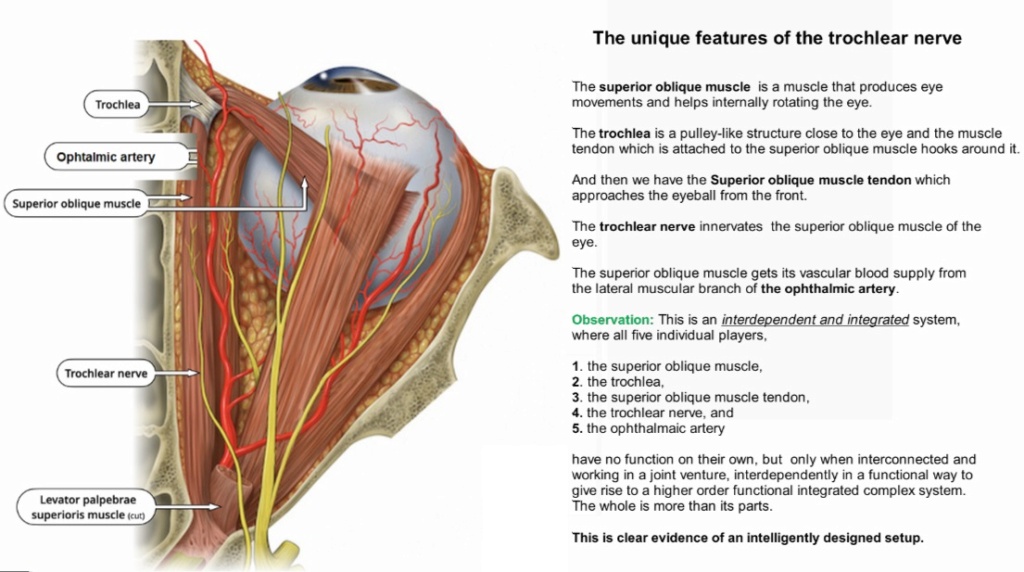
The unique features of the trochlear nerve

https://reasonandscience.catsboard.com/t2695-main-topics-on-the-origin-of-eyes#7360

Main topics on the origin of eyes Eye_in13

The superior oblique is a muscle that produces eye movements and helps internally rotate the eye. The trochlea is a pulley-like structure close to the eye and the muscle-tendon which is attached to the superior oblique muscle hooks around it. And then we have the muscle tendon which approaches the eyeball from the front. The trochlear nerve innervates the superior oblique muscle of the eye. The superior oblique muscle gets its vascular blood supply from the lateral muscular branch of the ophthalmic artery.

Observation: This is an interdependent and integrated system, where all five individual players have no function on their own, but only when interconnected and working in a joint venture, interdependently in a functional way. This is clear evidence of an intelligently designed setup. 

Visual data processing
1. Sight is the result of complex data processing. Data input requires a common understanding between sender and receiver/retrieval of the data coding and transmission protocols.
2. The rules of any communication system are always defined in advance by a process of deliberate choices. There must be a prearranged agreement between the sender and receiver, otherwise, communication is impossible.
3. Light can be considered an encoded data stream that is decoded and re-encoded by the eye for transmission via the optic nerve and decoded by the visual cortex. The sophisticated control systems in the human eye processes images and controls the eye muscles with speed and precision. 2 Synchronized 576MP cameras.  Data is multiplexed, that is multiple signals are combined into  one signal to 1MP to enter the optic nerve. Then de-multiplexed in the brain. Chromatic aberration is corrected by software. 2 pictures processed together to generate a 3D model. The Brain processes that model, extracts data from millions of coordinates and then sends very precise control data back to the muscles, the iris, lens and so on.
4. Only a Master Designer could have imagined/conceptualized and implemented these four things—language, the transmitter of language, multiplexing and de-multiplexing the message, and receiver of language since all have to be precisely defined in advance before any form of communication can be possible at all.

The interdependence of vision system
1. Rhodopsin proteins, the main players in vision, the visual cycle, the signal transduction pathway, the eye, eye-nerve, and visual cortex in the brain, and visual data processing are  irreducible, integrated, systems, which each by themselves are irreducibly complex, but on a higher structural level, are interdependent, and work together in a joint venture to create vision.
2. If each of the mentioned precursor systems would evolve through a gradual emergence by slight, gradual modifications, these would result in nonfunctional subsystems. Since natural selection requires a function to select, these subsystems are irreconcilable with the gradualism Darwin envisioned.
3. But lets suppose that even if in a freaky evolutionary accident the individual subsystems would be generated, they would still have to be assembled into an integrated functional system of higher order, requiring meta-information directing the assembly process to produce the final functional system, that is genetic information to regulate parts availability, synchronization, and manage interface compatibility. The individual parts must precisely fit together.
4. All these steps are better explained through a super intelligent and powerful designer, rather than mindless natural processes by chance, or/and evolution since we observe all the time minds capabilities 4. All these steps are better explained through a super intelligent and powerful designer, rather than mindless natural processes by chance, or/and evolution since we observe minds having the capabilities to produce cameras, telescopes, and information processing systems.


Homologous trochlear nerves are found in all jawed vertebrates. The unique features of the trochlear nerve, are seen in the primitive brains of sharks.

Observation: Convergence is a significant problem for evolution
Biologists are uncovering numerous examples of organisms that cluster together morphologically (structurally), and yet are genetically distinct. Frogs, lizards, or herbs that appear to be identical are actually different at the genetic level. An evolutionary interpretation of this data, then, demands that the morphologically identical organisms must have evolved independently of one another in a “repeatable” fashion.

Stephen J. Gould, Wonderful Life: The Burgess Shale and the Nature of History (New York, NY: W.W. Norton & Company, 1989), 51.
“…No finale can be specified at the start, none would ever occur a second time in the same way, because any pathway proceeds through thousands of improbable stages. Alter any early event, ever so slightly, and without apparent importance at the time, and evolution cascades into a radically different channel.

Gould’s metaphor of “replaying life’s tape” asserts that if one were to push the rewind button, erase life’s history, and let the tape run again, the results would be completely different. The very essence of the evolutionary process renders evolutionary outcomes as nonreproducible (or nonrepeatable). Therefore, “repeatable” evolution is inconsistent with the mechanism available to bring about biological change.

Paleontologist J. William Schopf, one of the world’s leading authorities on early life on Earth, has made this very point in the book Life’s Origin.
Because biochemical systems comprise many intricately interlinked pieces, any particular full-blown system can only arise once… Any complete biochemical system is far too elaborate to have evolved more than once in the history of life

https://www.facebook.com/search/top/?q=superior%20oblique&epa=SEARCH_BOX

Eye muscles anatomy⁠
The extraocular muscles are a group of six extrinsic muscles of the eye which move the eyeball. They surround the eyeball, facilitating its movement in various directions.

The superior rectus muscle is required to turn the eye upward, with some contribution by the inferior oblique muscle.⁠
The Inferior rectus is required to turn the eye downward, with some contribution by the superior oblique.⁠
⁠The lateral rectus is required to turn the eye outward toward the ear.⁠
⁠The medial rectus is required to turn the eye inward toward the nose.⁠

The eye is rotated medially by the superior rectus and superior oblique and is rotated laterally by the inferior rectus and inferior oblique. The levator palpebrae superioris muscle elevates the eyelid.

Question: Which of the six eye muscles evolved first ? It seems not feasible that this  functionally integrated system of parts could evolve gradually. This is an integrated, irreducible system, where all eyeball muscles must be in place in order together for the eye to be able to make all necessary movements.

THE OVERALL BLOCK DIAGRAM OF ANIMAL VISION from PROCESSES IN BIOLOGICAL VISION
https://neuronresearch.net/vision/files/overallblock.htm?fbclid=IwAR3xaKnTS28AMKV2c9iFBQL8OfJMfOU28n2FRVxKZKKJ_aDkYlj-tlI7zd8


Last edited by Otangelo on Sun Aug 14, 2022 10:08 am; edited 5 times in total

https://reasonandscience.catsboard.com

Otangelo


Admin

The extraordinary capabilities of the human eye points to intelligent design

How many megapixels equivalent does the eye have?
On most digital cameras, you have orthogonal pixels: they're in the same distribution across the sensor (in fact, a nearly perfect grid), and there's a filter (usually the "Bayer" filter, named after Bryce Bayer, the scientist who came up with the usual color array) that delivers red, green, and blue pixels.

So, for the eye, imagine a sensor with a huge number of pixels, about 130 million. There's a higher density of pixels in the center of the sensor, and only about 6 million of those sensors are filtered to enable color sensitivity. Somewhat surprisingly, only about 100,000 sense for blue! Oh, and by-the-way, this sensor isn't made flat, but in fact, semi-spherical, so that a very simple lens can be used without distortions -- real camera lenses have to project onto a flat surface, which is less natural given the spherical nature of a simple lens (in fact, better lenses usually contain a few aspherical elements).

This is about 22mm diagonal on the average, just a bit larger than a micro four-thirds sensor... but the spherical nature means the surface area is around 1100mm^2, a bit larger than a full-frame 35mm camera sensor. The highest pixel re
solution on a 35mm sensor is on the Canon 5Ds, which stuffs 50.6Mpixels into about 860mm^2.

So that's the hardware. But that's not the limiting factor on effective resolution. The eye seems to see "continuously", but it's cyclical, there's kind of a frame rate that's really fast... but that's not the important one. The eye is in constant motion from ocular microtremors that occur at around 70-110Hz. Your brain is constantly integrating the output of your eye as it's moving around into the image you actually perceive, and the result is that, unless something's moving too fast, you get an effective resolution boost from 130Mpixels to something more like 520Mpixels, as the image is constructed from multiple samples.

Except you don't. For one, your luminance-only rod cells, being sensitive in low light, actually saturate in bright light. So in full daylight or bright room light, they're completely switched off. That leaves you 6 million or so cone cells alone as your only visual function. With microtremors, you may have about 24 million inputs at best… not exactly the same as 24 megapixels. And per eye, of course, so call it 48 megapixels if you want to draw that equivalence.

In the dark, the cones don't detect much, it's all rods at that point. Technically that's more “pixels,” but your eye and brain are dealing with a low photon flux density — the same thing that causes ugly “shot noise” in low light photographs. So you brain is only getting input from rods that actually detect something.

And all of the 130 million sensors are “wired” down to about 1.2 million axions of the ganglion cells that wire the eye to the brain. There is already processing and crunching on your visual data before it gets to the brain,

Which makes perfect sense -- our brains can do this kind of problem as a parallel processor with performance comparable to the fastest supercomputers we have today. When we perceive an image, there's this low-level image processing, plus specialized processes that work on higher level abstractions. For example, we humans are really good at recognizing horizontal and vertical lines, while our friendly frog neighbors have specialized processing in their relatively simple brains looking for a small object flying across the visual field -- that fly he just ate. We also do constant pattern matching of what we see back to our memories of things. So we don't just see an object, we instantly recognize an object and call up a whole library of information on that thing we just saw.

Another interesting aspect of our in-brain image processing is that we don't demand any particular resolution. As our eyes age and we can't see as well, our effective resolution drops, and yet, we adapt. In a relatively short term, we adapt to what the eye can actually see... and you can experience this at home. If you're old enough to have spent lots of time in front of Standard Definition television, you have already experienced this. Your brain adapted to the fairly terrible quality of NTSC television (or the slightly less terrible but still bad quality of PAL television), and then perhaps jumped to VHS, which was even worse than what you could get via broadcast. When digital started, between VideoCD and early DVRs like the TiVo, the quality was really terrible... but if you watched lots of it, you stopped noticing the quality over time if you didn't dwell on it. An HDTV viewer of today, going back to those old media, will be really disappointed... and mostly because their brain moved on to the better video experience and dropped those bad-TV adaptations over time.

Back to the multi-sampled image for a second... cameras do this. In low light, many cameras today have the ability to average several different photos on the fly, which boosts the signal and cuts down on noise... your brain does this, too, in the dark. And we're even doing the "microtremor" thing in cameras. The recent Olympus OM-D E-M5 Mark II has a "hires" mode that takes 8 shots with 1/2 pixel adjustment, to deliver what's essentially two 16Mpixel images in full RGB (because full pixel steps ensure every pixel is sampled at R, G, B, G), one offset by 1/2 pixel from the other. Interpolating these interstitial images as a normal pixel grid delivers 64Mpixel, but the effective resolution is more like 40Mpixel... still a big jump up from 16Mpixels. Hasselblad showed a similar thing in 2013 that delivered a 200Mpixel capture, and Pentax is also releasing a camera with something like this built-in.

We're doing simple versions of the higher-level brain functions, too, in our cameras. All kinds of current-model cameras can do face recognition and tracking, follow-focus, etc. They're nowhere near as good at it as our eye/brain combination, but they do ok for such weak hardware.

They're only few hundred million years late...

Human Eye Visual Hyperacuity: A New Paradigm for Sensing?
The human eye appears to be using a low number of sensors for image capturing. Furthermore, regarding the physical dimensions of cones–photoreceptors responsible for the sharp central vision–, we may realize that these sensors are of relatively small size and area. Nonetheless, the eye is capable to obtain high-resolution images due to visual hyperacuity and presents an impressive sensitivity and dynamic range when set against conventional digital cameras of similar characteristics. This article is based on the hypothesis that the human eye may be benefiting from diffraction to improve both image resolution and the acquisition process. 2

Visual acuity (VA) commonly refers to the clarity of vision, but technically rates an examinee's ability to recognize small details with precision. Visual acuity is dependent on optical and neural factors, i.e., (1) the sharpness of the retinal image within the eye, (2) the health and functioning of the retina, and (3) the sensitivity of the interpretative faculty of the brain.
https://en.wikipedia.org/wiki/Visual_acuity

This excerpt from a paper about hyperacuity shows how stunning the ability really is:

“While in some tasks (e.g., in telling apart two nearby dots) thresholds are in the range of 30-60 arcsec, in other tasks such as the vernier, the threshold may be as low as 5 arcsec. A threshold of 5 arcsec means that the observer reliably resolves features that are less than 0.02 mm at a 1 m distance, or the size of a quarter-dollar coin viewed at 17 km! One can better appreciate the astonishing precision of this performance by considering the optical properties of the eye. In the spatially most sensitive region of the retina, the fovea, the diameter of the photoreceptors is in the range of 30-60 arcsec and the sizes of the receptive fields of the retinal ganglion cells may be even larger. Thus, humans can resolve detail with an accuracy of better than one fifth of the size of the most sensitive photoreceptor.

The Sensitivity of the Human Eye (ISO Equivalent)
Our warm, wet, multicellular eyes have evolved such a high level of sensitivity that they can, on occasion, detect a single photon aimed at the retina. Even the most sophisticated man-made devices require a cool, temperature-controlled environment to achieve the same feat. 5

A single photon is the the smallest particle that light is made of, and it is extremely hard to see.

Eyes are actually incredibly sensitive. The human eye is so sensitive it can detect even a single photon of light! Our eyes are more sensitive than any camera sensor out there. Sure, we can all marvel at the 4,000,000 ISO Canon ME20F-SH and the kind of footage it can capture, but it doesn’t hurt to marvel at the ‘tech’ built into our own skull-mounted cameras from time to time. 4 Direct detection of a single photon by humans 3 Humans can detect a single-photon incident on the cornea.


The cell biology of vision
https://www.ncbi.nlm.nih.gov/pmc/articles/PMC3101587/

Looking at the big picture: the retina is a neural circuit composed of different cell types
Each eye’s photoreceptors include around 120 million rods, which react to light intensity, and 6 to 7 million color-sensitive cones. Rods occupy the majority of retinal real estate, but the very center is a tiny, highly concentrated population of cones called the fovea.  As the only photosensitive cells in the human body, the rods and cones are essential for the conversion of visual data into electrochemical signals. Neurons in the retina can then begin to parse the visual field by registering contrasts in the photoreceptor data. Contrasts — or “edges” — are the basic units of all visual processing. Like a camera, the eye must be pointed directly at something in order to see it with as much clarity as possible; even the most powerful lenses can’t capture details with maximum resolution across an entire image. Your eyes can only see in the sharpest resolution, or in 100 percent acuity, in the fovea, a very small fraction of your visual field. “About 0.1 percent of your visual field, at any given time, is the only place you’ve ever had 20/20 vision. The fact that you don’t notice the rest of the world transforming into a blurry dreamscape every time you glance at your watch is a testament to the sublime engineering in the visual cortex. As you take in the view of a room, your brain sees not only the picture in front of you, but also the images from your most recent involuntary, staccato twitches called saccades. These images, plus your visual memory, together form a mental model of the space around you that is updated with every glance. Thus, even though only a tiny fraction of the field of vision is in focus at any given moment, the entire panorama seems equally sharp, no matter where you’re looking.

The visual cortex uses unknown means to create visual information out of thin air. Dan Sasaki, the VP of Optical Engineering at Panavision, discussed in a 2017 presentation that the greater sub-pixels in the image “provides the viewer with much more information from which to render the images in their brains, and this provides a sense of greater depth and more realism.”

So the theoretical limit on how much detail the human eye can actually process may be more of a guideline than rule. Dr. Martinez-Conde points out that the enigma encompasses all types of perception. “Fundamentally,” she adds,” “we don’t understand the neural basis of experience.” One thing is clear, however: The 33 million pixels that 8K TVs are able to display are changing the way we watch television, and making it a truly immersive viewing experience.

The retina carries out considerable image processing through circuits that involve five main classes of cells (i.e., photoreceptors, bipolar cells, amacrine cells, horizontal cells, and ganglion cells;

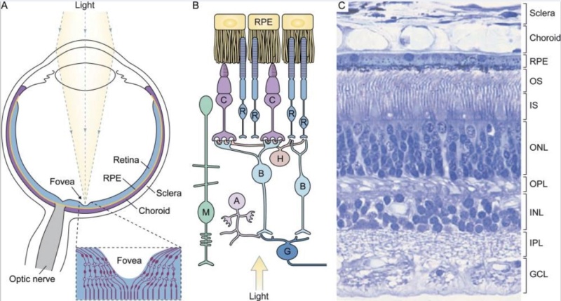
Main topics on the origin of eyes Retina10
The visual sense organ.
(A) Diagrams of the eye; an enlarged diagram of the fovea is shown in the box. Retina forms the inner lining of the most of the posterior part of the eye. The RPE is sandwiched between the retina and choroids, a vascularized and pigmented connective tissue.
(B) Diagram of the organization of retinal cells. R, rod; C, cone; B, bipolar cell; H, horizontal cell; A, amacrine cell; G, ganglion cells; M, Müller cell.
(C) An H&E-stained transverse section of human retina. Retina has laminated layers. The nuclei of the photoreceptors constitute the outer nuclear layer (ONL). The nuclei of the bipolar cells, amacrine cells, horizontal cells, and Müller glial cells are found in the inner nuclear layer (INL), and the nuclei of ganglion cells form the ganglion cell layer (GCL). The outer plexiform layer (OPL) contains the processes and synaptic terminals of photoreceptors, horizontal cells, and bipolar cells. The inner plexiform layer (IPL) contains the processes and terminals of bipolar cells, amacrine cells, and ganglion cells. The processes of Müller glial cells fill all space in the retina that is not occupied by neurons and blood vessels.

These processes collectively amplify, extract, and compress signals to preserve relevant information before it gets transmitted to the midbrain and the thalamus through the optical nerves (axons of the ganglion cells). The retinal information received by the midbrain is processed to control eye movement, pupil size, and circadian photoentrainment. Only the retinal input that terminates at the lateral geniculate nucleus of the thalamus is processed for visual perception and gets sent to the visual cortex. There, information about shade, color, relative motion, and depth are all combined to result in one’s visual experience.

Question: How could human eye sight have evolved, if the five beforementioned cells work collectively to perserve relevant information ?

Visual perception begins when the captured photon isomerizes the chromophore conjugated with the visual pigment in the photoreceptor cell. The photoexcited visual pigment then initiates a signal transduction cascade that amplifies the signal and leads to the closure of cation channels on the plasma membranes. As a result, the cells become hyperpolarized. The change in membrane potential is sensed by the synapses, which react by releasing fewer neurotransmitters

Observation: This is a interdependent, irreducible system, where all players form an integrated system, which ony works with all players in place.

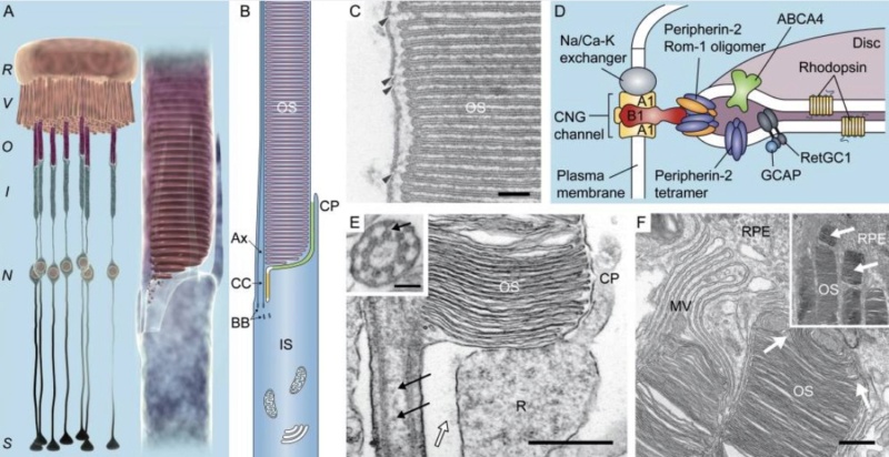
Main topics on the origin of eyes The_mo11
The morphological and molecular characteristics of vertebrate rod.
(A) 3D cartoons depict the inter-relationship between rod and RPE (left) and IS–OS junction (right); RPE apical microvilli interdigitate the distal half of the OS. R, RPE; V, microvilli; O, OS; I, IS; N, nucleus, S, synaptic terminal. (B) A schematic drawing of a mammalian rod depicting its ciliary stalk and microtubule organizations; the axonemal (Ax) and cytoplasmic microtubules (not depicted) are anchored at the basal body in the distal IS. CP, calycal process; BB, basal body. The interactions between opposing membranes are depicted in color. The yellow shade indicates that the putative interaction of the ectodomains of usherin–VLGR1–whirlin complexes appear on both CC plasmalemma and the lateral plasmalemma of the IS ridge complex. The green shade indicates the putative chlosterol–prominin-1–protocadherin 21 interaction. 
(C) Electron micrographs reveal the hairpin loop structures of the disc rims and the fibrous links across the gap between the disc rims and plasma membranes (arrowheads). Bar, 100 nm. 
(D) The OS plasma membrane and disc membrane have distinctive protein compositions; molecules are either expressed on the plasma membrane or the disc membranes, but not both. The only exception is rhodopsin; rhodopsin is present on disc membrane (with a much higher concentration) and plasma membrane (not depicted). The cGMP-gated channel: Na/Ca-K exchanger complex on the plasma membrane directly binds to the peripherin-2–ROM-1 oligomeric complex on the disc rim. The cGMP-gated channel is composed of three A1 subunits and one B1 subunit. ABCA4, a protein involved in retinoid cycle, is also enriched on the disc rim. RetGC1, retinal guanylyl cyclase; CNG channel, cGMP-gated channel. Adapted from Molday (2004). 
(E) Electron micrograph showing the longitudinal sectioning view of IS–OS junction of rat rod. Arrows point to the CC axonemal vesicles. An open arrow points to the fibrous structures linking the opposing membranes. Bar, 50 nm. Inset: a transverse section through the CC shows 9+0 arrangement; an arrow points to the cross-linker that gaps the microtubule doublet and adjacent ciliary membrane. R, apical IS ridge. Bar,100 nm. (F) Electron micrographs of a low-power (inset) and high-power images of the rat retina, at the junction between the rod OS and the RPE. MV, RPE microvillar processes enwrapped the distal OS. A white arrow points to a group of saccules from the tip of OS curls and upwards. White arrows in inset point to two distal OS fragments that are engulfed by RPE. Bar, 500 nm.

The vertebrate rod: elegance and efficiency
Rods have evolved a unique structure to detect and process light with high sensitivity and efficiency; human rods can detect single photons (Hecht et al., 1942; Baylor et al., 1979). Each rod contains four morphologically distinguishable compartments: the OS, inner segment (IS), nucleus, and axon/synaptic terminal (Fig. 2 A). The length of the rod OS ranges from ∼30 to 60 µm in length (and ∼1.4–10 µm in diameter), depending on the species. Basically, the rod OS is a cylindrically shaped membrane sac filled with ∼1,000 flattened, lamellar-shaped membrane discs that are orderly arrayed perpendicular to the axis of the OS. These discs appear to be floating freely, although filamentous structures bridging adjacent discs and disc rims to the nearby plasma membrane do exist. The visual pigment of the rod, rhodopsin, comprises ∼95% of the total amount of disc protein; it is densely packed within the disc lamellae (i.e., ∼25,000 molecules/µm2). The high density of rhodopsin, together with its ordered alignment with respect to the light path, increases the probability of capturing an incident photon. 6

Main topics on the origin of eyes Nihms-816341-f0001
The human visual system
(a) Visual perception begins in the eye, where the cornea and lens (1) project an inverted image of the world onto the retina (2), which converts incident photons into neural action potentials. (b) The retina consists of three layers of cells. The photoreceptors (PR), which are in contact with the retinal pigment epithelium (RPE), convert light into neural signals that propagate to the horizontal (HC), bipolar (BC) and amacrine cells (AC) of the inner nuclear layer. The axons of the retinal ganglion cells (RGCs) form the retinal nerve fiber layer (RNFL). They converge onto the optic disk (3), where they congregate to form the optic nerve (4), which relays neural signals to the brain. (c) Signals from the left and right visual fields of both eyes are combined at the optic chiasm (5). The lateral geniculate nucleus (6) relays the left visual field to the right visual cortex and the right visual field to the left visual cortex through neuron axons called the optic radiation. Higher visual processing finally takes place in the visual cortex (7), and further downstream in the brain.

Photoreceptors are graded-response neurons (i.e. they do not generate action potentials) that transduce photons into changes in their membrane potential by means of light-sensitive proteins called opsins. The vertebrate retina is inverted, so that photoreceptors are located at the back of the eye in contact with the retinal pigment epithelium (RPE), which is essential to the health and function of the photoreceptors. RPE cells regenerate photopigments and digests outer segments shed by the photoreceptors. Without support from the RPE, photoreceptor cells progressively atrophy and die.


Photoreceptors relay visual information to the neurons in the inner nuclear layer of the retina, where 2 types of horizontal cells, about 12 types of bipolar cells, and as many as 30 types of amacrine cells process the visual signals.
Humans have ∼130 million photoreceptors, ∼5 million bipolar cells, and ∼1 million ganglion cells.Rods outnumber cones by ∼20-fold, and are distributed throughout the retina with the exception of the fovea region.

The vertebrate rod: elegance and efficiency
Rods have a unique structure to detect and process light with high sensitivity and efficiency; human rods can detect single photons. Each rod contains four morphologically distinguishable compartments: the OS, inner segment (IS), nucleus, and axon/synaptic terminal (Fig. 2 A). The length of the rod OS ranges from ∼30 to 60 µm in length (and ∼1.4–10 µm in diameter), depending on the species. Basically, the rod OS is a cylindrically shaped membrane sac filled with ∼1,000 flattened, lamellar-shaped membrane discs that are orderly arrayed perpendicular to the axis of the OS. These discs appear to be floating freely, although filamentous structures bridging adjacent discs and disc rims to the nearby plasma membrane do exist (Fig. 2, C and D;) The visual pigment of the rod, rhodopsin, comprises ∼95% of the total amount of disc protein; it is densely packed within the disc lamellae (i.e., ∼25,000 molecules/µm2). The high density of rhodopsin, together with its ordered alignment with respect to the light path, increases the probability of capturing an incident photon.

Transduction of the light message: from the photon to the optic nerve 
One truly fascinating aspect of retinal neurotransmission is that it is a meeting point for neurophysiology and biophysics. Light, as an electromagnetic wave or stream of energy quanta is essentially a physical agent; through interaction with the retinal tissue, light stimulus results in the excitation of a nerve fiber which generates an electrical signal. In this way, the retina achieves the overall equivalent of a photoelectric effect. 7 We know that the photoreceptor cell, as a “photon detector”, operates in two phases; the first phase is the absorption of incident photons and is photochemical; the second phase, activated by the first, is electrophysiological. Thus, even at the photoreceptor stage, the light signal is already electrical in nature. It is noteworthy that the message remains electrical right to the nerve fibers that emerge from the retina. Meanwhile, the initial signal is modulated to encode the visual information. Each step of this modulation, which constitutes the neurotransmission, may be considered as an electrical circuit; accordingly, the term retinal microcircuitry is often employed. As we shall see, we are still a long way from a complete and accurate picture of all the processes that occur, but considerable progress has been made especially in recent years, so that a coherent description of this neurocircuitry can now be outlined.

The membrane depolarization-hyperpolarization duality and information encoding at the optic nerve
The depolarization-hyperpolarization duality of the nerve fiber membrane is fundamental to the understanding of the retinal microcircuitry. Except for the highly differentiated photoreceptor cells that are specialized in photon detection, the other excitable cells in the retina, which make up the neurocircuits, are “classical” neurons that respond to excitation by a variation in membrane potential. As regards the encoding of the information at the nerve, the nature of the elecrophysiological response (variation of the membrane potential over time) suggests two possible mechanisms a priori: i) an amplitude code; ii) a time code. As the nerve fiber operates in an “all or nothing” mode, an amplitude code can be ruled out. The code is in fact a time code, the variable being the duration of the prepotential, which encodes the intensity of the stimulus; the more intense the stimulus, the shorter the prepotential. Taking into account the refractory period of the nerve, it is the frequency of occurrence of action potentials along the nerve fiber which is a measure of the intensity of the stimulation. This is a highly specific feature of the neuron due to the existence of a threshold for the occurrence of a membrane potential response to a stimulus. At the optic nerve, the encoding of the information thus uses a time code, and the frequency of the action potentials indicates the intensity of the light stimulation of the retinal photoreceptor cells. Let us consider the situation in which the photoreceptor (cone or rod) is operationally linked to a first associated neuron via a chemical neuromediator (fig 1). Here just as elsewhere, it is the depolarization of the cell upstream which enables the release of the neurotransmitter. 

Main topics on the origin of eyes Neurot10

Since the photoreceptor is depolarized in the dark and hyperpolarized in light, we must assume that the neuromediator is being constantly released by the photoreceptor in the dark and that light restricts and ultimately suppresses this release. As for the neuron associated with the photoreceptor, there are two possibilities: i) the neurotransmitter is excitatory and causes membrane depolarization of the cell; ii) the neurotransmitter is inhibitory and causes hyperpolarization of the neuron membrane. Actually, the same neurotransmitter can be excitatory for some cells and inhibitory for others. Thus, one class of cells are depolarized, ie excited by light; these are the cells for which the neurotransmitter is inhibitory; another cell category is hyperpolarized, ie inhibited by light; these are the cells for which the neurotransmitter released in the dark is excitatory. This situation holds of course for all the neurons that make up the retinal circuitry. We have therefore to distinguish between two types of excitable retinal neurons: i) those which are hyperpolarized in the light are termed OFF neurons; ii) those which are depolarized in the light are termed ON neurons. This second ONOFF duality, a direct consequence of the depolarization-hyperpolarization alternative, means that there are two types of retinal circuit; ON circuits and OFF circuits.

Organization of the mammalian retina
It is still usual to adopt a radial description of the retina which follows the light path. Thus, the “functional triad”: i) photoreceptor cells; ii) bipolar cells; iii) ganglion cells (fig 2) has become conventional. 

Main topics on the origin of eyes Neurot11


For the cones, the situation is both simpler and more complex; simpler because the saccules stacked in the outer segment are formed by successive folds of the segment membrane and so Q priori need no intracellular messenger; more complex because of the photopic nature of the stimulus that implies a mechanism of color discrimination. We now know that the spectral sensitivity of the cones is determined by the photopigment they contain. Like rhodopsin, these are proteins coupled to chromophores. We also know that the phototransduction mechanism is similar to that for the rods, involving a G-protein with transmission of information linked to guanoside-phosphates. Experiments have shown that the photoexcitation of the cones, like that of the rods, results in an hyperpolarization of their outer segment membranes. Thus, the first electrophysiological signal resulting from the absorption of light by the retina is the hyperpolarization of the photoreceptor cells [9]. These are among the rare excitable cells of the organism whose excitation by natural stimuli results in hyperpolarization.

The photoreceptor cell has a highly specific organization. In this scheme the bipolar cells are the first associated neurons: these are entirely intraretinal, and correspond to the inner nuclear layer. The ganglion cells are the second associated neurons; their axons belong to the optic nerve. This radial description was soon completed by a “tangential description” which allows for the presence of three types of neuron situated in planes parallel to the retina:

i) the horizontal cells of the outer plexiform layer; 
ii) the amacrine cells of the inner plexiform layer; 
iii) the interplexiform cells associating the two plexiform layers. 

Thus, it was for a long time classical to assert that the neurosensory axis of the retina was the photoreceptor - bipolar cell - ganglion cell association and that the horizontal and amacrine cells served to “modulate” the transfer of visual information at the photoreceptor - bipolar cell synapses and the bipolar ceIl - ganglion cell synapses respectively. However, the diversity of the cell types identified in the retina, and a more thorough study of their interconnections, have led to a more specific functional approach to the organization of the retina. The various cell categories fall into several subgroups:

I Photoreceptors: cones and rods. 2 Bipolar cells: - those related to the cones, termed cone bipolar cells, divided into ON bipolar cells which are depolarized by light and OFF bipolar cells which are hyperpolarized by light, - those related to the rods, termed rod bipolar cells, which are all ON, ie depolarized by light. 3 Ganglion cells, which schematically can be either: - OFF-center ganglion cells that respond (hyperpolarization) to offset of light and which connect in sublamina a of the inner plexiform layer, - ON-center ganglion cells which respond (depolarization) to the onset of light and which connect in sublamina b of the inner plexiform layer. Here too an ON-OFF duality operates. 4 Amacrine cells: the same retina can contain up to 30 morphologically distinct types of amacrine cell. For simplicity, we shall mention here only certain sub-groups characterized by their connections or the neurotransmitters they contain: - cholinergic, indolaminergic, dopaminergic amacrine cells etc. - a particularly important sub-group for the understanding of the retinal microcircuitry: the rod amacrine cells or A11 cells. 5 Horizontal cells: two distinct types, which are specifically involved in the encoding of information, as we shall see: - type A horizontal cells which have a wide range of action, receiving an excitatory message from a cone and inhibiting, in response, the cones in their field with which they are connected, - type B horizontal cells which have a more restricted range of action, and which, when excited by a cone, excite in turn the cones to which they are connected, in effect amplifying the information. In the detailed description of the retinal circuitry given further on we shall see the specific role of each of these cell types.

Origin of the electrophysiological signal 
The phototransduction takes place entirely inside the outer segment of the photoreceptor, which contains large amounts of photopigment composed of a functional association of a protein macromolecule and a chromophore. The role of the photopigment is to absorb the photons impinging on the retina, which is the first step in their detection. This absorption of radiant energy brings about a change in the membrane potential of the outer segment of the photoreceptor. In the rods, the saccules containing the photopigment (rhodopsin) are totally independent of the outer segment, in which they float. This implies some messenger between the saccule membrane and the outer segment membrane. This messenger is cGMP. Thus, the sequence of identified events in the transduction include - absorption of light by rhodopsin with resulting photoisomerization, - activation by rhodopsin of transducin, the G-protein with which it is associated, - the alpha sub-unit of transducin thus released can act on its effector, a phosphodiesterase, by suppressing its inhibition, - the phosphodiesterase is responsible for the hydrolysis of molecules of cGMP to 5’-GMP. The visual message that comes from the saccules thus consists of a decrease in the cytosol level of cGMP. Since the permeability to sodium ion of the outer segment membrane is directly dependent on cGMP, the effect of this sudden drop in concentration is to close the membrane sodium channels; the sodium ions accumulate over the outer surface of the outer segment membrane of the rod, resulting in hyperpolarization involving a G-protein with transmission of information linked to guanoside-phosphates. Experiments have shown that the photoexcitation of the cones, like that of the rods, results in an hyperpolarization of their outer segment membranes. Thus, the first electrophysiological signal resulting from the absorption of light by the retina is the hyperpolarization of the photoreceptor cells [9]. These are among the rare excitable cells of the organism whose excitation by natural stimuli results in hyperpolarization.

Description of retinal neurocircuits
At the outset, the neurocircuits leading from the cones to the ganglion cells have to be distinguished from those which originate at the rods. The first type of circuit is more direct and simpler then the second, but  the two can interpenetrate under certain circumstances. 

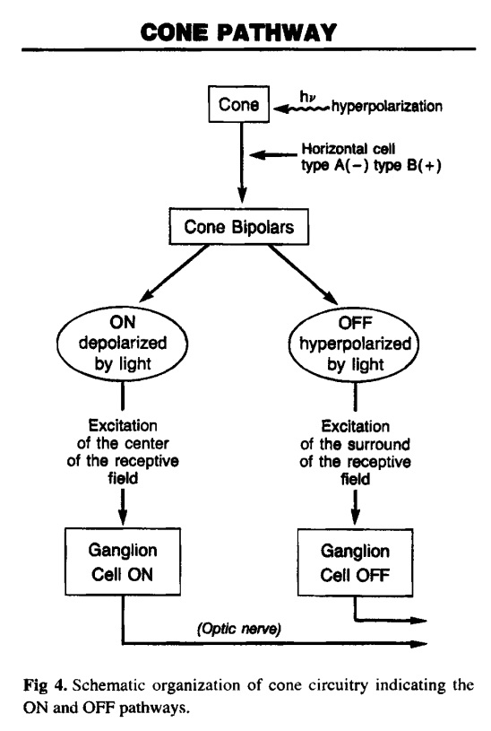
The cone circuit 
Each cone is linked to two types of bipolar cell; ON cone bipolar cells, depolarized by light, and OFF cone bipolar cells, hyperpolarized by light. Thus the ON-OFF duality occurs in the cone circuit at the very first relay (fig 4).

Main topics on the origin of eyes Neurot12


1. https://www.theverge.com/ad/18113053/pixels-human-vision-8k-television
2. https://arxiv.org/ftp/arxiv/papers/1703/1703.00249.pdf
3. https://www.nature.com/articles/ncomms12172
4. https://petapixel.com/2016/07/25/human-eye-sensitive-can-detect-single-photon-light/
5. https://www.latimes.com/science/sciencenow/la-sci-sn-human-eye-photon-20160719-snap-story.html
6. https://www.ncbi.nlm.nih.gov/pmc/articles/PMC3101587/
7. https://sci-hub.st/https://onlinelibrary.wiley.com/doi/abs/10.1111/j.1472-8206.1994.tb00791.x

https://reasonandscience.catsboard.com

4Main topics on the origin of eyes Empty Re: Main topics on the origin of eyes Sun Nov 01, 2020 1:43 pm

Otangelo


Admin

Creationist Quote-Miner - Genetics
https://www.youtube.com/watch?v=0S9ISPvxIqM&t=84s

Claim: one of the favorite tactics of creationists trying to undermine confidence in science 
Reply: Norman Geisler:
The creation-evolution debate is not religion versus science or the Bible versus science, it's about good science versus bad science. Likewise, it's not faith versus reason, it's about reasonable faith, versus unreasonable faith.

The deepest intellectual battle is not between science and religion (which can operate with a great deal of accord), but between naturalism and theism—two broad philosophical (or metaphysical) ways of looking at the world. Neither view is a scientific view; neither view is based on or inferable from empirical data. Metaphysics, like numbers and the laws of logic, lies outside the realm of human sense experience. So the issue of naturalism versus theism must be decided on philosophical grounds

Claim: is their use of mind quotations meaning that religious apologists will sift through volumes of scientific studies ignoring all of the evolutionary evidence they're in looking for that one line that if taken out of context sounds like it's saying the very opposite of what the whole study actually says
Reply: One thing is the scientific evidence, and another the philosophical interpretation. Richard C. Lewontin who is a well-known geneticist and an evolutionist from Harvard University claims that he is first and foremost a materialist and then a scientist. He confesses;
“It is not that the methods and institutions of science somehow compel us to accept a material explanation of the phenomenal world, but, on the contrary, that we are forced by our a priori adherence to material causes to create an apparatus of investigation and a set of concepts that produce material explanations, no matter how counter-intuitive, no matter how mystifying to the uninitiated. Moreover, that materialism is absolute, so we cannot allow a Divine Foot in the door.”(Lewontin 1997)

Materialism regards itself as scientific, and indeed is often called “scientific materialism,” even by its opponents, but it has no legitimate claim to be part of science. It is, rather, a school of philosophy, one defined by the belief that nothing exists except matter, or, as Democritus put it, “atoms and the void.” 2

Claim: for example some dogmatic science denialist read through the entirety of darwin's origin of species and plucked out the one line that says to suppose that the eye could have been formed by natural selection seems i freely confess absurd in the highest degree they'll show you only that quote ignoring everything else  he said immediately after that about what reeves it tells me so darwin said it seems absurd but and then he filled the next few pages with evidence supporting what only seemed absurd at first until you get to see the facts to know better
Reply:  What Darwin said and his opinion that evolution would be possible is irrelevant. He lived in a time where molecular biology was not known. We know today why he was wrong.

https://reasonandscience.catsboard.com/t2695-main-topics-on-the-origin-of-eyes

Otangelo Grasso
February 24, 2020, 4:08 AM
The Evolution of the Eye, Demystified
https://evolutionnews.org/2020/02/the-evolution-of-the-eye-demystified/?fbclid=IwAR3fgWoc02Xy0jgggCKa2TLLofsQkuSh3vUWf2xSNk73jU1iMJGzwiTJk2Y

1. Rhodopsin proteins, the main players in vision, the visual cycle, the signal transduction pathway, the eye, eye-nerve, and visual cortex in the brain, and visual data processing are  irreducible, integrated, systems, which each by themselves are irreducibly complex, but on a higher structural level, are interdependent, and work together in a joint venture to create vision.
2. If each of the mentioned precursor systems would evolve through a gradual emergence by slight, gradual modifications, these would result in nonfunctional subsystems. Since natural selection requires a function to select, these subsystems are irreconcilable with the gradualism Darwin envisioned.
3. But lets suppose that even if in a freaky evolutionary accident the individual subsystems would be generated, they would still have to be assembled into an integrated functional system of higher order, requiring meta-information directing the assembly process to produce the final functional system, that is genetic information to regulate parts availability, synchronization, and manage interface compatibility. The individual parts must precisely fit together.
4. All these steps are better explained through a super intelligent and powerful designer, rather than mindless natural processes by chance, or/and evolution since we observe all the time minds capabilities 4. All these steps are better explained through a super intelligent and powerful designer, rather than mindless natural processes by chance, or/and evolution since we observe minds having the capabilities to produce cameras, telescopes, and information processing systems.

Visual data processing
1. Sight is the result of complex data processing. Data input requires a common understanding between sender and receiver/retrieval of the data coding and transmission protocols.
2. The rules of any communication system are always defined in advance by a process of deliberate choices. There must be a prearranged agreement between the sender and receiver, otherwise, communication is impossible.
3. Light can be considered an encoded data stream that is decoded and re-encoded by the eye for transmission via the optic nerve and decoded by the visual cortex. The sophisticated control systems in the human eye processes images and controls the eye muscles with speed and precision. 2 Synchronized 576MP cameras.  Data is multiplexed, that is multiple signals are combined into  one signal to 1MP to enter the optic nerve. Then de-multiplexed in the brain. Chromatic aberration is corrected by software. 2 pictures processed together to generate a 3D model. The Brain processes that model, extracts data from millions of coordinates and then sends very precise control data back to the muscles, the iris, lens and so on.
4. Only a Master Designer could have imagined/conceptualized and implemented these four things—language, the transmitter of language, multiplexing and de-multiplexing the message, and receiver of language since all have to be precisely defined in advance before any form of communication can be possible at all.

Rhodopsin
1. Rhodopsin, the first player of vision, is a light-sensitive receptor protein involved in visual phototransduction. Rhodopsin consists of two components, a protein molecule called opsin and a covalently-bound cofactor called retinal. If both were not just right from the beginning, rhodopsin would not be functional, and vision, not possible. Retinal is finely tuned and right-sized to fit the binding pocket. It is a molecule obtained by a complex multistep biosynthesis pathway.
2. Both, opsin, and retinal, are highly constrained and thus well conserved across organisms because mutations within these sites are deleterious.
3. An article in Nature magazine, in 2016, confessed: Darwin’s original puzzle over ocular evolution seems still to be with us but now at a molecular level.
4. The inference is that Rhodopsin is irreducibly complex, and its origin, therefore, best explained by design!
https://reasonandscience.catsboard.com/t2638-volvox-eyespots-and-interdependence#5768

Claim:  these are not myths like creationism is none of them were disproved by modern science like creationism was and they don't break any natural laws like creationism does the problem creationists have is that we're not the ones ignoring decades of contradictory evidence science has working models that are tried and true based on demonstrable fact with profound explanative and predictive power that are always borne out and made stronger by further discoveries without any contradiction
Reply:  Science has an answer: Abiogenesis is mathematically  impossible
https://reasonandscience.catsboard.com/t1279-abiogenesis-is-mathematically-impossible
Some of the worlds leading scientists in the field of synthetic chemistry, biochemistry, and computational biology, like James Tour, Graham Cairns-Smith, Eugene Koonin and Steve Benner have stated that solving the mystery of the origin of life is categorically not possible, that science has no clue how to solve the riddle, that abiogenesis research is a failure, and the most difficult problem that faces evolutionary biology and, arguably, biology in general.

Eugene V. Koonin: The Logic of Chance: page 351:
" Despite many interesting results to its credit, when judged by the straightforward criterion of reaching (or even approaching) the ultimate goal, the origin of life field is a failure—we still do not have even a plausible coherent model, let alone a validated scenario, for the emergence of life on Earth.

Steve Benner:  Paradoxes in the origin of life
Discussed here is an alternative approach to guide research into the origins of life, one that focuses on “paradoxes”, pairs of statements, both grounded in theory and observation, that (taken
together) suggest that the “origins problem” cannot be solved.

Graham Cairns-Smith: Genetic takeover, page 66:
Now you may say that there are alternative ways of building up nucleotides, and perhaps there was some geochemical way on the early Earth. But what we know of the experimental difficulties in nucleotide synthesis speaks strongly against any such supposition. However it is to be put together, a nucleotide is too complex and metastable a molecule for there to be any reason to expect an easy synthesis.

Garrett: Biochemistry, 6th ed,  page 665
Key compounds, such as arginine, lysine, and histidine; the straight-chain fatty acids; porphyrins; and essential coenzymes, have not been convincingly synthesized under simulated prebiotic conditions.

Robert Shapiro: A Replicator Was Not Involved in the Origin of Life
A profound difficulty exists, however, with the idea of RNA, or any other replicator, at the start of life. Existing replicators can serve as templates for the synthesis of additional copies of themselves, but this device cannot be used for the preparation of the very first such molecule, which must arise spontaneously from an unorganized mixture. The formation of an information-bearing homopolymer through undirected chemical synthesis appears very improbable.

Where Do Complex Organisms Come From?
https://reasonandscience.catsboard.com/t2316-evolution-where-do-complex-organisms-come-from
The morphogenesis of organismal structure and shape is classified into two groups: The various instructional codes and languages using molecules that provide complex instructional cues of action based on information through signaling and secondly by force-generating molecules that are precisely directed through those signals, which are responsible for fantastic cell morphogenesis. Blueprints, instructional information, and master plans, which permit the striking autonomous self-organization and control of complex machines ( molecular machines) and exquisite factory parks ( cells) upon these are both always tracked back to an intelligent source which made both for purposeful, specific goals.   That brings us unambiguously to intelligent design. To the origin by an intelligent designer.


Claim:  there was never any evidence that creationism is true 
Reply: Syllogistic - Arguments of Gods existence based on positive evidence
https://reasonandscience.catsboard.com/t2895-syllogistic-arguments-of-gods-existence-based-on-positive-evidence

Intelligent design theory is like a sword with two edges

Intelligent design wins using eliminative induction based on the fact that its competitors are false. Materialism explains basically nothing consistently in regards to origins but is based on unwarranted consensus and scientific materialism, a philosophical framework, that should never have been applied to historical sciences. Evidence should be permitted to lead wherever it is. Also, eventually, to an intelligent agency as the best explanation of origins.

And intelligent design wins based on abductive reasoning, using inference to the best explanation, relying on positive evidence, on the fact that basically all-natural phenomena demonstrate the imprints and signature of intelligent input and setup. We see an unfolding plan, a universe governed by laws, that follows mathematical principles, finely adjusted on all levels, from the Big Bang, to the earth, to permit life, which is governed by instructional complex information stored in genes and epigenetically, encoding, transmitting and decoding information, used to build, control and maintain molecular machines ( proteins ) that are build based on integrated functional complex parts ( primary to quaternary polypeptide strands and active centers ), which are literally nanorobots with internal communication systems, fully automated manufacturing production lines, transport carriers, turbines, transistors, computers, and factory parks, employed to give rise to a wide range, millions of species, of unimaginably complex multicellular organisms.

Claim: and we actually have proof that it isn't finally anyone who honestly investigates the validity of the evidence will accept evolution as fact
Reply: 
https://reasonandscience.catsboard.com/t2806-main-topics-about-evolution

What is fact in regards of evolution :
1. Change over time; history of nature; any sequence of events in nature
2. Changes in the frequencies of alleles in the gene pool of a population
3. Limited common descent: the idea that particular groups of organisms have descended from
a common ancestor.
4. The mechanisms responsible for the change required to produce limited descent with modification; chiefly pre-programmed selection acting on random variations or mutations
5. Natural selection acting up to two random mutations as shown in malaria ( See Behe's Edge of evolution )

What is not fact:
6. Universal common descent: the idea that all organisms have descended from a single common ancestor.
7. Blind watchmaker thesis: the idea that all organisms have descended from common ancestors through unguided, unintelligent, purposeless, material processes such as natural
selection acting on random variations or mutations; the idea that the Darwinian mechanism of natural selection acting on random variation, and other similarly naturalistic mechanisms, completely suffice to explain the origin of novel biological forms and the appearance of design in complex organisms.

Claim:  for example dr kenneth miller he's a christian he believes in god and he is also one of the most famous evolutionary biologists working today he debated all the intelligent design creations from the pseudoscience propaganda mill known as the discovery institute and he disproved every one of their arguments for irreducible complexity both scientifically and in a court of law
Reply: I had a dialogue at his Facebook timeline, and he stopped and refused to answer me and my inquiries. I asked him about Micoplasma mobile, how it evolved, and he failed to provide an
explanation. I accept a public debate with him anytime.

The extraordinary cellular propulsion system of Mycoplasma mobile - faster than Usain Bolt
https://www.youtube.com/watch?v=Jz_5BXIOrHw

https://reasonandscience.catsboard.com/t2765-the-extraordinary-cellular-propulsion-system-of-mycoplasma-mobile-faster-than-usain-bolt

1. Mycoplasma mobile uses flexible legs, that literally walk on the surface. Better maybe to say, they run. While Usain Bolt runs 5.3 of its own body length per second,(5.3 bl/sec.) M. mobile runs a top burst speed of ~10 bl/sec
2. Four component proteins are directly involved in the mechanism, and at least 10 proteins are involved in the cytoskeleton. None of the four proteins has significant sequence similarities with proteins of any other bacterias.
3. Since none of these components can be dismissed without harming the motility function, this system is irreducible and interdependent, and on top, none of the components could be co-opted from other organisms.

https://reasonandscience.catsboard.com

Otangelo


Admin

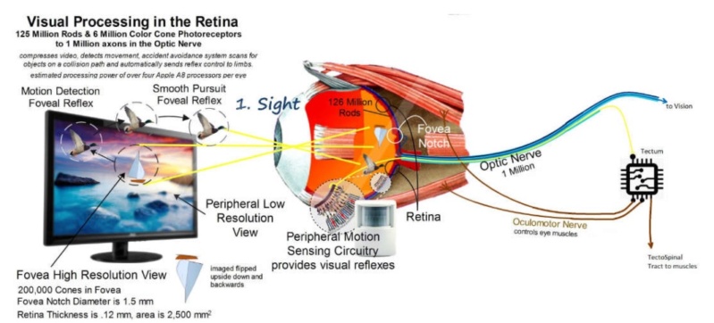
Microprocessors in the Eye, by chance, or design?

https://reasonandscience.catsboard.com/t2695-main-topics-on-the-origin-of-eyes#8591

We are able to react immediately when an object comes towards us at high speed. The electronics behind the reflex start with a motion detection micro processor right inside the human eye. 125 million rod photoreceptors in the retina detect and process motion to send coordinate data to the tectum when objects are moving towards our path or come towards us at high speed. The tectum has it's own spinal tract which controls our limbs to produce avoidance maneuvers like when a pitcher throws a ball at your head. Tectospinal circuits from the eye can inhibit all motor circuits and pain reflex circuits to take full control when objects are rapidly approaching.  For example, if we were walking towards the road and a car is detected in our peripheral vision, the eye can inhibit the current motor task before it takes control of the muscle. The circuitry in the spinal column is obviously highly engineered in its function to preserve our lives and must require accurate assembly instructions stored in our DNA to wire correctly.  Based on the number of synapse connections in the retina, the circuits in one eye contain more processing power then 5 iPhone processors. The superior colliculus is the main nuclei of the tectum where signals from the optic nerve, that are not used to produce vision but rather visual reflexes, are wired.

1. 125 million rod photoreceptors in the retina host Peripheral Motion Sensing Circuitry that detects and processes motion to send coordinate data to the tectum when objects are moving into our path.
2. The tectum controls and directs our limbs to produce avoidance maneuvers
3. Oculomotor nerves are required to be connected to control the eye muscles
4. The spinal tract is required and wired to body muscles

This is an irreducibly complex system, where all players must be in place, fully developed, and in a functional interplay to permit the reflexes which permit taking coordinated action and avoid danger.

https://www.thehighestofthemountains.com/evolution.php

Main topics on the origin of eyes Sem_tz16

https://reasonandscience.catsboard.com

6Main topics on the origin of eyes Empty Re: Main topics on the origin of eyes Sat Oct 29, 2022 12:18 pm

Otangelo


Admin

Dr. Jerry Bergman (2022): The camera eye has numerous parts, including a cornea, a lens, a set of extraocular muscles, and ocular adnexa (the parts adjoining the organ), all of which must be functional and highly integrated to be able to work as a unit. 1

A vertebrate camera eye has 3 separate parts (cornea, lens, retina) that must develop synergistically within ontogeny in mathematical optical harmony.

https://www.youtube.com/watch?v=GWkMAzsItc0&t=569s


1. Dr. Jerry Bergman: Evolution Fails to Explain the Vertebrate Camera-Type Eye   August 31, 2022
https://answersresearchjournal.org/evolution/vertebrate-camera-type-eye/

https://reasonandscience.catsboard.com

Sponsored content


Back to top  Message [Page 1 of 1]

Permissions in this forum:
You cannot reply to topics in this forum