ElShamah - Reason & Science: Defending ID and the Christian Worldview
Would you like to react to this message? Create an account in a few clicks or log in to continue.
ElShamah - Reason & Science: Defending ID and the Christian Worldview

Otangelo Grasso: This is my library, where I collect information and present arguments developed by myself that lead, in my view, to the Christian faith, creationism, and Intelligent Design as the best explanation for the origin of the physical world.


You are not connected. Please login or register

Apoptosis, Cell's essential mechanism of programmed suicide points to design

Go down  Message [Page 1 of 1]

Otangelo


Admin

Apoptosis, Cell's essential mechanism of  programmed suicide points to design

https://reasonandscience.catsboard.com/t2193-apoptosis-cell-s-essential-mechanism-of-programmed-suicide-points-to-design

Survival of the Fittest or Altruistic Suicide? 1

Like engineers carefully blowing up a bridge, cells have intricate, programmed suicide mechanisms. The signal is sent and an apparatus of destruction is activated. But suicide hardly fits the evolutionary narrative. Wasn’t this all about survival, reproductive advantages and leaving more offspring? Why would a cell evolve intricate and complex suicide machinery?

The answer is that suicide at the cellular level doesn’t kill the whole organism. Such self-destruction serves a range of purposes, from guiding development to keeping cancer at bay. In short, cell death in a multicellular organism can be a good thing.

But if cell suicide in multicellular organisms passes the evolutionary test, what about recent findings of suicide in unicellular organisms? New genome data from the Great Barrier Reef demosponge (Amphimedon queenslandica) reveals high levels of unexpected complexity, for this lowly sponge has an impressive complement of genes. As one proponent of evolution put it, “This flies in the face of what we think of early metazoan evolution.”

Another Darwinist asked perhaps an even more telling question. “What I want to know now,” he asked, “is what were all these genes doing prior to the advent of sponge?”

That’s a good question because some of those genes are for programmed suicide. What this sponge genome apparently tells us is that programmed cell death would have to have arisen in single-cell organisms. Suicide at the cellular level did kill the whole organism—and that doesn’t make evolutionary sense.

With evolution what we must believe is that programmed cell death did not arise in multicellular species, but in unicellular species. In other words, an intricate, highly complex, set of tools and signals somehow arose and, rather than leading to enhanced survival as evolution calls for, they led to destruction. Proponents of evolution will need yet another one of their just-so stories to rationalize this.


Programmed Cell Death
Cells have built-in error correction and repair mechanisms. Such mechanisms cannot be accounted for by any Darwinian hypothesis since error detection implies knowledge. Knowledge of correct system state and planning of appropriate repair measures.
Here’s one problem with the Darwinian “it just evolved” hypothesis : 
Q: How can a repair mechanism arise without pre-knowledge of correct system state? 
A: It cannot. Not when the damage caused interrupts and corrective facilities are this specifically complex


Implication? DNA had to be designed by an intelligent agent.
In software development, programmers build what we call “exception trapping” mechanisms. 
Such mechanisms ‘watch’ a given function progress and trap errors (exceptional system events) when detected. The trapping code then directs program flow either to analysis functions and correctional code or if the error is minor simply continue processing after the code block that failed (caused the exception) and may possibly alert the user to a faulty input situation.
DNA has it’s own special codes for detecting and ‘catching’ the exceptions that occur. Triggered by a diverse range of cell signals.
Neo Darwinian evolution (NDE) cannot explain the existence of such built-in functions.
Worse (for NDE) : Not only is there repair of damage available to the cell’s system but there is even a last resort “correction” (but not repaired) measure called apoptosis – pre-programmed cell death!Apoptosis is “a type of cell death in which the cell uses specialized cellular machinery to kill itself; a cell suicide mechanism that enables metazoans to control cell number and eliminate cells that threaten the animal’s survival” (also plays a role in preventing cancer). Its a key process in multicellular organisms.
This too is not explicable under NDE.
So, in a more engineering-like term we can look at this process as something like a control feedback loop. Such ‘loops’ exist in many places in the cell; like the circadian oscillator (Paley's watch!) – “a clockwork mechanism that controls these global rhythms of transcription, chromosomal topology, and cell division.”
Well then, the evolutionary ancestor to that was what?
Darwinism utterly fails to predict sophisticated repair mechanisms like these. Only pre-knowledge of correct system state can foresee and then construct code sequences that operate repairs to or destruction of faulty parts in a complex machine.
DNA is such a machine.


Programmed cell death (PCD, or apoptosis) appears to be universally present and indeed should be regarded as one of the hallmarks of the multicellular state itself 2  From a purely systemic point of view, PCD in a multicellular organism appears to be just as inevitable as law enforcement in a state. In any differentiated community (of specialized cells or of citizens), rogue elements will necessarily emerge in as (pre)cancerous cells with impaired division control or as criminals with impaired social responsibility, and to protect the community, these need to be subdued or destroyed by dedicated agencies. Those agencies also contribute to the defense against invaders, such as viruses, pathogenic bacteria or rival states, and, at least in the case of PCD, to the normal development of the multicellular organism. Eukaryotes had to come up with a PCD systems in order for any complex form of multicellularity to emerge and, accordingly, for us to be here and ponder the mysteries of biological evolution, the origin of apoptosis among them.Thus, the question that needs to be addressed regarding the origin of eukaryotic PCD is not so much why, but how, i.e. where have the suitable components for the making of the apoptosis molecular machinery come from and what particular innovation(s) did it take to piece them together?

Koonin mentions a few possibilities, like co-option fro prokaryotes, horizontal gene transfer, or just evolution. None is convincing. So basically, no apoptosis, no multicellular life. That is a clear indication that an evolutionary, stepwise or evolutionary manner is not possible, but the mechanism had to be fully working from day 1. 

The apoptosome: signaling platform of cell death
Recent work on the initial switches that trigger cell death has revealed surprising inventions of nature that ensure the ordered suicide of a cell that has been selected for demise. Particularly intriguing is how a signal — the release of cytochrome c from the mitochondria — is translated into the activation of the death cascade, which leads to a point of no return. Now there is new understanding of how this crucial process is delicately handled by a cytosolic signaling platform known as the apoptosome. The formation of the apoptosome and the activation of its effector, caspase-9, reveals a sophisticated mechanism that might be more common than was initially thought. 1

Cell-signaling pathways convert a stimulus into cellular action, and they generally have an initiation phase and an effector phase. The initiation phase usually transmits information from a ligand, through interaction with its receptor, to the first biochemical event, resulting in propagation of the pathway. The effector phase executes the biochemical events that result in the desired (often complex) cellular response. In contrast to signaling pathways that use phosphorylation, glycosylation, ubiquitylation or acetylation to alter protein properties, those that require proteolysis are irreversible, because cells are not known to possess a mechanism to re-ligate cut peptide bonds. 

Apoptosis is such a pathway; indeed, one can appreciate that once a cell has made the decision to commit suicide, there is not much point in going back. Apoptosis pathways in mammals can have one of two initiation phases, and which pathway is selected depends on the nature of the death signal to be integrated

1. http://darwins-god.blogspot.com.br/search?q=multicellular
2. http://www.nature.com/cdd/journal/v9/n4/full/4400991a.html


Last edited by Otangelo on Sun Feb 28, 2021 1:42 pm; edited 10 times in total

https://reasonandscience.catsboard.com

Otangelo


Admin

Apoptosis: cell ‘death’ reveals Creation 1

Apoptosis* (‘programmed cell death’) is a biologically ubiquitous phenomenon that deserves to be much more widely known among non-biologists and laypeople. Put quite simply, without apoptosis, all multicellular life would be impossible. From a creationist perspective, the vast literature that exists on this subject is seen to be entirely compatible with its origin as a created process. It is genetically programmed, exquisitely regulated and exhibits a mind-blowing complexity that speaks eloquently of its design by our Creator God. This is demonstrated first by looking at the many and varied roles it serves in healthy organisms (particularly human beings). Secondly, malfunctioning apoptotic mechanisms are shown to cause many of the debilitating diseases. Not surprisingly, this is a hot research area, the 1990s seeing a literally exponential rise in new publications about apoptosis. This article reviews the main findings of this burgeoning literature and offers perspectives on the relevance of apoptosis for the creation/evolution debate.


* Terms marked with an asterisk are defined in the Glossary at the end of this article.

Apoptosis, Cell's essential mechanism of  programmed suicide points to design 5467fig1
Figure 1. A schematic of some of the components and signalling pathways involved in apoptosis. Note that this is highly simplified, as the known level of complexity is immense (let alone that which still awaits discovery).


Very few of the general public will have heard of apoptosis (programmed cell death; PCD). In fact, the majority of professional scientists are only vaguely aware of what it is. This is partly due to its total absence from secondary school science curricula and partly because it has remained in the domain of research biologists. Apoptosis has actually been known for some time—first described around 30 years ago. During the 1990s, the research literature saw an explosion of interest in this area, even spawning two new scientific journals,1,2 devoted solely to this topic. This was due, in no small part, to the growing realisation that, here was a phenomenon that had far-reaching implications for our understanding of human diseases such as cancer. Far from being limited to a few obscure organisms, apoptosis has turned out to be a ubiquitous biological phenomenon. It is known to occur in all multicellular organisms, from the humble nematode, Caenorhabditis elegans (where it has been much studied), to man. It now constitutes an essential part of our understanding in many diverse biological and medical disciplines. Without the cells’ ability to undergo apoptosis there would be severe dysregulation* of the maintenance and development of multicellular animals: embryogenesis*, tissue differentiation* and morphological development would not be possible; clonal selection* in the immune system could not occur; haemopoiesis (the production of blood cells) would not be properly controlled, if at all; processes such as metamorphosis, neural development and epithelial cell turnover would be disrupted. Conversely, in human beings, aberrant apoptotic mechanisms are now known to be involved in such illnesses as cancer, leukaemia, lymphoma, stroke, heart disease, rheumatoid arthritis and AIDS.

What is apoptosis?


In spite of its discovery by Carl Vogt3 as far back as 1842, research lay dormant until recent decades. An online search of the U.S. National Library of Medicine’s PubMed (which provides access to over 11 million medical citations back to the mid-1960s and additional life science journals) produced just six papers on PCD prior to 1972. The term apoptosis was coined in 1972, from the Greek apo (=off) + ptosis (= a falling or dropping off), and defined as ‘a basic biological phenomenon with wide-ranging implications in tissue kinetics’.4 There is still no consensus among scientists about how to pronounce apoptosis and both the following are equally popular: ã-pop-to’sis and ã-po-to’sis. Until quite recently, the word was not listed in major dictionaries. Stedman’s Medical Dictionary (1995) defines it as ‘a single deletion of scattered cells by fragmentation into membrane-bound particles which are phagocytosed by other cells; believed to be due to programmed cell death’. Another synonym that is sometimes used is ‘cell suicide’. For a decade, the average number of research papers about apoptosis published per year barely reached double figures. That would soon change.

An intrinsic cellular program


All cells eventually die, sooner or later. This longevity of cells varies from approximately 120 days (for red blood cells) to 120 years (pluripotent stem cells*), the current, approximate upper limit for human life span. These figures, however, depend on whether the cell is aberrant or normal (e.g. sickle-celled versus normal erythrocytes*). Even without injury or adverse conditions, cells may ‘die’. That is, an in-built cellular programme is enacted which terminates the existence of the cell after a predetermined period. This is an entirely natural, physiological process; indeed it is essential to the health of an organ or the organism as a whole (see later). The process itself is under genetic control, is very finely tuned, involves macromolecular synthesis and incorporates multiple signalling pathways. Furthermore, numerous components interact with one another, allowing many ramification points, so that the same trigger can induce different intracellular effects.5
An apoptotic pathway, once triggered, involves an energy-dependent cascade of biochemical and molecular changes in the cell. In this respect, it has similarities to the blood-clotting process6 and the complement pathways (part of the innate immune system);7 i.e. all components have to be present for functionality, an insurmountable barrier to gradualist explanations of its genesis.

Avenues of early apoptosis research


In the mid 1980s, work on the 1 mm long nematode, Caenorhabditis elegans, revealed that apoptosis was vital to its normal development. The sequential development of the 1,090 cells leading to the adult worm was well known. Researchers were able to knock out specific cells (using a laser) and examine the effects of this on the subsequent growth; conveniently, C. elegans takes just three days to mature. They were then able to elucidate the precise location and timing of 131 cells that underwent apoptosis. The activation of two genes was found to be necessary for apoptosis to occur: ced-3 (cell death gene 3) and ced-4.8 In 1992, it was reported that apoptosis in C. elegans could also be inhibited. A new gene, ced-9, was found to control the activation of ced-3 and ced-4. Only when ced-9 was inactivated was the block on apoptosis removed.9 Just months later, Bcl-2, a well-known gene involved in human B-cell lymphoma, was also shown to be able to protect against apoptosis, when transfected into C. elegans. Such results led to the belief that apoptosis was as universal a cellular phenomenon as proliferation.10


A rapidly expanding field


Since 1993, the number of articles on this subject published per year has increased by an order of magnitude. Today, scientific knowledge of the apoptotic regulatory machinery has increased enormously, with numerous genes and proteins now known to be involved. In a 1997 analysis of the impact of this research phenomenon, the authors stated ‘PCD is now one of the hottest areas in science. In fact, among the 10 most-cited scientific papers published in 1995 and cited in 1995, four concern PCD.’11 In the year 2000, more than 700 papers (on some aspect of apoptosis-regulation) were published in scientific and medical journals each month! This contrasts with approximately 65 new papers related to mitosis (cell division) each month in the same year.12


Mechanisms and their regulation


The first line proteins are those containing a so-called ‘death domain’ (DD). Some of these are trans-membrane proteins with a cell-surface receptor portion: the much-studied Fas(APO-1/CD95), the subject of well over 3,000 research articles to date, including one by this author;13 TNFR1; DR3(TRAMP/wsl-1/APO-3/LARD/AIR); and DR4(TRAIL-R1/APO-2). The many alternative names arose from the fact that previous reports of different apoptosis-related proteins turned out, with subsequent research, to be describing the same protein. In 1995, two competing groups simultaneously reported that trimerisation* of the Fas and TNFR1 ‘death receptors’ by ligand (Fas-L and TNF-α respectively) induced apoptosis.14,15
Intracellular ‘adaptor’ proteins (such as TRADD, FADD/MORT1, RIP, RAIDD/CRADD and Reaper) have death domain homologous regions* that bind to the DDs of Fas and TNFR1,16-19 thereby relaying signals to a further series of proteins, variously known as the ICE/CED-3/caspase family (of which numerous types have been described20 ). These caspases* are basically protease enzymes and have been dubbed the apoptosis ‘executioners’21 because their activation seems to be a universal occurrence in apoptosis, although alternate/redundant apoptotic pathways may exist. The mitochondria* play a central role in this caspase activation by releasing molecules into the cytoplasm that help execute apoptosis, chiefly cytochrome c,22,23 but also AIF (apoptosis-inducing factor).24
The picture of apoptosis activation/regulation is further complicated by yet other functional protein families, the members of which variously act as promoters or inhibitors of the process. For example Bcl-2, mentioned earlier, gives its name to a family of proteins that regulate the caspases.25,26 Bcl-2, Bcl-xL, A1, Mcl-1, Ced-9 and BHRF1 (Epstein-Barr virus) act as inhibitors, whereas Bax, Bcl-xS, Bad, Bak and Bik/Nbk,act as promoters of apoptosis. If the sheer numbers of different components involved in apoptosis and its regulation were not startling enough—and there are many more than mentioned above (such as p53,27 Rb,28 perforins,29 granzymes30 and ceramide31 )—their multifarious interactions (additive, synergistic, antagonistic, promotor, inhibitive, etc.) result in a level of complexity that is truly awesome (Figure 1). This, of course, is powerful testimony to our omniscient Creator God, who designed these cellular systems so perfectly in the beginning.

What apoptosis is not


It is crucial, at this juncture, to state that apoptosis was not an unwelcome intrusion into the perfect world that God created ([url=http://biblia.com/bible/esv/Genesis 1.31]Genesis 1:31[/url]). Superficially, this assertion may seem incongruous with the descriptions ‘programmed cell death’ and ‘cell suicide’ but these terms are simply anthropomorphisms—human attempts to give a name to a process, whereby cells are removed/deleted from the body.
The Bible insists that death, the ‘last enemy’ ([url=http://biblia.com/bible/esv/1 Corinthians 15.26]1 Corinthians 15:26[/url]), was a consequence of Adam’s sin ([url=http://biblia.com/bible/esv/Genesis 2.17]Genesis 2:17[/url]; 3:17–19; [url=http://biblia.com/bible/esv/Romans 5.12]Romans 5:12[/url],[url=http://biblia.com/bible/esv/Romans 5.14]14[/url], [url=http://biblia.com/bible/esv/Romans 5.17]17[/url], [url=http://biblia.com/bible/esv/Romans 5.21]21[/url]; [url=http://biblia.com/bible/esv/1 Corinthians 15.21%E2%80%9322]1 Corinthians 15:21–22[/url]). Death, disease and suffering are familiar symptoms of our fallen world. As has been well documented by creationists, the physical death that was inaugurated by the Fall referred to ‘nephesh life’.32–35 In fact, the presence of apoptosis throughout the living world is, as we shall see, a powerful testimony to a Super-Intelligent Designer, our Creator God Himself. It is universally true that certain diseases (with the associated suffering and death) occur precisely because of errors in apoptotic mechanisms, as we shall see later. Disease does not result from the occurrence of apoptosis per se.
It is because I want to avoid the negative connotations of death, that I choose to use the term ‘apoptosis’ whenever possible. It is more appropriate to view apoptosis as a cellular response to stimuli, as this implies the existence of target cells and the optimum levels of specific stimuli.

Apoptosis versus necrosis


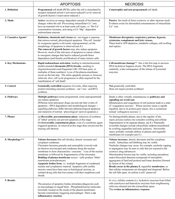
Apoptosis, Cell's essential mechanism of  programmed suicide points to design 5467table1
Table 1. Comparison between apoptosis and necrosis. The data in this table were compiled from twelve separate papers.37–49
 Note that some xenobiotic agents can induce apoptosis at low doses and necrosis at higher doses.
 An injurious agent may activate at least four major ‘pathways’ leading to a loss of membrane integrity (of particular relevance to necrotic death): 1) Membrane phospholipid degradation; 2) Production of amphipathic lipids; 3) Damage to cytoskeleton; 4) Generation of toxic oxygen species and free radicals. 
The contrasting changes in cell volume for apoptosis and necrosis are both osmotic effects. For instance, in the case of necrosis, the swelling of the cytoplasm is probably due to an increased ‘osmotic load’, resulting from the accumulation of waste products of metabolised xenotoxin or of the toxin itself.


Necrosis* can be thought of as the second fundamental ‘mode’ of cellular death and occurs when cells are injured in such a way that the normal apoptotic mechanisms are circumvented. Unlike necrosis (see below), apoptosis is accompanied by characteristic morphological changes that invariably include a loss of the normal membrane asymmetry*, reduced cell volume, chromatin* condensation and fragmentation of the chromosomal DNA (Figure 2). If/when a cell is deleted (‘dies’) by apoptosis, all traces of it are efficiently removed by other ‘dedicated’ cells, with a total absence of inflammation*. This culminating phagocytosis* of the residual nuclear and cytoplasmic components (so-called ‘apoptotic bodies’) by macrophages* is part of the natural cellular economy; indeed, it facilitates the reprocessing and re-utilisation of materials. As we saw earlier, apoptosis actually requires genetic programming and therefore information input36 and often involves protein and RNA* synthesis. In stark contrast, necrosis is chaotic and catastrophic; i.e. a cellular example of increaseddisorder, in perfect accord with the Second Law of Thermodynamics. Whereas apoptosis is absolutely essential to increases in order and complexity of multicellular organisms, necrosis results in the accumulating detritus of large numbers of dead/dying cells, which cause problems for the organism unless it is swiftly dealt with. Necrotic cells are therefore indicative of a world where decay has set in, a consequence of Adam’s sin. Those readers who are interested in a more detailed comparison of apoptosis and necrosis are referred to Table1. While the precise details of apoptotic morphology and molecular biology may vary for different cell types, the same goal of physiological cell deletion is accomplished in each case.

Apoptosis versus mitosis


It is now realised that the processes of apoptosis and mitosis are really ‘two sides of the same coin’. Mitosis, as all biology students are taught, is cell multiplication, leading to tissue growth. Apoptosis is cell deletion, leading to tissue shrinkage locally. The intentional rhyming of apoptosis with mitosis shows that there is normally a balance between cell ‘death’ and cell birth respectively. If apoptosis is a universal phenomenon among eukaryotic*, multicellular organisms, why did it escape the attention of biologists for so long and why is it still absent from most dictionaries and school science curricula? Lockshin offers the following reflections:

‘Mitosis is a relatively short and evanescent event, but once it has occurred there are traces of its passing: a single-celled egg becomes two, four, and eight … . Apoptosis is far more furtive. Cells simply vanish, usually in less than one hour.’50

It is this transient nature of the apoptotic signature that seems to have kept its true relevance hidden from the scientific community until relatively recently. As we shall now consider, apoptosis is literally vital to multicellular life.

1) http://creation.com/apoptosis-cell-death-reveals-creation


Last edited by Admin on Tue Oct 06, 2015 5:07 pm; edited 2 times in total

https://reasonandscience.catsboard.com

Otangelo


Admin

Domains for the occurrence of apoptosis


It is beyond the scope of this paper to elaborate on the various roles of apoptosis in detail, but the following examples (in healthy animals and humans) illustrate how ubiquitous apoptosis is in biology:

[1]Apoptosis is essential during vertebrate embryological development in order to correctly sculpt the multifarious tissues and organs that are forming. The classical example is that of the developing limbs. These initially form as ‘buds’, the digits (e.g. fingers) forming later on, by virtue of apoptosis of the cells in the interdigital areas.51,52

[2]Apoptosis plays a pivotal role during both T-and B-lymphocyte maturation.53 For instance, the elimination of mature T-cell clones in the peripheral blood seems to be a mechanism for establishing tolerance to self antigens.54 Also, apoptosis may be activated during normal B-cell ontogeny*.55,56

[3]The human eye is a classic example of so-called ‘immunologic privilege’ by virtue of the fact that the exposed eye surfaces express the Fas-L(CD95L) molecule. If the eye is attacked by virus, a massive inflammatory response ensues, but no long-term damage to the eye occurs because the Fas-L triggers apoptosis in the infiltrating cells (neutrophils* and T cells), which are all removed in a few hours.57 This is obviously essential to healthy vision!

[4]Apoptosis provides a safe, disposal mechanism for neutrophil granulocytes at inflamed sites. Human neutrophils ingest foreign microbes at sites of injury, but their prolonged survival (once the job is done) would cause chronic tissue damage. It is now thought that they actively generate ROIs (reactive oxygen intermediates) in order to mediate their own speedy demise.58

[5]Apoptosis serves a vital role in controlling the number of germ cells in the testis59 and selectively eliminates cells with high proliferative activity, that acquire irreparable genetic abnormalities; e.g. testicular temperature rise causes more mutations in prospective gametes but heating to higher temperatures results in aspermia*.60 That is, apoptosis is very important in limiting teratogenesis (the production of neonatal abnormalities)

[6]In the ovary, a vast amount of apoptosis occurs in the germ-line, throughout the later stages of pre-natal development and on into post-natal life. One example is that it sets the absolute number of oocytes (eggs) that are available for development and ovulation in adult life.61Figure 2. Schematic diagram of the morphological features of apoptosis and necrosis (adapted and redrawn from103).Click here for larger view.

[7]In non-pregnant women, the cyclic fluctuations of the menstrual cycle hormones determine the cellular fate (proliferation, differentiation, or apoptosis) of both mammary gland epithelium*62 and the uterine endometrium*.63 Menstruation itself is brought about by apoptosis of a specific endometrial cell population. In post-pregnancy women and mammals generally, regular suckling ensures that lactation continues. Conversely, at weaning, the gradual or abrupt cessation of milking of the mammary gland results in its gradual or rapid involution respectively. This involution (leading to milk stasis) is due to a net loss of glandular mammary tissue by apoptosis.64

[8]Human breast milk contains the common protein alpha-lactalbumin which, in its multimeric* form, has been reported to have potent apoptosis-inducing effects in tumour cell lines, but much less effect on a variety of normal cell types.65 I have personally confirmed these results in a series of experiments on fresh lymphocytes from healthy volunteers and from patients with chronic lymphocytic leukaemia.66 The original discovery was serendipitous67 and the precise function of this capability of breast milk is not fully understood. Perhaps the induction of apoptosis in transformed, but not mature, epithelial cells helps direct the normal growth of neonatal mucosal*epithelium and helps prevent neoplasia*.65

[9]In the nematode Caenorhabditis elegans, the morphological development is very precisely governed by which cells divide and which cells undergo apoptosis at specific stages.8,9

[10]Metamorphosis brings about pronounced morphogenetic changes in a short time as organisms change from the larval to adult stages. Apoptosis plays a central role in this process. Metamorphosis is well-known in numerous insect species, as well as certain vertebrate organisms. For instance, metamorphosis involves the complete remodelling of virtually every tissue/organ as tadpoles transform into frogs. Most organs, if they are not completely reabsorbed, at least undergo apoptotic deletion of certain cells.68[/list]

It is not really possible to do justice to the role of apoptosis in each of the above but the interested reader is referred to the references. A recommended recent book that covers some of this material in more detail is When Cells Die. In the preface, the editors make the following, interesting comment:

‘Throughout the book, the clearestconsensus is thatan organism uses cell death in a very positive way—to sculpt its development, to arrange for rapid expansion and subsequent contraction of a cell population in the immune and reproductive systems, and to defend itself by destroying cells that have been infected or attacked’ [emphasis added].69


A biblical perspective on the purpose of apoptosis


It is clear from the foregoing, that apoptosis is an essential physiological mechanism and as such, would have been present in the pre-Fall world. However, as some of the above examples show, some apoptotic pathways are also triggered as a toxicological reaction. Thus, they are vital to an organism’s health (even survival) during exposure to pathogenic* microorganisms or environmental stress. Apoptosis can be viewed as the ultimate mechanism for maintaining phenotypic fidelity* in multicellular organisms.70
From a biblical perspective, the capacity for deployment of these abortive cell-processes (as a ‘stress-response’) was built into the genetic potential of all organisms by our Omniscient Creator. After the Fall (and the resulting curse on all Creation), the role for apoptosis must have diversified substantially. Conditions in the radically altered, post-Flood world would, no doubt, have added to the ‘work-load’ of the originally created apoptotic mechanisms. Obviously, with the passage of time, entropy was increasingly manifest by these originally perfect systems and we see the results today, when defective apoptotic responses sometimes lead to disease (see below).
It is of interest to creationists that failure of normal apoptosis has been proposed as a possible means to facilitate the aging process.70Various experimental studies of the longevity of human lymphocytes have correlated apoptosis with aging.71 Perhaps degeneration of apoptotic control increases with time so that it has a rate-limiting effect on the aging process. This has obvious implications for our thinking about the antediluvian long lifespans. Bergman has recently discussed this, with respect to the telomeres that protect the ends of normal cellular chromosomes.72 These telomeres shorten with each cell division, eventually triggering senescence* and apoptosis. Bergman speculates that:

‘At some point in history human longevity could have changed as a result of some alteration of the telomerase system.’73


When apoptosis goes awry


Rather, apoptotic pathways have all the hallmarks of irreducible complexity and attempting to construct them in a step-wise fashion, whilst maintaining functionality at each step, would be futile in the extreme.
The perfect functioning of an apoptotic mechanism depends on the genetic information being uncorrupted, as it was at Creation. Therefore, any loss of information, such as occurs when a gene that codes for a protein in an apoptotic cascade mutates, will almost certainly have drastic consequences. Indeed, the sensitivity of these mechanisms to the slightest change in the configuration of a single protein component, is a powerful argument against neo-Darwinism. It is truly inconceivable that randomchanges to gene-encoded information for these pathways could ever produce an improvement. Rather, apoptotic pathways have all the hallmarks of irreducible complexity74 and attempting to construct them in a step-wise fashion, whilst maintaining functionality at each step, would be futile in the extreme.
Dysregulation (too much or too little) of apoptosis can cause a wide spectrum of defects:


[1]Cancer. Defects of normal apoptotic processes have been discovered in many forms of cancer.75Normal cells maintain a balance between the rates of mitosis and apoptosis. However, apoptotic failure of a cell that has sustained one or more somatic mutations results in an immortalised ‘cellular anarchist’ (neoplastic* cell). Solid tumours (cancers) or an uncontrolled proliferation of haematopoietic cells (leukaemia and lymphoma) are the inevitable consequence. Since many genes regulate apoptosis, it is no surprise that defective genes have been noted in many cancers (so-called oncogenes), such as p5376 and bcl-2.77,78 Over-expression of Bcl-2 protein, as a result of bcl-2 oncogenesis, confers resistance to the neoplastic cells so that they are much less susceptible to chemotherapeutic drugs and radiotherapy.79[/list]


[2]AIDS. Apoptosis research has thrown light onto the causes of this immune deficiency syndrome. Among other things, it seems that expression of Fas(APO1/CD95), a ‘cell death’ receptor, is enhanced in individuals infected with HIV (human immunodeficiency virus), contributing to/causing an increase in the apoptotic rate of CD4+ T-lymphocytes.80 This is however, somewhat controversial among AIDS researchers.81,82[/list]


[3]Alzheimer’s disease (AD). Individuals with AD suffer premature or excessive neuronal cell loss in the brain during the aging process, together with other pathological effects (the formation of plaques, gliosis* and neurofibrillary tangles*). Compromised mitochondria may release a significant amount of calcium ions into the cytoplasm, so stimulating the caspases and DNases involved in apoptosis.83However, there is some ambiguity regarding the role of caspase-dependent neuronal apoptosis in AD due to contradictory experimental observations and a lack of convincingly apoptotic neurons in AD brains.84 Other AD research indicates that the apoptotic signal may not reach the terminal caspases.85 Thus, while apoptosis certainly seems to be implicated in AD, a causative role is questionable.[/list]


[4]Rheumatoid arthritis (RA). RA is an autoimmune disorder in which the body attacks its own cartilage in the synovial joint linings, leading to inflammation, painful swelling and eventually loss of joint function. It has been shown that chondrocytes (cartilage cells) of articulating bone surfaces are more prone to apoptosis in RA patients.86 This was recently found to be strongly associated with expression of pro-apoptotic proteins such as Fas(APO1/CD95) Fas-L and p53.87 The cause of RA is essentially due to multi-level aberrations that lead to defective apoptosis or hyperapoptosis.88[/list]


[5]Embryonic lethality. Mice are widely used as disease models in medical research. There are many reports in the scientific literature of murine embryos dying due to excessive apoptosis, either because a particular protein is overexpressed, or because it is deficient. For example, embryos die in utero around mid-gestation if they are deficient in cytochrome c89 or the nuclear factor kappaB.90 In both of the latter cases, the authors demonstrate that the protein deficiency renders the mice susceptible to apoptotic signals, mediated by TNF-α/TNFR1 signalling.[/list]


[6]Eye problems. Apoptosis has critical (and contrasting) roles in the various ocular tissues (cornea, lens and retina) and extraocular tissues (e.g. optic nerve) that contribute to vision. We saw earlier that it contributes to immunologic privilege in the healthy eye. It may also initiate healing of eye wounds but malfunctioning apoptotic pathways are also associated with opthalmological disease.91[/list]

In every case, small perturbations of apoptotic mechanisms (often resulting from a single somatic mutation) have debilitating, even lethal effects, belying ideas of genetic gradualism and corroborating Behe’s thesis of ‘irreducible complexity’.74 It is clear that impairment of apoptosis compromises the body’s ability to effectively eliminate damaged or mutated cells, which would affect the organism’s survival if they lived on.

The therapeutic potential of manipulating apoptosis


Leading researchers in this field recently stated that:

‘Programmed cell death and apoptosis are very important aspects of a healthy life, and our access to manipulation of it will have vast consequences in many fields of medicine and agriculture.’92

Apoptosis, Cell's essential mechanism of  programmed suicide points to design 5467fig3
Figure 3. Photomicrographs of cells (B-lymphocytes) from patients with Mantle cell lymphoma (MCL; A) and Chronic lymphocytic leukaemia (CLL; B). The cells were isolated from peripheral blood specimens (from patients with these neoplasms) using lymphocyte separation medium, cytocentrifuged onto glass microscope slides, air dried, fixed in methanol and stained with a Romanowsky (red/blue) stain.95 The photomicrographs are both to the same scale. The four smaller objects in A are erythrocytes (red blood cells). The immunophenotype (pattern of expression of cell proteins) of MCL cells is very similar to, but distinct from, that of CLL cells96 and the two malignancies have very different prognoses. However, both MCL and CLL cells only rarely express the ‘death receptor’ protein, Fas(APO1/CD95), in contrast to most other types of non-Hodgkin’s lymphoma.13
Click here for larger view.


Restoration of the apoptotic response would be beneficial in many cases. This is a particularly fruitful avenue of research with respect to treating many cancers. Clearly, the very existence of cancer is testimony to the ongoing genetic degeneration that started with the Edenic curse.93 In cancer and haematological* neoplasms, restoration of the apoptotic response to therapeutics would help to solve the longstanding problem of multi-drug resistance of malignant cells.94 For example, in healthy people, Fas(APO-1/CD95)-mediated apoptosis is thought to be responsible for the removal of anergic*, autoreactive B-lymphocytes from the peripheral blood circulation; i.e. part of a normally functioning immune system. Once activated, mature B-cells acquire surface Fas-expression and concomitant sensitivity to apoptosis, induced by Fas-ligand on Th1 (T-helper) cells. However, malignant cells from patients with B-cell chronic lymphocytic leukaemia or mantle cell lymphoma (see Figure 3) usually lack surface Fas expression, rendering them resistant to Fas-mediated apoptosis. I recently co-authored the report of a study, in which we attempted to re-sensitize such neoplastic lymphocytes to apoptosis using the cytokine, interleukin‑2.97

Inhibition of inappropriate apoptosis could be beneficial in other cases. This is particularly the case, where cellular degeneration results from a disease process that causes too much apoptosis. Approaches aimed at reducing apoptosis are an active research area for scientists studying viral pathogenesis (e.g. HIV infection),80 neuronal degeneration (e.g. Alzheimer’s disease)83 and rheumatoid arthritis,87,88 for example.

Conclusions: apoptosis from a creation/evolution perspective


Faced with the growing realisation that apoptosis is one of the fundamentally important biological processes, some evolutionists are beginning to grapple with such crucial questions as how it originated and how it was selected for, according to Darwinian, selectionist dogma. Comments such as the following are predictable:

‘Many of the genes that control apoptosis are conserved throughout evolution from mammals to nematodes, flies, and viruses.’98

Similarly,

‘The preservation of a process throughout evolution indicates that the process is fundamental and too important to be modified.’99

Clearly however, this type of remark merely begs the question of how apoptosis is supposed to have evolved in the first place.100 Another paper by this author reviews the few evolutionists’ attempts to explain apoptotic origins and argues that the very existence of apoptosis actually falsifies evolution.101 Contrary to the impression given, the great diversity exhibited by apoptotic pathways, notwithstanding fundamental similarities, is a hallmark of Creative Design.102 Apoptosis is widely distributed among disparate species and tissues. Also, whereas a single mechanism of action would be predicted by reductionists, we observe multiple effectors.
So, just how far back did apoptosis originate in an evolutionary world? According to the evolutionary paradigm, injurious agents (e.g. irradiation, cosmic radiation, free radicals, hypoxia/hyperoxia*, environmental chemicals) and hyperthermic conditions have characterised the early Earth’s history at one time or another. Thus, a sophisticated apoptotic response would have been especially necessary at the very stage in Earth’s history when multicellular, eukaryotic cells are supposed to have evolved (during the late Precambrian)! Even today, apoptosis serves an essential role in terms of ‘cellular altruism’. It helps to ensure that an organism’s genetic integrity is not compromised, by removing some somatic cells that have sustained irreparable, genetic mutations. Crucially, apoptosis also helps to maintain a species’genetic integrity, by eliminating aberrant germ cells that would otherwise carry intact but faulty genes into the next generation. Thus, on the hypothetical early Earth of the evolutionist, fully-functional apoptosis would have been much more important than it is today!
In summary, a creationist perspective is cognizant that apoptosis:


    [1]is as vital and ubiquitous to life as mitosis;


    [2]is programmed and tightly regulated—speaking of a Programmer who put the information into cells originally;


    [3]is irreducibly complex—it only takes one part of the mechanism to malfunction and the whole process is badly affected, especially illustrated by cancer;


    [4]shouts Design (Romans 1:20);


    [5]is a Created mechanism—part of the ‘very good’ declaration of God (Genesis 1:31).


Acknowledgements


I am sincerely grateful to the Lord for planting the idea for this article in the first place, to Carl Wieland for his encouragement, and to the two reviewers, whose comments have helped me to enhance the paper.


anergicin a state of being unable to react to antigenic (immunogenic, allergenic) substances.
apoptosisan active process (requiring energy) involving the programmed deletion of scattered cells by fragmentation into membrane-bound bodies which are phagocytosed by other cells. No inflammatory response occurs.
aspermiaa total absence of spermatozoa (sperm cells) in an ejaculate.
caspasesa group of enzymes which are particularly involved in the transduction of signals for apoptosis.
chromatinthe genetic material of the nucleus, made up of DNA and protein. During mitosis, the chromatin condenses into chromosomes.
clonalselectionthe specific recognition of foreign protein (antigen) by a small proportion of the body’s circulating lymphocytes, followed by the rapid expansion of this lymphocyte clone for specific antibody production and immunologic memory.
differentiation (cells/tissues)the development of morphology and/or functions that were not part of the original cells/tissues; occurs to bring about greater specialisation.
dysregulationdys is a prefix meaning mis- or un-, so this refers to abnormal regulation.
embryogenesisthe formation of the characteristic configuration of an embryo’s body.
endometriumthe mucous membrane that forms the uterus lining, consisting of columnar epithelium and glands; its structure and thickness vary in accordance with the menstrual cycle.
epitheliumthe cellular layer that covers all free body surfaces, e.g. cutaneous (skin) and mucous layers.
erythrocytea red blood cell; lacking a nucleus and specialised for oxygen transport.
eukaryotic/eukaryoteliterally means those organisms with ‘good nuclei’ in their cells (i.e. all animals, plants and fungi); the nucleus is membrane-bound and its DNA is associated with proteins to form chromosomes. The cytoplasm also contains many organelles (e.g. mitochondria) that are absent from prokaryotes (e.g. bacteria and ‘blue-green algae’).
gliosisan overgrowth of the astrocytes (star-shaped neural cells) in an area of brain or spinal cord damage.
haematologicalpertaining to the blood and blood-forming tissues.
homologous regionssimilar/equivalent amino-acid sequence; an evolutionist would interpret this as evidence of their common molecular origin in a putative ancestor.
hypoxia/hyperoxiarespectively, too little oxygen or too much oxygen; i.e. oxygen tension (a key physical property for living organisms) changes with pressures that differ from 1 atmosphere.
inflammationessentially, a pathological process that results from some sort of injury, usually involving redness, heat, swelling and pain.
macrophageslarge, long-lived, phagocytic cells that are a major part of the body’s immune defences; their morphology varies a great deal, but all are derived from monocytic stem cells of the bone marrow.
membrane asymmetrythe phospholipid (‘fatty’) membranes of living cells have many associated protein molecules, some spanning the entire membrane, some located towards the outside of the cell and some inside; basically a non-symmetrical arrangement.
mitochondriasubcellular organelles; the power houses found in all eukaryotic cells that provide the energy (in the form of ATP) to drive all cellular reactions.
mucosalpertaining to a mucous membrane (tissue layer); various types of mucosa line the body’s tubular cavities, such as the stomach, intestines, trachea, uterus etc. They are composed of epithelium and mucous glands.
multimerica grouped arrangement of several identical molecules, e.g. a trimer consists of three molecules.
necrosiscell death that results from injury or a pathological condition; it is a passive, chaotic process.
neoplasia/neoplastica pathological process that results in neoplastic tissue; i.e. abnormal cells that proliferate uncontrollably and generally more rapidly.
neurofibrillarytanglesthe accumulations of disorganized filamentous tissue between nerve cells; patients with Alzheimer’s disease exhibit these in the hippocampus and cerebral cortex (brain regions).
neutrophilsa class of granulocytes, mature white blood cells with a lobed nucleus and a granular cytoplasm; so-called because their nuclear material does not show a particular affinity for either the acidic or basic stains that are commonly used in haematological laboratories.
ontogenythe history of an organism’s development.
pathogenichaving the capacity to cause disease.
phagocytosisliterally means ‘cell-eating’; phagocytes are cells which ingest (then digest) foreign particles, bacteria, necrotic tissue, etc.
phenotypic fidelitydescribes how faithfully the observable characteristics (manifestation of the genetic makeup) of an organism are conserved.
pluripotentstem cellsnon-specialised, primordial cells that can differentiate into a wide variety of cell types.
RNAabbreviation for ribonucleic acid, a class of single-stranded molecules involved in protein synthesis.
senescencea state of being old, in which normal function is declining as death approaches.
trimerisationthe process of forming a substance that is composed of three molecules of a monomer.

http://creation.com/apoptosis-cell-death-reveals-creation

https://reasonandscience.catsboard.com

Otangelo


Admin

On the origin, evolution, and nature of programmed cell death: a timeline of four billion years 1

Programmed cell death is a genetically regulated process of cell suicide that is central to the development, homeostasis and integrity of multicellular organisms. Conversely, the dysregulation of mechanisms controlling cell suicide plays a role in the pathogenesis of a wide range of diseases. While great progress has been achieved in the unveiling of the molecular mechanisms of programmed cell death, a new level of complexity, with important therapeutic implications, has begun to emerge, suggesting (i) that several different self-destruction pathways may exist and operate in parallel in our cells, and (ii) that molecular effectors of cell suicide may also perform other functions unrelated to cell death induction and crucial to cell survival. In this review, I will argue that this new level of complexity, implying that there may be no such thing as a 'bona fide' genetic death program in our cells, might be better understood when considered in an evolutionary context. And a new view of the regulated cell suicide pathways emerges when one attempts to ask the question of when and how they may have become selected during evolution, at the level of ancestral single-celled organisms.


https://reasonandscience.catsboard.com

Otangelo


Admin

The apoptosome: signalling platform of cell death

Recent work on the initial switches that trigger cell death has revealed surprising inventions of nature that ensure the ordered suicide of a cell that has been selected for demise. Particularly intriguing is how a signal — the release of cytochrome c from the mitochondria — is translated into the activation of the death cascade, which leads to a point of no return. Now there is new understanding of how this crucial process is delicately handled by a cytosolic signalling platform known as the apoptosome. The formation of the apoptosome and the activation of its effector, caspase-9, reveals a sophisticated mechanism that might be more common than was initially thought. 1

Cell-signalling pathways convert a stimulus into cellular action, and they generally have an initiation phase and an effector phase. The initiation phase usually transmits information from a ligand, through interaction with its receptor, to the first biochemical event, resulting in propagation of the pathway. The effector phase executes the biochemical events that result in the desired (often complex) cellular response. In contrast to signalling pathways that use phosphorylation, glycosylation, ubiquitylation or acetylation to alter protein properties, those that require proteolysis are irreversible, because cells are not known to possess a mechanism to re-ligate cut peptide bonds. 

Apoptosis is such a pathway; indeed, one can appreciate that once a cell has made the decision to commit suicide, there is not much point in going back. Apoptosis pathways in mammals can have one of two initiation phases, and which pathway is selected depends on the nature of the death signal to be integrated

Apoptosis, Cell's essential mechanism of  programmed suicide points to design Apopto11

The proteolytic caspase cascade in the initiation and execution of apoptosis. 
Both the intrinsic and extrinsic pathways use related principles in sensing an apoptotic signal and executing apoptosis. In the intrinsic pathway, an apoptotic stimulus in the cell leads to the assembly of a signalling platform, the apoptosome, which activates the initiator caspase-9. An extrinsic apoptotic signal, by contrast, is mediated by binding of an extracellular ligand to a transmembrane receptor, leading to the formation of the death-inducing signalling complex (DISC), which is capable of activating the initiator caspase-8. Once activated, either caspase-9 or caspase-8 cleaves executioner caspase-3 and caspase-7, which represents the execution level of the caspase cascade and leads to apoptosis of the doomed cell. Negative regulators of the caspase cascade can be found on both levels. Whereas FLIP blocks the activation of the initiator caspase-8 in the DISC, XIAP can block both the initiation phase, by inhibiting caspase-9, and the execution phase, by blocking caspase-3 and caspase-7, of the cascade.


Apoptosis is, minimally, a twostep proteolytic pathway, and its objective is to activate effector (also known as executioner) proteases that cleave a number of cellular proteins to drive forward the biochemical events that culminate in death and the dismantling of the cell. The proteases that transmit the apoptotic pathway are members of the caspase family.
The name caspase is derived from Cys-dependent Aspspecific protease1, which signifies that the catalytic activity is governed by a conserved Cys side chain of the protease and by a stringent specificity for cleaving after Asp residues in substrates, which is very unusual for cellular proteases

Proteases likely arose at the earliest stages as destructive enzymes necessary for protein catabolism and the generation of amino acids in primitive organisms. 2 Proteases act as sharp scissors and catalyze highly specific reactions of proteolytic processing, producing new protein products. They are relevant in the control of multiple biological processes in all living organisms. Proteases regulate the fate, localization, and activity of many proteins, modulate protein-protein interactions, create new bioactive molecules, contribute to the processing of cellular information, and generate, transduce, and amplify molecular signals.

As a direct result of these multiple actions, proteases influence DNA replication and transcription, cell proliferation and differentiation, tissue morphogenesis and remodeling, heat shock and unfolded protein responses, angiogenesis, neurogenesis, ovulation, fertilization, wound repair, stem cell mobilization, hemostasis, blood coagulation, inflammation, immunity, autophagy, senescence, necrosis, and apoptosis.

Consistent with these essential roles of proteases in cell behavior and survival and death of all organisms, alterations in proteolytic systems underlie multiple pathological conditions such as cancer, neurodegenerative disorders, and inflammatory and cardiovascular diseases.

Proteases also play key roles in plants and contribute to the processing, maturation, or destruction of specific sets of proteins in response to developmental cues or to variations in environmental conditions.


It is important to understand that caspases are signalling proteases, which are intended for specific protein cleavage and not for protein degradation. They make a limited number of cuts in cellular proteins (usually only one or two) and they therefore do not destroy protein structure, but instead they modify it. The caspase cascade itself perhaps includes the best example of this specific cleavage: the proteolytic action of the initiator caspases on the latent forms of the effector caspases.

Proteolytic pathways (such as blood coagulation, fibrinolysis, gastru lation and apoptosis) proceed by the subsequent action of one protease on the latent form (zymogen) of the next one in line. The objective of these sequential actions either amplifies the original signal (in a cascade-like manner) or allows for additional regulatory points in the pathway.
But the first event in a proteolytic pathway, the actual sensing of the signal, cannot involve proteolysis because there is no protease active at the initiation point of a pathway. Therefore, the generation of the first proteolytic signal in a pathway is a crucial point. This review details the accumulated structural and biochemical evidence that defines the origin of the first proteolytic signal of the intrinsic apoptosis pathway — the activation of the initiator caspase, caspase-9, by the cytosolic signalling platform known as the apoptosome.  This is an elegant mechanism of how apical caspasesignalling platforms  functioning as soluble counterparts of classic transmembrane receptors.

Death: membrane versus soluble receptors
The extrinsic pathway of apoptosis is triggered by extracellular ligands, such as FasL and TRAIL, that bind in a conventional manner to the extracellular domains of transmembrane receptors. The death signal is transmitted to the cytosol by receptor clustering and by recruitment of the initiator caspases of the extrinsic pathway, caspase-8 and caspase-10

Apoptosis, Cell's essential mechanism of  programmed suicide points to design Apopto12

Shape and structure of the apoptosome. 
Oligomeric models of the apoptosome and a close up showing one single unit in detail. The domain organization is colour coded. 
a |  It shows the heptameric wheel-like arrangement of Apaf-1 molecules. Two rings at the centre of the wheel build up the monomer: an inner caspase-recruitment domain (CARD) ring surrounded by an outer ring, built up of the ATPase and winged-helix domains of Apaf-1. In this model, the winged helix ‘bridges’ two neighbouring ATPase domains to build the oligomer — a novel mode of oligomerization compared with other AAA+ ATPases. 
b | In the AAA+-like model, a classic AAA+-type stacking has been proposed31 in which the ATPase domains solely form the oligomer by binding back to back onto each other. In this model, the CARD domains form a looser ring and sit on top of the nucleotide-binding and oligomerization domains (NODs, also known as NB-ARCs) in a crown-like manner. 
c | For comparison, the crystal structure of a heptameric AAA+-ATPase domain is shown. The heptamer formed by the ATPase domain of the bacterial AAA+ protein NtrC1 is shown in surface rendering. The α/β- and helical domains of the ATPase regions are shown in the same colour coding used for Apaf-1.

1. https://sci-hub.tw/https://www.nature.com/articles/nrm2153
2. https://www.ncbi.nlm.nih.gov/pmc/articles/PMC2576539/

https://reasonandscience.catsboard.com

Otangelo


Admin

The individual cell commits suicide to aid the larger organism in killing off dangerous microbes that have created a home in the cell. 60 billion human cells die each day. Apoptosis is a form of death that is pre-programmed and in taking apart the cell, it doesn’t create inflammation and a scar as other types of cell death would. The cell shrinks, including the nucleus, where DNA is broken into pieces and the membrane forms blebs. The enzymes that perform these functions are caspases, which cut more than 600 different molecules into pieces including other caspase enzymes.

The apoptosis process starts when a large multiprotein complex is assembled—the apoptosome—triggered by a protein from the mitochondria (cytochrome c) and one from the cell (APAF1 or apoptotic protease-activating protein). Cytochrome, normally, is part of the respiratory cycle and moves electrons. When it is sent into the cytoplasm it joins to trigger apoptosis. There are, in fact, several other molecules from the mitochondria that can do this and these all come through pores in the mitochondria membrane. The well-known protein BCL-2 that is linked with protection from cancer controls these pores. Many factors can trigger this process including damage to DNA with defective repair, toxic medications, radiation and other signals. Microbes have many toxins that affect this process in both directions. ­

https://jonlieffmd.com/blog/intelligent-microbes-attack-organelles

https://reasonandscience.catsboard.com

Sponsored content


Back to top  Message [Page 1 of 1]

Permissions in this forum:
You cannot reply to topics in this forum