ElShamah - Reason & Science: Defending ID and the Christian Worldview
Would you like to react to this message? Create an account in a few clicks or log in to continue.
ElShamah - Reason & Science: Defending ID and the Christian Worldview

Otangelo Grasso: This is my library, where I collect information and present arguments developed by myself that lead, in my view, to the Christian faith, creationism, and Intelligent Design as the best explanation for the origin of the physical world.


You are not connected. Please login or register

DNA replication of prokaryotes

Go to page : 1, 2, 3  Next

Go down  Message [Page 1 of 3]

16DNA replication of prokaryotes - Page 2 Empty The Hexameric DnaB Helicase Sun Nov 15, 2015 5:27 pm

Otangelo


Admin

The Hexameric DnaB Helicase


DNA HELICASES: UNWINDING OF THE DOUBLE HELIX

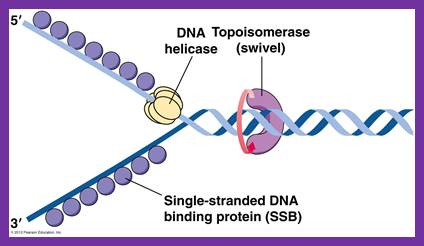
DNA helicase unwinds double-stranded DNA for copying


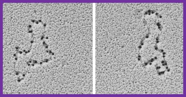
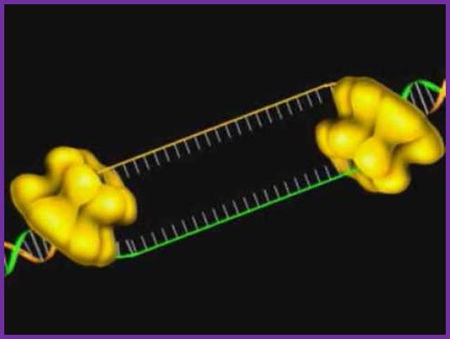
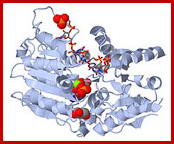
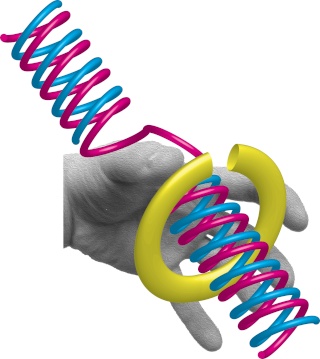
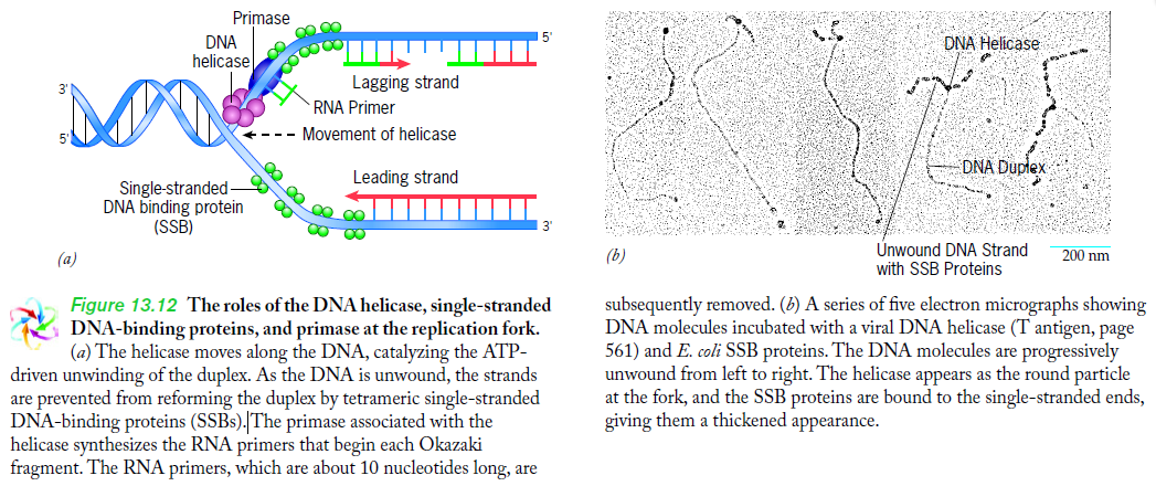
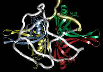
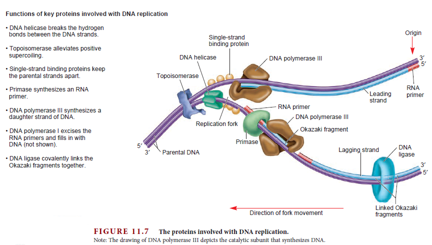
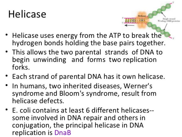
Having now considered the key enzymes involved in the actual synthesis of DNA during replication, let us now turn to an enzyme that has an important function before synthesis can actually occur – the unwinding of the double-stranded helix of the parent DNA. When base-paired in the double-stranded helical DNA molecule, nucleotides are inaccessible to polymerases, making copying impossible. Both primase and polymerases need access to single-stranded DNA. To achieve this, the internal base-pairing must be broken and the helix unwound. The first step in this unwinding process is the initial opening of the helix, a step performed by the initiator protein at the origin of replication. Once opened, the unwinding of the double helix to expose single-stranded DNA for copying can begin. This unwinding process is catalyzed by an enzyme called DNA helicase.  The cell also needs to open the helix for DNA repair and recombination, and there are a unique set of DNA helicases for this particular case of DNA unwinding. Helicases open the double-stranded DNA and then travel with the replication fork, continuously unwinding the DNA to provide a template for the polymerase to copy. The helicases involved in replication in both bacteria and eukaryotes are composed of six subunits that form a ring structure that surrounds one strand of the DNA. The structure of the replicative DNA helicase from papillomavirusprovides insights into the mechanism of helicase action. This viral helicase is a hexamer of one protein, E1. In the co-crystal of protein and DNA, a single strand of DNA fits into the center of the channel formed by the hexamer ring, as depicted in Figure below.

DNA replication of prokaryotes - Page 2 Helica10

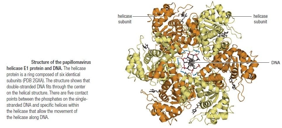
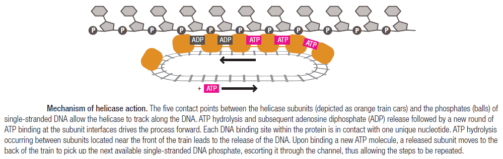
The other strand of the DNA is displaced by the helicase and bound by single-stranded binding proteins. Each monomer possesses a series of DNAbinding loops such that, when the six monomers come together to form the hexamer, the DNA binding loops form a spiral staircase on the inside of the channel which binds the DNA, and move along it one nucleotide at a time. Each movement of the DNA binding loop within each monomer requires adenosine triphosphate (ATP) hydrolysis; thus to move six nucleotides requires six ATP molecules, making DNA unwinding an energy costly process. 

DNA replication of prokaryotes - Page 2 Helica10

In E. coli, the helicase associated with DNA replication is known as DnaB and is a hexamer of six identical subunits. In eukaryotes and archaea, the replicative helicase is called MCM and is a complex of six different proteins, Mcm2–7, which assemble to form a ring. The eukaryotic replicative helicase is conserved throughout evolution and requires accessory factors for helicase activity.


DNA replication of prokaryotes - Page 2 Helica11

Functions of DnaB 2

This is a fascinating video which shows how DnaB helicase functions: 


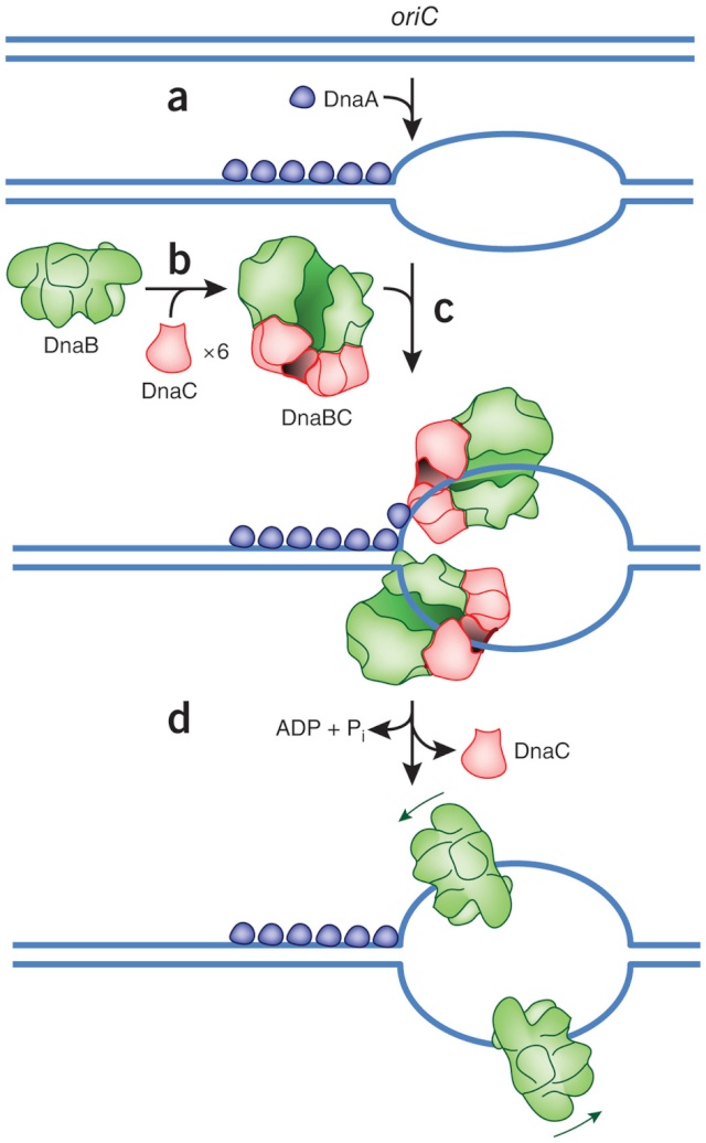
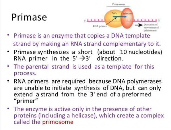
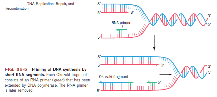
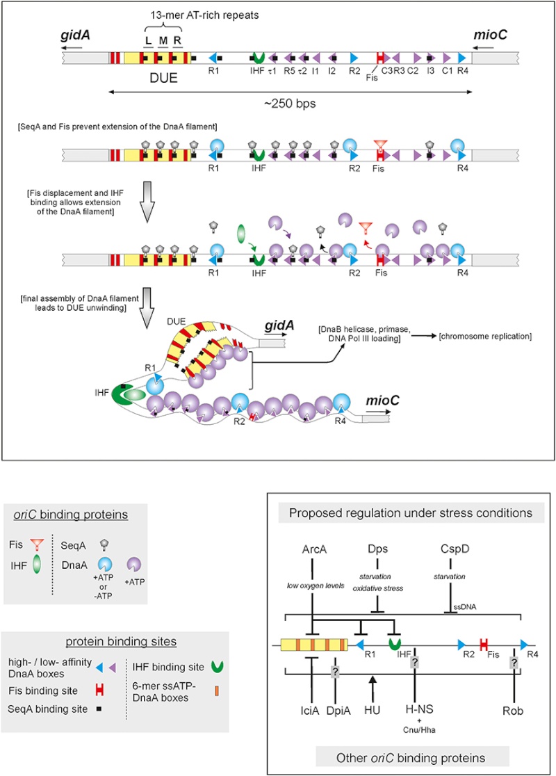
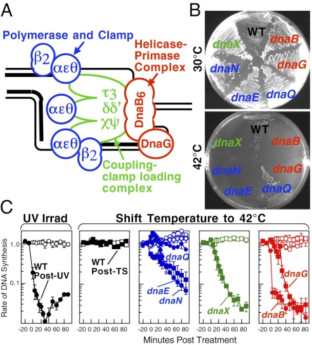
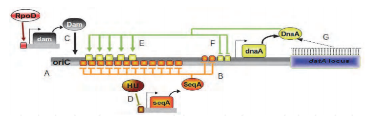
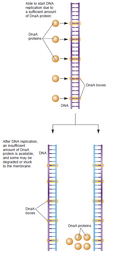
DNA replication begins at the oriC. This is the site where proper replication occurs.  The site is 245 base pairs long.  This origin contains 4 nine-mers which are the binding sites for a protein called Dna A.  The Dna A protein controls the binding of Dna B to the origin.  This binding occurs when Dna A initiates the melting of 3 13-mers on the other end of the origin site.  This melting of the base pairs causes the Dna B to bind to the origin. Although there is still a problem with Dna B finding the site.  So Dna C binds to Dna B to help deliver the complex to the open complex or DNA melting region of origin.  After Dna B binds to the 13-mer melted region, this stimulates the binding of Dna G (primase).  Thus completing the primosome.  The Tao subunit of the DNA polymerase stabilizes and stimulates the Dna B helicase.  As the Dna B is stabilized, the protein begins to unwind the dsDNA as it hydrolyzes ATP.  The hydrolyzed ATP causes a slight structure change which helps in the ability to connect with the correct sites and move the ssDNA in one direction.  Once replication has started there are two main functions the Dna B helicase serves

1.   Consistently needs to bind for priming on the lagging strand because the lagging strand produces Okazaki fragments

2.   Dna B unwinds the parental DNA to give the pol III templates for the leading and lagging strand, this occurs in the direction of 5’ to 3’.  This is the same direction as the replication fork is moving


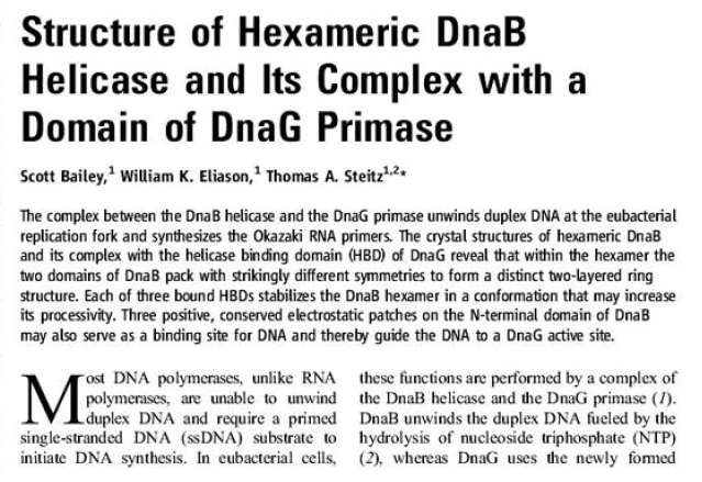
Structure of Hexameric DnaB Helicase and Its Complex with a Domain of DnaG Primase 1

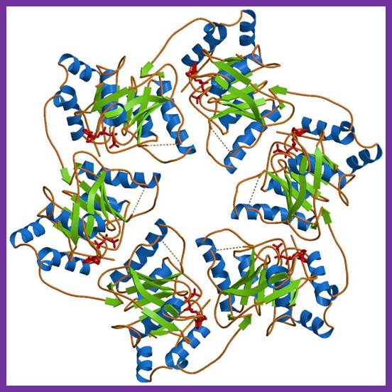
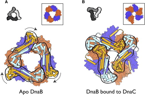
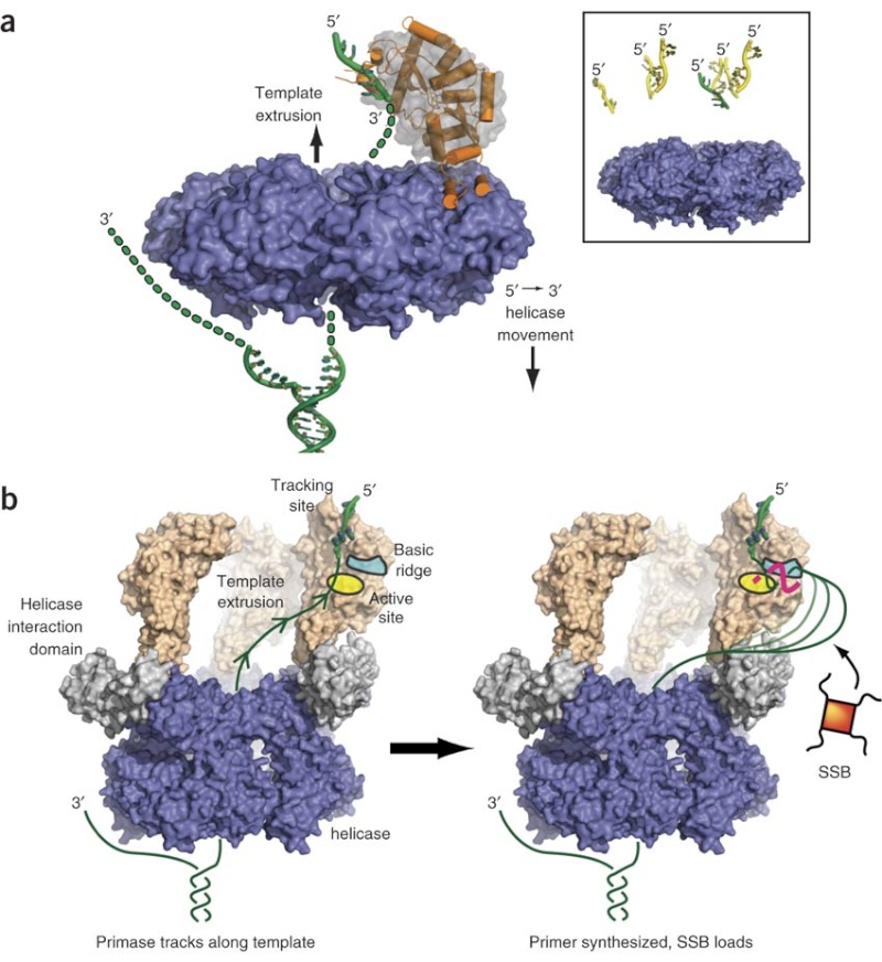
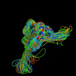
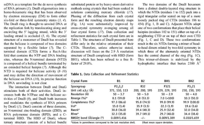
The complex between the DnaB helicase and the DnaG primase unwinds duplex DNA at the eubacterial replication fork and synthesizes the Okazaki RNA primers. The crystal structures of hexameric DnaB and its complex with the helicase binding domain (HBD) of DnaG reveal that within the hexamer the two domains of DnaB pack with strikingly different symmetries to form a distinct two-layered ring structure. Each of three bound HBDs stabilizes the DnaB hexamer in a conformation that may increase its processivity. Three positive, conserved electrostatic patches on the N-terminal domain of DnaB may also serve as a binding site for DNA and thereby guide the DNA to a DnaG active site.

DNA replication of prokaryotes - Page 2 Hexame10

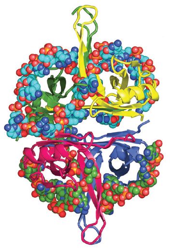
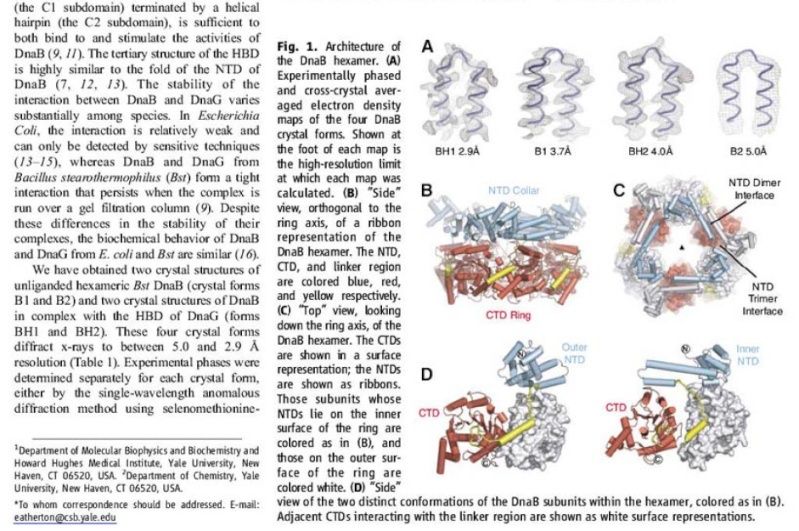
Architecture of the DnaB hexamer. 
(A) Experimentally phased and cross-crystal averaged electron density maps of the four DnaB crystal forms. Shown at the foot of each map is the high-resolution limit at which each map was calculated. 
(B) “Side” view, orthogonal to the ring axis, of a ribbon representation of the DnaB hexamer. The NTD, CTD, and linker region are colored blue, red, and yellow respectively. 
(C) “Top” view, looking down the ring axis, of the DnaB hexamer. The CTDs are shown in a surface representation; the NTDs are shown as ribbons. Those subunits whose NTDs lie on the inner surface of the ring are colored as in (B), and those on the outer surface of the ring are colored white. 
(D) “Side” view of the two distinct conformations of the DnaB subunits within the hexamer, colored as in (B). Adjacent CTDs interacting with the linker region are shown as white surface representations.

DNA replication of prokaryotes - Page 2 Hexame11

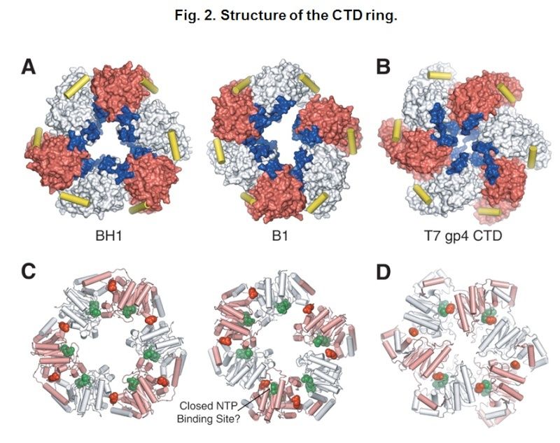
Structure of the CTD ring. 
(A) Surface representation of the CTD rings of crystal forms BH1 (left) and B1 (right). Alternate subunits are colored white and red. The predicted DNA binding loops are colored blue, and the linker helices are shown as yellow cylinders. 
(B) The structure of the T7 gp4 helicase domain (23), displayed as in panel (A). 
(C) Ribbon representations of the CTD rings of crystal forms BH1 (left) and B1 (right). Alternate subunits are colored white and pink. NTP modeled at the six potential NTP binding sites of DnaB (22) are shown as green spheres; the Arginine fingers (Arg420) are displayed as red spheres. 
(D) The structure of the T7 gp4 hexamer with four NTD binding sites occupied, displayed as in (C).


DNA replication of prokaryotes - Page 2 Hexame12

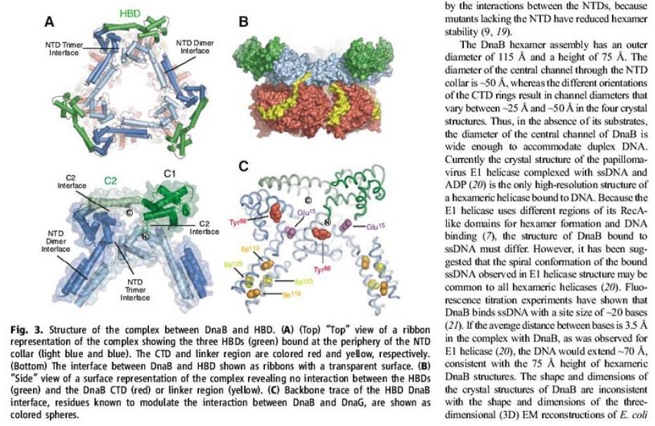
Structure of the complex between DnaB and HBD. 
(A) (Top) “Top” view of a ribbon representation of the complex showing the three HBDs (green) bound at the periphery of the NTD collar (light blue and blue). The CTD and linker region are colored red and yellow, respectively. (Bottom) The interface between DnaB and HBD shown as ribbons with a transparent surface. 
(B) “Side” view of a surface representation of the complex revealing no interaction between the HBDs (green) and the DnaB CTD (red) or linker region (yellow). [/ltr]
(C) Backbone trace of the HBD DnaB interface, residues known to modulate the interaction between DnaB and DnaG, are shown as colored spheres.
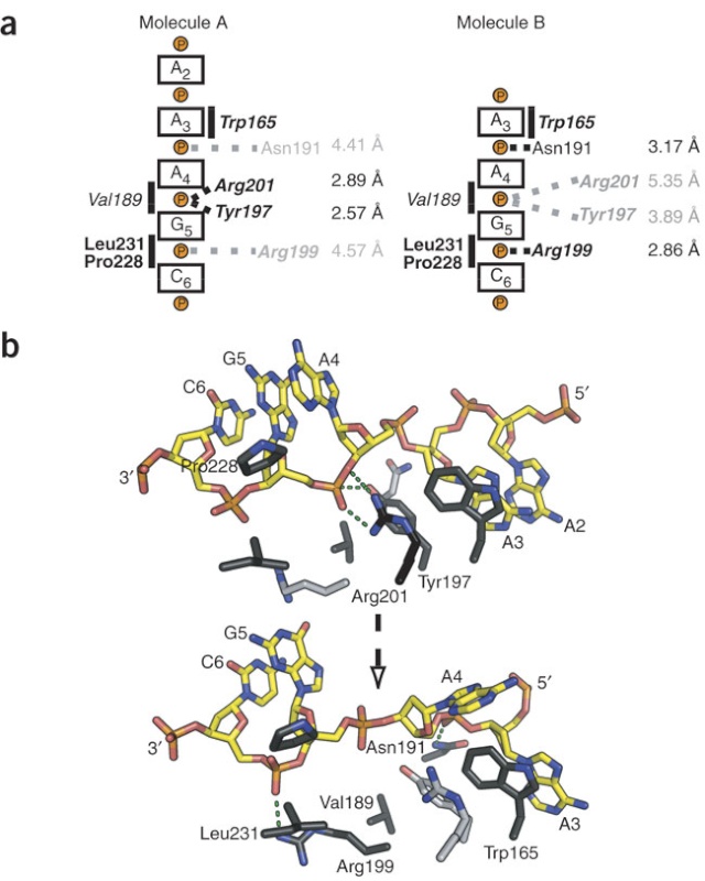

DNA replication of prokaryotes - Page 2 Hexame13


DNA interactions. 
(A) (Left) “Top” view of a surface representation of the NTD collar colored blue for positive and red for negative electrostatic potentials. An asterisk highlights the proposed ssDNA binding sites. (Right) A detailed “side” view of the proposed ssDNA binding site boxed in (A). 
(B) Speculative model of DnaB complexed with DnaG and replication fork DNA. The proteins are shown in a surface representation (DnaB NTD, light blue; DnaB CTD, red; DnaG HBD, green; DnaG RPD, pink; and DnaG ZBD, orange). The modeled DNA is shown as white- and wheat-colored spheres; the RNA primer is shown in dark blue.


DNA replication of prokaryotes - Page 2 Swerwe10


DNA replication of prokaryotes - Page 2 Ertert10


DNA replication of prokaryotes - Page 2 Ertert10


DNA replication of prokaryotes - Page 2 Ertert11

DNA replication of prokaryotes - Page 2 Ertert12

DNA replication of prokaryotes - Page 2 Ertert13

DNA replication of prokaryotes - Page 2 Ertert14

DNA replication of prokaryotes - Page 2 Ertert15

DNA replication of prokaryotes - Page 2 Aasdas10

1) http://www.sciencemag.org/content/318/5849/459.abstract
2) http://www.biochem.umd.edu/biochem/kahn/molmachines/replication/dna_b_protein.htm


Last edited by Admin on Wed Nov 25, 2015 8:51 am; edited 8 times in total

https://reasonandscience.catsboard.com

17DNA replication of prokaryotes - Page 2 Empty Re: DNA replication of prokaryotes Mon Nov 16, 2015 12:23 pm

Otangelo


Admin

Dna-B:


The protein, to begin with it is a monomer, but with the binding of Dna-C in one to one manner, subunits start assembling into hexamers to form of a ring. Then with the assistance of Dna-C, helicase ring opens and loads on to the strand, at the joint region of the fork. This assembles only on lagging strand, which is identified by the strand orientation from 5’ towards 3’ direction.  Two such helicases load, one at each fork joints.


DNA replication of prokaryotes - Page 2 Image007


https://www.biochem.wisc.edu
DNA replication of prokaryotes - Page 2 Image008
SSBs bound to single replicating DNA strands; http://www.mun.ca/

DNA replication of prokaryotes - Page 2 Image009


E.coli DNA-B subunits in the form a Hexameric ring

Once the Dna-B ring is formed Dna-C dissociates.  The Dna-B complex is a motor protein and acts as DNA dependent ATPase and using the energy it drives into the fork and unwinds the DNA progressively like unzipping the helical DNA.  Its direction of movement is from 5’ to 3’ on the lagging strand.  There is another protein called Rep A, which as monomer also binds to fork region but to the leading strand and moves in 3’ to 5’ direction.  However involvement of this protein in E.coli DNA replication is not substantiated.

DNA replication of prokaryotes - Page 2 Image010


Bacterial 3’ to 5’ DNA helicase; http://ww2.d155.org/
Prokaryotic Helicases:


Helicase II (Uvr-D)


DNA repair
Uvr AB complex


DNA repair
Helicase-IV


DNA repair
Rec-BCD


Recombination
T4 Dda?


Displaces SsBs
Helicase-I (encoded by F-plasmids)


Involved in DNA transfer during conjugation
Helicase-III


Unknown function


On RNA transcript, performs transcriptional termination
Helicase DnaB
hexamer
5’>-3’
Fork opening

Monomer
3’-5>’
Fork opening
DNA replication of prokaryotes - Page 2 Image011


Helicase in motion unwinding ds DNA; http://iespoetaclaudio.centros.educa.jcyl.es/


DNA replication of prokaryotes - Page 2 Image012


The unwinding of double stranded DNA using Dna B helicase. Dna G is the primase. This picture was gathered from“ http://chem-mgriep2.unl.edu/replic/Helicase.html


Last edited by Admin on Sun Nov 22, 2015 9:06 pm; edited 15 times in total

https://reasonandscience.catsboard.com

18DNA replication of prokaryotes - Page 2 Empty Scientists Watch Motors Unwind DNA Mon Nov 16, 2015 12:24 pm

Otangelo


Admin

Scientists Watch Motors Unwind DNA   1

Andrew Taylor and Gerald Smith from Fred Hutchinson Cancer Research Center (Seattle, WA) announced in Nature June 19 that “RecBCD enzyme is a DNA helicase with fast and slow motors of opposite polarity.”  In the same issue, Mark S. Dillingham, Maria Spies and Stephen C. Kowalczykowski of U.C. Davis came to a similar conclusion.  Working independently, these teams watched an important molecular motor in action and determined that it is two motors in one, with a slow motor and fast motor working side by side on the same track.  How can that be, and why?

    RecBCD helicase is the molecular machine that travels along a DNA double helix, unwinds it, and separates the strands so that the translation machinery can get to it.  This combination enzyme (RecB + RecC + RecD) is a member of a superfamily of helicases, or enzymes able to unwind and separate DNA.  Simpler helicases separate the two DNA strands into a Y-like tail, but RecBCD has the unusual property of creating a loose tail on the RecD side and a loop and a short tail on the RecB side (RecC, not a motor, appears to help RecB in its action).  Combined, RecBCD is among the fastest of helicases: it can cover 370 base pairs per second, according to Taylor and Smith, or up to 1000 base pairs per second, according to Kowalczykowski et al.
  Both the RecB and RecD motors can travel along DNA separately, but are polar opposites: one moves along one strand, one along the other.  Of the two, RecD is the speed demon; RecBC only moves 20% as fast.  The motors are not nearly as fast or stable acting alone.  Separately, they fall off the track after 50 base pairs, but together, can cover 400-600 times as much ground: 20,000 (Taylor and Smith) or 30,000 (Kowalczykowski) at full speed.
    So why two engines in this race car?  Taylor and Smith suggest that it adds stability; a motor is less likely to fall off the DNA track when combined with another, but why the speed difference?  This will take more study.  All they can conclude is, “This asymmetric feature might impart RecBCD enzyme’s asymmetry in other aspects of its promotion of genetic recombination.”


We’re going to stick our neck out and offer a hypothesis.  First of all, it is apparent from the speed and processivity (ability to process lots of letters without failure) that RecBCD is very well designed.  It doesn’t seem to slow RecD down to have the slower RecBC motor on the other track, but why don’t they both run at the same speed?  There must be a reason, and maybe the loop that RecBC forms is the clue.  In a fast winding device, like a tape drive, engineers often design a slack-uptake mechanism to prevent breakage if there is a sudden stop.  In older computer tape drives, for instance, a vacuum column maintained a loop of tape that could act as a buffer when the motors stopped or reversed direction.  Because RecBCD is so fast, maybe it was designed with a similar slack-adjusting loop on one side.
    We’ll have to wait and see whether this hunch has any merit.  Suffice it to say that we have again watched scientists uncover a superbly-efficient, highly-accurate biological machine, made up of multi-component parts, that does just what the cell needs doing.  For security reasons, DNA is tightly wrapped and hard to get to.  Once the helicase machinery is authenticated and allowed in, it needs to do its job fast, and that it does, exceptionally well.  1,000 base pairs a second: imagine!  It has to “melt” the chemical bonds between DNA letters at that high rate without causing collateral damage for its 20 to 30 second roller-coaster ride down the DNA tracks.  A good typist works about 70 words per minute; with an average word length of 5, that’s 350 letters per minute, or just under 6 letters per second.  A speed reader can go faster, but can anyone claim to read 200 words per second?  Behold RecBCD, the champ.  It’s busy at work inside your every cell, right now.  And oh, by the way, these scientists did their studies on those simple, primitive, lower forms of life: bacteria.  As you might expect, neither paper dares mention how these little machines could have evolved.

DNA replication of prokaryotes - Page 2 Helica13
DNA replication of prokaryotes - Page 2 Helica15
DNA replication of prokaryotes - Page 2 Helica13
DNA replication of prokaryotes - Page 2 Sem_ty12
DNA replication of prokaryotes - Page 2 Sem_ty11
DNA replication of prokaryotes - Page 2 Sem_ty13


1) http://creationsafaris.com/crev0603.htm


Last edited by Admin on Mon Nov 23, 2015 7:53 am; edited 12 times in total

https://reasonandscience.catsboard.com

Otangelo


Admin

DnaC, and strategies for helicase recruitment and loading in bacteria 3

Introduction


DnaC is a monomer, binds to Dna-B in one to one manner (1:1). 5  It helps or facilitates the helicase to be loaded onto ssDNA at replication fork in ATP dependent manner. The DnaC-ATP binds to helicase hexamer and induces the opening of the helicase hexamer ring so that it can load on to single strand and encircle the strand at fork joint.  Once helicase loads on, Dna-C dissociates from the helicase subunits and helicase hexamer in association with ATP acts like a motor protein that moves into the fork and unwinds the DNA ahead of the fork.

The replication of eukaryotic and prokaryotic chromosomes, viruses and bacterial plasmids involves several analogous events and similarities in replisome architecture. For many systems, it has been demonstrated that specific initiation proteins, including bacterial DnaA protein, phage λ O protein, SV40 T antigen, plasmid replication initiation proteins (generally termed Rep) and the eukaryotic origin recognition complex (ORC), form complexes at the origin that serve as platforms for subsequent DNA replication initiation events. Despite certain similarities, the specific mechanism for replication initiation of a given replicon is dependent on both the structure of the replication origin and the nature of the replication initiation protein. The replication of extra-chromosomal replicons, such as plasmids, phages and viruses, is generally limited to a single host or a few closely related hosts (narrow host range). However, promiscuous plasmids of bacteria are able to replicate and maintain themselves in many distantly related bacterial species (broad host range). Consequently, versatile interactions of plasmid-encoded proteins and replication origins with host-specific replication factors might determine the mode of broad-host-range replicon initiation.

Origin structure and opening

The origins of prokaryotic and some eukaryotic replicons such as DNA viruses and Saccharomyces cerevisiae possess characteristic functional elements, including specific binding sites for the appropriate initiation protein and an AT-rich region where DNA duplex destabilization occurs. Plasmid origins usually contain multiple binding sites (iterons) for the plasmid-specific replication initiation protein as well as one or more binding sites for the host replication initiation protein, DnaA (DnaA boxes; Fig. 1).
Figure 1
Structural organization of some prokaryotic origins. The multiple repeat sequences (iterons), AT-rich and GC-rich regions, and DnaA box sequences are indicated. (Maps are not drawn to scale.)
Several lines of evidence suggest that these structural elements of the origin are employed for broad-host-range plasmid replication and maintenance in different host bacteria species. For example, the minimal origin of the broad-host-range plasmid RK2 (oriV; Fig. 1) possesses five iterons and is functional inEscherichia coli. However, the presence of three additional iterons stabilizes RK2 plasmid maintenance in Pseudomonas putida (Schmidhauser et al., 1983). In addition, the region with four DnaA boxes is essential for RK2 replication in E. coli, but is dispensable for replication of the plasmid in Pseudomonas aeruginosa(Shah et al., 1995; Doran et al., 1999). In the E. coli chromosome, the replication origin (oriC) contains five DnaA box sequences (Fig. 1).


DNA replication of prokaryotes - Page 2 4-embo10

Structural organization of some prokaryotic origins. The multiple repeat sequences (iterons), AT-rich and GC-rich regions, and DnaA box sequences are indicated.

The binding of multiple DnaA molecules in the presence of the histone-like HU protein and the site-specific DNA-binding protein IHF (integration host factor) results in destabilization of the duplex DNA within the nearby AT-rich sequences of the oriC of E. coli (Messer et al., 2001). Origin opening of the narrow-host-range plasmids P1, F, R6K and pSC101 requires, in addition to E. coli DnaA, HU and/or IHF proteins, the binding of plasmid-encoded replication initiation proteins (Mukhopadhyay et al., 1993; Kawasaki et al., 1996; Lu et al., 1998; Park et al., 1998; Kruger et al., 2001; Sharma et al., 2001). Similarly, the formation of an open complex at the replication origin of the broad-host-range plasmid RK2 by the plasmid-encoded TrfA initiation protein requires E. coli HU, and is stabilized by E. coli DnaA (Konieczny et al., 1997).
In contrast to the chromosomal oriC, but similar to bacteriophage λ, plasmid origins do not require ATP for open complex formation (Schnos et al., 1988;Mukhopadhyay et al., 1993; Kawasaki et al., 1996; Lu et al., 1998; Park et al., 1998). A basis for this lack of dependence on ATP might be the intrinsic DNA curvature of these origins as well as origin bending, induced in an ATP-independent mode, by the complex of the plasmid-encoded Rep protein and the host HU or IHF (Stenzel et al., 1991; Doran et al., 1998; Lu et al., 1998; Komori et al., 1999; Sharma et al., 2001).


DnaB and other replicative helicases

The DnaB protein, the major replicative DNA helicase in E. coli (LeBowitz & McMacken, 1986), is a member of the hexameric DNA helicase family, which includes the T4 and T7 DNA helicases and plasmid RSF1010-encoded RepA, as well as the SV40 T antigen and the human MCM (minichromosome maintenance) protein (Patel & Picha, 2000). Although no sequence identity has yet been defined, these helicases form a ring structure with a central opening and are associated with DNA replication complexes. Mammalian MCM helicase is a complex of several different but related peptides. Interestingly, it was recently shown that the Methanobacterium thermoautotrophicum MCM protein can form heptameric rings (Yu et al., 2002). Several lines of evidence suggest that DNA passes through the central opening of the helicase ring, although an alternative model of DNA wrapping around the outside of the helicase ring has also been proposed (Patel & Picha, 2000).
E. coli DnaB is a multifunctional enzyme with a number of distinct activities including DNA binding, ATP hydrolysis, DNA unwinding and the stimulation of the DnaG primase for primer synthesis, which is required to start the polymerization reaction by the DNA polymerase holoenzyme. DnaB interacts with a number of proteins, including E. coli DnaA (Marszalek & Kaguni, 1994), DnaC (Wickner & Hurwitz, 1975), DnaG primase (Lu et al., 1996; Tougu & Marians, 1996) and the τ subunit of DNA polymerase (Kim et al., 1996), as well as the plasmid-encoded replication initiation proteins RepA of pSC101 (Datta et al., 1999), π of R6K (Ratnakar et al., 1996) and TrfA of RK2 (Pacek et al., 2001). The E. coli DnaB hexamer is present in vivo in a protein complex with six monomers of the DnaC protein and six ATP molecules (Wickner & Hurwitz, 1975; Lanka & Schuster, 1983; Fig. 2).
Figure 2


DNA replication of prokaryotes - Page 2 4-embo11

Models for helicase recruitment and loading at plasmid, phage and bacterial chromosomal origins. Protein requirements and interactions required for helicase recruitment and loading are depicted. Thick arrows indicate the crucial interactions; dotted arrows indicate the direction of replication. 
(A) A physical interaction between E. coli DnaA and DnaB helicase as well as the activity of an accessory DnaC ATPase are essential for delivering the helicase to E. coli oriC. 
(B) During bacteriophage λ replication, the role of DnaC is performed by the λP protein, which binds the E. coli DnaB helicase and delivers it to oriλ by means of an interaction with the λO protein. 
(C) In addition to E. coli DnaA and DnaC proteins, helicase recruitment at narrow-host-range plasmid origins requires plasmid-specific replication initiation proteins (Rep). 
(D) Alternative mechanisms for helicase recruitment and loading at the origin of the broad-host-range plasmid RK2. The host-specific DnaA–DnaB interaction used for helicase recruitment at the DnaA boxes of the RK2 plasmid origin is applicable to plasmid replication inE. coli. In P. aeruginosa, helicase is recruited and loaded onto the RK2 origin in a DnaA- and DnaC-independent mode, through a specific interaction with the plasmid TrfA-44 replication initiation protein.

Models for helicase recruitment and loading at plasmid, phage and bacterial chromosomal origins. Protein requirements and interactions required for helicase recruitment and loading are depicted. Thick arrows indicate the crucial interactions; dotted arrows ...

E. coli DnaB helicase recruitment and loading

The E. coli DnaB helicase binds to single-stranded DNA (ssDNA) in an ATP-dependent manner. However, this activity alone is not sufficient for helicase loading at a replication origin because the DnaB hexamer by itself has no affinity for ssDNA bound by SSB (single- stranded binding protein). Thus, entry of the DnaB helicase complex into the unwound oriC depends on additional protein factors, and the mechanism behind this event is not fully understood. Chemical crosslinking, enzyme-linked immunosorbent assays and monoclonal antibody interference studies have shown that a physical interaction between E. coli DnaA and DnaB is essential for delivering the helicase to oriC (Marszalek & Kaguni, 1994). The loading of DnaB probably depends not only on DnaA binding to the DnaA boxes present in the E. coli oriC sequence (Fig. 2A) but also on DnaA binding to the open region of the origin, which is then stabilized for subsequent helicase loading (Speck & Messer, 2001). A specific physical interaction between DnaA and DnaB was also shown to be crucial during plasmid RK2 replication initiation in E. coli (Konieczny & Helinski, 1997; Fig. 2D). This DnaA–DnaB complex was found at the DnaA box region of oriV, which is separated by more than 200 base pairs (bp) from the RK2 origin opening and has a strict DnaA box sequence requirement for stable formation (Pacek et al., 2001).
In addition to the interaction between DnaB and DnaA, the helicase accessory ATPase protein, DnaC, is also required for helicase complex formation and helicase loading at E. coli oriC, as well as at several plasmid origins including RK2 (Konieczny & Helinski, 1997), R6K (Lu et al., 1998) and pSC101 (Datta et al., 1999). The T4 gp59 and B. subtilis DnaI proteins have a role similar to that of DnaC, serving as helicase loading factors during bacteriophage T4 and B. subtilisDNA replication initiation, respectively (Kreuzer & Morrical, 1994; Imai et al., 2000). Two eukaryotic proteins, Cdc6 and Cdt1, the latter recently identified as a novel component of the pre-replication complex, have been proposed to recruit the MCM complex in Xenopus and Saccharomyces (Baker & Bell, 1998; Bell & Dutta, 2002). During the replication of bacteriophage λ, the role of DnaC is performed by the λP protein, which is also an ATPase but shares no sequence similarity with DnaC. λP protein binds the E. coli DnaB helicase and delivers it to the λ origin (oriλ) by means of an interaction with the λO protein (Dodson et al., 1985; Fig. 2B). After the helicase complex has been formed at oriλ, it must be remodelled by the concerted actions of the E. coli chaperones DnaK, DnaJ and GrpE (Konieczny & Zylicz, 1999). At this step, DnaB is released from a tight interaction with λP. The requirement for molecular chaperones is decreased by a mutation in the λP gene, which weakens the interaction between the λP protein and the DnaB helicase (Konieczny & Marszalek, 1995).
The detailed molecular mechanism of helicase loading onto ssDNA is not fully understood. It has been shown that ssDNA-binding activity of λP and DnaC is involved in DnaB loading (Learn et al., 1997), and also that DnaC is released with concomitant hydrolysis of ATP during helicase loading onto oriC (Funnell et al., 1987; Allen & Kornberg, 1991). Recently, it was proposed that DnaC is a dual ATP/ADP switch protein, with DnaB and ssDNA triggering ATP hydrolysis by DnaC (Davey et al., 2002). Surprisingly, ATP is not required for the loading of DnaB by DnaC onto ssDNA, and the model proposes that DnaC–ATP loads the helicase onto oriC, but that conversion to DnaC–ADP is required before the helicase is active (Davey et al., 2002).
During replication initiation of plasmid RK2 in E. coli, the DnaA protein directs DnaB, in complex with DnaC, to the DnaA boxes in oriV of RK2. However, the helicase complex fails to unwind the template unless the plasmid initiation protein TrfA is also present (Konieczny & Helinski, 1997). It has been proposed that the helicase is repositioned from the DnaA boxes onto the AT-rich region via a direct contact with TrfA (Pacek et al., 2001). The loading of DnaB onto the ssDNA depends on the precise positioning of the DnaA boxes at oriV, as was shown by the disruption of helicase loading by the insertion of 6 bp between the DnaA boxes and the iterons at oriV, although opening was normal (Doran et al., 1998). Interactions between E. coli DnaB and plasmid Rep proteins have also been reported for the plasmids R6K (Ratnakar et al., 1996) and pSC101 (Datta et al., 1999), and these have been shown to be crucial for initial helicase complex formation at these plasmid origins. A mutant form of the DnaB protein that does not interact with pSC101 RepA fails to activate replication initiation at this origin. However, the mutant maintains its ability to support replication initiation at oriC (Datta et al., 1999). The plasmid R6K π protein and pSC101 RepA have also been shown to interact with the E. coli DnaA initiator (Lu et al., 1998; Sharma et al., 2001), which suggests a complex interaction involving the plasmid Rep protein in the formation of the prepriming complex, helicase loading, and activation.


Species-specific helicase recruitment and loading?

An intriguing question pertaining to DNA replication is whether or not the mechanism for helicase recruitment and loading described for E. coli oriC is also responsible for replication initiation of chromosomal and plasmid origins in other bacterial species. E. coli has traditionally been used as a model organism, but it is not clear whether these studies really do provide universal rules for all prokaryotes. This limitation can be overcome by studying promiscuous plasmids, which provide unique systems for exploring species-dependent replication mechanisms. Genetic and biochemical studies have suggested that these replicons have developed two major strategies to facilitate DNA replication in different genetic backgrounds: (1) initiation independent of host-DNA replication initiation factors, and (2) initiation dependent on versatile communication between plasmid and host-DNA replication initiation factors. Broad-host-range plasmids belonging to the IncQ incompatibility group (for example RSF1010) employ the first strategy by encoding three replication proteins that obviate the need for certain host proteins. The product of the repAgene of RSF1010 was found to have ssDNA-dependent ATPase and DNA helicase activity (Scherzinger et al., 1997), the repC product binds to the iterons and opens the origin region, creating the entry site for the RepA helicase (Scherzinger et al., 1991), and the repB product encodes a primase.
Broad-host-range plasmids belonging to the IncP group (e.g. RK2) rely on replication proteins from the host cell, and might therefore use a helicase loading mechanism adapted to the genetic background of the specific host bacterium. Recent results indicate that this is so (Caspi et al., 2001). In vitroexperiments with purified helicase from E. coliP. putida and P. aeruginosarevealed that, unlike the E. coli DnaB helicase, both Pseudomonas helicases could be delivered and activated at the RK2 oriV in the absence of a DnaC-like ATPase accessory protein (Fig. 2D). Further versatility is provided by two forms of the RK2 initiation protein (TrfA; 44 and 33 kDa), which are generated by alternative in-frame translational start sites (Kornacki et al., 1984; Shingler & Thomas, 1984). The requirement for each of these forms is hostspecific. Either form of the TrfA protein binds to the iterons located at oriV (Perri et al., 1991) and opens the origin at the AT-rich region (Caspi et al., 2001). Both are also functional in E. coliand P. putida, but only the 44-kDa protein is active in P. aeruginosa (Durland & Helinski, 1987; Fang & Helinski, 1991). Consistent with these observations are in vitro experiments showing that E. coli or P. putida DnaB is active with either TrfA-33 or TrfA-44, whereas P. aeruginosa DnaB specifically requires TrfA-44 for helicase complex formation and template unwinding (Caspi et al., 2001). The molecular basis for this difference has recently been elucidated (Y. Jiang, M. Pacek, D.R. Helinski, I.K. and A. Toukdarian, unpublished observations). Size-exclusion chromatography and helicase-activity assays with altered oriVtemplates showed that neither DnaA nor the DnaA box sequences were required for the formation and activity of the Pseudomonas helicase complex at RK2 oriV. Furthermore, biospecific interaction analysis with BIAcore revealed thatPseudomonas helicases form complexes with TrfA-44 but not with TrfA-33 bound to the oriV iterons. The deletion of a putative helical region at the amino terminus of TrfA-44 completely abolished Pseudomonas helicase complex formation at the iterons (Z. Zhong, D. Helinski and A. Toukdarian, unpublished observations).
These results suggest that, depending on the bacterial host, RK2 uses either a DnaA-dependent or a DnaA-independent pathway for helicase recruitment and activation (Fig. 2D). The DnaA-dependent pathway is specific for RK2 replication initiation in E. coli. The second pathway, employed in P. putida and P. aeruginosa, involves helicase recruitment through its interaction with TrfA-44 bound to iterons. Moreover, for the Pseudomonas sp. helicases, the host DnaA protein is not essential for helicase complex formation and activity at oriV.


Structure of a helicase–helicase loader complex reveals insights into the mechanism of bacterial primosome assembly 1


During the assembly of the bacterial loader-dependent primosome, helicase loader proteins bind to the hexameric helicase ring, deliver it onto the oriC DNA and then dissociate from the complex. Here, to provide a better understanding of this key process, we report the crystal structure of the ~570-kDa prepriming complex between the Bacillus subtilis loader protein and the Bacillus stearothermophilus helicase, as well as the helicase-binding domain of primase with a molar ratio of 6:6:3 at 7.5 Å resolution. The overall architecture of the complex exhibits a three-layered ring conformation. Moreover, the structure combined with the proposed model suggests that the shift from the ‘open-ring’ to the ‘open-spiral’ and then the ‘closed-spiral’ state of the helicase ring due to the binding of single-stranded DNA may be the cause of the loader release.

The replication of the bacterial chromosome is initiated at oriC where the initiator protein DnaA binds to start the assembly of the enzymatic replisome machine1. The early stages of this process involve the assembly of the primosome and the formation of a functional primosome2, 3. Subsequent to the remodelling of the replication origin induced by DnaA, the assembly of the bacterial loader-dependent primosome occurs in discrete steps and involves at least four different proteins (DnaA, helicase, helicase loader and primase) that act in a coordinated and sequential manner.
In the Escherichia coli system, the helicase loader protein, DnaC, complexed with ATP, binds to hexameric helicase DnaB and forms a DnaB6–DnaC6 complex, which has been confirmed by cryo-electron microscope (cryo-EM) studies4, 5. The loader protein delivers the helicase onto the melted DNA single strands of the DnaA–oriC nucleoprotein complex at the origin of replication2,6

The Bacterial DnaC Helicase Loader Is a DnaB Ring Breaker  4

DNA replication of prokaryotes - Page 2 011


 The DnaB-DnaC complex forms a topologically open, three-tiered toroid. ► DnaC remodels DnaB to produce a cleft in the helicase ring suitable for DNA passage. ► DnaC’s AAA+ fold is dispensable for DnaB loading and activation. ► DnaB possesses autoregulatory elements that control helicase loading and unwinding


What we see here are highly coordinated , goal oriented tasks with specific  movements designed to provide a specific outcome. Auto-regulation and control   that seems required beside constant energy supply through ATP enhances the difficulty to make the whole mechanism work in the right manner. All this is awe inspiring and evidences the wise guidance and intelligence required to make all this happening in the right way. 


DNA replication of prokaryotes - Page 2 1-s2.0-S0092867413002924-gr5
Figure 5. 
The DnaB N-Terminal Domain Collar Is Remodeled by DnaC
(A) The N-terminal homodimers of DnaB in the absence of nucleotide form a wide, closed-triangular collar (PDB 2R6A; Bailey et al., 2007b). DnaB RecA domains are displayed as surfaces, whereas the N-terminal domains and linker helices are shown as light-blue/orange cylinders.
(B) The N-terminal domains of DnaB within the DnaBC complex undergo a marked positional shift from the closed-ring state, forming new packing arrangements between dimers. The DnaB RecA domains form a cracked spiral (boxed inset); DnaC is omitted for clarity. A schematic of the arrangement for the N-terminal domain dimers is shown in the upper-left corner of each panel.
Arrows in (A) indicate the movement of the DnaB NTDs to the state shown in (B). See Figures S5, S6, andMovies S2 and S3.

DNA replication of prokaryotes - Page 2 1-s2.0-S0092867413002924-figs6
Figure S6. 
Comparison of the Conformational States of DnaB Bound to Single-Stranded DNA and within the DnaBC Complex, Related to Figure 5
(A) The pitch of the DnaB RecA ATPase domains differs significantly between DnaBC (upper) and when bound to DNA and nucleotide (lower) (PDB ID 4ESV (Itsathitphaisarn et al., 2012)). Each subunit is shown as a surface representation and colored differently. DNA is not shown for clarity.
(B) Conformational differences between DnaB hexamers bound to either DnaC (top) or single-stranded DNA and nucleotide (bottom, (Itsathitphaisarn et al., 2012)). Top down (left) and side views (right) are shown. Each subunit is shown as a surface representation and colored differently, with the N-terminal domains (cylinders) depicted as brighter shades than their associated RecA domains (surfaces). The positions of the linker helix that connects each N-terminal and C-terminal domain within a subunit are also shown and labeled. The linker helix is visible in all DnaB crystal structures and serves to anchor the RecA domain of each subunit to the RecA domain of an adjoining protomer (Bailey et al., 2007b; Itsathitphaisarn et al., 2012; Lo et al., 2009; Wang et al., 2008); in the EM model, its position is inferred based on known crystallographic arrangements. In the DnaB⋅ssDNA complex, the N-terminal collar of helicase is cracked open at one subunit interface, but the packing between homodimers at other points uses the same surfaces as observed in closed-ring states (left, see also Figure 5), and hence differs from that seen in DnaBC. The RecA domains in the DnaB⋅ssDNA complex also split apart at one point, but the split occurs between a different subunit pair (colored orange and red) than in DnaBC (colored yellow and orange). This difference is due to the topological linkages between N-terminal homodimers (which obey dyadic symmetry) and their respective C-terminal domains (which follow cyclic symmetry), and by the ability of the N-terminal domains to shift between the two states – sitting on either their own RecA folds (as in DnaBC), or on a neighboring subunit’s RecA domain (DnaB⋅ssDNA). Because of the positional shift in the DnaB⋅ssDNA complex, and the relatively shallow pitch of this particle’s open-ring state, the linker helix between each N-terminal and C-terminal domain can pair with a partner subunit, bridging the one gap in the ring to topologically seal off the system (orange/red subunit pair). By contrast, the N-terminal domain configuration seen in DnaBC, coupled with the large subunit-to-subunit rise evident in the complex, creates a gap that is too large be spanned by the linker helix of one subunit (shown in yellow). As a consequence, the helicase ring in the DnaBC complex is topologically breached.

DNA replication of prokaryotes - Page 2 1-s2.0-S0092867413002924-gr7
Figure 7. 
Mechanism of DnaC Action
(A) DnaC opens and remodels DnaB to facilitate DNA loading and unwinding. Closed-ring DnaB cannot engage a topologically closed DNA substrate. DnaC associates with the helicase, remodeling the N-terminal collar, and triggering helicase opening. In the presence of ATP, DnaC AAA+ domains further assemble into a helical conformation that stabilizes the open-ring complex and assists with DNA binding. ATP hydrolysis by DnaC and/or DnaG helps disengage the loader (Davey et al., 2002; Makowska-Grzyska and Kaguni, 2010), leaving an active helicase encircled around DNA.
(B) Hypothetical model showing how DnaC is free to associate with a DnaA filament even when bound to DnaB (as proposed in Mott et al., 2008). The model was generated by aligning a DnaA filament bound to ssDNA (PDB 3R8F; Duderstadt et al., 2011)—which bears an exposed arginine finger at the 5′ end of the complex—with the solvent-accessible nucleotide-binding face of the terminal DnaC protomer in DnaBC in a manner consistent with typical AAA+/AAA+ interactions. The superposition coaligns the pores of all three proteins, positioning DNA bound by the central channel of DnaA to enter into the helicase/loader complex.


In vivo, this delivery is associated with the initiator protein DnaA2, 7, whose amino-terminal domain (NTD) is thought to have a role in loading the helicase and helicase loader complex onto the oriC by interacting with helicase DnaB8. After the loader protein dissociates from the helicase ring, the NTD of the helicase interacts with the carboxy-terminal domain (CTD) of the primase and forms a functional primosome that synthesizes RNA primers9. Primosome assembly in Gram-positive bacteria is different in the details, including that the corresponding helicase is named DnaC in some bacteria such as Bacillus subtilis, and the loader protein is DnaI; the assembly of the helicase and loader protein complex onto the replication origin is assisted by a pair of co-loader proteins DnaB and DnaD in B. subtilis10, 11, 12, 13.


The mechanism of helicase loading in bacteria 2

DNA replication of prokaryotes - Page 2 Nsmb_213

(a) The initiator DnaA binds to oriC, thus leading to DNA melting. 
(b) DnaB assembles with DnaC, thus leading to opening of the DnaB ring. 
(c) DnaA recruits the DnaB–DnaC complex to origins, where it assembles around ssDNA. 
(d) DNA-induced ATP hydrolysis promotes disassembly of DnaC, thus leaving DnaB encircling DNA. Pi, inorganic phosphate.


DNA replication of prokaryotes - Page 2 Nsmb_219

Upon ATP binding, the clamp loader transitions from a planar conformation into a right-handed helical conformation.
(a) The clamp loader binds to ATP and changes conformation. 
(b) The ATP-bound form of the clamp loader interacts with and forces open the clamp. 
(c) The complex of the clamp and clamp loader binds to a primer template. 
(d) Once bound to DNA, the clamp loader undergoes ATP hydrolysis, thus leading to dissociation from the clamp and leaving the clamp encircling the DNA.


DNA replication of prokaryotes - Page 2 Nsmb_215

(a,b) ORC binds to DNA (a) and recruits Cdc6 
(b). (ch) Two mechanisms for the recruitment of MCM2–7. Right, if MCM2–7–Cdt1 is in a closed conformation, it must form a partial contact with ORC–Cdc6 to open the MCM2–7 ring 
(c) so that DNA can enter the MCM2–7 ring, thus allowing MCM2–7 to stack coaxially with the ORC–Cdc6 complex for OCCM formation 
(d). Left, if MCM2–7–Cdt1 is normally open, it could accommodate DNA in its central channel 
(e) and then slide toward ORC–Cdc6 for OCCM formation 
(f). 
(g) ATP hydrolysis promotes dissociation of Cdt1 and conversion of OCCM to OCM. 
(h) A second MCM2–7 complex associates with OCM for MCM2–7 double-hexamer formation, the details of which are described in Figure 4.


DNA replication of prokaryotes - Page 2 Nsmb_217

(ae) Five possible models for double-hexamer formation. The first model
(a), in which two MCM2–7 complexes are loaded simultaneously, does not involve the OCM intermediate. The other four models 
(be) involve conversion of the OCM to a double hexamer. Complexes are depicted as in Figure 3. See main text for details.


1) http://www.nature.com/ncomms/2013/130919/ncomms3495/full/ncomms3495.html
2) http://www.nature.com/nsmb/journal/v21/n1/fig_tab/nsmb.2738_F1.html
3) http://www.ncbi.nlm.nih.gov/pmc/articles/PMC1315803/
4) http://www.sciencedirect.com/science/article/pii/S0092867413002924
5) http://mol-biol4masters.masters.grkraj.org/html/Prokaryotic_DNA_Replication3-E_coli_DNA_Replication.htm


Last edited by Admin on Mon Nov 23, 2015 7:30 am; edited 3 times in total

https://reasonandscience.catsboard.com

Otangelo


Admin

Unwinding the DNA Double Helix Requires DNA Helicases,Topoisomerases, and Single- Stranded DNA Binding Proteins

During DNA replication, the two strands of the double helix must unwind at each replication fork to expose the single strands to the enzymes responsible for copying them. Three classes of proteins with distinct functions facilitate this unwinding process: DNA helicases, topoisomerases, and single-stranded DNA binding proteins

DNA replication of prokaryotes - Page 2 Primas13

FIGURE 19-12 Proteins Involved in Unwinding DNA at the Replication Fork. Three types of proteins are involved in DNA unwinding. The actual unwinding proteins are the DNA helicases; the principal one in E. coli, which is part of the primosome, operates along the template for the lagging strand, as shown here. Single-stranded DNA binding proteins (SSB) stabilize the unwound DNA in an extended position. A topoisomerase forms a swivel ahead of the replication fork; in E. coli, this topoisomerase is DNA gyrase.

The proteins responsible for unwinding DNA are the DNA helicases. Using energy derived from ATP hydrolysis, these proteins unwind the DNA double helix in advance of the replication fork, breaking the hydrogen bonds as they go. In E. coli, at least two different DNA helicases are involved in DNA replication; one attaches to the lagging strand template and moves in a direction; the other attaches to the leading strand template. Both are part of the primosome, but the helicase is more important for unwinding DNA at the replication fork. The unwinding associated with DNA replication would create an intolerable amount of supercoiling and possibly tangling in the rest of the DNA were it not for the actions of topoisomerases. These enzymes create swivel points in the DNA molecule by making and then quickly resealing single- or double-stranded breaks in the double helix. Of the ten or so topoisomerases found in E. coli, the key enzyme for DNA replication is DNA gyrase, a type II topoisomerase (an enzyme that cuts both DNA strands). Using energy derived from ATP, DNA gyrase introduces negative supercoils and thereby relaxes positive ones. DNA gyrase serves as the main swivel that prevents overwinding (positive supercoiling) of the DNA ahead of the replication fork. In addition, this enzyme has a role in both initiating and completing DNA replication in E. coli—in opening up the double helix at the origin of replication and in separating the linked circles of daughter DNA at the end. The situation in eukaryotic cells is not as well understood, although topoisomerases of both types have been isolated. Once strand separation has begun, molecules of single-stranded DNA binding protein (SSB) quickly attach to the exposed single strands to keep the DNA unwound and therefore accessible to the DNA replication machinery. After a particular segment of DNA has been replicated, the SSB molecules fall off and are recycled, attaching to the next single-stranded segment.


Separating the duplex into the leading and lagging template strands (helicases)

DNA helicases are essential during DNA replication because they separate double-stranded DNA into single strands allowing each strand to be copied.

1) http://www.nature.com/scitable/definition/helicase-307

http://mol-biol4masters.masters.grkraj.org/html/Prokaryotic_DNA_Replication5-Mechanism_of_Replication.htm


Last edited by Admin on Fri Nov 27, 2015 4:56 pm; edited 14 times in total

https://reasonandscience.catsboard.com

21DNA replication of prokaryotes - Page 2 Empty DNA Polymerase Wed Nov 18, 2015 11:52 am

Otangelo


Admin

DNA Polymerase

DNA polymerase was first identified in lysates of Ecoli by Arthur Kornberg in 1956. 1 The ability of this enzyme to accurately copy a DNA template provided a biochemical basis for the mode of DNA replication that was initially proposed by Watson and Crick, so its isolation represented a landmark discovery in molecular biology. Ironically, however, this first DNA polymerase to be identified (now called DNA polymerase I) is not the major enzyme responsible for Ecoli DNA replication. Instead, it is now clear that both prokaryotic and eukaryotic cells contain several different DNA polymerases that play distinct roles in the replication and repair of DNA.
The multiplicity of DNA polymerases was first revealed by the isolation of a mutant strain of Ecoli that was deficient in polymerase I (Figure 5.1). Cultures of Ecoli were treated with a chemical (a mutagen) that induces a high frequency of mutations, and individual bacterial colonies were isolated and screened to identify a mutant strain lacking polymerase I. Analysis of a few thousand colonies led to the isolation of the desired mutant, which was almost totally defective in polymerase I activity. Surprisingly, the mutant bacteria grew normally, leading to the conclusion that polymerase I is not required for DNA replication. On the other hand, the mutant bacteria were extremely sensitive to agents that damage DNA (e.g., ultraviolet light), suggesting that polymerase I is involved primarily in the repair of DNA damage rather than in DNA replication per se.

The conclusion that polymerase I is not required for replication implied that Ecoli must contain other DNApolymerases, and subsequent experiments led to the identification of two such enzymes, now called DNA polymerases II and III. The potential roles of these enzymes were investigated by the isolation of appropriate mutants. Strains of Ecoli with mutations in polymerase II were found to grow and otherwise behave normally, so the role of this enzyme in the cell is unknown. Temperature-sensitive polymerase III mutants, however, were unable to replicate their DNA at high temperature, and subsequent studies have confirmed that polymerase III is the major replicative enzyme in Ecoli.
It is now known that, in addition to polymerase III, polymerase I is also required for replication of Ecoli DNA. The original polymerase I mutant was not completely defective in that enzyme, and later experiments showed that the residual polymerase I activity in this strain plays a key role in the replication process. The replication of Ecoli DNA thus involves two distinct DNA polymerases, the specific roles of which are discussed below.

All known DNA polymerases share two fundamental properties that carry critical implications for DNA replication (Figure 5.2). First, all polymerases synthesize DNA only in the 5′ to 3′ direction, adding a dNTP to the 3′ hydroxyl group of a growing chain. Second, DNA polymerases can add a new deoxyribonucleotide only to a preformed primer strand that is hydrogen-bonded to the template; they are not able to initiate DNA synthesis de novo by catalyzing the polymerization of free dNTPs. In this respect, DNA polymerases differ from RNA polymerases, which can initiate the synthesis of a new strand of RNA in the absence of a primer. As discussed later in this chapter, these properties of DNA polymerases appear critical for maintaining the high fidelity of DNA replication that is required for cell reproduction.

The synthesis of new DNA strands complementary to both strands of the parental molecule posed an important problem to understanding the biochemistry of DNA replication. Since the two strands of double-helical DNA run in opposite (antiparallel) directions, continuous synthesis of two new strands at the replication fork would require that one strand be synthesized in the 5′ to 3′ direction while the other is synthesized in the opposite (3′ to 5′) direction. ButDNA polymerase catalyzes the polymerization of dNTPs only in the 5′ to 3′ direction. How, then, can the other progeny strand of DNA be synthesized?
This enigma was resolved by experiments showing that only one strand of DNA is synthesized in a continuous manner in the direction of overall DNA replication; the other is formed from small, discontinuous pieces of DNA that are synthesized backward with respect to the direction of movement of the replication fork (Figure 5.4). These small pieces of newly synthesized DNA (called Okazaki fragments after their discoverer) are joined by the action of DNAligase, forming an intact new DNA strand. The continuously synthesized strand is called the leading strand, since its elongation in the direction of replication fork movement exposes the template used for the synthesis of Okazaki fragments (the lagging strand).

Although the discovery of discontinuous synthesis of the lagging strand provided a mechanism for the elongation of both strands of DNA at the replication fork, it raised another question: Since DNA polymerase requires a primer and cannot initiate synthesis de novo, how is the synthesis of Okazaki fragments initiated? The answer is that short fragments of RNA serve as primers for DNA replication (Figure 5.5). In contrast to DNA synthesis, the synthesis of RNA can initiate de novo, and an enzyme called primase synthesizes short fragments of RNA (e.g., three to ten nucleotides long) complementary to the lagging strand template at the replication fork. Okazaki fragments are then synthesized via extension of these RNA primers by DNA polymerase. An important consequence of such RNA priming is that newly synthesized Okazaki fragments contain an RNA-DNA joint, the discovery of which provided critical evidence for the role of RNA primers in DNA replication.

To form a continuous lagging strand of DNA, the RNA primers must eventually be removed from the Okazaki fragments and replaced with DNA. In Ecoli, RNA primers are removed by the combined action of RNase H, an enzyme that degrades the RNA strand of RNA-DNA hybrids, and polymerase I. This is the aspect of Ecoli DNA replication in which polymerase I plays a critical role. In addition to its DNA polymerase activity, polymerase I acts as an exonuclease that can hydrolyze DNA (or RNA) in either the 3′ to 5′ or 5′ to 3′ direction. The action of polymerase I as a 5′ to 3′ exonuclease removes ribonucleotides from the 5′ ends of Okazaki fragments, allowing them to be replaced with deoxyribonucleotides to yield fragments consisting entirely of DNA (Figure 5.6). In eukaryotic cells, other exonucleases take the place of Ecoli polymerase I in removing primers, and the gaps between Okazaki fragments are filled by the action of polymerase δ. As in prokaryotes, these DNA fragments can then be joined byDNA ligase.


DNA Polymerase III Is a Processive Enzyme that uses deoxyribonucleoside triphosphates


DNA replication of prokaryotes - Page 2 Dna_po18


Let’s now turn our attention to other enzymatic features of DNA polymerase. As shown in Figure 11.12 , DNA polymerases catalyze the covalent attachment between the phosphate in one nucleotide and the sugar in the previous nucleotide.

DNA replication of prokaryotes - Page 2 Dna_po15

Prior to bond formation, the nucleotide about to be attached to the growing strand is a dNTP. It contains three phosphate groups attached at the 5ʹ–carbon atom of deoxyribose. The dNTP first enters the catalytic site of DNA polymerase and binds to the template strand according to the AT/GC rule. Next, the 3ʹ–OH group on the previous nucleotide reacts with the phosphate group adjacent to the sugar on the incoming nucleotide. The breakage of a covalent bond between two phosphates in a dNTP is a highly exergonic reaction that provides the energy to form a covalent (ester) bond between the sugar at the 3ʹ end of the DNA strand and the phosphate of the incoming nucleotide. The formation of this covalent bond causes the newly made strand to grow in the 5ʹ to 3ʹ direction. As shown in Figure 11.12, pyrophosphate (PPi) is released. The term phosphodiester linkage (also called a phosphodiester bond) is used to describe the linkage between a phosphate and two sugar molecules. As its name implies, a phosphodiester linkage involves two ester bonds. In comparison, as a DNA strand grows, a single covalent (ester) bond is formed between adjacent nucleotides (see Figure 11.12). The other ester bond in the phosphodiester linkage—the bond between the 5ʹ-oxygen and phosphorus—is already present in the incoming nucleotide. DNA polymerase catalyzes the covalent attachment of nucleotides with great speed. In E. coli, DNA polymerase III attaches approximately 750 nucleotides per second! DNA polymerase III can catalyze the synthesis of the daughter strands so quickly because it is a processive enzyme. This means it does not dissociate from the growing strand after it has catalyzed the covalent joining of two nucleotides. Rather, as depicted in Figure 11.8a, it remains clamped to the DNA template strand and slides along the template as it catalyzes the synthesis of the daughter strand. The β subunit of the holoenzyme, also known as the clamp protein, promotes the association of the holoenzyme with the DNA as it glides along the template strand (refer back to Table 11.2). The β subunit forms a dimer in the shape of a ring; the hole of the ring is large enough to accommodate a double-stranded DNA molecule, and its width is about one turn of DNA. A complex of several subunits functions as a clamp loader that allows the DNA polymerase holoenzyme to initially clamp onto the DNA. The effects of processivity are really quite remarkable. In the absence of the β subunit, DNA polymerase can synthesize  DNA at a rate of approximately only 20 nucleotides per second. On average, it falls off the DNA template after about 10 nucleotides have been linked together. By comparison, when the β subunit is present, as in the holoenzyme, the synthesis rate is approximately 750 nucleotides per second. In the leading strand, DNA polymerase III has been estimated to synthesize a segment of DNA that is over 500,000 nucleotides in length before it inadvertently falls off.

Certain Enzymes of DNA Replication Bind to Each Other to Form a Complex


DNA replication of prokaryotes - Page 2 Dna_po16
FIGURE 11.15 A three-dimensional view of DNA replication. DNA helicase and primase associate together to form a primosome. The
primosome associates with two DNA polymerase enzymes to form a replisome.


Figure 11.15 provides a more three-dimensional view of the DNA replication process. DNA helicase and primase are physically bound to each other to form a complex known as a primosome. This complex leads the way at the replication fork. The primosome tracks along the DNA, separating the parental strands and synthesizing RNA primers at regular intervals along the lagging strand. By acting within a complex, the actions of DNA helicase and primase can be better coordinated. The primosome is physically associated with two DNA polymerase holoenzymes to form a replisome. As shown in Figure 11.15, two DNA polymerase III proteins act in concert to replicate the leading and lagging strands. The term dimeric DNA polymerase is used to describe two DNA polymerase holoenzymes that move as a unit toward the replication fork. For this to occur, the lagging strand is looped out with respect to the DNA polymerase that synthesizes the lagging strand. This loop allows the lagging-strand polymerase to make DNA in a 5ʹ to 3ʹ direction yet move toward the opening of the replication fork. Interestingly, when this DNA polymerase reaches the end of an Okazaki fragment, it must be released from the template DNA and “hop” to the RNA primer that is closest to the fork. The clamp loader complex (see Table 11.2), which is part of DNA polymerase holoenzyme, then reloads the enzyme at the site where the next RNA primer has been made. Similarly, after primase synthesizes an RNA primer in the 5ʹ to 3ʹ direction, it must hop over the primer and synthesize the next primer closer to the replication fork.

The Fidelity of DNA Replication Is Ensured by Proofreading Mechanisms

With replication occurring so rapidly, one might imagine that mistakes can happen in which the wrong nucleotide is incorporated into the growing daughter strand. Although mistakes can happen during DNA replication, they are extraordinarily rare. In the case of DNA synthesis via DNA polymerase III, only one mistake per 100 million nucleotides is made. Therefore, DNA synthesis occurs with a high degree of accuracy or fidelity. Why is the fidelity so high? First, the hydrogen bonding between G and C or A and T is much more stable than between mismatched pairs. However, this stability accounts for only part of the fidelity, because mismatching due to stability considerations accounts for 1 mistake per 1000 nucleotides. Two characteristics of DNA polymerase also contribute to the fidelity of DNA replication. First, the active site of DNA polymerase preferentially catalyzes the attachment of nucleotides when the correct bases are located in opposite strands. Helix distortions caused by mispairing usually prevent an incorrect nucleotide from properly occupying the active site of DNA polymerase. By comparison, the correct nucleotide occupies the active site with precision and undergoes induced fit, which is necessary for catalysis. The inability of incorrect nucleotides to undergo induced fit decreases the error rate to a range of 1 in 100,000 to 1 million. A second way that DNA polymerase decreases the error rate is by the enzymatic removal of mismatched nucleotides. As shown in Figure 11.16 , DNA polymerase can identify a mismatched
nucleotide and remove it from the daughter strand. A second way that DNA polymerase decreases the error rate is by the enzymatic removal of mismatched nucleotides. As shown in Figure 11.16 , DNA polymerase can identify a mismatched nucleotide and remove it from the daughter strand.

DNA replication of prokaryotes - Page 2 Dna_po17
FIGURE 11.16 The proofreading function of DNA polymerase. When a base pair mismatch is found, the end of the newly made strand is shifted into the 3ʹ exonuclease site. The DNA is digested in the 3ʹ to 5ʹ direction to release the incorrect nucleotide.

This occurs by exonuclease cleavage of the bonds between adjacent nucleotides at the 3ʹ end of the newly made strand. The ability to remove mismatched bases by this mechanism is called the proofreading function of DNA polymerase. Proofreading occurs by the removal of nucleotides in the 3ʹ to 5ʹ direction at the 3ʹ exonuclease site. After the mismatched nucleotide is removed, DNA polymerase resumes DNA synthesis in the 5ʹ to 3ʹ direction.

DNA Polymerase I :

DNA Polymerase I (or Pol I) is an enzyme that participates in the process of DNA replication. Discovered by Arthur Kornberg in 1956,[1] it was the first known DNA polymerase (and, indeed, the first known of any kind ofpolymerase). It was initially characterized in E. coli and is ubiquitous in prokaryotes. In E. coli and many other bacteria, the gene that encodes Pol I is known as polA. The E. coli form of the enzyme is composed of 928 amino acids, and is an example of a processive enzyme—it can sequentially catalyze multiple polymerisations.
Pol I possesses four enzymatic activities:

A 5'→3' (forward) DNA-Dependent DNA polymerase activity, requiring a 3' primer site and a template strand


A 3'→5' (reverse) exonuclease activity that mediates proofreading


A 5'→3' (forward) exonuclease activity mediating nick translation during DNA repair.


A 5'→3' (forward) RNA-Dependent DNA polymerase activity. Pol I operates on RNA templates with considerably lower efficiency (0.1–0.4%) than it does DNA templates, and this activity is probably of only limited biological significance.[2]


In the replication process, RNase H removes the RNA primer (created by Primase) from the lagging strand and then Polymerase I fills in the necessary nucleotides between the Okazaki fragments (see DNA replication) in a 5'→3' direction, proofreading for mistakes as it goes. It is a template-dependent enzyme—it only adds nucleotides that correctly base pair with an existing DNA strand acting as a template. DNA Ligase then joins the various fragments together into a continuous strand of DNA.
Despite its early characterisation, it quickly became apparent that Polymerase I was not the enzyme responsible for most DNA synthesis—DNA replication in E. coli proceeds at approximately 1,000 nucleotides/second, while the rate of base pair synthesis by Polymerase I averages only between 10 and 20 nucleotides/second. Moreover, its cellular abundance of approximately 400 molecules per cell did not correlate with the fact that there are typically only two replication forks in E. coli. Additionally, it is insufficiently processive to copy an entire genome, as it falls off after incorporating only 25-50 nucleotides. Its role in replication was proven when, in 1969, John Cairns isolated a viable Polymerase I mutant that lacked the polymerase activity.[3] Cairns' lab assistant, Paula De Lucia, created thousands of cell free extracts from E.coli colonies and assayed them for DNA-polymerase activity. The 3,478th clone contained the polA mutant, which was named by Cairns to credit "Paula" [De Lucia].[4] It was not until the discovery of DNA polymerase III that the main replicative DNA polymerase was finally identified.

http://biocyc.org/ECOLI/NEW-IMAGE?type=POLYPEPTIDE&object=CPLX0-3803
1) http://www.ncbi.nlm.nih.gov/books/NBK9940/


Last edited by Admin on Sun Nov 29, 2015 7:48 am; edited 12 times in total

https://reasonandscience.catsboard.com

22DNA replication of prokaryotes - Page 2 Empty The DNA sliding clamp and clamp loader Wed Nov 18, 2015 3:28 pm

Otangelo


Admin

The sliding clamp and clamp loader

DNA clamp, also known as a sliding clamp, is a protein fold that serves as a processivity-promoting factor in DNA replication. As a critical component of the DNA polymerase III holoenzyme, the clamp protein binds DNA polymerase and prevents this enzyme from dissociating from the template DNA strand. The clamp-polymerase protein–protein interactions are stronger and more specific than the direct interactions between the polymerase and the template DNA strand; because one of the rate-limiting steps in the DNA synthesis reaction is the association of the polymerase with the DNA template, the presence of the sliding clamp dramatically increases the number of nucleotides that the polymerase can add to the growing strand per association event. The presence of the DNA clamp can increase the rate of DNA synthesis up to 1,000-fold compared with a nonprocessive polymerase.[2]

Sliding clamps are DNA-tracking platforms that are essential for processive DNA replication in all living organisms 5

Following 3 illustrations: 
DNA replication of prokaryotes - Page 2 F4_lar11
Figure 4.
The clamp-loading mechanism. The catalytic reaction cycle for the loading of sliding clamps onto DNA by clamp loaders is shown as a schematic diagram. 
(A) Opening of the sliding clamp ring. In the absence of ATP, clamp loaders bind their respective clamps very weakly. On binding ATP, clamp loaders undergo a conformational change, which permits optimal interaction with the carboxy-terminal face of their respective clamp and subsequent opening of the clamp ring. 
(B) PT junction binding. The open clamp–clamp loader complex together specifically recognizes and binds a PT junction, adopting a “notched screw cap arrangement,” which matches the helical geometry of the DNA duplex and properly aligns the interfacial ATPase sites for hydrolysis. 
(C) Closure of the clamp ring. On hydrolysis of ATP, clamp loaders revert back to a low-affinity DNA-binding state and eject, leaving the PT DNA positioned within an opened sliding clamp ring. Concurrent or subsequent to ejection, electrostatic interactions between the positively charged inner surface of the sliding clamp ring and the negatively charged DNA drives closure of the open sliding clamp around DNA.


the whole movement of the clamp loading can be seen starting at 6,12min on this video :

https://www.youtube.com/watch?v=6mKNOlhw50w#t=371


DNA replication of prokaryotes - Page 2 Slidin13

Clamp loaders place sliding clamps at primer-template junctions for processive DNA replication. 2 When bound to ATP, clamp loaders are competent to bind and open the sliding clamp protein. This ternary complex can now bind to a primer-template junction, which activates the ATPase activity of the clamp loader. ATP hydrolysis causes the clamp loader to dissociate from the clamp and DNA, resulting in a loaded clamp that is competent for acting as a processivity factor for DNA polymerase. 


DNA replication of prokaryotes - Page 2 Clamp_10

Clamp loading mechanism. 3 The 3 subunits are arranged as a circular pentamer as indicated in diagram A. This “closed form” of complex does not bind  tightly because of steric hindrance of . In diagram B, ATP binding to the subunits activates the clamp loader by inducing conformational changes that open up the N-terminal region of the pentamer, allowing N-terminal domains to bind . In diagram C, the activated complex binds to  via contacts to N-terminal domains of , , and possibly . The subunit cracks the interface of the  ring. Diagram D shows positioning of DNA into the open ring, which triggers ATP hydrolysis and ring closing indicated in diagram E. After hydrolysis, complex dissociates leaving the  ring on the DNA (diagram F). 

DNA replication of prokaryotes - Page 2 Clamp_10

FIGURE 6. Model for the temporal order of events in loading the clamp on DNA. On the left,  complex, with ATP, forms a ternary complex composed of in an open conformation and DNA. DNA binding triggers hydrolysis of ATP followed by closing of around DNA. On the right, once is closed,  complex releases the DNA complex, resulting in a loaded clamp and freeing  complex to load another clamp.


The clamp loading reaction cycle is complex and composed of multiple steps driven by ATP binding and hydrolysis at multiple sites and interactions with two other ligands, the clamp and DNA. 6 These interactions with the clamp, DNA, and ATP likely promote conformational changes in the clamp loader that facilitate the next step in the reaction cycle to generate an ordered clamp loading mechanism that ensures that is loaded quickly, in the correct position, and with as little wasted effort as possible. This type of mechanism could potentially give the clamp loading reaction the efficiency required to keep pace with the moving replication fork. This work, through the use of unique fluorescent assays, helps to fill in the gaps of the known  complex clamp loading mechanism and give a better understanding of how this remarkable enzyme, as well as the highly conserved clamp loaders from other organisms, functions in the cell. 


DNA replication of prokaryotes - Page 2 Crucia11
4

Clamp loaders are pentameric ATPases of the AAA+ family that operate to ensure processive DNA replication.They do so by loading onto DNA the ring-shaped sliding clamps that tether the polymerase to the DNA.
Initially thought to be a motor , the clamp loader is now better thought of as a timing device or molecular switch , related conceptually and in molecular mechanism to small GTPases such as Ras . The clamp loader must be bound to ATP in order to bind and open the clamp  and to bind primer-template DNA . ATP hydrolysis is, however, not necessary for clamp opening, which is thought to depend simply on the affinity of the ATP-bound clamp loader for the open conformation of the clamp: in the ADP or empty state, the clamp loader has low affinity for the clamp. The ATPase activity of the clamp loader is stimulated by binding both to the clamp and to DNA , and upon ATP hydrolysis the affinity of the clamp loader for both clamp and DNA is greatly diminished, leading to ejection of the clamp from the clamp loader.

STRUCTURES OF CLAMP-LOADER COMPLEXES ARE KEY TO DNA REPLICATION

Every time a cell divides, whether in humans or in other organisms, its chromosomes must be copied quickly but without mistakes. When copying errors do occur, the resulting mutations can lead to cancer or other life-threatening diseases, so understanding the copying process is important for improving human health. The protein that copies DNA (DNA polymerase) requires a ring-shaped protein complex, called the sliding clamp, to hold it onto the DNA, so that the polymerase can move at high speed. As it sequentially copies the nucleotides that make up the DNA strand, synthesis can occur as fast as 1000 nucleotides per second. However, the sliding clamp cannot get onto DNA by itself and requires a separate complex of proteins, called the clamp loader, to wrap the sliding clamp ring around DNA.


Recent research by Kelch et al. provides snapshots of clamp loaders in action. Their work clarifies the mechanism of DNA replication on a very detailed level, showing the many proteins involved in the process and how they interact synergetically to physically move along the DNA strand. For example, it shows how clamp loaders first break open the ring, so that DNA can slip into the central pore of the sliding clamp ring. Once DNA is bound, a switch flips in the clamp loader to close and release the sliding clamp around DNA, so that a polymerase can bind the clamp and start copying DNA.

DNA replication occurs when the enzyme DNA polymerase moves along DNA strands at high speed, copying nucleotides as it goes. A separate ring-shaped protein complex, called the sliding clamp, attaches the polymerase to the DNA with the help of a molecular machine, the clamp loader, whose action depends on ATP. How the clamp loader accomplishes this task was unknown until researchers from University of California, Berkeley, and Rockefeller University solved structures of the clamp loader bound to the sliding clamp, DNA, and an ATP analog. The structures, obtained at ALS Beamlines 8.2.1 and 8.2.2, reveal key insights into the mechanism by which the sliding clamp that facilitates replication of chromosomes is loaded onto DNA.
DNA replication is the most crucial step in cellular division, a process necessary for life, and errors can cause cancer and many other diseases. High-speed replication of chromosomal DNA (up to 1000 nucleotides per second) requires the DNA polymerase to be attached to a sliding clamp that prevents the polymerase from diffusing away from the DNA when it releases the DNA substrate during synthesis. In forms of all cellular life, sliding clamps are protein complexes that form rings around DNA, thereby providing a topological link for DNA polymerases to the DNA. Sliding clamps are also crucial components of various other cellular pathways, such as DNA repair, cell cycle control, and chromatin structure.
Sliding DNA clamps are loaded onto DNA by pentameric clamp-loader complexes. Among the two strands of the separated DNA (leading and lagging strands), the lagging-strand replication is semi-discontinuous; that is, it breaks into a chain of fragments (the Okazaki fragments). Clamp loaders must place a clamp at the start of each of these fragments in order to accomplish lagging-strand synthesis. 


This requires pre-programming  of the required steps at the right time, at the right place, in the right sequential order. No intermediately evolved clamp could do the job. It had to be functional right from the start. Energy must also be available in the form of ATP to do the mechanical movements. But the clamp-loader has no function by its own, since it helps the clamp to find its right place. Unless there is a end-goal, there would be no reason for this protein to emerge.  


Thus, the clamp loader is a crucial aspect of the DNA replication machinery. But the ring shape of the sliding clamp presents a topological problem: How is a closed circle loaded onto a chromosome?
Here is where the ATP comes in. Clamp loaders belong to the AAA+ family of ATPases, an important family of molecular enzymes that convert the chemical energy of ATP to mechanical work. When clamp loaders are in the ATP-bound state, they can bind with high affinity to the sliding clamp and, importantly, break the ring open and hold it in an open state. The open clamp/clamp-loader complex can then bind to so-called primer-template DNA to start the replication. Binding of DNA triggers the activation of the ATPase active sites. The subsequent hydrolysis of ATP causes the clamp loader to release from the clamp, which closes around the DNA. However, how clamp loaders accomplish these tasks was not known at the structural level.
To address the mechanism of clamp loading, the researchers solved the structure of the ternary complex of the clamp loader from bacteriophage T4 bound to a sliding clamp and DNA. The structure revealed that clamp-loader AAA+ modules form an ATP-dependent, right-handed spiral that matches the helical symmetry of DNA. The AAA+ spiral, in turn, holds the clamp in a right-handed, open, lock-washer shape, causing the clamp to be broken at one of the subunit interfaces.


DNA replication of prokaryotes - Page 2 249kelch1


Top : The structure of the clamp loader bound to an open sliding clamp and primer template DNA. The clamp loader comprises five subunits (A through E), each consisting of an AAA+ module and a collar region. The collar domains assemble into a circular cap. The AAA+ modules form a symmetric, right-handed spiral that wraps around the DNA and holds the sliding clamp into an open lock-washer shape. 


Bottom: The conformation of the open sliding clamp. The clamp is opened by ~ 9 Å. It consists of six domains, which are distorted from the closed, planar conformation by the clamp loader. The relative domain rotations are mapped onto the clamp (right).
Further, the researchers identified a mechanism occuring away from the ATPase active site (allosteric mechanism) for activation of the ATPases in response to DNA binding and also defined a conformational change that occurs in response to ATP hydrolysis. Hydrolysis of ATP begins from an end of the AAA+ spiral, which causes the symmetric AAA+ spiral to break down. This allows the clamp to close around DNA and causes the clamp loader to lose its symmetric recognition of the clamp and DNA, resulting in an elegant mechanism for ejection of the clamp loader. Thus the ATP-dependent spiral of AAA+ modules is the key for controlling the clamp loader's function.


DNA replication of prokaryotes - Page 2 249kelch2


Left: Structure of the clamp loader fully loaded with ATP and bound to an open clamp. The schematic at the bottom illustrates the clamp and the AAA+ modules of the clamp loader from the side such that all subunits can be seen simultaneously. In the ATP-loaded state, all AAA+ modules are positioned perfectly to match the clamp binding sites. 
Right: Structure of the clamp loader mimicking a post ATP-hydrolysis state in the B subunit and bound to a closed clamp. ATP hydrolysis causes the B subunit to disengage from the symmetric AAA+ spiral and from its binding site in the clamp, allowing the closure of the ring.


DNA replication of prokaryotes - Page 2 249kelch3


A detailed mechanism for the clamp loading reaction. 
(1) In the absence of ATP, the clamp loader AAA+ modules cannot organize into a spiral shape. 
(2) Upon ATP binding, the AAA+ modules form a spiral that can bind and open the clamp. 
(3) Primer-template DNA must thread through the gaps between the clamp subunits I & III and the clamp loader A and A’ domains. 
(4) Upon DNA binding in the interior chamber of the clamp loader, ATP hydrolysis is activated. 
(5) ATP hydrolysis at the B subunit breaks the interface at the AAA+ modules of the B and C subunits and allows the clamp to close around primer-template DNA. Further ATP hydrolyses at the C and D subunits dissolve the symmetric spiral of AAA+ modules, thus ejecting the clamp loader because the recognition of DNA and the clamp is broken. The clamp is now loaded onto primer-template DNA, and the clamp loader is free to recycle for another round of clamp loading.

Research conducted by: B.A. Kelch, D.L. Makino, and J. Kuriyan (University of California, Berkeley and the Howard Hughes Medical Institute) and M. O'Donnell (Rockefeller University and the Howard Hughes Medical Institute).
Research funding: U.S. National Institutes of Health. Operation of the ALS is supported by the U.S. Department of Energy, Office of Basic Energy Sciences.
Publication about this research: B.A. Kelch, D.L. Makino, M. O'Donnell, and J. Kuriyan, "How a DNA polymerase clamp loader opens a sliding clamp," Science 334, 1675 (2011).

https://www-als.lbl.gov/index.php/contact/669-structures-of-clamp-loader-complexes-are-key-to-dna-replication.html

further readings : 

http://elifesciences.org/content/3/e03273


DNA replication of prokaryotes - Page 2 Slidin12

http://cshperspectives.cshlp.org/content/5/4/a010165.full
2) https://openi.nlm.nih.gov/detailedresult.php?img=3331839_1741-7007-10-34-2&req=4
3) http://www.jbc.org/content/276/50/47185.full.pdf
4) http://phys.org/news/2013-04-crucial-human-dna-replication.html
5) http://www.nature.com/nature/journal/v429/n6993/full/nature02585.html
6) http://www.jbc.org/content/288/2/1162.full.pdf


Last edited by Admin on Thu Nov 26, 2015 9:02 pm; edited 20 times in total

https://reasonandscience.catsboard.com

23DNA replication of prokaryotes - Page 2 Empty Single-strand binding protein (SSB) Wed Nov 18, 2015 5:55 pm

Otangelo


Admin

Single-strand binding protein (SSB)

Single-stranded binding proteins (not to be confused with the E. coli protein, Single-strand DNA-binding protein, SSB) are a class of proteins that have been identified in both viruses and organisms from bacteria to humans. The only organisms known to lack them are Thermoproteales, a group of extremophile archaea, where they have been displaced by the protein ThermoDBP (Thermoproteales-specific DNA Binding Protein). While many phage and viral single stranded binding proteins function as monomers and eukaryotes encode heterotrimeric RPA (Replication Protein A), the best characterized SSB is that from the bacteria E. coli which, like most bacterial single stranded binding proteins exists as a tetramer.

These proteins, though small (19.5-20kd), as they are translated they bind to negative charged phosphate (P^2-) groups of P-S-P backbone of ssDNA as tetramers, hence they are called single strand DNA binding proteins. The ssDNA wraps around the tetramer SSBs. Binding of the said proteins stabilizes the DNA and makes it as a rigid template and also prevents reannealing of the strands.  When they are bound they cover approximately 40-65 ntds. The DNA polymerase, while it is generating complementary strands SSBs are displaced. They bind DNA only when the single strand is free from polymerases. In addition they provide the DNA strand as a straight and unbent template, and stable structures for the enzyme to perform complementary strand synthesis.  They are also required for fork movement.

DNA replication of prokaryotes - Page 2 Ssb11

Single-stranded DNA-binding proteins keep DNA strands separated

The helicase exposes a region of single-stranded DNA that must be kept open for copying to proceed. This is achieved by coating the strand with single-stranded binding proteins. In bacteria, a monomeric protein called Single-Stranded Binding protein (SSB protein) associates to form tetramers around which the DNA is wrapped in a manner that significantly compacts the single-stranded DNA. In eukaryotes, the single-stranded binding protein is a complex of three different subunits called replication protein A (RPA). The SSB and RPA proteins both stabilize the single-stranded DNA and interact specifically with other proteins needed for replication. Coating of single strands is particularly important on the lagging strand, because long stretches of singlestranded DNA are generated as a result of the discontinuous nature of replication on this strand. (Remember that replication of the lagging strand requires the synthesis of a series of short fragments of DNA, which are later joined to form a continuous strand. The regions of single-stranded DNA are protected by a coat of single-stranded binding proteins before they are copied.)


Single-Strand Binding Protein Prevents DNA from Reannealing. The separated DNA strands behind an advancing helicase do not reanneal to form dsDNA because they become coated with single-strand binding protein (SSB). The SSB coat also prevents ssDNA from forming secondary structures (such as stem-loops) and protects it from nucleases. Evidently, DNA polymerase displaces SSB from the template strand as replication proceeds. E. coli SSB is a homotetramer of 177-residue subunits that can bind to DNA in several different ways. In the major binding mode 

DNA replication of prokaryotes - Page 2 Dnab_h10
FIG. 25-14 X-Ray structure of SSB in complex with dC(pC)34. The homotetramer, which has D2 symmetry, is viewed along one of its twofold axes with its other twofold axes horizontal and vertical. Each of its subunits
(which include the N-terminal 134 residues of the 177-residue polypeptide) are differently colored. Its two bound ssDNA molecules are drawn in space-filling form colored according to atom type with the upper strand C cyan, lower strand C green, N blue, O red, and P orange. (The lower strand is partially disordered and hence appears to consist of two fragments).

each U-shaped strand of ssDNA is draped across two of SSB’s four subunits. This would permit an unlimited series of SSB tetramers to interact end-to-end along the length of a ssDNA. The DNA-binding cleft of SSB, which is contained in its N-terminal 115 residues, is positively charged so that the protein can interact electrostatically with DNA phosphate groups. The cleft is too narrow to accommodate dsDNA.

SSBs from the OB domain family play an essential role in the maintenance of genome stability, functioning in DNA replication, the repair of damaged DNA, the activation of cell cycle checkpoints, and in telomere maintenance. The importance of SSBs in these processes is highlighted by their ubiquitous nature in all kingdoms of life 1

DNA replication of prokaryotes - Page 2 Qeqwee10
http://biochem.wustl.edu/lohmant/pubs/NV-Keck-SSB-2009.pdf
DNA replication of prokaryotes - Page 2 Slip_s12


DNA replication of prokaryotes - Page 2 Slip_s13
New Imaging Tools Show Protein Slip-Sliding Along DNA 

DNA replication of prokaryotes - Page 2 15567
Single stranded DNA (gray tube) around a single stranded DNA binding protein (SSB, colored ribbons) mimicking a seam on the baseball surface. Scientists found that SSB protein does not sit on the DNA. Instead, SSB rapidly migrates on the DNA, likely utilizing the thermal fraying of ends of the DNA.

The job of single-strand DNA binding protein has always been to protect and preserve. The sticky surface of the protein attaches to single-stranded DNA and stabilizes the molecule during replication and repair. New research shows that the protein, once thought to be stiff and immobile, actually does a lot of slipping and sliding as it stabilizes DNA by wrestling it into place.

The new observations, made possible by new ways of looking at DNA, fit into a growing body of experimental evidence that is providing researchers with a greater appreciation of the dynamic interplay between DNA and the entourage of protein courtiers that shadow and groom the molecule.
In a paper published October 11, 2009, in the journal Nature, Howard Hughes Medical Institute investigator Taekjip Ha at the University of Illinois, Urbana-Champaign and his former graduate student, Rahul Roy, presently at Harvard University, collaborated on the studies with Alexander Kozlov and Timothy Lohman at Washington University School of Medicine in St. Louis. The researchers have shown for the first time that single-strand binding protein (SSB) does not stand in one place like a Buckingham Palace guard, but rather scoots along single-stranded DNA (ssDNA). Visualizing this movement is important, Ha says, because it may lead to greater understanding of the machines that repair and replicate DNA, which are intimately linked to cancer and aging.


http://bcove.me/br71p2xh

SSB diffusion movie in three segments. In the first, SSB diffusion via the rolling mechanism is illustrated. In the second, RecA filament growth via monomer addition biases SSB diffusion in a directional manner. In the third, SSB can melt secondary structures transiently via diffusion and promotes RecA filament formation.
SSB protein diffusion on single-stranded DNA stimulates RecA filament formation Rahul Roy, Alexander G. Kozlov, Timothy M. Lohman & Taekjip Ha Nature advance online publication 11 October 2009


“There is increasing evidence that the SSB protein-ssDNA complex, rather than being inert, is a dynamic, perhaps functionally important, unit,” Ha says of the findings.
“This work gives us an intimate understanding of how fluid and dynamic these SSB protein-ssDNA interactions actually are,” says Eric Greene, an HHMI Early Career Scientist and assistant professor in the Department of Biochemistry and Molecular Biophysics at Columbia University. “It’s really rather amazing that this protein slips and slides along DNA, and makes one wonder whether this might be a much more common behavior among DNA binding proteins than previously believed."
The iconic representation of DNA depicts the molecule as a double helical structure in which two strands of genetic material are intertwined. The double helix is the more common and stable form of DNA. But the two strands must occasionally separate—creating two single strands of DNA—so that enzymes can have access to copy or repair DNA. At these times, SSB steps in to keep the individual strands of DNA apart so these processes can proceed.
In the bacteria Escherichia coli, single-stranded DNA attaches to the SSB protein by winding around it like a seam on a baseball. Ha and his colleagues were curious about the relationship between SSB protein and single-stranded DNA. Their studies began by asking the simple question: How much force would it take to pull the single-stranded DNA from the SSB protein?
To find out, they planned to use a new tool that Ha’s group developed to measure both tension and fluorescence from individual biological molecules. To begin their experiments, the scientists synthesized a single strand of DNA that was just long enough to wind around the SSB protein and leave the ends protruding. This extra DNA ensured that the scientists would have something to grab and pull on. They placed a red fluorescent tag on one end of the single-stranded DNA, where it first joined the protein, and green on the other, where it left the protein.
The researchers used the fluorescent tags to measure the proximity of the DNA ends to one another. Ha explained that when the two tags are very close, they can transfer energy to one another and change color. The researchers were surprised to see significant FRET (fluorescence resonance energy transfer) fluctuations between the two tagged ends, suggesting that the single-stranded DNA ends moved in relation both to each other and to the SSB protein. That was the first clue that the SSB protein was not stuck on one spot of single-stranded DNA as had been assumed, prompting Ha’s team to shelve the original plan to measure forces and instead began to look into the possibility of the proteins’ migration on DNA.
“We were studying how the protein interacts with DNA but had no expectation that the protein would diffuse along the single-stranded DNA,” says Ha. Diffusion describes the sliding movement of the protein along DNA. The findings were surprising, but made sense considering the job the protein has to do, he adds. If the SSB protein attaches randomly to the single-stranded DNA, with no ability to adjust its position later—as scientists once thought—then there would be many unprotected gaps along the single-stranded DNA. But instead if the protein can diffuse on the DNA, such gaps would be quickly filled.
In order to confirm that the single-stranded DNA was, indeed, moving in relation to the SSB protein, Ha’s group next did a technically difficult experiment using three-color FRET. They used green dye to tag the SSB protein; red dye to tag one end of the single-stranded DNA; and purple dye for the other end. When the purple end approaches the green tag and the green tag is excited, the purple tag fluoresces. On the other hand, when the red tag is close to the green tag, which is then excited, the red end fluoresces. The FRET fluctuations on each single-stranded DNA end, tagged with red and purple, are inversely correlated indicating that the SSB protein indeed traveled from one end to the other.
“That’s another way of showing that motion is actually going on,” explains Ha.
Next the researchers wondered if the SSB protein might do more than just stabilize single strands of DNA. They made a strand of DNA in which part of the molecule was fused together in a hairpin-like structure. Hairpins occur naturally in single-stranded DNA, creating a kink that presents problems during replication and repair. Again using FRET, the researchers showed that SSB protein moves along the strand and melts hairpin structures. In effect, the protein irons out the strand – smoothing the way so that other key proteins involved in the replication or repair process can continue their work.
Ha said that one such protein, RecA, normally gets stuck if it hits a hairpin in the DNA. “RecA does not know how to extend over this hairpin structure, but SSB protein can dissolve it,” says Ha.


Cool. How did it eventually " learn " that feat ?? 


Ha hopes in the future to determine whether this same SSB protein diffusion mechanism occurs in human proteins. Two genes that are frequently mutated in breast and ovarian cancer—BRCA1 and BRCA2—produce proteins that help with DNA repair requiring single-stranded DNA intermediaries. Ha says that understanding the SSB protein-single-stranded DNA interaction in human proteins might help advance cancer research.
In the longer term, Ha dreams of using multi-color fluorescence and optical tweezers to look at as many as 10 different proteins as they interact with one another in real time.
“That’s a dream experiment,” he says. “Of course, we start simple.”


1) http://www.biomedcentral.com/1471-2199/14/9
3) http://www.hhmi.org/news/new-imaging-tools-show-protein-slip-sliding-along-dna

http://www.nature.com/nrmicro/journal/v11/n5/fig_tab/nrmicro2994_ft.html


Last edited by Admin on Sun Nov 29, 2015 10:16 am; edited 7 times in total

https://reasonandscience.catsboard.com

Otangelo


Admin

The Primase (DnaG) enzyme, and the primosome complex 

The primosome complex:


A group of proteins, that bind to origin site and synthesize primers to initiate replication, is called Primosomes.5
Constituents of Primosomes differ from one system to the other.
The said components assemble at a particular site called “pas”; means primosome assembling site.
In molecular biology, a primosome is a protein complex responsible for creating RNA primers on single stranded DNA during DNA replication.

The primosome consists of seven proteins: 

DnaG 
primase, 
DnaB helicase, 
DnaC helicase assistant, 
DnaT, 
PriA, 
Pri B, 
PriC. 


At each replication fork, the primosome is utilized once on the leading strand of DNA and repeatedly, initiating each Okazaki fragment, on the lagging DNA strand. Initially the complex formed by PriA, PriB, and PriC binds to DNA. Then the DnaB-DnaC helicase complex attaches along with DnaT. This structure is referred to as the pre-primosome. Finally, DnaG will bind to the pre-primosome forming a complete primosome. The primosome attaches 1-10 RNA nucleotides to the single stranded DNA creating a DNA-RNA hybrid. This sequence of RNA is used as a primer to initiate DNA polymerase III. The RNA bases are ultimately replaced with DNA bases byRNase H nuclease (eukaryotes) or DNA polymerase I nuclease (prokaryotes). DNA Ligase then acts to join the two ends together.


Primosomal protein DnaT DnaT is required for chromosomal DNA replication and for induction of replication in the absence of protein synthesis during the SOS response [Lark78, Masai86]. Though its precise role in cellular DNA replication is unknown, DnaT is a required component of the primosome, a complex of proteins capable of priming phiX174 DNA replication in vitro, and suspected of being involved in the restart of stalled replication forks in vivo [Arai81, Allen93]. DnaT complexes with PriA during primosome formation [Liu96]. DnaT appears to be specifically required for primosome function when replication stalls with a leading nascent strand rather than a gapped fork [Heller05].

DnaT is required for replication of plasmid pBR322 in vivo and in vitro, being specifically necessary for synthesis on the lagging strand [Masai89, Masai88]. It is also necessary for plasmid RSF1010 replication, and for rolling-circle replication of plasmid DNA generally [Scherzinger91, Allen93a]. It is not, however, needed for plasmid R1 replication either in vivo or in vitro [Masai89].


Synthesizes RNA Primers

All DNA synthesis, both of leading and lagging strands, requires the prior synthesis of an RNA primer. Primer synthesis in E. coli is mediated by an 600-kD protein assembly known as a primosome, which includes the DnaB helicase and an RNA synthesizing primase called DnaG, as well as five other types of subunits. DNA primase is the polymerase that synthesises small RNA primers for the Okazaki fragments made during discontinuous DNA replication. DNA primase is a heterodimer of two subunits, the small subunit Pri1 (48 kDa in yeast), and the large subunit Pri2. Both subunits participate in the formation of the active site, but the ATP binding site is located on the small subunit 

DnaG is a bacterial DNA primase and is encoded by the dnaG gene. The enzyme DnaG, and any other DNA primase, synthesizes short strands of RNA known as oligonucleotides during DNA replication. These oligonucleotides are known as primers because they act as a starting point for DNA synthesis. DnaG catalyzes the synthesis of oligonucleotides that are 10 to 60 nucleotides (the fundamental unit of DNA and RNA) long, however most of the oligonucleotides synthesized are 11 nucleotides.[1] These RNA oligonucleotides serve as primers, or starting points, for DNA synthesis by bacterial DNA polymerase III (Pol III). DnaG is important in bacterial DNA replication because DNA polymerase cannot initiate the synthesis of a DNA strand, but can only add nucleotides to a preexisting strand.[2] DnaG synthesizes a single RNA primer at the origin of replication. This primer serves to prime leading strand DNA synthesis. For the other parental strand, the lagging strand, DnaG synthesizes an RNA primer every few kilobases (kb). These primers serve as substrates for the synthesis of Okazaki fragments.[3]

DNA primase, a ubiquitous bacterial protein that synthesises the RNA primers for the Okazaki fragments in lagging strand DNA synthesis. Escherichia coli family member DnaG has been shown to interact with the replicative DnaB helicase, single-stranded DNA binding protein (SSB), and DNA polymerase III holoenzyme 3

DnaG Primases synthesize RNA oligonucleotides (primers) on single stranded DNA (ssDNA) in both prokaryotic and eukaryotic organisms at the start of DNA replication.  Primases are also involved in lagging strand synthesis and replication restart.  They are essential for the initiation of such phenomena because DNA polymerases are incapable of de novo synthesis and can only elongate existing strands (4); as such, primases are foundationally important for cell proliferation. 2 

RNA Primers Initiate DNA Replication

Since DNA polymerase can only add nucleotides to an existing nucleotide chain, how is replication of a DNA double helix initiated? Shortly after Okazaki fragments were first discovered, researchers implicated RNA in the initiation process through the following observations: (1) Okazaki fragments often have short stretches of RNA, usually 3–10 nucleotides in length, at their ends; (2) DNA polymerase can catalyze the addition of nucleotides to the end of RNA chains as well as to DNA chains; (3) cells contain an enzyme called primase that synthesizes RNA fragments about ten bases long using DNA as a template; and (4) unlike DNA polymerase, which adds nucleotides only to the ends of existing chains, primase can initiate RNA synthesis from scratch by joining two nucleotides together. These observations led to the conclusion that DNA synthesis is initiated by the formation of short RNA primers. RNA primers are synthesized by primase, which uses a single DNA strand as a template to guide the synthesis of a complementary stretch of RNA (Figure 19-11) 

DNA replication of prokaryotes - Page 2 Primas12

FIGURE 19-11 The Role of RNA Primers in DNA Replication. DNA synthesis is initiated with a short RNA primer in both bacteria and eukaryotes. This figure shows the process as it occurs for the lagging strand in E. coli.

Primase is a specific kind of RNA polymerase used only in DNA replication. Like other RNA polymerases, but unlike DNA polymerases, primases can initiate the synthesis of a new polynucleotide strand complementary to a template strand; they do not themselves require a primer. In E. coli, primase is relatively inactive unless it is accompanied by six other proteins, forming a complex called a primosome. The other primosome proteins
function in unwinding the parental DNA and recognizing target DNA sequences where replication is to be initiated. The situation in eukaryotic cells is slightly different, so the term primosome is not used. The eukaryotic primase is not as closely associated with unwinding proteins, but it is very tightly bound to DNA polymerase a, the main DNA polymerase involved in initiating DNA replication. Once an RNA primer has been created, DNA synthesis can proceed, with DNA polymerase III (or DNA polymerase a followed by polymerase d or ε in eukaryotes) adding successive deoxynucleotides to the end of the primer (Figure 19-11, 2). For the leading strand, initiation using an RNA primer needs to occur only once, when a replication fork first forms; DNA polymerase can then add nucleotides to the chain continuously in the direction. In contrast, the lagging strand is synthesized as a series of discontinuous Okazaki fragments, and each of them must be initiated with a separate RNA primer. For each primer, DNA nucleotides are added by DNA polymerase III until the growing fragment reaches the adjacent Okazaki fragment. No longer needed at that point, the RNA segment is removed and DNA nucleotides are polymerized to fill its place.

These are  extremely precise, pre-programmed sequences of steps exercised in a machine-like manner . If someone thinks about it unbiased, its self-evident that such a complex proceeding which requires several different, essential proteins and protein complexes and holoenzymes and molecular energy supply , helicases that unwind the dna strands like a turbine, could not have emerged naturally, in a step-wise manner. A planning mind had to invent, program, regulate, and bring the parts together in a meaningfull and functional way, the parts interacting and fitting correctly,  into a interlocked, interdependent, irreducible complex machinery, where if one peace missing, the whole process of replication would cease to function. Each of these proteins do not have any function by their own, only if correctly integrated into the replication system. 

 In E. coli, the RNA primers are removed by a exonuclease activity inherent to the DNA polymerase I molecule (distinct from the exonuclease activity involved in proofreading). At the same time, the DNA polymerase I molecule synthesizes DNA in the normal direction to fill in the resulting gaps (Figure 19-11,3 ). Adjacent fragments are subsequently joined together by DNA ligase. Why do cells employ RNA primers that must later be removed rather than simply using a DNA primer in the first place? Again, the answer may be related to the need for error correction. We have already seen that DNA polymerase possesses a exonuclease activity that allows it to remove incorrect nucleotides from the end of a DNA chain. In fact, DNA polymerase will elongate an existing DNA chain only if the nucleotide present at the end is properly base-paired. But an enzyme that initiates the synthesis of a new chain cannot perform such a proofreading function because it is not adding a nucleotide to an existing base-paired end. As a result, enzymes that initiate nucleic acid synthesis are not very good at correcting errors. By using RNA rather than DNA to initiate DNA synthesis, cells ensure that any incorrect bases inserted during initiation are restricted to RNA sequences destined to be removed by DNA polymerase I.


So how would nature have find out that  enzymes that initiate nucleic acid synthesis are not very good at correcting errors ? Did nature make some tests ? trial and error ?  

DNA replication of prokaryotes - Page 2 Primas11

 E. coli DnaG is a monomeric protein whose catalytic domain does not resemble any of the other DNA and RNA polymerases of known structure. Nevertheless, it catalyzes the same polymerization reaction to produce an RNA segment of 11 nucleotides. The primosome is propelled in the 5¿ S 3¿ direction along the DNA template for the lagging strand (i.e., toward the replication fork) in part by DnaB catalyzed ATP hydrolysis. This motion, which displaces the SSB in its path, is opposite in direction to that of template reading during DNA chain synthesis. Consequently, the primosome reverses its migration momentarily to allow primase to synthesize an RNA primer in the 5¿ S 3¿ direction (Fig. 25-5).

DNA replication of prokaryotes - Page 2 Qeqwee11

The primosome is required to initiate each Okazaki fragment. The single RNA segment that primes the synthesis of the leading strand can be synthesized, at least in vitro, by either primase or RNA polymerase (the enzyme that synthesizes RNA transcripts from a DNA template; Section 26-1), but its rate of synthesis is greatly enhanced when both enzymes are present. The pol /primase complex synthesizes a 7- to 10-nt RNA primer and extends it by an additional 15 or so deoxynucleotides. Its lack of proofreading activity is not problematic, since the first few residues of newly synthesized DNA are typically removed and replaced along with the RNA primer.

Elongation The elongation phase of replication includes two distinct but related operations: leading strand synthesis and lagging strand synthesis. Several enzymes at the replication fork are important to the synthesis of both strands. Parent DNA is first unwound by DNA helicases, and the resulting topological stress is relieved by topoisomerases. Each separated strand is then stabilized by SSB. From this point, synthesis of leading and lagging strands is sharply different. Leading strand synthesis, the more straightforward of the two, begins with the synthesis by primase (DnaG protein) of a short (10 to 60 nucleotide) RNA primer at the replication origin. DnaG interacts with DnaB helicase to carry out this reaction, and the primer is synthesized in the direction opposite to that in which the DnaB helicase is moving. In effect, the DnaB helicase moves along the strand that becomes the lagging strand in DNA synthesis; however, the first primer laid down in the first DnaG-DnaB interaction serves to prime leading strand DNA synthesis in the opposite direction. Deoxyribonucleotides are added to this primer by a DNA polymerase III complex linked to the DnaB helicase tethered to the opposite DNA strand. Leading strand synthesis then proceeds continuously, keeping pace with the unwinding of DNA at the replication fork. Lagging strand synthesis, as we have noted, is accomplished in short Okazaki fragments (Fig. 25–13a).

DNA replication of prokaryotes - Page 2 Dna_sy11

 First, an RNA primer is synthesized by primase and, as in leading strand synthesis, DNA polymerase III binds to the RNA primer and adds deoxyribonucleotides (Fig. 25–13b). On this level, the synthesis of each Okazaki fragment seems straightforward, but the reality is quite complex. The complexity lies in the coordination of leading and lagging strand synthesis. Both strands are produced by a single asymmetric DNA polymerase III dimer; this is accomplished by looping the DNA of the lagging strand as shown in Figure 25–14, bringing together the two points of polymerization. The synthesis of Okazaki fragments on the lagging strand entails some elegant enzymatic choreography. DnaB helicase and DnaG primase constitute a functional unit within the replication complex, the primosome. DNA polymerase III uses one set of its core subunits (the core polymerase) to synthesize the leading strand continuously, while the other set of core subunits cycles from one Okazaki fragment to the next on the looped lagging strand. DnaB helicase, bound in front of DNA polymerase III, unwinds the DNA at the replication fork (Fig. 25–14a) as it travels along the lagging strand template in the 5n3 direction. 

DNA replication of prokaryotes - Page 2 Dna_sy10
DnaG primase occasionally associates with DnaB helicase and synthesizes a short RNA primer (Fig. 25–14b). A new  sliding clamp is then positioned at the primer by the clamp-loading complex of DNA polymerase III (Fig. 25–14c). When synthesis of an Okazaki fragment has been completed, replication halts, and the core subunits of DNA polymerase III dissociate from their  sliding clamp (and from the completed Okazaki fragment) and associate with the new clamp (Fig. 25–14d, e). This initiates synthesis of a new Okazaki fragment. As noted earlier, the entire complex responsible for coordinated DNA synthesis at a replication fork is known as the replisome. The proteins acting at the replication fork are summarized in Table 25–4.  This complex binds to ATP and to the new  sliding clamp. The binding imparts strain on the dimeric clamp, opening up the ring at one subunit interface (Fig. 25–15). The newly primed lagging strand is slipped into the ring through the resulting break. The clamp loader then hydrolyzes ATP, releasing the  sliding clamp and allowing it to close around the DNA.

DNA replication of prokaryotes - Page 2 Dna_sy12
The replisome promotes rapid DNA synthesis, adding 1,000 nucleotides/s to each strand (leading and lagging). Once an Okazaki fragment has been completed, its RNA primer is removed and replaced with DNA by DNA polymerase I, and the remaining nick is sealed by DNA ligase

The helicase-binding domain of Escherichia coli DnaG primase interacts with the highly conserved C-terminal region of single-stranded DNA-binding protein

Replication Initiation Requires Helicase and Primase

The E. coli chromosome is a supercoiled DNA molecule of 4.6  106 bp. Since DNA polymerase requires a single-stranded template, other proteins participate in DNA replication by locating the replication initiation site, unwinding the DNA, and preventing the single strands from reannealing. Replication in E. coli begins at a 245-bp region known as oriC. Elements of this sequence are highly conserved among gram-negative bacteria. Multiple copies of a Section 2 Prokaryotic DNA Replication 52-kD protein known as DnaA bind to oriC and cause 45 bp of an AT-rich segment of the DNA to separate into single strands. This melting requires the free energy of ATP hydrolysis and is probably also facilitated by both the AT-rich nature of the DNA segment and the negative supercoiling (underwinding) of the circular DNA chromosome [the latter being generated by DNA gyrase, a type II topoisomerase  whose activity is required for prokaryotic DNA replication]. Helicases Unwind DNA. DnaA bound to oriC recruits two hexameric complexes of DnaB, one to each end of the melted region. DnaB is a helicase that further separates the DNA strands. Helicases are a diverse group of enzymes that unwind DNA during replication, transcription, and a variety of other processes. DnaB is one of 12 helicases expressed by E. coli. Helicases function by translocating along one strand of a double-helical nucleic acid so as to mechanically unwind the helix in their path, a process that is driven by the free energy of NTP hydrolysis. E. coli DnaB, a hexamer of identical 471-residue subunits, separates the two strands of the parental DNA by translocating along the lagging strand template in the 5¿ S 3¿ direction, while hydrolyzing ATP (it can also use GTP and CTP but not UTP). Some helicases move in the 3¿ S 5¿ direction, and some are dimers rather than hexamers. The E1 protein of bovine papillomavirus, a 605-residue hexameric helicase, translocates along ssDNA in the 3¿ S 5¿ direction (the opposite direction of DnaB). The X-ray structure of its C-terminal 274 residues in complex with a 13-nt poly(dT) and ADP  was determined by Leemor Joshua-Tor. Each protein subunit consists of two domains: a 74-residue Nterminal oligomerization domain and a 200-residue C-terminal AAA domain (AAA for ATPases associated with cellular activities; a functionally diverse protein family). The protein forms a two-layered hexagonal ring in which the oligomerization domains form a rigid collar with nearly perfect sixfold symmetry.

DNA replication of prokaryotes - Page 2 Dnab_h10

In contrast, the AAA domains deviate significantly from this symmetry (Fig. 25-13a). An ADP is bound at a radially peripheral site between each neighboring pair of AAA domains. The poly(dT) forms a right-handed helix that binds in the minimally 13-Å-diameter central channel of the AAA domain hexamer (which is too narrow to admit dsDNA) with its 5¿ end toward the top of the hexamer in Fig. 25-13. The DNA’s phosphate groups each interact with a positively charged loop (residues 505–508) that extends radially inward from each AAA domain such that these loops form an arrangement that resembles a right-handed spiral staircase that tracks the ssDNA’s sugar–phosphate backbone. Apparently, the protein steps through a series of ATP-driven conformational changes that, via interactions with the loops, pushes the ssDNA through the channel from bottom to top in Fig. 25-13b. During this process, each loop maintains its grip on the same phosphate group. ATP hydrolysis occurs toward the bottom of the spiral staircase and ADP release occurs between subunits located toward its top. A new ATP then binds to this site, which causes the topmost loop to drop to the bottom of the staircase, where it binds the next available phosphate group and repeats the catalytic cycle. Thus the E1 helicase mechanically separates the strands of dsDNA by pulling itself along the groove of one strand in its 3¿ S 5¿ direction but without turning relative to the DNA.

Identification of a DNA primase template tracking site redefines the geometry of primer synthesis


Primases are essential RNA polymerases required for the initiation of DNA replication, lagging strand synthesis and replication restart. Many aspects of primase function remain unclear, including how the enzyme associates with a moving nucleic acid strand emanating from a helicase and orients primers for handoff to replisomal components. Using a new screening method to trap transient macromolecular interactions, we determined the structure of the Escherichia coli DnaG primase catalytic domain bound to single-stranded DNA. The structure reveals an unanticipated binding site that engages nucleic acid in two distinct configurations, indicating that it serves as a nonspecific capture and tracking locus for template DNA. Bioinformatic and biochemical analyses show that this evolutionarily constrained region enforces template polarity near the active site and is required for primase function. Together, our findings reverse previous proposals for primer–template orientation and reconcile disparate studies to re-evaluate replication fork organization.


DNA replication of prokaryotes - Page 2 Nsmb_110


(a) Nucleic acid (sticks) occupies a binding groove formed by two DNA replication of prokaryotes - Page 2 Glyph-hairpins (green). Conserved residues previously known to be involved in primer synthesis are shown as gray sticks. Inset left, simulated annealing omit mFo – dFc difference map contoured at 3DNA replication of prokaryotes - Page 2 Glyph, with the final modeled conformation of the ssDNA shown in yellow. Inset right, side view showing how the DNA replication of prokaryotes - Page 2 Glyph-hairpins buttress ssDNA. 
(b) The template-binding groove is the most strongly basic (blue) feature of primase's surface. The active site, which binds divalent metals, is highly acidic (red) and located opposite the basic ridge.


DNA replication of prokaryotes - Page 2 Nsmb_111


(a) Contact map of primase's interaction with DNA as seen for the two molecules in the asymmetric unit. Hydrogen bonds or electrostatic interactions are indicated as dotted lines with atomic distances noted; solid vertical bars indicate van der Waals contacts. Conserved residues are shown in bold and evolutionarily coupled residues (Fig. 3) are shown in italics. Interactions within hydrogen-bonding distance are colored black; those that are too distant to allow hydrogen bonds are gray. 
(b) Close-up of nucleic acid-protein contacts within the template binding groove (molecule A above, molecule B below). 

Supplementary Movie

Interpolated ssDNA tracking via the basic groove.
Supplementary Movie -Download Movie (2MB)


DNA replication of prokaryotes - Page 2 Nsmb_112


(a) HSQC spectra of the wild-type Escherichia coli DnaG catalytic domain titrated with increasing concentrations of ssDNA. 
(b) Conservation of the ssDNA binding groove. DNA replication of prokaryotes - Page 2 GlyphEstat, a quantitative measure of amino acid conservation20, is relatively large for several of the ssDNA binding residues (orange). Surrounding surface-exposed residues that do not contact the DNA (white) tend to have relatively low DNA replication of prokaryotes - Page 2 GlyphEstat values. The positional frequencies of the residues are shown below the histogram as percentages. 
(c) Heat map showing the coupling between amino acids in the binding groove. DNA replication of prokaryotes - Page 2 GlyphDNA replication of prokaryotes - Page 2 GlyphEDNA replication of prokaryotes - Page 2 Glyph jstat measurements of coupling efficiency indicate that ssDNA binding residues within the ssDNA binding site covary in a manner that correlates with the structurally observed handoff of substrate within the protein. Gly194, which is relatively conserved but does not contact nucleic acid, is shown for reference. The perturbed amino acid position i is shown in each row, and the corresponding DNA replication of prokaryotes - Page 2 GlyphDNA replication of prokaryotes - Page 2 GlyphEDNA replication of prokaryotes - Page 2 Glyph jstat at position j within the basic groove is shown in columns. Self-coupling is shown in white.

DNA replication of prokaryotes - Page 2 Nsmb_113


(a) Table of single-stranded DNA (ssDNA) binding constants of full-length primase mutants. 
(b) Activity assays. De novo primer synthesis by full-length primase is severely impaired by single mutations within the template binding groove, either in the absence or presence of the DnaB helicase. 
(c) Polarity of DNA binding. ssDNA with a cross-linkable base near the 3' end forms a stable complex with a cysteine residue outside the entryway into the template binding groove in a time-dependent manner. The same template with a reactive base near the 5' end does not appreciably cross-link to primase, demonstrating that the groove binds DNA with a unique orientation.


DNA replication of prokaryotes - Page 2 Nsmb_114


(a) Model for heteroduplex orientation as defined by the observed structure of primase bound to single-stranded DNA (ssDNA). The observed template strand is shown in yellow. An A-form RNA-DNA hybrid (template strand, orange; product strand, blue) is modeled into the active site on the basis of the binding sites for catalytic metal ions within the active site (red spheres)6. Basic residues that surround the modeled nucleic acid and are known to be important for primase processivity are labeled. 
(b) Previous model for primer synthesis based on nucleic acid–free structures of the primase RNA polymerase domain9, 10. This configuration is incompatible with the ssDNA binding site observed here. 
(c) Solution structure of the E. coli primase zinc binding domain (ZBD) and RNA polymerase domains, obtained from small angle X-ray scattering reconstructions19, accommodates ssDNA in the observed binding site. Note that the ZBD also occludes the proposed exit channel for heteroduplex based on apo-DnaG models (Fig. 5b). The SAXS-modeled ZBD is shown in blue with the associated zinc ion as a sphere; the RPD is shown in gray with bound nucleic acid in CPK colors.


This video  illustrates beautyfully the movements of primase. The action is beautyful, almost like a ballet. Wonderful 


DNA replication of prokaryotes - Page 2 Nsmb_116


(a) Superposition of the single-stranded DNA (ssDNA)-bound E. coli primase RNA polymerase domain (RPD) (orange ribbons) on the T7 primase–helicase (blue and gray surface, PDB ID 1Q57). The orientation of the template binding groove would allow it to associate with and track along ssDNA as it is released from the helicase (blue surface) in the appropriate polarity. For clarity, only one primase molecule is shown. Inset, alignment of the RPD on each of the seven T7 gp4 primase modules consistently positions the tracking groove above and parallel to the central helicase axis, regardless of which gp4 protomer is chosen for the superposition. The nucleic acid strand shown in the main panel is highlighted in green. 
(b) Docking of the primase–ssDNA complex into the DnaB–DnaG helicase binding domain assembly28 orients the template tracking groove above the helicase in a manner similar to that observed from superposition with T7 gp4, shown in a. Following template capture and tracking (left), looping of ssDNA into the active site would result in primer synthesis and extrusion towards the outside of the primase–helicase complex (right), setting the stage for subsequent events in primer processing, such as handoff of the heteroduplex to the clamp loader via an interaction between primase and ssDNA binding protein (SSB)14. The zinc binding domain of primase has been omitted for clarity. The locations of the basic ridge and active site are highlighted.


http://www.ncbi.nlm.nih.gov/pmc/articles/PMC24018/
2) https://collab.itc.virginia.edu/access/content/group/f85bed6c-45d2-4b18-b868-6a2353586804/P/Ch28_Lim_S_DnaG/Ch28_DnaG_Lim_S_index_DnaG.html
3) https://www.ebi.ac.uk/interpro/entry/IPR006295
4) http://www.nature.com/nsmb/journal/v15/n2/full/nsmb.1373.html
5) http://mol-biol4masters.masters.grkraj.org/html/Prokaryotic_DNA_Replication8-Primosomes.htm


Last edited by Admin on Sat Nov 28, 2015 4:44 pm; edited 17 times in total

https://reasonandscience.catsboard.com

25DNA replication of prokaryotes - Page 2 Empty Histone-Like HU Proteins Sun Nov 22, 2015 6:30 am

Otangelo


Admin

Histone-Like HU Proteins

DNA replication of prokaryotes - Page 2 Image010

Bacterial histone-like HU proteins are critical to maintenance of the nucleoid structure. 1
HU is a small (10 kDa[11]) bacterial histone-like protein that resembles the eukaryotic Histone H2B. 2 HU acts similarly to a histone by inducing negative supercoiling into circular DNA with the assistance of topoisomerase. The protein has been implicated in DNA replication, recombination, and repair. With an α-helical hydrophobic core and two positively charged β-ribbon arms, HU binds non-specifically to dsDNA with low affinity but binds to altered DNA—such as junctions, nicks, gaps, forks, and overhangs—with high affinity. The arms bind to the minor groove of DNA in low affinity states; in high affinity states, a component of the α-helical core interacts with the DNA as well. 
HU was shown to participate in vitro in the initiation of DNA replication as an accessory factor to assist the action of DnaA protein in the unwinding of oriC DNA. 3
The DNA-binding protein HU plays an important role in the replication, recombination, and transcriptional regulation (Kamashev and Rouviere-Yaniv 2000)

HU protein in E. coli is a small, basic heat stable protein which is Nucleoid-associated.  This heterotypic dimmer protein is composed of two subunits; HUa and HUb, which weigh 9kDa each (4,8,10).  The main function of HU protein is to inhibit DNA supercoiling and to regulate DNA replication process (1,10).  Many different characteristics of HU protein help to initiates DNA replication.  These characteristics are nuceloid-assoicated (formed at origin of replication site), histone-like proteins (accelerating open complex formation, serves as a signal in the cell cycle), and architectural protein (interacting with supercoiled ds DNA) (4,10).  The HU protein in E.coli is a DNA binding protein that is bound to double stranded DNA (dsDNA) in the pre-initiation stage, before the primosome binds to the separated single strands (ssDNA). It is believed that the HU protein induces a conformational change in the dsDNA which destablizes it at the origin of replication, OriC. HU protein has also been shown to have a similar effect as histones on dsDNA, when complexed with topoisomerase, causing a localized supercoil. These findings were also described in a study two years earlier by Nicholas Dixon and Arthur Kornberg.
 
Some research scientists hypothesized that in vivo, HU protein is required for proper synchrony of replication initiation (7).  Also, it was suggested that HU protein acts as a negative modulator of seqA expression, which adjusts the replication initiation by avoiding fast re-initiation (9).  However, there is another suggestion that in the mutant cell, other histone-like proteins can replace HU protein in the initiation of replication 

In the course of the characterization of hupA hupB mutants, we observed that the simultaneous absence of the HU2 subunit and the MukB protein, implicated in chromosome partitioning, is lethal for the bacteria; 4 


1) http://www.horizonpress.com/cimb/v/v13/1.pdf
2) https://en.wikipedia.org/wiki/Bacterial_DNA_binding_protein
3) http://www.ncbi.nlm.nih.gov/pubmed/11278072
4) http://www.ncbi.nlm.nih.gov/pmc/articles/PMC179140/

https://reasonandscience.catsboard.com

26DNA replication of prokaryotes - Page 2 Empty FIS proteins Sun Nov 22, 2015 7:10 am

Otangelo


Admin

FIS proteins

The Fis protein is a nucleoid associated protein that has previously been reported to act negatively in initiation of replication in Escherichia coli. In this work we have examined the influence of this protein on the initiation of replication under different growth conditions using flow cytometry. The Fis protein was found to be increasingly important with increasing growth rate. During multi-fork replication severe under-initiation occurred in cells lacking the Fis protein; the cells initiated at an elevated mass, had fewer origins per cell and the origins were not initiated in synchrony. These results suggest a positive role for the Fis protein in the initiation of replication. 

Fis (Factor for Inversion Stimulation) regulates many genetic systems. 2 

Another negative regulator of initiation in E. coli, the Fis protein, associates with oriC throughout most of the cell cycle; 3 similar to SeqA, Fis negatively influences replication initiation by regulating the occupation of DnaA on low-affinity sites (Cassler et al., 1995; Ryan et al., 2004). Fis specifically binds to a single site that is located between R2 and R3, and overlaps with the C3 DnaA binding site (Figure 1) 


DNA replication of prokaryotes - Page 2 Fmicb-10


Figure 1. A model of initiation replication and its regulation in E. coli by origin binding proteins (oriBPs). Large panel presents assumed sequence of events during the replication initiation and roles of particular oriBPs. The unwound DUE is accessible to the replication proteins complex (e.g., helicase DnaB, primase, and DNA Pol III). Small panel shows additional oriBPs divided in two subgroups, those involved in alternative scenarios that may occur under environmental stress conditions (upper part of the panel) and others, including those of unknown function (bottom part of the panel). Triangles' directions represent orientations of DnaA binding sites. Nucleotide bound status of DnaA is represented by blue and violet incomplete circles. Small arrows below gene names indicate gene orientations. In the small panel, different types of vertical lines represent type of action, activation (arrow), inhibition (bar-headed line) or unknown (question mark line). Horizontal lines indicate unspecific binding to oriC.


(Gille et al., 1991; Filutowicz et al., 1992). Fis binding is thought to competitively inhibit the interaction of DnaA with this region (Ryan et al., 2004), and Fis exhibits a DNA-bending activity that plays a yet-unknown role (Finkel and Johnson, 1992; Ryan et al., 2004).
In addition to competing with DnaA for binding to oriC, both Fis and SeqA also negatively regulate the interaction of another oriBP, IHF, with the origin. In contrast to the former two proteins, IHF positively regulates replication initiation (Hwang and Kornberg, 1992;Grimwade et al., 2000; Ryan et al., 2002). As the time of initiation draws near, increasing levels of DnaA trigger the displacement of Fis and the full methylation of DNA weakens SeqA binding, ending the repressive activities of these proteins (Slater et al., 1995; Ryan et al., 2004). The release of SeqA reveals the IHF binding site; displacement of Fis promotes IHF binding; and IHF binding leads to bending of the DNA (Polaczek, 1990; Cassler et al., 1995; Rice et al., 1996; Weisberg et al., 1996; Swinger and Rice, 2004). IHF then stimulates the binding of DnaA-ATP to low-affinity sites (thus redistributing the DnaA protein) and induces the unwinding of oriC (Grimwade et al., 2000). Notably, the transcription of the dnaA gene is also subject to regulation by the SeqA protein (Campbell and Kleckner, 1990;Theisen et al., 1993; Bogan and Helmstetter, 1997). Thus, the increased DnaA concentrations that trigger the displacement of Fis displacement presumably reflect the earlier release of the dnaA promoter from inhibition by SeqA.

1) http://journals.plos.org/plosone/article?id=10.1371/journal.pone.0083562
2) https://schneider.ncifcrf.gov/paper/fisinfo/fisinfo.pdf
3) http://journal.frontiersin.org/article/10.3389/fmicb.2014.00735/full

https://reasonandscience.catsboard.com

27DNA replication of prokaryotes - Page 2 Empty IHF binding proteins Sun Nov 22, 2015 7:27 am

Otangelo


Admin

IHF binding proteins

The fold of IHF  is essentially the same as that of HU . The subunits of IHF, IHFα and IHFβ, are intertwined to form a body with two long β sheet arms  that extend from it . The arms interact exclusively with the minor groove of DNA and wrap around it. Each IHF subunit contains 5 Beta-sheets(S)  and 3 Alpha-Helices(H)  .The order of S and H is H1-H2-S1-S2-S'2-S'3-S3-H3 . The majority of the bending occurs at two kinks 9 base pairs(bp) apart and proline residues at the tip of the arm intercalates between base pairs  .

The phosphate backbone contacts 26 positively charged side chains  and interacts with the N-termini of all six helices  of the heterodimer. The ends of H1 and H3 form a clamp  by binding to opposite sides of the minor groove with respect to the intercalating proline. H2 forms a hydrogen bond from the bottom of the protein to adjacent DNA fragments.


Last edited by Admin on Fri Nov 27, 2015 3:16 am; edited 2 times in total

https://reasonandscience.catsboard.com

Otangelo


Admin

Several Proteins Are Required for DNA Replication at the Replication Fork


Figure 11.7 provides an overview of the molecular events that occur as one of the two replication forks moves around the bacterial chromosome, and Table 11.1 summarizes the functions of the major proteins involved in E. coli DNA replication. 

DNA replication of prokaryotes - Page 2 Protei12

DNA replication of prokaryotes - Page 2 Protei13

Let’s begin with strand separation. To act as a template for DNA replication, the strands of a double helix must separate. The function of DNA helicase is to break the hydrogen bonds between base pairs and thereby unwind the strands; this action generates positive supercoiling ahead of each replication fork. As shown in Figure 11.7, an enzyme known as a topoisomerase (type II), also called DNA gyrase, travels in front of DNA helicase and alleviates positive supercoiling. After the two parental DNA strands have been separated and the supercoiling relaxed, they must be kept that way until the complementary daughter strands have been made. What prevents the DNA strands from coming back together? DNA replication requires single-strand binding proteins that bind to the strands of parental DNA and prevent them from re-forming a double helix. In this way, the bases within the parental strands are kept in an exposed condition that enables them to hydrogen bond with individual nucleotides.The next event in DNA replication involves the synthesis of short strands of RNA (rather than DNA) called RNA primers. These strands of RNA are synthesized by the linkage of ribonucleotides via an enzyme known as primase. This enzyme synthesizes short strands of RNA, typically 10 to 12 nucleotides in length. These short RNA strands start, or prime, the process of DNA replication. In the leading strand, a single primer is made at the origin of replication. In the lagging strand, multiple primers are made. The RNA primers are eventually removed. A type of enzyme known as DNA polymerase is responsible for synthesizing the DNA of the leading and lagging strands. This enzyme catalyzes the formation of covalent bonds between adjacent nucleotides and thereby makes the new daughter strands. In E. coli, five distinct proteins function as DNA polymerases and are designated polymerase I, II, III, IV, and V. DNA polymerases I and III are involved in normal DNA replication, whereas DNA polymerases II, IV, and V play a role in DNA repair and the replication of damaged DNA. DNA polymerase III is responsible for most of the DNA replication. It is a large enzyme consisting of 10 different subunits that play various roles in the DNA replication process ( Table 11.2 ).

DNA replication of prokaryotes - Page 2 Dna_po11

The α subunit actually catalyzes the bond formation between adjacent nucleotides, and the remaining nine subunits fulfill other functions. The complex of all 10 subunits together is called DNA polymerase III holoenzyme. By comparison, DNA polymerase I is composed of a single subunit. Its role during DNA replication is to remove the RNA primers and fill in the vacant regions with DNA. Though the various DNA polymerases in E. coli and other bacterial species vary in their subunit composition, several common structural features have emerged. The catalytic subunit of all DNA polymerases has a structure that resembles a human hand. As shown in Figure 11.8 the template DNA is threaded through the palm of the hand; the thumb and fingers are wrapped around the DNA. 

DNA replication of prokaryotes - Page 2 Dna_po12
FIGURE 11.8 The action of DNA polymerase. (a) DNA polymerase slides along the template strand as it synthesizes a new strand by connecting deoxyribonucleoside triphosphates (dNTPs) in a 5ʹ to 3ʹ direction. The catalytic subunit of DNA polymerase resembles a hand that is wrapped around the template strand. In this regard, the movement of DNA polymerase along the template strand is similar to a hand that is sliding along a rope. (b) The molecular structure of DNA polymerase I from the bacterium Thermus aquaticus. This model shows a portion of DNA polymerase I that is bound to DNA. This molecular structure depicts a front view of DNA polymerase; part (a) is a schematic side view.


The incoming deoxyribonuleoside triphosphates (dNTPs) enter the catalytic site, bind to the template strand according to the AT/GC rule, and then are covalently attached to the 3ʹ end of the growing strand. DNA polymerase also contains a 3ʹ exonuclease site that removes mismatched bases, as described later. As researchers began to unravel the function of DNA polymerase, two features seemed unusual 

DNA replication of prokaryotes - Page 2 Unusua10

FIGURE 11.9 Unusual features of DNA polymerase function.
(a) DNA polymerase can elongate a strand only from an RNA primer or existing DNA strand. (b) DNA polymerase can attach nucleotides only in a 5ʹ to 3ʹ direction. Note the template strand is in the opposite, 3ʹ to 5ʹ, direction.

DNA polymerase cannot begin DNA synthesis by linking together the first two individual nucleotides. Rather, this type of enzyme can elongate only a preexisting strand starting with an RNA primer or existing DNA strand (Figure 11.9a). A second unusual feature is the directionality of strand synthesis. DNA polymerase can attach nucleotides only in the 5ʹ to 3ʹ direction, not in the 3ʹ to 5ʹ direction (Figure 11.9b). Due to these two unusual features, the synthesis of the leading and lagging strands shows distinctive differences (Figure 11.10 ).

DNA replication of prokaryotes - Page 2 Unusua11
FIGURE 11.10 The synthesis of DNA at the replication fork.

The synthesis of RNA primers by primase allows DNA polymerase III to begin the synthesis of complementary daughter strands of DNA. DNA polymerase III catalyzes the attachment of nucleotides to the 3ʹ end of each primer, in a 5ʹ to 3ʹ direction. In the leading strand, one RNA primer is made at the origin, and then DNA polymerase III can attach nucleotides in a 5ʹ to 3ʹ direction as it slides toward the opening of the replication fork. The synthesis of the leading strand is therefore continuous. In the lagging strand, the synthesis of DNA also elongates in a 5ʹ to 3ʹ manner, but it does so in the direction away from the replication fork. In the lagging strand, RNA primers must repeatedly initiate the synthesis of short segments of DNA; thus, the synthesis has to be discontinuous. The length of these fragments in bacteria is typically 1000 to 2000 nucleotides. In eukaryotes, the fragments are shorter—100 to 200 nucleotides. Each fragment contains a short RNA primer at the 5ʹ end, which is made by primase. The remainder of the fragment is a strand of DNA made by DNA polymerase III. The DNA fragments made in this manner are known as Okazaki fragments, after Reiji and Tuneko Okazaki, who initially discovered them in the late 1960s. To complete the synthesis of Okazaki fragments within the lagging strand, three additional events must occur: removal of the RNA primers, synthesis of DNA in the area where the primers have been removed, and the covalent attachment of adjacent fragments of DNA (see Figure 11.10 and refer back to Figure 11.7). In E. coli, the RNA primers are removed by the action of DNA polymerase I. This enzyme has a 5ʹ to 3ʹ exonuclease activity, which means that DNA polymerase I digests away the RNA primers in a 5ʹ to 3ʹ direction, leaving a vacant area. DNA polymerase I then synthesizes DNA to fill in this region. It uses the 3ʹ end of an adjacent Okazaki fragment as a primer. For example, in Figure 11.10, DNA polymerase I would remove the RNA primer from the first Okazaki fragment and then synthesize DNA in the vacant region by attaching nucleotides to the 3ʹ end of the second Okazaki fragment. After the gap has been completely filled in, a covalent bond is still missing between the last nucleotide added by DNA polymerase I and the adjacent DNA strand that had been previously made by DNA polymerase III. An enzyme known as DNA ligase catalyzes a covalent bond between adjacent fragments to complete the replication process in the lagging strand (refer back to Figure 11.7). In E. coli, DNA ligase requires NAD+ to carry out this reaction, whereas the DNA ligases found in archaea and eukaryotes require ATP. Figure 11.11 shows how new strands are constructed from a single origin of replication. To the left of the origin, the top strand is made continuously, whereas to the right of the origin it is made in Okazaki fragments. By comparison, the synthesis of the bottom strand is just the opposite. To the left of the origin it is made in Okazaki fragments and to the right of the origin the synthesis is continuous.

DNA replication of prokaryotes - Page 2 Unusua12

FIGURE 11.11 The synthesis of leading and lagging strands outward from a single origin of replication.


Last edited by Admin on Tue Nov 24, 2015 6:18 pm; edited 1 time in total

https://reasonandscience.catsboard.com

29DNA replication of prokaryotes - Page 2 Empty Termination of DNA replication Sun Nov 22, 2015 5:48 pm

Otangelo


Admin

Termination of DNA replication

DNA replication can be divided into three distinct steps: initiation, elongation, and termination. The bidirectional replication of a circular chromosome of bacteria terminates at a position where the two replication forks meet. Bacteria have  a system that ensures termination will occur within a restricted terminus region. This is achieved by a combination of a DNA motif of 20 to 30 bp, called the ter sequence, and a cognate termination protein that recognizes ter sites and binds to them tightly.

Termination Eventually, the two replication forks of the circular E. coli chromosome meet at a terminus region containing multiple copies of a 20 bp sequence called Ter (Fig. 25–18). The Ter sequences are arranged on the chromosome to create a trap that a replication fork can enter but cannot leave. The Ter sequences function as binding sites for the protein Tus (terminus utilization substance). The Tus-Ter complex can arrest a replication fork from only one direction. Only one Tus-Ter complex functions per replication cycle—the complex first encountered by either replication fork. Given that opposing replication forks generally halt when they collide, Ter sequences would not seem to be essential, but they may prevent overreplication by one fork in the event that the other is delayed or halted by an encounter with DNA damage or some other obstacle. So, when either replication fork encounters a functional Tus-Ter complex, it halts; the other fork halts when it meets the first (arrested) fork. The final few hundred base pairs of DNA between these large protein complexes are then replicated (by an as yet unknown mechanism), completing two topologically interlinked (catenated) circular chromosomes (Fig. 25–19).

DNA replication of prokaryotes - Page 2 Dna_ca10
FIGURE 25–19 Role of topoisomerases in replication termination. Replication of the DNA separating opposing replication forks leaves the completed chromosomes joined as catenanes, or topologically interlinked circles. The circles are not covalently linked, but because they are interwound and each is covalently closed, they cannot be separated—
except by the action of topoisomerases. In E. coli, a type II topoisomerase known as DNA topoisomerase IV plays the primary role in the separation of catenated chromosomes, transiently breaking both DNA strands of one chromosome and allowing the other chromosome to pass through the break.

Eventually, the two replication forks of the circular E. coli chromosome meet at a terminus region containing multiple copies of a 20 bp sequence called Ter (Fig. 25–18).

DNA replication of prokaryotes - Page 2 Dna_re21

The Ter sequences are arranged on the chromosome to create a trap that a replication fork can enter but cannot leave. The Ter sequences function as binding sites for the protein Tus (terminus utilization substance). The Tus-Ter complex can arrest a replication fork from only one direction. Only one Tus-Ter complex functions per replication cycle—the complex first encountered by either replication fork. Given that opposing replication forks generally halt when they collide, Ter sequences would not seem to be essential, but they may prevent overreplication by one fork in the event that the other is delayed or halted by an encounter with DNA damage or some other obstacle. So, when either replication fork encounters a functional Tus-Ter complex, it halts; the other fork halts when it meets the first (arrested) fork. The final few hundred base pairs of DNA between these large protein complexes are then replicated (by an as yet unknown mechanism), completing two topologically interlinked (catenated) circular chromosomes (Fig. 25–19). DNA circles linked in this way are known as catenanes. Separation of the catenated circles in E. coli requires topoisomerase IV (a type II topoisomerase). The separated chromosomes then segregate into daughter cells at cell division. The terminal phase of replication of other circular chromosomes, including many of the DNA viruses that infect eukaryotic cells, is similar.

DNA termination of replication in E. coli & B. subtilis

Introduction

In prokaryotes such as E. coli and B. subtilis, chromosomal DNA exists in a circular fashion whereby DNA replication takes place at a common origin (oriC[1]. Two replication forks move bidirectionally from oriC to replicate DNA until they eventually meet, and the forks fuse with one another to form two circular daughter chromosomes [2]. The region where the two replication forks meet is defined as the “terminus region”, located roughly opposite of oriC [3]. Bacteria use a “replication fork trap” system for successful termination of replication and fork fusion. This requires two factors:

- DNA terminator (Ter) sites
- A specific terminator protein that can bind Ter

DNA terminator (Ter) sites 5


Ter is a short consensus DNA sequence (around 20 base pairs long) that enables binding of its cognate terminator protein in order to arrest or halt replication fork progression in a polar manner i.e. it blocks replication fork coming in one direction (the non-permissive side) but allows passage when replication fork approaches from the other direction (the permissive side) [5]. In both E. coli and B. subtilis, multiple Ter sites are organized into two subgroups that flank the terminus region. Since replication fork arrest is unidirectional, Ter sites are distributed so that one subgroup only arrests the clockwise-moving fork while the other subgroup only arrests the anti-clockwise moving fork [4]. A suggestive reason for the presence of multiple Ter sites is to act as a safety measure to ensure termination of replication and fork fusion occur within the terminus region even if one of the replication forks managed to precede the innermost Ter sites.

DNA terminator proteins

DNA terminator proteins are proteins that can recognize and bind Ter DNA to form a complex in order to achieve polar trapping of replication forks [4]. In E. coli, this protein is called Tus (terminus utilization substance) whilst in B. sutilis, it is called RTP (replication termination protein). Based on experimental data using mutated Ter sites and DNA terminator mutants suggests that both protein-DNA (terminator-Ter) and protein-protein (terminator-replisome) interactions are important for successful polar fork arrest [6].

Biological roles of replication fork traps

As a matter of fact, it is not entirely essential to have replication fork traps in E. coli and B. subtilis to terminate DNA replication. This is because no deleterious effects were observed with respect to tus and rtp gene deletion experiments [4]. Therefore one possible reason of having replication fork traps is to reduce collisions of DNA replication and transcription apparatus as most genes are oriented in the origin to termination direction. Another role which replication fork traps may perform is to prevent over-replication of the chromosome by ensuring the two opposite replisomes dislodge within the termination complex.


DNA replication of prokaryotes - Page 2 800px-10


The circular chromosomes of E. coli (left) and B. subtilis (right) showing their respective origin of replication (Ori C), direction of the two replication forks (red arrows) and their subsequent fork traps (blue and green). Modified from [4].

Ter sites

There are ten functional Ter sites (TerA-J) in E. coli, each 23 base pairs in length and are arranged in two opposite subgroups of five. No sequence symmetry or direct repeats occur across all Ter sites. For this reason, along with the fact that its cognate DNA terminator protein partner – Tus is asymmetric in nature, enables Tus to bind as a monomer. The core sequence of Ter is between base positions 6-19 and in particular, the G-C base pair at position 6 is strictly conserved [1][7]. These consensus sequences are important in Tus-Tercomplex formation.

Crystal structure of Tus when bound to Ter

The crystal structure of the Tus-Ter complex was first unraveled by Kamada and co-workers [5]. Tus is a 36 kDa protein that binds Ter as a monomer. It consists of two asymmetrical domains: a larger N-terminal domain and a smaller C-terminal domain. Both domains are classified as α+β structures, made up of α-helices (αI-V) and β-sheets (βA-O), and are linked by 4 long loops (L1, L2, L3 and L4) that separate in between. Ter binding motif occurs in the interdomain β cleft of Tus which connects between the N- and C- terminal domains. It composes of two twisted antiparallel β strands (βF-βG and βH-βI) containing lots of basic residues, hence is positively charged. This large interdomain cleft is responsible for base specific recognition of Ter and intercalates tightly into the major groove of DNA. Furthermore, asymmetry in Tus binding gives rise to polar replication fork arrest. α-helices protruding at two sides are biased and mainly make contacts at the non-permissive side. This helps to protect the interdomain β cleft from direct contacts with replisomal proteins, which would be enough to displace Tus at the permissive side. Overall Tus embraces 13 base pairs of the DNA duplex through sugar-phosphate backbone contacts mediated byhydrogen bonds (H-bonds) or van der Waal interactions, as well as base contacts via H-bonds responsible for Ter recognition. Whilst the α-helical regions clamp the DNA at a girth-like manner, Tus is stabilized by high affinity binding of the interdomain.

Mechanism of polar fork arrest

Two models were proposed for halting replication fork movement at the non-permissive or blockage end [5]:

The “interaction” model
The “clamp” model


DNA replication of prokaryotes - Page 2 Dna_re22
Tof1 and Csm3 block the molecular ‘sweepase’ Rrm3 from removing Fob1 from Ter. 6
A. Tof1–Csm3 interacts with Mcms and travels with the replication fork. In wild-type S. cerevisiae, Tof1 and Csm3 function cooperatively to prevent Rrm3 from dislodging Fob1 from Ter. When the replisome approaches the non-permissive face of Fob1, Fob1 mediates arrest of the replication fork.
B. In cells deleted for tof1 or csm3, Rrm3 acts as a molecular ‘sweepase’ to remove Fob1 from chromosomal DNA. Polar arrest of a replication fork does not occur in these mutant cells.
C. In cells deleted for tof1 and rrm3 or csm3 and rrm3, Fob1 remains bound to Ter, and Fob1 mediates polar arrest of the replication fork.

The "Interaction" model

This model encapsulates the idea that since helicase DnaB is the first replisomal protein to encounter the Tus-Ter complex as it unwinds DNA in the 5’-3’ direction on the lagging strand, protein-protein interactions between DnaB and Tus may be involved in replication fork arrest [7]. In fact, based on site-directed mutagenesis studies, this special contact occurs at the L1 loop of Tus [8]. Besides, specificity of binding is elicited as some helicases in E. coli such as Rep is not blocked by Tus at the non-permissive end [6].

The "Clamp" model

The model as the name suggests, refers to phosphate backbone contacts of Tus via α-helical regions of N- and C-terminal domains that completely surround Ter at the non-permissive face,and this tight binding forms a physical barrier for stalling helicase DnaB thus fork progression [5]. Also, the idea was put forward that N-terminal α-helices can tangle the unwound 3’-5’ DNA which can strip off DnaB.
Furthermore based on base substitution experiments of Ter sites, it is demonstrated that exposure of the flipped Cytosine at position 6 of Ter - C(6) due to DnaB unwinding activity leads to binding within a hydrophobic pocket between Ile79 and Phe140 of Tus, forming a locked complex [7] which has a even higher binding affinity for Ter than normal double-stranded Ter binding. This conformation further enhances the stability of polar fork arrest and may act as a back-up system in case e.g. a DnaB-Tus-Terarrest mechanism fails [6].

Although there is evidence to support either model, possibly a more feasible and logical mode of replication fork arrest in E. coliis to employ both models as a “fail-safe mechanism” [6]. As the replication machinery approaches the Tus-Ter complex on the non-permissive side, interactions between DnaB and Tus should be sufficient to halt replisome progression and this serves as the primary fork arrest mechanism. In case where this first blockage fails, further unwinding of DNA into C(6) by DnaB leads to lock complex formation, giving Tus a second attempt to stop the replication fork.

On the other hand, at the permissive or passage end of Tus, absence of a α-helical barrier [2] as well as strand separation by DnaB causes progressive loss of Tus-Ter contacts [7]. As a result Tus can easily dissociate and the replisome passes through the Ter site.

In B. subtilis: RTP-Ter complex


Such systems were discovered in Escherichia coli and Bacillus subtilis, through the identification of the accumulation of Y-shaped replication intermediates at specific sites in the terminus regions (1), and they have been extensively characterized genetically and biochemically (2-4).


In summary, the termination sequences and replication terminator protein structures are dissimilar in E. coli and B. subtilis, indicating independent origin of evolution.


That means the same mechanism would have evolved twice independently. The termination mechanism of replication had to be existing and functional in the first living organism. If from there on it would have evolved  and diverged, the existing structures in bacteria should be similar, and possible to be traced back to a common ancestor. Since that seems not to be the case, the only alternative would be that the system arised twice in two separate origin of life events.  

Replication termination in E. coli and B. subtilis 

DNA replication of prokaryotes - Page 2 Dna_re19


 The E. coli termination protein is encoded by the tus (terminus utilization substance) gene and has a molecular weight of 36 kDa (309 amino acid residues). The Tus protein specifically binds to the ter sites containing the consensus sequence of about 20 bp, and the Tus-Ter complex arrests a replication fork approaching from one direction but not from the other. This arrest is thought to be due to the orientation-dependent inhibition of unwinding of the DNA duplex by the DnaB DNA helicase at the apex of the replication fork (Fig. 1). Seven ter sites have been identified in the terminus region of the E. coli chromosome, as shown schematically in Figure 2. 


DNA replication of prokaryotes - Page 2 Dna_re20


The clockwise replication fork can pass through the group1 ter sites, but if it arrives at the group2 ter sites before it meets the counterclockwise replication fork, it will stall there. Similarly, the counterclockwise replication fork will stall at the group1 ter sites if it has not met the clockwise fork. Thus, the termination event is regulated to occur in the terminus region opposite the oriC replication origin.


Replication Termination in Escherichia coli: Structure and Antihelicase Activity of the Tus-Ter Complex 4

The arrest of DNA replication in Escherichia coli is triggered by the encounter of a replisome with a Tus protein-Ter DNA complex. A replication fork can pass through a Tus-Ter complex when traveling in one direction but not the other, and the chromosomal Ter sites are oriented so replication forks can enter, but not exit, the terminus region. The Tus-Ter complex acts by blocking the action of the replicative DnaB helicase, but details of the mechanism are uncertain. One proposed mechanism involves a specific interaction between Tus-Ter and the helicase that prevents further DNA unwinding, while another is that the Tus-Ter complex itself is sufficient to block the helicase in a polar manner, without the need for specific protein-protein interactions. This review integrates three decades of experimental information on the action of the Tus-Ter complex with information available from the Tus-TerA crystal structure. We conclude that while it is possible to explain polar fork arrest by a mechanism involving only the Tus-Ter interaction, there are also strong indications of a role for specific Tus-DnaB interactions. The evidence suggests, therefore, that the termination system is more subtle and complex than may have been assumed. 

DNA replication in Escherichia coli initiates at oriC, the unique origin of replication, and proceeds bidirectionally (119). This creates two replication forks that invade the duplex DNA on either side of the origin. The forks move around the circular chromosome at a rate of about 1,000 nucleotides per second and so meet about 40 min after initiation in a region opposite oriC. In this region are located a series of sites, called termination or Ter sites, that block replication forks moving in one direction but not the other (Fig. ​(Fig.1).1). This creates a “replication fork trap” that allows forks to enter but not to leave the terminus region (66,67).


DNA replication of prokaryotes - Page 2 Zmr00310

Replisome of E. coli and mechanism of replication fork arrest by a Tus-Ter complex. 
(A) The replisome moving along the DNA template approaches Tus, and the DnaB helicase assists primase to lay down the last lagging-strand primer. 
(B) DnaB helicase action isblocked by Tus, and DnaB dissociates from the template. 
(C) DNA polymerase III (Pol III) holoenzyme completes leading-strand synthesis up to the Tus-Ter complex and 
(D) synthesizes the last Okazaki fragment on the lagging strand, which will eventually be ligated by DNA ligase to the penultimate fragment following removal of its RNA primer by DNA polymerase I (not shown). (E) The holoenzyme then dissociates, leaving a Y-forked structure that is single stranded on the lagging strand near the Tus-Ter complex.


Replication Is Terminated When the Replication Forks Meet at the Termination Sequences

On the opposite side of the E. coli chromosome from oriC is a pair of termination sequences called ter sequences. A protein known as the termination utilization substance (Tus) binds to the ter sequences and stops the movement of the replication forks. As shown in Figure 11.13 , one of the ter sequences designated T1 prevents the advancement of the fork moving left to right, but allows the movement of the other fork (see the inset to Figure 11.13).

DNA replication of prokaryotes - Page 2 Tus10


FIGURE 11.13 The termination of DNA replication. Two sites in the bacterial chromosome, shown with rectangles, are ter sequences designated T1 and T2. The T1 site prevents the further advancement of the fork moving left to right, and T2 prevents the advancement of the fork moving right to left. As shown in the inset, the binding of Tus prevents the replication forks from proceeding past the ter sequences in a particular direction.


The fork moving right to left, but allows the advancement of the other fork. In any given cell, only one ter sequence is required to stop the advancement of one replication fork, and then the other fork ends its synthesis of DNA when it reaches the halted replication fork. In other words, DNA replication ends when oppositely advancing forks meet, usually at T1 or T2. Finally, DNA ligase covalently links the two daughter strands, creating two circular, double-stranded molecules. After DNA replication is completed, one last problem may exist. DNA replication often results in two intertwined DNA molecules known as catenanes (Figure 11.14 ).

DNA replication of prokaryotes - Page 2 Catena10
FIGURE 11.14 Separation of catenanes. DNA replication can result in two intertwined chromosomes called catenanes. These catenanes can be separated by the action of topoisomerase.


Fortunately, catenanes are only transient structures in DNA replication. In E. coli, topoisomerase II introduces a temporary break into the DNA strands and then rejoins them after the strands have become unlocked. This allows the catenanes to be separated into individual circular molecules.

The high-affinity binding of the Tus protein to specific 21-bp sequences, called Ter, causes site-specific, and polar, DNA replication fork arrest in E coli. The Tus-Ter complex serves to coordinate DNA replication with chromosome segregation in this organism. 2

Replication Termination in Escherichia coli: Structure and Antihelicase Activity of the Tus-Ter Complex 1

The arrest of DNA replication in Escherichia coli is triggered by the encounter of a replisome with a Tus protein-Ter DNA complex. A replication fork can pass through a Tus-Ter complex when traveling in one direction but not the other, and the chromosomal Ter sites are oriented so replication forks can enter, but not exit, the terminus region. The Tus-Ter complex acts by blocking the action of the replicative DnaB helicase, but details of the mechanism are uncertain. One proposed mechanism involves a specific interaction between Tus-Ter and the helicase that prevents further DNA unwinding, while another is that the Tus-Ter complex itself is sufficient to block the helicase in a polar manner, without the need for specific protein-protein interactions. This review integrates three decades of experimental information on the action of the Tus-Ter complex with information available from the Tus-TerA crystal structure. We conclude that while it is possible to explain polar fork arrest by a mechanism involving only the Tus-Ter interaction, there are also strong indications of a role for specific Tus-DnaB interactions. The evidence suggests, therefore, that the termination system is more subtle and complex than may have been assumed. 

The presence of the fork trap constructs has several important and advantageous consequences for the organism in question. 7 These include:

Due to the high conservation of sequences within a species, the presence of multiple trap regions introduces a level of redundancy, whereby if a single base mutation in the terelement was to inactivate the region, another ter element further towards the terminus-to-origin direction might be used [2].

Multiple ter sites allow for a level of speed regulation, such that the faster of 2 replication forks might be slowed down when progressing faster than the other. This might occur if one side of the replicating chromosome had to pause to allow DNA repair mechanisms to be completed [12].


However these advantages do not explain the developmental pressures leading to the development of these systems individually, nor do they explain why the removal of activity of these sites by knockout causes no functional phenotype.
The functional significance of the replication fork trap construct is that without it, replication would not be forced to terminate at 180˚ from the origin, and it may continue back in the terminus-to-origin direction. The development of a fork trap construct in circular chromosomes suggests that this would be undesirable for the organism. Reasons for this may include the fact that the majority of transcribed and translated genes are oriented for transcription in origin-to-terminus direction. If replication machinery was allowed to continue on in a terminus-to-origin orientation, there would be the potential for head-on-collision between transcription and replication machinery, which has been proven in the past to have deleterious affects [6].
More recent studies have showed a highly important and genome wide regulatory role for the ter sites and their cognate binding proteins. Study of E. coli shows that when mutations or knockouts are introduced to DNA polymerase A, the loss of function of the ter sites leads to increased levels of DNA overproduction. Furthermore, cells with Tus-terB deletions also exhibited increased rates of DNA overproduction. When Tus protein was provided to such cells, this overproduction was corrected, confirming that the absence of Tus (and not the loss of polA function) was responsible for the DNA overproduction [8]. Similar studies in B. subtilis show that when mutations are introduced to partitioning genes in combination with mutation to the rtp gene, an increase in anucleate cell production results. Partitioning genes are genes responsible for the accurate separation of replication products into daughter cells, and include the proteins spoIIIE and ripX. B. subtilis studies show that whilst the loss of rtp does not cause partitioning defects in wild-type background, when combined with partitioning defects an increase in anucleate cell production results [9]. These studies suggest a more global role for the the ter sites and their cognate binding proteins, and suggests their global responsibility for maintainance of the termination of replication as a safeguard against the affects of mutations in the highly important replication machinery.

1) http://www.ncbi.nlm.nih.gov/pmc/articles/PMC1197808/
2) http://www.tandfonline.com/doi/pdf/10.4161/15384101.2014.958912
3) http://what-when-how.com/molecular-biology/termination-of-dna-replication-molecular-biology/
4) http://www.ncbi.nlm.nih.gov/pmc/articles/PMC1197808/
5) http://proteopedia.org/wiki/index.php/User:Meng_Han_Liu/DNA_termination_of_replication_in_E._coli_%26_B._subtilis
6) http://onlinelibrary.wiley.com/doi/10.1111/j.1365-2958.2009.06656.x/full
7) http://proteopedia.org/wiki/index.php/Lauren_Fowler/Replication_termination_in_E._coli_and_B._subtilis


Last edited by Admin on Fri Nov 27, 2015 6:26 pm; edited 8 times in total

https://reasonandscience.catsboard.com

30DNA replication of prokaryotes - Page 2 Empty Re: DNA replication of prokaryotes Sun Nov 22, 2015 5:48 pm

Otangelo


Admin

DNA replication of prokaryotes - Page 2 Dna_re23
DNA replication of prokaryotes - Page 2 Dna_re10
DNA replication of prokaryotes - Page 2 Dna_replication_termination_nature_123
DNA replication of prokaryotes - Page 2 Dna_replication_termination_nature_1234
DNA replication of prokaryotes - Page 2 Dna_replication_termination_nature_12345
DNA replication of prokaryotes - Page 2 Dna_replication_termination_nature_123456
DNA replication of prokaryotes - Page 2 Dna_replication_termination_nature_1234567
DNA replication of prokaryotes - Page 2 Dna_replication_termination_nature_12345678
DNA replication of prokaryotes - Page 2 Dna_replication_termination_nature_123456789
DNA replication of prokaryotes - Page 2 Dna_replication_termination_nature_12345678910

http://www.nature.com/nature/journal/v525/n7569/abs/nature14866.html


Last edited by Admin on Fri Nov 27, 2015 3:08 pm; edited 1 time in total

https://reasonandscience.catsboard.com

31DNA replication of prokaryotes - Page 2 Empty DNA damage and repair Sun Nov 22, 2015 5:49 pm

Otangelo


Admin

DNA damage and repair


Fate of the replisome following arrest by UV-induced DNA damage in Escherichia coli.  7

ABSTRACT


Accurate replication in the presence of DNA damage is essential to genome stability and viability in all cells. In Escherichia coli, DNA replication forks blocked by UV-induced damage undergo a partial resection and RecF-catalyzed regression before synthesis resumes. These processing events generate distinct structural intermediates on the DNA. However, the fate and behavior of the stalled replisome remains a central uncharacterized question. Here, we use thermosensitive mutants to show that the replisome’s polymerases uncouple and transiently dissociate from the DNA in vivo. Inactivation of α, β, or τ subunits within the replisome is sufficient to signal and induce the RecF-mediated processing events observed following UV damage. By contrast, the helicase–primase complex (DnaB and DnaG) remains critically associated with the fork, leading to a loss of fork integrity, degradation, and aberrant intermediates when disrupted. The results reveal a dynamic replisome, capable of partial disassembly to allow access to the obstruction, while retaining subunits that maintain fork licensing and direct reassembly to the appropriate location after processing has occurred.

The replisome consists of several, multisubunit protein complexes and is responsible for duplicating the genome. In Escherichia coli, it is comprised of three DNA polymerase complexes tethered to the DNA template by dimeric processivity factors, a τ complex that couples leading and lagging strand synthesis, and a helicase–primase complex that separates the duplex DNA and primes lagging strand synthesis (1–3).

When the replisome encounters DNA damage that blocks its progression, the potential for mutagenesis, rearrangements, and lethality increases significantly. Replication in the presence of DNA damage can generate mutations if the wrong base is incorporated, rearrangements if it resumes from the wrong site, or lethality if the obstructing lesion cannot be overcome. Following the arrest of replication at UV-induced damage, the nascent lagging strand is partially resected by the combined action of the 

RecQ helicase  In prokaryotes RecQ is necessary for plasmid recombination and DNA repair from UV-light, free radicals, and alkylating agents. This protein can also reverse damage from replication errors. 8 The RecQ family of helicases are enzymes that unwind DNA so that replication, transcription, and DNA repair can occur. 9
RecQ helicases are highly conserved from bacteria to man. 10, 11. Germline mutations in three of the five known family members in humans give rise to debilitating disorders that are characterized by, amongst other things, a predisposition to the development of cancer. One of these disorders — Bloom's syndrome — is uniquely associated with a predisposition to cancers of all types. So how do RecQ helicases protect against cancer? They seem to maintain genomic stability by functioning at the interface between DNA replication and DNA repair. 

RecJ nuclease (4, 5). The RecJ exonuclease from Escherichia coli degrades single-stranded DNA (ssDNA) in the 5′–3′ direction and participates in homologous recombination and mismatch repair. 12

RecF-O-R, along with RecA, limit this degradation and promote a transient regression of the DNA branch point, which is thought to be important for restoring the damaged region to a form that can be acted on by repair enzymes or translesion DNA polymerases (4–10). These processing events generate distinct structural intermediates on the DNA, a technique that allows one to identify the shape and structure of DNA molecules (5, 11).

Although the processing that occurs on the DNA is well characterized, little is known about the behavior or composition of the replisome itself during these events. If the replisome remains bound to the arresting lesion, it may sterically obstruct repair or bypass from occurring. Conversely, complete dissociation of the replisome would likely abolish the licensing for the replication fork and expose DNA ends that have the potential to recombine, generating deletions, duplications, or rearrangements on the chromosome. Recent studies in vitro have suggested that dynamic interactions between replisome components may play a role in allowing the machinery to overcome specific challenges such as collisions with the transcription apparatus or DNA-bound proteins (11213).  UV-induced photoproducts is a biologically relevant lesion that is known to block the progression of the replisome when located in the leading strand template (6,1416). The results demonstrate that the DNA polymerases can dissociate from the replisome in a modular manner without compromising the integrity of the replication fork. Dissociation of the DNA polymerase from the replisome is sufficient and can serve to initiate the processing of the replication fork DNA via the RecF pathway, similar to that seen when replication is arrested by UV-induced damage. By comparison, the helicase complex remains associated with the replication fork throughout the recovery process. If the helicase is disrupted, aberrant intermediates, degradation, and loss of fork integrity ensue. We propose that the retention of the helicase is needed to maintain licensing for the replication fork and direct reassembly to the appropriate location after processing has occurred.


A schematic of each of the components of the replisome tested in this study and their function is presented in Fig. 1A. Temperature-sensitive mutants exist in subunits from each of replisome’s complexes for which viability or functionality is supported at 30 °C, but not at 42 °C (Fig. 1B). Although replication proceeds normally at the permissive temperature, it rapidly decreases following inactivation of the thermosensitive protein at the restrictive temperature, similar to that seen after UV irradiation (Fig. 1C). The exception to this is in the proofreading subunit ε, encoded by dnaQts, which is mutagenic at the restrictive temperature, but is not essential for viability or replication (17).


DNA replication of prokaryotes - Page 2 Pnas_112

Replication is disrupted by UV-induced damage or following inactivation of the DNA polymerase, τ complex, or helicase–primase complex. 
(A) A diagram of the replisome, indicating the subunits of each protein complex. 
(B) Thermosensitive mutants that inactivate the polymerase core, τ complex, or helicase complex are viable at 30 °C but fail to grow at the restrictive temperature of 42 °C following overnight incubation. 
(C) The rate of DNA synthesis is inhibited following UV-induced damage or inactivation of the replisome’s essential subunits. Wild-type or mutant cultures, grown at 30 °C were pulse-labeled with 1 µCi per 10 µg/mL [3H]thymidine for 2 min at the indicated times following mock treatment (open symbols), 50 J/m2 UV irradiation (filled symbols), or a shift to 42 °C (filled symbols). The amount of radioactivity incorporated into the DNA, relative to pretreated cultures is plotted. Error bars represent SE of two experiments.


DNA replication of prokaryotes - Page 2 Pnas_113

Model of replisome at UV-induced damage. Upon encountering an arresting lesion (PD, pyrimidine dimer) 
(i), DNA synthesis becomes uncoupled and the polymerases transiently dissociate. 
(ii) This serves as a signal to initiate the replication fork DNA processing by the RecF-pathway gene products (gray circles) allowing repair enzymes (NER) or translesion polymerases to access the lesion. 
(iii) The helicase–primase complex remains bound to the template DNA and serves to maintain the licensing and integrity of the replication fork, directing replisome reassembly to the correct location once the lesion has been processed.


Cellular Characterization of the Primosome and Rep Helicase in Processing and Restoration of Replication following Arrest by UV-Induced DNA Damage in Escherichia coli  4

Following arrest by UV-induced DNA damage, replication is restored through a sequence of steps that involve partial resection of the nascent DNA by RecJ and RecQ, branch migration and processing of the fork DNA surrounding the lesion by RecA and RecF-O-R, and resumption of DNA synthesis once the blocking lesion has been repaired or bypassed. In vitro, the primosomal proteins (PriA, PriB, and PriC) and Rep are capable of initiating replication from synthetic DNA fork structures, and they have been proposed to catalyze these events when replication is disrupted by certain impediments in vivo. Here, we characterized the role that PriA, PriB, PriC, and Rep have in processing and restoring replication forks following arrest by UV-induced DNA damage. We show that the partial degradation and processing of the arrested replication fork occurs normally in both rep and primosome mutants. In each mutant, the nascent degradation ceases and DNA synthesis initially resumes in a timely manner, but the recovery then stalls in the absence of PriA, PriB, or Rep. The results demonstrate a role for the primosome and Rep helicase in overcoming replication forks arrested by UV-induced damage in vivo and suggest that these proteins are required for the stability and efficiency of the replisome when DNA synthesis resumes but not to initiate de novo replication downstream of the lesion.

PriA, PriB, and PriC were originally identified as proteins required for replication of single-strand ϕX174 phage DNA in vitro and in vivo (7071). In vitro, the proteins function as a complex that is required for processive priming to occur behind the replicative helicase, DnaB (12). PriA initially binds a hairpin structure on the ϕX174 chromosome, followed by PriB, DnaT, and PriC. The resulting complex then recruits DnaC, which loads the DnaB helicase onto the chromosome. The DnaG primase is then able to associate with DnaB to synthesize RNA primers. While DnaG and DnaB are sufficient for primer synthesis on ϕX174 DNA (1), specific and processive priming of single-stranded DNA binding protein-coated phage DNA requires PriA (2). In vivo, conversion of ϕX174 from its plus-strand form to its minus-strand replication intermediate requires PriA and other Escherichia coli host proteins (40). E. coli strains lacking PriA have reduced viability, growth rates, and culture densities relative to wild-type cells (36). priA mutants are also constitutively induced for the SOS response, and cells lacking PriA produce filaments extensively (49). Taken together, these observations led early researchers to propose that the primosomal proteins promote efficient priming for Okazaki fragments during lagging-strand replication (3538).


Replication forks must deal with a variety of obstacles that may impede their progress, including DNA-bound proteins, secondary structures, strand breaks, and adducts or damage to the DNA bases themselves. With respect to DNA base damage, UV irradiation with 254-nm light has often served as a model to address the question of how replication recovers following encounters with this form of impediment. UV irradiation induces two primary photoproducts, cissyn-cyclobutane pyrimidine dimers (CPDs) and 6,4 pyrimidine-pyrimidone photoproducts (6-4 PPs) (596768). Although these lesions block DNA polymerases and arrest replication (2858), growing E. coli cultures survive doses that produce more than 2,000 lesions per genome (30), indicating that cells contain efficient mechanisms to process these lesions when they are encountered during replication.

The recovery of replication following arrest by UV-induced DNA damage occurs through a sequence of well-characterized steps. Following arrest, the nascent lagging strand is partially degraded by the combined action of the RecJ nuclease and RecQ helicase. This processing is thought to restore the lesion-containing region to a double-stranded form that can be accessed and repaired by the nucleotide excision repair complex (17). 

RecQ and RecJ process blocked replication forks prior to the resumption of replication in UV-irradiated Escherichia coli 5

All cells must faithfully replicate their genomes in order to reproduce. However, if not repaired, DNA damage that blocks replication can lead to a loss of genomic stability, mutations, or cell death. Despite the importance of the process by which replication recovers, the cellular mechanism(s) by which this occurs in DNA repair proficient cells remains largely uncharacterized. Irradiation of cells with near UV light induces lesions in the DNA which block replication. In E. coli, replication is transiently inhibited following a moderate dose of UV irradiation, but it eficiently recovers following the removal of the UV-induced lesions . The eficient recovery of replication in wild-type cells is accompanied by the partial degradation of the nascent DNA at the replication fork prior to the resumption of DNA synthesis . However, it is not known whether this degradation is required for, or contributes in any way to, the normal recovery process. The resumption of replication following UV-induced DNA damage is largely dependent upon the removal of the lesions by nucleotide excision repair . However, a large body of work with repair-deficient mutants has shown that UV irradiation can lead to recombination events when replication forks encounter DNA damage that cannot be repaired. In these mutants, the recovery of replication is severely inhibited, resulting in loss of semiconservative replication, high frequencies of chromosomal exchanges, and extensive cell death . In contrast, these recombination events are eficiently suppressed in normal, repair-proficient cells; survival is greatly enhanced and the recovery of replication is much more eficient, suggesting that the normal mechanism of recovery may be quite di€fferent from that observed in repair-deficient mutants. In addition to removal of the lesions, however, the recovery of replication also requires the function of RecA and the recF pathway proteins Historically, because most of these proteins were identified through recombination 


RecQ helicase and RecJ nuclease provide complementary functions to resect DNA for homologous recombination 6

Recombinational DNA repair by the RecF pathway of Escherichia coli requires the coordinated activities of 


RecA, 
RecFOR, 
RecQ, 
RecJ,  
single-strand DNA binding (SSB) proteins.


These proteins facilitate formation of homologously paired joint molecules between linear double-stranded (dsDNA) and supercoiled DNA. Repair starts with resection of the broken dsDNA by RecQ, a 3′→5′ helicase, RecJ, a 5′→3′ exonuclease, and SSB protein. The ends of a dsDNA break can be blunt-ended, or they may possess either 5′- or 3′-single-stranded DNA (ssDNA) overhangs of undefined length. Here we show that RecJ nuclease alone can initiate nucleolytic resection of DNA with 5′-ssDNA overhangs, and that RecQ helicase can initiate resection of DNA with blunt-ends or 3′-ssDNA overhangs by DNA unwinding. We establish that in addition to its well-known ssDNA exonuclease activity, RecJ can display dsDNA exonuclease activity, degrading 100–200 nucleotides of the strand terminating with a 5′-ssDNA overhang. The dsDNA product, with a 3′-ssDNA overhang, is an optimal substrate for RecQ, which unwinds this intermediate to reveal the complementary DNA strand with a 5′-end that is degraded iteratively by RecJ. On the other hand, RecJ cannot resect duplex DNA that is either blunt-ended or terminated with 3′-ssDNA; however, such DNA is unwound by RecQ to create ssDNA for RecJ exonuclease. RecJ requires interaction with SSB for exonucleolytic degradation of ssDNA but not dsDNA. Thus, complementary action by RecJ and RecQ permits initiation of recombinational repair from all dsDNA ends: 5′-overhangs, blunt, or 3′-overhangs. Such helicase–nuclease coordination is a common mechanism underlying resection in all organisms.

Homologous recombination is a relatively error-free mechanism to repair double-stranded DNA (dsDNA) breaks (DSBs) and single-stranded DNA (ssDNA) gaps, which are produced by UV light, γ-irradiation, and chemical mutagens (1). In wild-type Escherichia coli, the labor of recombinational repair is divided between the RecBCD and RecF pathways of recombination, which are responsible for the repair of DSBs and ssDNA gaps, respectively (2–5). However, the proteins of the RecF pathway are capable of DSB repair, as well as ssDNA gap repair: in recBC mutant cells containing the suppressor mutations, sbcB andsbcC (suppressors of recBC), the proteins of the RecF pathways provide the needed recombinational DNA repair functions (2, 6).

Consistent with this, in the absence of either repair or nascent DNA degradation, the recovery of replication is delayed, and both survival and recovery become dependent on translesion synthesis by DNA polymerase V (1213). 


RecF
RecO, and 
RecR


limit the RecJ/RecQ-mediated degradation and enhance the formation of RecA filaments at the arrested region (11146064). Biochemical characterizations suggest that the RecA filament formed in the presence of RecFOR is capable of promoting branch migration at the fork in a manner that could promote regression away from the lesion and subsequently reset the 3′ end of the fork once the impediment has been removed or overcome (47606469). In vivo, cells lacking any one of these gene products fail to resume DNA synthesis, and the DNA at the replication fork is extensively degraded (111415).

Several lines of evidence suggest that Rep and the primosome also participate in restoring replication following arrest at a UV-induced lesion, either through direct resumption of the arrested replisome or de novo initiation of a replisome downstream of the arrest site. Both priA and rep contribute to the DNA synthesis that occurs during recombinational processes (2632415265). Although no single gene by itself is essential for viability, double mutants in priA and priC or priA and rep are lethal, and both priA and rep mutants are hypersensitive to DNA damage (53). It has also been widely postulated that frequent replication disruptions by endogenous DNA damage in vivo account for the poor growth and low viability of priA and rep mutants (84557). In addition, one study has reported a delayed recovery of DNA synthesis in PriA mutants following low doses of UV light (51). In vitro, the addition of PriA and PriB, PriA and PriC, or PriC and Rep allows DNA synthesis to occur at synthetic DNA fork structures in the presence of the other core replication proteins (2526). However, the role of PriA, PriB, PriC, and Rep in the progressive steps of resection, processing, or resumption following replication arrest at UV-induced DNA damage has not been directly examined in vivo. Here, we characterize the molecular events that occur during the progressive steps associated with the recovery of replication in UV-irradiated cultures of mutants lacking each of these gene products.


Two models that have been proposed for how the primosome and Rep helicase participate in restoring an active replisome following arrest by DNA damage are summarized in Fig. 6. Both models propose late functions for the primosome and Rep helicase but differ in the mechanism by which they promote replication recovery. The first model proposes that following arrest, the replisome and helicase are disrupted. Combinations of either PriA, PriB, or Rep with PriC participate in the displacement of the nascent lagging strand. These proteins then facilitate a transient loading of the helicase and primase complex on the leading-strand template, which serves as a primer, allowing a replisome to reinitiate downstream from the site of arrest (Fig. 6A). This model arose from the observation that, in vitro, the helicase activity of either PriA or Rep was capable of displacing the strands of a synthetic replication fork structure. In the presence of the helicase loader, DnaC, this is sufficient for the helicase and primase to prime the resulting single-stranded regions that are generated on the leading- and lagging-strand templates in vitro (2227).

The second model proposes that the primosome's primary contribution relates to enhancing the replisome's stability or priming efficiency during basal replication. Following arrest by UV-induced DNA damage, the helicase remains associated with the lagging strand, but other components of the holoenzyme may be displaced or disrupted. RecQ and RecJ contribute to the displacement and partial degradation of the nascent lagging strand, while the RecFOR proteins, together with RecA, process the fork DNA such that the lesion can either be repaired or bypassed. Once the block to replication has been overcome, the replisome can resume from the original arrest site. However, reestablishing an efficient replisome requires the primosome protein PriA and, to a lesser extent, PriB and PriC to coordinate the helicase/primase complex with the progressing replisome. The Rep helicase may also contribute to this reaction by helping to clear the region of other protein factors, such as recombination proteins, repair enzymes, or translesion polymerases, that may impair or compete with the replisome's ability to bind its forked substrate (Fig. 6B).


DNA replication of prokaryotes - Page 2 Iii_i_11

FIG 6
Two models for primosome and Rep function following disruption by DNA damage. 
(A) A model proposing that PriA and Rep function specifically to reinitiate DNA synthesis following disruption events. 
(i) Following the disruption of the replication machinery (grayed circles) by DNA damage (∧), 
(ii) PriA or Rep functions in a reaction to transiently load DnaB and DnaG to prime the leading strand and then 
(iii) stably load DnaB and DnaG on the lagging strand (2227). 
(iv) The leading-strand primer allows for the de novo formation of an active replisome downstream from the site of disruption. 
(B) A model in which PriA and Rep are required by the replisome to maintain efficient replication. 
(i) Following disruption by DNA damage, the recovery of DNA synthesis requires that the lesion is either repaired 
(ii) or bypassed 
(iii) by translesion synthesis (not shown), as found in previous studies (13). 
(iv) Since PriA and Rep are needed to maintain replication in the absence of damage, PriA and Rep would also be required for an active replisome to be maintained once the replisome is reestablished and DNA synthesis resumes.


Structural Insight into the DNA-Binding Mode of the Primosomal Proteins PriA, PriB, and DnaT 1

Replication restart primosome is a complex dynamic system that is essential for bacterial survival. This system uses various proteins to reinitiate chromosomal DNA replication to maintain genetic integrity after DNA damage. The replication restart primosome in Escherichia coli is composed of PriA helicase, PriB, PriC, DnaT, DnaC, DnaB helicase, and DnaG primase. The assembly of the protein complexes within the forked DNA responsible for reloading the replicative DnaB helicase anywhere on the chromosome for genome duplication requires the coordination of transient biomolecular interactions. Over the last decade, investigations on the structure and mechanism of these nucleoproteins have provided considerable insight into primosome assembly. In this review, we summarize and discuss our current knowledge and recent advances on the DNA-binding mode of the primosomal proteins PriA, PriB, and DnaT.

Genome integrity should be maintained from generation to generation to ensure proper cell function and survival [1–3]. In bacteria, some exogenous and endogenous sources of DNA damage can inactivate a large proportion of replication forks [4, 5]. When DNA is damaged, the replication machinery, originally initiated at oriC, can be arrested and disassembled anywhere along the DNA, leading to replication failure [5, 6]. To reload DnaB helicase for oriC-independent DNA replication, collapsed DNA replication forks must be reactivated by the replication restart primosome [7, 8]. Primosome is the protein complex responsible for the conversion of single-stranded circular DNA to the replicative-form DNA in the replication cycle of ϕX174 phage [9, 10]. After DNA repair, the replication restart primosome [11–13], a formidable enzymatic machine, can translocate along the single-stranded DNA-binding protein (SSB), unwind the duplex DNA, and prime the Okazaki fragments required for the progression of replication forks [14].
In Escherichia coli, the replication restart primosome is composed of 
PriA helicase,
PriB, 
PriC, 
DnaB helicase, 
DnaC, 
DnaT, and 
DnaG primase [3].
To date, two DnaB helicase-recruiting pathways are known: PriA-PriB-DnaT-DnaC-dependent and PriC-DnaC-dependent systems; the former system uses fork structures without gaps in the leading strand, whereas the latter system preferentially uses fork structures with large gaps (>5 nucleotides) in the leading strand [3]. As shown in Figure 1, PriA can bind directly and assemble a primosome on the template without gaps in the leading strand, and PriC initiates the assembly of a primosome on a fork containing gaps in the leading strand.
DNA replication of prokaryotes - Page 2 19516212
Figure 1: Two DnaB helicase-recruiting pathways for DNA replication restart at the stalled replication fork in vitro. The PriA-directed pathway (i.e., PriA-PriB-DnaT-DnaC-dependent reaction) preferentially uses fork structures without gaps in the leading strand, whereas the PriC-directed pathway (i.e., PriC-DnaC-dependent system) preferentially uses fork structures containing large gaps (>5 nucleotides) in the leading strand.
A hand-off mechanism for PriA-directed primosome assembly [15] has been proposed (Figure 2), whereby 


(i) PriA recognizes and binds to a replication fork; 
(ii) PriB joins PriA to form a PriA-PriB-DNA ternary complex; 
(iii) DnaT participates in this nucleocomplex to form a triprotein complex, in which PriB is released from ssDNA due to recruitment of DnaT; 
(iv) the PriA-PriB-DnaT-DNA quaternary complex loads the DnaB/C complex; 
(v) DnaB is loaded on the lagging strand template. Genetic analyses suggest that these primosomal proteins are essential replication proteins for bacterial cell growth [12, 16–21].


These proteins are required for reinitiating chromosomal DNA replication in bacteria; thus, blocking their activities would be detrimental to bacterial survival [22, 23]. Several primosomal proteins, such as PriA, PriB, PriC, and DnaT, are not found in humans;

DNA replication of prokaryotes - Page 2 19516213

Figure 2: A hand-off mechanism for the replication restart primosome assembly. The proposed assembly mechanism is as follows. 
(i) PriA recognizes and binds to a replication fork, 
(ii) PriB joins PriA to form a PriA-PriB-DNA ternary complex, 
(iii) DnaT participates in this nucleocomplex to form a triprotein complex, in which PriB is released from ssDNA due to recruitment of DnaT, 
(iv) the PriA-PriB-DnaT-DNA quaternary complex loads the DnaB/C complex, and 
(v) DnaB is loaded on the lagging strand template.

Over the past 10 years, considerable progress has been made in the structural mechanisms of the replication restart primosome assembly. The structural information is a prerequisite for formulating any model of the assembly mechanism of the primosome (Table 1). In the following sections, we summarize and discuss our current knowledge and recent advances on the DNA-binding mode of the primosomal proteins PriA, PriB, and DnaT.


PriA Helicase

PriA functions as a scaffold that recruits other primosomal proteins. It was originally discovered as an essential factor for the conversion of single-stranded circular DNA to the replicative-form DNA of ϕX174 single-stranded phage in vitro [27]. The priA mutant of E. coli exhibits complex phenotypes that include reduced viability, chronic induction of SOS response, rich media sensitivity, decreased homologous recombination, sensitivity to UV irradiation, defective double-stranded break repair, and both induced and constitutive stable DNA replication [6, 12, 28–30]. The native PriA is a monomer with a molecular mass of ~82 kDa. The tertiary structure of the monomer contains two functional domains, namely, the helicase domain (HD), which encompasses ~540 amino acid residues from the C-terminus, and the DNA-binding domain, which comprises ~181 amino acid residues from the N-terminus [31–33]. PriA is a DEXH-type helicase that unwinds DNA with a 3′ to 5′ polarity [34]. Fuelled by the binding and hydrolysis of ATP, PriA moves along the nucleic acid filaments with other primosomal proteins and separates double-stranded DNA into their complementary single strands [35]. PriA preferentially binds to a D-loop-like structure by recognizing a bend at the three-way branched DNA structures and duplex DNA with a protruding 3′ single strand [32, 36, 37]. PriA interacts with SSB [38], PriB [15, 39, 40], and DnaT [15]. PriA can unwind the nascent lagging strand DNA to create a suitable binding site to help PriC load the DnaB helicase onto stalled replication forks where a gap exists in the nascent leading strand [41, 42]. The crystal structures of the N-terminal 105 amino acid residue segment of E. coli PriA (EcPriA) in complex with different deoxydinucleotides show a feasible interaction model for the base-non-selective recognition of the 3′-terminus of DNA between the nucleobase and the DNA-binding sites of EcPriA [43].
PriA helicase and SSB interact physically and functionally 2

PriA helicase is the major DNA replication restart initiator in Escherichia coli and acts to reload the replicative helicase DnaB back onto the chromosome at repaired replication forks and D-loops formed by recombination. We have discovered that PriA-catalysed unwinding of branched DNA substrates is stimulated specifically by contact with the single-strand DNA binding protein of E.coli, SSB. This stimulation requires binding of SSB to the initial DNA substrate and is effected via a physical interaction between PriA and the C-terminus of SSB. Stimulation of PriA by the SSB C-terminus may act to ensure that efficient PriA-catalysed reloading of DnaB occurs only onto the lagging strand template of repaired forks and D-loops.

INTRODUCTION

Genome duplication presents a formidable enzymatic challenge, requiring the high fidelity replication of millions of bases of DNA. Moreover, DNA replication occurs in a complex environment. The template is an inherently unstable polymer subject to a constant barrage of chemical insults (1), whilst conflicts between replication and other essential processes such as transcription are unavoidable (2–4). As a result, replication forks may stall frequently and require some form of repair to allow completion of chromosomal duplication (5,6). Failure to solve these replicative problems comes at a high price, with the consequences being genome instability, cell death and, in higher organisms, cancer. Prokaryotic studies have highlighted the central role played by recombination enzymes in fork repair (7). Damaged replication forks appear to have two fates in Escherichia coli. First, they may be processed so that the original blocking lesion is removed or bypassed, and replication resumed once the replicative machinery has been reloaded back onto the DNA fork structure (3,8,9). Second, stalled replication forks may break to leave one intact duplex and a free duplex DNA end (911). Recombination of the free duplex end with the intact sister duplex creates a D-loop onto which the replication machinery can be reloaded (12).
In both proposed replication repair pathways, the final stage of repair requires the restart of DNA replication. The key to initiation of DNA replication is loading of the replicative helicase DnaB onto ssDNA. DnaB catalyses unwinding of the parental DNA strands (13) and facilitates assembly of the remaining components of the replisome (14). Loading of DnaB during initiation of chromosomal duplication in E.coli is catalysed by DnaA in a tightly regulated manner at the start of the cell cycle and at a specific locus within the chromosome, oriC (15). In contrast, replication fork repair and hence reloading of DnaB may be needed away from oriC at any point within the chromosome and at any stage during chromosomal duplication. The potentially catastrophic effects of uncontrolled initiation of chromosomal duplication on genome stability suggests that replication restart must be regulated as tightly as DnaA-directed replication initiation at oriC. This implies reloading of DnaB must occur only on ssDNA at repaired forks or D-loops rather than onto other regions of ssDNA, such as those created by blocks to lagging strand synthesis (16,17). Thus an alternative replication initiator protein, PriA helicase, is utilized during replication restart to reload DnaB back onto the chromosome (18).
The requirement to reload DnaB only onto repaired forks and D-loops is thought to be reflected in the preferential binding of PriA to branched DNA structures in vitro (19,20). At such structures, PriA displays two activities. PriA facilitates loading of DnaB onto the lagging strand template via a complex series of protein–protein interactions involving PriB, PriC and DnaT (21–24). However, DnaB can bind only to ssDNA (13). Thus the second enzymatic function of PriA, a 3′ to 5′ DNA helicase activity (25), acts to unwind any lagging strand DNA present at the fork to generate a ssDNA binding site for DnaB (26). The importance of PriA-directed replication restart is underlined by the decrease in viability, defective homologous recombination and extreme sensitivity to exposure to DNA damaging agents exhibited by priA null strains (27–29). There exists also an alternative pathway of replication restart that is not dependent on PriA but on Rep helicase (24). Although rep mutants do not show the extreme phenotypes displayed by priA defective cells (30), rep and priA mutations are synthetically lethal (9,24). This suggests that Rep helicase may provide an accessory replication repair function. However, molecular details of the interplay between PriA- and Rep-dependent replication repair pathways remain unknown.
Here we show that SSB stimulates PriA-catalysed unwinding of branched DNA substrates. This stimulation requires binding of SSB to the initial DNA substrate and contact between PriA and the C-terminus of SSB. In contrast, neither a physical nor a functional interaction was detected between SSB and Rep helicase. A mutation within the C-terminus of SSB impairs interaction with PriA in vitro, and correlates with the DNA repair and recombination defects seen in strains carrying this ssb mutation. Contact between SSB and PriA may therefore play a critical role in coordinating reloading of the replisome at repaired forks and D-loops.

Crystal structure of DnaT 84–153-dT10 ssDNA complex reveals a novel single-stranded DNA binding mode 3

ABSTRACT
DnaT is a primosomal protein that is required for the stalled replication fork restart in Escherichia coli. As an adapter, DnaT mediates the PriA-PriB-ssDNA ternary complex and the DnaB/C complex. However, the fundamental function of DnaT during PriAdependent primosome assembly is still a black box. Here, we report the 2.83 A DnaT ˚ 84–153-dT10 ssDNA complex structure, which reveals a novel three-helix bundle single-stranded DNA binding mode. Based on binding assays and negative-staining electron microscopy
results, we found that DnaT can bind to phiX 174 ssDNA to form nucleoprotein filaments for the first time, which indicates that DnaT might function as a scaffold protein during the PriA-dependent primosome assembly. In combination with biochemical analysis, we propose a cooperative mechanism for the binding of DnaT to ssDNA and a possible model for the assembly of PriA-PriB-ssDNA-DnaT complex that sheds light on the function of DnaT during the primosome assembly and stalled replication fork restart. This report presents the first structure of the DnaT C-terminal complex with ssDNA and a novel model that explains the interactions between the three-helix bundle and ssDNA.

DNA replication of prokaryotes - Page 2 Dnat_110

DNA replication of prokaryotes - Page 2 Dna_T_12

DNA replication of prokaryotes - Page 2 Dna_T_123

DNA replication of prokaryotes - Page 2 Dna_T_1234

DNA replication of prokaryotes - Page 2 Dna_T_12345


1) http://www.hindawi.com/journals/bmri/2014/195162/
2) http://www.ncbi.nlm.nih.gov/pmc/articles/PMC535688/
3) http://nar.oxfordjournals.org/content/early/2014/07/22/nar.gku633.full.pdf
4) http://jb.asm.org/content/194/15/3977.full
5) http://web.pdx.edu/~justc/papers/recJrecQpaper.pdf
6) http://www.ncbi.nlm.nih.gov/pmc/articles/PMC4260596/
7) http://europepmc.org/articles/PMC3710785
8 ) https://en.wikipedia.org/wiki/RecQ_helicase
9) http://www.bio.davidson.edu/Courses/Molbio/MolStudents/spring2003/Baxter/BLMgene.html
10) http://www.ebi.ac.uk/interpro/downloads/protein/prot_foc_12_06.pdf
11) http://www.nature.com/nrc/journal/v3/n3/full/nrc1012.html
12) http://www.ncbi.nlm.nih.gov/pmc/articles/PMC1373692/


Last edited by Admin on Sat Nov 28, 2015 5:23 pm; edited 18 times in total

https://reasonandscience.catsboard.com

32DNA replication of prokaryotes - Page 2 Empty Bacteria Are Champion Proofreaders Mon Nov 23, 2015 6:39 am

Otangelo


Admin

Bacteria Are Champion Proofreaders  1

A team of Australian biochemists has examined the structure of just one of the “proofreading enzymes” in E. coli bacteria in unprecedented detail, and formulated a hypothesis for how it works.  That it does work, and works extremely well, is described in the introduction to their paper published in the April issue of Structure:

Fidelity of DNA replication is determined by three processes: base selection by a DNA polymerase, editing of polymerase errors by an associated 3'-5' exonuclease, and postreplicative mismatch repair.  In Escherichia coli, these processes contribute to duplication of the genome by the replicative DNA polymerase III (Pol III) holoenzyme with error frequency ~10-10 per base pair replicated.

In other words, with its proofreading machinery, the bacterium makes a error once in 10 billion DNA letters.  Assuming 2000 letters on a page of single-spaced printed text, that would be roughly equivalent to one typo in about five million pages.

This high degree of fidelity is necessary; without it, errors would accumulate rapidly, causing the complete breakdown of the genome in a phenomenon called error catastrophe.  How did a lowly bacterium achieve such accuracy?  The paper mentions evolution three times, but never explains how such a system evolved; it just assumes that it did, and notes that the equipment is “highly conserved” (unchanged throughout living things).
In the new film on intelligent design, Unlocking the Mystery of Life, Dr. Michael Behe describes how he came to doubt Darwinian evolution.  He went through college never hearing how Darwinian evolution could explain the cell, but just was told that it did explain it, and assumed it to be true.  When he heard some convincing scientific arguments against Darwinism, he said he became somewhat angry, because he felt he had been led down the primrose path– he had gone through a graduate program, obtained a doctorate in biochemistry and became a university faculty member, and he had never heard of these things.  The arguments made him very interested in the subject of evolution, and he since concluded that Darwinian mechanisms are not sufficient to explain the complexity of life; they are “very inadequate,” in his opinion.
High-fidelity proofreading is just one of thousands of evidences that undirected natural processes are insufficient to produce the wonders in living cells.  That is part of what makes the case for intelligent design so convincing to more and more scientists and science teachers.  Too bad the authors of this otherwise wonderful scientific paper are still on the primrose path.

1) http://creationsafaris.com/crev0402.htm

https://reasonandscience.catsboard.com

33DNA replication of prokaryotes - Page 2 Empty Re: DNA replication of prokaryotes Mon Nov 23, 2015 6:56 am

Otangelo


Admin

Did DNA replication evolve twice independently? 1

DNA replication is central to all extant cellular organisms. There are substantial functional similarities between the bacterial and the archaeal/eukaryotic replication machineries, including but not limited to defined origins, replication bidirectionality, RNA primers and leading and lagging strand synthesis. However, several core components of the bacterial replication machinery are unrelated or only distantly related to the functionally equivalent components of the archaeal/eukaryotic replication apparatus.
Consequently, the modern-type system for double-stranded DNA replication likely evolved independently in the bacterial and archaeal/eukaryotic lineages.

This should be one more reason to doubt of the endosymbiotic theory


1) http://nar.oxfordjournals.org/content/27/17/3389.full

https://reasonandscience.catsboard.com

34DNA replication of prokaryotes - Page 2 Empty Re: DNA replication of prokaryotes Mon Nov 23, 2015 7:14 am

Otangelo


Admin

Origin and Evolution of DNA and DNA Replication Machineries


Origin and Evolution of DNA Replication Mechanism

1

Viral DNA Replication Mechanisms


In contrast to cellular genomes, which are all made of double-stranded DNA, viral DNA genomes are very diverse; some viruses have circular or linear double-stranded DNA genomes, while others have circular single-stranded DNA genomes.11 Single-stranded DNA genomes are replicated via rolling circle replication with a double-stranded DNA intermediate, whereas double-stranded viral DNA genomes are replicated either via classical theta or Y-shaped replication (for circular and linear genomes, respectively), by rolling circle, or by linear strand displacement11 (for recent reviews on eukaryal viral DNA replication, see ref. 31). In addition, replication can be symmetric, with both strands replicated simultaneously, but also asymmetric (the two strand are replicated not simultaneously but one after the other) or semi-asymmetric (the initiation of DNA replication on one strand being delayed until the first one is already partly replicated) (fig. 1). Some viral replication mechanisms are also used by plasmids (rolling circle) and some plasmids encode DNA replication proteins homologous to viral ones (see below), suggesting that plasmids originated from ancient viruses that have lost their capsid genes.26
The initiation of viral DNA replication needs a specific viral encoded initiator protein that can be a site-specific endonuclease (rolling-circle replication) or a protein that trigger double-stranded unwinding. Interestingly, plasmid and viral endonucleases involved in rolling-circle replication are evolutionary related.32The minimal recruitment for DNA chain elongation is a DNA polymerase. In contrast to RNA polymerases, all DNA polymerases (viral or cellular) need a 3'OH primer to initiate strand synthesis. This primer can be a tRNA (for reverse transcriptases), or a short RNA, either produced by a classical RNA polymerase (also involved in transcription) or a DNA primase. This use of RNA to initiate DNA synthesis is also often considered as a relic of the RNA world.
Some primases have a strong DNA polymerase activity, suggesting that primases testify for the transition between RNA and DNA polymerases.33 The definition of a DNA polymerase is thus becoming less straightforward, as also demonstrated by the recent characterization of DNA polymerases of the Y family that are involved in DNA repair and synthesize very short patches of DNA (much like a primase)25,34 and by the discovery of structural similarities between eukaryal primase and DNA polymerases of the family X.35
As a consequence of the ancient metabolic pathway producing only 5' nucleotides, the strand moving in the 3' to 5' direction in symmetric or semi asymmetric replication has to be replicated backward in the form of short DNA pieces (Okazaki fragments) (fig. 3). These fragments are primed by DNA primase and later on assembled by a DNA ligase, after removal of the RNA primer by RnaseH or various exonuclease activities, sometimes associated to DNA polymerases. In some cases of asymmetric replication (Adenovirus, bacteriophage Φ29, mitochondrial linear plasmids), the DNA polymerases use a protein priming system to produce a free 3'OH for the DNA polymerase. All polymerases using this system belong to a subfamily of the DNA polymerase B family.25
Some DNA polymerases can perform strand displacement that is required for asymmetric DNA replication, while others, in order to improve the efficiency of this process associate with DNA helicases and/or single-stranded DNA binding proteins (ssb) to unwind the two DNA strands. The processivity of many viral DNA polymerases is further enhanced by specific processivity factors. In the case of T4, these include ring-shaped DNA clamps, and hand-shaped clamp-loader complexes that can open and close the ring-shaped DNA clamp around the DNA molecule.
In symmetric replication, the syntheses of the leading and lagging strands are coupled via an interaction between the primase and the helicase (fig. 4). In some bacteriophages (T7, P4) and eukaryal viruses (Herpes), this coupling is achieved by the fusion of the helicase and the primase activities into a single polypeptide.36,37 This is clearly a case of convergent evolution, since bacteriophages and Herpes primases belong to different protein families.38


Figure 4


DNA replication of prokaryotes - Page 2 Ch314f10

Evolution of DNA replication mechanisms from the simple asymmetric mode to the symmetric mode (or vice versa). In the fully asymmetric mode (top) that occured in RNA and DNA viruses, one strand is replicated entirely before the initiation of replication of the displaced strand. The minimal requirement of this mechanism is a DNA polymerase and a priming system. Strand displacement can be made more efficient by the recruitment of processivity factors and a helicase. In the semi-asymmetric mode (middle) a DNA primase initiates replication on the displaced strand before termination of the replication of the first (leading) strand. In the symmetric mode (bottom) coupling between primase and helicase allows the displaced strand (now the lagging strand) to be replicated together with the leading strand.

The two DNA polymerases that replicate the lagging and the leading strands can be also physically linked. As a consequence, the lagging strand loops upon itself, and the two strands are replicated at once very rapidly, limiting the presence of single-stranded DNA to the fork vicinity. This is in striking contrast with asymmetric replication that requires complete denaturation of the two strands before replication of the lagging strand (fig. 4).
Some DNA viruses replicate their genome using only replication proteins encoded by their host (with the exception of initiator proteins). However, many large DNA viruses encode also several proteins involved in the elongation step of DNA replication. Some of them (e.g., T4-phages) have reached a high level of complexity in their DNA replication machinery, and consequently encode functional analogs for all proteins involved in cellular DNA replication (fig. 5).39


Figure 5

DNA replication of prokaryotes - Page 2 Ch314f11
The universal replication fork for symmetric theta replication. Proteins with different activities are indicated with different colours and their usual names indicated for A= Archaea (Ae=euryarchaea, Ac=crenarchaea, B=Bacteria, E=Eukarya, and bacteriophage T4. Homologous proteins performing the same function are framed together. Letters in brackets indicate DNA polymerase families. The looping of the lagging strand, which allows concomitant replication of the leading and the lagging strand by a single replicase factory, is supported by experimental evidence for Bacteria and T4 phage. For an exhaustive analysis of the phylogenetic relationships between different cellular replication proteins see reference 48.


Considering that replication of double-strand RNA viruses is completely asymmetric, it is likely that DNA replication first occurred via the asymmetric mode and evolved toward fully symmetric theta mode via the semi-asymmetric mode (fig. 3). If viruses recruited their DNA replication mechanisms from the cells, as proposed in the “escaped theory” for viral origin, this means either that viruses originated from early DNA cells that have not yet reached the stage of the symmetric mode of replication, or that this mode has been modified in many viruses to produce simpler systems. The latter possibility cannot be excluded, since there is some plasticity in the evolution of DNA replication mechanisms, and this evolution is not necessarily unidirectional (fig. 4). For example, the replication of bacterial chromosome during conjugation can be changed from the symmetric theta mode to the asymmetric rolling-circle mode upon the integration of a conjugative plasmid.11
On the contrary, if DNA originated in viruses (7), one can even imagine that several DNA replication systems emerged and evolved independently from different lineages of RNA viruses. This hypothesis thus allows for a long period of DNA replication evolution purely in the viral world (fig. 2). This would nicely explain the existence of different version of functionally analogs but nonhomologous DNA replication proteins. The diversity of viral replication proteins can be exemplified by those of Pox virus, Herpes viruses or T4, that are completely different from each others, and are no more related to the archaeal/bacterial systems (in term of protein similarities) than these systems are related between each others.31,36,37,39 Recent sequencing of the 280 kbp bacteriophage phiKZ of Pseudomonas aeruginosa failed to identify virus-encoded DNA replication-associated proteins, suggesting that they may be strongly divergent from known homologous proteins.40 Finally, it is noteworthy that several families of proteins involved in DNA replication also appears restricted to the virus world, such as helicase of the superfamily III,41 the Herpes primases,38 or protein-primed DNA polymerases of the B family.25 Some linear mitochondrial plasmids also encode the latter enzyme, again suggesting a connection between viruses and plasmids. The recent discovery of a completely new family of DNA polymerase/primase encoded by the archaeal plasmid pRN2 once more emphasizes the potential of viruses and plasmids as source of novel DNA replication proteins.78 It is difficult to understand the existence of all these viral and/or plasmid specific DNA replication proteins in the framework of the “escaped theory” for the origin of viruses. On the contrary, in the viral origin hypothesis, these enzymes have simply originated in viruses and were never been transferred to the cells.


Cellular DNA Replication: Two Independent Inventions


In all cells, DNA replication occurs by a symmetric (theta) mode of replication. The proteins involved and their mechanisms of action have been analyzed in much details during these last decades in several bacterial and eukaryal model systems.11,31,43 The basic principles of DNA replication are very similar inBacteria and Eukarya, and probably in Archaea as well (fig. 5).44,45 For the initiation step, initiator proteins recognize specific DNA sequences at replication origin(s). A loading factor then brings the replication helicase to the initiation complex to start the assembly of the replisome. The movement of the replication forks involves the concerted action of primases, DNA helicases, ssb proteins, and at least two processive DNA polymerases (with clamp and clamp loading factors) to couple replication of the leading and lagging strands, allowing the efficient replication of large cellular genomes. In turn, type II DNA topoisomerases became essential to solve the topological problems due to the unwinding of the double-helix in such large molecules, counteracting the production of positive superturns ahead of the forks and allowing separation of daughter molecules. This mechanism of DNA replication strikingly resembles those of some large DNA bacteriophages, such as T4 (fig. 5).

Originally, the striking similarity between the enzymatic activities involved in bacterial and eukaryal DNA replication suggested that they originated from a common ancestral DNA replication mechanism already present in LUCA (in the nomenclature of the evolutionists, the bacterial and eukaryal DNA replication proteins were supposed to be orthologues, i.e., to have evolved in parallel to speciation from a common ancestor). In that case, bacterial, eukaryal and archaeal DNA replication proteins performing analogous function should be orthologous. However, comparative genomic analyses have shown that this is not the case (fig. 5).46-48 On the contrary, several critical DNA replication proteins identified in Bacteria by genetic and in vitro analyses have no homolog in Archaea or Eukarya, whereas others have only very distantly related homologues that are probably not orthologues. Similarly, most DNA replication proteins previously identified in Eukarya turned out to have readily detectable homologues only in Archaea.
The similarity between DNA replication proteins in Archaea and Eukarya is especially remarkable. It cannot be due to functional convergence since they have somewhat different modes of replication (unique origin and high-speed in Archaea, multiple origin and low speed in Eukarya),49 whereas Archaea andBacteria have dissimilar replication proteins but identical replication mode (unique origin, high speed, hot spot of recombination at the replication terminus, and major genomic recombination events occurring between bi-directional replication forks.)49-50 The high level of similarities between the archaeal and eukaryal DNA replication proteins also cannot be explained by similar chromatin structure (as suggested by Cavalier-Smith),51 since most archaeal proteins involved in DNA replication are similar in the two archaeal phyla the Crenarchaeota and the Euryarchaeota, whereas the presence of eukaryal-like histones is restricted to the Euryarchaeota.
Five alternative hypotheses have been proposed to explain the evolutionary gap between the bacterial and the eukaryal/archaeal replication systems (fig. 6).


[list="margin: -1em 0px 1.5em 1.5em; padding-right: 0px; padding-left: 0px; border: 0px; font-style: inherit; font-variant: inherit; font-weight: inherit; font-stretch: inherit; font-size: inherit; line-height: inherit; font-family: inherit; vertical-align: baseline; list-style: none;"]
[*]the replication proteins of Bacteria and Archaea/Eukarya are actually orthologues, but they have diverged to such an extent that their homology cannot be detected anymore at the sequence level.46


[/list]
[list="margin-right: 0px; margin-bottom: 1.5em; margin-left: 1.5em; padding-right: 0px; padding-left: 0px; border: 0px; font-style: inherit; font-variant: inherit; font-weight: inherit; font-stretch: inherit; font-size: inherit; line-height: inherit; font-family: inherit; vertical-align: baseline; list-style: none;"]
[*]two different replication systems were present in the LUCA; one was retained in Bacteria, the other in Archaea/Eukarya.46

[*]LUCA had an RNA genome, and DNA and DNA replication were invented twice independently, once in Bacteria and once in the ancestral lineage common to Archaea and the Eukarya.47-48

[*]The ancestral replication mechanism present in LUCA has been displaced either in Bacteria or in Archaea/Eukarya by a new one, corresponding to a nonorthologous displacement.46,52 More specifically, it has been suggested that the bacterial replication system, or part of the eukaryal one, are of viral origin.52 - 53

[*]Both bacterial, archaeal and eukaryal replication mechanisms are of viral origin and have been transferred to cells independently.7


[/list]

Figure 6

DNA replication of prokaryotes - Page 2 Ch314f12
The different hypotheses for the origin and evolution of DNA and DNA replication mechanisms. A=Archaea, B=Bacteria, E=Eukarya. The universal trees of life are unrooted, except in the case of hypotheses 1, 3 and 5, which favor the bacterial rooting.3-5 White circle: the archaeal/eukaryal DNA replication proteins: black circle: the bacterial DNA replication protein). The large gray circle represents LUCA. See the text for explanations.

The different hypotheses for the origin and evolution of DNA and DNA replication mechanisms. A=Archaea, B=Bacteria, E=Eukarya. The universal trees of life are unrooted, except in the case of hypotheses 1, 3 and 5, which favor the bacterial rooting.- White (more...)
The hypotheses 4 and 5 can be combined, if a first transfer from viruses to cells occurred before LUCA, and a second one displaced this ancestral cellular mechanism later on.
In addition several authors have proposed that the eukaryal nucleus itself originated from a large DNA virus (possibly an archaeal virus) that could be related to Poxviruses.54-55
The first hypothesis (the hidden orthology) can be clearly ruled out, since the bacterial and the archaeal/eukaryal versions of the two central players in the elongation step of DNA replication, the replicative polymerases and the primases, belong to different protein families.25,35,48 In the case of primases, structural analyses have shown that the bacterial and the eukaryal/archaeal versions are completely unrelated, the latter being member of the DNA polymerase X family.35 In the case of the replicase, the structure of the bacterial one (PolC/DnaE) has not yet been solved, but in-depth sequence analysis failed to detect any similarity with the superfamily of RNA polymerases, reverse transcriptase and DNA polymerases of the A and B families.48
In other cases (the replicative helicase, the single-stranded DNA binding proteins, the initiator proteins), comparative structural analyses and/or PSI-BLAST searches have shown that the bacterial and eukaryal/archaeal proteins belong to same superfamilies, since they share homologous domains. However, they are clearly not orthologues, since they belong to different families. For example, in the case the initiator protein (DnaA in Bacteria, Cdc6/Orc1 inArchaea and Eukarya) the bacterial and archaeal proteins share a common ATPase module of the same family (AAA+), but these modules are associated to different modules that are probably involved in DNA binding.57
The bacterial and archaeal/eukaryal versions of many DNA replication proteins have thus been certainly invented independently, probably by recruitment and modification of proteins previously involved in RNA replication and/or RNA gene regulation. However, a few DNA replication proteins (the clamp, the clamp loader, DNA ligase) could be orthologous in the three domains of life since they share sequence similarities that can be detected by elaborated PSI-BLAST analyses or structural similarity with unique fold and fold arrangement.48Furthermore, they are more similar to each other's, from one domain to another, than to any other proteins. We should thus explain why different replication systems that have emerged independently use some homologous accessory proteins. It is possible that these proteins originated late in the history of DNA replication and were independently recruited by evolving DNA replication systems. Alternatively, they might have predated DNA replication itself and were independently used by different emerging systems.
In order to better understand the evolution of the DNA replication apparatus, it would be necessary to determine with some confidence when and where the independent inventions of the bacterial and the eukaryal/archaeal versions of nonorthologous DNA replication mechanisms occurred (either before or after LUCA, either in cells or in viruses?). We will discuss now several specific points of the above hypotheses (except hypothesis 1 that we have ruled out) in an attempt to answer some of these questions.


1) http://www.ncbi.nlm.nih.gov/books/NBK6360/

https://reasonandscience.catsboard.com

35DNA replication of prokaryotes - Page 2 Empty Re: DNA replication of prokaryotes Mon Nov 23, 2015 12:34 pm

Otangelo


Admin

Under this replication-centered perspective, the emergence of complexity is an enigma: Why are there numerous life forms that are far more complex than the minimal, simplest device for replication? We cannot know “for sure” what these minimally complex devices are, but there are excellent candidates —namely, the simplest autotrophic bacteria and archaea, such as Pelagibacter ubique or Prochlorococcus sp. These organisms get by with about 1,300 genes without using any organic molecules, and generally without any dependence on other life forms. Incidentally, these are also the most “successful” organisms on Earth. They have the largest populations that have evolved under the strongest selection pressure—and consequently have the most “streamlined” genomes. A complete biosphere consisting of such highly effective unicellular organisms is easily imaginable; indeed, the Earth biota prior to the emergence of eukaryotes (that is, probably for the 2 billion years of the evolution of life or so) must have resembled this image much closer than today’s biosphere (although more complex prokaryotes certainly existed even at that time).

So why complex organisms?


One answer that probably appeared most intuitive to biologists and to everyone else interested in evolution over the centuries is that the more complex organisms are also the more fit. This view is demonstrably false. Indeed, to accentuate the paradox of complexity, the general rule is the opposite: The more complex a life form is, the smaller effective population size it has, and so the less successful it is, under the only sensible definition of evolutionary success. This pattern immediately suggests that the answer to the puzzle of complexity emergence could be startlingly simple: Just turn this trend around and posit that the smaller the effective population size, the weaker the selection intensity, hence the greater the chance of non-adaptive evolution of complexity. This is indeed the essence of the population-genetic non-adaptive concept that Lynch propounded.

from the book: The Logic of Chance: The Nature and Origin of Biological Evolution By Eugene V. Koonin page 266

https://reasonandscience.catsboard.com

36DNA replication of prokaryotes - Page 2 Empty Re: DNA replication of prokaryotes Mon Nov 23, 2015 1:33 pm

Otangelo


Admin

Tiny Bacteria's Big Challenge to Darwin 1

Bacteria (prokaryotes) are found everywhere and are a critical foundation of the earth's ecosystem. The prokaryotes are designed to be saprotrophs or "decomposers," breaking down wastes and organic material so that chemical components such as nitrogen can be recycled.
Evolutionary theory states that some ancient prokaryotes ("simple" forms) evolved into eukaryotes ("complex" forms). Eukaryotes are cells with a membranebound nucleus and DNA structured into linear chromosomes, versus the circular chromosomes in bacteria. However, the Creator has designed bacteria with some amazing properties that should cause one to be openly skeptical of Darwinian claims regarding bacteria's origin and alleged evolution over time into completely different life forms. (For example, see my article "Just How Simple Are Bacteria?"1)
Secular scientists have no credible idea how the DNA molecule may have evolved from non-life--especially without the aid of proteins (which must be coded by DNA 2) or the critical DNA repair system.3
The genetic material (DNA) in most bacteria is found as a single circular chromosome in an area called the nucleoid region and contains some 4.7 million base pairs. Stretched out, this DNA molecule would be about 1,000 times longer than the bacterium itself. The bacterial chromosome, though chemically identical, is structured unlike linear chromosomes of eukaryotic cells that make up people (46 chromosomes), plants (e.g., corn, 20 chromosomes) and animals (e.g., fruit fly, 8 chromosomes).
All cells (except mature red blood cells) must duplicate their genetic material for the next generation. The process of DNA duplication in bacteria, called replication, into two exact copies--one for each new daughter cell--is quite complex. This involves an origin site on the circular molecule (called oriC) where replication begins. Then, bidirectional replication of the two strands at identical speeds is carried out with precision. As you can see, this is hardly simple and involves many enzymes, including topoisomerases. These large molecules are designed with the important job of relaxing and uncoiling the DNA. Some anti-cancer drugs work by interfering with topoisomerases in targeted cancer cells.
As impossible as it would have been for such a process to have evolved through time, chance, and random genetic mistakes, three evolutionists ask if DNA replication could have evolved twice independently!4
Replication difficulties aside, fitting the convoluted mass of DNA in the confines of a tiny bacterium requires an amazing process called supercoiling. The Creator has designed enzymes that rapidly and efficiently twist the bacterial DNA upon itself. For example, Type II topoisomerases (DNA gyrase that produces negatively supercoiled DNA by cutting it) maintain a precise, steady-state degree of supercoiling. Fully supercoiled, the chromosome is about 1 μm (a micrometer, 1 millionth of a meter) in diameter, while its relaxed configuration is approximately 430 μm.
Far from supercoiling just being an efficient manner in which the bacterium stores its DNA, researchers are discovering that "supercoiling acts as a second messenger that transmits information about the environment to many regulatory networks in the cell." 5 A second messenger (e.g., cyclic AMP) is an intermediary compound that can alter fundamental patterns of gene (DNA) expression.
So, not only must the DNA of bacteria replicate error-free at an amazing rate (30,000 "letters" per minute), but it must also be compacted to fit inside an impossibly small space. During replication, certain genes must also be immediately available for necessary bacterial functions, some actually being expressed by their sensitivity to supercoiling--which in turn is stimulated by environmental changes. And this is just the "simple" bacterium. As we say in creation science, "If it's living--it's complex!"


References


[1]Sherwin, F. 2001. Just How Simple Are Bacteria? Acts & Facts. 30 (2).


[2]See Sherwin, F. 2002. The Egg/Chicken Conundrum. Acts & Facts. 31 (5).


[3]See Sherwin, F. 2004. Mending Mistakes--The Amazing Ability of Repair. Acts & Facts. 33 (6).


[4]Leipe, D.D., L. Aravind, and E.V. Koonin. 1999. Did DNA replication evolve twice independently? Nucleic Acids Research. 27 (17): 3389-3401.


[5]Peter, B.J., et al. 2004. Genomic transcriptional response to loss of chromosomal supercoiling in Escherichia coliGenome Biology. 5 (11): R87.

1) http://www.icr.org/article/3942

https://reasonandscience.catsboard.com

37DNA replication of prokaryotes - Page 2 Empty Behe, Darwin's black box Tue Nov 24, 2015 10:10 am

Otangelo


Admin

DNA REPLICATION

There comes a time in the life of every cell when it turns to thoughts of division. One major consideration in cell division is ensuring that the genetic information be copied and handed down uncorrupted; a great deal of effort is invested in that task. In 1957 Arthur Komberg demonstrated that a certain enzyme could polymerize the activated forms of deoxynucleotides into a new DNA molecule that was an exact copy of whatever «template» DNA Komberg threw into the reaction mixture. He called the enzyme DNA polymerase I (Pol I). The scientific community was ecstatic about the find. Over the years, however, it has been shown that Pol I's primary role is not to synthesize DNA during cell division; rather, it is to repair DNA that has been damaged by exposure to ultraviolet light, chemical mu-THE CHEMISTRY OF LIFE 293 tagens, or other environmental insults. Two other DNA polymerases, Pol II and Pol III, were later discovered. The role of Pol II remains murky: mutant cells lacking the enzyme exhibit no observable defects, Pol III has been identified as the major enzyme involved in DNA replication in prokaryotes. DNA polymerase III is actually a complex of seven different sub-units, ranging in length from about 300 to about 1,100 amino acid residues. Only one of the subunits does the actual chemical joining of nucleotides; the other subunits are involved in critical accessory functions. For instance, the polymerizing subunit tends to fall off the template DNA after joining only ten to fifteen nucleotides. If this happened in the cell the polymerase would have to hop back on hundreds of thousands of times before replication was complete, slowing replication enormously. However, the complete Pol III—with all seven sub-units— does not fall off until the entire template DNA (which can be more than a million base pairs long) is copied. In addition to a polymerizing activity Pol III possesses, ironically, a 3'5' nuclease activity. This means that it can degrade polymerized DNA into free nucleotides, starting at a free 3' end and working back toward the 5' end. Now, why would a polymerase also degrade DNA? It turns out that the nuclease activity of Pol III is very important in ensuring the accuracy of the copying procedure. Suppose that the wrong nucleotide became incorporated into the growing DNA chain. Pol Ill's nuclease function allows it to step back and remove the incorrect, mis-paired nucleotide. Correctly paired nucleotides are resistant to the nuclease activity. This activity is called «proofreading»; without it, thousands of times more errors would creep in when DNA was copied. DNA replication begins at a certain DNA sequence, known appropriately as an «origin of replication,» and proceeds in both directions at once along the parent DNA. The first task to be tackled during replication, as for transcription, is the separation of the two parent DNA strands. This is the job of the DnaA protein. After the strands are separated two other proteins, called DnaB and DnaC, bind to the single strands. Two more proteins are recruited to the growing «bubble» of open DNA: single strand binding protein (SSB), which keeps the two parent DNA strands separated while the DNA is copied; and gyrase, which unknots the tangles that occur as the complex plows through double stranded DNA.

WHAT DOES THE BOX TELL US?

At this point DNA polymerase can begin synthesis. But several problems arise. DNA polymerase cannot start synthesizing by joining two nucleotides the same way that RNA polymerase starts transcription; the DNA enzyme can only add nucleotides to the end of a preexisting polynucleotide. Thus the cell employs another enzyme to make a short stretch of RNA on the exposed DNA template. This enzyme can begin RNA synthesis from two nucleotides. Once the RNA chain has gotten to be about ten nucleotides long, the DNA polymerase can then use the RNA as a «primer,» adding deoxynucleotides to its end. The second problem occurs as the replication «fork» opens up. The synthesis of one strand of new DNA can proceed without difficulty; this is the strand that the polymerase makes as it reads the template in a 3'5' direction, making a new strand in a 5'3' orientation, as all polymerases do. But how to synthesize the second strand? If done directly, the polymerase would have to read the template in a 5'3' direction and thus synthesize the new strand in a 3'5' direction. Although there is no theoretical reason why this could not occur, no known polymerase synthesizes in a 3'5' direction. Instead, after a stretch of DNA has been opened up, an RNA primer is made near the fork and DNA synthesis proceeds backward, away from the replication fork, in a 5'3' direction.  Further synthesis on this «lagging» strand must wait until the replication fork opens up another stretch of DNA; another RNA primer must then be made, and DNA synthesis proceeds backward toward the previously synthesized fragment. The RNA primers must then be removed, the gaps filled in with DNA, and the ends of the DNA pieces «stitched together.» This requires several more enzymes. The above description of prokaryotic DNA replication has been pieced together by the enormous efforts of a large number of laboratories. The replication of eukaryotic DNA appears to be much more complex, and therefore much less is known about it.

Behe, Darwin's black box, page 222

https://reasonandscience.catsboard.com

38DNA replication of prokaryotes - Page 2 Empty Prime-time looping Wed Nov 25, 2015 5:02 am

Otangelo


Admin

Prime-time looping

When the replication machinery copies DNA, it must unwind the double helix in one direction while synthesis of one of the strands proceeds in the other. Making transient DNA loops may solve this directional dilemma.

If you are a cell about to divide, you will first need to use a multi-protein machine called a replisome to simultaneously make copies of both strands of your chromosomal DNA so that one strand can be passed to each daughter cell. Replisomes have long been thought to couple synthesis of both DNA strands by forming a ‘trombone loop’ of DNA that expands and relaxes as synthesis takes place discontinuously on one of the strands. Two papers, one by Pandey et al.1 and another by Manosas et al. published in Nature Chemical Biology, show that a second type of loop, called the ‘priming loop’, is transiently produced in the replisome. The replisome faces special challenges as it makes new DNA at rates that can approach 1,000 nucleotides per second. Unlike the machines that make proteins and RNA, which work relatively sluggishly and in a linear fashion, the replisome must simultaneously copy two strands of DNA that are aligned in opposite directions (5ʹ to 3ʹ and 3ʹ to 5ʹ). Replisome chemistry obeys two rules. How did they arise with that cabability to " obey two rules " ?  The first is that a DNA polymerase (the component of the replisome that synthesizes new DNA from a template strand) can extend the newly formed DNA chain only in the 5ʹ to 3ʹ direction. This means that it can continuously copy only one of the two DNA strands, called the leading strand. The lagging strand must be made in shorter pieces that are joined together later. These pieces, or Okazaki fragments, are a few thousand bases in length and each is made every few seconds. The second rule is that a DNA polymerase cannot start a DNA chain — it can only extend a pre-existing DNA or RNA chain, called a primer. So all cells have a specialized enzyme, the primase, that makes the first RNA primer for each DNA chain. Question : Had the primase not have to arise at the same time together with the other proteins to make the primer? A new primer must therefore be made every few seconds to be used for Okazaki-fragment synthesis on the laggingstrand template. This single-stranded template DNA is produced by the helicase, a component of the replisome that, in bacteria, moves in a 5ʹ to 3ʹ direction to separate the two strands of the double helix (Fig. 1). 

DNA replication of prokaryotes - Page 2 Dna_po13

Figure 1 | DNA replication by a minimal replisome. During DNA replication by the replisome components, the DNA strands are separated by the helicase enzyme and replicated by the leadingand lagging-strand DNA polymerases. As DNA can be copied only in the 5ʹ to 3ʹ direction, the polymerase continuously copies the leading strand, but the lagging strand is made in shorter pieces, or Okazaki fragments, that are joined together later. DNA synthesis begins by extending a nucleic-acid primer that is synthesized at priming sites by the primase enzyme.

And herein lies the problem — the primase needs to be associated with the helicase to function, but the primers on the lagging strand are made in the direction opposite to the movement of the helicase. Moreover, primer synthesis is relatively sluggish, taking about a second or so. There are three possible solutions to the replisome’s problem. One is for the whole replisome to pause while the primer on the lagging strand is made, then to resume its work; such pauses have been reported by the van Oijen group3 during primer synthesis by the bacterial virus (bacteriophage) T7 replisome (Fig. 2a).

DNA replication of prokaryotes - Page 2 Dna_po14

Figure 2 | Three priming mechanisms. Interaction of the primase with the helicase is necessary for primer synthesis at a lagging-strand priming site. The primase makes primers in the opposite direction to helicase movement, leaving three ways by which the replisome might resolve this directional problem. a, The whole replisome can pause for primer synthesis; b, it can promptly release the primase; or c, as described for the first time by Pandey et al.1 and Manosas et al.2, the replisome can continue to move forward while the primase–helicase interaction persists. This produces a priming loop that eventually collapses into the lagging-strand trombone loop, probably on transfer of the primer to the lagging-strand polymerase.

The second solution is for the primase, once clamped onto the lagging-strand template by the helicase, to be promptly released to make its primer at leisure, as happens with the Escherichia coli replisome4 (Fig. 2b). The third solution is for the replisome to continue leading-strand synthesis while the helicase–primase complex takes its time to make the primer. The helicase continues to unwind DNA in the forward direction while the physically linked primase makes a primer in the opposite direction. This arrangement produces a transient single-stranded DNA loop in the lagging-strand template, termed the priming loop, which is subsequently released to become part of the trombone loop when the primer is passed to the lagging-strand polymerase (Fig. 2c). The new reports1,2 use elegant single-molecule experiments to provide the first direct experimental evidence for priming-loop formation by the bacteriophage T7 and T4 replisomes. Pandey et al.1 worked with the whole T7 replisome, which has an unusual structure in that its primase and helicase are part of the same protein, so primase release is impossible. The authors used short DNA templates that were already primed on the leading strand, with priming sites (DNA sequences required for primer synthesis) on the lagging strand. Although lagging-strand primer synthesis occurred about 50% of the time, synthesis of the leading strand showed no sign of pausing while a primer was made. Next, the authors1 employed a technique called fluorescence resonance energy transfer (FRET), which uses the interaction between fluorescent dyes as a read out of the proximity of molecules to each other. The dyes were arranged on the laggingstrand template so that they would come close enough together for FRET to occur if a priming loop were formed. FRET was observed only under conditions where, and about as often as, primers were made. The FRET data1 can be explained only by the formation of a priming loop on the lagging-strand template while leading-strand synthesis continues (Fig. 2c). In another single-molecule study, Manosas et al.2 studied the T4 replisome, in which the primase and helicase are separate proteins that interact during primer synthesis. They used an ingenious experimental design consisting of a double-stranded DNA hairpin structure that contains priming sites when in a singlestranded form. The DNA is attached to a magnetic bead that is stretched at a constant low force by a magnetic field. Videomicroscopy of the bead movement allows measurement of the length of the DNA. As the helicase converts the hairpin to single-stranded DNA, the DNA lengthens and then subsequently contracts as the hairpin reanneals behind it. The changes in DNA length allow measurement of the rate of helicase action in real time. Using this system, the authors2 showed that helicase–primase interaction and subsequent primer synthesis did not result in helicase pausing. Most of the time, reannealing of the hairpin was blocked by the persistence of a primase-bound primer, indicating that the primase had been released promptly by the helicase at the priming site (Fig. 2b). Less frequently, the rate of DNA lengthening decreased for about half a second, and then there was an immediate jump in length. This observation can be explained only by the formation and subsequent release of a priming loop (Fig. 2c). When the helicase and primase were artificially fused together as in the T7 replisome, priming-loop formation was markedly increased, and blocks to re annealing (by released primase-bound primer) were not observed. An unusual aspect of Pandey and colleagues’ work1 is the high efficiency of priming achieved by the T7 primase on their short templates. Priming sites are trinucleotides that occur frequently in single-stranded DNA templates. They are generally used inefficiently by the primase for primer synthesis, and it is thought that only a fraction of primers are functionally extended by the lagging-strand polymerase. These factors account for the relatively long (1–2 kilobases) Okazaki fragments. When studying lagging-strand priming during leading- strand synthesis by the T7 replisome on long templates, the van Oijen group3 clearly observed pauses coincident with primer synthesis. These occurred at relatively low frequency, consistent with the size of Okazaki fragments — but the authors’ single-molecule experimental set-up could not detect priming loops. Reconciliation of these observations3 with those of Pandey et al.1 is not straight forward, and may indicate that replisome pausing occurs during or soon after functional primer synthesis, while mechanisms involving primase dissociation and priming-loop formation ensure that the replisome is not unnecessarily slowed during more frequent, non-productive priming events.

https://reasonandscience.catsboard.com

Otangelo


Admin

Mechanisms and Controls of DNA Replication in Bacteria 1

Introduction

DNA is the polymeric molecule that contains all the genetic information in a cell. This genetic information encodes the instructions to make a copy of itself, for the cellular structure, for the operative cellular machinery and also contains the regulatory signals, which determine when parts of this machinery should be on or off. The operative machinery in turn, is responsible for the cells functions either metabolically or in interactions with the environment. Part of this cellular machinery devoted to DNA metabolism is responsible for DNA replication, DNA-repair and for the regulation of gene expression. In this chapter we will focus our discussion on the mechanisms and controls that conduct DNA replication in bacteria, including the components, functions and regulation of replication machinery. Most of our discourse will consider this biological process in Escherichia coli but when possible we will compare it to other bacterial models, mainly Bacillus subtilis and Caulobacter crescentus as examples of organisms with asymmetrical cell division. In order to maintain a bacterial population it is necessary that cells divide, but before the physical division of a daughter cell from its mother, it is necessary among other check points, that the DNA has been replicated accurately. This is done by the universal semiconservative replication process of DNA-strands, which generates two identical strand copies from their parent templates. To better understand this process it has been divided into three phases: initiation, elongation and termination of DNA replication. In each of these steps, multiple stable and transient interactions are involved and we have summarized them below.

The study of the cell-cycle in bacteria is usually divided into three stages: the period between cell-division (cell birth) and the initiation of chromosome replication, the period required to complete DNA replication (elongation of DNA) and, the final phase, which goes from the end of DNA replication until the completion of celldivision (Wang & Levine, 2009). Under the best growing-conditions, DNA replication starts immediately after cell division in most cells (Wang et al., 2005). Since replication of the chromosome takes more time than that the necessary for cell division under optimal culture conditions, such as E. coli growing in rich media, at 37ºC with good aeration, it can happen that more than one event of DNA replication can occur per cell cycle (Zakrzewska-Czerwinska et al., 2007). For the purposes of this work we shall divide the DNA replication process in bacteria into three steps: initiation, elongation and termination as follows.

3. Regulation of DNA replication 
The regulation of DNA replication is a vital cellular process. In a general view, DNA replication is controlled by a series of mechanisms that are centered on the control of cellular DnaA levels, its availability as a free protein and modulation of its activity by binding the small-molecule ligand ATP (Leonard & Grimwade, 2009); the other point of control is by modulating the accessibility of replisome components to the oriC region on the DNA. We discuss some aspects of these regulatory mechanisms below. 

DNA replication of prokaryotes - Page 2 110
DNA replication of prokaryotes - Page 2 210
DNA replication of prokaryotes - Page 2 310
3.1 Regulatory mechanisms of DNA replication in E. coli 

One of the main mechanisms associated with DNA replication is the so-called RIDA system (Regulatory Inactivation of DnaA). The elements of this system are the sliding-clamp of DNA polymerase III and Hda (Homologous to DnaA). This mechanism takes place when DnaA is activated by its binding to ATP. The accumulation of DnaA in this active form leads to the initiation of chromosomal replication since it facilitates its binding to the oriC on the DNA. DnaA reverts to its inactive form DnaA-ADP by hydrolysis of ATP (Katayama et al., 1998). Hda-ADP is the monomeric active form for promoting the hydrolysis of DNA-ATP, a process which is mediated by the slider-loader clamp (Su’etsugu et al., 2008). This inactivating regulation of DnaA is key for preventing the over-initiation of replicative events during the cell cycle (Katayama & Sekimizu, 1999). The free-living bacteria C. crescentus also presents this regulatory mechanism, as it has HdaA, a protein similar to the E. coli Hda. In C. crescentus HdaA also inactivates DnaA in a replication-coordinated manner, if DNA replication is successfully initiated then HdaA and the ┚-sliding clamp promote the hydrolysis of DnaA-ATP to DnaA-ADP and force DnaA to leaves the oriC (Collier & Shapiro, 2009). A conserved bacterial protein, YabA, has been found in B. subtilis and other Gram-positive bacteria where it acts as a repressor for initiation of DNA replication. This is achieved by forming a complex with DnaA and the ┚-sliding clamp independently of the DNA, a common activity shared between Hda and YabA (Mott & Berger, 2007). Thus the RIDA system is present in B. subtilis and is also the primary mechanism for regulation of DNA replication in this bacterium (Noirot-Gros et al., 2006). The formation of the oriC and DnaA complex is assisted by the protein DiaA, which forms homo-tetramers and binds various DnaA molecules, especially in the active form of DnaA-ATP but it can also stimulate the formation of the DnaA-ADP-oriC complex, this is an inactive complex for initiation of replication (Ishida et al., 2004).

Another mechanism that regulates the initiation of DNA replication is by controlling the availability of free DnaA to bind to DnaA boxes on the oriC . Here the role of the 1kb datA locus, which is localized near (downstream) from the oriC is important. The datA locus shows high affinity for DnaA, even more than the DnaA boxes on the oriC. Thus the datA region is able to bind over 300 DnaA molecules whereas oriC binds to 45 DnaA monomers (Kitagawa et al., 1998). The operability of this mechanism is facilitated by the fact that the oriC had only few DnaA boxes compared to the datA locus and by the close proximity of data in respect to oriC on the DNA molecule (Figure 6). 

DNA replication of prokaryotes - Page 2 Dd10
Fig. 6. Mechanisms that regulate DNA replication in E. coli. A) The newly replicated DNA duplex is in a hemimethylated state. B) SeqA binds to the hemimethylated GATC sites immediately after they are replicated. C) RpoD activates the transcription of dam and Dam methylates GATC sites of the newly synthesized strand. D) HU represses the transcription of SeqA. E) DnaA binds to the DnaA boxes on the oriC region. F) when there are many DnaA molecules they repress the transcription of the dnaA gene. G) datA locus binds many DnaA molecules. 

One related control system depends on the property of DnaA to act as a transcription factor and to the presence of DnaA boxes in the promoter regions of several genes. In most cases DnaA represses the expression of the associated gene but in some cases it can activates certain genes (Messer & Weigel, 1997). DnaA regulates around 10 genes in E. coli as documented in RegulonDB (Gama-Castro et al., 2010). The transcription of DnaA is one of the most important regulatory mechanisms that directly affect the replication of DNA and one of the proteins that negatively regulate the expression of dnaA is DnaA itself (Figure 6). At high levels DnaA binds to the DnaA boxes in the promoter region and impedes transcription. This auto-repressive process directly affects the amount of DnaA-ATP available and controls the efficiency of initiation of DNA replication (Mott & Berger, 2007). In C. crescentus, it was found that DnaA also auto-represses the transcription of its own gene but additionally DnaA is highly unstable in this organism and gradually degrades after initiating a replication event (Gorbatyuk & Marczynski, 2005). 

3.2 Regulation of DNA replication by DNA methylation

 A requirement for initiation of DNA replication is that both DNA strands are methylated, principally the adenine nucleotide in the GATC motifs, this process is mediated by Dam (DNA adenine methyltranferase), (Wion & Casadésus, 2006). Dam binds to the DNA nonspecifically, and methylates the GATC motifs (Figure 6). On DNA strands recently synthesized these motifs are rapidly methylated and exist in the hemimethylated state only during a fraction of the time needed for the replication of the entire DNA (Casadésus & Low, 2006). The methylation process occurs asynchronously on the newly synthesized strands; i. e. methylation on the lagging arm occurs only after the ligation of the Okazaki fragments. It is postulated that Dam is always present in a complex bound near the replication origin, thus the methylation of nascent DNA strands occurs as soon as polymerization begins. In summary, the presence of hemimethylated GATC sites provides a cue to indicate that DNA replication has just occurred (Stancheva et al., 1999). Another way to repress the transcription of dnaA is that which occurs immediately after the initiation of DNA replication. Here, SeqA binds to the hemimethylated GATC sequences in the regulatory regions of the dnaA gene (Lu et al., 1994; Brendler et al., 2000). Similarly, SeqA also represses the replication of DNA by binding to the hemimethylated GATC sequence at the oriC, this is possible because SeqA DNA-binding sites overlap with those of low affinity for DnaA (DnaA boxes) on the oriC. This overlap impedes the complete access of DnaA-ATP to the oriC (Han et al., 2004). This prevention of replication, dependant of DNA methylation, has been considered as an epigenetic regulatory mechanism because it depends on the chemical modification of the nucleotide residues of the DNA and not in its sequence. 


Conclusions

The replication of DNA is a complex process in which a great number of regulators and mechanisms are involved, one of the most important is the DnaA protein. Replication normally begins by the formation of a complex of DnaA at the oriC region, with the assistance of DiaA, and the incorporation of some proteins that form the replisome, subsequently the formation of the open complex takes place, followed by a complex interaction of the proteins needed to execute and complete the DNA replication. The process finalizes with the recognition of the ter site and disassembly of the replisome. Many of the proteins are broadly conserved within the bacteria but some special factors are required in bacteria which undergo particular processes such as asymmetrical cell division. In general these processes are controlled by a series of circuits, which usually center on the oriC and affect the activity of DnaA. The result is regulation of the initiation step of DNA replication. Some of the regulatory mechanisms are time-dependent allowing only one DNA replication event per cell cycle. The methylation state of the DNA-strands is another important condition that not only controls the possibility of starting DNA replication but also regulates the transcription of many genes important for the execution of this function. All or certain of these mechanisms are adjusted under some special conditions, such as when the stringent response is triggered by amino acid starvation. In some bacteria with extremely reduced genomes it is still a mystery as to how DNA replication takes place and how it is controlled. Many of these latter organisms lack several important proteins implicated in the control and execution of DNA replication, and these bacteria can be useful as models for generating a system with the minimal components necessary for DNA replication.

 
1) http://cdn.intechopen.com/pdfs-wm/20627.pdf

https://reasonandscience.catsboard.com

Otangelo


Admin

Bacterial DNA Replication Is Coordinated with Cell Division

Bacterial cells can divide into two daughter cells at an amazing rate. Under optimal conditions, certain bacteria such as E. coli can divide every 20 to 30 minutes. DNA replication should take place only when a cell is about to divide. If DNA replication occurs too frequently, too many copies of the bacterial chromosome will be found in each cell. Alternatively, if DNA replication does not occur frequently enough, a daughter cell will be left without a chromosome. Therefore, cell division in bacterial cells must be coordinated with DNA replication. Bacterial cells regulate the DNA replication process by controlling the initiation of replication at oriC. This control has been extensively studied in E. coli. In this bacterium, several different mechanisms may control DNA replication. In general, the regulation prevents the premature initiation of DNA replication at oriC. After the initiation of DNA replication, DnaA protein hydrolyzes its ATP and therefore switches to an ADP-bound form. DnaA-ADP has a lower affinity for DnaA boxes and does not readily form a complex. This prevents premature initiation. In addition, the initiation of replication is controlled by the amount of the DnaA protein (Figure 11.17 ).

DNA replication of prokaryotes - Page 2 Dna_re14
FIGURE 11.17 The amount of DnaA protein provides a way to regulate DNA replication. To begin replication, enough DnaA protein must be present to bind to all of the DnaA boxes. Immediately after DNA replication, insufficient DnaA protein is available to reinitiate a second (premature) round of DNA replication at the two origins of replication. This is because twice as many DnaA boxes are found after DNA replication and because some DnaA proteins may be degraded or stuck to other chromosomal sites and to the cell membrane.


To initiate DNA replication, the concentration of the DnaA protein must be high enough so it can bind to all of the DnaA boxes and form a complex. Immediately following DNA replication, the number of DnaA boxes is double, so an insufficient amount of DnaA protein is available to initiate a second round of replication. Also, some of the DnaA protein may be rapidly degraded and some of it may be inactive because it becomes attached to other regions of chromosomal DNA and to the cell membrane during cell division. Because it takes time to accumulate newly made DnaA protein, DNA replication cannot occur until the daughter cells have had time to grow.
Another way to regulate DNA replication involves the GATC methylation sites within oriC. These sites can be methylated by an enzyme known as DNA adenine methyltransferase (Dam). The Dam enzyme recognizes the 5ʹ–GATC–3ʹ sequence, binds there, and attaches a methyl group onto the adenine base, forming methyladenine (Figure 11.18a ).

DNA replication of prokaryotes - Page 2 Dna_re15
FIGURE 11.18 Methylation of GATC sites in oriC. (a) The action of Dam (DNA adenine methyltransferase), which covalently attaches a methyl group to adenine to form methyladenine (Ame). (b) Prior to DNA replication, the action of Dam causes both adenines within the GATC sites to be methylated. After DNA replication, only the adenines in the original strands are methylated. Several minutes will pass before Dam methylates these unmethylated adenines.


DNA methylation within oriC helps regulate the replication process. Prior to DNA replication, these sites are methylated in both strands. This full methylation of the 5ʹ–GATC–3ʹ sites facilitates the initiation of DNA replication at the origin. Following DNA replication, the newly made strands are not methylated, because adenine rather than methyladenine is incorporated into the daughter strands (Figure 11.18b). The initiation of DNA replication at the origin does not readily occur until after it has become fully methylated. Because it takes several minutes for Dam to methylate the 5ʹ–GATC–3ʹ sequences within this region, DNA replication does not occur again too quickly.

https://reasonandscience.catsboard.com

Sponsored content


Back to top  Message [Page 1 of 3]

Go to page : 1, 2, 3  Next

Permissions in this forum:
You cannot reply to topics in this forum