ElShamah - Reason & Science: Defending ID and the Christian Worldview
Would you like to react to this message? Create an account in a few clicks or log in to continue.
ElShamah - Reason & Science: Defending ID and the Christian Worldview

Otangelo Grasso: This is my library, where I collect information and present arguments developed by myself that lead, in my view, to the Christian faith, creationism, and Intelligent Design as the best explanation for the origin of the physical world.


You are not connected. Please login or register

DNA replication of eukaryotes

Go down  Message [Page 1 of 1]

1DNA replication of eukaryotes  Empty DNA replication of eukaryotes Sun Nov 22, 2015 6:11 pm

Otangelo


Admin

DNA Replication in eukaryotes

DNA replication in prokaryotes, overview

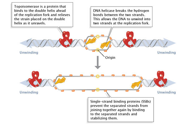
Eukaryotic DNA replication is not as well understood as bacterial replication. Much research has been carried out on a v ariety of experimental organisms, particularly yeast and mammalian cells. Many of these studies have found extensive similarities between the general features of DNA replication in prokaryotes and eukaryotes. For example, DNA helicases, topoisomerases, single-strand binding proteins, primases, DNA polymerases, and DNA ligases. Nevertheless, at the molecular level, eukaryotic DNA replication appears to be substantially more complex. These additional intricacies of eukaryotic DNA replication are related to several features of eukaryotic cells. In particular, eukaryotic cells have larger, linear chromosomes, the chromatin is tightly packed within nucleosomes, and cell cycle regulation is much more complicated. This section emphasizes some of the unique features of eukaryotic DNA replication. 

DNA replication of eukaryotes  Replic16


Initiation Occurs at Multiple Origins of Replication on Linear Eukaryotic Chromosomes

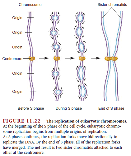
Because eukaryotes have long, linear chromosomes, the chromosomes require multiple origins of replication so the DNA can be replicated in a reasonable length of time. In 1968, Joel Huberman and Arthur Riggs provided evidence for multiple origins of replication by adding a radiolabeled nucleoside (3H-thymidine) to a culture of actively dividing cells, followed by a chase with nonlabeled thymidine. The radiolabeled thymidine was taken up by the cells and incorporated into their newly made DNA strands for a brief period. The chromosomes were then isolated from the cells and subjected to autoradiography. As seen in Figure 11.21 , radiolabeled segments were interspersed among nonlabeled segments. This result is consistent with the hypothesis that eukaryotic chromosomes contain multiple origins of replication. As shown schematically in Figure 11.22 , DNA replication proceeds bidirectionally from many origins of replication during S phase of the cell cycle. The multiple replication forks eventually make contact with each other to complete the replication process.

The molecular features of eukaryotic origins of replication may have some similarities to the origins found in bacteria. At the molecular level, eukaryotic origins of replication have been extensively studied in the yeast Saccharomyces cerevisiae. In this organism, several replication origins have been identified and sequenced. They have been named ARS elements (for autonomously replicating sequence). ARS elements, which are about 50 bp in length, are necessary to initiate chromosome replication. ARS elements have unique features of their DNA sequences. First, they contain a higher percentage of A and T bases than the rest of the chromosomal DNA. In addition, they contain a copy of the ARS consensus sequence (ACS), ATTTAT(A or G)TTTA, along with additional elements that enhance origin function. This arrangement is similar to bacterial origins.

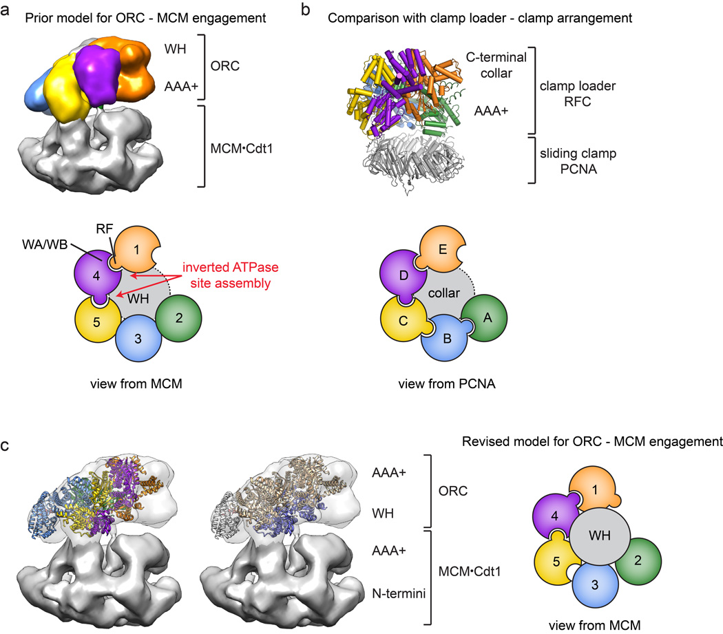
DNA replication in eukaryotes begins with the assembly of a prereplication complex (preRC) consisting of at least 14 different proteins. Part of the preRC is a group of six proteins called the origin recognition complex (ORC) that acts as the initiator of eukaryotic DNA replication. The ORC was originally identified in yeast as a protein complex that binds directly to ARS elements. DNA replication at the origin begins with the binding of ORC, which usually occurs during G1 phase. Other proteins of the preRC then bind, including a group of proteins called MCM helicase.

MCM is an acronym for minichromosome maintenance. The genes encoding MCM proteins were originally identified in mutant yeast strains that are defective in the maintenance of minichromosomes in the cell. MCM proteins have since been shown to play a role in DNA replication.

The binding of MCM helicase at the origin completes a process called DNA replication licensing; only those origins with MCM helicase can initiate DNA synthesis. During S phase, DNA synthesis begins when preRCs are acted on by at least 22 additional proteins that activate MCM helicase and assemble two divergent replication forks at each replication origin. An important role of these additional proteins is to carefully regulate the initiation of DNA replication so that it happens at the correct time during the cell cycle and occurs only once during the cell cycle. The precise roles of these proteins are under active research investigation.

Eukaryotes Contain Several Different DNA Polymerases

Eukaryotes have many types of DNA polymerases. For example, mammalian cells have well over a dozen different DNA polymerases (Table 11.4 ).

DNA replication of eukaryotes  Eukary11
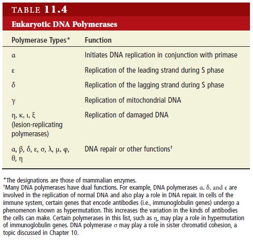
Four of these, designated α (alpha), ε (epsilon), δ (delta), and γ (gamma), have the primary function of replicating DNA. DNA polymerase γ functions in the mitochondria to replicate mitochondrial DNA, whereas α, ε, and δ are involved with DNA replication in the cell nucleus during S phase. DNA polymerase α is the only eukaryotic polymerase that associates with primase. The functional role of the DNA polymerase α/primase complex is to synthesize a short RNA-DNA primer of approximately 10 RNA nucleotides followed by 20 to 30 DNA nucleotides. This short RNA-DNA strand is then used by DNA polymerase ε or δ for the processive elongation of the leading and lagging strands, respectively. For this to happen, the DNA polymerase α/primase complex dissociates from the replication fork and is exchanged for DNA polymerase ε or δ. This exchange is called a polymerase switch. Accumulating evidence suggests that DNA polymerase ε is primarily involved with leading- strand synthesis, whereas DNA polymerase δ is responsible for lagging-strand synthesis. What are the functions of the other DNA polymerases? Several of them also play an important role in DNA repair. DNA polymerase β, which has been studied for several decades, is not involved in the replication 
of normal DNA, but plays an important role in removing incorrect bases from damaged DNA. More recently, several additional DNA polymerases have been identified. While their precise roles have not been elucidated, many of these are in a category called lesion-replicating polymerases. When DNA polymerase α, δ, and ε encounter abnormalities in DNA structure, such as abnormal bases or cross-links, they may be unable to replicate over the aberration. When this occurs, lesion-replicating polymerases are attracted to the damaged DNA and have special properties that enable them to synthesize a complementary strand over the abnormal region. Each type of lesion-replicating polymerase may be able to replicate over a different kind of DNA damage.

Flap Endonuclease Removes RNA Primers During Eukaryotic DNA Replication

Another key difference between bacterial and eukaryotic DNA replication is the way that RNA primers are removed. Bacterial RNA primers are removed by DNA polymerase I. By comparison, a DNA polymerase enzyme does not play this role in eukaryotes. Instead, an enzyme called flap endonuclease is primarily responsible for RNA primer removal. Flap endonuclease gets its name because it removes small pieces of RNA flaps that are generated by the action of DNA polymerase δ. In the diagram shown in Figure 11.23 , DNA polymerase δ elongates the left Okazaki fragment until it runs into the RNA primer of the adjacent Okazaki fragment on the right. This causes a portion of the RNA primer to form a short flap, which is removed by the endonuclease function of flap endonuclease.

DNA replication of eukaryotes  Flap_r10

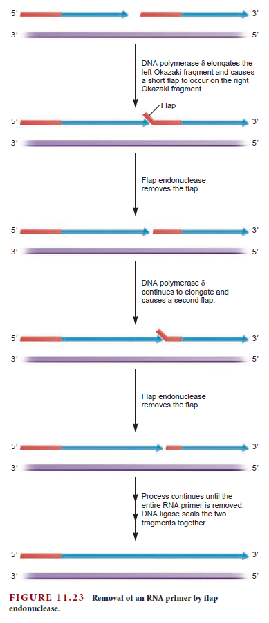
 As DNA polymerase δ continues to elongate the DNA, short flaps continue to be generated, which are sequentially removed by flap endonuclease. Eventually, all of the RNA primer is removed, and DNA ligase can seal the DNA fragments together. Though flap endonuclease is thought to be the primary pathway for RNA primer removal in eukaryotes, it is unable to remove a flap that is too long. In such cases, a long flap is cleaved by the enzyme called Dna2 nuclease/helicase. This enzyme can cut a long flap, thereby generating a short flap. The short flap is then removed via flap endonuclease.

The Ends of Eukaryotic Chromosomes are Replicated by Telomerase

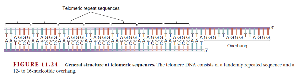
Linear eukaryotic chromosomes contain telomeres at both ends. The term telomere refers to the complex of telomeric sequences within the DNA and the special proteins that are bound to these sequences. Telomeric sequences consist of a moderately repetitive tandem array and a 3ʹ overhang region that is 12 to 16 nucleotides in length (Figure 11.24 ).

DNA replication of eukaryotes  Telome10

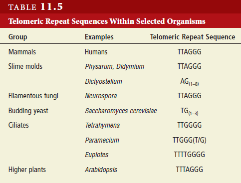
The tandem array that occurs within the telomere has been studied in a wide variety of eukaryotic organisms. A common feature is that the telomeric sequence contains several guanine nucleotides and often many thymine nucleotides (Table 11.5 ).

DNA replication of eukaryotes  Telome11

Depending on the species and the cell type, this sequence can be tandemly repeated up to several hundred times in the telomere region. One reason why telomeric repeat sequences are needed is because DNA polymerase is unable to replicate the 3ʹ ends of DNA strands. Why is DNA polymerase unable to replicate this region? The answer lies in the two unusual enzymatic features of this enzyme. As discussed previously, DNA polymerase synthesizes DNA only in a 5ʹ to 3ʹ direction, and it cannot link together the first two individual nucleotides; it can elongate only preexisting strands. These two features of DNA polymerase function pose a problem at the 3ʹ ends of linear chromosomes. As shown in Figure 11.25 the 3ʹ end of a DNA strand cannot be replicated by DNA polymerase because a primer cannot be made upstream from this point. Therefore, if this problem were not solved, the chromosome would become progressively shorter with each round of DNA replication.

DNA replication of eukaryotes  Telome12

To prevent the loss of genetic information due to chromosome shortening, additional DNA sequences are attached to the ends of telomeres. In 1984, Carol Greider and Elizabeth Blackburn discovered an enzyme called telomerase that prevents chromosome shortening. It recognizes the sequences at the ends of eukaryotic chromosomes and synthesizes additional repeats of telomeric sequences. They received the 2009 Nobel Prize in physiology or medicine for their discovery. Figure 11.26 shows the interesting mechanism by which telomerase works. The telomerase enzyme contains both protein subunits and RNA.

DNA replication of eukaryotes  Telome14

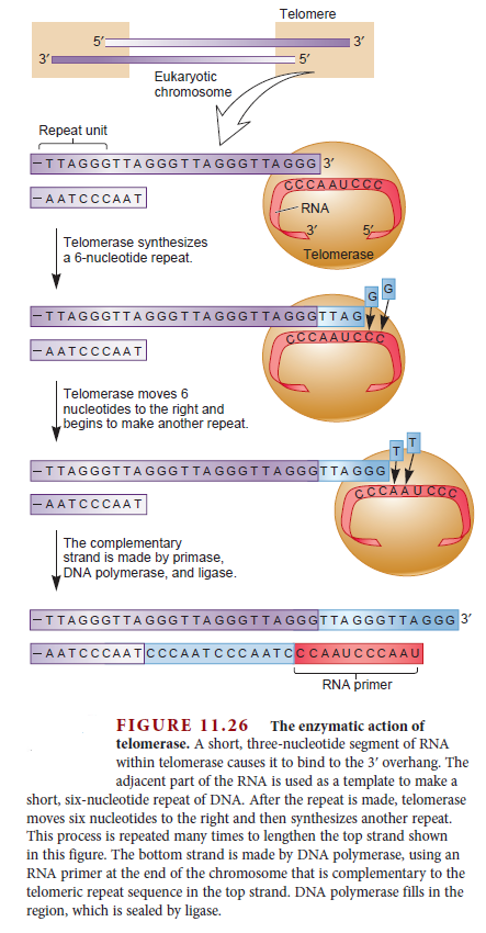
The RNA part of telomerase contains a sequence complementary to the DNA sequence found in the telomeric repeat. This allows telomerase to bind to the 3ʹ overhang region of the telomere. Following binding, the RNA sequence beyond the binding site functions as a template allowing the synthesis of a six-nucleotide sequence at the end of the DNA strand. This is called polymerization, because it is analogous to the function of DNA polymerase. It is catalyzed by two identical protein subunits called telomerase reverse transcriptase (TERT) . TERT’s name indicates that it uses an RNA template to synthesize DNA. Following polymerization, the telomerase can then move—a process called translocation—to the new end of this DNA strand and attach another six nucleotides to the end. This binding-polymerization-translocation cycle occurs many times in a row, thereby greatly lengthening the 3ʹ end of the DNA strand in the telomeric region. The complementary strand is then synthesized by primase, DNA polymerase, and DNA ligase.


Last edited by Admin on Tue Dec 08, 2015 6:06 am; edited 11 times in total

https://reasonandscience.catsboard.com

2DNA replication of eukaryotes  Empty Re: DNA replication of eukaryotes Sun Nov 22, 2015 6:22 pm

Otangelo


Admin

Eukaryotes contain a number of DNA polymerases. Pol  extends primers, the highly processive pol  is the lagging strand polymerase, and pol ε appears to be the leading strand polymerase. Eukaryotic replication has multiple origins and proceeds through nucleosomes. In order to replicate the 5" end of the lagging strand, eukaryotic chromosomes end with repeated telomeric sequences appended by the ribonucleoprotein telomerase. The 3" extension at the end of each chromosome serves as a template for primer synthesis. Somatic cells lack telomerase activity, which may prevent them from transforming to cancer cells.


Eukaryotes possess much of the same replication machinery found in prokaryotes. For example, like prokaryotes, a DNA clamp protein acts along with DNA polymerase during DNA synthesis. One such eukaryotic clamp protein, proliferating nuclear cell antigen (PCNA), was originally identified as an antigen that is expressed in the nuclei of dividing cells during S phase. PCNA is a clamp protein for DNA polymerase d. In eukaryotic cells, the enormous length and elaborate folding of the chromosomal DNA molecules pose additional challenges for DNA replication. For example, how are the many replication origins coordinated, and how is their activation linked to other key events in the cell cycle? Answering such questions requires a better understanding of the spatial organization of DNA replication within the nucleus. When cells are briefly incubated with DNA precursors that make the most recently formed DNA fluorescent, microscopic examination reveals that the new,

Parts required in eukaryotes

Origin of replication 
The origin of replication (also called the replication origin) is a particular sequence in a genome at which replication is initiated.[1] This can either involve the replication of DNA in living organisms such as prokaryotes and eukaryotes, or that of DNA or RNA in viruses, such as double-stranded RNA viruses.

Origin Recognition Complex ( ORC )
The origin recognition complex (ORC), a heteromeric six‐subunit protein, is a central component for eukaryotic DNA replication.

Primosome ( consists of seven proteins: DnaG primase, DnaB helicase, DnaC helicase assistant, DnaT, PriA, Pri B, and PriC. )
helicase-loading proteins Cdc6 and Cdt1
a protein called DnaC
DNA polymerase
DNA polymerase I for Primer removal after okazaki fragment maturation


In eukaryotes, the budding yeast Saccharomyces cerevisiae has the best characterised replication origins. These origins were first identified by their ability to support the replication of mini-chromosomes or plasmids, giving rise to the name Autonomously replicating sequences or ARS elements. Each budding yeast origin consists of a short (~11 bp) essential DNA sequence (called the ARS consensus sequence or ACS) that recruits replication proteins.
In other eukaryotes, including humans, the DNA sequences at the replication origins vary. Despite this sequence variation, all the origins form a base for assembly of a group of proteins known collectively as the pre-replication complex (pre-RC):


  • First, the origin DNA is bound by the origin recognition complex (ORC) which, with help from two further protein factors (Cdc6 and Cdt1), load the mini chromosome maintenance (or MCM) protein complex.
  • Once assembled, this complex of proteins indicates that the replication origin is ready for activation. Once the replication origin is activated, the cell's DNA will be replicated.


In metazoans, pre-RC formation is inhibited by the protein geminin, which binds to and inactivates Cdt1. Regulation of replication prevents the DNA from being replicated more than once each cell cycle.
In humans an origin of replication has been originally identified near the Lamin B2 gene on chromosome 19 and the ORC binding to it has extensively been studied.[8]

Question: How did these DNA sequence motifs arise, and how did the proteins " learn " that these specific sequences mean " place to open the DNA strand and start replication ? " trial and error ?

The replisome is a complex molecular machine that carries out replication of DNA. The replisome first unwinds double stranded DNA into two single strands. For each of the resulting single strands, a new complementary sequence of DNA is synthesized. The net result is formation of two new double stranded DNA sequences that are exact copies of the original double stranded DNA sequence.
In terms of structure, the replisome is composed of two replicative polymerase complexes, one of which synthesizes the leading strand, while the other synthesizes the lagging strand. The replisome is composed of a number of proteins including helicase, RFC, PCNA, gyrase/topoisomerase, SSB/RPA, primase, DNA polymerase I, RNAse H, and ligase.

The replisome is a system in which various factors work together to solve the structural and chemical challenges of DNA replication. Chromosomes size and structure varies between organisms, but since DNA molecules are the reservoir of genetic information for all forms of life, many replication challenges and solutions are the same for different organisms. As a result, the replication factors that solve these problems are highly conserved in terms of structure, chemistry, functionality, or sequence. General structural and chemical challenges include the following:


  • Efficient replisome assembly at origins of replication (origin recognition complexes or specific replication origin sequences in some organisms)

  • Separating the duplex into the leading and lagging template strands (helicases)

  • Protecting the leading and lagging strands from damage after duplex separation (SSB and RPA factors)

  • Priming of the leading and lagging template strands (primase or DNA polymerase alpha)

  • Ensuring processivity (clamp loading factors, ring-shaped clamp proteins, strand binding proteins)

  • High-fidelity DNA replication (DNA polymerase III, DNA polymerase delta, DNA polymerase epsilon. All have intrinsically low error rates because of their structure and chemistry.)

  • Error correction (replicative polymerase active sites sense errors; 3' to 5' exonuclease domains of replicative polymerases fix errors)

  • Synchronised polymerisation of leading and lagging strands despite anti-parallel structure (replication fork structure, dimerisation of replicative polymerases)

  • Primer removal (DNA polymerase I, RNAse H, flap endonucleases such as FEN1, or other DNA repair factors)

  • Formation of phosphodiester bonds at gaps between Okazaki fragments (ligase)


In general, the challenges of DNA replication involve the structure of the molecules, the chemistry of the molecules, and, from a systems perspective, the underlying relationships between the structure and the chemistry.


Regulated eukaryotic DNA replication origin firing with purified proteins
http://www.nature.com/nature/journal/v519/n7544/full/nature14285.html


DNA codes for the DNA replicating enzyme, DNA polymerase. DNA polymerase reads the chemical code of DNA and faithfully creates another exact duplicate molecule. So for its existence, DNA is dependent on DNA polymerase, the existence of which is dependent on DNA itself. 3

DNA replication of eukaryotes  Dna_in10

Replication: Mechanism of Replication

During DNA replication, both strands of the double helix act as templates for the formation of new DNA molecules. Copying occurs at a localized region called the replication fork, which is a Y shaped structure where new DNA strands are synthesised by a multi-enzyme complex. Here the DNA to be copied enters the complex from the left. One new strand is leaving at the top of frame and the other new strand is leaving at bottom. The first step in DNA replication is the separation of the two strands by an enzyme called helicase. This spins the incoming DNA to unravel it: at ten thousand RPM in the case of bacterial systems. The separated strands are called three prime and five prime, distinguished by the direction in which their component nucleotides join up. . The 3' DNA strand, also known as the leading strand, is diverted to a DNA polymerase and is used as a continuous template for the synthesis of the first daughter DNA helix. The other half of the DNA double helix, known as the lagging strand, has the opposite 3' to 5' orientation and consequently requires a more complicated copying mechanism. As it emerges from the helicase, the lagging strand is organised into sections called Okazaki fragments. These are then presented to a second DNA polymerase enzyme in the preferred 5' to 3' orientation. These sections are then effectively synthesised backwards. When the copying is complete, the finished section is released and the next loop is drawn back for replication. Intricate as this mechanism appears, numerous components have been deliberately left out to avoid complete confusion. The exposed strands of single DNA are covered by protective binding proteins. And in some systems, multiple Okazaki fragments may be present. The molecular reality is very different from the iconic image of the double helix neatly separating into two DNA copies as so often depicted.

Major Molecular Events of DNA Replication

Arthur Kornberg compared DNA to a tape recording of instructions that can be copied over and over. How do cells make these near-perfect copies, and does the process ever vary?
Scientists have devoted decades of effort to understanding how deoxyribonucleic acid () replicates itself. In simple terms, replication involves use of an existing strand of DNA as a template for the synthesis of a new, identical strand. American enzymologist and Nobel Prize winner Arthur Kornberg compared this process to a tape recording of instructions for performing a task: "[E]xact copies can be made from it, as from a tape recording, so that this information can be used again and elsewhere in time and space" (Kornberg, 1960).
In reality, the process of replication is far more complex than suggested by Kornberg's analogy. Researchers typically utilize simple bacterial cells in their experiments, but they still do not have all the answers, particularly when it comes to eukaryotic replication. Nonetheless, scientists are familiar with the basic steps in the replication process, and they continue to rely on this information as the basis for continued research and experimentation.


The Molecular Machinery of Bacterial DNA Replication


A typical bacterial has anywhere from about 1 million to 4 million base pairs of DNA, compared to the 3 billion base pairs in the genome of the common house mouse (Mus musculus). Still, even in bacteria, with their smaller genomes, DNA replication involves an incredibly sophisticated, highly coordinated series of molecular events. These events are divided into four major stages: initiation, unwinding, primer synthesis, and elongation.


Initiation and Unwinding


During initiation, so-called initiator proteins bind to the replication origin, a base-pair sequence of nucleotides known as oriC. This binding triggers events that unwind the DNA double helix into two single-stranded DNA molecules. Several groups of proteins are involved in this unwinding (Figure 1). For example, the DNA helicases are responsible for breaking the hydrogen bonds that join the complementary nucleotide bases to each other; these hydrogen bonds are an essential feature of James Watson and Francis Crick's three-dimensional DNA model. Because the newly unwound single strands have a tendency to rejoin, another group of proteins, the single-strand-binding proteins, keep the single strands stable until elongation begins. A third family of proteins, the topoisomerases, reduce some of the torsional strain caused by the unwinding of the double helix.


DNA replication of eukaryotes  4699_90

Figure 1: Facilitation of DNA unwinding.
During DNA replication, several proteins facilitate the unwinding of the DNA double helix into two single strands. Topoisomerases (red) reduce torsional strain caused by the unwinding of the DNA double helix; DNA helicase (yellow) breaks hydrogen bonds between complementary base-pairs; single-strand binding proteins (SSBs) stabilize the separated strands and prevent them from rejoining. The location at which a DNA strand begins to unwind into two separate single strands is known as the origin of replication. As shown in Figure 1, when the double helix unwinds, replication proceeds along the two single strands at the same time but in opposite directions (i.e., left to right on one strand, and right to left on the other). This forms two replication forks that move along the DNA, replicating as they go.

Primer Synthesis


Primer synthesis marks the beginning of the actual synthesis of the new DNA molecule. Primers are short stretches of nucleotides (about 10 to 12 bases in length) synthesized by an RNA polymerase enzyme called primase. Primers are required because DNA polymerases, the enzymes responsible for the actual addition of nucleotides to the new DNA strand, can only add deoxyribonucleotides to the 3'-OH group of an existing chain and cannot begin synthesis de novo. Primase, on the other hand, can add ribonucleotides de novo. Later, after elongation is complete, the primer is removed and replaced with DNA nucleotides.

Elongation


Finally, elongation--the addition of nucleotides to the new DNA strand--begins after the primer has been added. Synthesis of the growing strand involves adding nucleotides, one by one, in the exact order specified by the original (template) strand. Recall that one of the key features of the Watson-Crick DNA model is that adenine is always paired with thymine and cytosine is always paired with guanine. So, for example, if the original strand reads A-G-C-T, the new strand will read T-C-G-A.
DNA is always synthesized in the 5'-to-3' direction, meaning that nucleotides are added only to the 3' end of the growing strand. As shown in Figure 2, the 5'-phosphate group of the new nucleotide binds to the 3'-OH group of the last nucleotide of the growing strand. Scientists have yet to identify a polymerase that can add bases to the 5' ends of DNA strands.

DNA replication of eukaryotes  4609_91

Figure 2: New DNA is synthesized from deoxyribonucleoside triphosphates (dNTPs).
(A) A deoxyribonucleoside triphosphate (dNTP). (B) During DNA replication, the 3'-OH group of the last nucleotide on the new strand attacks the 5'-phosphate group of the incoming dNTP. Two phosphates are cleaved off. (C) A phosphodiester bond forms between the two nucleotides, and phosphate ions are released.

The Discovery of DNA Polymerase


While studying E. coli bacteria, enzymologist Arthur Kornberg discovered that DNA polymerases catalyze DNA synthesis. Kornberg's experiment involved mixing all of the basic "ingredients" necessary for E. coli DNA synthesis in a test tube, including nucleotides, E. coli extract, and , and then purifying and testing the enzymes involved. Using this method, Kornberg not only discovered DNA polymerases, but he also performed some of the initial work demonstrating how enzymes add new nucleotides to growing DNA chains (Kornberg, 1959).
Scientists have since identified a total of five different DNA polymerases in E. coli, each with a specialized role. For example, DNA polymerase III does most of the elongation work, adding nucleotides one by one to the 3' end of the new and growing single strand. Other enzymes, including DNA polymerase I and RNase H, are responsible for removing the RNA primer after DNA polymerase III has begun its work, replacing it with DNA nucleotides (Ogawa & Okazaki, 1984). When these enzymes finish, they leave a nick between the section of DNA that was formerly the primer and the elongated section of DNA. Another enzyme called DNA ligase then acts to seal the bond between the two adjacent nucleotides.

DNA Polymerase Only Moves in One Direction


After a primer is synthesized on a strand of DNA and the DNA strands unwind, synthesis and elongation can proceed in only one direction. As previously mentioned, DNA polymerase can only add to the 3' end, so the 5' end of the primer remains unaltered. Consequently, synthesis proceeds immediately only along the so-called leading strand. This immediate replication is known as continuous replication. The other strand (in the 5' direction from the primer) is called the lagging strand, and replication along it is called discontinuous replication. The double helix has to unwind a bit before the synthesis of another primer can be initiated further up on the lagging strand. Synthesis can then occur from the 3' end of that new primer. Next, the double helix unwinds a bit more, and another spurt of replication proceeds. As a result, replication along the lagging strand can only proceed in short, discontinuous spurts (Figure 3).

DNA replication of eukaryotes  35272_92

Figure 3: Replication of the leading DNA strand is continuous, while replication along the lagging strand is discontinuous.
After a short length of the DNA has been unwound, synthesis must proceed in the 5' to 3' direction; that is, in the direction opposite that of the unwinding.
The fragments of newly synthesized DNA along the lagging strand are called Okazaki fragments, named in honor of their discoverer, Japanese molecular biologist Reiji Okazaki. Okazaki and his colleagues made their discovery by conducting what is known as a pulse-chase experiment, which involved exposing replicating DNA to a short "pulse" of isotope-labeled nucleotides and then varying the length of time that the cells would be exposed to nonlabeled nucleotides. This later period is called the "chase" (Okazaki et al., 1968). The labeled nucleotides were incorporated into growing DNA molecules only during the initial few seconds of the pulse; thereafter, only nonlabeled nucleotides were incorporated during the chase. The scientists then centrifuged the newly synthesized DNA and observed that the shorter chases resulted in most of the radioactivity appearing in "slow" DNA. The sedimentation rate was determined by size: smaller fragments precipitated more slowly than larger fragments because of their lighter weight. As the investigators increased the length of the chases, radioactivity in the "fast" DNA increased with little or no increase of radioactivity in the slow DNA. The researchers correctly interpreted these observations to mean that, with short chases, only very small fragments of DNA were being synthesized along the lagging strand. As the chases increased in length, giving DNA more time to replicate, the lagging strand fragments started integrating into longer, heavier, more rapidly sedimenting DNA strands. Today, scientists know that the Okazaki fragments of bacterial DNA are typically between 1,000 and 2,000 nucleotides long, whereas in eukaryotic cells, they are only about 100 to 200 nucleotides long.

The Challenges of Eukaryotic Replication


Bacterial and eukaryotic cells share many of the same basic features of replication; for instance, initiation requires a primer, elongation is always in the 5'-to-3' direction, and replication is always continuous along the leading strand and discontinuous along the lagging strand. But there are also important differences between bacterial and eukaryotic replication, some of which biologists are still actively researching in an effort to better understand the molecular details. One difference is that eukaryotic replication is characterized by many replication origins (often thousands), not just one, and the sequences of the replication origins vary widely among species. On the other hand, while the replication origins for bacteria, oriC, vary in length (from about 200 to 1,000 base pairs) and sequence, except among closely related organisms, all bacteria nonetheless have just a single replication origin (Mackiewicz et al., 2004).
Eukaryotic replication also utilizes a different set of DNA polymerase enzymes (e.g., DNA polymerase δ and DNA polymerase ε instead of DNA polymerase III). Scientists are still studying the roles of the 13 eukaryotic polymerases discovered to date. In addition, in eukaryotes, the DNA template is compacted by the way it winds around proteins called histones. This DNA-histone complex, called a nucleosome, poses a unique challenge both for the cell and for scientists investigating the molecular details of eukaryotic replication. What happens to nucleosomes during DNA replication? Scientists know from electron micrograph studies that nucleosome reassembly happens very quickly after replication (the reassembled nucleosomes are visible in the electron micrograph images), but they still do not know how this happens (Annunziato, 2005).
Also, whereas bacterial chromosomes are circular, eukaryotic chromosomes are linear. During circular DNA replication, the excised primer is readily replaced by nucleotides, leaving no gap in the newly synthesized DNA. In contrast, in linear DNA replication, there is always a small gap left at the very end of the chromosome because of the lack of a 3'-OH group for replacement nucleotides to bind. (As mentioned, DNA synthesis can proceed only in the 5'-to-3' direction.) If there were no way to fill this gap, the DNA molecule would get shorter and shorter with every generation. However, the ends of linear chromosomes—thetelomeres—have several properties that prevent this.
DNA replication occurs during the S phase of cell division. In E. coli, this means that the entire genome is replicated in just 40 minutes, at a pace of approximately 1,000 nucleotides per second. In eukaryotes, the pace is much slower: about 40 nucleotides per second. The coordination of the protein complexes required for the steps of replication and the speed at which replication must occur in order for cells to divide are impressive, especially considering that enzymes are also proofreading, which leaves very few errors behind.

Summary


The study of DNA replication started almost as soon as the structure of DNA was elucidated, and it continues to this day. Currently, the stages of initiation, unwinding, primer synthesis, and elongation are understood in the most basic sense, but many questions remain unanswered, particularly when it comes to replication of the eukaryotic genome. Scientists have devoted decades to the study of replication, and researchers such as Kornberg and Okazaki have made a number of important breakthroughs. Nonetheless, much remains to be learned about replication, including how errors in this process contribute to human disease.

References and Recommended Reading


Annunziato, A. T. Split decision: What happens to nucleosomes during DNA replication? Journal of Biological Chemistry 280, 12065–12068 (2005)
Bessman, M. J., et al. Enzymatic synthesis of deoxyribonucleic acid. II. General properties of the reaction. Journal of Biological Chemistry 233, 171–177 (1958)
Kornberg, A. The biological synthesis of deoxyribonucleic acid. Nobel Lecture, December 11, 1959. (link to transcript)
———. Biological synthesis of deoxyribonucleic acid. Science 131, 1503–1508 (1960)
Lehman, I. R., et al. Enzymatic synthesis of deoxyribonucleic acid. I. Preparation of substrates and partial purification of an enzyme from Escherichia coliJournal of Biological Chemistry 233, 163–170 (1958)
Losick, R., & Shapiro, L. DNA replication: Bringing the mountain to Mohammed.Science 282, 1430–1431 (1998)
Mackiewicz, P., et al. Where does bacterial replication start? Rules for predicting the oriC region. Nucleic Acids Research 32, 3781–3791 (2004)
Ogawa, T., & Okazaki, T. Function of RNase H in DNA replication revealed by RNase H defective mutants of Escherichia coliMolecular and General Genetics193, 231–237 (1984)
Okazaki, R., et al. Mechanism of DNA chain growth. I. Possible discontinuity and unusual secondary structure of newly synthesized chains. Proceedings of the National Academy of Sciences 59, 598–605 (1968)

Study reveals the architecture of the molecular machine that copies DNA 4


DNA replication of eukaryotes  43-studyreveals


A series of electron micrographs show the barrel-shaped helicase, which is the enzyme that separates the two DNA strands, along with other components of the replisome, including polymerase-epsilon (green). Credit: Brookhaven National Laboratory
DNA replication is essential to all life, yet many basic mechanisms in that process remain unknown to scientists. The structure of the replisome—a block of proteins responsible for unwinding the DNA helix and then creating duplicate helices for cell division—is one such mystery.

Now, for the first time, a team of researchers from The Rockefeller University, Brookhaven National Laboratory, and Stony Brook University has revealed that vital complex's molecular architecture. And to their surprise, it does not look as they had expected.
"Our finding goes against decades of textbook drawings of what people thought the replisome should look like," says Michael O'Donnell, Anthony and Judith Evnin Professor, head of Rockefeller's Laboratory of DNA Replication and a Howard Hughes Medical Institute investigator. He is the senior author of the study, published November 2 in Nature Structural and Molecular Biology. "However, it's a recurring theme in science that nature does not always turn out to work the way you thought it did."
A complete rendering of the eukaryotic replisome
The findings focus on the replisome found in eukaryotic organisms, a category that includes a broad swath of living things, including humans and other multicellular organisms. O'Donnell and others have long studied replisomes in simpler, single-celled bacteria, but the more complex version has over 30 different gears, or proteins, and required about 15 years for O'Donnell's lab to obtain. Through these previous studies, his lab has learned how to assemble the more complex replisome from its components.
But until now, no pictures existed to show just how everything fit together in the eukaryotic replisome. To create them, the team began building the complete structure piece by piece, and examining its shape in the electron microscope—a powerful device used to study protein structures, and a specialty of co-author Huilin Li, a molecular biologist at Brookhaven National Laboratory and Stony Brook University. The pictures Li and members of his lab captured were the first ever made of a complete replisome from any type of cell.


DNA replication of eukaryotes  44-studyreveals


Previously (left), the replisome's two polymerases (green) were assumed to be below the helicase (tan), the enzyme that splits the DNA strands. The new images reveal one polymerase is located at the front of the helicase, causing one strand …more
The DNA helix has two DNA strands, and each is duplicated by a separate DNA polymerase, an enzyme that creates DNA molecules by pairing single nucleotides, the basic units of DNA, with their matching partners. Another enzyme in the replisome is the helicase that, like a zipper, is responsible for separating DNA into two single strands in preparation for replication. For years, the two polymerases were thought to follow along behind the helicase, or below it, as it unzips the strands. But the new pictures of the replisome showed that one polymerase sits above the helicase.
A link to epigenetics
To identify which polymerase was at the top of the helicase, the team enlisted the help of co-authors postdoc Yi Shi and Brian Chait, the Camille and Henry Dreyfus Professor at Rockefeller and head of the Laboratory of Mass Spectrometry and Gaseous Ion Chemistry. They identified the top polymerase as Pol-ε.

Three-dimensional structure of the active DNA helicase, which unzips DNA strands, is bound to the polymerase called Pol epsilon, which copies them. The DNA polymerase epsilon (green) sits on top rather than the bottom of the helicase. Credit: Brookhaven National Laboratory
Why the eukaryotic replisome developed such a structure is not known. O'Donnell and his colleagues suspect that it may have something to do with the evolution of multicellularity. As the helicase unzips two strands of DNA, it encounters nucleosomes, particles that tightly bundle DNA to fit it into a cell's nucleus. These must be dislodged and then distributed to one new strand or the other. Previous work has shown that Pol-ε binds nucleosomes, and it may be that while riding atop the helicase, Pol-ε is in charge of distributing nucleosomes to the two strands, O'Donnell suggests.
"Changes to nucleosomes carry epigenetic information that instructs different cells to become the different tissues of the body, such as heart, brain, and other organs during embryonic development," O'Donnell says. "So we can speculate that Pol-ε's interaction with nucleosomes could play a role in assigning different epigenetic identities to the two new daughter cells after cell division, that instruct them to form different organs during development of a multicellular animal."

Genesis of the enzyme that divides the DNA double helix during cell replication 5

DNA replication of eukaryotes  Genesisofthe


Averaged electron microscope images of two intermediate helicase structures. The top shows an ORC binding the two ring structures together, and the bottom shows the completed double-ring structure with the ORC detached.


The proteins that drive DNA replication—the force behind cellular growth and reproduction—are some of the most complex machines on Earth. The multistep replication process involves hundreds of atomic-scale moving parts that rapidly interact and transform. Mapping that dense molecular machinery is one of the most promising and challenging frontiers in medicine and biology.
Now, scientists have pinpointed crucial steps in the beginning of the replication process, including surprising structural details about the enzyme that "unzips" and splits the DNA double helix so the two halves can serve as templates for DNA duplication.
The research combined electron microscopy, perfectly distilled proteins, and a method of chemical freezing to isolate specific moments at the start of replication. The study—authored by scientists from the U.S. Department of Energy's Brookhaven National Laboratory, Stony Brook University, Cold Spring Harbor Laboratory, and Imperial College, London—published on Oct. 15, 2014, in the journal Genes and Development.
"The genesis of the DNA-unwinding machinery is wonderfully complex and surprising," said study coauthor Huilin Li, a biologist at Brookhaven Lab and Stony Brook University. "Seeing this helicase enzyme prepare to surround and unwind the DNA at the molecular level helps us understand the most fundamental process of life and how that process might go wrong. Errors in copying DNA are found in certain cancers, and this work could one day help develop new treatment methods that stall or break dangerous runaway machinery."
The research picks up where two previous studies by Li and colleagues left off. They first determined the structure of the "Origin Recognition Complex" (ORC), a protein that identifies and attaches to specific DNA sites to initiate the entire replication process. The second study revealed how the ORC recruits, cracks open, and installs a crucial ring-shaped protein structure (Mcm2-7) that lies at the core of the helicase enzyme.
But DNA replication is a bi-directional process with two helicases moving in opposite directions. The key question, then, was how does a second helicase core get recruited and loaded onto the DNA in the opposite orientation of the first?
"To our surprise, we found an intermediate structure with one ORC binding two rings," said Brookhaven Lab biologist and lead author Jingchuan Sun. "This discovery suggests that a single ORC, rather than the commonly believed two-ORC system, loads both helicase rings."


DNA replication of eukaryotes  1-genesisofthe


Three-dimensional model (based on electron microscopy data) of the double-ring structure loaded onto a DNA helix.
One step further along, the researchers also determined the molecular architecture of the final double-ring structure left behind after the ORC leaves the system, offering a number of key biological insights.
"We now have clues to how that double-ring structure stably lingers until the cell enters the DNA-synthesis phase much later on in replication," said study coauthor Christian Speck of Imperial College, London. "This study revealed key regulatory principles that explain how the helicase activity is initially suppressed and then becomes reactivated to begin its work splitting the DNA."
Precision methods, close collaboration
Examining these fleeting molecular structures required mastery of biology, chemistry, and electron microscopy techniques.
"This three-way collaboration took advantage of each lab's long standing collaboration and expertise," said study coauthor Bruce Stillman of Cold Spring Harbor. "Imperial College and Cold Spring Harbor handled the challenging material preparation and functional characterization, while Brookhaven and Stony Brook led the sophisticated molecular imaging and three-dimensional image reconstruction."
The researchers used proteins from baker's yeast—a model organism for the more complex systems found in animals. The scientists isolated the protein mechanisms involved in replication and removed structures that might otherwise complicate the images.


DNA replication of eukaryotes  2-genesisofthe


Collaborating scientists and study coauthors Zuanning Yuan of Stony Brook University (standing), Huilin Li of Stony Brook and Brookhaven Lab (seated, back), and Jingchuan Sun of Brookhaven Lab (seated, front) examining protein structures.


Once the isolated proteins were mixed with DNA, the scientists injected chemicals to "freeze" the binding and recruitment process at intervals of 2, 7, and 30 minutes.
They then used an electron microscope at Brookhaven to pin down the exact structures at each targeted moment in a kind of molecular time-lapse. Rather than the light used in a traditional microscope, this technique uses focused beams of electrons to illuminate a sample and form images with atomic resolution. The instrument produces a large number of two-dimensional electron beam images, which a computer then reconstructs into three-dimensional structure.
"This technique is ideal because we're imaging relatively massive proteins here," Li said. "A typical protein contains three hundred amino acids, but these DNA replication mechanisms consist of tens of thousands of amino acids. The entire structure is about 20-nanometers across, compared to 4 nanometers for an average protein."
Unraveling the DNA processes at the most fundamental level, the focus of this team's work, could have far-reaching implications.
"The structural knowledge may help others engineer small molecules that inhibit DNA replication at specific moments, leading to new disease prevention or treatment techniques," Li said.


1) https://www.dnalc.org/resources/3d/04-mechanism-of-replication-advanced.html
2) http://www.nature.com/scitable/topicpage/major-molecular-events-of-dna-replication-413
3) http://dialogue.adventist.org/articles/15_2_ward_e.htm
4) http://phys.org/news/2015-11-reveals-architecture-molecular-machine-dna.html
5) http://phys.org/news/2014-10-genesis-enzyme-dna-helix-cell.html


Last edited by Admin on Tue Dec 08, 2015 4:55 am; edited 1 time in total

https://reasonandscience.catsboard.com

3DNA replication of eukaryotes  Empty Re: DNA replication of eukaryotes Sun Nov 22, 2015 6:24 pm

Otangelo


Admin

The telomeric Cdc13 protein interacts directly with the telomerase subunit Est1 to bring it to telomeric DNA ends in vitro

Telomeres, the specialized nucleoprotein structures that cap the ends of linear chromosomes, are essential for genome integrity and hence cell viability because they protect chromosome ends from fusions and degradation. They also counter the progressive loss of terminal sequences due to the inability of the conventional DNA polymerase to replicate the very ends of linear DNA molecules. In most eukaryotes, telomeres are maintained by the enzyme telomerase, a conserved reverse transcriptase that extends telomeric DNA using an integral RNA component as the template. In the absence of telomerase activity, most cells undergo progressive telomere shortening, eventually stop dividing, and senesce (1). In the budding yeast Saccharomyces cerevisiae, EST2 and TLC1 encode the telomerase reverse transcriptase subunit (2, 3) and RNA template (4), respectively. Additionally, EST1 and EST3, each of which encodes a telomerase accessory factor, and CDC13, which encodes the sequence-specific telomeric single-stranded (ss)DNA binding protein (5–7), are needed for telomere maintenance in vivo ( 8 ). Deletion or mutation of any of these genes leads to an ever-shorter telomeres phenotype (1, 8 ) that is shared with est2Δ and tlc1Δ mutants (4).

http://www.ncbi.nlm.nih.gov/pmc/articles/PMC3251085/

https://reasonandscience.catsboard.com

4DNA replication of eukaryotes  Empty Re: DNA replication of eukaryotes Sun Nov 22, 2015 6:26 pm

Otangelo


Admin

Efficient replisome assembly at origins of replication (origin recognition complexes or specific replication origin sequences in some organisms)

Origin recognition complex (ORC)

In eukaryotes, the initiator is a protein complex called the Origin Recognition Complex (ORC). This complex was first identified in the budding yeast Saccharomyces cerevisiae, where it has six essential subunits, Orc1–6. Five of these (Orc1–5) are members of the AAA+ family. The binding and hydrolysis of ATP is used to regulate the initiation process. ATP binding by Orc1, for example, is required for the ORC to bind to DNA, while for DnaA it regulates the ability to form oligomers. There are homologs of ORC proteins in all eukaryotes, and despite the differences in origin structure, it appears that all eukaryotes require the ORC for origin activation.

As in E. coli, the origin DNA is wrapped around the ORC complex. ORC recruits two helicase-loading proteins Cdc6 and Cdt1, which in turn recruit the helicase Mcm2–7. The complex of all these proteins is called the prereplicative complex. Unlike assembly of the initiation complex at the bacterial originassembly of this complex is not sufficient to initiate replication. Instead, it is loaded onto DNA well before DNA replication is actually due to begin, and the complex must be specifically activated to initiate replication. This regulation ensures that replication occurs at the correct time in the cell cycle

Multiple Functions of the Origin Recognition Complex 1

The origin recognition complex (ORC), a heteromeric six‐subunit protein, is a central component for eukaryotic DNA replication. The ORC binds to DNA at replication origin sites in an ATP‐dependent manner and serves as a scaffold for the assembly of other key initiation factors. Sequence rules for ORC–DNA binding appear to vary widely. In budding yeast the ORC recognizes specific ori elements, however, in higher eukaryotes origin site selection does not appear to depend on the specific DNA sequence. In metazoans, during cell cycle progression, one or more of the ORC subunits can be modified in such a way that ORC activity is inhibited until mitosis is complete and a nuclear membrane is assembled. In addition to its well‐documented role in the initiation of DNA replication, the ORC is also involved in other cell functions. Some of these activities directly link cell cycle progression with DNA replication, while other functions seem distinct from replication. The function of ORCs in the establishment of transcriptionally repressed regions is described for many species and may be a conserved feature common for both unicellular eukaryotes and metazoans. ORC subunits were found at centrosomes, at the cell membranes, at the cytokinesis furrows of dividing cells, as well as at the kinetochore. The exact mechanism of these localizations remains to be determined, however, latest results support the idea that ORC proteins participate in multiple aspects of the chromosome inheritance cycle. In this review, we discuss the participation of ORC proteins in various cell functions, in addition to the canonical role of ORC in initiating DNA replication.

Eukaryotic Origins and Discovery of the Origin Recognition Complex

According to the replicon model proposed in 1963 (Jacob and Brenner,1963), two components are required for the initiation of DNA replication:the Initiator and the Replicator. The Initiator in this model binds a specific DNA sequence located within a genetic element called the Replicator, which is the site of initiation of DNA replication. Numerous studies in prokaryotes,viruses, and eukaryotic cells resulted in discoveries of initiator proteins,confirming this basic model. In all cases, genome duplication involves the assembly of pre replicative complex (pre‐RC) at a specific Ori and subsequent activation of bidirectional replication forks under the control of initiatorproteins. For example, in the Escherichia coli initiator protein, DnaA binds the replication origin,oriC, at repeated motifs. DnaA then locally unwinds the DNA in an ATP‐dependent manner, facilitates helicase loading, and organizes the assembly of polymerases, primases, and other components of the replication fork.Despite the significantly greater complexity of eukaryotic chromosomes,studies performed in S. cerevisiae indicate that its genome replicates in a similar way . However, unlike prokaryotes, which use a single replicator, eukaryotic chromosomes require multiple replication origins to ensure complete replication of their genome during S phase of the cell cycle. Yeast ORC is composed of six tightly associated protein subunits, ranging from 104 kDa(Orc1) to 50 kDa (Orc6). Since its original discovery, evidence has steadily accumulated that ORC plays a central role in the initiation of DNA replication and in the recruitment of other essential replication factors to the Ori.

Crystal structure of the eukaryotic origin recognition complex 2

Initiation of cellular DNA replication is tightly controlled to sustain genomic integrity. In eukaryotes, the heterohexameric origin recognition complex (ORC) is essential for coordinating replication onset. Here we describe the crystal structure of Drosophila ORC at 3.5A˚ resolution, showing that the  initiator core complex comprises a two-layered notched ring in which a collar of winged-helix domains from the Orc1–5 subunits sits atop a layer of AAA1 (ATPases associated with a variety of cellular activities) folds.  Comparative analyses indicate that ORC encircles DNA, using its winged-helix domain face to engage the mini chromosome maintenance 2–7 (MCM2–7) complex during replicative helicase loading; however, an observed out-of-plane rotation of more than 906 for the Orc 1AAA1 domain disrupts interactions with catalytic amino acids in Orc4, narrowing and sealing off entry into the central channel. Prima facie, our data indicate that Drosophila ORC can switch between active and autoinhibited conformations, suggesting a novel means for cell cycle and/or developmental control of ORC functions.

The faithful replication of chromosomes relies on conserved initiator proteins to recruit ring-shaped helicases to DNA in a cell-cycle-regulated manner

Study reveals how protein machinery binds and wraps DNA to start replication 3
March 6, 2012


The DNA replication origin recognition complex (ORC) is a six-protein machine with a slightly twisted half-ring structure (yellow). ORC is proposed to wrap around and bend approximately 70 base pairs of double stranded DNA (red and blue). When a replication initiator Cdc6 (green) joins ORC, the partial ring is now complete and ready to load another protein onto the DNA. This last protein (not shown) is the enzyme that unwinds the double stranded DNA so each strand can be replicated.

Before any cell - healthy or cancerous - can divide, it has to replicate its DNA. So scientists who want to know how normal cells work - and perhaps how to stop abnormal ones - are keen to understand this process. As a step toward that goal, scientists at the U.S. Department of Energy's Brookhaven National Laboratory and collaborators have deciphered molecular-level details of the complex choreography by which intricate cellular proteins recognize and bind to DNA to start the replication process. The study is published in the March 7, 2012, issue of the journal Structure.
"Every cell starts to replicate its genome at defined DNA sites called 'origins of replication,'" said Huilin Li, a biologist at Brookhaven Lab and Stony Brook University, who led the study. "A cell finds those origins in its vast genome with a protein 'machine' called the 'origin recognition complex,' or ORC."


In a typical bacterial genome, comprised of several million base pairs - the "letters" of the genetic code - there is only one such origin. However, in more complex eukaryotic organisms, such as humans with a genome of 3.4 billion base pairs, there may be tens of thousands of replication origins so that DNA replication can be carried out simultaneously at these sites to duplicate the genome in a reasonable time.
The goal of the current effort was to understand the first steps of the enormously complex task of duplicating a eukaryotic genome: how the protein machinery ORC recognizes and binds to the origin DNA, and how the origin-bound ORC enables the attachment of additional protein machinery that unwinds the DNA double helix into two single strands in preparation for DNA copying.
"This level of detail on the shape of the origin recognition complex and its interaction with DNA provides insight into a key cellular process, the initiation of DNA replication," said Daniel Janes, who oversees DNA replication grants at the National Institutes of Health's National Institute of General Medical Sciences, which partially supported the work. "Because DNA replication is closely tied to cell division, a thorough understanding of the process may lead to new ways to fight the uncontrolled cell division that characterizes cancer."
Previous studies have looked at similar but simpler protein machines in bacteria and other prokaryotes. In eukaryotic organisms, which have more complex cellular structure, the proteins themselves are more complicated - and larger - making them harder to study.


Some studies have looked at the eukaryotic proteins in relatively low resolution and in isolation. But none has taken a more detailed look and included how they bind with DNA - until now.
Jingchuan (Jim) Sun, a Brookhaven biologist who works with Li, used an imaging method known as cryo-electron microscopy to make higher resolution images of the eukaryotic ORC, in isolation, as it binds to DNA, and one step further in the process, when another protein unit binds to activate the entire structure. The research team used proteins from baker's yeast, which is a model system for eukaryotes.
The cryo-EM images produced a map, or outline, of the entire ORC structure as it changes during the activation process.
To explore the details of these changes, the scientists then turned to atomic-level x-ray crystal structures of small protein subunits that had been produced by other scientists. These subunit structures were made from prokaryotic cells known as archaea, which are evolutionarily related to eukaryotes, and so could serve as "stand-ins" for the eukaryotic subunit structures, which are still unknown.
By fitting these subunits into the cryo-EM maps, the scientists were able to propose a detailed structure and mechanism for how the ORC may work: In simplest form, it starts as a two-lobed, crescent-shaped protein complex that wraps around and bends the origin DNA along the interior curve of the crescent. Sequential binding of a "replication initiator" known as Cdc6 (for cell division cycle 6) then induces a significant conformational change in the origin-bound ORC structure.


This structural conformation, the scientists say, is likely what opens the way for the attachment of the next piece of protein machinery essential to the DNA-replication process - the one that unwinds the two strands of the DNA double helix so that each can be copied.
"Our study is at a very basic level, trying to answer the fundamental biological questions about how this process works," said Li. "But our work has strong implications for health and disease, because unregulated or disregulated chromosomal duplication and uncontrolled cellular proliferation are the hallmarks of cancer. So understanding details of the mechanisms of DNA replication could potentially lead to new ways to fight cancer," he said.

Origin Recognition Complex (ORC)  4

ORC  = Origin Recognition Complex  
Mcm  = Minichromosome maintenance proteins
Cdc6  = cell-division-cycle
Cdk   = cyclin-dependent kinases


BACKGROUND 

      The heterohexameric origin recognition complex (ORC) is the eukaryotic analog to prokaryotic and viral initiator proteins (Bielinsky 2000).  As a DNA replication initiator protein complex, ORC fulfills three basic functions: 
      1) Unwinding the origin by using helicases (in eukaryotes it is believed that 
          Mcm’s function as a helicase) 
      2) Guiding the replication machinery to the origin. 
      3) Linking cell cycle control to origin activation (Bielinsky 2000).  
The ORC can be thought of as a molecular machine in that it recruits other proteins (such as cell cycle-regulated Cdc6p) to the origin at the crucial time and in the key sequence for replication to begin.  DNA binding of the ORC requires ATP (Bell 2002).  

      ORC is composed of 6 subunits, ORC 1p-6p.  Generally, the function and size of the ORC subunits are conserved across many eukaryotic genomes, however, which unit actually contacts the DNA is distinctly different.  The most widely studied origin replication complex is that of Saccharomyces cerevisiae (yeast) which binds to the ARS (autonomously replicating sequence). The origin replication sites in higher eukaryotes are comprised of similar A-T rich regions and may have their evolutionary history in the ARS site of yeast (Bielinsky 2000).

      In yeast, the pre-Replication Complex (pre-RC) is composed of the ORC, Cdc6p, and Mcm2p-7p.  ORC binds to A-T rich regions of the DNA, and there is some evidence that additional interactions with a B1 domain are vital to ORC binding affinity (Bell 2002).  In some organisms, an additional factor, Cdt1, is necessary for loading Mcm onto the chromatin. The ORC-Cdc6 complex may couple ATP hydrolysis to conformational changes in the Mcm complex.  During late mitosis when Mcm binds, it has been suggested that ORC and Cdc6 may no longer be required and may be removed (Kelly 2000).  In mammalian cells, the order of complex formation is still being debated.  It has been shown that ORC 2p-5p make stable complexes, but ORC 1 is difficult to detect in vivo (Fujita 2002).  

THE INITIATION COMPLEX
 Formation of the initiation complex. The origin of replication is bound by the ORC followed by Cdc18/Cdc6 and  the MCM proteins. CDKs inhibit the formation of the initiation complex, in part by blocking the expression, nuclear localization, or activity of Cdc6/Cdc18. Possible points of regulation are indicated in the figure 
               
ACTIVATION OF THE INITIATION COMPLEX
Activation of the initiation complex: Once the MCM complex has been loaded onto the replication origin, the initiation of DNA synthesis is triggered by the action of CDK and Cdc7 family protein kinases. One consequence of CDK activity is the loading of Cdc45, DNA polymerase alpha (Pol alpha ) and probably other proteins that function at the replication fork. One of the final steps in the initiation reactionmay be the activation of the helicase activity of the MCM complex, possibly mediated by the Cdc7 protein kinase . 

THE SIX SUBUNITS 
(Note:  All Molecular Weight approximations are from Saccharomyces cerevisiae, however, most eukaryotic ORC subunits are similar in size to this yeast)

Figure: Association of origin recognition complex (ORC) with known replicators: The association of ORC 
                  with Saccharomyces cerevisiae origins involves four of the six subunits (Orc1p, Orc2p, Orc4p, and 
                  Orc5p) recognizing the 11-bp autonomous replicating sequence (ARS) consensus sequence (ACS) 
                  as well as several less well-defined adjacent DNA sequences including a subset of B1 element 
                  sequences (Bell 2002). Binding of Schizosaccharomyces pombe ORC (SpORC) to its cognate origins 
                  involves a clearly distinct mechanism involving nine repeats of an AT-hook motif found uniquely at 
                  the N-terminus of the S. pombe Orc4 subunit. A discrete binding site has not been identified, 
                  however recent studies indicate that SpORC recognizes stretches of A-rich DNA. Less is known 
                  about the sequences recognized by Drosophila ORC (DmORC), however, ATP- dependent binding 
                  to both the ACE3 and ori-B elements of the third chromosome have been detected in vivo 
                  and in vitro. Unlike ScORC, all six subunits of DmORC are required for DNA binding (Bell 2002). 


ORC 1p
      ORC 1p is a 120 kD subunit that forms close contact to the DNA and binds ATP (Bell 2002).  When ORC binds to the DNA, the rate of hydolysis of ATP by ORC 1p changes.  Thus, it is the central regulator of ORC function.  Protein cross-linking experiments suggest that this subunit makes direct contact with the major groove of the DNA within 10 Å of the ACS site in yeast (Kelly 2000). Expression of ORC 1p is driven by elongation factor E2F (Fujita 10366).

ORC 2p
     ORC 2p is a 72 kD subunit that also forms close contacts with DNA.  It has been speculated that phosphorylation of ORC 2p inhibits re-initiation of replication through a feedback mechanism (Vas 2001).  It, too, makes contact with the major groove of DNA near the ACS site. Expression of ORC 2p is cell growth independent (Fujita 2002).

ORC 3p
    Little is known about this 62 kD subunit.  It appears to lack any contact with the DNA, and may sit on top of the entire complex (Newlon 1993).

ORC 4p
     ORC 4p is approximately 56 kD in size.  This subunit is always in contact with origin DNA when the ORC complex is bound.  It has been nicknamed the “anchor” because it tethers the origin recognition complex to the DNA (Kelly 2000).  Protein linking experiments have revealed that ORC 4p contact the major groove of the DNA very close to the ARS site in yeast.  In the S. pombe species, ORC 4p contains an N-terminal domain that binds to origin DNA in the absence of other subunits.  In this domain, 9 copies of a hook bind to the minor groove of DNA in repeating 4-6 nucleotide A-T tracks (Kelly 2000).  The binding of this subunit may be regulated by ATP. 

ORC 5p
    ORC 5p has a molecular weight of 53 kD.  It is characterized by close interactions with the DNA replication origin sites and is thought to be regulated by ATP binding in much the same way as ORC 1p (Newlon 1993). 

ORC 6p
      ORC 6p is 50 kD and contains much of the same activities of ORC 2p except that it does not form close contacts with the DNA (Newlon 1993).


1) http://www.researchgate.net/profile/Igor_Chesnokov/publication/6561001_Multiple_functions_of_the_origin_recognition_complex/links/53e3c3ee0cf25d674e91c1fe.pdf
2) Nature 519, 321–326 (19 March 2015) doi:10.1038/nature14239
3) http://phys.org/news/2012-03-reveals-protein-machinery-dna-replication.html#nRlv
4) http://www.biochem.umd.edu/biochem/kahn/molmachines/ORC/ORC.html


Last edited by Admin on Tue Aug 04, 2020 9:25 am; edited 1 time in total

https://reasonandscience.catsboard.com

5DNA replication of eukaryotes  Empty Re: DNA replication of eukaryotes Sun Nov 22, 2015 6:27 pm

Otangelo


Admin

Key Step in Molecular 'Dance' that Duplicates DNA Deciphered 2


Scientists identify new details of biochemical interactions necessary for cell division
July 14, 2013


DNA replication of eukaryotes  D2380212-jingchuansun-huilinli-720px


Jingchuan (Jim) Sun and Huilin Li


UPTON, NY—Building on earlier work exploring the complex choreography by which intricate cellular proteins interact with and copy DNA prior to cell division, scientists at the U.S. Department of Energy's Brookhaven National Laboratory and collaborators have captured a key step—molecular images showing how the enzyme that unwinds the DNA double helix gets drawn to and wrapped around its target. Details of the research, published in the journalNature Structural & Molecular Biology, enhance understanding of an essentialbiological process and may suggest ways for stopping cell division when it goes awry.
"This was truly collaborative work where molecular biology expertise from Christian Speck's lab at Imperial College, London, Bruce Stillman's group at Cold Spring Harbor Laboratory, and the cryo-electron microscopy expertise at Brookhaven were all essential," said Huilin Li, a biologist at Brookhaven Lab and Stony Brook University and a lead author on the paper. 
"Our work is aimed at understanding the molecular details and mechanism of DNA replication at a fundamental level," said Li, "But our findings could have important implications, possibly pointing to new ways to fight cancer, because irregularities in DNA duplication and uncontrolled cell division are hallmarks of the disease."
The current research picks up where a study conducted last year left off. That research determined the structure of a piece of protein machinery called the "origin recognition complex" (ORC), which identifies and binds to DNA-replication "start" sites. When joined by a replication initiation factor, the ORC undergoes conformational changes that set in motion the whole replication process. The new study reveals how this previous structure recruits and interacts with the enzyme that eventually unwinds the DNA double helix into two separate strands.
"What we've uncovered in this study was a kind of missing link—what happens to this helicase enzyme before it encircles the DNA and starts unwinding the two strands," Li said.
Speck, Group Head at the MRC Research Institute in London, commented, "Our international collaboration has now revealed how the different protein components are assembled to generate a helicase loading complex. It is fascinating to see for the first time the architecture of this molecular machine."
Catching the molecular machinery in action is no simple task. Intermediate protein structures exist on fleeting timescales, and the interactions take place at the atomic level. Researchers working in Speck and Stillman's labs used tools of molecular biology and biochemistry to slow down the process. They purified and then remixed together pieces of the protein puzzle (including the origin recognition complex, the replication initiator, the core of the helicase, and other components) and a slow-acting energy agent so the energy-requiring reaction is unable to proceed to completion. Like dancers paused in place by a sudden stop of music, the molecular components "froze" partway through the helicase recruitment/assembly process.


DNA replication of eukaryotes  Orchelicase-355px


Caught in action: The origin recognition complex protein "machine" (yellow), already activated by an initiation factor (brown), grabs onto the helicase core (purple blue) to load the helicase ring onto the DNA double helix (red). The background is a cryo-electron micrograph of many of these complexes (dark) frozen in ice.
Jingchuan Sun at Brookhaven then literally froze the samples, embedding them in ice, and took tens of thousands of pictures with a cryo-electron microscope. He then used computer software to reconstruct the 3-D structure from the 2-D electron microscope pictures. 
"The 3-D reconstruction gave us a snapshot of the elusive intermediate structure," Sun said. 
Comparing the new structure (components of the helicase bound to the origin recognition complex) with the structures of the ORC produced last year revealed conformational changes. Binding of the helicase core components appears to shift the ORC into a spiral conformation that closely matches the spiral shape of double-stranded DNA.
"This shape-shifting of the ORC appears to be an important step in facilitating binding of the ring-shaped helicase to the DNA,"
Sun said.
The scientists also note that the spiral-shaped ORC is similar to another spiral protein complex that loads a different ring structure to keep DNA polymerase enzymes from falling off the DNA while synthesizing new strands to complete the replication process. 
"Both of these complexes were discovered in the Stillman lab nearly two decades ago. It's rewarding to see now that these two energy-requiring protein machines form similar spiral structures to recruit and load their 'cargo' onto DNA for these crucial steps in the replication process," said Li.
Said co-author Stillman, president of Cold Spring Harbor Laboratory, "It is amazing how two seemingly separate steps in the process of duplicating our genome are so similar in their biochemical mechanism. Using the advanced microscope facilities at Brookhaven Lab has once again generated a surprising result."
This research was funded by the National Institutes of Health (GM45436, GM74985), the U.K. Medical Research Council, the Japan Society for the Promotion of Science, and the Uehara Memorial Foundation. Huilin Li and the EM facility at Brookhaven Lab are partially supported by Brookhaven National Laboratory institutional funding via his joint appointment with Stony Brook University


Crystal structure of the eukaryotic origin recognition complex 1

Abstract


Initiation of cellular DNA replication is tightly controlled to sustain genomic integrity. In eukaryotes, the heterohexameric origin recognition complex (ORC) is essential for coordinating replication onset. The 3.5 Å resolution crystal structure of Drosophila ORC reveals that the  initiator core complex comprises a two-layered notched ring in which a collar of winged-helix domains from the Orc1-5 subunits sits atop a layer of AAA+ ATPase folds. Although canonical inter-AAA+ domain interactions exist between four of the six ORC subunits, unanticipated features are also evident, including highly interdigitated domain-swapping interactions between the winged-helix folds and AAA+ modules of neighboring protomers, and a quasi-spiral arrangement of DNA binding elements that circumnavigate a ~20 Å wide channel in the center of the complex. Comparative analyses indicate that ORC encircles DNA, using its winged-helix domain face to engage the MCM2-7 complex during replicative helicase loading; 


however, an observed >90° out-of-plane rotation for the Orc1 AAA+ domain disrupts interactions with catalytic amino acids in Orc4, narrowing and sealing off entry into the central channel. Prima facie, our data indicate that Drosophila ORC can switch between active and autoinhibited conformations, suggesting a novel means for cell cycle and/or developmental control of ORC functions.

Introduction


The faithful replication of chromosomes relies on evolutionarily conserved initiator proteins to recruit ring-shaped helicases to DNA in a cell-cycle regulated manner (reviewed in1,2,3). Replication initiators belong to the AAA+ (ATPases associated with a variety of cellular activities) protein superfamily, a large group of multi-subunit nucleotide hydrolases that function as motors or molecular switches in many cellular processes4. AAA+ NTPases assemble into homo- or hetero-oligomeric complexes that actively alter the conformation or position of client macromolecules in response to ATP binding and hydrolysis.
In eukaryotes, replication initiation is promoted by a six-subunit origin recognition complex (ORC)5,6. Five of ORC’s six subunits (Orc1-5) retain AAA+ modules4,7–9, while the sixth (Orc6) is composed of tandem cyclin-box folds similar to transcription factor IIB (TFIIB)10,11. During initiation, ORC binds replication origins, recruiting another AAA+ ATPase, Cdc6, to DNA in a nucleotide-dependent manner (reviewed in3,12). The DNA-bound ORC•Cdc6 complex in turn recruits the MCM2-7 replicative helicase and its associated Cdt1 chaperone to origins, promoting the loading of MCM2-7 complexes onto DNA (reviewed in3,13).
To date, many of the molecular mechanisms by which ORC assembles and operates have remained enigmatic. To better understand ORC function, we therefore determined the crystal structure of the Drosophila complex to 3.5 Å resolution. The structure highlights a domain-swapped organization for ORC and captures the complex in an unanticipated, autoinhibited conformation. Analysis of the structure leads to a revised model for DNA binding and a re-evaluation of proposed ORC•MCM2-7 contacts, which together define a stepwise series of assembly and conformational intermediates that help account for how the complex acts during the early stages of replication initiation.

Crystal structure of the ORC hexamer


Sequence analyses had indicated that the Orc1-5 subunits would share a domain architecture similar to that of archaeal Orc proteins, with AAA+-type ATPase folds fused to at least one C-terminal winged-helix (WH) DNA-binding domain (Fig. 1a)7,8. For its part, the Orc6 C-terminus has been reported to bind to ORC1-5 through a domain insertion in Orc3, leaving its TFIIB-like domain conformationally independent of the ORC core (Fig. 1a)14. For crystallizingDrosophila ORC, we designed a “trimmed” construct lacking the flexible N-terminal extensions of Orc1, Orc2 and Orc314, and the Orc6 TFIIB region (Fig. 1a). Neither modification interfered with ORC assembly, nor did they affect the overall architecture of ORC (Extended Data Fig. 1a–c). This ORC core (referred to as ORC hereafter) crystalized in space group I222 with one Orc1-6 heterohexamer per asymmetric unit. The structure was phased by single-wavelength anomalous dispersion and refined to 3.5 Å with an Rwork/Rfree of 0.22/0.26 (Extended Data Fig. 2a–c and Extended Data Table 1).
Figure 1
Structure of Drosophila ORC. a) Domain organization of ORC subunits. Dashed lines demarcate the ORC core used for crystallization (TFIIB and CTD – transcription factor II-like and C-terminal domains in Orc6; BAH – bromo-adjacent homology ...

AAA+ and WH domains interlock within the ORC body


The ORC structure shows that the complex forms a lopsided, two-tiered ring with a cashew-shaped protuberance off of one edge (Fig. 1b, c andSupplementary Video 1). Orc1 through Orc5 comprise the ring body, which bears a prominent central channel, while a large domain insertion in Orc3 forms a bi-lobed, α-helical protuberance that engages a short α-helix formed at the Orc6 C-terminus. In contrast to models based on EM reconstructions14,15, the AAA+ subunits are arranged in the order of Orc1-Orc4-Orc5-Orc3-Orc2, thus revising the placement for Orc2 and Orc3 within the pentameric ORC ring (Fig. 1b).
In the structure, Orc1-5 each comprises one AAA+ fold, followed by a single C-terminal WH domain (Extended Data Fig. 3). The AAA+ and WH regions co-associate, but are segregated between the two ring tiers (Fig. 1b, c andSupplementary Video 1). Interestingly, the collar of WH domains is rotationally offset from the AAA+ domains, leading to a domain-swapped organization wherein, apart from Orc2, the WH domain of one subunit packs against the AAA+ domain of its adjoining partner (Fig. 1c). Domain swapping is facilitated by long linkages between the AAA+ and WH modules, a region known to be conformationally flexible in archaeal Orc homologs (Extended Data Fig. 3)16–19. Other deviations from archetypal AAA+ structures include the absence of a small α-helical “lid” subdomain in the AAA+ fold of Orc2, a larger-than normal lid in Orc4, and a complete domain insertion in the lid of Orc3; the latter augmentation forms the protuberance that extends from the principal ORC body to bind Orc6 (Extended Data Figs. 3 and ​and4;; Supplementary Discussion). Globally, the interdigitated interactions between the WH and AAA+ collars, as well as the extensive protein-protein contacts between adjoining WH domains indicate that the WH domains are an important determinant of ORC stability (5,660 Å2 of total surface area are buried between the WH domains, and 15,052 A2 between the WH and AAA+ tiers).

Orientation of DNA binding elements in the WH domain collar


In archaeal Orc homologs, the WH element is responsible for recognizing origin sequences, in which a helix-turn-helix (HTH) motif and a β-hairpin “wing” interact with the adjacent major and minor grooves of double-stranded DNA16,17,20,21 (Fig. 2a). Given the conservation between archaeal Orcs and eukaryotic Orc1-5 proteins, we anticipated that ORC’s WH domains would bind DNA in a similar manner. However, the second α-helix of the HTH in the WH domain (corresponding to the DNA “recognition helix”) is buried against the AAA+ tier in all subunits but Orc2 (Fig. 2b). This arrangement leaves the β-hairpin wings of Orc1, Orc4, Orc5 and Orc3 solvent exposed, which in turn co-localize to form a portion of the interior surface within the central channel in the ORC body (Fig. 2b, c). Given the extensive contacts between the WH and AAA+ tiers, sequestration of the recognition helix seems necessary to maintain ORC integrity. Thus, certain aspects of DNA recognition by the WH domains of ORC likely differ from the approach used by archaeal Orcs.
Figure 2
Eukaryotic ORC and archaeal Orc WH domains. a) WH domain – DNA interactions in archaeal Orc1-1 (PDB code 2qby chain A)16. The WH domain (grey) uses a helix-turn-helix (HTH, black and white) motif and a β-hairpin wing (cyan) motif to engage ...

Quasi-canonical AAA+ domain packing within ORC


AAA+ ATPases tend to oligomerize into either closed-ring, open-ring, or helical assemblies4,9,22. Based on phylogenetic, biochemical, and EM data, ORC has been proposed to follow a similar trend7,8,14,23,24. The ORC structure shows that, of the various AAA+ domain interactions, the one between Orc4 and Orc5 is most like that seen in typical AAA+ systems, whereby the two subunits associate to form a bipartite nucleotide-binding site at the inter-protomer interface (Fig. 3a and Extended Data Fig. 5a). Metazoan Orc4 manifests conserved Walker A and B ATPase motifs (GKT and D(D/E), respectively), which in related AAA+ ATPases typically contact both β- and γ-phosphates of bound nucleotide and a catalytically important Mg2+ ion. Interestingly, Orc5 donates a well-conserved “Arginine-finger” (Arg144) into the Orc4 active site (Extended Data Fig. 5a, b). Moreover, although Orc4 does not possess a typical “Sensor II” arginine (an amino acid motif in AAA+ ATPases that often aids hydrolysis), its Walker A region does retain a conserved basic amino acid (Arg58)(Extended Data Fig. 5c) that occupies an analogous position (Extended Data Fig. 5a). Overall, the observed structural organization and conservation of catalytically important residues in the Orc4•Orc5 interface raises the possibility that in some organisms, Orc4 might be able to support some level of ATP turnover, conformationally modulate ORC function in response to ATP, or require nucleotide to promote ORC stability (as has been observed for the human complex25,26). Any such activity likely varies between species, however, as S. cerevisiae Orc4 neither contains a conserved Walker A motif nor has been found to bind/hydrolyze ATP27.
Figure 3
AAA+/AAA+ domain interactions in ORC. a) Pairwise AAA+ interactions in the Orc2-Orc5 oligomer compared to canonical AAA+ interactions in an ATP-bound DnaA dimer (PDB code 2hcb22). Superpositions were carried out using the AAA+ domain of the left-most ...
Other AAA+/AAA+ interactions within ORC deviate to a greater or lesser extent from the canonical packing arrangements exemplified by Orc4•Orc5. For example, the Orc5•Orc3 interface is relatively open, with fewer contacts between AAA+ modules (Fig. 3a and Extended Data Fig. 5d). Although Orc5 possesses a canonical Walker A motif in most eukaryotes, the same is not true for its Walker B motif, nor is there a candidate Orc5 Sensor II residue or an Orc3 arginine finger. These structural features are consistent with biochemical observations showing that Orc5 binds ATP but lacks ATP hydrolysis activity26–30. By comparison, residues for nucleotide binding and hydrolysis are not conserved at the Orc3•Orc2 AAA+/AAA+ interface; hence, the Orc2 and Orc3 AAA+ domains and their interactions appear to play a predominantly structural part in ORC assembly.
Consistent with its blend of typical and atypical local AAA+/AAA+ interactions, the global organization between ORC’s AAA+ domains is also mixed. Of AAA+ proteins, ORC is most closely related phylogenetically to other replication initiators, including bacterial DnaA, and to DNA polymerase clamp loaders9. In comparing the ORC ATPase assembly to these systems, it can be seen that the Orc4•Orc5 and Orc3•Orc2 AAA+ folds are organized similarly to protomers in ATP-assembled DnaA (Fig. 3a). By contrast, the AAA+/AAA+ organization at the Orc5•Orc3 interface is relatively open. Consequently, the clean helical symmetry observed in ATP-assembled DnaA oligomers22 is broken within the Orc5•Orc3 junction of ORC, a configuration that creates a cracked ring-like architecture reminiscent of clamp loaders31 (Fig. 3b–d). Despite the somewhat more planar arrangement of ATPase folds in ORC, the Orc2-5 initiator specific motifs (or “ISMs”, an α-helical modification that both distinguishes AAA+-family replication initiators and binds DNA directly8,16,17,22,32) nonetheless cluster together, forming a shallow, quasi-spiral shaped set of “threads” that line the interior of the ~20 Å wide central ORC channel (Fig. 3b). Thus, ORC’s structural features are a hybrid of both clamp loader and prokaryotic initiator systems.

An unanticipated Orc1 conformation


In addition to Orc4 and Orc5, one other ORC subunit also known to bind ATP is Orc127,30. Indeed, Orc1 serves as the major source of ATPase activity in ORC and requires a conserved arginine residue from Orc4 for catalytic activity27–29,33. Given this activity, we expected, as with Orc2-Orc5, to see relatively canonical AAA+ interactions between Orc1 and Orc4 in the structure. Surprisingly, the AAA+ domain of Orc1 is completely disengaged from Orc4, due to buckling of the linker at a single region between the Orc1 ATPase and WH folds (residues 819–821) that gives rise to a ~105° out-of-plane rotation from the Orc2-5 AAA+ domains (Fig. 4a). Although this Orc1 AAA+/WH domain juxtaposition is unique compared to other subunits in the structure (Extended Data Fig. 3), it is similar to one of the conformations adopted by an ADP-bound archaeal Orc homolog, A. pernix Orc218 (Extended Data Fig. 6a, b). Notably, the movement of Orc1 does not significantly affect the relative arrangement of its two AAA+ subdomains, which is maintained as in Orc3-5 and archaeal Orcs (Extended Data Fig. 6c–e). The en bloc reorientation of Orc1 appears stabilized by contacts between the Orc1 AAA+ domain and the WH domains of Orc1, Orc2 and Orc3, together burying a total of 4,256 Å2 at the interface (Fig. 4b, Extended Data Fig. 6f–h).
Figure 4
An unanticipated but naturally-occurring Orc1 conformation. a) The Orc1 AAA+ domain is disengaged from Orc4 and sits above the plane of the Orc2-Orc5 AAA+ oligomer. Only the AAA+ domains of Orc1-5 and the WH domain of Orc1 are depicted; the hinge point ...
A consequence of Orc1’s disposition within the complex is that its nucleotide binding cleft resides ~40 Å away from the arginine finger of Orc4. Importantly, comparison of the crystal structure with a prior 3D EM reconstruction of ATPγS-bound Drosophila ORC14 shows excellent agreement between the two models (Fig. 4c and Supplementary Video 2), indicating that the Orc1 conformation in the crystal corresponds to the predominant state of the complex in solution. Moreover, co-crystallization of ORC with the ATP analog ATPγS, while showing clear density for nucleotide binding to the Orc1, Orc4, and Orc5 AAA+ folds (Extended Data Fig. 7a–c), recapitulates the configuration seen in apo-ORC. Together, these data indicate that Drosophila Orc1 must undergo a large structural change to support ATPase activity, but that ATP-binding is itself insufficient to drive such a rearrangement in a majority of ORC particles.

Implications of the structure for DNA binding by ORC


In the ORC structure, the central channel within the body of the complex is formed by both the Orc2-5 ISMs and the β-hairpin wings of Orc1 and Orc3-5. In archaeal Orcs, these two elements both bind to duplex DNA16,17. To investigate if the ORC central channel could accommodate any of the DNA interactions typified by archaeal Orcs, we superposed the DNA-bound crystal structure ofSulfolobus solfataricus Orc1-116 onto Orc4 (after Orc1, Orc4 is most closely related to archaeal Orcs). Notably, superpositioning of the AAA+ domains of the two proteins (Fig. 5a) resulted in a placement for DNA that aligns the duplex coaxially with the central ORC channel (Fig. 5b). Inspection of the resultant ORC•DNA model not only reveals that the quasi-spiral formed by the Orc2-5 ISMs approximates that of the docked duplex, but that the β-hairpin wings of the Orc1 and Orc3-5 WH domains also reside in a position where they can access the nucleic-acid segment (Fig. 5b).
Figure 5
The central channel in ORC likely binds DNA. a) Structural alignment of Orc4 onto DNA-bound S. solfataricus Orc1-1 (PDB code 2qby chain A16). b) Reciprocal superposition of DNA-bound archaeal Orc1-1 onto Orc4 in the context of ORC aligns the DNA duplex ...
The superpositioning between DNA-bound archaeal Orc1-1 and the DNA-freeDrosophila ORC imaged here has important implications for understanding how the eukaryotic initiator engages origin regions. One is that ORC likely does not bend DNA into a U-turn, as has been suggested23, but instead binds DNA using a mechanism similar to that of sliding clamp loaders, which encircle primer-template junctions34,35. This binding mode is congruent with a recent proposal based on the EM analysis of an ORC•Cdc6•Cdt1•MCM2-7 complex, which posits that ORC helps align the ring of an MCM2-7 complex around DNA24. The apparent sequence-specificity of DNA binding by S. cerevisiae ORC5 (in contrast to metazoan ORC36,37), likely results from specific interactions between amino acid side chains in the ORC channel and nucleotide bases in specific ARS regions.
At present it is unclear from the structure why Drosophila ORC prefers to bind negatively-supercoiled DNA over linear segments36, a binding preference also reported for S. pombe ORC38. Archaeal Orcs are known to underwind DNA upon binding16,17, suggesting that the eukaryotic ORC AAA+ and WH domains may cooperate to do likewise; topological changes in DNA structure induced by S. pombe and human ORC are consistent with such an interpretation38,39. Alternatively, if ORC were to bind B-form DNA, conformational transitions within the ORC body and its associated DNA-binding elements would be required to accommodate the DNA duplex. In this instance, specificity for negatively-supercoiled substrates could arise from the relative positioning of the TFIIB-like DNA-binding elements in Orc610,11,40 and the DNA-recognition elements in the central ORC channel.

Mechanism for ORC activation and implications for helicase loading


Modeling indicates that the DNA binding elements in the central ORC channel can encircle a DNA duplex; however, certain features of the complex would appear to preclude Drosophila ORC from doing so in the state seen both here and by EM. For example, passage of the DNA through the entirety of the central channel is prevented by a constriction formed by both the Orc2 WH domain and the Orc1 AAA+ fold (Fig. 1b). Similarly, although the ATPase region of the ORC ring is cracked open, the Orc2 WH domain and the Orc1 AAA+ fold occlude this crack, thereby preventing the lateral entry of DNA into the central channel from the side of the complex (Figs. 1b and ​and5c).). This observation likely helps explain the weak effect that nucleotide has on DNA binding by Drosophila ORC purified from fly embryos or recombinant sources28,36. It is also interesting to note that by preventing DNA binding, the placement of the Orc1 ATPase region and the Orc2 WH fold also blocks the known interaction site for another critical component of replication initiation, Cdc623,24. Overall, the simplest interpretation of the ORC conformation imaged here is that it corresponds to a naturally autoinhibited form of the complex, and that in some organisms, only a fraction of the total ORC pool that can be obtained from asynchronously dividing cells may be capable of productively altering its interactions with DNA in response to ATP.
If Drosophila ORC first assembles into an inactive form, then what manner of transition might push the complex into a new state in which its ATPase region is now competent to bind DNA (or Cdc6)? Insights into a simple structural rearrangement that could support such a switch can be gleaned from what is known about archetypal AAA+ ATPase organization. Using the Orc4•Orc5 interaction seen in the crystal structure as a template, we generated a model for the expected arrangement of a functional Orc1•Orc4 AAA+ ATPase center by swiveling the Orc1 AAA+ fold around a single hinge point in the linker region just prior to its WH domain (Supplementary Video 1). The resultant model not only restores expected AAA+ interactions between the Orc1 active site and the Orc4 arginine finger (Fig. 6a and Extended Data Fig. 7d), but also both removes the Orc1-mediated blockage of the putative path for DNA in the central channel and co-aligns the Orc1 ISM with the ISM helix formed by Orc2-Orc5 (Fig. 6b andSupplementary Video 1). Docking of the rearranged model into the cryo-EM densities of S. cerevisiae ORC23,24 shows a reasonable fit for Drosophila ORC containing the repositioned Orc1 AAA+ domain, and further reveals that a region of EM density, which extends from the center of the ORC body, actually corresponds to DNA (Extended Data Fig. 8a, b). In accord with a two-state model, the EM density for S. cerevisiae ORC cannot accommodate the AAA+ domain of Orc1 in the state imaged crystallographically (Extended Data Fig. 8a), nor can the Drosophila ATPγS-ORC EM volume14 account for a remodeled Orc1 conformation for ORC (Extended Data Fig. 8c and Supplementary Video 2).
Figure 6
Model for ORC activation and its functional consequences. a) A ~105° rotation of the Orc1 ATPase fold about a hinge point within the AAA+/WH linker region juxtaposes the Orc1 active site with the arginine finger surface of Orc4 (only Orc1, Orc4 ...
Collectively, the structure and analysis presented here provides a framework for understanding how Drosophila ORC interfaces with its partner proteins and DNA during the initial stages of MCM2-7 loading (Fig. 6c). In this scheme, ORC would start off in an ATP-bound but autoinhibited form – either dissociated from chromatin or in a chromatin-bound state via secondary binding sites/partners – that is restricted in its ability to either bind DNA in its central channel or bind Cdc6 to its ring. Conversion of this state into an activated configuration would involve the en bloc movement of the Orc1 ATPase domain (Supplementary Video 1), allowing Orc1 to engage the arginine finger of Orc4, and unlatching the Orc2 WH domain to open a gap in the Orc1-5 ring. Once open, DNA would bind to the ISM and β-hairpin elements in the central ORC channel, after which Cdc6 would dock into the Orc1/Orc2 gap, trapping DNA within the center of the complex. One prediction of this model is that Cdc6 should bind to ORC using its ATPase center to engage an arginine finger in Orc1 (Drosophila residue Arg734). After formation of a ternary ORC•DNA•Cdc6 complex, the WH domains of Cdc6 and Orc2 would be expected to engage the AAA+ folds of Orc1 and Cdc6, respectively, creating a circuit of WH domains (and their associated β-hairpin elements) that help lock the complex into place. Interestingly, our structural findings and analyses reinterpret a recent 3D EM reconstruction concerning the disposition of ORC and MCM2-7 in a helicase-loading intermediate complex24: rather than using its AAA+ domains to bind MCM2-7, which requires an inverted order of ATPase site-arginine finger interactions around the ORC ring (Extended Data Fig. 9a, b), ORC likely uses its WH domain collar instead (Extended Data Fig. 9c).
A major question still remaining is what event might trigger ORC rearrangement, or why ORC should exist in an autoinhibited state. Based on our EM and co-crystallization data, ATP binding alone is incapable of efficiently driving such a transition for most of the particles in a purified ORC preparation. Interestingly, phosphatase treatment of Drosophila ORC stimulates DNA binding41, suggesting that removal of one or more post-translational marks might help convert ORC to an active form. Moreover, metazoan ORC associates with chromatin in a cell cycle-dependent manner (reviewed in1,42), and the targeting of ORC to chromosomes in metazoans (and in fission yeast) is known to require protein-DNA contacts distinct from those in budding yeast, such as the TFIIB-homology domain in metazoan Orc610,40 or the AT-hook in S. pombeOrc443. The action of these elements, together with the recognition of nucleosomes by the N-terminal BAH domain of Orc144–46, suggest that the formation of nucleotide-dependent contacts between the ATPase region of ORC and DNA may take place after the formation of initial ORC•chromatin encounter complexes. Moreover, metazoans need to stockpile ORC in oocytes, yet keep this pool of ORC from prematurely initiating replication prior to fertilization; hence, ORC may be maternally stored in an inactive form during oogenesis. In these contexts, autoinhibition could provide a novel mechanism for regulating ORC’s productive association with DNA in a cell-cycle dependent and/or developmental stage-specific manner.

Materials and Methods


ORC construct design for crystallization


Initial crystallization trials with full-length Drosophila ORC did not yield crystals. To obtain a crystallizable complex, we therefore identified regions in ORC subunits likely to be unconserved, disordered, or flexibly tethered to ORC by multiple sequence alignments and electron microscopy14. Based on these analyses, we designed a Drosophila ORC construct lacking the N-terminal regions that precede the AAA+ domains of Orc1 (amino acids 1–532), Orc2 (amino acids 1–265) and Orc3 (amino acids 1–46). These truncated subunits were found to assemble into a stable hexameric complex and yielded 2D class averages very similar to full-length ORC when analyzed by negative-stain EM, indicating that removal of the termini does not affect overall ORC architecture (Extended Data Fig. 1). In addition, we previously found that the TFIIB-like domain of Orc6 is flexibly tethered to ORC via the Orc6 C-terminal domain14; this region was also removed for ORC crystallization. All biochemical experiments described in the present paper were performed with this “trimmed” ORC construct.

Cloning and baculovirus generation


ORC subunits Orc1 to Orc5 (Orc1: amino acid residues 533–924, Orc2: amino acid residues 266–618, Orc3: amino acid residues 47–721, Orc4: amino acid residues 1–459, Orc5: amino acid residues 1–460) were cloned into a pFastBac-derived polycistronic BioBricks vector (UC Berkeley MacroLab). A hexa-histidine (6xHis) tag was added to the N-terminus of Orc1 and a maltose binding protein (MBP) tag to the N-terminus of Orc4, both followed by a tobacco etch virus (TEV) protease cleavage site. The C-terminus of Orc6 (amino acid residues 187–257) was cloned into a separate pFastBac vector. For Orc6 binding and crosslinking experiments, Orc6 (amino acid residues 187–257) was cloned with an N-terminal 6xHis tag into a ligation-independent-cloning (LIC)-compatible pFastBac vector14. Point mutations (Y225S, A236E, and A236C) were introduced by site-directed mutagenesis and verified by DNA sequencing.
Bacmids were generated in DH10Bac cells and isolated as per the Bac-to-Bac protocol (Invitrogen Life Technologies). Sf9 cells were transfected with bacmid DNA using Cellfectin II (Invritrogen Life Technologies), also according to the manufacturer’s instructions. Baculoviruses were amplified twice in Sf9 cells before infecting large-scale cultures for protein expression.

Expression and purification of ORC for crystallization


For expression, 8 L of High5 cells grown in spinner flasks were co-infected with two baculoviruses: the multibac virus expressing Orc1 through Orc5 and the baculovirus expressing Orc6. 48 h post-infection, cells were harvested by centrifugation and ORC was purified as described in14 with a few modifications. Briefly, harvested cells were resuspended in 200 mL of lysis buffer (50 mM Tris-HCl (pH 7.9), 300 mM KCl, 50 mM imidazole (pH 7.9), 10% glycerol, 200 µM PMSF, 1 µg/L leupeptin) and lysed by sonication. The lysate was clarified by centrifugation, treated with a 20% (NH4)2SO4 precipitation on ice for 30 min, and re-centrifuged. ORC was purified by passing the supernatant solution over a 5 mL HisTrap HP nickel-affinity chromatography column (GE Healthcare), followed by amylose-affinity chromatography using a 20 mL column (New England Biolabs). Purification tags were removed from ORC by incubation with 6xHis-tagged TEV protease 49 overnight at 4°C. TEV and any uncleaved His-tagged Orc1-containing material was then removed by an additional nickel-affinity chromatography step in 50 mM Tris-HCl (pH 7.9), 300 mM KCl, 50 mM imidazole (pH 7.9), and 10% glycerol. The flow-through was concentrated in 30K Amicon Ultra-15 concentrators (Millipore) and purified by gel filtration chromatography using a HiPrep 16/60 Sephacryl S-300 HR column (GE Healthcare) equilibrated in 50 mM Tris-HCl (pH 7.9), 300 mM KCl, and 5% glycerol. Peak fractions were pooled and concentrated using 30K Amicon Ultra-15 concentrators (Millipore). Protein was used immediately thereafter for crystallography.

Crystallization


Prior to crystallization, ORC was dialyzed overnight into crystallization buffer (50 mM Tris-HCl (pH 7.Cool, 200 mM KCl, 5% glycerol, and 0.5 mM TCEP). Crystals grew by sitting-drop vapor diffusion as showers of small plates within a few hours upon combining equal volumes of 10 mg/mL ORC with reservoir solution (50 mM PIPES (pH 7.5), 100 mM ammonium acetate, 10–35 mM MgCl2, and 2.5–5% PEG 20,000) at 19–22°C. These crystals were used as a source for streak seeding to obtain larger crystals, which grew to their maximum size within 2 days. For cryo-protection, the mother liquor was stepwise exchanged (typically 2 steps for 3 h and 15 min, respectively) into 50 mM PIPES (pH 7.5), 25 mM KCl, 10 mM MgCl2, 50 mM ammonium acetate, 4% PEG 20,000, 10% glycerol, 10% ethylene glycol, 20% xylitol, and 0.25 mM TCEP. Crystals were then harvested by looping and flash-frozen in liquid nitrogen (NB – during optimization, a subset of crystals was transferred to 4°C and, after several days of equilibration, cryo-protected and harvested at this temperature; this procedure seemed to slightly increase the number of usable crystals). For phasing, crystals were soaked into cryo-protecting solution containing 1 mM GdCl3 for 3–4 h prior to harvesting. Additional heavy atom-derivatized crystals were obtained by incubating crystals in cryo-protecting solution containing 100 µM ethyl mercuric phosphate for 3 h; data from these soaks were used to identify metal-binding sites for confirming amino acid registers but were not used for phasing.
Crystallization and crystal harvesting for ATPγS-bound ORC was performed as described for apo-ORC but with the following modifications: 1) ORC was dialyzed into crystallization buffer containing MgCl2 (50 mM Tris-HCl (pH 7.Cool, 200 mM KCl, 5% glycerol, 0.5 mM TCEP, 5 mM MgCl2), 2) prior to crystallization, ATPγS was added to ORC to a final concentration of 1 mM, and 3) for harvesting, 0.5 mM ATPγS was added to the cryo-protectant to prevent dissociation of the nucleotide.

Data collection and structure determination


The diffraction quality of individual crystals varied greatly, necessitating that many hundreds of different crystals be screened to identify acceptable targets for data collection. Crystal screening was performed at beamlines 8.3.1 at the Advanced Light Source (ALS) at Lawrence Berkeley National Lab, X25 at the National Synchrotron Light Source (NSLS) at Brookhaven National Lab, and 23-ID-B at the Advanced Photon Source (APS) at Argonne National Lab. Native diffraction datasets (λ=1.0332 Å) as well as single-wavelength anomalous dispersion (SAD) datasets for gadolinium (λ=1.71083 Å) and sulfur (λ=1.7712 Å, see below) were collected at APS 23-ID-B equipped with a MAR CCD detector. Although complete datasets were typically obtained by exposing multiple regions within a single crystal using the “vector data collection” option to minimize radiation damage, the best native dataset was collected at a single site from a crystal harvested at 4°C. Datasets of ethyl mercuric phosphate-derivatized crystals were collected at NSLS X25 (λ=1.006 Å) on a Pilatus 6M detector.
Diffraction data were processed with XDS50,51. Merging with the programAimless of the CCP4 software package52 (Extended Data Table 1) indicated that the crystals belonged to space group I222, with unit cell dimension of a=145.5 Å, b=259.0 Å and c=257.0 Å for the best native crystal. The Gd-SAD dataset was obtained by merging data from four different crystals. Despite slight non-isomorphism between crystals, merging data from multiple crystals significantly improved the anomalous signal, phases, and electron density interpretability compared to datasets collected from single crystals.
For initial phasing by SAD, gadolinium sites were identified with SHELXD53. The strongest sites were then used as input into PHASER54 as implemented in PHENIX55 to find additional sites and to obtain initial phases to ~4 Å. Maximum likelihood density modification with RESOLVE56 was used to break the phase ambiguity and to improve electron density maps. At this point, experimental phases were next applied (again using PHENIX) to the native dataset, which was of better quality than the gadolinium derivative. The resulting electron density maps (at 4 Å resolution) allowed identification of all five AAA+ domains and four of the five winged-helix domains, and also revealed density for bulky side chains as expected for this resolution. In parallel to data processing with SHELXD/PHASER/RESOLVE, experimental phases and density maps were also calculated with SHARP and improved by solvent flipping in SOLOMON57,58. Although slightly less featured, the SOLOMON electron density maps were overall very similar to those obtained from RESOLVE but additionally revealed clear protein density in some regions that were poorly defined in RESOLVE density maps. Thus, while model building was performed predominantly into RESOLVE density maps, SOLOMON density maps were used as an additional guide to trace the main chain of the model. Phases were gradually improved by iterative cycles of model building, density modification with phase combination of experimental and model phases in RESOLVE and phase extension to 3.7 Å.
Model building was initiated by automated searches using MOLREP52,59 and by manual docking using UCSF Chimera60,61 to place the AAA+ and winged-helix domains of archaeal Orc1/Cdc6 (PDB codes: 2qby16; 1fnn19) and/or homology models for Drosophila ORC subunits (as generated by Phyre262) into electron density maps. These docked structures were valuable reference points and facilitated tracing of most of the main chain; insights into likely subunit positions (from low-resolution electron microscopy studies of DrosophilaORC14), together with knowledge of the domain architecture of ORC subunits (from sequence predictions), allowed assignment of specific subunits to map density regions. Using COOT63, a nearly complete model of ORC was manually built de novo into phase-combined and B-factor sharpened RESOLVE density maps, guided by the topology of archaeal Orc1/Cdc6 AAA+ and winged-helix domains, as well as by secondary structure prediction and multiple sequence alignments. The initial model was improved by iterative rounds of refinement in PHENIX (real-space, individual xyz, individual ADP), using secondary structure and (in early stages of building) experimental phase restraints, as well as stereochemistry and ADP weight optimization; subsequent rounds of model rebuilding were performed using COOT. During the course of refinement, a slightly higher-resolution and more complete native dataset (to 3.5 Å) was obtained and used for the final rounds of refinement, which also included refinement of TLS parameters. The final model contains the AAA+ and winged-helix domains of Orc1 to Orc5, the Orc3 domain insertion, and the conserved C-terminal helix of Orc6; an N-terminal region of Orc2 (preceding the AAA+ domain) was built as a poly-alanine model, since the amino acid register for this region could not be assigned unambiguously. The final model was validated with MOLPROBITY64 and has excellent geometry (MolProbity score 1.88), with no Ramachandran outliers and only a small fraction (1.9%) of rotamer outliers (Extended Data Table 1).
During the course of model building, several approaches were used to validate the sequence register of the various ORC chains. These included: 1) using Hg-binding sites in ethyl mercuric phosphate-derivatized crystals as fiducials for cysteines and histidines, 2) using sulfur sites in native S-SAD datasets to verify the location of a subset of cysteines and methionines (the weak signal present in these data precluded the use of this information for phasing), and 3) conducting Orc3-Orc6 crosslinking experiments to confirm the register of the Orc6 C-terminal helix (Extended Data Fig. 4h). Hg and S sites were identified from log-likelihood-gradient maps calculated using the MR-SAD option in PHASER54. Additionally, MR-SAD for sulfur sites also revealed the position of two ions with anomalous scattering properties at the wavelength of data collection (λ=1.7712 Å). These ions showed clear density in experimental and 2Fo-Fc maps and were interpreted as a chloride ion in the P-loop of Orc5 and as a potassium ion bound to Orc2.
Once a satisfactory apo-ORC model was obtained, it was used as a search model for molecular replacement (using PHENIX-PHASER) to phase data collected from ORC co-crystallized with ATPγS. The resulting solution (Z-score = 86.7, LLG = 7747) revealed clear (>2-4σ) positive difference density in the nucleotide binding clefts of Orc1, Orc4 and Orc5 that could accommodate ATPγS (Extended data Fig. 7a–c). Since the resolution of these crystals was limited to 4 Å, and since only small structural changes were observed throughout the remainder of ORC, we refrained from building and refining a model against this dataset.

Structure analysis


Structural superpositions and docking into EM maps were performed using UCSF Chimera60,61. Buried surface area at domain/subunit interfaces was calculated with PyMOL (The PyMOL Molecular Graphics System, Version 1.7.0.0 Schrödinger, LLC). Multiple protein sequence alignments were performed with MAFFT65,66 and conservation scores calculated and mapped onto the structure with Consurf67. Sequence logos were generated with WEBLOGO68. Figures were rendered both using PyMOL (The PyMOL Molecular Graphics System, Version 1.7.0.0 Schrödinger, LLC) and UCSF Chimera60,61.

Expression and purification of ORC1-5 and Orc6 for binding and crosslinking studies


ORC containing subunits Orc1 to Orc5 (referred to as ORC1-5) was expressed in High5 cells using a single virus with the multibac approach. Expression and purification were performed as described above for expression of ORC1-6, except that: a) 1 mM β-mercaptoethanol or 1 mM DTT were included in buffers during nickel- and amylose-affinity steps or gel filtration chromatography, respectively, and b) 10% glycerol was maintained in all buffers throughout purification. For crosslinking experiments, 6xHis and MBP tags were removed from ORC1-5, whereas for fluorescence anisotropy and pull-down experiments the affinity tags were left on ORC1-5 by omitting the TEV cleavage and subsequent nickel-affinity steps.
The C-terminus of Orc6 (CTD, residues 187–257) was purified as described previously14. Briefly, 6xHis-tagged Orc6-CTDWT, Orc6-CTDA236E, Orc6-CTDA236C, and Orc6-CTDY225S constructs were expressed in High5 cells and purified by nickel-affinity chromatography in lysis buffer (50 mM Tris-HCl (pH 7), 600 mM KCl, 10% glycerol, 50 mM imidazole, 1 mM β-mercaptoethanol, 200 µM PMSF, and 1 µg/L leupeptin). During the second of two wash steps, the salt concentration was decreased to 300 mM KCl. Protein was eluted with 50 mM Tris-HCl (pH 7), 300 mM KCl, 10% glycerol, 250 mM imidazole and 1 mM β-mercaptoethanol, concentrated in 3K Amicon Ultra-15 concentrators (Millipore), and further purified on an HiPrep 16/60 Sephacryl S-200 HR column (GE Healthcare) equilibrated in 50 mM Tris-HCl (pH 7), 300 mM KCl, 10% glycerol, and 1 mM DTT. Peak fractions were pooled, concentrated in 3K Amicon Ultra-15 concentrators (Millipore), and stored at −80°C.

Fluorescence anisotropy


Orc6 proteins were N-terminally labeled with Alexa Fluor 488 5-SDP ester (Invitrogen Life Technologies) as described previously14. Binding reactions were performed for 30 min in a total volume of 140 µL containing 30 nM fluorescently labeled wild-type Orc6-CTD, Orc6-CTDY225S, or Orc6-CTDA236E, and different concentrations of ORC1-5 (ranging from 122 pM to 1 µM in two-fold serial dilutions) in a buffer consisting of 50 mM Tris (pH 7.81), 300 mM KCl, 5% glycerol, 1 mM DTT, and 0.1 mg/mL BSA. 40 µL of each reaction were transferred to 384-well plates in triplicates and fluorescence polarization (FP) was measured in a POLARstar Omega plate reader (BMG Labtech) with a 485 nm excitation filter and a 520 nm emission filter. For data analysis, FP measurements were converted into anisotropy units (FA), which ranged from 0.01 to 0.16. The mean FA values obtained from three (for Orc6-CTDY225S and Orc6-CTDA236E) or six (for wild-type Orc6-CTD) independent experiments were fitted to the quadratic binding equation:
A=Af+(Ab−Af)*[L]+KD+[R]−([L]+KD+[R])2−4*[L]*[R]2*[L]
where Af and Ab are the measured anisotropy of free and bound, fluorescently labeled Orc6, respectively, [L] is the concentration of Orc6 used in binding assays, [R] is the concentration of ORC1-5, and KD is the apparent binding constant. Due to the limited sensitivity of our plate reader, binding experiments were performed at Orc6 concentrations above the apparent KD for wild-type Orc6 binding to ORC1-5; hence, curve fits are meant to aid visualization rather than to explicitly model the data. Mean and standard deviations of FA values were plotted as a function of ORC1-5 concentration after subtracting Af values from respective mean FA values for visual comparison (Extended Data Fig. 4f).


1) http://www.ncbi.nlm.nih.gov/pmc/articles/PMC4368505/
2) https://www.bnl.gov/newsroom/news.php?a=11550

https://reasonandscience.catsboard.com

6DNA replication of eukaryotes  Empty Re: DNA replication of eukaryotes Sun Nov 22, 2015 6:29 pm

Otangelo


Admin

Crosslinking of Orc6 to ORC1-5


To independently check that the Orc6 C-terminal α-helix was oriented correctly with respect to Orc3, crosslinking studies were performed with either wild-type or an A236C Orc6-CTD mutant and untagged ORC1-5 using the homobifunctional maleimide crosslinker BMOE (bis(maleimido)ethane; Thermo Scientific) under non-reducing conditions. Ala236 of Orc6 was chosen for mutation because inspection of the structure revealed that the insert of Orc3 contained a nearby native cysteine (Cys372), which we reasoned could form a crosslink with an A236C Orc6 mutant if our build register were correct (Extended Data Fig. 4e). Prior to crosslinking, both wild-type Orc6-CTD and Orc6-CTDA236C proteins, as well as untagged ORC1-5, were dialyzed overnight into 50 mM PIPES (pH 7.5), 300 mM KCl, and 10% glycerol to remove reducing agents. Binding of the mutant or wild-type Orc6 protein to ORC1-5 was performed in 50 µL reactions containing 4 µM of each protein in 50 mM PIPES (pH 7.5), 300 mM KCl, and 10% glycerol for 30 min. BMOE was then added to reactions to a final concentration of 0.2 mM for 5 min, after which crosslinking reactions were stopped by adding SDS-PAGE sample-loading dye containing β-mercaptoethanol (100 mM final concentration). Stopped reactions were analyzed by SDS-PAGE and Coomassie staining.
To ensure that Orc6 still bound to ORC1-5 under non-reducing conditions, we performed binding reactions using ORC1-5 containing MBP-tagged Orc4 and either wild-type or A236C Orc6-CTD under non-reducing conditions as described for the crosslinking experiments, but instead subjected the reactions to pull-downs using amylose beads (New England Biolabs). Beads were washed three times with 1 mL of 50 mM PIPES (pH 7.5), 300 mM KCl and 10% glycerol, after which bound proteins were eluted with buffer containing 20 mM maltose and analyzed by SDS-PAGE.

Electron microscopy


4 µL of a 30 nM Drosophila ORC solution containing N-terminally truncated Orc1, Orc2 and Orc3 subunits (in 20 mM Tris (pH 7.Cool, 125 mM potassium glutamate, 5 mM MgCl2, 1 mM ATPγS) were spotted onto glow-discharged, continuous-carbon film EM grids and stained with four drops of 2% uranyl formate 10 sec each, and blotted. Grids were imaged in a Tecnai T12 BIOTWINN transmission electron microscope operated at 120 keV with a LaB6 cathode as electron source. Data collection, image processing, and 2D classification were performed as described previously14, and 2D class averages were compared to class averages of the full-length ATPγS-ORC dataset reported in14 (Extended Data Fig. 1c).

Extended Data


Extended Data Figure 1


DNA replication of eukaryotes  Nihms656779f7


Deletion of variable N-terminal extensions in Orc1, Orc2 and Orc3 alters neither ORC stability nor overall ORC architecture. a) Gel-filtration chromatography trace of the ORC core used for crystallography, together with b) SDS-PAGE of respective ORC peak fractions from (a), indicate the formation of a stable hexameric complex. c) Full-length ORC and ORC containing N-terminal truncations display similar structural features in 2D EM class averages. Both complexes were imaged by negative-stain EM in the presence of ATPγS. Note that although class averages from ORC with truncated Orc1-3 subunits contain full-length Orc6, Orc6 is not visible due to its flexibile nature14. Class averages for full-length ORC are derived from a dataset used in14.

Extended Data Figure 2


DNA replication of eukaryotes  Nihms656779f8

[url=http://www.ncbi.nlm.nih.gov/core/lw/2.0/html/tileshop_pmc/tileshop_pmc_inline.html?title=Click on image to][/url]
Experimental electron density contoured at 1σ for different regions of the ORC structure. The Orc1 winged-helix (WH) domain is shown in (a), the Orc3 insertion in (b), and the Orc4 AAA+ domain in (c).

Extended Data Figure 3


DNA replication of eukaryotes  Nihms656779f9


Structure of individual ORC subunits compared to S. solfataricus Orc1-1 (PDB code 2qby chain A16) and A. pernix Orc2 (PDB codes 1w5s chain A (left) and 1w5t chain C (right)18). Different structural elements are colored as indicated. The initiator specific motif (ISM) of the AAA+ ATPase fold is shown in the inset. No electron density was observed for the region linking the AAA+ and WH domains of Orc5 (indicated by a dashed line). The very N-terminal region of Orc2, which could only be built as stretches of polyalanine, is not shown.

Extended Data Figure 4


DNA replication of eukaryotes  Nihms656779f10


The Orc3 domain insertion forms a conserved, hydrophobic binding platform for Orc6. a) Surface representation of ORC. The Orc3 insertion, which extends from the Orc3 AAA+ lid subdomain and interacts with the C-terminal helix of Orc6, is boxed. b) Secondary structure representation of the boxed region shown in (a). The Orc3 insertion forms a bi-lobed, α-helical fold, three helices of which create a binding site for Orc6. c) Surface conservation of the Orc3 insertion. Conserved Orc3 residues cluster in the region that interacts with the Orc3 lid and in the Orc6 binding pocket. The latter region contacts highly conserved residues in Orc6 (Y225 and W228). d) Close-up view of Orc3•Orc6 interactions, showing a primarily hydrophobic binding site in Orc3 for Orc6 residues (Y225, W228, M232, A236). The Meier-Gorlin syndrome equivalent in Drosophila Orc6, Y225, appears positioned within hydrogen-bonding distance of E354 in Orc3 (dashed line). Colors are as in (b). e to h) Biochemical validation of the binding register forDrosophila Orc6. e) Close-up of the Orc6•Orc3 interface. Orc6-Ala236 faces a hydrophobic surface formed by Orc3 residues and is also in close proximity to a natural cysteine in Orc3 (Cys372). To validate the register of the short C-terminal Orc6 helix and the Orc6•Orc3 interface, we mutated Orc6-Ala236 to either glutamate, which we hypothesized would impede binding to ORC1-5 due to clashes with hydrophobic residues in Orc3, or to cysteine, which we presumed would not affect Orc3 binding but would allow site-specific crosslinking to Orc3-Cys372. f) Orc6A236E has a reduced affinity for the ORC1-5 complex. The C-terminal domains (CTDs) of wild-type (WT) Orc6, Orc6A236E or the Meier-Gorlin syndrome equivalent Orc6Y225S were each N-terminally labeled with Alexa Fluor 488 and tested for ORC1-5 binding using fluorescence anisotropy. As previously shown14, the C-terminal domain of Orc6 binds ORC1-5 with low nanomolar affinity, whereas the Y225S mutation strongly reduces binding. As predicted based on the structure of the Orc6•Orc3 interface, the A236E mutation also reduces the affinity of the Orc6-CTD for ORC1-5. Mean and standard deviations from three (for Orc6Y225S and Orc6A236E) or six (for wild-type Orc6) independent experiments are shown. g) Orc6A236C is able to bind to the ORC1-5 complex. Orc6-CTDWT or Orc6-CTDA236C were incubated with ORC1-5 (containing MBP-tagged Orc4) and subjected to pull-down experiments using amylose resin. Both Orc6-CTDWT and Orc6-CTDA236C co-purified with ORC1-5. The pull-down experiment was performed under non-reducing experimental conditions similar to the crosslinking experiment in panel (h). Asterisks mark two likely proteolytic fragments of Orc3. h) The Orc6-CTDA236C mutant, but not the wild-type Orc6-CTD, specifically crosslinks to Orc3 within the ORC1-5 complex. Orc6-CTDWT or Orc6-CTDA236C, either alone or in the presence of ORC1-5, was incubated with a bifunctional maleimide crosslinker and the proteins subsequently analyzed by SDS-PAGE. In reactions containing ORC1-5 and Orc6-CTDA236C, crosslinking gives rise to a novel band with higher molecular weight than Orc3; the appearance of this band correlates with a decrease in the amount of uncrosslinked Orc3 and Orc6-CTD, and does not appear with reactions containing ORC1-5 and wild-type Orc6-CTD, indicating that this species corresponds to an Orc3-Orc6 crosslink (a moderately strong higher molecular-weight band that appears in the absence of Orc6 likely corresponds to homotypic adducts between exposed cysteines in Orc3). These results are consistent with the structure, which places Orc6-Ala236 in close proximity to Orc3-Cys372. Note that ORC1-5 contained MBP-tagged Orc4 in (g) but that the tag was removed in (h).

Extended Data Figure 5


DNA replication of eukaryotes  Nihms656779f11


ATP-binding site configuration at the Orc4•Orc5 and Orc5•Orc3 interfaces. a) Inter-AAA+ interactions between Orc4 and Orc5 are similar to canonical AAA+ interactions between DnaA protomers (top panel, only Orc4 is used for superpositioning onto the left (light gray) AAA+ domain of an ATP-bound DnaA dimer, PDB code 2hcb22). Close-up views of the nucleotide-binding site are shown for Orc4 (bottom panel) and for DnaA for comparison (middle panel). The resemblance of the Orc4 nucleotide-binding pocket to the active site of functional AAA+ ATPases is somewhat surprising considering that mutations in the active site of Drosophila and human Orc4 have no reported effect on the ATPase activity of ORC as measured in vitro28,29, but may help explain why aDrosophila ORC mutant bearing a Walker A or B substitution in Orc4 exhibits modest DNA replication defects in extracts28. b) The putative arginine finger in Orc5 is well conserved across homologs. A sequence logo of a multiple sequence alignment of the region containing the putative arginine finger (marked with an arrow) in eukaryotic Orc5 protein sequences is shown. Amino acid numberings correspond to the Drosophila Orc5 sequence. c) A potential Sensor-II equivalent arginine (marked with an arrow) in the Orc4 Walker A motif is conserved in eukaryotic Orc4 homologs. A sequence logo of the Walker A motif from a multiple sequence alignment of eukaryotic Orc4 protein sequences is shown. Amino acid positions are numbered as in Drosophila Orc4. d) Inter-AAA+ interactions between Orc5 and Orc3. The top panel shows a superposition derived from placing the AAA+ domain of Orc5 atop the AAA+ domain of the left (dark gray) protomer of an ATP-bound DnaA dimer; the bottom panel shows a close-up view of the nucleotide-binding site at the Orc5•Orc3 interface. Side chains of conserved residues known to be involved in nucleotide binding and hydrolysis in AAA+ ATPases are represented as sticks in both (a) and (d). WA – Walker A, WB – Walker B, SI – Sensor I, SII – Sensor II, RF – arginine finger.


Extended Data Figure 6


DNA replication of eukaryotes  Nihms656779f12


The conformation of Orc1 arises from a reorientation between its AAA+ and WH domains, and not from changes within the AAA+ ATPase domain itself. a) Superpositioning of the WH domains of Orc1 and S. solfataricus Orc1-1 (PDB code 2qby chain A16) reveals different conformations for both proteins, resulting from a large domain rotation of the Orc1 AAA+ domain around a pivot point in the linker preceding its WH domain. b) The Orc1 conformation is most similar to a state seen for A. pernix Orc2 (PDB code 1w5t chain C18). The WH domains of both proteins were superposed as in (a). c to e) Superposing the AAA+ base subdomains of Orc1 and S. solfataricus Orc1-1 (panel (c), PDB code 2qby chain A16), A. pernix Orc2 (panel (d), PDB code 1w5t chain C18), and Orc3, Orc4 or Orc5 (panel (e)) shows that the typical AAA+ configuration between the base and lid subdomains are maintained in Orc1. Only a slight opening of the nucleotide-binding cleft is observed in Orc1, which is likely due to the absence of bound nucleotide. f and g) The most C-terminal α-helix of the Orc1 WH domain mediates interactions with the Orc1 lid subdomain. An overview of the interaction is shown in (f), with a close-up view of contacts between a conserved tyrosine (Tyr915) in the C-terminal Orc1 helix and a hydrophobic pocket of the Orc1 lid depicted in (g). h) The tyrosine in the C-terminal helix of Orc1 is well conserved across metazoan but not fungal Orc1 homologs. Alignments are shown as sequence logos. The numbering of amino acids is based on DrosophilaOrc1, and the tyrosine is marked by an arrow.

Extended Data Figure 7


DNA replication of eukaryotes  Nihms656779f13


Nucleotide binding by Orc1, Orc4 and Orc5. For panels (a) to (c), molecular replacement with the apo-ORC model was used to phase diffraction data collected from an ORC-ATPγS co-crystal. Positive Fo-Fc difference density contoured at different sigma levels reveals clear features for nucleotide binding to the AAA+ domains of: a) Orc1, b) Orc4, and c) Orc5. ATPγS is docked into the difference density for reference; due to the moderate (4.0 Å) resolution of the data, this structure was not refined. d) Modeling of canonical AAA+ interactions between Orc1 and Orc4, generated using the Orc4•Orc5 interaction as a reference. Upper panel: structural overview of modeled AAA+ domain positioning between Orc1 and Orc4. Lower panel: Close-up of the modeled Orc1•Orc4 ATPase site. Side chains (taken from their place in the apo-ORC model as a reference) are shown for conserved catalytically important residues. WA – Walker A, WB – Walker B, SI – Sensor I, SII – Sensor II, RF – arginine finger.

Extended Data Figure 8


DNA replication of eukaryotes  Nihms656779f14


Docking of the observed and remodeled ORC structures into the cryo-EM density of S. cerevisiae and Drosophila ORC indicates that the ATPase domain of Orc1 is repositioned into a canonical AAA+/AAA+ interaction with Orc4 when Cdc6 is present, and supports a model where DNA passes through the central channel in ORC. a) The 3D EM volume for S. cerevisiae ORC (as present in a complex with Cdc6, Cdt1, and MCM2-7 and assembled in the presence of DNA – EMD-562524) contains Orc1 in the activated conformation. ORC with Orc1 in the autoinhibited conformation (left panel, as observed in the crystal structure) and remodeled conformation (right panel, remodeled) were docked into the ORC•Cdc6•Cdt1•Mcm2-7 cryo-EM map (only the density for ORC•Cdc6 is shown). The ORC•Cdc6 EM density readily accommodates Orc1 in the activated conformation, but not in its autoinhibited state. The EM density corresponding to Cdc6 is indicated in the right panel. b) DNA passes through the central ORC channel in the DNA•ORC•Cdc6 complex. ORC (with Orc1 in the remodeled conformation) was first docked into the cryo-EM map derived from a DNA•ORC•Cdc6 complex (EMD-538123). The AAA+ domain and its associated DNA from either S. solfataricus Orc1-1 or Orc1-3 (PDB code 2qby16) were then superposed using the AAA+ domain of Orc4 as a guide. Both dockings indicate that a region of density previously assigned to the Orc6 subunit23 actually corresponds to the DNA duplex. Although superpositioning of the AAA+ domain of S. solfataricus Orc1-3 onto Orc4 better positions duplex DNA in the observed EM density (than does the comparable exercise using the Orc1-1•DNA complex), the curvature of the DNA (as present in the Orc1-3•DNA co-crystal structure) results in a greater number of clashes between DNA and ORC subunits. Nevertheless, both docking scenarios are consistent with a DNA binding mode of ORC where DNA runs through the central channel. Note that the handedness of the EM map (EMD-538123) has been corrected in this figure because it has been reported that the original handedness was inverted24. For clarity, the winged-helix domain of Orc2 is omitted from the remodeled ORC structure in (b). c) The autoinhibited ORC conformation observed in the crystal is, unlike the remodeled Orc1 configuration, similar to the ORC conformation observed in DrosophilaORC EM reconstructions. Docking of the ORC crystal structure (top panels) or the remodeled activated ORC structure (bottom panels) into a prior 3D EM reconstruction of Drosophila ORC (EMD-2479)14 reveals excellent agreement between EM and crystal structures, but not between EM and modeled activated ORC structures. The poor fit of the remodeled Orc1 conformation into the EM density suggests that the EM structure represents the autoinhibited state of ORC as seen in the crystal, indicating it is the predominant state in solution. See alsoSupplementary Video 2.

Extended Data Figure 9


DNA replication of eukaryotes  Nihms656779f15


Docking of the ORC structure into the cryo-EM structure of an S. cerevisiaereplication initiation intermediate indicates that ORC recruits the MCM2-7 complex by binding to the ORC winged-helix (WH) domains. A prior model for ORC•MCM2-7 engagement24, proposed from an ORC•Cdc6•Cdt1•MCM2-7 cryo-EM structure generated in the presence of DNA (shown in (a), EMD-562524), used the crystal structure of replication factor C (RFC) bound to the sliding clamp PCNA (shown in (b), PDB code 1SXJ31) to suggest that ORC’s AAA+ domains engage the MCM2-7•Cdt1 complex. However, using the handedness of the EM volume as reported24, this organization of ORC subunits leads to an inverted ATPase site assembly, requiring that the Orc4 arginine finger (which is known to stimulate Orc1 ATP hydrolysis33) points toward the Orc5 nucleotide-binding site rather than the appropriate Orc1 active site. Schematics for the ATP site assemblies of ORC and RFC derived from these structures are shown in the lower panels in (a) and (b). The location of the WH domain collar of ORC and the C-terminal collar of RFC is indicated by a gray circle (WA – Walker A, WB – Walker B, RF – arginine finger). c) Docking of the ORC crystal structure (with Orc1 in its remodeled or “activated” conformation) into the cryo-EM map shown in panel (a) reveals that the WH domains of ORC face an MCM2-7 complex. This switched polarity of WH domains and AAA+ domains in the EM map corrects the ATPase site assembly and is schematized in the right panel.

https://reasonandscience.catsboard.com

Otangelo


Admin

Mechanism for priming DNA synthesis by yeast DNA Polymerase α

http://www.ncbi.nlm.nih.gov/pmc/articles/PMC3628110/

https://www.neb.com/tools-and-resources/feature-articles/polymerase-fidelity-what-is-it-and-what-does-it-mean-for-your-pcr

https://reasonandscience.catsboard.com

8DNA replication of eukaryotes  Empty Re: DNA replication of eukaryotes Fri Nov 27, 2015 4:33 am

Otangelo


Admin

The Replication Fork: Understanding the Eukaryotic Replication Machinery and the Challenges to Genome Duplication

1) http://www.mdpi.com/2073-4425/4/1/1/htm


http://genesdev.cshlp.org/content/19/16/1905/F1.expansion.html

http://www.mdpi.com/2073-4425/5/1/147?trendmd-shared=0

http://www.biochemj.org/content/443/1/13

https://reasonandscience.catsboard.com

9DNA replication of eukaryotes  Empty Re: DNA replication of eukaryotes Wed Feb 24, 2016 5:47 pm

Otangelo


Admin

MECHANISM THAT UNWINDS DNA MAY FUNCTION SIMILAR TO AN OIL RIG “PUMPJACK” 1

Mechanism that Unwinds DNA may Function Similar to an Oil Rig “Pumpjack” 
Stony Brook researchers and colleagues use high-resolution imaging of proteins to develop the theory


DNA replication of eukaryotes  HuilinLiLabTeam-sized
Members of the research team, from front: Zuanning Yuan, Stony Brook University graduate student; Huilin Li, Professor of Biochemistry and Cell Biology; and Lin Bai and Jingchuan Sun, postdoctoral scientist and research scientist, respectively, at Brookhaven National Lab. 
DNA replication of eukaryotes  Helicase%20pics%20copy
Two images showing the structure of the helicase protein complex from above. (a) A surface-rendered three-dimensional electron density map as obtained by cryo-EM. (b) A computer-generated “ribbon diagram" of the atomic model built based on the density map. The helicase has three major components: the Mcm2-7 hexamer ring in green, which encircles the DNA strand; the Cdc45 protein in magenta; and the GINS 4-protein complex in marine blue. Cdc45 and GINS recruit and tether other replisome components to the helicase, including the DNA polymerases that copy each strand of the DNA.
STONY BROOK, NY, February 10, 2016 — A team of scientists led by Stony Brook University biochemist Huilin Li, PhD, have proposed that DNA is unwound by a type of “pumpjack” mechanism, similar to the way one operates on an oil rig. Their finding, published in Nature Structural & Molecular Biology , is based on new close-up images of the proteins that unwind DNA inside the nucleus of a yeast cell and could offer insight into ways that DNA replication can go awry and trigger disease. 
“DNA replication is a major source of errors that can lead to cancer,” explained Li, a Professor in the Department of Biochemistry & Cell Biology at Stony Brook University, a scientist at Brookhaven Lab, and lead author of the paper. “The entire genome—all 46 chromosomes—gets replicated every few hours in dividing human cells,” Li said, “so studying the details of how this process works may help us understand how errors occur.”
The investigative team includes scientists from Stony Brook University, the U.S. Department of Energy’s Brookhaven National Laboratory, Rockefeller University, and the University of Texas. Their research builds on previous collaborative work led by Dr. Li. In 2015, they produced the first-ever images of the complete DNA-copying protein complex, called the replisome. That study revealed a surprise about the location of the DNA-copying enzymes—DNA polymerases.
In the new paper, titled “Structure of the eukaryotic replicative CMG helicase suggests a pumpjack motion for translocation,” the research team focused on the atomic-level details of the “helicase” portion of the protein complex—the part that encircles and splits the DNA double helix so the polymerases can synthesize two daughter strands by copying from the two separated parental strands of the “twisted ladder.”
The scientists produced high-resolution images of the helicase using a technique known as cryo-electron microscopy (cryo-EM). One advantage of this method is that the proteins can be studied in solution, which is how they exist in the cells.
“You don’t have to produce crystals that would lock the proteins in one position,” Li said, adding that this is essential because the helicase is a molecular “machine” made of 11 connected proteins that must be flexible to work. “You have to be able to see how the molecule moves to understand its function.”
“The whole mechanism operates similar to an old style pumpjack oil rig, with one part of the protein complex forming a stable platform, and another part rocking back and forth,” Li explained. “Each rocking motion could nudge the DNA strands apart and move the helicase along the double helix in a linear fashion,” he suggested.


The top movie shows the helicase protein complex from all angles, and reveals how its shape changes back and forth between two forms, like an old-style pumpjack oil rig. The research team hypothesizes that the rocking action associated with this conformational change splits the DNA double helix and moves the helicase along one strand so it can be copied by DNA polymerase (see cartoon movie directly above). 
Using computer software to sort out the images revealed that the helicase has two distinct conformations—one with components stacked in a compact way, and one where part of the structure is tilted relative to a more “fixed” base.
The atomic-level view allowed the scientists to map out the locations of the individual amino acids that make up the helicase complex in each conformation. Then, combining those maps with existing biochemical knowledge, they came up with a mechanism for how the helicase works.
“One part binds and releases energy from a molecule called ATP. It converts the chemical energy into a mechanical force that changes the shape of the helicase,” Li said. After kicking out the spent ATP, the helicase complex goes back to its original shape so a new ATP molecule can come in and start the process again.
“It looks and operates similar to an old style pumpjack oil rig, with one part of the protein complex forming a stable platform, and another part rocking back and forth,” Li said. Each rocking motion could nudge the DNA strands apart and move the helicase along the double helix in a linear fashion, he suggested.
This linear translocation mechanism appears to be quite different from the way helicases are thought to operate in more primitive organisms such as bacteria, where the entire complex is believed to rotate around the DNA, Li said. But there is some biochemical evidence to support the idea of linear motion, including the fact that the helicase can still function even when the ATP hydrolysis activity of some, but not all, of the components is knocked out by mutation.
“We acknowledge that this proposal may be controversial and it is not really proven at this point, but the structure gives an indication of how this protein complex works and we are trying to make sense of it,” he said.
The study was funded by the U.S. National Institutes of Health and the Howard Hughes Medical Institute (HHMI), with additional support from the Brookhaven Lab Biology Department. High-resolution cryo-EM data were collected at HHMI and the University of Texas Health Science Center.


1) http://sb.cc.stonybrook.edu/news/general/2016-02-10-mechanism-that-unwinds-dna-may-function-similar-to-an-oil-rig-pumpjack.php

https://reasonandscience.catsboard.com

10DNA replication of eukaryotes  Empty Re: DNA replication of eukaryotes Thu Mar 09, 2017 3:37 pm

Otangelo


Admin

Quality control mechanisms exclude incorrect polymerases from the eukaryotic replication fork

https://www.ncbi.nlm.nih.gov/pmc/articles/PMC5278475/

DNA replication is a central life process and is performed by numerous proteins that orchestrate their actions to separate the strands of duplex DNA and produce two new copies of the genome for cell division. While the antiparallel architecture of DNA is elegant in its simplicity, replication of DNA still holds many mysteries. For example, many essential replication proteins still have unknown functions. In eukaryotes the two DNA strands are duplicated by different DNA polymerases. The mechanism by which these different polymerases target to their respective strands is understood. This report examines the mechanisms that eject incorrect polymerases when they associate with the wrong strand.

https://reasonandscience.catsboard.com

11DNA replication of eukaryotes  Empty Re: DNA replication of eukaryotes Tue Aug 04, 2020 10:22 am

Otangelo


Admin

Proteomic analyses of the eukaryotic replication machinery  2017

DNA replication in a human cell involves hundreds of proteins that copy the DNA accurately and completely each cell division cycle. In addition to the core DNA copying machine (the replisome), accessory proteins work to respond to replication stress, correct errors and repackage the DNA into appropriate chromatin structures. The genome must be copied completely and accurately each cell division cycle, and the newly synthesized DNA must be re-packaged into chromatin in ways that control epigenetic regulation of gene expression and other aspects of chromosome function. Numerous challenges impede these processes including DNA damage, difficult to replicate DNA sequences, and conflicts with transcription. Large and dynamic protein machines overcome these challenges and maintain genome stability.

The replisome is the DNA copy machine that contains helicases to unwind the DNA duplex, polymerases to copy the DNA, as well as nucleases and ligases to process the discontinuous stretches of DNA on the lagging strand. The replisome also contains accessory factors that increase efficiency and serve to couple leading and lagging strand synthesis. Proteins involved in chromatin disassembly, reassembly, and modification are tethered to the replisome ensuring a tight coordination between chromatin and DNA replication. Error correction mechanisms such as mismatch repair proteins, and replication stress response proteins that deal with replication challenges are also tethered via direct interactions with replisome subunits or are recruited to replication forks following changes in DNA structure. 


1. http://europepmc.org/backend/ptpmcrender.fcgi?accid=PMC5962280&blobtype=pdf

https://reasonandscience.catsboard.com

Sponsored content


Back to top  Message [Page 1 of 1]

Permissions in this forum:
You cannot reply to topics in this forum