ElShamah - Reason & Science: Defending ID and the Christian Worldview
Would you like to react to this message? Create an account in a few clicks or log in to continue.
ElShamah - Reason & Science: Defending ID and the Christian Worldview

Otangelo Grasso: This is my library, where I collect information and present arguments developed by myself that lead, in my view, to the Christian faith, creationism, and Intelligent Design as the best explanation for the origin of the physical world.


You are not connected. Please login or register

The oxygen evolving complex (OEC) of photosystem II is irreducible complex.

Go down  Message [Page 1 of 1]

Otangelo


Admin

The oxygen evolving complex (OEC) of photosystem II is irreducible complex. 

https://reasonandscience.catsboard.com/t1583-the-oxygen-evolving-complex-oec-of-photosystem-ii-is-irreducible-complex

1. One of the most important and fundamental biochemical reactions on which all advanced life-forms depend is performed by the oxygen-evolving complex (OEC) in oxygenic photosynthesis, responsible for catalyzing the light-driven oxidation of water to molecular oxygen in plants, algae, and cyanobacteria. It is also described as "undoubtedly one of the most remarkable inventions in all of biology." OEC is surrounded by 4 core proteins of photosystem II at the membrane-lumen interface.  It remains a fundamental mystery of how this complicated, four-electron transfer process originated. Enigmatic is how the precise geometry and unique mechanism of the OEC came about. Key differences exist between oxygenic and anoxygenic photosynthetic machinery with no apparent homologs or transitional forms that would provide clues to their development. Foremost among these differences is the presence and key role of manganese at the site of water oxidation in photosystem II. This is distinct from bacterial anoxygenic reaction centers, which rely on redox-active periplasmic proteins as electron donors.

2. According to peer-reviewed scientific papers, each of the four extrinsic proteins, (PsbO, PsbP, PsbQ, and PsbR)  of plants are ESSENTIAL, and each was tested upon mutated form, and the mechanism was found inefficient and compromising the OEC function. Furthermore, a water network around the Mn4CaO5 cluster and D1 protein subunit of PSII is also indispensable, and irreducible.  Site-directed mutants show severe impairment of the water oxidation cycle and fail to grow photoautotrophically. 

3. That means evolutionary intermediates are non-functional.  There is a precise fit and size matching of the residues with the individual atoms of the clusters. This is evidence, that this most fundamental biochemical reaction could not have emerged by evolutionary, step-wise mechanisms, and therefore, Darwin's theory has been falsified and refuted. The only plausible alternative to darwinian evolution is intelligent design.


In leaves, there is a special assembly called Photosystem II (named because it was discovered second). A photon strikes this, and it is channeled into a type of chlorophyll called P680. There it knocks out an electron from an atom, and this energetic electron eventually helps make sugars from CO2. But then, the P680 must replenish the lost electron. This is a big problem for artificial photosynthesis—human chemists have also so far been unable to produce a system that replenishes the electrons knocked out by the photons. Photosynthesis would have quickly ground to a halt without this, so how are the electrons replaced?

They come from a special catalytic core, which removes the required electrons from water, again with the help of light. The light breaks two molecules of water into a molecule of oxygen, four electrons and two hydrogen ions.

The core has a unique arrangement of atoms, with an unusual cube of three atoms of Mn, one Ca and four O, attached to a single Mn [update: see Where Water Is Oxidized to Dioxygen: Structure of the Photosynthetic Mn4Ca Cluster, Science 314(5800):821–825, 3 November 2006; Learning how nature splits water]. This core builds up enough energy, in the form of redox potential,8 in stages by absorbing four photons.

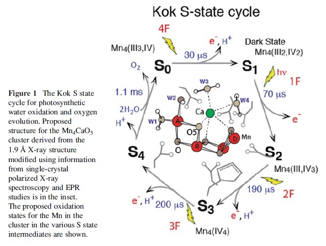
The redox potential of water is +2.5 V, while each photon raises the catalytic core’s redox potential by 1 V. So after the third stage, there is enough energy for the single Mn to remove an electron from a water molecule, leaving an OH radical and H+ ion. Then the catalytic core gets to the fourth stage, and provides the Mn atom with enough power to attack the OH radical and leaves a highly reactive O atom and another H+ ion. At this moment, the Ca atom in the cube plays its essential role. It is holding another water molecule in just the right place, so it can be attacked by this O atom, producing an O2 molecule, two more H+ ions and two electrons.The unique Mn3CaO4–Mn arrangement is present in all plants, algae and cyanobacteria, which suggests that this arrangement is essential. Not surprising, because it must be able to store the energy from four photons, and hold water molecules in just the right positions. This structure had to be complete otherwise it would not work at all—in splitting water and replenishing electrons. Therefore it could not be built up gradually by small changes by natural selection. This is because an incomplete intermediate system is no use at all, so it would not be selected.


And even this core would be useless without many other coordinated features. For example, as above, the energy involved is damaging for biological molecules. Yet there are key proteins required, but must be constantly repaired, so these mechanisms must be in place too. In fact, instability of these proteins made it hard to work out the core’s structure.9

If the most intelligent human designers can’t duplicate photosynthesis, then it’s perfectly scientific to believe that photosynthesis had a far more intelligent designer. 
This is especially so since Darwinian processes could not have generated photosynthesis, because there are too many intricate mechanisms necessary for it to work at all.
Plants were there right at the beginning

The core of the water-splitting complex is a heterodimer of two closely related proteins, namely D1 and D2. D1 is among the most highly conserved proteins in nature  It works for approximately 10 000 electron transfers and then becomes irreversibly damaged. The damage leads to a loss of photosynthetic capacity, especially at high light under nutrient-limiting conditions. 16 

Why has not nature developed a better oxygen-evolving machine? We suggest that the answer is: ‘Because it can't’. D1 is one of the most conserved proteins in the photosynthetic apparatus; the sequences are more than 80% identical between cyanobacteria and higher plants, and approximately 90% similar. The extraordinary conservation of this protein, which is irreversibly damaged by a product of its own catalysis, suggests that the restrictions imposed by protein–protein, protein–prosthetic group and protein–lipid interactions cannot be overcome.

There have been literally thousands of papers discussing the mechanism of the degradation of D1 in vivo and while the exact mechanism for the process remains controversial and the repair mechanism remains almost completely unknown, it is clear that the production of a reactive oxygen species, an ‘unintended’ consequence of the production and consumption of O2, is almost certainly the proximal cause.

It takes a large amount of energy to split water into hydrogen and oxygen. This is because oxygen really wants to acquire the two extra electrons necessary to fill its outer shell, and hydrogen is a relatively easy place to get them. This, in turn, means that water is a very stable molecule, and it, therefore, takes a lot of effort – in this case, the power of a car battery – to split it apart.  The structure of the Oxygen Evolving Complex was
determined only in 2006, and it is only in the last few years that the locations of each of the 46,630 atoms in photosystem II have been mapped.

"Of all the biochemical inventions in the history of life, the machinery to oxidize water — photosystem II — using sunlight is surely one of the grandest." (Sessions, A. et al, 2009)

The overwhelming source ofO2 on Earth is photobiological oxidation of water; neither the evolution nor the mechanism of this process is completely understood. Apparently, it arose once in a single clade of bacteria and was then appropriated via a single event, in which one cell engulfed another (endosymbiosis) to form a new symbiotic organism. The core of the oxidation machinery is photosystem II, a large protein complex containing four manganese atoms that are photocatalytically oxidized to create electron holes upstream.

Deep in the thylakoid membrane, the key process that produces breathable oxygen in the Earth’s atmosphere takes place, namely, the splitting of water molecules and the subsequent release of molecular oxygen. Without the OEC, no advanced life on earth would be possible.  This process, which occurs in the oxygen-evolving complex (OEC) of the intricately arranged multiprotein and pigment complex known as photosystem II (PSII), is driven by the strong electrochemical potential generated from the capture and conversion of visible light to chemical energy. First, chlorophyll molecules in PSII absorb photons and lose electrons. These electrons are then passed through an electron transport chain, creating redox potential strong enough to oxidize water, which leads to the evolution of molecular oxygen and the release of protons into the thylakoid lumen. This proton gradient is a major contributor to the proton motive force (PMF), which is used to biosynthesize ATP from ADP via ATP synthase.

The OEC contains an inorganic, flexible Mn4CaO5 cluster resembling a distorted chair, which is bound to a pocket formed by six amino acids from the D1 core subunit protein and one amino acid from the light-harvesting protein CP43 . The Mn4CaO5 cluster is stabilized, at least in part, by the PsBO subunits of the OEC. The OEC is composed of a cluster of manganese, calcium and chloride ions bound to extrinsic proteins. In cyanobacteria there are five extrinsic proteins in OEC (PsbO, PsbP-like, PsbQ-like, PsbU and PsbV), while in plants there are only three (PsbO, PsbP and PsbQ). Maintenance of the highly dynamic Mn4CaO5 cluster also requires the delivery of a constant supply of the proper levels of Mn2+ and Ca2+. 

The functional features of PSII in all oxygenic photosynthetic organisms are remarkably similar. The mechanism of water oxidation has remained virtually unchanged between green plants and cyanobacteria, and is similar in all higher plants. Studies on PSII from plants, algae, and cyanobacteria have revealed several PSII proteins that collectively regulate the unique redox environment of this inorganic catalytic center 12

Mainstream scientific papers quoted and cited below state that each of the extrinsic proteins, (PsbO, PsbP, PsbQ and PsbR)  of plants are ESSENTIAL, and each was tested upon mutated form, and the mechanism was found inefficient, and compromising the OEC function. Furthermore, a water network around the Mn4CaO5 cluster, and D1 protein subunit of PSII are also indispensable, and irreducible.  

PsbO
PsbO appears to be the most important extrinsic protein for oxygen evolution. PsbO lies closest to the Mn cluster where water oxidation occurs, and has a stabilising effect on the Mn cluster. As a result, PsbO is often referred to as the Mn-stabilising protein (MSP), although none of its amino acids are likely ligands for Mn. Calcium ions were found to modify the conformation of PsbO in solution 

The photosystem II (PSII) manganese-stabilizing protein (PsbO) is known to be the essential PSII extrinsic subunit for stabilization and retention of the Mn and Cl(-) cofactors in the oxygen evolving complex (OEC) of PSII, but its function relative to Ca(2+) is less clear.

What happens if PsbO is mutated ? 
The data presented here show that Asn, Glu, or Lys mutations in PsbO-Asp157 modify PsbO thermostability in solution, which is consistent with the previously reported perturbation of the functional assembly of PsbO-Asp157 mutants into PSII that caused inefficient Cl(-) retention by PSII.

PsbP
This family represents the PSII OEC protein PsbP. Both PsbP and PsbQ are regulators that are necessary for the biogenesis of optically active PSII. PsbP increases the affinity of the water oxidation site for chloride ions and provides the conditions required for high affinity binding of calcium ions [PMID: 9039496,PMID: 8910540]. The crystal structure of PsbP from Nicotiana tabacum (Common tobacco) revealed a two-domain structure, where domain 1 may play a role in the ion retention activity in PSII, the N-terminal residues being essential for calcium and chloride ion retention activity [PMID: 15031714]. PsbP is encoded in the nuclear genome in plants.

What happens if PsbO is mutated ?
these mutants show striking plant developmental and morphological defects. 4
These data indicate that assembly and/or maintenance of the functional MnCa cluster is perturbed in absence of PsbP, which impairs accumulation of final active forms of PSII supercomplexes. 5
Both psbPand psbQ inactivation mutants exhibited reduced photoautotrophic growth as well as decreased water oxidation activity under CaCl2-depleted conditions. 11

PsbQ
This family represents the PSII OEC protein PsbQ. Both PsbQ and PsbP (IPR002683) are regulators that are necessary for the biogenesis of optically active PSII. The crystal structure of PsbQ from spinach revealed a 4-helical bundle polypeptide. The distribution of positive and negative charges on the protein surface might explain the ability of PsbQ to increase the binding of chloride and calcium ions and make them available to PSII  6.
PsbP and PsbQ proteins are extrinsic subunits of photosystem II (PSII) and optimize the oxygen evolution reaction by regulating the binding properties of the essential cofactors Ca2 + and Cl−. These data suggest that the major function of PsbQ is to stabilize PsbP binding, thereby contributing to the maintenance of the catalytic Mn cluster of the water oxidation machinery in higher plant PSII. 8

What happens if PsbO is mutated ?
plants lacking PsbR or both PsbR and PsbQ are characterized by more pronounced defects in PSII activity. 7

PsbR 1
Additionally, the  PsbR protein has been found in plant PSII and anticipated to play a role in water oxidation, yet the physiological significance of PsbR has remained obscure.
What happens if PsbO is mutated ?
Using the Arabidopsis psbR mutant, we showed that the light-saturated rate of oxygen evolution is strongly reduced in the absence of PsbR, particularly in low light-grown plants.

Further essential parts :

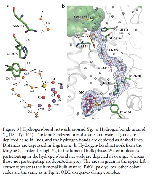
Role of a Water Network around the Mn4CaO5 Cluster in Photosynthetic Water Oxidation:  9
Around the Mn4CaO5 cluster, a hydrogen bond network is formed by several water molecules, including four water ligands.
These results suggest that the water network around the Mn4CaO5 cluster plays an essential role in the water oxidation mechanism particularly in a concerted process of proton transfer and water insertion during the S2 → S3 transition.
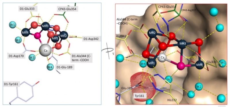
The reaction center cofactors involved in charge separation and water oxidation are coordinated by a pair of homologous protein subunits, known as D1 (PsbA) and D2 (PsbD). The D1 protein provides most of the ligands to the Mn4CaO5 cluster where water oxidation occurs. 10
Residue E354 of the CP43 coordinates Mn3 and Mn2 of the Mn4CaO5 cluster and R357 offers a hydrogen bond to O2 and O4 11
Direct ligation of the manganese ions appears to come from at least five amino acid residues of the D1 protein and one residue of the CP43 subunit 12


What happens if the two residues are mutated?
Site-directed mutants of these two residues show a severe impairment of the water oxidation cycle and fail to grow photoautotrophically. 

That means evolutionary intermediates are non-functional.  As we can see, there is a precise fit and size matching of the residues with the individual atoms of the clusters. How was this precision achieved? trial and error ? 

In addition to this, both CP43 and CP47 play important roles in water and proton access into or out of the cluster

The oxygen evolving complex (OEC) of photosystem II is irreducible complex. Fpls-010

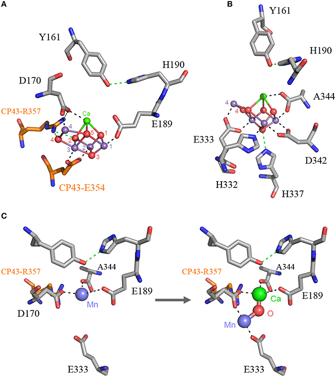
Figure 2. The Mn4CaO5 cluster of Photosystem II as resolved in the crystal structure  
Panel (A) shows the cluster coordinated by the inner ligands D170 and E189 and the ligands provided by the CP43 subunit, E354, and R357. 
Panel (B) shows the ligands provided from the C-terminus of the D1 protein. 
Panel (C) shows a proposal for the high-affinity Mn binding site. After oxidation of the first Mn(II) to Mn(III), which might occur concomitantly with the deprotonation of a ligating water molecule, Ca2+ binds. The binding of Ca2+ shifts the initially bound Mn(III) to a position similar to that of Mn4 in the intact cluster.

Anthony W. Larkum Early Archean origin of Photosystem II 09 November 2018
How and when Type II reaction centers diversified, and how and when one of these reaction centers evolved the capacity to oxidize water are questions that still remain to be answered.
https://onlinelibrary.wiley.com/doi/10.1111/gbi.12322

1) http://www.jbc.org/content/281/1/145.full
2) http://www.life.illinois.edu/govindjee/Electronic%20Publications/2011/2011_Najafpour_Gocindjee.pdf
3) http://www.ebi.ac.uk/interpro/entry/IPR002628
4) http://journals.plos.org/plosone/article?id=10.1371/journal.pone.0028624
5) http://www.sciencedirect.com/science/article/pii/S0005272809000929
6) http://www.ebi.ac.uk/interpro/entry/IPR008797
7) http://www.ncbi.nlm.nih.gov/pubmed/23647309
8  http://www.sciencedirect.com/science/article/pii/S0005272812000114
9) http://www.ncbi.nlm.nih.gov/pubmed/26716470
10) http://mbe.oxfordjournals.org/content/32/5/1310.full
11) http://journal.frontiersin.org/article/10.3389/fpls.2016.00257/full
12) http://www.ncbi.nlm.nih.gov/pmc/articles/PMC519205/
13) http://www.plantcell.org/content/early/2016/04/06/tpc.16.00269.full.pdf
14) http://science.sciencemag.org.sci-hub.cc/content/322/5901/540.full
15 ) Brian Cox, Wonders of life, page 37
16) https://www.ncbi.nlm.nih.gov/pmc/articles/PMC2606772/#fn2
17. https://creation.com/green-power-photosynthesis
18. https://sci-hub.tw/https://www.sciencedirect.com/science/article/abs/pii/S0010854507001877#:~:text=The%20invention%20of%20oxygenic%20photosynthesis,history%20of%20life%20on%20Earth.&text=This%20is%20distinct%20from%20bacterial,periplasmic%20proteins%20as%20electron%20donors.


Last edited by Otangelo on Tue May 17, 2022 4:51 am; edited 29 times in total

https://reasonandscience.catsboard.com

2The oxygen evolving complex (OEC) of photosystem II is irreducible complex. Empty Structure of the oxygen evolving complex Sun May 01, 2016 12:22 am

Otangelo


Admin

The oxygen-evolving complex and the mechanism of water oxidation by Photosystem II

"Of all the biochemical inventions in the history of life, the machinery to oxidize water — photosystem II — using sunlight is surely one of the grandest." (Sessions, A. et al, 2009) 1

During this process, four electrons are split off (one by one) from the manganese complex and are subsequently donated, one by one, to the oxidized P680, that is, P680+ , which is formed after the separation of charges, following a light reaction.

The overwhelming source ofO2 on Earth is photobiological oxidation of water; neither the evolution nor the mechanism of this process is completely understood. Apparently, it arose once in a single clade of bacteria and was then appropriated via a single event, in which one cell engulfed another (endosymbiosis) to form a new symbiotic organism.

The origin of the OEC is an enigma. There are several hypotheses for the origin of the OEC. One involves binuclear-manganese active sites, including ribonucleotide reductase, catalase, and arginase.

Without question, the most fascinating and complicated aspect of Photosystem II is the oxygen evolving activity (Vinyard et al., 2013). However, this remains one of the most active research areas in the entire field of photosynthesis, so the picture is changing rapidly, and the complexity of the process is such that there is still much to learn. The chemical reaction carried out by the OEC is the oxidation of water to molecular oxygen:

2H2O → O2 + 4H+ + 4 e− 

The thermodynamics of this redox process are formidable. Water is an extremely poor electron donor, which is as expected because oxygen is such a powerful electron acceptor. The redox potential for the half-reaction shown in the equation above  is +0.82 V at pH 7, and is somewhat higher in the acidic environment in which water oxidation takes place. In order to oxidize water, it is essential that an even stronger oxidant be available. This is provided by the oxidized reaction center chlorophyll P680+, which has a redox potential estimated to be as high as +1.2 V. The electrons that re-reduce P680+ ultimately come from water, and the protons that result from water oxidation are expelled into the thylakoid lumen. They are released into the lumen because of the vectorial nature of the membrane and the fact that the OEC is localized on the interior surface of the thylakoid. These protons are eventually released from the lumen to the stroma through the process of ATP synthesis. In this way, the electrochemical potential formed by the release of protons during water oxidation contributes to ATP formation. The chemistry of water oxidation is inherently a four-electron process, as four electrons must be extracted to make a single molecule of oxygen. The photochemistry of the reaction center takes place one electron at a time, so there is a fundamental mismatch between the intrinsic chemistries of these two processes. One could imagine several possible solutions to this problem, including at one extreme the cooperation of four reaction centers that all contribute to oxidize a single OEC, or at the other extreme, the storage of four oxidizing equivalents by a single reaction center/OEC working alone, which then oxidizes water to oxygen in a concerted step.

The latter possibility was given dramatic support in a classic series of experiments performed in the 1960s, first by Pierre Joliot and slightly later by Bessel Kok. Their experiments were a variation on the flashing light oxygen measurements made in the Emerson and Arnold experiment. However, these workers devised extremely sensitive electrochemical methods capable of detecting oxygen produced in a single flash.When this technique was applied to a sample that had been allowed to dark-adapt for several minutes, a remarkable pattern of oxygen production was observed, as shown in Fig. 7.5 (Kok et al., 1970). 

The oxygen evolving complex (OEC) of photosystem II is irreducible complex. Oxygen10

Little or no oxygen is produced on the first two flashes; a large amount is produced on the third, with lesser maxima with a period of four, until the oxygen production finally damps to a constant value by about the twentieth flash. Kok and coworkers proposed a schematic model (Fig. 7.5) explaining these observations. The model consists of a series of five states, known as S0 to S4, which represent successively more oxidized forms of the OEC. Light flashes advance the system from one S state to the next, until state S4 is reached. State S4 produces O2 without further light input and returns the system to S0. Occasionally, a center does not advance to the next S state upon flash excitation, and, less frequently, a center is activated twice by a single flash. These “misses” and “double hits” cause the synchrony achieved by dark adaptation to be lost, and the oxygen yield eventually damps to a constant value. After this steady state has been reached, a complex has the same probability of being in any of the states S0 to S3 (S4 is unstable and occurs only transiently) and the yield of O2 becomes constant.
States S2 and S3 decay in the dark, but only as far back as S1, which is stable in the dark. Therefore, after dark adaptation, approximately three-fourths of the oxygen-evolving complexes appear to be in state S1, and one-fourth in state S0. This distribution of states explains why the maximum yield of O2 is observed after the third of a series of flashes given to dark adapted chloroplasts. This S state mechanism formally explains the observed pattern of O2 release, but not the chemical nature of the S states or the actual chemical mechanism of the process. It has been known for many years that Mn is an essential cofactor in the water-oxidizing process, and for many years it was suspected that the S states represent successively oxidized states of an Mn-containing enzyme. 

This hypothesis has been confirmed by a variety of experiments, most notably X-ray absorption and EPR studies, both of which detect the Mn directly. Analytical experiments indicate that four Mn atoms are associated with each oxygenevolving complex. Other experiments have shown that Cl− and Ca2+ ions are essential for O2 evolution, although their precise mechanistic roles have not yet been determined. A structural model for the Mn cluster is shown in Fig. 7.6.

The oxygen evolving complex (OEC) of photosystem II is irreducible complex. Oec10

 Many of the amino acid residues that serve as ligands for the Mn cluster come from the D1 protein, as well as the C terminal carboxyl terminus of the D1 protein. Consequently, the OEC is located off center towards the D1 side of the core reaction center complex (Fig. 7.3).

The oxygen evolving complex (OEC) of photosystem II is irreducible complex. Photos10

 An additional protein that is important in the oxygen evolution process is a 33 kDa peripheral membrane protein (PsbO protein). This protein is found in all oxygenic photosynthetic organisms. Removal of this protein does not abolish oxygen evolution activity entirely, but it destabilizes the Mn cluster and perturbs the system. Two additional peripheral proteins, with masses of 23 and 17 kDa (PsbP and PsbQ), are part of the complex that stabilizes the OEC in eukaryotic organisms, but are not found in cyanobacteria. Instead, two different proteins, PsbU and PsbV, are found in this position. All these extrinsic proteins, as they are often called, help to stabilize the OEC and protect it against damage, but they do not directly bind the Mn cluster . There is one intermediate electron carrier between P680 and the OEC. After P680 is oxidized in the primary photochemistry, it is re-reduced in tens of nanoseconds by electron transfer from a tyrosine amino acid side chain (Y161) from the D1 protein, thus forming a tyrosine radical . This tyrosine residue is known as YZ, and it serves to connect the OEC to P680 (Fig. 7.5). The YZ radical can be observed by EPR. A second tyrosine in the symmetry-related position in the D2 protein is known as YD. It appears not to be involved in mainstream electron flow, but, surprisingly, it is usually found in the oxidized radical form. Many mechanisms have been put forward to explain how the OEC accomplishes the difficult chemistry of oxygen evolution. Most of these mechanisms involve the essential role of the tyrosine in a coupled proton and electron (hydrogen atom) transfer process . This proton coupled electron transfer (PCET) mechanism stresses the importance of maintaining charge neutrality in the OEC. Otherwise, the energetics of the removal of additional electrons from a highly positively charged center found in the higher S states would be prohibitively difficult. Current ideas about the detailed chemical mechanism of oxygen evolution incorporate information from the structure of the OEC, biochemical data, theoretical calculations, and principles of inorganic chemistry .

The OEC in PS II contains a heteronuclear Mn4CaO5 cluster (Figure 1, inset) which catalyzes the water oxidation reaction ,that couples the four-electron oxidation of water with the one-electron photochemistry occurring at the PS II reaction center, P680. The OEC cycles through five intermediate S states (S0 to S4, known as the Kok cycle) that corresponds to the abstraction of four successive electrons from the OEC (Figure 1)

The oxygen evolving complex (OEC) of photosystem II is irreducible complex. Kik_cy10

The dark stable S1 state is the first oxidized state and subsequent illumination leads to the formation of the S2 and S3 states. Once four oxidizing equivalents are accumulated
(S4 state), a spontaneous reaction occurs that results in the release of O2 and the formation of the most reduced state, the S0 state. Upon further light excitation, the initial S1 state is formed once again, and the catalytic cycle is resumed. Given the importance of PS II in maintaining life and the anticipated role of lightinduced water-splitting for building a renewable energy economy, understanding the structure of the Mn4CaO5 catalyst and the mechanism of the water oxidation reaction is considered to be one of science’s grand challenges [4]. Although details of the chemistry involved in water oxidation are slowly emerging, the mechanism of the reaction is not yet clear. In this chapter, we describe results from X-ray spectroscopy and diffraction studies, especially the use of time-resolved X-raymethods for room temperature studies using the recently introduced X-ray lasers. We will also describe the use of membrane inlet mass spectrometry for the elucidation of the mechanism of water-oxidation and its utility for time-resolved X-ray spectroscopy and diffraction measurements.

Speaking of photosynthesis, Japanese scientists have achieved the imaging of the “Crystal structure of oxygen-evolving photosystem II at a resolution of 1.9?Å,”  zooming in almost twice as far as previous studies.  Their paper, published in Nature,1 spoke of the reactor as “indispensable for sustaining life on Earth.”  It includes detailed drawings of the 20 subunits involved with numerous molecular contacts.
The particular part of the reactor that splits water molecules and combines oxygen atoms into the O2 gas we breathe they said is “one of nature’s most fascinating and important reactions.”  Understanding Photosystem II may help humans to mimic plants’ ability to split water efficiently at ambient temperatures, leading to renewable energy for a multitude of applications.  The ability lives all around us if we can tap into its secrets. 3


Crystal structure of oxygen-evolving photosystem II at a resolution of 1.9 Å 2

The electron densities of the four manganese atoms and the single calcium atom in the oxygen-evolving complex were well defined and clearly resolved, and the electron density for the calcium atom was lower than those of the manganese atoms, allowing us to identify the individual atoms unambiguously 4,5 (Fig. 2a).

The oxygen evolving complex (OEC) of photosystem II is irreducible complex. Mn4cao10

Figure 2 | Structure of the Mn4CaO5 cluster. 
a, Determination of individual atoms associated with the Mn4CaO5 cluster. The structure of the cluster was superimposed with the 2Fo2Fc map (blue) contoured at 5s formanganese and calcium atoms, and with the omit map (green) contoured at 7s for oxygen atoms and water molecules. 
b, Distances (in a˚ngstro¨ms) between metal atoms and oxo bridges or water molecules. 
c, Distances between each pair of manganese atoms. 
d, Distances between the manganese and the calcium atoms. e, Stereo view of the Mn4CaO5 cluster and its ligand environment. The distances shown are the average distances between the two monomers. Manganese, purple; calcium, yellow; oxygen, red; D1, green; CP43, pink.

In addition, five oxygen  atoms were found to serve as oxo bridges linking the five metal atoms from the omit map (Fig. 2a). This gives rise to a Mn4CaO5 cluster. Of these five metals and five oxygen atoms, three manganese, one calcium and four oxygen atoms form a cubane-like structure in which the calcium and manganese atoms occupy four corners and the oxygen atoms occupy the other four. The bond lengths between the
oxygens and the calcium in the cubane are generally in the range of 2.4–2.5A˚ , and those between the oxygens and manganeses are in the range of 1.8–2.1A˚ (Fig. 2b). However, the bond length between one of
the oxygens at the corner of the cubane (O5) and the calcium is 2.7A˚ , and those between O5 and the manganeses are in the range of 2.4–2.6A˚ . Owing to these differences in bond lengths, the Mn3CaO4 cubane is not an ideal, symmetric one. The fourth manganese (Mn4) is located outside the cubane and is linked to two manganeses (Mn1 and Mn3) within the cubane by O5 and the fifth oxygen (O4) by a di-m-oxo bridge. In this way, every two
adjacent manganeses are linked by di-m-oxo bridges: Mn1 and Mn2 are linked by a di-m-oxo bridge via O1 and O3, Mn2 and Mn3 are linked via O2 and O3, and Mn3 and Mn4 are linked via O4 and O5. The calcium is linked to all four manganeses by oxo bridges: to Mn1 via the di-m-oxo bridge formed by O1 and O5, to Mn2 via O1 and O2, to Mn3 via O2 and O5, and to Mn4 via the mono-m-oxo bridge formed by O5. The whole structure of the Mn4CaO5 cluster resembles a distorted chair, with the asymmetric cubane serving as the seat base and the isolated Mn4 and O4 serving as the back of the chair. The cubane-like structure has been reported previously 4,9–12, but the oxo bridges and exact distances among the individual atoms could not be determined at the medium resolution achieved previously 4.

The distances among the four manganeses determined for monomer A are 2.8A˚ (Mn1–Mn2), 2.9A˚ (Mn2–Mn3), 3.0A˚ (Mn3–Mn4; 2.9A˚ for monomer B), 3.3A˚ (Mn1–Mn3) and 5.0A˚ (Mn1–Mn4) (Fig. 2c). The distances between the calcium and the four manganeses are 3.5A˚ (Ca–Mn1), 3.3 A˚ (Ca–Mn2), 3.4A˚ (Ca–Mn3) and 3.8 A˚ (Ca–Mn4) (Fig. 2d; for the corresponding distances inmonomer B and the average distances between the two monomers, see Supplementary Table 3). These distances are largely different fromthose reported in the previous crystal structures3–6; however, they are comparable to those reported from extended X-ray absorption fine structure studies 13,14 if we consider that there is an error of 0.16A˚ in the distances determined from the X-ray structural analysis (Methods).

In addition to the five oxygens, four water molecules (W1 to W4) were found to be associated with the Mn4CaO5 cluster, of which W1 and W2 are coordinated to Mn4 with respective distances of 2.1 and 2.2A˚ , and W3 and W4 are coordinated to the calcium with a distance of 2.4A˚ . No other water molecules were found to associate with the other three manganeses, suggesting that some of the four waters may serve as the substrates for water oxidation.In addition to the direct ligands of the Mn4CaO5 cluster, we found that D1-Asp 61, D1-His 337 and CP43-Arg 357 are located in the second coordination sphere and may have important roles in maintaining the structure of the metal cluster, in agreement with various reports showing the importance of these three residues in maintaining the oxygen-evolving activity15–19. One of the guanidinium g-nitrogens of CP43-Arg 357 is hydrogen-bonded to both O2 and O4 of the Mn4CaO5 cluster, whereas the other is hydrogen-bonded to the carboxylate oxygen of D1-Asp 170 and to that of D1-Ala 344. The imidazole e-nitrogen of D1-His 337 is hydrogen-bonded to O3. These two residues may thus function to stabilize the cubane structure of the metal cluster as well as to provide partial positive charges to compensate for the negative charges induced by the oxo bridges and
carboxylate ligands of the metal cluster. The carboxylate oxygen of D1-Asp 61 is hydrogen-bonded to W1, and also to O4 indirectly through another water molecule, suggesting that this residue may also contribute to stabilizing the metal cluster. Furthermore, D1-Asp 61 is located at the entrance of a proposed proton exit channel involving a chloride ion (Cl21; see below), suggesting that this residue may function in facilitating proton exit from the Mn4CaO5 cluster5,20,21.

The most significant structural feature of the Mn4CaO5 cluster, which may be important for elucidating the mechanism of the water-splitting reaction, is its distorted chair form. The large distortion is principally caused by the existence of the calcium and O5 in the Mn4CaO5 cluster, as described above. The apparently longer distances between O5 and metal atoms suggested that the corresponding bonds are weak, and that O5 may therefore have a lower negative charge than the valence of 22 expected for normal oxygen atoms in oxo bridges. This in turn suggests that O5 may exist as a hydroxide ion in the S1 state and may provide one of the substrates for dioxygen formation. Because both W2 and W3 are within the hydrogen-bond distances to O5, one of these two waters may provide another substrate. Because the transition between S0 and S1 is fastest in the Kok cycle, the proton released during this transition may be accepted by D1-Tyr 161 (also termed YZ), which is deprotonated by means of protoncoupled electron transfer (PCET; see below).W3is closer to YZ than is O5 (Fig. 3a)

The oxygen evolving complex (OEC) of photosystem II is irreducible complex. Mn4cao11

and may be a more favourable candidate than O5 as the proton-releasing group. Thus, W3 rather than O5 may be a hydroxide ion in the S1 state, suggesting that O–O bond formation may occur between W2 and W3. In any case, our results suggest that the O–O bond formation occurs in two of the three species O5, W2 and W3.

Conclusion

The high-resolution structure of PSII reveals the geometric arrangement of the Mn4CaO5 cluster as well as its oxo bridges and ligands, and four bound water molecules. This provides a basis for unravelling the mechanism of water splitting and O–Obond formation, one of nature’s most fascinating and important reactions. In addition, our determination of the precise arrangement of amino-acid side chains and cofactors gives us a solid structural understanding of energy migration, electron transfer and water-splitting reactions taking place within PSII.


Structure of manganese–calcium cluster in PSII 2

In 2011, Umena et al. 7 reported the crystal structure of manganese–calcium cluster of PSII at an atomic resolution using a sliding-oscillation method to reduce the X-ray dose illuminated on a unit volume of the crystal. In this structure, one calcium and four manganese ions are bridged by five oxygens (Fig. 1).

The oxygen evolving complex (OEC) of photosystem II is irreducible complex. Dddd10

Four water molecules were also found in this structure; and two of them may serve as the substrate for water oxidation. The whole structure of the Mn4CaO5 cluster resembles a distorted chair, with the asymmetric cubane serving as the seat base and the isolated Mn4 and O4 atoms serving as the back of the chair1 (Fig. 1). The structural models from structure refinement of computational models based on EXAFS simulations by Brudvig’s group and Siegbahn’s group25 are also close to the structure found in the 1.9A˚ crystal structure of Umena et al.

The origin of the OEC is an enigma.

Sauer and Yachandra proposed a possible evolutionary origin for the Mn4 cluster of the photosynthetic water oxidation complex from natural MnO2 precipitates in the early oceans. In the past fewyears, there has been a tremendous surge in research on the synthesis of various manganese compounds aimed at simulating the OEC of PSII  but very few inorganic complexes have been claimed to be functional models for the OEC.

It appears that an effective mechanism coupled to structural changes in the Mn4Ca cluster, with the optimum positions of associated ligands, during the Kok’s S-state cycle, for water to reach the Mn may operate in PSII. The functional PSII is characterized by a branched water supply structure with multiple control points.

A number of experiments demonstrate a requirement for Ca2+ for the functional assembly and stability of the OEC.

1) http://www.life.illinois.edu/govindjee/Electronic%20Publications/2011/2011_Najafpour_Gocindjee.pdf
2) http://www.nature.com/nature/journal/v473/n7345/full/nature09913.html
3) http://www.creationsafaris.com/crev201105.htm


Last edited by Admin on Fri Sep 29, 2017 4:58 pm; edited 14 times in total

https://reasonandscience.catsboard.com

3The oxygen evolving complex (OEC) of photosystem II is irreducible complex. Empty Assembly of the Mn Cluster Sun May 01, 2016 2:08 pm

Otangelo


Admin

Photoactivation: The light-driven assembly of the water oxidation complex of photosystem II  2

Introduction 39 A decline in the photosynthetic activity of oxygenic photosynthetic organisms due to light stress has been  described as photoinhibition. The  primary damage occurs within the reaction center of Photosystem II (PSII). It is distinct from the concurrent oxidative  damage to the machinery of protein synthesis, which compounds the problem since de novo protein synthesis is necessary for the replacement of damaged PSII proteins. The precise mechanism of PSII photoinhibition in vivo remains under debate. Despite this uncertainty, it is evident that the D1 reaction center protein is the primary target for photodamage and this leads to an increased turnover rate of D1, in comparison to other PSII proteins, upon exposure to high light intensities. To cope with light  stress, all oxygenic photosynthetic organisms have developed protective mechanisms both to minimize the effects of exposure to excess light and to efficiently repair the damage when it occurs. Overall, the efficiency of  photosynthetic electron transfer decreases markedly only when the rate of damage exceeds the rate of repair. A  crucial phase of the de novo biogenesis of PSII, as well as the damage repair process, is the assembly of the Mn4CaO5 complex. This involves the oxidative assembly of Mn2+ and Ca2+ 52 ions into the coordination environment of the PSII 53 water-oxidation complex (WOC) in a light-driven process called photoactivation. 

PSII damage and D1 replacement 

The entire process of PSII damage-repair cycle can be described as follows:

 i) damage occurring to PSII,
 ii) signaling of this damage
 iii) monomerization of PSII dimer and partial disassembly of PSII monomer
 iv) degradation  of D1 and insertion of a newly synthesized D1 into PSII sub-complex, and
 v) reassembly of holoenzyme and  photoactivation of the Mn4CaO5 cluster  (Fig. 1).

The oxygen evolving complex (OEC) of photosystem II is irreducible complex. Psii_d10
Figure 1. Schematic repair pathway for photodamaged PSII. The process can be divided into the three main phases:
1.) damage recognition and partial disassembly of photodamage PSII complexes,
2.) D1 degradation and replacement,and
3.) reassembly of the subunits and light-driven assembly (photoactivation) of the Mn4O5Ca metal cluster.

We briefly  outline some features of the overall PSII assembly and repair process to place the assembly of the Mn4CaO5 cluster  in context. Monomerization of dimeric PSII has been suggested to result from the detachment or rearrangement of PsbO,  one of three luminal extrinsic subunits of PSII . The basis of this assessment is the failure to accumulate dimeric PSII in a mutant of Synechocystis sp. PCC 6803 (hereafter Synechocystis 6803) lacking PsbO. In plants and green algae, it has also been proposed that PSII core phosphorylation might  trigger disassembly of PSII dimer to form monomer by acting alone or in conjunction with PsbO. Detachment of CP43 from PSII monomer leads to the formation of so-called RC47 complex which is a pivotal sub-complex for further replacement of damaged D1 during PSII repair. Given the  fact that PsbO functions as PSII manganese-stabilizing protein and CP43 participates with D1 in ligating the Mn4CaO5  cluster, it is conceivable that photodamage to Mn4CaO5 cluster might cause the detachment of these two subunits. 

It is also interesting to note that the assembly and disassembly of the Mn4O5Ca regulates the coupling of the phycobilisome to the cyanobacterial PSII reaction center such that centers without an intact metal cluster are not  efficiently coupled with respect to energy transfer from the phycobilisome. Radioactive pulse-chase experiments showed that translation inhibitors slow D1 degradation, suggesting that D1 degradation and new D1 synthesis are synchronized. Increased turnover of D1 could  be a generalized response to damage-promoting light conditions, with all D1 copies prone to increased probability  of replacement or there could be a specific targeting mechanism that replaces only damaged D1 copies. Intuitively, a targeting mechanism seems more likely. However, despite good circumstantial evidence, direct evidence for the  specific targeting of PSII centers with damaged D1 has not been obtained, mainly because it is technically difficult to  separately track damaged and undamaged forms of D1 through the replacement process. Recently, targeting has  been inferred from experiments where cells are allowed to express two alternative forms of the D1 protein in the  same cell, with one wild-type form and the other a light-sensitive form. The analysis indicates that only the light-sensitive version of D1 and not the wild-type version is turned over very rapidly.

PratA interacts with the D1 C-terminus and may bind Mn2+  possibly facilitating the  assembly of the Mn4O5Ca . Psb27 is found to bind to forms of the PSII 149 complex thought to represent assembly and/or disassembly intermediates  and deletion of the protein affects photoactivation of the complex. Thus, Psb27 and PratA are especially good candidates for facilitating  photoactivation of the Mn4CaO5. Indeed, there is good reason to believe that the published in vitro assembly  experiments are missing assembly cofactors, which may explain why the yield of active PSII centers produced by in  vitro photoactivation of Mn4CaO5 clusters by biochemical methods is invariably lower than intact cells.

The oxygen evolving complex (OEC) of photosystem II is irreducible complex. Psii_d11

Figure 2. Coordination environment of the assembled Mn4CaO5 H2O-oxidation complex of PSII. 
The high affinity site of Mn2+  binding and photooxidation during the initial phase of the assembly process minimally involves D1- Asparate located in the vicinity of Mn4 in the final complex. The initial state of the complex for photoassembly appears to involve the binding of one Mn2+  at the high affinity site together with one Ca2+ ion that modulates the ligand environment of the Mn2+  possibly via the formation of a bridging water or hydroxide, although the presence of the Ca2+  does not appreciably change the binding affinity of the Mn2+  at the high affinity site. The C-terminal polypeptide backbone  carboxylate of D1-Alanine344, which is available only following proteolytic cleavage of the precursor form of the D1 protein (pD1), is also critical for the assembly process, although it too does not markedly alter the binding of Mn2+  at the high affinity site. Figures developed upon 3D coordinates (PDB 4UB6)  of the published X-ray diffraction model.

Mechanism of photoactivation 

Coordinating residues of Mn4CaO5 cluster
According to 1.9 Å PSII crystal structure, Mn4O5Ca cluster coordinated by one nitrogen  ligand from D1-His332 and six carboxylate ligands from D1-Asp170, D1-Glu189, D1-Glu333, D1-Asp342, D-Ala344, 172 CP43-Glu354 (Fig. 2.). Three of them, D1-Glu333, D1-Asp342, and CP43-Glu354, form μ–carboxylate bridges between  Mn (Mn(1)–Mn(2) (Asp342), Mn(2)–Mn(3) (CP43-Glu354), and Mn(3)–Mn(4) (Glu333)). D1-Asp170 and the C- 174 terminal carboxylate group of D1-Ala344 bridge Ca with Mn(4) and Mn(2), respectively. The Mn(4) has been referred  to as the ‘dangler manganese’  because it is located outside the semi-cubic cluster formed by  the other four metals of the cluster, Ca, Mn(1), Mn(2), and Mn(3). Both D1-Glu189 and D1-His332 serve as  monodentate ligands to Mn(1). The D1-Asp170 plays an especially crucial role during the assembly process since it helps form the so-called ‘high affinity site’ involved in the initial photooxidation of Mn2+

Two-quantum model of photoactivation 
The assembly of the metals of the Mn4O5Ca requires light to induced charge separation to oxidize and strongly bind of Mn ions. It is important to note that the assembly is an oxidative process that involves removal of electrons  from the Mn ions and the formation of oxo-bridges between the metals of the cluster with the bridging oxygen  atoms (shown in red, Fig. 2) derived from water molecular coordinated to the metal ions. The oxidative assembly  utilizes the same light-driven charge separation events within the photochemical reaction center that subsequently  drive photosynthetic electron transfer in the fully functional enzyme. Apart from the definition of the Mn-binding  site characteristics and some very well-defined kinetic features that govern the development of H2O-oxidation  activity, photoactivation remains poorly understood. The quantum efficiency of photoactivation is very low, typically in the range of ~1%, which is much lower than for photosynthetic water oxidation in the assembled PSII (>90%) even in intact systems. The kinetic model of photoactivation, termed as “two-quantum series model”, was originally observed during photoactivation as a function of either light intensity or flash interval using fixed numbers of Xe light flashes. 

These pioneering studies showed that the quantum efficiency for photoactivation is low at low light intensities, reached a maximum at intermediate intensities, and were again low at high light intensities. Equivalently, the  quantum efficiency is low when saturating, single turnover flashes are given at long intervals, maximum at  intermediate flash frequencies (~1 per second), and were again low when the flashes are given with short intervals  between flashes. From these features, Cheniae derived a minimal model, the so-called two-quantum model that  postulated the light-induced Mn assembly with at least one unstable chemical intermediate as depicted in Fig. 3. The first photoevent involves the high quantum yield photooxidation of a single Mn2+ to Mn3+ ion  at the unique high affinity Mn-binding site. 

The resultant Mn3+ 200 species (B) can spontaneously convert to C in the dark with a 100-150 millisecond half-time, with a kinetic constant designated kR in the scheme in  Fig. 3. A second quantum of light must be absorbed to convert the nascent complex into the first stable intermediate  D as shown in Fig. 3 as C⇒D. 

The oxygen evolving complex (OEC) of photosystem II is irreducible complex. Psii_d12

The formation of a labile intermediate, t1/2 ~1-2 seconds, accounted for the optimum in light intensity or, alternatively, flash frequency, utilized for the assembly process. Photoactivation using saturating  single turnover flashes is optimal with flash spacing of ~1 second, which is enough time to allow the dark  rearrangement to occur (kR), but short enough to minimize the decay of the intermediate(s). If, however, the flash  interval is too long, the second flash is not in time to trap forward progress and the reactants decay ((kD1, KD2, Fig. 3). The molecular nature of the process occurring during this dark rearrangement period (B→C) is not clear, and its  understanding is key to understanding the overall molecular mechanism. After the initial two Mn are photoligated, subsequent Mn appear to be added with high quantum yield.

Of the many examples providing experimental support for the two quantum mechanism, perhaps the most  striking are the experiments of Miyao, which showed a minimal two quantum requirement in an experiment where  as few as five flashes restoring nearly 20% of the maximal activity . This amounts  to several percent assembly per flash, which is remarkable given that the typical per flash yield is often on the order  of 1% or even lower. That experiment and others also showed that the instability of the intermediates could be  minimized by preventing the back-reaction of the electrons from the acceptor side of the PSII reaction center. This also fits with another early result showing that the intermediates of assembly are highly sensitive to reductant  and fits with the concept that the  formation of state C (eligible for utilizing the second quantum) occurs with low frequency and/or once formed, the quantum yield of photooxidation of the second Mn2+  occurs with low quantum yield. It has been speculated the rearrangement (kR) involves a protein conformational change required for the binding and subsequent photooxidation of the second Mn2+. However, if (B→C) is indeed a protein structural change, then it is unlikely a large scale conformational rearrangement since carboxy terminal ligands are already close to high affinity site ligand D1-Asp170  during the first photooxidation (A⇒B) . Also, whether the dark unstable intermediate is B or C  (or both) remains unresolved. Given this uncertainty, Fig. 3 shows both decays are possible (kD1 and kD2). The development of a highly sensitive and fast Clark-type oxygen electrode  led to the assignment of additional photoactivation intermediates and has provide alternative parameter estimates for the kinetic components. At the same time, the use of this apparatus makes comparisons difficult primarily because to the different illumination regimes.

 Most of the original experiments utilized single turnover Xe flashes for actinic illumination. In contrast, the photoactivation studies using the fast Clark-type oxygen electrode employed 30  millisecond red LED pulses promote optimum yields of assembled center. This relatively long duration of the LED light pulses allows greater mixing of different assembly states because of the  possibility of having multiple ‘hits’ per center per pulse. That said, the 30 millisecond duration of the pulse is  relatively short with respect to the t1/2 ~150 millisecond of the B→C rearrangement and therefore the majority of  those centers in the initial state that were excited (i.e. those undergoing A⇒B), will not be ready to utilize the second  quantum and would thus the LED pulse would be effectively similar to a single turnover flash distributed in time over the population of centers. Variations and refinements of the original two-quantum model have been advanced base  upon alternative techniques for illumination and O2 detection during photoactivation . The  multiflash experiments of Hwang  using staggered Xe single turnover flashes revealed a new kinetic  intermediate, more rapid rearrangement, although where it is in the sequence could not be established owing to  high miss factor (low quantum efficiency) and the associated de-phasing of the assembly during the flash induced  assembly process.


Assembly of the Mn Cluster 1

The Mn4CaO5 cluster is not chemically stable. It must be assembled during the biogenesis of PSII centers and during the repair of damaged PSII centers (reviewed in Vinyard et al., 2013). Damage to PSII occurs frequently and would limit the photosynthetic productivity without a repair mechanism. The damaged PSII complexes can be recycled by replacement of damaged protein subunits followed by the reassembly of the OEC. This latter process, which requires the incorporation of manganese, calcium and chloride, can be mimicked in vitro and is then referred to as photoassembly or photoactivation. Photoassembly studies provide a deeper understanding
of the synthesis of the water-splitting machinery. Many studies provide insight into the photoassembly process, including the necessary cofactors, the protein environment and the experimental conditions, such as pH, light intensity and exogenous electron acceptors (reviewed in Becker et al., 2011). Photoassembly studies with PSII membranes are favored because chloroplasts and thylakoids are at risk of a restricted accessibility of the soluble cofactors and PSII cores are more vulnerable to photoinhibition (Debus, 1996). The photoassembly process was found to have a high complexity and up to now, there is no completed model for this process. However, Cheniae and Martin suggested a ‘two-quantum model’ for the first steps of photoassembly, which was successively confirmed . In this model, the first step of photoassembly consists of the binding of one Mn2+ at a high affinity site. That represents the initial intermediate (IM0) (Fig 3.2).

The oxygen evolving complex (OEC) of photosystem II is irreducible complex. Ewrwer10

The first photooxidation produces Mn3+ by oxidizing the Mn2+ (IM1) and a light independent rearrangement occurs (IM1*). This rearrangement is probably induced by the incorporation of Ca2+  and is accompanied by the binding of the second Mn2+ . It is expected that the next photooxidation leads to a Mn(III)2 complex. The next binding steps of the two missing manganese ions and photooxidations could not be kinetically resolved because of the fast cooperative binding to the Mn(III)2 complex. 

1) http://solarfuels.eu/wp-content/uploads/2015/09/Bachelorarbeit-Daniel-Ohde.pdf
2) http://journal.frontiersin.org/article/10.3389/fpls.2016.00578/abstract


Last edited by Admin on Mon May 02, 2016 4:09 am; edited 1 time in total

https://reasonandscience.catsboard.com

Otangelo


Admin

Understanding photosynthesis: How plants use catalytic reactions to split oxygen from water

Splitting hydrogen and oxygen from water using conventional electrolysis techniques requires considerable amounts of electrical energy. But green plants produce oxygen from water efficiently using a catalytic technique powered by sunlight – a process that is part of photosynthesis and so effective that it is the Earth's major source of oxygen.

If mimicked by artificial systems, this photocatalytic process could provide abundant new supplies of oxygen and, possibly hydrogen, as a by-product of producing electricity. However, despite its importance to the survival of the planet, scientists don't fully understand the complex process plants use to harness the sun's energy.

A paper to be published April 2 in the journal Proceedings of the National Academy of Sciences moves scientists closer to that understanding by showing the importance of a hydrogen bonding water network in that portion of the photosynthetic machinery known as photosystem II. Using Fourier transform infrared spectroscopy (FT-IR) on photosystem II extracted from ordinary spinach, researchers at the Georgia Institute of Technology tested the idea that a network of hydrogen-bonded water molecules plays a catalytic role in the process that produces oxygen.

"By substituting ammonia, an analog of the water molecule that has a similar structure, we were able to show that the network of hydrogen-bonded water molecules is important to the catalytic process," said Bridgette Barry, a professor in Georgia Tech's School of Chemistry and Biochemistry and the Petit Institute for Bioengineering and Biosciences. "Substituting ammonia for water inhibited the activity of the photosystem and disrupted the network. The network could be reestablished by addition of a simple sugar, trehalose."

The research was supported by the National Science Foundation (NSF) and published in the Early Online edition of the journal.

In the chloroplasts of green plants, algae and cyanobacteria, oxygen is produced by the accumulation of photo-induced oxidizing equivalents in a structure known as the oxygen-evolving complex (OEC). The OEC contains manganese and calcium ions. Illumination causes oxidation of manganese ions in the OEC. Short laser flashes can be used to step through the reaction cycle, which involves four sequential light-induced oxidation reactions. Oxygen is produced on the fourth step, and then is released from the OEC.

This so-called S state cycle resets with the binding of the substrate, water. Scientists have proposed that a hydrogen bond network, which includes multiple water molecules bound to manganese ions, calcium ions, and protein amide carbonyl (C=O) groups, forms an electrostatic network surrounding the OEC. In this scenario, the extensive hydrogen-bond network would then serve as a component of the catalyst, which splits off oxygen.

To study the process, Barry and graduate student Brandon Polander used precision FT-IR spectroscopy to describe how the network reacts to a short laser flash. The second harmonic of a pulsed Nd-Yag laser was used as the light source. This illumination causes the OEC to undergo one step in its catalytic cycle, the so-called S1 to S2 transition. An infrared spectrum was recorded before and after a laser flash to the photosystem sample, which was isolated from supermarket spinach.

The exquisite sensitivity of FT-IR spectroscopy allowed them to measure changes in the bond strength of the protein C=O groups. The energies of these C=O groups were used as markers of hydrogen bond strength. The brief laser flash oxidized a manganese ion and caused a change in the strength of the C=O bond, which reported an increase in hydrogen bonding to water molecules. When ammonia was added as an inhibitor, a decrease in C=O hydrogen bonding was observed instead. Addition of trehalose, which is known to change the ordering of water molecules at the surface of proteins, blocked this effect of ammonia.

The study describes the coordinated changes that must occur in the protein to facilitate the reaction and shows that the strength of the hydrogen-bonded network is important.

"This research helps to clarify how ammonia inhibits the photosystem, which is something that researchers have been wondering about for many years," Barry explained. "Our work suggests that ammonia can inhibit the reaction by disrupting this network of hydrogen bonds."

The research also suggests that in design of artificial devices that carry out this reaction, sustaining a similar hydrogen-bonding network may be important. The stabilizing effect of trehalose discovered by Polander and Barry may also be important.

Beyond the importance of understanding the photosynthetic process, the work could lead to new techniques for producing hydrogen and oxygen using sunlight. One possibility would be to add a biomimetic photocatalytic process to a photovoltaic system producing electricity from the sun.

"In terms of providing new sources of energy, we still have lessons to learn from plants about how they carry out these critical processes,"
Barry said. "It would be a great advance for the planet to have new, sustainable, and inexpensive processes to carry out this reaction."

Ultimately, she hopes the full water oxidizing cycle can be explored and potentially harnessed or imitated for oxygen and energy production.

"We are only looking at a single part of the overall reaction now, but we would like to study the entire cycle, in which oxygen is produced, to see how the interactions in the water network change and how the interactions with the protein change," Barry said. "The work is another step in understanding how plants carry out this amazing series of photosynthetic reactions."

Functional Models for the Oxygen-Evolving Complex of Photosystem II


Water splitting: Ultrahigh resolution data reveals reaction mechanisms

Oxygenic photosynthetic organisms utilize energy from the sun to split water into protons, electrons and oxygen—products vital to life on earth. The process takes place through light-induced electron transfer reactions in a membrane protein complex photosystem II, but so far the resolution of structural studies on the protein complex has been too limited to ascertain the mechanism of these reactions in detail.

Now Jian-Ren Shen at Okayama University in collaboration with researchers at Osaka City University in Japan has solved the structure of the photosystem II complex at an unprecedented resolution. They improved the quality of the photosystem II crystals significantly, and obtained X-ray diffraction data with a resolution of 1.9 Å.

A time-resolved vibrational spectroscopy glimpse into the oxygen-evolving complex of photosynthesi

The oxygen evolving complex (OEC) of photosystem II is irreducible complex. F1.large

Oxygen release pattern and Kok S state model for photosynthetic oxygen evolution. (A) Typical oxygen release pattern observed for all organisms carrying out oxygenic photosynthesis. The oxygen yield initially peaks on the third flash. Thereafter, the oxygen yield peaks on every fourth flash. This oscillatory pattern dampens with increasing flash number due to small proportions of photosystem II centers that experience misses, double hits, and deactivations during the flash train. (B) Kok cycle illustrating the S state transitions. The sequential progression of S states is driven by the primary photoact of photosystem II, the light-driven oxidation of P680 to P680 +. The oxidizing equivalent is first transferred to YZ • and then to the oxygen-evolving complex (OEC), where it is accumulated, with a concomitant S state transition, S n → Sn+1, taking place. The accumulation of four oxidizing equivalents leads to the release of dioxygen, with the subsequent binding of two water molecules and a resetting of the S state to S0. For simplicity, the protons released during water oxidation are not illustrated. Shown in the background is the 3.0-Å resolution crystal structure of photosystem II from the cyanobacterium Thermosynechococcus elongatus (5).


Spinach, Or The Search For The Secret Of Life As We Know It

Deep in the heart of this nest of proteins lies the manganese cluster, whose precise arrangement of atoms remains one of biology's outstanding problems.

Light-driven oxygen production from superoxide by Mn-binding bacterial reaction centers

Abstract

One of the outstanding questions concerning the early Earth is how ancient phototrophs made the evolutionary transition from anoxygenic to oxygenic photosynthesis, which resulted in a substantial increase in the amount of oxygen in the atmosphere. We have previously demonstrated that reaction centers from anoxygenic photosynthetic bacteria can be modified to bind a redox-active Mn cofactor   Cofactor     , thus gaining a key functional feature of photosystem II, which contains the site for water oxidation in cyanobacteria, algae, and plants [Thielges M, et al. (2005) Biochemistry 44–7394]. In this paper, the Mn-binding reaction centers are shown to have a light-driven enzymatic function; namely, the ability to convert superoxide into molecular oxygen. This activity has a relatively high efficiency with a kcat of approximately 1 s-1 that is significantly larger than typically observed for designed enzymes, and a Km of 35–40 μM that is comparable to the value of 50 μM for Mn-superoxide dismutase, which catalyzes a similar reaction. Unlike wild-type reaction centers, the highly oxidizing reaction centers are not stable in the light unless they have a bound Mn. The stability and enzymatic ability of this type of Mn-binding reaction centers would have provided primitive phototrophs with an environmental advantage before the evolution of organisms with a more complex Mn4Ca cluster needed to perform the multielectron reactions required to oxidize water.

https://reasonandscience.catsboard.com

5The oxygen evolving complex (OEC) of photosystem II is irreducible complex. Empty Origin of the Mn4CaO5 Cluster Mon May 02, 2016 5:58 am

Otangelo


Admin

Origin of the Mn4CaO5 Cluster

Several hypotheses regarding the origin of the Mn4CaO5 cluster have been proposed before. One of them suggested that the tetramanganese cluster evolved from the interaction of an anoxygenic Photosystem II with a manganese catalase. In this case, the dinuclear Mn cluster of catalase was somehow transferred to Photosystem II. An interaction with a second catalase, should have donated the second pair of Mn ions. A second hypothesis proposed that the cluster originated from natural Mn oxide precipitates present in the ocean . A third hypothesis proposed that the ancestral Photosystem II used bicarbonate as the direct electron donor before the use of water, and this was complexed with Mn . Fortunately, with the great surge in genomic and structural data from Cyanobacteria, it is now possible to reconstruct the origin of the catalytic cluster at a level of detail uncommon for other metalloenzymes. Phylogenetic and structural analysis of the D1 protein of Photosystem II showed that some of them appeared to have diverged at different stages during the evolution of the Mn4CaO5 cluster . The different types of D1, listed from the earliest to the latest diverging groups, are:

• An atypical D1 sequence found in the genome of Gloeobacter kilaueensis JS-1 (Saw et al., 2013; Cardona et al., 2015)
• Group 1: a type of D1 associated with chlorophyll f -producing cyanobacteria, also known as super-rogue D1 (Murray, 2012; Gan et al., 2014)
• Group 2: a type of D1 expressed in the night or in darkness, also known as rogue D1 or sentinel D1 (Murray, 2012; Wegener et al., 2015)
• Group 3: a type of D1 expressed under low-oxygen conditions, also known as D1' (Summerfield et al., 2008; Sicora et al., 2009)
• Group 4: the dominant form of D1 expressed under normal conditions and found in all Cyanobacteria and photosynthetic eukaryotes. This group also includes the so-called “high-light” forms of D1.

The common trait of the earliest  forms of D1, including the unusual sequence from Gloeobacter kilaueensis, Group 1, and Group 2, is that all of them are missing ligands to the Mn4CaO5 cluster . I will call these forms of early  D1, “atypical sequences” or “atypical D1 forms.”

On the other hand, the latest  D1 forms, those of Group 3 and Group 4, have a complete set of ligands to the cluster. I will refer to these two groups as “standard sequences” or “standard D1 forms.” It is therefore tempting to suggest that when the atypical sequences appeared for the first time, the Mn4CaO5 cluster had not evolved yet to its standard form. Only the standard form of D1, those of Group 4, has been characterized in detail. Unfortunately, the function of all other forms of D1 remains quite poorly understood and somewhat mysterious, but they might confer advantages under particular environmental circumstances, such as under anaerobic conditions  or challenging light conditions. It is important to note that the function of these early evolving D1 forms now might not be the same as when they first evolved.

The D1 protein of Photosystem II provides seven ligands to the Mn4CaO5 cluster. These can be divided in two groups: 

(1) D170 and E189, which are located in the CD loop between the 3rd and 4th helix; and 
(2) the ligands located in the C-terminal lumenal extension beyond the 5th transmembrane helix, H332, E333, H337, D342, and A344. 

Then, how did the ligand sphere around the Mn4CaO5 cluster evolve? The first ligand to have appeared was a glutamate at position equivalent to aspartate 170 (D170) of the crystal structures from Thermosynechococcus vulcanus (Figure 2). This is because there is a glutamate at this position in both the L and M subunits of the Chloroflexi and in the M of the Proteobacteria. There is also a glutamate at this position in some of the early branching forms of the D1 protein, see Figure 5 and Cardona et al. This suggests that the ancestral Type II reaction center protein, II1, probably had a glutamate at this position.

Assembly Intermediates of Photosystem II may Represent Evolutionary Transitions

Levy et al. (2008) suggested that the evolution of multiprotein complexes can be viewed as the sequential assembly of these complexes over a long period of time. From this perspective the starting point in the evolution of oxygenic photosynthesis is a simple anoxygenic Type II reaction center and culminates with the complex water-oxidizing enzyme we know today, with each new layer of complexity built upon the other. The implication of this is that the key evolutionary transitions that led to the appearance of water oxidation may be preserved in the assembly of the protein complex and in the process of photoactivation of the Mn4CaO5 cluster.

The assembly of Photosystem II is modular and a highly organized process (Komenda et al., 2012; Nickelsen and Rengstl, 2013). At the earliest stage of assembly, the D1 protein binds PsbI and separately D2 binds the Cytochrome b559. Then these two modules come together to make what looks like a primitive reaction center, composed of the two core subunits, a cytochrome, a small subunit, and devoid of antenna proteins, the Mn4CaO5 cluster, and the extrinsic polypeptides (Komenda et al., 2004; Dobáková et al., 2007). I have shown now how the earliest Type II reaction center made of II1 was probably interacting with additional subunits of some sort via a protein extension located between the 1st and 2nd transmembrane helix, which in Photosystem II serves as the place for protein-protein interactions with—specifically—PsbI and the Cytochrome b559. Once the early reaction center made of D02 developed a special pair capable of oxidizing tyrosine, it is expected that the oxidation of Mn becomes possible. Somewhat intriguingly, it has been suggested that at this early stage of assembly, Mn is preloaded into the system via an assembly factor termed PratA in Cyanobacteria (Stengel et al., 2012). This occurs before the C-terminus of D1 is completely processed, suggesting that at this stage complete photoactiavtion is not possible. This early stage in biogenesis could mimic an ancestral metal-binding photosystem before the origin of the tetramanganese cluster.

Separately, CP43 forms a subcomplex with at least PsbK and PsbZ; and CP47 makes a subcomplex with PsbH, PsbL, and PsbT. The antenna subcomplexes then bind to the D1-D2-Cytochrome b559-PsbI reaction center to form a complete Photosystem II monomer (Sugimoto and Takahashi, 2003; Boehm et al., 2011), but still lacking the completely assembled cluster. Only after this stage can photoactivation of the Mn4CaO5 cluster occur. Plausibly mirroring evolution, the Mn4CaO5 cluster could have only evolved after the antenna proteins were associated with the reaction center, as the CP43 protein provides ligands to the cluster. I have also mentioned how Photosystem I binds numerous additional subunits that interact with the reaction center in a way very similar to Photosystem II. It is possible then, that many of these subunits were recruited quite early during the origin of the first reaction centers. If the antenna proteins of Photosystem II evolved from a Type I reaction center protein that was interacting with additional subunits, then it is not surprising that CP43 and CP47 bind a series of small polypeptides even before associating with D1 and D2. Upon photoactivation and relatively late in biogenesis, the extrinsic polypeptides bind the lumenal side of Photosystem II to isolate and stabilize the Mn4CaO5 cluster.

https://reasonandscience.catsboard.com

Otangelo


Admin

The Evolution of Photosynthesis and its .....Environmental Impact  1

The evolution pressure for the change in properties of the new photosystem could have been changing environmental conditions, in particular changing redox conditions. Because of the variability of the environment it would have been adventageous for the organism to keep both photosystems, and a regulatory switch evolved which made it possible for the organism to transcribe the gene most appropriate for the moment. With increasing scarcity of other electron donors, the PSII-like system evolved towards a state where it could connect to a manganese compound that was already able to be photooxidized by ultraviolet radiation, but could from now on be oxidized through PSII by light of longer wavelength. The mechanism for switching between transcription of one or the other photosystem gene then became superfluous and disappeared. The first cyanobacterium had evolved. 

1) http://www.life.illinois.edu/govindjee/recent_papers_files/Ch.12a03B&G_07.pdf

https://reasonandscience.catsboard.com

Otangelo


Admin


alright welcome back to photosynthesis in the last video we talked about charge separation we also talked about the function of the p680 reaction center something that is very very critical for photosynthetic electron transport and we get this charge separation and I want to do a very quick review this is not by any means a molecular diagram this is just an energy and electron diagram remember that we get these electrons that start off in the donor which is p680 special pair paired up low energy as soon as the UV photon strikes the p680 reaction center one of those electrons goes up into an excited energy state this high-energy electron right here and by the way this part of the diagram is this over here okay the higher energy electron can be transferred to a nearby electron acceptor which in the case of photosystem 2 is this acceptor pheophytin okay and when that electron is transferred to the acceptor feel fightin the excited molecule is no longer excited it's now it now has an electron hole it's lost an electron it transferred that electron this D plus is p680 plus or the special pair plus it's lost an electron and it needs another electron to fill that hole

The acceptor a minus this is our pheophytin has now been reduced it's now a minus whereas before the transfer it was just a this electron that pheophytin will be transferred to another acceptor down the and you can go back to this slide and look and see exactly what the electron acceptor zaria plastic Quinn on a plasiquinone b and be and will transfer all the way up until we get to this proton pumping complex cytochrome b6f okay but the fact remains we have this electron hole for p680 it needs another electron so it can get back down to this state over here on the bottom left how does it get those electrons well we have to go back to something we probably learned in high school biology or some kind of general biology class plants make oxygen we have this big concept in biology that organisms like us we intake the oxygen we know that the plants make it

but what is the reason that plants make oxygen they don't make it for themselves they certainly don't necessarily make it for us the oxygen is a waste product of plant metabolism particularly a photo system two plants require water it's the water that they're using and the net reaction is to take water and slice in one at a time all four electrons off of water and that's going to give us oxygen that's the oxygen we breathe and so this oh this enzyme that we're going to look at the oxygen-evolving complex is going to split water it's going to split all four electrons off of water thus one name for it is the water splitting complex but the fact that oxygen sort ofbubbles off or evolves from the complex gives it the other name oxygen-evolving complex so those two names oxygen-evolving complex and water splitting complex refer to the same enzyme all right let's look at the very basic mechanics of the oxygen-evolving complex

There is this inorganic kind of coenzyme or cofactor that exists in the active site it has for manganese ions and it there they're basically connected ultimately through oxygen and calcium okay and the idea here is that the manganese ions can ultimately they can accept electrons okay in other words they'll be reduced now what this is trying to show right here is that the oxygen-evolving complex has a main catalytic unit that's mostly inorganic in nature it has for manganese ions and they actually have different charges at rest there and they're held together through oxygen bonds and there's a calcium in there also, however, the direct electron transferring units are going to be the manganese ions now what this is trying to show for lack of simplicity is that electrons are ultimately given off one at a time okay and that's very important because remember p680 only transfers one electron time so p680 transfers an electron the electron hole is filled by this complex by giving off an electron then pick p680 gives off another electron and an electron is filled from another electron that's given off by this enzyme and then p680 donates another one and it's one at a time one time one at a time now one of the things that's really important to understand is this is not fully resolved but there is a little bit that we know all right

Ultimately at rest we have for manganese ions three of them have a three plus charge and one of them has a four plus charge there's also a water in there that also has some role in electron transfer at least that's what it's thought so sequentially one at a time electrons are donated away until ultimately three of these manganese ions have had their three plus charge turned into a four plus and then this particular water has been oxidized into a hydroxyl radical in order to regenerate all of the resting states of these ions and including the water that became a hydroxyl radical water needs to donate electrons and ultimately it's going to be turned into molecular oxygen and this is the oxygen that bubbles off so literally all the water is used to do is to give electrons back to this complex and the oxygen is just a waste product now what this is showing is that these electrons are actually transferred to a critical tyrosine residue and then through a number of steps those electrons from this tyrosine are transferred to p680s special care ok

The point of this is is that the electrons are going to be transferred one at a time to p680 special pair one fat special pair has given up and these electrons will only go to the p680 once it's given up an electron so the main thing to understand is this is a one at a time process this electron goes to p680 once p680 gives up an electron and then this electron goes to t680 once p680 a's given up electron and on and on and on but this water is just used to give its electrons back to the oxygen-evolving complex and then oxygen bubbles off as a waste product all right so hopefully that kind of brings some of this together and some details that you learned a long time ago so water donates electrons to p680 to fill the electron holes that are produced from these electron transfers and here's our electron hole state right there now we're going to get back to this state,have to receive an electron from the oxygen-evolving complex okay and then just keep in mind we can breathe that oxygen

https://reasonandscience.catsboard.com

Sponsored content


Back to top  Message [Page 1 of 1]

Permissions in this forum:
You cannot reply to topics in this forum