ElShamah - Reason & Science: Defending ID and the Christian Worldview
Would you like to react to this message? Create an account in a few clicks or log in to continue.
ElShamah - Reason & Science: Defending ID and the Christian Worldview

Otangelo Grasso: This is my library, where I collect information and present arguments developed by myself that lead, in my view, to the Christian faith, creationism, and Intelligent Design as the best explanation for the origin of the physical world.


You are not connected. Please login or register

Life's Blueprint: The Essential Machinery to Start Life

Go to page : 1, 2, 3  Next

Go down  Message [Page 1 of 3]

Otangelo


Admin

Life's Blueprint: The Essential Machinery to Start Life

LUCA is hypothesized to be a chemolithoautotroph
Metabolic Adaptations of Organisms in Deep-Sea Hydrothermal Vents
RNA's Role 
The Last Universal Common Ancestors Proteome
Nucleotide Synthesis and Salvage in LUCA
Amino acid biosynthesis
Regulatory Enzymes and Proteins in Amino Acid synthesis
Fatty Acid and Phospholipid Synthesis  in LUCA
One-Carbon Metabolism
RNA
Peptidoglycan Synthesis
Cofactor and Metal Cluster Biosynthesis
Polyamine Synthesis
Energy Metabolism, Central Carbon Metabolism, and Other Specific Pathways
DNA Processing/Replication in LUCA
Gene expression and regulation in the LUCA
Transcription/regulation in the LUCA
Translation/Ribosome in the LUCA
Biosynthesis and Assembly of the Bacterial Ribosome
Post-Translational Protein Processing in LUCA
Epigenetic, manufacturing, signaling, and regulatory codes in LUCA
Families/functions involved in various aspects of cell division in LUCA
Thermo protection in the LUCA
Proteolysis in the LUCA
Membrane Proteins, and Transport
The Intrinsic Complexity of Minimal Life: Unraveling the Odds
Proteins / Enzymes with Metal Clusters in LUCA
List of 60 Proteins / Enzymes with Metal Clusters in LUCA
Catch-22: The Intelligent Design of CODH/ACS Metal Cluster assembly

The Intrinsic Complexity of Minimal Life: Unraveling the Odds

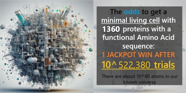
Life at the cellular level exhibits complex molecular processes, with each component playing a vital role in maintaining the life-essential homeostasis, and functions of the cell. These processes, ranging from protein synthesis to metabolism, demand the seamless coordination of numerous complex biomolecular machines.  Central cellular processes, like transcription and translation, stand as testaments to this complexity. These systems, dependent on each other, function through precise molecular languages and codified, instructional information, and metainformation that contains the know-how, of when, and how to extract and express that information to direct the making of all essential parts, like proteins, of the cell. For instance, distinct DNA sequences signal the commencement and termination of transcription. Furthermore, cellular coordination is achieved through extensive signaling pathways that enable the cell to adapt and respond. These pathways, in turn, are a complex web of interdependent components. Regulatory mechanisms further orchestrate these processes by decoding genetic information, controlling gene expression, and ensuring that cellular processes don't go haywire. Pelagibacter Ubique, the smallest free-living bacteria known today, operates with approximately 1,360 proteins. Each of these proteins is a sophisticated molecular machine with a specific, life-essential function. Proteins often only function if they are part of complex production chains that work synergistically. The odds of forming a minimal cell by sheer chance are astronomically low: 1 in 10^530,280. Considering the limitations of our universe, probabilities beyond 1 in 10^139 are deemed statistically impossible. Given this framework, the odds of a functional cell forming randomly are not just improbable but statistically impossible. This brings us to a compelling conclusion: the intricate and interdependent nature of cellular life points more convincingly towards intentional design than to unguided events.


The scientific community has historically highlighted simpler organisms like Mycoplasma to emphasize minimalistic life ( which would align with a naturalistic origin of life), but this perspective overlooks the inherent minimal complexity for life to start. Mycoplasma, often touted as the smallest life form, is symbiotic and relies on its host for survival and is, therefore, not a good candidate, and representative. A more fitting representation of minimal free-living life might be the bacterium, Pelagibacter Ubique, which boasts approximately 1,350 proteins. While 1,350 might seem like a mere number, it's crucial to recognize each protein as a complex molecular machine with life-essential specific functions, each having a unique role. Many of these proteins are intricately linked, forming elaborate micro-production lines. These systems often operate in harmony, leveraging synergy to produce vital cellular components.

In my in-depth exploration into the foundational needs of the smallest known life forms, I've cataloged a plethora of proteins and their functions for a supposed free-living cell in a hydrothermal vent, which is today the predominant hypothesis of where life started. This endeavor alone has occupied around 150 A4 pages. 

This exercise offers a hint at the magnitude of intricacy inherent in even the simplest organisms.

Recent science papers, like Bill Martins from 2021, posit a minimal metabolome consisting of 407 nodes/proteins, while my calculation doubles that. According to my calculations, that would be at least 862 nodes/enzymes. 
If we consider, that LUCA's proteome and interactome was a trial & error affair, (which is the alternative to design) the odds of getting a minimal size with 1226 proteins would be:

Calculating the odds of forming a proteome with 1,226 proteins, each of an average size of 300 amino acids, purely by random trial and error, is a monumental challenge due to the vastness of the numbers involved. Here's a basic way to think about the odds: Single Protein Formation: Each of the 20 common amino acids has a 1 in 20 chance of being selected for each position in the protein chain. For a protein with a sequence of 300 amino acids, the probability of getting one specific sequence purely by chance is: (1/20) ^300, or 1 in 10^390.  Now, if we consider that we need 1,360 such specific sequences, the odds become even more minuscule: ((1/20)^300)^1,360, or
1 in 10^530,280.

Premise 1: For a functional cell to exist, it requires an intricate and interdependent system of codes, languages, and proteins, with the odds of these systems forming randomly being 1 in 10^477,660.
Premise 2: Probabilities beyond 1 in 10^139 (the maximum number of possible events in a universe that is 13,8 Billion years old (10^16 seconds) where every atom (10^80) is changing its state at the maximum rate of 10^40 times per second is 10^139.
Conclusion: Therefore, the random, unguided formation of a functional cell's interdependent systems in our universe is statistically impossible, suggesting intentional design.

Functional Proteins: Even among the huge number of possible sequences, only a minuscule fraction will fold into functional proteins. So the odds are even worse when considering the formation of functional proteins.
Interactions & Dependencies: Beyond individual proteins, many proteins need to be in specific forms and require specific partners or cofactors to function. So, the complexity doesn't just come from the formation of the proteins themselves but from their interactions and dependencies.
Simultaneous Occurrence: For a functional cell, many of these proteins would need to come into existence simultaneously, or within a lifespan that allows for meaningful interaction.

The sheer complexity of a single cell and the astronomical odds against its components coming together purely by chance showcase the incredible intricacy and sophistication embedded in life. When we dive deep into cellular processes, the statistical improbability of life originating and operating through unguided processes becomes exceedingly clear.

Take the odds of forming a proteome with 1,360 proteins, each of an average size of 300 amino acids, purely by chance: 1 in 10^530,280. This number is unfathomably vast. To put it into perspective, the total number of atoms in the observable universe is estimated to be around 10^80. Just to illustrate the size of this number:

If you shuffle a standard deck of 52 cards, there are approximately 10^67 possible arrangements. This means that the odds of achieving a specific arrangement of proteins are vastly greater than the odds of shuffling a deck of cards into a specific order not just once, but billions and billions of times consecutively.

If every atom in the observable universe was a stopwatch, and each stopwatch could count to a trillion (10^12) in just one second, and they all counted simultaneously from the beginning of the universe (roughly 14 billion years ago) until now, we still wouldn’t have approached 10^477,660. In fact, we wouldn't even be close.

The odds of forming these proteins by random processes are so remote that they are beyond any conceivable event we can think of in our universe. Moreover, the complexity doesn't stop with just forming proteins. Many proteins require specific shapes and configurations, or they won't function. These functional shapes represent a minute subset of all possible protein forms. Therefore, the already astronomical odds of randomly generating any protein sequence become even more improbable when considering only those sequences that result in functional proteins. Furthermore, proteins don't operate in isolation. They are part of an intricate network of interactions and dependencies. Many proteins require specific partners, cofactors, or conditions to perform their tasks. This means multiple proteins must simultaneously exist and interact in precise ways for cellular functions to proceed. The manufacturing, signaling, and regulatory codes and languages underpinning cellular processes exemplify irreducibility and interdependence. Consider the genetic code, where DNA sequences are transcribed into RNA and then translated into proteins. Each of these steps relies on a suite of molecular machinery, and each part of this machinery is vital. Without transcription, the information in DNA remains locked. Without translation, the messages in RNA are meaningless. These processes are interdependent, with one being pointless without the other. Furthermore, the languages and codes of the cell allow for intricate communication and crosstalk. Signaling pathways allow cells to respond to their environment, regulate gene expression, and coordinate activities. Without these communication networks, the cell would be a collection of parts with no coordination.
The cellular machinery also needs regulatory mechanisms. Without regulation, cellular processes could run amok, leading to disease or cell death. The regulatory mechanisms ensure that everything happens when it should, where it should, and in the right amounts. It's challenging to envision how such interdependent systems could have emerged step by step over millions, even billions of years. If one part of the system was missing or not fully functional, the entire system would likely fail. These systems are crystal clear evidence that all components required to be present from the beginning, suggesting they were designed to operate as interconnected wholes.

LUCA is a theoretical entity, a single-cell organism from which supposedly all life forms on Earth descended through evolution.

A minimal proteome for the Last Universal Common Ancestor (LUCA): The following sequence starts with foundational biochemical reactions and cellular energy production, then progresses to information carriers, structural components, and finally defense and repair mechanisms.

Metal clusters: Enzymes/proteins estimate: 46
Essential for various biochemical reactions and protein structures.
Energy Metabolism, Central Carbon Metabolism, and Other Specific Pathways: Enzymes/proteins estimate: 74
Fundamental pathways that provide energy and precursors for other biosynthetic processes.
Nucleotide Synthesis and Salvage: Enzymes/proteins estimate: 89
The basis for the generation of genetic information carriers.
Amino acid biosynthesis: Enzymes/proteins estimate: 135
Building blocks for protein synthesis.
Regulatory Enzymes and Proteins in Amino Acid synthesis: Enzymes/proteins estimate: 76
Regulate the synthesis of amino acids.
Translation/Ribosome in the LUCA: Enzymes/proteins estimate: 125
Processes and machinery for protein synthesis.
Biosynthesis and Assembly of the Bacterial Ribosome: Enzymes/proteins estimate: 104
Further elaboration on ribosome assembly and function.
Transcription/regulation in the LUCA: Enzymes/proteins estimate: 63
Processes for reading genetic information and regulation.
DNA Processing in LUCA: Enzymes/proteins estimate: 48
Managing and replicating genetic information.
Families/functions involved in various aspects of cell division in LUCA: Enzymes/proteins estimate: 96
Cell division and proliferation.
Peptidoglycan Synthesis: Enzymes/proteins estimate: 91
Essential for bacterial cell wall synthesis.
Fatty Acid and Phospholipid Synthesis in LUCA: Enzymes/proteins estimate: 48
For making cellular membranes.
Cofactors: Enzymes/proteins estimate: 85
Essential helpers for enzymatic reactions.
NAD Metabolism: Enzymes/proteins estimate: 63
Important for redox reactions in the cell.
Reactive oxygen species (ROS): Enzymes/proteins estimate: 3
Deal with oxidative stress and byproducts of metabolism.
Uncharacterized: 136

The total sum for all the entries provided is  1,360.

The number of 1,226 proteins, is in the ballpark of what might be considered a minimal proteome. For context, the bacterium Pelagibacter ubique (a member of the SAR11 clade, one of the most abundant and smallest marine microbes). P. ubique also has a small genome (1,308,759 bp), currently the smallest genome known for a free-living organism. The genome encodes 1,354 predicted proteins, 1 rRNA operon , and 32 tRNAs. Given that P. ubique has a streamlined genome adapted for its specific oceanic environment, its protein count is near the lower limit for free-living organisms.

Some general categories and enzymes that might be considered fundamental for a wide range of cellular life forms:

Membrane Transport Systems: While some transporters were mentioned, an organism would need transport systems for ions, water (aquaporins), and other essential molecules not specified in your list.
ATP Synthesis: The ATP synthase complex, crucial for producing ATP, the primary energy currency of the cell, was not mentioned.
DNA Replication Machinery: The fundamental DNA polymerase enzymes, helicases, primases, ligases, and topoisomerases, which are involved in DNA replication and maintenance, were not detailed.
Protein Folding and Degradation: Chaperones (like GroEL/GroES and DnaK/DnaJ) assist in protein folding. Proteasome or ClpXP machinery help degrade unneeded or misfolded proteins.
Glycolysis and TCA Cycle Enzymes: Central carbon metabolism enzymes, like those in glycolysis and the TCA (Krebs) cycle, provide vital precursors for various biosynthetic pathways and produce ATP.
Cell Division Machinery: FtsZ and other proteins essential for cell division and septum formation.
Stress Response Systems: Various enzymes and proteins help cells cope with oxidative stress, DNA damage, and other environmental stressors. For example, superoxide dismutase (SOD) and catalase help neutralize reactive oxygen species.
Lipopolysaccharide Synthesis: In Gram-negative bacteria, enzymes involved in lipopolysaccharide synthesis are vital for outer membrane biogenesis.
RNA Processing and Degradation: While you mentioned ribonucleases, other enzymes and proteins associated with RNA splicing, maturation, and degradation in various organisms could be included.
Signal Transduction Systems: Two-component systems, protein kinases, and other signaling molecules help the cell respond to environmental changes.
Autotrophic Processes: If considering autotrophs, the Calvin cycle, and other carbon fixation pathways, along with enzymes for nitrogen fixation, might be considered essential.

Size of the Central Metabolome 

The central metabolome generally refers to a set of metabolic pathways and processes that are ubiquitous and fundamental to cellular life. Based on that understanding, the following can be considered part of the central metabolome:

Energy Metabolism, Central Carbon Metabolism, and Other Specific Pathways: Enzymes/proteins estimate: 74
Fundamental pathways that provide energy and precursors for other biosynthetic processes.
Nucleotide Synthesis and Salvage: Enzymes/proteins estimate: 89
The basis for the generation of genetic information carriers.
Amino acid biosynthesis: Enzymes/proteins estimate: 135
Building blocks for protein synthesis.
Regulatory Enzymes and Proteins in Amino Acid synthesis: Enzymes/proteins estimate: 76
Regulation is crucial for maintaining metabolic homeostasis and ensuring the efficient use of cellular resources.
Translation/Ribosome in the LUCA: Enzymes/proteins estimate: 125
Processes and machinery for protein synthesis.
Biosynthesis and Assembly of the Bacterial Ribosome: Enzymes/proteins estimate: 104
Further elaboration on ribosome assembly and function.
Transcription/regulation in the LUCA: Enzymes/proteins estimate: 63
Processes for reading genetic information and regulation.
DNA Processing in LUCA: Enzymes/proteins estimate: 48
Managing and replicating genetic information.
Cofactors: Enzymes/proteins estimate: 85
Essential helpers for enzymatic reactions.
NAD Metabolism: Enzymes/proteins estimate: 63
Important for redox reactions in the cell.

Total 862 enzymes/proteins

The other processes and entities listed are essential for life, but they might be considered more specialized rather than part of the central core of metabolism. For instance, while "Metal clusters" and "Reactive oxygen species (ROS)" are vital for many organisms, they might be viewed as ancillary to the central metabolic processes in some contexts.

Further considerations

The intricate dance of molecular processes within a cell showcases an orchestra of interwoven systems, languages, and codes. The sheer complexity and precision of these processes hint at a foundational conundrum: How could such intricacy arise step by step when intermediate stages would bear no function? Take, for example, the process of transcription and translation. Transcription reads genetic information, and translation converts that message into proteins. Each of these steps relies on a suite of molecular machinery. Without transcription, the information in DNA remains inaccessible. Without translation, the messages in RNA serve no purpose. The two processes are interdependent, rendering one pointless without the other. Moreover, the languages and codes that underpin these processes are irreducible in their complexity. The genetic code, with its specific sets of letters, words, and rules, must be perfectly synchronized with the translational machinery. A change or absence in one would render the others nonsensical. Then there's cellular signaling, a vast network of communication channels that coordinate every cellular activity. Signaling pathways enable cells to sense their environment, regulate gene expression, and orchestrate complex tasks. Without these pathways, a cell would merely be an assortment of parts with no directive. Furthermore, these signaling networks cannot function in isolation. They require specific receptors, secondary messengers, and effector proteins—all of which are interdependent. The absence or malfunction of one component can cascade through the system, causing widespread dysfunction. But the complexity doesn't stop there. Regulatory mechanisms govern these systems, ensuring that everything occurs when and where it should. This intricate choreography requires a sophisticated set of codes and languages. For instance, a particular sequence of DNA, known as a promoter, signals where transcription should begin. Another sequence called a terminator, signals where it should end. These regulatory codes are irreducible; without them, transcription would either not start or would produce meaningless sequences. It's challenging to imagine how such intertwined systems could have arisen through a linear, stepwise process, even over billions of years. Intermediate stages, lacking full functionality, wouldn't provide a survival advantage. The interdependence of these systems suggests they needed to be present and fully functional from their inception. When we dive deep into these systems, it becomes evident that they couldn't have emerged step by step. One without the other is like a lock without a key—rendered useless. This interconnected complexity points to a design that is deliberate and intentional, requiring a profound understanding of cellular processes. In conclusion, the molecular world within a cell is not just a jumble of reactions. It's a harmonized symphony of codes, languages, and systems that are irreducibly complex and interdependent. The implausibility of such a coordinated dance emerging through random, stepwise processes underscores the marvel of cellular life and hints at an intelligent orchestration behind its existence.

Life's Blueprint: The Essential Machinery  to Start Life Latam_10


Last edited by Otangelo on Tue Mar 12, 2024 6:12 am; edited 23 times in total

https://reasonandscience.catsboard.com

Otangelo


Admin

LUCA: Deciphering the Root of the Universal Tree of Life

Within the diverse realm of life on Earth, the scientific community posits the existence of LUCA or the Last Universal Common Ancestor. This entity stands not as an organism fossilized in ancient rock but rather as a theoretical construct inferred from comparative genomics and molecular biology. Representing a shared lineage for all extant life forms, the characterization of LUCA provides a glimpse into early evolutionary paradigms. It's generally postulated that LUCA thrived between 3.5 to 3.8 billion years ago, based on molecular clock analyses and ancient fossil evidence. However, LUCA does not mark life's origin; instead, it signifies a state of molecular maturity, underscoring a plethora of biological processes already in operation. Comparative genomics offers insights into the potential genomic framework of LUCA. This progenitor would have housed genes pivotal for RNA synthesis, DNA replication, protein production, and rudimentary metabolic processes. Such molecular mechanisms form the foundation for the vibrant thriving of life we observe today. A core feature believed to be present within LUCA's cellular machinery is its capacity for nucleic acid synthesis and maintenance. Contrary to a simplistic model, the genetic machinery of LUCA would be competent, enabling DNA replication and repair, akin to a seasoned maestro overseeing a complex orchestra. The DNA polymerases of LUCA, far from rudimentary, would have deftly decoded genetic blueprints, creating coherent genetic narratives. The debate regarding LUCA's cellular structure persists. While some assert its resemblance to contemporary prokaryotic entities, others argue for a cellular architecture that might have been more rudimentary, yet uniquely adapted to its primordial environment. The crux of LUCA's existence rests not merely in its DNA or RNA, but in its overarching biochemical machinery. Envision a cellular system already equipped to harness energy, process nutrients, and maintain genetic fidelity, preparing the groundwork for life's subsequent diversification. Beyond its genetic prowess, LUCA would have navigated challenges like sourcing energy and maintaining an internal milieu. The potential metabolic pathways it utilized are hoped to offer windows into early adaptations. As the scientific community delves deeper into LUCA's attributes and significance, it stands as a beacon, illuminating life's early constitution.  To comprehend LUCA is to appreciate a pivotal juncture and starting point for an evolutionary narrative. While not representing life's genesis, LUCA demarcates a state where foundational molecular systems were operational, paving the supposed way for the evolutionary dance that would have sculpted the diverse biosphere we now observe.

The Primordial Stage: Scrutinizing Earth's Early Environment

The quest to understand life's genesis has pointed the scientific community towards diverse environments ranging from deep-sea hydrothermal vents to surface pools rich in prebiotic compounds. Positioned in the depths of oceans, hydrothermal vents, with their mineral-rich emissions and gradients of temperature, are postulated as potential cradles of life. These environments offer a blend of chemicals and the requisite energy that might favor prebiotic reactions. However, several questions remain. How would delicate organic molecules, crucial for life, withstand the intense conditions and extreme pressures at these depths? Additionally, the rapid dilution of any synthesized compounds in vast ocean volumes may challenge the accumulation of critical biomolecules. The concept of the primordial "soup" evokes visions of surface pools teeming with a mix of organic compounds, possibly driven by lightning and ultraviolet (UV) radiation. Yet, the constant exposure to UV rays also poses a conundrum. While they may catalyze some reactions, they could also degrade sensitive organic compounds. The balance between creation and destruction in such scenarios is a subject of scrutiny. External events and conditions have been implicated in possibly steering the pathways leading up to life. Yet, were these events truly deterministic, nudging a non-living world toward the brink of life? Meteorites, laden with organic compounds, have been suggested as external donors that might have seeded Earth with prebiotic molecules. However, the journey through Earth's atmosphere subjects these compounds to extreme temperatures. Would these meteoritic compounds truly survive and remain unaltered after such a fiery descent? The early Earth's atmosphere, believed to be reducing in nature, might have facilitated the synthesis of organic compounds. But, the exact composition and its implications for the formation of biomolecules remain subjects of debate. Was the atmosphere truly conducive, or do we yet grapple with understanding its nuances?
Given the myriad conditions and hypotheses presented, the question arises: Were these circumstances sufficiently deterministic to conclude life's inevitable emergence? While it's acknowledged that certain conditions might favor the synthesis of organic compounds, progressing from simple molecules to complex, functional cellular machinery presents a wider chasm. A mere assemblage of molecules doesn't equate to life. How, then, did the leap from chemistry to cellular complexity transpire? Life's machinery hinges on synchronized interactions between biomolecules. The orchestration of these interactions in the absence of a guiding cellular framework stands as a considerable challenge to our understanding. In examining the potential birthplaces of life and the external factors influencing it, it's clear that the narrative is far from complete. While certain conditions may favor the synthesis of biomolecules, the progression to a functional, self-replicating system remains enigmatic. As we venture further into this domain, it's pivotal to approach these hypotheses with a discerning lens, appreciating the nuances and seeking clarity amidst the complexities.

From Molecules to Life: Delving into the Enigma of Biopolymer Emergence

At the most fundamental level, life's machinery is driven by a suite of molecules that serve as building blocks: amino acids, nucleotides, and lipids, to name a few. While laboratory experiments have shown that certain basic molecules can form under simulated early Earth conditions, there are still lingering questions about the yield, purity, and stability of these molecules. For instance, the Miller-Urey experiment famously produced amino acids. Yet, it's worth asking: Were the conditions used in such experiments reflective of early Earth? Furthermore, in a prebiotic setting devoid of enzymatic guidance, how were the right molecules selectively synthesized and maintained amidst a plethora of unwanted byproducts?

Natural selection did not operate during the prebiotic era on Earth

In the primeval environment, directed selection mechanisms were absent. Complex systems in contemporary organisms produce the building blocks of life.  Before life as we know it flourished, the early Earth was a blank canvas, devoid of the guiding hand of natural selection. Imagine a world where meticulously orchestrated molecular symphonies of the present day did not exist. Instead of precise, deliberate formations of nucleic acids and the twenty amino acids, there existed a cacophonous chaos of molecules. Within the sophisticated cellular machinery of modern life, missteps such as right-handed amino acids are meticulously corrected. Nucleobases, phospholipids, and carbohydrates, now with such definite roles, were lost amidst a sea of countless molecular contenders. One must wonder: in a world teeming with chemical randomness, how did unguided processes sift through and pick the molecular elite that constitutes life today?

The Origin and Organization of Life's Fundamental Molecules

The four principal molecules of life—DNA, RNA, proteins, and lipids—serve as the cornerstone for cellular function and replication. Understanding their origin is paramount in our quest to grasp the inception of life on Earth. The chemical milieu of early Earth, without the frameworks of cellular life and hereditary systems, lacked a discernible mechanism for choosing or giving preference to one molecule over another. This, indeed, raises intricate questions about how such sophisticated and information-laden molecules emerged from a prebiotic environment. The concept of self-organization or autocatalysis, where molecules spontaneously order themselves into structured arrangements or catalyze their formation, has been posited by many in the scientific community. Such a notion would imply that under specific conditions, certain molecules possess an inherent propensity to organize or catalyze reactions that would lead to life's building blocks. Yet, the emergence of molecules as information-rich as DNA and RNA requires a context in which specificity and order are paramount. The inception of proteins, with their vast array of potential amino acid sequences and configurations, needs more than mere random chance. Moreover, the lipid molecules that form cellular barriers are not just simple hydrophobic chains but are involved in vital cellular functions and signaling. Considering these complexities, one would have to infer that the early Earth had to present conditions where specificity was not just possible but probable. The environments of this primordial world, with its myriad of micro-niches, would have to offer a wide array of physical and chemical favorable conditions. It would have to be conceived that in such a diverse landscape, the formation of life's basic building blocks was not a solitary event, but a series of events, each fine-tuned by the particular conditions of its niche. The notion of molecules with inherent properties that drive them towards life-promoting configurations suggests a level of precision and sophistication in their design. 

Andrew H. Knoll (2012):  The emergence of natural selection: Molecular selection, the process by which a few key molecules earned key roles in life’s origins, proceeded on many fronts. (Comment: observe the unwarranted claim) Some molecules were inherently unstable or highly reactive and so they quickly disappeared from the scene. Other molecules easily dissolved in the oceans and so were effectively removed from contention. Still, other molecular species may have sequestered themselves by bonding strongly to surfaces of chemically unhelpful minerals or clumped together into tarry masses of little use to emerging biology. In every geochemical environment, each kind of organic molecule had its dependable sources and its inevitable sinks. For a time, perhaps for hundreds of millions of years, a kind of molecular equilibrium was maintained as the new supply of each species was balanced by its loss. Such equilibrium features nonstop competition among molecules, to be sure, but the system does not evolve. 1

Commentary: The fabric of life, as understood through current scientific analysis, displays an intricate orchestration of molecular machinery. The Fundamental Biomolecules are not simple assemblages of atoms; they are sophisticated entities operating in highly coordinated metabolic pathways. When we contemplate the early Earth environment, it becomes a formidable challenge to envision how such specified complex molecules would be readily available. They would have needed to form naturally, without the mediation of enzymes, and amass in considerable amounts at a singular site, ready to contribute to the foundation of life. 

1. The intricate dance of life hinges upon a finite collection of detailed biomolecules, united in a universally recognized convention. This core consortium comprises the quintessential building blocks: RNA, DNA, amino acids, phospholipids, and carbohydrates. With remarkable precision, cells leverage these elements through meticulously coordinated metabolic pathways, paths seemingly absent in Earth's early stages. Considering the principle of abiogenesis, it's imperative these biomolecules existed in a naturally occurring state, formed without enzyme mediation. Furthermore, they would have required organization to lay down the foundation for the inception of the first living cells. An abundance of these biomolecules, concentrated at a singular focal point, would have been essential.
2. Creation with an endpoint in mind necessitates a directional intent. This premise raises challenges when addressing naturalistic propositions concerning life's genesis. On the ancient Earth, a vast molecular expanse would have precluded any systematic competition or selection, making the isolation of life-essential molecules from non-essential counterparts seemingly implausible. To attribute the entirety of life's organized structure, its preservation, and its evolutionary progression from non-living entities solely to selection appears to be an oversimplified assertion. Suggesting that selection is the sole orchestrator of life's grand symphony might miss nuances essential to our understanding.
3. From our observations, a series of random events without direction is highly improbable to produce components with distinct purposes, particularly when looking at large integrated systems. Analogously, bricks don't self-assemble from raw materials nor position themselves to form walls. There’s a guiding hand behind their formation. Similarly, phospholipids, vital for cell membranes, don't spontaneously arise from their basic components. Their formation and subsequent organization hint at an overarching guidance. The question that beckons: could there be an architect behind these marvels?

Alan W. Schwartz (2007): A problem that is familiar to organic chemists is the production of unwanted byproducts in synthetic reactions. For prebiotic chemistry, where the goal is often the simulation of conditions on the prebiotic Earth and the modeling of a spontaneous reaction, it is not surprising – but nevertheless frustrating – that the unwanted products may consume most of the starting material and lead to nothing more than an intractable mixture, or -gunk.. The most well-known examples of the phenomenon can be summarized quickly: Although the Miller –Urey reaction produces an impressive set of amino acids and other biologically significant compounds, a large fraction of the starting material goes into a brown, tar-like residue that remains uncharacterized; i.e., gunk. While 15% of the carbon can be traced to specific organic molecules, the rest seems to be largely intractable  Even if we focus only on the soluble products, we still have to deal with an extremely complex mixture of compounds. The carbonaceous chondrites, which represent an alternative source of starting material for prebiotic chemistry on Earth, and must have added enormous quantities of organic material to the Earth at the end of the Late Heavy Bombardment (LHB), do not offer a solution to the problem just referred to. The organic material present in carbonaceous meteorites is a mixture of such complexity that much ingenuity has gone into the design of suitable extraction methods, to isolate the most important classes of soluble (or solubilized) components for analysis. Whatever the exact nature of an RNA precursor which may have become the first selfreplicating molecule, how could the chemical homogeneity which seems necessary to permit this kind of mechanism to even come into existence have been achieved? What mechanism would have selected for the incorporation of only threose, or ribose, or any particular building block, into short oligomers which might later have undergone chemically selective oligomerization? Virtually all model prebiotic syntheses produce mixtures. 2

A.G.Cairns-Smith (1985):  It is one of the most singular features of the unity of biochemistry that this mere convention is universal. Where did such agreement come from? You see non-biological processes do not as a rule show any bias one way or the other, and it has proved particularly difficult to see any realistic way in which any of the constituents of a 'prebiotic soup' would have had predominantly 'left-handed' or right-handed' molecules. It is thus particularly difficult to see this feature as having been imposed by initial conditions. 3

A.G.Cairns-Smith (1985): genetic takeover, page 70: Suppose that by chance some particular coacervate droplet in a primordial ocean happened to have a set of catalysts, etc. that could convert carbon dioxide into D-glucose. Would this have been a major step forward towards life? Probably not. Sooner or later the droplet would have sunk to the bottom of the ocean and never have been heard of again. It would not have mattered how ingenious or life-like some early system was; if it lacked the ability to pass on to offspring the secret of its success then it might as well never have existed. So I do not see life as emerging as a matter of course from the general evolution of the cosmos, via chemical evolution, in one grand gradual process of complexification. Instead, following Muller (1929) and others, I would take a genetic View and see the origin of life as hinging on a rather precise technical puzzle. What would have been the easiest way that hereditary machinery could have formed on primitive Earth? 4

William Dembski (2002): The problem is that nature has too many options and without design couldn’t sort through all those options. The problem is that natural mechanisms are too unspecific to determine any particular outcome. Natural processes could theoretically form a protein, but also compatible with the formation of a plethora of other molecular assemblages, most of which have no biological significance. Nature allows them full freedom of arrangement. Yet it’s precisely that freedom that makes nature unable to account for specified outcomes of small probability. Nature, in this case, rather than being intent on doing only one thing, is open to doing any number of things. Yet when one of those things is a highly improbable specified event, design becomes the more compelling, better inference. Occam's razor also boils down to an argument from ignorance: in the absence of better information, you use a heuristic to accept one hypothesis over the other. 5

Fry, Iris. (2010): How successful are the RNA-first, RNA-later, metabolism-first, and, preparatory metabolism theories in elucidating the emergence of life as an evolutionary process? So far, none of these paradigms can claim decisive experimental support. 6

Essential Biomolecules: Beyond the Realm of Selection

Regarding the selection and specificity of nucleic acids in the prebiotic context:

1. Purity of Starting Molecules: Amidst a vast primordial concoction, how did the critical purity necessary for RNA and DNA synthesis emerge so distinctly? Such purity, considering the vastness and randomness of molecular mixtures present, points towards a potential guidance mechanism, suggesting an overarching blueprint may have been guiding this synthesis against all odds.
2. Nucleobase Selection: In an environment teeming with molecular candidates, it's perplexing how only five distinct nucleobases became prominent. This remarkable selection precision, amidst a probable sea of molecular candidates, resonates with the idea of a process that might have been intelligently choreographed rather than a product of sheer happenstance. The hydrogen bond strength in DNA is finely tuned to ensure stability and specificity in the double helix formation with adenine pairing with thymine and guanine pairing with cytosine. Correct tautomeric forms of nucleotide bases are crucial for maintaining the appropriate hydrogen bonding patterns, ensuring the accurate replication and transmission of genetic information. While numerous possible analog structures for nucleotide bases exist, only specific bases are employed in biological systems due to their unique chemical properties and base pairing specificity.
3. Structural Specificity of Purines: The intricate two-ring structure of purines, which emerged as dominant, is fascinating given the countless molecular configurations possible. Purines, with their specific double-ring structure, were selected for their unique ability to form stable and specific hydrogen bonds with their complementary pyrimidines in DNA, ensuring accurate genetic information storage and transmission. Their particular atomic arrangement and functional groups provide the necessary chemical properties and base pairing specificity crucial for the integrity and function of DNA within biological systems. This striking structural consistency, especially in primitive conditions, seems to advocate for a purposeful structural blueprint.
4. Structural Specificity of Pyrimidines:  How did pyrimidines, with their single ring made up of 4 carbon and 2 nitrogen atoms, emerge as the preferred structure, given the innumerable potential configurations? Pyrimidines, with their distinct single-ring structure, were chosen because they can establish specific and stable hydrogen bonds with their complementary purines in DNA. This specific structure and the associated functional groups ensure precise base pairing, which is essential for the accurate storage and transmission of genetic information in living organisms. The very emergence of such distinct structural specificity hints towards a possible intentional molecular choreography.
5 Base Separation in Complex Mixtures: Amidst the plethora of similar molecules that could have formed, how were the functional nucleobases of DNA and RNA selectively isolated? The capability to filter out the essential from the non-essential implies a system that favors precision.
6 Ribose Ring Selection: Why does life predominantly use ribose with its 5 carbon sugar ring in the backbone of RNA and DNA, even though other configurations like 4 or 6 carbon rings could have formed, but are non-functional? This specificity hints at an optimized design.
7 Correct Atom Linkage: How were the right nitrogen atom of the base and the appropriate carbon atom of the sugar pinpointed and linked, especially when various connections are possible? Such accuracy in molecular bonding demonstrates the intricate design in foundational biochemistry.
8 Homochirality in RNA and DNA: In a world that could produce both left and right-handed molecules, how did life come to exclusively use the right-handed configurations of RNA and DNA, particularly ribose in its D form? The consistent selection of certain chiral forms over others remains a profound mystery, suggesting a precise orchestration in the genesis of life.
9 Absence of Prebiotic Selection Mechanism: Without the machinery of life in place, how would the prebiotic environment have favored the formation of RNA and DNA molecules over others? The lack of a natural selection mechanism prior to life presents a conundrum in accounting for their emergence.
10 RNA's Dual Role in Information and Function: Given that RNA can serve as both an information carrier (like DNA) and a catalyst (like proteins), how did it come to acquire this dual role without a pre-existing selection mechanism? The multi-functionality of RNA seems to defy a simple, undirected origin.
11 Specificity of Nucleotide Pairing: Adenine always pairs with Thymine (or Uracil in RNA), and Cytosine with Guanine. How did this highly specific base pairing evolve without a mechanism to favor correct pairings over incorrect ones?
12 Origin of Replication Mechanism: How did the mechanism for the replication of RNA and DNA molecules come into being, given that the replication process itself seems to require some form of selection? Without an error-correction mechanism, the fidelity of replication would be compromised.
13 Transition from RNA to DNA World: If life began with an RNA world, as some suggest, how did the transition to the more stable DNA occur, especially in the absence of prebiotic selection mechanisms to guide such a transition?
14 Lack of Stable Prebiotic Environment: Given the potential instability and reactive nature of early Earth, how could delicate molecules like RNA and DNA form and be preserved without a stable environment and without specific mechanisms to protect and favor them?
15 Problem of Initial Functionality: How did the first RNA and DNA molecules gain the functionality necessary for them to be advantageous and subject to any form of selection, given that they would need to have some level of utility from the outset?

The challenges enumerated, especially when considered in their cumulative effect, make it clear why the origin of life is one of the most daunting and debated questions in both the fields of biology and philosophy. When we dive deep into the minutiae of molecular configurations and the specificity required for functional nucleic acids, it becomes evident that a multitude of highly specific conditions and sequences of events would have been necessary for life to emerge naturally. Many of the problems highlight the sheer specificity required at multiple stages — from selecting the right nucleobases to forming the right bonds and achieving functional sequences. This specificity seems to be at odds with random, unguided processes. The early Earth environment was not as conducive to life as it is today. High UV radiation, lack of protective atmosphere, and a plethora of reactive molecules would make the survival and formation of delicate RNA/DNA chains incredibly challenging. The importance of catalysts in modern biological systems is evident. In their absence during the prebiotic era, achieving the right reaction speeds becomes another monumental hurdle. At this prebiotic stage, the mechanisms of natural selection, which guide the evolution of life forms, weren't operational. Natural selection acts on functional, replicating entities. If you don't yet have a functional, replicating entity (like the first RNA/DNA molecules), you can't have natural selection. Given the combined weight of these challenges, it makes the idea of life emerging from purely naturalistic and unguided processes highly improbable.

Regarding the selection and specificity of amino acids and proteins in the prebiotic context:

1. Optimal Amino Acid Selection: Given that there are over 500 naturally occurring amino acids on Earth, why are only 20 of them specifically utilized in life's proteins? This unique selection seems to go beyond mere chance, pointing towards a deliberate arrangement tailored to these 20 amino acids' exceptional properties.
2. Bifunctional Monomer Preference: How is it that bifunctional monomers, those with two functional groups, became dominant in protein formation when unifunctional monomers could disrupt the process? This preference highlights the importance of structure in the creation of functional polymers.
3. Homochirality Mystery: Why do life forms chiefly use left-handed amino acids and right-handed sugars, especially when non-chiral precursors could lead to either form? The exclusive use of certain chiral forms speaks to a specificity in biological design.
4. Natural Amino Acid Choice: Considering the vast number of naturally occurring amino acids, how is it that life on Earth primarily utilizes a consistent subset for protein construction? This consistency underscores the idea of a finely-tuned selection for optimal function.
5. Avoidance of Unwanted By-Products: What mechanism ensures that the synthesis of biological amino acids does not result in undesired or irrelevant by-products? The precision in this process suggests a higher level of orchestration.
6. β, γ, δ Amino Acid Sorting: How were non-alpha amino acids filtered out in the primordial soup, given their potential presence? This filtering hints at a process that prioritizes functionality and efficiency.
7. Addressing Homochirality: How could an environment with achiral precursors lead exclusively to left-handed amino acids? This phenomenon remains one of the great mysteries of biochemistry.
8. NH3 Accumulation: How did ammonia, essential for amino acid synthesis, amass on the early Earth despite its short lifetime due to photochemical dissociation? This suggests conditions were somehow optimal for the emergence of life.
9. Organosulfur Compound Formation: How did early Earth processes generate the organosulfur compounds necessary for certain amino acids, considering the state of sulfur in nature? The existence of these compounds implies a pathway that was conducive to life.
10. Enzymatic Transition: How did the shift from prebiotic enantiomer selection to enzymatic reactions like transamination take place, given its necessity for life's emergence? This intricate transition speaks to the complexity and finesse in biological systems.
11. Amino Acid Suitability: How is it that the chosen amino acids are perfectly suited for forming structures with tight cores, enabling binding pockets within proteins? The optimal nature of these amino acids hints at purposeful design.
12. Unique Amino Acid Set: Why does the set of amino acids used by life seem to be near-optimal in properties like size, charge, and hydrophobicity, especially compared to millions of alternatives? This optimization suggests a design geared for maximal functionality.
13. Emergence of Synthesis Regulation: How did the regulation of amino acid synthesis pathways emerge, ensuring supply meets demand? Such regulation indicates a system that is not just complex but also adaptive.
14. Metabolic Pathway Transition: How did the shift from prebiotic synthesis to metabolic pathway-based amino acid synthesis occur, especially given the numerous enzymes involved? This complex transition emphasizes the intricacies in the development of life's systems.

From the selection of the right building blocks (amino acids) to the formation of complex three-dimensional structures (proteins), each step poses significant challenges to naturalistic explanations for the origins of life. These problems not only require answers for individual steps but also necessitate an understanding of how these steps might sequentially and synergistically come together. While research in the field of abiogenesis is ongoing and some strides have been made, the cumulative weight of these challenges often leads to intense debates among scientists, philosophers, and theologians regarding life's origins.

Regarding the selection and specificity of phospholipids in the prebiotic context:

1. Hydrocarbon Chain Selection: In the midst of prebiotic conditions, how did processes naturally select hydrocarbon chains of precisely 14 to 18 carbons in length? Given the lack of any physical directive, chains could have formed in a variety of lengths, raising the question of specificity in the emergence of cellular membranes. The specificity required in the length of hydrocarbon chains is nothing short of fascinating. The early Earth's environment would have been a tumultuous mix of various elements and compounds. The emergence of such precision amidst chaos challenges our understanding of spontaneous order. If chains of any length were plausible, the consistent development of this exact range seems beyond mere coincidence.
2. Phospholipid Composition: Cellular membranes aren't made up of a singular type of phospholipid. How did random events lead to the development of membranes comprising diverse phospholipid species, often combined with sterols like cholesterol? The pathway for the selection and synthesis of this intricate mixture remains puzzling in a prebiotic context. The intricate mixture of phospholipid species in cellular membranes is another testament to the precision implemented in nature. In a setting without directed guidance or cellular machinery, achieving such a refined composition raises significant questions about the prebiotic processes at play.
3. Formation of Bilayers: Phospholipids in cell membranes naturally form bilayers, a critical structure for cell integrity and function. How did spontaneous events promote the consistent formation of bilayers in the absence of cellular machinery or directives? Bilayers are integral to cell functionality. Their spontaneous formation in a prebiotic setting, devoid of cellular directives, seems like a formidable hurdle. The inherent organization this requires is a profound mystery in understanding the genesis of life, unless one posits intelligent design. 
4. Molecular Symmetry: Phospholipids display a head and two fatty acid tails, implying a certain degree of symmetry. How were the conditions favorable to consistently produce this dual-tailed structure, as opposed to other possible configurations? The symmetry displayed by phospholipids, with a head and two tails, implies an underlying order. Achieving this structure consistently in early Earth's turbulent conditions speaks to processes and phenomena that remain enigmatic unless they were instantiated with intent.
5. Specific Phospholipid Functions: Different phospholipids serve distinct roles within cellular membranes, influencing fluidity, curvature, and interaction with proteins. How did prebiotic conditions facilitate the formation of a wide variety of phospholipids, each with its unique function? Beyond their formation, each phospholipid serves a unique role, contributing to the membrane's properties. How prebiotic conditions produced such functional diversity remains a major puzzle in the understanding of life's origins, unless one infers design. 
6. Environmental Stability: Given the dynamic and potentially harsh conditions of the early Earth, how were phospholipids stabilized? Without the protective environment of a cell, these molecules could be degraded by various factors. Phospholipids, vulnerable to degradation, faced the harsh and variable conditions of early Earth. Their persistence and eventual dominance in cellular membranes suggest mechanisms of stability and protection that could only be implemented with foresight and intent.
7. Concentration and Assembly: For bilayers to form, a sufficient concentration of phospholipids would be required. How were these concentrations achieved and maintained amidst the vast prebiotic "soup"?  The formation of any structured entity requires a threshold concentration of its components. How phospholipids achieved such concentrations in the vastness of prebiotic environments raises profound questions about the early dynamics of molecular assembly.
8. Integration of Other Membrane Components: Membranes also consist of proteins, cholesterol, and other components. How did the early membranes incorporate these elements in the right proportions and configurations for functionality? Modern membranes are complex mosaics of various components. The harmonious integration of these components, each contributing to membrane function, points to an inherent sophistication that challenges simplistic, undirected origin models.
9. Functional Asymmetry: Modern cell membranes display asymmetry, with different phospholipids in the inner and outer leaflets of the bilayer. How was such a complex arrangement achieved in the absence of enzymes and transporters? Asymmetry in cell membranes is not a mere structural detail; it's fundamental to function. Achieving this intricate arrangement without enzymes or transporters adds another layer of complexity to the origin puzzle.
10. Selective Permeability: One of the vital functions of the membrane is to regulate the passage of substances. In the absence of specialized proteins, how did early membranes achieve any semblance of selective permeability? The ability to regulate substance passage is vital for any proto-cell. The emergence of selective permeability, especially in the absence of specialized proteins, remains one of the most intriguing questions about the early stages of life.

In essence, these challenges, especially when viewed cumulatively, present significant hurdles to simplistic, undirected narratives of life's origins. Each question underscores the sophistication and specificity inherent in even the most basic components of life. Understanding these intricacies not only deepens our appreciation for the wonder of life but also reinforces the enormity of the quest to unravel its genesis.

Regarding the selection and specificity of carbohydrates in the prebiotic context:

1. Emergence of Vital Sugars: In the vast prebiotic landscape, how did essential sugars like glucose and ribose distinctively form? The precision and specificity of these simple sugars in life's matrix hint at an intentional blueprint underpinning their presence.
2. Pentose Sugar's Role: Amidst the countless molecular structures, what guided the consistent selection of pentose sugars for RNA's backbone? Such a unique choice in the early molecular dance suggests an underlying design.
3. Evolution of Polysaccharides: The progression from simple monosaccharides to complex polysaccharides seems more than mere molecular serendipity. Their formation and function in early life forms indicate a pre-conceived architectural plan.
4. Glycosidic Bond's Precision: In an environment devoid of sophisticated enzymes, how did the accurate glycosidic bonds between sugars emerge?  Glycosidic bonds refer to the covalent linkages that connect monosaccharide units in disaccharides, oligosaccharides, and polysaccharides. In other words, they are the bonds that link individual sugar molecules together in larger carbohydrate structures.  It's likely that LUCA had some rudimentary form of a protective exterior, possibly incorporating simple carbohydrates or carbohydrate-like structures to maintain cellular integrity and mediate interactions with its environment.The meticulous nature of these bonds alludes to an inherent design principle.
5. Carbohydrates in Energy Storage: The primordial role of carbohydrates like glycogen and starch in energy storage seems beyond random. Their precise structures and functions in LUCA's descendants suggest a blueprint aimed at energy optimization.
6. Cell-Surface Carbohydrate Code: How did the complex carbohydrate patterns on cell surfaces, crucial for cell recognition and signaling, come into existence? Their intricate design suggests an intentional matrix for early cellular communication.
7. Origin of Glycolysis: The glycolytic pathway, central to most life forms, exhibits a remarkable sequence of reactions. How did this elegant process, vital for energy extraction from glucose, originate? Its precision hints at a designed mechanism pivotal to early life's sustenance.
8. Chirality in Sugars: With multiple chiral configurations possible, what determined the dominance of specific orientations in biological carbohydrates? The selectivity of D-sugars in life implies an underlying design strategy.

Carbohydrates, being central to numerous life processes ranging from energy storage to cell signaling, indeed present a myriad of challenges when it comes to explaining their origins. The way they precisely bond, their chiral preferences, their unique roles in various cellular pathways, and their consistent structures across numerous life forms raise compelling questions about how such order and complexity could arise in a presumably chaotic prebiotic environment. The focus on LUCA (Last Universal Common Ancestor) also brings forth the idea that even the most primitive life forms exhibited remarkable molecular precision and complexity, suggesting that the foundational principles of life are rooted in a deep-seated order and design.

Life's Blueprint: The Essential Machinery  to Start Life Image115


Last edited by Otangelo on Tue Mar 12, 2024 6:13 am; edited 21 times in total

https://reasonandscience.catsboard.com

Otangelo


Admin

Amino Acids and Chirality

When amino acids form, they can manifest in one of two spatial configurations: left-handed or right-handed. These configurations are mirror images of each other and can't be superimposed, much like our left and right hands. Intriguingly, when a left-handed amino acid combines with its right-handed counterpart, the resulting compound is not suitable for forming proteins – a state referred to as racemic. What's truly captivating is the fact that all amino acids in proteins within living organisms are exclusively left-handed. When amino acids are synthesized chemically, the result is an equal mix of right and left-handed forms, a combination termed “racemates,” which naturally tend to bond together. So, the pertinent questions are: How is it that proteins in all living organisms consist only of left-handed amino acids? If life arose from non-life, how did a specific set of amino acids, exclusively left-handed, align in the correct sequence and fold appropriately to produce the first functional protein? Life utilizes left-handed (L) amino acids and right-handed (D) nucleotides, a peculiarity termed chirality. In non-biological syntheses, however, both L and D forms are usually produced in equal amounts. How did a prebiotic world manage the selectivity that contemporary life showcases?

Tan, Change; Stadler, Rob (2020): In all living systems, homochirality is produced and maintained by enzymes, which are themselves composed of homochiral amino acids that were specified through homochiral DNA and produced via homochiral messenger RNA, homochiral ribosomal RNA, and homochiral transfer RNA. No one has ever found a plausible abiotic explanation for how life could have become exclusively homochiral. 7

Cairns-Smith, A. G. (1990): There are CONVENTIONS in the universal system, features that could easily have been otherwise. The exact choice of the amino acid alphabet, and the set of assignments of amino acid letters to nucleic acid words - the genetic code - are examples. A particularly clear case is in the universal choice of only 'left-handed' amino acids for making proteins, when, as far as one can see, 'right-handed' ones would have been just as good. Let me clarify this. Molecules that are at all complex are usually not superposable on their mirror images. There is nothing particularly strange about this: it is true of most objects. Your right hand, for example, is a left hand in the mirror. It is only rather symmetrical objects that do not have 'right-handed' and 'left-handed' versions. When two or more objects have to be fitted together in some way their 'handedness' begins to matter. If it is a left hand it must go with a left glove. If a nut has a right-hand screw, then so must its bolt. In the same sort of way the socket on an enzyme will generally be fussy about the 'handedness' of a molecule that is to fit it. If the socket is 'left-handed' then only the 'left-handed' molecule will do. So there has to be this kind of discrimination in biochemistry, as in human engineering, when 'right-handed' and 'left-handed' objects are being dealt with. And it is perhaps not surprising that the amino acids for proteins should have a uniform 'handedness'. There could be a good reason for that, as there is good reason to stick to only one 'handedness' for nuts and bolts. But whether, in such cases, to choose left or right, that is pure convention. It could be decided by the toss of a coin. 8 

Cairns-Smith, A. G. (1990)
It is commonly believed that proteins of a sort, or nucleic acids of a sort (or both) would have been necessary for the making of those first systems that could evolve under natural selection and so take off from the launching platform provided by prevital chemical processes. We have already come to a major difficulty here: Much of the point of protein and the whole point of nucleic acid would seem to be lost unless these molecules have appropriate secondary/tertiary structures, and that is only possible with chirally defined units. As we saw, the ‘abiotic‘ way of circumventing this problem (by prevital resolution of enantiomers) seems hopelessly inadequate, and ‘biotic’ mechanisms depend on efficient machinery already in action. 9

Wang, L  (2018): The origin of homochirality in L-amino acid in proteins is one of the mysteries of the evolution of life. Experimental studies show that a non-enzymatic aminoacylation reaction of an RNA minihelix has a preference for L-amino acid over D-amino acid. 10

López-García, P.,(2019): How L-chiral proteins emerged from demi-chiral mixtures is unknown.The lack of understanding of the origins of the breaking of demi-chirality found in the molecules of life on Earth is a long-standing problem, and models to date either focused on the RNA world hypothesis, which does not explain how RNA became chiral, or the use of chiral templates (e.g., chiral crystal surfaces). The alternative view due to Dyson conjectures that metabolism, likely from proteins, came first, followed by replication. But how did the ultimately homochiral proteins responsible for metabolism emerge from the short peptides that formed spontaneously and probably contained a mixture of D and L amino acids? The foldamer hypothesis suggests that such oligomers acted as templates to catalyze the synthesis of likely demi-chiral proteins. Other mechanisms such as molecular mutualism or the spontaneous peptide formation from aminonitriles might have been operative. By whatever means, we assume that, somehow, proteins, whose lengths range from 50 to 300 residues, were generated. 11

Wang, L.(2020): Homochirality is a common feature of amino acids and carbohydrates, and its origin is still unknown. 12

Takashi, O.(2020) How homochirality concerning biopolymers (DNA/RNA/proteins) could have originally occurred (i.e., arisen from a non-life chemical world, which tended to be chirality-symmetric) is a long-standing scientific puzzle.13

1. Fundamentals of Geobiology Editor(s): Andrew H. Knoll, Donald E. Canfield, Kurt O. Konhauser Published: 30 March 2012 Print ISBN: 9781118280812 | Online ISBN: 9781118280874 | DOI: 10.1002/9781118280874
2. Schwartz, A.W. (2007). Intractable mixtures and the origin of life. Chem Biodivers, 4(4), 656-64. Link. (This paper delves into the complexities surrounding mixtures and their implications for the origins of life.)
3. Cairns-Smith, A.G. (Reprint Edition). Seven Clues to the Origin of Life: A Scientific Detective Story (Canto). Link. (This book takes a detective's approach to unraveling the mysteries behind the origins of life.)
4. Cairns-Smith, A. G. (1982). Genetic Takeover: And the Mineral Origins of Life 1st Edition. Cambridge University Press. Link. (This book delves into the theory of life's origins through mineral processes.)
5. Dembski, W.A. (2002). Naturalism’s Argument from Invincible Ignorance: A Response to Howard Van Till. Intelligent Design. Link. (This article offers a response to Howard Van Till's "E. coli at the No Free Lunchroom" and delves into the debate surrounding intelligent design and naturalism.)
6. Fry, Iris. (2010). The Role of Natural Selection in the Origin of Life. Origins of Life and Evolution of Biospheres, 40(2). Link.
7. Tan, C. L., & Stadler, R. (2020). The Stairway To Life: An Origin-Of-Life Reality Check. Link. (This book delves into the challenges and complexities surrounding the origin of life, providing a critical perspective on mainstream theories.)
8. Cairns-Smith, A. G. (1990) page 40: Seven Clues to the Origin of Life: A Scientific Detective Story. Link. (This book takes a detective story approach to uncover the mysteries and clues surrounding the origin of life, presenting scientific investigations and findings in an engaging manner.)
9. Cairns-Smith, A. G. (1987) page 53 Genetic Takeover: And the Mineral Origins of Life 1st Edition. Link. (This work delves into the idea that life may have had its origins in a world of inorganic crystals, offering a fresh perspective on the question of life's beginning.)
10. Wang, L., Zhou, M., & Xiao, H. (2018, November 30). Principles of chemical geometry underlying chiral selectivity in RNA minihelix aminoacylation. Link. (The article investigates the mysterious origin of homochirality in L-amino acid in proteins through experimental studies on the non-enzymatic aminoacylation reaction of an RNA minihelix.)
11. López-García, P., Zahnle, K., & Pohorille, A. (2019, December 26). On the possible origin of protein homochirality, structure, and biochemical function. Link. (This paper discusses the mystery surrounding the emergence of L-chiral proteins from demi-chiral mixtures, as well as potential models and hypotheses surrounding the origin of this chirality in proteins.)

12. Wang, L. & Xiao, H. (2020, November 20). Homochirality Originates from the Handedness of Helices. Link. (Homochirality is a common feature of amino acids and carbohydrates, and its origin remains a subject of scientific exploration.)
13. Takashi, O. & Kunihiko, K. (2020, January 8 ). The origin of biological homochirality along with the origin of life. Link. (The paper addresses the longstanding scientific puzzle of how homochirality in biopolymers, such as DNA, RNA, and proteins, could have arisen from a non-life chemical world that was chirality-symmetric.)


Critical Evaluation of Nucleic Acids and Cell Formation

The polymerization of nucleotides to form RNA or DNA without enzymatic assistance remains an enigmatic topic without scientific answers. While mineral surfaces or other catalysts are claimed to promote such reactions, the efficiency and fidelity of such processes are unresolved mysteries. How did sequences with meaningful information arise in the absence of a template or an enzymatic guide? Before modern cells, it is claimed that there might have been protocells—simple lipid-bound entities encapsulating biomolecules. The spontaneous formation of lipid vesicles in aqueous environments is a known phenomenon. However, these primitive membranes would need to be selectively permeable, allowing nutrient uptake and waste removal. How did protocells manage this balance without the sophisticated proteins present in contemporary cellular membranes?

The RNA World Hypothesis: Probing its Boundaries

The RNA World Hypothesis postulates that an early Earth was dominated by RNA molecules that could both carry genetic information and catalyze chemical reactions. However, several facets of this hypothesis beckon further scrutiny. Though RNA's versatility as both a genetic and catalytic molecule is recognized, its spontaneous synthesis under prebiotic conditions presents challenges. Ribose, RNA's sugar, is notoriously difficult to form in appreciable amounts without degradation. Coupled with the aforementioned chirality concerns and the instability of RNA in certain conditions, how did stable, functional RNA molecules first come into existence? Assuming an RNA-dominated world existed, the transition to a world where DNA stores information and proteins perform most catalytic activities is another area of inquiry. Given RNA's multifunctionality, what would drive the emergence and dominance of DNA and proteins, each specialized in their roles? The path from basic molecules to the complex orchestration of life encompasses steps of increasing intricacy. Each stage, while proposed to be driven by unguided processes, brings to light significant challenges. The synthesis of biomolecules, the genesis of information-carrying polymers, and the emergence of self-contained protocells each present unbridgeable hurdles, without compelling naturalistic solutions and answers that are not only practical, but also conceptual. 

LUCA is hypothesized to be a chemolithoautotroph

The hypothesis that LUCA was a chemolithoautotroph (an organism that derives energy from inorganic compounds and uses carbon dioxide as its main carbon source) is based on several lines of evidence and reasoning. 

Wimmer, E., & Martin, W. (2022): In 1910, the Russian biologist Constantin Mereschkowsky concluded that the first cells were probably anaerobes that had the ability to survive temperatures near the boiling point of water and that were able to synthesize proteins and carbohydrates from inorganic compounds without the help of chlorophyll. s. By dry weight, a typical cell (a unit of life) is made of about 50 % protein, 20 % RNA, 3 % DNA, ca. 10 % saccharides and cell wall, ca. 10 % lipids, and some metabolites. The cell contains about 10,000 ribosomes, which make the proteins, and the process of protein synthesis consumes about 75 % of the biosynthetic ATP budget with the proteins mainly serving as enzymes that catalyze the reactions that make more of the ribosomes that make more of the proteins that make more of the cell, etc., in what sometimes seems like an endless chicken and egg problem designed to frustrate scientists trying to understand life’s origin. Cells self-organize matter into likenesses of themselves. The self-organization property of cells is not obvious. For Schrödinger, the self-organization aspect seemed to counter the concept of entropy. It is fair to say that catalysts were essential at origins because without catalysts too many chemical reactions of life are just too slow.  Without enzymes as catalysts, many of the reactions in a cell would be more than 10 orders of magnitude slower than without the enzyme. If only one essential reaction of a cell is 10 orders of magnitude slower than the others, the doubling time for growth changes from about 20 minutes to about 150,000 years.  1

Commentary: One cannot help but be amazed by the sheer complexity and precision that exists within even the most basic units of life, the cell. When one delves into the inner workings of these microcosms, the astonishing harmony and order found within is evident. How, one might wonder, could such precise, coordinated systems arise through mere chance? Consider the initial hypothesis posited in 1910 by the Russian biologist, Constantin Mereschkowsky. He thought the first cells were likely hardy anaerobes, capable of withstanding near boiling temperatures, crafting proteins and carbohydrates without the use of chlorophyll. Such an original cell seems almost like a marvel of engineering - efficient, resilient, and incredibly adept. Moving forward in complexity, when we analyze the composition of a typical cell, the intricacy is again evident. Approximately half of its dry weight is protein. But these aren't just any proteins. These are highly specialized molecular machines, each designed for specific tasks within the cell. The ratio of components, from proteins to RNA to DNA, and even lipids and saccharides, suggests a deliberate balance. The components are not haphazardly thrown together but are in ratios that allow the cell to function optimally. Furthermore, the cell's protein synthesis machinery, the ribosomes, are another marvel. The fact that about 75% of the cell's biosynthetic ATP budget is consumed in this process is a testament to how crucial it is. And yet, this process almost seems paradoxical, as proteins are making ribosomes that in turn make more proteins. It's a beautifully orchestrated cycle that seems almost infinite in its design, a masterful dance of molecular components. Perhaps one of the most profound mysteries is the self-organizing property of cells. In a universe governed by the second law of thermodynamics, where systems tend to move towards disorder, the cell stands as a beacon of order and organization. It takes in raw materials and crafts them into precise, functional structures, defying entropy in a way that would have puzzled even great minds like Schrödinger. Lastly, the significance of catalysts in this grand design cannot be understated. Without enzymes to speed up reactions, cellular processes would be so slow that life as we know it would be impossible. The sheer fact that one slowed down reaction could change a cell's growth rate from minutes to millennia underscores the precision required for life to exist.  One of the most perplexing questions in biology concerns the emergence of catalysts, specifically enzymes, that can accelerate reactions by several orders of magnitude. Such efficiency is not immediately intuitive when considering the randomness inherent in the early molecular milieu. Enzymes today exhibit remarkable specificity, acting on particular substrates with a precision that aligns with a lock and key mechanism. This high degree of specialization suggests a process of fine-tuning that goes beyond mere trial and error. The initial appearance of rudimentary catalysts poses another puzzle. How does one reconcile the existence of early molecules capable of accelerating reactions, even slightly, without the framework of complex metabolic pathways that we observe in modern cells? The intricacy of these pathways, where one enzyme prepares a substrate for another in a vast interconnected network, mirrors the planning observed in city infrastructure. Just as city roads, highways, and intersections are laid out with a clear end goal in mind, the sequences and feedback loops in metabolic pathways exhibit a level of foresight. The sequences are so meticulously orchestrated that even a minor disruption can lead to a cascade of failures, underscoring the importance of each component's role and timing. Moreover, the environment in which these reactions occur cannot be overlooked. Modern cells are encased in specialized membranes that maintain homeostasis, ensuring that conditions are optimal for the myriad reactions taking place within. The early appearance of such protective barriers, even in a rudimentary form, raises questions about their emergence. These barriers not only concentrate essential molecules but also shield them from external adversities, allowing for the kind of precision chemistry that underpins life. The degree of precision, foresight, and interconnectedness observed in enzymatic reactions, metabolic pathways, and the cellular environment points to a level of orchestration that challenges conventional explanations based solely on unguided happenstance. The meticulous arrangements and dependencies observed in these systems suggest a guiding intelligence behind their formation and optimization.

The core metabolic pathways that are conserved across the three domains of life (Bacteria, Archaea, and Eukaryota) provide insights into the metabolism of LUCA. The presence of certain pathways, like the Wood-Ljungdahl pathway (a CO₂-fixation pathway), in both Bacteria and Archaea suggests that LUCA had an autotrophic metabolism. Molecular studies have shown that many of the genes related to autotrophic CO₂ fixation are ancient and widely distributed across the tree of life. Their early appearance and widespread distribution suggest that these metabolic capabilities were present in LUCA. Chemolithoautotrophy harnesses energy from inorganic compounds. Early Earth's environment is believed to have been replete with inorganic substrates like hydrogen, iron, and sulfur. These substrates could serve as energy sources for primitive metabolic reactions without the need for light or organic molecules. Earth's early atmosphere is claimed to have lacked oxygen, making it reducing in nature. This would have promoted reactions that derive energy from the oxidation of inorganic molecules, which is the basis for chemolithoautotrophy. The metabolic pathways associated with chemolithoautotrophy are some of the most ancient and fundamental, suggesting they would have been among the earliest metabolic systems. They are found across various extremophiles and are considered ancient metabolic strategies. Many scientists hypothesize that life began at hydrothermal vents deep in the ocean. These environments are rich in inorganic molecules like hydrogen, methane, and sulfur compounds, which can serve as energy sources for chemolithoautotrophic processes. This makes the idea of RNA life forms utilizing such a mechanism even more plausible. For the RNA world hypothesis to be viable, the primordial environment would need to continuously supply ribonucleotides and other precursors to RNA. Chemolithoautotrophic reactions could produce the necessary organic compounds, such as formaldehyde and cyanide, that can be involved in the prebiotic synthesis of ribonucleotides.

Geological evidence provides the basis for the claim that Earth's atmosphere was supposedly anoxic (lacking oxygen) until about 2.4 billion years ago, which is after the time when LUCA is thought to have existed. In such an environment, aerobic metabolism would not have been possible, making anaerobic and chemolithoautotrophic metabolisms more likely for early life forms. One theory about the origin of life places it near hydrothermal vents in the deep ocean, where mineral-laden water provides a rich source of chemicals. The organisms found in these environments today are often chemolithoautotrophs, using the inorganic compounds spewing from the vents (like hydrogen and reduced sulfur compounds) to produce organic matter. The conditions at these vents are seen as a plausible setting for life's origins, making a chemolithoautotrophic metabolism a logical candidate for early organisms, including LUCA. Autotrophy is a simple and direct way to produce organic molecules from abundant inorganic precursors like CO₂, H₂, and nitrogenous compounds. Such a metabolism would be well-suited to the early Earth, where organic molecules might have been sparse. While the chemolithoautotrophic hypothesis for LUCA is compelling based on current data, it's essential to recognize that our understanding of early life is continually evolving. As more evidence is gathered from fields like biochemistry, geology, and astrobiology, our picture of LUCA and the early conditions of life on Earth might be refined further. Regarding nitrogen sources, while there is evidence for the presence of nitrogen in the early Earth's atmosphere and in extraterrestrial bodies (like comets and meteorites), the exact mechanisms by which this nitrogen would have been made biologically available to LUCA or its predecessors are, indeed, more speculative.

The Currently Closest Organism to Luca

The hypothesis that the LUCA inhabited hydrothermal vent environments is primarily based on its inferred metabolic capabilities and the presence of certain genes in both bacteria and archaea. For the closest known organism to LUCA, it’s challenging to pinpoint a single organism due to the vast diversity of life on Earth. However, some studies suggest that extremophile organisms living in high-temperature environments such as deep-sea hydrothermal vents might resemble early forms of life. For example, the bacteria Thermotoga maritima is often studied due to its ancient lineage and its ability to thrive in high-temperature environments. Another candidate is the archaea found in a field of hydrothermal vents known as Loki’s Castle. These archaea are believed to be related to the archaea that created eukaryotes through endosymbiosis with bacteria3. It’s important to note that these organisms are not LUCA, but they might share some characteristics with what scientists believe LUCA could have been like based on the study of phylogenetics.

Challenges of the Hydrothermal-Vent Theory in Life's Origins

Miller, S. L. (1996): What about submarine vents as a source of prebiotic compounds? I have a very simple response to that . Submarine vents don't make organic compounds, they decompose them. Indeed, these vents are one of the limiting factors on what organic compounds you are going to have in the primitive oceans. At the present time, the entire ocean goes through those vents in 10 million years. So all of the organic compounds get zapped every ten million years. That places a constraint on how much organic material you can get. Furthermore, it gives you a time scale for the origin of life. If all the polymers and other goodies that you make get destroyed, it means life has to start early and rapidly. If you look at the process in detail, it seems that long periods of time are detrimental, rather than helpful.2 

High Temperatures Give Higher Reaction Rates, But There Is a Price to Pay

Miller, S. L. (1995): High temperatures can indeed accelerate chemical reactions, allowing primitive enzymes to be less efficient. However, the drawback is the degradation of organic compounds and a reduction in the stability of genetic materials. This issue with monomers is significant, but even more critical with polymers such as RNA and DNA. In hyperthermic conditions, the stability of these molecules, especially in the absence of efficient repair enzymes, is too limited to maintain genetic integrity. RNA and DNA are too unstable to exist in a hot prebiotic environment, making the concept of an RNA world with ribose seem incompatible with a high-temperature origin of life. The stability of ribose is a primary concern, but the stability of pyrimidines, purines, and some amino acids is also problematic. For instance, the half-life of ribose at 100°C and pH 7 is only 73 minutes. The half-life for the deamination of cytosine at 100°C is 21 days, and it's 204 days for adenine. While some amino acids like alanine are stable at these temperatures, others like serine are not. Arguments for the existence and growth of organisms at extremely high temperatures such as 250°C or 350°C, or the possibility of life originating at these temperatures, are very improbable. It's evident that if life originated at temperatures of 100°C or higher, the organic compounds involved would have to be utilized immediately post their prebiotic synthesis. Another theory suggests an autotrophic origin of life, where the first life forms produced all their cellular materials from CO2, N2, and H2O. While this idea is intriguing, there hasn't been experimental evidence supporting this theory. 3

A lot of origin-of-life reactions involve getting rid of water

The fundamental constituents of life, the monomers, face a considerable challenge when it comes to their spontaneous polymerization under prebiotic conditions. Notably, all primary biopolymers undergo condensation polymerization, releasing a water molecule for each bond created. However, given Earth's vast water reserves, specifically the oceans, these reactions in aqueous environments tend to lean towards hydrolysis. This dynamic, known as the "water problem," suggests a counterintuitive relationship between water and polymer formation. RNA's composition, including its sizable structure and carbohydrate units, makes it particularly challenging for spontaneous formation. With several bonds that are thermodynamically unstable in water, the “water problem” becomes a significant impediment for RNA stability. Some of RNA's bonds, under conditions believed to resemble early Earth, have yet to be observed forming spontaneously. While some theories suggest life's initial appearance within oceanic depths, only to later evolve to surface levels, they too come with intrinsic challenges. For instance, the U.S. National Academy of Sciences has pointed out the uphill battle faced by nucleosides, nucleotides, and oligonucleotides when trying to form spontaneously in water. Similarly, peptide chains have a natural tendency to break down in water, reverting to their amino acid states.This natural predilection against the formation of complex molecules in aqueous environments underscores the significant hurdles life would face if it originated in oceanic settings. Another factor to consider is the absence of certain crucial minerals in marine environments that are vital to cell cytoplasm, such as potassium, zinc, manganese, and phosphate ions. Ultraviolet irradiation is necessary for generating hydrated electrons, which challenges the viability of deep-sea environments as birthplaces for life. Strong arguments centered around bioenergetics and structure further distance the plausibility of deep-sea vent origins. Furthermore, if life originated at hydrothermal vents, one would anticipate observing proto-cell development in various stages at these sites continuously. The lack of such evidence raises critical questions. The environments around these vents are believed to remain largely unchanged for billions of years. Hence, the continuous emergence of life, represented by proto-cells at various developmental stages and neighboring fully-formed cells, would be an expected phenomenon, yet remains conspicuously absent. The absence of these developmental stages raises fundamental questions about the origins of life in these environments. 4,5,6,7

From Raw Energy to ATP: Deciphering the Design within Cellular Mechanisms

One of the central challenges in understanding the origins of life revolves around the concept of energy, specifically its harvesting, utilization, and management within cellular environments. All life hinges upon its ability to extract nutrients from its surroundings and convert them into biochemically functional forms. This vital aspect of metabolism speaks to the dependency of living organisms on external factors, drawing a clear line between an organism's internal and external metabolic phases. Internally, the orchestrated synthesis and degradation of small molecules take center stage. The metaphorical comparison of the process to a spring being compressed, ready to be released when triggered, underscores the importance of controlled energy release. Just as shattered glass or damaged dishes exemplify uncontrolled energy release, life requires the absorption of energy in specific ways. This ensures activation of "healthy" motions, pivotal for sustaining life, while negating potential "unhealthy" disturbances. It’s pivotal to grasp that the configurations in which life exists are specialized and rare, unlikely to be stumbled upon randomly. A central puzzle within this discussion lies in the juxtaposition of life's innate organization against the universe's inherent drift toward chaos, as postulated by the Second Law of Thermodynamics. While nature may inherently favor disorganization, living systems demonstrate an uncanny ability to maintain remarkable structural integrity and order. This organized state, essential for viable biological function, is upheld via the continual utilization of energy. Analogously, just as cars burn gasoline to drive uphill against gravitational forces or refrigerators use electricity to combat external heat, living organisms harness energy from external sources to maintain their integrity and functionality. In cells, adenosine triphosphate (ATP) emerges as a quintessential player. Being the principal chemical energy reservoir for living systems, the hydrolysis of ATP into adenosine diphosphate (ADP) and inorganic phosphate ion (Pi) releases a substantial amount of free energy, fueling myriad cellular reactions. The inherent high-energy phosphate bonds in ATP store this energy, and its hydrolysis represents a release of this stored potential. In fact, the significance of ATP is such that it’s often denoted as the cell’s primary energized molecule, essential for myriad cellular processes ranging from metabolism to DNA replication. However, a significant enigma emerges: ATP, central to modern life, was not present in the prebiotic environment. This presents a profound question regarding the trajectory from a non-ATP to an ATP-dominated landscape. In the prebiotic world, potential energy sources were diverse, ranging from sunlight and chemical compounds to hydrothermal vents and cosmic rays. Understanding the intricacies and pathways that bridged the chasm between these raw, unchanneled energy sources to the sophisticated ATP-driven systems in contemporary life provides invaluable insights into the mechanisms that underpin life's foundations. In probing these mechanisms and systems, the evident precision, order, and sophistication invariably point towards a remarkably designed setup underlying these processes. The continuous, flawless orchestration of complex biochemical pathways, the maintenance of life’s organized state against natural entropic tendencies, and the centralized role of ATP—all underscore the intricate design inherent within the fabric of life. 

Cellular Energy Systems: A Complex Interplay of Components and Their Origins

ATP, often referred to as the energy currency of the cell, is indispensable for myriad cellular processes. The chemiosmotic mechanism, introduced by Peter Mitchell, describes a process where metabolic energy is employed to pump protons across a biological membrane, creating a concentration gradient. As protons then move back down this gradient, ATP synthesis is driven forward. This chemiosmotic hypothesis, initially viewed as unconventional, underscores the intricate, multi-component system that fuels cellular activities. There's a stark comparison made between this system and a hydroelectric dam. Oxidation of food produces energy that moves protons across a membrane, forming a reservoir of these protons on one side. As these protons flow back, they drive the function of protein turbines embedded within the membrane, very similar to how water drives turbines in a dam to produce electricity. This flow causes rotation in the ATP synthase stalk, and the resulting structural changes promote ATP synthesis. When examining the original emergence of such a complex system, questions arise. For instance, how did these proton gradients first come into being? The universality of proton gradients across life forms suggests they might have ancient origins. However, the intricacies and requirements for such a system to function seamlessly are profound. There are not just the rotor-stator ATP synthases to consider, but also the impermeability of lipid membranes and advanced proton pumps necessary to establish electrochemical ion gradients. Such interdependencies within this system pose challenges when contemplating a gradual, step-by-step origination. There are hints in the literature suggesting that the ATP synthase evolved from a mechanism called the translocase. Translocases are integral to cellular life, responsible for transporting proteins across membranes, a process fundamental for the formation of membranes, cell walls, and other crucial functions. Importantly, this transportation is powered by ATP itself. This brings forth a conundrum: the machinery (ATP synthase) that synthesizes ATP possibly evolved from a machinery (translocase) that requires ATP for its function. Such a scenario suggests a paradoxical situation wherein the product and its prerequisite appear to be intricately linked, begging the question of which came first. The complexity, specificity, and interdependence observed in these systems compel one to consider how they might have originated. Systems where one component bears no function without the simultaneous existence of others, present challenges to traditional stepwise evolutionary narratives. A system so complex, with components and processes that seem to rely on each other from the onset, evokes contemplation on the nature and origins of life's intricate designs.

The Limitations of Natural pH Gradients in Abiogenesis

The idea that natural pH gradients across inorganic membranes, specifically between oceanic environments and the effluents of hydrothermal alkali vents, could serve as an energy source to drive the chemical reactions fundamental to the origin of life, offers an interesting parallel to modern chemiosmotic ATP synthesis. However, significant challenges arise when investigating the feasibility of such a model. To begin, the current observations from modern hydrothermal alkali vents, like those found at the Lost City near the Mid-Atlantic Ridge, show no evidence of thin inorganic membranes capable of holding sharp pH gradients. This absence already brings into question the validity of this model, as the proposed environment lacks the very structure needed to generate the energy in question. Furthermore, hypothetical models of non-protein forms of the H+-pyrophosphate synthase, posited to act as potential molecular machines exploiting this natural pH gradient, are found wanting. The idea of non-protein motors harnessing a natural pH gradient to initiate redox reactions, while theoretically plausible, is hampered by the inherent complexity such molecular machines would present. These complex motors, comprising hundreds of atoms, are unlikely to have spontaneously assembled during prebiotic times, particularly within the confines of the relatively thick (>1 μm) inorganic membranes proposed in the pH gradient model. The proton gradient is also a central tenet of chemiosmotic coupling in cellular energy production. This gradient is fundamental to the synthesis of ATP, the primary energy currency of the cell. However, even if we postulate a mechanism to develop this gradient, it cannot function independently. For example, proton pumping mechanisms are meaningless without the accompanying membrane to maintain the proton gradient. Likewise, any means of ATP production would be non-functional without a proton gradient across a membrane. This interconnectedness presents a problem for gradualistic, step-by-step evolutionary explanations. The individual components, in isolation, lack functional significance, making them unlikely candidates for natural selection. Consistent observations across modern life forms highlight that the proton ATPase, an enzyme central to energy harnessing, requires a hydrophobic layer for its activity. This necessity suggests that a supposed Last Universal Common Ancestor (LUCA) would have required some form of membrane. This observation, however, doesn't necessarily endorse the existence of biogenic membranes from the onset, as the very nature of these membranes – their lipid biosynthetic pathways, for instance – appear to have emerged independently among different domains of life. To address these challenges, some propose alternative models. One such suggestion is that the pores within the rocks of alkaline vents were lined with layers of 'green rust', hypothesized to act as the first cellular membranes. This green rust could potentially serve as a means to exploit proton gradients to form pyrophosphates without the necessity of enzymes. However, even this innovative idea awaits empirical validation. The intricate nature of these cellular systems – the enzymatic machinery, the interconnected processes of proton gradient formation and ATP synthesis, and the requirement for hydrophobic layers – begs the question of how such a network of interdependent components could have emerged through incremental evolutionary processes. One component in isolation, devoid of its counterparts, would not serve a beneficial function. As such, the evolutionary model faces a conundrum: how can interrelated systems, each of which is non-functional without the other, have evolved in a piecemeal fashion? The interconnected nature of these systems, the specific requirements for their proper function, and the lack of clear evolutionary intermediates suggest that these mechanisms, with their intricate design, might have been established all at once, fully operational, defying the likelihood of a stepwise evolutionary path.

Interwoven Complexity: Delving into Serpentinization and Cellular Processes

Serpentinization is a process that has been associated with the generation of conditions that might have favored the emergence of life on Earth. This hydrothermal process involves the reaction of water with peridotite, a type of rock rich in olivine and pyroxene, creating an environment where complex reactions can occur. The alkaline nature of hydrothermal effluents in such systems naturally establishes pH and redox gradients across precipitates, providing the milieu that might support early life forms. However, delving deeper into this hypothesis raises several questions that challenge its feasibility. The alkaline solutions arising from serpentinization would meet the relatively acidic seawater, creating a marked pH gradient. While this gradient is postulated to drive the essential chemical processes for the emergence of life, several scientists have pointed out the challenges it presents. The membranes formed by minerals in hydrothermal vents are indeed porous, which might be suitable for retaining the products of organic synthesis. Yet, they also have considerable thickness. The energy-harvesting process, like chemiosmosis, relies on a sharp pH gradient. Having a thick membrane, as observed in vent minerals, would be akin to expecting energy generation from a slow-moving river instead of a sharp waterfall, making the process inefficient. Furthermore, the inherent assumption that these mineral structures could catalyze CO2 reduction and other significant reactions remains contested. Without definitive evidence, it's challenging to envisage how such a mechanism could come about and stabilize spontaneously. Given the complexity involved, one must consider that a series of unguided, random events resulting in this harmonized system might be implausible. The central role of ATP in the sustenance of life provides another perspective on this topic. ATP, with its high-energy bonds, serves as the energy currency of the cell. Its universal prevalence and central importance beg the question: How could such a molecule, so fundamental to life, emerge in a gradual, stepwise manner? The energy stored within ATP is not just a random byproduct; it's a meticulously designed storage unit that maintains life's delicate balance. Its emergence in an evolutionary setup without an accompanying machinery to harness its energy, or systems that rely on its energy, would offer no selective advantage.  Contemplating the intricate and interdependent systems in life brings one to a conclusion: the sheer complexity and coordination observed in cellular processes and signaling pathways surpass a mere product of stepwise evolution. It hints at a sophisticated design, where components were brought into existence in a synchronized and coherent manner, ensuring functionality from the very beginning.

LUCA's Metabolism

The exploration of the metabolic pathways of the LUCA in the context of hydrothermal vent environments remains a significant scientific pursuit. These environments, abundant in molecular hydrogen and various reduced sulfur compounds, provide a unique setting for examining early life energy mechanisms. Such pathways include hydrogen metabolism, sulfur oxidation, iron oxidation, and more. Hydrogen metabolism emerges as a hypothetized candidate due to the abundant presence of molecular hydrogen in hydrothermal vent environments. LUCA would have utilized hydrogen as an electron donor, setting the stage for the development of more complex electron transport chains. The simplicity of this mechanism and its early emergence in evolutionary history provide support for this hypothesis. However, the evidence remains circumstantial, and the absence of a direct hydrogen utilization pathway in many modern organisms calls this hypothesis into question. The oxidation of sulfur compounds presents another alternative pathway. Reduced sulfur compounds, such as hydrogen sulfide, are also abundant in hydrothermal vent environments. Organisms employing the Sox system for sulfur oxidation have the ability to harness these compounds for energy production. The presence of sulfur-oxidizing bacteria in modern vent environments lends support to this hypothesis. Beyond sulfur, the oxidation of ferrous iron (Fe(II)) to ferric iron (Fe(III)) offers another avenue for energy production. Organisms like Mariprofundus ferrooxydans, which thrives in iron-rich hydrothermal vent environments, utilize the Cyc2 protein to facilitate this oxidation process. The resultant electron transfer produces energy, once again contributing to ATP synthesis. The specific adaptation to iron oxidation in extant vent-dwelling organisms highlights the plausibility of this metabolic pathway in LUCA. Alongside these mechanisms, the utilization of molecular hydrogen by organisms like Aquifex aeolicus further emphasizes the variety of metabolic pathways available in early hydrothermal vent environments. Hydrogen oxidation, facilitated by hydrogenase enzymes, offers a streamlined and effective route for energy production and ATP synthesis. Amidst these diverse metabolic pathways, the process of methanogenesis stands out. Organisms like Methanothermococcus thermolithotrophicus showcase the reduction of carbon dioxide to methane as a potent mechanism for ATP production. This process, mediated by methyl-coenzyme M reductase, echoes the energy mechanisms potentially employed by LUCA in early hydrothermal vent environments. In sum, the hydrothermal vent environments present a multitude of potential metabolic pathways for LUCA, ranging from hydrogen metabolism and sulfur oxidation to iron oxidation and methanogenesis. Each pathway offers unique insights into the energy mechanisms that could have been employed by early life forms, painting a complex picture of life’s metabolic origins. Many scientists argue in favor of a "patchwork" origin of metabolism, where different metabolic pathways evolved in different communities and later merged due to horizontal gene transfer. As research progresses and as more ancient microbes are discovered and studied, the picture of LUCA's metabolism will become clearer.

If pressed to choose one, hydrogen metabolism emerges as a relatively compelling scenario for the metabolism of LUCA. Hydrothermal vents are abundant in molecular hydrogen, a potential energy source for primitive life forms. The availability of hydrogen could have driven the evolution of metabolic pathways to utilize it. Hydrogen metabolism is fundamentally simpler than other proposed pathways. This simplicity may align better with the expected characteristics of early life forms, which likely had limited metabolic complexity. Certain extant organisms thriving in similar environments to hypothesized early Earth conditions utilize hydrogen metabolism, indicating its viability as a primitive metabolic pathway. Despite these points, it's essential to maintain a level of scientific caution. The absence of hydrogen metabolism in many modern organisms signals that this pathway might not be foundational for all life. New research could shed further light on this question, potentially supporting or undermining the hypothesis that hydrogen metabolism was key to LUCA's energy production. This choice does not exclude the potential for other metabolic pathways to have been present in LUCA or early life forms, and it is indeed possible that multiple pathways were utilized, potentially in combination. The true answer remains a subject of ongoing scientific inquiry and debate.

LUCA's minimal gene content

In 2005, the paper “A minimal estimate for the gene content of the last universal common ancestor—exobiology from a terrestrial perspective” by Christos A. Ouzounis et al aimed to reconstruct the gene content of the last universal common ancestor (LUCA), a hypothetical life form that presumably was the progenitor of the three domains of life. Using an algorithm for ancestral state inference of gene content, given a large number of extant genome sequences and a phylogenetic tree, they found that LUCA’s gene content appears to be substantially higher than that proposed previously, with a typical number of over 1000 gene families, of which more than 90% are also functionally characterized. More precisely, when only prokaryotes are considered, the number varies between 1006 and 1189 gene families while when eukaryotes are also included, this number increases to between 1344 and 1529 families depending on the underlying phylogenetic tree. Therefore, the common belief that the hypothetical genome of LUCA should resemble those of the smallest extant genomes of obligate parasites is not supported by recent advances in computational genomics. Instead, a fairly complex genome similar to those of free-living prokaryotes, with a variety of functional capabilities including metabolic transformation, information processing, membrane/transport proteins, and complex regulation, shared between the three domains of life, emerges as the most likely progenitor of life on Earth. 8

Genetic Machinery

Nucleotide Synthesis and Recycling

At the heart of this machinery were the biosynthetic enzymes. These molecules were not just simple catalysts; they were the maestros of early life, responsible for crafting the very building blocks of genetic information: purine and pyrimidine nucleotides. Every strand of DNA and every fragment of RNA that exists today can trace its lineage back to these enzymes' diligent work. Accompanying these biosynthetic enzymes in this ancient biochemical ballet were the nucleotide diphosphate kinases. Their role was equally pivotal. Acting as the great interchangers, these enzymes ensured that nucleotides were in their right forms, ready to participate in the great genetic dance that would lead to life as we know it. It's an awe-inspiring thought. Every piece of genetic material, every thread of DNA in every cell of every organism, owes its existence to these early processes and molecules. This intricate dance of enzymes and nucleotides set the stage for the vast and varied pageant of life. This paints a picture of LUCA not as a single point but as a bustling hub of molecular activity, laying down the blueprint for all life to come.

DNA replication

A fundamental element of LUCA's machinery would have been the processes surrounding nucleic acid polymerization and maintenance. Imagine enzymes like DNA polymerases, master builders, meticulously constructing the DNA strand by adding one deoxyribonucleotide at a time. These enzymes would ensure the faithful replication of LUCA's genetic material, passing on its instructions with precision. Yet, DNA is more than just a static string of information; it's a dynamic entity. To manage the stresses and strains of this double helix, enzymes like DNA gyrase and topoisomerases would have come into play. They'd act like skilled artisans, ensuring the DNA doesn't become overly twisted or tangled during its operations. The dance wouldn't stop there. DNA ligase, akin to a craftsman, would seal any breaks in the DNA backbone, ensuring continuity. Meanwhile, ribonucleotide reductase would labor in the background, producing the essential building blocks needed for DNA synthesis. But what happens when the DNA needs to replicate, when it needs to unwind and reveal its secrets? Enter DNA helicase, the unsung hero, tirelessly working to unzip the helix, making the genetic code accessible for replication. And to set the stage for this replication, primase would take its place, laying down RNA primers to signal where the process should begin. This portrayal of LUCA's genetic machinery paints a picture of a sophisticated and coordinated system. While the simplicity of a solitary ancestor holds appeal, the depth and complexity of the molecular world suggest something grander. If LUCA were indeed a consortium of life forms, the combined genetic tools, and mechanisms they might have shared provide a window into the vast potential and adaptability of early life.

Transcription (from DNA to RNA)

LUCA is thought to have possessed the process of transcription which stands as a fundamental pillar. At the heart of this procedure lies the RNA Polymerase, a diligent enzyme that takes on the task of converting the information coded within DNA into RNA. Acting much like a skilled scribe, it reads the genetic instructions and crafts a complementary RNA strand, ensuring that the story of life can be relayed to the next stages of cellular function. Yet, the process isn't left unchecked. Transcription factors, akin to editors, step into the scene. These proteins are crucial in determining which sections of DNA get transcribed and when. They serve to fine-tune gene expression, making certain that the right genes are active at the right times, orchestrating a harmonious performance within the cell. Together, RNA Polymerases and transcription factors represent a vital duo in the dance of genetics, mirroring the legacy of LUCA in the living world today.

Translation (from RNA to Protein)

Imagine translating RNA to protein, an essential process ensuring life's continuity. Central to this were the ribosomes, those cellular marvels made up of ribosomal RNAs and proteins. They stand as the stage upon which the drama of protein synthesis unfolds. Playing a starring role in this spectacle are the transfer RNAs (tRNAs), the diligent couriers ferrying amino acids to the ribosome, ensuring that each protein strand is crafted accurately. Yet, to ensure the tRNAs carry the right amino acid for the task, the aminoacyl-tRNA synthetases serve a pivotal role. Think of them as meticulous matchmakers, ensuring each tRNA finds its perfect amino acid partner. The entire process, from its dramatic beginning to its concluding act, is guided by initiation, elongation, and termination factors. These factors ensure that protein synthesis starts with precision, proceeds with care, and concludes with finesse. The dance of LUCA's genetic machinery, as we understand it, showcases a coordinated performance, reflecting a time when life, though young, was incredibly adept. It beckons us to dive deeper into the mysteries of the early Earth, urging us to unravel the intricate interplays that birthed life amidst ancient terrains. Each discovery draws us nearer to piecing together the grand puzzle of life's earliest moments.

Protein Folding and Post-translational Modifications

Ponder on the subsequent fate of proteins. The proteins, once constructed, face another set of challenges: assuming the right form and maintaining their functionality. Here, LUCA's story introduces us to chaperones, the unsung heroes that ensure proteins fold correctly. Much like a sculptor ensuring the clay bends and molds just so, these chaperones would have guided proteins to assume their functional shapes, ensuring the smooth operation of cellular processes. With the wisdom injected into nature, there were also contingency plans. Enter the proteases, the guardians of cellular quality control. Whenever proteins erred in their ways, taking on forms that could be detrimental or simply became redundant, proteases would have been on standby. Acting with precision, these enzymes dismantled the misfolded or unneeded proteins, ensuring the cell's milieu remained unperturbed. The subtle dance of protein folding and modification paints a vivid image of LUCA: not just a passive recipient of life's whims but an active participant, navigating challenges with tools both delicate and decisive. As we delve deeper into the early Earth's conditions, the environments that birthed these mechanisms, and the interplays of fledgling life forms, our journey becomes an exploration, one where every revelation nudges us closer to understanding the profound beginnings of life.

Repair and Protection

Enter the DNA repair enzymes. These vigilant sentinels roamed the nascent genetic landscape, constantly on the lookout for damages and anomalies. Like artisans delicately mending a tapestry, these enzymes meticulously worked to repair any faults, ensuring the preservation of precious genetic information. But even with the best artisans at work, there might be occasional inconsistencies. This is where the mismatch repair system played its part. It functioned as a quality control mechanism, proofreading the freshly replicated DNA. If it found any errors - even the minutest discrepancies - it promptly rectified them, ensuring that the DNA's message remained clear and accurate for generations to come. Yet, life, even at its earliest, wasn't just about preserving the old; it was also about creating the new. The recombination proteins were the harbingers of novelty. They orchestrated a dance of genes, weaving together strands of DNA in new and unique patterns. This process of genetic recombination allowed for a blending of information, a molecular melding that brought forth new possibilities and potentials. Together, these systems and proteins worked in harmony, safeguarding the genetic treasures of LUCA. They formed the pillars that held up the grand edifice of life, ensuring its continuity while allowing for the birth of diversity. Through their ceaseless endeavors, the ancient narrative of life was not just preserved but also enriched, setting the stage for the myriad forms that would later populate our world.

Other Proteins and Complexes

The RNA degrading enzymes ensured that the RNA molecules didn't overstay their welcome. Acting as efficient molecular custodians, they decomposed RNA once its part was played, ensuring that the cellular environment remained orderly and efficient. But while RNA played its fleeting role, DNA's story was a longer, more intricate saga. To replicate this archive of life's instructions, an ensemble of sophisticated machinery was required. Among them were the protein complexes designed specifically for DNA replication. The sliding clamps were the anchors, holding the DNA polymerase in place, allowing it to read and replicate long stretches of DNA without interruption. Like a skilled craftsman using a steady hand to draw a straight line, these clamps ensured precision in the replication process. And, of course, no tool is of use if not positioned correctly. The clamp loader proteins were the unsung heroes, adeptly placing the sliding clamps at their rightful positions on the DNA. They ensured the process ran smoothly, coordinating each phase of replication like a master conductor leading an orchestra. Together, these entities, both the RNA degrading enzymes and the protein complexes, played their part in the elegant dance of life inside LUCA. They bore witness to the primal symphony of creation, where information flowed, was used, and was faithfully replicated, ensuring the continuation of life's grand narrative.

Can Evolution Explain the Diverse Metabolic Processes in Hydrothermal Vent Organisms, Proposed as Life's Genesis?

Hydrothermal Vents: Deep-Sea Catalysts for Life

Hydrothermal vents are deep-sea formations predominantly located along mid-ocean ridges, sites of seafloor spreading due to tectonic activity. The process begins when cold seawater infiltrates the ocean crust. As this water moves deeper into the Earth, it comes into proximity with magma chambers, resulting in the water's rapid heating. This heated water, now enriched with dissolved minerals from the surrounding rocks, rises and eventually exits through openings in the seafloor, forming the vents. These vents are classified based on the temperature of the effluent: high-temperature vents (>350°C) and low-temperature vents (<100°C). The significant difference in temperature results from varying degrees of seawater mixing before the fluid's emergence. A notable characteristic of hydrothermal vent zones is the presence of 'black smokers' and 'white smokers'. Black smokers emit dark clouds of particle-laden fluids, rich in sulfide minerals. In contrast, white smokers release lighter-hued fluids due to the presence of barium, calcium, and silicon. Hydrothermal vents foster unique ecosystems, largely independent of solar energy, that instead rely on chemosynthesis. In these systems, primary production is driven by chemoautotrophic bacteria, which harness energy by oxidizing inorganic compounds like hydrogen and hydrogen sulfide. These bacteria form the base of the food web, sustaining a diverse array of organisms, from giant tube worms to various crustaceans and mollusks. The idea that life would have originated at hydrothermal vents is based on several arguments. First, the vents provide a constant supply of necessary chemicals, including methane, hydrogen, and sulfides. Second, the steep temperature and chemical gradients around the vents is hypothesized to promote the formation of organic molecules. Lastly, the mineral-laden walls would potentially catalyze biochemical reactions.


Last edited by Otangelo on Tue Mar 12, 2024 6:14 am; edited 10 times in total

https://reasonandscience.catsboard.com

Otangelo


Admin

Metabolic Adaptations of Organisms in Deep-Sea Hydrothermal Vents

Deep-sea hydrothermal vents, characterized by extreme conditions of temperature, pressure, and chemical composition, host organisms that have distinct metabolic pathways to harness the energy-rich compounds emitted from these vents. Key metabolic variations among these organisms are:

Chemolithoautotrophy: Unlike surface dwellers that primarily rely on sunlight for energy, many hydrothermal vent organisms derive their energy from the oxidation of inorganic compounds, using the energy to convert carbon dioxide into organic molecules. This mode of metabolism is dominant in the vent ecosystem due to the absence of light and the presence of various reduced chemicals.
Hydrogen Oxidation: Hydrothermal fluids are rich in hydrogen, serving as an electron donor for several bacteria and archaea in the vent ecosystem. These organisms oxidize hydrogen to harness energy, an essential metabolic process in this environment.
Sulfide Oxidation: Reduced sulfur compounds, particularly hydrogen sulfide, are abundant in these environments. Several organisms exploit these compounds as energy sources, oxidizing them to either elemental sulfur or sulfate.
Iron Oxidation: Certain specialized bacteria have the ability to oxidize ferrous iron to ferric iron, a process that allows them to derive energy.
Methanogenesis and Methanotrophy: The hydrothermal vent ecosystem also supports organisms involved in methane metabolism. Some archaea generate methane by reducing carbon dioxide or fermenting acetate, a process termed methanogenesis. In contrast, methanotrophic bacteria and archaea utilize methane as both a carbon and energy source.

Carbon Fixation 

The process of converting inorganic carbon, primarily in the form of carbon dioxide, into organic compounds is pivotal for life in the deep-sea vent environment. Unlike the Calvin cycle utilized by photosynthetic organisms, vent organisms often employ the reductive tricarboxylic acid (rTCA) cycle or the Wood-Ljungdahl pathway to fix carbon, depending on the specific species and environmental factors. Given these metabolic variations, it's evident that hydrothermal vent organisms have specialized mechanisms to thrive in an environment that is markedly different from more familiar terrestrial and shallow-water habitats.

Claim: Deep-sea hydrothermal vents, often perceived as uniform environments, have in reality a variety of microenvironments, each teeming with its own unique chemical profile. The variations in chemical compositions between and within these vents create specialized niches. In such an intricate setting, organisms are claimed to be driven to evolve in ways that minimize competition for the same resources. As a result, one would find a myriad of metabolic pathways, each tailored to the specific conditions its host organism encounters. This process of diversification would be further enhanced by symbiotic relationships formed within the vent ecosystem. One organism's metabolic byproduct would be another's treasure, setting the stage for a complex web of interdependence and ensuring that waste is minimized and resources are utilized to their fullest. This interplay would not just be a product of current environmental conditions but is also shaped by historical contingencies. The ancestral metabolic traits of these vent inhabitants would have laid a foundation, which, over time, would have been refined and adapted, resulting in the metabolic diversity observed today. This diverse metabolic toolkit not only would have reduced direct competition but also equipped the vent community with a robustness that ensures its survival against environmental shifts. From this vantage point, proponents of naturalistic evolutionary processes argue that the myriad of metabolic strategies observed in the vent ecosystems is a direct consequence of both the fine-scale variations in the environment and the evolutionary legacy of the organisms that inhabit them. The resulting metabolic diversity would stand as a testament to life's ability to innovate and adapt in response to the multifaceted challenges posed by the environment. 
Response:  Two primary energy-harnessing methods involve the oxidation of hydrogen and the oxidation of sulfides. The shift from a hydrogen oxidation metabolic pathway to a sulfide oxidation pathway involves significant biochemical and enzymatic changes.

Hydrogen Oxidation Pathway: In hydrogen oxidation, hydrogen gas (H₂) acts as the primary electron donor. When microbes oxidize hydrogen, they harness electrons from the H₂ molecules using hydrogenase enzymes. These electrons are then shuttled through a series of protein complexes, eventually reducing a terminal electron acceptor (often oxygen in aerobic organisms, but in the anoxic conditions of hydrothermal vents, other molecules like sulfate can act as acceptors). The flow of electrons generates a proton gradient across the cell membrane, which is used by ATP synthase to produce ATP, the primary energy currency of the cell.
Sulfide Oxidation Pathway: Shifting from hydrogen oxidation to sulfide oxidation means primarily focusing on harnessing energy from the oxidation of reduced sulfur compounds, like hydrogen sulfide (H₂S). In this pathway, sulfur-oxidizing enzymes, such as sulfide:quinone oxidoreductase (SQR) and sulfur dioxygenase, play crucial roles. They facilitate the conversion of hydrogen sulfide to elemental sulfur or further oxidize it to sulfate. As in hydrogen oxidation, the transfer of electrons through this process helps in generating a proton gradient, which is harnessed for ATP production.

Metabolic Shift from Hydrogen to Sulfide Oxidation: Challenges and Implications

The transition from a hydrogen-based metabolism to a sulfide-based one would necessitate several biochemical alterations:

Enzymatic Changes: The organism would need to either upregulate the expression of or acquire through horizontal gene transfer the specific enzymes required for sulfide oxidation, like SQR. Simultaneously, it might downregulate or lose the hydrogenase enzymes if hydrogen is no longer a primary energy source.
Electron Transport Chain Modifications: The organism's electron transport chain would have to undergo modifications to accommodate the different redox potentials of hydrogen and sulfide oxidation. This would involve changes in the types or proportions of protein complexes involved in electron transport.
Detoxification Mechanisms: High concentrations of sulfides can be toxic to cells. Therefore, organisms shifting to sulfide oxidation must develop or enhance mechanisms to handle or store excess sulfur, potentially in the form of intracellular granules.
Regulatory Changes: On a cellular level, regulatory proteins and pathways would need to adapt to recognize and respond to the presence of sulfides and the absence of hydrogen, ensuring that the right metabolic pathways are activated under the appropriate conditions.
Cellular Structures: In some cases, organisms develop specialized structures, like sulfur granules, to store elemental sulfur produced during the oxidation of sulfides.

Enzymatic changes, electron transport chain modifications, detoxification mechanisms, regulatory changes, and specific cellular structures all present formidable challenges when considering a gradualistic evolutionary model for the shift from hydrogen oxidation to sulfide oxidation. Each of these mechanisms exhibits intricate specificity, interdependence, and coordination, emphasizing a significant roadblock for step-by-step evolutionary scenarios. Firstly, the enzymes required for sulfide oxidation, like SQR, are highly specialized. They possess unique active sites tailored to bind and modify sulfide molecules. These active sites are the product of precise amino acid sequences, and even minor alterations can render the enzyme nonfunctional. For a functional SQR to emerge in an organism primarily relying on hydrogen oxidation, the precise sequence needed would have to arise spontaneously, even without an immediate benefit to the organism. In evolutionary terms, non-beneficial or neutral traits are not subject to positive selection. Furthermore, the electron transport chain's modification is not a trivial task. It entails coordinating changes across multiple protein complexes, each performing specific roles in the electron transfer process. Without a coordinated change across these complexes, the result would likely be an inefficient or nonfunctional electron transport chain, leading to decreased ATP production, and potentially, cell death. Detoxification mechanisms present another hurdle. Sulfides, at high concentrations, are toxic. If an organism started oxidizing sulfides without an immediate mechanism to handle or store the excess sulfur, it would face severe cytotoxic effects. This implies that both sulfide oxidation capability and detoxification mechanisms must arise nearly simultaneously for the organism's survival. Regulatory changes add an additional layer of complexity. Regulatory proteins and pathways are akin to cellular management systems, ensuring the right processes occur at the right times. Shifting from hydrogen to sulfide oxidation without the corresponding regulatory adjustments would likely result in metabolic chaos. The organism would inefficiently use resources or produce molecules it doesn't need, leading to wasted energy and materials. Lastly, specialized cellular structures, such as sulfur granules, emphasize the intricacy of the shift. The ability to form these structures involves not just one or two genes but likely an entire suite of genetic instructions dictating the granules' formation, maintenance, and regulation. Considering these challenges, it becomes evident that the simultaneous emergence of these coordinated systems seems a prerequisite for a successful metabolic shift. The interdependence of these mechanisms implies that the absence or malfunction of one would compromise the entire process. Such intricate coordination, specificity, and interdependence could lead one to argue that these systems, with their apparent hallmarks of design, might be best explained by an intentional, purposeful arrangement rather than a gradual, unguided evolutionary process.

Enzymatic shifts, alterations in electron transport chains, detoxification mechanisms, regulatory changes, and the formation of new cellular structures are intricate processes demanding the harmonious integration of various cellular components and systems. Each component, and the language it communicates through, is deeply interwoven with others, forming a nexus of irreducible complexity and interdependence that warrants close examination. Starting with the enzymes, like SQR for sulfide oxidation, they are molecular marvels. Their functionality is born from the precise arrangement of amino acids, coded by the DNA sequence. But enzymes don't operate in isolation. They are part of vast metabolic networks, relying on other enzymes, substrates, and cofactors. The manufacturing codes that oversee their synthesis and modifications are deeply ingrained in the genetic code and the cellular machinery that interprets this code. These codes govern transcription and translation, ensuring that the right protein is made at the right time and in the right place. Next, the electron transport chain showcases the marvel of cellular engineering. Protein complexes, ion gradients, and electron carriers must function in harmony. However, this isn't a simple ballet of molecules. It's directed by signaling and regulatory codes. Each protein complex has a place and role, determined by cellular signaling. Should one part falter, the entire chain can collapse. Adjusting this chain to handle a new redox potential from sulfide oxidation isn't merely about swapping out one component for another. It involves a recalibration of the entire system. Detoxification, while crucial, adds another layer of complexity. As organisms potentially produce harmful byproducts from sulfide oxidation, the cellular machinery must detect, respond to, and safely store or dispose of these compounds. The language of the cell, in this case, involves intricate signaling pathways that detect rising sulfide levels, kickstarting detoxification mechanisms, and regulating them based on real-time feedback. Without such signaling and feedback loops, the cell could be overwhelmed. Regulatory changes are perhaps one of the most pivotal shifts. Beyond the physical changes, the cell's decision-making processes must be updated. The cellular language of regulation encompasses a vast array of proteins, small molecules, and even RNA sequences that interact in feedback loops, ensuring metabolic harmony. If an organism were to start oxidizing sulfide, this entire regulatory language would need an overhaul, coordinating new metabolic pathways and ensuring the efficient use of resources. The emergence of novel cellular structures, like sulfur granules, further exemplifies the integrated complexity. These aren't just passive storage units; they're dynamic structures regulated by cellular signaling. Their formation, maintenance, and utilization are governed by a suite of genetic instructions, adding another dimension to the cellular language. The crux hinges on the fact that the entirety of these systems, their codes, languages, signaling pathways, and feedback loops, exhibit irreducible complexity. An isolated change in one area, without concurrent adjustments in others, would not confer any survival advantage and might even be detrimental. The very nature of these systems means that for a cell to transition from one state to another, such as from hydrogen oxidation to sulfide oxidation, a multitude of changes across various systems would need to occur nearly simultaneously. This synchronization is where the concept of unguided evolution faces its most significant challenge. The sheer coordination required for these systems to transition, and the interdependence of their components, makes a stepwise, piecemeal approach seem implausible. Rather, the integrated complexity of these systems speaks to a design that appears purposeful and intentional, rooted in an understanding of the entire system's architecture, rather than isolated components. Such design nuances may suggest that these systems, with their precision, were purposefully arranged, operating in harmony from their inception.

When considering a shift from methanogenesis to iron oxidation, we can uncover further profound challenges that arise, shedding light on the issues of irreducibility, interdependence, and integrated complexity of cellular systems. Methanogenesis, an ancient metabolic process, primarily takes place in archaea, where carbon dioxide is reduced or acetate is fermented to produce methane. In contrast, the oxidation of ferrous iron to its ferric form provides energy for certain specialized bacteria. The shift from producing methane as an energy source to deriving energy from iron oxidation paints a vivid picture of cellular adaptability. However, the sheer intricacy and coordination required for such a shift cannot be understated. The enzymatic machinery, for starters, would have to undergo drastic alterations. Methanogenesis and iron oxidation rely on separate sets of enzymes, each finely tuned to their specific substrates. The emergent properties of these enzymes, arising from their precise amino acid configurations, are not mere modular components that can be easily swapped out. Their functional integration into the cell's metabolic processes is deep-rooted, with systemic complexity governing their operation. In tandem with these enzymatic changes, the signaling and regulatory languages of the cell would face a revamp. The feedback mechanisms that sense and respond to the presence of substrates or the absence thereof, are tightly woven into the fabric of cellular operation. This holism ensures that resources are utilized optimally, and energy is produced efficiently. A shift in metabolic strategy would require these signaling codes to be rewritten, establishing new crosstalk pathways and communication systems between cellular components. Furthermore, the cellular machinery's cohesion becomes evident when considering the transport and handling of substrates and products. Iron and methane, though both elements have vastly different chemical properties. The cellular systems responsible for their uptake, transport, and processing would require a complete overhaul. This isn't just about adapting to a new substrate; it's about rewiring the very core of cellular operation while ensuring that the organism remains viable. The symbiotic relationships that these organisms may have with others in their ecosystem present yet another layer of complexity. A shift in metabolic strategy could ripple through these relationships, demanding adaptability not just from the organism in question, but from its partners as well. Considering these challenges, the notion of a gradual, step-by-step shift seems fraught with obstacles. The systems in place are so intricately linked, so deeply interdependent, that the absence or malfunction of one could compromise the entire operation. Their irreducible nature implies that they had to emerge fully formed and operational, a feat seemingly beyond the scope of random, unguided processes. Given the holistic intricacies observed, such systems bear hallmarks of intentional design.

Evolutionary Challenges: Navigating Metabolic Shifts at Life's Origin

The concept of a LUCA living in deep-sea vents suggests that life's earliest form arose in the high-pressure, mineral-rich environment of hydrothermal vents. If one subscribes to this idea, it is logical to anticipate that as LUCA multiplied and thrived, pressures from competition, limited resources, or simply the innate biological drive to exploit available niches would push it to adapt and explore other metabolic avenues. This process of diversifying from a singular metabolic pathway to a multitude of them is not a straightforward task. While evolution is often pictured as a tree branching outwards, the leap from one branch to another, especially at the metabolic level, is fraught with immense challenges. Imagine LUCA, comfortably harnessing energy from one metabolic process. As an entity, LUCA would have been a cohesive system where every component played a role in the overall function. A sudden change in one aspect of its metabolism wouldn't be an isolated event—it would reverberate through the entire organism. The emergent properties of its metabolic system would change, potentially destabilizing other processes and the organism's balance with its environment.  Given these challenges, it's evident that even the earliest steps in evolution, at least in the context of metabolic diversification, present significant hurdles. While adaptation and mutation over long periods can lead to new traits, the leap from one metabolic pathway to another, especially in an organism as foundational as LUCA, seems to be a Herculean task. The interdependence and cohesion of biological systems, even at their most basic, emphasize the intricate design and coordination that life exhibits, challenging simplistic evolutionary narratives. The transition from LUCA's initial metabolic state to a diversified range of pathways isn't merely about acquiring new genes or mutations—it's about the profound restructuring and reorganization of life at its core.

Life's Blueprint: The Essential Machinery  to Start Life Urn_ca10

Metabolic and Structural Transitions: From Deep-Sea Origins to Terrestrial Adaptation

The proposed transition of early life forms, hypothesized to be chemolithoautotrophic organisms from deep-sea hydrothermal vents to the ocean's surface and eventually onto land, would require a series of intricate, coordinated metabolic adjustments. 

From Deep-Sea Hydrothermal Vents to Ocean Surface

Light Exposure: Upon reaching the photic zones of the ocean's surface, unicellular organisms would encounter sunlight. Upon reaching the photic zones of the ocean's surface, unicellular organisms would encounter sunlight. This sunlight, particularly in the early Earth environment, would not only have presented opportunities for energy harvesting but also significant challenges. One of the chief challenges was the lack of a protective UV ozone layer. The ozone layer, as we understand it today, primarily serves as a shield, absorbing the majority of the sun's harmful ultraviolet (UV) radiation. Without this ozone protection, the Earth's surface would have been bathed in much higher levels of damaging UV light. For unicellular organisms, UV radiation is particularly lethal. It can directly damage cellular components, with DNA being especially susceptible. UV-induced DNA lesions, such as pyrimidine dimers, can distort the DNA molecule, causing errors during replication or transcription, or even leading to breaks in the DNA strand. Such damage can render genes non-functional, disrupt vital cellular processes, or trigger cell death. Therefore, in the absence of the protective ozone layer, early unicellular life would have died.
Oxygen Levels: The ocean surface, with variable oxygen concentrations, would challenge these organisms. It is claimed that the appearance of simple oxidative pathways and the molecules involved in managing oxygen, such as primitive cytochromes, would permit an evolutionary progression in developing new adaptive metabolic pathways.
Reduced Chemical Dependence: Moving away from a chemical-rich environment would have driven the need for alternative metabolic strategies. There would be an emergence of enzymes that enable organisms to exploit new energy sources, such as simple photosynthetic processes or the degradation of organic compounds. 

What Would Have Driven Life from the Ocean's Depths to its Surface?

Remarkably, up to date, I did not find any science paper addressing how this transition would have occurred. But, based on common evolutionary storytelling, one could hypothesize that the story would go as follows: One reason could be centered on the idea of population pressure. The spatially limited environment of hydrothermal vents would, over time, become crowded. As organism populations burgeoned, the intensified competition for nutrients would hypothetically compel some species to venture into new habitats where resources would have been more abundant and competition less acute. The transition of organisms from the vents to the ocean's surface would not have been a deliberate evolutionary move. Instead, environmental factors such as ocean currents and geological activities would inadvertently displace these organisms. In these new environments, any inherent ability to adapt would offer a selective advantage, allowing certain species to establish themselves in these novel territories. But external factors aren't the sole considerations. The intrinsic nature of hydrothermal vents, known for their dynamic characteristics, would have played a significant role. These vents undergo changes in both activity and chemical composition. Organisms tailored to specific vent conditions would then face a choice: adapt to the evolving conditions or seek more consistent surroundings elsewhere. Another layer to this hypothesis would be the concept of metabolic versatility. While certain organisms thrived in the vent environments, they would possess metabolic systems that find greater utility near the ocean's surface. Primitive photosynthetic or oxidative mechanisms, dormant in the vents, would become active in the photic zones. This would enable these organisms to harness sunlight as an energy source, a significant shift from their previous vent-based metabolism. Furthermore, the journey from the depths to the surface wouldn't be direct. Between the deep hydrothermal vents and the sunlit open ocean, intermediary gradient environments would exist. It's within these transition zones that organisms take evolutionary steps, adjusting and adapting to the changing conditions as they move from the deep to the surface. Underlying all these hypotheses is the principle of evolutionary pressure. Upon entering a new environment, organisms would undergo stringent survival tests. The challenging conditions would eliminate many, but those with beneficial adaptations or mutations would prevail, potentially leading to evolutionary diversification in their new habitats. Collating these hypotheses, it's apparent that the theorized transition from hydrothermal vents to the ocean's surface would be viewed as a result of a multifaceted interplay of environmental shifts, inherent biological capacities, and the relentless force of evolutionary adaptation.

From Vents to Surface: The Evolutionary Challenge of Oxygen and ROS Management in Early Marine Life

Transitioning from a chemosynthetic to a photosynthetic energy-harvesting method, while conceivable in theory, entails the simultaneous emergence and integration of a myriad of new cellular components and pathways. Photosynthesis, for instance, isn't a mere reaction but an orchestrated series of events involving specialized pigments, enzymes, and membrane structures. Moreover, the ocean surface environment presents a new set of challenges. While oxygen is vital for many life forms today, for early life accustomed to the reduced environment of the vents, this reactive molecule would have been toxic. The evolution of pathways to not just tolerate but harness oxygen for energy would be a paramount leap. The appearance of simple oxidative pathways and molecules to manage oxygen would need to coincide with the surface migration, or these pioneers would face swift elimination. The hydrothermal vents present a unique environment characterized by steep chemical gradients and a lack of sunlight. Organisms inhabiting these depths derive energy primarily through chemosynthesis, specifically by exploiting the redox reactions between chemicals like hydrogen sulfide and oxygen. Given this environment, these organisms have to deal with some levels of reactive oxygen species (ROS). Reactive oxygen species can naturally occur as a byproduct of metabolic reactions, especially those involved in electron transport and redox reactions, which are central to chemosynthetic processes. Therefore, organisms residing in hydrothermal vents would likely possess mechanisms to detoxify or neutralize ROS to prevent cellular damage. However, transitioning to the surface of the ocean would introduce these organisms to significantly higher oxygen concentrations, compared to the microenvironments of the deep-sea vents. 

Kadoya, S.(2020): Life populated the ancient ocean as shown by a global modulation of carbon isotopes of marine carbonates and organic matter, dating from at least 3.5 Ga (Buick, 2001). Hence, it is essential to constrain the early environment to make progress in our understanding of the origin of life and the subsequent survival and dispersal of life. However, it is difficult to determine environmental constraints during the Hadean eon (4.5 to 4 Ga), because geological evidence is limited.1

Catling, D.,(2020): The amount of oxygen on the ancient Earth's surface would have been remarkably lower than today, at less than one-millionth of current levels. 2    The oxygen levels on the ancient Earth's surface, even though they were less than one-millionth of present-day concentrations, might still have been considerably higher compared to those in deep-sea hydrothermal vents. Hydrothermal vents are located on the seafloor, typically at tectonic plate boundaries where seawater interacts with magma. These environments are characterized by minimal oxygen, often bordering on anoxic conditions. To put it in context, modern atmospheric oxygen levels are around 21% (or 210,000 ppm, parts per million). One-millionth of that would be approximately 0.21 ppm. In comparison, the oxygen concentrations in deep-sea hydrothermal vents are typically less than 0.001 ppm and can even approach near-zero values in some regions due to the high temperature and unique chemical conditions. Therefore, while the ancient Earth's surface had oxygen levels of around 0.21 ppm, the deep-sea hydrothermal vents had concentrations of less than 0.001 ppm. This means that the surface oxygen concentration, albeit exceedingly low by today's standards, was still over 200 times higher than that found in hydrothermal vent environments.

Facing Dual Adversaries: Oxygen and UV Radiation in Early Earth's Transitional Epoch

The journey of early life from the deep-sea hydrothermal vents towards the surface of the ocean embodies an evolutionary odyssey fraught with multiple challenges. As these pioneering organisms ventured towards the surface, they were met by two formidable adversaries: the comparatively elevated oxygen levels and the relentless barrage of UV radiation.

The Oxygen Dilemma: While the surface oxygen levels of the early Earth were a mere 0.21 ppm, this was substantially higher than the concentrations less than 0.001 ppm found in the hydrothermal vent environments. To the primitive life forms that evolved in the oxygen-scarce depths of the vents, even this modest surface oxygen concentration represented a potential toxin. Oxygen, in its reactive forms, can wreak havoc on cellular machinery and biochemistry. Organisms that evolved in an environment where oxygen was a rare commodity would likely not have had the necessary cellular machinery to cope with elevated oxygen levels. The transition from an oxygen-poor to an oxygen-rich environment would necessitate the development of new metabolic pathways, enzymes, and molecules tailored to harness, and not just tolerate the increased oxygen. This is no small feat; it signifies an extensive overhaul of cellular biochemistry and physiology.

The UV-C Conundrum: Simultaneous with the oxygen challenge was the pernicious threat posed by UV-C radiation. In the absence of a protective ozone layer in the early Earth's atmosphere, the surface was awash with this high-energy radiation, known for its capacity to induce mutations by altering the structure of organic molecules, particularly DNA. While organisms deep within the Earth or in the ocean's depths were shielded from this radiation, surface-dwelling life had no such respite. The energy carried by UV-C radiation can disrupt the genetic code, threatening the integrity and continuity of life. For early organisms, evolving effective defense mechanisms against this onslaught would have been crucial. Yet, as outlined earlier, the sheer potency of UV-C radiation might have posed significant challenges to the phased evolution of protective adaptations.

Confronting these twin challenges simultaneously complicates the evolutionary narrative. Each challenge, on its own, demands a suite of adaptations, biochemical innovations, and possibly even morphological changes. When combined, they amplify the level of adaptability required from these early organisms.  The shift from the seclusion and stability of the deep sea vents to the dynamic and challenging realms of the ocean's surface is not a mere change of address but represents a monumental overhaul in metabolic, physiological, and genetic systems. The organisms in these deep environments are tailored to harness energy from chemical reactions, specifically those facilitated by the unique chemical cocktail ejected from the vents. This system, while efficient in the deep sea, would be virtually redundant in the sunlit zones of the ocean surface. The increased oxygen levels would lead to a higher generation of ROS, primarily due to the inadvertent reduction of oxygen during various cellular metabolic activities. To thrive in this oxygen-rich environment, these organisms would require more robust ROS protection mechanisms. They would need to either enhance the efficiency of their existing antioxidant systems or evolve new mechanisms altogether. This might include enzymes like superoxide dismutase, catalase, and various peroxidases, which are crucial in contemporary oxygen-respiring organisms to manage ROS and prevent oxidative stress. The transition from the vents to the ocean's surface would thus not only involve adapting to harness oxygen for energy (through processes like aerobic respiration) but would also necessitate the development or enhancement of protective mechanisms against the increased oxidative stress associated with higher oxygen concentrations. Therefore, the evolution from a system wholly dependent on rich vent chemicals to one that could exploit surface resources, including sunlight, represents a huge challenge in terms of the simultaneous and coordinated emergence of enzymes, pathways, and regulatory systems. When we evaluate the enormity of these transitions - metabolic, protective, and physiological - the idea of a stepwise evolutionary progression from the vent's depths to the ocean's surface seems riddled with huge difficulties and problems. The organisms wouldn't just be adapting; they would be fundamentally transforming their very essence, all while grappling with the relentless challenges of their new habitat. The magnitude of change required, the simultaneity of a systems overhaul, and the immediate adaptive needs make the gradualistic narrative appear deeply improbable, especially in light of the lethal challenges, like UV radiation, and oxygen increase that would meet these pioneering organisms.

The Leap from Aquatic to Terrestrial Habitats: Requirement of Molecular and Metabolic Transformations

The transition of life from aquatic to terrestrial environments would have been a critical juncture in evolutionary history. The narrative is that life on Earth began with organisms resembling present-day bacteria and archaea. These early life forms thrived and diversified in the vastness of Earth's primordial oceans. Stromatolites, microbial mats predominantly formed by cyanobacteria, provide some of the earliest indications of life's venture toward terrestrial settings. These formations suggest that cyanobacteria inhabited shallow, intertidal zones, illustrating an early example of life at the water-land interface. Cyanobacteria would have played a pivotal role in Earth's history, as their photosynthetic activity led to the oxygenation of the atmosphere, setting the stage for subsequent aerobic life forms. It is claimed that initially, organisms from the deep-sea vents venturing into sunlit environments would have sought habitats offering natural protection from UV radiation. This would include subsurface niches, the underside of rocks, or any environment that provides a physical barrier from direct sunlight. Over time, the challenge of UV radiation would have imposed selective pressures on these populations. Organisms with mutations that conferred even minimal protection against UV damage would have a survival advantage. This would lead to the potential development of protective mechanisms. For instance, bacteria have the capability to repair DNA damage through certain enzymatic pathways. An increase in the efficiency of such pathways would offer greater protection against UV-induced DNA damage. Bacteria are proficient in the exchange of genetic material via horizontal gene transfer. This ability would have facilitated the acquisition of UV protective genes from surface-dwelling bacteria that already possessed such mechanisms. Once incorporated, these genes would further enhance the survival of vent organisms in UV-exposed environments.

The transition from an aquatic to a terrestrial environment would have had to be an evolutionary tour de force, with organisms purportedly navigating a multitude of challenges to establish themselves on land. However, when one examines the myriad transformations and adaptations postulated for such a transition, the magnitude and complexity of the required changes raise serious questions about its plausibility.

Membrane Evolution

Terrestrial environments would pose a significant threat of desiccation. Advocates suggest the evolution of specific lipid alterations in cell membranes to combat this. But such lipid modifications, particularly to ensure a controlled water and ion balance, would be no small feat. It would necessitate significant adjustments at the cellular level, particularly in the lipid composition of cell membranes. One central claim postulates lipid alterations in these membranes to counteract desiccation. Cell membrane lipids form part of a vast metabolic network. Changing their composition wouldn't be a mere adjustment of one facet of an organism's biochemistry. It implies modifications in an array of interrelated metabolic processes. Every enzyme responsible for lipid synthesis, every transport protein handling lipid movement, and every regulatory mechanism overseeing lipid balance would need coordination and concurrent refinement. Further, the introduction or alteration of lipids likely demands the evolution of entirely new biosynthesis systems. This not only encompasses the machinery for producing these lipid molecules but also the regulatory structures ensuring they are synthesized timely, appropriately, and in the correct cellular locations. The proposition's complexity is compounded when considering the requirement for these components to emerge and function synergistically from the onset. In the vast biochemical landscape of a cell, the emergence of a new lipid, an enzyme to create it, a protein to transport it, and a system to regulate it, all in harmony, poses a significant challenge. Such an event assumes an orchestrated, holistic change where the functionality is retained even as these profound shifts occur. Resource allocation presents another layer of intricacy. Organisms would need to channel resources and energy into these new or refined processes. The need to allocate these resources without compromising other vital processes is another puzzle piece in this elaborate transition.
Moreover, the cell membrane does not operate in isolation. Its properties have implications for and are influenced by, other cellular constituents, including proteins and carbohydrates. Changes in lipid composition might, therefore, necessitate adjustments in these systems. A protein optimized for one lipid environment might be less functional or even maladaptive in another. Considering the interconnected nature of cellular processes and the proposed changes' magnitude, the timeframe for these evolutionary shifts becomes a pivotal concern. Rapid development of these adaptations, while concurrently maintaining the organism's viability, presents a dichotomy that warrants close scrutiny. In shedding light on the complexity behind the notion of lipid alterations in cell membranes during the aquatic-to-terrestrial transition, it becomes evident that the challenges are multifaceted and intricately intertwined. The feasibility of such a profound and coordinated evolutionary leap, given the current understanding, remains a subject of profound skepticism.

Metabolic Reshuffling

Terrestrial environments would necessitate a different metabolic playbook, given the altered nutrient availability. The supposed evolution of entirely new enzymatic processes is a staggering proposition, one that assumes an organism can readily recalibrate its foundational metabolic mechanisms.  The idea of a metabolic reshuffling during the transition from aquatic to terrestrial habitats undoubtedly adds another layer of complexity to our understanding of early life adaptation. While the metabolic needs of an organism are indeed shaped by its environment, the proposed shift from an aquatic to a terrestrial metabolic playbook entails profound and intricate transformations. Metabolism isn't just a backdrop to an organism's function; it's central to its very essence. It's a harmonized collection of chemical reactions that maintain the organism's state of life, from energy production and nutrient assimilation to waste disposal and cellular repair. Any claim suggesting an overhaul of this foundational system is substantial. In aquatic environments, the availability of certain nutrients, the concentrations of dissolved gases, and the overall ionic composition differ substantially from what is encountered on land. Adapting to terrestrial habitats would not just mean recalibrating a few enzymes but potentially redefining entire metabolic pathways. For every new substrate or compound that an organism encounters on land, there would need to be an enzymatic counterpart to process it. Furthermore, any new enzymatic process would demand its own set of co-factors, regulatory mechanisms, and, possibly, cellular structures for effective function. Moreover, the intricate web of metabolic feedback loops, where the product of one pathway becomes the substrate for another, implies that changing one pathway could have cascading effects on several others. This interconnectedness means that a tweak in one corner of the metabolic web might necessitate adjustments in another. A key point of contention then becomes the viability of an organism during this reshuffling. If an organism were to commence the development of a new metabolic pathway, how would it ensure that intermediate stages, which might not be fully functional or optimized, wouldn't compromise its survival? The emergence of a new enzymatic process or metabolic pathway isn't an overnight event. It requires a sequence of mutations, each of which needs to offer a selective advantage or at least not be detrimental. Furthermore, there's the matter of genetic regulation. For new metabolic pathways to emerge, not only would the genes encoding the necessary enzymes need to evolve, but the regulatory elements controlling when, where, and how much of each enzyme is produced would need fine-tuning. Given the profound complexity of metabolic processes and the sheer number of variables at play, the idea of a wholesale recalibration of an organism's metabolic framework during its aquatic-to-terrestrial transition is a subject that merits cautious evaluation. The intricate dance of enzymes, substrates, and regulators, all set to the tune of evolutionary pressures, presents a puzzle of unparalleled intricacy. The feasibility of such a metabolic metamorphosis, within the bounds of our current understanding, certainly invites a measure of scientific skepticism.

DNA Protection and Repair

The claim that efficient DNA repair pathways would evolve to combat UV exposure is problematic. The development of such pathways presupposes that organisms survived long enough under intense UV radiation to develop these mechanisms. But without prior protection, wouldn't they be fatally compromised first? The proposition that efficient DNA repair mechanisms spontaneously arose to tackle the onslaught of UV radiation is indeed a challenging concept to accept without reservation. DNA is the repository of an organism's genetic information, and any disruption to its integrity jeopardizes the organism's functionality, and by extension, its survival. UV radiation, particularly UV-C, is notorious for its ability to induce pyrimidine dimers in DNA, leading to mutations, disrupted replication, and potential cell death. Now, to suggest that DNA repair mechanisms evolved as a direct response to UV radiation presupposes a scenario where organisms are regularly exposed to UV radiation, suffer DNA damage, but still manage to survive and reproduce, eventually leading to the evolution of repair pathways. This implies a sort of catch-22 situation. Without an existing repair mechanism, it seems improbable that early organisms would endure and thrive under the severe UV conditions long enough for the mechanisms to evolve. Conversely, if they already had some rudimentary repair mechanisms, it would challenge the premise that these pathways evolved as a direct response to UV exposure. Another complexity is the intricate nature of DNA repair pathways themselves. Systems like nucleotide excision repair, which rectifies UV-induced DNA damage, are multifaceted. They involve a series of orchestrated steps, with each step relying on specific proteins and enzymes. The evolution of such a system isn't merely about the advent of a single protein or enzyme but an entire coordinated pathway. It implies that multiple genes encoding these proteins would have had to emerge and function cohesively. Furthermore, DNA repair isn't a standalone process. It's integrated within the larger cellular network, with checkpoints, feedback loops, and regulatory mechanisms ensuring that the repair is accurate and timely. This integration means that the genesis of DNA repair pathways would also require concurrent evolution of regulatory systems. Lastly, there's the genetic cost to consider. Efficient DNA repair mechanisms come at a metabolic price, as producing repair enzymes and orchestrating the repair process demands energy and resources. In an environment where resources might already be scarce, dedicating additional energy for DNA repair could be detrimental unless it provides a clear survival advantage. Given these intricate dynamics, the idea that DNA repair mechanisms, as comprehensive and precise as they are today, evolved spontaneously under persistent UV exposure is not without its challenges. The multifaceted nature of these pathways, combined with the foundational role of DNA in cellular function, underscores the complexity of this evolutionary narrative. Thus, from a skeptical vantage point, the notion prompts deeper reflection and analysis.

Evolution of Protective Structures

Proponents argue for the evolution of thick cell walls or cuticle-like structures. However, the spontaneous development of such structures, especially in a coordinated manner across an organism's body, seems far-fetched. The hypothesis that protective structures like thick cell walls or cuticle-like formations emerged in response to terrestrial challenges does raise several questions. At the outset, such structures are not just superficial shields; they are complex and often intricately layered, with specialized components performing specific functions. For unicellular organisms transitioning to terrestrial environments, the introduction of a thick cell wall or a cuticle-like layer implies a significant overhaul of their cellular architecture. These barriers would necessitate the concurrent evolution of transport mechanisms to facilitate nutrient and waste exchange across the enhanced protective layer. Without these concomitant changes, the protective barrier, however effective against desiccation or UV radiation, might inadvertently isolate the cell from its environment, thereby inhibiting its basic life processes. Additionally, these protective structures are composed of unique biochemical constituents. A thick cell wall in plants, for instance, contains cellulose, hemicellulose, and often lignin. Each of these components is synthesized through specific metabolic pathways involving multiple enzymes. The appearance of a cell wall, therefore, isn't merely about the manifestation of a physical barrier but entails the genesis of new metabolic routes, enzyme systems, and regulatory processes. It's not just about "building a wall" but about synthesizing the very bricks and mortar of that wall. Moreover, the protective layer's functionality isn't purely defensive. In many organisms, it plays roles in communication, differentiation, and reproduction. This multifunctionality implies that the evolution of such a structure isn't a singular event but a series of coordinated adaptations. Furthermore, the timing of this evolution is crucial. If the protective structures appeared prematurely, before the organism ventured into terrestrial habitats, they might prove to be a metabolic burden without any discernible advantage. On the other hand, if they emerged too late, the organism might already be too compromised by terrestrial challenges to benefit from the protection. Given this backdrop, the notion that complex protective structures emerged spontaneously, fully formed, and precisely when needed, presents a daunting proposition. From a skeptical perspective, the intricacies involved in the formation and function of these structures demand a more nuanced understanding than a straightforward evolutionary explanation might suggest.

Sensory and Signaling Adaptation 

The purported evolution of sensory structures for terrestrial conditions assumes that organisms can suddenly evolve these systems from scratch, or drastically modify existing ones, in response to entirely new challenges. The assertion that early life forms readily developed or significantly altered sensory structures to accommodate terrestrial conditions is a matter of debate. Such a claim hinges on the assumption that organisms can, on the fly, craft entirely new sensory systems or radically recalibrate existing ones to face unfamiliar challenges. Sensory structures in organisms are not standalone entities. They are deeply embedded within an intricate network of signaling pathways and feedback mechanisms. To sense an external cue, an organism doesn't just need a receptor on its surface. It needs a series of transduction events that convert the external stimulus into a cellular response. This involves numerous proteins, enzymes, second messengers, and often cross-talk with other cellular pathways. Therefore, the appearance of a new sensory system is not just about the emergence of a receptor molecule but about the orchestration of an entire cascade of intracellular events. Let's take a hypothetical scenario: An aquatic organism, accustomed to sensing specific chemical gradients in water, ventures onto land. Now, it encounters new cues – perhaps changes in humidity, light intensity, or atmospheric chemicals. Even if we assume that this organism has some rudimentary receptors that can detect these cues, translating this detection into a meaningful cellular response is a monumental task. The organism would need to channel this new sensory input through a series of intracellular events, leading to an appropriate response. And each step in this cascade would require specific molecular players, all fine-tuned to work in concert. Moreover, the evolution of sensory structures would have to be complemented by behavioral adaptations. Detecting a new stimulus is only half the battle; an organism must also evolve ways to react or respond to this stimulus. This necessitates a link between sensory detection and motor or physiological responses, which adds another layer of complexity to the equation. Furthermore, the idea that existing sensory systems can be drastically modified to accommodate new terrestrial challenges presupposes a high degree of plasticity in these systems. While evolutionary adaptability is a cornerstone of biological theory, the extent and speed at which these adaptations can happen, especially for something as intricate as sensory systems, remain contentious. From a skeptical vantage point, the emergence and refinement of sensory structures for terrestrial living seem like a sequence of highly coordinated molecular and cellular events. The sheer number of changes, both at the molecular and systems level, and the precision with which they need to be executed, call for a more detailed exploration than a generalized evolutionary narrative might offer.

Respiratory Adaptations

The idea that specialized structures for terrestrial respiration would spontaneously emerge is perplexing. Aquatic and terrestrial gas exchange are fundamentally different processes. How can organisms pivot from one mechanism to another without a transitional phase, during which they might be incredibly vulnerable? The shift from an aquatic to a terrestrial habitat is not just a change in scenery. It brings with it a new set of rules for how organisms obtain vital gases like oxygen and expel carbon dioxide. The proposition that organisms can spontaneously generate specialized structures tailored for terrestrial respiration is a topic that warrants scrutiny. In aquatic environments, organisms have developed methods to extract dissolved oxygen from water. The rate of oxygen diffusion in water is significantly slower than in air. As a result, aquatic respiratory systems, like gills in fish, have evolved to be highly efficient at extracting this limited resource. These structures are equipped with a vast surface area and thin membranes to facilitate maximum gas exchange. In contrast, terrestrial environments offer a more abundant oxygen supply, but it comes with its challenges. The need to minimize water loss, for example, is a critical factor that shapes the respiratory systems of land-dwelling organisms. Terrestrial organisms, such as insects, have evolved tracheal systems, while vertebrates have developed lungs. These structures not only extract oxygen from the air but also manage to retain moisture effectively. The assumption that an aquatic organism can seamlessly develop structures like lungs or tracheae is ambitious. Such a leap would imply not only the formation of new anatomical structures but also the recalibration of underlying molecular and cellular processes that support these structures. Think about the complex blood or hemolymph circulation required, the necessary alterations in cell types to support gas and ion exchange, and the neural controls to manage and regulate this new form of breathing. Moreover, there's the challenge of the transitional phase. An organism evolving from relying on gills to lungs, for example, would presumably go through a period where neither system is fully functional. How would such an organism survive, given that efficient gas exchange is crucial for nearly every cellular process? Gills, which are efficient in water, would be inadequate in the air, and the rudimentary beginnings of a lung would not yet be equipped for terrestrial respiration. This intermediary stage poses an existential risk to the organism. Also, we must consider the metabolic costs. Evolving and maintaining new respiratory structures would demand significant energy. Unless these changes offer immediate and tangible benefits, it's challenging to see how they would confer a competitive advantage. From a skeptical viewpoint, the emergence of terrestrial respiratory systems seems like a monumental undertaking, requiring a series of precise and well-coordinated evolutionary steps. The intricacies and potential pitfalls of such a transformative process demand a more thorough examination than what is often outlined in broad evolutionary narratives.

Reproductive Innovations

 The assertion that organisms would develop new reproductive structures for terrestrial conditions is another point of contention. Transitioning from aquatic to terrestrial reproduction would require not just one, but a series of intricate and coordinated changes.  Transitioning from an aquatic environment to a terrestrial one imposes significant challenges to an organism's reproductive strategies. Aquatic environments generally provide a medium where gametes can be dispersed and fertilization can occur, often externally. Terrestrial environments, however, lack this liquid medium, and reproductive mechanisms need to be far more precise and coordinated. Consider the idea that organisms might develop novel reproductive structures for terrestrial settings. In aquatic environments, many organisms rely on the strategy of releasing vast numbers of gametes into the water, playing a numbers game where only a few of these gametes successfully fertilize and develop into mature organisms. This method is inherently unsuitable for a terrestrial setting. Without the water medium, the unprotected gametes would desiccate quickly, rendering the strategy ineffective. The shift to internal fertilization, as seen in many terrestrial organisms, requires an incredible level of synchronization. It's not just about having the right structures, but also about having the right behavior, hormonal cycles, and physiological responses to ensure gametes meet at the right time and place. This would necessitate the development of specialized organs, mechanisms to protect and nourish the embryo, and changes in behavior to ensure successful fertilization. Furthermore, the protection of the embryo or young becomes even more vital. While aquatic embryos are often suspended in a protective medium of water, terrestrial embryos would be exposed to predators, environmental fluctuations, and the risk of desiccation. The emergence of structures like eggshells, amniotic sacs, or even more advanced placental systems, would be essential to provide the required protection. Each of these adaptations in itself is a complex structure that involves a plethora of genetic, molecular, and physiological changes. Another pivotal aspect is the transition from aquatic larvae to more direct forms of development. Many aquatic organisms have a larval stage that is morphologically distinct from the adult form and thrives in water. In transitioning to land, this stage would either need to adapt to terrestrial conditions or be eliminated entirely, with the organism adopting direct development. From the standpoint of skepticism, the evolutionary leap from aquatic to terrestrial reproduction seems riddled with complexities. It's not merely about evolving new structures but about integrating these structures into a cohesive and functional reproductive strategy that ensures the continuation of the species. The intricacy and specificity of the required changes make it challenging to conceptualize how such a process would unfold progressively without encountering insurmountable hurdles.  

In scrutinizing the alleged evolutionary path from water to land, one can't help but be struck by the sheer number of specific, profound transformations posited. Each transformation would be an evolutionary marvel in its own right. Taken together, they paint a picture of a process so complex and multifaceted that its occurrence seems more like an extreme improbability than a compelling straightforward evolutionary narrative. The leap from aquatic to terrestrial life, when viewed skeptically, appears to be an evolutionary chasm of such breadth and depth that its actual traversal becomes a subject of significant doubt.

Life's Blueprint: The Essential Machinery  to Start Life Archea10

1. Kadoya, S., Krissansen‐Totton, J., & Catling, D. (2020). Probable Cold and Alkaline Surface Environment of the Hadean Earth Caused by Impact Ejecta Weathering. Geochemistry, 21. Link. (This paper discusses the likely conditions on the Hadean Earth's surface due to the weathering of impact ejecta.)
2. Catling, D., & Zahnle, K. (2020). The Archean atmosphere. Science Advances, 6. Link. (This article delves into the composition and characteristics of the Earth's atmosphere during the Archean eon.)


Last edited by Otangelo on Tue Mar 12, 2024 6:15 am; edited 31 times in total

https://reasonandscience.catsboard.com

Otangelo


Admin

RNA's Role 

Given the potential that LUCA existed in an RNA-dominated phase, it's conceivable that RNA performed various central cellular functions beyond just protein synthesis. Here's an overview of the protein machinery LUCA might have had to support RNA's diverse roles.

RNA Synthesis and Maintenance

In LUCA's bustling molecular metropolis, RNA Polymerases were the master builders. These diligent enzymes meticulously assembled strands of RNA, one ribonucleotide at a time. Like skilled artisans crafting a tapestry, they worked with precision, ensuring that the resulting RNA molecules mirrored the blueprint set by the DNA. But crafting was just one part of the narrative. Enter RNA Helicase. Picture a librarian sorting through a tightly packed shelf of ancient scrolls. The RNA Helicase, with its unwinding prowess, made sense of coiled and knotted RNA structures, ensuring they were readable and usable. Whether aiding in the delicate process of RNA splicing, ensuring the smooth operation of translation, or assisting in the grand assembly of the ribosome, the RNA Helicase was an unsung hero, working behind the scenes to maintain order and functionality. These two, the RNA Polymerases and the RNA Helicase, were central figures in LUCA's life story, shaping the flow of genetic information and ensuring the smooth orchestration of cellular processes that made life possible.

RNA Processing and Modification

The RNase P, akin to a seasoned sculptor, took to its role with finesse. Tasked with the responsibility of shaping tRNA precursors, it ensured the birth of mature tRNA molecules, essential players in the symphony of protein creation. On the sidelines, the RNA Editing Enzymes acted with precision and delicacy. Their role can be likened to that of editors, diligently amending RNA sequences after their transcription, ensuring the narrative remained coherent and true to its purpose. Lastly, the Pseudouridine Synthases and Ribose Methyltransferases, the adept artisans of LUCA's realm, embellished the ribosomal and transfer RNAs. These modifications, subtly introduced, optimized the function and structure of these RNA molecules, akin to a jeweler adding the final touches to a masterpiece. These components, in their specialized roles, collectively weaved the complex tapestry of LUCA's RNA world.

RNA's Role in Protein Synthesis

Foremost among them, Ribosomal RNAs (rRNA) stood tall. Partnered with ribosomal proteins, they crafted the ribosome's heart and soul. This collaboration was pivotal, forming the very stage upon which the dance of protein synthesis would be choreographed. Transfer RNAs (tRNAs) were the interpreters of this dance. With a grace all their own, they read the intricate notes of mRNA sequences. Their role was clear: discern the rhythm, and bring forth the precise amino acids that would set the tempo for protein creation. In this orchestra, Messenger RNAs (mRNA) held a crucial role. Like messengers delivering scrolls of ancient lore, they carried the tales written in the DNA and relayed them to the ribosome. Theirs was the language that told what song the protein would sing. And behind the scenes, tRNA-modifying Enzymes worked tirelessly. These meticulous maestros introduced subtle tweaks into the tRNAs, ensuring that the rhythm of protein synthesis remained accurate and flawless. Their touch ensured that every note played in the grand symphony of life was pitch-perfect.

RNA in Catalysis and Other Functions

Enter the Ribozymes, not just any RNA molecules, but those gifted with the power of catalysis. Among them, standouts like the ribosomal peptidyl transferase center and self-splicing introns, exhibited their unique ability to accelerate chemical reactions, akin to the role enzymes play. They remind us that RNA isn't just a passive transmitter of genetic instructions but can take on dynamic, active roles in the cell. Then there are the mysterious influencers of the RNA world: Small Interfering RNAs (siRNAs) and microRNAs (miRNAs). Quietly, they weave their magic, guiding RNA interference and overseeing the regulation of genes after transcription. These small yet mighty molecules influence the genetic narrative, dictating which stories get amplified and which remain hushed. And amidst this bustling RNA city, RNase MRP finds its niche. Specializing in the meticulous task of ribosomal RNA processing, it ensures the ribosomes are equipped and ready for the essential task of protein synthesis. With each of these molecular players in place, LUCA's world becomes a mesmerizing dance of life's earliest processes.

RNA Protection and Degradation

RNA Chaperones are the meticulous conductors. With grace and precision, they ensure that RNA strands fold correctly, setting the stage for optimal function. These chaperones ensure that every RNA molecule assumes its intended shape, facilitating the many processes they partake in. And then, in this delicate balance of creation and degradation, enter the Ribonucleases. Their task may seem destructive, but it's essential. Like vigilant overseers, they ensure that the cellular realm isn't flooded with unwanted or damaged RNA. By controlling both the quality and quantity of RNA, they maintain harmony, allowing the cell to function without being overwhelmed. Together, these entities represent the yin and yang of the RNA world within LUCA, striking a balance between formation and dissolution, and setting the rhythm for life's earliest beats.

Metabolism

Given LUCA's speculated chemoautotrophic nature, it likely harnessed energy from inorganic substances and geochemical processes in an environment rich in hydrogen and metals. Here's a comprehensive list of the protein machinery LUCA might have utilized for its metabolic needs:

Energy Generation and Conservation

In LUCA's metabolic functions, Hydrogenases take center stage. These enzymes, essential in both the creation and usage of molecular hydrogen, drive vital reactions that breathe energy into early life processes. Their actions, seamlessly weaving into the larger metabolic tapestry, shape the foundational energy exchanges of life. Next, we have the Iron-sulfur proteins. These aren't just mere participants; they are vital connectors in the flow of life's energy. Engaged in the critical tasks of electron transport, they act as bridges, channeling electrons efficiently and ensuring that the energy processes run smoothly. But no discussion of LUCA's energetic functions would be complete without mentioning the master performer: ATP synthase. In an environment where every bit of energy matters, ATP synthase acts like a powerhouse, deftly converting ADP to ATP, the primary energy currency, leveraging the potential of a proton gradient. Its role is analogous to a skilled craftsman, meticulously generating the very fuel that powers the dynamism of early life. Together, these components shed light on the incredible machinery that must have buzzed within LUCA, guiding and energizing the earliest chapters of life's story.

LUCA's gas fixation mechanisms

Carbon fixation likely played a pivotal role in LUCA’s metabolic mechanisms. LUCA could have possessed the ability for carbon fixation through the Wood-Ljungdahl pathway, a pathway essential for harnessing carbon's potential to fuel life.  This mechanism captures carbon dioxide, transforming it into organic molecules, and is found in both bacteria and archaea today. Given its widespread presence across diverse life forms, it's plausible to posit this pathway as a fundamental aspect of LUCA’s metabolic repertoire. The Wood-Ljungdahl pathway is particularly relevant for hydrothermal vent bacteria, enabling them to utilize the carbon dioxide abundant in their environment.  The reductive tricarboxylic acid (rTCA) cycle, also known as the reverse TCA cycle, is another plausible pathway for carbon fixation in early life forms, including LUCA and hydrothermal vent bacteria. This cycle is an autotrophic CO_2 fixation pathway present in various extant microorganisms and is considered one of the most ancient carbon fixation pathways. Hydrogen is another significant component. Hydrothermal vent bacteria commonly utilize hydrogen as an electron donor. This energy-harnessing reaction allows the formation of organic molecules by facilitating the reduction of carbon dioxide, further bolstering hydrogen's importance in supporting early life forms. Methane's role in LUCA is less clear. Methanogenesis, a key process for methane production, primarily occurs through the biological activity of methanogenic archaea. While methanogenesis is essential for these organisms, it's uncertain whether this process was a feature of LUCA’s metabolic capabilities. Hydrogen sulfide, abundant near hydrothermal vents, might have been used by early life forms, including LUCA, as an electron donor. Oxidizing this compound would allow organisms to tap into a critical energy source, making hydrogen sulfide a crucial player in early life’s energy transactions. The roles of sulfur dioxide and elemental sulfur in LUCA’s metabolism are ambiguous. Some modern microbes exhibit metabolic flexibility, utilizing these sulfur compounds as either electron donors or terminal electron acceptors. However, it’s unclear whether this capability was present in LUCA. Oxygen was scarce in early Earth’s atmosphere, suggesting that LUCA and early life forms were likely anaerobic, negating the necessity of oxygen for their survival and metabolic processes. The emergence of oxygenic photosynthesis, which led to a significant increase in atmospheric oxygen levels, occurred later in Earth's history. The role of carbon monoxide in LUCA’s metabolism is also uncertain. Despite its toxic nature, some microorganisms today can oxidize carbon monoxide to carbon dioxide for energy. However, there's no conclusive evidence to suggest this was a metabolic pathway in LUCA. Lastly, the involvement of phosphine in early Earth biochemistry remains speculative. Recent interest in phosphine as a potential biosignature has spurred additional research, yet its footprint on early Earth and its role in LUCA’s biochemistry, if any, are still matters of ongoing investigation.

Carbon Fixation and Assimilation: Deep within LUCA's metabolic orchestra, two notable enzymes emerge, playing pivotal roles in carbon fixation — a process vital for harnessing carbon's potential to fuel life. First, there's the Carbon monoxide dehydrogenase/acetyl-CoA synthase or CODH/ACS. This enzyme duo doesn't just participate; it leads a central act in the Wood-Ljungdahl pathway. Their primary role? To capture carbon dioxide and craftily weave it into the fabric of organic molecules. Their handiwork ensures that carbon dioxide, abundant in early Earth, is efficiently utilized, acting as a linchpin in LUCA's carbon harnessing mechanism. Then, there's the famed Ribulose-1,5-bisphosphate carboxylase/oxygenase, more commonly known as RuBisCO. Taking center stage in the Calvin cycle, its reputation is well-earned. With meticulous precision, RuBisCO assists in capturing and converting carbon dioxide, laying the groundwork for the synthesis of sugars — life's essential energy stores. These two enzymes, each with its unique role, offer a glimpse into LUCA's metabolic prowess, exemplifying how this ancient entity adeptly maneuvered the carbon-rich environment to pave the path for life's progression.
Hydrogen (H₂): Abundant near Earth's early hydrothermal vents, hydrogen stands out as a pivotal player in ancient biochemical pathways. Acting as an electron donor, hydrogen facilitates the reduction of carbon dioxide, resulting in the formation of organic molecules. This energy-harnessing reaction not only underscores hydrogen's pivotal role in early metabolic processes but also highlights its potential in supporting nascent life forms.
Methane (CH₄): Produced primarily through the biological activity of methanogenic archaea, methane's emergence stems from methanogenesis—a key metabolic process. However, the intricate web of early Earth's chemistry beckons a closer inspection of methane. While its current metabolic role is understood, methane's significance in primordial biochemistry still poses intriguing questions for researchers.
Hydrogen Sulfide (H₂S): Another compound enriched near hydrothermal vents, hydrogen sulfide served as an electron donor for certain life forms. By oxidizing this compound, organisms could tap into a vital energy source. Hydrogen sulfide's abundance and potential utility make it a candidate molecule in the search for the origins of early life's energy transactions.
Sulfur Dioxide (SO₂) and Elemental Sulfur (S⁰): Some microbes display a unique metabolic flexibility, utilizing these sulfur compounds in diverse ways. Depending on the metabolic avenue, these compounds can act either as electron donors or terminal electron acceptors. Their varied roles in microbial metabolism showcase the adaptability and diversity of life's biochemical toolkits.
Oxygen (O₂): A scarce entity in early Earth's atmosphere, oxygen's presence, even in trace amounts, may have shaped the trajectory of nascent life. With the emergence of oxygenic photosynthesis in cyanobacteria, the Earth underwent a profound atmospheric shift, reshaping the biological landscape and setting the stage for complex life forms.
Carbon Monoxide (CO): Beyond its toxic reputation, carbon monoxide assumes a vital role in some microbial metabolisms. Certain microorganisms harness this molecule for energy by oxidizing it to carbon dioxide. This ability exemplifies the myriad ways life can extract energy from seemingly inhospitable molecules.
Phosphine (PH₃): Recent buzz surrounding its potential presence on Venus has catapulted phosphine into the limelight as a prospective biosignature. While its footprint on early Earth remains speculative, some theories propose its involvement in prebiotic chemistry, hinting at a yet undiscovered chapter in the story of life's origins.

The myriad compounds and their roles in the early Earth paint a dynamic picture of the planet's ancient biochemistry. Each molecule, with its unique properties and interactions, contributes to the grand narrative of life's emergence. Delving deeper into their individual and collective roles offers not just a look back into our primordial past but also insights into life's potential beyond our planet.

Metabolism of Inorganic Substrates

Next, we encounter an ensemble of enzymes and proteins that perform an intricate task, reflecting the mastery of nature's elemental play. At the forefront are the sulfur reductases. These diligent workers don't simply handle sulfur; they transform it. Their primary task? To elegantly reduce sulfate, converting it into sulfide. This transformation captures the essence of LUCA's ability to tap into Earth's elemental resources, utilizing sulfur's potential in processes essential for life. Parallel to this, nitrogenases take the limelight. With atmospheric nitrogen as their stage, these enzymes showcase a remarkable act. They work tirelessly, reducing the ambient nitrogen—a gas that remains largely inert—to ammonia, a compound teeming with potential. Through this transformation, they pave the way for incorporating nitrogen, an essential building block, into the living world. Yet, LUCA's metabolic symphony wouldn't be complete without the unsung heroes: the metal transporters. Their role might seem humble, but it's crucial. These proteins act as vigilant gatekeepers, ensuring the uptake of metals like iron, zinc, and copper. Each metal, with its unique properties, is integral for various biochemical reactions, and these transporters ensure they reach where they're most needed. Together, these components illustrate LUCA's profound adaptability, weaving a narrative of life's early mastery over the elemental realms.

Electron Transfer Processes

Enter the cytochromes. These proteins are more than just molecular entities; they are the maestros of electron movement. Stationed strategically in the electron transport chain, they facilitate the careful handoff of electrons, ensuring that energy is harnessed efficiently, driving processes essential for survival. In tandem with the cytochromes, quinones take the stage. These aren't mere molecules; they are the couriers of the cellular world. As they move within the confines of the cellular membrane, they adeptly shuttle electrons. Acting in concert, they serve as a bridge, linking various components of the electron transport process and ensuring a smooth flow of energy. This coordinated dance between cytochromes and quinones paints a vivid picture of LUCA's sophisticated energy management system, a testament to life's early ability to capture and utilize energy in a world teeming with challenges.

Synthesis and Degradation of Biomolecules

In LUCA, the rise of molecular craftsmen took center stage, shaping the intricate narrative of life's early playbook. First up, the amino acid biosynthesis enzymes. These aren't just your run-of-the-mill proteins; they're the artisans of the cellular realm, meticulously crafting amino acids. Just as an artist shapes clay, these enzymes brought forth the essential building blocks of proteins, laying down the very foundations upon which the intricate machinery of life would be built. But what's a machine without its protective casing? Enter the fatty acid synthesis enzymes. Their role was undeniably crucial. Tasked with producing fatty acids, these enzymes sculpted the primary constituents of cellular membranes, ensuring a protective barrier for the delicate operations within. Last, but certainly not least, the nucleotide synthesis enzymes took their bow. Masters of molecular architecture, they were responsible for creating purine and pyrimidine nucleotides. These nucleotides are the keystones of the genetic world, vital for storing and transmitting the coded instructions that drive the cell. Together, this trio of enzymes carved out a harmonious existence, weaving together the foundational fabrics of life. Their collaborative efforts not only set the stage for LUCA's complex biochemical ballet but also hinted at the magnificence that life would eventually achieve.

Ecology and Environment

LUCA's existence is speculated to have been marked by its ability to thrive in extreme environments. Given the geological conditions and chemical processes of early Earth, hydrothermal vents located deep in the sea are often highlighted as the likely habitats. LUCA's possible thermophilic nature suggests it could have thrived in high-temperature zones. Here's an overview of LUCA's potential environmental characteristics and adaptive features:

Adaptation to Extreme Environments

Amidst LUCA's vibrant biochemical tapestry, certain proteins were paramount in ensuring survival in challenging environments. Think of a bustling city during a heatwave; just as the city's infrastructure has to adapt to the sweltering heat, so too did LUCA's internal machinery. Enter the Heat Shock Proteins, the heroes of the high-temperature realm. Their specialty? Ensuring that, even in the face of searing heat, proteins maintained their proper form. They're the unsung protectors, stepping in when temperatures soar, ensuring that the cell's machinery doesn't falter or break down. But the intricacies of a cell's inner workings aren't governed by these proteins alone. The chaperones, aptly named, are like the patient instructors of the cellular world. Whether it's assisting in the careful folding of new proteins or guiding the graceful dance of macromolecular assembly and disassembly, chaperones are there, ensuring each process unfolds seamlessly. Together, these guardians of the cellular realm serve as both protectors and facilitators, ensuring the smooth operation of life's most vital processes. They remind us that, even in the earliest chapters of life's story, there existed a level of sophistication and resilience, a testament to the marvel that is the living cell.

Deep-sea Hydrothermal Vents Adaptations

In the profound depths of the ocean, where the weight of the water above creates an environment of incredible pressure, life thrives in ways unimaginable to those living on the surface. Here, amongst the pitch-black, where sunlight is a mere myth, life has devised its own unique methods to persist. Consider the Pressure-resistant Proteins. They are like the deep-sea divers of the cellular world, built to withstand and function under the colossal pressures of the abyss. When other proteins might falter and lose their shape under such intense conditions, these proteins remain steadfast, ensuring that the cell's operations continue unhindered. Deep below, where hydrothermal vents spew forth sulfurous clouds, another set of remarkable molecules come into play: the Sulfide-utilizing Enzymes. In a world where sulfides are plentiful, these enzymes have evolved to harness this compound, deriving energy from what would be toxic for many surface-dwellers. They represent the epitome of adaptability, turning a challenge into an opportunity. But life's ingenuity in these depths doesn't stop there. Given the rich metal content surrounding hydrothermal vents, life has taken a metallic turn. The Metal-binding Proteins emerge as the expert metallurgists of the cellular domain. Binding and utilizing metals, they've turned the metallic bounty of the vents into a tool, facilitating various vital cellular functions. Together, these molecular innovations showcase life's tenacity and adaptability. Deep in the ocean's abyss, where the environment is as harsh as it is alien, life has not only found a way to survive but to thrive, painting a vivid picture of resilience and evolution at work.

Thermophilic Adaptations

In the blistering heat of extreme environments, where many molecules would denature and lose their function, certain champions of adaptability emerge, ensuring life's persistence amidst scorching conditions. The Thermosome is one such marvel. Found primarily within the mysterious realm of archaea, these unique chaperonins undertake the Herculean task of assisting in protein folding. Imagine a meticulous craftsman, working diligently to mold and shape in an inferno, ensuring that proteins achieve their correct configurations. Under conditions where most proteins would lose their shape, the thermosome ensures continuity and function. But it's not just proteins that face the wrath of extreme heat. DNA, the blueprint of life, needs stability. Enter the DNA Gyrase. This ingenious enzyme adds twists, introducing negative supercoils to DNA. Much like twisting a rubber band to store energy, these supercoils impart stability to the DNA's double helix, allowing it to withstand high temperatures that would otherwise wreak havoc on its structure. And as for the machinery of protein synthesis, the challenge of high heat is met head-on by Thermostable Ribosomal RNA. This isn't your typical rRNA. Evolved to endure, these molecules remain both stable and functional even when the thermometer soars. They serve as a testament to nature's ability to adapt, ensuring that the vital process of protein synthesis continues uninterrupted. Such molecular adaptations paint a picture of life's resilience, reminding us that even in the harshest of conditions, life finds a way, backed by an arsenal of specialized tools and mechanisms.

Cellular Complexity

LUCA represents a unique intersection in the history of life, posited to contain the precursors to both prokaryotic and eukaryotic cellular structures. Although the exact cellular architecture of LUCA remains speculative, it is thought that it may have had complexities bridging the gap between the simplicity of prokaryotes and the intricacy of eukaryotes. Here's an overview of LUCA's potential cellular features and the building blocks hinting at future eukaryotic developments:

Cellular Structures and Components

Delving into the early epochs of life's narrative,  we surmise the existence of rudimentary mechanisms and structures. Though elementary, these primordial components set the stage for the cellular masterpieces we observe in modern organisms. Picture the Proto-Cytoskeleton Elements — these are believed to not be the sophisticated networks of contemporary eukaryotic cells. Rather, envision them as basic scaffolds, offering structural anchorage and a hint of morphology during the nascent phases of life. These elements are thought to be the harbingers, setting a trajectory for the advanced cytoskeletons that modulate cell shape, motility, and intercellular exchanges today. Yet, the dawn of cellular advancements wasn't solely in structural domains. Inside the cell, early signs of compartmentalization are believed to have manifested with the Primitive Endomembrane Systems. Far from the adept organelles of modern cells, these systems are considered the first glimmers of intracellular organization. They hint at what might have been the early renditions of structures like the endoplasmic reticulum and the Golgi apparatus. Their emergence is thought to have prefigured the compartmentalized sophistication that epitomizes today's cells. And as these ancient cells are believed to have matured, the imperative of conveying molecules between these nascent compartments would have arisen. Thus, it's posited that the Protein Transport Systems came into play. These proto-transporters, the unsung heroes of the foundational cell, would have ensured that molecules were duly dispatched to their designated locales. Their believed existence accentuates the budding intricacies within early cells and the importance of regimented molecular conveyance for cellular equilibrium. Such ancestral components, in their conjectured simplicity, seem to distill the very spirit of life's progression — an unyielding stride towards refinement and adeptness, continually building upon the precursors. They serve as a reminder that the intricate cellular machinery we know today is thought to have originated from more fundamental, yet undeniably pivotal, beginnings.

Cellular Complexity Indicators

While many envision LUCA as a solitary organism, a singular point of commencement, scientific discourse, and exploration suggest a more nuanced scenario. The machinery that governed LUCA's cellular functions would likely be a testament to both simplicity and nascent complexity. Central to this machinery is the idea of Compartmentalized Biochemical Reactions. It is hypothesized that even in those early times, cellular reactions didn't just occur haphazardly. Instead, there might have been designated areas, primitive compartments if you will, where certain biochemical processes took precedence. Such compartmentalization would have been critical, offering the cell an opportunity to conduct vital reactions without interference, ensuring a semblance of order in a world that was largely chaotic. Further diving into LUCA's cellular constitution, one would surmise the significance of Lipid Diversity. Though it's challenging to pinpoint the exact nature of these lipids, it's believed that they weren't just a monotonous ensemble. There might have been a diverse range of lipids, each with potential roles, possibly suggesting a level of specialization. Could this diversity hint at the early foundations of specialized cellular membranes or the rudiments of membrane-bound entities? While definitive answers remain elusive, such lipid variance would likely have been crucial for LUCA's survival, offering flexibility and adaptability in a dynamic environment. Lastly, no discussion about LUCA would be complete without contemplating its interactions with its surroundings. The idea of Symbiotic Relationships surfaces here. It's postulated that LUCA might not have been entirely self-reliant. Perhaps, in the vast expanse of primordial Earth, LUCA engaged in beneficial interactions with other entities. These interactions, mutualistic in nature, could set the stage for what we recognize today as symbiotic relationships. While one could only speculate, such interactions might foreshadow the intricate relationships that later organisms would develop, such as the endosymbiotic theories surrounding mitochondria and chloroplasts. Piecing together LUCA's life story is akin to assembling a puzzle with many of its pieces still buried, waiting to be unearthed. While our understanding remains fragmented, every hypothesis and postulation adds another layer to this captivating narrative. Delving into LUCA's world reminds us of the enduring quest for knowledge, a journey to understand the very roots of life's grand narrative.

1. Wimmer, J. L. E., & Martin, W. F. (2022). Origins as Evolution of Catalysts. Bunsen-Magazin. Link.
2. Miller, S. L. (1996). From Primordial Soup to the Prebiotic Beach. [Interview]. Access Excellence at the National Health Museum. Link. (An interview with exobiology pioneer Dr. Stanley L. Miller, shedding light on the origins of life research at the University of California San Diego.)
3. Miller, S. L., & Lazcano, A. (1995). The Origin of Life: Did It Occur at High Temperatures? Department of Chemistry and Biochemistry, University of California, San Diego & Departamento de Biología, Facultad de Ciencias, UNAM. Link.
4. Sutherland, J. D. (2016). The Origin of Life—Out of the Blue. Angewandte Chemie International Edition, 55(4), 104-121. Link. (This paper discusses challenges in prebiotic chemistry, including the aqueous environment issue and potential solutions.)
5. Bowman, J. C., Lenz, T. K., Hud, N. V., & Williams, L. D. (2018). Cations in charge: magnesium ions in RNA folding and catalysis. Current Opinion in Structural Biology, 49, 95-103. Link. (This study explores the role of magnesium ions, which can potentially mitigate some of the challenges presented by hydrolysis in RNA formation and stability.)
6. Cafferty, B. J., & Hud, N. V. (2014). Abiotic synthesis of RNA in water: a common goal of prebiotic chemistry and bottom-up synthetic biology. Current Opinion in Chemical Biology, 22, 146-157. Link. (A look into how RNA might be synthesized abiotically in water, directly addressing the challenges posed by hydrolysis.)
7. Damer, B., & Deamer, D. (2020). The hot spring hypothesis for an origin of life. Astrobiology, 20(4), 429-452. Link. (This paper explores the potential of hot springs as environments where the challenges of hydrolysis might be mitigated.)
8. Ouzounis, C. A., Kunin, V., Darzentas, N., & Goldovsky, L. (2006). A minimal estimate for the gene content of the last universal common ancestor--exobiology from a terrestrial perspective. Research in Microbiology, 157(1), 57-68. Link. (This study provides an estimate of the gene content of the Last Universal Common Ancestor, offering insights into its molecular features and potential exobiological implications.)

Evolutionary Framework

LUCA stands as a pivotal entity in the evolutionary chronicle, with debates around whether it was a singular organism or a representation of an early biological network. The nature of LUCA's existence has implications for understanding the fluidity and complexity of early life evolution, especially considering the intricate genomic and cellular landscape. Here's a breakdown of the conceptualizations around LUCA within the evolutionary framework:

Community Over Singular Entity

Within the vast spectrum of life's origins, the traditional portrayal of the Last Universal Common Ancestor (LUCA) as a solitary sentinel has undergone a profound transformation. Emerging from the depths of scientific inquiry is the concept of the Community-Based LUCA. Instead of envisioning LUCA as a lone forerunner, it is now postulated that LUCA could embody a collective—a consortium of early organisms interconnected by shared genetic and metabolic pathways. This perspective casts LUCA not as a singular pinpoint in the vastness of life's timeline but rather as a bustling commune, a confluence where primordial life forms coexisted and possibly collaborated. Accompanying this revised portrayal of LUCA is the idea of Horizontal Gene Transfers. Beyond the conventional perspective of genetic inheritance, which visualizes lineage as a linear tree with branches diverging from a common trunk, the realm of horizontal gene transfers introduces a more interconnected, web-like pattern. It's hypothesized that in the early epochs of life, genetic material wasn't just passed vertically from ancestor to offspring. Instead, organisms engaged in extensive genetic exchanges with one another. This fluidity in genetic sharing suggests a web of life, where genetic boundaries were more porous, and organisms could acquire and integrate genetic innovations from their neighbors. Together, these concepts—of a community-based LUCA and the widespread nature of horizontal gene transfers—challenge and enrich our understanding of early life on Earth. They paint a picture of a primordial world defined not by isolation but by collaboration and interconnectedness, where the flow of genetic information wove a complex tapestry of relationships. This evolving narrative reminds us that life's origins might be even more intertwined and multifaceted than previously thought.

Evolutionary Implications

The annals of biology, when charting the course of life's descent, have often depicted its journey as a branching tree—a visual metaphor where life diverges from common points of origin into myriad species. Yet, with the advent of modern genomic studies and deeper insights into early life, this tree-based model finds itself sharing space with a newer paradigm: Network-Based Evolution. Instead of portraying life's trajectory as a simple bifurcation from common nodes, this novel concept envisions it as a dense web of connections. The implication is profound: early organisms might not have strictly diverged but rather intermingled their genetic destinies, forming an intricate network of shared genes, pathways, and evolutionary trajectories. Amidst this lattice of connections emerges another intriguing observation: the Archaeal and Eukaryotic Bridge. As researchers delve into the attributes of LUCA, they've hypothesized the presence of shared characteristics between archaea and the antecedents of eukaryotes. Rather than viewing these domains of life as distinct evolutionary avenues, it's postulated that they may have once shared a common platform, with shared genomic and cellular features. This bridge, metaphorically speaking, could serve as a testament to the ancient collaborations or convergences in the primordial world. Such revelations reshape our comprehension of early life on Earth. The portrait of life's genesis is no longer just about discrete paths diverging from singular points but also about the interconnected paths that organisms might have journeyed together. In this revised narrative, life's early phase is not merely a tale of separation but also one of convergence and collaboration, underscoring the complexity and dynamism of Earth's primeval biosphere.

Community Dynamics

LUCA's existence remains a topic of intrigue, especially when pondering its potential representation. Instead of being a single, well-defined entity, LUCA might have symbolized a community marked by genetic fluidity. This community-centric perspective reshapes our understanding of early life's dynamics, especially in the context of rampant genetic exchanges. 

Genetically Fluid Community

Within the vast expanse of life's historical record, the question of LUCA's nature has been a tantalizing enigma. The Fluid Genetic Representation adds another layer to this riddle. Rather than viewing LUCA as a stable, well-defined genetic entity, this hypothesis presents it as an ever-changing community of organisms, characterized by malleable genetic traits. Imagine a dynamic mosaic of life, where individual organisms are not bound by rigid genetic blueprints but are part of a collective that continually shares and reshapes its genetic repertoire. In this scenario, LUCA represents not a pinpointed snapshot of early life but a broader, evolving tableau. This fluidity offers a captivating explanation: that early life was not about rigid boundaries but about adaptability and cooperation, allowing organisms to navigate the challenges of a nascent Earth. Such a community-based perspective transforms our understanding of LUCA from a fixed progenitor to a dynamic consortium of early organisms. Instead of being a mere stepping stone in life's lineage, LUCA emerges as a vibrant ecosystem, reflecting the adaptability and resilience that have been hallmarks of life throughout its journey on Earth. Through this lens, the genesis of life gains a dimension of fluidity and interconnection, painting a picture of an ancient world teeming with collaborative potential.

Horizontal Gene Transfers

Enter the concept of Rampant Genetic Exchanges. The term paints a picture of a bustling market, not of goods and wares, but of genetic material. In the ancient arenas of primordial Earth, early proto-cells weren't isolated entities safeguarding their genetic treasures. Instead, they appeared to be part of an open consortium, freely exchanging genetic codes. Such an idea suggests that the dawn of life was not characterized by rigid cellular individualism but by an interconnected community, where the lines between one organism and another were blurred. This ongoing genetic dialogue among proto-cells presents a world where survival and adaptability were communal endeavors. As challenges arose, solutions weren't just the products of individual innovation but were born from the collective wisdom of many, transferred through horizontal gene transfers. In this vibrant genetic bazaar, attributes beneficial to survival could quickly disseminate, not just through direct descent but across unrelated organisms. Such a scenario reshapes our understanding of early life. It was not an arena of isolated competitors but a cooperative landscape, where success was shared, and challenges were tackled collaboratively. The idea of Rampant Genetic Exchanges underscores the profound interconnectedness that might have characterized life's initial steps, emphasizing cooperation over competition in navigating the primal challenges of ancient Earth.

Life's Emergence and LUCA

LUCA's significance isn't solely rooted in its position as a possible ancestral organism. Its existence also points towards a critical juncture in the evolution of life on Earth. This nexus, potentially bridging non-biological geochemical processes with the intricate world of living entities, underlines LUCA's paramount role in the narrative of life's emergence. Let's explore these intersections more deeply:

Abiotic Geochemistry and Biological Processes

Embarking on a journey to uncover life's origins inevitably leads us to a delicate intersection, where the solid grounds of geology blend seamlessly with the fluid realm of biology. This is the realm of the Geo-Biological Nexus, and LUCA - the Last Universal Common Ancestor - is its emblematic harbinger. Imagine the early Earth, a swirling crucible of dynamic geochemical processes. Volcanic eruptions spewing molten rock, vast oceans churning with primordial brews, and an atmosphere filled with gases that seem alien to us today. But in this chaotic environment, where every rock and every droplet was abuzz with chemical reactions, a transition was taking place. Simple geochemical processes, governed by the laws of physics and chemistry, were giving rise to something more organized, something that started to hint at the rhythms and cycles of life. LUCA, in this grand tableau, was not just an organism or even a community of organisms. It stood as a beacon, marking the point where the planet's geochemistry began to give birth to the earliest biological processes. Here, in this nexus, molecules born from simple geochemical reactions began to assemble in ways that mimicked the patterns of life. They started forming chains, networks, and cycles, echoing the metabolic pathways we associate with living organisms. The Geo-Biological Nexus challenges our understanding of what life truly is. It suggests that life's emergence was not a singular, magical event, but a gradual transition. It was a spectrum, with geochemistry at one end and biology at the other, and LUCA was somewhere in between, capturing the essence of both worlds. Through the lens of LUCA and the Geo-Biological Nexus, we are compelled to reevaluate our understanding of life's inception. It becomes evident that our planet's geological and biological narratives are deeply intertwined, each shaping the other in an eternal dance, tracing back to the very dawn of time.

Evolutionary Transition

In the vast timeline of Earth's history, few moments are as profound as the transition from the geochemical to the biological. Here, amidst the primordial cauldron of organic molecules, stirred and shaped by the relentless forces of geochemical processes, the semblance of life supposedly began to emerge, and LUCA - the Last Universal Common Ancestor - would have been at the heart of this monumental shift. Visualize the young Earth, a realm governed by geochemical might. Oceans rich with molecular concoctions, skies heavy with unbridled energy, and a landscape continually reshaped by the planet's volatile temperament. In these turbulent waters, a myriad of organic molecules, synthesized by the forces of nature, would have begun to gather. These molecules, initially a byproduct of Earth's geochemical dynamism, slowly began to find harmony amongst themselves. Driven by chance, and the intrinsic chemistry of their being, they would have started to form more complex structures, establishing the first hints of molecular order. LUCA stands as a sentinel at this pivotal juncture, embodying the transition from mere chemistry to the earliest inklings of biology. It is thought that within LUCA, or perhaps within the community it represented, organic molecules, once passive products of the planet's geochemical fervor, began to actively orchestrate processes that mirrored life. They formed rudimentary cycles, networks, and chains, processes that would lay the groundwork for the more intricate dance of life that would follow. The narrative of "From Geochemical to Biological" is not just the story of LUCA or of life's origins; it's a hypothesized testament to the adaptability and resilience of matter itself. In the face of Earth's early chaos, molecules found a way to coalesce, cooperate, and eventually create. And in this transformative journey, they laid the first, crucial steps from the geochemical realm towards the rich tapestry of biology.

Journey from Non-Life to Life

Based on our current understanding, LUCA (Last Universal Common Ancestor) appears to be far too complex to reasonably infer that it could have arisen solely from unguided random events. The intricate molecular machinery, genetic information, and intricate cellular structures found even in supposedly "simple" life forms challenge the notion that such complexity could emerge without a guiding intelligence. The rapid progression of scientific knowledge, from limited microscopic insight to advanced imaging and bioinformatics, has highlighted the profound intricacies of life at various levels. This complexity aligns with the perspective of Intelligent Design, which suggests that the intricacy and precision observed in biological systems are better explained by an intelligent agent than by purely natural processes.

Decoding the Genetic Blueprint in LUCA: A Deep Dive into Unresolved Complexities

Grappling with the Advent of Genetic Mechanisms

As we delve into the realm of genetic systems, we are confronted with an intricate puzzle that demands an astute analysis. The foremost point of contention is the mechanism behind the origination of such systems. How did these primitive systems, if we consider them as precursors to today's sophisticated genetic machinery, emerge? What processes led to the generation of genetic information carriers capable of catalysis and information storage? Among the most debated areas in the exploration of life's origins is RNA's dual role. RNA, in contemporary biology, is renowned for its catalytic properties and its capability to store information. The assertion that RNA once played a simultaneous role as both a catalyst and information repository challenges our understanding. In the envisaged RNA world, this molecule would have been tasked with the dual responsibility of fostering life's biochemical reactions and safeguarding its genetic heritage. How RNA could successfully execute these dual functions without a support system remains an unresolved quandary. As previously discussed, amino acids appear in two distinct spatial configurations: left-handed and right-handed.  While a plethora of amino acids are naturally present, only a limited subset are involved in protein synthesis. An unresolved question is: in an early Earth scenario, how did only this specific set, among the multitude available, come together to fashion the first proteins? The emergence of this select group, exclusively left-handed, assembling in precise sequences, and folding appropriately, presents a formidable challenge to any narrative asserting a non-guided origin. Understanding the dawn of genetic mechanisms necessitates unraveling the process that led to the origin of RNA's dual role, achieving homochirality, and the selection of specific amino acids for protein synthesis. Accepting these as outcomes of mere chance encounters or unguided processes raises more questions than it answers. How did these mechanisms, critical for life, materialize in the absence of an underlying directive or guiding principle? Each facet, be it RNA's dual role, the homochirality of amino acids, or the specificity in protein synthesis, casts doubt on the simplistic narrative of random, undirected processes driving the origin and evolution of life.


Last edited by Otangelo on Tue Mar 12, 2024 6:16 am; edited 13 times in total

https://reasonandscience.catsboard.com

Otangelo


Admin

Luca's minimal physiology and proteome


Link


The Last Universal Common Ancestors Proteome

Nucleotide Synthesis and Salvage in LUCA

De novo Nucleotide Biosynthesis

The following enzymes likely existed in LUCA because nucleotides are the building blocks of DNA and RNA. Thus, their synthesis was crucial for the storage of genetic information, replication, and the emergence of complex biochemical pathways in the earliest life forms.

De novo purine biosynthesis pathway  in LUCA

Ribose-phosphate diphosphokinase (EC 2.7.6.1): Catalyzes the synthesis of PRPP from ribose-5-phosphate and ATP, playing a critical role in nucleotide synthesis in cells.
Amidophosphoribosyl transferase (GPAT) (EC 2.4.2.14):  Catalyzes the transfer of an amide group from glutamine to PRPP, essential for purine biosynthesis.
Glycinamide ribotide (GAR) transformylase (GART) (EC 2.1.2.2):  Catalyzes the synthesis of FGAR from PRA and glycine.
Formylglycinamide ribotide (FGAR) amidotransferase (GART) (EC 3.5.4.10):  Catalyzes the transfer of a formyl group to FGAR.
Formylglycinamidine ribotide (FGAM) synthetase (GART) (EC 6.3.5.3):  Catalyzes the synthesis of FGAM from FGAR, critical in purine biosynthesis.
5-aminoimidazole ribotide (AIR) carboxylase (PurK) (EC 4.1.1.21):  Catalyzes the conversion of FGAM to AIR.
5-aminoimidazole-4-(N-succinylocarboxamide) ribotide (SACAIR) synthetase (PurE) (EC 6.3.2.6):  Catalyzes the synthesis of SACAIR from AIR.
Carboxyaminoimidazole ribotide (CAIR) mutase (PurK) (EC 5.4.99.18): Catalyzes the conversion of SACAIR to CAIR.
5-aminoimidazole-4-carboxamide ribotide (AICAR) transformylase (PurN) (EC 2.1.2.3): Catalyzes the conversion of CAIR to AICAR.
5-formaminoimidazole-4-carboxamide ribotide (FAICAR) cyclase (PurM) (EC 3.5.4.21):  Catalyzes the conversion of AICAR to FAICAR.
IMP cyclohydrolase (PurH) (EC 3.5.4.10): EC: 3.5.4.10 Catalyzes the conversion of FAICAR to inosine monophosphate (IMP).

De novo Pyrimidine Synthesis in LUCA

Pyrimidine synthesis is fundamental for the formation of RNA, and thus, the emergence of LUCA. Having a complete pathway ensures the availability of all necessary ribonucleotides for RNA-based genetic storage and transmission in early cellular life.

Carbamoyl phosphate synthetase II (EC 6.3.5.5): - Catalyzes the ATP-dependent synthesis of carbamoyl phosphate from glutamine or ammonia and bicarbonate.
Aspartate transcarbamoylase (EC 2.1.3.2): - Catalyzes the condensation of carbamoyl phosphate and aspartate to produce N-carbamoylaspartate.
Dihydroorotase (EC 3.5.2.3):- Converts N-carbamoylaspartate into dihydroorotate.
Dihydroorotate dehydrogenase (EC 1.3.5.2):- Oxidizes dihydroorotate to produce orotate.
Orotate phosphoribosyltransferase (EC 2.4.2.10):- Links orotate to 5-phosphoribosyl-1-pyrophosphate (PRPP) to produce orotidine 5'-monophosphate (OMP).
Orotidine 5'-monophosphate decarboxylase (EC 4.1.1.23): - Catalyzes the decarboxylation of OMP to produce uridine 5'-monophosphate (UMP).
Nucleoside monophosphate kinase: EC: 2.7.4.14  - Phosphorylates UMP to produce uridine 5'-diphosphate (UDP).
Nucleoside diphosphate kinase : EC: 2.7.4.6 - Converts UDP to UTP through phosphorylation.
CTP synthetase (EC 6.3.4.2): - Catalyzes the conversion of UTP to CTP using glutamine as the nitrogen source.

The following enzymes highlight the subsequent steps after IMP synthesis, leading to the formation of adenine and guanine, uracyl, and cytosine ribonucleotides.
the biosynthesis pathways for adenine (A), guanine (G), uracil (U), and cytosine (C) nucleotides, listing the key enzymes involved in each. Remember, nucleotide synthesis often involves shared pathways, especially at the beginning, so some enzymes are involved in the biosynthesis of multiple nucleotides.

Adenine (A) Ribonucleotide Biosynthesis

Phosphoribosylaminoimidazole carboxylase (PurE): EC: 4.1.1.21 Converts 5'-phosphoribosyl-5-aminoimidazole (AIR) into 5'-phosphoribosyl-4-carboxy-5-aminoimidazole (CAIR). This enzyme is vital in the de novo purine biosynthesis pathway.
Adenylosuccinate synthetase (PurA): EC: 6.3.4.4 Synthesizes adenylosuccinate from IMP and aspartate. It plays a key role in the synthesis of adenine nucleotides.
Adenylosuccinate lyase (PurB): EC: 4.3.2.2 Cleaves adenylosuccinate into AMP and fumarate. This reaction helps to regulate adenine nucleotide pools within cells.

Guanine (G) Ribonucleotide Biosynthesis

IMP dehydrogenase (IMPDH): EC: 1.1.1.205 Oxidizes IMP, producing xanthosine monophosphate (XMP). Critical for the purine biosynthesis pathway, ensuring proper DNA and RNA synthesis.
GMP synthetase (GuaA): EC: 6.3.5.2 Converts XMP into GMP using glutamine as a nitrogen source. This enzyme aids in the production of guanine nucleotides.

Uracil (U) Ribonucleotide Biosynthesis (leading to UMP)

Carbamoyl phosphate synthetase II (CPSII) (EC 6.3.4.16): Synthesizes carbamoyl phosphate. This enzyme initiates pyrimidine biosynthesis in cells.
Aspartate transcarbamoylase (ATCase) (EC 2.1.3.2):  Produces N-carbamoylaspartate from carbamoyl phosphate and aspartate. A crucial enzyme in pyrimidine biosynthesis.
Dihydroorotase (DHOase) (EC 3.5.2.3): Converts N-carbamoylaspartate to dihydroorotate. An important step in the production of pyrimidine nucleotides.
Dihydroorotate dehydrogenase (DHODH) (EC 1.3.3.1):  Produces orotate by oxidizing dihydroorotate. This enzyme is a key player in pyrimidine biosynthesis.
Orotate phosphoribosyltransferase (OPRT) (EC 2.4.2.10): Links orotate to PRPP, yielding orotidine 5'-monophosphate (OMP). It provides a connection between orotate and the ribose phosphate backbone.
Orotidine 5'-monophosphate decarboxylase (OMPDC) (EC 4.1.1.23): Converts OMP into UMP. This reaction represents the last step in the synthesis of the pyrimidine nucleotide UMP.

Cytosine (C) Ribonucleotide Biosynthesis (leading to CTP from UTP)

Nucleoside monophosphate kinase (UMP/CMP kinase): EC: 2.7.4.14 Converts UMP to UDP. This is a pivotal step in nucleotide biosynthesis and is essential for RNA and DNA synthesis.
Nucleoside diphosphate kinase (NDK): EC: 2.7.4.6 Phosphorylates UDP, producing UTP. This enzyme plays a critical role in maintaining the nucleotide pool inside the cell.
CTP synthetase (CTPS): EC: 6.3.4.2 Transforms UTP to CTP using glutamine as a nitrogen source. It is essential for RNA synthesis and phospholipid biosynthesis.

These pathways and enzymes were likely essential in LUCA for the synthesis of RNA, which is central to the storage and transmission of genetic information in early life forms.
Next, the synthesis of thymine nucleotides (as deoxythymidine monophosphate, or dTMP) and then the formation of the deoxyribonucleotides, which are the building blocks of DNA.

Thymine (T) Deoxyribonucleotide Biosynthesis (leading to dTMP from dUMP):

Ribonucleotide reductase (RNR): EC: 1.17.4.1 Converts NDPs (nucleoside diphosphates) into dNDPs (deoxynucleoside diphosphates), providing the necessary deoxyribonucleotides for DNA synthesis.
Dihydrofolate reductase (DHFR): EC: 1.5.1.3 Reduces dihydrofolate to tetrahydrofolate. Tetrahydrofolate is crucial in various cellular reactions, including nucleotide biosynthesis.
Thymidylate synthase (TYMS or TS): EC: 2.1.1.45 Methylates dUMP to produce dTMP using methyl-tetrahydrofolate as a methyl donor. After donating its methyl group, the methyl-tetrahydrofolate becomes dihydrofolate. This enzyme is critical in ensuring the integrity of DNA replication and repair.


Deoxynucleotide Biosynthesis

Ribonucleotide reductase (RNR) (EC 1.17.4.1)  is central to the formation of deoxynucleotides and is responsible for converting ribonucleotide diphosphates (NDPs) to deoxyribonucleotide diphosphates (dNDPs). Here are the four principal reactions catalyzed by RNR, along with their respective KEGG identifiers:

ADP to dADPEC: 1.17.4.1 Converts adenosine diphosphate (ADP) to deoxyadenosine diphosphate (dADP). Essential for producing the DNA building block, dADP.
CDP to dCDPEC: 1.17.4.1 Converts cytidine diphosphate (CDP) to deoxycytidine diphosphate (dCDP). Vital for producing the DNA building block, dCDP.
GDP to dGDPEC: 1.17.4.1 Converts guanosine diphosphate (GDP) to deoxyguanosine diphosphate (dGDP). Critical for producing the DNA building block, dGDP.
UDP to dUDPEC: 1.17.4.1 Converts uridine diphosphate (UDP) to deoxyuridine diphosphate (dUDP). Fundamental for producing the DNA building block, dUDP.

These reactions are vital for providing the dNDPs that are then converted to dNTPs, the building blocks used in DNA synthesis.

Nucleoside Diphosphate Kinase (NDK)  (EC 2.7.4.6) Activity in dNDP Phosphorylation:

NDKEC: 2.7.4.6 Converts deoxyadenosine diphosphate (dADP) to deoxyadenosine triphosphate (dATP). Ensures an ample supply of dATP for DNA synthesis.
NDKEC: 2.7.4.6 Converts deoxyguanosine diphosphate (dGDP) to deoxyguanosine triphosphate (dGTP). Ensures an ample supply of dGTP for DNA synthesis.
NDKEC: 2.7.4.6 Converts deoxyuridine diphosphate (dUDP) to deoxyuridine triphosphate (dUTP). Ensures an ample supply of dUTP for DNA synthesis.
NDKEC: 2.7.4.6 Converts deoxycytidine diphosphate (dCDP) to deoxycytidine triphosphate (dCTP). Ensures an ample supply of dCTP for DNA synthesis.

dUTPase (dUTP pyrophosphatase): EC: 3.6.1.23 Converts deoxyuridine triphosphate (dUTP) to deoxyuridine monophosphate (dUMP) and pyrophosphate (PPi). This enzyme is crucial for maintaining the integrity of DNA by preventing the misincorporation of uracil.

This enzymatic activity is essential for maintaining DNA integrity, as it reduces the chance of dUTP being mistakenly incorporated into DNA. If incorporated, dUTP can lead to DNA instability, which is why cells maintain a low dUTP concentration via the action of dUTPase. These pathways and enzymes were instrumental in the emergence of early life forms. The synthesis and availability of both ribonucleotides and deoxynucleotides were essential for LUCA and its descendants, enabling the dual storage of genetic information in RNA and DNA and the diversified functions that come with it.

Supporting Enzymes and Transporters for the De Novo Purine and Pyrimidine Biosynthesis Pathway in LUCA

Nucleotide Biosynthesis and Transport

ATP-binding cassette (ABC) transporters: Use ATP hydrolysis to transport various molecules across cellular membranes.
Adenine phosphoribosyltransferase (APRT): EC: 2.4.2.7 Transforms adenine into adenine monophosphate (AMP).
Hypoxanthine-guanine phosphoribosyltransferase (HGPRT): EC: 2.4.2.8 Converts hypoxanthine to inosine monophosphate (IMP) and guanine to guanosine monophosphate (GMP).
Glutamine transporters: Transport glutamine into cells, crucial for nucleotide biosynthesis.
Tetrahydrofolate (THF) and its derivatives: Essential for transferring single carbon units in biosynthesis.
S-adenosylmethionine (SAM) transporters: SAM, a methyl donor, participates in various methylation reactions.
Amino acid synthetases: Synthesize amino acids, crucial for protein formation.
Nucleotidases: Regulate cellular nucleotide pools by hydrolyzing nucleotide monophosphates, diphosphates, or triphosphates.
Dihydrofolate reductase: EC: 1.5.1.3 Converts dihydrofolate (DHF) into tetrahydrofolate (THF).
Purine Transporters: Facilitate the transport of purine bases or nucleosides.
Pyrimidine Transporters: Facilitate the movement of pyrimidine bases or nucleosides.
Phosphate Transporters: Facilitate the uptake of inorganic phosphate, essential for ATP synthesis.
Ribose/Deoxyribose Transporters: Ensure the transport of ribose and deoxyribose, crucial for RNA and DNA synthesis, respectively.

Magnesium transporters 

Magnesium ions (Mg2+) serve as cofactors for numerous enzymes, including those involved in purine biosynthesis. Specific transport proteins facilitate the uptake of magnesium ions into cells and their delivery to the enzymes that require them. Magnesium (Mg^2+) is fundamental for the function of numerous enzymes and is vital for early cellular life, including the Last Universal Common Ancestor (LUCA). The mechanisms by which LUCA might have maintained magnesium homeostasis are not as well-documented as in modern eukaryotes. However, based on evolutionary traces and the importance of magnesium, one can infer the possible systems involved:

Magnesium transporters (Mgt): Primary active transport proteins responsible for the uptake of magnesium in modern organisms.
CorA: A conserved magnesium transporter family that facilitates passive magnesium ion flow, suggesting LUCA may have had a CorA precursor for magnesium regulation.
Magnesium efflux systems: Mechanisms that maintain magnesium homeostasis by expelling excess magnesium, though specifics in LUCA remain speculative.
Magnesium-binding proteins: Proteins that store or use magnesium, assisting in buffering intracellular magnesium concentrations.
Magnesium-sensing proteins: Proteins that detect magnesium levels, indicating the possibility that LUCA might have had an early version of these sensors.
Enzymatic cofactors: Enzymes that rely on magnesium as a cofactor, affecting intracellular magnesium distribution and stability.
RNA structures: Ribosomal RNA and tRNA structures, likely present in LUCA, that use magnesium ions for stabilization and intracellular magnesium regulation.

Amino Acid Transporters in LUCA

Some amino acids, like glutamine, play a role in nucleotide synthesis. These transporters ensure the availability of such amino acids by transporting them into the cell.

Amino Acid Antiporters: Transport proteins exchanging one type of amino acid from inside the cell with another from the environment, utilizing secondary transport mechanisms.
Amino Acid/H+ Symporters: Responsible for the co-transport of an amino acid and a proton into the cell, leveraging ion gradients for nutrient movement.
ATP-binding Cassette (ABC) Amino Acid Transporters: Primary active transporters using ATP hydrolysis to move amino acids across the cell membrane.
Passive Diffusion: The process by which smaller, neutral amino acids might diffuse passively through the cell membrane based on concentration gradients.
These transport mechanisms would have been pivotal for LUCA, ensuring the necessary amino acids were available within the cell for protein synthesis and other metabolic processes.

Nucleotide Transporters in LUCA

After the synthesis of purine and pyrimidine nucleotides, these need to be transported to various cellular locations for further use, such as in DNA and RNA synthesis.

Nucleotide Antiporters: Transporters exchanging one type of nucleotide from inside the cell with another from the environment, helping maintain nucleotide balance.
Nucleotide/H+ Symporters: Responsible for the co-transport of nucleotides and protons, using ion gradients for movement against concentration gradients.
ATP-binding Cassette (ABC) Nucleotide Transporters: Primary transporters moving nucleotides across the cell membrane through ATP hydrolysis
Nucleotide-specific Channels: Facilitate passive diffusion of specific nucleotides based on concentration gradients.
Vesicular Transport: Encloses nucleotides in vesicles for transport to required cell regions.
Nucleoside Transporters: Essential for recycling nucleosides, which are phosphorylated to regenerate nucleotides.
P4-ATPases: ATPases involved in the translocation of specific nucleotides across membranes.
Facilitated Diffusion Nucleotide Transporters: Allow nucleotides to move down their concentration gradient, aiding their spread within the cell.
These transport mechanisms would have been essential for LUCA, ensuring that synthesized nucleotides were efficiently delivered to various cellular locations for crucial processes such as DNA replication, RNA transcription, and energy transactions.

Nucleoside Transporters in LUCA

Facilitate the uptake of nucleosides like adenosine and guanosine, which can serve as precursors for purine nucleotide synthesis.

Concentrative Nucleoside Transporters (CNTs): Sodium-coupled symporters that move nucleosides against their concentration gradient.
Equilibrative Nucleoside Transporters (ENTs): Facilitate passive diffusion of nucleosides down their concentration gradient.
ATP-binding Cassette (ABC) Nucleoside Transporters: Use ATP hydrolysis for the active transport of nucleosides.
Nucleoside/H+ Symporters: Co-transport nucleosides with protons, leveraging the proton motive force.
Nucleoside Antiporters: Exchange nucleosides between the cell's interior and exterior.
Vesicular Nucleoside Transport: Transport nucleosides via endocytosis or within cellular vesicles.
Specific Channel-formed Nucleoside Transporters: Allow selective diffusion of specific nucleosides.
Nucleoside-specific Pore-forming Proteins: Create membrane pores designed for passive nucleoside transport.

Ensuring the proper transport of nucleosides was vital for LUCA as nucleosides are important precursors for nucleotide synthesis. Efficient transport mechanisms would have enabled LUCA to maintain a steady supply of nucleosides for various cellular functions, including DNA replication and energy metabolism.

Phosphate Transporters in LUCA

Phosphate is a key component of nucleotides. These transporters ensure an adequate intracellular supply of phosphate.

PiT Family Transporters: Sodium-phosphate co-transporters for inorganic phosphate and sodium ions.
Pst Phosphate Transport System: An ABC transporter complex specialized for inorganic phosphate uptake.
Pho89 Sodium-Phosphate Transporter: A sodium-dependent transporter for inorganic phosphate uptake in certain organisms.
Low Affinity Phosphate Transporters: Uptake phosphate when abundant externally.
High Affinity Phosphate Transporters: Capture minimal available phosphate during scarcity.
Phosphate Antiporters: Exchange internal phosphate with external anions.
Phosphate/H+ Symporters: Use proton motive force for active phosphate uptake against its gradient.
Vesicular Phosphate Transport: Internalize phosphate compounds via endocytosis.
Passive Phosphate Channels: Allow passive phosphate diffusion when its external concentration is high.

Ensuring a consistent and adequate supply of phosphate was vital for LUCA, as phosphate is not only a critical component of nucleotides but also essential for energy storage and transfer (as in ATP) and various other cellular processes. Proper phosphate transport mechanisms would have been key to the survival and growth of early cellular life forms.

Folate Transporters in LUCA

Folate is a cofactor essential for one-carbon metabolism, which plays a pivotal role in nucleotide synthesis. These transporters ensure folate is available inside the cell.

Folate-Binding Protein (FBP) Transporters: Bind folates with high affinity and facilitate their transport.
Proton-Coupled Folate Transporter (PCFT): Uptake of folate, especially in acidic pH conditions.
Reduced Folate Carrier (RFC): Transports reduced folates into cells for folate homeostasis.
Multidrug Resistance Protein (MRP) Transporters: Some transport folate compounds besides their primary drug resistance role.
Folate Receptors (FRs): Bind folate and related compounds for uptake via endocytosis.
ABC Transporters: Some members transport folate or its analogs.

Ensuring adequate uptake and availability of folate was likely pivotal for LUCA, given the central role of folate in one-carbon metabolism and its significance for nucleotide synthesis. The right transport mechanisms would have been instrumental in maintaining cellular folate levels and ensuring smooth functioning of various biochemical pathways reliant on folate.

SAM Transporters in LUCA

Transport SAM across various cellular compartments, ensuring its availability to enzymes that require it for methylation reactions.

SAM Transporter (SAMT): Transport SAM across cellular membranes.
ABC Transporters: Some transport SAM among other molecules.
Solute Carrier Family Transporters: Some transport SAM, speculative role in LUCA.
Multidrug Resistance Proteins (MRPs): Some transport SAM and related compounds.
Vesicular Transport Mechanisms: SAM transported in vesicles for various enzymatic reactions.

The transport of SAM to the right cellular compartments in LUCA would have been vital, given SAM's central role as a methyl group donor in numerous biochemical reactions. Efficient transport mechanisms would ensure that SAM is readily available wherever methylation reactions are taking place, promoting metabolic fluidity and cellular function.

Carbon Source Transporters in LUCA

Glucose/Galactose Transporter (GLUT): Uptake of glucose for pathways like glycolysis.
ABC Glucose Transporters: Actively transport glucose against concentration gradients.
Hexose Transporter (HXT): Uptake of hexoses like glucose for nucleotide precursor pathways.

Amino Acid Precursors for Nucleotide Synthesis Transporters in LUCA

Glutamine Transporters: Uptake of glutamine for nucleotide synthesis.
Aspartate Transporters: Uptake of aspartate for pyrimidine synthesis.
Glycine Transporters (GlyT): Uptake of glycine for purine synthesis.

Co-factor Transporters for Nucleotide Synthesis in LUCA

Vitamin B6 Transporters: Uptake of Vitamin B6, a co-factor for nucleotide metabolism.
Tetrahydrofolate (THF) Transporters: Uptake of THF for nucleotide synthesis.

Ion Transporters in LUCA with Relevance to Nucleotide Synthesis

Potassium (K+) Transporters: Maintain proper intracellular potassium concentration, crucial for enzymes in nucleotide metabolism.
Zinc (Zn2+) Transporters: Ensure intracellular availability of zinc, a key cofactor for enzymes in nucleotide biosynthesis.

While these transporters are indirectly associated with nucleotide synthesis, they play a foundational role by ensuring the required precursors and co-factors are available for the biosynthetic pathways.

RNA Recycling

RNA Phosphatases:

RNA 3'-terminal phosphate cyclase (EC 3.1.3.43) - Catalyzes the conversion of RNA 3'-phosphate ends to cyclic 2',3'-phosphates.
Ribonucleases

RNase II: EC: 3.1.26.4 Degrades RNA into nucleotide monophosphates. RNase II is a highly processive 3' to 5' exoribonuclease involved in RNA turnover and degradation. It plays a crucial role in maintaining RNA homeostasis within bacterial cells.
RNase R: EC: 3.1.26.3 An exoribonuclease known to degrade RNA in a 3' to 5' direction. It has the ability to degrade structured RNA molecules, making it essential for various cellular functions including the quality control of ribosomal RNA (rRNA) and the turnover of messenger RNA (mRNA).


Exoribonucleases:

Exoribonuclease II: EC: 3.1.13.4 Degrades RNA from the 3' end.

Exoribonuclease III: EC: 3.1.13.1 Involved in RNA degradation.

DNA Recycling

DNA Phosphatases:

Polynucleotide 5'-phosphataseEC: 3.1.4.47 Hydrolyzes the 5'-phosphate of single-stranded DNA.

Deoxyribonucleases

Deoxyribonuclease IEC: 3.1.11.2 Hydrolyzes DNA to produce deoxynucleotide monophosphates.

Exonucleases

Exonuclease III: EC: 3.1.11.1 Involved in DNA degradation.
Exonuclease I: EC: 3.1.11.1 Degrades single-stranded DNA from the 3' end.

Endonucleases

Endonuclease IVEC: 3.1.21.2 Participates in DNA repair and degradation.


These enzymes and proteins play crucial roles in the recycling of RNA and DNA components, ensuring the efficient breakdown and utilization of nucleic acids in cellular processes. The provided KEGG identifiers link to detailed information about each enzyme's function and role in nucleic acid recycling.

References

Oró, J., & Kimball, A.P. (1962). Synthesis of purines under possible primitive earth conditions: II. Purine intermediates from hydrogen cyanide. Archives of Biochemistry and Biophysics, 96(2), 293-313. Link. (This research discusses the potential synthesis of purine intermediates from hydrogen cyanide, providing insights into the early biochemical pathways that may have led to nucleotide synthesis under primitive Earth conditions.)


Reactive oxygen species (ROS)

Reactive oxygen species (ROS) can cause damage to DNA, proteins, and lipids in cells, making mechanisms to detoxify these species vital for cellular organisms. LUCA, despite being a primitive entity, would have required some mechanism to deal with ROS. Here's a list of some enzymes that are thought to be ancient and might have been present in LUCA to counteract ROS:

Superoxide Dismutases (SODs)

These enzymes convert superoxide radicals to hydrogen peroxide and oxygen.

Superoxide dismutase (EC 1.15.1.1) - Catalyzes the dismutation of superoxide into oxygen and hydrogen peroxide.

Catalases

Catalases convert hydrogen peroxide, a harmful byproduct of many normal metabolic processes, to water and oxygen.

Catalase (EC 1.11.1.6) - Catalyzes the decomposition of hydrogen peroxide to water and oxygen.

Peroxiredoxins

Peroxiredoxins reduce hydrogen peroxide and organic hydroperoxides.

Peroxiredoxin (EC 1.11.1.15) - Reduces hydrogen peroxide and organic hydroperoxides.

However, it's essential to consider that LUCA's exact environment and the state of oxidative stress it encountered are still subjects of research. While the enzymes listed above are ancient and conserved across various life forms, pinpointing their exact presence in LUCA is speculative. Catalase  Catalyzes the decomposition of hydrogen peroxide to water and oxygen.


Last edited by Otangelo on Tue Mar 12, 2024 6:19 am; edited 15 times in total

https://reasonandscience.catsboard.com

7Life's Blueprint: The Essential Machinery  to Start Life Empty Amino acid biosynthesis: Fri Sep 22, 2023 5:33 pm

Otangelo


Admin

Amino acid biosynthesis

The Last Universal Common Ancestor (LUCA) is hypothesized to have had a complex metabolism, and many of its biochemical pathways would have been retained and diversified among its descendants. Bacterial biosynthesis of the 20 standard amino acids can serve as a model, and be grouped into several families based on their precursors and the biosynthetic pathways. 

From 3-phosphoglycerate (Glycolysis intermediate):
Serine
Glycine (via serine)
Cysteine (from serine, with incorporation of sulfur)

From Pyruvate:
Alanine (directly via transamination)
Valine
Leucine
Isoleucine (Also synthesized from threonine)

From Ribose-5-phosphate (Pentose Phosphate Pathway):
Histidine

From Erythrose-4-phosphate (Pentose Phosphate Pathway):
Phenylalanine
Tyrosine (from phenylalanine)
Tryptophan

From Oxaloacetate:
Aspartate
Asparagine (from aspartate)
Methionine (from aspartate)
Lysine (from aspartate, but via a different pathway than methionine)
Threonine (from aspartate)

From 2-Oxoglutarate:
Glutamate
Glutamine (from glutamate)
Arginine (from glutamate)
Proline (from glutamate)   

Life's Blueprint: The Essential Machinery  to Start Life VCuhOlu

Serine Synthesis:

In the process of serine biosynthesis, the precursor molecule is 3-phosphoglycerate, a compound integral to the glycolytic pathway. This molecule undergoes a series of enzymatic transformations to ultimately yield serine, a non-essential amino acid vital for protein synthesis and other cellular functions. The initial step in the transformation of 3-phosphoglycerate involves the enzyme Phosphoserine aminotransferase (EC 2.6.1.52). This enzyme catalyzes the transfer of an amino group to 3-phosphoglycerate, converting it into phosphohydroxypyruvate and further into phosphoserine. The next crucial step in the pathway is mediated by the enzyme Phosphoserine phosphatase (EC 3.1.3.3). This enzyme facilitates the dephosphorylation of phosphoserine, producing serine. Through these orderly enzymatic steps, 3-phosphoglycerate is efficiently converted to serine, which then partakes in various cellular activities including protein synthesis, nucleotide synthesis, and other metabolic processes. These processes underscore the interconnected nature of metabolic pathways within the cell, where molecules can serve multiple roles in diverse pathways, reinforcing the complexity and adaptability of cellular metabolism. The described sequential enzymatic actions showcase the precise and regulated nature of biochemical processes ensuring cellular function and survival. The precursor in serine biosynthesis is 3-phosphoglycerate, an intermediate of glycolysis.

Phosphoserine phosphatase: EC: 3.1.3.3 An enzyme that catalyzes the hydrolysis of phosphoserine, producing serine and inorganic phosphate. It plays a role in the serine biosynthesis pathway.
Phosphoserine aminotransferase: EC: 2.6.1.52 This enzyme is involved in the biosynthesis of serine from 3-phosphoglycerate.

Glycine Synthesis 

The conversion of serine to glycine is a straightforward but essential biochemical transformation within cellular metabolism. This process begins with the enzyme Serine hydroxymethyltransferase (EC 2.1.2.1), which catalyzes the reaction of serine and tetrahydrofolate. This conversion results in the formation of glycine and 5,10-methylenetetrahydrofolate, pivotal compounds in cellular reactions. Further processing of glycine involves the Glycine cleavage system, which encompasses multiple protein components each playing a distinctive role. The P Protein or Glycine decarboxylase (EC 1.4.4.2), facilitates the decarboxylation of glycine, producing a methylene group. This group is subsequently transferred to tetrahydrofolate, forming 5,10-methylene-tetrahydrofolate. The T Protein or Aminomethyltransferase (EC 2.1.2.10) operates next in the sequence. It is responsible for the transfer of the aminomethyl group from tetrahydrofolate to the lipoate cofactor of the H protein. The H Protein or Glycine cleavage system H protein, holds the aminomethyl intermediate on its lipoate prosthetic group, demonstrating its essential role in this complex enzymatic system. The final enzyme in the glycine cleavage system is the L Protein or Dihydrolipoyl dehydrogenase (EC 1.8.1.4). This protein's function is to regenerate the lipoamide cofactor of the H protein to its oxidized form, completing the cycle and ensuring the continuity of the glycine conversion process. Through the coordinated actions of these enzymes and protein complexes, the transformation of serine to glycine is efficiently achieved, underscoring the meticulous and systematic nature of cellular metabolic pathways. The described processes highlight the precise and interconnected mechanisms that maintain the balance and flow of metabolic intermediates, essential for cellular health and function.

Glycine can be synthesized from serine, a simple conversion.

Serine hydroxymethyltransferase: EC: 2.1.2.1 Catalyzes the conversion of serine and tetrahydrofolate to glycine and 5,10-methylenetetrahydrofolate.
Glycine decarboxylase (P Protein): EC: 1.4.4.2 Decarboxylates glycine, producing a methylene group that is transferred to tetrahydrofolate, forming 5,10-methylene-tetrahydrofolate.
Aminomethyltransferase (T Protein): EC: 2.1.2.10 Transfers the aminomethyl group from tetrahydrofolate to the lipoate cofactor of the H protein.
Glycine cleavage system H protein (H Protein): Holds the aminomethyl intermediate on its lipoate prosthetic group.
Dihydrolipoyl dehydrogenase (L Protein): EC: 1.8.1.4 Regenerates the lipoamide cofactor of the H protein to its oxidized form, completing the cycle.

Cysteine Metabolism

Cysteine synthesis is a critical biological process involving multiple enzymatic reactions. Serine, acting as the carbon backbone, is the starting molecule for cysteine biosynthesis. This amino acid is transformed into O-acetylserine through the action of the enzyme Serine O-acetyltransferase (EC 2.3.1.30). This transferase uses acetyl-CoA to modify serine, setting the stage for the subsequent enzymatic activities. The sulfur atom essential for cysteine can be contributed by sulfide or sulfate, depending on the organism and specific biochemical pathway. The next step involves Cysteine synthase (EC 2.5.1.47), which catalyzes the conversion of O-acetylserine and sulfide into cysteine, the final product of this pathway. This enzyme plays a crucial role in linking the carbon backbone from serine with a sulfur atom to form cysteine. In another related pathway, methionine is transformed into S-adenosylmethionine (SAM) by Methionine adenosyltransferase (EC 2.5.1.6). The subsequent hydrolysis of SAM by S-Adenosylhomocysteine hydrolase (EC 3.3.1.1) produces homocysteine. The combination of homocysteine and serine, mediated by Cystathionine gamma-synthase (EC 2.5.1.48), results in the production of cystathionine. These sequences illustrate alternative enzymatic steps that converge on the synthesis of cysteine, highlighting the complexity and versatility of cellular metabolic pathways. These pathways ensure the biosynthesis of cysteine, a sulfur-containing amino acid essential for protein synthesis and other biological functions, affirming the coordinated, sequential activity of distinct enzymes in achieving the synthesis of vital biomolecules.

Precursors for Cysteine:

Serine: Acts as the carbon backbone for cysteine synthesis.
Sulfide or Sulfate: Contributes the sulfur atom essential for cysteine. The specific precursor can vary among different organisms, and the pathway can differ accordingly.


Serine O-acetyltransferase: EC: 2.3.1.30 Transforms serine into O-acetylserine using acetyl-CoA.
Cysteine synthase: EC: 2.5.1.47 Catalyzes the conversion of O-acetylserine and sulfide into cysteine.
Methionine adenosyltransferase: EC: 2.5.1.6 Modifies methionine into S-adenosylmethionine (SAM).
S-Adenosylhomocysteine hydrolase: EC: 3.3.1.1 Hydrolyzes S-adenosylhomocysteine (SAH) into homocysteine.
Cystathionine gamma-synthase: EC: 2.5.1.48 Combines homocysteine and serine to produce cystathionine.

Alanine Metabolism

The synthesis of alanine, an essential amino acid, begins primarily with pyruvate, a pivotal molecule in the glycolysis pathway. Pyruvate acts as the primary precursor for alanine synthesis, showing the interconnected nature of cellular metabolic processes. This central intermediate can also be derived from lactate and alanine through various enzymatic reactions. Additionally, glutamate or another amino donor provides the necessary amino group in the transamination reaction, further leading to alanine synthesis. Aspartate 4-decarboxylase (EC 4.1.1.12) plays a notable role in alanine biosynthesis, transforming aspartate into alanine, ensuring the continuous supply of alanine for cellular processes. Furthermore, Alanine transaminase (EC 2.6.1.2) is another critical enzyme, catalyzing the conversion of pyruvate and glutamate into alanine and α-ketoglutarate, thereby establishing a direct link between pyruvate, the main precursor, and the final product, alanine. Additional enzymes also play a role in alanine metabolism. Alanine-glyoxylate transaminase (EC 2.6.1.44) modifies alanine to glyoxylate, showcasing the flexibility and adaptability of metabolic pathways in utilizing alanine for various biochemical processes. Alanine dehydrogenase (EC 1.4.1.1) facilitates the conversion of alanine back into pyruvate, thereby connecting alanine metabolism with glycolysis and other metabolic pathways. Moreover, Alanine racemase (EC 5.1.1.1) is responsible for interconverting L-alanine and D-alanine, highlighting the enzyme's role in maintaining the balance between different forms of alanine. These outlined enzymatic steps and associated enzymes emphasize the comprehensive and multi-faceted nature of alanine synthesis and metabolism. The various enzymatic reactions ensure the efficient production, utilization, and recycling of alanine within cellular metabolic networks, reaffirming the crucial role of alanine in biological systems.

Precursors for Alanine:

Pyruvate: The main precursor for alanine synthesis. Pyruvate is a central intermediate in glycolysis and can also be derived from lactate and alanine via various reactions.
Glutamate (or another amino donor): Provides the amino group in the transamination reaction that results in alanine synthesis.

Aspartate 4-decarboxylase: EC: 4.1.1.12 Transforms aspartate into alanine, playing a role in alanine biosynthesis.
Alanine transaminase: EC: 2.6.1.2 Catalyzes the conversion of pyruvate and glutamate into alanine and α-ketoglutarate, involved in alanine and glutamate metabolism.
Alanine-glyoxylate transaminase: EC: 2.6.1.44 Modifies alanine to glyoxylate, connecting alanine metabolism with the glyoxylate pathway.
Alanine dehydrogenase: EC: 1.4.1.1 Converts alanine into pyruvate, playing a critical role in the catabolism of alanine.
Alanine racemase: EC: 5.1.1.1 Interconverts L-alanine and D-alanine, essential for the synthesis of D-alanine, a component of bacterial cell walls.

Valine biosynthesis

In the intricate web of cellular metabolism, pyruvate stands as a central intermediate molecule, holding a pivotal position for the biosynthesis of branched-chain amino acids, namely Valine, Leucine, and Isoleucine. The synthesis pathway begins with the condensation of two molecules of pyruvate, an event facilitated by the action of the enzyme Acetolactate synthase (EC 2.2.1.6). This enzyme catalyzes the transformation of pyruvate to acetolactate, establishing the initial step in the pathway leading to the synthesis of branched-chain amino acids. The formed acetolactate undergoes further transformation by the enzyme Acetohydroxy acid isomeroreductase (EC 1.1.1.86), which converts acetolactate into dihydroxyisovalerate. This conversion forms an essential step, ensuring the proper progress of the metabolic pathway towards amino acid synthesis. Subsequently, the molecule dihydroxyisovalerate is processed by Dihydroxyacid dehydratase (EC 4.2.1.9), which acts to convert it into alpha-ketoisovalerate. This step signifies a critical transition, steering the pathway towards the final steps of amino acid formation. The final transformation in this path is overseen by Branched-chain amino acid aminotransferase (EC 2.6.1.42). This enzyme catalyzes the transamination of alpha-ketoisovalerate, leading to the formation of valine, an essential branched-chain amino acid. This terminal step culminates the series of reactions that began with pyruvate, showcasing the systematic and efficient metabolic pathways that govern amino acid biosynthesis. Through this complex series of enzyme-catalyzed reactions, the cell ensures the consistent and reliable synthesis of essential branched-chain amino acids from the central metabolic intermediate, pyruvate, highlighting the interconnected and tightly regulated nature of cellular metabolic processes.

Precursors: 

Pyruvate is a central intermediate in many metabolic pathways and serves as the primary precursor for the biosynthesis of the branched-chain amino acids: Valine, Leucine, and Isoleucine. The subsequent metabolic steps and transformations, often involving a series of enzymatic reactions, lead to the formation of these amino acids.

Acetolactate synthase: EC: 2.2.1.6 Catalyzes the condensation of two molecules of pyruvate to form acetolactate, initiating the biosynthesis of branched-chain amino acids.
Acetohydroxy acid isomeroreductase: EC: 1.1.1.86 Converts acetolactate to dihydroxyisovalerate, a step in the biosynthesis of branched-chain amino acids.
Dihydroxyacid dehydratase: EC: 4.2.1.9 Converts dihydroxyisovalerate to alpha-ketoisovalerate, advancing the synthesis of valine.
Branched-chain amino acid aminotransferase: EC: 2.6.1.42 Transaminates alpha-ketoisovalerate to form valine, concluding the valine biosynthesis pathway.

Leucine Biosynthesis in Bacteria (precursors same as Valine)

Acetolactate synthase: EC: 2.2.1.6 Catalyzes the condensation of two molecules of pyruvate to form acetolactate, playing a crucial role in branched-chain amino acid biosynthesis.
Dihydroxy-acid dehydratase: EC: 4.2.1.9 Catalyzes the dehydration of 2,3-dihydroxy-isovalerate to alpha-ketoisovalerate, a pivotal step in leucine biosynthesis.
3-isopropylmalate synthase: EC: 2.3.3.13 Condenses acetyl-CoA and alpha-ketoisovalerate to form 3-isopropylmalate, an intermediate in leucine synthesis.
3-isopropylmalate dehydratase: EC: 4.2.1.33 Catalyzes the dehydration of 3-isopropylmalate to 2-isopropylmalate, continuing the leucine biosynthesis process.
3-isopropylmalate dehydrogenase: EC: 1.1.1.85 Catalyzes the conversion of 2-isopropylmalate to alpha-ketoisocaproate, a precursor for leucine formation.
Branched-chain amino acid aminotransferase: EC: 2.6.1.42 Transaminates alpha-ketoisocaproate to form leucine, aiding in the synthesis of branched-chain amino acids.

Isoleucine Metabolism (from Threonine):

Threonine deaminase: EC: 4.3.1.19 Catalyzes the conversion of threonine to 2-ketobutyrate, a vital step in isoleucine biosynthesis.
3-methyl-2-oxobutanoate hydroxymethyltransferase: EC: 2.1.2.11 Catalyzes the conversion of 2-ketobutyrate to 2-isopropylmalate through intermediary steps, essential for isoleucine formation.
3-isopropylmalate dehydratase: EC: 4.2.1.33 Dehydrates 3-isopropylmalate to a keto derivative, contributing to the branched-chain amino acid biosynthesis.
3-isopropylmalate dehydrogenase: EC: 1.1.1.85 Converts the keto derivative to 2-oxoisocaproate, which is then transaminated to isoleucine, facilitating isoleucine production in organisms.

For the pyruvate to isoleucine pathway in bacteria, it would be a more direct sequence of enzymatic reactions leading to the formation of isoleucine from pyruvate. This sequence would involve the enzymes mentioned above but wouldn't meander into ketone body or fatty acid metabolic pathways.

Histidine Synthesis

Precursor: Phosphoribosyl pyrophosphate (PRPP): This molecule serves as the initial substrate for the histidine biosynthetic pathway. PRPP is derived from ribose 5-phosphate, which is a product of the pentose phosphate pathway.

Phosphoribosylamine--glycine ligase: EC: 6.3.4.13 Catalyzes the formation of N-formylglycinamide ribonucleotide (FGAR). This enzyme plays a key role in purine biosynthesis.
Phosphoribosylformylglycinamidine synthase: EC: 6.3.5.3 Forms formylglycinamidine ribonucleotide (FGAM) from FGAR. It's an essential enzyme in purine biosynthesis pathway.
Phosphoribosylformylglycinamidine cyclo-ligase: EC: 6.3.3.1 Converts FGAM to aminoimidazole ribonucleotide (AIR). It is involved in the multi-step process of purine synthesis.
Phosphoribosylformimino-5-amino-1-(5-phosphoribosyl)imidazolecarboxamide isomerase (EC 5.3.1.16): Converts CAIR to 5-(carboxyamino)imidazole ribonucleotide (N5-CAIR). Critical for the synthesis of imidazole ring in purines.
Imidazoleglycerol-phosphate synthase (EC 4.1.3.15): Converts N5-CAIR and glutamine to imidazoleglycerol phosphate. It's crucial for histidine and purine biosynthesis.
Imidazoleglycerol-phosphate hydrolase (EC 3.13.1.5): Hydrolyzes imidazoleglycerol phosphate to produce imidazole acetol phosphate. It's a part of histidine biosynthesis pathway.
Histidinol-phosphate aminotransferase (EC 2.6.1.9): Transaminates imidazole acetol phosphate to form histidinol phosphate. Important in the later stages of histidine biosynthesis.
Histidinol-phosphate phosphatase (EC 3.1.3.15): Dephosphorylates histidinol phosphate to histidinol. It's a late-stage enzyme in the histidine biosynthetic pathway.
Histidinol dehydrogenase: EC: 1.1.1.23 Oxidizes histidinol to histidine. The final step in histidine biosynthesis.
Histidine ammonia-lyase: EC: 4.3.1.3 Converts histidine to urocanate, producing ammonia. Part of histidine catabolism and crucial for histidine degradation.

Phenylalanine/Tyrosine Synthesis pathway

The precursor for this pathway is chorismate, which is synthesized via the shikimate pathway, as discussed earlier.

Chorismate mutase: EC: 5.4.99.5 Converts chorismate to prephenate.

For Tyrosine synthesis

Prephenate dehydrogenase: EC: 1.3.1.12 Converts prephenate to hydroxyphenylpyruvate.
4-Hydroxyphenylpyruvate dioxygenase: EC: 1.13.11.27 Converts hydroxyphenylpyruvate to homogentisate.
Homogentisate 1,2-dioxygenase: EC: 1.13.11.5 Converts homogentisate to maleylacetoacetate.

For Phenylalanine synthesis

Prephenate aminotransferase: EC: 2.6.1.78 Converts prephenate to arogenate.
Arogenate dehydratase: EC: 4.2.1.91 Converts arogenate to phenylalanine.

Tryptophan Synthesis

Chorismate pyruvate-lyase: EC: 4.2.99.21 Converts chorismate to anthranilate.
Anthranilate phosphoribosyltransferase: EC: 2.4.2.18 Converts anthranilate to N-(5'-phosphoribosyl)anthranilate.
Phosphoribosylanthranilate isomerase: EC: 5.3.1.24 Converts N-(5'-phosphoribosyl)anthranilate to 1-(2-carboxyphenylamino)-1-deoxyribulose-5-phosphate.
Indole-3-glycerol-phosphate synthase: EC: 4.1.1.48 Converts 1-(2-carboxyphenylamino)-1-deoxyribulose-5-phosphate to indole-3-glycerol phosphate.
Tryptophan synthase: EC: 4.2.1.20 This enzyme has two subunits: α and β. The α subunit converts indole-3-glycerol phosphate to indole, which then moves to the β subunit where it's combined with serine to produce tryptophan.

Aspartate Metabolism

Precursors: Aspartate metabolism is an integral part of amino acid metabolism, facilitating both the synthesis and degradation of the amino acid aspartate. The precursor molecule for aspartate biosynthesis in many organisms is oxaloacetate, which is converted to aspartate through the action of aspartate transaminase (AST). Given the enzyme's critical role, it's conceivable that AST or a precursor enzyme with a similar function existed in LUCA. Below is an overview of key reactions involving aspartate:

Aspartate transaminase: EC: 2.6.1.1 Catalyzes the conversion of oxaloacetate and glutamate into aspartate and α-ketoglutarate. Essential for aspartate biosynthesis and degradation.
Aspartate carbamoyltransferase: EC: 2.1.3.2 Converts aspartate into N-carbamoyl-L-aspartate, a crucial step in pyrimidine biosynthesis.
Aspartokinase: EC: 2.7.2.4 Phosphorylates aspartate to produce 4-phospho-L-aspartate. A significant player in amino acid synthesis.
Adenylosuccinate synthase: EC: 6.3.4.4 Uses aspartate to synthesize adenylosuccinate from inosine monophosphate (IMP). Central to nucleotide synthesis.

Asparagine Metabolism

Precursors: The precursor molecule for asparagine biosynthesis is oxaloacetate, similar to aspartate metabolism.

Asparagine synthetase: EC: 6.3.5.4 Converts L-aspartate and L-glutamine to L-asparagine and L-glutamate, utilizing ATP. Fundamental for asparagine synthesis.
Asparaginase: EC: 3.5.1.1 Hydrolyzes asparagine to aspartate and ammonia. A vital enzyme for amino acid catabolism.
Asparagine aminotransferase: EC: 2.6.1.14 Transaminates asparagine, producing β-aspartate. Plays a role in amino acid interconversion.

It's essential to emphasize that both aspartate and asparagine participate in various reactions and pathways. The reactions detailed above are the primary ones directly involving these amino acids.

Methionine Metabolism

Precursors:

Aspartate: Initial precursor for the biosynthesis pathway.
Cysteine: Provides the sulfur atom for methionine in the cystathionine intermediate step.

Homoserine dehydrogenase: EC: 1.1.1.3 Catalyzes the conversion of aspartate semi-aldehyde to homoserine. A key enzyme for methionine synthesis.
O-succinylhomoserine (thiol)-lyase: EC: 2.5.1.48 Catalyzes the conversion of O-succinylhomoserine and cysteine to cystathionine and succinate. Essential for sulfur incorporation into methionine.
Cystathionine beta-lyase: EC: 4.4.1.8 Catalyzes the conversion of cystathionine to homocysteine, alpha-ketobutyrate, and ammonia. Key player in methionine synthesis.
Methionine synthase: EC: 2.1.1.13 Catalyzes the conversion of homocysteine to methionine using methylcobalamin as a cofactor. Central to methionine metabolism.
Methylthiotransferase: EC: 2.8.4.4 Plays a role in methionine metabolism, particularly in methanogenesis in certain archaea.

Note: The exact details of methionine metabolism can vary among organisms, and some steps might be bypassed or replaced by alternative reactions in certain bacterial species.

Lysine Biosynthesis

Precursors: Lysine biosynthesis in prokaryotes, particularly in bacteria, primarily occurs via the diaminopimelate (DAP) pathway.

D-Erythrose 4-phosphate (E4P): This molecule, derived from the pentose phosphate pathway, is one of the initial precursors.
Phosphoenolpyruvate (PEP): Originating from glycolysis, PEP is another crucial starting material.

The meso-diaminopimelate (DAP) pathway describes the series of enzymatic steps that generate meso-diaminopimelate, which is the immediate precursor to L-lysine in these organisms. The combination of PEP and E4P, through a series of enzyme-catalyzed reactions, leads to the formation of 2,3-dihydrodipicolinate. This compound is then subjected to several reactions to eventually produce L-lysine.

Dihydrodipicolinate synthase: EC: 4.2.1.52 Catalyzes the initial step in the lysine biosynthesis pathway by condensing pyruvate and L-aspartate-semialdehyde to produce dihydrodipicolinate.
Dihydrodipicolinate reductase: EC: 1.3.1.26 Converts dihydrodipicolinate to tetrahydrodipicolinate. An essential enzyme in bacterial lysine biosynthesis.
2,3,4,5-tetrahydropyridine-2,6-dicarboxylate N-succinyltransferase (EC: 2.3.1.117): Transfers a succinyl group. It is involved in the modification of the tetrahydropyridine-2,6-dicarboxylate intermediate.
2,3,4,5-tetrahydropyridine-2,6-dicarboxylate N-acetyltransferase (EC: 2.3.1.89): Transfers an acetyl group. Plays a role in lysine biosynthesis in certain bacteria.
Diaminopimelate reductase: EC: 1.3.1.26 Reduces meso-2,6-diaminoheptanedioate to L,L-2,6-diaminoheptanedioate. Crucial for lysine biosynthesis in certain bacteria.
Diaminopimelate epimerase: EC: 5.1.1.7 Interconverts the stereochemistry of alpha-amino acid residues. Critical in lysine biosynthesis.
Diaminopimelate decarboxylase: EC: 4.1.1.20 Decarboxylates diaminopimelate to produce lysine. The final step in the bacterial lysine biosynthesis pathway.

Note: There's variation in the specific reactions and enzymes involved in lysine biosynthesis across bacterial species. The above pathway represents a general sequence of reactions commonly found in many bacteria.

Threonine Metabolism

Precursors: Aspartate is the primary precursor for threonine biosynthesis. From threonine, isoleucine can be synthesized through several additional steps.

Threonine Biosynthesis (from Aspartate):

Aspartokinase: EC: 2.7.2.4 Converts aspartate to 4-phospho-L-aspartate. The initial step in the threonine biosynthesis pathway.
Aspartate-semialdehyde dehydrogenase: EC: 1.2.1.11 Oxidizes L-aspartate-semialdehyde to L-homoserine. Critical for the synthesis of threonine.
Homoserine dehydrogenase: EC: 1.1.1.3 Catalyzes the reduction of aspartate-4-semialdehyde to homoserine. Plays a role in threonine biosynthesis.
Homoserine kinase: EC: 2.7.1.39 Phosphorylates L-homoserine. Critical for the formation of O-phospho-L-homoserine, an intermediate in threonine synthesis.
Threonine synthase: EC: 4.2.3.1 Converts O-phospho-L-homoserine to L-threonine. The final step in the threonine biosynthesis pathway.

Glutamine/Glutamate Synthesis

Precursors for the pathway are α-ketoglutarate (from the TCA cycle) and Ammonia (NH3). While we can't definitively say how LUCA might have taken up ammonia from hydrothermal vents or its surroundings, we can make informed speculations based on current knowledge of extant organisms and the characteristics of primitive cellular systems.

Passive Diffusion: Ammonia (NH₃) is a small, uncharged molecule. Due to its properties, ammonia can diffuse passively across lipid bilayers. This could have allowed LUCA to take up ammonia directly from its environment without the need for specialized transport proteins.
Ammonia Transporters: Modern cells have proteins known as ammonia transporters that can facilitate the movement of ammonia across the cell membrane. While it's speculative, primitive versions of these transporters or other protein channels might have been present in LUCA to help it efficiently acquire ammonia from its surroundings.
Co-transport Mechanisms: Some modern cells use co-transport mechanisms where the movement of one molecule into the cell is linked to the movement of another molecule out of the cell. LUCA might have had primitive versions of such systems, which could indirectly aid in the uptake of ammonia.
Vesicle Uptake: It's also conceivable that early cells might have engulfed bits of the surrounding environment through a primitive form of endocytosis, capturing dissolved molecules, including ammonia.


Glutamate dehydrogenase (NAD+): EC: 1.4.1.2 Converts alpha-ketoglutarate to L-glutamate using NAD+ as a cofactor, playing a central role in nitrogen and amino acid metabolism.
Glutamate dehydrogenase (NADP+): EC: 1.4.1.4 Similar function as above, but uses NADP+ as a cofactor, critical in amino acid catabolism and the citric acid cycle.
Glutamate 5-kinase: EC: 2.7.2.11 Phosphorylates L-glutamate to L-glutamate 5-phosphate, initiating proline and arginine synthesis.
Glutamine synthetase: EC: 6.3.1.2 Converts L-glutamate to L-glutamine using ammonia, central to nitrogen metabolism and recycling.
Glutamine-dependent NAD+ synthetase: EC: 6.3.5.1 Utilizes L-glutamine to synthesize NAD+, essential for redox reactions and cellular energy transfer.

Arginine/Ornithine Synthesis

Precursors for Arginine/Ornithine Synthesis:

Glutamate: This amino acid is the primary precursor for ornithine synthesis, which involves steps like acetylation, reduction, transamination, and phosphorylation.

N-acetylglutamate synthase: EC: 2.3.1.1 Converts glutamate to N-acetylglutamate, initiating the arginine biosynthesis pathway.
N-acetylglutamate kinase: EC: 2.7.2.8 - Phosphorylates N-acetylglutamate, another step in arginine biosynthesis.
N-acetyl-gamma-glutamyl-phosphate reductase: EC: 1.2.1.38 - Produces N-Acetylglutamate semialdehyde, progressing in the arginine synthesis.
Acetylornithine aminotransferase: EC: 2.6.1.11 Produces ornithine from N-Acetylglutamate semialdehyde, which is a key intermediate in arginine biosynthesis.

Ornithine then combines with carbamoyl phosphate to produce citrulline. In bacteria, carbamoyl phosphate is synthesized by carbamoyl phosphate synthetase II from ammonium ion (NH₄⁺) and bicarbonate (HCO₃⁻).

Ornithine carbamoyltransferase: EC: 2.1.3.3 Converts ornithine to citrulline, an essential step in arginine and urea cycle.
Argininosuccinate synthase: EC: 6.3.4.5 Forms argininosuccinate from citrulline and aspartate, playing a key role in the urea cycle.
Argininosuccinate lyase: EC: 4.3.2.1 Splits argininosuccinate into arginine and fumarate, a critical step in the urea cycle.

Arginine and Proline Metabolism

Precursors: In prokaryotes, the metabolism of arginine and proline are interconnected.

Arginine Metabolism in Prokaryotes

L-Glutamate: For arginine biosynthesis in some bacteria, L-glutamate gets acetylated and is converted to L-ornithine.
L-Citrulline: An intermediate in the biosynthesis of arginine from ornithine.
Ornithine: In many prokaryotes without a full urea cycle, ornithine is primarily a precursor for arginine biosynthesis.

Proline Metabolism in Prokaryotes

L-Glutamate: In prokaryotes, L-glutamate is first converted to glutamate-5-phosphate by an ATP-dependent glutamate 5-kinase. This intermediate is then reduced to form L-glutamate-5-semialdehyde, a key component in proline biosynthesis. The L-glutamate-5-semialdehyde spontaneously cyclizes to L-pyrroline-5-carboxylate, which is then reduced to proline.
Ornithine: Some bacteria can convert ornithine to L-glutamate-5-semialdehyde, linking arginine catabolism and proline biosynthesis.
L-Glutamate-5-semialdehyde: This compound can transform into either proline or glutamate. The integration of arginine and proline metabolic pathways in prokaryotes is crucial for environmental adaptation, with the availability of precursors or the demand for end products influencing the pathway direction.

Ornithine carbamoyltransferase: EC: 2.1.3.3 Converts ornithine to citrulline, an essential step in the arginine and urea cycles in many organisms.

Ornithine decarboxylase: EC: 4.1.1.17 Converts ornithine to putrescine, involved in polyamine synthesis which affects cell proliferation and differentiation.
Acetylornithine deacetylase: EC: 3.5.1.16 Converts N-acetyl-L-ornithine to ornithine, a significant step in arginine biosynthesis.
Proline dehydrogenase: EC: 1.5.5.2 Converts proline to 1-pyrroline-5-carboxylate, playing a role in the interconversion between proline and glutamate.
Pyrroline-5-carboxylate reductase: EC: 1.5.1.2 Converts 1-pyrroline-5-carboxylate to proline, which aids in proline biosynthesis, critical for cell structure and function.

Amino Acid degradation

Alanine Degradation

Alanine dehydrogenase: EC: 1.4.1.1 Converts alanine to pyruvate, serving a vital role in amino acid metabolism.

Arginine Degradation

Arginase: EC: 3.5.3.1 Metabolizes arginine to ornithine and urea, crucial for the urea cycle in liver cells.

Asparagine Degradation

Asparaginase: EC: 3.5.1.1 Hydrolyzes asparagine to aspartate and ammonia, playing a role in nitrogen metabolism.
Asparagine aminotransferase: EC: 2.6.1.14 Transforms asparagine to aspartate, involved in amino acid metabolism.

Aspartate Degradation

Aspartate transaminase: EC: 2.6.1.1 Facilitates the interconversion of aspartate and oxaloacetate, essential in the citric acid cycle.
Aspartate carbamoyltransferase: EC: 2.1.3.2 Converts aspartate to carbamoylaspartate, crucial in pyrimidine biosynthesis.
Aspartokinase (EC 2.7.2.4)  Converts aspartate via phosphorylation, a key step in amino acid biosynthesis.

Cysteine Degradation

O-succinylhomoserine (thiol)-lyase: EC: 2.5.1.48 Converts O-succinylhomoserine to homoserine, integral to threonine and methionine biosynthesis.

Glutamate Degradation

Glutamate synthase: EC: 1.4.1.13 Facilitates the conversion of glutamate to alpha-ketoglutarate, essential for nitrogen assimilation in plants.
Glutaminase: EC: 3.5.1.2 Hydrolyzes glutamine to glutamate, playing a role in amino acid catabolism.
Glutamate dehydrogenase: EC: 1.4.1.3 Converts glutamate to alpha-ketoglutarate, an important enzyme in nitrogen and carbon metabolism.

Glutamine Degradation

Glutaminase (EC 3.5.1.2) - Converts glutamine to glutamate.

Glycine Degradation

Glycine cleavage system: EC: 1.4.4.2, EC: 1.8.1.4, EC: 2.1.2.10 Metabolizes glycine to ammonia and glyoxylate, critical in one-carbon metabolism.
Serine hydroxymethyltransferase: EC: 2.1.2.1 Transforms serine to glycine, playing a role in serine and glycine interconversion.

Histidine Degradation

Histidinol-phosphate phosphatase (EC 3.1.3.15): Converts histidinol phosphate to histidinol. This enzyme is essential for histidine biosynthesis, playing a crucial role in providing cells with the amino acid histidine.
Histidinol dehydrogenase (EC 1.1.1.23): Converts histidinol to histidinal. This enzyme is part of the histidine biosynthesis pathway, contributing to the production of histidine, which is essential for protein synthesis and various cellular processes.
Histidine ammonia-lyase (EC 4.3.1.3): Converts histidinal to ammonia and urocanate. This enzyme is involved in the degradation of histidine, playing a role in recycling amino acids and generating intermediates for other metabolic pathways.

Isoleucine Degradation:

Threonine deaminase (EC 4.3.1.19): Converts threonine to alpha-ketobutyrate. This enzyme is part of the pathway for threonine degradation, contributing to the conversion of threonine into other metabolites.

Leucine Degradation

3-isopropylmalate dehydratase: EC: 4.2.1.33 An enzyme that plays a crucial role in the biosynthesis of leucine from 2-oxoisovalerate, by catalyzing the isomerization of 3-isopropylmalate to 2-isopropylmalate.
3-isopropylmalate dehydrogenase: EC: 1.1.1.85 An enzyme involved in leucine biosynthesis, converting 2-isopropylmalate to alpha-ketomethylvalerate through an oxidative decarboxylation process.

Lysine Degradation

Diaminopimelate epimerase: EC: 5.1.1.7 - This enzyme participates in lysine biosynthesis, isomerizing meso-diaminopimelate to L-lysine, a vital amino acid in protein synthesis.
Diaminopimelate decarboxylase: EC: 4.1.1.20 - Another crucial enzyme in lysine biosynthesis, converting diaminopimelate to lysine, further aiding in protein synthesis.

Methionine Degradation

Homoserine dehydrogenase: EC: 1.1.1.3 - Vital for amino acid metabolism, specifically in the methionine and threonine pathways. It catalyzes the conversion of homoserine to aspartate beta-semialdehyde.

Phenylalanine Degradation

Arogenate dehydratase: EC: 4.2.1.91 - A part of the phenylalanine and tyrosine biosynthetic pathways, this enzyme facilitates the conversion of arogenate to phenylpyruvate.

Proline Degradation

Pyrroline-5-carboxylate reductase: EC: 1.5.1.2 - Participates in proline biosynthesis by converting pyrroline-5-carboxylate into proline, an amino acid important for protein synthesis and structure.
Proline dehydrogenase: EC: 1.5.5.2 - An enzyme in proline degradation, converting proline to pyrroline-5-carboxylate, thus maintaining cellular proline homeostasis.

Serine Degradation

Serine hydroxymethyltransferase: EC: 2.1.2.1 - Vital for amino acid metabolism, catalyzing the conversion of serine to glycine, also participating in one-carbon metabolism via tetrahydrofolate production.

Tryptophan Degradation

Tryptophanase: EC: 4.1.99.1 - Important in the tryptophan degradation pathway, this enzyme catalyzes the conversion of tryptophan to indole, pyruvate, and ammonia, playing a role in nitrogen balance within cells.

Tyrosine Degradation

Tyrosine phenol-lyase: EC: 4.1.99.2 - Involved in tyrosine catabolism, it converts tyrosine to p-cresol and pyruvate, serving as an alternate pathway to the main tyrosine degradation route.


Last edited by Otangelo on Tue Oct 17, 2023 11:52 am; edited 65 times in total

https://reasonandscience.catsboard.com

Otangelo


Admin

Amino Acid Transport and Related Enzymes

Many of the amino acid metabolic pathways and enzymes you've listed are considered fundamental for life and are likely to have been present in LUCA or early microbial ancestors. These pathways and enzymes are central to the synthesis, utilization, and regulation of amino acids, which are essential building blocks for proteins and other cellular molecules.

Amino Acid Transaminases

Function: Amino acid transaminases play a crucial role in regulating amino acid metabolism by catalyzing the reversible transfer of amino groups between different amino acids. They help maintain the balance of amino acid levels in the body. Chemolithoautotrophic bacteria in hydrothermal vents often have unique metabolic pathways adapted to their extreme environments. While the specific enzymes involved can vary between species, here are  amino acid transaminases that may be found in these bacteria:

Methionine Transaminase: EC: 2.6.1.40 - Plays a role in the synthesis of methionine by facilitating the conversion of α-ketoglutarate and homocysteine.
Alanine Transaminase: EC: 2.6.1.2 - Converts pyruvate and glutamate into alanine and α-ketoglutarate.
Aspartate Transaminase: EC: 2.6.1.1 - Involved in the synthesis of aspartate.
Glutamate-pyruvate Transaminase: EC: 2.6.1.2 - Participates in the synthesis of glutamate.
Glutamate-oxaloacetate Transaminase: EC: 2.6.1.1 - Plays a role in the synthesis of glutamate.
Phenylalanine Transaminase: EC: 2.6.1.79 - Involved in the synthesis of phenylalanine.
Tyrosine Transaminase: EC: 2.6.1.5 - Participates in the synthesis of tyrosine.
Tryptophan Transaminase: EC: 2.6.1.7 - Involved in the synthesis of tryptophan.
Alanine--glyoxylate Transaminase: EC: 2.6.1.44 - Participates in the glyoxylate cycle.
Serine--glyoxylate Transaminase: EC: 2.6.1.43 - Similar to alanine--glyoxylate transaminase.
Cysteine--glyoxylate Transaminase: EC: 2.6.1.23 - Participates in the metabolism of cysteine and glyoxylate.

Amino Acid Dehydrogenases

Function: Amino acid dehydrogenases are involved in oxidative deamination of amino acids, regulating the breakdown of amino acids and energy production.

Alanine Dehydrogenase: EC: 1.4.1.1 Catalyzes the oxidative deamination of alanine to produce pyruvate and ammonia. It's a key player in amino acid metabolism.
Glutamate Dehydrogenase: EC: 1.4.1.3 Catalyzes the reversible conversion of glutamate to α-ketoglutarate, essential for amino acid and energy metabolism.
Tyrosine Dehydrogenase: Catalyzes the oxidation of tyrosine to produce p-hydroxyphenylpyruvate. Important in the catabolism of the aromatic amino acid tyrosine.
[url=https://www.ncbi.nlm.nih.gov/pmc/articles/PMC2832993/#:~:text=l%2DLysine dehydrogenase catalyzes the,(LysDH2; EC 1.4.]Lysine Dehydrogenase[/url]: Catalyzes the oxidative deamination of lysine to produce α-aminoadipate-semialdehyde. Critical for lysine catabolism.
Proline Dehydrogenase: EC: 1.5.99.8 Involved in the oxidative deamination of proline to form Δ1-pyrroline-5-carboxylate. Plays a role in proline metabolism.
Arginase: EC: 3.5.3.1 Catalyzes the hydrolysis of arginine to form ornithine and urea. Essential for the urea cycle, which detoxifies ammonia.
Arginine Deiminase: Converts arginine into citrulline and ammonia, a component of the arginine deiminase pathway.
Glutamine Synthetase: EC: 6.3.1.2 Catalyzes the ATP-dependent synthesis of glutamine from glutamate and ammonia. Regulates nitrogen metabolism.

Amino Acid Kinases

Function: Amino acid kinases are responsible for phosphorylating specific amino acids, which can activate or inhibit various enzymes and metabolic pathways.

Alanine Kinase: EC: 2.7.1.29 Catalyzes the phosphorylation of alanine to produce L-alanyl-L-alanine. This enzyme is crucial in amino acid metabolism, playing a role in the synthesis of dipeptides.
Aspartate Kinase: EC: 2.7.2.4 Catalyzes the phosphorylation of aspartate to produce L-aspartyl 4-phosphate. It is the first enzyme in the aspartate-derived biosynthesis pathway and controls the production of several essential amino acids.
Glutamate Kinase: EC: 2.7.2.11 Catalyzes the phosphorylation of glutamate to produce L-glutamyl 5-phosphate. This enzyme is involved in glutamate metabolism, impacting nitrogen balance in cells.
Arginine Kinase: EC: 2.7.3.3 Catalyzes the phosphorylation of arginine to produce arginyl phosphate. This enzyme is essential for the urea cycle and nitrogen metabolism.
Histidine Kinase: EC: 2.7.13.3 Catalyzes the phosphorylation of histidine to produce histidyl phosphate. This kinase is essential for bacterial two-component signaling systems.
Tyrosine Kinase: EC: 2.7.10.1 Tyrosine kinases are enzymes responsible for the activation of many proteins by phosphorylation of tyrosine residues. They play crucial roles in cell division, growth, and death, as well as in many cellular signaling pathways.

Amino Acid Transporters

Function: Amino acid transporters regulate the uptake of amino acids into cells and tissues, ensuring a balanced supply of amino acids for protein synthesis and metabolic processes.

Alanine Transporter: Facilitates the transport of alanine across the cell membrane in prokaryotes.
Aspartate Transporter: Facilitates the transport of aspartate across the cell membrane in prokaryotes.
Glutamate Transporter: Facilitates the transport of glutamate across the cell membrane in prokaryotes.
Methionine Transporter: Facilitates the transport of methionine across the cell membrane in prokaryotes.
Proline Transporter: Facilitates the transport of proline across the cell membrane in prokaryotes.
Tryptophan Transporter: Facilitates the transport of tryptophan across the cell membrane in prokaryotes.
Cysteine Transporter: Facilitates the transport of cysteine across the cell membrane in prokaryotes.
Lysine Transporter: Facilitates the transport of lysine across the cell membrane in prokaryotes.
Histidine Transporter: Facilitates the transport of histidine across the cell membrane in prokaryotes.
Threonine Transporter: Facilitates the transport of threonine across the cell membrane in prokaryotes.
Glycine Transporter: Facilitates the transport of glycine across the cell membrane in prokaryotes.


Last edited by Otangelo on Mon Oct 16, 2023 8:48 am; edited 14 times in total

https://reasonandscience.catsboard.com

Otangelo


Admin

Fatty Acid and Phospholipid Synthesis in LUCA

Acetyl-CoA, derived from glucose metabolism or other carbon sources, serves as the basic building block for fatty acid synthesis. The glycolytic pathway or a variant of it would have been essential for LUCA to produce Acetyl-CoA.

To form a complete list that encompasses the synthesis of fatty acids through the Fatty Acid Synthase Complex and complements the earlier list you provided, we can follow a logical order from initiation to elongation. Here's a comprehensive, ordered list:

Initiation of Fatty Acid Synthesis:

Acetyl-CoA carboxylase: EC: 6.4.1.2 Catalyzes the carboxylation of acetyl-CoA to form malonyl-CoA, a critical step in fatty acid synthesis.
Malonyl-CoA-acyl carrier protein transacylase: EC: 2.3.1.39 Transfers the malonyl group from malonyl-CoA to acyl carrier protein, facilitating the elongation of the fatty acid chain.

Elongation through Fatty Acid Synthase Complex:

Fatty Acid Synthase - Malonyl/Acetyltransferase: EC: 2.3.1.39 A domain of fatty acid synthase responsible for loading malonyl groups onto the ACP, which will be used in the next elongation step.
Fatty Acid Synthase - 3-ketoacyl-ACP synthase: EC: 2.3.1.41 Catalyzes the condensation reaction between acyl-ACP and malonyl-ACP, extending the fatty acid chain by two carbons.
Fatty Acid Synthase - 3-ketoacyl-ACP reductase: EC: 1.1.1.100 Reduces the 3-keto group, a necessary step for subsequent elongation of the fatty acid chain.
Fatty Acid Synthase - 3-hydroxyacyl-ACP dehydratase: EC: 4.2.1.59 Dehydrates the 3-hydroxyacyl group, preparing for the final reduction step in the fatty acid synthesis cycle.
Fatty Acid Synthase - Enoyl-ACP reductase: EC: 1.3.1.9 Reduces the double bond in the fatty acid chain, completing the cycle and making the molecule ready for another round of elongation.

Termination and Modification

Fatty acid synthase: EC: 2.3.1.86 A multi-domain enzyme responsible for catalyzing the synthesis of long-chain saturated fatty acids from acetyl-CoA and malonyl-CoA.
Stearoyl-CoA desaturase: EC: 1.14.19.1 Introduces a double bond into stearoyl-CoA, leading to the production of unsaturated fatty acids.


Fatty Acid Elongation (if needed)

Enoyl-ACP reductase: EC: 1.3.1.9 Reduces enoyl-CoA for chain elongation in fatty acid synthesis.

This list offers a stepwise breakdown of the fatty acid synthesis pathway, from the initiation to elongation, followed by termination and specific modifications. The Fatty Acid Synthase Complex encompasses a series of enzymatic reactions that sequentially elongate the growing fatty acid chain.

Phospholipid Synthesis in LUCA

Glycerol-3-phosphate (G3P) formation: G3P is a central molecule in phospholipid synthesis. LUCA might have obtained G3P either through glycolysis or from dihydroxyacetone phosphate (DHAP), a glycolytic intermediate.
Attachment of Fatty Acids to G3P: Two fatty acyl groups, usually derived from acyl-CoA molecules, are esterified to the G3P at the sn-1 and sn-2 positions to produce phosphatidic acid. For the synthesis of phosphatidic acid through the attachment of two fatty acyl groups to glycerol-3-phosphate (G3P), the enzymatic steps are as follows:

Glycerol-3-phosphate O-acyltransferase (GPAT): EC: 2.3.1.15 Catalyzes the esterification of a fatty acyl group from acyl-CoA to the sn-1 position of glycerol-3-phosphate, producing lysophosphatidic acid (LPA).
Lysophosphatidic acid acyltransferase (LPAAT): EC: 2.3.1.51 Acylates the lysophosphatidic acid at the sn-2 position with another fatty acyl-CoA, resulting in the formation of phosphatidic acid.

Formation of the Phospholipid Head Group: Various head groups can be added to phosphatidic acid to form different phospholipids. The CDP-diacylglycerol pathway is one way to achieve this. For instance, in the synthesis of phosphatidylethanolamine and phosphatidylserine, the head groups ethanolamine and serine would be activated and subsequently attached.

Formation of phospholipid head groups

The formation of phospholipid head groups via the CDP-diacylglycerol pathway entails several enzymatic steps. Here are the primary enzymatic reactions involved:

Phosphatidate cytidylyltransferase: EC: 2.7.7.41 Converts phosphatidic acid (PA) into CDP-diacylglycerol (CDP-DAG), using CTP as a substrate.

For phosphatidylethanolamine (PE) synthesis:

Ethanolaminephosphate cytidylyltransferase: EC: 2.7.7.14 Catalyzes the formation of CDP-ethanolamine from ethanolamine phosphate and CTP.
CDP-diacylglycerol—ethanolamine O-phosphatidyltransferase: EC: 2.7.8.1 Combines CDP-DAG and CDP-ethanolamine to form phosphatidylethanolamine (PE) and CMP.
CDP-diacylglycerol—serine O-phosphatidyltransferase: EC: 2.7.8.8 Directly combines CDP-DAG and serine to generate phosphatidylserine (PS) and CMP.
Phosphatidylserine decarboxylase: EC: 4.1.1.65 Decarboxylates phosphatidylserine to produce phosphatidylethanolamine; this alternative route of PE synthesis is utilized in some organisms.

It's worth noting that the enzymatic processes in the last universal common ancestor (LUCA) might have had differences or nuances not captured by this modern description.

Formation of Phospholipids

As previously discussed, two fatty acid molecules (usually in the form of acyl-CoA) are attached to a glycerol-3-phosphate (G3P) molecule through esterification reactions, resulting in the formation of phosphatidic acid (PA).
The phospholipid head group is then attached to the phosphatidic acid. In the CDP-diacylglycerol pathway, for example, the activated head group displaces the cytidyl group from CDP-diacylglycerol, leading to the formation of the final phospholipid. The nature of the head group determines the specific type of phospholipid (e.g., phosphatidylethanolamine, phosphatidylcholine, phosphatidylserine, etc.).

CDP-diacylglycerol pathway

Here's the CDP-diacylglycerol pathway detailed with the essential enzymes in bacterial phospholipid synthesis, presented in BBCode format:

Phosphatidic Acid Formation:

Glycerol-3-phosphate O-acyltransferase : EC: 2.3.1.15 Catalyzes the esterification of a fatty acid from an acyl-CoA to the sn-1 position of glycerol-3-phosphate. This is a critical step in phospholipid synthesis as it begins the process of attaching fatty acid chains to the G3P backbone.
1-acylglycerol-3-phosphate O-acyltransferase: (EC 2.3.1.51) Catalyzes the esterification of another fatty acid from acyl-CoA to the sn-2 position, forming phosphatidic acid. This enzyme completes the esterification process, ensuring that two fatty acids are attached to the G3P, an essential structure for most phospholipids.

Phosphatidate cytidylyltransferase: EC: 2.7.7.41 Converts phosphatidic acid to CDP-diacylglycerol. This conversion is crucial as CDP-diacylglycerol is a direct precursor for various phospholipid head groups.

Synthesis of Different Phospholipids from CDP-diacylglycerol:

Phosphatidylglycerophosphate synthase: EC: 2.7.8.5 Forms phosphatidylglycerophosphate from CDP-diacylglycerol. This enzyme aids in the synthesis of phosphatidylglycerol, a key phospholipid found in many membranes.
Phosphatidylserine synthase: EC: 2.7.8.8 Combines CDP-diacylglycerol and serine to form phosphatidylserine. Phosphatidylserine is essential for cell signaling and is predominantly found in the inner leaflet of plasma membranes.
Phosphatidylethanolamine synthase: EC: 2.7.8.1 Combines CDP-diacylglycerol and ethanolamine, yielding phosphatidylethanolamine. This enzyme is responsible for the formation of phosphatidylethanolamine, a primary component of cellular membranes that assists in membrane fusion and cell signaling.

LUCA would have needed a robust system to produce and maintain its lipid bilayer. The enzymes listed play fundamental roles in phospholipid synthesis in modern bacteria, so they are good candidates for being part of LUCA's metabolic machinery.

Membranes always come from membranes

Every new cell originates from a pre-existing cell through a process of cell division. This idea is part of the Cell Theory, one of the fundamental principles of biology. When a cell divides, its plasma membrane pinches in and eventually splits to form two daughter cells, each with its own enclosing membrane. The membrane of the daughter cells arises directly from the membrane of the parent cell. As cells grow, they need to increase the surface area of their membranes. This is achieved by adding new lipid molecules (phospholipids, cholesterol, etc.) and proteins to the existing membrane. The new lipids and proteins are synthesized within the cell and then transported to the membrane, where they are incorporated.  The creation of lipid asymmetry and lipid transport mechanisms is a complex topic, and much of what we understand comes from piecing together bioinformatics data, comparative biology, and structural biology. P-type ATPases, including those that function as flippases, are ancient and diverse proteins found across all domains of life: Bacteria, Archaea, and Eukarya. Given their widespread distribution and essential roles in maintaining membrane asymmetry, it's conceivable that a primitive form of flippase was present in LUCA. The phospholipid translocating flippases, especially those of the P4-ATPase family (like ATP8A1 and ATP8B1 you mentioned), are particularly interesting because they have been identified in both eukaryotes and some bacterial lineages. ATP-binding cassette (ABC) transporters, like the floppases you mentioned, are also ancient and ubiquitous, found across all three domains of life. Their primary roles often involve the transport of various substrates across cellular membranes. Given their broad distribution and diversity, it's plausible that a primitive form of ABC transporter, perhaps with floppase-like activity, existed in LUCA.

Flippases (P-type ATPases)

They move phospholipids from the extracellular side (or luminal side in intracellular compartments) to the cytoplasmic side of the lipid bilayer.

ATP8A1: A member of the P4-ATPase family, involved in translocating phosphatidylserine and phosphatidylethanolamine from the outer to the inner leaflet. This process ensures the asymmetric distribution of these phospholipids, essential for numerous cell functions including cell signaling.
ATP8B1: Another member of the P4-ATPase family, it has a role in moving phosphatidylserine and phosphatidylcholine to the cytoplasmic leaflet. Maintaining lipid asymmetry in cell membranes is critical for cellular homeostasis and function.

Floppases (ABC Transporters)

They move phospholipids from the cytoplasmic side to the extracellular side (or luminal side in intracellular compartments) of the lipid bilayer.

ABCA1: An ATP-binding cassette transporter, it is involved in the outward transport of phospholipids and cholesterol. It plays a key role in the formation of high-density lipoprotein (HDL) and is associated with cholesterol efflux from cells.
ABCB1 (or MDR1/P-glycoprotein): Recognized for its drug transport activity, it also flips phospholipids towards the extracellular leaflet. This transporter is vital in the efflux of xenobiotics, and its overexpression is linked to multidrug resistance in cancer cells.

Ion and Nutrient Transport

TrkA family potassium uptake protein: 217 aa, involved in potassium ion transport.

Molecule Transport for phospholipid production

For phospholipid production in bacterial cells (or a LUCA-like entity), various precursor molecules need to be transported across the membrane and within cellular compartments. Here's a breakdown of transport mechanisms that would likely be involved:

Uptake of Glycerol-3-phosphate (G3P) for the Glycerol Backbone:

GlpT (Glycerol-3-Phosphate Transporter): Transports G3P from the extracellular environment into the cell.

Uptake of Fatty Acids or Precursors:

Fatty Acid Transport Proteins (FATPs): Facilitate the uptake of fatty acids.
ABC Transporters: Might be involved in the uptake of fatty acid precursors.

Uptake of Phosphate for the Phospho-head Group:

Pst Phosphate Transport System: ABC transporter complex specialized for inorganic phosphate uptake.
Pho89 Sodium-Phosphate Transporter: Sodium-dependent transporter for inorganic phosphate uptake in certain organisms.

Uptake of Nucleotide Precursors for CDP-diacylglycerol Synthesis:

Nucleotide Transporters: Directly transport nucleotides into the cell for the synthesis of CDP-diacylglycerol.

Uptake of Amino Acids for the Phospholipid Head Group:

Serine Transporters: For serine uptake, which can be used in phosphatidylserine synthesis.
Ethanolamine Transporters: For ethanolamine uptake, which can be used in phosphatidylethanolamine synthesis.

Phospholipid Recycling

Some enzymes related to phospholipid turnover, like phospholipases, are found across different domains of life, suggesting that they might have been present in LUCA. The exact mechanisms and specificities might have diverged over time, but the general capability for phospholipid remodeling could have been extant in LUCA. The remodeling of phospholipids through deacylation and reacylation (Lands' Cycle) is considered a fundamental process in lipid metabolism.

Phospholipid Degradation

Phospholipase A1 (PlaA): EC: 3.1.1.32 Hydrolyzes the sn-1 ester linkage of phospholipids, playing a role in lipid metabolism and cell signaling.
Phospholipase A2 (PlaB): EC: 3.1.1.4 Hydrolyzes the sn-2 ester linkage of phospholipids, contributing to the generation of eicosanoids and lysophospholipids in the cell.
Phospholipase C (Plc): EC: 3.1.4.3 Cleaves glycerophospholipids to release diacylglycerol and the corresponding phosphorylated head group. It is involved in various cellular responses including cell proliferation and differentiation.
Phospholipase D (Pld): EC: 3.1.4.4 Hydrolyzes phosphatidylcholine, generating phosphatidic acid and choline, which plays a role in lipid signaling and membrane trafficking.

Lipid Reuse and Recycling

Glycerophosphodiester phosphodiesterase (GlpQ): EC: 3.1.4.2 Hydrolyzes glycerophosphodiesters to yield glycerol-3-phosphate and the corresponding alcohol, facilitating lipid reuse.

Conversion and Recycling of Head Groups

CDP-diacylglycerol-serine O-phosphatidyltransferase (PSS): EC: 2.7.7.15 Forms phosphatidylserine from CDP-diacylglycerol and serine, aiding in phospholipid synthesis.
Phosphatidate phosphatase (PAP): EC: 3.1.3.4 Converts phosphatidic acid to diacylglycerol, a crucial step in lipid metabolism.
Diacylglycerol kinase (DGK): EC: 2.7.1.137 Phosphorylates diacylglycerol to form phosphatidic acid, involved in signal transduction and lipid metabolism.

Metabolites

Diacylglycerol
Phosphatidic acid
Glycerol-3-phosphate
CDP-diacylglycerol
Diacylglycerol

Regulation and Signaling

Signaling pathways linked to phospholipid metabolism and turnover in bacteria are intricate and involve a variety of components. The best-known signaling pathways associated with bacterial lipids pertain to the two-component regulatory systems, which enable bacteria to sense and respond to environmental stimuli. Some of these systems are linked to lipid metabolism, either directly or indirectly. Below, I'm listing some of the players in these signaling pathways related to lipid turnover and homeostasis:

Two-component systems (TCS)

Histidine kinase (HK): EC: 2.7.13.3 Autophosphorylates in response to external signals. Transfers the phosphate group to a response regulator (RR), playing a critical role in two-component signal transduction systems.
Response regulator (RR): EC: 2.7.7.59 Becomes phosphorylated by a histidine kinase and typically acts as a transcription factor to effect changes in gene expression, serving as a fundamental element in bacterial signal transduction.

Signaling related to cardiolipin synthesis and homeostasis

Cardiolipin synthase (Cls): EC: 2.7.8.41 Catalyzes the formation of cardiolipin from phosphatidylglycerol and CDP-diacylglycerol, playing a pivotal role in the electron transport chain.

Phosphate regulation and signaling

PhoR: EC: 2.7.1.63 Histidine kinase that is part of the Pho regulon, involved in phosphate sensing and adaptation to phosphate scarcity.
PhoB: EC: 2.7.7.59 Response regulator in the Pho regulon. Works with PhoR to regulate genes associated with phosphate uptake and utilization, crucial for bacterial adaptation to phosphate-limited environments.

Metabolites involved in signaling

(p)ppGppppGpp: Alarmone involved in the stringent response, influencing lipid metabolism during nutrient limitation and affecting various cellular processes like RNA synthesis and ribosome assembly.
Cyclic-di-GMP: Secondary messenger regulating various cellular processes in bacteria, including biofilm formation, motility, and virulence.

Quorum Sensing

Bacteria use lipid-based molecules for cell-cell communication in quorum sensing, but the specifics can vary widely among species. Examples include N-acyl homoserine lactones (AHLs) in Gram-negative bacteria.
Given the speculative nature of LUCA's potential quorum sensing mechanisms and the lack of direct evidence, I can provide an illustrative example of a conserved component that might hint at ancient forms of cell-cell communication:

Signal molecules

Autoinducer-2 (AI-2): C12289 A signaling molecule used in interspecies communication, suggesting potential ancient origins. AI-2 regulates diverse bacterial behaviors, including biofilm formation.

Response regulators and kinases

LuxQ: A sensor kinase responding to AI-2 in Vibrio species, playing a role in quorum sensing signal transduction.
LuxU: Phosphotransfer protein transferring phosphate from LuxQ to LuxO in response to AI-2, integral for signaling cascade in Vibrio quorum sensing.
LuxO: Response regulator receiving phosphate from LuxU and involved in regulating gene expression in response to AI-2, central to the quorum sensing network in Vibrio species.

Gene regulators

LuxR: Transcriptional regulator that binds to AI-2 and regulates gene expression in response. Notably, not all LuxR proteins respond to AI-2; the name encompasses a family of related proteins with various ligands.

Remember, while these components play roles in modern quorum sensing systems (like that of Vibrio harveyi), their presence in LUCA is speculative. The components listed serve as examples and are not definitive evidence of LUCA's quorum-sensing capabilities.

Transcriptional regulators

CrtJ/PpsR: Transcriptional repressor controlling genes related to carotenoid and bacteriochlorophyll synthesis. Potential involvement in lipid metabolism in certain chemolithoautotrophic bacteria.
SoxR: A transcriptional activator for genes involved in response to superoxide stress. Since oxidative stress can influence membrane properties, there's potential indirect relevance to lipid metabolism.
Dnr: Regulator associated with denitrification. As with NsrR, changes in redox conditions can influence lipid metabolic processes indirectly.

Enzyme activity regulation through post-translational modifications

Acyl carrier protein (ACP): EC 2.7.8.-. Phosphopantetheinylated ACP is essential for the fatty acid synthesis pathway.

Sensory systems and two-component systems

PhoR: Phosphate regulon sensor protein (histidine kinase) involved in the regulation of phosphate uptake and metabolism.
PhoB: Phosphate regulon transcriptional regulatory protein (response regulator) that works in tandem with PhoR.

Phospholipid-cardiolipin balance

Cardiolipin synthase: EC: 2.7.8.41 Catalyzes the reversible reaction where two molecules of phosphatidylglycerol form cardiolipin and a glycerol molecule, playing a critical role in maintaining cell membrane integrity.
NsrR: Transcriptional repressor that regulates nitric oxide detoxification processes. Changes in redox balance due to its activity can indirectly influence lipid metabolism.

Feedback regulation mechanisms

CTP: phosphocholine cytidylyltransferase: EC: 2.7.1.41 Catalyzes the rate-limiting step in phosphatidylcholine and phosphatidylethanolamine biosynthesis. Its activity is key for determining the lipid composition of the cell.
Phosphatidate cytidylyltransferase: EC: 2.7.7.15 Part of the CDP-diacylglycerol biosynthesis pathway and is crucial for the cellular synthesis of phosphatidylglycerol and cardiolipin.

Peptidoglycan Synthesis and Associated Functions

With the emergence of a lipid barrier, a protective layer or primitive cell wall would be beneficial, especially for early bacterial life. Peptidoglycan synthesis is a critical cellular process for bacteria, providing structural integrity to the bacterial cell wall. The Last Universal Common Ancestor (LUCA) is thought to have been a simple, single-celled organism from which all life on Earth descends. It is still debated whether LUCA was more similar to present-day bacteria or archaea, or if it represents a unique category of life. Peptidoglycan is not found in archaea (which are thought to be more similar to LUCA than modern-day bacteria), it's possible that LUCA did not have a peptidoglycan cell wall at all. Instead, LUCA would eventually have had a simpler type of cell wall or a completely different cell envelope structure.

Peptidoglycan Synthesis Enzymes

GlmS (EC: 2.6.1.16): ~274 aa - Initiates the biosynthesis of peptidoglycan precursor.
NagB (EC: 3.5.99.6): ~256 aa - Converts glucosamine-6-phosphate to fructose-6-phosphate.
GlmU (EC: 2.3.1.157): ~468 aa - Forms N-acetylglucosamine-1-phosphate.
MraY (EC: 2.7.8.13): ~378 aa - Transfers phospho-N-acetylmuramoyl-pentapeptide.
MurE (EC: 6.3.2.13): ~491 aa - Adds third amino acid in peptide chain.
MurF (EC: 6.3.2.10): ~506 aa - Adds D-alanyl-D-alanine dipeptide to precursor.
MurG (EC: 2.4.1.227): ~372 aa - Adds N-acetylglucosamine to muramyl pentapeptide.

Bactoprenol: A lipid carrier molecule for transporting peptidoglycan precursors.

Flippase: Assists in translocating peptidoglycan precursors across the membrane.

Cross-Linking Enzymes:
Transglycosylase: Polymerizes glycan chains. Specific EC numbers may vary.
Transpeptidase (PBP): Cross-links peptide subunits, forming a mesh-like structure. Specific EC numbers may vary.

Flagellar Proteins (of Aquifex)

Basal Body and Rod Components

FlgF: Flagellar basal-body rod protein.
FlgG: Rod protein.
FlgB: Basal-body rod protein.
FlgC: Another basal-body rod protein.

Flagellar Hook and Associated Proteins

FlgE: Flagellar hook protein.
FlgD: Involved in hook assembly; it's a hook capping protein.
FlgK: Hook-associated protein that helps connect the hook to the filament.
FlgL: Another hook-associated protein involved in connecting the hook to the filament.

Flagellar Assembly

FliR: Flagellar biosynthesis protein.
FliI: Flagellum-specific ATP synthase.
FliH: Flagellar assembly protein.
FliS: Flagellar export chaperone.
FliD: Capping protein for the filament.
FliC: Flagellar filament protein (flagellin).

Flagellar Movement

MotB: Flagellar motor protein.
MotA: Another flagellar motor protein component.
FliG: Part of the rotor component of the motor.
FliM: Part of the rotor and involved in switching the direction of rotation.
FliN: Also involved in switching the direction of rotation.

Flagellar Export Apparatus

FlhA: Component of the flagellar export apparatus.
FlhB: Another component of the flagellar export apparatus.

Flagellar Regulation and Other Associated Proteins

FlgM: Anti-sigma factor involved in flagellar gene regulation.
FlgN: Flagellar chaperone aiding in the transport of specific flagellar proteins.

Flagellar Assembly

FliR: Flagellar biosynthesis protein.
FliI: Flagellum-specific ATP synthase.
FliH: Flagellar assembly protein.
FliS: Flagellar export chaperone.
FliD: Capping protein for the filament.
FliC: Flagellar filament protein (flagellin).

Flagellar Movement

MotB: Flagellar motor protein.
MotA: Another flagellar motor protein component.
FliG: Part of the rotor component of the motor.
FliM: Part of the rotor and involved in switching the direction of rotation.
FliN: Also involved in switching the direction of rotation.

Flagellar Regulation and Other Associated Proteins

FlgN: Flagellar chaperone aiding in the transport of specific flagellar proteins.
FlgK: Hook-associated protein that helps connect the hook to the filament.
FlgL: Another hook-associated protein involved in connecting the hook to the filament.

Other Flagellar Proteins

FliQ: Flagellar biosynthetic protein.
FlgE: Flagellar hook protein.
FlhA: Component of the flagellar export apparatus.
FlhB: Another component of the flagellar export apparatus.
FlgM: Anti-sigma factor involved in flagellar gene regulation.
FlgB: Basal-body rod protein.
FlgC: Another basal-body rod protein.
FlgI: P-ring protein located in the periplasmic space and essential for flagellar rotation.

Components of the Flagellar Export Apparatus

FliP: Component of the flagellar export apparatus.
FliQ: Another component of the flagellar export apparatus.
FliR: Yet another component of the flagellar export apparatus.
FlhF: Involved in flagellar placement and biosynthesis regulation.
FlhG: A protein that regulates flagellar number and affects the cell division process.
FlgD: Involved in hook assembly; it's a hook capping protein.

Flagellar Transcription and Chemotaxis

FliA: Flagellar transcriptional activator and sigma factor for flagellar operons.
CheY: Response regulator in chemotaxis signaling.
CheW: Links the chemotaxis receptors to the flagellar motor components.


Last edited by Otangelo on Tue Oct 17, 2023 9:08 am; edited 16 times in total

https://reasonandscience.catsboard.com

10Life's Blueprint: The Essential Machinery  to Start Life Empty One-Carbon Metabolism Sat Sep 23, 2023 5:54 pm

Otangelo


Admin

One-Carbon Metabolism

Foundational biochemical reactions likely emerged early, supporting the synthesis of various vital molecules. One-carbon metabolism involves the transfer and regulation of single-carbon units in various biochemical pathways. This process is vital for many cellular processes, including the synthesis of amino acids, nucleotides, and cofactors. Considering the importance of one-carbon metabolism, it's probable that many of these pathways or their precursors were present in the supposed Last Universal Common Ancestor (LUCA). However, it's challenging to definitively say which exact components were present in LUCA. Tetrahydrofolate (THF) and its derivatives are central to one-carbon metabolism. THF is involved in the transfer and utilization of one-carbon units during various enzymatic reactions, particularly in the synthesis of methionine, purines (adenine and guanine), and thymidine. Based on current knowledge and comparative genomics, the following components and pathways might have been present in LUCA or have ancient origins that trace back to around the time of LUCA:

Folate Metabolism: Folate and its derivatives are central to one-carbon metabolism across diverse organisms. Given the universal importance of folate in DNA synthesis and repair, amino acid metabolism, and methyl transfers, it's conceivable that elements of folate metabolism were present in LUCA.
S-Adenosylmethionine (SAM) Metabolism: Given the fundamental role of SAM in cellular processes across all domains of life, it's likely that SAM-dependent methylation was present in LUCA. SAM is a major methyl donor in cellular processes. It's derived from methionine and ATP. Once it donates its methyl group, it's converted to S-Adenosylhomocysteine (SAH), which can further be hydrolyzed to homocysteine.
Biotin: Biotin-dependent carboxylation reactions are fundamental and are observed across the domains of life. Thus, biotin metabolism might also have ancient origins.
Glycine Cleavage System (GCS): Given the fundamental nature of this system in one-carbon metabolism across diverse organisms, it's possible that GCS or a rudimentary version of it was present in LUCA.
Carbon Monoxide Dehydrogenase (CODH): The presence of a bifunctional acetyl-CoA synthase/carbon monoxide dehydrogenase enzyme complex across many archaea and some bacteria suggests that this enzyme, or a precursor variant of it, could have been present in LUCA.
Formate and Formate Dehydrogenase: The presence of formate metabolism in diverse organisms suggests that it might have ancient origins, although its exact presence in LUCA remains speculative.
Vitamin B12 (cobalamin): Cobalamin is a complicated molecule, both in its structure and biosynthesis. Vitamin B12 is primarily produced by certain bacteria and archaea, and its synthesis requires numerous enzymes and is energy-intensive. This vitamin plays a crucial role in certain methyltransfer reactions and is used by both prokaryotes and eukaryotes

In chemolithoautotrophs, organisms that obtain energy from the oxidation of inorganic substances and carbon from CO2, the one-carbon (C1) metabolism is central to their existence. They have unique pathways to assimilate C1 compounds.

Many of these enzymes and pathways are present in chemolithoautotrophic organisms that inhabit hydrothermal vents, where inorganic substances are abundant and can be utilized for energy.

Carbon Monoxide Dehydrogenase/Acetyl-CoA Synthase (CODH/ACS): Relevance to Vent Organisms: Many vent-dwelling bacteria utilize the CODH/ACS complex for carbon fixation by reducing CO2 to CO and synthesizing acetyl-CoA. This pathway is part of the reductive acetyl-CoA pathway, which is used by many thermophilic organisms in hydrothermal vents.
Hydrogenases: Relevance to Vent Organisms: Hydrothermal vent environments are rich in hydrogen, and vent-dwelling microorganisms often use hydrogenases to oxidize hydrogen, generating reducing power for C1 compound reduction.
Formate Dehydrogenase: Relevance to Vent Organisms: Formate dehydrogenase is crucial for many vent-dwelling microorganisms in oxidizing formate to CO2.
Methanogens and Methanotrophs: Relevance to Vent Organisms: Methanogens are common in anaerobic hydrothermal vent environments, where they produce methane from CO2 and other C1 compounds. Methanotrophs in vents can oxidize this methane, converting it back to CO2 or incorporating it into biomass.
Serine Pathway: Some vent-dwelling microorganisms use the serine pathway for C1 assimilation.
Reductive Acetyl-CoA Pathway: This is a significant pathway for CO2 fixation in many thermophilic organisms found in hydrothermal vents.
3-Hydroxypropionate/4-Hydroxybutyrate Cycle: Used by some archaea in hydrothermal vent environments for carbon fixation.

While many of the molecules and enzymes you've listed (SAM, Biotin, Cobalamin, and Folate) are also crucial for one-carbon metabolism in chemolithoautotrophs, these organisms have unique and additional pathways due to their specialized ecological niches and metabolic needs.

Folate

This list provides a comprehensive look into the core metabolic enzymes related to folate metabolism in chemolithoautotrophic organisms.

Folate Synthesis

Dihydropteroate synthase (DHPS): Involved in the synthesis of 7,8-dihydropteroate from p-aminobenzoate and 6-hydroxymethyl-7,8-dihydropteroate.
Folylpolyglutamate synthase (FPGS): Catalyzes the addition of glutamate residues to folates.
Dihydrofolate synthase: Converts 7,8-dihydropteroate to dihydrofolate (DHF).

Utilization of Tetrahydrofolate (THF) Derivatives

Methenyltetrahydrofolate cyclohydrolase (MTHFC): EC: 3.5.4.9 Converts 5,10-methenyltetrahydrofolate to 10-formyltetrahydrofolate.
Methylenetetrahydrofolate reductase (MTHFR): Converts 5,10-methylenetetrahydrofolate to 5-methyltetrahydrofolate.
Methenyltetrahydrofolate synthetase (MTHFS): Converts 5,10-methylenetetrahydrofolate to 5,10-methenyltetrahydrofolate.
5,10-Methenyltetrahydrofolate cyclohydrolase: EC: 3.5.4.9 Converts 5,10-methenyltetrahydrofolate to 5,10-methylenetetrahydrofolate.

Recycling and Conversion of Tetrahydrofolate (THF)

Dihydrofolate reductase (DHFR): Converts dihydrofolate (DHF) to tetrahydrofolate (THF).
Serine hydroxymethyltransferase (SHMT): Catalyzes the conversion of serine and THF.
Methylene tetrahydrofolate dehydrogenase (MTHFD): Catalyzes the interconversion of forms of THF.

Other Related Enzymes in Folate Metabolism

5,10-Methenyltetrahydrofolate cyclohydrolase / 5,10-methylenetetrahydrofolate dehydrogenase.
Glycinamide ribonucleotide formyltransferase (GARFT): Converts glycinamide ribonucleotide (GAR) to formylglycinamide ribonucleotide (FGAR).
10-formyltetrahydrofolate dehydrogenase: Converts 10-formyltetrahydrofolate to CO2, THF, and NADP+.
Methylene tetrahydrofolate dehydrogenase (NADP+).

Thiamine Biosynthesis

Phosphomethylpyrimidine synthase (ThiC): EC: 4.1.99.17 -457 aa, Catalyzes the formation of hydroxymethylpyrimidine phosphate from aminoimidazole ribotide.
Phosphomethylpyrimidine kinase (ThiD): EC: 2.7.1.49 - Phosphorylates hydroxymethylpyrimidine phosphate to produce hydroxymethylpyrimidine diphosphate.
Thiamine-phosphate pyrophosphorylase (ThiE): EC: 2.5.1.3 - Combines hydroxymethylpyrimidine diphosphate and thiazole phosphate to produce thiamine phosphate.
Thiamine-monophosphate kinase (ThiL): EC: 2.7.4.16 - Phosphorylates thiamine monophosphate to produce thiamine diphosphate (active form of thiamine).

S-Adenosylmethionine (SAM) Metabolism

Enzymes involved in the synthesis, utilization, and recycling of S-adenosylmethionine (SAM): The following comprehensive list provides a detailed look into the core metabolic enzymes related to SAM and THF metabolism, particularly relevant to chemolithoautotrophic organisms.

Synthesis of S-Adenosylmethionine (SAM)

Methionine adenosyltransferase (MAT): EC: 2.5.1.6 Converts methionine and ATP to S-adenosylmethionine (SAM).
Methylenetetrahydrofolate reductase (MTHFR): Converts 5,10-methylenetetrahydrofolate to 5-methyltetrahydrofolate, which donates a methyl group to homocysteine in the synthesis of methionine.
Betaine-homocysteine methyltransferase (BHMT): EC: 2.1.1.5 Utilizes betaine as a methyl donor to convert homocysteine to methionine.
Cystathionine β-synthase (CBS): EC: 4.2.1.22 Converts homocysteine to cystathionine as part of the transsulfuration pathway.

Utilization of Tetrahydrofolate (THF) Derivatives

Methenyltetrahydrofolate cyclohydrolase (MTHFC): Converts 5,10-methenyltetrahydrofolate to 10-formyltetrahydrofolate.
Methylenetetrahydrofolate reductase (MTHFR): As mentioned above.
Methenyltetrahydrofolate synthetase (MTHFS): Converts 5,10-methylenetetrahydrofolate to 5,10-methenyltetrahydrofolate.
5,10-Methenyltetrahydrofolate cyclohydrolase: Converts 5,10-methenyltetrahydrofolate to 5,10-methylenetetrahydrofolate.

Recycling and Conversion of Tetrahydrofolate (THF)

Dihydrofolate reductase (DHFR): Converts dihydrofolate (DHF) to tetrahydrofolate (THF).
Serine hydroxymethyltransferase (SHMT): Catalyzes the conversion of serine and THF.
Folylpolyglutamate synthase (FPGS): Adds glutamate residues to folates.
Methylenetetrahydrofolate reductase (MTHFR): As mentioned above.
Methylene tetrahydrofolate dehydrogenase (MTHFD): Catalyzes the interconversion of forms of THF.

Central enzymes and transporters related to the methionine cycle and SAM/SAH metabolism

Methionine adenosyltransferase (MAT) (EC 2.5.1.6): Converts methionine and ATP to SAM.
S-adenosylhomocysteine hydrolase (SAHH) (EC 3.3.1.1): Hydrolyzes S-adenosylhomocysteine to adenosine and homocysteine.
Methionine synthase (MS) (EC 2.1.1.13): Uses a methyl group from 5-methyltetrahydrofolate to convert homocysteine to methionine.

Methyl transfer with S-adenosylmethionine (SAM)

S-adenosylmethionine (SAM): Principal methyl donor in the cell.
S-adenosylhomocysteine hydrolase: EC: 3.3.1.1 Regenerates homocysteine and adenosine from S-adenosylhomocysteine.

Biotin Biosynthesis

Biotin is crucial for numerous cellular processes, especially in the synthesis of fatty acids. The provided pathways and enzymes illustrate the typical steps involved in biotin's biosynthesis, utilization, and recycling.

Synthesis

Lysine 6-aminotransferase: EC: 2.6.1.36 - Conversion of lysine to 2,6-diaminopimelate.
7,8-Diaminononanoate synthase: EC: 6.3.1.25 - Synthesis of 7,8-diaminononanoate.
7,8-Diaminononanoate synthase (biotin synthesis): EC: 6.3.1.25 - Another instance in biotin synthesis.
Dethiobiotin synthetase: EC: 6.3.3.3 - Formation of dethiobiotin from 7,8-diaminononanoate.
Biotin synthase: EC: 2.8.1.6 - Conversion of dethiobiotin to biotin.

Utilization of Biotin

Acetyl-CoA carboxylase: EC: 6.4.1.2 - Utilizes biotin to carboxylate acetyl-CoA to malonyl-CoA.

Recycling and Conversion of Biotin

Biotinidase: EC: 3.5.1.76 Hydrolyzes biocytin to release biotin for recycling.

Biotinidase: EC: 3.5.1.76 - Hydrolyzes biocytin to release biotin for recycling.

Carbon Monoxide Dehydrogenase (CODH)

Synthesis

CO Dehydrogenase/Acetyl-CoA Synthase (CODH/ACS): EC: 1.2.7.4 Involved in the Wood-Ljungdahl pathway, fixes CO and CO2 to produce acetyl-CoA, crucial for autotrophic growth.

Recycling and Conversion

Carbon Monoxide Dehydrogenase (CODH): EC: 1.2.99.2 Oxidizes CO to CO2, playing a significant role in carbon cycling.

Formate

Synthesis and Utilization

Formate--tetrahydrofolate ligase: EC: 6.3.4.3 Catalyzes the reversible conversion of formate and tetrahydrofolate to 10-formyltetrahydrofolate, an essential intermediate in purine biosynthesis.
Methenyltetrahydrofolate cyclohydrolase: EC: 3.5.4.9 Involved in the biosynthesis of 5,10-methylenetetrahydrofolate, a critical coenzyme in various one-carbon transfer reactions.
Methenyltetrahydrofolate synthetase: EC: 6.3.4.3 Converts formyltetrahydrofolate to methenyltetrahydrofolate in the folate biosynthesis pathway.
10-Formyltetrahydrofolate synthetase: EC: 6.3.4.3 Catalyzes the conversion of formate and tetrahydrofolate to 10-formyltetrahydrofolate, a crucial step in purine biosynthesis.
Formate dehydrogenase: EC: 1.2.1.2 Catalyzes the oxidation of formate to carbon dioxide and couples it with the reduction of an electron acceptor (e.g., NAD+).

Recycling and Conversion

Formate dehydrogenase: EC: 1.2.1.2 Also involved in the reverse reaction, converting carbon dioxide to formate during anaerobic respiration.

Vitamin B12 (cobalamin)

Chemolithoautotrophs in hydrothermal vents primarily utilize certain energy-rich compounds like hydrogen, hydrogen sulfide, and reduced metals to fix carbon dioxide and sustain their growth. These organisms have specific adaptations to utilize cobalamin (Vitamin B12) in this extreme environment. However, specifics regarding cobalamin utilization in these microbes can be somewhat generalized since many details might not have been exhaustively studied or documented by 2021. Below are some potential key players based on our understanding of both cobalamin utilization and the biology of chemolithoautotrophs in hydrothermal vents.

Synthesis of cobalamin

Cobyrinic acid a,c-diamide adenosyltransferase: EC: 2.5.1.17 - Catalyzes the adenylation of cobyrinic acid a,c-diamide. This enzyme plays a role in cobalamin (vitamin B12) biosynthesis.
Cobyrinic acid a,c-diamide synthase: EC: 6.3.5.10 - Catalyzes the formation of cobyrinic acid a,c-diamide, a precursor in cobalamin biosynthesis.
Cob(II)yrinate a,c-diamide reductase: EC: 1.3.7.17 - Involved in the reduction of Cob(II)yrinate a,c-diamide, an intermediate step in cobalamin synthesis.
Adenosylcobyrinate a,c-diamide amidohydrolase: EC: 3.5.1.90 - Catalyzes the amidohydrolysis of adenosylcobyrinate a,c-diamide.
Adenosylcobinamide kinase: EC: 2.7.1.156 - Catalyzes the phosphorylation of adenosylcobinamide, a crucial reaction in cobalamin biosynthesis.
Adenosylcobinamide phosphate guanylyltransferase: EC: 2.7.7.62 - Catalyzes adenosylcobinamide-phosphate guanylylation, which is vital for cobalamin synthesis.
Cobalamin biosynthetic protein CobS: Part of cobalamin biosynthesis.
Adenosylcobinamide-GDP ribazoletransferase: Involved in the transfer of ribazole from GDP-ribazole to adenosylcobinamide, an essential step in cobalamin synthesis.
Adenosylcobinamide kinase/adenosylcobinamide phosphate guanylyltransferase: EC: 2.7.1.156, EC: 2.7.7.62 - Catalyzes both the phosphorylation and guanylylation of adenosylcobinamide.
Adenosylcobinamide-phosphate synthase: EC: 2.7.8.25 - Catalyzes the formation of adenosylcobinamide-phosphate, a precursor in cobalamin biosynthesis.
CobU, 
CobT, 
CobO: All are involved in cobalamin biosynthesis, playing vital roles in the conversion of intermediates to active cobalamin forms.
Cobaltochelatase: EC: 4.99.1.3 - Catalyzes the insertion of cobalt into the corrin ring, an essential step for the maturation of cobalamin.
Cobalt-factor III methyltransferase: EC: 2.1.1.272 - Methylates cobalt-factor III.
Cobalt-precorrin-4 methyltransferase: EC: 2.1.1.271 - Methylates cobalt-precorrin-4.
Cobalt-precorrin-5A hydrolase: EC: 3.7.1.12 - Hydrolyzes cobalt-precorrin-5A.
Cobalt-precorrin-5B methyltransferase: EC: 2.1.1.195 - Methylates cobalt-precorrin-5B.
Cobalt-precorrin-6A reductase: EC: 1.3.1.54 - Reduces cobalt-precorrin-6A.
Cobalt-precorrin-6B methyltransferase: EC: 2.1.1.210 - Methylates cobalt-precorrin-6B.
Cobalt-precorrin-6X reductase: EC: 1.3.1.76 - Reduces cobalt-precorrin-6X.
Cobalt-precorrin-7 (C15)-methyltransferase: EC: 2.1.1.211 - Methylates cobalt-precorrin-7 at the C15 position.
Cobalt-precorrin-8 methyltransferase: EC: 2.1.1.271 - Methylates cobalt-precorrin-8.
Cobalt-precorrin-8X methylmutase: Involved in the methylation of cobalt-precorrin-8X.
Cobinamide amidohydrolase: EC: 3.5.1.90 - Hydrolyzes cobinamide.
Cobinamide kinase: EC: 2.7.1.156 - Phosphorylates cobinamide.
Cobinamide phosphate guanylyltransferase: EC: 2.7.7.62 - Guanylylates cobinamide-phosphate.
Hydrogenobyrinic acid a,c-diamide synthase: EC: 6.3.5.10 - Synthesizes hydrogenobyrinic acid a,c-diamide.
Hydrogenobyrinic acid a,c-diamide corrinoid adenosyltransferase: Involved in the adenylation of hydrogenobyrinic acid a,c-diamide.
Hydrogenobyrinic acid-binding periplasmic protein: Binds to hydrogenobyrinic acid in the periplasmic space.
Precorrin-2 dehydrogenase: EC: 1.3.1.76 - Catalyzes the dehydrogenation of precorrin-2.
Precorrin-3B synthase: EC: 1.14.13.83 - Catalyzes the formation of precorrin-3B.
Precorrin-6Y methyltransferase: EC: 2.1.1.131 - Methylates precorrin-6Y.
Precorrin-6B synthase: EC: 1.14.13.83 - Catalyzes the formation of precorrin-6B.

Utilization and conversion

Cobyrinic acid a,c-diamide synthase: Essential for cobalamin biosynthesis.
Cob(II)yrinate a,c-diamide reductase: Part of the cobalamin biosynthesis pathway.
Adenosylcobyrinate a,c-diamide amidohydrolase: Further processing of cobalamin precursors.
Adenosylcobinamide kinase/adenosylcobinamide phosphate guanylyltransferase: Essential for the formation of the cobalamin coenzyme.
url=https://www.uniprot.org/uniprot/?query=Cobalamin+biosynthetic+protein+CobS&sort=score]Cobalamin biosynthetic protein CobS[/url]: Crucial for the final steps of cobalamin biosynthesis.
Cobalamin biosynthetic protein CobU: Key enzyme in the cobalamin biosynthesis pathway.

Cobalamin recycling

This is a complex process that involves multiple players to ensure the efficient usage and conservation of this essential cofactor. Specifically, during intracellular recycling, cobalamin is released from proteins and then reattached as needed. Some of the steps include:

The removal of the upper ligand from cobalamin when it is attached to a protein.
The conversion of one form of cobalamin to another (e.g., conversion of methylcobalamin to adenosylcobalamin).
The reattachment of cobalamin to proteins.
The proteins and enzymes involved in these steps, as found in various organisms, are:

Cob(I)alamin adenosyltransferase: Catalyzes the conversion of cob(I)alamin to adenosylcobalamin.
Cobalamin reductase: Converts cob(II)alamin to cob(I)alamin, which is crucial for the activation of cobalamin.
Methylcobalamin--homocysteine methyltransferase: Uses methylcobalamin as a cofactor to convert homocysteine to methionine, releasing cob(I)alamin.
Ribonucleotide triphosphate reductase: Uses adenosylcobalamin as a cofactor and is part of the cobalamin recycling process.


Last edited by Otangelo on Mon Oct 16, 2023 3:24 pm; edited 10 times in total

https://reasonandscience.catsboard.com

11Life's Blueprint: The Essential Machinery  to Start Life Empty RNA Sun Sep 24, 2023 2:32 am

Otangelo


Admin

RNA 

RNA processing in the LUCA

1. Aminoacyl-tRNA Synthetases: These enzymes are responsible for correctly linking specific amino acids to their corresponding tRNA molecules. In LUCA, the presence of these enzymes suggests that a fundamental translation mechanism was already established. By ensuring the accurate pairing of tRNAs with amino acids, they played a foundational role in protein synthesis.
2. Chaperone Proteins: Chaperone proteins assist in the proper folding of other proteins, preventing misfolding and aggregation. In the primitive cellular environment of LUCA, these proteins would have been crucial in ensuring the proper function of newly synthesized proteins, especially given the lack of sophisticated protein quality control systems seen in modern organisms.
3. Nucleotide Salvage Pathways: These pathways allow cells to recycle the nucleotide components of RNA and DNA, converting them back into active nucleotide triphosphates. In LUCA, the ability to salvage and reuse these valuable molecules would have been vital for conserving energy and resources in potentially nutrient-limited environments.
4. Nucleotide Synthesis Pathways: These enzymatic pathways produce the basic building blocks of RNA and DNA from simpler precursors. LUCA would have required these pathways to synthesize RNA and possibly DNA, enabling both the storage of genetic information and its expression into functional molecules.
5. Primitive Translational Regulators: These regulators control the process of translating mRNA into proteins. Their presence in LUCA suggests that not only was there a mechanism for protein synthesis, but there was also a need to regulate this process, perhaps in response to environmental conditions or cellular needs.
6. Protein-RNA Interaction Motifs: These are structural motifs that allow specific interactions between proteins and RNA molecules. In LUCA, these motifs would have been essential for processes like translation, where ribosomal proteins interact intimately with rRNA, or in RNA processing events, where proteins recognize and modify specific RNA structures.
7. Pseudouridine Synthases: Pseudouridine is a modified form of uridine found in various RNA molecules. The presence of enzymes introducing this modification suggests that LUCA had a need to modify its RNA, possibly for stability or functional reasons, pointing towards a sophisticated RNA world in LUCA.
8. RNA Polymerase: This enzyme synthesizes RNA using DNA as a template. Its presence in LUCA implies the organism had already transitioned from an RNA-world scenario to one where DNA was the primary genetic material and RNA served intermediary roles in gene expression.
9. Ribonucleases (RNases): These enzymes process and degrade RNA. In LUCA, RNases would have played a crucial role in maturing precursor RNA molecules, removing misfolded or damaged RNA, and recycling nucleotides.
10. RNA Helicases: These enzymes unwind RNA secondary structures. In LUCA, RNA helicases would have facilitated processes like RNA splicing, ribosome assembly, and the translation of mRNAs with complex secondary structures.
11. RNA Methyltransferases: These enzymes add methyl groups to specific bases in RNA. Methylation can alter the function, stability, and interactions of RNA. Its presence in LUCA suggests a level of RNA processing and modification similar to more evolved organisms.
12. tRNA modification enzymes: These ensure that tRNAs undergo specific modifications necessary for their stability and function. In LUCA, this implies a sophisticated translation machinery, capable of ensuring accuracy and efficiency in protein synthesis.
13. Ribosomal Proteins and rRNA: Constituents of ribosomes, the molecular machines that synthesize proteins. Their presence in LUCA underscores the organism's capability for protein synthesis, a cornerstone of cellular life.
14. Sigma and Transcription Factors: These play roles in initiating transcription of DNA into RNA. In LUCA, their existence indicates regulatory mechanisms that controlled which genes were expressed under different conditions.
15. S-Adenosyl Methionine (SAM): This universal methyl group donor is essential for many methylation reactions in cells. Its role in LUCA underscores the importance of methyl group transfer in early life's metabolic and regulatory processes.
16. tRNA Charging Factors: These ensure the correct amino acid is attached to its corresponding tRNA, a process vital for accurate protein synthesis. Their presence in LUCA further emphasizes the intricacies of its translation apparatus.
17. RNA Decay Machinery: This is crucial for the degradation of RNAs that are no longer needed or that may be damaged. In LUCA, this machinery would have maintained RNA quality and cellular homeostasis.
18. RNA Secondary Structure Stabilizing Elements: These molecules stabilize the shapes and structures of RNA, which is essential for their function. In LUCA, this would have ensured that RNAs, like ribozymes or functional RNAs, maintained their correct shapes.
19. tRNA Intramolecular Ligases: These suggest the presence of intron-containing tRNAs in LUCA. Such ligases would have been necessary to splice and re-ligate the tRNA after intron removal, pointing towards an early form of RNA splicing.

RNA's Role 

Given the potential that LUCA existed in an RNA-dominated phase, it's conceivable that RNA performed various central cellular functions beyond just protein synthesis. Here's an overview of the protein machinery LUCA might have had to support RNA's diverse roles.

RNA Synthesis and Maintenance

In LUCA's bustling molecular metropolis, RNA Polymerases were the master builders. These diligent enzymes meticulously assembled strands of RNA, one ribonucleotide at a time. Like skilled artisans crafting a tapestry, they worked with precision, ensuring that the resulting RNA molecules mirrored the blueprint set by the DNA. But crafting was just one part of the narrative. Enter RNA Helicase. Picture a librarian sorting through a tightly packed shelf of ancient scrolls. The RNA Helicase, with its unwinding prowess, made sense of coiled and knotted RNA structures, ensuring they were readable and usable. Whether aiding in the delicate process of RNA splicing, ensuring the smooth operation of translation, or assisting in the grand assembly of the ribosome, the RNA Helicase was an unsung hero, working behind the scenes to maintain order and functionality. These two, the RNA Polymerases and the RNA Helicase, were central figures in LUCA's life story, shaping the flow of genetic information and ensuring the smooth orchestration of cellular processes that made life possible.

Transcription (from DNA to RNA)

Transcription/regulation in the LUCA


RNA Splicing & Cleavage

Removes intronic sequences from the primary RNA transcript to produce mature messenger RNA (mRNA). Splicing in bacteria is a different process than splicing in eukaryotes, and it primarily involves the removal of introns from transfer RNA (tRNA) precursors. The bacterial splicing process is generally simpler than eukaryotic splicing. 

RNA's Role in Protein Synthesis

Foremost among them, Ribosomal RNAs (rRNA) stood tall. Partnered with ribosomal proteins, they crafted the ribosome's heart and soul. This collaboration was pivotal, forming the very stage upon which the dance of protein synthesis would be choreographed. Transfer RNAs (tRNAs) were the interpreters of this dance. With a grace all their own, they read the intricate notes of mRNA sequences. Their role was clear: discern the rhythm, and bring forth the precise amino acids that would set the tempo for protein creation. In this orchestra, Messenger RNAs (mRNA) held a crucial role. Like messengers delivering scrolls of ancient lore, they carried the tales written in the DNA and relayed them to the ribosome. Theirs was the language that told what song the protein would sing. And behind the scenes, tRNA-modifying Enzymes worked tirelessly. These meticulous maestros introduced subtle tweaks into the tRNAs, ensuring that the rhythm of protein synthesis remained accurate and flawless. Their touch ensured that every note played in the grand symphony of life was pitch-perfect.

mRNA (Messenger RNA): Serves as a template for protein synthesis.  It carries the genetic information copied from DNA in the form of a series of three-base code “words,” each of which specifies a particular amino acid.
tRNA (Transfer RNA): Delivers the appropriate amino acids to the ribosome for incorporation into the growing polypeptide chain. It has a cloverleaf structure and carries an amino acid at one end and an anticodon at the other end, which ensures the correct alignment of amino acids on the mRNA template.
rRNA (Ribosomal RNA): Combines with proteins to form ribosomes, the cellular machinery for protein synthesis. It ensures the proper alignment of mRNA and the ribosomal subunits, and it catalyzes the formation of the peptide bond between adjacent amino acids in the growing polypeptide chain.

Ribosomal RNAs

Translation/Ribosome in the LUCA

RNA in Catalysis and Other Functions

Enter the Ribozymes, not just any RNA molecules, but those gifted with the power of catalysis. Among them, standouts like the ribosomal peptidyl transferase center and self-splicing introns, exhibited their unique ability to accelerate chemical reactions, akin to the role enzymes play. They remind us that RNA isn't just a passive transmitter of genetic instructions but can take on dynamic, active roles in the cell. Then there are the mysterious influencers of the RNA world: Small Interfering RNAs (siRNAs) and microRNAs (miRNAs). Quietly, they weave their magic, guiding RNA interference and overseeing the regulation of genes after transcription. These small yet mighty molecules influence the genetic narrative, dictating which stories get amplified and which remain hushed. And amidst this bustling RNA city, RNase MRP finds its niche. Specializing in the meticulous task of ribosomal RNA processing, it ensures the ribosomes are equipped and ready for the essential task of protein synthesis. With each of these molecular players in place, LUCA's world becomes a mesmerizing dance of life's earliest processes.

RNA Protection and Degradation

RNA Chaperones are the meticulous conductors. With grace and precision, they ensure that RNA strands fold correctly, setting the stage for optimal function. These chaperones ensure that every RNA molecule assumes its intended shape, facilitating the many processes they partake in. And then, in this delicate balance of creation and degradation, enter the Ribonucleases. Their task may seem destructive, but it's essential. Like vigilant overseers, they ensure that the cellular realm isn't flooded with unwanted or damaged RNA. By controlling both the quality and quantity of RNA, they maintain harmony, allowing the cell to function without being overwhelmed. Together, these entities represent the yin and yang of the RNA world within LUCA, striking a balance between formation and dissolution, and setting the rhythm for life's earliest beats.

References

 Gilbert, W. (1986). Origin of life: The RNA world. Nature, 319, 618. Link. (A seminal paper introducing the RNA World hypothesis.)
 Wolf, Y. I., & Koonin, E. V. (2007). On the origin of the translation system and the genetic code in the RNA world by means of natural selection, exaptation, and subfunctionalization. Biology Direct, 2(1), 14. Link. (An exploration into the origin of the translation system, providing insights into early RNA processing in LUCA.)
 Bernhardt, H. S. (2012). The RNA world hypothesis: the worst theory of the early evolution of life (except for all the others). Biology Direct, 7(1), 23. Link. (This paper discusses the RNA World hypothesis, a dominant idea about the earliest forms of life, and its implications on LUCA.)
 Bowman, J.C., Lenz, T.K., Hud, N.V., & Williams, L.D. (2012). Cations in charge: magnesium ions in RNA folding and catalysis. Current Opinion in Structural Biology, 22(3), 262-272. Link. (Discusses the vital role of magnesium ions in RNA folding, offering insights into early RNA-based life.)
 Petrov, A. S., Bernier, C. R., Hsiao, C., Norris, A. M., Kovacs, N. A., Waterbury, C. C., ... & Fox, G. E. (2014). Evolution of the ribosome at atomic resolution. Proceedings of the National Academy of Sciences, 111(28), 10251-10256. Link. (A deep dive into the evolution of the ribosome, a fundamental structure central to the RNA and protein world transition.)
 Higgs, P. G., & Lehman, N. (2015). The RNA World: molecular cooperation at the origins of life. Nature Reviews Genetics, 16(1), 7-17. Link. (A comprehensive review on the RNA World hypothesis and how molecular cooperation could have driven the emergence of early life.)
 Stairs, C. W., Leger, M. M., & Roger, A. J. (2015). Diversity and origins of anaerobic metabolism in mitochondria and related organelles. Philosophical Transactions of the Royal Society B: Biological Sciences, 370(1678), 20140326. Link. (Although this paper focuses on mitochondria, it discusses the ancient RNA processing mechanisms that were likely present in early anaerobic life forms.)


Last edited by Otangelo on Fri Oct 06, 2023 7:44 pm; edited 13 times in total

https://reasonandscience.catsboard.com

12Life's Blueprint: The Essential Machinery  to Start Life Empty Metal Cluster Biosynthesis Mon Sep 25, 2023 5:34 am

Otangelo


Admin

Cofactor and Metal Cluster Biosynthesis 

Pantothenate kinase: EC: 2.7.1.33 Catalyzes the phosphorylation of pantothenate.
Pantothenate kinase: EC: 2.7.1.33 Another entry for the enzyme involved in pantothenate phosphorylation.
Dephospho-CoA kinase: EC: 2.7.1.24 Catalyzes the phosphorylation of dephospho-CoA.
Coenzyme M synthase: Involved in coenzyme M synthesis.
Phosphopantothenoylcysteine decarboxylase: EC: 4.1.1.36 Catalyzes the decarboxylation of phosphopantothenoylcysteine.

Coenzyme F420 Biosynthesis

Coenzyme F420-0:GTP 3'-phosphotransferase: Catalyzes the transfer of a phosphate group from GTP to coenzyme F420-0.
Coenzyme F420-1:GTP 3'-phosphotransferase: Involved in the phosphorylation of coenzyme F420-1 using GTP.
(2S)-phospholactate:GTP 2-phosphotransferase: EC: 2.7.8.42 Catalyzes the transfer of a phosphate group from GTP to (2S)-phospholactate.
Coenzyme F420-0:LPPG 2-phosphotransferase: Involved in the phosphorylation of coenzyme F420-0 using LPPG.

Coenzyme F430 Biosynthesis

Coenzyme F430 biosynthetic protein FbiC: A protein involved in the biosynthesis of coenzyme F430.
Coenzyme F430 biosynthetic protein FbiD: Another protein involved in the biosynthesis of coenzyme F430.

Metal clusters

Based on the ubiquity and conservation of various metalloproteins across the tree of life, LUCA is presumed to have contained several types of metal clusters. Here are the main types of metal cofactors/clusters that would likely have been present in LUCA's proteome:

Cobalamin (Vitamin B12): While the spread of B12-dependent enzymes is not universal, the deep evolutionary roots of some of these enzymes might indicate that LUCA utilized cobalamin or a precursor.
Copper (Cu) Centers: Copper proteins, like cytochrome c oxidase and plastocyanin, are essential for electron transport in many organisms. The widespread nature of some copper proteins suggests that LUCA may have utilized copper.
Iron-Sulfur (Fe-S) Clusters: Found in a variety of proteins involved in electron transport, enzyme catalysis, and regulation. They can come in several forms like [2Fe-2S], [3Fe-4S], and [4Fe-4S] clusters. The presence of Fe-S cluster biogenesis systems in nearly all known organisms suggests that LUCA had proteins that utilized these clusters.
Heme Groups: These are found in cytochromes and other proteins involved in electron transport and oxygen binding. The conservation of heme biosynthesis pathways suggests that LUCA had heme-containing proteins.
Manganese (Mn) Centers: Some proteins use manganese for catalysis. For instance, the Mn-containing superoxide dismutase (SOD) is a widespread enzyme that detoxifies superoxide radicals.
Molybdenum/Tungsten (Mo/W) Cofactors: These are found in certain enzymes involved in the redox metabolism of nitrogen, sulfur, and carbon. The presence of molybdenum cofactor biosynthesis genes in diverse organisms hints that LUCA may have utilized Mo or W cofactors.
Nickel (Ni) Centers: Nickel is found in enzymes like hydrogenase and urease. Given that these enzymes are present in diverse lineages, it's possible LUCA had nickel-dependent enzymes.
Zinc (Zn) Centers: Zinc fingers and other zinc-binding motifs are prevalent in proteins across all domains of life. They are often involved in DNA-binding, protein-protein interactions, and catalysis.

While these are among the most conserved and widespread metal centers, predicting LUCA's exact proteome is challenging. The above list is based on the presumption that if a metal cofactor's utilization is widespread and deeply conserved across different domains of life, it might have been present in LUCA.

Cobalamin (Vitamin B12) Biosynthesis

(See one carbon reaction)

Copper (Cu) Centers

Copper proteins, like cytochrome c oxidase and plastocyanin, are essential for electron transport in many organisms. The widespread nature of some copper proteins suggests that LUCA may have utilized copper.

Cytochrome c oxidase (COX)

Cytochrome c oxidase - Catalyzes the reduction of oxygen to water as a part of the electron transport chain.

Iron-Sulfur Cluster Biosynthesis, overall description

The biosynthesis of Iron-Sulfur Clusters involves a series of coordinated steps beginning with iron uptake and sulfur mobilization. The process includes the roles of siderophores, nonribosomal peptides, and specialized transport and assembly proteins, each contributing to the efficient and effective synthesis and incorporation of Fe-S clusters into various proteins. The successful biosynthesis and assembly of Fe-S clusters are essential for the survival and functioning of various organisms, given the significant roles these clusters play in multiple cellular processes, including electron transfer, enzyme catalysis, and regulatory functions. The mechanisms for Fe-S cluster biosynthesis, including the associated proteins and cofactors, have been conserved across various organisms, highlighting the fundamental importance of Fe-S clusters in cellular biochemistry and physiology.

This comprehensive understanding of the Iron-Sulfur Cluster Biosynthesis pathway, including all its contributing components and steps, is crucial for developing insights into the metabolic processes of various organisms, as well as for exploring potential therapeutic interventions targeting these pathways in pathogenic organisms. The biosynthesis of Fe-S clusters is a highly coordinated and regulated process, essential for the life and functioning of cells, and understanding this process in detail provides valuable insights into the cellular and metabolic processes of various organisms.

Iron Uptake in Hydrothermal Vents: In hydrothermal vents, where LUCA is theorized to have resided, the iron would be more soluble due to the high temperature and reduced, anaerobic conditions. This environment would allow for the presence of ferrous iron (Fe2+), which is more soluble than ferric iron (Fe3+) and thus more easily taken up by organisms. The acidic and reducing conditions of hydrothermal vents would facilitate iron solubility and availability, possibly negating or reducing the need for specialized iron uptake systems like siderophore production. However, in more oxidizing and neutral pH environments, such systems are crucial for iron acquisition.
LUCA and Siderophore Production: LUCA, living in such an iron-rich environment, may not have needed to produce siderophores for iron acquisition.
Siderophore Varieties: Siderophores can be classified based on the functional groups involved in iron binding, which include hydroxycarboxylate, catecholate, and hydroxamate types. Each of these types would have different efficiencies and specificities for iron binding and uptake, allowing different organisms to adapt to a variety of environmental conditions and iron availability.

Although iron is one of the most abundant elements on Earth, the environment is usually oxygenated, non-acidic, and aqueous. Under these conditions, extracellular iron is predominantly found in the poorly soluble ferric (oxidized Fe3+) state. One way that organisms such as yeast improve iron bioavailability is by acidifying the local environment.  By lowering the pH of the surrounding environment, organisms facilitate solubilization and uptake of iron. ATP-driven proton transporters move H+ ions from the cytosol across the plasma membrane to control the pH at the cell surface. Many microorganisms, including some fungi, also secrete low molecular weight compounds known as siderophores into their surroundings, which form high-affinity (~10−33 M) complexes with ferric iron to make it bioavailable for uptake. Transporters on the cell surface then recapture the Fe3+-siderophores complexes.  

Siderophores

Many organisms produce siderophores that bind iron extracellularly and that are subsequently transported together with the iron into the cell. Nitrogenase contains iron as a cofactor and the electron donor to nitrogenase, ferredoxin, requires iron. Siderophores are low-molecular-weight, high-affinity Fe(III)-binding ligands secreted by bacteria under conditions of iron stress to scavenge and transport iron. In order to confine iron from solid minerals of marine aswell as freshwater environments (e.g., iron oxide hydrates), stones and rocks, etc., siderophores must recognize, bind, and sequester iron from solid minerals. Siderophores bind to Fe3+ to form a ferrisiderophore complex which facilitates the transport of ferric ions into cells. In an aerobic, neutral-pH environment, the concentration of free Fe3+ is limited to 10-18 M by the insolubility of Fe(OH)3; this concentration is well below that generally required by cells. Many microorganisms circumvent this nutritional limitation by producing siderophores (siderous= iron, phorus= bearer), low-molecular-weight compounds secreted under iron-limited conditions. These chelating agents strongly and specifically bind, solubilize, and deliver iron to microbial cells via specific cell surface receptors. Siderophores are small molecular iron chelators that are produced by microbes and whose most notable function is to sequester iron from the host and provide this essential metal nutrient to microbes. Currently, there are almost 500 compounds that have been identified as siderophores. Although siderophores differ widely in their overall structure, the chemical natures of the functional groups that coordinate the iron atom are not so diverse. Siderophores incorporate either α-hydroxycarboxylic acid, catechol, or hydroxamic acid moieties into their metal binding sites  and thus can be classified as either

- hydroxy carboxylate
- catecholate
- hydroxamate

type siderophores. The three broad groups are distinguished by the chemical structure of the metal-binding functionality. The maturation of iron-sulfur (Fe-S) clusters is a crucial process in cells, given the central role of these cofactors in numerous enzymes across various metabolic pathways. The process involves the mobilization of sulfur and iron and the assembly of Fe-S clusters on apoproteins.

Siderophore Biosynthesis

Siderophore biosynthesis, orchestrated by nonribosomal peptide synthetases (NRPS), is an intricate and modular process. Each module of NRPS is specialized for the selection, modification, and incorporation of specific monomers into the developing siderophore chain. The versatility in siderophore structures stems from the variety in NRPS modules, which allows for the choice of different phenolic acids as caps, various amino acid modifications during elongation, diverse chain termination modes, and the nature of the capturing nucleophile for the released acyl chain. The availability of these biosynthetic and tailoring gene clusters within a bacterium influences the final assembled siderophore, accounting for the wide diversity observed.

Iron Chelation

It is possible that NRPs or similar compounds could have played a role in iron chelation. Organisms require iron for various biological processes, and it needs to be made bioavailable. In environments with limited available iron, microorganisms secrete siderophores (which could be NRPs or other molecules) to chelate (bind) iron, enhancing its solubility and availability. The siderophore-iron complex binds to specific receptors on the cell membrane, and the iron is transported into the cell, often through active transport mechanisms. Once inside the cell, iron is used for various purposes, including the synthesis of iron-sulfur (Fe-S) clusters. Iron and sulfur are assembled into Fe-S clusters with the help of dedicated protein machinery. Fe-S clusters are incorporated into various proteins, where they play critical roles in electron transfer, enzyme catalysis, and other processes. LUCA is hypothesized to have lived in an iron-rich environment and would have had mechanisms for iron uptake and utilization. It's speculated that LUCA had Fe-S cluster-containing proteins, given the fundamental roles these proteins play in various cellular processes. The synthesis and utilization of Fe-S clusters in LUCA would have been crucial for various biochemical reactions and energy transduction processes. In this context, any molecules (including potential NRPs) that aided in iron solubilization, transport, and utilization would have been of significant importance to LUCA and early life forms.

Iron-Sulfur Cluster Biosynthesis

The maturation of iron-sulfur (Fe-S) clusters is a crucial process in cells, given the central role of these cofactors in numerous enzymes across various metabolic pathways. The process involves the mobilization of sulfur and iron and the assembly of Fe-S clusters on apoproteins. Here's an extended list of enzymes involved in the maturation of iron-sulfur clusters based on the given information, which could have been present in LUCA (Last Universal Common Ancestor), given the fundamental nature of these processes: The maturation of iron-sulfur (Fe-S) clusters is a crucial process in cells, given the central role of these cofactors in numerous enzymes across various metabolic pathways. The process involves the mobilization of sulfur and iron and the assembly of Fe-S clusters on apoproteins. Here's an extended list of enzymes involved in the maturation of iron-sulfur clusters based on the given information, which could have been present in LUCA (Last Universal Common Ancestor), given the fundamental nature of these processes:

Sulfur carrier protein thiocarboxylate synthase:  Facilitates sulfur transfer to scaffold proteins for cluster assembly.
Sulfur carrier protein thiocarboxylate synthase:  Another enzyme facilitating sulfur transfer.
Cysteine desulfurase (IscS in many organisms):  Converts cysteine to alanine, producing a persulfide intermediate which is a sulfur source for Fe-S cluster assembly.
Cysteine-tyrosine lyase: EC: 4.1.99.7 - Catalyzes the release of sulfide from cysteine, used in Fe-S cluster assembly.
Sulfur carrier protein adenylyltransferase: EC: 2.7.7.4 - Activates sulfur carrier proteins by adenylation.
Fe-S cluster assembly ATPase: EC: 2.7.7.9 - Drives Fe-S cluster assembly using ATP hydrolysis.
Aconitase: EC: 4.2.1.3 - Catalyzes the isomerization of citrate to isocitrate in the tricarboxylic acid cycle.
IscA-like iron-sulfur cluster assembly proteins:  - These proteins are believed to play a role in Fe-S cluster biogenesis, possibly acting as alternate scaffold or carrier proteins.
Ferredoxins (e.g., Fdx):  These are small iron-sulfur proteins that mediate electron transfer in a range of metabolic reactions. They may have a role in providing the reducing equivalents during Fe-S cluster assembly.

Fe-S cluster assembly scaffold proteins (e.g., IscU): While KEGG does not provide an R number for reactions mediated by scaffold proteins directly, the role of IscU and its homologs is well-documented in Fe-S cluster assembly. Scaffold proteins are essential for temporarily holding Fe and S atoms to facilitate Fe-S cluster assembly. Fe-S cluster transfer proteins (e.g., HscA and HscB): Similarly, these chaperone proteins do not have specific R numbers in KEGG for their role in transferring Fe-S clusters, but their function is widely recognized. The iron-sulfur (Fe-S) cluster biogenesis pathways are conserved, complex systems. The ISC (Iron-Sulfur Cluster) system, for example, is one of the primary systems involved in the assembly of Fe-S clusters in prokaryotes. Here's a comprehensive list of key proteins and enzymes involved in the ISC pathway:

IscS (Cysteine desulfurase): Participates in iron-sulfur cluster biosynthesis. It removes sulfur from cysteine and provides it for Fe-S cluster synthesis.
IscU (Fe-S cluster scaffold protein): Serves as a scaffold for the assembly of Fe-S clusters. Once the cluster is assembled, IscU transfers it to recipient proteins.
IscA (A-type Fe-S cluster carrier or assembly protein): Functions in the maturation of Fe-S proteins. It may act to transfer Fe-S clusters from IscU to target apoproteins.
Fdx (Ferredoxin): Small iron-sulfur proteins that mediate electron transfer in a range of metabolic reactions. Important for redox reactions in cells.
HscA (Specialized Hsp70-type ATPase): EC: 3.6.3.- Chaperone protein that assists in Fe-S cluster biogenesis. It has ATPase activity and interacts with IscU during cluster formation.
SufB: Part of the SUF system, an alternate system for Fe-S cluster biosynthesis, especially under stress or iron-limiting conditions.
SufC: Another component of the SUF system, which is believed to play a role in ATP binding and hydrolysis during Fe-S cluster assembly.
SufD: Involved in the SUF system for Fe-S cluster assembly. Its exact role is not entirely clear but may act in conjunction with other SUF proteins for efficient cluster assembly.
HscB (HscA co-chaperone): Acts as a co-chaperone to HscA, playing a role in the iron-sulfur cluster biosynthesis pathway.
IscR (Transcriptional regulator): Transcription factor involved in regulating genes of the iron-sulfur cluster assembly pathway.
SufE (Fe-S cluster biosynthesis sulfur transfer protein): Facilitates sulfur transfer during iron-sulfur cluster assembly.
SufS (Cysteine desulfurase, involved in the SUF system): EC: 2.8.1.7 (shared with IscS). An enzyme that removes sulfur from cysteine and provides it for iron-sulfur cluster assembly.
SufB/SufC/SufD (Involved in the SUF system for Fe-S cluster assembly under stress): Proteins involved in the SUF system pathway which operates especially under oxidative stress conditions to ensure proper Fe-S cluster assembly.
This list represents the primary proteins/enzymes involved in the ISC system and the SUF system (another system for Fe-S cluster biogenesis, especially under iron-limited or oxidative stress conditions). There are other proteins and systems (like the NIF system for nitrogenase maturation) involved in Fe-S cluster assembly and transfer in specific organisms or under certain conditions. Still, the above list covers the main components that would likely have been relevant for LUCA, given the ancient and conserved nature of Fe-S cluster biogenesis.

Iron Uptake and Utilization

In microbial life, the quest for iron, an essential element, unfolds as a complex and meticulously coordinated series of events. Our story begins with Nonribosomal Peptide Synthetases (NRPS), the architects of siderophore chains. The first module of NRPS takes charge, awakening and embedding the initial amino acid into the budding siderophore chain. As the chain grows, the second module of NRPS diligently elongates it, adding and modifying amino acids to fortify the structure. This growing chain, a future siderophore, is the key to the outside world, the harbinger of iron. The newly synthesized siderophore is then entrusted to the Siderophore Export Protein, the guardian that ensures the siderophore’s safe passage from the cozy cytoplasm to the vast extracellular realm. Here, the siderophore embarks on its crucial mission, binding to scarce ferric iron, forming a complex and ensuring iron's availability to the cell. Upon capturing the iron, the ferric siderophore complex signals the Ferrisiderophore Transporter, the gateway to the cell’s interior. The transporter escorts the complex into the cytoplasm, where the Ferrisiderophore Reductase or Hydrolase awaits, ready to release the precious iron from the grip of the siderophore, setting it free for the cell’s myriad functions. As the iron begins its new chapter within the cell, a parallel story unfolds - the tale of iron-sulfur cluster biogenesis. The sulfur mobilization stage sets the scene, with enzymes like IscS and SufS transforming cysteine to alanine, liberating sulfur in the process. This sulfur will soon play a crucial role in the formation of iron-sulfur clusters. In the next act, sulfur transfer and carrier proteins such as SufE and IscA enter the scene, gracefully handling and delivering sulfur to the waiting scaffold proteins like IscU. IscU cradles both iron and sulfur atoms in a temporary embrace, allowing the formation of iron-sulfur clusters, structures vital for various cellular activities. Chaperones like HscA and co-chaperones like HscB make their entrance, providing assistance and stability to the ongoing process of cluster assembly. Their roles, though understated, are pivotal in the seamless formation of iron-sulfur clusters. In the final scene, additional players like SufB, SufC, and SufD, components of the SUF system, make their appearance, aiding in the iron-sulfur cluster assembly, especially under stress conditions, ensuring the cell's survival and functionality against all odds.

Nonribosomal Peptide Synthetases and Related Proteins

NRPS Module 1: Responsible for the activation and incorporation of the first amino acid or other building block in the siderophore chain.
NRPS Module 2: Facilitates further chain elongation, including the addition of other amino acids or modifications.

Siderophore Export Protein

Siderophore Export Protein: Transports the synthesized siderophore from the cytoplasm to the extracellular environment.

Ferrisiderophore Transport and Utilization

Siderophore: Binds to extracellular ferric iron to form the ferrisiderophore complex.
Ferrisiderophore Transporter: Recognizes and transports the ferrisiderophore complex across the cell membrane into the cytoplasm.
Ferrisiderophore Reductase or Hydrolase: Facilitates the release of iron from the ferrisiderophore complex within the cytoplasm.

Iron-Sulfur Cluster Biogenesis

Sulfur Mobilization

Cysteine desulfurase (IscS in many organisms): EC: 2.8.1.7 Converts cysteine to alanine, playing a pivotal role in the Fe-S cluster assembly which is essential for various cellular functions.
SufS: Cysteine desulfurase, involved in the SUF system: EC: 2.8.1.7 An enzyme that provides sulfur for the synthesis of Fe-S clusters, which are crucial cofactors for a variety of cellular processes.

Sulfur Transfer, Carrier Proteins, and Other Components

Sulfur carrier protein thiocarboxylate synthase: EC: 2.8.1.16 Participates in the transfer of sulfur to scaffold proteins, facilitating Fe-S cluster assembly.
S-sulfanyl-L-cysteine: 2.8.1.11 Another enzyme facilitating sulfur transfer. This enzyme helps in the biosynthesis of Fe-S clusters, which are critical for cellular electron transport and enzyme catalysis.
Cysteine-tyrosine lyase: EC: 4.1.99.7 Plays a role in the liberation of sulfide from cysteine, aiding in Fe-S cluster assembly.
Sulfur carrier protein adenylyltransferase: EC: 2.7.7.4 Activates sulfur carrier proteins through adenylation, streamlining the process of Fe-S cluster assembly.
Aconitase: EC: 4.2.1.3 An enzyme of the tricarboxylic acid cycle, it converts citrate to isocitrate, and plays a key role in energy production in cells.
IscA-like iron-sulfur cluster assembly proteins: These proteins might function as alternative scaffolds or carriers in the biosynthesis of Fe-S clusters, which are essential for cellular respiration and enzyme activity.
Ferredoxins (e.g., Fdx): Small iron-sulfur proteins that might play a part in providing the reducing equivalents during Fe-S cluster assembly, facilitating electron transfer in various metabolic reactions.

Scaffold Proteins

IscU: Fe-S cluster scaffold protein, no specific EC number.

Chaperones and Co-chaperones

HscA: Specialized Hsp70-type ATPase, EC 3.6.3.-.
HscB: HscA co-chaperone, no specific EC number.

Additional Components in SUF System

In the intricate cellular world, the SUF system plays a critical role in iron-sulfur (Fe-S) cluster assembly, essential for numerous proteins' function.
SufB provides a scaffold for holding iron and sulfur atoms together, playing a pivotal role in the assembly of Fe-S clusters. This process transforms apoproteins into functional entities.
SufC is an ATPase within the SUF complex, contributing the energy necessary for the assembly and transfer of Fe-S clusters by hydrolyzing ATP.
SufD adds stability to the system, ensuring the entire process of Fe-S cluster assembly and transfer unfolds smoothly and efficiently.

This list represents the primary proteins/enzymes involved in the ISC system and the SUF system (another system for Fe-S cluster biogenesis, especially under iron-limited or oxidative stress conditions). There are other proteins and systems (like the NIF system for nitrogenase maturation) involved in Fe-S cluster assembly and transfer in specific organisms or under certain conditions. Still, the above list covers the main components that would likely have been relevant for LUCA, given the ancient and conserved nature of Fe-S cluster biogenesis.

Heme and Porphyrin Biosynthesis

5-Aminolevulinate synthase (ALAS): EC: 2.3.1.37 A vital enzyme in heme biosynthesis that initiates the process by combining glycine and succinyl-CoA.
Porphobilinogen synthase (PBGS): EC: 4.2.1.24 Forms porphobilinogen, a crucial intermediate in heme production.
Porphobilinogen deaminase: EC: 2.5.1.61 Facilitates the progression of heme synthesis by producing hydroxymethylbilane.
R04124: Uroporphyrinogen III synthase: EC: 4.2.1.75 Drives the cyclization process, forming uroporphyrinogen III.
Uroporphyrinogen III decarboxylase: EC: 4.1.1.37 Transforms uroporphyrinogen III to coproporphyrinogen III.
Coproporphyrinogen III oxidase: EC: 1.3.3.3 Key enzyme in the biosynthesis of heme, producing protoporphyrinogen IX.
Protoporphyrinogen IX oxidase: EC: 1.3.3.4 Oxidizes protoporphyrinogen IX, setting the stage for the final steps of heme synthesis.
Ferrochelatase: EC: 4.99.1.1 Completes heme synthesis by inserting an iron atom into protoporphyrin IX.

Metal Transporters and Centers

Manganese transporters

Manganese transport protein: Transports manganese ions into and out of the cell, essential for the function of manganese-dependent enzymes.
Manganese-dependent superoxide dismutase (Mn-SOD): EC: 1.15.1.1 An antioxidant enzyme crucial for defense against oxidative stress. Catalyzes the dismutation of superoxide into oxygen and hydrogen peroxide.

Unfortunately, the processes that insert manganese into proteins are not as well-understood, and the above enzymes and proteins are more about manganese utilization than manganese cluster maturation per se. The specifics of how manganese is incorporated into protein centers are not as well-defined in the literature as for other metals. As always, the understanding of LUCA's specific metabolic repertoire is still a topic of active research, and this list is based on the current state of knowledge.

Molybdenum/Tungsten (Mo/W) Cofactors

Here are the key enzymes associated with the biosynthesis and maturation of molybdenum and tungsten cofactors, especially those thought to be ancient and possibly present in LUCA. The enzymes are based on the proposed molybdenum cofactor biosynthesis pathway that is conserved across the three domains of life, suggesting its ancient origin and possible presence in LUCA.

Molybdenum cofactor biosynthesis protein A (MoaA): EC: 1.14.99.53 Involved in the initial step of Moco biosynthesis by converting a guanosine derivative into cyclic pyranopterin monophosphate (cPMP).
Molybdenum cofactor biosynthesis protein C (MoaC): EC: 4.6.1.17 Acts downstream of MoaA to further process the cPMP into precursor Z.
Molybdopterin converting factor (MoaD/MoaE): Involved in converting precursor Z into molybdopterin.
Molybdenum cofactor biosynthesis protein B (MoaB): Involved in the final steps of Moco biosynthesis, assisting in the final transformation to produce the molybdenum cofactor.

Nickel (Ni) Centers

Nickel (Ni) is an essential metal for a variety of enzymes, particularly in methanogenic archaea and certain bacteria. Here's a list of enzymes associated with the biosynthesis, incorporation, and maturation of nickel centers, especially those that might have been ancient and thus could be part of the repertoire of LUCA:

Hydrogenase nickel incorporation protein HypB: EC: 3.6.1.15 A GTPase necessary for Ni insertion into hydrogenase and required for the maturation of [NiFe]-hydrogenases.
Hydrogenase maturation protein HypA: Involved in the maturation of [NiFe]-hydrogenase alongside HypB.

UreE, UreG, UreF, UreH - Proteins involved in Ni insertion into urease, an enzyme that catalyzes the hydrolysis of urea. The associated reactions aren't given specific R numbers in KEGG, but these proteins play pivotal roles in urease maturation.

Zinc (Zn) Centers

Zinc (Zn) is an essential trace metal that is widely utilized in enzymes for a variety of functions, including catalysis and structural stabilization. The biosynthesis, incorporation, and maturation pathways of zinc centers in proteins are less intricate than those of other metal cofactors because zinc is a redox-inert metal. However, there are proteins dedicated to its uptake, storage, and regulation. Here's a list of enzymes and proteins associated with the utilization and management of zinc, especially those that might be ancient and therefore could have been a part of the repertoire of LUCA:

ZnuA - Part of the ZnuABC system, responsible for high-affinity zinc uptake in many bacteria.- Zinc ABC transporter, periplasmic zinc-binding protein ZnuA (No specific EC given) - Binds zinc with high affinity in the periplasm and delivers it to ZnuB.
Zur - Zinc uptake regulator. Zinc uptake regulator protein Zur (No specific EC given) - Represses genes associated with zinc uptake in the presence of zinc.
Zinc-transporting ATPase (ZntA): EC: 7.2.2.10 Responsible for zinc efflux to counteract zinc toxicity and catalyzes the translocation of zinc from the cytoplasm to the exterior of the cell. Vital for maintaining cellular zinc homeostasis.

Given the general importance of zinc in biology and its widespread use in various protein domains, it's reasonable to assume that LUCA had mechanisms to handle zinc, either for uptake or for its incorporation into zinc-dependent enzymes.

Cobalamin (Vitamin B12) Biosynthesis

(See one carbon reactions)

Copper (Cu) Centers

Copper proteins, like cytochrome c oxidase and plastocyanin, are essential for electron transport in many organisms. The widespread nature of some copper proteins suggests that LUCA may have utilized copper.

Cytochrome c oxidase (COX):
Cytochrome c oxidase: EC: 1.9.3.1 Catalyzes the reduction of oxygen to water as a part of the electron transport chain.
Superoxide dismutase [Cu-Zn]: EC: 1.15.1.1 Catalyzes the dismutation of superoxide into oxygen and hydrogen peroxide.
Laccase: No specific EC number available. Oxidizes a variety of phenolic compounds, transferring electrons to oxygen.
Nitrous oxide reductase: EC: 1.7.2.4 Catalyzes the reduction of nitrous oxide to dinitrogen.

Manganese transporters

Manganese transport protein: These proteins transport manganese ions into and out of the cell, essential for the function of manganese-dependent enzymes.
Manganese-dependent superoxide dismutase (Mn-SOD): EC: 1.15.1.1 An antioxidant enzyme crucial for defense against oxidative stress. It catalyzes the dismutation of superoxide into oxygen and hydrogen peroxide.

Unfortunately, the processes that insert manganese into proteins are not as well-understood, and the above enzymes and proteins are more about manganese utilization than manganese cluster maturation per se. The specifics of how manganese is incorporated into protein centers are not as well-defined in the literature as for other metals. As always, the understanding of LUCA's specific metabolic repertoire is still a topic of active research, and this list is based on the current state of knowledge.

Molybdenum/Tungsten (Mo/W) Cofactors

Here are the key enzymes associated with the biosynthesis and maturation of molybdenum and tungsten cofactors, especially those thought to be ancient and possibly present in LUCA. The enzymes are based on the proposed molybdenum cofactor biosynthesis pathway that is conserved across the three domains of life, suggesting its ancient origin and possible presence in LUCA.

Molybdenum cofactor biosynthesis protein A (MoaA): EC: 1.14.99.53 Involved in the initial step of Moco biosynthesis by converting a guanosine derivative into cyclic pyranopterin monophosphate (cPMP).
Molybdenum cofactor biosynthesis protein C (MoaC): EC: 4.6.1.17 Acts downstream of MoaA to further process the cPMP into precursor Z.
Molybdopterin converting factor (MoaD/MoaE): Involved in converting precursor Z into molybdopterin.
Molybdenum cofactor biosynthesis protein B (MoaB): Assists in the final transformation to produce molybdenum cofactor.

Nickel (Ni) Centers

Nickel (Ni) is an essential metal for a variety of enzymes, particularly in methanogenic archaea and certain bacteria. Here's a list of enzymes associated with the biosynthesis, incorporation, and maturation of nickel centers, especially those that might have been ancient and thus could be part of the repertoire of LUCA:

Hydrogenase nickel incorporation protein HypB: EC: 3.6.1.15 A GTPase necessary for Ni insertion into hydrogenase.
Hydrogenase maturation protein HypA: Involved in [NiFe]-hydrogenase maturation.
UreE, UreG, UreF, UreH: Proteins involved in Ni insertion into urease.

Zinc (Zn) Centers

Zinc (Zn) is an essential trace metal that is widely utilized in enzymes for a variety of functions, including catalysis and structural stabilization. The biosynthesis, incorporation, and maturation pathways of zinc centers in proteins are less intricate than those of other metal cofactors because zinc is a redox-inert metal. However, there are proteins dedicated to its uptake, storage, and regulation. Here's a list of enzymes and proteins associated with the utilization and management of zinc, especially those that might be ancient and therefore could have been a part of the repertoire of LUCA:

ZnuA - Part of the ZnuABC system: Responsible for high-affinity zinc uptake in many bacteria.
Zur - Zinc uptake regulator: Represses genes associated with zinc uptake in the presence of zinc.
Zinc-transporting ATPase (ZntA): EC: 7.2.2.10 Responsible for zinc efflux to counteract zinc toxicity.

Given the general importance of zinc in biology and its widespread use in various protein domains, it's reasonable to assume that LUCA had mechanisms to handle zinc, either for uptake or for its incorporation into zinc-dependent enzymes.


Last edited by Otangelo on Mon Oct 16, 2023 2:33 pm; edited 19 times in total

https://reasonandscience.catsboard.com

Otangelo


Admin

Polyamine Synthesis

Polyamines, such as putrescine, spermidine, and spermine, are small organic cations that are present in all living cells. While it's hard to establish concrete proof of their necessity for the origin of life, polyamines play several crucial roles in modern cellular life, providing possible insight into their importance in prebiotic chemistry and the origin of life. Polyamines stabilize the structure of RNA by neutralizing the negative charge of the phosphate backbone, potentially facilitating the role of RNA in early life forms. Polyamines can interact with fatty acids, which are components of primitive cell membranes. This interaction might have helped stabilize the membranes of cells in the early stages of life on Earth. Polyamines, particularly spermine and spermidine act as co-factors for some enzymes, potentially enhancing the rates of biochemical reactions necessary for life. Polyamines are known to protect modern cells against various forms of stress, such as oxidative stress, heat shock, and osmotic imbalance. Their presence in early life forms might have offered similar protective effects, aiding the survival of these entities. Polyamines bind to ribosomes and tRNA, promoting protein synthesis, a fundamental cellular process. Early life forms would have needed a way to synthesize proteins efficiently, and polyamines could have played a role in this process. These points highlight the various ways in which polyamines contribute to cellular stability and functionality.

Ornithine is synthesized from arginine through the urea cycle. Prokaryotes, including those living in hydrothermal vents, have various ways to deal with ammonia and synthesize amino acids like ornithine. In the context of bacteria living in extreme environments such as hydrothermal vents, the pathway for dealing with nitrogen and the synthesis of amino acids might be somewhat different. Some of these organisms might use the reductive amination pathway, which is common in prokaryotes. Following enzymes and transporters work together in a cyclical manner to detoxify ammonia in the form of urea, a less toxic compound that is excreted from the body, while also generating ornithine in the process.

Glutamate Dehydrogenase or Glutamine Synthetase/Glutamate Synthase Pathway:

Glutamine Synthetase (EC 6.3.1.2) - Synthesizes glutamine from glutamate and ammonia.
Glutamate Synthase (EC 1.4.1.13 or EC 1.4.7.1) - Converts glutamine to glutamate.
Ornithine Aminotransferase (EC 2.6.1.13) - Transfers an amino group from glutamate to pyrroline-5-carboxylate, forming ornithine.

The metabolic pathway to synthesize ornithine includes the following reactions and enzymes:

Arginase (EC 3.5.3.1) - Catalyzes the hydrolysis of arginine to form ornithine and urea.
Argininosuccinate Synthase (EC 6.3.4.5) - Condenses citrulline and aspartate to form argininosuccinate in the presence of ATP.
Argininosuccinate Lyase (EC 4.3.2.1) - Cleaves argininosuccinate to produce arginine and fumarate.
Carbamoyl Phosphate Synthetase I (EC 6.3.4.16) - Catalyzes the conversion of ammonia and bicarbonate to carbamoyl phosphate.
Ornithine Transcarbamylase (EC 2.1.3.3) - Catalyzes the transfer of a carbamoyl group from carbamoyl phosphate to ornithine, forming citrulline.

Polyketide Synthesis

Polyketide Synthase: (EC 2.3.1.-) - Involved in the synthesis of polyketides, a class of secondary metabolites with diverse biological activities.

Non-Ribosomal Peptide Synthesis

Non-ribosomal peptide synthetase: (EC 6.3.2.-) - Involved in the synthesis of non-ribosomal peptides, another class of secondary metabolites with diverse biological activities.

Terpenoid Backbone Synthesis

The mevalonate pathway

The mevalonate pathway produces sterols, terpenoids, and other isoprenoids. If LUCA had a lipid bilayer membrane, molecules related to sterols would have played a role in maintaining membrane structure and function. In LUCA, secondary metabolites like terpenoids could have played a role in interactions with the environment. Certain terpenoids may offer protection against high temperatures, and antioxidants, they could help protect hydrothermal vent prokaryotes from oxidative stress caused by the chemically reactive environment. Early life forms might have had some primitive signaling or regulatory molecules that were structurally similar to sterols or steroids.

Acetoacetyl-CoA thiolase: (EC 2.3.1.9) - Catalyzes the formation of acetoacetyl-CoA.
HMG-CoA synthase: (EC 2.3.3.10) - Catalyzes the synthesis of HMG-CoA.
HMG-CoA reductase: (EC 1.1.1.34) - Catalyzes the conversion of HMG-CoA to mevalonate.
Mevalonate kinase: (EC 2.7.1.36) - Phosphorylates mevalonate.
Phosphomevalonate kinase: (EC 2.7.4.2) - Phosphorylates mevalonate-5-phosphate.
Diphosphomevalonate decarboxylase: (EC 4.1.1.33) - Converts mevalonate-5-diphosphate to IPP.

The non-mevalonate pathway (also known as the MEP/DOXP pathway)

There are two distinct pathways that converge on the same end products:

Mevalonate pathway - primarily found in animals, fungi, and archaea, and in the cytosol of plants.
Non-mevalonate (MEP/DOXP) pathway - found in many bacteria, the plastids of plants, and in the malaria parasite.
Both pathways are critical for the synthesis of isoprenoids in different organisms, and they have distinct histories. The presence of both pathways in various life forms indicates the ancient and essential nature of isoprenoid biosynthesis. It's an ongoing topic of debate whether LUCA had one, both, or neither of these pathways. The presence of components of these pathways in ancient bacterial lineages like Aquificae does suggest their ancient origins, but pinpointing their presence in LUCA is more challenging. Having different pathways allows for more intricate regulation of isoprenoid synthesis. The two pathways might be differentially regulated in response to different signals or conditions. For instance, some organisms, like certain algae and plants, possess both pathways and can differentially regulate them depending on developmental stages or environmental conditions.

1-deoxy-D-xylulose-5-phosphate synthase (A0A432PTB9_9AQUI): Initiates the non-mevalonate pathway by forming DXP.
1-deoxy-D-xylulose 5-phosphate reductoisomerase (DXR): Converts DXP to 2-C-methyl-D-erythritol 4-phosphate.
2-C-methyl-D-erythritol 4-phosphate cytidylyltransferase (MCT): Produces 4-diphosphocytidyl-2-C-methyl-D-erythritol.
4-diphosphocytidyl-2-C-methyl-D-erythritol kinase (CMK): Phosphorylates the previous molecule.
2-C-methyl-D-erythritol 2,4-cyclodiphosphate synthase (MECS): Forms a cyclic molecule.
1-hydroxy-2-methyl-2-(E)-butenyl 4-diphosphate synthase (HDS): Produces the HMBPP molecule.
1-hydroxy-2-methyl-2-(E)-butenyl 4-diphosphate reductase (HDR): Produces IPP and DMAPP.

Nitrogen Fixation

Nitrogen fixation is the process of converting atmospheric nitrogen (N2) into ammonia (NH3) or other compounds that can be readily used by organisms for building essential biomolecules like proteins and nucleic acids. This process is crucial for life because nitrogen is a key element required for the synthesis of these biomolecules, but it is relatively inert and unavailable in its atmospheric form. One hypothesis suggests that Luca might have possessed the enzymatic machinery for nitrogen fixation, similar to the nitrogenase enzyme complex found in some modern bacteria and archaea. This enzyme complex is capable of breaking the strong triple bond of atmospheric nitrogen and converting it into ammonia or other nitrogen-containing compounds. Another hypothesis proposes that Luca might have relied on non-enzymatic nitrogen fixation processes, such as those occurring in certain chemical reactions driven by lightning or other high-energy sources. These processes could have generated reactive nitrogen species that Luca could then incorporate into its biomolecules. It is also possible that Luca did not have the ability to fix atmospheric nitrogen directly but instead relied on other sources of fixed nitrogen, such as ammonia or nitrates produced by geological processes or prebiotic chemistry. In this scenario, Luca would have scavenged these fixed nitrogen compounds from its environment and utilized them for its metabolic needs. Regardless of the specific mechanism, it is generally accepted that Luca must have had some means of acquiring fixed nitrogen, either through fixation or by utilizing available nitrogen compounds in its environment. This capability would have been essential for the synthesis of the complex biomolecules necessary for life and the subsequent evolution of the diverse array of organisms we see today. It is important to note that our understanding of Luca and the early stages of life's evolution is based on inferences from studying modern organisms and reconstructing their evolutionary histories. As more evidence becomes available through ongoing research, our understanding of Luca's metabolic capabilities, including nitrogen fixation, may be further refined or revised.

Certain nitrogen-fixing bacteria are known to inhabit extreme environments. If present in hydrothermal vents, these bacteria could facilitate nitrogen fixation. Hydrothermal vents release substantial amounts of hydrogen, which could be used as a reducing agent in the nitrogen fixation process. The geothermal energy from vents could potentially provide the energy necessary for the nitrogen fixation process.
Challenges: The high temperatures at hydrothermal vents might inactivate the nitrogenase enzyme responsible for nitrogen fixation, as it is sensitive to oxygen and heat. While the lack of oxygen in deep-sea vents would be favorable for nitrogenase activity, other essential life processes that require oxygen might be hindered. There is limited experimental or observational evidence to support the occurrence of nitrogen fixation at hydrothermal vents.

Dinitrogenase: (EC 1.18.6.1) - Consists of the NifD and NifK subunits and is responsible for the reduction of nitrogen gas to ammonia.
Dinitrogenase Reductase: (EC 1.18.6.1) - Generally composed of two NifH subunits, responsible for transferring electrons to dinitrogenase.

Energy Metabolism, Central Carbon Metabolism, and Other Specific Pathways

The Last Universal Common Ancestor (LUCA) is hypothesized to have had an intricate and resilient metabolic network capable of adeptly managing carbon, nitrogen, and energy, hinting at the early evolutionary advancements in life on Earth. The presence of sophisticated metabolic pathways such as the Pentose Phosphate Pathway (PPP) and Gluconeogenesis in present-day organisms lends credence to the belief in LUCA's metabolic versatility and complexity.
The PPP plays a pivotal role by generating essential reducing equivalents like NADPH, which is instrumental in the biosynthesis of vital molecules and providing defense against oxidative stress. This pathway would have been crucial for LUCA to adeptly manage its redox state, a fundamental aspect for the survival and proliferation of life, especially in the diverse and fluctuating environmental conditions of early Earth. Additionally, the process of Gluconeogenesis underpins the conversion of non-carbohydrate precursors to glucose and other sugars, underscoring another layer of LUCA's metabolic adaptability. This pathway would have ensured LUCA's survival in environments with diverse nutrient availability, enabling the efficient utilization of various substrates for energy production and the synthesis of essential macromolecules. In essence, these pathways highlight LUCA's ability to efficiently harness and utilize available resources, adapt to the varying environmental conditions of early Earth, and lay the foundation for the metabolic complexity observed in contemporary life forms.

Chorismate metabolism is part of central carbon metabolism because chorismate is a crucial compound that serves as a precursor for the synthesis of various essential biomolecules in organisms. It is a key intermediate in the shikimate pathway, which is a seven-step metabolic route used by bacteria, archaea, fungi, algae, and plants for the biosynthesis of folates, ubiquinones, and aromatic amino acids (phenylalanine, tyrosine, and tryptophan).

Energy Metabolism

Glycolysis Pathway: Breakdown of glucose to produce ATP, NADH, and pyruvate.
Pyruvate Dehydrogenase Complex Pathway: Conversion of pyruvate to acetyl-CoA.
Citric Acid Cycle (TCA): Oxidation of acetyl-CoA to produce energy, carbon dioxide, and water.
Electron Transport Chain in Prokaryotes (General): Production of ATP using the electrons derived from nutrient breakdown.
Anaerobic Respiration: Production of ATP in the absence of oxygen.
Gluconeogenesis Pathway: Synthesis of glucose from non-carbohydrate precursors.

Central Carbon Metabolism

Pentose Phosphate Pathway (PPP):
Oxidative Phase: Produces NADPH and ribulose-5-phosphate.
Non-Oxidative Phase: Interconverts pentose phosphates and fructose-6-phosphate/glyceraldehyde-3-phosphate.
Reverse Citric Acid Cycle (TCA) and Related: Anabolic pathway for the biosynthesis of cellular intermediates.
CO2 Fixation: R10092: Carbonic Anhydrase (EC 4.2.1.1): Catalyzes the conversion of carbon dioxide to bicarbonate.

Other Specific Pathways

Chorismate Metabolism: Produces the important precursor chorismate for the synthesis of aromatic amino acids and other aromatic compounds.
Beta-alanine Biosynthesis: Produces beta-alanine, a component of pantothenate (vitamin B5) and the molecule coenzyme A.
Chemosynthesis (Specific to Hydrothermal Vent Bacteria and similar environments): Production of organic compounds using energy derived from the oxidation of inorganic molecules.

It is worth noting that while the pyruvate dehydrogenase complex pathway can be considered a separate pathway, it serves as a critical link between glycolysis and the TCA cycle, allowing the products of glucose metabolism in the cytoplasm to enter the mitochondria for further oxidation and energy production.

Energy Metabolism

In the context of hydrothermal vent prokaryotes, they may also utilize other metabolic pathways for energy production, such as sulfur oxidation, methanogenesis, or the Calvin cycle for carbon fixation, each involving their specific sets of enzymes.  The listed enzymes are involved in the most common pathway of methanogenesis, the reduction of carbon dioxide with hydrogen. This pathway is known as the methanogenesis pathway or methanogenic pathway, which is a form of microbial metabolism that generates methane as the end product. Specifically, the series of reactions you listed is a portion of the pathway known as the hydrogenotrophic methanogenesis, wherein carbon dioxide is reduced to methane using hydrogen as an electron donor.

Methanogenesis Pathway

CO₂ Reduction Pathway (Hydrogenotrophic methanogenesis)
Formate dehydrogenase: Catalyzes the conversion of CO₂ to formate.
Formylmethanofuran dehydrogenase: Converts formate to formylmethanofuran.
Formylmethanofuran:tetrahydromethanopterin formyltransferase: Transfers the formyl group to tetrahydromethanopterin.
Methenyltetrahydromethanopterin cyclohydrolase: Produces methenyltetrahydromethanopterin.
Methylene tetrahydromethanopterin dehydrogenase: Converts it to methylene-tetrahydromethanopterin.
Methylene tetrahydromethanopterin reductase: Produces methyl-tetrahydromethanopterin.

Acetate Conversion to Methane (Acetoclastic methanogenesis)
Acetyl-CoA synthetase: Produces acetyl-CoA from acetate.
Carbon monoxide dehydrogenase/acetyl-CoA synthase: Splits acetyl-CoA into CO₂ and a methyl group.

Methylamine Reduction Pathway (Methylotrophic methanogenesis)
Various enzymes are involved depending on the specific methylamine compound being reduced, such as Methylamine methyltransferase for monomethylamine.

Final Step in Methane Production (common to all pathways)
Methyl-coenzyme M reductase: Converts methyl-coenzyme M and coenzyme B into methane and a heterodisulfide.

Glycolysis Pathway

Glycolysis is a central metabolic pathway that is of paramount importance in cells because it provides them with a source of energy, in the form of ATP, as well as precursors for various other metabolic pathways.  Glycolysis is one of the most conserved and universal metabolic pathways. It is present in all domains of life: bacteria, archaea, and eukaryotes. This widespread occurrence indicates its ancient origin, and it is presumed to have been present in the LUCA. It is the primary pathway for the generation of ATP, which is used as a source of energy for various cellular processes. Even in the absence of oxygen, cells can produce ATP through glycolysis, which is crucial for organisms that live in anaerobic environments. Glycolysis produces important metabolic intermediates (like pyruvate, NADH, and ATP) which are used in various other metabolic pathways, including the citric acid cycle, the pentose phosphate pathway, and fatty acid synthesis. Glycolysis is the first stage of both aerobic and anaerobic respiration. It breaks down glucose into pyruvate, releasing energy that is used to make ATP. In aerobic organisms, pyruvate is further oxidized to produce even more ATP. It interacts with various other metabolic pathways and allows cells to respond flexibly to changes in nutritional and energy conditions. Given its presence in nearly all known organisms, glycolysis is believed to be one of the oldest metabolic pathways, suggesting it was present in LUCA. For LUCA, which is presumed to have lived in an environment where the energy source might have been scarce, glycolysis would have played a crucial role in energy generation.

Hexokinase: EC: 2.7.1.1 Catalyzes the phosphorylation of glucose to glucose-6-phosphate, initiating glycolysis and ensuring glucose uptake in cells.
Glucose-6-phosphate isomerase: EC: 5.3.1.9 Converts glucose-6-phosphate to fructose-6-phosphate, a key step in glycolysis.
Phosphofructokinase: EC: 2.7.1.11 Catalyzes the conversion of fructose-6-phosphate to fructose-1,6-bisphosphate, a regulatory step of glycolysis.
Fructose-bisphosphate aldolase: EC: 4.1.2.13 Cleaves fructose-1,6-bisphosphate to glyceraldehyde-3-phosphate and dihydroxyacetone phosphate, facilitating energy production.
Pyruvate kinase: EC: 2.7.1.40 Converts phosphoenolpyruvate to pyruvate, concluding glycolysis and producing ATP.

Pyruvate Metabolism

These enzymes are considered likely to have been present in LUCA based on their ancient metabolic roles, especially in anaerobic contexts which are believed to resemble the early Earth environments where LUCA existed.

Pyruvate kinase: EC: 2.7.1.40 Catalyzes the final step of glycolysis, converting phosphoenolpyruvate to pyruvate.
Lactate dehydrogenase: EC: 1.1.1.27 Converts pyruvate to lactate under anaerobic conditions, providing a vital pathway during oxygen deficiency.
Pyruvate decarboxylase: EC: 4.1.1.1 Decarboxylates pyruvate to produce acetaldehyde in fermentation pathways, important for ethanol fermentation in microorganisms.
Pyruvate, phosphate dikinase: EC: 2.7.9.1 Involved in the interconversion of pyruvate and PEP, a critical enzyme in C4 and CAM plants.
Phosphoenolpyruvate carboxylase: EC: 4.1.1.31 Catalyzes the irreversible carboxylation of phosphoenolpyruvate (PEP) to produce oxaloacetate, central in gluconeogenesis and C4 photosynthesis.
Pyruvate ferredoxin oxidoreductase: EC: 1.2.7.1 Catalyzes the oxidative decarboxylation of pyruvate, transferring electrons to ferredoxin, crucial in anaerobic bacteria and archaea.

Next, below, is a description of the enzymes and the steps involved in the Pentose Phosphate Pathway and Gluconeogenesis Pathway. 

Gluconeogenesis Pathway

Pyruvate Carboxylase: EC: 6.4.1.1 Converts pyruvate to oxaloacetate, playing a significant role in gluconeogenesis.
Phosphoenolpyruvate Carboxykinase: EC: 4.1.1.49 Converts oxaloacetate to phosphoenolpyruvate, also vital in gluconeogenesis.
Fructose-bisphosphatase: EC: 3.1.3.11 Catalyzes the conversion of fructose-1,6-bisphosphate to fructose-6-phosphate, an essential step in glucose production.
Glucose-6-Phosphatase: EC: 3.1.3.9 Converts glucose-6-phosphate to glucose, the final step in gluconeogenesis.

Electron Transport Chain in Prokaryotes (General)

Complex I: NADH-quinone oxidoreductase (NADH dehydrogenase)
NADH dehydrogenase Complex I: EC: 1.6.5.3 Transfers electrons from NADH to ubiquinone, integral for the electron transport chain.
NADH-quinone oxidoreductase subunit A (NuoA): Involved in the electron transfer from NADH to quinone.
NADH-quinone oxidoreductase subunit B (NuoB): Contributes to the formation of the quinone-binding site.
NADH-quinone oxidoreductase subunit C (NuoC): Plays a role in quinone binding and electron transfer.
NADH-quinone oxidoreductase subunit D (NuoD): Helps in creating the binding site for NADH.
NADH-quinone oxidoreductase subunit E (NuoE): Assists in the transfer of electrons to ubiquinone.
NADH-quinone oxidoreductase subunit F (NuoF): Integral to the formation of the quinone-binding pocket.
NADH-quinone oxidoreductase subunit G (NuoG): Facilitates electron transfer.
NADH-quinone oxidoreductase subunit H (NuoH): Involved in NADH binding and electron transfer.
NADH-quinone oxidoreductase subunit I (NuoI): Integral for the proton-pumping mechanism.
NADH-quinone oxidoreductase subunit J (NuoJ): Important for the structure and function of the complex.
NADH-quinone oxidoreductase subunit K (NuoK): Contributes to the binding of NADH.
NADH-quinone oxidoreductase subunit L (NuoL): Crucial for the correct assembly of the complex.
NADH-quinone oxidoreductase subunit M (NuoM): Involvement in the binding of ubiquinone and electron transfer.
NADH-quinone oxidoreductase subunit N (NuoN): Critical for the electron transfer process.

Note: This list targets bacterial Complex I of Aquifex aeolicus. The roles of the subunits might vary slightly based on the specific literature reference.

Complex II: Succinate dehydrogenase (SDH)

Succinate dehydrogenase Complex II: EC: 1.3.5.1 Oxidizes succinate to fumarate, transferring electrons to ubiquinone, functioning in both the citric acid cycle and the electron transport chain.
Succinate dehydrogenase subunit A (SdhA): Binds the FAD cofactor and is responsible for the oxidation of succinate to fumarate.
Succinate dehydrogenase subunit B (SdhB): Contains iron-sulfur clusters and transfers electrons from succinate to ubiquinone.
Succinate dehydrogenase subunit C (SdhC): Anchors the complex to the inner mitochondrial/cellular membrane and helps in ubiquinone binding.
Succinate dehydrogenase subunit D (SdhD): Also anchors the complex to the membrane and assists in transferring electrons to ubiquinone.

Note: This list is specifically targeted to bacterial Complex II of Aquifex aeolicus. The roles and details of the subunits might vary based on specific literature and organism reference.

Hydrogenase Alternative Complexes: EC: 1.12.1.2 Involved in the reversible reduction of protons to hydrogen gas, playing a role in anaerobic respiration.
Quinone Pool: Ubiquinone transfers electrons to the next complex in the chain. Different bacteria may use different types of quinones, serving as an essential electron carrier.

Complex III: Cytochrome bc1 complex (Ubiquinol-cytochrome c oxidoreductase)

Cytochrome bc1 complex III: Ubiquinol-cytochrome c oxidoreductase Transfers electrons from ubiquinol to cytochrome c, a key part of the electron transport chain.
Cytochrome b subunit: Contains two b-type heme groups and participates in electron transfer.
Ubiquinol-cytochrome c reductase iron-sulfur subunit (ISP): 181 aa, central in the electron transport chain, containing a 2Fe-2S cluster.
Cytochrome c1: 240 aa, a component of the cytochrome bc1 complex, involved in the electron transport chain.

Note: This list represents the core subunits of Complex III for Aquifex aeolicus. The roles and details of the subunits can vary depending on the organism and literature reference.

Complex IV: Cytochrome c oxidase

Cytochrome c oxidase Complex IV: EC: 1.9.3.1 Transfers electrons from cytochrome c to oxygen, finalizing the electron transport chain in aerobes.
Cytochrome c oxidase subunit 1: Central to the catalytic activity of the enzyme, plays a role in electron transfer to oxygen.
Cytochrome c oxidase subunit 2: 195 aa, integral component for electron transfer from cytochrome c to the active site of the complex.
Cytochrome c oxidase subunit 3: Critical for maintaining the structural integrity of the complex.
Note: These are the main subunits of Cytochrome c oxidase in most organisms. 

ATP Synthesis and Cellular Energy

ATP Synthase (Complex V): EC: 7.1.2.2 Utilizes the proton gradient to synthesize ATP from ADP and inorganic phosphate, the primary mechanism for cellular energy production.
ATP synthase subunit alpha: Plays a key role in ATP synthesis by rotational catalysis.
ATP synthase subunit beta (A0A432PUN0_9AQUI): Essential for cellular energy, binding site for ATP synthesis.
ATP synthase subunit c (A0A432PUV0_9AQUI): Forms the transmembrane channels that permit hydrogen ion flow.
ATP synthase subunit a: Forms part of the stator stalk, linking F1 and Fo domains.
ATP synthase gamma chain: 291 aa, central rotor axis of the ATP synthase complex.
ATP synthase subunit A (F0F1 ATP synthase subunit A): 46 aa, helps in the proton transfer within the ATP synthase complex.
ATP synthase subunit b: Integral part of the stator stalk, providing stability to the complex.
ATP synthase subunit delta: Aids in the coupling efficiency of the enzyme.
ATP synthase subunit epsilon: Modulates ATP synthase activity in response to cellular conditions.

Note: These are the core ATP synthase subunits of Aquifex aeolicus. Additional minor or associated proteins/subunits may exist but are not listed here for brevity.

Alternative Electron Acceptors

Nitrate: Utilized by some bacteria as an electron acceptor under anaerobic conditions.
Fumarate: Acts as an alternative electron acceptor in anaerobic conditions.

Alternative Electron Donors

Formate: Can donate electrons to the electron transport chain under specific conditions.
Lactate: Another electron donor for the electron transport chain.
Hydrogen: Acts as an electron donor in some bacteria.

Quinone Diversity

Menaquinone: A type of quinone that varies among bacteria, indicative of specific metabolic pathways.
Plastoquinone: Another variant of quinone found in some bacteria.

Mobile Electron Carriers

Ferredoxin: An electron carrier that plays a role similar to cytochrome c in some bacteria.

Role of Lipids

Cardiolipin: A lipid crucial for the function of several complexes in the electron transport chain.

Regulation

Phosphorylation, redox state, or availability of substrates and cofactors can regulate the electron transport chain.

Anaerobic Respiration

Ferredoxin-NADP+ Reductase: EC: 1.18.1.3 Involved in electron transport, crucial for various biosynthetic reactions.
Hydrogenase: EC: 1.97.1.9 Oxidizes hydrogen, playing a significant role in microbial metabolism.
Nitrate Reductase: EC: 1.7.5.2 Reduces nitrate to nitrite, crucial for nitrogen metabolism.
Nitrite Reductase: EC: 1.7.2.2 Converts nitrite to nitric oxide, part of the nitrogen cycle.
Nitric Oxide Reductase: EC: 1.7.2.5 Reduces nitric oxide to nitrous oxide, aiding in detoxification processes.
Nitrous Oxide Reductase: EC: 1.7.2.4 Reduces nitrous oxide to nitrogen gas, final step in denitrification.
Sulfurtransferase: EC: 2.3.1.61 Involved in sulfur metabolism, fundamental for various cellular functions.

Shikimate Pathway

Phospho-2-dehydro-3-deoxyheptonate aldolase (DAHP synthase) - EC: 2.5.1.54: Catalyzes the condensation of phosphoenolpyruvate and erythrose-4-phosphate to form 3-deoxy-D-arabino-heptulosonate-7-phosphate (DAHP).
3-Dehydroquinate synthase - EC: 4.2.3.4: Catalyzes the conversion of DAHP to 3-dehydroquinate (DHQ).
3-Dehydroquinate dehydratase - EC: 4.2.1.10: Catalyzes the conversion of DHQ to 3-dehydroshikimate.
Shikimate dehydrogenase - EC: 1.1.1.25: Catalyzes the reduction of 3-dehydroshikimate to shikimate.
Shikimate kinase - EC: 2.7.1.71: Phosphorylates shikimate to form shikimate-3-phosphate.
3-Phosphoshikimate 1-carboxyvinyltransferase (EPSP synthase) - EC: 2.5.1.19: Catalyzes the transfer of the enolpyruvyl moiety of phosphoenolpyruvate to the 5-hydroxyl of shikimate-3-phosphate to produce 5-enolpyruvylshikimate-3-phosphate.
Chorismate synthase - EC: 4.2.3.5: Catalyzes the conversion of 5-enolpyruvylshikimate-3-phosphate to chorismate.

From chorismate, other enzymes then branch off to synthesize the aromatic amino acids and other aromatic compounds.

Central Carbon Metabolism

Pentose Phosphate Pathway (PPP)

Oxidative Phase

Glucose-6-phosphate dehydrogenase: EC: 1.1.1.49 Converts glucose-6-phosphate to 6-phosphogluconolactone, producing NADPH, a key cofactor.
6-Phosphogluconolactonase: EC: 3.1.1.31 Converts 6-phosphogluconolactone to 6-phosphogluconate.
6-Phosphogluconate dehydrogenase: EC: 1.1.1.44 Converts 6-phosphogluconate to ribulose-5-phosphate, producing another NADPH, aiding in cellular reduction reactions.

Non-Oxidative Phase

Transketolase: EC: 2.2.1.1 Catalyzes the transfer of a two-carbon ketol group, central in carbohydrate metabolism.
Transaldolase: EC: 2.2.1.2 Catalyzes the transfer of a three-carbon dihydroxyacetone unit, playing a vital role in the PPP for the synthesis of nucleotides and amino acids.

These enzymes play a significant role in the non-oxidative phase of the Pentose Phosphate Pathway, where they rearrange the carbon atoms of ribulose-5-phosphate to produce fructose-6-phosphate and glyceraldehyde-3-phosphate, intermediates that can enter glycolysis.

Citric Acid Cycle (TCA)

While it is challenging to ascertain the exact metabolic pathways present in LUCA, both the TCA and rTCA cycles are contenders, and more research and evidence would be needed to make a definitive statement.

Malate Dehydrogenase: EC: 1.1.1.37 Catalyzes the conversion of malate to oxaloacetate, producing NADH in the process.
Fumarase: EC: 4.2.1.2 Catalyzes the hydration of fumarate to form malate.
Aconitase: EC: 4.2.1.3 Isomerizes citrate to isocitrate via cis-aconitate in the citrate cycle.
Citryl-CoA Lyase: EC: 4.1.3.34 Participates in the conversion of citrate to acetyl-CoA.
Citrate Synthase: EC: 2.3.3.1 Catalyzes the condensation of acetyl-CoA with oxaloacetate to form citrate.
Aconitate Hydratase: EC: 4.2.1.3 Catalyzes the isomerization of citrate to isocitrate.

These enzymes collectively participate in the Citric Acid Cycle, which is a fundamental metabolic pathway in cells that generates energy and intermediates for various biosynthetic pathways. If you have any more specific information or further questions, please let me know.

Reverse Citric Acid Cycle (TCA) and Related

Fumarase: EC: 4.2.1.2 Facilitates the addition of a water molecule to fumarate, producing malate.
Pyruvate kinase: EC: 2.7.1.40 Catalyzes the conversion of phosphoenolpyruvate to pyruvate, yielding one molecule of ATP.
Pyruvate, phosphate dikinase: EC: 2.7.9.1 Converts pyruvate to phosphoenolpyruvate, aiding in the gluconeogenesis process.
Phosphoenolpyruvate carboxykinase: EC: 4.1.1.32 A key enzyme in gluconeogenesis, converting oxaloacetate to phosphoenolpyruvate.
Succinate dehydrogenase: EC: 1.3.5.1 Participates in both the citric acid cycle and the electron transport chain, converting succinate into fumarate.
Isocitrate dehydrogenase: EC: 1.1.1.42 Catalyzes the oxidative decarboxylation of isocitrate, producing alpha-ketoglutarate and CO2.
Citrate synthase: EC: 2.3.3.1 Facilitates the conversion of acetyl-CoA and oxaloacetate to citrate.
Aconitase: EC: 4.2.1.3 Catalyzes the conversion of citrate to isocitrate in the TCA cycle.
Malate dehydrogenase: EC: 1.1.1.37 Facilitates the oxidation of malate to oxaloacetate.
Oxoglutarate:ferredoxin oxidoreductase: EC: 1.2.7.3 Part of the citric acid cycle, it catalyzes the conversion of alpha-ketoglutarate to succinyl-CoA.

CO2 Fixation

R10092: Carbonic anhydrase (EC 4.2.1.1)

Other Specific Pathways

Chorismate Metabolism

Chorismate synthase: EC: 4.2.3.5 Catalyzes the formation of chorismate from 5-O-(1-carboxyvinyl)-3-phosphoshikimate, a vital step in the biosynthesis of aromatic amino acids.
Chorismate mutase: EC: 5.4.99.5 Converts chorismate to prephenate, acting as a branch point in aromatic amino acid biosynthesis.
Anthranilate synthase: EC: 4.1.3.27 Catalyzes the conversion of chorismate to anthranilate, an initial step in tryptophan biosynthesis.
Isochorismate synthase: EC: 5.4.4.2 Converts chorismate to isochorismate, an intermediate used in siderophore biosynthesis and other pathways.
Isochorismate pyruvate-lyase: EC: 4.2.99.21 Catalyzes the conversion of isochorismate to 2,3-dihydroxybenzoate, relevant in secondary metabolism.
Chorismate pyruvate-lyase: EC: 4.1.3.40 Splits chorismate into pyruvate and 4-hydroxybenzoate, serving various roles in secondary metabolism.
4-Amino-4-deoxychorismate lyase: EC: 4.1.3.38 Catalyzes the formation of 4-aminobenzoate from 4-amino-4-deoxychorismate, a step in folate and ubiquinone biosynthesis.
Chorismate mutase/prephenate dehydratase: (EC 4.2.1.51) Catalyzes the dehydration of prephenate to phenylpyruvate, a pivotal step in phenylalanine and tyrosine biosynthesis.

Carbonic anhydrase: EC: 4.2.1.1 An enzyme that catalyzes the rapid interconversion of carbon dioxide and water to bicarbonate and protons (or vice versa), crucial for various physiological processes including pH regulation and CO2 transport.

Beta-alanine biosynthesis

Beta-alanine biosynthesis is a significant metabolic pathway in prokaryotes and can provide insights into the LUCA. In prokaryotes, beta-alanine plays a crucial role in the biosynthesis of coenzyme A, an essential cofactor in various cellular reactions. The universal presence of CoA in prokaryotic cells emphasizes the centrality of beta-alanine in metabolism. Prokaryotes utilize anaplerotic reactions to maintain the balance of key metabolic intermediates. Beta-alanine might participate in these reactions, interfacing with fundamental metabolic pathways. In some bacteria, beta-alanine is a component of peptidoglycan, an essential compound in bacterial cell walls. This feature underscores the importance of beta-alanine in bacterial physiology. The role of beta-alanine in the synthesis of coenzyme A, a molecule found in virtually all known organisms, suggests that these pathways were present in LUCA. This is supported by the conservation of coenzyme A biosynthesis pathways across diverse species. Understanding beta-alanine metabolism in prokaryotes offers insights into the biochemistry of LUCA, given that LUCA is hypothesized to have been a prokaryotic-like organism. The presence of beta-alanine pathways in modern prokaryotes may reflect the ancient metabolic processes that were operational in LUCA, providing a window into the metabolic complexity of early life.

Aspartate decarboxylase: EC: 4.1.1.11 Catalyzes the direct conversion from aspartate to beta-alanine, playing a crucial role in the synthesis of beta-alanine, which is an important component of coenzyme A and the neurotransmitter pantothenic acid.

Chemosynthesis

Chemosynthesis in hydrothermal vent bacteria often involves the oxidation of hydrogen sulfide (H2S) to produce energy, which is then used to fix carbon dioxide into organic compounds via the Calvin cycle. This pathway represents the Calvin cycle, which is a major component of chemosynthesis, converting carbon dioxide into glucose. The actual enzymatic processes involved in the oxidation of hydrogen sulfide in hydrothermal vent bacteria may vary and are not as well characterized as the Calvin cycle. Specific enzymes may include sulfur oxidizing enzymes, but the details would depend on the specific organism and its metabolic capabilities. Here's an example of a simplified pathway, focusing on the Calvin cycle for CO2 fixation:

Ribulose-bisphosphate carboxylase (RuBisCO): EC: 4.1.1.39 The most abundant protein on Earth, catalyzes the fixation of CO2 to ribulose bisphosphate, forming 3-phosphoglycerate.
Phosphoglycerate kinase: EC: 2.7.2.3 Catalyzes the phosphorylation of 3-phosphoglycerate, producing ATP.
Glyceraldehyde-3-phosphate dehydrogenase: EC: 1.2.1.13 Plays a pivotal role in glycolysis and the Calvin cycle, converting 1,3-bisphosphoglycerate to glyceraldehyde-3-phosphate.
Triosephosphate isomerase: EC: 5.3.1.1 Facilitates the interconversion of glyceraldehyde-3-phosphate and dihydroxyacetone phosphate in glycolysis.
Aldolase: EC: 4.1.2.13 A key enzyme in glycolysis and gluconeogenesis, it cleaves fructose-1,6-bisphosphate into two triose phosphates.
Fructose-1,6-bisphosphatase: EC: 3.1.3.11 Central to gluconeogenesis, it converts fructose-1,6-bisphosphate to fructose-6-phosphate.
Glucose-6-phosphate isomerase: EC: 5.3.1.9 Interconverts glucose-6-phosphate and fructose-6-phosphate in glycolysis and gluconeogenesis.
Glucose-6-phosphate dehydrogenase: EC: 1.1.1.49 A rate-limiting enzyme in the pentose phosphate pathway, critical for the generation of NADPH.

Life's Blueprint: The Essential Machinery  to Start Life New_do10

Wimmer, J. L. E., Xavier, J. C., Vieira, A. d. N., Pereira, D. P. H., Leidner, J., Sousa, F. L., Kleinermanns, K., Preiner, M., & Martin, W. F. (2021). Energy at Origins: Favorable Thermodynamics of Biosynthetic Reactions in the Last Universal Common Ancestor (LUCA). Front. Microbiol., 12. Link


Last edited by Otangelo on Fri Apr 12, 2024 1:50 pm; edited 19 times in total

https://reasonandscience.catsboard.com

Otangelo


Admin

NAD Metabolism

NAD (Nicotinamide adenine dinucleotide) and FAD (Flavin adenine dinucleotide) are essential cofactors in the cell, playing crucial roles in various metabolic and catabolic processes.  Following are the key enzymes associated with the core NAD and FAD metabolism as seen across diverse life forms. 

NAD+ synthase: EC: 6.3.5.1 Catalyzes the conversion of NaAD to NAD+. Important in the final step of NAD+ biosynthesis from nicotinic acid adenine dinucleotide (NaAD).
NAD kinase: EC: 2.7.1.23 Phosphorylates NAD to produce NADP. Plays a pivotal role in the balance of NAD and NADP pool in cells.
Nicotinamide mononucleotide adenylyltransferase: EC: 2.7.7.1 Catalyzes the reaction to form NAD from NMN and ATP. Key enzyme in the NAD+ biosynthesis pathway.

FAD Metabolism

FAD synthetase: EC: 2.7.7.2 Responsible for the phosphorylation of FMN to form FAD. Vital for FAD biosynthesis which acts as a coenzyme in numerous biological reactions.
Riboflavin kinase: EC: 2.7.1.26 Converts riboflavin to FMN. Important for the production of FMN, a precursor to FAD.
NADH-flavin oxidoreductase: EC: 1.5.1.42 Involved in the redox reaction using NADH. Plays a role in cellular redox reactions and energy production.
NADPH-flavin oxidoreductase: EC: 1.5.1.42 Similar to the above enzyme, but uses NADPH. Essential for the maintenance of cellular redox balance.

Nicotinate and Nicotinamide Metabolism

Nicotinate (niacin) and nicotinamide (a form of niacin) are both precursors and derivatives of the coenzyme nicotinamide adenine dinucleotide (NAD) and its phosphorylated form, nicotinamide adenine dinucleotide phosphate (NADP). NAD and NADP play critical roles in a myriad of biochemical reactions, including those in glycolysis, the tricarboxylic acid (TCA) cycle, and fatty acid synthesis.

Now, let's address why nicotinate and nicotinamide metabolism might be discussed in the context of amino acid synthesis:

NAD and NADP in Amino Acid Synthesis: These coenzymes are crucial for the redox reactions involved in amino acid synthesis. For instance, NADPH provides the reducing power necessary for the synthesis of amino acids.
Tryptophan Metabolism: Nicotinic acid and nicotinamide can be synthesized from tryptophan, an essential amino acid. This interconversion means there's a direct link between amino acid metabolism and niacin metabolism.
Shared Enzymes and Pathways: Some enzymes and metabolic pathways are involved in both niacin metabolism and amino acid synthesis or catabolism. For instance, quinolinate phosphoribosyltransferase is an enzyme involved in the de novo synthesis of NAD from tryptophan, bridging the connection between amino acid and niacin metabolism.
Historical and Pedagogical Reasons: When discussing metabolic pathways, it's often convenient to group them based on shared intermediates or enzymes. While nicotinate and nicotinamide metabolism might not be directly involved in amino acid synthesis, their interconnectedness through shared compounds, intermediates, or cofactors can make it pedagogically practical to discuss them in conjunction.

Nicotinamidase: EC: 3.5.1.19 Hydrolysis of nicotinamide to nicotinic acid, an essential step in the biosynthesis of NAD+.
Nicotinate phosphoribosyltransferase: EC: 2.4.2.11 Converts nicotinate to NaMN, playing a role in NAD+ synthesis.
Quinolinate phosphoribosyltransferase: EC: 2.4.2.19 Converts quinolinate to NaMN, important for NAD+ production.
Nicotinate-nucleotide pyrophosphorylase [carboxylating]: EC: 2.4.2.19 Converts nicotinate ribonucleotide to nicotinate adenine dinucleotide.
Nicotinamide phosphoribosyltransferase: EC: 2.4.2.12 Converts nicotinamide to nicotinamide mononucleotide, vital in NAD+ synthesis.
Nicotinamide riboside kinase: EC: 2.7.1.173 Phosphorylates NR to NMN, participating in NAD+ biosynthesis.
Nicotinate-nucleotide adenylyltransferase: EC: 2.7.7.18 Converts deamido-NAD+ to NAD+.
NAD+ synthase: EC: 6.3.5.1 Converts NaMN to NAD+, key in cellular metabolism.
NR 5'-phosphate adenylyltransferase: EC: 2.7.7.1 Converts NMN to NaAD, assisting in NAD+ production.
Nicotinate dehydrogenase: EC: 1.17.1.5 Oxidizes nicotinate.
NADH pyrophosphatase: EC: 3.6.1.22 Hydrolyzes NADH to produce nicotinamide and ADP-ribose, aiding in NADH recycling.

Nitrogen metabolism

In the context of nitrogen metabolism, listing the key enzymes:

Carbon monoxide dehydrogenase: EC: 1.2.99.2 Catalyzes the oxidation of carbon monoxide, coupled to the reduction of water. Important in certain anaerobic bacterial metabolic processes.
Nitrogenase: EC: 1.18.6.1 Catalyzes the reduction of atmospheric nitrogen (N2) to ammonia (NH3) in nitrogen-fixing organisms. Fundamental for nitrogen fixation in certain bacteria.
Nitrate reductase: EC: 1.7.99.4 Reduces nitrate (NO3-) to nitrite (NO2-). Key step in nitrogen metabolism in many organisms.
Nitrite reductase [NO-forming]: EC: 1.7.2.2 Catalyzes the reduction of nitrite to nitric oxide (NO). Part of the denitrification pathway in certain bacteria.
Glutamine synthetase: EC: 6.3.1.2 Catalyzes the ATP-dependent synthesis of glutamine from glutamate and ammonia. Central enzyme for nitrogen assimilation in many organisms.
Glutamate synthase: EC: 1.4.1.13 Catalyzes the conversion of glutamine and 2-oxoglutarate to two molecules of glutamate. Key enzyme in nitrogen metabolism.
Glutamate dehydrogenase: EC: 1.4.1.2 Catalyzes the reversible oxidative deamination of glutamate to 2-oxoglutarate and ammonia. Important for amino acid catabolism.
Nitric oxide reductase: EC: 1.7.99.7 Reduces nitric oxide to nitrous oxide. Part of the denitrification process in bacteria.
Nitrous oxide reductase: EC: 1.7.99.6 Reduces nitrous oxide to dinitrogen. The last step in the denitrification process.
Nitrite reductase [NAD(P)H]: EC: 1.7.1.4 Reduces nitrite to ammonia. Part of the nitrogen metabolism in certain microorganisms.

Oxaloacetate Metabolism

These enzymes play pivotal roles in central metabolism, allowing for the efficient processing of oxaloacetate and related intermediates, as well as the integration of energy production, carbon flow, and biosynthesis.

ATP citrate lyase: EC: 2.3.3.8 Cleaves citrate into acetyl-CoA and oxaloacetate in the presence of ATP and CoA. The reaction plays a role in the conversion of carbohydrates into fatty acids.
Aconitase: EC: 4.2.1.3 Catalyzes the isomerization of citrate to isocitrate via cis-aconitate in the citric acid cycle, a central metabolic pathway.
Succinyl-CoA ligase [ADP-forming]: EC: 6.2.1.5 Converts succinyl-CoA to succinate in the citric acid cycle, while simultaneously forming ATP from ADP and phosphate. This step links the citric acid cycle and the production of ATP.

Pantothenate and CoA Biosynthesis

Ketopantoate reductase: EC: 1.1.1.169 Catalyzes the NADPH-dependent reduction of 2-dehydropantoate to D-pantoate, a step in pantothenate biosynthesis.
Phosphopantothenoylcysteine decarboxylase: EC: 4.1.1.36 Converts 4'-phospho-N-pantothenoyl-L-cysteine to 4'-phosphopantetheine, decarboxylating the cysteine moiety, an important step in CoA biosynthesis.
Phosphopantothenate-cysteine ligase: EC: 6.3.2.5 Catalyzes the ATP-dependent ligation of cysteine to 4'-phosphopantothenate, forming 4'-phospho-N-pantothenoyl-L-cysteine as part of the CoA biosynthesis pathway.

Pantothenate (Vitamin B5) is a precursor for the synthesis of coenzyme A (CoA), a vital coenzyme in cellular metabolism that plays a central role in energy production, as well as the synthesis and breakdown of fatty acids. The aforementioned enzymes are critical for the conversion of pantothenate into CoA, ensuring the cell's metabolic processes function smoothly.

Phosphonate and Phosphinate Metabolism

L-Serine:3-phosphohydroxy-2-aminopropylphosphonate phospho-L-aminotransferase: EC: 2.6.1.115 Catalyzes the transamination of L-serine and 3-phosphohydroxy-2-aminopropylphosphonate, forming 3-phosphonooxypyruvate and 3-phosphonooxy-2-aminopropanoate. It's involved in the metabolic pathways of phosphonates and phosphinates, which are organic molecules containing a direct carbon-phosphorus bond.

Diaminopimelate Metabolism

Here's a list of some primary enzymes involved in diaminopimelate metabolism. This list covers some of the major enzymes associated with diaminopimelate metabolism, essential for lysine biosynthesis and cell wall synthesis in many bacteria. 

N-Acetylornithine deacetylase: EC: 3.5.1.16 Catalyzes the deacetylation of N-acetyl-L-ornithine, producing L-ornithine, a precursor in the diaminopimelate and arginine biosynthetic pathways.
N-Succinyl-L,L-diaminopimelic acid desuccinylase: EC: 3.5.1.18 Acts on N-succinyl-L,L-diaminopimelic acid, converting it into L,L-diaminopimelic acid, essential for bacterial peptidoglycan biosynthesis.
Aspartate-semialdehyde dehydrogenase: EC: 1.2.1.11 Produces aspartate semialdehyde, a precursor for both lysine and methionine biosynthesis.
4-Hydroxy-tetrahydrodipicolinate reductase: EC: 1.17.1.8 Converts 4-hydroxy-tetrahydrodipicolinate into tetrahydrodipicolinate in the lysine biosynthesis pathway.
Diaminopimelate epimerase: EC: 5.1.1.7 Interconverts the stereoisomers LL-diaminopimelate and meso-diaminopimelate.
Diaminopimelate decarboxylase: EC: 4.1.1.20 Catalyzes the conversion of L,L-diaminopimelate into L-lysine, a critical amino acid in protein synthesis.

Redox Reactions

Ferredoxin-NADP+ reductase: EC: 1.18.1.2 Transfers electrons between NADP and ferredoxin during photosynthesis.
NADH:quinone oxidoreductase: EC: 1.6.5.2 Central in the electron transport chain, transferring electrons from NADH to quinones.
Succinate dehydrogenase: EC: 1.3.5.1 Participates in the citric acid cycle and electron transport chain, catalyzing the oxidation of succinate to fumarate.

Riboflavin Biosynthesis Precursor

3,4-Dihydroxy 2-butanone 4-phosphate synthase: EC: 4.1.99.12 Involved in riboflavin precursor formation.

Riboflavin Biosynthesis

Riboflavin, also known as vitamin B2, is an essential nutrient for all living organisms. It plays a crucial role as the precursor of the coenzymes flavin mononucleotide (FMN) and flavin adenine dinucleotide (FAD). These coenzymes are vital for a wide range of biological processes

Nicotinate-nucleotide adenylyltransferase: EC: 2.7.7.18 Catalyzes the formation of deamido-NAD and AMP from nicotinate mononucleotide.
alpha-Ribazole phosphatase: Involved in the dephosphorylation of alpha-ribazole.
Riboflavin synthase: EC: 2.5.1.9 Catalyzes the conversion of two molecules of 6,7-dimethyl-8-ribityllumazine to riboflavin.
Riboflavin biosynthesis protein RibD (EC 3.1.3.104): Has both deaminase and reductase activities involved in riboflavin synthesis.
6,7-dimethyl-8-ribityllumazine synthase: EC: 2.5.1.78 Catalyzes the formation of 6,7-dimethyl-8-ribityllumazine, a direct precursor to riboflavin.
Riboflavin biosynthesis protein RibE: EC: 3.5.4.26 Converts 5-amino-6-(5-phospho-D-ribitylamino)uracil into 5-amino-6-(5-phospho-D-ribosylamino)uracil.
FMN adenylyltransferase: EC: 2.7.1.26 Catalyzes the conversion of FMN and ATP to FAD and pyrophosphate.
Riboflavin biosynthetic protein RibD: EC: 2.1.1.156 Involved in the synthesis of 5-amino-6-(5-phospho-D-ribitylamino)uracil.
FMN adenylyltransferase: EC: 2.7.7.2 Another enzyme that catalyzes the conversion of FMN and ATP to FAD and pyrophosphate.

Sulfur Metabolism

(2R)-3-sulfolactate sulfo-lyase: EC: 4.2.1.115 Involved in breaking down (2R)-3-sulfolactate.
NAD+-dependent 3-sulfolactate dehydrogenase: EC: 1.1.1.337 Catalyzes the NAD+-dependent dehydrogenation of 3-sulfolactate.
Sulfolactate dehydrogenase: [url=https://www.bing.com/search?q=Enzyme Commission number]EC[/url] 1.1.1.310 Plays a role in the degradation of sulfolactate.
3-sulfolactaldehyde synthase: Involved in the synthesis of 3-sulfolactaldehyde.
Cysteine desulfurase: EC: 2.8.1.7 Catalyzes the conversion of L-cysteine to L-alanine and contributes to iron-sulfur cluster formation.
Sulfate adenylate transferase: EC: 2.7.7.4 Involved in the activation of sulfate to adenylyl sulfate (APS).
Adenylylsulfate kinase: EC: 2.7.1.25 Converts APS to 3'-phosphoadenylyl sulfate (PAPS).
Thiosulfate/3-mercaptopyruvate sulfurtransferase: EC: 2.8.1.1 Plays a role in the formation of thiocyanate or other S-containing molecules.
Sulfate permease: Facilitates the uptake of sulfate ions into the cell.

Transaminase Reactions

Branched-chain amino acid aminotransferase: EC: 2.6.1.42 Catalyzes the transfer of an amino group from branched-chain amino acids to an alpha-keto acid.

Oxydoreductases

2-Oxoglutarate ferredoxin oxidoreductase: EC: 1.2.7.3 Catalyzes the decarboxylation of 2-oxoglutarate in anaerobic conditions.
Pyruvate ferredoxin oxidoreductase: EC: 1.2.7.1 Involved in the oxidative decarboxylation of pyruvate, transferring electrons to ferredoxin in anaerobic organisms.
Pyruvate ferredoxin oxidoreductase: EC: 1.2.7.1 Another form of the enzyme that plays a key role in the metabolism of pyruvate in anaerobic conditions.
NADH:ferredoxin oxidoreductase: EC: 1.18.1.3 Transfers electrons from NADH to ferredoxin, an ancient electron carrier.
Ferredoxin:NAD+ oxidoreductase: EC: 1.18.1.2 Catalyzes the reverse reaction, transferring electrons from ferredoxin to NAD+.
Acetyl-CoA synthase: EC: 2.3.1.169 Facilitates the synthesis of acetyl-CoA from CO, CoA, and a reduced ferredoxin.

Tetrapyrrole Biosynthesis (Includes heme, chlorophyll, etc.)

Glutamyl-tRNA reductase: EC: 1.2.1.70 This enzyme catalyzes the first committed step in tetrapyrrole biosynthesis but is a universal step in the synthesis of all tetrapyrroles, including heme.


Last edited by Otangelo on Mon Oct 16, 2023 2:19 pm; edited 11 times in total

https://reasonandscience.catsboard.com

15Life's Blueprint: The Essential Machinery  to Start Life Empty DNA Processing/replication in LUCA Wed Sep 27, 2023 6:40 am

Otangelo


Admin

DNA Processing in LUCA

The Astonishing Precision of DNA Replication

The astonishing accuracy and speed of DNA replication in organisms like E. coli underscore the remarkable efficiency of the molecular machinery involved in this essential biological process. Let's examine the implications of these statistics and the incredible capabilities of the replication machinery at a larger scale. With an error rate of approximately 1 in 1,000,000,000, DNA replication in E. coli achieves a level of fidelity that is unparalleled in human-made processes. This precision is a testament to the extremely accurate operating mechanisms and quality control systems in place during DNA synthesis. Such low error rates are crucial for maintaining the genetic integrity of an organism over countless generations. Moreover, the speed at which DNA replication occurs is equally remarkable. E. coli, a model organism for studying this process, can replicate at a rate of about one thousand nucleotides per second. When scaled up to human proportions, this would mean that the replication machinery, comprised of various enzymes and proteins, would be moving at an astounding speed of approximately 600 kilometers per hour (375 miles per hour). To put this into perspective, if we were to imagine DNA as a massive, one-meter-diameter circle, the replication machinery would be akin to a swift vehicle hurtling along at highway speeds. Visualizing this, it becomes clear that the replication machinery is not only swift but also highly efficient, as it manages to process and replicate DNA accurately at such high velocities. Now, consider the scenario where DNA is scaled up to such proportions that it is one meter in diameter. In this hypothetical scenario, the protein-based machinery responsible for DNA replication would be colossal, comparable in size to a FedEx delivery truck. This analogy underscores the complex and intricate nature of the molecular components involved in the replication process. Furthermore, let's contemplate the practical implications of this speed and accuracy. If we were to embark on a journey to replicate the entire E. coli genome, which consists of approximately 4.6 million base pairs, using this machinery, it would be a remarkably swift endeavor. The replication process would take a mere 40 minutes to complete a 400-kilometer (250-mile) journey. To put it in perspective, during this brief journey, these molecular machines, while moving at a breakneck pace, would only make an error in the genetic code once every 170 kilometers (106 miles). This astonishing level of precision allows organisms like E. coli to maintain their genetic information with incredible fidelity as they reproduce and pass their DNA on to future generations. The combination of extreme accuracy and rapidity in DNA replication in organisms like E. coli is a testament to the efficiency and sophistication of the molecular machinery involved. These attributes ensure the faithful transmission of genetic information, a fundamental requirement for the perpetuation of life on Earth.

A fundamental process that the supposed LUCA relied upon for genetic continuity was DNA replication. This process ensures the faithful duplication of genetic information, a cornerstone for the perpetuation of life. DNA replication begins with the separation of the double-stranded DNA molecule. Helicase, an enzyme, plays a critical role in this initial step by unwinding the DNA helix, exposing the complementary nucleotide bases. Once the strands are separated, the next enzyme, DNA polymerase, comes into play. DNA polymerase's function is to synthesize new DNA strands using the original strands as templates. In the synthesis phase, DNA polymerase adds complementary nucleotides to the exposed bases on each template strand, forming two new DNA molecules. It is noteworthy that DNA replication proceeds in a 5' to 3' direction, and since the two strands run in opposite directions, the synthesis of the leading strand is continuous, while the lagging strand is synthesized in short fragments called Okazaki fragments. To connect the Okazaki fragments and join the newly synthesized DNA fragments into a continuous strand, DNA ligase intervenes. This enzyme catalyzes the formation of phosphodiester bonds, effectively sealing the gaps between the fragments and generating two complete and identical DNA molecules. Accuracy in DNA replication is crucial, and to ensure fidelity, the exonuclease activity of DNA polymerase proofreads the newly synthesized DNA strands. Any mismatched base pairs are corrected, thus reducing the chances of mutations and preserving the integrity of the genetic code. The process of DNA replication in LUCA, as well as in all life forms that followed, is a precisely orchestrated sequence of events governed by a set of enzymes. This process guarantees the accurate duplication of genetic information, a fundamental prerequisite for the perpetuation of life and the evolutionary diversification that ensued.

DNA Processing Functions and Enzymes in the LUCA

1. Adenine Glycosylase: This enzyme is involved in DNA repair mechanisms. DNA repair is fundamental for maintaining genome integrity, suggesting that DNA damage and repair processes were essential from the early stages of cellular life.
2. Chromosome Segregation SMC: Known as the structural maintenance of chromosomes protein, it's involved in chromosome partitioning. The presence of this protein suggests some form of chromosome organization and segregation in early cellular entities.
3. DNA Clamp Loader Proteins: These proteins function to load the DNA clamp onto the DNA during replication, signifying the importance of advanced DNA replication machinery from the inception of cellular life.
4. DNA Clamp Proteins: These proteins enhance the processivity of DNA polymerases by encircling the DNA, emphasizing the evolution of efficient DNA synthesis mechanisms.
5. DNA Gyrase: This enzyme is involved in DNA replication and supercoiling, pointing towards the necessity of managing DNA topology in ancestral cells.
6. DNA Helicases: These are enzymes that unwind the DNA double helix during replication, underscoring the need for proper DNA unwinding for replication in primitive cells.
7. DNA Ligase: This enzyme connects DNA fragments by forming phosphodiester bonds, indicating early mechanisms for sealing breaks in the phosphodiester backbone of DNA.
8. DNA Mismatch Repair MutS: This protein recognizes and repairs mispaired nucleotides during replication, suggesting early recognition and correction systems for DNA synthesis errors.
9. DNA Polymerase: This enzyme synthesizes the new DNA strand during replication, a clear indication of the foundational role of DNA replication in ancient cells.
10. Endonucleases: These enzymes cut DNA strands at specific sites and are often involved in DNA repair, signifying early mechanisms for DNA maintenance and integrity.
11. Excinuclease ABC: This enzyme complex is involved in nucleotide excision repair, hinting at early systems for repairing larger DNA lesions.
12. HAM1: As a potential nucleotide-sanitizing enzyme, it's involved in avoiding mutations, pointing to early cellular mechanisms for maintaining genetic fidelity.
13. Integrase: This enzyme integrates viral DNA into host DNA, suggesting that interactions between primitive cellular life and viral entities might have been prevalent.
14. Methyladenine Glycosylase: This enzyme is involved in DNA repair by removing methylated adenines, indicating early processes for repairing specific types of DNA modifications.
15. Methyltransferase: This enzyme modifies DNA by adding methyl groups and can be involved in protection or gene regulation, suggesting early mechanisms for DNA modification and regulation.
16. MutT: This enzyme prevents mutations by hydrolyzing specific oxidized nucleotides, indicating early cellular strategies for countering oxidative damage.
17. NADdependent DNA Ligase: This enzyme connects DNA fragments using NAD, pointing to diverse energy sources for DNA repair mechanisms in primitive cells.
18. RecA: This protein is essential for homologous recombination and DNA repair, indicating foundational systems for genetic exchange and repair.
19. Sir2: This protein is involved in various aspects of genomic stability, suggesting early cellular mechanisms for genome maintenance.
20. TatD: As a recently discovered DNase enzyme, its role in early cellular entities remains to be elucidated.
21. Topoisomerase: This enzyme alters DNA supercoiling and solves tangles and knots in the DNA, emphasizing the early need for managing DNA topology and ensuring smooth replication and transcription processes.

DNA Replication

Initiation

The initiation of bacterial DNA replication is a critical and meticulously coordinated process, ensuring that the genome duplication is precise and accurate. The process begins with the binding of the DnaA protein to the origin of replication, a specific genomic sequence known as oriC in E. coli and other bacteria. The binding of DnaA to oriC induces localized DNA unwinding, creating a single-stranded region of DNA. DiaA, another protein, interacts directly with DnaA, stabilizing the DnaA-oriC complex and facilitating further unwinding of the DNA. This unwound region permits the loading of the DnaB helicase, with the assistance of the DnaC protein, onto the single-stranded DNA. The helicase unwinds the double-stranded DNA, enabling other replication machinery to access the DNA template for replication. Simultaneously, the DAM methylase is at work, methylating adenine residues in the GATC sequence within the oriC region. This methylation is essential for the proper timing and initiation of DNA replication. Hemimethylated DNA recognition protein identifies the newly synthesized DNA strand by its lack of methylation, ensuring the correct temporal regulation of DNA methylation post-replication. The SeqA protein further coordinates the timing of replication initiation by binding to hemimethylated GATC sequences, delaying the onset of new rounds of replication until the prior round is complete. This delay ensures that the genome is fully and accurately replicated before the cell proceeds to the division. Concurrently, other nucleoid-associated proteins such as HU, IHF, and Fis proteins play roles in the proper organization and initiation of DNA replication. These proteins modulate the DNA structure, enabling efficient replication initiation and progression. For instance, the IHF protein bends the DNA, assisting in the formation of the open complex at oriC, while Fis protein contributes to the proper organization of the DNA for replication initiation. The Hda protein adds another layer of regulation, interacting with DnaA to modulate its activity, ensuring that DnaA is available in its active form at the right time for initiation. Together, these proteins and their coordinated activities ensure the precise and timely initiation of bacterial DNA replication, safeguarding the integrity of the genome as it is passed from one generation to the next.

DnaA: Initiator protein for DNA replication, binds to the origin of replication and unwinds DNA.
DiaA: Regulates the initiation of chromosomal replication via direct interactions with DnaA.
DAM methylase: Essential for gene expression; requires full methylation of GATC at its promoter region.
Hemimethylated DNA Recognition Protein: Controls the timing of methylation post-replication.
SeqA Protein: Delays the initiation of new rounds of replication.
DnaB helicase: Unwinds the DNA double helix to allow replication machinery to synthesize new strands.
DnaC: Assists DnaB helicase in loading onto the DNA.
HU-proteins: Required for proper synchrony of replication initiation.
IHF Protein: Integration Host Factor, bends DNA and is involved in the initiation of replication and other processes.
Fis Protein: Factor for inversion stimulation, plays a role in the organization and initiation of DNA replication.
Hda Protein: Regulates the activity of DnaA.

Helicase Loading

In the intricate choreography of DNA replication, where precision and coordination are paramount, two essential players, DnaC and DnaB helicase, take center stage to facilitate the unwinding of the DNA double helix—a crucial step in the replication process. DnaC acts as an indispensable assistant, playing a pivotal role in ensuring the proper loading of DnaB helicase onto the DNA template. This collaboration is essential for the initiation of DNA replication. Here's a detailed account of their functions:

Preparing for Unwinding: DNA replication begins with the unwinding of the double-stranded DNA. The first step involves the assembly of a complex known as the primase-polymerase complex. However, before this complex can function, the DNA helix must be unwound and stabilized.
DnaC's Role: DnaC steps into this process by binding to DnaB helicase, a specialized enzyme responsible for unwinding the DNA. This binding not only keeps DnaB in an inactive state but also prevents it from forming complexes with other DNA structures, ensuring it's available for replication.
Loading DnaB Helicase: As the replication machinery assembles at the origin of replication, DnaC assists in loading DnaB helicase onto the DNA. This loading process is essential for the unwinding of the double helix. DnaB helicase is capable of separating the DNA strands, creating a single-stranded template for DNA replication.
Helicase Action: Once loaded onto the DNA, DnaB helicase becomes active and starts unwinding the double-stranded DNA. It moves along the DNA, separating the two strands, and creates a replication bubble, exposing the single-stranded DNA template.
Replication Complex Formation: With the DNA strands unwound, the primase-polymerase complex can now bind to the single-stranded DNA template. DNA polymerase can then initiate the synthesis of new DNA strands, using the single-stranded template as a guide.

DnaC's assistance in loading DnaB helicase onto the DNA template is a critical step in DNA replication. This process ensures that the DNA helix is efficiently unwound, allowing the replication machinery to synthesize new DNA strands accurately and rapidly. DnaC acts as a molecular partner, facilitating the loading of DnaB helicase onto the DNA. Together, they enable the unwinding of the DNA double helix, a crucial step in DNA replication, and set the stage for the faithful duplication of the genetic material.

DnaC: Assists DnaB helicase in loading onto the DNA.
DnaB helicase EC 3.6.4.12 Unwinds the DNA double helix to allow replication machinery to synthesize new strands.

Primase Activity

In the intricate landscape of DNA replication, where accuracy and efficiency are paramount, DnaG Primase emerges as a crucial player. This enzyme is responsible for a fundamental task—synthesizing RNA primers essential for the initiation of DNA synthesis by DNA polymerases. The process of DNA replication necessitates the synthesis of new DNA strands, and this begins with the creation of RNA primers. These primers serve as starting points for DNA polymerases, providing them with the necessary template to initiate DNA synthesis. Here's how DnaG Primase operates:

Initiation: DnaG Primase recognizes specific DNA sequences called origins of replication. These are regions where the double helix unwinds, exposing the single-stranded DNA template required for primase action.
RNA Primer Synthesis: Once bound to the single-stranded DNA template, DnaG Primase catalyzes the synthesis of short RNA molecules. These RNA primers are complementary to the DNA template and serve as the initial building blocks for the new DNA strand.
Primer Accessibility: The RNA primers generated by DnaG Primase are crucial for DNA polymerases. DNA polymerases require a primer with a free 3' end onto which they can add nucleotides. The RNA primers fit this requirement precisely.
DNA Polymerase Action: DNA polymerases, such as DNA polymerase III in prokaryotes, can now bind to the RNA primers and commence DNA synthesis. They extend the RNA primers by adding complementary DNA nucleotides, effectively replicating the DNA strand.
Removal of RNA Primers: As DNA synthesis proceeds, a different enzyme, DNA polymerase I, comes into play. It removes the RNA primers synthesized by DnaG Primase and replaces them with DNA nucleotides, ensuring a continuous DNA strand.
DnaG Primase's role in RNA primer synthesis is pivotal in the intricate process of DNA replication. It kickstarts the synthesis of new DNA strands, setting the stage for accurate and efficient genetic duplication. This precision and coordination among enzymes are crucial for the faithful transmission of genetic information during cellular replication.
DnaG Primase is an enzyme that synthesizes RNA primers, which are essential for DNA polymerases to initiate DNA synthesis during replication. This enzymatic activity ensures the seamless and accurate duplication of the genetic code.

DnaG Primase: Synthesizes RNA primers needed for DNA polymerases to begin DNA synthesis.

Elongation

Enzymes play a pivotal role in the intricate process of DNA replication, orchestrating a choreography of molecular events that ensure the faithful duplication of genetic material. Among these enzymes, EC 2.7.7.7, commonly known as DNA polymerase III, holds a central position. This enzyme is indispensable during the elongation phase of DNA replication and is tasked with synthesizing both the leading and lagging strands of DNA. Its accuracy and efficiency are critical for the faithful replication of the genetic code, as it adds nucleotides complementary to the template strand. Another vital player in DNA replication is DNA polymerase I. While its primary function differs from DNA polymerase III, it serves a crucial role. DNA polymerase I is responsible for the removal of RNA primers that are initially laid down for DNA polymerase III to initiate synthesis. This enzymatic activity ensures that the newly synthesized DNA strands are continuous and devoid of RNA fragments, contributing to genomic integrity. DNA replication on the lagging strand involves the synthesis of Okazaki fragments, which are later joined together. DNA Ligase, a vital enzyme, takes on this role, effectively sealing the gaps between Okazaki fragments and ensuring the continuity of the newly synthesized strand. Single-Strand Binding Proteins (SSB) are essential guardians of single-stranded DNA during replication. They protect these vulnerable DNA regions from degradation and prevent the formation of secondary structures that could impede the replication process. In prokaryotes, the Sliding Clamp, often referred to as the β-clamp, acts as a molecular clamp that enhances the processivity of DNA polymerases. It achieves this by tethering DNA polymerases to the DNA template, enabling them to move continuously along the strand as they synthesize new DNA. The process of loading the sliding clamp onto the DNA is a task assigned to the Clamp Loader. This molecular machine ensures the efficient attachment of the sliding clamp to the DNA template, a crucial step for initiating processive DNA synthesis. Primase, the RNA primer synthesizer, plays a pivotal role in DNA replication. It initiates the synthesis of short RNA primers complementary to the DNA template, which serve as starting points for the synthesis of Okazaki fragments on the lagging strand. These enzymes and proteins are integral components of the DNA replication machinery, working together with precision to ensure the accurate and efficient duplication of the genetic code. DNA polymerases III and I are the primary catalysts for DNA synthesis, while DNA Ligase, SSB, Sliding Clamp, Clamp Loader, and Primase are essential accessory proteins that contribute to the success of the replication process.

DNA polymerase III EC 2.7.7.7  Crucial for the elongation phase of DNA replication. Responsible for the synthesis of both the leading and lagging strands, ensuring accurate and efficient DNA synthesis.
DNA polymerase I: EC 3.1.11.1  Plays a specific role in the removal of RNA primers, ensuring that the newly synthesized DNA strands are continuous and free of RNA.
DNA Ligase: Joins Okazaki fragments on the lagging strand, ensuring the continuity of the newly synthesized strand.
Single-Strand Binding Proteins (SSB): Protect single-stranded DNA from degradation and prevent the formation of secondary structures.
Sliding Clamp (β-clamp in prokaryotes): Enhances the processivity of DNA polymerases by tethering them to the DNA template.
Clamp Loader: Responsible for loading the sliding clamp onto DNA.
Primase: Synthesizes RNA primers needed for the initiation of Okazaki fragment synthesis on the lagging strand.

Accessory Proteins

HU proteins, crucial participants in the intricate process of DNA replication, play a pivotal role in ensuring the precise synchrony of replication initiation. Their function extends to the meticulous organization and timing of the initiation of the replication process, contributing significantly to the seamless orchestration of DNA duplication within the cell. The role of HU proteins encompasses the regulation of replication initiation, a fundamental event in the life of a cell. These proteins aid in structuring and coordinating the initiation process, ensuring that it occurs with precision and efficiency. By promoting proper synchrony, HU proteins help prevent irregularities and discrepancies in DNA replication timing, which is essential for maintaining genomic stability and integrity across cellular generations. SSB (Single-Stranded DNA-Binding Protein), another vital player in the realm of DNA replication, is dedicated to safeguarding the integrity of single-stranded DNA (ssDNA) during the replication process. ssDNA is highly vulnerable to degradation and damage due to its exposed nature, making its protection a critical task. This protective protein binds avidly to ssDNA, forming a shield that shields it from potential degradation and maintains its availability for the replication machinery. By preventing the untimely degradation of ssDNA, SSB ensures that the replication process proceeds without interruptions, contributing to the faithful duplication of genetic material and the maintenance of genomic stability. The sliding clamp, an integral component of the DNA replication machinery, serves as a vital link between DNA polymerase and the DNA strand itself. This ring-shaped protein plays a pivotal role in ensuring the attachment of DNA polymerase to the DNA template, facilitating efficient and processive DNA synthesis. During DNA replication, the sliding clamp encircles the DNA strand, creating a stable platform for DNA polymerase to engage with the template. This interaction enables the polymerase to move along the DNA strand, synthesizing new DNA with precision and accuracy. The sliding clamp's role is instrumental in ensuring the seamless progression of DNA synthesis and, consequently, the maintenance of genomic fidelity. Clamp loader, an essential component of the DNA replication machinery, fulfills the critical task of loading the sliding clamp onto the DNA template. This action is a pivotal step in initiating processive DNA synthesis, as the sliding clamp serves as the anchor that tethers DNA polymerase to the DNA strand. The clamp loader functions as a molecular machine, adeptly positioning the sliding clamp onto the DNA template with precision. This loading event is essential for the commencement of DNA synthesis, as it ensures that DNA polymerase remains stably associated with the DNA during replication. The clamp loader's role is indispensable for the initiation and continuation of the DNA synthesis process, underlining its significance in the realm of genomic replication. HU proteins, SSB, sliding clamp, and clamp loader are pivotal components in the complex choreography of DNA replication. They contribute to the precise coordination of replication initiation, protection and processing of single-stranded DNA, and efficient DNA synthesis, all of which are essential for maintaining the fidelity and integrity of the genomic blueprint.

HU proteins: Essential for proper synchrony of replication initiation, playing a role in the organization and timing of the initiation of the replication process.
SSB (Single-Stranded DNA-Binding Protein): Protects and processes single-stranded DNA during replication, preventing it from degradation and ensuring its availability for the replication machinery.
Sliding clamp: A ring-shaped protein that binds to DNA polymerase and the DNA strand, ensuring the attachment of the polymerase to the DNA for efficient DNA synthesis.
Clamp loader: Loads the sliding clamp onto the DNA, a crucial step for the initiation of processive DNA synthesis.

Termination

Tus Protein, an integral component in the realm of DNA replication, exerts precise control over the termination of this vital cellular process. Its function is closely associated with Ter sites on the DNA, which serve as specific recognition points for Tus Protein. These Ter sites are strategically located within the bacterial chromosome to regulate DNA replication and ensure that it proceeds in an orderly and controlled manner. Tus Protein's primary role is to bind firmly to the Ter sites, effectively acting as a roadblock for the replication machinery. By doing so, it prevents the replication fork from advancing further along the DNA strand. This binding serves as a regulatory mechanism, ensuring that DNA replication does not extend beyond the designated Ter sites. Consequently, Tus Protein plays a pivotal role in orchestrating the termination of DNA replication, allowing the cell to conclude this process accurately and avoid unwanted genomic duplications. DNA Ligase, an essential enzyme in DNA metabolism, plays a central role in the maintenance of genomic integrity. Its primary function revolves around the seamless joining of DNA strands. DNA consists of two complementary strands, each comprising a phosphate backbone and deoxyribose sugar molecules. During various cellular processes, such as DNA replication and repair, breaks or nicks may occur in these strands. DNA Ligase steps in as a molecular "glue" to bridge these discontinuities. It catalyzes the formation of phosphodiester bonds between the phosphate backbone and the deoxyribose sugar, effectively sealing the gaps in the DNA structure. This action results in the complete restoration of the DNA molecule, ensuring its structural integrity and functional continuity. Without DNA Ligase's critical function, DNA would remain fragmented, impeding essential cellular processes and potentially leading to genomic instability. Topoisomerase, a pivotal enzyme within the realm of DNA metabolism, plays a multifaceted role in alleviating the topological challenges posed by the DNA double helix. The DNA double helix has a natural tendency to become intertwined and supercoiled during cellular processes such as DNA replication and transcription. These topological irregularities can impede the progression of these processes, posing a significant challenge to the cell. Topoisomerase acts as a molecular "untangler" by introducing reversible breaks in the DNA strands, allowing them to rotate and relieve the torsional strain caused by supercoiling. Afterward, it expertly reseals these DNA breaks. This dynamic process enables the DNA to maintain its appropriate topology, ensuring that DNA replication, transcription, and other cellular functions can proceed smoothly and without hindrance. Thus, Topoisomerase plays an indispensable role in preserving the structural and functional integrity of the DNA molecule. Tus Protein, DNA Ligase, and Topoisomerase, through their distinct yet crucial functions, contribute significantly to the management and maintenance of DNA integrity and stability. Tus Protein regulates replication termination, DNA Ligase ensures DNA strand continuity, and Topoisomerase manages DNA topology. Together, these enzymes are indispensable for the accurate transmission of genetic information and the overall functionality of the cell.

Tus Protein: Binds to Ter sites on the DNA to regulate the termination of DNA replication.
DNA Ligase: Facilitates the joining of DNA strands together by forming a phosphodiester bond between the phosphate backbone and the deoxyribose sugar, completing the DNA molecule.
Topoisomerase: Alleviates supercoiling and untangles the DNA strands, enabling the smooth completion of replication.

These components work in tandem to ensure the proper and efficient termination of DNA replication, safeguarding the genetic information and ensuring the readiness of the daughter cells for subsequent cellular processes.

Other Related Proteins

Ribonuclease H, a pivotal enzyme in the realm of DNA replication, assumes critical roles in ensuring the integrity and precision of this fundamental cellular process. Its primary function revolves around the management of RNA molecules that act as primers during DNA synthesis. These RNA primers are essential to kickstart DNA replication, serving as templates for the synthesis of new DNA strands. However, to maintain genomic integrity, these RNA primers must eventually be removed and replaced with their DNA counterparts. Ribonuclease H plays a crucial role in this process. It possesses the unique capability to recognize and cleave the RNA segments of RNA-DNA hybrids, which form when RNA primers anneal to single-stranded DNA templates. By selectively removing these RNA fragments, Ribonuclease H ensures that the DNA synthesis process proceeds seamlessly. Moreover, it creates the necessary openings for DNA polymerases to extend the DNA strands accurately. Thus, this enzyme is instrumental in preserving the continuity and correctness of newly synthesized DNA strands during replication. Rep Protein, another indispensable player in DNA replication, is primarily responsible for the unwinding of DNA at the replication fork. The replication fork is a dynamic structure where the DNA double helix is separated into two single strands to facilitate DNA synthesis. This unwinding process is essential because it allows the replication machinery, including DNA polymerases, to access the genetic information encoded in the DNA strands. Rep Protein acts as a proficient DNA helicase, employing its energy to disrupt the hydrogen bonds between the complementary DNA strands, causing them to separate. This unwinding action ensures that the DNA templates are accessible for replication, enabling the replication machinery to accurately copy the genetic information. By promoting the efficient unwinding of DNA, Rep Protein plays a pivotal role in maintaining the fidelity and effectiveness of DNA replication, contributing to the faithful transmission of genetic information during cellular division. Ribonuclease H and Rep Protein, through their distinct yet complementary functions, exemplify the precision and coordination underlying DNA replication. Ribonuclease H ensures the removal of RNA primers, while Rep Protein's unwinding prowess provides the essential access required for DNA synthesis. Together, these enzymes contribute to the accuracy and fidelity of DNA replication, safeguarding the cell's genomic integrity.

Ribonuclease H: This enzyme plays critical roles in the processes of DNA replication by generating and clearing RNAs that act as primers for DNA synthesis. It ensures that the RNA primers are removed and replaced with the appropriate DNA sequences, ensuring the integrity and continuity of the newly synthesized DNA strands.
Rep Protein: The Rep protein is fundamentally involved in the unwinding of DNA at the replication fork. This unwinding is essential for allowing the replication machinery to access and copy the genetic information encoded in the DNA strands. By facilitating the unwinding of the DNA, Rep protein plays a vital role in ensuring the efficiency and fidelity of DNA replication.

DNA Repair

In the complex architecture of cellular functioning, DNA repair stands as a critical component ensuring genomic integrity and stability. Various enzymes orchestrate a concert of mechanisms, each finely tuned to address specific types of DNA damage, ensuring the faithful transmission of genetic information through generations.  Adenine Glycosylase embarks on the repair journey by identifying and eliminating damaged adenine bases. This precision prevents the perpetuation of mutations arising from damaged DNA, effectively safeguarding the genomic blueprint. The next key player,  Methyladenine Glycosylase, meticulously scans the DNA, excising methylated adenines. This critical action averts potential errors in the DNA sequence, reinforcing the cellular defense against genetic anomalies.  The Excinuclease ABC complex actively participates in nucleotide excision repair, a crucial process for maintaining genomic integrity. This complex identifies and expertly removes bulky DNA adducts and other DNA irregularities, effectively averting potential genomic damage and subsequent cellular malfunction. Contributing to the fortification against DNA damage,  MutT efficiently hydrolyzes oxidized nucleotides. This action prevents the integration of damaged nucleotides into the DNA during replication, thereby averting the incorporation of faulty building blocks into the genomic structure.  The RecA protein stands as a sentinel for genomic stability, executing an essential role in homologous recombination. It diligently navigates the search for homology and strand pairing stages of DNA repair, ensuring efficient and accurate DNA repair and recombination.  DNA Polymerase, another crucial enzyme, undertakes the task of synthesizing new DNA strands during various repair processes including the repair of double-strand breaks, base excision repair, and nucleotide excision repair. This action ensures the restoration of DNA sections affected by damage, reinforcing the continuous integrity of the genomic structure. In the sequence of repair,  DNA Ligase meticulously seals the nicks between adjacent nucleotides, completing the repair process. This action fortifies the continuous and intact structure of the DNA, ensuring its readiness for subsequent cellular processes. Lastly,  DNA Helicase plays a pivotal role by unwinding the DNA double helix, facilitating the accessibility and repair of damaged DNA segments. This unwinding is crucial for the effective repair of DNA, ensuring that the repaired sections are seamlessly reintegrated into the genomic structure. In conclusion, the intricacies of DNA repair rely on a symphony of specialized enzymes, each contributing its unique function to ensure the preservation and continuity of the genomic structure, effectively safeguarding the cellular and organismal heritage.

Adenine Glycosylase: Recognizes and removes damaged adenine bases, maintaining genomic integrity by preventing mutations from damaged DNA.
Methyladenine Glycosylase: Recognizes and excises methylated adenines, preventing errors in the DNA sequence.
Excinuclease ABC: Engaged in nucleotide excision repair by identifying and removing bulky DNA adducts and other irregularities from the DNA.
MutT: Hydrolyzes oxidized nucleotides, preventing the incorporation of damaged nucleotides into the DNA during replication.
RecA: Essential for homologous recombination, playing a vital role in the search for homology and strand pairing stages of DNA repair.
DNA Polymerase: Involved in synthesizing new strands during the repair of double-strand breaks, base excision repair, and nucleotide excision repair.
DNA Ligase: Seals the nicks between adjacent nucleotides to complete the repair process.
DNA Helicase: Unwinds the DNA double helix to facilitate the repair of damaged DNA.

These are among the many proteins that contribute to the intricate process of DNA repair, ensuring the maintenance of genomic stability and integrity across cellular divisions and the lifetime of the cell.

DNA Modification and Regulation

In the intricate arena of DNA modification and regulation, several essential players contribute to the maintenance and management of genomic stability and function. These key molecular components ensure the proper organization, structuring, and regulation of DNA, crucial for accurate genetic expression and cellular functionality.  Chromosome Segregation SMC is considered to significantly influence chromosome partitioning. It holds a reputed role in assuring the proper and efficient segregation of chromosomes during the vital process of cell division. This function is fundamental for maintaining genetic continuity and integrity, preventing chromosomal anomalies that could result in cellular dysfunction.  DNA Methyltransferase is a pivotal enzyme in the DNA modification landscape. In prokaryotes, it plays a prominent role in gene regulation and protection against foreign DNA through the addition of methyl groups to specific DNA sequences. This modification serves as a regulatory signal for gene expression, thus impacting cellular activities and functions.  DNA Topoisomerase, an essential enzyme class, crucially adjusts the topological states of DNA. This action is indispensable for processes such as DNA replication and transcription, ensuring that the DNA structure remains stable and accessible for the cellular machinery involved in these processes. It plays a significant role in untangling the DNA double helix, allowing for the efficient and accurate replication and expression of genetic material. These molecular components collectively contribute to the comprehensive and multifaceted processes of DNA modification and regulation, ensuring the stability and integrity of the genetic material within cells. Their roles are crucial for the proper functioning and survival of cells, underlying the importance of understanding these components and their interactions in the molecular biology landscape.

Chromosome Segregation SMC: Believed to play a role in chromosome partitioning and ensuring proper segregation during cell division.
DNA Methyltransferase: While DNA methylation is prevalent in prokaryotes for gene regulation and protection against foreign DNA, it's uncertain to what extent LUCA utilized methylation.
DNA Topoisomerase: Essential enzymes that adjust the topological states of DNA, crucial for replication and transcription.

DNA Mismatch and Error Recognition

In the world of molecular biology, understanding the mechanics and actors involved in DNA replication and repair is paramount for gaining insight into the foundational processes that sustain life. DNA, a delicate structure, is prone to damage and mutations, necessitating a robust system for repair and replication. Among the cast of enzymatic characters involved in this intricate drama, some stand out for their vital roles, their presence traced back to the Last Universal Common Ancestor (LUCA), a hypothetical ancestor from which all life on Earth descends.  DNA Helicase, a crucial enzyme, holds a significant place in this molecular ensemble. This enzyme is dedicated to the unwinding of the DNA double helix, a necessary step for both DNA replication and repair. By unzipping the double-stranded DNA, it facilitates other enzymes to perform their functions, ensuring the fidelity and continuity of the genetic information as it is passed down through generations. The probable presence of DNA Helicase in LUCA underscores its fundamental role in life’s molecular machinery. Next in line is the  DNA Ligase, is another essential enzyme in the DNA replication and repair pathway. Its primary function is to seal the nicks between adjacent nucleotides, a critical step in completing the DNA repair process. By joining the broken strands of DNA, DNA Ligase ensures the structural integrity and stability of the genetic material, safeguarding the genomic information. Its expected presence in LUCA highlights its indispensable role in maintaining the genomic integrity essential for life's continuity.  Primase is another enzyme whose role is paramount in the initiation of DNA replication. It synthesizes RNA primers, short strands of RNA that provide a starting point for DNA synthesis. This function is crucial for the seamless and efficient replication of DNA, ensuring that the genetic material is accurately and completely copied, laying the foundation for the transmission of genetic information to the next generation. Moreover, the  DNA Mismatch Repair MutS, part of the mismatch repair system, is responsible for recognizing and repairing mispaired nucleotides during DNA replication. Its function is vital for preventing mutations by ensuring that the newly synthesized DNA is a correct copy of the original template. Given the ubiquity and conservation of the MutS/MutL system among prokaryotes, it is thought that a basic form of this repair system was present in LUCA, further emphasizing the essential role of DNA repair mechanisms in the early forms of life. These fundamental enzymes, DNA Helicase, DNA Ligase, Primase, and DNA Mismatch Repair MutS, each play an indispensable role in the processes of DNA replication and repair, ensuring the preservation and accurate transmission of genetic information, critical for the continuation of life across generations. Their likely presence in LUCA highlights their fundamental and ancient roles in the molecular machinery of life.

DNA Helicase: Likely present in LUCA, these enzymes unwind the DNA double helix and are necessary for replication and repair.
DNA Ligase: Given its essential role in DNA replication and repair, a rudimentary form of this enzyme is expected to have been present in LUCA.
Primase: Synthesizes RNA primers, which are crucial for DNA replication to begin.
DNA Mismatch Repair MutS: Recognizes and repairs mispaired nucleotides. Given the ubiquity and conservation of the MutS/MutL system among prokaryotes, it is thought that LUCA had a rudimentary mismatch repair system.

Other Functions

In the intricate cellular machinery where various enzymes perform distinct roles, it's imperative to understand the significant functions carried out by some specialized enzymes in managing DNA topology and promoting genetic exchange. These roles, although seemingly understated, hold paramount importance in maintaining genomic integrity and facilitating crucial cellular processes such as DNA replication, transcription, and repair.  DNA Gyrase holds a critical position in the management of DNA topology. This enzyme introduces negative supercoils into the DNA structure, a fundamental process that plays a vital role in DNA replication and transcription. By altering the coiling of the DNA, DNA Gyrase helps in efficiently managing the spatial arrangement of the DNA within the cell, thereby aiding in the seamless progression of replication and transcription processes. Its role is crucial for maintaining the stability and integrity of the DNA structure during these cellular processes, ensuring that the genetic information is accurately replicated and transcribed for further cellular activities.  Topoisomerase, another significant enzyme, is entrusted with the responsibility of altering DNA supercoiling and relieving the torsional strain that arises during DNA replication and transcription. This function is indispensable in ensuring that the DNA does not get excessively coiled or tangled during these processes, maintaining the structural integrity of the DNA and preventing potential damage or breaks. Topoisomerase effectively manages the topological states of DNA, ensuring that it remains in an optimal state for successful replication and transcription, essential for the continuity and stability of the genetic material.  RecA plays a pivotal role as an essential protein for genetic exchange. Its critical function lies in DNA repair, where it contributes significantly to the process of homologous recombination. RecA's role in facilitating the search and pairing of homologous DNA strands is fundamental for efficient DNA repair, ensuring that damaged or broken DNA is accurately repaired, preserving the integrity and continuity of the genetic material. This function is vital for preventing potential genetic anomalies or mutations, safeguarding the cell's genomic stability. The roles of DNA Gyrase, Topoisomerase, and RecA, each distinct, coalesce in ensuring the maintenance and regulation of DNA topology and promoting efficient genetic exchange and repair. Their critical functions underscore the intricate and highly coordinated network of enzymatic activities that work in unison to preserve and protect the genomic material, ensuring the proper functioning and survival of the cell.

DNA Gyrase: Manages DNA topology by introducing negative supercoils into DNA, essential for DNA replication and transcription.
Topoisomerase: Alters DNA supercoiling and relieves the torsional strain that occurs during DNA replication and transcription.
RecA: Essential protein for genetic exchange, playing a critical role in DNA repair by homologous recombination.

References

  Leipe, D. D., Aravind, L., Koonin, E. V., & Orth, A. M. (1999). Toprim–a conserved catalytic domain in type IA and II topoisomerases, DnaG-type primases, OLD family nucleases and RecR proteins. Nucleic Acids Research, 27(21), 4202-4213. Link. (While this doesn't specifically focus on LUCA, it deals with the conservation of topoisomerase functions and other related enzymes across various organisms, suggesting their ancient origins.)
  Koonin, E. V. (2003). Comparative genomics, minimal gene-sets and the last universal common ancestor. Nature Reviews Microbiology, 1(2), 127-136. Link. (A review on the genes and functions that were likely present in LUCA, based on comparative genomics.)
  Harris, J. K., Kelley, S. T., Spiegelman, G. B., & Pace, N. R. (2003). The genetic core of the universal ancestor. Genome Research, 13(3), 407-412. Link. (An exploration of the genes that were likely present in the universal common ancestor, which might touch upon some of the enzymes and functions you listed.)
 Srinivasan V, Morowitz HJ. (2009) The canonical network of autotrophic intermediary metabolism: minimal metabolome of a reductive chemoautotroph. Biol Bull. 216:126–130. Link. (This paper explores the minimal metabolome of a reductive chemoautotroph, shedding light on intermediary metabolism.)
  Forterre, P. (2015). The universal tree of life: An update. Frontiers in Microbiology, 6, 717. Link. (A comprehensive review on the tree of life, discussing the features and characteristics that could be attributed to LUCA.)
  Weiss, M. C., Sousa, F. L., Mrnjavac, N., Neukirchen, S., Roettger, M., Nelson-Sathi, S., & Martin, W. F. (2016). The physiology and habitat of the last universal common ancestor. Nature Microbiology, 1(9), 1-8. Link. (This paper presents a detailed reconstruction of the possible physiology and environmental conditions of LUCA, based on conserved genes across major life domains.)


Last edited by Otangelo on Mon Oct 16, 2023 9:46 am; edited 6 times in total

https://reasonandscience.catsboard.com

16Life's Blueprint: The Essential Machinery  to Start Life Empty Gene expression and regulation in the LUCA Wed Sep 27, 2023 5:15 pm

Otangelo


Admin

Gene expression and regulation in the LUCA

In the complex world of cellular machinery, the Last Universal Common Ancestor (LUCA) stands as an enigmatic figure. Its gene regulatory network is speculated upon, based on the fundamental principles and mechanisms observed in the three domains of life: Bacteria, Archaea, and Eukarya. One can envisage a rudimentary architecture of this network, bearing in mind certain basic assumptions such as a potential RNA-dominated world, as suggested by the RNA World Hypothesis, and the emergence of simple protein regulators. RNA molecules are believed to have carried out significant roles in the gene regulatory network of LUCA, engaging in binding activities with other RNA molecules to influence their stability and functional roles. The assumption here aligns with the hypothesis that RNA molecules played more diverse roles in early life forms, including catalytic activities and gene regulation, a speculation derived from the RNA World Hypothesis. Moreover, the introduction of protein-based transcription factors would have marked a significant development in the gene regulatory network. These protein elements, while basic in structure and function, could bind to specific DNA sequences, exerting influence over the transcription process, thereby enhancing or inhibiting gene expression in response to environmental stimuli or cellular needs. This would have supposedly set the stage for the development of more complex regulatory networks observed in contemporary life forms. The organization of genes in operon-like clusters is another feature posited in LUCA’s gene regulatory architecture. This organization would facilitate the coordinated regulation of genes with related functional roles, ensuring a synchronized response to specific cellular events or signals. Such a structure is observed in modern bacterial genomes, hinting at its ancient origins. The emergence of feedback loops in the gene regulatory network would have added a layer of control and refinement to gene expression. Both RNA and protein elements would have been involved in these feedback mechanisms, contributing to the balance and stability of genetic expression in response to internal and external changes. Post-transcriptional regulation mechanisms would have further played a role in LUCA's gene regulatory network, encompassing modifications affecting RNA stability and translation. These post-transcriptional modifications would have offered additional levels of control over gene expression, ensuring the precise timing and levels of protein production. Finally, the capability to respond to environmental signals and stress conditions is a fundamental feature of living organisms. In LUCA, simple RNA and protein sensors would have had to be in place to detect and respond to such environmental changes, initiating appropriate cellular responses to ensure survival and adaptation in a fluctuating environment. This conceptual blueprint provides a foundational understanding of the gene regulatory network in LUCA, giving insight into gene regulation from the earliest life forms. The understanding of these processes, while still incomplete, continues to expand, revealing the intricate and finely tuned networks.

RNA molecules

Ribozymes: Catalytic RNA molecules that can catalyze specific biochemical reactions, similar to the action of protein enzymes. Ribozymes could have been vital in RNA processing, modulation, and catalytic activities, playing a crucial role in RNA stability and interactions.
Ribonucleoproteins: Complexes of RNA and protein, possibly involved in various cellular processes including regulation of gene expression. The interplay between RNA and protein elements in ribonucleoproteins could have been fundamental in early gene regulatory networks.
siRNA: Small RNA molecules potentially involved in RNA interference pathways, regulating the expression of genes by interfering with the translation of mRNA. siRNA molecules could have provided an additional layer of gene regulation in LUCA.
miRNA: Small non-coding RNA molecules that function in RNA silencing and post-transcriptional regulation of gene expression. miRNA, similar to siRNA, could have played roles in modulating gene expression in early life forms.

Protein-based transcription factors

The specifics regarding the protein-based transcription factors in LUCA are highly speculative and not conclusively known. However, to provide some insight, consider the basic kinds of transcription factors and regulatory proteins that could have been present. These rudimentary regulatory proteins and transcription factors would have laid the groundwork for more intricate and nuanced gene regulatory networks that would supposedly emerge in later evolutionary stages, facilitating the diverse array of life forms that populate the Earth today. The theoretical nature of this discussion should be emphasized, as definitive evidence regarding the exact nature and function of these entities in LUCA is lacking. It's difficult to determine a fixed number of transcription factors in the most simple bacteria because the number and types of transcription factors vary greatly among different bacterial species. Even in relatively simple bacteria, many different transcription factors may be present, each with specific functions related to gene expression regulation. LUCA  might have had a basic set of transcription factors necessary for responding to environmental changes and regulating its metabolism and replication. These transcription factors might have been similar to some of the most fundamental and widely conserved transcription factors observed in modern organisms.

The modulation of genetic expressions is largely governed by a plethora of transcription factors. In the LUCA, the operation and interaction of transcription factors represent a fundamental aspect of genetic regulatory mechanisms.
Within the confines of LUCA, transcription factors play a cardinal role in the management and modulation of gene expression, exerting control over the transcriptional machinery and ensuring the appropriate and timely synthesis of RNA from DNA templates. Various transcription factors work in concert to bind specific DNA sequences, recruiting RNA polymerase and other essential transcriptional machinery to the gene's promoter region, thereby facilitating or inhibiting the initiation of transcription. An example in the milieu of transcription factors within LUCA is the Sigma Factor. This essential protein guides RNA polymerase to specific promoter sequences, ensuring the precise initiation of transcription and the subsequent synthesis of the desired RNA molecules. The function of Sigma Factor is critical for the operational efficacy of the transcriptional apparatus, orchestrating the intricate dance of molecular interactions required for accurate RNA synthesis. Additionally, within LUCA, the Leucine zipper stands as a notable DNA-binding domain present in many transcription factors. This structural motif enables transcription factors to effectively bind to specific DNA sequences, exerting control over the transcriptional process. The Leucine zipper's role in facilitating transcription factor-DNA interactions underscores its importance in the regulation of gene expression, reinforcing the complexity and precision required for effective genetic control. In the world of LUCA, the Helix-turn-helix is another significant motif within transcription factors, contributing to the accurate and specific binding of these regulatory proteins to DNA. This motif augments the functional capacity of transcription factors, enabling them to exert granular control over gene expression by precisely targeting and binding to specific DNA sequences. The operation of these varied transcription factors within the supposed LUCA epitomizes the intricacy and efficiency of the gene regulatory network, underscoring the critical importance of accurate and regulated gene expression in maintaining cellular function and integrity. The orchestrated actions of these transcription factors ensure the seamless operation of the transcriptional machinery, facilitating the appropriate expression of genes and contributing fundamentally to cellular life's dynamism and versatility. The exploration of the gene regulatory network and the diverse assortment of transcription factors in LUCA lays bare the sophisticated and intricate machinery underpinning genetic regulation, highlighting the essential roles these molecular components play in ensuring the accurate and timely expression of genes, critical for maintaining and promoting the vitality and functionality of cellular life.  

Each of the following transcription factors plays a distinct role in the regulation of gene expression, contributing to the complexity and adaptability of bacterial cellular functions. Escherichia coli (E. coli) is one of the most extensively studied bacteria, and a significant amount of information is available regarding its transcription factors and related components. E. coli utilizes a large number of transcription factors and regulatory proteins to finely control gene expression in response to various environmental cues and internal signals. If we hypothesize that the complexity of organisms has generally increased over time, with the development of more intricate gene regulatory networks, we might imagine that LUCA had fewer transcription factors than modern organisms.  Below is some information about the transcription factors and other regulatory proteins in E. coli:

One of the most studied model organisms for growth on H2 and CO2 is the chemolithoautotrophic β-proteobacterium Ralstonia eutropha H16 (also known as Cupriavidus necator)1. This organism is capable of synthesizing O2-tolerant [NiFe]-hydrogenases, which can be used as anode biocatalysts in enzyme fuel cells1. It’s a biotechnologically relevant bacterium capable of synthesizing a range of metabolites and bioplastics both heterotrophically from organic substances and lithoautotrophically1. Therefore, Ralstonia eutropha H16 could serve as a good model organism to study chemolithoautotrophy. However, please note that the choice of a model organism can depend on the specific research question and experimental conditions.

LUCA's Transcription Factor Repertoire

Transcription factors are integral proteins in the cellular machinery, holding a commanding role in the regulation of gene expression. They function by binding to specific DNA sequences, primarily in the promoter regions of genes, and modulating the transcription of genetic information from DNA to messenger RNA. These molecules serve as essential switches, effectively turning genes on or off, thereby ensuring the correct genes are expressed at the appropriate times and in the precise cells. This intricate regulation is pivotal for maintaining cellular homeostasis, coordinating developmental processes, and responding to environmental cues. RNA Polymerase, a fundamental enzyme involved in the transcription process, collaborates with various transcription factors to ensure the accurate and efficient synthesis of RNA from a DNA template. Sigma factors, a class of transcription factors in bacteria, play a crucial role in the initiation phase of transcription, aiding RNA Polymerase in recognizing the correct starting point on the DNA sequence for transcription to commence. Transcription activators and repressors further modulate the transcription process, enhancing or inhibiting the binding of RNA Polymerase to DNA, consequently regulating gene expression. The concerted actions of these transcription factors and enzymes underlie the complexity of gene regulation, ensuring the harmonious functioning of cellular activities and processes. This operation of transcription factors, with their diverse roles and interactions, exemplifies the cellular commitment to precise and timely gene expression, pivotal for the overall health and functionality of the organism. The intricate interplay among these molecular entities underscores the importance of understanding their mechanisms, offering insights into cellular function, development, and adaptation.

J. Gogarten (1996): The large number of characters that reflect the close association between archaea and eubacteria suggest that a substantial portion of the eubacterial genome participated in this transfer. Horizontal gene transfer as a possible evolutionary mechanism gives as a result net-like species phylogenies that complicate inferring the properties of the last common ancestor. Even so, the data strongly indicate that the last common ancestor was a cellular organism, with a DNA based genome, and a sophisticated transcription and translation machinery. 1

One of the well-studied extremophiles from hydrothermal vents that might provide insights into the repertoire of the LUCA in regard to transcription factors is the genus Thermotoga. One species within this genus is Thermotoga maritima. In light of the profound effort to discern the mysteries surrounding the Last Universal Common Ancestor (LUCA), the examination of extant extremophiles such as Thermotoga maritima proves to be essential. The characterization of Thermotoga maritima offers pivotal information, providing a glimpse into the potential attributes and conditions of early life forms and environments. Thermotoga maritima's remarkable ability to thrive in high-temperature environments akin to hydrothermal vents is noteworthy. This attribute, aligning with hypotheses of early Earth conditions, underscores its significance in the study of LUCA. This organism’s position in phylogenetic analyses further emphasizes its relevance. It's classified among the most ancient bacteria, possessing shared features with archaea, thereby fortifying its utility in evolutionary studies. Thermotoga maritima's ancient lineage and extremophilic nature grant crucial insights into LUCA’s hypothesized potential environmental conditions and adaptive strategies, aiding the reconstruction of early life's path. The sequenced genome of Thermotoga maritima is a treasure trove of data. This information bolsters the analysis of transcription factors and gene regulatory networks, vital for understanding gene expression and regulation in LUCA. The study of transcription factors in Thermotoga maritima might unveil homologous proteins from LUCA. However, the specialized extremophilic adaptations of Thermotoga maritima pose a limitation. These unique traits might have directed distinctive transcription factors unrepresentative of LUCA. Despite the aforementioned limitations, the ancient lineage and extremophilic nature categorically position Thermotoga maritima as a noteworthy organism for the investigation of LUCA’s transcription factors and gene regulatory networks, particularly within hydrothermal vent contexts. This exploration is fundamental to piecing together the intricate puzzle of life's origins, offering a clearer, more detailed image of early genetic regulatory systems and structures. 

Gene Regulatory Network (GRN): This is the interconnected system of genes and their products that govern when and which genes are expressed.
Transcription Factors (TFs): These proteins influence the transcription of specific genes by assisting or hindering RNA polymerase's DNA binding.
Sigma Factors: These proteins help RNA polymerase identify promoter sequences, especially in prokaryotes.
Epigenetic Factors: Molecular changes on DNA or associated proteins that can modify gene activity without changing the DNA sequence.
Small RNAs (sRNAs): Non-coding RNA molecules that play various roles in RNA silencing and post-transcriptional regulation of gene expression.
Operons: A functioning unit of DNA that contains a cluster of genes under a single promoter's control.
Repressor and Activator Proteins: These proteins can inhibit or promote transcription based on environmental or internal cues by binding to DNA.
DNA Methylation: The addition of methyl groups to the DNA molecule can modify gene activity without changing the DNA sequence.
DNA Binding Domains: These are specific protein regions that enable them to bind to DNA, crucial for transcriptional regulation.
Two-component Signaling Systems: They consist of a sensor kinase and a response regulator, enabling cells to sense and respond to environmental shifts, predominantly in prokaryotes.
Co-factors and Metabolites: These small molecules can influence transcription by binding to particular proteins, affecting the transcriptional outcome.

References

 Jacob, F., & Monod, J. (1961). Genetic regulatory mechanisms in the synthesis of proteins. Journal of Molecular Biology, 3(3), 318-356. Link. (This groundbreaking paper introduced the concept of operons, discussing their role in the coordinated expression of genes.)
 Ptashne, M., Jeffrey, A., Johnson, A. D., Maurer, R., Meyer, B. J., Pabo, C. O., ... & Sauer, R. T. (1980). How the λ repressor and cro work. Cell, 19(1), 1-11. Link. (A seminal paper discussing the role of repressors in regulating gene expression, using the lambda phage as a model.)
 Winge, D. R., & Roberts, J. M. (1992). Cooperativity in transcription factor binding to the regulatory elements of the yeast metallothionein gene. Journal of Biological Chemistry, 267(18), 12744-12748. Link. (Investigates the role of cooperativity among transcription factors in gene regulation.)
 Stock, A. M., Robinson, V. L., & Goudreau, P. N. (2000). Two-component signal transduction. Annual Review of Biochemistry, 69(1), 183-215. Link. (A detailed overview of the two-component signaling system, especially common in prokaryotes.)
 Goll, M. G., & Bestor, T. H. (2005). Eukaryotic cytosine methyltransferases. Annual Review of Biochemistry, 74(1), 481-514. Link. (This review delves deep into the role of DNA methylation in gene regulation, exploring its mechanisms and significance.)
 Davidson, E. H. (2010). Emerging properties of animal gene regulatory networks. Nature, 468(7326), 911-920. Link. (Provides insights into the complexity of gene regulatory networks, discussing their evolution and implications.)
 Storz, G., Vogel, J., & Wassarman, K. M. (2011). Regulation by small RNAs in bacteria: expanding frontiers. Molecular Cell, 43(6), 880-891. Link. (A comprehensive review on the roles of small RNAs in bacterial gene regulation.)
 Smith, Z. D., & Meissner, A. (2013). DNA methylation: roles in mammalian development. Nature Reviews Genetics, 14(3), 204-220. Link. (Examines the significance of DNA methylation in development, shedding light on its wider implications in gene expression.)
 Gagler, D., Karas, B., Kempes, C., Goldman, A., Kim, H., & Walker, S. (2021). Scaling laws in enzyme function reveal a new kind of biochemical universality. Proceedings of the National Academy of Sciences of the United States of America, 119. Link.

1. Gogarten, J., Hilario, E., & Olendzenski, L. (1996). Gene duplications and horizontal gene transfer during early evolution. Origins of life and evolution of the biosphere, 26, 284-285. https://doi.org/10.1007/BF02459760.


Last edited by Otangelo on Fri Oct 06, 2023 7:39 pm; edited 21 times in total

https://reasonandscience.catsboard.com

17Life's Blueprint: The Essential Machinery  to Start Life Empty Transcription/regulation in the LUCA Thu Sep 28, 2023 2:15 pm

Otangelo


Admin

Transcription/regulation in the LUCA

In the streamlined cellular machinery of prokaryotes, the transcription process is a paramount event, enabling the conversion of genetic information from DNA to RNA. This transition is facilitated by a well-coordinated series of steps and involves the active participation of critical molecular entities, ensuring the accurate relay of genetic instructions for protein synthesis. Initially, the RNA polymerase enzyme binds to a specific DNA sequence, known as the promoter region. This sequence is upstream of the gene to be transcribed. The binding of RNA polymerase to the promoter signals the onset of the transcription process, setting the stage for the subsequent unwinding of the DNA double helix. This unwinding exposes the DNA template strand, making it accessible for transcription. As the transcription progresses, RNA polymerase methodically reads the DNA template strand and synthesizes a complementary RNA strand. This new strand, composed of ribonucleic acids, is assembled in a 5' to 3' direction. The precision of RNA polymerase in this phase ensures that each DNA nucleotide is paired with its corresponding RNA counterpart, laying the foundation for accurate protein synthesis. Following the transcription of the entire gene, the process reaches the termination phase. In prokaryotes, specific termination sequences in the DNA prompt RNA polymerase to release the newly synthesized RNA strand and detach from the DNA template. This detachment concludes the transcription process, leaving the RNA strand ready for subsequent translation into a protein. Thus, the transcription process in prokaryotes epitomizes molecular accuracy and efficiency, where RNA polymerase plays a central role, ensuring that genetic information is impeccably transcribed from DNA to RNA, laying the groundwork for the synthesis of proteins essential for cellular function and survival.

LUCA is thought to have possessed the process of transcription which stands as a fundamental pillar. At the heart of this procedure lies the RNA Polymerase, a diligent enzyme that takes on the task of converting the information coded within DNA into RNA. Acting much like a skilled scribe, it reads the genetic instructions and crafts a complementary RNA strand, ensuring that the story of life can be relayed to the next stages of cellular function. Yet, the process isn't left unchecked. Transcription factors, akin to editors, step into the scene. These proteins are crucial in determining which sections of DNA get transcribed and when. They serve to fine-tune gene expression, making certain that the right genes are active at the right times, orchestrating a harmonious performance within the cell. Together, RNA Polymerases and transcription factors represent a vital duo in the dance of genetics, mirroring the legacy of LUCA in the living world today.

Processes related to transcription

1. Initiation of Transcription Proteins: Facilitate RNA polymerase binding to DNA, setting the stage for the transcription start.
2. Transcription Factors: Proteins that influence the ability of RNA polymerase to begin transcription by assisting or hindering its binding to specific DNA sequences.
3. Transcription Error-Checking Proteins: Monitor the synthesis of RNA to ensure accurate copying of the DNA code.
4. RNA Capping Enzymes: Add a protective cap to the start of the emerging RNA molecule, ensuring its stability and functionality.
5. Transcription Elongation Factors: Aid in the synthesis of RNA as the RNA polymerase moves along the DNA.
6. RNA Splicing Machinery: Removes intronic sequences from the primary RNA transcript to produce mature messenger RNA (mRNA).
7. RNA Cleavage Proteins: Involved in the cutting of the RNA molecule at specific sites, allowing for further processing and maturation.
8. Polyadenylation Factors: Enzymes that add a tail of adenine nucleotides to the end of the RNA molecule, which plays roles in RNA stability and export.
9. Termination Factors: Proteins that signal the end of transcription, ensuring that RNA polymerase stops transcription accurately.
10. Nuclear Export Proteins: Facilitate the movement of mature mRNA molecules from the nucleus to the cytoplasm, preparing them for translation.

Initiation of Transcription 

It involves several key players, including:

RNA Polymerase: The enzyme responsible for synthesizing RNA from a DNA template.

The RNA polymerase holoenzyme complex consists of multiple subunits, each with its specific role. Here's a list of the subunits that form the RNA polymerase holoenzyme complex in bacteria (E. coli) and their respective functions:

Alpha subunit (α): Involved in assembly and stability of the RNA polymerase complex.
Alpha prime subunit (α'): Involved in assembly and stability of the RNA polymerase complex.
Beta subunit (β): Involved in RNA synthesis and DNA binding.
Beta prime subunit (β'): Part of the RNA polymerase active site.
Sigma factor (σ70 in E. coli): Guides the RNA polymerase to specific promoter sequences on the DNA.
Omega subunit (ω): Involved in assembly and stability of the RNA polymerase complex.
Gamma subunit (γ): Part of the RNA polymerase core enzyme.
Delta subunit (δ): Part of the RNA polymerase core enzyme.
Epsilon subunit (ε): Part of the RNA polymerase core enzyme.
Theta subunit (θ): Part of the RNA polymerase core enzyme.
Zeta subunit (ζ): Part of the RNA polymerase core enzyme.

These subunits work together to form the complete RNA polymerase holoenzyme, which is responsible for the transcription of genes in bacteria. Each subunit plays a specific role in the transcription process, including initiation, elongation, and termination of transcription.

Promoter Sequences: Specific DNA sequences that RNA polymerase recognizes and binds to.

Promoter sequences in DNA are essential for initiating the transcription process. They serve as recognition sites for RNA polymerase and transcription factors. Here are some of the key players related to promoter sequences:

Sigma factor (σ70 in E. coli): Guides the RNA polymerase to specific promoter sequences on the DNA.
Transcription Factors: Proteins that help RNA polymerase bind to the promoter and initiate transcription. Transcription factors can include activators and repressors.
Sigma Factors: These are subunits of bacterial RNA polymerase that assist in recognizing promoter sequences. Sigma factors play a crucial role in promoter recognition and transcription initiation.

Promoter sequences themselves are specific DNA sequences located upstream of the genes they regulate. They contain essential elements such as the TATA box, -10 box (Pribnow box), and -35 box, which are recognized by RNA polymerase and transcription factors. Promoter sequences vary between genes and organisms, and their structure influences the efficiency and regulation of transcription.

Transcription Factors: Proteins that help RNA polymerase bind to the promoter and initiate transcription.

Transcription factors encompass a diverse group of proteins that include activators, repressors, and other regulatory proteins. These factors interact with promoter sequences and play various roles in enhancing or inhibiting the transcription process. Here are some examples of transcription factors and their roles:

Activators: These proteins enhance transcription by facilitating RNA polymerase binding to the promoter or promoting the assembly of the transcription initiation complex.

CAP protein (Catabolite Activator Protein): An activator that binds to the lac operon promoter in E. coli, promoting gene expression in the presence of cAMP.
GAL4 protein: An activator in yeast that regulates the GAL genes involved in galactose metabolism.
TATA-binding protein (TBP): Part of the TFIID complex in eukaryotes, which binds to the TATA box promoter element and helps initiate transcription.

Repressors: These proteins inhibit transcription by preventing RNA polymerase from binding to the promoter or interfering with the transcription process.

LacI repressor: Inhibits transcription of the lac operon in E. coli by binding to the operator sequence and blocking RNA polymerase.
Trp repressor: Inhibits transcription of the trp operon in E. coli by binding to the operator sequence in the presence of tryptophan.
Histone deacetylase (HDAC): Represses transcription in eukaryotes by removing acetyl groups from histone proteins, leading to chromatin condensation.

Other Regulatory Proteins: Transcription factors can also include a wide range of regulatory proteins that modulate gene expression in response to specific signals or conditions.

Heat shock factor: Activates transcription of heat shock genes in response to elevated temperatures.
p53 protein: Acts as a transcription factor that regulates genes involved in cell cycle control and DNA repair, often in response to DNA damage.
Nuclear factor kappa-B (NF-κB): Regulates genes involved in the immune response and inflammation.

Transcription factors are highly diverse and numerous, and they play critical roles in the precise control of gene expression in all organisms. Different genes and regulatory pathways involve specific transcription factors tailored to their functions and regulatory needs.

Sigma Factor (in bacterial RNA polymerase): A subunit of bacterial RNA polymerase that assists in recognizing promoter sequences.

Sigma factor 70 (σ70): Guides the RNA polymerase to specific promoter sequences on the DNA.
Sigma factor S (σS or RpoS): Involved in the transcription of stationary phase genes.
Sigma factor 32 (σ32 or RpoH): Regulates the heat shock response genes.
Sigma factor 54 (σ54 or RpoN): Involved in the transcription of nitrogen assimilation genes.
Sigma factor 28 (σ28 or FliA): Involved in the transcription of flagellar and chemotaxis genes.
Sigma factor 24 (σ24 or RpoE): Regulates the extracytoplasmic stress response genes.
Sigma factor 19 (σ19 or SigG): Involved in the transcription of sporulation genes.
Sigma factor 38 (σ38 or RpoS): Involved in the transcription of stationary phase genes in some bacteria.
Sigma factor 29 (σ29 or SigF): Involved in the transcription of sporulation genes.
Sigma factor 22 (σ22 or SigA): Involved in the transcription of housekeeping genes.
Sigma factor 17 (σ17 or SigB): Regulates general stress response genes.
Sigma factor 43 (σ43 or SigB): Involved in the transcription of nitrogen fixation genes in some bacteria.
Sigma factor 60 (σ60 or SigA): Involved in the transcription of genes related to cell envelope stress.

Please note that the presence and functions of these sigma factors can vary among different bacterial species, and not all sigma factors are present in every bacterium. σS, σ32, σ54, σ28, σ24, σ19, σ38, σ29, σ22, σ17, σ43, and σ60, have specific roles in regulating gene expression in response to various environmental conditions, stressors, or developmental stages, but they do not directly interact with RNA polymerase. Instead, they influence RNA polymerase's specificity for different promoter sequences, allowing the cell to adapt its gene expression profile to changing circumstances.

Transcription Regulation Factors:

Enhancers: DNA sequences that can enhance or increase the rate of transcription. Enhancers are bound by specific transcription factors.
Silencers: DNA sequences that can repress or decrease the rate of transcription. Silencers are bound by specific transcription factors.
Activators: Transcription factors that enhance gene expression by binding to enhancer sequences and facilitating the binding of RNA polymerase to the promoter.
Repressors: Transcription factors that inhibit gene expression by binding to silencer sequences and preventing the binding of RNA polymerase to the promoter.
Coactivators: Proteins that interact with transcription factors and RNA polymerase to increase transcriptional activity.
Corepressors: Proteins that interact with repressors to decrease transcriptional activity.
Mediator Complex: A multiprotein complex that acts as a bridge between transcription factors, RNA polymerase, and the promoter region, facilitating the initiation of transcription.

Transcription Elongation

Transcription Elongation involves:

RNA Polymerase: Continues the synthesis of RNA along the DNA template.
Nucleoside Triphosphates (NTPs): Building blocks used to add nucleotides to the growing RNA strand.
Elongation Factors: Proteins that assist in the process of RNA synthesis, such as aiding in the movement of RNA polymerase along the DNA template.
DNA Template: The DNA strand from which RNA is synthesized.
RNA Transcript: The growing RNA molecule that is complementary to the DNA template.

In RNA polymerase transcription, there is primarily one elongation factor, which is the sigma factor (σ), but it's mainly involved in promoter recognition and initiation. Once transcription is initiated, the sigma factor dissociates, and elongation of the RNA molecule occurs without the need for additional elongation factors as seen in translation. Therefore, there are no specific elongation factors analogous to those in translation (e.g., EF-Tu, EF-Ts) in RNA polymerase transcription.

So, to recap, there is only one main factor relevant to RNA polymerase transcription:

Sigma factor (σ70 in E. coli): Guides the RNA polymerase to specific promoter sequences on the DNA and is mainly involved in promoter clearance during transcription initiation.

Transcription regulation in the LUCA

In LUCA, transcription regulation was likely primitive and relied on fundamental mechanisms to control gene expression. Here are some components that might have been present or played a role in LUCA's transcription regulation:

RNA Polymerase: LUCA probably had a basic RNA polymerase enzyme responsible for synthesizing RNA from DNA templates. This RNA polymerase would have been involved in transcription initiation, elongation, and termination.
Promoter Sequences: LUCA likely possessed simple DNA sequences that served as promoters, allowing RNA polymerase to recognize and bind to specific regions on the DNA to initiate transcription.
Transcription Factors: LUCA may have had rudimentary transcription factors or regulatory proteins that influenced the binding of RNA polymerase to promoters. These factors might have acted as activators or repressors of gene expression.
Sigma Factors: The concept of sigma factors, which are subunits of bacterial RNA polymerase involved in promoter recognition, might have been present in a basic form in LUCA.
Enhancers and Silencers: LUCA might have had simple DNA sequences that functioned as enhancers or silencers, influencing transcription rates.

Termination of Transcription

Proteins and Accessory Proteins Involved:

Rho Factor (in bacteria): Involved in the Rho-dependent termination process, where it facilitates the termination of transcription by dissociating the RNA polymerase from the DNA template.

Likely Presence in LUCA: Considering that the rho-dependent termination process is found in bacteria, it's plausible to suggest that a primitive form of the rho factor or a similar protein might have been present in LUCA to facilitate the termination of transcription.

Small RNAs

Role in Transcription: Small RNAs, including small interfering RNAs (siRNAs) and microRNAs (miRNAs), play a significant role in gene expression and regulation. They are involved in RNA interference (RNAi), a process that regulates gene expression post-transcriptionally.

Likely Presence in LUCA: Small RNAs' role in gene regulation, though not directly connected to transcription termination, is fundamental, and it's possible that LUCA had a rudimentary form of small RNA-mediated gene regulation.

DNA repair mechanisms

In the complex world of DNA repair and transcription in prokaryotes, several crucial proteins are believed to have played a significant role, potentially dating back to the era of the LUCA. This supposition is grounded in the fundamental nature of the processes these proteins are involved in and the imperative need for genomic stability and integrity in all living organisms. The MutS, MutL, and MutH proteins are integral to the Mismatch Repair (MMR) system, a critical pathway for ensuring genomic fidelity. These proteins work synergistically to recognize and correct mismatched nucleotides, thereby averting potential mutations. The existence of such a system in LUCA is plausible given the essential role of genomic integrity for cellular survival and reproduction. Photoreactivation, or Light Repair, is another indispensable repair mechanism, particularly relevant for organisms in sun-exposed environments. The enzyme Photolyase is central to this process, harnessing light energy to repair DNA damage caused by ultraviolet radiation. It is conceivable that a rudimentary form of this enzyme and process could have been present in LUCA, contingent on its environmental context. Transcription-Coupled Repair (TCR) is a further pivotal process, safeguarding the transcriptional machinery from being stalled by DNA lesions. The Mfd protein plays a notable role in this pathway, facilitating the removal of stalled RNA polymerase, thereby allowing the repair machinery access to the DNA damage. The presence of a TCR-like system in LUCA is a rational hypothesis, given the essential nature of transcription for gene expression and cellular function. In this exploration of potential ancient repair and transcription systems, it is fundamental to note the speculative nature of these propositions. While contemporary understanding and evidence provide some basis for these hypotheses, the exact molecular landscape of LUCA remains an area of active research and debate. The precise processes and proteins of LUCA's time, while a subject of informed scientific conjecture, are ultimately shrouded in the mists of history.

RNA Polymerase (with proofreading functions)

The RNA polymerase in prokaryotes has intrinsic error-checking mechanisms to ensure the accuracy of transcription.
It can correct mistakes by backtracking and allowing the incorrect nucleotide to be removed before continuing transcription. This ensures that the synthesized RNA is a correct copy of the DNA template.

Mismatch Repair (MMR)

MutS: Recognizes the mismatched nucleotides.
MutL: Couples ATP hydrolysis to DNA repair functions.
MutH: Endonuclease that nicks the daughter strand near the mismatch.

Photoreactivation (Light Repair)

Photolyase: An enzyme that uses energy from visible light to break the covalent bonds formed in DNA by ultraviolet light.

Transcription-Coupled Repair (TCR)

Mfd: Protein that removes RNA polymerase stalled at DNA lesions so that repair can occur.

References

 Woese, C. R. (1987). Bacterial evolution. Microbiological Reviews, 51(2), 221-271. Link. (An influential paper that discusses bacterial evolution and provides insights into the nature of LUCA.)
 Forterre, P., Philippe, H., & Duguet, M. (1994). Reverse gyrase from hyperthermophiles: probable transfer of a thermoadaptation trait from archaea to bacteria. Trends in Genetics, 10(11), 427-428. Link. (This paper provides evidence for horizontal gene transfer, which affects the transcription machinery in early life forms.)
 Kyrpides, N. C., Woese, C. R., & Ouzounis, C. A. (1996). KOW: a novel motif linking a bacterial transcription factor with ribosomal proteins. Trends in Biochemical Sciences, 21(11), 425-426. Link. (This work identifies a motif connecting transcription factors to ribosomal proteins, potentially important for early transcriptional processes.)
 Mushegian, A. R., & Koonin, E. V. (1996). Gene order is not conserved in bacterial evolution. Trends in Genetics, 12(8 ), 289-290. Link. (Discusses the gene order in bacterial evolution, providing insights into the early regulatory mechanisms.)
 Harris, J. K., Kelley, S. T., Spiegelman, G. B., & Pace, N. R. (2003). The genetic core of the universal ancestor. Genome Research, 13(3), 407-412. Link. (An examination of genes that were likely present in LUCA, providing insights into its transcriptional apparatus.)
 Andam, C. P., & Gogarten, J. P. (2011). Biased gene transfer in microbial evolution. Nature Reviews Microbiology, 9(7), 543-555. Link. (An overview of the role of horizontal gene transfer in the evolution of transcription and regulation mechanisms.)
 Spang, A., Saw, J. H., Jørgensen, S. L., Zaremba-Niedzwiedzka, K., Martijn, J., Lind, A. E., ... & Ettema, T. J. (2015). Complex archaea that bridge the gap between prokaryotes and eukaryotes. Nature, 521(7551), 173-179. Link. (This study unveils a group of archaea that possess many eukaryotic features, shedding light on the evolutionary bridge between the two domains and potentially the gene regulation mechanisms present in LUCA.)


Last edited by Otangelo on Fri Oct 06, 2023 7:47 pm; edited 8 times in total

https://reasonandscience.catsboard.com

18Life's Blueprint: The Essential Machinery  to Start Life Empty Translation/Ribosome in the LUCA Thu Sep 28, 2023 2:58 pm

Otangelo


Admin

Translation/Ribosome in the LUCA

In the realm of cellular biology, the process of ribosome translation stands as a central pillar in the intricate journey of genetic information expression. It is a precisely orchestrated and highly regulated endeavor, where the genetic code, encoded in messenger RNA (mRNA), is faithfully deciphered to synthesize functional proteins. The translation process unfolds within the ribosome, a sophisticated molecular machine composed of ribosomal RNA (rRNA) and ribosomal proteins. The journey begins with the aminoacylation, or charging, of transfer RNA (tRNA) molecules by Aminoacyl-tRNA Synthetases. These enzymes ensure that each tRNA is loaded with the correct amino acid, an essential step in maintaining the fidelity of translation. Once charged, tRNAs are primed to participate in the assembly of the ribosome. In the initiation phase, Translation Initiation Factors come into play. They orchestrate the proper alignment of mRNA, the small ribosomal subunit, and the initiator tRNA, marking the beginning of protein synthesis. This phase sets the stage for the ribosome to begin its work. The elongation phase is a pivotal part of translation, where Elongation Factors EF-G and EF-Tu take the lead. These factors ensure the smooth and accurate addition of amino acids to the growing polypeptide chain. Ribosomal proteins also play their role, contributing to the structural framework of the ribosome as it advances along the mRNA. Termination, the next phase, involves Release Factors. These specialized proteins recognize the stop codon on the mRNA, prompting the ribosome to release the completed protein chain. This phase marks the culmination of the protein synthesis process. Beyond these primary phases, ribosome translation is a finely tuned symphony involving Ribosomal RNAs (rRNAs), which serve as both structural and functional components of the ribosome. Their active involvement in peptide bond formation and the maintenance of the ribosomal structure is crucial for the accurate synthesis of proteins. The assembly of ribosomes, a complex process, relies on the assistance of Ribosome Assembly Factors and the action of Ribosome Biogenesis Enzymes. These molecular players ensure that ribosomal subunits are correctly formed, paving the way for functional ribosomes. Furthermore, Ribosome Modification Enzymes contribute to the post-translational modification of ribosomes, enhancing their function and stability. These enzymes add another layer of regulation to the translation process. As the protein synthesis machinery works tirelessly, Translation-Associated Protein SUA5 may also come into play, participating in tRNA modification and possibly influencing cellular responses to DNA damage. The rRNA Methyltransferase Sun Family enzymes take on the responsibility of rRNA methylation, a modification that can impact ribosome function. Similarly, Modification Enzymes Queuine tRNA-Guanine Ribosyltransferase Transglycosylase are involved in the post-transcriptional modification of tRNAs, ensuring their functionality. Finally, in the background, Chaperones for Ribosomal Assembly quietly assist in the folding and assembly of ribosomal components, guaranteeing the formation of fully functional ribosomes. Ribosome translation is a highly orchestrated and essential cellular process. It involves a multitude of proteins, enzymes, and RNA molecules, each with its designated role in ensuring the accurate synthesis of proteins from the genetic information encoded in mRNA. This precise and regulated process is fundamental for the functioning of all living organisms.

1. Aminoacyl-tRNA Synthetases (17 types): Enzymes that charge tRNAs with the appropriate amino acids. This includes bi-functional Gln/Glu-tRNA synthetase and the two subunits of Phe-tRNA synthetase, covering 18 or 19 amino acids depending on considerations.
2. Ribosomal Proteins: Certain ribosomal proteins are identified, specifically 12 small subunit proteins and 9 large subunit proteins.
3. Ribosomal RNAs: RNA molecules that are the structural and functional components of the ribosome. They play an active role in peptide bond formation and are essential for protein synthesis in all living organisms.
4. Ribosome Assembly Factors: Proteins and RNAs that aid in the complex process of assembling the ribosomal subunits. This process ensures correct ribosomal function.
5. Ribosome Biogenesis Enzymes: Enzymes involved in the synthesis and maturation of ribosomal components.
6. Ribosome Modification Enzymes: Enzymes that post-translationally modify ribosomes, improving their function and stability.
7. Translation Initiation Factors: Proteins that assist the initiation of the translation process by ensuring the proper assembly of the ribosome, mRNA, and the first tRNA.
8. Elongation Factors EF-G and EF-Tu: Proteins that play key roles during the elongation phase of protein synthesis, ensuring accuracy and efficiency.
9. Translation-Associated Protein SUA5: Involved in tRNA modification and possibly in the cellular response to DNA damage.
10. rRNA Methyltransferase Sun Family: Enzymes responsible for methylation of rRNA, a modification that can influence ribosome function.
11. Modification Enzymes Queuine tRNA-Guanine Ribosyltransferase Transglycosylase: Enzymes involved in the post-transcriptional modification of tRNAs.
12. tRNA Pseudouridine Synthase: Enzyme that catalyzes the isomerization of uridine to pseudouridine in tRNA molecules, which may play a role in the function of tRNA.
13. Chaperones for Ribosomal Assembly: Proteins that assist in the correct folding and assembly of ribosomal components, ensuring functional ribosome formation.

1. Aminoacylation (Charging) Phase

In the realm of molecular biology, a crucial step in the accurate translation of genetic information into functional proteins is the aminoacylation, or charging, of transfer RNA (tRNA) molecules. This process is facilitated by a group of enzymes known as aminoacyl-tRNA synthetases. There are twenty different types of these enzymes, each dedicated to a specific amino acid. These enzymes play a fundamental role in ensuring the fidelity and precision of protein synthesis. Arginyl-tRNA synthetase, for instance, is responsible for catalyzing the attachment of arginine to its corresponding tRNA molecule. Likewise, aspartyl-tRNA synthetase attaches aspartic acid to its respective tRNA, and glutaminyl-tRNA synthetase performs the same function for glutamine. This pattern continues with glutamyl-tRNA synthetase, which attaches glutamic acid, and histidyl-tRNA synthetase, dedicated to histidine, and so on. These enzymes, collectively referred to as aminoacyl-tRNA synthetases, ensure that each tRNA molecule is accurately loaded with the precise amino acid it requires. Collectively, these enzymes are responsible for attaching their corresponding amino acids to the appropriate tRNA molecules, ensuring the precise and accurate translation of genetic instructions into functional proteins. This meticulous process of aminoacylation is fundamental to the fidelity of protein synthesis and cellular function. It exemplifies the intricate and highly regulated mechanisms that underlie the expression of genetic information. The role of aminoacyl-tRNA synthetases in this process is undeniable, highlighting their significance in the precise orchestration of molecular events within the cell.

Aminoacyl-tRNA Synthetase (AlaRS)

Arginyl-tRNA synthetase: EC: 6.1.1.19 Catalyzes the attachment of arginine to its corresponding tRNA.
Aspartyl-tRNA synthetase: EC: 6.1.1.12 Catalyzes the attachment of aspartic acid to its corresponding tRNA.
Glutaminyl-tRNA synthetase: EC: 6.1.1.18 Catalyzes the attachment of glutamine to its corresponding tRNA.
Glutamyl-tRNA synthetase: EC: 6.1.1.17 Catalyzes the attachment of glutamic acid to its corresponding tRNA.
Histidyl-tRNA synthetase: EC: 6.1.1.21 Catalyzes the attachment of histidine to its corresponding tRNA.
Isoleucyl-tRNA synthetase: EC: 6.1.1.5 Catalyzes the attachment of isoleucine to its corresponding tRNA.
Leucyl-tRNA synthetase: EC: 6.1.1.4 Catalyzes the attachment of leucine to its corresponding tRNA.
Lysyl-tRNA synthetase: EC: 6.1.1.6 Catalyzes the attachment of lysine to its corresponding tRNA.
Methionyl-tRNA synthetase:EC: 6.1.1.10 Catalyzes the attachment of methionine to its corresponding tRNA.
Phenylalanyl-tRNA synthetase: EC: 6.1.1.20 Catalyzes the attachment of phenylalanine to its corresponding tRNA.
Prolyl-tRNA synthetase:EC: 6.1.1.15 Catalyzes the attachment of proline to its corresponding tRNA.
Seryl-tRNA synthetase:EC: 6.1.1.11 Catalyzes the attachment of serine to its corresponding tRNA.
Threonyl-tRNA synthetase: EC: 6.1.1.3 Catalyzes the attachment of threonine to its corresponding tRNA.
Tryptophanyl-tRNA synthetase: EC: 6.1.1.2 Catalyzes the attachment of tryptophan to its corresponding tRNA.
Tyrosyl-tRNA synthetase: EC: 6.1.1.1 Catalyzes the attachment of tyrosine to its corresponding tRNA.
Valyl-tRNA synthetase: EC: 6.1.1.9 Catalyzes the attachment of valine to its corresponding tRNA.
Cysteinyl-tRNA synthetase:EC: 6.1.1.16 Catalyzes the attachment of cysteine to its corresponding tRNA.

Aminoacyl-tRNA Synthetase Synthesis, maturation, modification, utilization, recycling

In Prokaryotes, the following factors are involved in the Synthesis, maturation, modification, utilization, and recycling of aminoacyl-tRNA synthetases.

Synthesis of Aminoacyl-tRNA Synthetases:

Ribosome: Synthesizes the polypeptide chain based on the sequence of the mRNA. It is an essential component of the cellular machinery in Prokaryotes, crucial for protein synthesis. In particular, it interacts with aminoacyl-tRNA synthetases (aaRS) to ensure accurate translation of the genetic code into proteins.
RNA Polymerase II: Transcribes the gene encoding aminoacyl-tRNA synthetases (aaRS) in eukaryotes. It is a key enzyme in the transcription process, responsible for the synthesis of precursor messenger RNA (pre-mRNA) in eukaryotic cells.
RNA Polymerase: In prokaryotes, this enzyme is responsible for transcription of the aaRS gene. It plays a pivotal role in the synthesis of RNA from DNA templates, facilitating the expression of genes into functional proteins.

Modification of Aminoacyl-tRNA Synthetases:

Molecular Chaperones (e.g., GroEL/GroES): Assist in the folding of the nascent aaRS into its functional conformation.
Peptidyl Prolyl Isomerase: Assists in the isomerization of proline residues in aaRS and helps in protein folding.
ATP: Provides the energy necessary for the aminoacylation reaction and other cellular processes.
Metal Ions (e.g., Mg2+, Zn2+): Often necessary for aaRS enzyme activity.

Utilization of Aminoacyl-tRNA Synthetases:

Aminoacyl-tRNA Synthetases (aaRS): Enzymes that attach the appropriate amino acid to its corresponding tRNA.
tRNA: Molecule that carries the amino acid to the ribosome for protein synthesis.
Signal Recognition Particle (SRP): Targets the nascent polypeptide to the correct cellular location.

Recycling of Aminoacyl-tRNA Synthetases:

ClpXP/ Lon Protease: Involved in the degradation of misfolded or unneeded aaRS.
Ubiquitin-Proteasome System: Degrades old, non-functional, or excess aaRS to maintain cellular homeostasis.

tRNAs

Proteins and Enzymes Involved in  tRNA Processing

In considering tRNA modifications in chemolithoautotrophs living in hydrothermal vents, the task involves contemplating the harsh and specific environmental conditions in which these organisms thrive. The organisms that survive in such environments have often unique molecular mechanisms to deal with the heat, pressure, and chemical extremes they encounter. Exact information regarding the presence or absence of specific tRNA modifications in such organisms may not be readily available or fully researched.  The RNase P, akin to a seasoned sculptor, took to its role with finesse. Tasked with the responsibility of shaping tRNA precursors, it ensured the birth of mature tRNA molecules, essential players in the symphony of protein creation. On the sidelines, the RNA Editing Enzymes acted with precision and delicacy. Their role can be likened to that of editors, diligently amending RNA sequences after their transcription, ensuring the narrative remained coherent and true to its purpose. Lastly, the Pseudouridine Synthases and Ribose Methyltransferases, the adept artisans of LUCA's realm, embellished the ribosomal and transfer RNAs. These modifications, subtly introduced, optimized the function and structure of these RNA molecules, akin to a jeweler adding the final touches to a masterpiece. These components, in their specialized roles, collectively had to be employed by the first life forms. 

Alanyl-tRNA synthetase: Catalyzes the attachment of alanine to its corresponding tRNA.
Arginyl-tRNA synthetase: Catalyzes the attachment of arginine to its corresponding tRNA.
Asparaginyl-tRNA synthetase: Catalyzes the attachment of asparagine to its corresponding tRNA.
Aspartyl-tRNA synthetase: Catalyzes the attachment of aspartic acid to its corresponding tRNA.
Cysteinyl-tRNA synthetase: Catalyzes the attachment of cysteine to its corresponding tRNA.
Glutaminyl-tRNA synthetase: Catalyzes the attachment of glutamine to its corresponding tRNA.
Glutamyl-tRNA synthetase: Catalyzes the attachment of glutamic acid to its corresponding tRNA.
Glycyl-tRNA synthetase: Catalyzes the attachment of glycine to its corresponding tRNA.
Histidyl-tRNA synthetase: Catalyzes the attachment of histidine to its corresponding tRNA.
Isoleucyl-tRNA synthetase: Catalyzes the attachment of isoleucine to its corresponding tRNA.
Leucyl-tRNA synthetase: Catalyzes the attachment of leucine to its corresponding tRNA.
Lysyl-tRNA synthetase: Catalyzes the attachment of lysine to its corresponding tRNA.
Methionyl-tRNA synthetase: Catalyzes the attachment of methionine to its corresponding tRNA.
Phenylalanyl-tRNA synthetase: Catalyzes the attachment of phenylalanine to its corresponding tRNA.
Prolyl-tRNA synthetase: Catalyzes the attachment of proline to its corresponding tRNA.
Seryl-tRNA synthetase: Catalyzes the attachment of serine to its corresponding tRNA.
Threonyl-tRNA synthetase: Catalyzes the attachment of threonine to its corresponding tRNA.
Tryptophanyl-tRNA synthetase: Catalyzes the attachment of tryptophan to its corresponding tRNA.
Tyrosyl-tRNA synthetase: Catalyzes the attachment of tyrosine to its corresponding tRNA.
Valyl-tRNA synthetase: Catalyzes the attachment of valine to its corresponding tRNA.

Based on the need for stability and adaptability in extreme conditions, below is a hypothetical consideration of the tRNA modifications and related processes. The process of tRNA maturation is a highly coordinated and sequential event, and the modification of specific bases in tRNA by methyltransferases, known as methylation, can occur at various stages of this process.

tRNA Methylation

tRNA Methyltransferases catalyze methylation of specific bases or the ribose backbone in tRNAs. In the LUCA, it is difficult to make precise statements about its enzymatic repertoire. However, it is widely believed that LUCA had a sophisticated metabolism, including various RNA modification enzymes. The presence of these tRNA methyltransferase enzymes in diverse extant organisms suggests that they have ancient origins, possibly dating back to LUCA. The methylation of tRNA molecules is a crucial post-transcriptional modification that affects the stability, structure, and function of tRNA and is thus essential for the proper functioning of the translation machinery. This suggests that some form of these enzymes might have been present in LUCA to ensure the stability and functionality of tRNA molecules. To make a more detailed and accurate inference about the presence of these enzymes in LUCA, a comprehensive phylogenetic analysis would be required, taking into account the sequence, structure, and function of these enzymes in various organisms across the Tree of life. Aquifex aeolicus, one of the most ancient bacteria, is an ideal model for studying tRNA methyltransferases due to its proximity to the base of the bacterial evolutionary tree, offering insight into early enzyme evolution. Its survival in extreme conditions, similar to early Earth, allows for the exploration of enzyme stability and function in such environments. The organism’s compact genome simplifies the analysis of tRNA methyltransferases and their pathways. It's challenging to determine the exact number of tRNA modifications that the Last Universal Common Ancestor (LUCA) would have had because this number varies widely across different organisms. While some organisms have a multitude of complex modifications, others have fewer, simpler tRNA modifications. 

tRNA (m7G46) methyltransferase [Aquifex aeolicus] 297 aa protein Accession: BAD51403.1 GI: 52313428 Methylates guanine at position 46 of tRNA to form m7G46, stabilizing the tRNA structure and functioning in the translation process.
tRNA guanine-N1 methyltransferase [Aquifex aeolicus VF5] 257 aa protein Methylates guanine at the N1 position in tRNA, which generally stabilizes the tRNA structure.
N2,N2-dimethylguanosine tRNA methyltransferase [Aquifex aeolicus VF5] 392 aa protein Methylates guanine at the N2 position twice in tRNA, contributing to tRNA stability and function.
tRNA (guanosine(18)-2'-O)-methyltransferase TrmH [Aquifex aeolicus] 211 aa protein Methylates the 2'-O-ribose of guanosine at position 18 in tRNA, involved in the stabilization of tRNA structure.
tRNA (guanosine(37)-N1)-methyltransferase TrmD [Aquifex aeolicus] 257 aa protein  Methylates guanine at position 37 in tRNA, playing a role in the stability and function of tRNA.
tRNA (guanosine(46)-N7)-methyltransferase TrmB [Aquifex aeolicus] 297 aa protein Methylates guanine at position 46 in tRNA, important for the stability of tRNA.
tRNA (5-methylaminomethyl-2-thiouridine)(34)-methyltransferase MnmD [Aquifex aeolicus] 308 aa protein Involved in the methylation of 5-methylaminomethyl-2-thiouridine at position 34 in tRNA, important for tRNA stability and function.
tRNA (guanine(10)-N(2))-dimethyltransferase [Aquifex aeolicus] 392 aa protein Accession: WP_010880509.1 GI: 499182969 Methylates guanine at position 10 in tRNA, contributing to the stability of tRNA structure.

RNase P RNA component: A catalytic RNA molecule that, along with protein subunits, forms the RNase P complex, which cleaves the 5' leader sequence from tRNA precursors to initiate tRNA maturation.
TSEN enzyme complex: An enzyme complex that removes the intron from pre-tRNAs, releasing the mature tRNA.
RNase Z: An enzyme responsible for cleaving the 3' trailer sequence from tRNA precursors, generating the mature 3' end of tRNAs.
Cca-Adding Enzyme (tRNA nucleotidyltransferase): Adds a CCA sequence to the 3' end of tRNA molecules, a crucial step for tRNA maturation.
Trm Enzymes (tRNA methyltransferases): Contribute to the methylation of specific bases in tRNA, aiding in tRNA maturation.
Pseudouridine Synthases: These enzymes are involved in the conversion of uridine to pseudouridine in tRNAs, contributing to tRNA modification.
Ribose Methyltransferases: Enzymes that catalyze the methylation of ribose sugars in tRNAs, contributing to tRNA maturation.
Thiouridylase: Involved in tRNA modification by adding sulfur to certain tRNAs.
Elongator Complex: Important for tRNA modification, it modifies uridine residues in the anticodon loop (wobble position) of tRNAs.
tRNA Guanine Transglycosylase: Engaged in post-transcriptional modification of tRNA, it exchanges guanine with queuine at the wobble position in tRNA.
AlkB Proteins: Participate in the repair and modification of methylated tRNA bases.

Each tRNA has a specific sequence and structure, and while many tRNAs share common processing enzymes, some tRNAs may require unique enzymes for specific modification steps. The enzymes listed above, such as RNase P, RNase Z, and the Cca-Adding Enzyme, are used in the processing of all tRNAs because they perform fundamental steps in tRNA maturation that are common to all tRNA molecules:

RNase P cleaves the 5' leader sequence of pre-tRNA.
RNase Z cleaves the 3' trailer sequence of pre-tRNA.
Cca-Adding Enzyme adds a CCA sequence to the 3' end of all tRNAs.

Other enzymes, such as those responsible for base modification (e.g., Trm enzymes, Pseudouridine Synthases, and Thiouridylase), may have specificity for particular tRNAs or tRNA sequences. For example:

Trm enzymes are responsible for the methylation of specific bases in tRNA, and different Trm enzymes have specificity for different bases and positions within the tRNA.
Pseudouridine Synthases convert uridine to pseudouridine in tRNAs, and different enzymes may act on different uridine residues in specific tRNAs.
Thiouridylase adds sulfur to certain tRNAs, likely acting specifically on those tRNAs that require sulfur modification.

Therefore, while many tRNA processing enzymes act on all tRNAs, some have specificity for particular tRNAs or tRNA sequences, contributing to the diversity and functionality of the mature tRNA molecules.

tRNA Synthesis, modification, utilization, and recycling

tRNAs do go through a cycle of synthesis, modification, utilization, and recycling. However, the details of these processes and the enzymes involved can vary considerably among different organisms.

tRNA Synthesis:

RNA Polymerase III (EC 2.7.7.6): In prokaryotes, RNA polymerase III synthesizes tRNA molecules.

tRNA Processing:

RNase P (EC 3.1.26.5): Initiates tRNA maturation by cleaving the 5' leader sequence from precursor tRNAs.
RNase Z (EC 3.1.26.11): Responsible for cleaving the 3' trailer sequence from precursor tRNAs.
TSEN Complex: Removes introns from some tRNA precursors in prokaryotes.
Thiouridylase: Involved in adding sulfur to certain tRNAs, important for tRNA stability.
AlkB Proteins: Participate in the repair and modification of methylated tRNA bases.

tRNA Maturation:

CCA-adding enzyme (EC 2.7.7.75): Adds CCA sequence to the 3' end of tRNA molecules, crucial for tRNA maturation.

tRNA Aminoacylation:

Aminoacyl-tRNA synthetases (EC 6.1.1.-): Charge tRNAs by attaching the appropriate amino acid to each tRNA.

tRNA Utilization (Translation):

Ribosome: Utilizes tRNAs in protein synthesis.

tRNA Recycling:

Elongation Factors (EF-Tu and EF-G): Assist in the recycling of tRNAs after they deliver their amino acids to the growing polypeptide chain.


2. Initiation Phase

In protein synthesis in prokaryotes, several factors work together to initiate the process, ensuring the correct assembly of the translation machinery and the successful start of protein synthesis. The initiation phase of protein synthesis is crucial for the accurate decoding of mRNA into a corresponding protein sequence. IF1 plays a foundational role in this phase, binding to the 30S ribosomal subunit and facilitating the dissociation of the 70S ribosome into its constituent subunits. This action enhances the binding of another factor, IF3, to the 30S subunit, promoting the correct assembly of the translation initiation complex. In tandem with IF1, IF2 also plays a crucial role. This factor binds to the initiator tRNA and GTP, working to promote the binding of the mRNA and the 30S and 50S ribosomal subunits. The collaboration of IF1 and IF2 ensures the accurate assembly of the translation initiation complex, setting the stage for the mRNA to be accurately and efficiently translated into a polypeptide chain. Another crucial player in this process is IF3. This factor binds to the small 30S ribosomal subunit and acts as a gatekeeper, preventing the larger 50S subunit from binding before the mRNA is properly attached. This action ensures the fidelity of protein synthesis, stabilizing the binding of the initiator tRNA to the 30S subunit and making certain that the process proceeds with high accuracy and precision. These factors, working in harmony, ensure the smooth and accurate initiation of protein synthesis in prokaryotes, each playing a unique and indispensable role in the process. Together, they contribute to the overall efficiency and accuracy of protein synthesis, laying the groundwork for the correct expression of genetic information as proteins. In this way, IF1, IF2, and IF3 act as the orchestrators of translation initiation, ensuring that the process unfolds with the necessary precision and coordination.

IF1: Binds to the 30S ribosomal subunit and aids in the dissociation of the 70S ribosome into subunits, enhancing the binding of IF3.
IF2: Binds initiator tRNA and GTP and promotes the binding of the mRNA and the 30S and 50S ribosomal subunits.
IF3: Binds to the small 30S ribosomal subunit and prevents the 50S subunit from binding before the mRNA is attached, ensuring the fidelity of protein synthesis by stabilizing the binding of initiator tRNA to the 30S subunit.

3. Elongation Phase

In the cellular environment, the role of ribosomal proteins is paramount for ensuring the precise translation of mRNA into a polypeptide chain. The 30S ribosomal subunit is composed of several ribosomal proteins, each having a unique role in the translation process. Proteins such as rpsA, rpsB, and rpsC are fundamentally involved in the initiation of translation, binding to tRNA, and ensuring the structural stability and function of the 30S subunit. Other proteins like rpsD are strategically located at the 5'end of the 16S rRNA, hindering the binding of the 30S and 50S subunits, which is essential for maintaining the structural integrity of the ribosome. In addition to the 30S subunit proteins, the 50S subunit harbors other crucial proteins. rplA, rplB, and rplC play essential roles in binding to 23S rRNA and ensuring the structural stability and function of the 50S ribosomal subunit. rplD is vital for initiating the assembly of the 50S ribosomal subunit by binding to 5S and 23S rRNA. rplE binds 5S rRNA and is necessary for the incorporation of 5S rRNA into the large ribosomal subunit, ensuring the efficient function and stability of the ribosome during the translation process. The elongation factors EF-G and EF-Tu are instrumental in the translation process. EF-G facilitates the translocation of the tRNA and mRNA down the ribosome during elongation, making room for the next aminoacyl-tRNA to enter the ribosome. EF-Tu, on the other hand, binds to aminoacyl-tRNA and transports it to the ribosome, ensuring the correct matching of the tRNA anticodon with the mRNA codon. This matching is critical for the accurate synthesis of polypeptide chains, underscoring the importance of these elongation factors in translation. The 50S ribosomal subunit hosts a series of ribosomal proteins such as rplM, rplN, and rplO which are essential for protein synthesis, ribosome assembly, and binding the 5S rRNA and other parts of the 50S subunit. They contribute to maintaining the structure and function of the 50S ribosomal subunit, ensuring the effective and accurate translation of mRNA into a polypeptide chain. Proteins such as rplP, rplQ, and rplR are involved in binding to 23S and 5S rRNA, crucial for assembly and stability of the 50S subunit. The efficient and accurate translation of mRNA to a polypeptide chain is essential for cellular function and viability. The ribosomal proteins and elongation factors play a critical role in ensuring the fidelity and efficiency of this process, contributing to the stability and function of the ribosomal subunits involved in translation. Their roles in binding to rRNA, tRNA, and ensuring the correct alignment and matching of tRNA and mRNA codons are fundamental to the cellular translation machinery, ensuring the synthesis of accurate polypeptide chains necessary for the structure and function of the cell.

Ribosomal RNAs

In general, the most basic bacterial ribosomes, such as those found in many prokaryotes, typically consist of three types of ribosomal RNA (rRNA) and numerous proteins. The three rRNAs present in the bacterial ribosome are:

5S rRNA: Present in the large subunit (50S in prokaryotes). Helps in the stabilization of the overall ribosome structure and is involved in the binding of tRNA.
16S rRNA: Present in the small subunit (30S in prokaryotes). Involved in the alignment and positioning of mRNA on the ribosome and has a significant role in initiating protein synthesis by recognizing the Shine-Dalgarno sequence in mRNA.
23S rRNA: Present in the large subunit (50S in prokaryotes). Plays a crucial role in the peptidyl transferase activity of the ribosome, catalyzing the formation of the peptide bond between adjacent amino acids during protein synthesis.

Life's Blueprint: The Essential Machinery  to Start Life Bacter33

The small subunit comprises 21 ribosomal proteins (labeled S1–S21) and a 16S ribosomal RNA (rRNA) with a length of 1,542 nucleotides (nt). On the other hand, the large subunit consists of 33 proteins (labeled L1–L36) and two rRNAs: the 23S rRNA, which is 2,904 nt in length, and the 5S rRNA, which is 120 nt in length.

Ribosomal Proteins: Contribute to the structure and function of the ribosome, ensuring the proper translation of mRNA into a polypeptide chain during the elongation phase.

30S ribosomal subunit

rpsA (Ribosomal Protein S1, E.coli): Involved in the initiation of translation.
rpsB (Ribosomal Protein S2, E.coli): Part of the 30S ribosomal subunit, involved in the process of translation.
rpsC (Ribosomal Protein S3, E.coli): Part of the 30S ribosomal subunit, binds to tRNA and is involved in translation.
rpsD (Ribosomal Protein S4, E.coli): Located at the 5'end of the 16S rRNA, where it prevents the binding of the 30S and 50S subunits.
rpsE (Ribosomal Protein S5, E.coli): Involved in the alignment of the mRNA during translation.
rpsF (Ribosomal Protein S6, E.coli): Part of the 30S ribosomal subunit and involved in the process of translation.
rpsG (Ribosomal Protein S7, E.coli): Part of the 30S ribosomal subunit, involved in the process of translation.
rpsH (Ribosomal Protein S8, E.coli): Part of the 30S ribosomal subunit, binds 16S rRNA and is involved in translation.
rpsI (Ribosomal Protein S9, E.coli): Part of the 30S ribosomal subunit; stabilizes the binding of tRNA to the A-site.
rpsJ (Ribosomal Protein S10, E.coli): Part of the 30S ribosomal subunit; facilitates proper alignment of mRNA by interacting with the 16S rRNA within the 30S subunit.
rpsK (Ribosomal Protein S11, E.coli): Part of the 30S ribosomal subunit; interacts with the 16S rRNA to stabilize the mRNA-tRNA interaction in the A-site.
rpsL (Ribosomal Protein S12, E.coli): Part of the 30S ribosomal subunit; critical for maintaining the accuracy of codon recognition and the integrity of the A-site.
rpsM (Ribosomal Protein S13, E.coli): Part of the 30S ribosomal subunit; assists in the correct positioning of the A-site tRNA.
rpsN (Ribosomal Protein S14, E.coli): Part of the 30S ribosomal subunit; binds near the 3’ end of 16S rRNA, aiding in the assembly of the 30S subunit.
rpsO (Ribosomal Protein S15, E.coli): Part of the 30S ribosomal subunit; essential for the assembly of the central domain of the 16S rRNA in the 30S subunit.
rpsP (Ribosomal Protein S16, E.coli): Part of the 30S ribosomal subunit; necessary for the assembly of the 30S subunit, binds to 16S rRNA.
rpsQ (Ribosomal Protein S17, E.coli): Part of the 30S ribosomal subunit; interacts with 16S rRNA to facilitate tRNA binding to the A-site.
rpsR (Ribosomal Protein S18, E.coli): Part of the 30S ribosomal subunit; stabilizes the structure of the 16S rRNA.
rpsS (Ribosomal Protein S19, E.coli): Part of the 30S ribosomal subunit; involved in the initiation of translation.
rpsT (Ribosomal Protein S20, E.coli): Part of the 30S ribosomal subunit; plays a role in the alignment and stabilization of mRNA during translation.
rpsU (Ribosomal Protein S21, E.coli): Part of the 30S ribosomal subunit; contributes to the correct folding of the 16S rRNA.

EF-G (Elongation Factor G): Facilitates the translocation of the tRNA and mRNA down the ribosome during elongation, making room for the next aminoacyl-tRNA to enter the ribosome.
EF-Tu (Elongation Factor Thermo Unstable): Binds to aminoacyl-tRNA and transports it to the ribosome, ensuring the correct matching of the tRNA anticodon with the mRNA codon.

50S ribosomal subunit

rplA (Ribosomal Protein L1, E.coli): Binds 23S rRNA, necessary for the assembly and stability of the 50S ribosomal subunit.
rplB (Ribosomal Protein L2, E.coli): Binds to 23S rRNA and is essential for its structural stability and the functioning of the 50S ribosomal subunit.
rplC (Ribosomal Protein L3, E.coli): Participates in peptide bond formation by interacting with the A-site and P-site of the peptidyl transferase center.
rplD (Ribosomal Protein L4, E.coli): Crucial for initiating the assembly of the 50S ribosomal subunit by binding to 5S and 23S rRNA.
rplE (Ribosomal Protein L5, E.coli): Binds 5S rRNA and is necessary for the incorporation of 5S rRNA into the large ribosomal subunit.
rplF (Ribosomal Protein L6, E.coli): Part of the 50S ribosomal subunit, involved in the formation of the central protuberance of the 50S subunit.
rplG (Ribosomal Protein L7, E.coli): Involved in the function and stability of the 50S ribosomal subunit.
rplJ (Ribosomal Protein L10, E.coli): Part of the 50S ribosomal subunit, involved in joining the 50S and 30S subunits.
rplK (Ribosomal Protein L11, E.coli): Binds to 23S rRNA, crucial for ribosome structure and function.
rplL (Ribosomal Protein L12, E.coli): Enhances GTPase activity of translation factors.
rplM (Ribosomal Protein L13, E.coli): Essential for protein synthesis and ribosome assembly.
rplN (Ribosomal Protein L14, E.coli): Participates in binding the 5S rRNA and other parts of the 50S subunit.
rplO (Ribosomal Protein L15, E.coli): Important for 50S subunit assembly and stability.
rplP (Ribosomal Protein L16, E.coli): Essential in binding 23S rRNA and for maintaining the structure of the 50S ribosomal subunit.
rplQ (Ribosomal Protein L17, E.coli): Involved in the assembly of the 50S ribosomal subunit.
rplR (Ribosomal Protein L18, E.coli): Binds to 5S rRNA, critical for assembly and stability of the 50S subunit.
rplS (Ribosomal Protein L19, E.coli): Part of the 50S ribosomal subunit, essential for peptidyl transferase activity.
rplT (Ribosomal Protein L20, E.coli): Essential for the assembly of the 50S ribosomal subunit, involved in processing of the 20S rRNA to 5S rRNA.
rplU (Ribosomal Protein L21, E.coli): Participates in binding the 5S and 23S rRNA.
rplV (Ribosomal Protein L22, E.coli): Integral for maintaining the structure of the 50S ribosomal subunit.
rplW (Ribosomal Protein L23, E.coli): Binds to 23S rRNA, crucial for the assembly of the 50S subunit.
rplX (Ribosomal Protein L24, E.coli): Plays a role in the assembly of the 50S ribosomal subunit and the initiation of translation.
rpmA (Ribosomal Protein L27, E.coli): Involved in the assembly and stability of the 50S ribosomal subunit.
rpmB (Ribosomal Protein L28, E.coli): Integral for maintaining the structure of the 50S ribosomal subunit.
rpmC (Ribosomal Protein L29, E.coli): Participates in the assembly of the 50S subunit.
rpmD (Ribosomal Protein L30, E.coli): Binds to 23S rRNA, essential for the function of the 50S subunit.
rpmE (Ribosomal Protein L31, E.coli): Involved in the stability and function of the 50S ribosomal subunit.
rpmF (Ribosomal Protein L32, E.coli): Contributes to the structure of the 50S ribosomal subunit.
rpmG (Ribosomal Protein L33, E.coli): Part of the 50S subunit, involved in translation.
rpmH (Ribosomal Protein L34, E.coli): Involved in maintaining the structure and function of the 50S subunit.
rpmI (Ribosomal Protein L35, E.coli): Contributes to the structure and stability of the 50S ribosomal subunit.
rpmJ (Ribosomal Protein L36, E.coli): Involved in the function and stability of the 50S ribosomal subunit.

4. Termination Phase

Release Factors: Proteins that recognize stop codons and promote the release of the completed polypeptide chain from the ribosome.

In the sophisticated cellular machinery of E. coli, the role of release factors is paramount in ensuring the proper termination of protein synthesis. These proteins facilitate the recognition of stop codons and actively partake in releasing the complete polypeptide chain from the ribosome. RF1 (prfA) is a class 1 release factor operating in E. coli. This enzyme adeptly identifies the UAA and UAG stop codons, undertaking a crucial role in catalyzing the hydrolysis of the ester linkage between the formed polypeptide chain and the tRNA. This hydrolysis is essential for the detachment and release of the finished polypeptide chain from the ribosomal complex, thereby concluding the protein synthesis process. Moving along the sequential operations, RF2 (prfB) emerges as another class 1 release factor in E. coli, which is similar to RF1 in function but distinguishes itself in the stop codons it recognizes. RF2 is attuned to the UAA and UGA stop codons. Just like RF1, it plays a significant role in breaking the ester linkage between the nascent polypeptide chain and the tRNA molecule. This action facilitates the smooth release of the completed polypeptide from the ribosome, ensuring the uninterrupted progression of cellular activities reliant on the newly synthesized protein. The termination phase is further bolstered by the presence of RF3 (prfC), a class 2 release factor in E. coli. It is characterized as a GTPase, a feature that underscores its role in the termination process. RF3 binds to the ribosome in a GTP-bound state, providing essential support for the release of RF1 or RF2 from the ribosome post the polypeptide release. This coordinated interaction and timely release enhance the efficiency and reliability of the protein synthesis termination, ensuring the constant replenishment of the cellular protein pool, crucial for maintaining the vitality and functionality of E. coli cells. These meticulously coordinated actions of RF1, RF2, and RF3 in E. coli underscore the significance of each release factor in the termination phase of protein synthesis. Their distinct yet complementary roles ensure the seamless, accurate, and efficient conclusion of protein synthesis, a process fundamental to the survival and functionality of the cell. The synergy of these release factors guarantees the robustness of the protein synthesis termination process, underlining their indispensable contribution to cellular health and sustainability.

RF1 (prfA): RF1 is a class 1 release factor in E. coli. It recognizes the UAA and UAG stop codons to catalyze the hydrolysis of the ester linkage between the polypeptide chain and tRNA, thus releasing the completed polypeptide chain from the ribosome.
RF2 (prfB): RF2 is another class 1 release factor in E. coli, which recognizes the UAA and UGA stop codons. Like RF1, it also catalyzes the hydrolysis of the ester linkage between the nascent polypeptide chain and the tRNA molecule to release the finished polypeptide from the ribosome.
RF3 (prfC): RF3 in E. coli is a class 2 release factor. It is a GTPase that binds to the ribosome in a GTP-bound state and facilitates the release of RF1 or RF2 from the ribosome after the polypeptide release.

References

 Noller, H. F. (1984). Structure of ribosomal RNA. Annual Review of Biochemistry, 53(1), 119-162. Link. (An early comprehensive review on the structure of ribosomal RNA and its significance in ribosome function.)
 Crick, F. H. (1988). What mad pursuit: A personal view of scientific discovery. Basic Books. Link (In this book, Crick, co-discoverer of the structure of DNA, discusses his thoughts on protein synthesis and the role of RNA. It's a broad perspective, but offers insights into the fundamental questions of the time.)
 Woese, C. R. (2002). On the evolution of cells. Proceedings of the National Academy of Sciences, 99(13), 8742-8747. Link. (Woese, a pioneer in understanding early life and the classification of life forms, discusses the origin and evolution of cells with an emphasis on the role of ribosomes.)
 Steitz, T. A. (2008). A structural understanding of the dynamic ribosome machine. Nature Reviews Molecular Cell Biology, 9(3), 242-253. Link. (This paper offers a deeper understanding of ribosomal dynamics and provides insights into the functioning of the translation machinery.)
 Rodnina, M. V., & Wintermeyer, W. (2009). Recent mechanistic insights into eukaryotic ribosomes. Current Opinion in Cell Biology, 21(3), 435-443. Link. (An overview of eukaryotic ribosomes with a focus on their similarities and differences from prokaryotic ribosomes, shedding light on evolution.)
 Goldman, A. D., Samudrala, R., & Baross, J. A. (2010). The evolution and functional repertoire of translation proteins following the origin of life. Biology Direct, 5, 15. Link. (This paper delves into the evolution and functionalities of translation proteins post the origin of life, providing insights into the early biochemical mechanisms underpinning protein synthesis.)
 Petrov, A. S., Bernier, C. R., Hsiao, C., Norris, A. M., Kovacs, N. A., Waterbury, C. C., ... & Fox, G. E. (2014). Evolution of the ribosome at atomic resolution. Proceedings of the National Academy of Sciences, 111(28), 10251-10256. Link. (A detailed investigation into the evolution of the ribosome, discussing ancient ribosomal components.)
 Higgs, P. G., & Lehman, N. (2015). The RNA World: molecular cooperation at the origins of life. Nature Reviews Genetics, 16(1), 7-17. Link. (While primarily focused on the RNA World hypothesis, this review also touches upon the early mechanisms of translation and the role of ribosomes.)


Last edited by Otangelo on Mon Oct 16, 2023 9:47 am; edited 7 times in total

https://reasonandscience.catsboard.com

Otangelo


Admin

Biosynthesis and Assembly of the Bacterial Ribosome

The assembly and functionality of the ribosome, a complex molecular machine responsible for protein synthesis, are critical. The bacterial ribosome comprises two major subunits: the 50S and the 30S, each composed of ribosomal proteins (RPs) and ribosomal RNA (rRNA) molecules. The 50S particle, the larger subunit, contains 33 ribosomal proteins and two rRNA molecules—23S and 5S. The 23S rRNA molecule is 2904 nucleotides in length, while the 5S is composed of 120 nucleotides. These elements join together in a precise manner to form a functional unit capable of facilitating peptide bond formation, a crucial step in the translation process. On the other side, the 30S subunit encompasses 21 ribosomal proteins and a 16S rRNA of 1542 nucleotides in length. The subunit plays a vital role in decoding the messenger RNA (mRNA) into amino acid sequences, ensuring the accurate translation of genetic information into proteins. The ribosomal proteins in both subunits are systematically numbered, with the 50S subunit proteins ranging from L1 to L36 and those of the 30S from S1 to S21, based on their increasing electrophoretic mobilities. Ribosome biogenesis is a coordinated, multistep process unfolding within the bacterial cell's cytosol. This process begins with the synthesis of rRNA and RPs, followed by their meticulous assembly into mature ribosomes. Despite the complexity of this process, bacterial cells have optimized it to be swift and efficient, necessitating a mere two minutes for the production of mature, active ribosomes. This efficiency underscores the essential role of ribosomes in cellular function, ensuring that the cellular machinery for protein synthesis is continually replenished and available to sustain the life processes of the cell. These detailed steps of ribosome assembly, involving various molecules and cellular components, demonstrate the intricate yet efficient processes that sustain life at the molecular level. The coordinated assembly of rRNA and RPs into functional ribosomal subunits within the bacterial cytosol highlights the cellular priority of ensuring uninterrupted protein synthesis, essential for maintaining cellular vitality and function. The biosynthesis and assembly of the E. coli ribosome are complex, multistep processes that involve the coordinated synthesis and assembly of rRNAs and ribosomal proteins, along with the participation of various assembly factors and enzymes. 


I. rRNA Synthesis

Various essential players coordinate sequentially to facilitate the production of functional rRNA and, ultimately, a fully assembled, operative ribosome. The elaborate process comprises multiple stages, each reliant on specialized enzymes and molecular entities, working in harmony. Transcription of rRNA commences under the direction of the σ Factor, which meticulously guides RNA Polymerase to the promoter regions, marking the initiation of rRNA transcription. Further control over transcription elongation is wielded by anti-termination factors including NusA, NusB, NusG, and NusE, and Small Regulatory RNAs. These components ensure smooth, uninterrupted elongation of the RNA strand. In the subsequent phase, the RNase III enzyme plays a crucial role in cleaving the large rRNA precursor into smaller, manageable fragments. Complementary activity by other Ribonucleases and Nucleases further processes these fragments, laying the groundwork for the generation of mature 16S, 23S, and 5S rRNAs. Further precision in rRNA functionality is guaranteed by the action of rRNA Methyltransferases and Pseudouridylation Enzymes, responsible for the methylation of rRNA molecules and conversion of uridine to pseudouridine in rRNA, respectively. Other critical contributors in this stage include Fibrillarin (Nop1) and Dyskerin (Nop2). For proper folding and processing of rRNA, RNA Helicases, RNA Chaperones, and Molecular Chaperones operate collaboratively. Additional participation by the Exosome Complex, Proteases, and Kinases refines the maturation process, preparing the rRNA for its role in the ribosome. The final stage sees the assembly of rRNA into the larger ribosomal structure. Here, the pivotal role is played by Ribosomal Proteins and Ribosome Assembly Factors, which together with GTPases and RNA-Binding Proteins, contribute to the successful formation of functional ribosomal units. This detailed narrative elucidates the systematic and orchestrated progression of events, from the transcription initiation of rRNA to the culmination in the assembly of functional ribosomes, highlighting the indispensable roles of diverse molecular components and enzymes in ensuring the efficiency and fidelity of this critical biological process.

In the complex world of rRNA synthesis, several crucial molecules play a significant role in ensuring the precise initiation and progression of this essential biological process. Transcription factors, beyond the well-known σ factor, hold a pivotal position in this intricate orchestration. The σ factor, as recognized, plays a cardinal role in guiding RNA polymerase to the correct promoter regions to initiate rRNA transcription. However, it doesn't work in isolation. Fis and H-NS, which are nucleoid-associated proteins, exert influence over the architectural modulation of the chromosomal structure, thereby impacting the accessibility of the DNA to the transcription machinery. Fis predominantly activates rRNA transcription, especially during rapid cellular growth. It binds to a specific DNA sequence and induces DNA bending, facilitating the RNA polymerase’s access to the rRNA genes. This action optimally positions the transcriptional machinery for efficient and timely synthesis of rRNA. IF3 (Initiation Factor 3) also plays a role in rRNA transcription. It operates by binding to the small ribosomal subunit, aiding in the initiation of protein synthesis and also ensuring the fidelity of mRNA translation. By its association with the small ribosomal subunit, IF3 indirectly impacts the rRNA synthesis process, ensuring the proper assembly and function of the ribosomal units, which is paramount for effective protein synthesis. Moreover, the DksA protein, functioning in conjunction with the alarmone ppGpp (guanosine tetraphosphate), plays a regulatory role in rRNA synthesis. During conditions of nutritional starvation, DksA-ppGpp modulates the activity of RNA polymerase, directing it away from rRNA gene transcription and towards the transcription of genes involved in amino acid biosynthesis and transport. This redirection serves as a survival mechanism, allowing the cell to adapt to nutrient scarcity by limiting rRNA synthesis and focusing on the synthesis of essential amino acids and nutrient uptake systems. In the cellular landscape, where the need for rRNA is continually changing based on the cell’s metabolic and growth status, these additional transcription factors and proteins play crucial roles. They work seamlessly together to ensure that rRNA synthesis is closely aligned with the cellular demands, ensuring efficiency and cellular well-being. By doing so, they contribute fundamentally to the cellular machinery of life, underlining the importance of the meticulous regulation of rRNA synthesis beyond the actions of the σ factor. The roles of these molecules, Fis, H-NS, IF3, and DksA, alongside the σ factor, reflect the multilayered and intricate control mechanisms governing rRNA synthesis, ensuring that it proceeds in harmony with the cellular context and needs. The integration of their actions sustains the cellular rhythm, promoting health and stability, and affirming the intricate design and control embedded in the cellular world. The continuous exploration of these factors and their interplay will further illuminate the intricate tapestry of cellular function and regulation, offering deeper insight into the essential processes that underlie the biology of life. This understanding will potentially open new avenues for therapeutic interventions, where the modulation of rRNA synthesis could serve as a strategy for managing various cellular dysfunctions and diseases.

rRNA Transcription: RNA polymerase synthesizes a long rRNA precursor (30S pre-rRNA) that contains the sequences of 16S, 23S, and 5S rRNAs. This transcription is regulated by various factors.

RNA Polymerase (EC 2.7.7.6): Responsible for synthesizing rRNA from DNA templates.
σ Factor: Guides RNA polymerase to the promoter regions for initiation of rRNA transcription.
RNase III (EC 3.1.26.5): Involved in the processing of rRNA precursors to yield functional rRNA molecules.
Stringent Response Proteins: Involved in the regulation of rRNA synthesis in response to nutrient availability.
Anti-termination factors (NusA, NusB, NusG, NusE): Involved in the regulation of rRNA transcription elongation.
Exoribonucleases and Endoribonucleases: Further process rRNA precursors to yield functional rRNA molecules.
RNA Helicases: Involved in the proper folding and processing of rRNA.
Ribosome Assembly Factors: Assist in the assembly of rRNA and ribosomal proteins into complete ribosomal subunits.
rRNA Methyltransferases: Catalyze the methylation of rRNA molecules, which is crucial for the proper function and stability of the ribosome.
Pseudouridylation Enzymes: Involved in the conversion of uridine to pseudouridine in rRNA, impacting the function and stability of the ribosome.
Small Nucleolar RNAs (snoRNAs): Guide modifications such as methylation and pseudouridylation of rRNA molecules.
Pre-rRNA Processing Factors: Involved in the cleavage and trimming of pre-rRNA to produce mature rRNA molecules.
GTPases: Assist in the proper assembly and function of the ribosome, impacting rRNA processing and ribosome biogenesis.
Ribonucleases: Play roles in the maturation and degradation of rRNA.
RNA Chaperones: Assist in the proper folding of rRNA.
Hfq Protein: An RNA-binding protein that can affect rRNA.
RNA Helicases: Involved in unwinding RNA structures, potentially playing a role in rRNA processing.
Ribosome Modifying Enzymes (Methyltransferases): Modify rRNA within the ribosome.
RNA Chaperones: Assist in the proper folding of rRNA.

II. tRNA Processing

The processing of tRNA (transfer RNA) is an essential cellular process that ensures the proper functioning of protein synthesis within the cell. This process includes several distinct and sequential steps, each contributing to the maturation of tRNA molecules. Initially, tRNA genes are transcribed as pre-tRNA by RNA polymerase III in the nucleus. These primary transcripts undergo end processing, intron splicing, and base modification to become functional tRNA molecules. The correct processing of pre-tRNA to mature tRNA is crucial for the accurate translation of the genetic code into proteins. In the initial step, the 5’ and 3’ extremities of the pre-tRNA are cleaved to produce a precursor tRNA with accurate ends. The RNase P enzyme processes the 5’ end, removing the 5’ leader sequence, while the 3’ end is typically processed by the RNase D enzyme, removing the 3' trailer sequence. This processing ensures that the tRNA molecule has the correct boundaries for further modifications. Following this, specific nucleotides within the tRNA molecule are modified by various enzymes. The tRNA nucleotidyl transferase adds the CCA sequence to the 3’ end of the tRNA, a universally conserved sequence essential for aminoacylation. Numerous other base modifications may occur, performed by specific tRNA modifying enzymes, which enhance the stability, functionality, and accuracy of the tRNA molecule during protein synthesis. The next crucial step is the removal of introns from the tRNA molecule. The tRNA splicing endonuclease recognizes intron-containing tRNA molecules and cleaves at the intron boundaries, excising the intron and producing two tRNA exons. These exons are then ligated by the tRNA ligase, forming a continuous, mature tRNA molecule ready for aminoacylation. Aminoacylation, or charging of the tRNA, is the final step in tRNA processing. The aminoacyl-tRNA synthetase enzyme is responsible for this process, attaching a specific amino acid to the 3’ end of the tRNA, enabling it to participate in protein synthesis within the ribosome. This accurate attachment is vital for ensuring the fidelity of translation, translating the genetic code into the correct sequence of amino acids in the polypeptide chain. The stepwise, sequential processing of tRNA, involving 5’ and 3’ end processing, base modification, intron excision, and aminoacylation, is a precisely coordinated process ensuring the production of functional tRNA molecules. These mature tRNAs play a pivotal role in protein synthesis, translating the genetic code into functional proteins. Each step, mediated by specific enzymes such as RNase P, RNase D, tRNA nucleotidyl transferase, tRNA splicing endonuclease, tRNA ligase, and aminoacyl-tRNA synthetase, contributes to the accuracy and efficiency of tRNA maturation, underlining its significance in cellular function and protein synthesis. This clear and sequential progression ensures that tRNA molecules are adequately processed and prepared for their essential role in translation, reinforcing the meticulous cellular mechanisms that sustain life.

RNase P: Involved in the processing of tRNA precursors, cleaving the 5’ leader sequence from pre-tRNA.
Ribonucleases: Further process the tRNA fragments to produce mature tRNA molecules.
Cca-adding Enzyme: Adds the CCA sequence to the 3' end of tRNA, essential for function.
TATA-Binding Protein: Guides RNA polymerase to the promoter regions for initiation of tRNA transcription.
RNA Polymerase III: Responsible for synthesizing tRNA from DNA templates.
La Protein: Binds to the 3’ end of newly transcribed pre-tRNA to stabilize and protect it from exonucleases.
Small Regulatory RNAs: Can bind to the tRNA promoter or other regions to modulate tRNA transcription.
RNA Helicases: Involved in the proper folding and processing of tRNA.
RNA-Binding Proteins: Proteins that bind to tRNA and are involved in its processing, modification, and assembly.
Aminoacyl tRNA Synthetases: Charge tRNA molecules with the correct amino acids.
Nucleases: Involved in the cleavage of tRNA precursors to yield mature tRNA molecules.
TATA Binding Proteins: Involved in the initiation of tRNA transcription.
Ribonucleases: Play roles in the maturation and degradation of tRNA.
Endoribonucleases: Involved in cleaving tRNA molecules in the middle as part of their processing.
Ribonucleoproteins: Complexes of tRNA and proteins that undergo processing to form functional tRNA molecules.
Nucleolar Proteins: Involved in tRNA processing within the nucleolus.
Pseudouridylation Enzymes: Involved in the conversion of uridine to pseudouridine in tRNA, impacting function and stability.
tRNA Methyltransferases: Catalyze the methylation of tRNA molecules, crucial for function and stability.
Thio Modification Enzymes: Involved in adding sulfur to specific tRNA nucleotides to enhance stability and function.
Tyrosyl-tRNA Synthetase: One of the aminoacyl-tRNA synthetases that charge tRNAs with their respective amino acids.

III. rRNA Modification

Ribosomal RNA (rRNA) modifications play an indispensable role in the function and assembly of the ribosome, a fundamental cellular machinery responsible for protein synthesis. The alterations made to rRNA include methylation, pseudouridylation, and specific base and ribose modifications, which collectively contribute to the accurate and efficient functioning of the ribosome in translation. These modifications occur post-transcriptionally and are vital for optimizing the structure and function of the ribosome. The enzymatic reactions involved in these modifications enhance the stability, decoding accuracy, and interaction sites within the ribosome, influencing the overall translation process. Methylation, one of the most common modifications, involves the addition of a methyl group to specific bases or the ribose sugar in the rRNA. This process is mediated by rRNA methyltransferases, which specifically recognize and modify certain nucleotides within the rRNA. Methylation generally aids in improving the stability and functionality of the rRNA within the ribosomal complex. Pseudouridylation, another significant modification, involves the isomerization of uridine to pseudouridine, leading to enhanced base stacking and hydrogen bonding within the rRNA. The pseudouridine synthases are responsible for this modification, contributing to the stability and structural integrity of the rRNA and subsequently the entire ribosome. In addition to these, various base and ribose modifications, facilitated by an array of specific modifying enzymes, further enhance the rRNA’s structural conformation, allowing optimal interaction with tRNAs and other essential factors during translation. The physical properties of the rRNA are meticulously tuned by these modifications to ensure proper ribosome assembly and function. Specific enzymatic activities, like those of rRNA methyltransferases and pseudouridine synthases, facilitate these intricate modifications, ensuring the correct folding, pairing, and functioning of the rRNA within the ribosomal complex. By mediating these vital modifications, the associated enzymes substantially influence the behavior of the ribosome, ensuring precise and reliable translation of the genetic code into proteins. They act as significant determinants of rRNA structure and function, reflecting the importance of rRNA modifications in the broader context of cellular protein synthesis and function. Through these precise and targeted modifications, the cellular machinery ensures the stability and efficiency of the protein synthesis process, reinforcing the role of rRNA modifications in the successful operation of the translational system.

Acetyltransferases (e.g., Kre33): Involved in the acetylation of rRNA.
RNA Helicases: Involved in the proper folding and processing of rRNA.
Endoribonucleases (e.g., RNase M5): Participate in the cleavage of rRNA molecules.
Exoribonucleases: Involved in trimming the excess segments of rRNA.
Small Subunit Ribosomal RNA (SSU rRNA): Undergoes extensive processing to form the small subunit of the ribosome.
Large Subunit Ribosomal RNA (LSU rRNA): Undergoes extensive processing to form the large subunit of the ribosome.
Decapping Enzymes: Involved in the removal of the cap structure from rRNA.
Ribonuclease P: Involved in tRNA processing, may play a role in rRNA processing.
GTPases: Assist in the proper assembly and function of the ribosome, impacting rRNA processing.
Kinases: Possibly involved in the phosphorylation of rRNA or associated proteins.
Proteases: May remove certain proteins from rRNA as part of processing.
Nucleolar Organizing Regions: Regions of chromosomes associated with the nucleolus and rRNA synthesis.
Ribonucleoproteins: Complexes of rRNA and proteins involved in the assembly of ribosomal subunits.
Ribosome Assembly Factors: Aid in the assembly of ribosomal subunits.
Ribosomal Proteins: Bind to rRNA to form ribosomes, playing roles in rRNA processing and assembly.

IV. Ribosomal Protein Synthesis

The biosynthesis of ribosomal proteins is a finely orchestrated process, integral for the proper assembly and functioning of ribosomes. The journey of ribosomal proteins commences with the transcription of their respective genes located within the nucleoplasm. Transcription is guided by RNA polymerase II which synthesizes a primary transcript that is further processed and transported from the nucleus to the cytoplasm. RNA Polymerase II plays a pivotal role in initiating the transcription of ribosomal protein genes. This transcriptional machinery specifically recognizes the promoter regions of these genes, leading to the synthesis of precursor messenger RNA (pre-mRNA). This pre-mRNA undergoes meticulous processing, including capping, splicing, and polyadenylation, which refines it into mature mRNA, primed for translation. Upon reaching the cytoplasm, ribosomes and associated translational machinery decipher the genetic code embedded within the mRNA, directing the sequence-specific incorporation of amino acids to synthesize ribosomal proteins. The ribosomal proteins are then transported back to the nucleolus, a subcompartment within the nucleus, for assembly. Transport proteins facilitate this migration. Among them, importins recognize the nuclear localization signals on ribosomal proteins, escorting them into the nucleus and further to the nucleolus. Here, these proteins converge with rRNA and other auxiliary factors to form the small and large subunits of the ribosome, a process guided by numerous chaperones and assembly factors. The assembly of ribosomal subunits is a complex and multistep process. The ribosomal proteins, along with rRNA, are intricately folded and assembled, guided by numerous factors including ribosomal assembly chaperones and small nucleolar RNAs (snoRNAs). The snoRNAs guide the site-specific modification of rRNA, and chaperones ensure the correct folding and association of ribosomal proteins with rRNA. After assembly, the subunits are exported to the cytoplasm where they unite for effective participation in the translation process. This elaborate and well-coordinated journey, from transcription and translation to assembly and final localization, underscores the vital importance of each step in ensuring the proper synthesis and function of ribosomal proteins, laying the foundation for accurate and efficient protein synthesis within the cell. This intricate process, from gene to functional ribosome, epitomizes the cell's commitment to maintaining the fidelity and efficiency of protein synthesis, a cornerstone for cellular vitality and function.

Translation: Ribosomal proteins are synthesized by the existing ribosomes.
Transport: Ribosomal proteins are transported to the nucleoid region where ribosome assembly occurs.
Ribosome Assembly: Ribosomal proteins and rRNA are assembled into complete ribosomes in the nucleolus.
Nuclear Pore Complex: Assists in the transport of ribosomal proteins to the nucleolus.
Nucleolus: The site where ribosomal subunits are assembled.
Small Nucleolar RNAs (snoRNAs): Involved in the modification and processing of rRNA molecules which are the components of ribosomes.
Ribosomal Proteins: Combine with rRNA to form the subunits of the ribosome.
RNA Chaperones: Assist in the proper folding of rRNA.
GTPases: Assist in the assembly and function of the ribosomes.
Molecular Chaperones: Assist in the proper folding and assembly of ribosomal subunits.
Ribosome Biogenesis Factors: Involved in the synthesis and assembly of ribosomal subunits.
Protein Transport Factors: Facilitate the transport of ribosomal proteins to the nucleolus.
Ribosome Assembly Factors: Assist in the assembly of rRNA and ribosomal proteins into complete ribosomal subunits.
Endoplasmic Reticulum: Involved in the synthesis of ribosomal proteins.
Nucleocytoplasmic Transport: The process of transporting ribosomal proteins to the nucleolus.
Proteasome: Degrades unneeded or damaged proteins, ensuring quality control in ribosomal protein synthesis.

V. Small Subunit (30S) Assembly

The assembly of the small subunit (30S) of the ribosome is a comprehensive process, encompassing the collaborative integration of ribosomal RNA and ribosomal proteins. This assembly is not merely a cellular routine, it is subject to modulation by various environmental factors, signifying the adaptability and responsiveness of cellular machinery to external cues. The foundation of the 30S subunit is the 16S ribosomal RNA, which collaborates with approximately 20 distinct ribosomal proteins. The RNA is initially transcribed as part of a larger rRNA precursor, which undergoes elaborate modifications and cleavages mediated by ribonucleases and small nucleolar ribonucleoproteins (snoRNPs). These environmental conditions, including nutrient availability, temperature, and stress conditions, play a substantial role in influencing the 30S subunit assembly. For instance, low temperatures can decelerate the rate of ribosomal assembly. The cells respond by upregulating the expression of cold shock proteins that assist in stabilizing the assembling ribosomal units. Similarly, nutrient limitation or other stress conditions can lead to the activation of stringent response pathways. This includes the accumulation of the signaling molecule ppGpp which binds to the RNA polymerase, reducing the transcription of rRNA and ribosomal proteins, and thereby slowing down the assembly process. The reduction in ribosome assembly under these conditions allows the cell to conserve resources and prioritize the synthesis of stress-responsive proteins. In contrast, favorable growth conditions with abundant nutrients stimulate the assembly of the 30S subunit. The cell augments the transcription of rRNA and ribosomal proteins, thereby enhancing the rate of ribosomal assembly. Regulatory proteins, such as ribosome modulation factor (RMF), interact with the 30S subunit, further refining the ribosomal assembly and function in response to environmental inputs. Moreover, the assembly of the 30S subunit is further modulated by ribosome-associated chaperones and assembly factors. These molecules ensure the correct and timely assembly of the 30S subunit, guiding the proper folding and incorporation of rRNA and ribosomal proteins. The intricate interplay of these factors, in response to environmental cues, ensures the precise and efficient assembly of the 30S subunit, bolstering the cell's adaptability and survival in varying environmental contexts. This dynamic process exemplifies the cell's acute sensitivity and adaptability to external conditions, ensuring optimal functioning and survival in diverse and fluctuating environments.

16S rRNA and Protein Association: 16S rRNA associates with small subunit ribosomal proteins (e.g., RpsA, RpsB, RpsC, RpsD, RpsE).
Ribosomal Proteins (e.g., RpsA, RpsB, RpsC, RpsD, RpsE): Associate with 16S rRNA to form the 30S subunit.
Assembly Factors: Proteins like RimM and RimP aid in the assembly of the 30S subunit.
Assembly Factors (e.g., RimM, RimP): Aid in 30S assembly.
Maturation: Involves finalizing the structure and function of the 30S subunit.
Ribosome Maturation Factors: Adjust and modify the 30S subunit structure.
Ribonucleases: Trim rRNA for 30S subunit maturation.
RNA Helicases: Properly fold and process 30S rRNA.
RNA Chaperones: Assist rRNA folding within the 30S.
GTPases: Play roles in 30S assembly and maturation.
rRNA Methyltransferases: Methylation of rRNA for 30S stability.
Small Subunit Ribosomal RNA (16S rRNA): The 30S subunit RNA component.
RNA Polymerase (EC 2.7.7.6): Synthesizes rRNA.
σ Factor: Directs RNA polymerase for rRNA transcription.
RNase III (EC 3.1.26.5): Processes rRNA precursors.
Pseudouridylation Enzymes: Convert uridine to pseudouridine in rRNA.
Stringent Response Proteins: Regulate rRNA synthesis based on nutrient availability.
Anti-termination factors (NusA, NusB, NusG, NusE): Oversee rRNA transcription elongation.
Exoribonucleases and Endoribonucleases: Process rRNA precursors.

VI. Large Subunit (50S) Assembly

The process of large subunit (50S) assembly is an intricate and highly regulated process within the cellular milieu, where the assemblage of the 23S and 5S rRNA with ribosomal proteins is a concerted effort, seamlessly coordinated by various factors both internal and external to the cell. The precursor rRNA is meticulously processed, trimmed, and modified to yield the mature 23S and 5S rRNAs. This procedure involves numerous endonucleases and exonucleases, responsible for the cleavage of the rRNA precursors at specific sites, and methyltransferases and pseudouridine synthases, which perform modifications essential for the optimal function of the rRNAs. The rRNA and ribosomal proteins converge, guided by assembly factors and chaperones, to form the functional 50S subunit. Here, external factors such as cellular stress conditions, temperature, and nutrient availability manifest their influence. In cellular environments marked by nutrient scarcity or other forms of stress, the stringent response is activated, leading to a marked reduction in rRNA transcription and, consequently, the assembly of the 50S subunit. The accumulation of the alarmone ppGpp, which binds and inhibits the RNA polymerase, is a key feature of this response. Fluctuations in temperature additionally pose a challenge to 50S subunit assembly. Elevated temperatures can induce misfolding of the rRNA and ribosomal proteins, while lower temperatures can substantially slow down the assembly process. The cell mitigates these impacts by modulating the expression of heat shock proteins and cold shock proteins, which assist in the stabilization and correct folding of the rRNA and ribosomal proteins, ensuring efficient assembly under varying temperature conditions. Furthermore, the cellular energy status affects the assembly of the 50S subunit. Adequate levels of ATP and GTP are fundamental for the proper functioning of several assembly factors and chaperones involved in the 50S subunit assembly. The availability of these energy molecules is thus crucial in ensuring the timely and efficient assembly of the 50S subunit. This detailed orchestration, under the influence of various internal and external factors, ensures the robust and adaptable assembly of the 50S subunit, pivotal for the proficient functioning of the cellular translational machinery. This exemplifies the cell's capacity for maintaining operational efficiency and adaptability under diverse and changing conditions, sustaining the intricate balance of its numerous functions.

Large Subunit Ribosomal Proteins: Associate with 23S and 5S rRNA to create the 50S subunit.
Assembly Factors: Oversee proper 50S subunit assembly, facilitating correct folding and component interaction.
Ribosome Maturation Factors: Finalize the structural and functional specifics of the 50S subunit.
Ribonucleases: Handle the precise rRNA trimming necessary for 50S maturation.
RNA Helicases: Navigate the appropriate folding and organization of the 50S subunit's rRNA.
RNA Chaperones: Guide rRNA in attaining proper conformation within the 50S subunit.
GTPases: Instrumental in the construction and finalization of the 50S subunit.
rRNA Methyltransferases: Undertake rRNA methylation to bolster the stability and efficacy of the 50S subunit.
Large Subunit Ribosomal RNA (23S and 5S rRNA): Undergo extensive refinement and meld with ribosomal proteins to constitute the 50S subunit.
Pseudouridylation Enzymes: Alter uridine to pseudouridine in rRNA, boosting its functionality and resilience.
Anti-termination factors: These elements modulate rRNA transcription elongation.
RNA Helicases: These molecules unwind RNA configurations, aiding in 50S rRNA processing.
Ribonucleases: Contribute to rRNA processing, furthering 50S maturation.
RNA Chaperones: These entities aid in the rRNA's appropriate conformation.
GTPases: Support the assemblage and performance of the ribosome, impacting rRNA processing.
Ribosomal Proteins: These constituents bind to rRNA to constitute the 50S subunit.
Pseudouridylation Enzymes: Facilitate the conversion of uridine to pseudouridine in rRNA, strengthening its functionality and sturdiness.

VII. 70S Ribosome Assembly:

In the realm of ribosome assembly, the culmination of the process lies in the precise and coordinated union of the small (30S) and large (50S) subunits to form the fully functional 70S ribosome. This union, imperative for the initiation of protein synthesis, is not merely a random collision of the subunits but a meticulously regulated and mediated process. The association of the 30S and 50S subunits to form the 70S ribosome is governed by the concerted action of a series of initiation factors and the availability of charged initiator tRNA. Specifically, the initiation factors IF1, IF2, and IF3 play key roles. IF3 prevents the premature association of the subunits, ensuring that the 30S subunit is properly assembled and capable of initiating protein synthesis. On the other hand, IF1 and IF2 collaborate to facilitate the binding of the initiator tRNA to the small subunit, thereby setting the stage for the large subunit to join and form the 70S ribosome. Moreover, the union of the subunits is highly dependent on the accurate alignment and pairing of the rRNA molecules within the subunits. The complementary regions of the 16S rRNA in the 30S subunit and the 23S rRNA in the 50S subunit interact to stabilize the 70S structure. Here, ribosomal proteins further fortify this interaction, enhancing the stability and functionality of the 70S ribosome. The energy for this crucial assembly process is provided by the hydrolysis of GTP, a reaction catalyzed by IF2, highlighting the necessity of energy investment for the efficient and accurate assembly of the 70S ribosome. Additionally, the cellular environment, including the presence of magnesium ions, plays a crucial role in this process, with optimal ion concentrations imperative for the stability of the 70S ribosome. This intricate coordination and regulation underline the significance of each step leading up to this union, emphasizing the crucial role of the various molecular players in ensuring the timely and efficient assembly of the 70S ribosome, a linchpin in the cellular machinery responsible for protein synthesis. This process underscores the cell's commitment to maintaining the fidelity and efficiency of protein synthesis, a cornerstone for cellular survival, growth, and adaptation to the ever-changing environmental conditions.

Small Subunit (30S) Components
16S rRNA: The RNA backbone of the 30S subunit.
Ribosomal Proteins (e.g., RpsA, RpsB, RpsC, RpsD, RpsE): Proteins that associate with 16S rRNA to form the 30S subunit.
Assembly Factors (e.g., RimM, RimP): Aid in the correct folding and assembly of the 30S subunit.

Large Subunit (50S) Components:
23S and 5S rRNA: The RNA components of the 50S subunit.
Large Subunit Ribosomal Proteins: Proteins that associate with 23S and 5S rRNA to form the 50S subunit.
Assembly Factors: Assist in the assembly and folding of the 50S subunit.

Factors Guiding Assembly
RNA Polymerase (EC 2.7.7.6): Synthesizes rRNA from DNA templates.
σ Factor: Directs RNA polymerase to specific promoter regions.
GTPases: Assist in ribosome assembly and maturation.
rRNA Methyltransferases: Modify rRNA through methylation, impacting ribosome function.

Ribosome Maturation and Quality Control
Ribosome Maturation Factors: Involved in final adjustments of ribosomal subunits.
Ribonucleases: Trim and process rRNA for proper maturation.
RNA Helicases: Aid in the correct rRNA folding.
RNA Chaperones: Support the proper rRNA folding.
 
Factors Supporting 70S Assembly
Initiation Factors: Assist in the start of translation by facilitating the joining of the 30S and 50S subunits.

Ribosome Quality Control and Recycling
Proteasome: Degrades misfolded or malfunctioning proteins.
Autophagy Mechanisms: Break down and recycle cellular components.


VIII. Quality Control and Recycling:

Quality control and recycling of ribosomes are indispensable for maintaining cellular health and optimizing protein synthesis. An efficient and dedicated system is operational within the cell to ensure that faulty ribosomes are either repaired or decommissioned, and components from disassembled ribosomes are recycled for new assembly. A specific group of proteins known as ribosome-rescue factors such as ArfA in bacteria, play a crucial role in recognizing and rescuing stalled ribosomes on aberrant or truncated mRNA. These factors aid in the release of incomplete peptide chains, thereby preventing the accumulation of faulty and potentially harmful proteins within the cell. Ribosome quality control is further fortified by RQC complex (Ribosome Quality Control complex). This complex identifies ribosomes that are stalled during translation, targets them for disassembly, and ensures the degradation of the incomplete polypeptide chains. The Ltn1 enzyme, a part of the RQC complex, plays an essential role in marking the incomplete polypeptides for degradation. Recycling of the ribosomal subunits is another pivotal aspect ensuring the sustainability of the protein synthesis machinery. The RRF (Ribosome Recycling Factor) and EF-G (Elongation Factor G) in prokaryotes work synergistically to dissociate the 70S ribosome into its 50S and 30S components post the completion of translation. This disassembly allows the subunits to participate in new rounds of protein synthesis, ensuring the efficient utilization of these cellular resources. Additionally, environmental factors significantly contribute to the regulation of these processes. For instance, nutrient availability can directly impact the pace and efficiency of ribosome recycling, aligning the cellular machinery's functionality with the environmental conditions and cellular metabolic status. These elaborate mechanisms of quality control and recycling emphasize the cellular commitment to ensuring the optimal functionality of the ribosomes, reflecting the paramount importance of accurate and efficient protein synthesis in the maintenance of cellular integrity, function, and adaptability in various environmental contexts.

Ribosomal RNAs (rRNAs)
16S rRNA: Essential for the formation of the 30S subunit.
23S rRNA: Essential for the formation of the 50S subunit.
5S rRNA: Essential for the formation of the 50S subunit.

Enzymes and Cofactors Involved in rRNA Processing and Ribosome Assembly
RNA Polymerase (EC 2.7.7.6): Synthesizes rRNA from DNA templates.
σ Factor: Guides RNA polymerase to the promoter regions for rRNA transcription initiation.
RNase III (EC 3.1.26.5): Processes rRNA precursors to functional rRNA.
GTPases: Aid in ribosome assembly and function.
rRNA Methyltransferases: Methylate rRNA, crucial for ribosome function and stability.
Ribonucleases: Involved in rRNA trimming and maturation.
RNA Helicases: Proper folding and processing of rRNA.
RNA Chaperones: Help in correct rRNA folding.

Small Subunit Ribosomal Proteins (associated with 30S)
Ribosomal Proteins (e.g., RpsA, RpsB, RpsC, RpsD, RpsE): Combine with 16S rRNA to form the 30S subunit.
Assembly Factors (e.g., RimM, RimP): Aid in correct folding and assembly of the 30S subunit.

Large Subunit Ribosomal Proteins (associated with 50S)
Ribosomal Proteins (associated with 50S): Combine with 23S and 5S rRNA to form the 50S subunit.
Assembly Factors (associated with 50S): Assist in 50S subunit assembly, ensuring correct rRNA-protein association.

70S Ribosome Complete Assembly and Quality Control
Ribosome Association: The 30S and 50S subunits come together to form the functional 70S ribosome.
Ribosome Quality Control: Ensures functional ribosomes are produced, and faulty ones are recycled or degraded.


This is a basic outline, and each step involves additional complexities, including the participation of various additional proteins and factors, and the exact mechanisms and components can vary. Further details can be found in specialized literature and resources on ribosome biosynthesis and assembly.


IX. Ribosome Function

The function of the ribosome is at the core of cellular activity, chiefly in the process of translation elongation and termination. The ribosome's ability to facilitate these processes highlights its essential role in protein synthesis. During elongation, the ribosome moves along the mRNA, reading the genetic information and allowing tRNA molecules to add the corresponding amino acids to the growing polypeptide chain. The EF-Tu enzyme in prokaryotes or its eukaryotic counterpart, eEF1A, play significant roles in binding tRNA to the ribosomes. Ribosomes' interaction with other cellular components such as tRNA and mRNA is a precise and coordinated mechanism. mRNA is read by the ribosome in sets of three nucleotides known as codons, each corresponding to a specific amino acid. The tRNA, bearing the complementary anticodon, aligns with the mRNA, ensuring the addition of the correct amino acid to the nascent polypeptide chain. Moreover, ribosome-associated quality control mechanisms are in place, wherein the ribosome itself monitors the quality of the proteins it synthesizes. The presence of RAC (Ribosome-associated complex) helps in identifying stalled ribosomes and directs them for appropriate quality management, ensuring the fidelity and efficiency of protein synthesis.

X. Regulation of Ribosome Biogenesis

The regulation of ribosome biogenesis and function is a complex and highly coordinated process. Various signaling pathways and factors orchestrate these regulatory mechanisms. The mTOR pathway (mechanistic Target of Rapamycin) is one of the central regulators of ribosome biogenesis, influencing various aspects from ribosomal RNA synthesis to the assembly of ribosomal proteins. The ribosome's response to cellular stress is another facet of its regulation. Cellular stresses such as nutrient deprivation or oxidative stress can lead to the downregulation of ribosome biogenesis and function, as part of the cell's adaptive mechanisms. For example, under nutrient stress, the eIF2α (eukaryotic initiation factor 2α) is phosphorylated, leading to a general downregulation of translation, allowing the cell to conserve resources. The regulation of ribosomal synthesis and function in response to different cellular stresses underscores the adaptability and resilience of the cellular translational machinery. Through these sophisticated mechanisms and interactions, the ribosome ensures the seamless synthesis of proteins, adeptly interacting with other cellular components and adeptly responding to cellular conditions and demands, highlighting its fundamental role in cellular function and survival.

RelA: Synthesizes (p)ppGpp, a signaling molecule, in response to amino acid starvation, which inhibits rRNA synthesis.
SpoT: Another synthetase/hydrolase of (p)ppGpp, playing a role in the stringent response under various stress conditions.
DksA: A transcription factor that, along with (p)ppGpp, regulates RNA polymerase to reduce rRNA transcription under stress conditions.
RMF (Ribosome Modulation Factor): Induces dimerization of 70S ribosomes under nutrient starvation, inhibiting protein synthesis.
hpf (hibernation promoting factor): Works along with RMF to form inactive 100S ribosome dimers during stationary phase.
IF3 (Initiation Factor 3): Prevents the association of the 30S and 50S ribosomal subunits unless mRNA and tRNA are present, ensuring fidelity in initiation.
Riboswitches: RNA elements usually found in mRNA leader sequences, which can bind small molecules and cause conformational changes affecting rRNA processing or translation initiation.
Era (E. coli Ras-like protein): A GTPase which is essential for the processing of 16S rRNA and assembly of 30S ribosomal subunit.
LacI (Lactose Repressor): In the absence of lactose, this protein binds to the operator sequence in the lac operon, preventing RNA polymerase from transcribing downstream genes, including those related to lactose metabolism and potentially ribosome functioning.
TrpR (Tryptophan Repressor): Binds to operator sites in the presence of tryptophan, preventing transcription of genes in the tryptophan operon, which can influence ribosomal activities.


Last edited by Otangelo on Mon Oct 16, 2023 9:48 am; edited 17 times in total

https://reasonandscience.catsboard.com

Otangelo


Admin

Post-Translational Protein Processing in LUCA

Protein Folding and Stability

Co-chaperonin GroES: Assists the main chaperonin in protein folding.
Chaperone protein DnaK: Assists in protein folding.
Molecular chaperone GroEL: Assists in the folding of proteins.
Trigger factor: Aids in protein folding right as they exit the ribosome.
Protein GrpE: Nucleotide exchange factor for DnaK (Hsp70).

Protein Modification and Processing

5'-3' exonuclease: DNA repair and replication.
Class I SAM-dependent methyltransferase: Methylation using SAM.
PpiC domain-containing protein: Potentially involved in protein folding as peptidyl-prolyl cis-trans isomerase.
C-type cytochrome biogenesis protein CcsB: Maturation of c-type cytochromes.
Methionine aminopeptidase: Processes the initial methionine from proteins.
Peptidyl-tRNA hydrolase: Recycling of tRNAs.

Protein Targeting and Translocation

LptF/LptG family permease: Transport of lipopolysaccharide to the gram-negative outer membrane.
Cytochrome c biogenesis protein: Proper folding and stabilization of cytochrome c.

Protein Degradation

Serine protease: Proteolysis of specific substrates.
Signal peptide peptidase SppA: Cleavage of signal peptides.
ATP-dependent Clp protease proteolytic subunit: Protein degradation.
ATP-dependent Clp protease ATP-binding subunit: Protein degradation.

Protein Post-translational Modification

Serine/threonine protein phosphatase: Protein dephosphorylation.
N-acetyltransferase: Acetyl group transfer to proteins.

Biotinylation

biotin--[biotin carboxyl-carrier protein] ligase: Protein modification involving biotinylation.

Protein Maturation and Breakdown
Aminopeptidase P family protein: Protein maturation and breakdown.

Bacterial Defense

VapC toxin family PIN domain ribonuclease: 137 aa, a toxin module of a toxin-antitoxin system in bacteria.
Restriction endonuclease EcoRI: Involved in restriction-modification system, a bacterial defense against foreign DNA.
CRISPR-associated protein Cas9: CRISPR-associated endonuclease, provides acquired immunity against foreign plasmids and phages.

Bacterial-host Interactions
Nodulation protein NfeD: 421 aa, might be involved in bacterial-host interactions during symbiosis.

Bacteriophage Structural Proteins
Phage tail protein I: 238 aa, structural protein of bacteriophage tails.

Bacterial Outer Membrane Biosynthesis
Lipid A biosynthesis N-terminal domain-containing protein: 95 aa, potentially involved in the biosynthesis of lipid A, a component of the bacterial outer membrane.

Bacteriophage Assembly and DNA Packaging
Phage portal protein: 443 aa, involved in bacteriophage assembly.
Terminase large subunit gp17-like C-terminal domain-containing protein: 424 aa, involved in packaging DNA into bacteriophage capsids.
Phage major capsid protein: Provides the main structure of the bacteriophage virion.
Phage DNA-binding protein: Involved in binding and protecting phage DNA.

Epigenetic, manufacturing, signaling, and regulatory codes in LUCA

The Acetylation Code: LUCA likely had rudimentary post-translational modifications, such as acetylation, which play a role in protein function and stability in modern prokaryotes.
The Adenylation Code: Adenylation, vital for energy and protein translation, might have been a part of LUCA's biology, given the ancient nature of AMP-based biochemistry.
The Allosteric Code: Allosteric regulation, fundamental for metabolic processes, was likely present in LUCA to some extent.
The Biosynthetic Code: Essential molecular synthesis pathways, forming a rudimentary biosynthetic code, would have been necessary for LUCA's survival.
The Cell Polarity Code: Primitive cell polarity mechanisms, aiding processes like nutrient uptake and environmental sensing, might have existed in LUCA.
The Chromosome Segregation Code: Proper segregation of genetic material, a vital aspect of cellular life, suggests LUCA had a basic chromosome segregation mechanism.
The DNA Repair/Damage Codes: To ensure genetic information's integrity, LUCA would have needed basic DNA repair mechanisms.
The DNA-Binding Code: Primitive gene regulation in LUCA would necessitate the existence of proteins capable of binding to DNA.
The Genetic Recombination Codes: Essential for adaptive capabilities, genetic recombination in LUCA would have facilitated the exchange of genetic material between early cellular entities.
The Genomic Code: This foundational system of genetic storage suggests LUCA had mechanisms to perpetuate life through generations.
The Molecular Recognition Code: Basic molecular interactions, especially between enzymes and substrates, would have been crucial for LUCA.
The N-Glycan Code: Basic glycosylation mechanisms, involving the attachment of sugars to proteins, might have been present in LUCA.
The Non-Ribosomal Code: Early forms of non-ribosomal peptide synthesis pathways, producing peptides without ribosomal involvement, might have existed in LUCA.
The Operon Code: Operon-like structures, allowing for coordinated gene expression, might have been present in LUCA given their existence in both bacteria and archaea.
The Quorum Sensing Code: Quorum sensing, an ancient form of cellular communication, might have existed in LUCA, allowing cells to respond to population density.
The RNA Editing Code: Given the central role of RNA in early life, rudimentary RNA editing processes might have existed in LUCA.
The Sticky-end Code: If LUCA had mechanisms to exchange genetic material, it might have employed some form of sticky-end recognition during these processes.
The Tubulin Code: The presence of proto-cytoskeletal elements suggests LUCA may have had rudimentary tubulin-like structures for maintaining cell shape and aiding division.

The Acetylation Code

LUCA likely had rudimentary post-translational modifications, such as acetylation, which play a role in protein function and stability in modern prokaryotes. The relevance of acetylation in the Last Universal Common Ancestor (LUCA), a hypothetical ancestral organism from which all life on Earth descends, is extrapolated from its fundamental role in modern cellular processes. LUCA is postulated to have had a basic metabolic setup and regulatory mechanisms, and acetylation could have been a part of its cellular machinery. For instance, the role of acetylation in controlling gene expression, performed by histone acetyltransferases, is crucial for the coordinated regulation of cellular activities. Histones, as we know them, are primarily associated with eukaryotes, where they play a significant role in packaging DNA into chromatin and regulating gene expression. However, similar proteins that perform analogous functions have been found in archaea, one of the domains of life that, along with bacteria, make up the prokaryotes. In the context of LUCA, it's worth considering that it likely did not have histones in the eukaryotic sense, but it may have had prototypical proteins or other molecules responsible for similar functions, such as compacting genomic material and potentially playing a role in gene regulation. In LUCA, such regulation would have been fundamental for ensuring the organized expression of genes necessary for survival and adaptation in its primordial environment. Similarly, the role of acetyl CoA synthetase in the acetylation of cellular molecules, critical for metabolic processes, might have been present in LUCA to modulate its metabolic activities, contributing to the energy production and synthesis of essential biomolecules. The understanding of acetylation in LUCA remains theoretical and is based on the assumption that this ancestral organism possessed basic mechanisms for gene regulation and metabolism similar to contemporary life forms. Acetylation would have contributed to the efficiency and adaptability of LUCA's cellular functions, laying a foundation for the diverse regulatory mechanisms observed in modern organisms.

A myriad of molecular players and components work collaboratively, each contributing to the intricate orchestration of cellular processes. Acetylation, as a crucial post-translational modification, plays a fundamental role in the modulation of protein function, gene expression, and metabolic activities. Here are the essential components in the landscape of acetylation:

Acetyltransferases: Encompassing enzymes like Histone Acetyltransferases, which are involved in the acetylation of histone proteins, vital for the regulation of gene expression.
Substrate Molecules:

Various proteins, including histones and non-histone proteins, which undergo acetylation, modifying their function, stability, and interactions.

Donor Molecule: Acetyl CoA, the molecule that donates the acetyl group in the acetylation process, is central to cellular metabolism and is utilized by Acetyl CoA Synthetase for activating acetate for use in cellular reactions.
Deacetylases: Enzymes that remove acetyl groups from proteins, opposing the action of acetyltransferases and playing a critical role in regulating acetylation balance.
Cellular Responders: Various cellular components and systems that respond to acetylation signals, effecting changes in cellular processes such as gene expression, cell cycle progression, and apoptosis.
Downstream Effectors: Molecules that are activated or inhibited in response to acetylation, leading to a cascade of events impacting diverse cellular pathways.
Interactions with Other Post-Translational Modifications: The acetylation process is intricately linked with other post-translational modifications like methylation, phosphorylation, and ubiquitination, influencing and being influenced by these modifications.
Regulatory Molecules: Molecules that regulate the acetylation process, ensuring that acetylation and deacetylation are finely balanced in response to cellular needs and signals.
External Signals and Influences: Factors such as environmental stressors, hormones, and nutrients that can influence the acetylation process, modulating the cellular response.
Cellular Context and Environment: Cellular and extracellular contexts that impact the acetylation process, including cellular health, energy status, and cellular developmental stage.

Histone Acetyltransferases: Involved in the acetylation of histone proteins, crucial for gene expression regulation.
Acetyl CoA: A central molecule in metabolism, used in the donation of acetyl groups in acetylation.

Together, these components and factors form the intricate network of acetylation, shaping and modulating numerous cellular processes and activities, and contributing to the overall cellular functionality and health. Understanding the delicate interplay among these elements is pivotal for a comprehensive insight into the roles and impacts of acetylation in cellular biology.

The Adenylation Code

Adenylation, vital for energy and protein translation, might have been a part of LUCA's biology, given the ancient nature of AMP-based biochemistry. The relevance of adenylation in the Last Universal Common Ancestor (LUCA) is also a noteworthy aspect to explore, given its fundamental role in contemporary biological processes. Adenylation, or the addition of an adenylyl group to a molecule, is integral to various cellular activities, including the regulation of protein function and involvement in key metabolic pathways. It can affect the activity and function of proteins and other molecules, impacting cellular processes including signal transduction and metabolism. In the context of LUCA, adenylation might have played a vital role in primordial cellular mechanisms. Even though specific details about LUCA’s molecular biology remain speculative, it’s conceivable that adenylation was part of its cellular toolkit. The process might have been facilitated by enzymes analogous to modern adenylate-forming enzymes, assisting in the regulation of protein activity and other cellular functions. LUCA might have utilized adenylation to modulate the function and activity of molecules crucial for its survival and adaptation in its ancient environment. Much like acetylation, adenylation could have been involved in the activation and modulation of metabolic processes in LUCA, contributing to the synthesis of essential biomolecules and energy production. The enzyme adenylate cyclase, for instance, might have had a counterpart in LUCA, participating in cyclic AMP synthesis, a molecule involved in many cellular signaling pathways. Various cellular proteins and enzymes that are modulated by the adenylation process, impacting cellular metabolism, gene expression, and other cellular functions.
The collaborative action of these entities orchestrates the intricate adenylation process, coordinating various cellular activities and ensuring cellular functionality and homeostasis.

Adenylate Kinase: Catalyzes the transfer of phosphate groups in nucleotide metabolism.
Adenylate Cyclase: Converts ATP to cAMP in response to an extracellular signal.
cAMP-Dependent Protein Kinase (PKA): Activated by cAMP, phosphorylates various target proteins.
Phosphodiesterase: Breaks down cAMP, terminating the signal.

Nucleotidyltransferases: This group of enzymes may also contribute to adenylation processes by transferring nucleotidyl groups to proteins or other molecules, potentially participating in cellular regulation.
Adenylate Forming Enzymes: These enzymes, such as acyl-CoA synthetases, play a role in the formation of adenylates, influencing cellular energy balance and metabolism.

Various metabolic and signaling pathways that are influenced by the adenylation process. These pathways can include energy metabolism, nutrient uptake, and cellular differentiation processes.Cellular structures and organelles that are affected by or interact with the adenylation process, contributing to the overall cellular response to adenylation signals. For example, the cell membrane, where adenylate cyclase is often located, and which plays a role in receiving extracellular signals that initiate the adenylation process. Mechanisms that regulate the adenylation process, ensuring that the cell maintains homeostasis and appropriately responds to changes in adenylation signals. These could include regulatory proteins or other molecules that modulate the activity of adenylate cyclase, PKA, and other enzymes involved in the process. All these components together form a complex and intertwined system that collaboratively works to ensure the efficient functioning of the adenylation process, influencing various aspects of cellular activity and behavior. The exploration and understanding of these numerous entities and their interactions provide deeper insight into the intricate world of cellular adenylation and its broad impacts on cellular life.

Nucleotidyltransferases: Contribute to adenylation processes by transferring nucleotidyl groups.
Adenylate Forming Enzymes: Involved in the formation of adenylates, impacting cellular energy and metabolism.

Cellular Responders: Various cellular components, including different enzyme groups and structural proteins, that respond to adenylation signals, effecting change within the cell based on these signals. This can impact numerous cellular processes, from gene expression to cellular metabolism and growth.
Downstream Effectors: Molecules and complexes that are activated or inhibited in response to adenylation signals, leading to a cascade of cellular events. These can include other kinases, phosphatases, and transcription factors, each contributing to the broad cellular response to adenylation.
Interaction with Other Cellular Processes: Adenylation signals often do not operate in isolation. They interact with various other cellular signaling and regulatory pathways, influencing and being influenced by other cellular messages and commands. This includes interactions with other post-translational modifications such as phosphorylation and ubiquitination.
External Signals and Influences: External factors that can influence the adenylation process, such as hormones, nutrients, and other extracellular signals. These factors can initiate or modulate the adenylation process by interacting with cellular receptors and signaling molecules.
Regulatory Molecules: Molecules that provide regulatory oversight to the adenylation process, ensuring its appropriate activation and deactivation in response to cellular needs and signals. This could include allosteric regulators, inhibitory proteins, and other regulatory elements that modulate the activity of enzymes involved in adenylation.
Environmental Factors: Environmental context can also play a significant role in the adenylation process. Variables such as cellular stress, energy availability, and extracellular conditions can influence the initiation and progression of adenylation signals, affecting the overall cellular response.

Each of these entities and factors play a significant role in the overarching system of adenylation, contributing to the complexity and diversity of cellular signaling and regulation seen in cellular biology. Understanding the intricate interplay among these various components provides a more comprehensive insight into the cellular adenylation process and its broader impacts on cell function, health, and disease.

The Allosteric Code 

Allosteric regulation, fundamental for metabolic processes, was likely present in LUCA to some extent.

Primitive Allosteric Proteins: Early proteins within LUCA, possessing a capacity to modify their structure and function in response to the binding of basic effector molecules, thereby assisting in the regulation of essential biochemical processes.
Basic Allosteric Effectors: Rudimentary molecules in LUCA, binding to allosteric sites on proteins, triggering functional alterations and assisting in the control of metabolic activities.
Active Sites and Allosteric Sites: Specific regions on proteins in LUCA where substrates and effectors bind, contributing to the regulation of enzyme activity and metabolic operations.
Elemental Intra-Molecular Signaling Pathways: Basic pathways within proteins in LUCA that conveyed the effect of effector binding to active sites, influencing protein activity.
Interactions with Substrates: Interactions between proteins and substrates in LUCA, modulated by elementary allosteric mechanisms, impacting substrate binding and product formation.
Simple Feedback Mechanisms: Fundamental feedback loops within LUCA, wherein the end product of a pathway could influence the activity of enzymes within the same pathway, assisting in the maintenance of metabolic equilibrium.
Interaction with Primitive Cellular Environment: The influence of LUCA’s cellular environment on allosteric interactions and protein functionality, including basic ionic conditions and molecule concentrations.
Downstream Cellular Effects in LUCA: The cellular consequences in LUCA resulting from allosteric regulation, affecting its metabolic pathways and other essential processes.

The Biosynthetic Code 

Essential molecular synthesis pathways, forming a rudimentary biosynthetic code, would have been necessary for LUCA's survival. The Biosynthetic Code unfolds a complex array of biological pathways and molecular interactions pivotal for the functioning and survival of cells. In the context of LUCA and prokaryotic cells, these biosynthetic pathways lay the foundation for the efficient synthesis and assembly of crucial biomolecules.

Ribonucleotide Reductase: Converts ribonucleotides to deoxyribonucleotides, essential for DNA replication and repair.
Glutamine Synthetase: Mediates the biosynthesis of glutamine, involved in various biosynthetic processes.
Fatty Acid Synthase: Central to the biosynthesis of fatty acids, vital for cellular membrane structure and energy storage.
DNA Gyrase: Maintains the supercoiling of DNA, crucial for DNA replication and transcription in prokaryotic cells.

The Cell Polarity Code

The intricacies of the cell polarity code underscore the importance of cellular organization and asymmetry in ensuring the efficient and directed functioning of cells, laying a foundation for the diverse and complex life forms observed today.

Active Sites and Allosteric Sites: Specific regions on proteins in LUCA where substrates and effectors bind, aiding in the regulation of enzyme activity and metabolic processes.
Elemental Intra-Molecular Signaling Pathways: Basic pathways within proteins in LUCA that transmitted the effect of effector binding to active sites, impacting protein activity.

Polarization Protein Complexes: Might have functioned to establish distinct cellular domains in LUCA, facilitating cellular component segregation and spatial orientation of cellular processes.
Cell Membrane Lipid Distribution: Specific lipids in the cell membrane of LUCA could have contributed to cellular asymmetry and functionality by creating lipid domains.
Motor Proteins: Could have played a role in cellular organization and dynamics in LUCA by transporting cellular components.
Cytoskeletal Elements: Potentially provided structural support and determined cell shape and polarity in LUCA.

The Chromosome Segregation Code

Proper segregation of genetic material, a vital aspect of cellular life, suggests LUCA had a basic chromosome segregation mechanism. Within the realm of cellular life, a concise understanding of the "Chromosome Segregation Code" proves essential, particularly for prokaryotic cells linked to the Last Universal Common Ancestor (LUCA). Ensuring the precise and orderly segregation of chromosomes is paramount for maintaining genetic continuity and cellular functionality. Chromosome segregation in prokaryotic cells relates to the correct partitioning of DNA into daughter cells during cell division, a process believed to have been present in LUCA.

DNA Topoisomerase: Essential enzymes that adjust the topological states of DNA, crucial for replication and transcription.
Chromosome Segregation SMC: Believed to play a role in chromosome partitioning and ensuring proper segregation during cell division.
DNA Methyltransferase: While DNA methylation is prevalent in prokaryotes for gene regulation and protection against foreign DNA, it's uncertain to what extent LUCA utilized methylation.

In a broader perspective, these enzymes and molecular systems work harmoniously to guarantee the accurate segregation and management of genetic material, a foundational element for the perpetuation and evolution of life from LUCA to the diverse cellular life forms present today.

The DNA Repair/Damage Codes

To ensure genetic information's integrity, LUCA would have needed basic DNA repair mechanisms. "DNA Repair/Damage Codes" hold pivotal importance, especially regarding the prokaryotic cells related to the Last Universal Common Ancestor (LUCA). Proper DNA repair mechanisms, and the timely management of DNA damage, are essential to ensure the integrity and continuity of genetic information passed down through generations of cellular replication.

DNA Polymerase: Essential for DNA replication, this enzyme also plays a significant role in DNA repair mechanisms.
Nucleotide Excision Repair Proteins: Crucial for the identification and correction of bulky DNA damage ensuring genetic integrity.
Mismatch Repair Proteins: Vital for the recognition and correction of mismatched base pairs maintaining genetic stability.

The Genetic Recombination Codes

Genetic recombination stands as a pivotal mechanism for enhancing genetic diversity and adaptability among organisms, providing avenues for evolution and adaptation to varying environmental conditions. This process would have been indispensable even for LUCA. Genetic recombination in LUCA is crucial for understanding the early mechanisms that facilitated the exchange of genetic material between primitive cellular entities. This exchange would have promoted genetic variability and adaptation, essential factors for the survival and evolution of life. This early form of genetic recombination would involve basic forms of DNA exchange and recombination, setting the stage for the intricate mechanisms of genetic variation and inheritance observed in current life forms. Essential for adaptive capabilities, genetic recombination in LUCA would have facilitated the exchange of genetic material between early cellular entities. Horizontal gene transfer (HGT) in prokaryotic cells, particularly in the context of LUCA (Last Universal Common Ancestor), involves multiple molecules and proteins for effective execution. The mechanisms are essential for enhancing genetic variability, playing a significant role in the adaptive evolution of microorganisms.

Integrons: Integral for assembly and distribution of antibiotic resistance genes.
Transposases: Catalyze the movement of DNA sequences within the genome.
Plasmids: Small, circular DNA fragments crucial for gene transfer.
Conjugative Pili: Facilitate the transfer of plasmids between bacterial cells.
Recombinases: Involved in the genetic recombination process.
Cas Proteins: Part of the CRISPR-Cas system impacting HGT.
Restriction-Modification Systems: Defend against foreign DNA and impact HGT.
Relaxases: Vital for initiation of conjugative DNA transfer.
Tra Proteins: Essential for pilus formation and stabilization.
Mob Proteins: Play roles in the mobilization of DNA for transfer.
Nuclease: Involved in the degradation of foreign DNA.
Primase: Synthesizes RNA primers necessary for DNA replication.
DNA Helicase: Unwinds the DNA double helix during replication.
Sigma Factors: Essential for the initiation of transcription in bacteria.
Single-Stranded Binding Proteins (SSBs): Bind to and protect single-stranded DNA.

Each one of these molecules and proteins has a distinct role in the HGT mechanisms, collaborating to ensure the smooth transfer and integration of genetic material, and promoting genetic diversity and adaptability among prokaryotic organisms.

The Genomic Code

A foundational system that implies LUCA possessed mechanisms to ensure the perpetuation of life through generations, safeguarding the genetic material and ensuring its faithful replication. The genomic code, essential for the continuation of life through successive generations, fundamentally rests on mechanisms that assure the protection and accurate duplication of genetic material. These complex processes were present in the Last Universal Common Ancestor (LUCA), underscoring their ancient and critical role in life's perpetuation.

The Molecular Recognition Code

The Molecular Recognition Code outlines the pivotal interactions, particularly between enzymes and substrates, crucial for the metabolic activities of the LUCA. In the primordial world of LUCA, these interactions laid the groundwork for life's chemical processes, driving the conversion of substrates into specific products, and regulating various cellular functions essential for survival and replication.

R10699: Lysine 6-aminotransferase (EC 2.6.1.36) - Conversion of lysine to 2,6-diaminopimelate.
R03182: 7,8-Diaminononanoate synthase (EC 6.3.1.25) - Synthesis of 7,8-diaminononanoate.
Chromosome Segregation SMC: Believed to play a role in chromosome partitioning and ensuring proper segregation during cell division.
DNA Methyltransferase: While DNA methylation is prevalent in prokaryotes for gene regulation and protection against foreign DNA, it's uncertain to what extent LUCA utilized methylation.
DNA Topoisomerase: Essential enzymes that adjust the topological states of DNA, crucial for replication and transcription.


The N-Glycan Code

Suggests the presence of basic glycosylation mechanisms in LUCA, involving the attachment of sugars to proteins, a fundamental process in current living cells. The N-Glycan Code conveys fundamental glycosylation mechanisms within the LUCA. Glycosylation is the enzymatic process that attaches saccharides to proteins or other organic molecules. It is an essential biological phenomenon in modern living cells, crucial for various cellular functions including protein folding, cellular signaling, and cell-cell adhesion. Given the importance of this mechanism, it is hypothesized that basic glycosylation processes were already present in LUCA, setting the foundation for advanced glycosylation systems seen in contemporary life forms. These enzymes and sites embody the central elements of the N-Glycan Code in LUCA, underlying the rudimentary glycosylation system that possibly characterized the biochemical landscape of our earliest ancestor, thus playing a fundamental role in the emergence and evolution of complex life forms.

Glycosyltransferases: Enzymes that facilitate the attachment of sugars to proteins, integral for various cellular functions.
Dolichyl-diphosphooligosaccharide-protein glycosyltransferase (EC 2.4.1.119): Responsible for attaching oligosaccharides to the asparagine residue of the nascent polypeptide chain.

The Non-Ribosomal Code

Early forms of non-ribosomal peptide synthesis pathways might have existed in LUCA, allowing the production of peptides without the involvement of ribosomes. The Non-Ribosomal Code addresses early non-ribosomal peptide synthesis pathways that may have been present in the LUCA, allowing the creation of peptides independent of ribosomal mechanisms. These ancient pathways would have facilitated the production of essential peptides, providing basic building blocks for life even before the evolution of ribosome-mediated synthesis. This process represents an alternative method of peptide synthesis, crucial for cellular activity and metabolism.

Non-ribosomal peptide synthetases (NRPS): These enzymes are central in non-ribosomal peptide synthesis, sequentially assembling peptides independent of mRNA and ribosomes.
Adenylation domains: Essential for the selection and activation of specific amino acids within non-ribosomal peptide synthesis.
Thiolation domains: Aid in the transfer and positioning of activated amino acids during non-ribosomal peptide synthesis.

The Operon Code

The Operon Code suggests that operon-like structures, essential for coordinated gene expression, may have been present in the Last Universal Common Ancestor (LUCA). The existence of operons in modern bacteria and archaea lends support to this hypothesis. The operon system in prokaryotic cells, particularly in LUCA, would have allowed for the efficient and coordinated expression of genes, playing a vital role in cellular function and adaptation to environmental conditions. In the operon system, multiple genes are organized into a single transcriptional unit. RNA polymerase binds to the promoter region of the operon, initiating transcription of the clustered genes, which are then expressed simultaneously. Another component is the operator, a DNA segment to which a repressor protein can bind, inhibiting the action of RNA polymerase and thus halting transcription.

RNA Polymerase: The enzyme responsible for synthesizing RNA from a DNA template, initiating transcription at the operon’s promoter region.
Operator: A segment of DNA to which a repressor protein binds, controlling the transcription of adjacent genes in the operon structure.

The Quorum Sensing Code

The Quorum Sensing Code postulates the existence of quorum sensing capabilities within the Last Universal Common Ancestor (LUCA), reflecting an ancient form of cellular communication allowing cells to monitor and respond to population density. This proposition is grounded in the contemporary understanding of quorum sensing in both bacterial and archaeal cells. Quorum sensing is a system of stimulus and response correlated to population density. Microorganisms release signal molecules, known as autoinducers, into the environment. When the concentration of autoinducers reaches a threshold, it triggers a signal transduction pathway leading to alterations in gene expression. This mechanism allows microbial populations to collectively regulate gene expression and behavior, coordinating activities that are more effective when performed en masse, such as pathogenesis, biofilm formation, and the secretion of virulence factors.

Autoinducer Synthase: The enzyme responsible for producing autoinducer molecules, signaling entities in quorum sensing pathways.
Response Regulator: Proteins that receive and process quorum sensing signals, leading to alterations in gene expression and cellular behavior.

The RNA Editing Code

The RNA Editing Code hypothesizes the presence of rudimentary RNA editing processes in the Last Universal Common Ancestor (LUCA). Given the central and crucial role of RNA in early life forms, including its role in protein synthesis and as a carrier of genetic information, the ability to edit RNA would have been a significant advantage for early cellular life. RNA editing involves the insertion, deletion, or substitution of nucleotides within an RNA molecule after it has been synthesized by RNA polymerase. This process allows for the diversification of the proteome, as edited RNA can code for different proteins than the original, unedited RNA. The modifications can be catalyzed by various enzymes, including Adenosine Deaminases Acting on RNA (ADARs) and Cytidine Deaminases, which act on specific sequences within the RNA to substitute one base for another.

Adenosine Deaminases Acting on RNA (ADARs): These enzymes deaminate adenosine residues, converting them to inosine in double-stranded RNA segments, a process integral to RNA editing.
Cytidine Deaminases: Enzymes that deaminate cytidine, converting it to uridine in RNA molecules, another critical process in RNA editing.

The Sticky-end Code

The Sticky-end Code postulates that LUCA (Last Universal Common Ancestor) might have possessed mechanisms for the exchange of genetic material, potentially utilizing a form of sticky-end recognition. This conjecture is rooted in the understanding of modern genetic processes, where sticky ends play a crucial role in the recombination and integration of DNA fragments. Sticky ends are generated when DNA is cut by certain restriction enzymes, leaving unpaired bases that can readily anneal with complementary sequences, facilitating DNA ligation and recombination. This mechanism is essential for genetic variability and adaptability, as it allows organisms to integrate new genetic material into their genomes. In LUCA, it is hypothesized that a rudimentary form of this mechanism could have existed, enabling the early exchange of genetic material and contributing to the genetic diversity and complexity observed in modern organisms. The restriction enzymes responsible for generating sticky ends, and ligase enzymes that seal the nicks between annealed DNA fragments, are considered crucial players in this proposed mechanism.

Restriction Enzymes: Enzymes that cut DNA at specific sequences, generating sticky ends that can anneal with complementary sequences.
Ligase Enzymes: Enzymes that facilitate the ligation of DNA fragments by sealing the nicks between annealed sequences.

The Sticky-end Code, by positing the existence of these mechanisms in LUCA, offers insight into the early methods of genetic material exchange, highlighting the foundational role of sticky-end recognition and ligation in the evolution and diversification of life on Earth. These mechanisms are complementary to horizontal gene transfer (HGT). Horizontal gene transfer is a broad term referring to the transfer of genetic material between organisms in a manner other than traditional reproduction. It encompasses various mechanisms, including transformation, transduction, and conjugation in bacteria. The concept of "sticky-end recognition" pertains to a specific molecular mechanism that could facilitate HGT. When DNA with complementary "sticky ends" (overhanging ends from DNA fragments) is present in a cellular environment, these ends can anneal or bind together, allowing the DNA to be integrated into the genome.

Sticky-End Recognition: Refers to the molecular mechanism where DNA fragments with complementary overhanging ends anneal together. Facilitates the integration of foreign DNA into a genome. Is a molecular detail that could be part of several HGT mechanisms.
Horizontal Gene Transfer: Refers to the overall process of transferring genetic material from one organism to another, not via reproduction. Can occur through various mechanisms (transformation, transduction, conjugation). Can involve the transfer of DNA fragments created with sticky-end recognition. In essence, the mechanism involving sticky ends can be considered as one of the molecular tools that enable the process of horizontal gene transfer, aiding in the successful integration of transferred genetic material into recipient genomes.

References

  Koonin, E. V., & Martin, W. (2005). On the origin of genomes and cells within inorganic compartments. Trends in Genetics, 21(12), 647-654. Link. (May address the concept of early cellular structures and genetic storage, relevant to The Genomic Code.)
  Lindås, A. C., Karlsson, E. A., Lindgren, M. T., Ettema, T. J., & Bernander, R. (2008). A unique cell division machinery in the Archaea. Proceedings of the National Academy of Sciences, 105(48), 18942-18946. Link. (Potentially related to The Chromosome Segregation Code.)
  Gribaldo, S., & Brochier-Armanet, C. (2012). Time for order in microbial systematics. Trends in microbiology, 20(5), 209-210. Link. (Could provide some insights into ancient cellular mechanisms.)
  Spang, A., Caceres, E. F., & Ettema, T. J. (2017). Genomic exploration of the diversity, ecology, and evolution of the archaeal domain of life. Science, 357(6351), eaaf3883. Link. (Discusses the ancient genetic systems which might include insights on the DNA-Binding and Operon Codes.)


Last edited by Otangelo on Mon Oct 16, 2023 4:04 pm; edited 9 times in total

https://reasonandscience.catsboard.com

Otangelo


Admin

Families/functions involved in various aspects of cell division in LUCA

FtsZ proteins 

FtsZ is an ancient tubulin-like protein involved in bacterial cell division. It forms a contractile ring at the division site and is pivotal for cytokinesis. Studies have pointed towards the presence of FtsZ or similar proteins in LUCA, highlighting its antiquity and importance in early cellular division processes. In the microbial world, the orchestration of cell division is a paramount process. One protein, FtsZ, stands as a central player in bacterial cytokinesis. This protein, reminiscent of tubulin in structure, aggregates at the prospective division site in bacterial cells, laying the foundation for the septal ring, a crucial apparatus for cell division. The septal ring acts as a scaffold for recruiting other cellular division proteins and facilitates the symmetrical division of the cell into two daughter cells, ensuring the proper allocation of cellular components between them. FtsZ's role does not stop at mere structural support. It possesses GTPase activity, facilitating GTP hydrolysis, which is essential for the dynamic remodeling of the FtsZ network during the cell division process. The regulated assembly and disassembly of FtsZ filaments drive the constriction of the cell envelope, culminating in the physical separation of the daughter cells. FtsZ is ubiquitously found in bacterial lineages, highlighting its fundamental role in prokaryotic cell division. Researchers conjecture the existence of FtsZ or analogous proteins in LUCA (Last Universal Common Ancestor), underscoring the ancestral and conserved nature of this protein in cellular division mechanisms. This hypothesis is bolstered by the presence of FtsZ in a diverse array of bacteria, underscoring its universal importance in bacterial cell division.

Cell Division Proteins: Alongside FtsZ, other proteins collaborate to execute cell division in bacteria efficiently. They include FtsA, ZipA, and various amidases, each contributing uniquely to the process.

FtsZ: A tubulin-like protein that forms a contractile ring at the division site, essential for bacterial cytokinesis.
FtsA: Acts alongside FtsZ, helping in the formation and stabilization of the Z ring.
ZipA: Binds to FtsZ, further stabilizing the Z ring structure.
Amidases: Involved in the final step of cell division, cleaving the peptidoglycan layer to facilitate daughter cell separation.
This organized cadre of proteins, with FtsZ at the helm, underscores the intricacy and precision embedded in bacterial cell division, a process fundamental to the propagation and survival of bacterial populations. The universal and conserved nature of FtsZ highlights its integral role in the cell division process, resonating through the vast expanses of microbial diversity.

Min Proteins 

Within the cellular architecture, Min proteins operate as fundamental determinants of cell division site placement in bacteria, ensuring the even distribution of cellular constituents among daughter cells. By inhibiting the assembly of the Z-ring near the cell poles, Min proteins guide the construction of the FtsZ ring at the cell's center, ensuring equal partitioning of cellular components during division. The meticulous activity of Min proteins contributes to the precision and regularity of bacterial cell division, promoting cellular continuity and consistent propagation.

The involvement of Min proteins in positioning the FtsZ ring correctly at the cell center and preventing its formation at cell poles in modern bacteria suggests the possibility of such sophisticated systems in LUCA. An early adaptation of the Min system would have provided LUCA with a robust mechanism to maintain genetic and structural integrity through uniform cell division, underscoring the ancient origins of precise cellular division processes.

Cell Division Regulatory Proteins

Min Proteins: Involved in the correct placement of the FtsZ ring at the cell center, ensuring symmetrical cell division by preventing its formation at cell poles.
FtsZ: A protein that forms a contractile ring at the division site, pivotal for bacterial cell division and placement of the septal ring.
This intricate interplay between Min proteins and FtsZ underscores the complexity and refinement inherent in the bacterial cell division process, a crucial biological event for the sustenance and proliferation of bacterial life. The proposed presence of such regulatory systems in LUCA accentuates the primordial and enduring nature of these cellular mechanisms, foundational for life's diversified evolutionary journey.

Nucleoid-Associated Proteins (NAPs) 

Nucleoid-Associated Proteins (NAPs) are central elements within cellular dynamics, more commonly noted for their role in DNA packaging. Beyond this, they play significant roles in the segregation of the nucleoid during the cellular division process, ensuring that genetic material is evenly and accurately distributed between daughter cells. In the context of LUCA, the presence of Nucleoid-Associated Proteins would serve as evidence of primordial strategies for efficient and accurate DNA management during cellular division. Their function would have been fundamental in ensuring the preservation of genetic material and its proper allocation during the division, preventing genetic anomalies and ensuring the stability and continuity of early life forms.

Nucleoid-Associated Proteins (NAPs): Participate in the segregation of the nucleoid during cell division, ensuring the equal and accurate distribution of genetic material between daughter cells.
The integral role of Nucleoid-Associated Proteins (NAPs) in the management of genetic material highlights their importance in maintaining the structural and functional integrity of cells, playing a vital part in the foundational processes of life and its evolution. Their presence in LUCA would mark an early adaptation for efficient DNA management, contributing significantly to the survival and proliferation of early life forms.

DNA Topoisomerases

DNA Topoisomerases are essential enzymes that manage the topological states of DNA, a critical function during the processes of DNA replication and cell division. During these processes, DNA supercoiling can occur, which, if not properly managed, can lead to complications such as DNA tangling and condensation. DNA Topoisomerases effectively manage and resolve these issues, ensuring the maintenance of the DNA's structural integrity and functionality.
In relation to LUCA, the existence of ancestral forms of DNA Topoisomerases would be indicative of early cellular mechanisms to handle DNA supercoiling. Efficient management of DNA topology during cellular division would have been fundamental to prevent DNA damage and ensure the successful replication and division of early life forms. It is postulated that the presence of these enzymes in LUCA would have greatly contributed to the stability and continuity of genetic information through successive generations of cellular division.

DNA Topoisomerase: Operates to prevent and resolve DNA tangling and condensation, playing a crucial role in ensuring the efficient replication and division of cells.
Through their activity, DNA Topoisomerases help ensure the preservation and accurate replication of genetic material. Their potential existence in LUCA highlights the importance of DNA topology management in the early stages of life, contributing to the stability and proliferation of primordial life forms by ensuring the integrity and continuity of genetic information.

Cell wall synthesis enzymes

Enzymes engaged in the synthesis and modification of cell wall components, although not directly implicated in the genetic facets of cell division, hold paramount importance in the physical aspects of cell division, especially within prokaryotic cells. The cell wall synthesis enzymes are essential for the formation and alteration of critical cell wall components, such as peptidoglycan, crucial for maintaining cell shape, integrity, and successful division.

In the context of Last Universal Common Ancestor (LUCA) and prokaryotic cells, the role of these enzymes emphasizes the evolutionary continuity of cellular integrity and the preservation of cellular shape and structure during division. Ensuring the robustness and resilience of the cell wall during division, these enzymes facilitate the successful and uninterrupted progression of cell division, preventing the rupture or collapse of cellular structure.

Peptidoglycan Synthesis Enzymes: Fundamental for forming peptidoglycan layers, crucial for cell wall structure and integrity in prokaryotic cells.
MurA: Catalyzes the first committed step in peptidoglycan biosynthesis, playing a vital role in the development of the bacterial cell wall.
MurB: Involved in the biosynthesis of peptidoglycan monomers, essential for building cell wall structure.
The functioning of cell wall synthesis enzymes is integral to the stability and sustainability of prokaryotic cells during division, underscoring their vital role in the cellular life cycle and their contribution to the evolutionary persistence of prokaryotic life forms. Their role in LUCA highlights the age-old and fundamental nature of cell wall preservation in ensuring the successful division and proliferation of cells, anchoring them as central elements in the continuity of life.


Last edited by Otangelo on Tue Oct 03, 2023 5:18 pm; edited 2 times in total

https://reasonandscience.catsboard.com

22Life's Blueprint: The Essential Machinery  to Start Life Empty Thermo protection in the LUCA Fri Sep 29, 2023 8:34 am

Otangelo


Admin

Thermo protection in the LUCA

For cellular processes related to thermoprotection given the hypothesized extreme environments in which LUCA might have lived, it's thought that multiple thermoprotection mechanisms would have been required very early on. (adaptations and mechanisms to survive high temperatures) that might have been present in LUCA:

Heat Shock Proteins (HSPs)

In light of the evidence, the presence of Heat Shock Proteins (HSPs) within the context of the LUCA and prokaryotic cells warrants exploration. HSPs have earned recognition for their role in response to elevated temperatures and other stressful conditions, underlining their potential association with early life forms residing in hydrothermal vent environments. In prokaryotic cells, particularly within the framework of LUCA, HSPs might have played a critical role. Given the hydrothermal vent hypothesis for the origin of life, where extreme conditions predominated, these proteins might have been indispensable. Their function, aiding in protein folding and offering protection against heat-induced damage, would have been essential for the survival and evolution of primordial life forms. The protective mechanisms of HSPs extend to safeguarding cellular components from degradation and ensuring the stabilization of newly synthesized proteins. In essence, they operate as molecular chaperones, mitigating the negative impact of environmental stressors and contributing to cellular adaptability and resilience. In the context of LUCA, these properties of HSPs might have supported the sustainability of early life forms in high-temperature environments, like those of hydrothermal vents. While no specific enzymes within the HSPs class are underlined for LUCA, modern prokaryotic cells employ various HSPs, including DnaK (Hsp70), GroEL (Hsp60), and others, all working in tandem to maintain cellular homeostasis under stress conditions. Notably, contemporary research acknowledges the limitations and the burgeoning nature of this field. Further investigations, employing advanced methodologies, are requisite for more conclusive insights into the intricate interactions and functionality of HSPs in LUCA and early prokaryotic cells. By evaluating the function and significance of HSPs within this ancient context, it's apparent that they likely held a pivotal role in supporting the adaptation and survival of early life forms in extreme environments. The evolutionary conservation of HSPs across diverse organisms today further echoes their fundamental biological importance. As a foundational aspect of cellular response mechanisms, their enduring presence across life's diverse panorama substantiates their crucial role in biological systems, perhaps stretching back to the era of LUCA, aligning with the conjecture of a hydrothermal vent origin for life on Earth.

Thermostable Membrane Lipids

Examining the role of thermostable membrane lipids in the Last Universal Common Ancestor (LUCA) allows us to venture into the rudiments of cellular adaptation to extreme environments. The lipid composition of a cell membrane profoundly impacts its properties, determining its fluidity, stability, and overall functionality, particularly under conditions of elevated temperature. In the context of LUCA, which is hypothesized to have thrived in high-temperature environments such as hydrothermal vents, the requirement for thermostable membrane lipids is brought to the fore. The environment's inherent thermal stress necessitated the evolution of specialized lipids or associated proteins, ensuring the maintenance of membrane integrity and functionality amidst such extremities. Thermostable membrane lipids in LUCA could have exhibited unique structural features, distinct from those in contemporary cellular membranes. The potential inclusion of ether linkages, as observed in archaeal membrane lipids, might have conferred enhanced stability and resistance to hydrolysis at high temperatures. Alternatively, cyclic or branched lipid structures could have been employed to augment membrane rigidity, further promoting thermal resilience. Alongside these specialized lipids, membrane-associated proteins might have additionally contributed to membrane stability. Proteins integrated within the lipid bilayer could have bolstered the membrane's structural integrity, potentially offering further protection against thermal stress and other environmental challenges. The interplay between these lipid and protein components within LUCA’s membrane would have operated synergistically to safeguard the cellular boundary against the rigors of its high-temperature habitat. Ensuring the preservation of this essential barrier would have been paramount for maintaining cellular homeostasis, facilitating the survival and eventual evolution of early life forms within such demanding contexts. Contemplating the theoretical lipid composition of LUCA's membrane and its potential adaptive mechanisms offers invaluable insights into early life's resilience and adaptability to extreme environments. The inferred existence of thermostable membrane lipids underpins the notion of life's remarkable capacity to evolve and thrive under diverse and often hostile conditions, echoing through the vast spectrum of life forms that populate the Earth today.

Proteins that contribute to thermostability, especially in the context of extremophiles like those presumed to be similar to LUCA, are commonly known as heat shock proteins or chaperone proteins. These proteins assist in proper protein folding and help stabilize other proteins under extreme conditions, ensuring they maintain their functional conformation.

Heat Shock Proteins (HSPs): These proteins are produced in response to high-temperature stress and assist in protein folding and stabilization, preventing aggregation and misfolding. The expression of HSPs is upregulated under stressful conditions to ensure cellular components are protected from heat-induced damage.

Additionally, there could be specialized membrane proteins or modifications that contribute to membrane stability at high temperatures. These proteins might be involved in the organization and alignment of membrane lipids, ensuring optimal membrane fluidity and integrity even under thermal stress.

Desaturases: Enzymes that introduce double bonds into fatty acyl chains, which can influence membrane fluidity and stability at high temperatures by preventing the lipids from packing too closely together.

The specific array of proteins and lipids within the membrane and cell would work collectively to preserve the integrity and functionality of cells in high-temperature environments, which is a plausible scenario for LUCA given its hypothesized hydrothermal vent origin.

Thermoprotective Metabolites 

Thermoprotective metabolites, or compatible solutes, are small organic molecules that can accumulate in cells at high concentrations without disrupting cellular processes or structures. They play crucial roles in osmoregulation and thermoprotection by stabilizing proteins and other cellular structures, thus helping organisms survive under extreme conditions such as high temperatures.

Trehalose is a non-reducing disaccharide known for its ability to protect cellular components from damage caused by heat, dehydration, and other stresses. It can stabilize proteins and membranes, preserving their structures and functions under adverse conditions. Trehalose is believed to function by forming hydrogen bonds with polar residues of proteins and lipids, replacing water molecules and thereby preventing denaturation and aggregation.
Proline is another molecule associated with thermoprotection. It is a unique amino acid that, when accumulated in cells, can act as a compatible solute to help stabilize proteins, DNA, and membranes. Like trehalose, proline can replace water molecules around cellular macromolecules, maintaining their structures and activities under heat stress.

Other compatible solutes that might have contributed to the thermal stability of LUCA include various sugars, polyols, and amino acids. These molecules could act individually or synergistically to enhance cellular stability and resistance to high temperatures, playing a crucial role in the survival and evolution of early life forms in extreme environments. The exact composition of compatible solutes in LUCA is speculative, but the presence of such molecules would have provided significant adaptive advantages in thermally challenging environments, aiding in the stability and function of cellular components. Hence, the plausible presence of thermoprotective metabolites like trehalose and proline in LUCA could represent an early form of chemical defense against thermal stress, contributing to the robustness and survival of ancient life forms in high-temperature environments.

RNA Stability Mechanisms

In the context of thermoprotection and the survival of LUCA in high-temperature environments, several molecular players, including enzymes and other macromolecules, might be significant. While not all directly linked to thermoprotection, they are crucial for the maintenance and functioning of cellular processes under stressful conditions.

Lysine 6-aminotransferase: EC: 2.6.1.36 This enzyme is involved in the conversion of lysine to 2,6-diaminopimelate, a key process in lysine biosynthesis and peptidoglycan formation, essential for cell wall stability in bacteria.
7,8-Diaminononanoate synthase: EC: 6.3.1.25 This enzyme is involved in the synthesis of 7,8-diaminononanoate, a precursor for the biosynthesis of biotin, which is essential for fatty acid synthesis and energy production in cells.
Chromosome Segregation SMC: This molecule is believed to play a role in chromosome partitioning and ensuring proper segregation during cell division, crucial for maintaining genetic integrity under stressful conditions.
DNA Methyltransferase: This enzyme, while involved in DNA methylation for gene regulation and protection against foreign DNA, is uncertain to what extent it was utilized by LUCA but plays a significant role in genomic stability and regulation.
DNA Topoisomerase: This essential enzyme adjusts the topological states of DNA, crucial for replication and transcription, ensuring the stability and integrity of the genetic material under various environmental conditions.

These molecular players contribute to the stability and functionality of cells under extreme conditions, such as high temperatures, possibly aiding LUCA in surviving and adapting to its thermal environment.


Last edited by Otangelo on Tue Oct 03, 2023 5:20 pm; edited 2 times in total

https://reasonandscience.catsboard.com

23Life's Blueprint: The Essential Machinery  to Start Life Empty Proteolysis in the LUCA Fri Sep 29, 2023 9:28 am

Otangelo


Admin

Proteolysis in the LUCA

For cellular processes related to proteolysis (the breakdown of proteins into smaller polypeptides or amino acids) that might have been present in LUCA: Proteolysis is an essential process for cellular health, ensuring that damaged, misfolded, or unneeded proteins are broken down and recycled. Given its fundamental nature, the mechanisms and machinery for proteolysis are expected to have ancient roots, possibly in LUCA.

Proteasomes

Proteasomes in the context of LUCA are fundamental in understanding the ancestral mechanisms for protein degradation and cellular homeostasis. In a high-temperature environment, LUCA would have had to maintain an efficient system for degrading and recycling damaged or misfolded proteins to ensure cellular survival and functionality.

Proteasomes are complex multi-protein structures responsible for the degradation of unneeded or damaged proteins. The proteasome's functionality is universally conserved, suggesting an ancient origin and a pivotal role in the survival and evolution of early life forms, including LUCA.

Lysine 6-aminotransferase (EC 2.6.1.36): This enzyme converts lysine to 2,6-diaminopimelate, a critical process in the biosynthesis of peptidoglycan, a component vital for bacterial cell wall stability. This function might have been essential for LUCA's survival in extreme thermal conditions by maintaining cellular integrity.
7,8-Diaminononanoate synthase (EC 6.3.1.25): This enzyme contributes to the synthesis of 7,8-diaminononanoate, a precursor in biotin biosynthesis, which is essential for fatty acid synthesis and energy production in cells.
Chromosome Segregation SMC: This molecule plays a significant role in chromosome partitioning, ensuring the proper and efficient segregation of chromosomes during cell division. This function is vital for maintaining genetic continuity and integrity under stressful conditions.
DNA Methyltransferase: Although its exact role in LUCA is unclear, DNA methyltransferase is essential in the current bacterial world for DNA methylation, a process crucial for gene regulation and protection against foreign DNA.
DNA Topoisomerase: This enzyme is crucial for adjusting the topological states of DNA, which is essential for replication and transcription. This enzyme's function ensures the stability and integrity of DNA under various environmental conditions, including high temperatures.

The precise integration and functioning of these components would have supported the adaptation and survival of LUCA in its extreme thermal environment, facilitating protein degradation, cellular integrity maintenance, and efficient genetic processing and expression.

Clp Proteases

The Clp proteases hold a significant role in the cellular machinery, ensuring the degradation of misfolded or damaged proteins, thereby maintaining the cell's health and functionality. Their function is crucial in cellular stress response, especially under conditions that might cause protein damage or misfolding. Clp Proteases belong to the ATP-dependent protease family. They have a key function in recognizing and degrading damaged or misfolded proteins that could otherwise accumulate and harm the cell. Their activity helps in regulating protein quality control, which is essential for cellular function and survival, particularly in stress conditions. Given their presence in both archaea and bacteria, it's plausible that Clp proteases have an ancient evolutionary origin, possibly traceable back to LUCA. Their ubiquity and essential function across diverse life forms underscore their critical role in cellular maintenance and stress response. This ancient and conserved mechanism highlights the fundamental need for efficient protein degradation pathways in life's sustainability and evolution, possibly providing LUCA with the means to survive in its extreme environmental conditions. The engagement of Clp proteases in these processes highlights their fundamental importance in maintaining cellular homeostasis and ensuring the adaptability and survival of organisms, including those in the early evolutionary stages of life.

In relation to proteolysis in prokaryotic cells and potential ancestral roles in LUCA, the following players are involved:

Clp Protease: Part of the ATP-dependent protease family, Clp proteases recognize and degrade misfolded or damaged proteins. These are universally conserved across various domains of life, highlighting their fundamental role in cellular homeostasis and stress response, and suggesting an ancestral origin possibly linked to LUCA.
Lon Protease: Another ATP-dependent protease that degrades damaged or misfolded proteins as well as certain regulatory proteins. It ensures the quality control of cellular proteins and the proper regulation of various cellular processes, potentially indicating its importance in the ancestral cell lineages.
ClpXP Protease: A specific type of Clp protease complex responsible for recognizing specific protein substrates and degrading them. Significance: ClpXP protease plays an essential role in controlling the levels of various proteins and degrading misfolded or damaged proteins, ensuring cellular function and survival under various conditions.
Proteasome: A complex multi-protein structure responsible for degrading unneeded or damaged proteins by proteolysis, a chemical reaction that breaks peptide bonds. The presence of proteasomes in both prokaryotic and eukaryotic cells suggests its primordial origin, underlining its critical role in cellular maintenance and survival. These essential components underscore the importance of proteolytic systems in cellular maintenance, homeostasis, and adaptation to environmental stress, highlighting their foundational role in the life of prokaryotic organisms and their potential presence and function in LUCA.
OmpT Protease is a notable outer membrane protease involved in the degradation of misfolded outer membrane proteins, aiding in the maintenance of membrane integrity.

Lon Protease 

Lon Protease stands out as a central figure in the management and regulation of cellular proteins. It is an ATP-dependent protease known for its ability to identify and break down defective or denatured proteins within the cell, maintaining the quality and functionality of the cellular machinery. This action is critical for the survival and efficiency of prokaryotic cells, ensuring that damaged proteins are promptly removed and do not interfere with cellular processes. The Lon Protease is notable for its wide distribution across various forms of life, suggesting it plays a crucial role not just in contemporary organisms but also hints at its ancient origin. This broad distribution underscores its importance in cellular maintenance, possibly tracing back to the Last Universal Common Ancestor (LUCA), where the ability to manage and degrade damaged or unnecessary proteins would have been essential for life in its earliest forms. This enzyme participates in the intricate network of protein regulation, contributing to the overall stability and functionality of the cell. Its operation in the cell involves a complex interaction with substrates, recognizing specific protein sequences for degradation. It acts on these proteins to hydrolyze them, breaking them down into their constituent parts for removal or recycling within the cell. This action is particularly significant in stress conditions where damaged or misfolded proteins may accumulate, and the role of Lon Protease becomes vital to ensure cellular survival and adaptation. Lon Protease, hence, stands as a significant component in the machinery of prokaryotic cells, tasked with the vital role of protein regulation and degradation. Its operation ensures the maintenance of cellular health, contributing to the robustness and adaptability of cells in various conditions, highlighting its fundamental role in cellular life, possibly extending back to the ancestral cellular forms.

Metalloproteases

In the comprehensive realm of proteolytic enzymes, Metalloproteases hold a pivotal position, playing a significant role in the modulation and hydrolysis of peptide bonds, a function they achieve with the aid of a metal ion, typically zinc, in their catalytic mechanism. The presence of metal ions in the active sites of these enzymes facilitates the nucleophilic attack on the carbonyl group of the peptide bond, leading to its cleavage and the subsequent breakdown of proteins into smaller fragments or amino acids. These enzymes, owing to their extensive distribution across various biological entities, bring forth a perspective suggesting their ancestral existence, possibly tracing back to the LUCA. Their fundamental role in protein degradation makes them indispensable in numerous biological processes, including protein turnover, and maturation, and signaling pathways, ensuring cellular functionality and integrity. In the context of LUCA and prokaryotic cells, Metalloproteases might have been instrumental in managing protein accumulation, degrading aberrant or unneeded proteins, and thus contributing to the maintenance of cellular homeostasis. Their function would have been especially vital for the survival of ancient life forms in variable environmental conditions, aiding in the adaptation and evolution of early organisms by ensuring the efficiency and specificity of protein degradation processes. The function of Metalloproteases in protein degradation pathways further accentuates their role in the sustenance and evolution of prokaryotic life forms, reflecting their significance in fundamental biological processes. The involvement of these enzymes in crucial cellular operations underscores their indispensable role in maintaining cellular balance and contributing to the evolutionary trajectory of life forms, reinforcing their position as key molecular entities in biological systems.

FtsH Protease is a zinc metalloprotease and ATP-dependent protease, involved in the degradation of membrane proteins and certain soluble proteins, playing a role in maintaining cellular function and homeostasis in prokaryotic cells.
HtpX Protease is a heat shock-induced zinc metalloprotease in Escherichia coli, which is involved in the removal of damaged or misfolded membrane proteins.
PitrlA Protease,  a metalloprotease found in prokaryotic organisms, is involved in the processing of leader peptides in precursor proteins, playing a role in protein maturation.

Serine Proteases

In prokaryotic cells, serine proteases play a crucial role. These proteases, utilizing a serine residue in their active site, cleave peptide bonds, contributing to various cellular processes. 

ClpXP Protease is a serine protease found in Escherichia coli and other prokaryotes. It is involved in the degradation of misfolded or damaged proteins, contributing to cellular homeostasis and health.
Lon Protease is an ATP-dependent serine protease in prokaryotes. It plays a role in the selective degradation of abnormal proteins and the regulation of various cellular processes.
HtrA Protease,  present in various prokaryotic organisms, contributes to protein quality control, ensuring that misfolded or damaged proteins are adequately degraded.

The mechanisms underlying these serine proteases include specific regions, known as Active Sites and Allosteric Sites, where substrates and effectors bind, thus aiding in the regulation of enzyme activity and metabolic processes. Elemental Intra-Molecular Signaling Pathways within these proteins transmit the effect of effector binding to active sites, positively impacting protein activity. Numerous serine proteases each hold a unique function and role within prokaryotic cells. 

Peptidases

In the diverse world of prokaryotic cells, including those related to LUCA (Last Universal Common Ancestor), peptidases hold an essential role. These are the enzymes responsible for breaking down smaller peptides into individual amino acids, crucial for various biological processes including protein turnover. Below are some of the known peptidases and brief descriptions, along with their respective links.

Leucine Aminopeptidase is common in prokaryotic cells. It is involved in the hydrolysis of amino acid residues from the N-terminus of peptides, playing a significant role in protein degradation.
Carboxypeptidase in prokaryotes is crucial for removing C-terminal amino acid residues from peptides and proteins, aiding in protein turnover and the recycling of amino acids.
Tripeptidase contributes to the hydrolysis of tripeptides into individual amino acids, which are essential for various cellular functions and protein synthesis.

These peptidases contain Active Sites and Allosteric Sites, specific regions on the enzymes where substrates and effectors bind, aiding in the regulation of enzyme activity and metabolic processes. Moreover, Elemental Intra-Molecular Signaling Pathways transmit the effect of effector binding to active sites within these enzymes, thus impacting their activity and the overall protein processing within the prokaryotic cells. The short list above provides a glimpse into the diverse world of peptidases in prokaryotic cells, underlying their vital roles in processes like protein turnover, which is fundamental for the maintenance and regulation of cellular functions. In the synthetic minimal cell created by the J. Craig Venter Institute, known as JCVI-syn3.0, there are approximately 473 genes. A portion of these genes likely codes for peptidases, though the exact number is not specified in the literature available. The presence of these enzymes across various life forms hints at their ancient lineage, possibly tracing back to when life started, underscoring their significance in life's intricate biochemical network.


Last edited by Otangelo on Tue Oct 03, 2023 5:21 pm; edited 1 time in total

https://reasonandscience.catsboard.com

24Life's Blueprint: The Essential Machinery  to Start Life Empty Membrane Proteins, and Transport Fri Sep 29, 2023 7:22 pm

Otangelo


Admin

Membrane Proteins, and Transport

ABC transporters

Amino acid and dipeptide

In exploring the LUCA, an examination of the molecular and cellular mechanisms highlights the complexity and precise coordination required for prokaryotic cellular functions. One such system includes the transport of amino acids and dipeptides, crucial for protein synthesis.

Amino Acid and Dipeptide Transporters: Integral for the uptake of essential components like amino acids and dipeptides, these transporters signify LUCA's reliance on external building blocks for cellular activities. The amino acid transporters enable the specific and efficient movement of amino acids across the cell membrane, vital for the synthesis of proteins necessary for various cellular functions.
Dipeptide Transporters (PepT1): These transporters are specifically responsible for the importation of dipeptides, which are essential for the continuous and efficient synthesis of proteins within the cell. LUCA, as a prokaryotic entity, would have utilized such transporters to ensure a stable internal environment conducive for its survival and propagation. The presence of these transporters underscores the significance of external building block acquisition in early cellular life forms.
Ribosomes: Fundamental components where protein synthesis takes place, aiding in the construction of complex proteins from amino acids and dipeptides. The role of ribosomes is central to the efficient operation and maintenance of cellular functions, marking them as essential molecular machinery in LUCA.
Protein Synthesis Enzymes: Enzymes are involved in the sequential and accurate synthesis of proteins, ensuring the construction of functional proteins for cellular operations. Their presence points to a highly coordinated system, optimized for the production of essential proteins.

The coordination and specificity evident in these mechanisms underscore the complexity inherent in even the most ancient cellular forms. The precise functioning of these transporters and molecular machines highlights the importance of each component in maintaining cellular integrity and functionality, pointing towards the intricate design evident in cellular life from its inception. Further insight would unveil more players in this intricate molecular dance, each contributing significantly to the panorama of prokaryotic cellular life. The inclusion of their specific functions and roles would provide a more comprehensive understanding of the cellular and molecular orchestration within LUCA.

Ammonium

Ammonium Transporters function as channels for the entry of ammonium into the cell, these transporters highlight the significance of nitrogen in cellular functionality. Ammonium transporters play a crucial role in prokaryotic cells by allowing the intake of ammonium, a potential source of nitrogen from the environment. This process is essential as nitrogen is a critical component for the synthesis of nucleotides and amino acids, the building blocks of nucleic acids and proteins, respectively. The ammonium transporters allow cells to accumulate nitrogen by transporting ammonium ions across the cell membrane from the external environment into the cell. The functioning of ammonium transporters is paramount to the survival and growth of cells as nitrogen is essential for the biosynthesis of vital biomolecules. The efficient transport of ammonium ensures that the cell maintains an optimal internal concentration of nitrogen, thereby supporting various biochemical pathways and processes within the cell. This includes the synthesis of amino acids, the basic units of proteins, which are essential for numerous cellular functions such as enzyme catalysis, structural support, and transportation of molecules. Moreover, ammonium transporters provide insights into the mechanisms that ensure the optimal uptake and utilization of essential nutrients, showcasing the intricate and highly regulated nature of cellular mechanisms. Efficient nitrogen intake and utilization, facilitated by ammonium transporters, underscores the coordinated and regulated mechanisms present in prokaryotic cells, highlighting the essential nature of nutrient uptake for cellular survival and functionality. Understanding these processes provides significant insights into the basic biochemical and cellular mechanisms that sustain life.

Cobalt

Cobalt, a trace element, is fundamental in the constitution of various coenzymes. It is integrated into the molecular structure of certain enzymes, endowing them with the capacity for catalytic activity.

Cobalt Transporters are essential for the uptake of cobalt, these transporters highlight the significance of cobalt in the enzymatic activities of cells. In the hypothesized LUCA and other related prokaryotic cells, the cobalt transporters would facilitate the transport of cobalt ions across the cell membrane. These transporters operate as specific channels, allowing the selective entry of cobalt from the extracellular environment into the cell's internal milieu. This transportation is crucial for the subsequent integration of cobalt into various coenzymatic structures, contributing to the functional competence of certain enzymatic entities within the cell. Cobalt, as an integral component of certain coenzymes, augments the catalytic efficacy and functional diversity of enzymes within the cellular environment. The coenzymes, embellished with cobalt, partake in numerous cellular reactions, facilitating the swift and precise progression of these biochemical processes. It reflects the ingenuity in harnessing diverse atomic elements for enhancing cellular functionality and adaptive resilience. 

Heavymetal

Heavy metal transporters likely played a pivotal role when life started, in maintaining cellular homeostasis. These transporters had the crucial task of regulating the cellular concentrations of various heavy metals, ensuring the uptake of essential metals and the expulsion of potentially toxic ones.

Heavy Metal Transporters are instrumental in the regulation of intracellular heavy metal concentrations, ensuring cellular functionality by managing the balance of these metals. The heavy metal transporters could selectively facilitate the import of necessary metals into the cell. Essential metals like zinc, iron, and copper play indispensable roles in various cellular processes, including enzymatic reactions where they might act as cofactors. The adequate import of these metals is essential for the optimal functioning of the cell, enabling the smooth progression of numerous biochemical reactions crucial for cellular life. Conversely, the same transporters would be responsible for the export of excess or toxic metals from the cell, preventing the potential cytotoxic effects associated with heavy metal accumulation. Metals like lead, cadmium, and mercury, when present in excessive amounts, can disrupt cellular processes and lead to cellular damage and death. The ability to expel these toxic metals is integral to preserving cellular integrity and function.

In a hydrothermal vent environment, the prokaryotic cells are exposed to high temperatures and pressure, along with the presence of various metals and minerals. The heavy metal transporters in these conditions would primarily be focused on dealing with the heavy metals commonly found in hydrothermal vent fluids, such as iron, zinc, copper, and manganese. Transporters related to other elements might not be as relevant in such a context. Given the harsh and metal-rich environment of hydrothermal vents, it is likely that many heavy metal transporters would have been essential for life forms existing there. These transporters help in maintaining metal homeostasis, ensuring that essential metals are taken up in adequate amounts while preventing toxicity from metal overload.

Fur (Ferric Uptake Regulator): Regulates iron uptake in many bacteria.
ZnuABC: High-affinity zinc uptake system in various bacteria.
CusCFBA: Copper efflux system present in Escherichia coli.
CorA: Magnesium and cobalt transporter found in many bacteria.
MntH: Manganese transporter in various bacteria.
PfeA: Iron transporter found in Pseudomonas aeruginosa.
Fur (Ferric Uptake Regulator): Involved in iron uptake regulation in many bacteria.
SitABCD: Involved in manganese and iron transport in some bacteria.
FeoB: Ferrous iron transport protein in various bacteria.
Iron: Essential for various enzymes, especially those involved in electron transport. Its transporters highlight LUCA's potential reliance on iron-containing enzymes.
Molybdenum: A trace element that serves as a cofactor in certain enzymes. Its transporter hints at metabolic pathways in LUCA that utilized molybdenum-dependent enzymes.
Copper and other P-ATPase: Essential for the active transport of metals like copper, regulating metal concentrations within the cellular environment.
F-type ATPase

Oligopeptide ABC

Oligopeptide ABC Transporters are thought to be present in LUCA and demonstrate the potential for uptake and utilization of oligopeptides. The existence of these transport systems suggests that LUCA had the mechanisms for transporting short chains of amino acids into the cell, playing a significant role in the assimilation and utilization of external peptides for various cellular functions. The oligopeptide ABC transporters would have facilitated the uptake of peptides from the external environment. This ability is paramount in environments where free amino acids may be scarce, allowing for the efficient use of available resources. The peptides imported into the cell could be used as nutrient sources, providing both amino acids for protein synthesis and energy for cellular activities. Furthermore, the role of oligopeptide ABC transporters aligns with the hypothesis of a protein-rich prebiotic world, where the abundance of peptides would necessitate efficient transport mechanisms for cellular uptake. The transporter's capability to incorporate larger peptides into its own proteins would have been vital for LUCA’s survival and adaptation in diverse environments. 

Phosphate

Phosphate Transporter: Phosphate is fundamental in the cellular metabolism of all living organisms. It plays a crucial role in the formation of essential biomolecules such as nucleotides and ATP, signifying its vital position in the early biochemical pathways of life. The phosphate transporter is indicative of the organism's reliance on external sources of phosphate to fulfill its metabolic requirements. The transport of phosphate into the cell ensures the availability of this key molecule for various biochemical processes, including the synthesis of nucleic acids and the production of ATP, which is central to cellular energy transfer. The presence of a dedicated transport system for phosphate underscores its significance in the metabolic infrastructure of all life forms. The phosphate transporter would have enabled LUCA to adapt to varying environmental phosphate levels, ensuring a consistent internal supply necessary for the synthesis of crucial biomolecules. This transport mechanism is essential for maintaining the stability and functionality of cellular processes, allowing for the efficient utilization of phosphate in diverse metabolic pathways. Additionally, the presence of a phosphate transporter in LUCA highlights the early establishment of phosphate utilization mechanisms, laying the groundwork for the sophisticated metabolic networks observed in modern organisms. It underpins the essential role of phosphate in cellular metabolism, supporting the continuity and evolution of life from its earliest forms.

Spermidine ABC

Spermidine Transporter: In the realm of cellular processes, spermidine holds a position of significant importance. This polyamine is actively involved in the stabilization of DNA structures, a fundamental aspect of the integrity and functionality of cellular operations, especially in the context of the supposed LUCA.The transporter mechanism ensures the efficient influx of spermidine into the cellular environment, a prerequisite for its participation in DNA stabilization and other critical biochemical activities.  DNA stabilization is imperative for safeguarding the genetic material and ensuring the accurate transmission of genetic information for cellular activities and replication. Spermidine contributes to this by binding to the DNA, ensuring its compact and structured form, and preventing damage or interference that could compromise cellular function and integrity. 

Sugar 

Sugar Transporters: In the context of LUCA, sugars play a vital role as fundamental energy sources and essential building blocks. The availability of specific transporters for sugars in LUCA highlights the organism's capacity to utilize external sugars, not just for energy, but also for structural formation and function. Sugar Transporters are integral in facilitating the influx of sugars into the cellular environment of LUCA. Their presence indicates an advantage allowing LUCA to harness external sugar molecules effectively, thereby ensuring a consistent energy supply and the availability of necessary molecular components for cellular structure and functionality. The sugar transporters facilitate the movement of sugar molecules across the cell membrane, a critical step for the utilization of these molecules within the cellular environment. The sugars, once inside the cell, are involved in various cellular processes including serving as a source of energy and as structural elements. They participate in the formation of essential biomolecules, contributing to the complexity and functionality of LUCA’s cellular architecture. Sugars are central to various metabolic pathways, contributing to the comprehensive energy dynamics and structural assembly within the cell. These pathways underscore the multifaceted role of sugars, beyond mere energy provision, in contributing to the structural and functional richness of cells.

Various other cellular processes in LUCA

The proteins and domains listed primarily pertain to cellular regulation, signaling, and protein quality management in the Last Universal Common Ancestor (LUCA). They highlight the possibility of a sophisticated system in LUCA for responding to environmental cues, ensuring correct protein folding, and facilitating cellular communication.

DnaJ: A co-chaperone involved in protein folding and repair, operating in conjunction with DnaK.
Chaperonin GroES: Part of a chaperonin complex involved in protein folding.
Chaperonin GroEL: A chaperonin protein essential for the correct folding of many proteins.
CheW: A protein involved in chemotaxis in bacteria, part of a two-component system.
CheA: A histidine kinase, part of the two-component system involved in bacterial chemotaxis.
CheR: Methyltransferase involved in chemotaxis in bacteria, part of a two-component system.
GTP binding: GTP-binding proteins play crucial roles in signal transduction and other cellular processes.
Ser/Thr kinase: Kinases that phosphorylate proteins on serine or threonine residues, indicative of post-translational modification mechanisms.
CrcB Camphor Resistance: Involved in resistance to camphor, indicating early cellular mechanisms to cope with harmful environmental substances.
Inorganic Pyrophosphatase: Enzyme that hydrolyzes inorganic pyrophosphate to inorganic phosphate. 3.6.1.1
Ankyrin Repeat Proteins: Characterized by repeated ankyrin domains, typically involved in various cellular processes through protein-protein interactions.
Signal Recognition Particle (SRP) in Prokaryotes: In prokaryotes like bacteria, the SRP is composed of a small RNA and a protein. It binds to the ribosome-nascent chain complex and specifically recognizes the N-terminal signal sequence on nascent polypeptides destined for the membrane. The SRP then targets these complexes to the Sec translocon in the plasma membrane, where the SRP is released, and protein synthesis resumes, with the nascent protein being inserted into or translocated across the membrane. This process is crucial for the correct localization of membrane proteins and secreted proteins in prokaryotic cells.

ATPases

AAA+ ATPases: Involved in various cellular processes, including protein degradation, DNA replication, and protein disaggregation, suggesting their ancient origins.
Copper and other P-ATPase: Essential for the active transport of metals, regulating intracellular metal concentrations.
F-type ATPase: Primary ATP synthases in cellular membranes involved in energy production, indicating sophisticated energy mechanisms in LUCA.
Magnesium and/or cobalt ATPases: Highlight mechanisms for maintaining appropriate intracellular ion concentrations.
Multidrug resistance ATPases: Involved in pumping various compounds out of cells, defending against toxic substances.
Rotary ATPases: Including V-type and A-type ATPases, involved in proton and sodium transport across membranes, helping in maintaining cellular ion balance.
SecA ATPase: Plays a role in protein secretion across the plasma membrane, showcasing LUCA's external environment interaction capabilities.
AAA+ ATPases
SecA ATPase: Involved in protein secretion across the plasma membrane.
Rotary ATPases (A-type and V-type)
Rotary ATPases (A-type and V-type): Involved in proton and sodium transport across membranes.

Ion ATPases

Glutathione-Na Antiporter: Aids in the exchange of glutathione and sodium ions, balancing cellular redox homeostasis.
Magnesium and/or Cobalt ATPase: Help maintain intracellular concentrations of essential ions, magnesium, and cobalt.
Multidrug Resistance ATPase: Involved in defending the cell against toxic substances by pumping them out of the cell.
Potassium ATPase A/B/C Chains: Maintain the potassium gradient crucial for many cellular functions.
Rotary ATPases (V-type and A-type): Important for transporting protons and sodium ions across membranes.
Sodium ATPase: Crucial for maintaining the sodium gradient across the cell membrane.

Ion Channels

Chloride Channel: Facilitates chloride ion movement across cell membranes.
Mechanosensitive Channel: Responds to mechanical stress on the cell.
Trk (126) and Other Potassium Channels and Uptake: Channels and transporters for potassium ion movement and uptake. 7.1.1 (Potassium transporting)
Voltage-Gated Sodium Channels (Nav): Involved in generating and propagating action potentials.
Calcium Channels: Central to signal transduction.
Proton Pumps: Essential for maintaining intracellular pH and establishing proton motive force. 7.1.2
Aquaporins: Facilitate water and possibly small solute transport across cell membranes.
Light-Gated Ion Channels: Might have had a role in early phototransduction pathways.
Sodium/Potassium Pumps: Maintain cellular ion balance by active transport. 7.2.2
Phosphate Transporters: Transport inorganic phosphate necessary for ATP synthesis. 7.1.3.1
Anion Exchangers: Aid in pH regulation and ion balance.
Iron Transporters: Transport iron ions.
Sulfate Transporters: Facilitate uptake of sulfate ions.
Lactate Transporters: Transport lactate.
Formate Transporters: Transport formate.

Protein Translocases

Export SecD/F: SecD/F components play a crucial role in protein translocation, a fundamental cellular process that could have been essential in early cellular life forms for proper protein localization and function.
SecY: Being at the core of the protein-conducting channel, SecY is integral for ensuring correct protein localization across membranes, a key process for cellular organization and function. 7.4.2.8 (SecYEG complex)
Translocase TatC: The Tat system, including TatC, assists in transporting folded proteins across membranes, which could have been vital in early life for efficient cellular operation and organization.
YidC/Oxa1/Alb3 family of insertases: These insertases are essential for integrating membrane proteins into lipid bilayers, a crucial aspect of cellular structure and functionality, possibly present in the Last Universal Common Ancestor (LUCA).
SecA: SecA, working alongside the SecYEG complex, is instrumental for pushing nascent proteins through the SecY channel, a fundamental process for protein secretion and cellular operation. EC Number: 7.4.2.8
TatA and TatB: Being components of the complete Twin-arginine translocation system with TatC, TatA and TatB have an essential role in transporting proteins, indicating a significant functional mechanism possibly present in early life.

General Secretion Pathway Components

Arsenical Pump Membrane: Involved in resistance to toxic arsenical compounds by actively transporting them out of the cell.
Bacterioferritin Comigratory Protein (Bcp): Assists in iron storage and regulation within the cell.
Mrp Subfamily of ABC Transporters: Involved in various cellular processes including multidrug resistance.
Non-specific Membrane Protein Families: May have provided a variety of functionalities in early cellular life.
Rhomboid Family: A family of serine proteases involved in various cellular processes. 3.4.21
SecB: Involved in targeting preproteins to the SecYEG translocon.
FFS (4.5S RNA): Works with SRP54 to ensure proper protein targeting.
SecE and SecG: Components of the SecYEG complex, crucial for protein translocation across the membrane.
R10699: Lysine 6-aminotransferase (EC 2.6.1.36) - Conversion of lysine to 2,6-diaminopimelate.
R03182: 7,8-Diaminononanoate synthase (EC 6.3.1.25) - Synthesis of 7,8-diaminononanoate.
DNA Methyltransferase: DNA methylation is prevalent in prokaryotes for gene regulation and protection against foreign DNA.

Acidocalcisome Components

V-H+-PPase (Vacuolar proton pyrophosphatase): Responsible for the acidification of the acidocalcisome, using energy from pyrophosphate hydrolysis to pump protons.
V-H+-ATPase (Vacuolar proton ATPase): Another proton pump contributing to acidocalcisome acidification.
Polyphosphate kinases: Enzymes involved in the synthesis and degradation of polyphosphates.
Exopolyphosphatase: Enzyme breaking down polyphosphate chains.


Last edited by Otangelo on Tue Oct 17, 2023 7:40 am; edited 1 time in total

https://reasonandscience.catsboard.com

Otangelo


Admin

.


Last edited by Otangelo on Mon Oct 16, 2023 10:58 pm; edited 7 times in total

https://reasonandscience.catsboard.com

Sponsored content


Back to top  Message [Page 1 of 3]

Go to page : 1, 2, 3  Next

Permissions in this forum:
You cannot reply to topics in this forum