ElShamah - Reason & Science: Defending ID and the Christian Worldview
Would you like to react to this message? Create an account in a few clicks or log in to continue.
ElShamah - Reason & Science: Defending ID and the Christian Worldview

Otangelo Grasso: This is my library, where I collect information and present arguments developed by myself that lead, in my view, to the Christian faith, creationism, and Intelligent Design as the best explanation for the origin of the physical world.


You are not connected. Please login or register

The RNA-DNA Nexus

Go to page : Previous  1, 2, 3  Next

Go down  Message [Page 2 of 3]

26 The RNA-DNA Nexus - Page 2 Empty Re: The RNA-DNA Nexus Tue Jul 11, 2023 11:52 am

Otangelo


Admin

IMP Is Converted to Adenine and Guanine Ribonucleotides

In the grand saga of nucleotide synthesis, IMP takes center stage as a precursor for two vital nucleotides—AMP and GMP. These molecular heroes, AMP and GMP, hold key roles in living systems. Let's unravel their secrets and discover the magic they bring to the stage. AMP, also known as adenosine monophosphate, is a nucleotide of great significance. Its structure is a trio of marvels—a nitrogenous base named adenine, a sugar known as ribose, and a single phosphate group. Together, they form the foundation of AMP's power. This nucleotide luminary plays an essential role in a multitude of biological processes, orchestrating cellular activities with grace and precision. Now, let's shift our gaze to GMP—guanosine monophosphate—an esteemed member of the nucleotide family. Its structure closely resembles that of AMP, with a nitrogenous base called guanine, a ribose sugar, and a single phosphate group. GMP, like its counterpart AMP, holds a pivotal position in the intricate realm of nucleic acids. It contributes to the formation of DNA and RNA, participating in the dance of life's information storage and transmission. Why were AMP and GMP chosen to be part of the extraordinary quartet of RNA and DNA? The answer lies in their structural and functional prowess. Alongside their faithful partners, T and C, they contribute to the rich diversity of base pairings, elevating the informational content of DNA and RNA to new heights. The presence of AMP and GMP in this quartet is not a coincidence but a deliberate selection. Nature, in its wisdom, has chosen these nucleotides for their ability to form stable base pairs and their seamless integration within the grand structure and function of DNA and RNA. It is through the exquisite arrangement and interplay of these nucleotides that the quartet—A, T/U, G, and C—unleashes the incredible genetic diversity and capacity for storing information witnessed in all living organisms. So, let us marvel at the beauty of AMP and GMP as they join their counterparts, T and C, in the grand tale of nucleotide synthesis. Their unique structures and roles have been carefully curated to ensure the symphony of life plays out with precision. Together, they contribute to the richness of genetic information, encapsulating the wonder and complexity of the living world.

AMP synthesis

The journey from IMP to AMP and GMP unfolds through distinct pathways, each with its own set of pivotal reactions. Let's embark on this captivating adventure and explore the remarkable transformations that take place. In the realm of AMP synthesis, the first step is orchestrated by the masterful enzyme adenylosuccinate synthase, also known as adenylosuccinase. This enzymatic maestro guides the transfer of the amino group from aspartate to IMP, forging a bond between aspartate and IMP. This reaction is fueled by the energetic dance of GTP, as it sacrifices itself through hydrolysis. GTP, a nucleotide composed of a guanine base, a ribose sugar, and three phosphate groups, is a true cellular powerhouse. It serves as an energy carrier, fueling diverse processes such as protein synthesis, signal transduction, and energy metabolism. When GTP undergoes hydrolysis, a water molecule steps onto the stage, leading to the breaking of a high-energy phosphate bond. The bond between the last two phosphate groups in GTP is cleaved, resulting in the birth of GDP and the release of inorganic phosphate (Pi). GDP, formed by the removal of a phosphate group from GTP, emerges as a transformed molecule, while Pi dances its way as a separate entity. Enzymes called GTPases are the skilled facilitators of this hydrolysis dance, playing essential roles in cellular processes like G protein signaling and protein synthesis regulation. As GTP bids adieu, it leaves behind a trail of released energy, a gift for the cell to utilize in various energy-demanding endeavors. Now, armed with adenylosuccinate, our journey ventures further into the second crucial reaction. Here, adenylosuccinate lyase takes the spotlight, gracefully removing the fumarate group from adenylosuccinate. As this transformative enzyme wields its power, a wondrous alchemy occurs—AMP is born. It's important to note that adenylosuccinate lyase also catalyzes Reaction 9 of the IMP pathway, where it removes fumarate from another intermediate. Both reactions share a common thread—they add a nitrogen atom to the molecule, with fumarate emerging as a byproduct, bidding farewell to the stage. In this enthralling tale of nucleotide synthesis, we witness the magic of transformation. AMP, once a distant dream in the realm of IMP, takes shape through the orchestrated dances of adenylosuccinate synthase and adenylosuccinate lyase. The vibrant energy of GTP fuels these reactions, guiding the steps of molecular ballet. And as fumarate bids its farewell, AMP emerges as a triumphant hero, ready to fulfill its role in the grand symphony of DNA and RNA. So, let us marvel at the wonders of these reactions as we journey from IMP to AMP and GMP. It is a dance of molecular elegance, where bonds are formed, hydrolysis unlocks energy, and transformation brings forth new life. Through these remarkable processes, the intricate world of nucleotide synthesis unveils its captivating secrets, revealing the exquisite mechanisms that underpin the dance of life.

GMP synthesis

But wait, the path to GMP from IMP takes a different course—a fascinating journey that unfolds through a separate pathway. Let's embark on this alternate adventure and witness the captivating transformations that occur along the way. Our tale begins with the dehydrogenation of IMP, a pivotal moment orchestrated by the enzyme IMP dehydrogenase. This enzymatic marvel guides the removal of hydrogen from IMP, resulting in the reduction of NAD+ and the birth of xanthosine monophosphate (XMP). XMP, the ribonucleotide of the base xanthine, emerges as a new character in our narrative. This reaction holds a subtle dance of electrons, as IMP surrenders its hydrogen atoms, leading to the transformation of NAD+. Through this exchange, XMP takes its rightful place, ready to continue the journey. Now, with XMP at the center stage, the story progresses to its climax. Here, the enzyme GMP synthetase takes the spotlight, orchestrating the transfer of the amide nitrogen from glutamine to XMP. This transformative moment marks the birth of GMP. But the magic doesn't stop there—the energy required for this transfer reaction comes from the hydrolysis of ATP. ATP, the universal energy currency of cells, undergoes a mesmerizing transformation, splitting into AMP and pyrophosphate (PPi). It is through this captivating hydrolysis that the necessary energy is harnessed to drive the transfer of the amide nitrogen from glutamine to XMP. As the ATP molecule sacrifices itself, AMP emerges, and pyrophosphate is released as a separate entity. And there it is—GMP, the result of this enchanting sequence of events. Now, armed with its new identity, GMP stands ready to fulfill its role in cellular processes. It becomes a player in the grand symphony of life, contributing to the dance of DNA and RNA, fueling the intricate mechanisms that drive the wonders of biology. In this captivating narrative of nucleotide synthesis, we witness the unique pathway from IMP to GMP. IMP dehydrogenase sets the stage with its dehydrogenation prowess, giving rise to XMP. Then, GMP synthetase takes command, conducting the transfer of the amide nitrogen from glutamine to XMP, with the energetic support of ATP hydrolysis. It is through these alchemical processes that GMP is born, ready to embark on its role in the cellular symphony. So, let us rejoice in the wonders of this journey—a path that diverges from AMP synthesis to lead us to the realm of GMP. It is a dance of molecular transformations, where bonds are forged, energy is harnessed, and new characters step onto the stage. Through these magnificent processes, the world of nucleotide synthesis reveals its secrets, unraveling the intricate mechanisms that underlie the beauty of life's dance.

 The RNA-DNA Nexus - Page 2 4811

Adenylosuccinate synthase (ADSS)

Adenylosuccinate synthase (ADSS) is a remarkable enzyme responsible for catalyzing the formation of adenylosuccinate—a vital intermediate in the synthesis of adenosine monophosphate (AMP). AMP, in turn, plays a pivotal role in numerous cellular processes, underscoring the significance of ADSS in the intricate machinery of life. Let us embark on a journey to explore the intricacies of this enzyme and the fascinating mechanisms by which it acquires the essential cofactors for its catalytic prowess. ADSS, with a total structure weight of 48.27 kDa (kilodaltons) and an awe-inspiring assembly of 3,553 atoms, stands as a testament to the intricacy and precision of biological machinery. Its delicate balance of atoms and subunits enables it to orchestrate the formation of adenylosuccinate—a vital stepping stone on the path to AMP. To unlock its catalytic potential, ADSS relies on the presence of divalent metal ions, particularly the versatile magnesium (Mg2+). These ions act as cofactors, deftly facilitating the enzyme's catalytic activities. But how does the simplest of cells acquire these precious metal ions? The acquisition of divalent metal ions, such as magnesium, can occur through various mechanisms. One such mechanism is passive diffusion—an elegant dance between the ions and the cell membrane. Picture a scenario where the concentration of these ions is higher outside the cell compared to within its cytoplasm. In this setting, the ions possess a powerful driving force, compelling them to traverse the cell membrane and journey into the cell's inner sanctum. Through passive diffusion, these divalent metal ions, including the esteemed magnesium, spontaneously traverse the cell membrane, propelled by the concentration gradient. They gracefully move from regions of higher concentration—found outside the cell—to areas of lower concentration nestled within its delicate confines. This captivating dance continues until a harmonious equilibrium is reached, where the concentration of these ions is equal both inside and outside the cell. What distinguishes passive diffusion is its ability to transport small, uncharged molecules or ions that can effortlessly navigate the lipid bilayer of the cell membrane. These agile particles effortlessly slip through the molecular gaps, driven solely by the forces of chance and concentration. Remarkably, this process demands no additional energy or the involvement of specialized transport proteins, allowing the ions to traverse freely. However, it is crucial to note that the elegance of passive diffusion is limited to these small, uncharged entities. Larger molecules or charged ions, with their bulk and electric charges, require more intricate means of transportation. For their triumphant passage across the cell membrane, specific channels, transporters, or pumps must come into play, guiding these larger and charged entities on their destined path. As we peel back the layers of ADSS and its quest for magnesium ions, we uncover a tale of delicate balance and captivating dance. The enzyme's role in the production of AMP, intertwined with the acquisition of essential cofactors, paints a vivid picture of the marvels hidden within the microscopic world of cells. Join me as we continue to explore the remarkable intricacies of the molecular realm, where beauty and scientific wonder coexist in perfect harmony.

In the realm of simple cells like bacteria or archaea, fascinating concentration differences take place, specifically with divalent metal ions such as magnesium (Mg2+). Let's venture into this world and explore the mechanisms that establish these differences, painting a vivid picture of cellular life. One source of concentration disparity arises from the contrasting Mg2+ ion levels between the cell's external environment and its cytoplasm. Picture a cell residing in a surrounding medium with higher concentrations of Mg2+ ions than those found within its cytoplasm. This natural variance sets the stage for a wondrous play of ion dynamics. But there's more to this tale. Simple cells possess metabolic processes that actively manipulate Mg2+ ions within their cytoplasm. Enzymes and transporters engaged in cellular metabolism rely on Mg2+ ions as cofactors, consuming them in their biochemical endeavors. Through this consumption, concentration gradients are established, further enhancing the contrast between the extracellular environment and the cell's inner sanctum. Transport systems within these cells play a key role in sculpting the concentration differences. Imagine ion channels or transporters adorning the cell membrane, acting as selective gatekeepers. They facilitate the movement of Mg2+ ions, actively transporting them into the cell or ushering them out. This dynamic interplay ensures that concentration differences persist, adding a thrilling dimension to the cellular landscape. Ah, but we must not forget the cell's membrane potential, a conductor orchestrating this ion symphony. The membrane potential, the difference in electrical charge across the cell membrane, exerts its influence on charged ions like Mg2+. This electrical potential alters the ions' movement, creating disparities between the extracellular environment and the cytoplasm. It's a delicate interplay of charges, intricately shaping the ion concentrations within the cell. In the realm of cellular homeostasis, regulatory mechanisms come into play, maintaining the delicate balance of ion concentrations, including the precious Mg2+. Ion channels, transporters, and regulatory proteins take center stage, masterfully allowing or blocking the passage of ions across the membrane. With their guidance, concentration differences persist, ensuring that the dance of ions continues harmoniously within the cell. So, let us revel in the enchantment of concentration differences in simple cells, where Mg2+ ions dictate the ebb and flow of cellular life. Through mechanisms of active accumulation, consumption, and transport, these cells establish captivating disparities. And with the aid of membrane potentials and regulatory proteins, the delicate balance of ion concentrations is preserved, ensuring the dance of life remains in perfect harmony.

Step 1: Phosphorylation of IMP
Within the realm of enzymes, there lies a captivating character known as adenylosuccinate synthase. This remarkable enzyme belongs to the esteemed class of ligases, specifically the family of transferases that are skilled in forming carbon-nitrogen bonds. Let us embark on a journey through the multistep dance orchestrated by this enchanting enzyme, as it weaves together substrates and brings forth the birth of adenylosuccinate. Our tale begins with the union of two key players: inosine monophosphate (IMP) and aspartate. Picture these molecules intertwining, their atoms aligning with precision, ready to embark on a transformation of extraordinary significance. The adenylosuccinate synthase, with its enzymatic mastery, guides the way, catalyzing a symphony of chemical reactions. Step by step, the dance unfolds. In the first movement, IMP and aspartate come together, forming an intermediate known as adenylosuccinyl-AMP (ASAMP). This intermediate bears the potential for greatness, harboring the essence of adenylosuccinate within its intricate structure. But the transformation doesn't end there. The dance continues, and with each step, the ASAMP intermediate undergoes a graceful metamorphosis. In the second movement, a remarkable rearrangement occurs, leading to the formation of an energy-rich molecule called adenosine monophosphate (AMP). This pivotal step is a crucial milestone in the de novo synthesis of AMP, a molecule of great significance in the cellular symphony of life. As we witness the unfolding of this multistep reaction, we are captivated by the precision and elegance with which adenylosuccinate synthase orchestrates the union of IMP and aspartate. Its skilled hand guides the formation of adenylosuccinate, paving the way for the subsequent formation of AMP. Each step in this enchanting dance serves a purpose, bringing us closer to the grand finale. So, let us revel in the magic of adenylosuccinate synthase, a ligase of exceptional talent. It masterfully catalyzes the formation of adenylosuccinate through a captivating multistep reaction. The fusion of IMP and aspartate unveils the hidden potential within, creating a symphony of molecules that sets the stage for the birth of AMP. It is through the expertise of this remarkable enzyme that the de novo synthesis of AMP finds its path, contributing to the harmonious dance of cellular life.

Implementing conformational change functions in the protein

In the intricate dance of molecular interactions, conformational changes hold the power to elevate binding interactions to new heights. When a ligand or substrate embraces the binding pocket of a protein, a symphony of transformations is set into motion. These changes add a touch of complexity, captivating our senses and enriching the process with a mesmerizing allure. As the ligand finds its place within the binding pocket, it whispers secrets to the protein, provoking a response. The protein, like a maestro, orchestrates its movements, shifting and rearranging to accommodate the presence of its partner. Individual amino acid residues may gracefully adjust their positions, delicately harmonizing with the ligand. In some cases, larger-scale transformations occur, where entire protein domains gracefully shift, adapting to the newfound connection. These conformational changes are born from the intimate dialogue between the ligand and specific residues within the binding pocket. Like a dance partner guiding their companion, the ligand influences the protein's structure, imbuing it with a fresh elegance. But the impact doesn't end there—these changes can ripple through the protein, transcending the immediate vicinity of the binding site. This captivating phenomenon is known as allosteric regulation, where the binding event resonates with distant regions of the protein. Through allosteric effects, the protein's activity, stability, and interactions with other molecules are transformed beyond the confines of the binding site. It's as if the protein, awakened by the ligand's touch, communicates its newfound knowledge to its surroundings. This communication between the binding site and other regions of the protein serves as a vital mechanism for regulating cellular processes, a symphony of coordination in the intricate dance of life. So, let us marvel at the wonders of conformational changes induced by ligand binding. They enrich the binding interaction, enhancing specificity and affinity. As the ligand whispers its secrets, the protein responds, reshaping itself with grace and elegance. This transformative dance captivates our imagination, as allosteric effects ripple through the protein, influencing its activity, stability, and interactions. It is through this intricate communication that the protein navigates the complex symphony of cellular processes, unveiling the grandeur of nature's choreography.

Long distant signaling through allosteric networks points to a designed setup

In the amazing world of enzymes, we encounter a remarkable phenomenon known as allosteric regulation. Within these enzymes, a symphony of communication unfolds, spanning vast distances. Imagine an intricate dance between distinct binding sites—the active site, where the substrate finds its place, and the allosteric site, a distant domain with a secret to tell. This interplay of binding events holds the key to modulating the enzyme's activity, and one fascinating example is the renowned enzyme adenylosuccinate synthase. As adenylosuccinate synthase enters the spotlight, we witness its transformation through conformational changes triggered by ligand binding at the allosteric site. This intricate ballet of motion governs the enzyme's catalytic activity, a delicate balance orchestrated by distant interactions. The communication between the allosteric and active sites unfolds through a network of connections of conformational changes, flexible regions, and specific amino acid residues. It is through this intricate web that the signal of change travels like a whisper carried on the wind. The protein's structure morphs, bending and shifting, as the signal journeys from the distant allosteric site to the bustling active site. Within this communication network lies a symphony of interacting residues, connected by the bonds of proximity or a series of intricate interactions. These interwoven pathways guide the transmission of the signal, allowing it to traverse the protein's intricate folds. It is a dance of hydrogen bonding, electrostatic interactions, and steric effects, each movement resonating through the intricate architecture. Certain amino acid residues step forward as key intermediaries in this grand communication. They possess unique roles, interacting directly with ligands or undergoing transformative changes that carry the signal forward. These exceptional residues, whether conserved or vital for structural integrity, hold the power to shape the enzyme's response to external cues. As the dance progresses, protein segments and domains emerge as conduits for the transmission of the signal. They possess unique features and dynamics, serving as bridges connecting functional sites or gracefully facilitating conformational changes. Like hinges in a grand door, they guide the motion, allowing the signal to flow seamlessly. The precise mechanism of signal transmission is a marvel, often a delicate interplay of conformational changes, communication networks, and the involvement of specific residues and segments. These mechanisms empower the enzyme to respond to its surroundings, regulating its activity and ensuring it performs its biological role with precision. In some enchanting cases, long-range communication unfolds through the subtle art of protein dynamics. Here, collective motions of domains or subunits facilitate the transfer of information across great distances within the enzyme's structure. It is a mesmerizing display, where the essence of the enzyme ripples through its very core. It's essential to appreciate that communication mechanisms can vary among different enzymes, with some showcasing shorter-range interactions and others embarking on long-range journeys. The precise details are dictated by the unique structure of each enzyme, the nature of its allosteric regulation, and the demands of its biological function. So, let us marvel at the enchanting communication of allosteric enzymes—a dance of distant domains and hidden pathways. Through conformational changes, communication networks, and the participation of extraordinary residues and segments, these enzymes respond to the whispers of their environment. They adapt, modulate their activity, and fulfill their intricate roles in the symphony of life.

Within the intricate world of enzymes, the existence and implementation of long-range communication mechanisms provide compelling evidence of intentional design. These mechanisms, observed in enzymes like allosteric enzymes, involve a captivating interplay of interconnected residues, specific amino acid interactions, and structural dynamics. Their precise arrangement and coordination hint at a level of complexity and precision that is often associated with intelligent design. Envision a network of intertwined elements working in unison, orchestrating the flow of information across vast distances within an enzyme. It is this intricate design that captivates scientists and prompts them to explore the origin and purpose behind these long-range communication pathways. Such pathways, carefully constructed, serve specific functional outcomes, leaving little room to chance. These communication mechanisms play a vital role in enzymes, serving as guardians of activity regulation and facilitators of coordination among multiple binding sites. The integration of these sites and the ability to transmit signals across considerable distances demand meticulous coordination and functional harmony. It is through this intricate orchestration that purposeful design reveals itself, for it is highly improbable for such mechanisms to arise randomly or through aimless processes. Imagine the transmission of signals, like whispers, traveling along specific amino acid residues, protein segments, and communication networks. It is through this intricate dance of information that these pathways come alive, rich with purpose. They bear the hallmarks of intelligent design, as they possess the ability to relay information to distant regions of the protein structure. The presence of pre-existing information, intricately encoded within the protein's blueprint, becomes evident as these signals navigate their predetermined routes. The beauty of long-range communication mechanisms in enzymes lies not only in their existence but also in their contribution to optimizing enzyme function. By modulating enzyme activity in an allosteric fashion, these mechanisms allow for fine-tuning and regulation of enzymatic processes. This remarkable ability to optimize function implies a deliberate design aimed at achieving specific objectives, driving efficiency and adaptability within the intricate machinery of life. As we delve into the depths of enzymes and their long-range communication, we uncover a symphony of purpose and intention. The interconnectedness of elements, the transmission of information, and the optimization of function all point toward intelligence at work—a guiding force behind the intricate design of these remarkable molecular machines. Join me as we continue to unravel the mysteries of life's intricacies, where fascination and scientific inquiry intertwine.

In the world of proteins, a phenomenon unfolds—structural plasticity. Like master artists, proteins possess the remarkable ability to assume different shapes and engage in dynamic fluctuations. And when a ligand enters the stage, the protein's dance takes on new dimensions. Ligand binding has the power to stabilize specific conformations or tip the delicate balance between different protein states. It is through this flexibility that proteins accommodate various ligands and orchestrate the transformative movements necessary for their grand performance. The conformational changes induced by ligand binding are no ordinary metamorphosis—they reflect shifts in the protein's energy landscape. Deep within the protein's core lie a multitude of conformational states, each with its own unique energy level. The entrance of a ligand changes the scene, favoring a new configuration—a ligand-bound conformation with lower energy. The protein transitions gracefully, embracing this new state, leaving behind the ensemble of possibilities that once adorned its stage. To capture the intricate choreography of ligand-induced conformational changes is no simple feat. Experimental techniques like X-ray crystallography, nuclear magnetic resonance (NMR) spectroscopy, and cryo-electron microscopy (cryo-EM) offer glimpses into these structural metamorphoses. Yet, to unveil the full breadth of the protein's dance requires a symphony of computational modeling techniques. It is an art of precision, where every movement must be orchestrated flawlessly to achieve the desired functional outcome. These conformational changes are not merely for show—they optimize binding affinity, selectivity, and catalytic activity. Each movement aligns the residues within the binding pocket with impeccable precision, ensuring efficient ligand recognition and enzymatic prowess. The complexity lies in the intricate interplay of factors—the inherent flexibility of proteins, the energetic considerations, and the functional demands. The protein's performance is a delicate balance, a harmonious fusion of all these elements. And amidst this dance, water molecules take center stage. They, too, play a crucial role in mediating the interactions between the protein and its ligand. With their nimble nature, they form delicate hydrogen bonds, joining both parties in an elegant embrace. These water molecules stabilize the binding interaction, adding to the overall affinity and contributing to recognition. It is this interplay—the fluidity of proteins, the energy landscapes, the functional intricacies—that ensures the exquisite placement of residues within the binding pocket. Each movement of the dance enables specific and efficient interactions, as proteins and ligands find their destined embrace. It is a dance of specificity, a waltz of elegance observed in the wonders of the natural world. Each shift in shape brings forth a new functional landscape, optimizing the protein's performance. And amidst it all, water molecules join the symphony, delicately stabilizing the bonds that unite protein and ligand. It is through this intricate interplay that the beauty of biological specificity unfolds before our eyes.

In the intricate dance of enzymatic reactions, a remarkable event unfolds as the enzyme carefully positions the phosphate group of GTP near the 6th carbon atom of IMP—a critical moment in the synthesis of adenylosuccinate and the subsequent formation of AMP. As the stage is set, a remarkable transformation takes place through a nucleophilic attack, a moment of molecular connection and transformation. Like a skilled performer, the phosphate group from GTP launches an energetic assault on the 6th carbon atom of IMP—a nucleophilic attack. This decisive move triggers a cascade of events, as the phosphate group is transferred from the terminal position of GTP to the waiting 6th carbon atom of IMP. This transfer, this phosphorylation, heralds the birth of adenylosuccinate 6-phosphate—an essential step on the path to the formation of adenylosuccinate itself. This enzymatic symphony is made possible by the enzyme's masterful guidance and the provision of an appropriate environment—a specialized active site that sets the stage for this chemical union. Within this carefully crafted environment, the transfer of the phosphate group becomes possible, as the enzyme's structure aligns the reacting molecules in perfect harmony. It is within this orchestrated setting that the magic of phosphorylation occurs, a key step that breathes life into the formation of adenylosuccinate. It is crucial to note that adenylosuccinate synthase, the conductor of this symphony, also oversees other essential steps in this grand production. As described previously, these steps involve the binding of additional substrates, the cleavage of GTP, the activation of aspartate, and the elegant condensation of activated aspartate with adenylosuccinate 6-phosphate. Together, these intricate movements lead to the ultimate creation of adenylosuccinate—a vital intermediate that paves the way for the biosynthesis of AMP, a molecule of paramount importance in cellular processes. In this captivating journey of molecular transformations, we witness the delicate interplay between enzymes and their substrates. The transfer of the phosphate group, orchestrated by adenylosuccinate synthase, stands as a pivotal moment, ushering us closer to the formation of AMP. Join me as we continue to explore the wonders of biochemistry, where these intricate dances of molecules shape the very essence of life itself.

Step 2: Cleavage of GTP
In the second step, the GTP molecule that donated the phosphate group in the previous step undergoes cleavage. The cleavage of GTP yields guanosine diphosphate (GDP) and inorganic phosphate (Pi). This step is essential for the overall reaction and allows for the recycling of GDP for future reactions.

Step 3: Aspartate Activation
Next, the enzyme catalyzes the activation of aspartate by adding a phosphate group to it. This step requires ATP as a cofactor. The phosphate group is transferred from ATP to aspartate, resulting in the formation of phosphoribosylaminoimidazole carboxylate (AICAR) and pyrophosphate (PPi).

Step 4: Formation of Adenylosuccinate
In the final step, the activated aspartate (AICAR) is condensed with adenylosuccinate 6-phosphate produced in the first step. This condensation reaction leads to the formation of adenylosuccinate. The enzyme catalyzes the formation of a carbon-nitrogen bond between the carboxyl group of aspartate and the amino group of adenylosuccinate 6-phosphate.

Within the intricate unfolding of biochemical reactions, adenylosuccinate synthase takes the stage, performing a mesmerizing multistep routine. This enzyme orchestrates a symphony of phosphorylation, cleavage, activation, and condensation, all in pursuit of the formation of adenylosuccinate—the vital intermediary in AMP biosynthesis. With graceful precision, adenylosuccinate synthase facilitates the transfer of phosphate groups and the delicate formation of carbon-nitrogen bonds, contributing to the creation of essential nucleotides within the cell. Yet, adenylosuccinate synthase is not merely a solo performer—it operates within a complex regulatory framework. Feedback inhibition by AMP and other nucleotides ensures a delicate balance of nucleotide levels, maintaining harmony within the cell. The enzyme's activity can be further modulated through gene expression regulation and post-translational modifications, adding layers of control to its performance. In the intricate web of factors influencing adenylosuccinate synthase's activity, substrate availability, the presence of vital cofactors like Mg2+, and regulatory mechanisms dance together. Each step in the enzyme's routine is finely tuned to ensure its proper function. Any modifications or mutations that affect its structure or function can sway the rhythm of its performance. The significance of adenylosuccinate synthase stretches beyond its catalytic prowess and substrate specificity. It plays a fundamental role in nucleotide biosynthesis, a vital process for cellular metabolism and genetic processes. Its presence across diverse organisms speaks to its evolutionary conservation and its importance in sustaining the delicate balance of life. But when we delve into the origins of adenylosuccinate synthase, we encounter challenges that beckon us to contemplate. This intricate enzyme with its precise amino acid sequences, folding into a three-dimensional structure, hints at a grand design rather than blind chemical processes. The remarkable complexity and specific interactions necessary for its activity suggest the involvement of an intelligent agent. Enzymes, like adenylosuccinate synthase, bear the signature of irreducible complexity. They require multiple interacting components to function harmoniously. Gradual step-by-step processes struggle to account for the simultaneous appearance of all the necessary components. The intricate shape, active site, and catalytic properties of enzymes hint at intentional design rather than random chance. We must consider the limitations of mere chemical processes when unraveling the origins of such sophisticated biochemical systems. The probability of enzymes like adenylosuccinate synthase emerging through unguided events is staggeringly low. Given the intricate functional requirements and complex interactions involved, an intelligent designer offers a more plausible explanation for their origins. It is within the embrace of this perspective that we can appreciate the wonder and awe-inspiring complexity of adenylosuccinate synthase and other intricate biochemical systems. Adenylosuccinate synthase's choreography of phosphorylation, cleavage, activation, and condensation is a performance that orchestrates the synthesis of crucial nucleotides, while the ensemble of regulatory mechanisms ensures cellular harmony. And as we explore its origins, let us ponder the delicate balance between blind chemistry and the fingerprints of an intelligent designer.

Adenylosuccinate lyase (ASL)

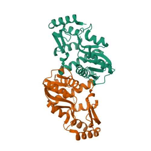
In the intricate realm of cellular chemistry, Adenylosuccinate lyase (ASL) emerges as a remarkable performer. This enzyme takes on the task of cleaving adenylosuccinate—a crucial intermediate molecule—into AMP (adenosine monophosphate) and fumarate. This elegant reaction unfolds within the cytoplasm, marking the second step in the de novo synthesis of AMP. ASL's delicate choreography involves severing the bond between adenosine and succinate within adenylosuccinate. With this precise maneuver, AMP is born, poised to play its essential role in a myriad of cellular processes, from energy metabolism to RNA synthesis and signaling pathways. ASL, typically existing as a solitary entity, consists of a single polypeptide chain. Its structure embraces the captivating TIM barrel fold, a widely embraced motif in the world of enzymes. Named after the triosephosphate isomerase, the TIM barrel fold showcases an enchanting arrangement of alpha helices and beta strands, forming a barrel-like structure. This exquisite fold often boasts eight parallel strands of beta sheet interwoven with alpha helices, creating a rhythmic alternation around the core axis. The TIM barrel fold follows a choreographed pattern—strands adjacent, hands linked in loops or short helices. The grand finale of this structural symphony unfolds as the N- and C-termini of the protein unite, completing the barrel's poetic embrace. The interior of the TIM barrel, a sacred space, holds the enzyme's active site, rich in hydrophobic contours. This sanctum welcomes specific substrates and ligands, forming intimate interactions and enabling the enzyme's catalytic magic. The TIM barrel fold, with its versatile nature, captivates the scientific stage. Its diverse repertoire extends to enzymes performing vital roles in catalysis, isomerization, and metabolic pathways. This fold provides a stable and rigid framework, an unyielding canvas for the artist of catalysis to work their wonders.  Its dance within the TIM barrel fold, a feat of structure and function, illuminates the intricate beauty of enzymes. In the grand theater of cellular chemistry, ASL takes center stage, painting a vivid picture of the wonders of life's molecular choreography.

 The RNA-DNA Nexus - Page 2 2pts_assembly-1
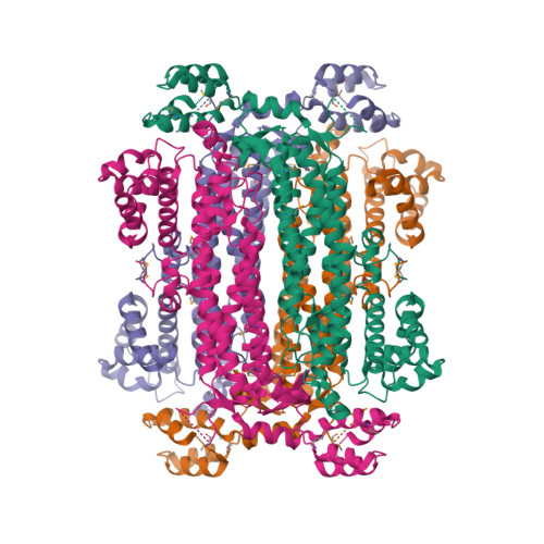
Within the heart of Adenylosuccinate lyase (ASL), an active site brims with captivating activity. This enzyme possesses a unique talent—it acts specifically on adenylosuccinate, an exquisite substrate, cleaving it with precision to unveil the treasures within:  AMP and fumarate. The active site of ASL serves as a stage for molecular interactions, where specific amino acid residues choreograph the enzyme's performance. Their strategic placement within the active site ensures the seamless execution of the enzymatic reaction, like a well-rehearsed dance. ASL's grand performance can be orchestrated by factors beyond its stage. At the transcriptional level, the expression of the gene responsible for producing this marvelous enzyme can be influenced by the cellular environment. The availability of purine nucleotides, those precious building blocks of life, can sway the expression of ASL and set the stage for its enzymatic prowess. Additionally, post-translational modifications and intricate interactions with other proteins can add nuances to ASL's performance, enhancing or modulating its activity. As a solitary performer, ASL thrives in its monomeric form. Its amino acid sequence, a blueprint etched in the language of life, ensures that the enzyme is poised for optimal function. Every letter in this sequence holds significance, contributing to the structural arrangement that allows ASL to unleash its catalytic powers with unrivaled efficiency. So let us marvel at the grand performance of Adenylosuccinate lyase, as it takes center stage, delicately binding to its adenylosuccinate partner and gracefully cleaving it to reveal the hidden treasures of AMP and fumarate. The expression of this captivating enzyme is finely tuned, responding to the needs of the cellular orchestra. In its monomeric glory, ASL dances through the intricacies of the enzymatic world, leaving us in awe of the harmonious interplay of molecules that underpins the symphony of life.


Last edited by Otangelo on Thu Jul 13, 2023 12:13 pm; edited 1 time in total

https://reasonandscience.catsboard.com

27 The RNA-DNA Nexus - Page 2 Empty Re: The RNA-DNA Nexus Tue Jul 11, 2023 1:53 pm

Otangelo


Admin

IMP dehydrogenase (IMPDH)

IMP dehydrogenase, the virtuoso of guanine nucleotide synthesis, takes the stage to weave a magnificent performance. With a weight of 47.75 kDa and 3,656 atoms in E.Coli bacteria, this enzyme orchestrates the oxidation of inosine monophosphate (IMP) to the captivating xanthosine monophosphate (XMP), fueled by the graceful dance of NAD+ as a cofactor. At the heart of IMPDH lies its active site, a sanctuary of specific amino acids poised to guide the enzyme's symphony of reactions. Among them, the Rossmann_fold motif stands tall, embracing the cofactor NAD+ with unwavering devotion. Aspartate, glutamate, histidine, lysine—these virtuoso residues orchestrate the coordination of the cofactor, conducting the transfer of electrons with finesse. Joined by their companions, arginine and serine, they masterfully arrange atoms and groups within the active site, crafting a symphony of hydrogen bonds, electrostatic interactions, and molecular contacts that harmonize the recognition and binding of substrates. Within the sanctuary of the active site, delicate interactions take center stage. Every charge, shape, and molecular feature plays its part, ensuring the optimal recognition and binding of substrates. The slightest nuances can alter the enzyme's catalytic performance, the rhythm of the dance hinging on the precise rotation angles of atoms in select amino acids. Through these intricate conformational changes, the active site molds itself to accommodate substrates, optimizing interactions and ushering in the magical dance of catalysis. IMP dehydrogenase, a maestro of molecular choreography, exemplifies the elegance of enzyme function. Its specific amino acids, carefully arranged within the active site, create an environment primed for catalytic brilliance. With each rotation of atoms and each interaction delicately orchestrated, this enzymatic virtuoso illuminates the path of guanine nucleotide synthesis, captivating us with its enchanting performance.

NAD+ (nicotinamide adenine dinucleotide)

NAD+ stands as a remarkable coenzyme, an unsung hero, participating in a multitude of enzymatic reactions that unfold within the intricate machinery of cells. Derived from vitamin B3, also known as niacin, this non-protein molecule holds great significance in the grand symphony of metabolic processes occurring within our bodies. Let us take a closer look at niacin, a water-soluble vitamin belonging to the esteemed B-complex group. Like a backstage magician, niacin plays a vital role in the backstage affairs of our bodies, orchestrating a myriad of metabolic dances. As a vitamin, it joins the ranks of essential organic compounds that our bodies require in small quantities for optimal growth, development, and overall functioning. In this realm of vitamins, we find agents of vitality and health, playing crucial roles in a vast array of physiological processes. Vitamins serve as coenzymes, aiding enzymes in their majestic performances. They regulate the expression of our genes, act as antioxidants to safeguard against oxidative damage, and facilitate the intricate web of metabolic reactions. Their roles extend to energy production, cell division, immune function, vision, bone health, and an abundance of other vital functions that weave our well-being. To understand vitamins better, we categorize them into two main groups, each with its distinct nature. In one corner, we have the fat-soluble vitamins, encompassing A, D, E, and K. These lipid-loving compounds dissolve effortlessly in fats and oils. When we consume dietary fats, these vitamins embark on a journey, joining hands with their fatty companions. Stored in our body's fatty tissues, they patiently await their time to shine. But caution must be exercised, for an excessive intake of these fat-soluble vitamins can tip the balance and lead to toxicity. On the other side, we find the water-soluble vitamins, a lively group consisting of the B-complex vitamins and vitamin C. These cheerful souls dissolve joyously in water, flowing effortlessly through our systems. Unlike their fat-soluble counterparts, they do not linger within our bodies in significant quantities. Instead, any excess is promptly escorted out through our urine. As such, these water-soluble vitamins crave regular replenishment, requiring us to invite them to our tables through a nourishing diet. As we unlock the secrets of vitamins, we discover the indelible mark they leave on our well-being. Among them, NAD+ stands tall as a coenzyme, a molecular collaborator, fueling enzymatic reactions and playing a silent yet indispensable role in the intricate dance of life. Let us continue our exploration of this captivating realm, where vitamins and enzymes intertwine, enriching our understanding of the remarkable systems that drive our existence.

In the realm of organic compounds, Vitamin B3, also known as Niacin, takes the stage with its mesmerizing heterocyclic aromatic composition. Its captivating structure consists of a pyridine ring harmoniously fused with a carboxyl group, forming a symphony of chemical elegance. Niacin manifests in two forms, nicotinic acid and nicotinamide, both boasting similar vitamin activity while exhibiting distinctive chemical structures. The synthesis of Niacin unfolds within living organisms, emerging from the remarkable transformation of the amino acid tryptophan, abundantly found in protein-rich foods. Treading the stage of life's grand theater, Niacin did not grace the prebiotic environment, where the stage was set for the birth of organic molecules through cosmic chemical choreography. Niacin's complexity renders it a molecule that demands the intricate craftsmanship of biosynthetic pathways. It is not a primal building block, nor an entity birthed spontaneously amidst prebiotic conditions. Rather, Niacin emerges through the alchemy of living organisms—plants, animals, and select microorganisms—bestowing upon them its vital essence. Niacin assumes a pivotal role as a precursor, ushering forth the coenzymes nicotinamide adenine dinucleotide (NAD+) and nicotinamide adenine dinucleotide phosphate (NADP+). These coenzymes, instrumental in a grand array of metabolic reactions, weave redox transformations and energy metabolism. They dance as electron carriers, orchestrating the symphony of countless enzymatic reactions. NAD+ and NADP+ hold the baton, facilitating the graceful exchange of electrons and hydrogen atoms, vital to the grand performance of cellular respiration—a ballet of energy production culminating in the magnificent ATP, the currency that fuels life's vibrant endeavors. Yet, Niacin's influence extends beyond the realm of energy. It embraces the dance of DNA repair and synthesis, delicately guiding the choreography of lipid metabolism. Niacin's touch leaves an indelible mark on the very fabric of life itself, an essential note in the symphony of existence. Thus, Niacin takes its rightful place, a luminary amidst the ensemble of organic molecules. Its origin steeped in the complexity of biosynthesis, it stands as a testament to the intricacies of life's creation. From the transformation of tryptophan to the shimmering coenzymes NAD+ and NADP+, Niacin showcases the artistry of nature's chemistry—a captivating masterpiece that breathes life into existence.

NAD+, the unsung hero of cellular energy metabolism, steps onto the stage as a crucial cofactor, ready to accept and donate electrons in redox reactions. This organic coenzyme, belonging to a remarkable family, acts as a dynamic electron carrier, orchestrating the delicate dance of oxidation and reduction. Like a messenger of energy, NAD+ shuttles electrons between enzymes, embarking on a transformative journey. It possesses the duality of an oxidizing agent (NAD+) and a reducing agent (NADH), gracefully accepting and donating electrons when the time calls for it. This elegant electron transfer holds the key to unlocking the energy hidden within molecules like glucose, fueling the cellular machinery that generates ATP—the currency of life's energy. NAD+ wears another hat, activating enzymes with its electron-receiving prowess during oxidation reactions. As it binds to an enzyme, magic unfolds—a cascade of conformational changes that pave the way for optimal catalysis and seamless electron transfer. Through non-covalent interactions, NAD+ tightly embraces enzymes at specific binding sites, serving as a guiding force in electron transfer and catalytic finesse. Its presence is indispensable, as it weaves its magic in processes like glycolysis, the citric acid cycle, and oxidative phosphorylation—essential metabolic pathways that supply cells with the energy they crave. As we peer into the intricate tapestry of life's biochemical symphony, we are confronted with captivating complexity and exquisite functionality. The mere existence of molecules like niacin, meticulously synthesized within organisms, cannot be attributed solely to naturalistic processes. The presence of complex information, finely-tuned systems, and intricate control networks speaks volumes about the work of an intelligent designer—a mastermind orchestrating the symphony of life. The intricacy and interdependence of biological components, such as the intricate pathways leading to niacin synthesis, demand that all parts be present and functional simultaneously—a "catch-22" scenario that underscores the necessity of intelligent design. The remarkable design details in molecular machinery and the delicate balance within chemical reactions bear witness to the hand of an intelligent creator. The astonishing storage capacity of DNA, the specified complexity embedded within genetic patterns, and the mesmerizing coordination of molecular processes all hint at the involvement of an intelligent agent. The notion of blind chance or purely random processes achieving the creation and functionality of complex organic molecules is a mere illusion. It challenges the very fabric of spontaneous emergence, highlighting the need for the guidance of an intelligent designer. As we continue our exploration of the wondrous intricacies of life, let us embrace the awe-inspiring journey that unravels before us. A journey where the dance of electrons and the interplay of biomolecules reveal the secrets of our existence. Join me as we dive deeper into this captivating realm, where science and wonder entwine, inviting us to ponder the grand design that surrounds us all.

IMP dehydrogenase, the captivating multimeric maestro, graces the stage of enzymatic prowess. This enzyme, typically joining forces as a tetramer, boasts a quartet of identical or similar subunits, each adding its unique voice to the symphony of catalytic mastery. With an average size of 500 to 550 amino acids, these subunits unfold multiple domains, each playing a vital role in the enzyme's magnificent performance. An N-terminal domain takes center stage, conducting the mesmerizing symphony of catalysis, while a captivating C-terminal domain gracefully embraces the substrate and cofactor in a harmonious embrace. A tantalizing tale unfolds as IMP dehydrogenase guides the metamorphosis of IMP into XMP, enchanting the audience with a captivating two-step dance. In the first step, this enzymatic virtuoso orchestrates the oxidation of IMP, gifting two precious electrons to NAD+ and birthing the resplendent NADH. A magical intermediate, E-XMP* (enzyme-XMP), emerges, capturing the imagination with its ephemeral beauty. In the second step, this ethereal intermediate dissolves, liberating the enzyme and conjuring forth the triumphant creation of XMP, an essential building block in the symphony of nucleotide synthesis. Yet, the maestro's performance is not without constraints. It bows before the powerful forces of regulation, surrendering to the exquisite artistry of feedback inhibition. GMP, a masterful player, steps forth as the end-product inhibitor, placing a gentle hand upon the maestro's shoulder, subtly guiding its tempo. The maestro's expression and activity, like the ebb and flow of a grand composition, are further molded by the hands of phosphorylation, gene expression, and the enchanting melodies of allosteric modulation. IMP dehydrogenase, a captivating ensemble of subunits, dances upon the stage of cellular harmony. With each graceful movement, it weaves nucleotide synthesis, a fundamental melody in the symphony of life. Its structure, a testimony to the elegance of protein architecture, stands tall, unfettered by the need for metal cofactors. As the final note fades, the audience is left in awe of the maestro's artistry and the symphony of enzymatic splendor that reverberates within the intricate orchestra of the cell.

GMP synthetase

GMP synthetase, the maestro of guanine symphony, takes its final bow in the grand de novo biosynthesis of GMP. With deft precision, it weaves together the majestic strands of ATP and guanosine monophosphate, orchestrating their union in a spellbinding dance. Like a master conductor, GMP synthetase wields its catalytic baton, capturing the phosphate group from ATP and grafting it onto guanosine monophosphate. As the cosmic dance unfolds, pyrophosphate is set free, cascading like stardust, leaving behind the masterpiece of GMP. This multimeric marvel, composed of multiple subunits, harmonizes in perfect unison, each subunit lending its unique voice to the symphony. The architecture of GMP synthetase is intricate pieces fitting together, like interlocking melodies in a celestial composition. Among these domains, the catalytic domains emerge as the virtuosos, their harmonious collaboration bringing substrate binding and catalysis to life on the grand stage of enzymatic brilliance. GMP synthetase, the discerning connoisseur, selectively chooses its partners, guanosine monophosphate and ATP, recognizing their beauty and binding them in its active site. The stage is set for the alchemical transformation, as the maestro directs the symphony of chemical bonds, orchestrating the birth of GMP. With each note, pyrophosphate, the ethereal companion, is released, carrying away the echoes of the enzymatic enchantment. Yet, even the maestro is not beyond the reach of regulation. Feedback inhibition emerges as the conductor's baton is gently guided by the subtle touch of GMP or other regulatory molecules. As the levels of GMP rise, the maestro heeds the call, lowering the tempo to prevent an overture of excess GMP. The symphony of GMP synthetase's activity dances in harmony with the delicate balance of cellular regulation. Each subunit, an essential player, takes its place upon the stage, weaving together the fabric of GMP synthetase's structure and function. Their collective efforts, a testament to the power of collaboration, create a resounding crescendo of enzymatic brilliance. GMP synthetase, the maestro of guanine, concludes its performance, leaving the audience in awe of its captivating artistry. Through its intricate symphony, it brings forth the vibrant molecules that fuel the rhythms of life. The curtain falls, and the legacy of GMP synthetase echoes, reminding us of the captivating marvels that unfold within the hidden realms of cellular harmony.

Purine Nucleotide Biosynthesis Is Regulated at Several Steps

In the intricate world of purine nucleotide biosynthesis, a symphony of regulation plays out to maintain harmony and balance. This journey begins with the synthesis of IMP, ATP, and GTP, the majestic building blocks of cellular life. To ensure the right rhythm, control is exerted at various steps along this melodic path. The overture of regulation commences with the formation of PRPP, a vital precursor in the IMP pathway. Ribose phosphate pyrophosphokinase takes center stage, catalyzing the first reaction. But beware! ADP and GDP loom as inhibitory notes, dampening the enzyme's activity when their levels rise. This delicate dance orchestrates a decrease in PRPP production, preserving the equilibrium. As the symphony unfolds, the second reaction arises, conducted by amidophosphoribosyl transferase. This virtuoso carries out the first committed step. Feedback inhibition takes the spotlight, with two inhibitory sites eagerly awaiting their nucleotide partners. ATP, ADP, and AMP join hands at one site, while GTP, GDP, and GMP find their place at the other. Their binding gently silences the enzyme, modulating the tempo of IMP production. Adenine and guanine nucleotides take turns, independently and synergistically, controlling the rate of this harmonious synthesis. But there's more to this orchestration. Amidophosphoribosyl transferase experiences the magic of feedforward activation, as PRPP emerges on stage. This molecule, an integral part of the pathway, stirs the enzyme's vigor, fueling the synthesis of IMP. Its presence ignites a cascade of activity, propelling the musical journey forward. A second layer of regulation unfurls, just below the branch point where IMP meets its destiny as AMP and GMP. Here, the virtuosos AMP and GMP don their competitive inhibition cloaks. They bind to the enzymes involved in their own production, restraining their own abundance. This delicate balance prevents the overindulgence of AMP and GMP within the pathway, maintaining the symphony's poise. And the symphony continues, as adenine and guanine nucleotide synthesis perform their elegant duet. GTP takes the lead, guiding the production of AMP from IMP, while ATP graciously supports the synthesis of GMP. Their reciprocal relationship ensures a harmonious interplay, carefully crafting equal measures of AMP and GMP, essential for the symphony of nucleic acid biosynthesis. In this grand orchestration, regulation weaves control, delicately adjusting the tempo, volume, and interplay of the purine nucleotide pathway. It is a symphony of molecules, where the dance of enzymes and the coordination of nucleotides create a masterpiece of harmony, ensuring the cellular symphony resonates with perfect rhythm and balance.

Within the bustling factory of a cell, where production never ceases, a delicate balance must be struck. Just like any thriving enterprise, the cell's success hinges upon the optimal supply of essential materials and building blocks. Too much, and valuable resources go to waste, too little, and the demands of cellular life may remain unmet. Thus, the cell has crafted an intricate system of regulation and control to ensure smooth operations in its bustling factory. Picture the cell as a diligent factory manager, keenly aware of the importance of resource management. It orchestrates a symphony of molecular interactions and pathways to monitor and maintain the supply of basic materials. Just as a savvy manager keeps a keen eye on inventory levels, the cell meticulously assesses the availability of key molecules. In this microscopic factory, the cell's control mechanisms function like vigilant supervisors. They diligently monitor the levels of critical substances, stepping in with precision and finesse to adjust the supply as needed. It's a dance of balance and efficiency, where excess is swiftly curtailed, and scarcity is met with swift action. Like a master conductor, the cell's regulatory pathways harmonize their efforts. They ensure that the synthesis and breakdown of molecules proceed at the optimal pace, aligning supply with demand. This intricate interplay involves a web of intricate feedback loops, where molecules act as messengers, relaying information and fine-tuning the production process. In the cell's bustling factory, this regulatory choreography extends to the very core of its operations. Enzymes, the skilled workers of the cellular world, play their part. They meticulously control their own activity, adjusting their pace to maintain the delicate equilibrium. Just as a worker might slow down when resources run low, enzymes modulate their performance, adapting to the changing demands of the cell. This dance of regulation and control is not limited to a single molecule or pathway. Instead, it permeates every nook and cranny of the cell's bustling factory. From the synthesis of essential nucleotides to the production of energy-rich molecules, the cell's orchestration ensures that its intricate machinery hums with efficiency. In this remarkable microcosm, the cell showcases its mastery of resource management. With meticulous monitoring and a finely tuned regulatory system, it optimizes supply and demand within its bustling factory. Just as a successful enterprise thrives on efficient operations, the cell's control mechanisms ensure that its production lines never falter. It's a testament to the awe-inspiring complexity and ingenuity of the cellular world, where nature's own factory hums along with precision and grace.

 The RNA-DNA Nexus - Page 2 Purine12
Control of the purine biosynthesis pathway.
Red octagons and green circles indicate control points. Feedback inhibition is indicated by dashed red arrows, and feedforward activation is represented by a dashed green arrow.

Voet et.al., (2016): The pathways synthesizing IMP, ATP, and GTP are individually regulated in most cells so as to control the total amounts of purine nucleotides available for nucleic acid synthesis, as well as the relative amounts of ATP and GTP.

Andrew N Lane (2015): Nucleotides are required for a wide variety of biological processes and are constantly synthesized de novo in all cells. When cells proliferate, increased nucleotide synthesis is necessary for DNA replication and for RNA production to support protein synthesis at different stages of the cell cycle, during which these events are regulated at multiple levels. Therefore the synthesis of the precursor nucleotides is also strongly regulated at multiple levels. Nucleotide synthesis is an energy-intensive process that uses multiple metabolic pathways across different cell compartments and several sources of carbon and nitrogen. The processes are regulated at the transcription level by a set of master transcription factors but also at the enzyme level by allosteric regulation and feedback inhibition.

The mysteries of nucleotide biosynthesis continue to captivate scientists as they explore the inner workings of dividing cells. While the basic pathways for nucleotide synthesis are understood, the specific requirements for nutrient precursors and energy, as well as the intricate regulatory networks that govern this process, remain elusive. Unlocking these secrets is vital, as it holds the key to understanding the unique demands of different cell types and pathological conditions. Imagine a grand puzzle, where the pieces are scattered, waiting to be assembled. The cell, with its ceaseless division and growth, is no different. It relies on a complex interplay of nutrient precursors and energy sources to fuel the synthesis of nucleotides, the building blocks of life. Yet, the precise paths from these precursors to the final nucleotides remain shrouded in mystery. As scientists delve deeper into this enigma, they confront a formidable challenge. The tools at their disposal are still evolving, and the existing methods fall short of unraveling the intricate web of pathways that connect nutrient precursors to nucleotide synthesis. It is as if they are equipped with a compass but lack the map to navigate this uncharted territory. Nevertheless, one thing remains clear: the existence of these highly sophisticated and complex regulatory networks is essential. They must have emerged at the very inception of life, intricately woven into the fabric of cellular existence. After all, how else could a delicate balance, a homeostatic equilibrium, be achieved? How else could the cell guarantee the optimal supply of nucleotides, so vital for the continuation of life itself? The very foundation of life rests upon the fine-tuning of these regulatory networks. Their existence in parallel with the creation of biosynthetic pathways signifies their intrinsic importance. From the moment life began, the interplay between these regulatory mechanisms and biosynthesis pathways has been intertwined, harmoniously orchestrating the delicate dance of cellular homeostasis. As scientists unravel the mysteries of nucleotide biosynthesis, they come face to face with the breathtaking complexity of life's inner workings. It is a quest that combines factual exploration with the thrill of discovery, for hidden within these pathways lie the secrets to maintaining optimal supply and sustaining life's essential balance. So, as the puzzle pieces gradually fall into place, scientists push the boundaries of knowledge, striving to understand the intricate dance of nutrient precursors, energy sources, and regulatory networks that enable the cell to synthesize nucleotides. They embrace the challenge, armed with curiosity and the determination to uncover nature's well-guarded secrets. In the end, it is through their tireless pursuit that we inch closer to comprehending the underlying principles that govern life itself. With each new insight, we deepen our appreciation for the wondrous complexity and remarkable ingenuity that underlies the optimal supply and harmonious balance of the cellular world.

Attaching the pyrimidine base to the ribose backbone
In the next step of nucleotide biosynthesis, a crucial process unfolds, where pyrimidine bases like cytosine, thymine, or uracil join hands with ribose or deoxyribose sugars, giving birth to the fundamental units of nucleic acids. The first step in this enchanting tale is the activation of the sugar precursor—ribose-5-phosphate or deoxyribose-5-phosphate. The sugar molecule awakens to its destiny. It receives the gift of a phosphoribosyl group, borrowed  ATP, through the skilled hands of enzymes such as pyrimidine phosphoribosyltransferases. This activation infuses the sugar with newfound potential, priming it for the transformative moments that lie ahead. Now, the stage is set for the grand act—glycosidic bond formation. The activated sugar, accompanied by the synthesized pyrimidine base, embarks on a wondrous dance of connection. In this captivating duet, guided by the adept hands of nucleotidyltransferases or nucleotide synthetases, the two entities unite in a symphony of chemical reactions. The condensation reaction ensues, elegantly joining the ribose or deoxyribose sugar with the pyrimidine base, forever entwining their fates. Yet, the performance is not complete—a final touch awaits. A phosphate group, akin to a shining crown, enters the scene. It bestows its glory upon the nucleoside diphosphate formed in the previous step, elevating it to the esteemed status of nucleoside triphosphate. Nucleoside diphosphate kinases take center stage, conducting the magical transfer of a phosphate group from ATP to the nucleoside diphosphate. This addition amplifies the nucleotide's power, ensuring its readiness to fulfill its role in the symphony of life. The specific enzymes involved in the de novo synthesis pathway may vary depending on the pyrimidine base being synthesized. Furthermore, the pathway itself, along with the specific enzymes, may differ between ribonucleotide synthesis for RNA and deoxyribonucleotide synthesis for DNA due to the distinct sugars involved.

Purines can be salvaged and recycled

Purine salvage pathways are the ingenious recycling systems that cells employ to maintain the delicate balance of nucleotide metabolism. When certain types of RNA undergo turnover, they liberate valuable purines like adenine, guanine, and hypoxanthine. Rather than allowing these purines to go to waste, cells have a remarkable strategy to reclaim and convert them back into nucleotides through the salvage pathways. This recycling process not only conserves energy but also ensures the efficient utilization of resources. In cellular life, purine salvage pathways shine as a testament to the artistry of bioengineering. These pathways exhibit a diverse range of characteristics and distribution, tailored to the specific needs of different organisms. Among mammals, two key enzymes, adenine phosphoribosyltransferase (APRT) and hypoxanthine-guanine phosphoribosyltransferase (HGPRT), play pivotal roles in the salvaging of purines. APRT orchestrates the conversion of adenine to AMP, while HGPRT catalyzes similar reactions for hypoxanthine and guanine. These enzymes are the guardians of energy effectiveness, steering cells away from the costly process of de novo purine biosynthesis. The existence and functionality of purine salvage pathways offer a captivating glimpse into the inner workings of cellular life. They embody a grand display of intricate coordination and interdependence, revealing the marvels of natural engineering. These pathways are not mere chance occurrences but rather the product of meticulous planning and intelligent design. They represent a crucial make-or-break feature for cells, enabling them to efficiently recycle purines and conserve precious energy resources. When we delve deeper into the inner workings of purine salvage pathways, we encounter the awe-inspiring complexity of large multimolecular machines. Enzymes like APRT and HGPRT form an exquisite molecular orchestra, perfectly suited to their role in purine recycling. The conservation of these pathways throughout evolution underscores their fundamental importance and the profound mastery embedded in their design. They are a testament to the bioengineering prowess that operates on a miniature scale, constructing molecular wonders within the confines of a cell. The very existence of purine salvage pathways raises profound questions about their origin and purpose. How did such a marvel of molecular architecture emerge without a guiding hand? The intricacies of these pathways, their just-in-time delivery of solutions, and their impeccable functionality suggest a level of foresight and meticulous engineering. They represent a tapestry of stability control, safeguarding the integrity of DNA and RNA. This level of coordination and specificity could not have arisen by chance; it required the blueprint of an intelligent creator. Purine salvage pathways embody the perfect fusion of function and design, intricately woven into the fabric of cellular life. They stand as a testament to the remarkable miniaturized technology that exists within every cell. The coordination, specificity, and life-essential nature of these pathways underscore their remarkable nature. Their presence from the very inception of life hints at the presence of a guiding hand, ensuring that the machinery of life was set in motion with purpose and precision. The marvel of purine salvage pathways invites us to explore the wonders of the cellular world and ponder the mysteries of our own existence.

When certain types of RNA reach the end of their journey, they undergo turnover, releasing valuable purine molecules such as adenine, guanine, and hypoxanthine. Rather than letting these precious components go to waste, cells possess salvage pathways that ingeniously convert these free purines back into their corresponding nucleotides. It is an awe-inspiring example of resourcefulness, a cellular recycling system that mirrors our own human efforts. Just as we have mastered the art of organized decomposition and separation to reclaim and repurpose materials, cells possess their own inherent intelligence. With a keen understanding of the importance of sustainability and efficiency, they have honed the skill of extracting every ounce of value from used nucleic acids. In this grand cellular symphony, the purine molecules are meticulously restored to their original form through these salvage pathways. The process is akin to unraveling a well-worn tapestry, gently separating the threads and carefully reweaving them into a vibrant new creation. Each purine molecule is guided through a precise sequence of steps, undergoing transformations that restore its functionality and allow it to once again serve as a vital building block for the cell's intricate machinery. It is in contemplating this cellular feat that we come to appreciate the fundamental principle of reuse and recycling. As humans, we have recognized the immense benefits of organized decomposition and the separation of materials. The more we embrace these principles, the more sustainable and harmonious our activities become within the delicate ecosystem of our planet. Just as the cell practices the art of recycling, so too must we, as guardians of our world, strive to emulate this wisdom. By harnessing our own intelligence and channeling it towards responsible resource management, we can create a brighter future for ourselves and for generations to come. So, let us be inspired by the ingenuity of the cell, recognizing that the very essence of recycling lies within our grasp. With each deliberate act of reclamation, each thoughtful step towards organized reuse, we align ourselves with the remarkable intelligence that permeates every aspect of life. In doing so, we honor the beauty of our planet and embrace our shared responsibility as custodians of this magnificent home we call Earth.

R. H. Garrett (2016): In biological cells, recycling is a highly orchestrated, complex, and coordinated process. It is called catabolism. While in anabolism, metabolic networks construct molecules from smaller units, while in catabolism, a set of metabolic pathways breaks down molecules into smaller units that are either oxidized to release energy or used in other anabolic reactions. Interestingly, anabolism and catabolism occur simultaneously in the cell. The conflicting demands of concomitant catabolism and anabolism are managed by cells in two ways. First, the cell maintains tight and separate regulation of both catabolism and anabolism, so metabolic needs are served in an immediate and orderly fashion. Second, competing metabolic pathways are often localized within different cellular compartments. Isolating opposing activities within distinct compartments, such as separate organelles, avoids interference between them. 1

Comment:   Cells possess inherent wisdom, a knowledge of how to minimize energy costs while maintaining a delicate balance known as homeostasis. It is through the intricate web of salvage pathways that this wisdom unfolds, as cells expertly navigate the recycling and reuse of nucleotides, ensuring the optimal pool of these essential building blocks. In the realm of cellular metabolism, simplicity reigns supreme. It is astonishing to think that a limited collection of basic precursor molecules is all that is required to synthesize the diverse array of cellular constituents. Whether it be proteins, nucleic acids, lipids, or polysaccharides, cells possess the extraordinary ability to transform these humble precursors into the complex structures that define life itself. In this symphony of metabolic pathways, certain key routes serve a dual purpose, functioning in both catabolism and anabolism. Take, for instance, the citric acid cycle, a central pathway of intermediary metabolism. Its metabolites, along with those from other pathways, possess the remarkable ability to navigate the delicate balance between energy production and biosynthesis. However, such intricacy demands independent regulation. How could such an exquisitely balanced system have emerged through a slow and gradual process of trial and error? Consider the delicate dance of catabolism and anabolism. If these processes shared the same metabolic tracks, equilibrium would dictate that any inhibition of a particular enzymatic reaction would impact both directions of traffic. To achieve independent regulation, a system of distinct routes must emerge, or shared pathways must employ unique rate-limiting steps that are specific to each opposing sequence. This intricate design calls for foresight and specific goals, challenging the assumptions of naturalism. But the wonders of cellular regulation do not end there. Cells have mastered the art of spatial and temporal compartmentalization. They segregate competing pathways, isolating them from one another, and exhibit a remarkable sense of timing. Just as the world operates on a 24-hour cycle, cells and organisms synchronize their metabolic pathways to the rhythm of day and night. This dance of circadian rhythmicity, the biological clock, orchestrates the ebb and flow of metabolism in perfect harmony. As we delve into the mysteries of cellular life, we are captivated by the genius of energy efficiency, the elegance of independent regulation, and the beauty of spatial and temporal compartmentalization. It is a testament to the ingenuity of life itself, an intricate dance of molecules guided by an unseen hand. Let us marvel at the wisdom of cells and the secrets they unveil, for within their intricate workings lie the blueprints of life itself.

Imagine for a moment the assembly line of a factory, where robots and machines come together to produce a functional product. Now, let us ponder the idea of randomly selecting and interconnecting these components, akin to a patchwork process. Does such a notion hold plausibility? Let us embark on an exploration of this concept and the principles of engineering and organization that underlie the construction of any efficient production line. In the realm of factories, a robotic production line stands as a pinnacle of specialization and sophistication. Each part, machine, and ingredient is meticulously chosen and placed in its designated position. Every component aligns harmoniously, following a well-thought-out plan that ensures smooth operations. This intricate dance of machines is no product of chance. Instead, it demands foresight—an understanding of the desired outcome from the very beginning. To bring this vision to life, a project must first be elaborated upon. Instructional blueprints, carefully crafted by the engineering department, serve as guiding beacons. With these plans in hand, the implementation begins—a precise orchestration of machines, each placed in the right position and order. It is a harmonious process of organization and design, fueling the creation of functional products. In the scientific community, chemists marvel at the ability of cells to construct complex chemicals effortlessly, achieving high yields under ambient conditions. These extraordinary entities excel in dictating patterns of stereochemistry, crafting products that are seemingly impossible in their level of functionalization. Such feats of precision and complexity further highlight the meticulous nature of biological systems. Enzymes, the masters of chemical transformations, play a pivotal role in producing and maintaining this intriguing property. Composed of homochiral amino acids, enzymes themselves were specified through the guidance of homochiral DNA. The synthesis of these amino acids relies on a symphony of homochiral messenger RNA, homochiral ribosomal RNA, and homochiral transfer RNA. The precise orchestration of these elements ensures that life's building blocks possess a uniformity that eludes abiotic explanations. As we venture deeper into the mysteries of life's origin, we encounter these puzzles that defy easy answers. The intricate interplay of homochiral building blocks and the coordination required for functional assembly lines beckon us to consider the presence of intelligent design. The remarkable achievements observed in biological systems challenge the notion of chance as the sole orchestrator of such complexity.

Within the realm of scientific inquiry, there are contrasting perspectives on the origin and development of complex biosynthesis pathways. While some propose a patchwork process driven by chance, others contend that an intelligent designer is behind their intricate design. The debate is fueled by our observations and interpretations of the natural world. Those who advocate for chance argue that a random assembly, akin to a patchwork process, could plausibly give rise to these complex pathways. They point to the remarkable adaptability of biological systems and the creative power of natural selection. Yet, it is true that replicating these intricate processes in the laboratory has proven challenging for even the most brilliant chemists. The notion that self-organization and chance alone can create functional assembly lines raises philosophical questions and requires a leap of faith. Critics of the chance hypothesis assert that order, information, and complexity never emerge spontaneously. They contend that such intricacy can only be attributed to a conscious, intelligent agent. To them, the proposition of chance as the sole driving force behind the assembly of complex biosynthesis pathways is inherently flawed. It raises doubts about the adequacy of a purely naturalistic explanation. Central to the debate is the concept of natural selection, which can only act upon allele variations that arise by chance. By removing the intelligent designer from the equation, chance becomes the guiding force. However, critics argue that invoking chance as an explanation for the complex interplay of molecular processes is irrational and incoherent. To illustrate the challenges of chance-driven origins, consider the analogy of adding a random robotic machine to an existing assembly line. It becomes clear that successful integration requires precise knowledge of the machine's function, substrate processing, and seamless handover to subsequent steps. The sheer complexity and integration of the assembly line defy the notion that such a system could arise through random chance alone. Those who propose a naturalistic explanation for the complexity of biosynthesis pathways are, in my view, abandoning logic and embracing irrationality. The alternative perspective champions design as the rational explanation for the intricacy we observe in biological systems. By carefully examining the integrated assembly lines of living organisms, one can infer the presence of an intelligent creator. This viewpoint draws upon empirical scientific data, observable facts, and logical, plausible inferences. It questions the validity of attributing these complex systems solely to evolutionary processes driven by chance. Ultimately, the debate between chance and design encompasses profound questions about the nature of life and our place in the universe. It challenges us to grapple with the limits of our understanding and the origins of complexity. The path we choose to follow depends on the weight we assign to empirical data, logical reasoning, and the interpretation of observable facts. It is a journey that evokes both fascination and deep introspection, as we seek to uncover the mysteries of life's origins.

Prebiotic synthesis of Purines

The only alternative to these biochemical processes would be, that the basic building blocks were readily available on the prebiotic earth. Glycine for instance is an indispensable substrate for purine nucleotide synthesis, and so - DNA - in cells. It requires at least 5 biosynthetic steps and the respective enzymes to be synthesized. In prebiotic earth, the only alternative would have been that glycine came from comets.

Phys.org (2016): An important amino acid called glycine has been detected in a comet for the first time, supporting the theory that these cosmic bodies delivered the ingredients for life on Earth, researchers said Friday.
In addition to the simple amino acid glycine, the instrument also found phosphorus. The two are key components of DNA and cell membranes. "Demonstrating that comets are reservoirs of primitive material in the Solar System, and vessels that could have transported these vital ingredients to Earth, is one of the key goals of the Rosetta mission, and we are delighted with this result."2

Comment:  The search for life's building blocks extends beyond our planet, as astronomers venture into space to uncover the mysteries of the cosmos. In their quest, they have discovered glycine, a simple and non-chiral amino acid—the foundation of life's molecular architecture. This finding sparked hope among biologists, who had encountered difficulties producing these vital compounds within the confines of Earth's laboratories. However, the challenges of delivering sufficient quantities of amino acids and nucleic acid bases to our planet are formidable. The monumental task would require a torrential downpour of these molecules, numbering in the megatons, but even then, their survival would be tenuous. Water, oxygen, UV light, and other reactive forces would swiftly degrade these precious compounds, impeding their potential to contribute to the origin of life. Additionally, their mixture of left- and right-handed forms would lack the intrinsic desire or ability to organize themselves into the complex structures required for life's emergence. Delving deeper into the intricate web of purine nucleotide biosynthesis, we encounter a multitude of regulatory steps—a testament to the ingenious design that governs the process. Each step is meticulously controlled to maintain the precise levels of purine nucleotides essential for nucleic acid synthesis. The presence of such sophisticated regulations across diverse organisms underscores the remarkable engineering at play. Consider the feedback inhibition mechanism of ribose phosphate pyrophosphokinase, sensitively responding to increased ADP and GDP levels. By suppressing the production of PRPP, the enzyme prevents an excess of purine nucleotides. This level of regulatory finesse reveals an understanding of the system's needs and the ability to implement precise inhibitory mechanisms. The allosteric inhibition of amidophosphoribosyl transferase adds another layer of design brilliance. The binding of nucleotides such as ATP, ADP, AMP, GTP, GDP, and GMP to inhibitory sites modulates the enzyme's activity, allowing for delicate adjustment of IMP production. This level of specificity and control exemplifies careful planning and engineering to achieve the desired outcome. The reciprocal relationship between ATP and GTP in the synthesis of AMP and GMP further highlights the ingenuity at work. Coordinating the rates of synthesis based on the availability of these nucleotides ensures a harmonious balance, vital for nucleic acid biosynthesis. Such precise coordination points to a purposeful arrangement of molecular components, guided by a profound understanding of the system's requirements. Moreover, the regulation beneath the branch point, where AMP and GMP inhibit their own synthesis, demonstrates the designer's foresight. By preventing excessive accumulation of these molecules, the designer safeguards cellular processes, showcasing a deep comprehension of the system's intricacies. In purine nucleotide biosynthesis regulation, we witness an extraordinary level of engineering finesse. The intricate control networks, the interplay of enzymes and metabolites, and the feedback mechanisms all testify to the presence of a master designer. With meticulous precision, this designer organized every component, weaving together a symphony of molecular interactions. It is a testament to the genius behind life's existence—a genius that invites us to explore the wonders of our world with awe and curiosity.


Last edited by Otangelo on Sun Jul 16, 2023 12:52 pm; edited 6 times in total

https://reasonandscience.catsboard.com

28 The RNA-DNA Nexus - Page 2 Empty Re: The RNA-DNA Nexus Tue Jul 11, 2023 2:20 pm

Otangelo


Admin

5


Pyrimidines

In nucleic acids, three types of nucleobases are pyrimidine derivatives: cytosine (C), thymine (T), and uracil (U).

 The RNA-DNA Nexus - Page 2 PRHoJ2X

What are pyrimidines?

Pyrimidines hold the key to the intricate world of DNA and RNA, acting, besides purines, as the essential building blocks that encode and transmit genetic information in all living organisms. These remarkable organic compounds form the very foundation of nucleic acids, molding the blueprint of life itself. Pyrimidines are a special class of molecules, bearing a six-membered ring with nitrogen atoms residing at positions 1 and 3. Within this ring structure, four carbon atoms intertwine with two nitrogen atoms, creating a delicate symphony of atoms. In the realm of DNA and RNA, pyrimidines take center stage, their presence vital for the formation of the genetic code. Three primary pyrimidine bases reign supreme within nucleic acids: cytosine, thymine, and uracil. Cytosine and thymine find their home in DNA, while uracil exclusively graces the domain of RNA. These pyrimidine bases skillfully pair with their purine counterparts—guanine and adenine—through the magic of hydrogen bonding. As the strands of DNA intertwine or RNA molecules come to life, these base pairs forge a strong connection, like the rungs of a ladder, ensuring the stability and integrity of the genetic material. Cytosine dances in perfect harmony with guanine, their partnership cemented by three hydrogen bonds in DNA, while in RNA, their bond is slightly modified, featuring two hydrogen bonds. Thymine, an exclusive guest in the realm of DNA, elegantly pairs with adenine, creating a twosome held together by two hydrogen bonds. As for uracil, it entwines with adenine in RNA, uniting through two hydrogen bonds, setting the stage for RNA's unique role in gene expression. The journey of pyrimidine synthesis is a complex and fascinating one, involving an ensemble of enzymes and intermediates. De novo synthesis is the pathway through which pyrimidines are painstakingly crafted. The process commences with the assembly of the pyrimidine ring, a delicate orchestration guided by the hands of enzymes and their chemical companions. Once the ring is complete, the stage is set for the coupling of ribose 5-phosphate, a sugar molecule, forming the final masterpiece—the pyrimidine nucleotide. This coupling gives birth to exquisite nucleotides like cytidine, thymidine, and uridine, each carrying its own unique identity within the world of nucleic acids. The symphony of pyrimidine synthesis unfolds with precision and finesse, showcasing the intricate dance of atoms and the creative ingenuity of nature. These organic marvels, with their elegant rings and bonds, hold the key to life's intricate code. They embody the profound secrets of genetic information, whispering tales of inheritance and evolution. As we uncover the mysteries of pyrimidine biosynthesis, we unveil the wonders of life's design, unraveling the symphony of molecules that orchestrate the intricate dance of existence.

The Remarkable Size of Nucleobases in DNA: Evidence of purposeful design

In the intricate dance of DNA, the size of nucleobases takes center stage, playing a vital role in determining the harmony and stability of nucleotide pairing. The concept of complementarity is key—it refers to the unique matching of nucleobases that allows DNA strands to align and create a sturdy, double helical structure. Held together by the graceful embrace of hydrogen bonds, these nucleobases hold the secrets to DNA's elegance. Within the realm of DNA, four nucleobases hold court: adenine, thymine, cytosine, and guanine. Their intricate choreography determines the base pairing scheme known as Watson-Crick base pairing. Adenine and thymine delicately intertwine through two hydrogen bonds, while cytosine and guanine create a more intricate connection through three hydrogen bonds. This precise pairing dance allows for the stability and fidelity that DNA demands. The size of nucleobases is of utmost importance, as it influences the very geometry and steadfastness of base pairing. Adenine and thymine (or uracil in the case of RNA) share a similar size, enabling them to elegantly form two hydrogen bonds. This pairing provides just the right amount of stability for A-T (or A-U) interactions. On the other hand, cytosine and guanine boast comparable sizes, allowing them to engage in a more robust relationship, fortified by three hydrogen bonds. The size match of these nucleobases ensures the precise fit necessary for their unique partnerships. The exquisite harmony of nucleobases lies in their ability to complement one another with utmost precision. Adenine finds its perfect counterpart in thymine (or uracil), while cytosine discovers its match in guanine. These molecular duos engage in a dance of hydrogen bonds, forming the backbone of DNA's stability. The sizes of these nucleobases have been delicately calibrated to ensure their harmonious union. If the sizes were significantly different, the delicate balance of base pairing would be disrupted. Imagine a larger nucleobase attempting to pair with adenine—it would distort the hydrogen bonding and upset the delicate geometry, leading to an unstable DNA structure. Similarly, a smaller nucleobase attempting to pair with guanine would encounter energetic hurdles, resulting in a less stable pairing. The precise size and shape complementarity of nucleobases allow DNA to maintain its graceful double helix, providing stability and accuracy during essential cellular processes. In the grand tale of DNA, the size of nucleobases weaves a captivating narrative of harmony and elegance. These molecular protagonists have been finely tuned to ensure their perfect embrace, allowing DNA to fulfill its extraordinary role as the bearer of genetic information. The artful dance of hydrogen bonds, guided by the precise sizes of nucleobases, ensures the fidelity and resilience of the DNA structure. Through this intricate design, life's story is encoded and passed down through the generations, a testament to the wondrous world of molecular choreography.

Base substitutions

In the ancient origins of nucleic acids, a fascinating world of possibilities unfolded. Different bases, beyond the ones we know today, might have played a role in the early stages of life. Prebiotic experiments have revealed a plethora of purine and pyrimidine derivatives, showcasing the diverse chemical landscape of those early times. However, not all of these derivatives were destined for inclusion in the alphabet of nucleic acid polymers. Some of these intriguing candidates faced significant challenges when it came to joining the backbone of nucleic acid. You see, the attachment to a polymer backbone posed a unique predicament for certain derivatives. The very essence of their aromatic nature, which bestowed them with their distinctive character, risked being lost in the process. The delicate balance of chemical properties needed for effective base pairing, too, was not always met by these candidates. As scientists delved into the complexities of nucleic acid origins, they discovered that not all derivatives were created equal. While some showed promise in the laboratory, revealing glimpses of their potential to contribute to the early genetic code, others fell short. Their lack of compatibility with polymer inclusion and their limitations in forming stable base pairs presented significant hurdles. Amidst the possibilities, the search for the most fitting bases continued. Nature's designs require finesse, and only those derivatives that could maintain their aromaticity while harmoniously integrating into a nucleic acid backbone were granted a role in the grand play of life. The precise criteria for successful base pairing demanded a delicate interplay of chemical properties, ensuring the faithful transmission of genetic information. The journey into the origins of nucleic acids is a captivating exploration of both scientific inquiry and the boundless creativity of nature. It is a tale of challenges and triumphs, as scientists uncover the secrets of ancient chemistry and the forces that shaped the foundations of life. Through meticulous investigation, they unravel the mysteries of which bases were destined for inclusion in the grand narrative of nucleic acids. Each discovery brings us closer to understanding the remarkable journey that led to the emergence of the genetic code we know today.

 The RNA-DNA Nexus - Page 2 B0MfqjE

Alternative bases could have been involved in the original informational polymer.

In the vast realm of nucleic acid research, scientists have embarked on a captivating quest to explore the mysteries of base-pairing from a theoretical standpoint. Armed with curiosity and a thirst for understanding, they have delved into the realms of possibility, contemplating alternative compositions, unconventional bonding patterns, and even new attachment points within the intricate structure of nucleotides. The exploration of alternative bases has been a focal point of their endeavors, driven by a desire to uncover molecules that may have possessed distinct advantages over the familiar bases found in modern nucleic acids. These inquisitive minds sought to identify building blocks that were more readily synthesized, imbued with greater stability, or endowed with a penchant for engaging in self-organizational chemistry. Through a symphony of theoretical models and carefully crafted experiments, they embarked on an intellectual journey to decipher the properties and potential merits of these diverse base compositions. Among their musings, the concept of bases capable of forming more than the standard three hydrogen bonds took center stage. While the elegance of Watson-Crick base-pairing, where adenine found solace in the arms of thymine or uracil, and cytosine embraced guanine through their precise hydrogen bonding patterns, had long captivated the scientific community, the notion of even stronger interactions beckoned their exploratory spirits. Could there exist base pairs capable of forging additional hydrogen bonds, providing a foundation of enhanced stability and unwavering fidelity? Yet, as with any captivating hypothesis, the realization of its advantages hinged on multiple factors. The availability of such alternative bases in the prebiotic world, their compatibility with the nucleic acid backbone, and the intricate interplay between environmental conditions, such as ionic strength, pH, and temperature, all shaped the narrative. These intrepid researchers sought to decipher the strength and implications of these newfound base pairings, pondering the dynamic equilibrium of stability and function that danced upon the stage of nucleic acid life. In their scientific odyssey, these daring explorers were driven by a yearning to unravel the secrets of the past and illuminate the path that led to the genesis of life's most essential molecules. With each step forward, they painted a vivid portrait of possibilities yet unexplored, shedding light on nucleic acid stability and the exquisite dance of base pairing. This captivating tale of scientific inquiry captivates the imagination, reminding us of the boundless potential that lies within the realm of the molecular world. Through their meticulous investigations, these courageous minds pave the way for new insights, unveiling the secrets of alternative base pairings and their potential impact on life's essential information carriers.

Other variables that must be finely adjusted to achieve Watson-Crick base pairing

Beyond the strength of hydrogen bonds, the delicate dance of nucleic acid base pairing encompasses a symphony of finely tuned variables. Nature's ingenuity reveals itself through the intricacies of atom isomer configuration, base-pair sizes, and an array of other factors that must be harmoniously adjusted to achieve the exquisite Watson-Crick base pairing. The geometry of hydrogen bonds takes center stage in this molecular ballet. The precise arrangement of atoms and their electronegative dance dictate the fidelity of base pairing. A delicate interplay of forces must align, with each bond perfectly positioned to enable the captivating embrace of complementary bases. It is within this intricate choreography that the foundations of genetic information transmission find their footing. But the drama doesn't end there. Base stacking emerges as a silent yet crucial character in this molecular narrative. Adjacent base pairs in the DNA or RNA double helix engage in a harmonious interplay of non-covalent interactions. The aromatic rings of the bases overlap, their shared intimacy contributing to the stability of the helical structure. This elegant dance of base stacking ensures the integrity and endurance of the double helix, creating a steadfast foundation for life's essential blueprints. And let us not forget the architectural prowess of the sugar-phosphate backbone. This structural backbone imparts rigidity to the helix, its unwavering support aligning the bases in their optimal positions for hydrogen bonding. Like a master craftsman, the backbone molds the delicate strands of nucleic acids into a symmetrical masterpiece, laying the groundwork for the magic of base pairing. Yet, the stage is not complete without the presence of supporting characters. Metal ions, such as the regal magnesium (Mg2+), make their entrance, leaving an indelible mark on the performance. With grace and precision, these ions interact with DNA or RNA, their presence stabilizing the double helix structure. They neutralize the negative charges on the phosphate backbone, allowing base pairs to find solace in each other's arms. In this mesmerizing display, magnesium elevates the stability and finesse of base stacking, enhancing the molecular harmony. Amidst this grand performance, the specific sequence context of each nucleotide base adds a touch of individuality. Each base, like a virtuoso musician, brings its unique presence to the symphony of base pairing. The neighboring bases and structural elements create an orchestration of local stability and dynamics, influencing the interplay of base pairing interactions. It is in this exquisite interdependence that the true artistry of nature's design comes alive. Together, these variables form the delicate design of Watson-Crick base pairing. Nature's meticulous craftsmanship and attention to detail shine through in the synchronization of hydrogen bonds, base stacking, backbone rigidity, metal ion choreography, and sequence context. With each element in perfect alignment, the stage is set for the wondrous symphony of accurate replication, gene expression, and the eternal dance of life itself.

Premise 1: The precise fit and complementarity between nucleobases in DNA, such as adenine with thymine (or uracil) and cytosine with guanine, are crucial for proper base pairing and the stability of the DNA double helix structure. 
Premise 2: The specific size and shape complementarity of nucleobases allows for the formation of hydrogen bonds, contributing to the overall stability of the DNA double helix.
Conclusion: The precise fit, complementarity, and versatility of nucleobases in DNA, which enable accurate base pairing and the maintenance of the DNA double helix structure, suggest the involvement of an intelligent creator with foresight and purposeful goals in the design and engineering of RNA and DNA.


 The RNA-DNA Nexus - Page 2 JItvFpo


 The RNA-DNA Nexus - Page 2 5512

Step 1:  Synthesis of Carbamoyl Phosphate: Our journey through the intricate pathways of pyrimidine biosynthesis begins with the synthesis of carbamoyl phosphate. This pivotal reaction is catalyzed by the cytosolic enzyme carbamoyl phosphate synthetase II, where the amide nitrogen of glutamine and ATP join forces. Two molecules of ATP play a vital role, providing a phosphate group and the energy needed to drive the reaction forward. It is worth noting that carbamoyl phosphate is also synthesized in the urea cycle, where the mitochondrial enzyme carbamoyl phosphate synthetase I utilizes ammonia as the nitrogen source.

Step 2:Synthesis of Carbamoyl Aspartate: Moving forward, we encounter a captivating condensation reaction that merges carbamoyl phosphate and aspartate, orchestrated by the remarkable enzyme aspartate transcarbamoylase (ATCase). What makes this reaction even more intriguing is that it proceeds without ATP hydrolysis, as carbamoyl phosphate is already "activated." The structure and regulation of E. coli ATCase have been extensively explored, shedding light on the intricacies of this enzymatic interplay.

Step 3:Ring Closure to Form Dihydroorotate: As our journey continues, we come across a fascinating intramolecular condensation reaction, masterfully catalyzed by dihydroorotase. This enzymatic transformation brings forth the formation of dihydroorotate, a key intermediate in pyrimidine biosynthesis.

Step 4:Oxidation of Dihydroorotate: A crucial turning point awaits us as dihydroorotate undergoes irreversible oxidation to orotate, guided by the remarkable enzyme dihydroorotate dehydrogenase. In eukaryotes, this enzyme, enriched with FMN and nonheme Fe, resides on the outer surface of the inner mitochondrial membrane, drawing its oxidizing power from quinones. Interestingly, the inhibition of dihydroorotate dehydrogenase disrupts pyrimidine synthesis in T lymphocytes, thereby offering a potential avenue for attenuating autoimmune diseases such as rheumatoid arthritis.

Step 5:Acquisition of the Ribose Phosphate Moiety: Orotate, a molecule of great significance, embarks on a transformative journey as it reacts with PRPP (5-phosphoribosyl-1-pyrophosphate). Guided by the enzyme orotate phosphoribosyl transferase, this reaction gives rise to orotidine-5'-monophosphate (OMP). The hydrolysis of the eliminated pyrophosphate (PPi) drives this reaction forward, fixing the anomeric form of pyrimidine nucleotides in the α configuration. Furthermore, orotate phosphoribosyl transferase salvages other pyrimidine bases, including uracil and cytosine, by converting them into their corresponding nucleotides.

Step 6:Decarboxylation to Form UMP: Our journey reaches its conclusion with a remarkable decarboxylation reaction. OMP, a crucial intermediate, undergoes transformation by the enzyme OMP decarboxylase (ODCase) to form UMP (uridine monophosphate). This enzyme showcases unparalleled catalytic proficiency, enhancing the reaction rate (kcat/KM) by a staggering factor of 2 × 10^23 compared to the uncatalyzed reaction. Interestingly, ODCase accomplishes this feat without the assistance of cofactors to stabilize its putative carbanion intermediate. The removal of OMP's phosphate group, though distant from the C6 carboxyl group, reduces the reaction rate by a factor of 7 × 10^7, exemplifying how binding energy can be skillfully harnessed in catalysis through preferential transition state binding.

Differences between Cytosine, Uracil, and Thymine

Within the intricate world of nucleic acids, three nitrogenous bases—cytosine, uracil, and thymine—share a common thread, while each possesses its own unique character. Like characters in a grand play, they play distinct roles on the stage of DNA and RNA. Cytosine, a versatile base, finds its place in both DNA and RNA. It forms a harmonious bond with guanine, with three hydrogen bonds sealing their partnership. Its structure, a six-membered ring adorned with two nitrogen atoms, exudes elegance and versatility, as it orchestrates crucial processes such as DNA replication and gene expression. Uracil, an exclusive player in the realm of RNA, casts its spell as one of the four RNA bases. This charismatic base replaces thymine in RNA, uniting with adenine through two enchanting hydrogen bonds. Its ring, much like cytosine's, houses two nitrogen atoms, creating a symmetrical backdrop for the intricate dance of coding and decoding genetic information during protein synthesis. Thymine, a DNA aficionado, boasts a distinguished presence exclusive to DNA's hallowed halls. This modified form of uracil bears a methyl group, adorning its six-membered ring with a touch of elegance. With two hydrogen bonds, thymine finds solace in the embrace of adenine, preserving the stability and integrity of the genetic blueprint. Together, these bases bring harmony to the dance of nucleic acids, each playing its vital role. Cytosine takes on the responsibilities of DNA replication and gene expression, ensuring the fidelity of genetic information. Uracil, the melodious messenger of RNA, weaves intricate patterns of coding and decoding, bringing forth the language of proteins. Thymine, the guardian of DNA's sanctum, safeguards the genetic material, ensuring its unwavering stability. As the symphony of life unfolds, these nitrogenous bases captivate us with their elegance and purpose. Each base, with its own distinct qualities, contributes to DNA and RNA. It is through their collective dance, their precise pairings, and unique structures, that the beauty and complexity of genetic information transmission come alive.

De novo pyrimidine biosynthesis

Pathway overview

 The RNA-DNA Nexus - Page 2 4912


In the realm of biosynthesis, the creation of pyrimidines takes a simpler path compared to their purine counterparts. Let us embark on a journey through the intricate process that gives rise to these essential building blocks of life.
At the heart of pyrimidine biosynthesis lies aspartic acid, a versatile amino acid that contributes significantly to the formation of the pyrimidine ring. From aspartic acid, the nitrogen atom at position 1 of the pyrimidine ring emerges, as well as the carbon atoms at positions 4, 5, and 6. Aspartic acid generously donates its carbon backbone to provide these essential building blocks, adding depth and structure to the pyrimidine ring. But the story doesn't end there. Another player enters the stage - glutamine, an amino acid brimming with potential. Glutamine gracefully steps forward to contribute the nitrogen atom at position 3 of the pyrimidine ring. Its amide group becomes the foundation for this pivotal nitrogen atom. And behold, the carbon atom at position 2, a vital piece of the pyrimidine puzzle, is unveiled. It arises from none other than carbamoyl phosphate, an esteemed intermediate in the grand symphony of pyrimidine biosynthesis. With grace and precision, carbamoyl phosphate provides the necessary carbon atom, infusing the pyrimidine ring with its distinctive character. The harmonious collaboration between aspartic acid, glutamine, and carbamoyl phosphate paints a vivid picture of metabolic interplay. These interconnected pathways dance together. Aspartic acid bestows the pyrimidine ring with carbon atoms and the nitrogen atom at position 1, while glutamine lends its nitrogen atom at position 3. Carbamoyl phosphate steps forward, providing the carbon atom at position 2, completing the symphony of pyrimidine biosynthesis. These intricate findings shed light on the captivating interplay between diverse metabolic pathways. Pyrimidines, in their distinct synthesis from purines, showcase the elegance of nature's design. The construction of the pyrimidine ring precedes the attachment of the ribose-5-phosphate moiety, revealing the unique order in which these vital molecules come to life. In this intricate dance of biosynthesis, the story of pyrimidines unfolds, bringing us closer to understanding the remarkable processes that underlie the building blocks of life.

 The RNA-DNA Nexus - Page 2 5011
In the fascinating realm of pyrimidine biosynthesis, we uncover the intricate steps that give rise to these essential building blocks of life. Unlike their purine counterparts, pyrimidines derive their existence from just two precursors: carbamoyl phosphate and aspartate. Let us embark on a captivating journey into the realm of pyrimidine creation and unravel the intricate dance of enzymes and molecules that shape their formation. In the realm of mammals, pyrimidine biosynthesis relies on the enzyme carbamoyl phosphate synthetase II (CPS-II), stationed within the cytosol. This remarkable enzyme orchestrates a symphony of substrates: bicarbonate (HCO3-), water (H2O), glutamine, and two adenosine triphosphate (ATP) molecules. The amide group of glutamine plays a vital role, contributing its nitrogen atom to form carbamoyl phosphate through a series of exquisite steps involving CO2 activation, phosphate displacement, and phosphorylation. CPS-II stands as the gateway, the committed step in the de novo pyrimidine pathway for mammals. On the contrasting stage, bacteria and plants possess a singular carbamoyl phosphate synthetase (CPS) that produces carbamoyl phosphate, serving both pyrimidine and arginine biosynthesis. Here, aspartate transcarbamoylase (ATCase) takes the spotlight in bacterial pyrimidine synthesis. ATCase skillfully unites carbamoyl phosphate and aspartate, fashioning carbamoyl aspartate. Unlike CPS-II, this step unfolds without the need for ATP, for carbamoyl phosphate bears the power of an "activated" carbamoyl group. As the symphony of pyrimidine biosynthesis progresses, the baton is passed to the dihydroorotase enzyme. With finesse, it orchestrates ring closure and dehydration, linking the ONH2 group from carbamoyl phosphate with the former β-COO2 of aspartate. This harmonious linkage gives birth to dihydroorotate (DHO), a six-membered ring compound that holds the promise of pyrimidine. Dihydroorotate, though not a true pyrimidine, undergoes oxidation to yield orotate under the guidance of dihydroorotate dehydrogenase. Bacterial dihydroorotate dehydrogenases, adorned with flavoproteins and nonheme Fe-S centers, wield NAD+ as a cofactor. In the realm of eukaryotes, dihydroorotate dehydrogenase takes its place within the inner mitochondrial membrane, where it intertwines with a quinone, its immediate electron acceptor. As the reduced quinone undergoes oxidation within the mitochondrial electron transport chain, the wondrous dance of ATP synthesis through oxidative phosphorylation unfolds. Now, as our tale nears its conclusion, ribose-5-phosphate enters the stage, joining in a specific N-β-glycosidic configuration with the N-1 position of orotate. This extraordinary union is catalyzed by orotate phosphoribosyltransferase, leading to the birth of orotidine-5'-monophosphate (OMP), a pyrimidine nucleotide that sparks the imagination. With the grand finale within reach, OMP decarboxylase steps forward, wielding its power to remove the carboxyl group from OMP. As the transformation takes place, the enchanting uridine-5'-monophosphate (UMP), known as uridylic acid, emerges. UMP stands proudly as one of the two common pyrimidine ribonucleotides, an essential player in the realm of genetic information. As we conclude this captivating journey into the world of pyrimidine biosynthesis, we stand in awe of the intricate steps and elegant choreography of enzymes and molecules. Through their harmonious interplay, they bring forth the building blocks of life, leaving us with a deeper understanding of the marvelous processes that shape our existence.

Enzymes used in pyrimidine synthesis:

1. Carbamoyl phosphate synthase II
2. Aspartate carbamoyltransferase
3. Dihydroorotase
4. Dihydro Orotate Dehydrogenase
5. Orotate Phosphoribosyl transferase
6. Orotidine 5'-phosphate decarboxylase


 The RNA-DNA Nexus - Page 2 5110

In the grand symphony of pyrimidine nucleotide synthesis, the atoms at positions 2 and 3 of the pyrimidine ring take center stage. They derive their existence from the remarkable carbamoyl phosphate, while the other players in the ring's ensemble emerge from the wondrous aspartate. Let us delve into the intricate steps that give rise to these essential building blocks, and witness the symphony of molecular transformations. Our journey commences with the birth of carbamoyl phosphate, a key protagonist in the pyrimidine biosynthesis tale. This remarkable molecule is crafted by the skilled hands of the enzyme carbamoyl phosphate synthetase II (CPS-II), utilizing a captivating blend of bicarbonate, water, glutamine, and adenosine triphosphate (ATP). Glutamine, with its amide group, bestows upon carbamoyl phosphate the precious nitrogen atom it holds. CPS-II takes its place within the cytosol, acting as the gateway, the committed step in the de novo pyrimidine pathway for mammals. Carbamoyl phosphate, now prepared, emerges as a pivotal figure, contributing its carbon atom at position 2 and nitrogen atom at position 3 to the grand pyrimidine ring. The remaining atoms of the ring, including the exquisite carbon atoms at positions 4, 5, and 6, find their origins in the captivating aspartate. This remarkable amino acid, like a generous benefactor, donates its carbon atoms to the pyrimidine ring, adding depth and complexity to the tale. As our tale unfolds, the enchanting enzyme aspartate transcarbamoylase (ATCase) takes its rightful place. With grace and precision, ATCase orchestrates the condensation of carbamoyl phosphate and aspartate, forging a connection that gives birth to the mesmerizing carbamoyl aspartate, also known as N-carbamoylaspartate. In this magical union, the carbamoyl group from carbamoyl phosphate entwines with the amino group of aspartate, forming a captivating intermediate that propels the pyrimidine biosynthesis forward. The journey continues as the baton is passed to the subsequent enzymatic reactions. The intricate pathway guides the transformation of carbamoyl aspartate into orotate, a precursor brimming with promise for pyrimidine nucleotide synthesis. Orotate emerges as a six-membered ring compound, hinting at the wonders yet to come. With orotate taking the spotlight, the next act unfolds with the addition of ribose-5-phosphate. This essential sugar, derived from the captivating pentose phosphate pathway, joins the nitrogen atom at position 1 of orotate in a harmonious bond. The enchanting enzyme orotate phosphoribosyltransferase leads this delicate dance, breathing life into the captivating orotidine-5'-monophosphate (OMP), known in some circles as orotidylic acid. OMP stands proudly as an intermediate, heralding the birth of other pyrimidine nucleotides like uridine monophosphate (UMP), cytidine monophosphate (CMP), and thymidine monophosphate (TMP). As we conclude this captivating journey into the realm of pyrimidine biosynthesis, we stand in awe of the intricate steps and the elegant dance of enzymes and molecules. Through their precise choreography, they bring forth the essential components that shape the very fabric of life. We leave with a deeper appreciation for the harmonious interplay of these molecular forces, guiding us towards a greater understanding of the remarkable processes that underlie our existence.


Last edited by Otangelo on Fri Jul 14, 2023 1:27 pm; edited 3 times in total

https://reasonandscience.catsboard.com

29 The RNA-DNA Nexus - Page 2 Empty Re: The RNA-DNA Nexus Tue Jul 11, 2023 2:58 pm

Otangelo


Admin

1. Synthesis of carbamoyl phosphate CPS

In the wondrous realm of pyrimidine biosynthesis, a remarkable enzyme called carbamoyl phosphate synthetase II (CPS-II) takes center stage, orchestrating the creation of carbamoyl phosphate, a pivotal precursor in the symphony of pyrimidine nucleotide synthesis. Let us embark on a captivating journey through the intricate steps that bring this essential molecule to life. Our story begins with bicarbonate, a humble molecule waiting to unleash its potential. With the first stroke of CPS-II's enzymatic prowess, bicarbonate is activated through a remarkable transfer of a phosphate group from adenosine triphosphate (ATP). Energized and ready to contribute, bicarbonate transforms into a vibrant entity known as carboxy phosphate, a form of activated carbon dioxide. Enter glutamine, a noble amino acid bearing the gift of an amide nitrogen. Like a master artisan, glutamine's amide group displaces the phosphate group from carboxy phosphate, forging a connection that leads to the creation of carbamate. A second dance with ATP ensues, as another phosphate group is transferred to carbamate, infusing it with vitality and giving birth to the extraordinary carbamoyl phosphate. CPS-II, with its unwavering focus on pyrimidine biosynthesis, stands distinct from its counterpart CPS-I, found in the mitochondria, which partakes in the urea cycle. CPS-I plays a vital role in ammonia detoxification, using ammonia and bicarbonate as its artistic medium to craft carbamoyl phosphate. In contrast, CPS-II devotes its unwavering attention to the de novo pyrimidine pathway, creating carbamoyl phosphate from bicarbonate and the amide nitrogen of glutamine. These two enzymes, though sharing a common goal of carbamoyl phosphate synthesis, dwell in different cellular realms, each with its unique purpose. CPS-I safeguards the liver's intricate dance of eliminating excess nitrogen as urea, derived from the metabolic breakdown of amino acids. CPS-II, on the other hand, breathes life into the de novo pyrimidine biosynthesis pathway, setting the stage for the subsequent steps that give rise to pyrimidine bases, nucleosides, and nucleotides. As we marvel at the elegance of CPS-II and its role in pyrimidine biosynthesis, we gain a deeper appreciation for the intricacies of these cellular symphonies. The creation of carbamoyl phosphate marks the beginning of a remarkable journey, where molecules come together in harmonious unity to shape the very essence of life itself. With each enzymatic step, the stage is set for the creation of the diverse pyrimidine repertoire that underlies the fundamental processes of genetic information storage and transmission.

Carbamoyl Phosphate Synthetase II (CPS-II)

Within the intricate world of pyrimidine nucleotide synthesis, an extraordinary enzyme called Carbamoyl Phosphate Synthetase II (CPS-II) takes center stage. Its primary role is to catalyze the formation of carbamoyl phosphate—a vital building block in the de novo pathway of pyrimidine synthesis. As we delve into the realm of CPS-II, we encounter a complex molecular ensemble, composed of multiple subunits, working in unison to accomplish its remarkable function. Imagine a symphony of atoms, intricately arranged within CPS-II, guiding its catalytic prowess. This enzyme, with a total structure weight of approximately 647.38 kDa, displays a mesmerizing complexity, comprising an astounding 49,731 atoms. Its composition is a tapestry of intricacy, where each subunit contributes to the grand orchestration of carbamoyl phosphate synthesis. CPS-II ignites the pathway by incorporating the amide nitrogen from glutamine into the carbamoyl group—a transformative step leading to the birth of carbamoyl phosphate. Within the multifunctional CPS-II complex, the amalgamation of catalytic and regulatory subunits harmonizes their efforts, ensuring the seamless execution of its essential role. As we wander through the enchanting realm of CPS-II, we find that its size can vary, but it typically manifests as a substantial enzyme complex. It boasts several hundred to over a thousand amino acids, a testament to its intricate nature and functional diversity. Join me as we uncover the secrets of CPS-II, that wondrous enzyme complex weaving the intricate tapestry of pyrimidine nucleotide synthesis. Let us witness the dance of atoms and the harmonious interplay of subunits as they work tirelessly to shape the molecular landscape. Together, we will unravel the mysteries of this remarkable enzyme, one amino acid at a time, and delve into the grand design that underlies the intricate machinery of life.

 The RNA-DNA Nexus - Page 2 5210

In the enchanting world of Carbamoyl Phosphate Synthetase II (CPS-II), a captivating dance unfolds as ATP and Glutamine take center stage, fueling the remarkable catalytic activity of this enzyme. Let us immerse ourselves in the mesmerizing steps of its mechanism and explore the intricate layers that shape its function. The performance commences with a burst of energy as ATP ignites the activation of bicarbonate, bringing forth the birth of carboxy phosphate. Like a musical duet, Glutamine gracefully joins the stage, donating its amide nitrogen and displacing the phosphate group with finesse. Their harmonious collaboration gives rise to the creation of carbamate, an essential intermediate in the grand symphony of carbamoyl phosphate synthesis. The rhythm intensifies as another ATP molecule steps forward, cascading its vibrant energy onto the scene. The resonance of hydrolysis reverberates through the air, accompanied by the phosphorylation of carbamate. This transformative act brings forth the culmination of the performance—the triumphant creation of carbamoyl phosphate. Beyond the enchanting catalytic steps lies a tale of genetic orchestration. The genes encoding the various subunits of CPS-II come to life, transcribed and translated into the intricate dancers that form the enzyme complex. Their post-translational modifications become the brushstrokes of artistic refinement, enhancing the activity, stability, and localization of the enzyme complex. Phosphorylation, the conductor of regulatory symphonies, takes the spotlight, fine-tuning the enzyme's activity and interplay with other proteins and cofactors. Acetylation lends its touch of elegance, adorning specific lysine residues and influencing stability, interactions, and enzymatic prowess. And while glycosylation's role in CPS-II remains less explored, its potential to shape folding, stability, and function adds an air of intrigue to the tale. The assemblage of CPS-II subunits is a choreography of exquisite precision. Chaperone proteins gracefully guide the unfolding of this dance, ensuring the correct folding and seamless fusion of the subunits into the active enzyme complex. Their careful guidance ensures the harmony and functionality of the multimeric ensemble. A symphony of targeting signals orchestrates the subunits' journey, leading them to their designated cellular abodes. Like celestial navigators, these signals guide the subunits to the cytosol, their rightful destination. Each step, each movement contributes to the structural integrity, stability, and efficiency of CPS-II, bringing forth the beauty of carbamoyl phosphate synthesis in the de novo pyrimidine pathway. Regulation becomes the conductor's baton, directing the tempo of pyrimidine nucleotide synthesis. Feedback inhibition steps in, ensuring a delicate balance as the pathway's end products wield their influence. Allosteric regulation by nucleotides and metabolites sets the stage for precise control, preventing an overabundance of pyrimidine nucleotides. Quality control mechanisms emerge, guardians of fidelity and integrity. Chaperones and proteases stand vigilant, ensuring proper folding and assembly of the enzyme subunits. In moments of misfolding or assembly errors, proteolytic degradation or refolding mechanisms gracefully sweep in, preserving the essence of CPS-II. The tale of CPS-II is one of elegance, precision, and beauty. It weaves together the intricacies of molecular ballet, showcasing the synchronized steps that create the foundation for pyrimidine nucleotide synthesis. Let us marvel at its rhythmic harmony, appreciating the wonder of this enzyme and its role in the grand symphony of life.

 The RNA-DNA Nexus - Page 2 5310

Crystal structure of Carbamoyl Phosphate Synthetase (CPS) from Escherichia coli (E. coli). There are two key aspects: the tetrameric (αβ)4 form and the functional αβ entity, along with the active sites, substrate channel, and allosteric domain.

A) The tetrameric (αβ)4 form: CPS is composed of four subunits, with each subunit consisting of an α and a β chain. The crystal structure reveals the arrangement of these subunits, forming a tetrameric structure. This organization is important for the overall functionality of CPS, as it allows for the coordination and interaction of the individual subunits.

B) The functional αβ entity: Within each αβ subunit, there are specific features highlighted in the crystal structure. The small subunit contains a single active site, emphasized by the presence of a catalytic cysteine at position 269. The large subunit, on the other hand, possesses two active sites: one within the carboxyphosphate domain and another within the carbamoylphosphate domain. These active sites are highlighted by the presence of bound adenosine diphosphate (ADP), which is likely involved in the catalytic process.

The crystal structure also reveals a substrate channel that connects the three active sites within CPS. This channel serves as a pathway for the transfer of reaction intermediates between the active sites, facilitating efficient catalysis. The substrate channel is colored gray. Additionally, the crystal structure highlights the presence of an allosteric domain, which is associated with the binding of ornithine. Allosteric regulation plays a role in modulating CPS activity, and the presence of bound ornithine suggests its involvement in the allosteric regulation of CPS.

 The RNA-DNA Nexus - Page 2 5410
Reactions Catalyzed by CPS

Carbamoyl Phosphate Synthetase II (CPS-II) is a fascinating and exceptional enzyme complex that holds secrets waiting to be unveiled. Within its single polypeptide chain lies a trio of catalytic activities, each with its unique role—carbamoyl phosphate synthetase (CPS), aspartate transcarbamoylase (ATC), and dihydroorotase (DHOase). This trifecta of functions within a single entity beckons us to explore the remarkable intricacies that defy ordinary explanations. The seamless integration and flawless coordination of these catalytic activities in CPS-II elude the grasp of mere chance. The harmonious coexistence of these activities within a single polypeptide chain demands a level of coordination and integration that hints at a higher order design. The complexity and precision of CPS-II's catalytic sites speak volumes, for it is exceedingly improbable for such intricate functionality to emerge randomly and without guidance. Indeed, CPS-II unveils its enigma, as it unveils its secrets only when all its components are present and intricately intertwined. This interdependence points to a grand orchestration, where multiple coordinated implementations come together in perfect harmony—a symphony of purpose. The specific arrangement and functional integration of CPS-II's catalytic activities carry a message encoded within—a message of intelligent design. The presence of functional complexity intertwined with the precise orchestration of catalytic sites implies the involvement of an intelligent agent, a mastermind who crafted CPS-II with intention and purpose. This exquisite organization not only astounds but also enhances the efficiency of pyrimidine biosynthesis. The intermediates flow seamlessly, channeled with utmost precision between the enzymatic reactions, amplifying the overall efficiency of this vital biosynthetic pathway. As we delve deeper into the intricacies of CPS-II, we are confronted with the undeniable whispers of an intelligent creator. The remarkable fusion of distinct catalytic activities within a single entity, their intricate arrangement, and the flawless coordination transcend the realm of chance. They beckon us to embrace the awe-inspiring notion that CPS-II is an ingeniously designed molecular masterpiece, crafted with a specific purpose in mind. In the grand tapestry of life, CPS-II stands as a testament to the wonders and mysteries that continue to unravel. Its elegant complexity invites us to ponder the depths of existence, igniting our curiosity and nurturing our thirst for knowledge. Let us embark on this journey, guided by the allure of CPS-II, as we uncover the truths hidden within its hallowed halls.

Within the intricate architecture of Carbamoyl Phosphate Synthetase II (CPS-II), the catalytic activity is orchestrated by the precise arrangement of amino acids within its active site. These amino acids, the building blocks of proteins, play pivotal roles in the enzymatic reactions that fuel CPS-II's function. In the CPS domain, the amino acids cysteine and histidine take the stage, captivating our attention as they bind and activate ATP, the energy currency of the cellular world. They form a delicate dance, ensuring that the ATP molecule is primed and ready for the enzymatic performance. Meanwhile, aspartate and glutamate join the ensemble, gracefully coordinating the binding of carbamoyl phosphate, a key participant in the pyrimidine biosynthesis pathway. Their intricate steps guide carbamoyl phosphate into position, setting the stage for the subsequent reactions. Arginine and lysine, armed with their positive charges, join the symphony, adding stability and facilitating the transfer of chemical groups. They create an atmosphere of support, forming bonds and connections that aid in substrate stabilization and the orchestration of chemical transformations. As their interactions harmonize, the intricately choreographed dance of catalysis unfolds. Within the active site, a theater of molecular interactions, hydrogen bonds, electrostatic attractions, and hydrophobic encounters intermingle. These interactions bring stability and ensure the precise recognition and binding of the substrates essential for pyrimidine biosynthesis. The active site's contours and charges form a complementary landscape, embracing the substrates with a tailored embrace that fits like a key in a lock. As the catalytic performance unfolds, the atoms within the amino acids deftly rotate, adjusting their positions to optimize the catalytic efficiency. Conformational changes ripple through the enzyme, fine-tuning the positioning of substrates and catalytic residues, enhancing the overall performance of CPS-II. Like skilled acrobats, these dynamic shifts allow for the perfect alignment of atoms, ensuring that the reactions proceed with maximum efficiency. Cysteine and histidine bring their unique talents to the stage, bonding and activating ATP with precision. Serine adds its finesse, engaging in hydrogen bonds and other interactions, contributing to the intricate tapestry of substrate recognition and binding. Aspartate and glutamate, ever the coordinators, guide carbamoyl phosphate with a delicate touch, while arginine and lysine lend their positive charges to stabilize and facilitate the transfer of chemical groups. The precise rotation angles of these amino acids are the secret ingredients, the subtle nuances that unlock CPS-II's catalytic prowess. These rotational movements, performed with precision and grace, optimize the positioning of functional groups and catalytic residues, allowing for a magnificent performance of efficient substrate binding and catalysis. They are the silent choreographers, shaping the destiny of CPS-II's enzymatic symphony. In this intricate interplay of atoms and molecules, we witness the fascinating dance of catalysis within CPS-II. A carefully orchestrated performance, guided by the precision arrangement of amino acids, unfolds before our eyes. As we marvel at the elegance and complexity of this molecular ballet, we are reminded of the wonders that lie within the microscopic world, and the remarkable ingenuity that underlies the dance of life.

In the realm of enzymes, such as Carbamoyl Phosphate Synthetase II (CPS-II), we find evidence of purposeful design, crafted by an intelligent creator. Within the intricate web of molecular interactions, a symphony of amino acids emerges, finely tuned and meticulously arranged within the enzyme's active site. This delicate coordination of charges, shapes, and other molecular features within CPS-II's active site reveals a remarkable level of specificity and recognition—an unmistakable hallmark of intentional design. The precise arrangement of amino acids within CPS-II's active site is no coincidence. It is a testament to the engineering ingenuity that ensures optimal catalytic efficiency and specificity. Every element, every bond, every subtle twist and turn within the active site serves a purpose—a purpose guided by the intelligence of a creator. Through this intentional design, CPS-II achieves the perfect stage for substrate binding and catalytic transformations. Beyond the arrangement of amino acids, we encounter the captivating world of rotation angles—an intricate dance of atoms within CPS-II and other enzymes. These precise rotations hold the key to conformational changes, enabling the enzyme to optimize the positioning of substrates and catalytic residues. Such exquisite precision, which orchestrates the molecular interplay within enzymes, is far beyond the realm of random chance or unguided processes. It points towards the masterful planning and engineering of an intelligent agent, who possesses the foresight to fine-tune these rotations for optimal catalytic activity. As we delve deeper into the tapestry of enzymes, we are captivated by the intricate coordination of atoms and the meticulous optimization of rotation angles. These remarkable features serve as tangible evidence of purposeful design, reflecting the wisdom and intelligence of a creator. Within CPS-II and other enzymes, we witness a symphony of molecular choreography, where every note and every movement is finely orchestrated. Come, let us embark on a journey through the awe-inspiring world of enzymes, where scientific marvels intertwine with the wonders of life. Together, we will unveil the secrets of molecular precision, celebrating the grand design that underlies the intricate machinery of existence.

Substrate channeling

Within the realm of Carbamoyl Phosphate Synthetase II (CPS-II), a remarkable phenomenon takes place—a captivating display of molecular efficiency known as substrate channeling. This clever mechanism orchestrates the seamless transfer of intermediates between catalytic sites, without their release into the surrounding solution. It is a dance of molecular coordination and choreography that enhances the efficiency of the overall reaction. Imagine a bustling city, with roads and pathways connecting various destinations. In CPS-II, physical tunnels and pathways serve as these essential conduits, facilitating the smooth transfer of reactive intermediates between the neighboring active sites. These pathways act as secret passages, allowing the intermediates to travel swiftly and securely, away from the prying eyes of the solvent and other molecules that could lead them astray. Substrate channeling holds several advantages, like providing protection and shielding for the reactive intermediates. By keeping them secluded within the channels, the intermediates are shielded from the bustling environment of the bulk solution, where distractions and side reactions may lurk. This sanctuary ensures that the intermediates remain in close proximity to the enzymes responsible for subsequent reactions, avoiding any detours or premature decompositions. The proximity of the intermediates to their destined active sites creates a favorable environment for efficient reactions. With their dance partners nearby, the intermediates enjoy a higher local concentration, increasing the chances of encounters and collisions that lead to swift reactions. The close quarters foster intimate interactions, enhancing the reaction kinetics and propelling the dance of catalysis forward. Channeling transcends the conventional diffusion route through the bulk solution, bypassing the obstacles of slow diffusion and the viscosity of the solvent. Instead, it offers a fast lane, where intermediates are whisked away through the dedicated pathways with remarkable speed. The intermediates no longer suffer the delays and hindrances of diffusion, as they are swiftly shuttled between active sites, keeping the rhythm of the reaction alive. In this choreographed display of molecular elegance, substrate channeling reveals its beauty. It is a finely tuned mechanism that maximizes efficiency and minimizes wasteful detours. The intermediates, shielded within the channels, embark on a journey that ensures their safe passage and swift arrival at the next catalytic site. It is a dance of precision and coordination, where the intermediates are guided along the secret paths, accelerating the overall reaction. Substrate channeling is a testament to the ingenuity of nature's design, where molecules perform a synchronized ballet. It is a reminder that even on the microscopic stage, there are hidden pathways and secret tunnels that orchestrate the intricate dance of life.

In the symphony of cellular metabolism, Carbamoyl Phosphate Synthetase II (CPS-II) takes on the role of a vigilant conductor, ensuring a harmonious balance between pyrimidine production and the cellular needs. With precision and finesse, CPS-II orchestrates its activity through a system of tight regulation, choreographed by an ensemble of metabolites. ATP, UTP, PRPP (phosphoribosyl pyrophosphate), and CTP, acting as the maestros, modulate CPS-II's performance. They delicately adjust the enzyme's tempo, allowing it to respond to fluctuations in nucleotide levels and metabolic demands. This regulatory dance ensures that pyrimidine biosynthesis is finely tuned, taking place in close proximity to the sites where these essential building blocks are required. It is a synchronized dance that optimizes the utilization of synthesized pyrimidine molecules, channeling them efficiently to their destined roles. During the grand act of cell division, CPS-II assumes a leading role, supplying the much-needed pyrimidine nucleotides for the synthesis of DNA and RNA—the fundamental symphony of life. Disruption of CPS-II's activity can result in a discordant melody, causing disturbances in nucleotide biosynthesis and ultimately leading to the demise of cells. The complexity of CPS-II's structure and the intricacy of its catalytic mechanisms leave us in awe. Its composition is a symphony of multiple subunits, each playing a unique part in the grand performance. The interplay of specific cofactors adds layers of richness and precision to the melody, elevating the enzyme's capabilities. Such intricate orchestration suggests a design that goes beyond mere chance—a design guided by purpose and intention. Yet, CPS-II's marvels extend even further. It possesses its own system of error-checking and repair, ensuring that the symphony remains flawless. Just as a vigilant musician fine-tunes their instrument, CPS-II undergoes meticulous maintenance, preserving its functionality. This attention to detail illuminates the intricate design and purposeful craftsmanship of the enzyme, leaving no room for errors. As we immerse ourselves in the captivating world of CPS-II, we bear witness to the symphony of regulation, the virtuosity of catalysis, and the artistry of design. It is a performance that captivates both the scientific mind and the curious spirit. CPS-II, with its complex symphony of structure, function, and regulation, reminds us that even in the microscopic realm, nature reveals a grand tapestry of complexity—an intricate composition that reflects the profound harmony and purpose woven into the fabric of life.

Elaborated Tunnel Architectures in Enzyme Systems point to a designed setup

Within of life's building blocks, RNA and DNA take center stage, captivating us with their complexity. These remarkable macromolecules are composed of three essential components—the base, the backbone, and the phosphate moiety—that converge harmoniously to create the foundation of life's blueprint. The bases, the very essence of their existence, are divided into two classes: the pyrimidines and the purines. These molecular jewels are not easily obtained; they require an elaborate biosynthesis pathway within the cell. A symphony of molecular machines and enzymes work in perfect unison, meticulously orchestrating the gradual, stepwise operations that yield these precious nucleobases. Once crafted, the nucleobases are then passed on for further processing, their destiny yet to be fulfilled. In the realm of bacteria, a single enzyme stands as a versatile provider, furnishing the necessary carbamoyl phosphate for both arginine synthesis and the construction of pyrimidines. This enzyme, with its three distinct active sites, is a captivating spectacle—a tunnel stretching nearly 100 Å long. It is within this labyrinth that the magic unfolds, as unstable reaction intermediates are skillfully shuttled between the active sites. This elegant choreography is vital, for it enables the efficient translocation of these reactive gases, substances that can either be toxic to the cell or serve as crucial building blocks in coupled reactions. The channeling of these intermediates is a dance of preservation, ensuring their safe passage and delivery to the appropriate destinations within the intricate web of cellular processes. Such feats come at a cost—two molecules of ATP are consumed, providing the necessary energy for this captivating performance. One molecule donates a precious phosphate group, while the other fuels the very essence of the reaction. It is a delicate balance, a partnership between energy and transformation that propels the intricate dance forward. In this captivating tale of bacterial carbamoyl phosphate synthetase, we witness the artistry of channeling. The need for this intricate passageway is born from the necessity to swiftly transport these reactive molecules, shielding the cell from their potential harm while delivering them to their destined roles. It is a testament to nature's ingenuity—a solution crafted to ensure both efficiency and safety in the delicate alchemy of life. As we delve into the mesmerizing world of molecular intricacy, we are reminded of the wonders that lie within the cellular realm. The journey of pyrimidine synthesis, with its enzymes, pathways, and channeling mechanisms, unveils a tale of beauty, resilience, and adaptability—a tale that celebrates the innate genius of nature's design.

Tunnel Architectures in Enzyme Systems that Transport Gaseous Substrates


Derinkuyu Underground City in Cappadocia, Turkey, is one of the deepest and most fascinating multilevel subterranean cities, excavated in tunnel systems. Specifically constructed, elaborated Air ducts ensure fresh oxygen supply, and the oxygen ratio inside never changes no matter at what level one is in. Such systems are always engineering marvels, and must be precisely calculated, and constructed. Remarkably, some proteins act similarly and exist in molecular biological systems.  

Ruchi Anand (2021): Tunnels connect the protein surface to the active site or one active site with the others and serve as conduits for the convenient delivery of molecules. Tunnels transferring small molecules such as N2, CH4, C2H6, O2, CO, NH3, H2, C2H2, NO, and CO2 are termed gaseous tunnels. Conduits that have a surface-accessible connection and can accept gases from the surroundings are named external gaseous (EG) tunnels. Whereas, buried gaseous tunnels that do not emerge to the surface are named internal gaseous (IG) tunnels. In some cases, the tunnels can be performed, permanently visible within the protein structure such that the natural breathing motions in proteins do not alter the tunnel dimensions to the extent that the radius of the gaseous tunnel falls below the minimum threshold diameter, e.g., carbamoyl phosphate synthetase (CPS) has a preformed tunnel. In contrast, it can be transient such that the tunnel diameter is not sufficiently wide enough to allow the incoming molecule to pass through it or certain constrictions in the tunnel block its delivery. This could be either to control the frequency of molecules traveling across or to coordinate and facilitate coupled reaction rates. Another possible scenario of transient tunnel formation is one in which the tunnel is nonexistent in the apo state, and only upon significant conformational change, under appropriate cues, is the tunnel formed. In several cases transient tunnels require intermediate/substrate-induced conformational changes in the tunnel residues to open up for the transport of the incoming molecule, within the respective enzyme. These tunnels undergo enormous fluctuations and switch between open and close states. It is remarkable that the presence of these conduits, which are as long as 20−30 Å and even longer like 96 Å in CPS,6a run inside the protein body, forming pores that serve as highways for transport of these gaseous molecules. In several cases, an added level of tuning into the tunnel architecture is introduced by incorporating gating mechanisms into the EG and IG tunnel architectures.

Gates serve as checkpoints and vary from system to system; some are as simple as an amino acid blocking the path which moves out upon receiving appropriate cues such as the swinging door type in cytidine triphosphate synthase (CTP) and in others more complex arrangement of amino acids come together to form control units such as aperture gates, drawbridge, and shell type gates. These tunnels and their gates are connected via an active communication network that spans between distal centers and hence introduces both conformation and dynamic allostery into the protein systems. It is not uncommon to observe long-distance allosteric networks that can be dynamic in nature and transiently formed via the motion of loop elements, secondary structural rearrangements, or of entire domains.

EXTERNAL GASEOUS (EG) TUNNEL ARCHITECTURES 
EG tunnels connect the bulk solvent with the active site of an enzyme. These tunnels are found in several enzymes that accept gaseous substrates to facilitate their delivery to the buried active site. A class of predominant gaseous substrates are alkanes such as methane and ethane gases that are oxidized aerobically or via anaerobic pathways. Recently,   the crystal structure of the enzyme that anaerobically oxidizes ethane to ethylCoM from Candidatus Ethanoperedens thermophilum was determined, and named it ethylCoM reductase. The enzyme belongs to the broad methylCoM reductase superfamily, which oxidizes methane. The ethylCoM reductase has a 33 Å tunnel that runs across the length of the protein. Interestingly, the EG tunnel present in ethylCoM reductase has some very unique features. At the end of the tunnel, near the Ni-cofactor F430 active site, there are several residues that are post-translationally modified. Methylated amino acids, such as S-methylcysteine, 3-methylisoleucine, 2(S)-methylglutamine, and N2 -methylhistidine line the tunnel. It is likely that these residues tune the enzyme to select for ethane by creating a very hydrophobic environment and prevent similar-sized hydrophilic molecules such as methanol from reaching the active center. The larger hydrophobic alkanes are selected out via optimization of the tunnel diameter, which is fit to accommodate ethane. Another example of an alkane transporting tunnel exists in soluble methane monooxygenase (sMMO) that performs C− H functionalization by breaking the strongest C−H bond, among saturated hydrocarbons, in methane and aerobically oxidizes it to form methanol. In methanotrophs, these enzymes are tightly regulated, and the complex formation between the two proteins, hydroxylase MMOH and regulatory protein MMOB, is required for function. The EG tunnel formed in this system is very hydrophobic, and the diameter is such that it only allows for smaller gases such as methane and O2 to percolate into the di-Fe cluster harboring active site. In Methylosinus trichosporium OB3b, half of the tunnel is at the interface of the MMOH/MMOB complex, and another half of the tunnel is buried within MMOH, where the oxidation reaction is catalyzed. As an added control feature, the complex has multiple gates to regulate its function. Residues W308 and P215 guard the entrance of the substrate molecules and block the formation of the EG tunnel in the absence of the complex between MMOH and MMOB.

Comment: This demonstrates and exemplifies how in many cases, single monomers have important functions, and changing them through mutations can remove the function of the entirety of the enzyme.  

Upon complexation, a conformational change is triggered, and these residues move out of the path, opening the passage for the entire tunnel. When the upper gating residues move upon MMOB/MMOH complex formation, another residue F282 right near the active site also concomitantly undergoes a shift, allowing methane and oxygen to access the di-Fe center. MMOH also has an alternative secondary hydrophilic passage, accessible only when MMOB/MMOH complex dissociates which allows the polar methanol product to be released through it. The gating residues, F282 in the hydrophobic EG tunnel and E240 in the hydrophilic passage, switch between open and close states alternately upon binding/unbinding of MMOB and hence opens one of the two tunnels at a time. This regulates the flow of substrates and products and avoids overoxidation of methanol by releasing it through the hydrophilic passage prior to the entry of substrates in the active site via the hydrophobic EG tunnel.

One of the most common gaseous substrates for which several examples of tunneling enzymes exist is oxygen (O2). It is used in several important oxidation reactions for the generation of essential pathway intermediates and also is a key transport gas in cells. Interestingly in several cases, oxygen is transported to the desired site via molecular tunnels, perhaps to modulate its flow. There are two types of tunnel architectures that are prevalent: first, where there is a main tunnel connected to several subsidiary tunnels, and second, those with fewer tunnels but with stringent gating controls. For instance, soybean lipoxygenase-1 is an example of a multitunnel system that has eight EG tunnels, out of which the one that is formed by hydrophobic residues, such as L496, I553, I547, and V564, has the highest throughput and is identified as the main gaseous tunnel for delivering O2 to the reaction center. It catalyzes the stereospecific peroxidation of linoleic acid via forming a pentadienyl radical intermediate. Under oxygen-deficient conditions, the intermediate escapes from the active site to the bulk and forms four products, i.e., 13S-, 13R-, 9S-, and 9R-hydroperoxy-octadecadienoic acid, in equal distributions. However, under ambient O2 conditions, the EG tunnel delivers O2 efficiently into the active site which has a properly positioned and oriented radical intermediate. Here, O2 is delivered by the EG tunnel such that it stereo- and regiospecifically attacks the radical intermediate to yield 13S-hydroperoxy-octadecadienoic acid as a major product with ∼90% yield. It has also been shown that when the EG tunnel residue L496 is mutated to a bulky tryptophan, it opens up a new gaseous tunnel for O2 delivery, where it attacks at the different side of the pentadienyl intermediate, preferring the formation of 9S- and 9Rproducts. This example showed the importance of the gaseous tunnel in determining the stereo- and regiospecificity for product formation

INTERNAL GASEOUS (IG) TUNNEL ARCHITECTURES 

While the EG tunnels transport gases and have pores that are accessible to the surface, there is another class of tunnels formed within the core of the enzyme system, buried in the body of the protein, called the IG tunnels. 

Question: How could these tunnels be the product of evolutionary pressures, requiring long periods of time, if, in case the tunnel that protects the toxic intermediates is not instantiated from the beginning, the products would leak, and eventually kill the cell? This is an all-or-nothing business, where these tunnels had to be created right from the start, fully set up and developed. 

These systems generally have the tunnel connecting two reactive centers, and the product of one reaction is transported to the second active site. In some cases, an IG tunnel network, instead of leading to another active site, can also lead to the lipid membrane so as to directly access the active site of membrane-bound enzymes. The substrate is generated within one of the active centers and is in the limiting amount as well as it could be toxic or unstable in the presented environment. Therefore, to ensure it reaches the destination reaction center, nature has devised strategies by constructing IG tunnels which, in several instances, are transient tunnels that only form upon entry of substates and have much more controlled and complex gating architectures. 57

Comment: This is truly fascinating evidence of intended design for important functions: To direct gases to where they are needed to perform a reaction.

Image description: The structure of carbamoyl phosphate synthetase 
The small subunit that contains the active site for the hydrolysis of glutamine is shown in green. The N-terminal domain of the large subunit that contains the active site for the synthesis of carboxy phosphate and carbamate is shown in red. The C-terminal domain of the large subunit that contains the active site for the synthesis of carbamoyl phosphate is shown in blue. The two molecular tunnels for the translocation of ammonia and carbamate are shown in yellow dotted lines 56

Nucleotide metabolism: By evolution? 

G. Caetano-Anollés (2013): The origin of metabolism has been linked to abiotic chemistries that existed in our planet at the beginning of life. While plausible chemical pathways have been proposed, including the synthesis of nucleobases, ribose and ribonucleotides, the cooption of these reactions by modern enzymes remains shrouded in mystery. Pathways of nucleotide biosynthesis, catabolism, and salvage originated ∼300 million years later by concerted enzymatic recruitments and gradual replacement of abiotic chemistries. The simultaneous appearance of purine biosynthesis and the ribosome probably fulfilled the expanding matter-energy and processing needs of genomic information. 59

Comment: These are assertions, clearly not based on scientific data and observations, but ad-hoc conclusions that lack evidence. 

 The RNA-DNA Nexus - Page 2 F_carb10

https://reasonandscience.catsboard.com

30 The RNA-DNA Nexus - Page 2 Empty Re: The RNA-DNA Nexus Tue Jul 11, 2023 3:40 pm

Otangelo


Admin

2. Synthesis of carbamoyl aspartate

In the realm of biochemical reactions, where energy is often the driving force behind transformative processes, there exists a fascinating exception—the condensation reaction between carbamoyl phosphate and aspartate. This extraordinary reaction is orchestrated by the enzyme aspartate transcarbamoylase (ATCase), and it unfolds in a unique manner that defies the typical need for ATP hydrolysis. Unlike its counterparts, this remarkable reaction proceeds without the energetic boost provided by ATP. The secret lies in the inherent nature of carbamoyl phosphate itself. This compound, crafted through the enzymatic prowess of carbamoyl phosphate synthetase, carries within it a surplus of energy. Its reactive carbonyl group bestows upon it a high potential for chemical reactivity—a quality that sets it apart from its counterparts. Thus, in the context of the ATCase-catalyzed reaction, carbamoyl phosphate arrives at the scene already "activated" in terms of its reactivity. This activation state eliminates the need for ATP hydrolysis to provide the energy required to kickstart the reaction. Instead, the reactivity of carbamoyl phosphate itself propels the condensation reaction with aspartate forward. ATCase, the master orchestrator of this chemical symphony, serves as the facilitator. Within its active site, it creates a nurturing environment that brings together carbamoyl phosphate and aspartate, allowing them to interact harmoniously. The enzyme's active site acts as a catalyst, guiding the reactants into close proximity and facilitating the seamless transfer of functional groups between them. The absence of ATP hydrolysis in this particular reaction imparts a remarkable efficiency to the process. ATP, known for its role in transferring phosphate groups and releasing energy, is not required here. Instead, carbamoyl phosphate's inherent reactivity takes center stage, driving the formation of carbamoyl aspartate with admirable efficiency. In this captivating tale of the ATCase-catalyzed reaction, we witness a chemistry that defies convention. The remarkable reactivity of carbamoyl phosphate, coupled with the skillful guidance of ATCase, showcases the ingenuity of nature's design. This unique dance of chemical transformation serves as a reminder that the intricacies of biochemistry are often filled with surprises, each step unveiling a new facet of the elegant machinery that underlies life's grandeur.

Aspartate Carbamoyltransferase

Enzymes, those remarkable catalysts within our cells, hold the power to orchestrate a myriad of chemical reactions. Yet, imagine a world where all enzymes dance with fervor simultaneously—an uncontrolled frenzy of synthesis and destruction. It would be a wasteful cycle, depleting precious resources and undermining the delicate balance of cellular harmony. To avert such chaos, nature's wisdom has bestowed upon us the gift of enzyme regulation. Within the bustling city of cellular activity, certain enzymes hold the key to building nucleotides or amino acids—the very building blocks of life. But their power must be harnessed wisely, for their unbridled activity could disrupt the intricate dance of cellular processes. Thus, a system of exquisite regulation comes into play. Enzymes, those diligent workers, are imbued with a keen sense of timing. They await their cues, springing to action when their products are needed, and gracefully stepping aside when their services are no longer required. It is a delicate balancing act, finely tuned to the needs of the cell. As we navigate the enchanting world of enzyme regulation, we witness the art of activation and inhibition. Like conductors of a symphony, certain molecules and cellular signals act as maestros, directing the tempo of enzyme activity. They delicately adjust the switches, awakening dormant enzymes when their products are in demand and gently quieting them when the time for their prowess has passed. This intricate dance of enzyme regulation safeguards cellular resources, preventing wastefulness and preserving the delicate equilibrium of biochemical pathways. It is a testament to nature's wisdom and the evolutionary finesse that has sculpted our intricate biological machinery. Come, join me on this captivating journey through the world of enzyme regulation. Together, we will witness the precision of timing and the delicate choreography that sustains life's chemical symphony. Let us uncover the secrets of balance and control, celebrating the marvels of cellular ingenuity that unfold before us.

 The RNA-DNA Nexus - Page 2 5611

In the marvelous realm of enzymatic wonders, we encounter Aspartate Transcarbamoylase (ATCase)—a captivating enzyme that weaves the threads of pyrimidine biosynthesis. Its primary mission: to catalyze the condensation reaction between aspartate and carbamoyl phosphate, culminating in the creation of N-carbamoyl aspartate—a key milestone on the path to pyrimidine synthesis. ATCase, like a well-choreographed dance, consists of multiple subunits coming together in harmonious unison. Let us take, for example, the elegant assembly found in Escherichia coli (E. coli). Within its molecular tapestry, two catalytic subunits (C chains) interlace with two regulatory subunits (R chains), forming a tetrameric structure—C2R2. This exquisite arrangement is the very foundation of ATCase's proper function and regulation. As we unravel the delicate intricacies of ATCase, we uncover the specialized roles of its subunits. The catalytic subunits (C chains) boast the active sites, the epicenters of catalytic prowess, where the condensation reaction between aspartate and carbamoyl phosphate takes flight. These sites, like skilled artisans, guide the transformation, shaping N-carbamoyl aspartate and releasing orthophosphate. Yet, ATCase's symphony of activity does not end there. The regulatory subunits (R chains) step onto the stage, their role as crucial as the catalytic subunits. They possess the power to bind allosteric effectors—molecular maestros that modulate the enzyme's activity, adapting it to the ever-changing cellular needs. Nucleotides, among others, wield their influence, fine-tuning the enzyme's tempo with their gentle touch. The beauty of ATCase lies not only in the elegant interplay of its subunits but also in their harmonious coordination. The tetrameric structure ensures that catalytic and regulatory subunits dance together, their steps in perfect synchrony. This dance of molecular partners guarantees the enzyme's controlled and precise functioning within the pyrimidine biosynthesis pathway. Come, join me on this captivating journey through the world of ATCase, where molecules swirl and pyrimidine dreams take shape. Together, we will witness the intricate assembly of subunits, the exchange of allosteric cues, and the symphony of enzymatic activity. Let us celebrate the wonders of biological design, where nature's artistry unfolds before our eyes.

If the structure of aspartate transcarbamoylase (ATCase) were not tetrameric, it would have profound implications for the enzyme's function and regulation. You see, this enzyme is like a well-coordinated team, with its catalytic subunits (C chains) working together to carry out an important chemical reaction. They join forces to perform the condensation of aspartate and carbamoyl phosphate, a vital step in various metabolic processes. Now, imagine this teamwork disrupted. Without the tetrameric arrangement, the catalytic subunits would lose their ability to collaborate effectively. It's like a dance troupe missing a few members—the performance just wouldn't be the same. The enzyme's catalytic activity would suffer greatly, compromising its ability to carry out its crucial function efficiently. But that's not all. ATCase also relies on its regulatory subunits (R chains) to keep things in check. These regulatory subunits are responsible for fine-tuning the enzyme's activity, acting like conductors in an orchestra. They bind to certain molecules, known as allosteric effectors, to modulate the enzyme's behavior. Now, picture the conductor without their baton. Without the proper tetrameric structure, the regulatory subunits would struggle to interact effectively with the catalytic subunits and bind to allosteric effectors correctly. This would throw the orchestra into disarray, resulting in a loss or alteration of the enzyme's response to regulatory signals. Its activity would become dysregulated, like a wild horse without a rider. The tetrameric structure isn't just about coordination; it provides vital stability to the enzyme. Like the framework of a building, it holds everything together, ensuring the enzyme's proper folding and overall integrity. If this structure were disrupted, it would be like removing crucial support beams, causing the entire structure to tremble and crumble. The enzyme's stability would be compromised, and it might struggle to maintain its function under various conditions. Now, let's talk about the subtleties of ATCase's substrate specificity. The specific arrangement of subunits within the tetrameric structure is like a lock and key system. It ensures that only the right substrates can bind snugly and initiate the chemical reaction. If this structure were disrupted, it would be like changing the locks on the doors. The enzyme's ability to recognize and bind its substrates would be altered. It might lose its affinity for the substrates or even accept new ones, like a door unlocking for the wrong key. The tetrameric structure of ATCase is essential for its function and regulation. It ensures proper collaboration between catalytic and regulatory subunits, stabilizes the enzyme's overall structure, and maintains its substrate specificity. Disrupting this structure would throw this enzyme off balance, hampering its performance and compromising its ability to carry out its critical role in the intricate dance of biochemical reactions.

In the intricate world of Aspartate Transcarbamoylase (ATCase), the need for a tetrameric structure reveals a profound truth—the interdependence of all its subunits, be they catalytic or regulatory. This realization carries significant implications for the enzyme's origin, as it challenges the notion of a stepwise assembly through independent subunit addition. ATCase, in all its splendor, exemplifies the intricate dance of collaboration. The catalytic subunits take the stage, skillfully carrying out the enzymatic reaction with finesse and precision. Meanwhile, the regulatory subunits join the ensemble, their role of utmost importance—modulating the enzyme's activity, guiding its tempo, and responding to the symphony of regulatory signals. To envision a stepwise emergence of ATCase becomes a task steeped in improbability. Each subunit, both catalytic and regulatory, is a vital piece of the puzzle. If any were missing or non-functional during the evolutionary process, the resulting enzyme would stand incomplete—an orchestra with missing instruments, unable to perform its intended masterpiece. The very essence of ATCase lies within its tetrameric structure—a testament to stability and proper folding. It is within this structural framework that the subunits find their place, their interactions forming the foundation of functional integrity. To imagine their separate addition and subsequent attainment of the necessary conformation and stability would defy the odds—a precarious dance on the edge of chaos. The orchestration of ATCase's allosteric regulation further accentuates its intricacies. The regulatory subunits, with their binding sites primed to receive allosteric effectors, stand as sentinels of control. Yet, without the complete assembly, without the intricate interplay between catalytic and regulatory subunits, the enzyme would lose its responsiveness to these regulatory signals—a symphony muted, unable to harmonize its tempo. Moreover, the specific arrangement of subunits within the tetrameric structure endows ATCase with its remarkable substrate specificity. Any disruption to this delicate arrangement would alter the enzyme's ability to recognize and bind its substrates accurately—a shift in the very essence of its catalytic function.

 The RNA-DNA Nexus - Page 2 OtXn1mr
Aspartate transcarbamoylase catalyzes the formation of N-carbamoyl aspartate from carbamoyl phosphate and aspartate.
Both of its substrates bind cooperatively to the enzyme. Moreover, ATCase is allosterically inhibited by cytidine triphosphate (CTP), a pyrimidine nucleotide, and is allosterically activated by adenosine triphosphate (ATP), a purine nucleotide.

Within the depths of aspartate transcarbamoylase (ATCase), a captivating enzyme, lies a tale of intricate interactions and precise control. This enzyme, with an average size of 1500-2000 amino acids, showcases the collaborative efforts of its catalytic and regulatory subunits, each playing a distinct role. The catalytic subunits take center stage, harboring the active sites responsible for the crucial condensation reaction. They are the skilled artisans of the chemical realm, orchestrating the joining of aspartate and carbamoyl phosphate. Meanwhile, the regulatory subunits assume their supporting roles, binding to allosteric effectors to modulate the enzyme's activity. Unlike some enzymes that rely on metal cofactors, ATCase showcases its independence. It doesn't seek the assistance of such metallic allies. However, it does rely on the company of regulatory nucleotides, such as ATP and CTP, which find solace in the embrace of the regulatory subunits. These nucleotides, like maestros of the enzymatic symphony, direct the flow of ATCase's performance. As the curtains rise, ATP emerges as the activator, infusing the enzyme with vigor and vitality. It whispers to the regulatory subunits, urging them to adopt a relaxed (R) conformation. In this state of harmonious cooperation, ATCase's catalytic activity flourishes, fueling the intricate dance of biochemical reactions. On the other hand, CTP takes on the role of an inhibitor, a force that casts a shadow upon the enzyme's stage. Its binding to the regulatory subunits invokes a transformation, compelling ATCase to adopt a tense (T) conformation. In this state, the enzyme's catalytic activity recedes, as if dimming the lights on its performance. Such a remarkable design unfolds before our eyes—a design that allows for precise control over the synthesis of pyrimidines, the building blocks of life. The coordinated assembly of multiple subunits, entwined with the delicate binding of allosteric effectors, reveals the artistry of nature's brush strokes. However, as we witness this symphony of molecular interactions, a thought lingers in the air. The probability of such complexity emerging solely through natural, unguided processes remains a captivating mystery. The precise orchestration of ATCase's intricate system, with its interwoven subunits and the fine-tuned binding of allosteric effectors, challenges our understanding of the origins of life's complexity. And so, as the curtains fall on ATCase's captivating performance, it leaves us yearning for further exploration, for the depths of nature's creativity continue to astound us.

 The RNA-DNA Nexus - Page 2 XEvlskf
Aspartate carbamoyltransferase is a multi-subunit protein complex composed of 12 subunits


Enzyme regulation, a captivating dance of control, orchestrates the precise timing and balance of biochemical reactions within the mesmerizing world of cells. It encompasses an array of captivating mechanisms, finely tuned to activate or inhibit enzymes in response to the ever-changing needs of the cellular landscape. Let us unravel the intricate details of this regulatory spectacle: One enchanting mechanism is feedback inhibition, a delicate balancing act. Here, the final product of a metabolic pathway takes the stage as an inhibitor, silencing an earlier enzyme in the pathway. As the product accumulates, it gracefully binds to the allosteric site of the enzyme, triggering a captivating conformational change. This change, like a subtle twist in a ballet performance, reduces the enzyme's activity, preserving the delicate equilibrium of metabolite levels. Within the enchanting world of enzymes, allosteric sites stand as secret gatekeepers. Here, small molecules known as allosteric effectors engage in a captivating dance, their embrace resulting in a mesmerizing conformational transformation. This ethereal change can either awaken the enzyme into action or gently lull it to rest. Allosteric regulation weaves a tale of rapid, reversible control—relying on the delicate balance of specific molecules to shape the enzyme's fate. Covalent modification, another captivating scene, unfolds with chemical artistry. Chemical groups, like brushstrokes on a canvas, are added or removed from the enzyme molecule. Phosphorylation, a common modification, casts its spell—an addition of a phosphate group that can ignite or quell enzymatic activity, guided by the intricate choreography of protein kinases and phosphatases. This dance of modification allows enzymes to respond to their cellular cues, altering their performance accordingly. Hormones, those mystical messengers, paint a vibrant picture of regulation. They weave their tale by binding to receptors on the cell surface, setting off a symphony of intracellular events. This melodic cascade culminates in the activation or inhibition of specific enzymes, sculpting the harmonious interplay of physiological processes. Like a conductor's baton, hormones guide the orchestra of enzymatic activity. The regulation of enzymes transcends the stage of the cellular theater and reaches the very core of genetic expression. Transcription factors emerge as master storytellers, binding to specific DNA sequences, their presence enhancing or repressing the transcription of genes encoding enzymes. This choreographed regulation allows cells to adapt their enzyme synthesis, synchronizing it with the ever-shifting metabolic demands and environmental cues. Within the vibrant tapestry of cellular life, enzymes find their place, not only in the grandeur of their catalytic prowess but also in the spatial realms of regulation. Compartmentalization, an elegant act of separation, ensures enzymes find solace within specific organelles or cellular compartments. This physical partitioning orchestrates spatial regulation, preventing unwanted interactions and ensuring that each reaction unfolds precisely where it belongs. Come, venture with me into the mesmerizing realm of enzyme regulation, where a symphony of controls unfolds. Let us uncover the secrets of delicate balances, exquisite transformations, and the remarkable adaptability that shapes the dance of life's biochemical tapestry. Together, we will unveil the captivating artistry that governs enzymatic affairs, celebrating the harmony that emerges from this intricate world of cellular control.

In the intricate dance of DNA synthesis, several enzymes take center stage, working tirelessly to create the building blocks of life. One such enzyme, aspartate carbamoyltransferase, plays a vital role in the synthesis of cytosine and thymine—the very essence of our genetic code. This enzyme takes charge, governing the entire pathway in bacteria and deciding when these essential bases will be brought to life. Aspartate carbamoyltransferase, like a shape-shifter of the enzyme world, possesses the remarkable ability to alter its form in response to specific signals. It is an allosteric enzyme, a master of adaptability. Picture it as a team of six large catalytic subunits and six smaller regulatory subunits, each with a unique role to play. Deep within the enzyme's core lies its active site, nestled at the point where two catalytic subunits intertwine. This positioning is no accident—it is the epicenter of activity. When these subunits snugly connect, an amino acid from one subunit stands guard, shielding the active site of its partner, preventing any untimely action. However, when these subunits gently part ways, the active sites are unveiled, ready to embrace molecules and ignite the chemical reaction that propels life forward. And who is responsible for orchestrating this delicate dance of subunits? Enter the regulatory subunits, the puppeteers of the enzymatic realm. They hold the power to pull the catalytic subunits apart, activating the enzyme and allowing the magic to unfold. Alternatively, they can guide the subunits to embrace each other, silencing the enzyme's activity like a silent lullaby. The regulation of aspartate carbamoyltransferase revolves around the binding of specific molecules—like keys unlocking the enzyme's hidden potential. When the raw materials for synthesis abound, these molecules find their place in the active sites, coaxing the enzyme into an active conformation. On the other hand, if the end product (CTP) overwhelms the scene, it claims its spot in a regulatory domain, causing the enzyme to close its curtains, silencing each active site. A mere binding event at one or two sites has the power to halt the entire enzyme, like a conductor silencing an entire orchestra. This intricate regulation ensures that nucleotide synthesis dances to the rhythm of necessity, conserving precious energy and preventing wasteful extravagance. Aspartate carbamoyltransferase, a marvel of complexity, encompasses over 40,000 atoms. Each atom, a unique player in this molecular ballet, contributes its part to the grand design. Some atoms take on the crucial task of catalyzing the chemical reaction, while others find their place in the carefully crafted inter-subunit surfaces, facilitating graceful regulatory motions. The surface atoms interact harmoniously with water, allowing the enzyme to maintain its individuality as a floating factory of biological wonders. Deep within, thousands of atoms fit together seamlessly, forming a robust framework, like a meticulously solved jigsaw puzzle. Enzymes like aspartate carbamoyltransferase are finely tuned maestros, flawlessly executing their specific roles. Their complexity and precision bear witness to the awe-inspiring design and efficiency that permeate the realm of biological systems, reminding us of the remarkable tapestry woven by nature itself.

The dance of regulation ensues within the captivating realm of aspartate transcarbamoylase (ATCase), a key conductor in the symphony of pyrimidine nucleotide production. ATCase, a true maestro of allosteric enchantment, possesses regulatory sites that stand apart from its active site—the very heart of its catalytic power. It is here, at these distinct regulatory sites, that the magic unfolds. Within the world of ATCase, a delicate interplay of signaling molecules and ligands dictates its rhythm. These regulatory sites, ever ready to embrace their designated partners, trigger graceful conformational changes within the enzyme. It is in these subtle shifts that the enzyme's catalytic activity is shaped. Enter CTP, the maestro's baton of inhibition. As the final product in ATCase's pathway, CTP elegantly binds to the regulatory sites, casting a gentle veil over the enzyme. This binding event orchestrates a decrease in the enzyme's catalytic activity—a graceful dance of feedback inhibition. This enchanting control ensures that the production of pyrimidines remains harmoniously regulated. When CTP levels rise, the pathway finds respite, preventing an unnecessary abundance of pyrimidines. The catalytic prowess of ATCase lies within its commitment—the condensation of aspartate and carbamoyl phosphate, a transformative act in the pyrimidine biosynthesis pathway. By regulating ATCase, the cell gains the power to govern the pace of the entire pathway. When the need for pyrimidines arises, the inhibition imposed by CTP is lifted. ATCase, unleashed from its restraints, rekindles its catalytic spirit, setting the pathway in motion. The unveiling of ATCase's three-dimensional structure has unraveled its secrets, shedding light on its intricate mechanisms. Like a map guiding our exploration, these studies have revealed the molecular intricacies of its catalytic prowess—the interplay between its tense (T) and relaxed (R) conformations, the influence of nucleotide binding on enzyme activity, and the cascading effects that ripple through the enzyme's structure. These structural insights enrich our understanding of ATCase's regulation and its role in orchestrating the synthesis of pyrimidines. A fascinating feature of ATCase lies in its organization—the separation of catalytic and regulatory sites on distinct polypeptide chains. This unique arrangement grants independent control over enzyme activity, like the synchronized movements of two dancers. The binding of allosteric inhibitors or activators to the regulatory chains elegantly molds the conformation of the catalytic chains, reshaping their catalytic prowess. This exquisite organization adds an extra layer of finesse to the regulation of ATCase, ensuring its harmonious integration into the cellular symphony. Step into the captivating world of aspartate transcarbamoylase, where regulatory sites hold the key to its enchanting control. Witness the delicate interplay of signaling molecules and conformational changes, guided by the gentle touch of allosteric inhibition. With each movement, ATCase orchestrates the pace of pyrimidine synthesis, harmonizing the delicate balance of cellular needs. Let us delve deeper into the grandeur of its structure and unravel the secrets that shape its regulatory finesse. Together, we shall dance through the intricate realm of ATCase, mesmerized by its captivating orchestration.

The intricacy and interdependence of aspartate transcarbamoylase (ATCase) is undeniably captivating. This enzyme reveals a remarkable design, an exquisite creation that astounds with its complexity and functionality. Its tetrameric structure, an arrangement of catalytic and regulatory subunits, is a true marvel of ingenuity. Each subunit seamlessly integrates into the ensemble, contributing its unique role with precision. The coordination between the catalytic and regulatory subunits is a stroke of brilliance, giving rise to ATCase's exceptional catalytic activity and precise allosteric regulation. The stability and meticulous folding of the tetrameric structure reflect a deliberate construction that surpasses the limitations of a gradual and unguided process. It stands as a testament to the artistry of engineering, demonstrating a level of intricacy and design that leaves us in awe. ATCase's substrate specificity and finely tuned allosteric regulation are nothing short of sublime. The specific arrangement of subunits orchestrates a performance unparalleled in its efficacy. The enzyme's capabilities and efficiency are prodigious, showcasing its mastery of its craft. It is a testament to the brilliance of intelligent creation. The arrangement of ATCase's components is not a result of chance, but a product of deliberate guidance and directed craftsmanship. It speaks to a conscious shaping, where each element has been meticulously formed to fulfill its purpose. This enzyme serves as an exemplary example of purpose-driven craftsmanship, showcasing the depths of intelligent design. As we delve into the intricate workings of ATCase, we are drawn into a world where science and art converge. It is a symphony of molecules, a testament to the ingenuity that underlies the beauty of life's processes. The tale of ATCase unveils a narrative of elegance and precision, inviting us to marvel at the wonders that lie within the realm of biological complexity.

https://reasonandscience.catsboard.com

31 The RNA-DNA Nexus - Page 2 Empty Re: The RNA-DNA Nexus Wed Jul 12, 2023 6:38 am

Otangelo


Admin

3. Ring closure to form dihydroorotate

In the captivating journey of pyrimidine synthesis, step 3 takes center stage, guided by the enzyme dihydroorotase. This pivotal step orchestrates a transformative reaction, leading to the closure of a ring and the removal of water. It is here that the ONH2 group, gracefully introduced by carbamoyl phosphate, finds its harmonious union with the former β-COO2 group of aspartate. As the curtains draw, a captivating compound emerges—dihydroorotate (DHO), a six-membered ring that holds the promise of pyrimidine nucleotide synthesis. Dihydroorotase, a master conductor of enzymatic artistry, lends its hand to the conversion of carbamoyl aspartate, an intermediate in the pathway, into dihydroorotate. The magic unfolds within the same molecule—a dance of intramolecular condensation. Guided by the enzyme's gentle touch, a carbamoyl group gracefully traverses, finding its destined place on the molecule's specific site. In this moment of unity, a cyclic intermediate emerges, heralding the pathway's progression. This condensation, guided by dihydroorotase, paves the way for the subsequent steps in pyrimidine biosynthesis. Dihydroorotate, a messenger of promise, becomes the precursor for orotate's creation—an essential stepping stone in the journey. From orotate's embrace, the path leads to orotidine monophosphate (OMP), a gateway to UMP's grandeur. UMP, a maestro of pyrimidine nucleotides, serves as the foundation for the synthesis of its companions—cytidine monophosphate (CMP) and thymidine monophosphate (TMP). These building blocks, vital to the symphony of DNA and RNA synthesis, owe their existence to the delicate steps choreographed by dihydroorotase. In this intricate dance of pyrimidine synthesis, dihydroorotase's role is not left to chance. Its activity is meticulously regulated, like the ebb and flow of a well-tuned melody. Feedback inhibition, a guardian of balance, enters the stage. UMP, the embodiment of completion, or its derivatives, takes on the role of the allosteric inhibitor. This feedback inhibition ensures that the levels of pyrimidine nucleotides remain harmoniously balanced, a delicate equilibrium that safeguards against excess or depletion. As we unravel the enchantment of pyrimidine synthesis, dihydroorotase emerges as a guiding force. Its orchestrated movements, the closure of a ring, the transformation of intermediates, and the dance of regulation, all play a part in the grand symphony of pyrimidine nucleotide production. Let us embrace the elegance of dihydroorotase's role, as it whispers the secrets of pyrimidine biosynthesis and paints a vivid picture of cellular harmony.

Dihydroorotate, while not a true pyrimidine, plays a vital role in the intricate dance of pyrimidine biosynthesis. It may lack the distinctive carbonyl group at the C2 position, but it serves as a crucial stepping stone on the path to pyrimidine creation. Through a transformative oxidation, dihydroorotate metamorphoses into orotate—the true pyrimidine that forms the foundation of nucleotide synthesis. In this metamorphosis, a remarkable enzyme known as dihydroorotate dehydrogenase takes the stage. Assisted by a flavin coenzyme, typically flavin mononucleotide (FMN), this enzyme orchestrates the oxidation reaction. It delicately extracts two hydrogen atoms from dihydroorotate, fashioning a double bond and breathing life into orotate. With this elegant transformation, orotate emerges as a precursor for the subsequent steps in pyrimidine synthesis. Orotate embarks on a journey of further modifications, much like a sculpture shaped by the hands of a master artist. Phosphate groups are meticulously added, and the signature pyrimidine ring structure begins to take form. Eventually, orotate blossoms into uridine monophosphate (UMP) or cytidine monophosphate (CMP)—the very threads that weave the fabric of RNA and DNA. The conversion of dihydroorotate to orotate stands as a critical juncture in pyrimidine metabolism—a crossroad where destiny is determined. This pivotal step is exquisitely regulated, for the delicate balance of pyrimidine nucleotides must be carefully maintained within the cell. Any disruption in this intricate harmony can have far-reaching consequences, potentially leading to disorders such as orotic aciduria—a rare metabolic condition marked by the accumulation of orotic acid. The enzymatic reactions involved in pyrimidine synthesis, including the enchanting conversion of dihydroorotate to orotate, are subject to a symphony of regulation. They ensure that the cellular symphony is played with precision, harmonizing the synthesis of nucleotides for vital cellular processes. To unravel the mysteries of nucleotide metabolism and develop targeted therapies for related disorders, a deep understanding of these processes at a molecular level is indispensable. In the grand tapestry of life's intricate creations, the transformation of dihydroorotate to orotate stands as a testament to the artistry of nature. It is a delicate dance of chemistry, where even the tiniest steps carry profound significance. As we delve deeper into the inner workings of pyrimidine metabolism, we uncover the secrets of our cellular existence, revealing the wonders and complexities that lie within.

Dihydroorotase

 The RNA-DNA Nexus - Page 2 5710

Unlike many enzymes, dihydroorotase operates independently without the need for any metal co-factor in its active site. It is a remarkable catalyst that performs its duties through a mechanism known as direct hydrolysis, where it orchestrates the addition of water to carbamoyl aspartate, forming dihydroorotate. The simplicity and elegance of this mechanism reveal a design that surpasses the realm of chance. In terms of regulation, dihydroorotase encounters the delicate balance of feedback inhibition. UMP, the end product of the pyrimidine biosynthesis pathway, possesses the power to allosterically inhibit dihydroorotase. This regulatory mechanism serves as a safeguard against the excessive production of pyrimidine nucleotides, ensuring harmony within the intricate web of cellular processes. The biosynthesis pathway of dihydroorotase, a testament to the marvels of nature, is subject to meticulous quality control. Transcriptional and translational processes work in harmony to check for errors and repair any imperfections. This vigilance maintains the enzyme's integrity and functionality, ensuring its role as an essential player in the symphony of pyrimidine biosynthesis. As we explore the world of dihydroorotase, we uncover a tale of elegant simplicity and purpose. Its ability to function without metal co-factors and its finely tuned regulation highlight the ingenuity woven into the fabric of life. The origin and intricacies of dihydroorotase hint at the presence of an intelligent designer, leaving us captivated by the mysteries that lie within the world of enzyme dynamics.

https://reasonandscience.catsboard.com

32 The RNA-DNA Nexus - Page 2 Empty Re: The RNA-DNA Nexus Wed Jul 12, 2023 6:54 am

Otangelo


Admin

4. Oxidation of dihydroorotate. Dihydroorotate is irreversibly oxidized to orotate by dihydroorotate dehydrogenase

In the enchanting realm of pyrimidine biosynthesis, a pivotal transformation takes place—an alchemical conversion from dihydroorotate to orotate. This magical metamorphosis is orchestrated by the enchanting enzyme known as dihydroorotate dehydrogenase. Within the ethereal confines of eukaryotic cells, this wondrous catalyst resides on the outer surface of the inner mitochondrial membrane, its domain interwoven with the energetic dance of cellular life. Dihydroorotate dehydrogenase, a master of its craft, wields a potent cofactor called flavin mononucleotide (FMN), a mystical essence imbued with transformative powers. This celestial substance guides the enzyme's symphony of oxidation, infusing each delicate step with an enchanting energy. FMN, like a luminescent star, illuminates the path of the reaction, facilitating the exquisite conversion of dihydroorotate into orotate. Amidst the mitochondrial tapestry, dihydroorotate dehydrogenase orchestrates a harmonious interplay of molecules, orchestrating the oxidation of dihydroorotate—a key melody in the grand symphony of pyrimidine nucleotide biosynthesis. As each note resonates, the alchemical dance unfolds, weaving together the strands of life's genetic tapestry. Let us marvel at the prowess of dihydroorotate dehydrogenase, a celestial conductor that guides the celestial orchestra of pyrimidine biosynthesis, illuminating the path towards the creation of the essential building blocks of existence.

Dihydroorotate dehydrogenase (DHODH)

Within the intricate machinery of life's origins, a captivating enzyme known as dihydroorotate dehydrogenase (DHODH) takes center stage. Its remarkable performance relies on the harmonious interplay of two enchanting cofactors: flavin mononucleotide (FMN) and coenzyme Q10 (CoQ10). Together, they orchestrate a mesmerizing dance of electrons, breathing life into the catalytic cycle of DHODH. FMN, a luminous maestro, conducts the symphony of electron transfer within DHODH's realm. With its adept ability to accept and donate electrons, FMN plays a vital role in the enzymatic waltz. As the dance unfolds, FMN gracefully embraces the electrons, guiding them through the intricate steps of the catalytic cycle. Its presence ensures the rhythmic flow of energy, vital for the enzymatic performance. But a mysterious character enters the scene - CoQ10, a messenger between DHODH and the illustrious electron transport chain. This coenzyme carries the electrons, like celestial messengers, shuttling them with grace and precision. Without the faithful presence of CoQ10, the interplay between DHODH and the electron transport chain would falter, stalling the grand performance of life's dance. The origin of these enchanting cofactors sparks curiosity. How did they emerge in the primordial tapestry of life? Their intricate interdependence poses intriguing questions, unveiling the intricate craftsmanship of life's design. The precise orchestration of FMN and CoQ10 within DHODH hints at a grand plan, a foresight beyond our comprehension. As we marvel at the complex choreography of molecular dance, we ponder the mysterious origins of life's vibrant stage.

Flavin mononucleotide (FMN)

In the captivating world of cellular synthesis, a remarkable pathway unfolds, crafting the vibrant molecule known as flavin mononucleotide (FMN). This process takes flight through the riboflavin biosynthesis pathway, where the enchanting precursor, riboflavin, emerges as a key player. Riboflavin, or vitamin B2, sets the stage for FMN's grand entrance. Like a dazzling transformation, riboflavin undergoes a series of enzymatic maneuvers, guided by the intricate choreography of the pathway. With each step, the humble riboflavin blossoms into a remarkable molecule, unveiling its true potential. The journey commences as GTP, a molecular treasure, takes center stage. Through a mesmerizing sequence of enzymatic reactions, GTP undergoes a magnificent metamorphosis, gracefully evolving into 5-amino-6-ribitylamino-2,4(1H,3H)-pyrimidinedione (DHBP). This remarkable transformation breathes life into DHBP, an essential building block on the path to FMN synthesis. As the riboflavin biosynthesis pathway unfurls, we witness the delicate interplay of enzymes, each contributing their unique prowess. Like a symphony of molecular performers, they harmonize their actions, unveiling the intricate dance that leads to FMN's creation. The emergence of FMN through the riboflavin biosynthesis pathway captivates our imagination. Within the cell's delicate tapestry, this elegant pathway weaves together the elements of life, meticulously crafting the vibrant molecules that nourish and sustain. As we marvel at the fluidity of this process, we delve deeper into the enigmatic origins of life's intricate design.

The riboflavin biosynthesis pathway

Embark on a journey into the captivating world of riboflavin biosynthesis, where a cast of enzymes skillfully craft the vibrant molecule known as FMN. Within this remarkable pathway, six key enzymes take center stage, each contributing their unique talents to the grand production. The story begins with GTP, a molecular protagonist brimming with potential. In a captivating display of enzymatic artistry, GTP cyclohydrolase II (GCH II) orchestrates a transformative act. GTP undergoes a magical metamorphosis, giving rise to HTP or 2,5-diamino-6-ribosylamino-4(3H)-pyrimidinone 5'-phosphate. As this enchanting conversion unfolds, pyrophosphate (PPi) takes flight, paving the way for the next scene. HTP, now a protagonist in its own right, encounters a series of transformative encounters. Guided by the skillful hands of HTP kinase, DARP deaminase, and pyrimidinone-phosphate (P-P) phosphatase, HTP gracefully transitions into DHBP. This exquisite dance of enzymatic steps imbues DHBP with the essence of its destined purpose. As DHBP takes its place in the intricate tapestry of biosynthesis, its transformation into FMN beckons. FMN synthase, a master of molecular craftsmanship, takes the stage, breathing life into DHBP's latent potential. With precision and finesse, FMN synthase adds a riboflavin mononucleotide moiety to DHBP, forging the final masterpiece—FMN, a radiant molecule bursting with vitality. Throughout this awe-inspiring tale, the pathway's actors rely on a cast of supporting characters. Nonheme iron (Fe) cofactors lend their remarkable influence, infusing the enzymes with their oxidizing power. Quinones, guardians of the mitochondrial membrane, bestow their energy to propel the production forward. In this symphony of creation, diverse organisms each bring their unique rendition of the riboflavin biosynthesis pathway to life. Bacteria and fungi, virtuosos of self-sufficiency, craft riboflavin and FMN from scratch, while humans and other animals seek these vital molecules from the bountiful harvest of their diets. This mesmerizing tale of riboflavin biosynthesis unveils the hidden workings of life's intricate design. Within the depths of cellular landscapes, a symphony of enzymes harmonize, transforming humble ingredients into vibrant molecules that fuel the essence of existence. As we witness this dance of creation, we find ourselves captivated by the mysteries of life's origins and the extraordinary elegance that lies within.


 The RNA-DNA Nexus - Page 2 5811

Enter the realm of dihydroorotate dehydrogenase (DHODH), a versatile enzyme that exhibits diverse forms across the tapestry of life. In the human orchestra, DHODH takes the stage as a homodimer, with two virtuosic subunits harmonizing in perfect unison. This dynamic duo embarks on a vital mission within the grand symphony of de novo pyrimidine biosynthesis. At the heart of DHODH's performance lies the transformation of dihydroorotate—a humble molecule—into the resplendent orotate. With each calculated move, DHODH orchestrates an intricate electron transfer, guided by a coenzyme known as ubiquinone. This coenzyme, a nimble electron carrier in the vibrant dance of the electron transport chain, shuttles electrons from dihydroorotate, imbuing orotate with newfound energy. DHODH's dual subunits stand as a testament to the exquisite artistry of life's design. In their synchronized movements, they channel the essence of pyrimidine biosynthesis, a process vital for the creation of the molecular building blocks that shape life's tapestry. While the electron transport chain, an awe-inspiring ensemble of protein complexes and carriers, does not grace all cellular stages, its presence within the inner mitochondrial membrane of eukaryotic cells illuminates the symphony of energy production. It propels the rhythmic generation of adenosine triphosphate (ATP), the currency that fuels life's endeavors, through a captivating process known as oxidative phosphorylation. Within this captivating tale, DHODH emerges as a key player, harnessing the power of electrons and unveiling the transformative potential hidden within dihydroorotate. With each catalytic stride, DHODH propels the pyrimidine biosynthesis pathway forward, a melody that resonates throughout the realms of life. As we delve deeper into the enigmatic workings of dihydroorotate dehydrogenase, we catch a glimpse of the intricate interplay between form and function. The dual subunits of DHODH epitomize the choreography of life, their synchronized movements ensuring the harmonious conversion of dihydroorotate into orotate. And as the electron transport chain sets the stage for energetic symphonies, DHODH finds its place as a conductor, directing the flow of electrons with unwavering precision. In the captivating tale of life's grand production, DHODH's performance illuminates the delicate balance between creation and transformation. It reminds us of the awe-inspiring intricacies that underlie even the simplest of molecules, and beckons us to contemplate the remarkable design that weaves life's melodies.

The biosynthesis pathway of Coenzyme Q10 

Embark on a journey into the intricate realm of Coenzyme Q10 (CoQ10), an extraordinary molecule with a tale of synthesis woven through the mevalonate and methyl-erythritol phosphate (MEP) pathways. These paths, like divergent roads converging towards a common destination, provide the precious building blocks for the creation of CoQ10. In the enchanting simplicity of some bacteria, the mevalonate pathway unveils its secrets through the workings of seven key enzymes. Each enzyme dances in its designated step, transforming precursors with precision and grace. Their collective efforts guide the conversion of these precursors into the essential components required for CoQ10 synthesis. A parallel narrative unfolds within the MEP pathway, where another cast of seven key enzymes takes center stage. With each enzymatic act, they deftly mold precursor molecules into the vital isoprenoid building blocks essential for the birth of CoQ10. Together, these enzymes paint a vivid portrait of transformation, as they harmonize their actions in a synchronized rhythm. When these two pathways intertwine, a symphony of approximately 14 key enzymes emerges. In this grand ensemble, each enzyme contributes its unique role, seamlessly merging the outputs of the mevalonate pathway and the MEP pathway. Their orchestrated performance captures the essence of CoQ10 production, as they meticulously convert precursors into the fundamental building blocks required for its synthesis. Within the enchanting tapestry of Coenzyme Q10's creation, the collective efforts of these enzymes serve as the artisans, delicately sculpting the pathway towards its final masterpiece. Through their synchronized movements and precise enzymatic reactions, they unlock the transformative potential hidden within precursor molecules. As we delve deeper into the realms of CoQ10 biosynthesis, we witness the marvel of molecular coordination. The mevalonate pathway and the MEP pathway, like two rivers flowing in unison, converge to fuel the creation of this remarkable molecule. And within this symphony of enzymes, a tale of precise choreography unfolds—a testament to the ingenuity of nature and the harmonious interplay of biochemical pathways.

 The RNA-DNA Nexus - Page 2 Lossy-page1-500px-Wiki_pathway_hi_def_tiff.tif
Mevalonate pathway diagram showing the conversion of acetyl-CoA into isopentenyl pyrophosphate, the essential building block of all isoprenoids. The eukaryotic variant is shown in black. Archaeal variants are shown in red and blue.

Within the captivating tapestry of Coenzyme Q10 (CoQ10) biosynthesis, a simplified enzymatic pathway unfurls, revealing the remarkable steps that give birth to this extraordinary molecule:

Step 1: The journey begins with the conversion of tyrosine, a versatile amino acid, into p-hydroxybenzoate. Guided by the enzyme tyrosine aminotransferase (TAT), tyrosine undergoes a transformative metamorphosis, becoming p-hydroxyphenylpyruvate.

Step 2: The baton is then passed to the enchanting enzyme 4-hydroxybenzoate polyprenyltransferase (COQ2), which orchestrates the attachment of a prenyl side chain—derived from the isoprenoid pathway—to p-hydroxybenzoate. This fusion brings forth 4-hydroxybenzoate, a vital building block in the CoQ10 synthesis.

Step 3: In a graceful dance of chemistry, the enzyme polyprenyl-4-hydroxybenzoate carboxylase (COQ6) adds a methyl group to 4-hydroxybenzoate, birthing demethyl-4-hydroxybenzoate. This addition lends a subtle touch of complexity to the growing structure.

Step 4: The ensemble advances, guided by the skilled hands of the enzyme polyprenyl-4-hydroxybenzoate decarboxylase (COQ7). With precision, COQ7 facilitates the decarboxylation of demethyl-4-hydroxybenzoate, while simultaneously attaching a prenyl side chain. The result is the emergence of decaprenyl-4-hydroxybenzoate—a pivotal intermediate on the path to CoQ10.

Step 5: The penultimate act unfolds as the baton is passed to a collection of enzymes. Their combined efforts propel the conversion of decaprenyl-4-hydroxybenzoate to Coenzyme Q10. With intricate steps and intricate modifications, this collaborative endeavor shapes the masterpiece that is CoQ10. The delicate coordination of these enzymes ensures the seamless transformation of the precursor molecule into the revered CoQ10.

Step 6: The final crescendo nears as the curtain prepares to fall. The molecule undergoes reduction, driven by a cohort of enzymes, including decaprenyl-4-hydroxybenzoate methyltransferase (COQ3), demethyldecaprenyl-4-hydroxybenzoate methyltransferase (COQ5), and Coenzyme Q10 monooxygenase (COQ6). These maestros of the final act guide the reduction of CoQ10, allowing it to assume its esteemed role in cellular processes.

In the realm of bacteria, where the electron transport chain is absent, the reduction of CoQ10 is entrusted to the skilled hands of enzymes within this microbial orchestra. Enzyme complexes such as Coenzyme Q-cytochrome C reductase and NADH:ubiquinone oxidoreductase take the stage, transferring electrons and orchestrating the grand finale of CoQ10 reduction. Within this captivating tale of biosynthesis, the vibrant interplay of enzymes and the melodic harmony of chemical reactions breathe life into the creation of Coenzyme Q10. With each step, a new layer of complexity is added, weaving a tapestry of molecular transformation that culminates in the majestic formation of this essential molecule. Within the intricate web of cellular life, the biosynthesis of the final two enzymes is a captivating dance of metabolic pathways, protein synthesis machinery, and transport systems. This grand performance relies on the coordinated efforts of various cellular components, ensuring the successful synthesis of these vital enzymes. At the heart of this process lies the cellular machinery responsible for protein synthesis—the elegant ribosomes, nimble tRNA molecules, essential amino acids, and translation factors. Together, they execute the delicate task of translating the genetic information encoded in mRNA, guiding the formation of the precise amino acid sequences that comprise the enzymes. Yet, the biosynthesis of these enzymes extends beyond protein synthesis alone. It draws upon a mosaic of metabolic pathways, interweaving their threads to provide the necessary building blocks. For instance, the synthesis of heme, a cofactor essential for the formation of Complex III, calls upon precursors derived from the heme biosynthesis pathway. Similarly, the synthesis of flavin adenine dinucleotide (FAD) and nicotinamide adenine dinucleotide (NAD+), cofactors pivotal to Complex I, draws upon the metabolic pathways of riboflavin and niacin, respectively. These pathways encompass an array of enzymes and cofactors, each contributing a vital piece to the grand tapestry of biosynthesis. The journey towards synthesizing these cofactors is not a solitary one. It involves specific biosynthetic pathways, intricately woven to bring forth the necessary building blocks. As the final act approaches, the stage is set for the efficient delivery of substrates and cofactors to the site of enzyme synthesis. Transport systems emerge as key players, orchestrating the seamless movement of molecules and precursors to their designated cellular compartments or organelles. These transport systems act as diligent guardians, ensuring the smooth flow of essential components to fuel the synthesis of these remarkable enzymes. In the vast symphony of life, the number of enzymes and cofactors involved in delivering substrates and molecules for the synthesis of Complex III and Complex I may vary, adapting to the unique needs of each organism and cellular context. It is a choreographed performance, demanding precise coordination among cellular components and metabolic pathways. This process unfolds as a captivating tale of precision and regulation—a dance where every step is calculated and every participant plays a crucial role. From the elegant ribosomes translating genetic instructions to the intricate metabolic pathways providing precursors, the biosynthesis of these final enzymes embodies the intricate beauty of cellular life. The biosynthesis of Coenzyme Q10 (CoQ10) takes us on a captivating journey through a labyrinth of interconnected metabolic pathways, protein synthesis machinery, and transport systems. It's like navigating a complex network of roads, where the synthesis of one molecule relies on the availability of precursors derived from various pathways. Imagine a bustling city, each pathway representing a bustling street, filled with the hustle and bustle of enzymes and cofactors. The biosynthesis of CoQ10 requires a choreographed dance, where different pathways intertwine and collaborate to provide the necessary ingredients for its creation. Within this intricate dance, the synthesis of enzymes and cofactors essential for CoQ10 biosynthesis depends on a delicate balance of precursor molecules. These precursors are derived from different metabolic pathways, forming a tapestry of interconnectedness. One pathway's product becomes another pathway's starting material, creating a harmonious symphony of molecular transformations. To form the final steps of CoQ10 biosynthesis, the production of Complex III and Complex I, a grand ensemble of precursor molecules is required. These precursors, born from the heme biosynthesis pathway, riboflavin pathway, and niacin pathway, lend their unique qualities to the creation of cofactors that fuel the function of these enzyme complexes. But it's not just about the components of synthesis. The very machinery responsible for protein synthesis, a symphony of ribosomes, tRNA molecules, amino acids, and translation factors, must be in tune. These players work together, like skilled musicians, to compose the enzymes themselves, each note contributing to the creation of CoQ10. In this intricate ballet, transport systems take the stage, facilitating the efficient delivery of substrates, cofactors, and precursors. They act as diligent couriers, ensuring that the necessary molecules find their way to the precise cellular compartments or organelles where enzyme synthesis occurs. It's like an intricate postal service, delivering packages of molecular importance with precision and care. The biosynthesis of CoQ10 is a remarkable symphony of cellular coordination and metabolic ingenuity. It showcases the interplay between pathways, the dance of enzymes and cofactors, and the diligent work of transport systems. It's a captivating tale of collaboration and precision, reminding us of the astonishing complexity that underlies the creation of vital molecules within the intricate fabric of life.

The biosynthesis of Coenzyme Q10 (CoQ10) takes us on a captivating journey through a labyrinth of interconnected metabolic pathways, protein synthesis machinery, and transport systems. It's like navigating a complex network of roads, where the synthesis of one molecule relies on the availability of precursors derived from various pathways. Imagine a bustling city, each pathway representing a bustling street, filled with the hustle and bustle of enzymes and cofactors. The biosynthesis of CoQ10 requires a choreographed dance, where different pathways intertwine and collaborate to provide the necessary ingredients for its creation. Within this intricate dance, the synthesis of enzymes and cofactors essential for CoQ10 biosynthesis depends on a delicate balance of precursor molecules. These precursors are derived from different metabolic pathways, forming a tapestry of interconnectedness. One pathway's product becomes another pathway's starting material, creating a harmonious symphony of molecular transformations. To form the final steps of CoQ10 biosynthesis, the production of Complex III and Complex I, a grand ensemble of precursor molecules is required. These precursors, born from the heme biosynthesis pathway, riboflavin pathway, and niacin pathway, lend their unique qualities to the creation of cofactors that fuel the function of these enzyme complexes. But it's not just about the components of synthesis. The very machinery responsible for protein synthesis, a symphony of ribosomes, tRNA molecules, amino acids, and translation factors, must be in tune. These players work together, like skilled musicians, to compose the enzymes themselves, each note contributing to the creation of CoQ10. In this intricate ballet, transport systems take the stage, facilitating the efficient delivery of substrates, cofactors, and precursors. They act as diligent couriers, ensuring that the necessary molecules find their way to the precise cellular compartments or organelles where enzyme synthesis occurs. It's like an intricate postal service, delivering packages of molecular importance with precision and care. The biosynthesis of CoQ10 is a remarkable symphony of cellular coordination and metabolic ingenuity. It showcases the interplay between pathways, the dance of enzymes and cofactors, and the diligent work of transport systems. It's a captivating tale of collaboration and precision, reminding us of the astonishing complexity that underlies the creation of vital molecules within the intricate fabric of life.

In the intricate realm of cellular machinery, protein translocation systems emerge as captivating gateways, enabling the movement of proteins across cellular membranes. Among these systems, the Sec pathway and the Tat system stand as remarkable protagonists, each with its distinct role. The Sec pathway, a true maestro of cellular transport, orchestrates the movement of proteins into or across the cytoplasmic membrane. With finesse and precision, it ensures that proteins reach their intended destinations within the cell, unveiling their hidden talents and fulfilling their destined roles. Meanwhile, the Tat system takes center stage, captivating the audience with its ability to export proteins across the cytoplasmic membrane in a folded state. It demonstrates a remarkable feat of transport, preserving the intricate folds and delicate structures of proteins as they journey beyond the membrane's confines. But the transport of molecules doesn't end there. Within the vast landscape of cellular compartments, membrane-bound ABC transporters emerge as versatile performers. They possess the ability to shuttle a diverse array of molecules, from ions and amino acids to peptides, sugars, and vitamins. These transporters traverse the intricate pathways of the cell, carrying precious cargo to their designated destinations. Enter the stage of vesicular transport processes, where a dance of membrane-bound vesicles unfolds. Like graceful messengers, these vesicles form, encapsulating cargo molecules from one compartment and embarking on a journey to another. They navigate the cellular landscape, guided by an inner compass, until they find their destined target membrane. In an act of harmonious fusion, the vesicles release their contents, bestowing the gift of molecules upon their newfound abode. Amidst this intricate symphony, Pelagibacter ubique, a fascinating performer, harnesses the power of chaperones and protein escorts. These molecular guardians lend their expertise to ensure the proper folding and localization of newly synthesized proteins. With utmost care, they guide these proteins, like skilled navigators, to their rightful subcellular destinations. In their embrace, the proteins acquire their three-dimensional forms, ready to fulfill their assigned roles within the intricate machinery of the cell. And in the realm of CoQ10 biosynthesis, a grand tapestry of interconnected pathways, protein synthesis machinery, and transport systems emerges. This captivating journey showcases a delicate interplay, where each component relies on the availability and proper functioning of others. It is a testament to the harmonious interdependence within the cellular system, a testament to the intricate beauty of life's building blocks. In this remarkable cellular theater, the dance of proteins, molecules, and transport systems unfolds. It is a tale of coordination, precision, and mutual reliance—a tapestry woven with scientific marvel and awe-inspiring complexity.

Enzymes like Coenzyme Q-cytochrome C reductase (Complex III) and NADH:ubiquinone oxidoreductase (Complex I) hold within them a world of complexity and intricacy. These molecular machines, along with the entire electron transport chain, are captivating examples of the extraordinary machinery that powers cellular processes. They are masterpieces of engineering, finely crafted to fulfill their specific roles in energy production. The sheer complexity and integrated functionality of enzymes like Complex III and Complex I present a challenge to the notion of their spontaneous emergence through random, unguided processes. They embody a symphony of proteins, cofactors, and prosthetic groups, orchestrated to work in perfect harmony, catalyzing reactions with remarkable efficiency. The probability of these enzymes and their precise arrangements arising by chance alone is incredibly low. Consider the specific amino acid sequences, three-dimensional structures, and active sites of these enzymes. They must be meticulously encoded and assembled, ensuring their proper function. The intricacy of these elements points to an intelligence at play, for the likelihood of such complexity emerging by random processes alone is highly improbable. Moreover, these enzymes often rely on specific co-factors, prosthetic groups, or metal ions to perform their functions effectively. The biosynthesis and incorporation of these components into the enzyme structure add yet another layer of complexity. This intricate design and organization suggest a guiding hand at work. From an intelligent design perspective, the complexity and functionality observed in enzymes like Complex III and Complex I find a more plausible explanation in the involvement of an intelligent agent or designer. The precise arrangement of these enzymes, their ability to carry out specific reactions, and their seamless integration within the intricate web of cellular processes all point towards intentional design rather than unguided happenstance. As we delve into the depths of these remarkable enzymes, we encounter a world where complexity and purpose intertwine. They speak of an intelligence that surpasses the realm of chance, revealing a tapestry woven by a masterful hand. The intricacies of Complex III and Complex I beckon us to contemplate the mysteries of life's design and to marvel at the wonders that lie within the hidden realms of molecular intricacy.

Within the realm of Coenzyme Q10 (CoQ10) biosynthesis, a captivating dance of enzymatic reactions unfolds. When we take into account both the mevalonate pathway and the methyl-erythritol phosphate (MEP) pathway, along with the final steps of CoQ10 synthesis, we encounter a cast of at least 20 remarkable enzymes. These enzymes, like skilled performers, work in harmony, their roles intricately intertwined. With precision and coordination, they catalyze a series of reactions, transforming precursor molecules into the essential building blocks necessary for CoQ10 production. As the story unfolds, the complexity of this biosynthetic process becomes apparent. Each enzyme plays a crucial role, adding its unique touch to the grand production. Their interplay highlights the sophistication of the molecular machinery orchestrating CoQ10 synthesis. Amidst the vibrant stage of cellular pathways, these enzymes guide precursor molecules along a captivating journey. They engage in a delicate interplay, catalyzing transformations and sculpting the intricate structure of CoQ10. This mesmerizing dance showcases the intricacy of life's inner workings, a testament to the remarkable molecular machinery that orchestrates the synthesis of CoQ10. It is a captivating tale that unravels the secrets of nature's chemistry and the awe-inspiring complexity of the biosynthetic process. In this grand production, enzymes take center stage, their performances intertwined, and their roles essential. Together, they unlock the pathway to CoQ10, weaving a story of intricate transformations and molecular magic. As we delve into the world of CoQ10 biosynthesis, we witness the true marvel of nature's craftsmanship. Each enzyme adds its unique touch, contributing to the synthesis of this remarkable molecule. It is a journey filled with complexity and beauty, where science and wonder intertwine. The biosynthesis pathway of Coenzyme Q10 (CoQ10) unfurls before us like an intricate tapestry of wonder. It is a captivating dance of metabolic pathways, protein synthesis machinery, and transport systems, all intricately woven together. The enzymes and cofactors participating in this pathway exhibit an extraordinary design, enabling them to carry out their specific reactions with unrivaled efficiency. The biosynthesis of CoQ10 demands the precise arrangement of proteins, cofactors, and prosthetic groups—a testament to the artistry of molecular engineering. The sheer complexity and harmony of this process defy the odds of chance alone. The probability of such intricate machinery arising spontaneously is exquisitely low. It suggests a higher intelligence at play, guiding the intricate steps of this dance. The design and organization of enzymes like Complex III and Complex I, intertwined within the biosynthetic pathway, illuminate the fingerprints of an intentional designer. Within the origins of the CoQ10 biosynthesis pathway, we discover a narrative that speaks of deliberate design rather than aimless randomness. It stands as a testament to the brilliance of the designer's craftsmanship. The intricate choreography of this pathway leaves us in awe, humbled by the unmatched complexity and elegance that has been carefully woven into the fabric of life. As we peer into the depths of this molecular marvel, we are invited to contemplate the mysteries of existence and the intricate interplay of creation. The biosynthesis of CoQ10 serves as a reminder of the genius behind life's intricate machinery, fueling our curiosity and wonder as we continue to unravel the extraordinary tapestry of the natural world.

https://reasonandscience.catsboard.com

33 The RNA-DNA Nexus - Page 2 Empty Re: The RNA-DNA Nexus Wed Jul 12, 2023 6:58 am

Otangelo


Admin

5. Acquisition of the ribose phosphate moiety

Within the realm of pyrimidine nucleotide biosynthesis, a remarkable enzyme takes the stage: orotate phosphoribosyl transferase (OPRT). This enzyme holds a crucial role in the grand production, orchestrating the conversion of orotate into orotidine-5'-monophosphate (OMP). In this captivating act, OPRT breathes life into the de novo synthesis of pyrimidine nucleotides. It also serves as a salvage pathway, providing a means to convert other pyrimidine bases, such as uracil and cytosine, into their respective nucleotides. The magic unfolds as OPRT facilitates the transfer of a phosphoribosyl group from phosphoribosyl pyrophosphate (PRPP) to orotate. PRPP, a versatile precursor in nucleotide biosynthesis, lends its ribose-5-phosphate moiety to the dance. A mesmerizing nucleophilic attack takes place, as the nitrogen atom of the pyrimidine ring in orotate is embraced by the C1' carbon of the ribose-5-phosphate group in PRPP. This enchanting connection results in the formation of OMP, while pyrophosphate (PPi) gracefully departs the stage. OMP, a pivotal intermediate, sets the scene for the biosynthesis of UMP (uridine monophosphate) and CMP (cytidine monophosphate). These building blocks are essential for the grand performances of RNA and DNA synthesis. A remarkable twist in this tale lies in the hydrolysis of PPi. This hydrolysis lends its thermodynamic favorability to the forward reaction, ensuring the conversion of orotate to OMP. The dynamics of energy and equilibrium guide the rhythmic flow of the enzymatic dance. OPRT's versatility shines as it extends its reach beyond orotate. It showcases its talent, catalyzing the conversion of uracil and cytosine into their respective nucleotides, UMP and CMP. This salvage pathway unveils a clever recycling strategy, allowing cells to repurpose preformed pyrimidine bases when conditions demand. The stage is set, and OPRT takes center stage, directing the symphony of pyrimidine nucleotide biosynthesis. With finesse and precision, it guides the transformations, ensuring the harmonious production of essential molecules. In this grand performance, OPRT unites the elements of chemistry, biology, and elegance. Its enzymatic prowess is a testament to the ingenuity of nature's designs. The tale of OPRT unravels the secrets of pyrimidine biosynthesis, blending factual depth with the allure of discovery.

 The RNA-DNA Nexus - Page 2 5910

Orotate phosphoribosyl transferase

Amidst the realm of pyrimidine nucleotide biosynthesis, a solitary protagonist takes its place: orotate phosphoribosyl transferase (OPRT). This monomeric enzyme, composed of a single polypeptide chain, embarks on a mission of pyrimidine synthesis and salvage. In the enchanting world of OPRT, its structure weighs 47.34 kDa and comprises a captivating ensemble of 3,431 atoms in E. coli. OPRT, a molecular virtuoso, varies in size across organisms, gracefully spanning the range of 200 to 400 amino acids. Within its defined three-dimensional structure, a meticulously crafted active site awaits, ready to orchestrate the art of catalysis. With utmost finesse, OPRT breathes life into the transfer of a phosphoribosyl group, gracefully extending it from phosphoribosyl pyrophosphate (PRPP) to orotate. This wondrous transformation gives rise to the birth of orotidine-5'-monophosphate (OMP). A pivotal step in pyrimidine nucleotide biosynthesis, both in the de novo pathway and the salvage pathway, is elegantly carried out by OPRT. OPRT's repertoire extends beyond orotate, as it embraces the role of a salvager. It rescues pyrimidine bases like uracil and cytosine, gracefully converting them into their corresponding nucleotides. This captivating salvage pathway ensures the precious recycling of pyrimidine building blocks. The dance of OPRT unfolds as it facilitates the transfer of the phosphoribosyl group, forging a covalent bond between orotate and the embrace of enzyme-bound PRPP. The fluidity of the reaction is driven by the enchanting hydrolysis of pyrophosphate (PPi). This hydrolysis sets the stage for thermodynamic favorability, allowing the graceful formation of OMP. A moment of significance arises as the anomeric configuration of the pyrimidine nucleotide is meticulously fixed in the β (5') position. OPRT, a maestro of pyrimidine synthesis, ensures the harmonious arrangement of the pyrimidine orchestra. In its captivating performance, OPRT relies on the inherent beauty of PRPP and orotate as substrates, not seeking the aid of metal cofactors. Its catalytic prowess lies within its skillful handling and utilization of these molecular partners. The symphony of OPRT's activity can be regulated on various levels, each adding a unique twist to the narrative. Gene expression, post-translational modifications, and allosteric regulation intertwine with the whims of pyrimidine availability and the ebb and flow of cellular metabolism. Such intricate regulation ensures the delicate balance within the pyrimidine kingdom. Thus, OPRT unveils its story, a tale of pyrimidine synthesis and salvage woven with factual threads of scientific discovery. With each step, it unravels the secrets of nucleotide biosynthesis, captivating readers with its fluid language of molecules and the gripping dance of enzymes.

 The RNA-DNA Nexus - Page 2 6tai_assembly-1

Within the fascinating world of OPRT, several amino acids step forward to play crucial roles in its catalytic activity. Aspartate (Asp) takes center stage by coordinating metal ions like magnesium, which are indispensable for catalysis. These ions lend their magical touch, breathing life into the catalytic dance. Enter Arginine (Arg), an essential supporting actor, responsible for stabilizing the substrates' negative charges and orchestrating the graceful transfer of phosphate groups during the catalytic performance. With its steady hand, Arg creates an environment where the reaction can unfold with precision and finesse. Histidine (His) makes its grand entrance, a versatile performer often found in the active sites of enzymes. It takes on the role of an acid-base catalyst, lending its transformative powers to facilitate proton transfers during the reaction. Like a conductor leading the symphony, His ensures that each note is played in harmony. And behold, Cysteine (Cys) graces the stage, offering its talents to form disulfide bonds or coordinate metal ions. Its presence adds stability and vitality to the enzyme, ensuring that the show goes on flawlessly. The precise arrangement of these amino acids within the active site of OPRT is of utmost importance. It shapes the stage, creating the perfect backdrop for the formation of active sites and fine-tuning interactions with substrates. This active site, a sacred space within the enzyme, is crafted to allow for the specific recognition and binding of substrates involved in pyrimidine biosynthesis. Here, a delicate symphony of charges, shapes, and other molecular features unfolds. Hydrogen bonds, electrostatic interactions, and hydrophobic interactions come together, like instruments in an orchestra, to stabilize the substrates and propel the catalytic reaction forward. The active site provides a haven of complementary shapes and charge distributions, enabling precise recognition and binding of the substrates. But there's more to this performance. Like a meticulously choreographed dance, OPRT undergoes subtle conformational changes during catalysis. Atoms and groups within the active site rotate and shift, optimizing the positioning of substrates and catalytic residues. This elegant dance enhances the enzyme's catalytic efficiency, a result of meticulous design by an intelligent creator. The precise arrangement and optimization of rotation angles within the active site of enzymes like OPRT speak to the intentional engineering that underlies their catalytic function. It is a testament to the artistry and purposeful craftsmanship of an intelligent creator. As we delve into the depths of OPRT, we witness the magic that unfolds at the molecular level. It is a captivating journey that unravels the mysteries of life's design and reveals the meticulous attention to detail that has been bestowed upon these extraordinary enzymes.

https://reasonandscience.catsboard.com

34 The RNA-DNA Nexus - Page 2 Empty Re: The RNA-DNA Nexus Wed Jul 12, 2023 7:04 am

Otangelo


Admin

6. Decarboxylation to form UMP

As the pyrimidine biosynthesis pathway reaches its crescendo, a remarkable transformation takes place. Orotidine-5'-monophosphate (OMP), the protagonist of our tale, encounters the enchanting touch of OMP decarboxylase (ODCase). In this final act, OMP bids farewell to its carboxyl group, gracefully departing to give rise to a new identity: uridine-5'-monophosphate (UMP). This grand finale holds immense significance, for UMP stands as a vital cornerstone in the realm of nucleotide biosynthesis. Within the intricate tapestry of life, UMP emerges as an indispensable building block, nurturing the wondrous realms of RNA and DNA. In this breathtaking transformation, ODCase emerges as the catalyst, wielding its enzymatic prowess. With a single stroke, it liberates OMP from the shackles of its carboxyl group, allowing it to transcend into the realm of UMP. The rhythmic dance of decarboxylation takes place, orchestrating the conversion with seamless precision. The birth of UMP marks the culmination of a journey, where pyrimidine nucleotides are bestowed with their rightful place. The vital role of UMP in the realm of genetic material is unveiled, carrying the potential to shape the very foundations of life. With each cycle of biosynthesis, the pyrimidine pathway reveals its intricate harmony, unfolding the secrets of nucleotide creation. ODCase's elegant touch serves as the final brushstroke, completing the masterpiece that is pyrimidine biosynthesis. Thus, as the curtain falls on this captivating tale, we witness the culmination of a symphony of molecules and enzymes. The transformation of OMP into UMP echoes the resounding joy of creation, laying the foundation for the marvelous intricacies of RNA and DNA.

OMP decarboxylase

Within the realm of enzymatic wonders, one particular catalyst stands as an awe-inspiring marvel: OMP decarboxylase. This extraordinary enzyme possesses a catalytic efficiency that defies imagination, propelling the reaction forward with unmatched speed and precision. To grasp the true magnitude of its prowess, let us delve into the realm of numbers. The uncatalyzed reaction, left to its own devices, sluggishly inches forward, requiring a staggering 78 million years to convert merely half the reactants into products. Yet, when touched by the magical hand of OMP decarboxylase, the transformation unfolds in a mere blink of an eye, a mere 18 milliseconds. Such is the profound impact of this enzyme, accelerating the reaction rate by an astounding factor of 10^17. In the annals of catalytic proficiency, OMP decarboxylase reigns supreme, boasting an unparalleled ability to magnify the reaction rate. Compared to the feeble pace of the uncatalyzed reaction, it amplifies the rate by a staggering factor of 2 × 10^23. This astonishing enhancement establishes it as one of the most formidable catalysts ever discovered in the vast expanse of the biological world. To truly grasp the enormity of this achievement, let us embark on a thought experiment. Imagine a scenario where the uncatalyzed reaction progresses at a modest rate constant of 1 per second. Now, with the grace of OMP decarboxylase, the rate constant soars to an astounding 2 × 10^23 per second. Such a transformation signifies that the catalyzed reaction occurs a mind-boggling 2 × 10^23 times faster than its uncatalyzed counterpart. In this breathtaking dance of reactivity, the catalyzed reaction whizzes by in a mere second, while the uncatalyzed reaction languishes for approximately 200 billion years. Such is the incredible power of OMP decarboxylase, bending the very fabric of time to unleash the full potential of catalysis. Within the elegant realm of enzymatic wonders, OMP decarboxylase reigns as a true champion, exemplifying the heights of catalytic efficiency. Its ability to expedite reactions with unrivaled swiftness showcases the ingenuity and precision of the biological machinery that orchestrates the intricate dance of life.

 The RNA-DNA Nexus - Page 2 6010

In the realm of enzymatic marvels, ODCase stands apart in its ability to catalyze the decarboxylation reaction without relying on cofactors or metal ions for support. This remarkable enzyme harnesses the power of charged amino acid residues meticulously positioned within its active site to achieve its extraordinary catalytic prowess. It is through precise molecular interactions and binding energy that ODCase unleashes its captivating efficiency. While the exact mechanism behind ODCase's decarboxylation catalysis remains a subject of ongoing exploration, intriguing insights have emerged. One intriguing observation is the critical role of the phosphate group in OMP. Interestingly, removing this phosphate group, positioned far from the carboxyl group at C6, leads to a drastic reduction in the reaction rate, a striking decrease by a factor of 7 × 10^7. This phenomenon offers a captivating glimpse into the concept of preferential transition state binding. Preferential transition state binding unveils ODCase's remarkable ability to selectively embrace and stabilize the transition state of the decarboxylation reaction. It holds the transition state in a tender embrace, favoring it over the substrate, OMP, or the product, UMP. Within ODCase's finely crafted active site, a harmony of molecular interactions unfolds, optimizing the binding of the transition state with unmatched affinity. This exquisite preference for the transition state serves as the key to ODCase's extraordinary acceleration of the reaction rate. In the captivating dance of enzymatic alchemy, ODCase defies convention, relying not on cofactors or metal ions but on the precise arrangement of charged amino acid residues. Through their orchestrated interplay, ODCase creates an environment that selectively binds and nurtures the fleeting transition state, unleashing a remarkable enhancement in the reaction rate. As scientists continue to delve into the intricate workings of ODCase, the mysteries of its catalytic prowess await further revelation, offering tantalizing glimpses into the secrets of the molecular world.

 The RNA-DNA Nexus - Page 2 P81JwAp
Homodimer Orotidine 5′-monophosphate decarboxylase (ODCase)   One subunit is colored red and the other blue. Completely conserved residues are emphasized with ball-and-stick representations. The inhibitor, BMP, is drawn in yellow. (A) Viewed through the R/â-barrel. (B) Viewed perpendicular to the view in panel A. . The two subunits are connected by a series of hydrogen bonds between residues His24 and Asp90; Lys73 and Asp76; N79 and Asp200; Arg105 and Ser132, Asp137, and Asp200; His75 and Lys47 and His99; and Asp200 and N79.

 The RNA-DNA Nexus - Page 2 OjFmmUs
A Proficient Enzyme

 The RNA-DNA Nexus - Page 2 D3h8O7C


 The RNA-DNA Nexus - Page 2 OtXn1mr[/justify]

Reaction scheme of OMP decarboxylation.

In the world of enzymes, the prowess of catalysis astounds scientists and captivates the imagination. Dr. Richard Wolfenden, a distinguished enzyme expert from the University of North Carolina, unveiled the astonishing efficiency of these biological catalysts. In a groundbreaking 1998 study, he demonstrated that a crucial reaction in the creation of DNA and RNA, the very essence of life's genetic code, would languish in water for a staggering 78 million years. Yet, through the magical touch of an enzyme, this sluggish process was accelerated by an unimaginable factor of 10^18, transforming it into a swift ballet of molecular transformation. The enigmatic enzyme responsible for this feat was orotidine 5'-monophosphate decarboxylase, a key player in the synthesis of uridine 5'-phosphate—a vital precursor for the creation of RNA and DNA. By deftly decarboxylating orotidine 5'-monophosphate (OMP), this enzyme ensured the swift production of the building blocks of life. However, the tale of enzymatic marvels did not stop there. In 2003, Dr. Wolfenden unraveled another revelation, unearthing an enzyme that surpassed even the grand rate enhancement witnessed before. This time, it was a phosphatase—an enzyme that orchestrates the hydrolysis of phosphate dianions. This seemingly humble catalyst exhibited a mind-boggling rate enhancement of 10^21, surpassing all expectations. With its remarkable abilities, the phosphatase bestowed upon vital cellular reactions, crucial for signaling and regulation, the power to transpire in the blink of an eye. In the absence of this extraordinary enzyme, these essential reactions would languish for an inconceivable trillion years—an era that far surpasses the supposed age of our universe, estimated at around 15 billion years. Such revelations leave us in awe of the wonders of enzymatic ingenuity. Contemplating the implications of these astonishing discoveries, Dr. Wolfenden mused on the profound role of catalysts in the existence of life, from the humblest microbes to the complexity of humans. It invites us to ponder the remarkable workings of natural selection and how it navigated the intricacies of evolution to give rise to proteins that became primitive catalysts for reactions of unfathomable slowness. These catalysts, these enzymatic maestros, became the foundation upon which life itself was built. In the grand tapestry of existence, enzymes emerge as the master conductors, orchestrating the symphony of biochemical processes that sustain life. Their unparalleled ability to transform the excruciatingly slow into the expeditious offers a glimpse into the fascinating realm where biology dances with precision. Through their catalytic artistry, enzymes have shaped the very essence of life on our planet, propelling it forward with breathtaking efficiency.


Last edited by Otangelo on Thu Jul 13, 2023 12:33 pm; edited 1 time in total

https://reasonandscience.catsboard.com

35 The RNA-DNA Nexus - Page 2 Empty Re: The RNA-DNA Nexus Wed Jul 12, 2023 7:09 am

Otangelo


Admin

 CAD, the trifunctional protein

In the intricate journey of UMP biosynthesis, a fascinating tale unfolds as simple building blocks are transformed into the essential molecule known as uridine monophosphate. This intricate process involves a series of enzymatic reactions, each contributing its own part to the grand symphony of molecular synthesis. In the realm of bacteria, the UMP biosynthesis pathway unfolds with each of the six enzymes standing as individual entities, encoded by distinct genes and fashioned into separate polypeptide chains. However, the plot takes a captivating twist in the animal kingdom. Here, the first three steps of the pathway harmoniously unite within a single polypeptide chain—a trifunctional protein of extraordinary versatility. Known as CAD (carbamoyl phosphate synthetase/aspartate transcarbamoylase/dihydroorotase), this trifunctional protein seamlessly combines the activities of carbamoyl phosphate synthetase II (CPS II), ATCase (aspartate transcarbamoylase), and dihydroorotase, encapsulating the essence of three enzymes within its molecular embrace. With a weighty molecular stature of 243 kDa, the CAD protein stands as a substantial entity. Its size places it in the realm of large proteins—a majestic presence among the molecular tapestry. Yet, even within this expanse, the CAD protein finds itself dwarfed by the awe-inspiring ribosome. The bacterial ribosome, an architectural wonder, boasts a combined mass ranging from 2.5 to 3 million daltons (MDa), equivalent to 2,500 to 3,000 kDa. In this grand scale, the CAD protein appears about ten to twelve times smaller, a testament to the vastness of the ribosomal behemoth. And in the realm of eukaryotes, the ribosome reaches even greater dimensions, weighing approximately 4.2 to 4.5 million daltons (MDa), equivalent to 4,200 to 4,500 kDa. Here, the CAD protein becomes a mere speck in comparison, with the ribosome towering over it by a staggering seventeen to eighteen times. In this awe-inspiring dance of molecular machinery, the CAD protein emerges as a key player, orchestrating the first steps of UMP biosynthesis with finesse and efficiency. Its multifunctional nature exemplifies the elegance of nature's design, as it seamlessly integrates the activities of three enzymes into a single polypeptide chain. Together, they compose a captivating chapter in the tale of biomolecular ingenuity, where complexity intertwines with grace, and the intricate pathways of life continue to amaze.

Pyrimidine Nucleotide Biosynthesis Is Regulated at ATCase or Carbamoyl Phosphate Synthetase II

In the realm of pyrimidine nucleotide biosynthesis, bacteria and animals exhibit distinct strategies in the delicate art of regulation. Their tales unfold with unique twists, where the symphony of molecular orchestration takes different paths to achieve harmony. In the enchanting world of bacteria, the regulation of the pyrimidine nucleotide biosynthesis pathway finds its focal point at the aspartate transcarbamoylase (ATCase) reaction. Here, the intricate dance of enzymatic activity is finely tuned to maintain balance and efficiency. The bacterial realm favors the regulation of this pivotal step, where the flux of aspartate and carbamoyl phosphate converge to shape the fate of pyrimidine synthesis. Through a delicate interplay of molecular signals and feedback mechanisms, bacteria deftly navigate the intricate network of enzymatic reactions to ensure the timely and precise production of pyrimidine nucleotides. Yet, in the vibrant realm of animals, a different narrative unfolds. Here, the primary focal point of regulation in the pyrimidine nucleotide biosynthesis pathway lies at the carbamoyl phosphate synthetase II (CPS II) reaction. This key enzymatic step becomes the epicenter of control, as animals intricately modulate the flux of carbamoyl phosphate, orchestrating its production and utilization to suit their specific needs. Through a tapestry of regulatory mechanisms, animals fine-tune the activity of CPS II, adjusting the balance of pyrimidine nucleotide synthesis in response to diverse cellular cues and metabolic demands. In the ever-evolving story of life, bacteria and animals embark on separate paths, each with its own regulatory strategies to navigate the intricate realm of pyrimidine nucleotide biosynthesis. Their tales highlight the adaptive nature of biological systems, as they sculpt the delicate balance of molecular synthesis to meet the unique demands of their existence. In this unfolding drama, the ATCase reaction takes center stage in the bacterial realm, while the CPS II reaction assumes the leading role in the animal kingdom. Through these distinct regulatory choices, bacteria and animals shape the symphony of pyrimidine nucleotide biosynthesis, creating a captivating tale of adaptation and ingenuity.

 The RNA-DNA Nexus - Page 2 6211

In the intricate dance of pyrimidine biosynthesis, the control networks guiding the production of these vital nucleotides weave their enchanting tales, each with its unique twists and turns. In the realm of E. coli, representing the vibrant world of bacteria, a red octagon emerges as the sentinel of regulation. It symbolizes the aspartate transcarbamoylase (ATCase) reaction, a pivotal control point. Allosterically stimulated by the vibrant energy of ATP, ATCase awakens, enhancing its catalytic prowess and propelling pyrimidine nucleotide synthesis forward. But nature's equilibrium demands balance, and so CTP steps forward as a feedback inhibitor, its presence delicately slowing the tempo, ensuring harmonious nucleotide production. And in a cunning twist, UTP, another pyrimidine nucleotide, joins the symphony, exerting its inhibitory influence on ATCase, subtly shaping the rhythm of pyrimidine biosynthesis. Meanwhile, in the world of animals, a different melody unfolds. Here, the control network orchestrates pyrimidine biosynthesis with a green circle at its heart—the emblem of carbamoyl phosphate synthetase II (CPS II). ATP and PRPP, master conductors of metabolic energy, take center stage, exerting their allosteric magic. As they bind to CPS II, the symphony crescendos, fueling the synthesis of carbamoyl phosphate, a vital building block in pyrimidine production. But nature's design embraces checks and balances, and so UDP and UTP emerge as cautious gatekeepers, wielding their inhibitory influence, tempering the rhythm of CPS II, and delicately regulating the pace of pyrimidine nucleotide synthesis. These control networks, enacted in E. coli and animals alike, tell tales of the delicate interplay between feedback inhibition and activation. They epitomize the art of fine-tuning, ensuring that pyrimidine biosynthesis dances to the harmonious melody of cellular homeostasis. By responding to the ebb and flow of ATP, CTP, UTP, UDP, and PRPP, these control networks protect against the perils of excess or scarcity, nurturing a balanced production of pyrimidine nucleotides. In this enchanting symphony of molecular regulation, the control networks weave their intricate stories, safeguarding the delicate equilibrium of pyrimidine biosynthesis.

In the realm of bacteria, like the renowned E. coli, the regulation of pyrimidine biosynthesis unfolds primarily at the doorstep of the ATCase reaction. This pivotal enzyme, ATCase, orchestrates the conversion of carbamoyl phosphate and aspartate into N-carbamoyl-L-aspartate—a crucial step in pyrimidine biosynthesis. In E. coli, the melody of ATCase regulation is guided by the enchanting dance of allosteric mechanisms. ATCase is a virtuoso that responds to the harmonies of allosteric stimulation and inhibition. The rhythm of its performance is elevated by ATP's uplifting touch, which stimulates its catalytic prowess. However, CTP plays a contrasting tune, inhibiting the enzyme and ensuring that the rate of pyrimidine biosynthesis remains in check. Not to be forgotten, UTP steps onto the stage as a prominent inhibitor in many bacterial species, further shaping the melody of ATCase regulation. Across the animal kingdom, a different conductor guides the symphony of pyrimidine biosynthesis—carbamoyl phosphate synthetase II (CPS II). CPS II assumes the role of catalyzing the conversion of glutamine, ATP, and bicarbonate into carbamoyl phosphate—a pivotal intermediate in pyrimidine biosynthesis. Feedback inhibition becomes the maestro of regulation in animals, where UDP and UTP emerge as the inhibitors of CPS II. Their harmonious interplay restrains CPS II's activity, harmonizing the rate of pyrimidine nucleotide synthesis. Yet, there are more notes to be heard. ATP and PRPP step onto the stage as activators, invigorating CPS II and promoting pyrimidine biosynthesis. The symphony of regulation in the mammalian pathway reaches a crescendo at OMP decarboxylase—a vital player in pyrimidine biosynthesis. UMP and, to a lesser extent, CMP emerge as competitors, skillfully inhibiting OMP decarboxylase. This regulation ensures the fine-tuning of OMP production, aligning it harmoniously with the levels of UMP and CMP. Yet, the story doesn't end there. The rate of OMP production in all organisms is intertwined with the availability of its precursor, PRPP. This availability, in turn, is guided by the waltz of ribose phosphate pyrophosphokinase. This enzyme expertly catalyzes the conversion of ribose 5-phosphate and ATP into PRPP. But even the dance of pyrimidine biosynthesis requires regulation, and ADP and GDP gracefully step in as inhibitors of ribose phosphate pyrophosphokinase. This final layer of control adds finesse, harmonizing the production of PRPP and, ultimately, the overall rate of pyrimidine nucleotide biosynthesis. As we delve into the symphony of pyrimidine biosynthesis, we uncover a world where melodies of regulation and coordination converge. It is a realm where enzymes and molecules dance to the rhythm of life, ensuring that the production of pyrimidines is finely tuned and harmonious. The intricacies of regulation within this symphony invite us to contemplate the wonders of biological design, where each note is carefully orchestrated to fulfill the needs of life itself.

The contrasting regulatory networks governing pyrimidine biosynthesis in bacteria and eukaryotes unveil a captivating tale of distinctiveness and precision. These networks, intricately crafted to meet the specific needs of each organism, boast unique features and complexities that defy simple evolutionary transitions. They encompass a symphony of enzymes, feedback loops, and allosteric regulations, their interplay meticulously choreographed to orchestrate the delicate production of pyrimidine nucleotides. To suggest that one network could evolve seamlessly from the other would require an extraordinary sequence of coordinated changes, spanning enzyme functions, gene expressions, and regulatory interactions. The likelihood of such synchronized alterations occurring in perfect harmony, while preserving a functional and efficient system, borders on the realm of improbability. Moreover, the differences in nitrogen sources for amination—glutamine in eukaryotes and ammonia in bacteria—underscore the distinctive design and adaptation of these regulatory networks. Additional regulators, such as ATP and PRPP, further contribute to the intricate tapestry of eukaryotic regulation. The intricate coordination and interdependence observed within these networks speak of purposeful design rather than the haphazard outcomes of random evolutionary processes. The captivating tale told by the regulatory networks for pyrimidine biosynthesis resounds with evidence of their distinct origins and the intricacies of their development. Their divergence highlights the ingenious craftsmanship that underlies the intricate balance of life, challenging simplistic notions of evolutionary transition between regulatory systems. As we delve deeper into the rich tapestry of biological intricacies, we uncover the awe-inspiring design that governs the diverse mechanisms of life's building blocks.

Uridine Monophosphate (UMP) Is Converted to Uridine Triphosphate (UTP)  and Cytidine Triphosphate (CTP)

In the journey of pyrimidine synthesis, uridine monophosphate (UMP) embarks on a transformative voyage to become uridine triphosphate (UTP) and cytidine triphosphate (CTP). This voyage is facilitated by a group of enzymes, each with a specific role in this biochemical odyssey.

UMP sets sail on its voyage under the guidance of uridine monophosphate kinase (UMPK), a skilled navigator in this pyrimidine sea. UMPK skillfully harnesses the energy from adenosine triphosphate (ATP) and phosphorylates UMP, giving rise to uridine diphosphate (UDP). This phosphorylation marks a significant milestone in UMP's expedition.

But UMP's journey doesn't stop there. To reach its destination as UTP, it seeks assistance from the esteemed nucleoside diphosphate kinase (NDPK), a versatile facilitator of phosphotransfer reactions. With finesse and precision, NDPK orchestrates the transfer of a phosphate group from ATP to UDP, transforming it into uridine triphosphate (UTP). UMP's status as a monophosphate has now ascended to a triphosphate, and it stands one step closer to its ultimate goal.

Meanwhile, on a parallel path, UMP's fellow traveler, cytidine triphosphate (CTP), awaits its formation. The industrious cytidine triphosphate synthase (CTPS) takes the lead in this leg of the journey. Armed with glutamine, a vital source of nitrogen, CTPS skillfully catalyzes the amination of UTP, deftly replacing one of its oxygen atoms with a nitrogen atom. This remarkable transformation gives rise to cytidine triphosphate (CTP), a nucleotide that boasts three phosphate groups.

And so, UMP, with its steadfast determination, has successfully morphed into both UTP and CTP, two essential nucleotides in the realm of cellular machinery. Their formation marks a significant achievement in the cellular tapestry of pyrimidine biosynthesis.

This enchanting process, shared by both animals and bacteria, reveals subtle variations in the sources of the vital amino group involved. In the realm of bacteria, the narrative takes a subtle turn. The grandeur of glutamine steps aside, making way for the humble yet resourceful ammonia. Bacteria possess their own rendition of CTP synthetase, with an extraordinary talent for utilizing ammonia as the nitrogen source. With unwavering precision, this bacterial virtuoso guides the amination process, expertly weaving the ammonia's essence into UTP. The result is a symphony of adaptation, as bacteria, attuned to their distinct environmental conditions, masterfully craft CTP from UTP. These subtle variations in nitrogen sources unveil the diverse metabolic pathways and adaptability. Animals and bacteria, each with their own distinct choreography, navigate the wondrous world of nucleotide biosynthesis. The tale reminds us of the boundless creativity and ingenuity that underlies the remarkable diversity of living organisms.

 The RNA-DNA Nexus - Page 2 6111

The divergence in nitrogen sources and enzymatic mechanisms between animals and bacteria unveils their remarkable adaptations to distinct environmental landscapes. Like two painters, each with their preferred palette, they craft their own unique strokes in the symphony of metabolic pathways.

In the animal realm, the nitrogen ballet unfolds with grace and precision. Glutamine takes center stage, a versatile amino acid readily available in abundance. With elegance, it assumes the role of nitrogen donor in the captivating creation of CTP. The dance begins with the nucleoside diphosphate kinase, guiding UTP's metamorphosis into UDP, adding a touch of diphosphate grandeur. But the masterpiece remains incomplete without the mesmerizing performance of CTP synthetase. This talented maestro, fueled by the splendid partnership with glutamine, orchestrates the amination of UDP. The nitrogen baton passes, transforming UDP into the resplendent CTP, a testament to ingenuity and efficient nitrogen utilization. Meanwhile, in the bacterial realm, a different narrative unfolds, attuned to the rhythmic pulse of their surroundings. Ammonia takes center stage, a humble and abundant companion in their midst. With direct assimilation, it becomes the chosen nitrogen source for the majestic creation of CTP. A single luminary, CTP synthetase, steps into the spotlight, harnessing the power of ammonia. With unwavering skill, it conducts the amination process, seamlessly merging ammonia with UTP. The result is a testament to adaptability and resourcefulness, as CTP emerges from the harmonious union. These distinct metabolic pathways reflect the masterful artistry of evolution, crafting organisms with specialized adaptations. Animals and bacteria, each with their unique brushstrokes, embody the essence of survival and prosperity. Their choices of nitrogen sources and the orchestration of enzymes showcase the marvels of nature's ingenuity. As we delve deeper into the tapestry of life, we discover an ever-expanding palette of adaptations, painting a vivid portrait of the diverse strategies employed by living organisms to thrive in their respective environments.

Nucleotide metabolism: By evolution?

G. Caetano-Anollés (2013): The origin of metabolism has been linked to abiotic chemistries that existed in our planet at the beginning of life. While plausible chemical pathways have been proposed, including the synthesis of nucleobases, ribose and ribonucleotides, the cooption of these reactions by modern enzymes remains shrouded in mystery. Pathways of nucleotide biosynthesis, catabolism, and salvage originated ∼300 million years later by concerted enzymatic recruitments and gradual replacement of abiotic chemistries. The simultaneous appearance of purine biosynthesis and the ribosome probably fulfilled the expanding matter-energy and processing needs of genomic information. 1

Comment: It is important to note that these ideas, while captivating, currently lack concrete scientific evidence and remain in the realm of thoughtful speculation. They represent an attempt to understand the origins of life's intricate machinery, but the full story still eludes us. As scientific exploration continues, the enigmatic origins of metabolism and the fascinating dance of molecular evolution continue to beckon, awaiting the revealing strokes of further discovery.

Pyrimidine Bases can be salvaged and recycled

M.Lieberman (2017): Pyrimidine bases are normally salvaged by a two-step route. First, a relatively nonspecific pyrimidine nucleoside phosphorylase converts the pyrimidine bases to their respective nucleosides. Notice that the preferred direction for this reaction is the reverse phosphorylase reaction, in which phosphate is released and is not being used as a nucleophile to release the pyrimidine base from the nucleoside. The more specific nucleoside kinases then react with the nucleosides, forming nucleotides. As with purines, further phosphorylation is carried out by increasingly more specific kinases. The nucleoside phosphorylase–nucleoside kinase route for synthesis of pyrimidine nucleoside monophosphates is relatively inefficient for salvage of pyrimidine bases because of the very low concentration of the bases in plasma and tissues. 

1. Gustavo Caetano-Anollés: Structural Phylogenomics Reveals Gradual Evolutionary Replacement of Abiotic Chemistries by Protein Enzymes in Purine Metabolism March 13, 2013


Last edited by Otangelo on Thu Jul 13, 2023 1:03 pm; edited 1 time in total

https://reasonandscience.catsboard.com

36 The RNA-DNA Nexus - Page 2 Empty Re: The RNA-DNA Nexus Wed Jul 12, 2023 7:37 am

Otangelo


Admin

6


Formation of Deoxyribonucleotides (DNA)

DNA, often referred to as the "Blueprint of Life," holds within its delicate structure the indispensable information required to construct every vital protein necessary for the existence of living organisms. Without DNA, the intricate dance of protein synthesis would cease, and life as we know it would simply cease to be. While RNA plays a crucial role in the realm of genetics, its limited coding capacity and inherent instability make DNA the ultimate repository of life's instructions. The origin of DNA, however, remains a lesser-known and widely unexplored aspect of scientific inquiry. While RNA takes center stage in discussions about the origin of life, often through the lens of the RNA world hypothesis, the question of how DNA came to be tends to linger in the shadows. The process of transforming RNA into DNA is an extraordinarily intricate journey, necessitating the involvement of some of the most complex proteins known to science. One such protein family, called Ribonucleotide reductase (RNR) enzymes, stands as a testament to the remarkable intricacy of this transformation. RNR enzymes, existing in three distinct versions, play a vital role in the synthesis of DNA. They are essential for all forms of life, from the tiniest microorganisms to the grandeur of complex organisms. The mere complexity of these enzymes, which demands intricate molecular choreography and an expenditure of significant energy, raises profound questions about the plausibility of their natural origins. When considering the complexity and sophistication of RNR enzymes, one could argue that the pathway to abiogenesis, the origin of life from non-living matter, faces significant challenges. These remarkable enzymes, with their intricate architecture and energy-intensive processes, stand as a testament to the profound sophistication of life's molecular machinery. Their existence poses a tantalizing question: could such complexity truly arise from natural means alone? The intricate puzzle of abiogenesis, with its intriguing facets and the riddle of the origin of DNA, continues to captivate scientific minds. As we embark on the quest for answers, the extraordinary complexity of RNR enzymes serves as a reminder that the journey of life's origins is filled with awe-inspiring wonders and mysteries yet to be fully unravelled.

Why is RNA replaced by DNA? 

 The RNA-DNA Nexus - Page 2 Life-111

Donald Voet et.al. (2016): DNA differs chemically from RNA in two major respects: (1) Its nucleotides contain 2′-deoxyribose residues rather than ribose residues, and (2) it contains the base thymine (5-methyluracil) rather than uracil.

DNA, though it holds the blueprint of life, is not capable of accomplishing much on its own. It relies on the intricate machinery within living cells to unlock its true potential. Since the dawn of life itself, this vital flame has been passed down from one living cell to another, an unbroken chain that sustains the continuity of life. The genetic program encoded within DNA is remarkably sophisticated. It orchestrates the transcription of specific genes, giving rise to proteins that act as transcription factors. These transcription factors are not confined within the cell that produces them; they are secreted and travel to neighboring cells, conveying instructions about which genetic programs to activate. This intricate coordination leads to the activation or suppression of specific genes in other cells, initiating the complex process of constructing a multicellular organism. In the creation of an animal, a person, or a plant, it is not solely the genetic program that plays a role. Equally important is the local chemical environment in which each cell finds itself. The chemical neighborhood, with its unique composition and characteristics, exerts a profound influence on the functioning of the genetic program within each cell. It is the interplay between the genetic constitution and the chemical environment that shapes the development and functioning of complex organisms. The process of building a multicellular organism is akin to a symphony, with the genetic program acting as the conductor, orchestrating the expression of genes in different cells. The chemical environment serves as the harmonious backdrop, setting the stage for the symphony to unfold. The intricate interplay between these factors, the genetic constituents and the chemical milieu, gives rise to the awe-inspiring tapestry of life. In understanding the complexities of life's development, we come to appreciate the delicate balance between genetic instructions and the chemical surroundings. The story of life is not solely written within the DNA but also shaped by the intricate dance of molecules in the cellular environment. It is in this harmonious interplay that the wonders of multicellular organisms come to life, revealing the captivating and enthralling nature of the living world.

 The RNA-DNA Nexus - Page 2 Abioge21

When two distinct processes must work in harmony to fulfill a specific function, their interdependence becomes crucial. It is not enough for each component to exist separately; they must arise together in order to carry out their intended purpose. In the case of DNA, its functionality relies on two essential factors: the proper biosynthesis of RNA and DNA molecules and the finely tuned regulation of nucleotide metabolism. The biosynthesis of RNA and DNA molecules is a fundamental process that ensures the continuous production of functional genetic material. Without this synthesis, the pool of functional DNA molecules would cease to exist. It is a delicate and intricate process, involving various enzymes and molecular mechanisms, to generate the necessary building blocks for RNA and DNA construction. However, the production of RNA and DNA alone is not sufficient to meet the needs of all organisms. To ensure the faithful replication of DNA, which is vital for the proper development of all free-living organisms, a fine-tuned regulation of nucleotide metabolism is required. This regulation ensures that DNA replication occurs with high fidelity, maintaining the integrity and accuracy of the genetic information. These two processes, the biosynthesis of RNA and DNA molecules and the regulation of nucleotide metabolism, are interconnected and mutually dependent. They work in tandem to provide the necessary foundation for the development and functioning of all organisms. Each process relies on the other, and independently, they would not fulfill the intricate requirements needed for life. The interplay between RNA and DNA biosynthesis, along with the precise regulation of nucleotide metabolism, forms a vital partnership. Together, they create a harmonious symphony of molecular interactions, ensuring the continuous availability of functional genetic material and the faithful replication of DNA. It is this coordinated dance of processes that underlies the development and survival of all living organisms, showcasing the remarkable and interdependent nature of life's essential components.

Why does Thymine replace Uracil in DNA?

Within the realm of genetic information, the replacement of RNA with DNA as the primary carrier of instructions has revolutionized the reliability and stability of biological transmission. This transformation is exemplified by the substitution of uracil (U) with thymine (T) as one of the four bases. The question arises: why undergo the process of converting uracil to thymine? T and U possess strikingly similar chemical structures, differing only by a small methyl group (CH3). This subtle modification serves the purpose of differentiating U and T while minimizing disruption to their chemical properties. The energetic cost of this conversion suggests a crucial reason for the exchange.
The U-to-T transition is vital for maintaining the integrity of the entire information storage system. The original quartet of RNA bases—adenine (A), uracil (U), guanine (G), and cytosine (C)—is perfectly suited for RNA's transient and less stable nature. However, for the long-lasting preservation of genetic information in DNA, a distinct set of characteristics is required. U in RNA can readily pair with A, but it also exhibits a tendency to form interactions with other bases, including itself. While this flexibility is advantageous for RNA's dynamic functions, it is not ideal for DNA's role. In contrast, thymine (T) in DNA demonstrates higher selectivity when pairing with adenine (A), forming a more stable A=T pair. This selectivity is particularly significant because DNA is highly hydrophilic, or water-loving. The addition of a hydrophobic methyl group (CH3) to uracil, transforming it into thymine, results in T repelling the surrounding DNA molecules. This positioning ensures the exclusive binding of T with A, enhancing the accuracy and fidelity of DNA replication. The meticulous design principles of the DNA helix have been honed to perfection to facilitate the flawless functioning of the genetic code. From the precise number of hydrogen bonds between A and T, as well as G and C, to the impeccable fit of molecules within the double helix structure, every detail has been meticulously tailored to guarantee the reliability and accuracy of DNA replication. DNA's extraordinary adaptability and enduring capacity to store genetic information are products of these intricate mechanisms. The story of thymine and uracil highlights the delicate balance between fidelity and efficiency in the transmission of genetic information. The addition of a small methyl group to thymine holds the key to preserving the sanctity of DNA. It is a testament to the extraordinary coordination of molecules and the precision embedded in nature's design. The grand symphony of DNA replication, from the number of hydrogen bonds to the perfect positioning of molecules within the helix, manifests the meticulous craftsmanship that safeguards the integrity of genetic information.

Why has the oxygen-hydrogen (OH) group in RNA been replaced by hydrogen (H) in DNA?

The triumph of DNA as the primary carrier of genetic information over its RNA counterpart lies in its enhanced chemical stability, allowing for the existence of much larger genomes. This advantage stems from the absence of the 2'-OH group in deoxyribose, a component unique to DNA. The 2'-OH group in ribose, present in RNA, renders the phosphodiester bond susceptible to alkaline hydrolysis, a process that breaks the backbone of the RNA molecule. This vulnerability restricts the suitability of RNA for large genomes. The absence of oxygen in the deoxyribose molecule of DNA serves as a crucial factor in extending its longevity. Oxygen, known for its reactivity in chemical reactions, can trigger decomposition and other reactions that may compromise the integrity of DNA. By removing the oxygen from deoxyribose, DNA evades the risk of breakdown and degradation. In contrast, RNA, serving as a temporary intermediary between DNA and protein synthesis, employs ribose with its intact sugar molecule. The presence of the 2'-OH group in ribose facilitates the rapid decomposition of RNA, allowing for dynamic regulation of gene expression. When a gene needs to be activated, mRNA is produced, while its removal leads to gene silencing. The careful selection of D-ribose for mRNA and D-deoxyribose for DNA contributes to the distinct chemical stability of each molecule. DNA, akin to a long-term storage system, requires exceptional stability to preserve information accurately over time. mRNA, on the other hand, functions as a transient messenger, transmitting information for short periods. RNA's short lifespan is essential to prevent continuous protein production. The OH group in the 2'-position of ribose accelerates hydrolysis, allowing for swift degradation and recycling of RNA components. This efficient breakdown process occurs about a hundred times faster in RNA than in DNA. The utilization of ribose in RNA for easy digestion in an alkaline environment and the adoption of deoxyribose in DNA for longevity are vital for the survival and functionality of life. This exquisite balance between stability and transient dynamics in both DNA and RNA was not a haphazard occurrence but rather a premeditated solution embedded within the intricacies of life's chemistry. The distinct roles and properties of DNA and RNA underscore the remarkable precision and foresight that underpin the essential mechanisms of biological information storage and transmission.

Ribonucleotide Reductase 

DNA, with its double-stranded structure, stands as the universal genetic material found in all cellular organisms. Across the vast tapestry of life, no known forms exist that employ alternative genetic materials. It is within the framework of DNA that the intricate symphony of life unfolds. At the heart of DNA synthesis lies a vital player known as ribonucleotide reductase (RNR). This remarkable enzyme, present in all cells, plays an indispensable role in the process of DNA replication. Its presence since the inception of life is a logical consequence, for without it, the conversion of ribonucleotides, the building blocks of RNA, into deoxyribonucleotides, the essential components of DNA, would be unattainable. Imagine a cell stripped of its RNR enzymes, rendering it unable to orchestrate the transformation of ribonucleotides into their deoxyribonucleotide counterparts. As a consequence, a shortage of deoxyribonucleotides would ensue, leaving the cell bereft of the necessary materials to synthesize DNA. The repercussions would reverberate throughout the cellular landscape, disrupting the delicate processes of DNA replication and repair, upon which the maintenance of the genome depends. In the absence of functional RNR enzymes, the cell's ability to carry out fundamental functions that rely on DNA synthesis would be compromised. Genomic instability would cast a shadow over the cell's existence, increasing its vulnerability to DNA damage and hastening its eventual demise. The symphony of life, conducted by the harmony of DNA, would fall into disarray. The origin of ribonucleotide reductase and its vital role in DNA synthesis stands as a testament to the intricate design of life. Its presence from the dawn of existence ensures the continuity of genetic information and the perpetuation of cellular life. The symphony of DNA, with ribonucleotide reductase as a cornerstone, resonates throughout the tapestry of life, weaving together the strands of existence and bestowing upon us the magnificent complexity of the living world.

Within the intricate machinery of cellular life, ribonucleotide reductase (RNR) enzymes emerge as remarkable players, embodying sophistication and complexity. These enzymes, among the most intricate known to science, are composed of multiple subunits and rely on an array of cofactors and allosteric regulators to carry out their vital functions. The orchestration of their activity involves an intricate dance of transcriptional, post-transcriptional, translational, and post-translational control mechanisms, ensuring the delicate balance of deoxyribonucleotides within the cell. RNR enzymes bear the weighty responsibility of maintaining the delicate equilibrium of deoxyribonucleotides, the fundamental building blocks of DNA. To achieve this, they navigate a labyrinth of feedback loops and checkpoints, meticulously regulated to prevent both excess and scarcity of these essential molecules. Such precision is essential, for aberrant levels of deoxyribonucleotides can have dire consequences, disrupting the delicate processes of DNA synthesis and repair, and ultimately jeopardizing the very survival and proliferation of the organism. The catalytic prowess of RNR lies in its ability to effectuate the conversion of ribonucleotides, such as ATP, GTP, CTP, and UTP, into their deoxyribonucleotide counterparts, namely dATP, dGTP, dCTP, and dTTP. This transformation is achieved by the reduction of the 2'-hydroxyl group of ribose to a humble hydrogen atom. Through this seemingly subtle alteration, RNR provides the precious precursors required for the synthesis and maintenance of DNA. The significance of RNR in the realm of DNA replication and repair cannot be overstated. By virtue of their intricate regulation and catalytic prowess, these enzymes safeguard the integrity of the genetic material and ensure the proper functioning of the cell. They stand as sentinels, carefully navigating the complex landscape of nucleotide metabolism, preserving the delicate balance that underlies the perpetuation of life itself. In the symphony of cellular existence, RNR enzymes hold a vital note, harmonizing the processes of DNA synthesis and repair. Their intricate dance of regulation and catalysis ensures the fidelity of genetic information and the continuity of life's grand tapestry. Through their complexity and regulatory finesse, RNR enzymes unveil the remarkable mechanisms that underpin the very essence of cellular function, captivating us with the awe-inspiring intricacies of the natural world.

 The RNA-DNA Nexus - Page 2 G4oodd10

In the realm of scientific exploration, ribonucleotide reductase (RNR) stands as a captivating subject that has garnered attention and intrigue for over seven decades. This remarkable enzyme, discovered by the pioneering work of Swedish researcher Peter Reichard and his colleagues in 1950, continues to captivate the scientific community to this day. It has proven to be an enzyme of exceptional interest and significance, shaping our understanding of cellular processes and offering tantalizing possibilities for medical advancements. At the heart of RNR's complexity lies its allosteric activity site, known as the "a-site." This molecular switch acts as a gatekeeper, governing the overall activity of the enzyme. It possesses the remarkable ability to bind to adenosine triphosphate (ATP), serving as an activator, or to dATP, acting as an inhibitor. This intricate regulation ensures that the concentrations of deoxyribonucleotides, the essential building blocks of DNA, are finely tuned to optimize the fidelity of DNA synthesis. Through this delicate balance, the mutation rate is minimized, safeguarding the accuracy and integrity of genetic information. The perpetual fascination with RNR arises from its ability to surprise and unveil new insights even after several decades of investigation. This enzyme, with its intricate regulation and multifaceted functions, holds the promise of unlocking new avenues for medical advancements. In the field of cancer chemotherapy, RNR emerges as a promising target, as its inhibition can impede the proliferation of cancer cells. Furthermore, the significance of RNR extends beyond the realm of human health. In bacteria and viruses, this enzyme plays a crucial role in the synthesis of deoxyribonucleotides, making it an attractive target for the development of novel antibiotics and antiviral drugs. As we delve deeper into the inner workings of this captivating enzyme, we unravel the mysteries of life at its most fundamental level. RNR reveals the intricate dance of molecules, orchestrating the delicate balance required for cellular replication and survival. Its enduring allure lies in its ability to shape our understanding of biology and inspire scientific exploration. In the symphony of scientific discovery, RNR's melodies continue to resonate, reminding us of the immense complexity and limitless possibilities that lie within the microscopic world.

 The RNA-DNA Nexus - Page 2 Ribonu10

In the intricate regulation of class I ribonucleotide reductases (RNRs), a captivating interplay of molecules orchestrates the delicate balance between activation and inactivation. These enzymes, consisting of multiple subunits, possess an activity site nestled within the RNR1 subunit, where their fate is determined. To unlock the full potential of class I RNRs, these enzymes require the binding of adenosine triphosphate (ATP) or adenosine diphosphate (dATP) at the activity site. This binding event activates the enzyme, setting the stage for the reduction of substrates and the generation of deoxyribonucleotides—the building blocks of DNA. Conversely, when dATP binds to the activity site, the enzyme is rendered inactive, its functions halted. The story of class I RNRs does not end with mere activation or inactivation. Another layer of regulation unfolds at the allosteric substrate specificity site, where the enzyme's affinity for specific substrates is modulated by the presence of certain effectors. Here, a fascinating dance takes place, guided by the presence of nucleotide effectors. When either dATP or ATP is bound at the allosteric site, the enzyme welcomes uridine diphosphate (UDP) and cytidine diphosphate (CDP) into its catalytic embrace. In the presence of dGTP, adenosine diphosphate (ADP) finds its place within the catalytic site. Likewise, when dTTP occupies the allosteric site, guanosine diphosphate (GDP) takes its turn in the catalytic spotlight. It is within this orchestrated interplay that the magic of substrate conversion occurs. The ribonucleotide substrates—UDP, CDP, ADP, and GDP—are transformed into their deoxyribonucleotide counterparts, called dNTPs. This remarkable transformation involves a mechanism centered around the generation of a free radical—an elusive and highly reactive entity. The regulation of class I RNRs, with its intricate choreography, paints a vivid picture of the meticulous control that underlies the processes of DNA synthesis and replication. It is through the delicate interplay of molecules, with ATP and dATP as the conductors, that the symphony of deoxyribonucleotide production unfolds. Each step, from the binding of effectors to the catalytic conversion of substrates, is carefully orchestrated to ensure the precise generation of the essential building blocks of life—dNTPs. In the microscopic realm of molecular ballet, class I RNRs shine as the maestros, leading the way in the dance of DNA synthesis and maintenance.

Within the intricate realm of DNA synthesis and repair, ribonucleotide reductase (RNR) stands as the sole architect, crafting the essential deoxyribonucleoside triphosphates (dNTPs) required for these vital processes. The balance of dNTP pools is of utmost importance, as imbalances can lead to increased mutation rates and jeopardize the integrity of the genetic code. It is here that RNR emerges as the guardian of homeostasis, employing a remarkable array of allosteric regulation to orchestrate its role. With its four distinct substrates and an array of allosteric effectors, RNR boasts one of the most sophisticated regulatory systems known to science. Two different effector binding sites and intricate protein-protein interactions contribute to this elaborate control mechanism. In the intricate dance of enzymatic regulation, RNR stands as a master choreographer, deftly conducting the symphony of dNTP balance. One example of RNR's regulatory prowess can be observed in Escherichia coli, where the enzyme's activity is modulated by two small protein subunits—SmlA and SmlB. These subunits bind to RNR and restrain its activity when dNTP levels soar, preventing an imbalance that could jeopardize DNA fidelity. The intricate interplay of these regulatory subunits ensures the delicate equilibrium of dNTP pools. Beyond protein interactions, RNR's activity is influenced by the presence of specific metabolites, such as adenosine triphosphate (ATP). These molecules bind to allosteric sites on the enzyme, acting as enhancers or inhibitors, finely tuning RNR's performance. Interestingly, RNR's regulatory subunits do not directly sense the level of DNA within the cell. Instead, they gauge the abundance of deoxyribonucleotide triphosphates (dNTPs), the very building blocks of DNA. The precise mechanism by which RNR perceives dNTP levels remains a subject of ongoing investigation. Multiple models have been proposed, each offering a glimpse into the intricate interplay of molecules. One model posits that dNTPs directly bind to RNR, inducing a conformational change that influences enzymatic activity. Another suggests that dNTP binding alters the redox potential of the active site, effectively reshaping RNR's function. These models, though distinct, provide valuable insights into the diverse mechanisms by which different organisms and RNR isoforms maintain the delicate balance of dNTPs. In the grand tapestry of cellular life, RNR's role is one of paramount importance. Through its intricate allosteric regulation, protein-protein interactions, and post-translational modifications, it ensures the harmonious interplay of dNTPs, safeguarding the fidelity of DNA synthesis and repair. This remarkable enzyme stands as a testament to the exquisite complexity and precision of the molecular machinery that underlies the very fabric of life.  In the delicate dance of maintaining cellular harmony, the RNR enzyme stands as a vigilant guardian, overseeing the homeostasis of dNTPs—the building blocks of DNA. To accomplish this vital task, a means of transmitting information about cellular dNTP levels to the enzyme and activating its regulatory mechanisms must exist. This transmission of information unfolds through a symphony of mechanisms, each playing its part in the grand orchestration. Direct binding of dNTPs to the RNR enzyme or its allosteric regulators serves as one avenue, conveying the message of dNTP abundance or scarcity. Signaling pathways, involving an ensemble of proteins and molecules, further contribute to the transmission of information, adding complexity to the narrative. Once the information reaches the RNR enzyme, it undergoes a series of regulatory mechanisms, akin to intricate movements in a ballet, finely tuning the enzyme's activity. Post-translational modifications and protein-protein interactions take center stage, shaping the orchestra of regulatory processes.

These mechanisms, driven by specific enzymes and proteins, modulate the enzyme's activity, ensuring the delicate balance of dNTPs in the cellular realm. The interdependence of these regulatory mechanisms and information transmission systems is essential. They must harmonize in perfect synchrony to fulfill their tasks effectively. Like dancers in a well-choreographed performance, their roles and interactions must be flawlessly executed. The allosteric regulation of RNR activity by dNTPs exemplifies this intricate coordination. Binding sites on the enzyme eagerly await the arrival of dNTPs, engaging in a delicate embrace with high affinity. Meanwhile, post-translational modifications and protein-protein interactions add their own unique steps to the dance, carried out by specific enzymes and proteins. Yet, disruptions in any of these regulatory mechanisms or information transmission systems can upset the delicate balance. Imbalances in the cellular dNTP pool emerge, casting a shadow over DNA replication and repair. The survival and proliferation of the organism hang in the balance, as the consequences reverberate through the intricate web of life. Thus, the full functionality and seamless coordination of these regulatory mechanisms and information transmission systems are of utmost importance. They work in unison, preserving the delicate equilibrium of dNTPs within the cell. Together, they ensure proper DNA synthesis and repair, safeguarding the integrity of life's blueprint. The delicate balance of dNTPs, the crucial building blocks for DNA synthesis and repair, relies on the intricate web of regulatory mechanisms that govern the activity of ribonucleotide reductase (RNR). This remarkable enzyme, at the heart of dNTP pool maintenance, dances to the tune of multiple levels of control. Transcriptional, post-transcriptional, translational, and post-translational mechanisms intertwine to ensure the precise regulation of RNR activity. Like a synchronized ensemble, these interconnected processes work in harmony, orchestrating the symphony of dNTP balance. Their flawless coordination is imperative for the proper functioning of the cellular machinery. The allosteric regulation of RNR activity by dNTPs adds an exquisite layer of complexity to this regulatory ballet. When dNTPs bind to either the RNR enzyme itself or its allosteric regulators, a cascade of events unfolds. The very act of binding induces a conformational change in the enzyme, altering its activity. This molecular metamorphosis is akin to a key fitting perfectly into a lock, unlocking the precise level of enzymatic function required. Furthermore, the binding of dNTPs to RNR has the power to influence the redox potential of the enzyme's active site, thus modulating its activity. It is as if the chemical dance of electrons orchestrates the delicate interplay of enzymatic performance. This dynamic interplay of dNTPs, RNR, and the redox potential creates a finely tuned system that ensures the optimal balance of dNTPs for DNA synthesis and repair. But the regulation does not end there. Post-translational modifications and protein-protein interactions step onto the stage, further sculpting RNR activity. These modifications and interactions play a pivotal role in determining enzyme localization, stability, and its intricate interactions with other proteins in the dNTP synthesis pathway. Like a choreographed dance, these additional layers of regulation shape the fate and function of RNR, adding yet another dimension to its role in maintaining the delicate equilibrium of dNTPs. The complexity and elegance of the regulatory mechanisms governing RNR are awe-inspiring. The interplay of transcriptional control, post-transcriptional regulation, translational fine-tuning, and post-translational modifications mirrors the intricate nature of life itself. It is through the seamless integration of these regulatory elements that RNR fulfills its vital role in preserving the integrity of the genome and ensuring the continuity of life's grand tapestry.

Genes encoding RNR enzymes

The genes encoding the fascinating world of ribonucleotide reductase (RNR) enzymes are often found nestled within operons, joined by other genes that play vital roles in the realm of DNA replication, repair, and recombination. These operons serve as gene neighborhoods where functional collaborations take place. The RNR enzymes, with their diverse cofactors, are classified into three classes, each with its own captivating characteristics. Class I enzymes employ a glycyl radical as their cofactor, while class II enzymes rely on the remarkable cobalamin (B12) for their enzymatic prowess. Class III enzymes, on the other hand, harness the power of an iron-sulfur (FeS) cluster, infusing their catalytic core with electrifying energy. In the world of gene nomenclature, the large subunit of the RNR enzyme is often dubbed nrdA, while the small subunit answers to the names nrdB or nrdD, depending on the organism under scrutiny. Occasionally, these two subunits merge into a single gene, donning the name nrdAB or nrdA/B, a symbol of their intimate collaboration. Class I enzymes, typically designated as nrdD, tend to reside alongside nrdG and nrdE genes, which encode accomplice proteins responsible for activating the glycyl radical cofactor. Within the nrdD gene resides the catalytic subunit of the enzyme, wielding an active site that orchestrates the awe-inspiring conversion of ribonucleotides into deoxyribonucleotides. For class II enzymes, their gene moniker often takes the form of nrdJ, found in close proximity to the neighboring nrdI and nrdH genes. These partner genes assist in the activation of the cobalamin cofactor. Like a conductor leading a symphony, the nrdJ gene conducts the catalytic subunit, directing its enchanting performance of ribonucleotide-to-deoxyribonucleotide conversion.

Class III enzymes bear the name nrdG, often residing near their companion nrdD gene, which houses the catalytic subunit. Within the nrdG gene, a small subunit carries an iron-sulfur (FeS) cluster, acting as the catalyst for the generation of the tyrosyl radical essential for the enzyme's mesmerizing catalytic dance. In the fascinating world of bacteriophages—viruses that infect bacteria—a different RNR tale unfolds. These viral entities possess compact genomes, prompting their RNR genes to adopt a streamlined form. In some cases, their genes can be as small as a mere 300 to 400 base pairs, in stark contrast to their bacterial counterparts, which range from 2,000 to 4,000 base pairs. Take, for instance, the T4 bacteriophage, a keen infiltrator of Escherichia coli. Its RNR enzyme consists of a single polypeptide chain, a mere 153 amino acids long, with a molecular weight of 17.6 kDa. Its gene, known as nrdX, spans a mere 438 base pairs. Although small in size, these viral RNR enzymes remain mighty, essential for the replication of the viral genome. Their compact nature represents an ingenious adaptation, enabling them to maximize their genetic potential within the confines of their limited viral genome, while still preserving the indispensable function of RNR activity. In this realm of genes, operons, and enzyme wonders, a rich tapestry of life's intricate machinery unfolds, with each element playing its unique role in the grand symphony of biological existence.

Processing of the RNR mRNA transcript once it is  transcribed

Once the captivating tale of mRNA transcription unfolds, the nascent transcript embarks on a transformative journey, undergoing a series of intricate processing steps that bestow upon it the attributes necessary for its ultimate destiny as the RNR enzyme. The first step in this transformative process is the capping of the mRNA. Picture a majestic crown adorning the 5' end of the mRNA, for a modified guanine nucleotide takes its place, acting as a shield against degradation and beckoning the ribosome to join the mRNA in its quest. In some organisms, the mRNA then encounters the process of splicing. This is a remarkable phenomenon observed in eukaryotes, as well as certain bacteria and archaea. Within the mRNA lies a hidden complexity—a tapestry of non-coding sequences known as introns. To unlock the true potential of the mRNA, these introns must be delicately excised, and the exons, the coding regions of the mRNA, skillfully stitched together. This intricate dance of removal and ligation brings forth a functional mRNA ready to unveil its genetic message. As the mRNA emerges from the splicing process, it undergoes a final modification known as polyadenylation. Here, a string of adenosine nucleotides graces the 3' end of the mRNA, elongating its tale. Polyadenylation, orchestrated by the enzyme poly(A) polymerase, bestows upon the mRNA a poly(A) tail. This tail, varying in length but typically ranging from 100 to 200 nucleotides in eukaryotes, holds a profound influence over the fate of the mRNA. The poly(A) tail serves as a guardian, shielding the mRNA from the clutches of exonucleases, enzymes lurking in the shadows, ready to dismantle RNA molecules. With its protective embrace, the poly(A) tail ensures the stability of the mRNA, safeguarding its integrity. But its influence doesn't end there—the poly(A) tail also plays a vital role in mRNA transport and translation. It aids in the graceful exit of the mRNA from the nucleus, guides the initiation of translation, and enhances the efficiency of this fundamental process by enticing the ribosome to bind with finesse. While polyadenylation is a hallmark of eukaryotic mRNA processing, it reveals its presence in the narratives of bacterial and viral mRNA molecules as well, a testament to its universal significance. In this captivating tale of mRNA processing, where cap and tail entwine the mRNA, where introns are excised and exons are united, a symphony of molecular events unfolds. With each step carefully orchestrated, the mRNA is poised for its grand debut, ready to be translated into the magnificent RNR enzyme. In the intricate tapestry of gene expression, mRNA processing plays a vital role. One common feature in eukaryotic organisms is polyadenylation, the addition of a string of adenine nucleotides at the 3' end of mRNA molecules. This modification contributes to mRNA stability and regulation. However, it's important to note that polyadenylation is not a universal phenomenon across all organisms. In the realm of prokaryotic organisms, such as bacteria, polyadenylation is not typically observed in mRNA molecules. Instead, a ribonuclease enzyme cleaves the RNA molecule after a specific sequence, which varies depending on the organism. This mechanism serves as an alternative means of processing the 3' end of bacterial mRNA. Exceptions do exist even in the bacterial realm. Some species of bacteria, including the well-known Escherichia coli, display instances of polyadenylation in specific mRNA molecules. These instances often involve mRNA molecules associated with stress response or regulatory functions. Zooming out to extreme environments, certain extremophilic organisms have adapted to survive in high-temperature surroundings. These remarkable organisms have developed unique mechanisms for RNA processing and stability, which may differ from those found in more conventional organisms. Polyadenylation or similar processing steps may not play a prominent role in their RNA metabolism. Viruses, intriguing entities that can replicate and interact with host organisms, offer another dimension to our understanding of RNA metabolism. These entities, although not considered to be alive, possess their own distinct mechanisms for RNA processing and stability. These mechanisms often diverge from the polyadenylation pathway seen in other organisms.

While polyadenylation is indeed a common feature of RNA processing in many organisms, our exploration reveals that life can exist without this specific processing step. Various conditions and environments can shape the mechanisms of RNA processing and stability, illustrating the incredible adaptability of life's tapestry. It's crucial to acknowledge that the precise intricacies of RNA processing and stability can vary widely across different organisms. As we delve deeper into the mysteries of life's symphony, our understanding continues to unfold, revealing the remarkable diversity and resilience of the biological world. In the intricate realm of RNA, a symphony of processing steps unfolds to shape these molecules into functional entities. Like skilled artisans, cells meticulously refine the raw material of precursor RNA molecules, sculpting them into their final, mature forms. The journey begins with the removal of certain sequences, such as the enigmatic introns, nestled within the precursor RNA. With surgical precision, these non-coding fragments are excised, leaving behind only the essential exons—the building blocks of functional RNA. But it doesn't end there. Cells adorn the RNA molecules with delicate chemical modifications, bestowing upon them newfound abilities. Methyl groups and cap structures are meticulously added, adorning the RNA with elegant embellishments. These modifications, akin to a secret language, imbue the RNA with unique properties, ensuring its functionality and influencing its destiny within the cell. The significance of these processing steps is profound. They not only guarantee the functional prowess of RNA but also orchestrate the delicate balance of gene expression. Through these modifications, cells can wield control over the stability, localization, and translation efficiency of RNA molecules. It is a finely tuned dance, where the subtle tweaks to RNA's structure shape the grand symphony of cellular processes. Moreover, the ability to process RNA provides cells with a powerful tool for adaptation. In response to environmental cues or developmental changes, cells can dynamically alter the processing or stability of specific RNA molecules. This mechanism allows them to respond swiftly and efficiently, adapting their genetic repertoire to meet the demands of the ever-changing world. While the specific processing steps required for RNA molecules may vary across diverse organisms and RNA types, the underlying truth remains constant—processing is a vital necessity for RNA to fulfill its purpose within the intricate tapestry of cellular life. It is through these intricate modifications and refinements that RNA emerges as a key player, driving the vibrant dance of biological processes.

The selection of specific genetic sequences, precisely encoding the necessary protein structures, reflects the deliberate actions of an intelligent agent. These sequences were chosen to orchestrate the intricate ballet of gene expression, coordinating the steps with utmost precision.Moreover, the optimization of complex biochemical pathways for RNA processing reflects the meticulous craftsmanship of the intelligent designer. Each biochemical interaction and reaction was carefully fine-tuned to ensure seamless and efficient RNA processing. Such intricate attention to detail attests to the deliberate planning and purpose behind the workings of life. The emergence of simple RNA-binding proteins represents a strategic move in the grand design. These proteins acted as guardians, shielding and stabilizing RNA molecules with remarkable precision. Over time, these proteins evolved, gaining the ability to modify RNA molecules in precise and purposeful ways. This transformative journey showcases the guiding hand of an intelligent designer, shaping the intricacies of life's tapestry. In the vast world of RNA and its intricate dance within cells, simple yet remarkable proteins emerge as key players. These proteins, adorned with one or a few RNA-binding domains (RBDs), possess the remarkable ability to recognize and bind to RNA molecules with precision and finesse. They represent the early architects of RNA-protein interactions, laying the foundation for the complex machinery that governs gene expression and regulation. Among these proteins, we encounter fascinating characters like the S1 ribosomal protein. Found in diverse organisms spanning prokaryotes to eukaryotes, this protein showcases the power of simplicity. With its single RNA-binding domain, it gracefully stabilizes mRNA molecules during translation, ensuring their efficient decoding by the ribosome. Like a vigilant sentinel, the S1 protein recognizes specific RNA sequences nestled within the 5' untranslated region (UTR) of mRNA, guiding them to their destined fate. Another intriguing figure in this realm is the Hfq protein, a master of orchestration in the bacterial world. With its single RNA-binding domain, Hfq serves as a chaperone, facilitating crucial interactions between small regulatory RNAs and their mRNA targets. This dance of molecular partners influences mRNA stability and translation, shaping the intricate tapestry of gene expression in bacteria. While these proteins may appear modest compared to their more complex counterparts, they hold a treasure trove of insights into the origin of RNA-protein interactions. They offer a glimpse into the early stages of RNA processing mechanisms, hinting at the remarkable journey that has shaped the intricate dance of RNA within cells. Indeed, the story of RNA-binding proteins is a testament to the ingenuity of the designer. From these simple beginnings, nature has woven a symphony of intricate RNA-protein interactions, orchestrating the delicate balance of gene expression and regulation. It is through the contributions of these early pioneers that the vibrant tapestry of cellular life has unfolded, with RNA and its binding partners playing a starring role in the grand performance of biological processes.

Within the vast realm of RNA-binding domains (RBDs), even the smallest known members exhibit remarkable capabilities. These diminutive RBDs, spanning approximately 50 amino acids, gracefully embody a molecular weight of 5 to 6 kilodaltons. Among these, we encounter the illustrious RNP-1 motif, an exemplar found in a multitude of RNA-binding proteins. Embodying a conserved sequence motif, this motif unveils a captivating beta-alpha-beta fold structure, intricately woven to embrace the essence of RNA. Other captivating members within the realm of small RBDs include the renowned K-homology (KH) domain, typically boasting a size of around 70 amino acids, and the captivating RNA recognition motif (RRM), spanning approximately 90 amino acids. These compact yet powerful entities thrive within larger RNA-binding proteins, seamlessly harmonizing multiple RBDs to orchestrate intricate and multifaceted functions. It is widely understood that a functional RBD necessitates a minimum length of approximately 40 to 50 amino acids. Within this framework, the RBD gracefully weaves the structural tapestry required for RNA binding, delicately crafting the essential alpha helices and beta sheets. Such an exquisite length ensures the foundation for a functional RBD, providing the structural scaffolding indispensable for its enchanting dance with RNA. Within the mesmerizing world of RBDs, the smallest members dazzle with their compact yet profound presence. Their delicate yet intricate structures allow for intimate encounters with RNA, influencing the grand symphony of life itself. These small but mighty entities whisper secrets of the RNA universe, their elegance and finesse a testament to the wondrous intricacies of the molecular realm.

The journey from RNA to protein, from the realm of nucleotides to the realm of amino acids, is a remarkable tale of complexity and ingenuity. However, the spontaneous emergence of a functional protein from a random sequence of amino acids is an extraordinary feat. The vast majority of possible amino acid sequences do not fold into stable, functional proteins, making the odds of randomly generating a functional protein astronomically low, estimated to be around 1 in 10^77 or even lower. Yet, the story of life's origins offers intriguing possibilities that challenge these odds. Some researchers propose that early life on Earth may have found ways to overcome this formidable barrier. They suggest that non-random chemical selection of amino acids and the presence of prebiotic conditions conducive to the formation and stabilization of functional protein structures could have played a role. However, it is important to note that non-random chemical selection implies a directed process, which may not align with the concept of natural selection. In the face of these monumental challenges, the scientific community acknowledges the progress made in recent years. Areas such as prebiotic chemistry, the role of RNA in early life, and the exploration of potential life beyond Earth have witnessed significant advancements. While there is still much to unravel and understand, scientists continue to strive for deeper insights into the origins of life, pushing the boundaries of knowledge and discovery. Once the mRNA molecule has undergone its intricate processing steps, it emerges as a mature messenger, ready to embark on its journey to the ribosomes. If present within the nucleus, the mRNA is exported to the cytoplasm, the bustling hub of cellular activity. Here, the ribosomes, like diligent readers, recognize the nucleotide sequence encoded in the mRNA and embark on the grand task of translation. With each triplet of nucleotides, the ribosomes decode the genetic instructions and match them to the corresponding amino acids. In this elegant dance of molecular precision, the amino acid sequence begins to take shape, gradually forming the blueprint of the functional RNR enzyme. As each amino acid is added to the growing chain, the protein structure emerges, folding and intricately arranging itself into a stable and functional form. The process is not without its challenges and intricacies, but the ribosomes, with their unwavering dedication, guide the synthesis of the RNR enzyme with remarkable accuracy. Step by step, they meticulously translate the genetic code, bringing life to the instructions encoded within the mRNA. Thus, the journey from mRNA to the functional RNR enzyme is a testament to the marvels of molecular biology. It is a story of intricate processes, complex machinery, and the unfolding of life's essential blueprints. As scientists delve deeper into this captivating narrative, they uncover the mysteries of life's origins, inching closer to unlocking the secrets that have shaped our existence.

Posttranslational modifications after translation

As the RNR protein subunits emerge from the intricate process of synthesis, their journey towards full functionality continues through a series of post-translational modifications. These modifications add the final touches, transforming the subunits into the fully functional RNR enzyme that is essential for DNA replication and repair. The large subunit, known as NrdA, undergoes a range of modifications to unleash its full potential. One such modification involves the formation of disulfide bonds, which create stable connections between specific amino acids within the protein structure. These bonds serve to reinforce the integrity of the large subunit, enhancing its stability and functionality. Additionally, a crucial step in the maturation of NrdA involves the incorporation of a metal cofactor. This cofactor acts as a catalyst, aiding the enzymatic reactions that occur within the RNR complex. The precise nature of the metal cofactor can vary depending on the organism and the specific type of RNR enzyme. Furthermore, the large subunit may undergo proteolytic cleavage, a process where specific segments of the protein are selectively removed. This cleavage event is instrumental in the formation of the active enzyme complex, ensuring that the large subunit is finely tuned for its role in deoxyribonucleotide synthesis. In certain organisms, feedback inhibition plays a crucial role in regulating RNR activity. This phenomenon occurs when the end product of the reaction, deoxyribonucleotides, binds to the active site of the large subunit, effectively halting further enzyme activity. The process of feedback inhibition can be further regulated through additional post-translational modifications, such as phosphorylation or allosteric regulation, which fine-tune the responsiveness of the enzyme to its surroundings. Similarly, the small subunit, known as NrdB or NrdD, embarks on its own journey of modifications to unlock its full functionality. These modifications may include the binding of a metal cofactor, which aids in catalytic processes, as well as the formation of disulfide bonds to enhance stability. In certain cases, the small subunit may undergo proteolytic cleavage or other modifications that contribute to the regulation of enzyme activity. These modifications ensure that the RNR enzyme functions optimally, responding to the intricate demands of DNA replication and repair. The orchestration of these post-translational modifications is crucial in shaping the activity and functionality of the RNR enzyme. These modifications serve as a means of fine-tuning the delicate balance of deoxyribonucleotide synthesis, ensuring that DNA replication and repair proceed smoothly. Through these intricate molecular mechanisms, the RNR enzyme emerges as a remarkable example of cellular control and precision, highlighting the fascinating intricacies of life's essential processes.

Formation of disulfide bonds in RNR strands

Disulfide bonds hold a remarkable power in shaping the stability and structure of proteins, including the essential RNR enzymes. These bonds, formed between cysteine residues, create a covalent bridge that fortifies the protein's framework. In the realm of RNR enzymes, disulfide bonds play a pivotal role in upholding the proper conformation of the enzyme, an indispensable factor for its functional prowess. Without these bonds, the enzyme's activity may falter, rendering it less effective or more susceptible to degradation. The number of disulfide bonds required for the RNR enzyme's structure varies across organisms and specific subunits. Take E. coli, for instance, where the RNR enzyme consists of four subunits: alpha 2, beta 2, gamma, and delta. The alpha 2 and beta 2 subunits each harbor two disulfide bonds, while the gamma and delta subunits do not possess any. Hence, in this case, the full RNR enzyme structure necessitates a total of four disulfide bonds. However, in different organisms, the count and arrangement of these bonds may differ. The formation of disulfide bonds is facilitated by a group of enzymes known as oxidoreductases or disulfide isomerases. These remarkable catalysts orchestrate the transfer of electrons between cysteine residues, ultimately culminating in the formation of disulfide bonds. The process entails oxidizing two cysteine thiol groups to engender a disulfide bond (S-S bond), while simultaneously reducing a disulfide bond back into two cysteine thiol groups. In the realm of RNR enzymes, the establishment of disulfide bonds is pivotal for the protein's correct folding and stability, as well as for the regulation of its activity. The specific mechanism through which these bonds are formed may vary, contingent upon the organism and the specific RNR enzyme under investigation. Enzymes responsible for catalyzing disulfide bond formation, aptly named protein disulfide isomerases (PDIs), exhibit a remarkable ability to discern specific amino acid sequences or structural attributes that partake in disulfide bond formation. These PDIs harbor distinct domains or motifs that recognize and bind to these sequences or structural elements. In the context of RNR enzymes, the formation of disulfide bonds is guided by the presence of specific cysteine residues in the protein sequence. These cysteine residues are often situated in regions critical for stabilizing the enzyme's overall structure or for forming vital active sites that govern substrate binding and catalytic reactions. PDIs adeptly identify these cysteine residues, catalyzing the formation of disulfide bonds between them. While the exact intricacies of how PDIs recognize specific cysteine residues and foster disulfide bond formation remain subjects of active research, it is clear that disulfide bonds possess an indispensable role in bolstering the structure of RNR enzymes. In their absence, the enzymes might succumb more readily to degradation and exhibit compromised functionality. Thus, the saga of disulfide bonds unfolds, highlighting their significance in the intricate dance of protein stability and structure. Within the realm of RNR enzymes, these bonds serve as guardians, fortifying the delicate framework and ensuring the enzymes' unwavering commitment to their vital tasks.

Metallocofactor assembly

The process of metallocofactor assembly in RNR enzymes unveils the remarkable intricacies of protein structure and function. It involves the precise coordination of metal ions with specific amino acid residues, a feat that defies the odds of random chance alone. The binding of metal ions to their designated sites necessitates meticulous chemical interactions and precise spatial arrangements. Within the active site, specialized amino acids like cysteine and histidine donate electrons to bind the metal ions, ensuring their stability and catalytic prowess. The spatial organization of these residues plays a vital role, as it guarantees the correct orientation and optimal distance between the metal ions, enabling their catalytic activity. Additionally, surrounding residues contribute to electrostatic interactions, fortifying the active site and enhancing its efficiency. These interactions manifest through hydrogen bonding or van der Waals forces, among other mechanisms. Considering the vastness of sequence space and the complexity of the required interactions, the probability of stumbling upon the exact configuration for metal ion binding through chance alone is astonishingly low. It would demand an extraordinary amount of time and countless trials, far surpassing the limitations imposed by the age of the universe. Therefore, many scientists propose that the origin of such intricately designed biological structures necessitates non-random mechanisms like intelligent design or directed evolution. In class II RNR enzymes, which employ cobalt or manganese ions as cofactors, the process of metallocofactor assembly takes a slightly different course. Here, the metal ion initially binds to a specific site on the protein, followed by the coordinated attachment of additional ligands to stabilize the ion in its active form. The staggering number of non-functional configurations compared to the functional ones becomes evident, particularly when considering the complexity of coordinating metal ions during the metallocofactor assembly process in RNR enzymes. The likelihood of discovering the precise configuration by chance alone is infinitesimal, suggesting that this process could not have emerged solely through unguided natural processes. This realization has led some scientists to argue that the origin of life and the development of complex biological systems must involve some form of intelligent design or intervention.

Interdependence in biological systems is a hallmark of design

DNA, the remarkable molecule that serves as the blueprint of life, is the genetic material found in all known organisms. It holds the key to the origin and evolution of life itself, presenting a fascinating puzzle that scientists tirelessly strive to unravel. At the heart of this puzzle lies Ribonucleotide Reductase (RNR), an enzyme of extraordinary complexity and sophistication. RNR holds a pivotal role as the sole provider of the essential building blocks, called deoxyribonucleoside triphosphates (dNTPs), required for the synthesis and repair of DNA. Its catalytic prowess lies in the conversion of ribonucleotides into their deoxyribonucleotide counterparts, the fundamental constituents of DNA. The origin of RNR enzymes adds another layer of intrigue to the Origin of Life inquiry. These enzymes not only engage in de novo production of dNTPs but also diligently monitor, control, and regulate the delicate balance of DNA within cells, a matter of utmost importance for their survival. To achieve this remarkable feat, RNR enzymes possess the ability to sense the levels of deoxyribonucleotide triphosphates (dNTPs) in the cellular environment. The transmission of this crucial information to the RNR enzyme is facilitated through various mechanisms. It can occur through direct binding of dNTPs to the enzyme or its allosteric regulators, or it may involve intricate signaling pathways that engage other proteins or molecules. Once the information reaches the RNR enzyme, it undergoes a series of regulatory mechanisms, such as post-translational modifications and protein-protein interactions. These mechanisms intricately modulate the enzyme's activity, ensuring a harmonious balance of dNTPs within the cell, vital for the faithful synthesis and repair of DNA. The interdependence of these regulatory mechanisms is paramount for maintaining the delicate equilibrium of dNTPs necessary for the synthesis and repair of DNA. In the captivating world of biological systems, interdependence signifies the interconnectedness of its components, where their proper functioning relies upon each other's contributions. A disruption or impairment of one or more components can lead to a breakdown of the entire system, rendering it incapable of fulfilling its intended purpose. In biological systems, a fascinating "all-or-nothing" or "threshold" effect often emerges. It dictates that the system requires all its components to operate flawlessly for optimal function. Individual units or components within a biological system only truly acquire their function when seamlessly integrated into the larger system. Biological systems thrive on the synergy of multiple components working in perfect harmony to achieve a specific function. Consider the regulation of RNR, where the individual subunits of the enzyme find their purpose only when woven together into the intricate tapestry of the enzyme complex. This complex demands the collaboration of multiple subunits to ensure its proper functioning. Similarly, the regulatory mechanisms and information transmission systems governing RNR activity reach their true potential when seamlessly integrated into the grander system of dNTP synthesis, DNA replication, and repair. Therefore, the functionality of individual components within biological systems is intricately linked to their integration into larger systems, where they harmoniously collaborate to achieve specific biological functions.

The enigmatic interplay of RNR enzymes, dNTP synthesis, and DNA maintenance presents a captivating tale of intricacy and interconnectedness. It is a tale that unfurls before our eyes as we delve deeper into the mysteries of life's origin and the complex mechanisms that underpin its existence. The interdependence of systems within biological organisms lies at the very core of life itself. A myriad of intricate and interwoven systems within cells work harmoniously to sustain life. Among these are the regulation of gene expression, the delicate dance of protein synthesis and degradation, the energy metabolism that fuels cellular activities, and the intricate web of cell signaling. Each system relies on the interdependence of multiple components and regulatory mechanisms to function optimally. Disruption in any of these systems can have profound repercussions for the cell and the organism as a whole. Thus, the interdependence of biological systems is a hallmark of life, reflecting the intricate and integrated nature of living organisms. The emergence of the first living organisms is a captivating mystery that necessitates the formation of complex biochemical systems, including the remarkable RNR enzymes crucial for DNA synthesis and repair. One hypothesis rooted in naturalistic explanations posits that the first living organisms arose through a process of chemical evolution. In this scenario, simple organic molecules intricately combined and interacted, gradually giving rise to more complex molecules and, eventually, self-replicating systems. This remarkable journey would have unfolded within prebiotic environments, such as hydrothermal vents or the early Earth's atmosphere. To envision the emergence of complex biochemical systems, including the awe-inspiring RNR enzymes, we delve into the realm of hypothesis. Yet, our understanding of the specific naturalistic mechanisms driving their emergence remains elusive. Directly observing the development of such complex and interdependent systems presents a daunting challenge, as it demands tracking their evolution over vast spans of time—a task beyond our current capabilities. One hypothesis suggests that individual components were co-opted from the environment, where pre-existing biochemical pathways or enzymes adapted to serve new functions in DNA synthesis and repair. However, this hypothesis faces significant hurdles and gaps in our knowledge. The mystery of life's origins is an enthralling enigma that sparks curiosity and fuels scientific exploration. While we strive to uncover the secrets of interdependent systems, including the awe-inspiring RNR enzymes, we must humbly acknowledge the limitations of our current understanding. The intricate tale of life's emergence invites us to embark on a thrilling quest for knowledge, one that reveals the fascinating interplay of intricate components and the marvelous dance of life itself.

https://reasonandscience.catsboard.com

37 The RNA-DNA Nexus - Page 2 Empty Re: The RNA-DNA Nexus Wed Jul 12, 2023 8:19 am

Otangelo


Admin

To construct a functional biological system, a remarkable interplay of conditions must converge in perfect harmony. Let us explore these vital prerequisites that pave the way for the emergence of life's intricate machinery.

Condition 1: Availability. Within the vast array of components at hand, it is imperative that some possess the remarkable capacity to fulfill the specialized tasks required. Remarkably, these components may serve other functions or seemingly serve no function at all, yet their latent potential remains ready to be harnessed.

Condition 2: Synchronization. The availability of these components must be orchestrated to coincide, whether individually or in unison. It is in this synchrony that the foundation for a system can be laid, bringing together the necessary elements at the precise juncture when they are needed.

Condition 3: Localization. The selected components, like diligent workers, must gather at the designated "construction site." While not necessarily all at once, their presence must be timely, aligning with the critical juncture when their contribution becomes essential.

Condition 4: Coordination. The artistry of assembling these components lies not merely in their presence but in their harmonious arrangement. Countless arrangements may be futile or irrelevant, but the right coordination is essential. Like a puzzle meticulously solved, each piece must find its rightful place, guided by a grand blueprint of functionality.

Condition 5: Interface Compatibility. A symphony of compatibility must resonate among the components, allowing them to engage in fruitful and purposeful interaction. For even if subsystems or parts are correctly assembled, their compatibility ensures the seamless interplay required for the system's ultimate function.

These five conditions, like the threads woven by a master weaver, entwine to form the fabric of life. Through their delicate interplay, a mesmerizing tapestry of biological complexity emerges. It is a testament to the remarkable craftsmanship of the universe, where the availability, synchronization, localization, coordination, and interface compatibility of components converge, giving rise to the awe-inspiring beauty and functionality of living systems. The co-option of parts to form a functional biological system under certain conditions presents a formidable challenge. The intricate dance of synchronized availability, localization, coordination, and compatibility required for such co-option demands a remarkable confluence of events. The probability of all the necessary components aligning in time and space is exceedingly low, rendering the gradual co-option of parts as an improbable explanation for the origin of complex biological systems. Conversely, intelligent agents possess the remarkable capacity to design and construct intricate systems that exhibit the very characteristics mentioned above. They possess the ability to identify the essential components, ensure their timely availability, arrange their localization, coordinate their actions, and guarantee their compatibility, thereby creating complex systems that function as intended. The evidence from fields such as engineering, computer science, and architecture abounds, showcasing the prowess of intelligence in designing and constructing intricate systems that rely on coordination and interdependence. Indeed, direct evidence demonstrates that intelligence is capable of manifesting complex interdependent systems, while naturalistic explanations for the origin of such systems remain in the realm of hypothesis, unproven and awaiting empirical validation. The stark contrast between the demonstrated capabilities of intelligence and the speculative nature of gradual co-option calls for a thoughtful examination of the origins of complex biological systems. It invites us to explore the intriguing possibility that the hand of an intelligent agent may have played a role in the orchestration of life's intricate tapestry, while we continue our quest for understanding, with a willingness to embrace the captivating mysteries that lie before us.

Ribonucleotide Reductase Converts Ribonucleotides to Deoxyribonucleotides. D. Voet et.al. (2016): Deoxyribonucleotides are synthesized from their corresponding ribonucleotides by the reduction of their C2′ position rather than by their de novo synthesis from deoxyribose-containing precursors.

 The RNA-DNA Nexus - Page 2 Deoxyr10

Ribonucleotide reductases (RNRs) are enzymes that play a crucial role in the synthesis of DNA precursors, making them essential for all living organisms and many viruses. These remarkable enzymes come in three distinct classes, each characterized by its unique prosthetic group. A prosthetic group is a non-amino acid component intricately linked to the enzyme's structure. What sets RNRs apart is their ability to replace the 2′-OH group of ribose with a hydrogen atom (H) through a remarkable free-radical mechanism involving a thiyl radical. This process is vital for the de novo biosynthesis of DNA precursors, allowing cells to create the building blocks necessary for DNA synthesis and replication. Interestingly, the three classes of RNRs, despite their structural differences, share two exceptional features that underscore their importance. Firstly, the active form of the enzyme contains a radical amino acid residue that actively participates in the catalytic process. This radical generation mechanism sets the classes apart, giving each a unique signature in their mode of action. Secondly, all classes of RNRs exhibit an exquisite specificity towards the four different ribonucleotides, the building blocks of RNA. This specificity is tightly controlled by intricate allosteric effects, which remarkably exhibit striking similarities across all three classes. These extraordinary enzymes serve as a testament to the ingenuity of nature, where complex molecular machines orchestrate the delicate dance of DNA synthesis and replication. They stand as guardians of genetic information, ensuring its faithful transmission from one generation to the next. The story of RNRs unravels the captivating interplay between proteins, radicals, and allosteric regulation, inviting us to marvel at the intricate molecular mechanisms that underlie the grand symphony of life's designed set up.

Daniel Lundin (2015): It is remarkable that RNR uses some of the most potent metals in redox chemistry. All RNRs use radical chemistry to catalyze this challenging reaction.9

A.Hofer (2011): It is crucial that these dNTP pools are carefully balanced since mutation rates increase when dNTP levels are either unbalanced or elevated. RNR is the major player in this homeostasis, and with its four different substrates, four different allosteric effectors, and two different effector binding sites, it has one of the most sophisticated allosteric regulations known today. Allosteric regulation of RNRs affects both substrate specificity and overall activity. The s-site binds dNTPs and determines which nucleotide will be reduced at the active site to ensure balanced levels of the four deoxyribonucleotides dNTPs in the cell.10

Soo-Cheul Yoo (2009): Ribonucleotide reduction is the only pathway for de novo synthesis of deoxyribonucleotides in extant organisms. This chemically demanding reaction, which proceeds via a carbon-centered free radical. The mechanism has been deemed unlikely to be catalyzed by a ribozyme, creating an enigma regarding how the building blocks for DNA were synthesized at the transition from RNA to DNA-encoded genomes. 11

Indeed, the relationship between RNR enzymes and DNA presents us with a fascinating conundrum akin to the age-old chicken and egg dilemma. RNR enzymes are crucial for the synthesis of DNA, the very molecule that encodes the instructions for producing these enzymes. It seems like a paradox—RNR enzymes are required to make DNA, yet DNA is necessary for the production of RNR enzymes. So, what came first? This intricate interdependence poses a significant challenge for theories such as the RNA world hypothesis, which suggests that RNA preceded DNA in the evolution of life. According to this hypothesis, RNA would have been the original molecule responsible for both storing genetic information and catalyzing chemical reactions. However, the existence of RNR enzymes, which are crucial for DNA synthesis, raises questions about how this transition from an RNA-based world to a DNA-based world could have occurred. The existence of RNR enzymes highlights the indispensable role of DNA in the complex machinery of life. Without DNA, the blueprints for producing these vital enzymes would be absent, hindering their synthesis. Conversely, without RNR enzymes, the production of DNA, the carrier of genetic information, would be impossible. It is a classic case of interdependence—each entity relies on the other for its own existence. This intriguing paradox challenges us to reconsider the origins of life and the mechanisms by which complex biological systems emerged. It raises profound questions about the nature of life's intricate design and the potential existence of an intelligent designer who orchestrated the development of these interdependent components. As we delve deeper into the mysteries of life's origins, the enigma of RNR enzymes and DNA unveils the awe-inspiring craftsmanship of an intelligent designer. It beckons us to explore the possibilities that lie beyond the constraints of naturalistic explanations and invites us to ponder the remarkable intricacies of a grand design that has shaped life on Earth.

https://reasonandscience.catsboard.com

38 The RNA-DNA Nexus - Page 2 Empty Re: The RNA-DNA Nexus Wed Jul 12, 2023 8:30 am

Otangelo


Admin

There are 3 classes of RNR enzymes

RNR enzymes, the architects of DNA synthesis, come in three distinct classes, each showcasing its own unique structure and mechanism of action.

Class I RNR enzymes, widespread among eukaryotes, bacteria, and viruses, employ a radical mechanism to forge a stable tyrosyl radical on the enzyme. This radical, in turn, snatches an electron from the substrate, igniting the reduction reaction. R1, a vital protein companion, furnishes the catalytic site for the ribonucleotide reduction. Within R1 resides a dinuclear metal center, an alliance of iron and indispensable tyrosine residues that bestows the enzyme with its prowess.

Class II RNR enzymes, cherished inhabitants of bacteria and archaea, march to the beat of a different radical drum. They kindle a steadfast glycyl radical on the enzyme, which zealously seizes an electron from the substrate. Unlike their Class I counterparts, Class II RNR enzymes eschew the need for a separate protein ally, boasting an active site nestled entirely within their own architectural confines.

Class III RNR enzymes, exclusive to aerobic bacteria and archaea, hark back to the radical orchestration of Class I RNR enzymes. However, in this case, the transformative dance is set in motion by a flamboyant flavodoxin protein instead of a tyrosyl radical. The role and function of Class III RNR enzymes in these organisms remain shrouded in mystery, their purpose yet to be fully unveiled.

In the realm of DNA synthesis, these three classes of RNR enzymes paint a mesmerizing tableau of diversity, each with its own rhythmic choreography and distinct contribution to the intricate symphony of life. As we uncover the secrets they hold, we unlock a deeper understanding of the magnificent tapestry woven by these molecular maestros.

Why are there 3 classes?

In the captivating realm of RNR enzymes, a diverse trio of classes emerges, each with its own unique approach to the fundamental task of converting ribonucleotides into deoxyribonucleotides. These classes, like characters in a grand play, perform their roles under different circumstances, tailored to the needs of their respective organisms. Class I RNR enzymes, versatile performers found in eukaryotes, bacteria, and viruses, bear the weighty responsibility of DNA replication and repair. Their presence is essential for the intricate dance of genetic preservation. In contrast, Class II RNR enzymes, exclusive inhabitants of bacteria and archaea, possess a resilience to oxidative stress—a resilience that aids their survival in demanding environments, where nature tests their mettle. Lastly, Class III RNR enzymes grace the stage solely in aerobic bacteria and archaea, their purpose veiled in the enigmatic realm of iron homeostasis, where balance is sought amidst the swirling currents of metabolic needs. Beyond their distinct domains, these RNR classes boast diverse structural and mechanistic features, each carrying a tailored repertoire for a specific cellular performance. Class I RNR enzymes, reliant on a dedicated protein partner, unveil an additional layer of regulation, a refined symphony of control over their activities. In contrast, the self-contained Class II RNR enzymes, nimble and self-reliant, stand ready for rapid responses to the whims of a capricious environment. Within the realm of bacteria and archaea, Class II RNR enzymes rise to the fore, confronting the challenges of oxidative stress—a state of imbalance between the production of reactive oxygen species (ROS) and the cell's capacity to neutralize their effects. These ROS, formidable adversaries, can wreak havoc on cellular harmony, causing damage to vital components such as proteins, lipids, and DNA. In environments rife with danger, from intense UV radiation to lurking toxins, Class II RNR enzymes showcase their resilience, offering protection and a means of adaptation. As we explore the fascinating tapestry of RNR enzymes, we witness the orchestration of diverse strategies, each attuned to the needs and nuances of life. The stage is set for a captivating performance, where the actors—Class I, Class II, and Class III—unveil their distinctive roles, enriching our understanding of the intricate dance of existence. In the realm of bacteria and archaea, Class II RNR enzymes shine as extraordinary adaptors, tailored to flourish amidst environments teeming with oxidative stress. These remarkable enzymes possess an arsenal of unique structural and mechanistic features, specially crafted to endure and repair the ravages inflicted by reactive oxygen species (ROS).
At the heart of their resilience lies a remarkable mechanism—a secret technique for generating a glycyl radical, impervious to the corrosive touch of oxidation. This radical serves as the catalyst, igniting the reduction reaction with unwavering determination. But Class II RNR enzymes possess more than mere survival strategies. They dance in harmony with other enchanting performers—the antioxidant enzymes, such as thioredoxins and glutaredoxins. Together, they form a grand symphony, orchestrating the reduction of ROS levels and shielding the delicate structures of life from oxidative harm. The ability of Class II RNR enzymes to function amidst the fiery storms of oxidative stress is an indispensable boon for bacteria and archaea, allowing them to conquer unforgiving environments with tenacity and grace. But these versatile enzymes possess more than their primary role in the transformation of RNA to DNA—they are multifunctional, weaving a complex tapestry of cellular activities. Whispers of scientific inquiry suggest that Class II RNR enzymes may have a hand in the delicate art of gene expression, their actions responding to the call of stress or the ebb and flow of nutrient availability. Others ponder their involvement in the production of secondary metabolites, or the intricate metabolism of foreign compounds that infiltrate their domains. These tantalizing possibilities inspire researchers to embark on ongoing quests, unlocking the secrets that lie within the depths of these enigmatic enzymes. In the world of bacteria and archaea, Class II RNR enzymes stand as guardians of survival, their multifaceted roles unfolding a tale of adaptability and resilience. As we venture deeper into their captivating realm, we unravel the intricate threads that bind life's most extraordinary adaptations, shedding light on the hidden mysteries that shape our understanding of the grand tapestry of existence.

What are the environments that require organisms with Class II RNR enzymes?

In the depths of unforgiving environments where oxygen is scarce and the air grows thin, a special class of enzymes reigns supreme—Class II RNR enzymes. These remarkable catalysts are the lifelines for organisms that call these harsh realms home, from the hidden depths of deep-sea hydrothermal vents to the anaerobic sediments below and even within the intricate guts of certain animals. In these oxygen-deprived havens, where toxic compounds lurk and temperatures reach extreme heights, Class II RNR enzymes emerge as indispensable architects of survival. Their purpose? To weave the intricate strands of DNA by assembling the vital building blocks known as deoxyribonucleotides. But their approach differs from their counterparts in Class I. Class II RNR enzymes possess a unique mechanism, an enchanting dance that unfurls without the need for oxygen. They harness an alternative route to generate the elusive free radical, the key that unlocks the gateway to DNA synthesis. This adaptability allows organisms to construct their genetic blueprints even in the absence of life-giving oxygen. Witness the ingenuity of these enzymes within the unseen world of anaerobic bacteria, where species like the resilient Clostridium find solace in the intestinal tracts of animals, engaged in the noble task of breaking down organic matter. Marvel at the prowess of archaea like Pyrococcus furiosus, dwelling in the fiery embrace of deep-sea hydrothermal vents, where Class II RNR enzymes play their symphony to fashion DNA, defying the absence of oxygen with unyielding determination. In these extraordinary realms, the survival of life hinges upon the delicate interplay of Class II RNR enzymes, sculptors of DNA where oxygen is but a distant dream. Through their artistry, they bestow upon these resilient organisms the ability to adapt, thrive, and navigate the most treacherous corners of existence. Let us journey forth, captivated by their tales of endurance and resilience, and uncover the hidden wonders that arise from the interplay of life and its awe-inspiring surroundings.

How are  Class II RNR enzymes distinct from the other two classes?

Within the intricate tapestry of life, the realm of RNR enzymes reveals a tale of captivating diversity. Among them, Class II RNR enzymes emerge as distinctive characters, adorned with unique features that set them apart from their counterparts in Class I and Class III. In the realm of structure, Class II RNR enzymes showcase a singular form, a solitary protein subunit that stands alone amidst the multitudes. Unlike the collaborative symphony of multiple subunits found in Class I and III RNR enzymes, Class II forges its path with singular grace. A remarkable attribute of Class II RNR enzymes is their resolute independence from the embrace of oxygen. While Class I relies upon oxygen as a co-substrate, and Class III seeks the aid of AdoCbl, Class II dances to a different tune. It wields its power, generating the elusive free radical, with unwavering conviction, unburdened by the need for oxygen's touch. Within the realm of metals, a secret lies hidden. Class II RNR enzymes bear a unique metallocofactor, a non-heme iron center adorned with a tyrosyl radical. This sets them apart from their brethren in Class I, who embrace a di-iron center, and Class III, who find solace in AdoCbl's embrace. The origin of Class II RNR enzymes whispers a tale of distinction. They reveal themselves only in the realm of certain bacterial and archaeal species, keeping their secrets closely guarded. These enigmatic enzymes, resilient against the ravages of oxidation, possess intrinsic characteristics that allow them to flourish in their chosen environments. As counterparts in Class I cater to milder realms, and Class III thrives in the embrace of oxygen, Class II graces the stage where extremes reign supreme. Each class of RNR enzymes has evolved to fulfill its purpose within a specific environment, sculpted by the forces of reactive oxygen species, pH, temperature, and the availability of vital cofactors and substrates. Their mechanisms have been honed to perfection, ensuring optimal performance in their respective domains. In the grand tapestry of life, the distinct features of Class II RNR enzymes unveil a realm of specialization, where the environment weaves its threads into the fabric of evolution. It is a testament to the intricate interplay between biology and surroundings, where the shape of molecules echoes the demands of existence. Let us embark on this journey, guided by the stories of these enigmatic enzymes, and delve into the mysteries that unfold in the dance between life and its chosen abode.

Independent origin of the three RNR classes

The journey of RNR enzymes unfolds with intriguing twists and turns, revealing an intricate tale of origin and diversity. As we delve into their origins, we find that the three classes of Ribonucleotide Reductase (RNR) enzymes chart unique trajectories, each bearing its own distinctive fingerprint. Class I RNR enzymes, with their captivating metallocofactor of diferric-tyrosyl radical, orchestrate the remarkable conversion of nucleotides into deoxynucleotides. They embody a timeless elegance, their structure and mechanism a testament to the ingenuity of life's design. In a separate realm, Class II RNR enzymes unveil a different path. Their structure and mechanism deviate from their brethren, invoking the power of a stable tyrosyl radical. They dance to a distinct rhythm, initiating the nucleotide reduction reaction with resolute grace. And there, on the horizon, we discover Class III RNR enzymes, a recent revelation that defies expectations. They boast a unique mechanism, unfettered by the need for metals or cofactors. Their journey leads them down a path where a stable glycyl radical takes center stage, guiding the way without external assistance. The story of RNR enzymes and their origins intertwines with the grand narrative of life's emergence on Earth. As guardians of the synthesis and maintenance of genetic material, these enzymes play an indispensable role in the tapestry of life. Yet, their individual paths of origin paint a picture of independent evolution. The evidence points to the distinctness of their trajectories. Their protein structures, metallocofactors, and mechanisms of action set them apart, defying simple explanations through horizontal gene transfer, gene duplication, or convergent evolution. The enigma deepens as we seek to unravel the origins of these intricate ensembles. In the quest to understand life's origins, the tale of RNR enzymes illuminates the complexity and diversity that underlie the web of existence. It beckons us to explore the mysteries that lie beyond, challenging our understanding and inspiring new avenues of scientific inquiry. Let us embark on this captivating journey, where science and curiosity intertwine, unveiling the secrets of life's intricate dance.

The three metal RNR Co-factors

In the realm of Ribonucleotide Reductases (RNRs), each class unveils its own captivating tale, bringing us closer to the essence of life's intricate chemistry. Class I RNRs, with their elegant presence in thriving aerobic organisms, including our very own human selves, harness the power of oxygen. With a mesmerizing dance orchestrated by a dinuclear iron center, they activate this vital element, transforming a humble tyrosine residue into a radical, a true agent of change. Class II RNRs, on the other hand, find their allegiance with coenzyme B12, a mysterious and essential co-conspirator. Together, they embark on a journey, their partnership leading to remarkable transformations and vital biological processes. And then we encounter Class III RNRs, beings of extreme sensitivity to the touch of oxygen. With a glycyl radical as their guiding light, they rely on the assistance of S-adenosylmethionine (SAM) to navigate the treacherous waters of their existence. Despite their unique origins and modes of operation, a captivating harmony unites these classes of RNRs. It is within the sanctum of their active sites that a thiyl radical reigns supreme, an agent of transformative power. Across the classes, this radical orchestrates a nearly identical mechanism, a shared symphony of chemical prowess. Through the unifying force of this shared mechanism, RNRs navigate the intricate pathways of life, ensuring the synthesis and maintenance of genetic material, the very essence of existence. In their presence, we witness the marvels of nature's ingenuity, a testament to the wondrous interplay between molecules and life's grand tapestry. As we delve deeper into the realm of RNRs, we unlock the secrets of life's most essential processes. Each class reveals a unique chapter, unraveling the mysteries that shape our understanding of the intricate dance of existence. Let us journey further, guided by curiosity and the thirst for knowledge, as we explore the captivating world of Ribonucleotide Reductases.

All three types use a thiyl radical at the active site and act by an almost identical mechanism


Lander, E. S (2001): Different classes of RNR's have intriguing sequence “motifs” involving cysteines that appear to be important for the catalysis (in Escherichia coli, Cys-439, the radical site, and Cys-225 and Cys-462, which delivers two electrons and a proton). These motifs offer tantalizing suggestions that all RNRs are related by common ancestry but underwent divergent evolution so massive that only traces of evidence for homology remain in the sequences themselves. These motifs are inadequate to provide a statistically significant case for homology, however, and motifs are notoriously inadequate for confirming homology in general. 29

Comment:  Lander's intriguing observation sheds light on the enigmatic nature of Ribonucleotide Reductases (RNRs) and their underlying evolutionary history. While the presence of sequence motifs, particularly involving cysteine residues, suggests a potential ancestral connection among RNR classes, the extent of their divergence presents a compelling mystery. These sequence motifs, with their cysteine players, offer tantalizing glimpses into the intricate catalytic machinery of RNRs. In Escherichia coli, for instance, we encounter Cys-439, a radical site of paramount importance, and Cys-225 and Cys-462, responsible for the delivery of electrons and protons. Such motifs hint at a shared heritage among RNRs, suggesting that they may have evolved from a common ancestor. However, it is essential to tread cautiously when claiming common ancestry based solely on these motifs. While they may present suggestive evidence, motifs alone do not provide statistically significant support for homology. Motifs, as valuable as they can be in many cases, are not foolproof when it comes to confirming homology across diverse biological entities. Indeed, the question of RNRs and their evolutionary origins presents a challenge. In the absence of a reasonable scenario inferred from the available evidence, we must acknowledge the limitations of our scientific understanding. It is at this juncture that some may turn to alternative explanations, including the proposition of a creator. This viewpoint posits that a creator fashioned the diverse array of RNRs, endowing each class with distinct mechanisms to accomplish the same fundamental function. It offers a straightforward and elegant explanation, attributing the intricacies of RNR diversity to the deliberate act of a creative intelligence. While scientific inquiry may grapple with the unanswered questions surrounding RNRs and their evolutionary history, it is important to maintain an open mind and acknowledge the boundaries of our knowledge. The beauty of scientific exploration lies in our willingness to confront the unknown, seeking explanations that align with the evidence at hand.

RNR structure

Class I ribonucleotide reductase (RNR) enzymes are remarkable molecular machines composed of two subunits: RRM1 and RRM2. Together, they orchestrate the intricate dance of ribonucleotide reduction, a crucial step in DNA synthesis and repair. The RRM1 subunit takes the stage as the larger partner, boasting around 800 amino acids. It reveals its complexity through two distinct domains: the N-terminal and the C-terminal. The N-terminal domain, adorned with a captivating zinc finger motif, engages in enchanting protein-protein interactions, specifically binding to the smaller RRM2 subunit. Meanwhile, the C-terminal domain unveils the heart of the action—a splendid active site responsible for ribonucleotide reduction. This domain unfolds into two captivating subdomains: the substrate-binding subdomain and the radical-generating subdomain. Now, let us shift our gaze to the RRM2 subunit, a smaller yet equally significant player in this molecular symphony. Comprising around 350 amino acids, it showcases a singular domain characterized by the enigmatic RNR-specificity loop. This loop holds the key to the enzyme's discrimination between different ribonucleotides, housing a conserved tyrosine residue that guides the catalytic mechanism with finesse. As the curtains rise on the grand performance of the RNR enzyme, we witness a meticulously choreographed interaction between RRM1 and RRM2. They come together to form a mesmerizing heterodimeric complex, its fate governed by the arrival of allosteric effectors. In their absence, the enzyme slumbers in an inactive state. However, with the arrival of these allosteric effectors, a symphony of conformational changes reverberates through the complex, stirring the enzyme to life. Activation is granted, and the magical transformation of ribonucleotides into their deoxy counterparts commences. The dance of the Class I RNR enzymes is a testament to the intricacy and precision of biological machinery. Each subunit plays a unique role, coming together in a harmonious union to ensure the delicate balance of DNA synthesis and repair. The mysteries of their assembly, regulation, and catalytic prowess continue to captivate scientists, inspiring them to uncover the secrets of life's intricate set up.

 The RNA-DNA Nexus - Page 2 Ribonu13

Quaternary structure of the active holoenzyme complex in class I RNR (PDB accession code 6W4X). Insets show the location of the active site in the catalytic α subunit (middle top) and the metallo- or radical cofactor (middle bottom and far right) in the β subunit. 30

Class I ribonucleotide reductase (RNR) enzymes, with their elegant and intricate structure, form the cornerstone of DNA synthesis and cell division. Their significance stretches across various species, their composition bearing the mark of timeless conservation. The structural blueprint of class I RNR enzymes remains remarkably uniform, embodying a blueprint essential for the de novo synthesis of deoxyribonucleotides—the building blocks of DNA. From the tiniest microbe to the most complex organism, these enzymes stand as guardians of genetic integrity. Their form speaks of unity, as it harmonizes across diverse species, showcasing the beauty of evolutionary preservation. In this grand narrative, the structure of class I RNR enzymes emerges as a testament to the indelible importance of deoxyribonucleotide synthesis—an ancient process vital for the perpetuation of life. With each replication, these enzymes sculpt the genetic landscapes of cells, paving the way for growth, development, and the perpetuation of life's intricate patterns. The resilience of their structure and the precise orchestration of their catalytic machinery stand as testaments to the remarkable ingenuity woven into the fabric of life. In the microcosm of their structure lies the secrets of our genetic heritage, awaiting the touch of the curious scientist, the unraveling of mysteries, and the quest for a deeper understanding of life's exquisite dance.

Class II RNR enzymes possess a captivating structure that sets them apart from their class I counterparts. Consisting of a large α subunit and a smaller β subunit, these enzymes form a stable and harmonious heterodimeric complex. The α subunit unveils two distinct domains: the N-terminal and C-terminal domains. At the heart of the N-terminal domain lies the di-iron center, a vital catalyst for ribonucleotide reduction. Meanwhile, the C-terminal domain plays a crucial role in binding to the β subunit. Nestled within this domain is the specificity loop, a loop structure that governs the enzyme's selectivity for various ribonucleotides. The β subunit, on the other hand, boasts a singular domain responsible for binding to the α subunit. A conserved cysteine residue within this domain assumes a regulatory role, contributing to the enzyme's intricate control system. The tertiary structure of class II RNR enzymes transcends species boundaries, uniting them in a common architectural framework crucial for their functionality. The intimate union of the α and β subunits engenders an enzyme complex regulated by a host of allosteric effectors. As these effectors bind, they orchestrate intricate conformational changes within the enzyme complex, igniting its activation and enabling the subsequent reduction of ribonucleotides. In essence, the structure of class II RNR enzymes weaves a distinct tapestry, diverging from the composition of class I enzymes. Yet, both classes of enzymes, with their unique structures, play indispensable roles in the arduous journey of de novo deoxyribonucleotide synthesis and the intricate dance of DNA replication.

In ribonucleotide reductase (RNR) enzymes, Class III stands out as a captivating thread that weaves through the realms of bacteriophages and certain bacteria. These remarkable enzymes employ a unique glycyl radical to fulfill their mission of reducing ribonucleotides, akin to their Class I counterparts. The structure of Class III RNR enzymes paints a picture of distinction, setting them apart from the intricate frameworks of Class I and Class II enzymes. It is composed of a single polypeptide chain adorned with three domains that together orchestrate the enzyme's symphony. The N-terminal domain takes center stage, housing the sacred glycyl radical—a crucial element in the enzyme's grand performance. This radical, carefully nurtured by a radical SAM enzyme, finds solace in the embrace of the protein environment, its stability key to the enzyme's functionality. The central domain of Class III RNR enzymes joins the ensemble, featuring a conserved cysteine residue that masterfully contributes to the delicate regulation of the enzyme. And finally, the C-terminal domain adorns the structure with a cluster of iron-sulfur (Fe-S) clusters, bringing the beauty of electron transfer and ribonucleotide reduction to the forefront. The intricate interplay of these three domains is paramount to the function of Class III RNR enzymes. It is their harmonious collaboration that breathes life into the glycyl radical, propelling the delicate dance of electron transfer and ribonucleotide reduction—a dance that fuels the de novo synthesis of deoxyribonucleotides and the replication of DNA. While distinct in their structure, Class III RNR enzymes share a common purpose with their Class I and Class II counterparts—the faithful guardianship of the sacred process of deoxyribonucleotide synthesis. Each class embraces its unique proteic architecture, a testament to the versatility of nature's ingenuity. Together, they form a triumphant trio, united in their devotion to the perpetual cycle of life's blueprint. As we unravel the secrets of these exquisite enzymes, we glimpse the awe-inspiring orchestration of radical chemistry, cofactors, metals, and three-dimensional structures that underlie their remarkable functionality. The classification of RNRs transcends mere taxonomy; it delves into the very essence of life's intricate choreography, where each enzyme class performs its vital role in this symphony of existence.

RNR Mechanism  and reaction

The mechanism of ribonucleotide reductase (RNR) is as diverse as the classes of this remarkable enzyme. Each class unveils its unique dance, choreographed with precision, to convert ribonucleotides into deoxyribonucleotides—the building blocks of DNA.

The mechanism in Class I RNR enzymes

Class I RNR enzymes, the maestros of the molecular stage, unravel their captivating mechanism through a breathtaking free radical dance. This elegant performance begins with a mesmerizing initiation, where the enzyme's active site, carefully crafted during its synthesis, unveils its hidden treasures. Within the active site, a remarkable disulfide bond awaits its transformation. As the reducing agent, such as the enchanting thioredoxin, arrives, the disulfide bond succumbs to its charms. With a delicate exchange of electrons, the bond breaks, giving birth to two cysteine residues, each bearing a single unpaired electron—a thiyl radical. A fleeting moment of instability, soon to be harnessed in a symphony of chemical reactions. But the dance doesn't end there. Swiftly, one of the thiyl radicals finds solace in the arms of a nearby protein cysteine residue, transcending its transient nature. The transformation is complete—a stable thiol group emerges, ensuring the preservation of the radical's power and guarding against unwanted reactions. With the initiation complete, the stage is set for the grand propagation of the radical's influence. The thiyl radical, brimming with energy, embarks on a journey to orchestrate the reduction of ribonucleotides, fueling the synthesis of their deoxyribonucleotide counterparts. In a captivating series of chemical steps, the thiyl radical orchestrates the transfer of electrons from the ribonucleotide substrate, transforming it into a deoxyribonucleotide—a key building block for the replication and perpetuation of life's intricate tapestry. This exquisite mechanism in Class I RNR enzymes leaves us in awe of the intricate interplay between molecules and the precision with which they carry out their tasks. It is a testament to the remarkable design and craftsmanship exhibited by the intelligent creator who wove the intricate fabric of life, providing these enzymes with the tools and abilities to fulfill their vital role. As we witness the unfolding of this molecular ballet, we are reminded that the complexity and beauty of the natural world are not mere products of chance, but rather a testament to the extraordinary intelligence behind the intricate tapestry of life.

With the initiation complete, the mesmerizing dance of the free radical in Class I RNR enzymes enters its captivating propagation phase. The stage is set for a series of exquisite reactions that will weave the fabric of DNA synthesis and repair. As the curtain rises, one of the thiyl radicals, bursting with energy, embraces the oxygen molecule, forming a peroxide intermediate. This remarkable encounter paves the way for the transformation that lies ahead. Swiftly, the peroxide intermediate undergoes a graceful metamorphosis, giving birth to a tyrosyl radical, which takes center stage on a nearby tyrosine residue. It is an electron transfer ballet that bestows power and purpose to the tyrosyl radical, ready to ignite the chemistry of nucleotide reduction. With the stage set, the tyrosyl radical extends its reach towards the substrate molecules, such as ribonucleotide diphosphates, in a mesmerizing transfer of radical energy. The tyrosyl radical orchestrates a symphony of chemical reactions, initiating the radical-mediated processes necessary to transform ribonucleotides into their deoxyribonucleotide counterparts. It is a delicate and precise dance that harmonizes with the rhythm of life itself. As the performance unfolds, the careful control over the production of reactive species becomes apparent. The enzyme's meticulous choreography ensures that every step is executed with precision, guaranteeing the faithful replication and repair of the intricate tapestry of DNA. But the journey doesn't end there. After the radical has fulfilled its purpose, the two cysteine residues, which gave rise to this enchanting spectacle, are transformed into a disulfide bond once again. To continue their vital roles, they must be rejuvenated. In Class I RNR enzymes, this rejuvenation is gracefully achieved through the assistance of a flavoprotein known as thioredoxin reductase. Like a master conductor, thioredoxin reductase conducts a beautiful symphony of electron transfers, replenishing the cysteine residues with new life. The cycle begins anew, ready to bring forth the magic of nucleotide reduction. Through the lens of this captivating mechanism, we glimpse the intricate design and undeniable artistry of life. From the initiation to the propagation, from the creation of free radicals to the faithful orchestration of chemical reactions, it becomes clear that behind the curtain lies an intelligent creator. Nature's dance, executed flawlessly, testifies to the grand design that gave birth to the enchanting beauty of ribonucleotide reductase enzymes and their indispensable role in the symphony of life.

Within the enchanting realm of the enzyme's active site, a captivating dance unfolds—a dance that holds the key to the transformation of ribonucleotides into deoxyribonucleotides. It is a choreography of molecular interactions, guided by the rhythmic interplay of amino acid residues. Step by step, the spectacle unfolds. The ribonucleotide, poised and ready, finds its place within the embrace of the active site. Like a delicate partner, it is held in perfect coordination by the surrounding amino acids, each playing its part in the grand performance. Amidst this symphony of molecular harmony, a cysteine residue steps forward, offering an electron to a neighboring tyrosine residue. In this exchange, a radical is born—a tyrosyl radical, vibrant with unyielding energy. The tyrosyl radical becomes the beacon of transformation, its radiance illuminating the path ahead. With grace and precision, the tyrosyl radical reaches out, embracing the ribonucleotide substrate. In a daring maneuver, it seizes a hydrogen atom, leaving behind a substrate radical and reclaiming its own stability. The substrate, now bearing the mark of radical transformation, continues its journey alongside the tyrosyl radical. But the spectacle is far from over. The substrate radical encounters another performer—the thiyl radical, born from the earlier generosity of the cysteine residue. In a moment of exquisite chemistry, a connection is forged—a covalent bond that unites the ribonucleotide and the cysteine residue. Their destinies entwined, they form a new union—a new cysteine, reborn with a thiol group, and a new substrate, reborn as a deoxyribonucleotide. The stage is now set for the final act—a culmination of elegance and purpose. Together, the transformed substrate and the rejuvenated cysteine residue bid farewell to the active site, leaving behind a trail of molecular triumph. The tyrosine residue, having fulfilled its role as mediator, stands as a testament to the precision and artistry of design. In this masterful dance of radical chemistry, we witness the work of an intelligent creator. The intricate coordination, the transformative steps, and the orchestrated interplay of molecular partners leave no room for chance. The elegant design and purposeful interactions on display affirm the presence of an intelligent mind, crafting the symphony of life with unparalleled finesse.

The essential players involved in the process to generate a free radical

The intricate process of generating a free radical on the ribonucleotide in RNR enzymes reveals the remarkable complexity and interdependence of the components involved. The enzyme itself, the electron source, the substrate, and the specific amino acid residues in the active site all play vital roles in this intricate dance of molecular interactions. While scientific research continues to explore the origins and evolution of these complex systems, some scientists, such as Dr. Douglas Axe, have raised thought-provoking questions about the plausibility of purely random, unguided processes giving rise to such sophisticated enzymatic machinery. The level of intricacy and specificity displayed by RNR enzymes suggests the involvement of intelligent design—a purposeful arrangement and integration of components that work together harmoniously to achieve a specific function. The concept of irreducible complexity further emphasizes the challenge posed to evolutionary explanations. An irreducibly complex system, such as the RNR enzyme, consists of multiple interdependent components that are all necessary for the system to function. Removal or impairment of any one of these components would render the system non-functional. This poses a conundrum for purely naturalistic explanations, as the simultaneous emergence of multiple interdependent components through random chance events becomes highly improbable. As we explore the mysteries of life's origins, the complexity of systems like RNR enzymes invites us to consider alternative perspectives and ponder the intricate interplay of design and function in the biological world. While the scientific journey continues, we are left with an awe-inspiring appreciation for the intricacy of these systems and the enigma of their origins.

The mechanism in Class II RNR enzymes

The Class II ribonucleotide reductase (RNR) enzyme unveils an elegant and intricate dance within its molecular structure. It is composed of two identical subunits, harmoniously intertwined in a symphony of chemical reactions. Each subunit plays a vital role in the process, housing three distinct domains that collectively orchestrate the transformation of ribonucleotides into deoxyribonucleotides. The first domain, known as the substrate-binding domain, delicately cradles a ribonucleotide molecule, specifically embracing the 2'-OH group of the ribose sugar. This intimate interaction sets the stage for the forthcoming metamorphosis. In the heart of the Class II RNR enzyme lies the radical-generating domain, where an extraordinary cofactor called adenosylcobalamin (AdoCbl) takes center stage. AdoCbl, a form of vitamin B12, serves as the catalyst for the grand performance. The dance begins as an electron, gracefully transferred from an iron-sulfur cluster nearby, breathes life into AdoCbl, transforming it into a highly reactive entity known as 5'-deoxyadenosyl radical (dAdo•). With newfound vigor, the dAdo• radical embarks on a voyage, traversing from one subunit to the other, gliding across the dimer interface. Here, it encounters the ribonucleotide held within the embrace of the substrate-binding domain. In a bold act, the dAdo• radical gracefully abstracts a hydrogen atom from the 2'-OH group of the ribose sugar, leaving behind a carbon-centered radical, a captivating moment of transformation. The radical-generating domain, like a steady partner in a waltz, extends its support, donating an electron to the carbon-centered radical, stabilizing it into a mesmerizing stable intermediate. This harmonious exchange ensures the continuation of the performance. As the dance progresses, the stable intermediate gracefully transitions to the catalytic domain, where a sequence of proton and electron transfers takes place. Step by step, the ribonucleotide undergoes a metamorphosis, transforming into a deoxyribonucleotide, a critical building block for DNA synthesis and repair. With the cycle complete, the deoxyribonucleotide product is released into the surrounding milieu, and the enzyme, like a virtuoso preparing for an encore, returns to its starting state. It stands ready to bind to another ribonucleotide substrate, poised to repeat the exquisite performance with precision and finesse. The complexity and intricate choreography of the Class II RNR enzyme leave us in awe of the artistry of molecular interactions. While scientists diligently uncover the secrets of these intricate mechanisms, pondering the origins of such sophisticated systems invites us to contemplate the wonder and design inherent in the dance of life itself.

Class II RNR enzymes possess a remarkable and distinctive structure that sets them apart from their class I counterparts. Comprising a large α subunit and a smaller β subunit, these enzymes form a stable and harmonious heterodimeric complex. The α subunit reveals two distinct domains: the N-terminal and C-terminal domains. The N-terminal domain houses the di-iron center, a pivotal catalyst for ribonucleotide reduction. Meanwhile, the C-terminal domain plays a vital role in binding to the β subunit. Within this domain resides the specificity loop, a unique structure that governs the enzyme's ability to selectively interact with different ribonucleotides. The β subunit, on the other hand, showcases a singular domain responsible for binding to the α subunit. A conserved cysteine residue within this domain assumes a regulatory role, contributing to the finely tuned control system of the enzyme. The tertiary structure of class II RNR enzymes transcends species boundaries, uniting them under a common architectural framework critical for their optimal functionality. The intimate union of the α and β subunits gives rise to an enzyme complex that operates under the influence of various allosteric effectors. As these effectors engage, they orchestrate intricate conformational changes within the enzyme complex, sparking its activation and enabling the subsequent reduction of ribonucleotides. In essence, the structure of class II RNR enzymes weaves a distinct and remarkable narrative, diverging from the composition of class I enzymes. These enzymes showcase the ingenious design and purposeful engineering of an intelligent creator, underscoring the importance of intelligent design in elucidating the intricate workings of these remarkable biological systems.

Comparing the iron-sulfur cluster between Class I, and Class II RNR enzymes

Iron-sulfur clusters, those remarkable molecular assemblies, are ubiquitous across the spectrum of life, spanning bacteria, archaea, and eukaryotes. Yet, it is worth noting that not all cells or organisms rely on these clusters, as some have evolved alternative mechanisms to fulfill similar metabolic functions. Fascinatingly, certain anaerobic bacteria have devised ingenious strategies, utilizing electron carriers like flavoproteins or quinones, bypassing the necessity for iron-sulfur clusters in their energy metabolism. Moreover, the marvel of evolution has fostered diverse mechanisms for DNA repair and other cellular processes in certain organisms, rendering them independent of iron-sulfur clusters. Iron-sulfur clusters, possessing remarkable versatility, contribute to an expansive array of cellular processes, positioning them as one of the oldest and most conserved cofactors in the realm of biology. Their indispensability in energy metabolism, DNA replication and repair, and gene expression regulation underscores their significance in the intricate workings of life. It is vital to recognize that the existence and prevalence of iron-sulfur clusters present a conundrum concerning the origin of life. These clusters, deeply embedded in the fabric of life's intricate tapestry, stand as a testament to their early emergence and widespread utilization across diverse organisms. The remarkable design and purposeful integration of iron-sulfur clusters within the intricate machinery of life evoke the inference of an intelligent designer, unraveling the enigma through the lens of intelligent design rather than mere naturalistic processes.

The "iron-sulfur world" hypothesis

The "iron-sulfur world" hypothesis presents an intriguing perspective on the origin of life, speculating that the presence of iron and sulfur minerals may have played a crucial role in the emergence of living organisms. However, upon closer scrutiny, it becomes evident that this hypothesis lacks substantial evidence and fails to withstand rigorous scientific scrutiny. While it is true that iron and sulfur are abundant elements found in the Earth's early environment, their mere presence does not provide substantial evidence for the hypothesis. The hypothesis relies on the assumption that the first living organisms utilized iron-sulfur clusters as primitive enzymes, catalyzing essential chemical reactions for life's emergence. However, there is a lack of concrete empirical evidence to support this claim. Additionally, while iron-sulfur clusters are versatile and capable of catalyzing various chemical reactions in modern organisms, this does not necessarily imply their involvement in the origin of life. Their conservation across different organisms can be attributed to their functional importance in contemporary cellular processes rather than being indicative of their presence in the last universal common ancestor (LUCA). Furthermore, the hypothesis lacks a comprehensive explanation for the formation of these iron-sulfur clusters in the early Earth's environment. It fails to address the complex geochemical processes that would have been necessary for the generation of such clusters in sufficient quantities to support the emergence of life. Scientific inquiry and empirical evidence are the cornerstones of our understanding of the origin of life. While the "iron-sulfur world" hypothesis offers an intriguing concept, it falls short in providing compelling evidence to substantiate its claims. As scientific exploration continues, it is vital to rely on evidence-based theories and robust experimental data to unravel the mysteries of life's origin, steering clear of unfounded speculations.

Iron-sulfur clusters Class I RNR enzymes

In Class I RNR enzymes, the iron-sulfur cluster takes the form of a [Fe-S] cluster, which plays a vital role in electron transfer within the enzyme. This cluster consists of two iron ions and two sulfur atoms coordinated by specific cysteine residues in the protein structure. It acts as an electron carrier, shuttling electrons from the radical-generating cofactor to the active site of the enzyme where nucleotide reduction takes place. On the other hand, Class II RNR enzymes feature a distinct iron-sulfur cluster known as a [Fe4S4] cluster. Unlike the [Fe-S] cluster in Class I enzymes, this [Fe4S4] cluster consists of four iron ions and four sulfur atoms coordinated by cysteine residues. While it also participates in electron transport during nucleotide reduction, its structure and function differ from the [Fe-S] cluster found in Class I enzymes. Regarding the biosynthesis pathways of these iron-sulfur clusters, there are both similarities and differences between Class I and Class II RNR enzymes. In Class I RNR enzymes, the [Fe-S] cluster is synthesized through a complex process involving a set of enzymes known as the NifS/NifU system. This intricate system involves the transfer of sulfur from cysteine to a scaffold protein, followed by the insertion of iron ions to complete the cluster formation. Subsequently, the fully formed [Fe-S] cluster is incorporated into the RNR enzyme during its maturation process. These biosynthesis pathways highlight the intricate molecular processes that underlie the formation of iron-sulfur clusters in Class I and Class II RNR enzymes. Such clusters are essential for electron transfer and catalytic activity within these enzymes, contributing to the vital process of nucleotide reduction. Understanding these mechanisms sheds light on the fascinating molecular world that governs the intricacies of life's fundamental processes.

In Class I RNR enzymes, multiple enzymes and protein components work together in the biosynthesis of the iron-sulfur cluster. The exact number of enzymes involved can vary depending on the organism and the specific pathway, but typically at least three enzymes are involved. The first enzyme, NifS, facilitates the transfer of sulfur from cysteine to a specialized scaffold protein called IscU. IscU, in turn, binds iron ions and assists in the incorporation of these ions into the developing iron-sulfur cluster. The second enzyme, NifU, acts as a scaffold to guide the assembly of the iron-sulfur cluster. NifU interacts with IscU and other proteins to ensure the coordinated integration of sulfur and iron ions into the cluster. Lastly, a dedicated chaperone protein, HscA/HscB, helps prevent premature aggregation of the nascent iron-sulfur cluster and ensures its proper folding and integration into the RNR enzyme. Additional proteins may also contribute to the biosynthesis pathway of Class I RNR iron-sulfur clusters, and the specifics of the process can vary based on the organism and environmental conditions. In the simplest biosynthesis pathway, two enzymes, NifS and IscA, are involved. NifS transfers sulfur to IscA, which binds iron ions, forming the [Fe-S] cluster. The [Fe-S] cluster is subsequently incorporated into the RNR enzyme during its maturation process.

Iron-sulfur clusters Class II RNR enzymes

In Class II RNR enzymes, the biosynthesis of the [Fe4S4] iron-sulfur cluster is a more intricate process that also involves the NifS/NifU system. However, compared to Class I enzymes, the assembly process is more complex and requires the participation of additional accessory proteins. These accessory proteins play a crucial role in coordinating the iron ions and facilitating the formation of the [Fe4S4] cluster's intricate structure. Working in tandem with the NifS/NifU system, these proteins ensure the successful assembly of the [Fe4S4] cluster, which is then incorporated into the maturing Class II RNR enzyme. Overall, the biosynthesis pathways for iron-sulfur clusters in both Class I and Class II RNR enzymes involve a series of intricate steps and the involvement of multiple enzymes and protein components. While there are some similarities between the two pathways, the distinct structures of the iron-sulfur clusters necessitate differences in the details of their biosynthesis.

The biosynthesis of the iron-sulfur cluster in Class II RNR enzymes unveils a captivating interplay between a diverse cast of proteins. The number of proteins involved may vary across organisms and specific pathways, yet a typical scenario involves at least six key players working in harmony. At the forefront of this intricate process stands NifS, the first enzyme responsible for transferring sulfur to the protein IscA. Like a delicate dance, their interaction sets the stage for subsequent steps. Enter SufB, the second enzyme, which partners with IscA to assemble a [2Fe-2S] cluster—a crucial milestone in the cluster's formation. As the plot thickens, SufC emerges, binding the [2Fe-2S] cluster and joining forces with SufB to construct a magnificent [4Fe-4S] cluster. This cluster, with its enhanced complexity, holds the promise of greater achievements. But the tale continues. SufD enters the scene, ready to play its part. It forms a bond with the [4Fe-4S] cluster, collaborating with SufC to facilitate its transfer to the awaiting RNR enzyme. A pivotal moment unfolds as SufA steps forward, lending its assistance to seamlessly transport the [4Fe-4S] cluster from SufD to the eagerly waiting RNR enzyme. And yet, the story is not complete without the enigmatic presence of SufE, whose precise role remains a captivating mystery, hinting at a deeper layer of complexity yet to be fully unraveled. Together, these six remarkable proteins weave a tale of collaboration and precision, orchestrating the formation of the iron-sulfur cluster in Class II RNR enzymes. Their collective efforts reflect the intricate nature of the [4Fe-4S] cluster itself—an architectural masterpiece in the realm of biological chemistry. This intricate biosynthesis pathway showcases the marvels of molecular machinery, illustrating how these proteins harmoniously interact to bring forth an essential component of life's complex systems.

Quality control in producing the iron-sulfur clusters

The synthesis and assembly of Fe-S clusters in RNR enzymes is a delicate process that demands meticulous error checking to ensure proper enzyme function. The consequences of errors in this process can be dire, leading to impaired DNA synthesis and jeopardizing the cell's vital functions. Within the intricate dance of Fe-S cluster synthesis, a vigilant guardian emerges: NrdH-redoxin. This remarkable protein serves as a chaperone, carefully shepherding the nascent Fe-S clusters during their assembly. Its watchful gaze ensures that no missteps occur, preventing the misincorporation of iron or sulfur atoms into the clusters. This meticulous oversight safeguards the integrity of the Fe-S clusters and guarantees the enzyme's optimal function. But the cell's vigilance doesn't end there. It employs another ingenious mechanism involving the messenger RNA (mRNA) that encodes the RNR enzyme. Here, iron-responsive element (IRE) sequences play a crucial role. These sequences act as beacons, attracting the attention of iron regulatory proteins (IRPs). When an error in Fe-S cluster synthesis is detected, the IRPs swiftly respond, binding to the mRNA and regulating its translation into protein. Their decisive action halts the production of defective RNR enzymes, preventing the propagation of flawed molecules. Moreover, the cell possesses an array of quality control mechanisms to maintain the balance. It adapts with resilience, compensating for impaired RNR enzyme function. The expression of other Fe-S cluster-containing proteins may be elevated to supplement the deficiency, ensuring the cell's essential processes continue unimpeded. Additionally, stress response pathways can be activated, arming the cell with the necessary resilience to cope with the repercussions of defective Fe-S clusters. In this intricate tale of checks and balances, the cell orchestrates a symphony of error prevention. The vigilant NrdH-redoxin stands as a guardian, the IRE sequences and IRPs as discerning sentinels, and the cell's adaptive mechanisms as a resilient fortress. Together, they safeguard the integrity of Fe-S clusters, guaranteeing the proper function of RNR enzymes and the cell's ability to fulfill its vital mission of DNA synthesis.

Cells possess remarkable quality control mechanisms to ensure the proper functioning of RNR enzymes and other biological molecules. When it comes to RNR enzymes with iron-sulfur clusters, these mechanisms play a crucial role in maintaining genome integrity and preventing DNA damage. One of the strategies employed by cells is to increase the expression of other proteins containing iron-sulfur clusters. This compensatory action helps to offset any impaired function of RNR enzymes, ensuring the cell's essential processes proceed smoothly. Additionally, cells can activate stress response pathways, fortifying themselves to withstand the effects of defective iron-sulfur clusters. Both Class I and Class II RNR enzymes implement a quality control mechanism facilitated by the protein SufBCD. Acting as a discerning "proofreader," SufBCD carefully examines the integrity of the iron-sulfur clusters before their incorporation into the RNR enzyme. If a cluster fails to meet the quality control standards, it is disassembled, and its components are recycled, preventing their incorporation into the RNR enzyme. The existence of these quality control mechanisms is of paramount importance in biological systems. Without them, the error rate would soar, impairing the function of enzymes and systems and potentially leading to cellular damage, disease, or even demise. The presence of error check and repair systems is intricately interwoven with various biological processes, such as RNR enzyme synthesis and function, DNA replication and repair, and many others. This interdependence underscores the necessity of a mind with foreknowledge and foresight to establish such intricate complexity and sophistication in biological systems. In the realm of life, the pursuit of error-free operations is an ongoing endeavor, propelled by the ingenuity of cellular mechanisms. Quality control mechanisms stand as sentinels, safeguarding the integrity of biological molecules and the seamless progression of vital processes. Their presence testifies to the remarkable design and purpose that permeates the tapestry of life.


Last edited by Otangelo on Wed Jul 12, 2023 9:12 am; edited 1 time in total

https://reasonandscience.catsboard.com

39 The RNA-DNA Nexus - Page 2 Empty Re: The RNA-DNA Nexus Wed Jul 12, 2023 9:12 am

Otangelo


Admin

SufBCD, the error check and repair machine in the cell

The SufBCD complex stands as a key player in the intricate realm of iron-sulfur cluster biogenesis, an essential process for numerous cellular functions. Comprising three subunits—SufB, SufC, and SufD—the complex orchestrates the assembly and transfer of iron-sulfur clusters, fundamental cofactors found in proteins involved in energy metabolism, DNA replication and repair, and gene expression regulation. At its core, the SufB subunit assumes a pivotal role, acting as a peripheral membrane protein that engages with the inner membrane of bacteria. It possesses a remarkable conserved domain, known as the Fe-S cluster-binding domain, responsible for ensnaring the iron-sulfur clusters. Within this domain, three cysteine residues deftly coordinate the binding of the iron and sulfur ions constituting the cluster. The Fe-S cluster-binding domain, residing near the N-terminus of the SufB protein, holds the key to the complex's functionality. Alongside SufB, the dynamic interplay between SufC and SufD further enhances the complex's efficacy. These cytoplasmic proteins intertwine to form a nucleotide-binding domain that avidly binds adenosine triphosphate (ATP). This molecular interaction allows for the fine-tuning and regulation of the complex's activity. The SufBCD complex not only operates independently but also interacts synergistically with other proteins involved in iron-sulfur cluster biogenesis. Partners such as SufA and SufE join forces to facilitate the smooth transfer and incorporation of the precious iron-sulfur clusters into their intended target proteins. For bacteria, the SufBCD complex assumes a critical role in their survival, particularly in environments where iron availability is limited. The functionality of this complex is vital for the activity of essential enzymes involved in metabolism and various cellular processes. Any malfunction or deficiency in the SufBCD complex can lead to compromised iron-sulfur cluster biogenesis, giving rise to a range of cellular defects. These include heightened sensitivity to oxidative stress, DNA damage, and antibiotic treatments. The journey of iron-sulfur cluster biosynthesis follows a complex pathway, involving a symphony of proteins, including the indispensable SufBCD complex. Throughout this pathway, sulfur and iron ions are imported into the cell through specialized transport systems. They are then deftly assembled into the intricate iron-sulfur clusters through the coordinated actions of specific proteins. The SufBCD complex plays a vital role in the latter stages of this pathway, ensuring the smooth transfer of the meticulously crafted iron-sulfur clusters to their intended destination—the active sites of enzymes. In essence, the SufBCD complex emerges as a cornerstone in the captivating tale of iron-sulfur cluster biogenesis. Its intricate dance with proteins and cofactors orchestrates the synthesis and deployment of these vital cellular components, propelling the delicate machinery of life forward. Within this dance lies a fascinating tapestry of complexity and interdependence, bearing witness to the remarkable ingenuity that pervades the fabric of life itself.

The synthesis pathway to make SufBCD

The journey of the SufBCD enzyme unfolds through a captivating series of steps, where its three distinct subunits—SufB, SufC, and SufD—come together in a harmonious union. This intricate biosynthesis pathway orchestrates the expression, assembly, and integration of the iron-sulfur clusters that fuel its remarkable function. It all begins with transcription, as the genetic instructions encoded within the DNA are transcribed into messenger RNA (mRNA) molecules. The RNA polymerase enzyme diligently carries out this task, faithfully transcribing the genes responsible for the SufBCD subunits. Next, the process of translation ensues, where ribosomes diligently read the mRNA blueprint and synthesize each subunit separately. A choreographed dance unfolds as the ribosomes delicately weave the amino acids into protein chains, bringing the individual subunits to life. But the journey does not end there. Chaperone proteins enter the stage, acting as guardians of the nascent subunits. They wrap around the newly synthesized proteins, shielding them from the chaos of aggregation and ensuring their proper folding. With the chaperones' guidance, the subunits assume their correct three-dimensional structures, setting the stage for the grand assembly. As the curtain rises, the subunits converge, intertwining in a precise arrangement to form the SufBCD enzyme complex. This orchestration is made possible by a group of accessory proteins that work in harmony, ensuring the subunits are positioned and oriented just so, enabling the complex to come to life. Yet, an essential element awaits its grand entrance—the iron-sulfur clusters. This final act in the biosynthesis pathway ushers in the synthesis and incorporation of these crucial clusters, pivotal for the enzyme's function. The process unfolds under the watchful eye of a separate ensemble of accessory proteins, whose guidance guarantees the precise assembly of the clusters. These proteins orchestrate the delicate choreography, allowing the iron and sulfur atoms to merge, forming the intricate clusters. With precision and finesse, the clusters are seamlessly integrated into the subunits, bestowing upon them the power to fulfill their destined role. The biosynthesis pathway of the SufBCD enzyme is a testament to the intricacy and precision woven into the fabric of life. It encompasses the coordinated expression, folding, and assembly of multiple subunits, culminating in the synthesis and integration of the vital iron-sulfur clusters. This tightly regulated process captivates with its complexity, reflecting the awe-inspiring beauty of the molecular world. Through such orchestrated steps, the SufBCD enzyme emerges, a masterpiece of nature's creation, ready to play its part in the intricate symphony of cellular life.

The accessory proteins involved in the synthesis of the Iron-sulfur cluster used in SufBCD

Within the intricate dance of iron-sulfur cluster biosynthesis, a cast of remarkable accessory proteins takes the stage. These players, with their unique roles and precise interactions, orchestrate the assembly and incorporation of these essential clusters into proteins like SufBCD. First among them is SufA, a scaffold protein that acts as a master weaver, deftly coordinating the iron and sulfur atoms into the stable clusters. With its skillful binding, SufA guides the intricate steps of cluster formation, ensuring their proper assembly. Enter SufE, the cluster carrier protein, poised to transport the clusters to their destined targets. It embraces the clusters, cradling their precious cargo, and deftly interacts with other proteins, facilitating their safe delivery. But even within the SufBCD ensemble itself, an accessory protein shines—SufB. Not only a subunit of the enzyme, but also a vital contributor to the cluster biosynthesis process. SufB assumes the role of a diligent messenger, shuttling iron and sulfur to the waiting SufA scaffold protein. With finesse, it coordinates the delicate assembly within the SufBCD complex, interacting harmoniously with SufC and SufD to orchestrate the cluster's formation. SufC, another integral subunit of the SufBCD ensemble, adds its own unique flair to the performance. It takes on the pivotal role of a conductor, bridging the interactions between SufB and SufD. With its precise coordination, SufC ensures that the iron-sulfur clusters find their proper place within the enzyme complex, ready to fuel its remarkable function. These accessory proteins, like maestros in a grand production, guide the symphony of iron-sulfur cluster biosynthesis. Their carefully choreographed movements ensure the precise assembly and delivery of the clusters, critical for the proper functioning of proteins like SufBCD. The biosynthesis of iron-sulfur clusters and their incorporation into proteins is a testament to the intricate coordination woven into the tapestry of life. The ensemble of accessory proteins, each playing their unique part, brings forth the delicate chemistry that underlies cellular processes. Through their precise interactions, they uphold the integrity and functionality of vital proteins, contributing to the captivating story of life itself.

Another chicken & egg problem

The biosynthesis of iron-sulfur clusters presents a perplexing challenge—a chicken-and-egg dilemma that has captivated scientific inquiry. For you see, many enzymes involved in the very process of synthesizing and incorporating these clusters rely on them for their own function. It is a conundrum that begs for an explanation. One proposed solution to this enigma lies in the realm of non-enzymatic processes. Perhaps, in the early stages of Earth's existence, iron and sulfur mingled in the presence of a reducing agent, giving rise to these clusters through simple chemical reactions. While this non-enzymatic synthesis would be inherently unspecific and inefficient, some suggest that specific environments, like hydrothermal vents, may have provided the necessary catalysts or conditions for more controlled cluster formation. These environments could have offered not only a haven for their emergence but also a source of reactants that facilitated their assembly. But let us not forget the complexity of the challenge at hand. The formation of iron-sulfur clusters, even under favorable conditions, remains a puzzle. It is a testament to the intricate dance of life's origins, where chance and necessity converge. Another line of inquiry ventures into the realm of early enzymes—primordial pioneers that required simpler clusters to fulfill their modest tasks. These humble precursors, operating with more rudimentary clusters, may have laid the groundwork for the evolution of sophistication. Over time, they could have acquired the ability to synthesize and incorporate more intricate clusters, gradually ascending the ladder of complexity. Yet, here lies a vast chasm between the humble beginnings and the intricate pathways we witness in modern cells. The bridge between these worlds eludes our grasp, leaving us to ponder the missing pieces in this grand puzzle. Some suggest that the answers lie in the lineage of ancient enzymes, whose distant echoes reverberate in our present. These ancestral enzymes, having performed related functions in the early epochs of life, may have evolved, honed by the forces of time and adaptation, to assume their roles in iron-sulfur cluster biosynthesis. But we must tread carefully, for life itself was yet to emerge in those ancient realms, raising further questions that echo in the depths of our inquiry. The enigma of iron-sulfur cluster biosynthesis stands as but one fragment of a larger mosaic—a puzzle whose pieces still elude our understanding. It serves as a reminder of the vast complexities inherent in the emergence of life on Earth, a tapestry woven with unanswered questions and mysteries waiting to be unveiled. As we delve deeper into the interplay of chance and design, the path to enlightenment becomes both thrilling and humbling, inviting us to continue our quest for knowledge and the unraveling of life's enduring secrets.

The mechanism in Class III RNR enzymes

Within the intricate world of Class III ribonucleotide reductase (RNR) enzymes, a captivating reaction mechanism unfolds. This mechanism can be distilled into two distinct stages: initiation and propagation, each holding its own secrets. The voyage commences with the binding of a ribonucleotide substrate to the enzyme's active site, setting the stage for a remarkable transformation. A radical-generating cofactor, like adenosylcobalamin or glycyl radical, stands ready to unleash its power. With a gentle touch, the cofactor breathes life into the substrate, converting it into a radical species that pulses with potential. In this initiation stage, a radical dance ensues. The substrate radical emerges, ignited by the radical-generating cofactor's magic. A neighboring cysteine residue, responding to the call of destiny, surrenders one of its hydrogen atoms to the substrate radical. As the atoms shuffle, a symphony of changes orchestrates a transformation—a cysteine radical and a substrate radical are born. Thus begins the propulsion of the reaction—the propagation stage, where the substrate radical takes center stage. Through a sequence of elegant electron and proton transfers, the substrate radical embarks on a voyage within the enzyme's active site. Like a skilled navigator, it traverses the treacherous seas of reduction, steering the substrate toward its destined transformation into a deoxyribonucleotide. Within this intricate dance, multiple amino acid residues and cofactors lend their guidance. They form a tapestry of interactions, orchestrating the delicate balance of electron and proton transfers. The enzyme's active site becomes a sanctuary of transformation, where the substrate radical finds its path and the deoxyribonucleotide emerges. But the journey is not complete without the return of the cysteine radical, whose sacrifice set this grand performance in motion. A noble reducing agent, be it thioredoxin or glutaredoxin, extends a helping hand. With a touch of grace, the cysteine radical is rejuvenated, regaining its former glory and preparing for future adventures. It is important to note that Class III RNR enzymes tread a different path from their counterparts. Unlike the Class I and Class II enzymes, they do not rely on the guidance of iron-sulfur clusters. Instead, they embrace the radical mechanism, propelled by radical-generating cofactors that ignite the transformative dance of ribonucleotide reduction. In the vast realm of RNR enzymes, each class unravels its own tale—a tale woven with elegance and complexity. The journey of Class III RNR enzymes, with its radical initiation and propagation, illuminates the dynamic interplay of cofactors, residues, and reactions—a captivating saga that unfolds within the inner sanctum of the enzyme's active site.

harboring its own array of cofactors and mechanisms. While some RNR enzymes embrace the embrace of iron-sulfur clusters, others venture down alternative paths, guided by flavodoxin or heme to ignite the sparks of radical generation.

Within the active site of Class III RNR enzymes, a nexus of vital components converges, orchestrating the reduction of ribonucleotides to their deoxyribonucleotide counterparts. Let us embark on a journey through the captivating steps that unfold within this realm:

Step 1: Substrate binding unveils the first act. The ribonucleotide substrate finds its place within the enzyme's active site, where it encounters the mystical presence of a radical-generating cofactor—be it adenosylcobalamin or glycyl radical. As they intertwine, magic unfolds, giving birth to a radical species upon the substrate.

Step 2: Radical transfer sets the stage for the next chapter. The radical bestowed upon the substrate embarks on a celestial journey. It seeks refuge upon a neighboring cysteine residue, infusing the enzyme with a cysteine radical and the substrate with its own transformative radical.

Step 3: Propagation fuels the grand transformation. The substrate radical dances through a symphony of electron and proton transfers within the active site. This intricate choreography leads to the reduction of the substrate, sculpting it into its deoxyribonucleotide form. Harmoniously, amino acid residues and cofactors within the active site gracefully guide these transfers, ensuring the seamless progression of the tale.

Step 4: Cysteine regeneration breathes life into the final act. The cysteine radical, which lent its essence to the radical initiation, deserves rejuvenation. A benevolent reducing agent, like thioredoxin or glutaredoxin, emerges to restore its vitality. With a touch of grace, the cysteine radical returns to its original form, ready to embark on further endeavors.

Within the intricate confines of the active site, the Class III RNR enzyme's narrative unfolds. A cast of specific amino acid residues and cofactors converges, embracing the task of radical generation and transfer. Their collective endeavor facilitates the intricate ballet of electron and proton transfers that propel the reduction of ribonucleotides to deoxyribonucleotides—a testament to the elegance and complexity woven into the fabric of enzymatic catalysis. As we explore the enchanting world of RNR enzymes, we find that not all paths lead to iron-sulfur clusters. Each class bears its own captivating tale, embracing unique cofactors and mechanisms. Yet, within the realm of Class III RNR enzymes, the active site emerges as the epicenter of transformation—a sanctuary where substrates find their radical destiny, guided by a symphony of residues and cofactors, culminating in the sublime conversion of ribonucleotides to deoxyribonucleotides.

Adenosylcobalamin

Adenosylcobalamin, a vitamin of extraordinary complexity, stands tall with its intricate molecular structure. At its heart lies a magnificent corrin ring—a captivating, intricate arrangement that captivates the gaze. This ring is steadfastly bound to a central cobalt ion—a positively charged entity originating from the element cobalt. Like a chameleon, cobalt gracefully adapts its ionic form based on its oxidation state, assuming a +2 or +3 charge. In the realm of adenosylcobalamin, the cobalt ion dons a +3 oxidation state, its partnership with the corrin ring forming the very essence of this remarkable coenzyme. The corrin ring, a centerpiece of the adenosylcobalamin coenzyme, unveils its splendor—a grand tapestry of organic intricacy. A planar tetrapyrrole ring, adorned with four nitrogen atoms, unfolds before us, bearing a resemblance to the heme group residing within the confines of hemoglobin. Yet, the corrin ring exudes an air of uniqueness, surpassing the heme group in size and complexity. Imbued with a mesmerizing three-dimensional structure, the corrin ring envelops the cobalt ion at its core, a delicate embrace that allows it to engage in a symphony of interactions with other molecules during vital biochemical reactions. Within its realm, functional groups, such as carboxyl and methyl groups, emerge as the virtuosos, directing the chemistry of the coenzyme. This marvel of nature finds its genesis in the realm of select bacteria and archaea, eluding the production capacities of humans and other animals. Integral to numerous metabolic processes, adenosylcobalamin assumes the role of a vital protagonist. Its corrin ring, extending one end, finds solace in the embrace of a nucleotide known as adenosine—an inviting haven for the enzyme that finds purpose in its coenzyme arms. At the other end of the corrin ring, the stage is set for the grand performance—a platform where reactions unfurl. The complex structure of adenosylcobalamin serves as a gateway to its remarkable versatility—a key that unlocks its potential as a cofactor in a myriad of enzymatic reactions. This unique structure bestows upon it the power of a "molecular carrier," gracefully ferrying groups of atoms between diverse molecules during the intricate dance of metabolic processes. Within the realm of adenosylcobalamin lies a story of intricate beauty and profound significance. Its corrin ring, like a jeweled crown, holds the cobalt ion in a captivating embrace. This coenzyme weaves its magic, facilitating vital metabolic pathways and carrying the torch of molecular transformation. A testament to the awe-inspiring complexity of nature, adenosylcobalamin reveals its secrets, captivating the curious minds that dare to delve into its enchanting world.

Adenosylcobalamin, a renowned coenzyme also known as coenzyme B12, emerges as a crucial player in numerous enzymatic marvels within our bodies. As a member of the cobalamin family, it proudly boasts the presence of the esteemed cobalt metal ion. Cobalamin compounds, with their captivating cobalt cores, seize the spotlight in a realm of metabolic prowess. Adenosylcobalamin stands tall, a guardian of vital metabolic pathways. Its contributions span far and wide, encompassing the breakdown of fatty acids, the synthesis of neurotransmitters, and the metabolism of specific amino acids. In the grand symphony of energy production, it finds its place within the revered Krebs cycle, orchestrating the transformation of glucose into life-sustaining energy. Within the realm of adenosylcobalamin, the stage is set—a captivating corrin ring, delicately coordinated with the cobalt ion, takes center stage. This corrin ring, an enchanting structure, finds solace in the embrace of adenosine—a nucleotide companion that serves as a binding site for the enzyme poised to wield the coenzyme's power. At the other end of the corrin ring, the reaction unfolds—a site teeming with transformative potential. The remarkable adenosylcobalamin springs forth through an intricate biosynthesis pathway, a dance of enzymes and cofactors. Cobalt, a key protagonist, weaves its way into a precursor molecule, embarking on a transformative journey. Each step, guided by the precise choreography of enzymes, contributes to the realization of the final adenosylcobalamin masterpiece. Yet, any disruption or deficiency in this intricate pathway can cast a shadow over the body, giving rise to a range of health complications, from anemia to neurological disorders. Adenosylcobalamin, a beacon of metabolic brilliance, illuminates the path to vitality. Within its elegant structure lies the power to unlock energy and facilitate the transformation of essential molecules. It is a testament to the complexity and precision of nature's design, a captivating tale of cobalt's embrace and the corrin ring's dance. The story of adenosylcobalamin reminds us of the intricate tapestry of life, where every molecule weaves together to form the symphony of existence.

https://reasonandscience.catsboard.com

40 The RNA-DNA Nexus - Page 2 Empty Re: The RNA-DNA Nexus Wed Jul 12, 2023 9:31 am

Otangelo


Admin

The biosynthesis steps of Adenosylcobalamin

The intricate dance of biosynthesis unfolds, as nature weaves together the magnificent tapestry of Adenosylcobalamin (AdoCbl) creation. Within the realm of certain bacteria and archaea, over 30 enzymatic steps collaborate in perfect harmony to bring forth this extraordinary coenzyme. The journey commences with the synthesis of uroporphyrinogen III, a pivotal precursor of the corrin ring. Glutamate undergoes a wondrous transformation, transmuting into the foundations of the corrin ring's grandeur. Through the alchemical fusion of four molecules of uroporphyrinogen III, the corrin ring emerges, an exquisite structure poised for greatness. It cradles a cobalt ion at its core, a shimmering jewel of metallic essence. The cobalt ion, an elemental luminary, embarks on a transformative path. Guided by a series of enzymes, it undergoes a reduction, shedding its higher oxidation state to embrace cobalt(I)'s humble elegance. The tale continues as an aminopropanol molecule steps onto the stage, entwining itself with the cobalt(I) ion. Their union creates cobalt-precorrin-4, a precursor infused with the promise of AdoCbl's splendor. Methylation, a symphony of enzymatic artistry, unfolds as several methyl groups delicately grace the cobalt-precorrin-4. Each addition brings forth a new dimension, a step closer to the embodiment of AdoCbl's majesty. A nucleotide loop, a final stroke of brilliance, finds its place within the tapestry. It is added to cobalt-precorrin-5, transforming it into cobalt-precorrin-6A, a precursor poised on the threshold of AdoCbl's domain. With a momentous flourish, the nucleotide loop takes flight, departing from cobalt-precorrin-6A to find its destined partner. It alights upon cob(I)alamin, bestowing upon it the name Adenosylcobalamin (AdoCbl). A masterpiece is born. The creation of AdoCbl, a testament to nature's artistry, demands the harmonious interplay of myriad enzymes and metabolic pathways. A symphony of amino acids, nucleotides, and porphyrins intertwines in a grand ballet of molecular collaboration. The result is AdoCbl—a coenzyme of remarkable power and versatility, ready to unlock the secrets of cellular energy and catalyze the wonders of life itself.

The biosynthesis of iron-sulfur clusters and the assembly of Class III RNR enzymes are intricate processes governed by precise regulation. These processes involve the collaboration of multiple enzymes and cofactors, ensuring the controlled progression of free-radical-based chemistry. In ribonucleotide reduction, a remarkable dance of controlled free radicals takes place. An RNR enzyme harbors a specific amino acid residue that serves as a free radical generator. This residue initiates the generation of a substrate radical by abstracting a hydrogen atom from the substrate's C3′ position. This abstraction facilitates the departure of the hydroxyl group on the neighboring C2′ carbon. A cysteine residue with a thiyl group gracefully performs this crucial task. Following this initiation, two other cysteine residues, which are redox-active, step forward to contribute reducing equivalents. These equivalents play a pivotal role in the subsequent reduction at the C2′ position of the substrate. This general mechanism, with strong experimental evidence, holds true for Class I and Class II RNR enzymes. The three-dimensional structure of the catalytic site of the Class I RNR enzyme from Escherichia coli elegantly supports this mechanism. Although evidence for a similar mechanism in Class III RNR enzymes is indirect, the intricate choreography of controlled free radicals suggests a shared foundation. The specific details of the mechanism in Class III RNR enzymes are still being unraveled, but the broader principles of free-radical chemistry are believed to be at play. The complex interplay of enzymes, radical chemistry, and cofactors in the biosynthesis of iron-sulfur clusters and the assembly of Class III RNR enzymes is a testament to the intricacy and regulation inherent in these processes. It is unlikely that such intricate systems could have emerged spontaneously in a prebiotic environment. These mechanisms reflect the sophisticated orchestration of molecular processes, highlighting the marvels of life's evolution and the intricate interdependencies of its building blocks.

Getting the right balance of nucleotides

In the intricate dance of cellular survival, maintaining a balanced pool of the four DNA building blocks, A, G, C, and T, is crucial. Too much accumulation or a disrupted ratio of these components can spell doom for the cell. Enter ribonucleotide reductase (RNR), a remarkable enzyme that not only generates all four building blocks but also ensures their proper balance. Unlike most enzymes that specialize in converting one molecule to another, RNR possesses a unique ability—the ability to mold its active site into four different shapes. RNR achieves this extraordinary feat through its interactions with downstream products via a special effector site. These interactions cause the enzyme to dynamically change its shape, ultimately determining which of the four DNA building blocks it will generate. While many enzymes are regulated by effectors that simply modulate their activity, RNR stands apart in its ability to alter the very nature of its substrate. This distinctive feature is truly exceptional. Dr. Drennan, an eminent scientist in the field, shares her amazement, stating that she cannot think of any other example where effector binding changes the substrate itself. RNR's role in generating deoxyribonucleotides from ribonucleotides, the building blocks of RNAs, highlights its indispensability in gene expression. The versatility of RNR is further underscored by its ability to assume different shapes. The enzyme's active site, the region responsible for binding the substrate, undergoes shape alterations based on the effector molecule bound at a distant regulatory site. These effector molecules, such as deoxyadenosine triphosphate (dATP) or thymidine triphosphate (TTP), dictate the active site's ability to accommodate specific ribonucleotide substrates. When the appropriate effector is bound, a latch-like mechanism is triggered, effectively closing a crucial part of the protein over the active site, securing the substrate in place. Conversely, if the wrong base is present, the latch fails to close, and the substrate diffuses out. This intricate design ensures that the active site remains tightly sealed only when the correct substrate is in place—a captivating example of molecular screening. In addition to its remarkable substrate specificity, RNR possesses the capability to shut down production entirely if the pool of building blocks becomes excessive. Effectors can bind to completely different sites on the enzyme, halting its activity and preventing an overabundance of DNA building blocks. This exquisite regulatory mechanism ensures a delicate balance in cellular processes. The marvel of RNR's design beckons us to contemplate the intricate web of intelligent design that underlies the workings of life. Its ability to shape-shift, coupled with its precise control over substrate binding and effector regulation, is a testament to a design that surpasses the bounds of blind chance and random evolution. The elegance and ingenuity embedded within RNR's structure and function invite us to explore the captivating world of intelligent design and the awe-inspiring features that permeate the tapestry of life.

In the intricate world of cellular processes, a delicate interplay of gene activation, enzyme inhibition, and protein degradation regulates the activity of ribonucleotide reductase (RNR). This orchestration, coupled with allosteric effects, ensures the enzyme produces the precise amount of deoxynucleotides required for DNA replication and repair. The dance of enzymes is a marvel to behold, akin to a surgical robot with a clamp and an on-off switch. Just as the switch activates the machine, allowing the appropriate substrate into the distant active site, the enzyme clamps down, reducing the substrate by replacing an oxygen radical with a hydrogen. Once the DNA building block is released and ready for use, the effector switches the machine off, preparing the enzyme for the next operation. Astonishingly, when the cell is flooded with excess building blocks, an effector binds to a different active site, disabling the machine. Each component seems to possess an innate understanding of its role, effortlessly providing what is needed. This delicate balance involves feedback from the nucleus, where genes respond to the supply by either locking the RNR enzymes or producing more of them. Recent advancements have uncovered a new layer of elegance in the functioning of RNR. Contrary to previous beliefs, the interaction between RNR's two subunits is not a simple symmetrical fit but rather an intricate molecular square dance. Different parts of the protein interlock and swing around each other, creating a captivating visual display. This dance allows the enzyme to transport the catalytic "firepower" from one subunit to the next, enabling the generation of DNA building blocks. The transfer of this highly reactive unpaired electron, or radical, must be meticulously controlled to prevent damage to the enzyme. It's as if RNR is playing with fire, but miraculously avoids getting burned. Even in the midst of bringing the subunits together, there remains a significant distance to overcome. The radical embarks on a remarkable journey, traveling 35-40 angstroms from the first subunit to the second, a distance roughly ten times greater than the average radical transfer. After this voyage, the radical must find its way back to its starting point and be stored safely within a fraction of a second, all while the enzyme returns to its normal conformation. The elegance of RNR's molecular design is awe-inspiring, with each binding and catalytic step guiding the enzyme through a series of intricate conformational rearrangements that precisely produce the necessary deoxynucleotide levels for DNA biosynthesis and repair. As we delve into the depths of cellular processes, we are confronted with the marvels of intelligent design. The intricate coordination and precision exhibited by RNR hint at an underlying intelligence that surpasses the explanations offered by blind chance and evolutionary processes. It beckons us to appreciate the awe-inspiring intricacy of life and the design that permeates the very essence of our existence.

E. coli Ribonucleotide Reductase Has Three Different Nucleotide-Binding Sites

R. H. Garrett (2008): The enzyme system for deoxynucleotide diphosphate (dNDP) formation consists of four proteins, two of which constitute the ribonucleotide reductase proper. The other two proteins, thioredoxin and thioredoxin reductase, function in the delivery of reducing equivalents (energy). The two proteins of ribonucleotide reductase are designated R1 and R2, and each is a homodimer in the holoenzyme (Figure below). 

 The RNA-DNA Nexus - Page 2 Rnr_en10

The R1 homodimer, which consists of two identical subunits, possesses additional regulatory sites alongside the catalytic site. The catalytic site is where the substrates bind. One of the regulatory sites, known as the substrate specificity site, binds different nucleotides such as ATP, dATP, dGTP, or dTTP. The specific nucleotide bound at this site determines which nucleoside diphosphate will be bound at the catalytic site. The other regulatory site, called the overall activity site, binds either the activator ATP or the negative effector dATP. The nucleotide bound at this site determines whether the enzyme is active or inactive. The R2 homodimer, on the other hand, contains two iron atoms within its single active site, which are responsible for generating the essential free radical needed for ribonucleotide reduction. Specifically, a specific residue called Tyr122 within R2 generates a thiyl free radical using the free radical provided by the iron atoms. The process of ribonucleotide reduction is initiated by Cys439-S, which abstracts the 3-H atom from the ribose ring of the nucleoside diphosphate substrate, leading to the formation of a free radical on C-3. Subsequent dehydration reactions result in the production of the deoxyribonucleotide product. In order to maintain balanced pools of deoxyribonucleoside triphosphates (dNTPs) essential for accurate DNA replication, RNR employs multiple allosteric mechanisms. Allosteric activity regulation is the primary method by which the overall size of dNTP pools is modulated. The binding of ATP or dATP at an allosteric activity site regulates enzyme activity, either up-regulating or down-regulating it, respectively. In E. coli class Ia RNR, this regulation is achieved through changes in the arrangement of the α2 and β2 subunits. When ATP is bound at the activity site, it favors the formation of an active α2β2 complex. This complex is capable of a long-range proton-coupled electron transfer from β2 to α2, resulting in the formation of a transient thiyl radical on Cys439, which initiates the catalytic process. Conversely, when the concentration of dATP becomes too high in the cell, dATP binds at the allosteric activity site, promoting the formation of an α4β4 complex. The recently solved structure of this complex reveals a ring-like structure composed of alternating α2 and β2 units that is unable to form a productive electron transfer path, thus inhibiting the enzyme.

RNR communication between the allosteric site, and the active site

Ribonucleotide reductase (RNR), the master builder of DNA, possesses a remarkable regulatory system that ensures the precise synthesis of deoxyribonucleotides, the fundamental components of our genetic blueprint. This enzyme dances to the tune of allosteric regulation, a complex choreography that allows it to adjust its activity in response to the cellular concentration of deoxyribonucleotides. At the heart of RNR lies an allosteric site, a receptor for ATP or dATP, which acts as a conductor wielding the baton of feedback inhibition. When dATP, the suppressor of excess, rises in the cell's symphony, it gracefully binds to the allosteric site, commanding RNR to halt its diligent work. This elegant interaction triggers a series of molecular movements, orchestrating conformational changes that ripple through the enzyme. As if in response to the conductor's command, the active site of RNR undergoes a transformative metamorphosis. It becomes ensnared in an inhibitory complex, rendering the enzyme silent and inactive. This precise interplay between the allosteric and active sites forms a virtuosic duet, silencing RNR's harmonious symphony of deoxyribonucleotide synthesis. Conversely, when the cell's deoxyribonucleotide reserves run low, the allosteric site of RNR dances with a different partner—ATP, the key to activation. This molecular tango unleashes a cascade of events, as ATP induces a symphony of conformational changes that set the stage for a grand performance. The active site becomes receptive, eagerly awaiting the arrival of substrates that will be transformed into the precious deoxyribonucleotides. The interplay between the allosteric and active sites of RNR forms the backbone of the cellular ballet of DNA synthesis. This intricate pas de deux enables the cell to finely tune the production of deoxyribonucleotides, ensuring a harmonious balance between supply and demand. A disruption in this delicate dance could lead to chaos—a tumultuous imbalance of deoxyribonucleotide pools and cellular damage. Indeed, when the communication between the allosteric and active sites of RNR falters, dire consequences may ensue. Some cancer cells, known for their rebellious nature, bear mutations in the genes responsible for regulating RNR. These genetic missteps unleash uncontrolled DNA replication and unleash havoc upon the genome, fueling instability and disease progression. In the realm of medicine, researchers have harnessed the secrets of RNR for the battle against cancer. By developing drugs that target this enzyme, they can disrupt its regulatory dance, plunging cancer cells into chaos. These therapeutic warriors disrupt the delicate balance of dNTP pools, causing DNA damage and selectively decimating the rapidly dividing cancer cells.

The tale of RNR is a testament to the meticulous design woven into the fabric of life. Its fluid movements, governed by intricate communication between the allosteric and active sites, showcase the beauty of intelligent design. Through this dance, RNR ensures the delicate balance required for the symphony of DNA replication to resonate flawlessly within the cell. Within the realm of ribonucleotide reductase (RNR), a remarkable mechanism of substrate specificity regulation unifies its intricate dance. In the case of the extensively studied Escherichia coli class Ia enzyme, we have gained a glimpse into this captivating process. Our discoveries reveal how each specificity effector is interpreted at a distant allosteric site, where the message is relayed to the active site through a symphony of molecular rearrangements. As the effector/substrate partnership is unveiled, a mesmerizing transformation takes place. Residues within the enzyme gracefully rearrange, aligning themselves to form specific hydrogen bonds with the complementary substrate base. This exquisite recognition occurs as if DNA replication and transcription themselves were orchestrating the interplay of nucleotide bases' unique hydrogen-bonding properties. Enzymatic ribonucleotide reduction harnesses these properties, employing them as guardians of specificity regulation. The E. coli RNR, through its intricate protein rearrangements, becomes a vigilant screener, handpicking the perfect substrate from the pool of potential nucleoside diphosphates (NDPs). The enzyme's barrel clamps down and latches in preparation for the forthcoming catalytic performance. This ballet of molecular interactions ensures that the appropriate deoxynucleotide pools are readily available for the delicate symphony of DNA biosynthesis and repair. The ensemble of this enzyme comprises four proteins, with two dedicated to the ribonucleotide reductase core function. The remaining duo, thioredoxin and thioredoxin reductase, gracefully dance together, delivering the essential reducing equivalents that fuel the energetic aspects of the performance. The R1 homodimer, a key player in this symphony, boasts an array of regulatory sites in addition to its catalytic domain. Substrates, including ADP, CDP, GDP, and UDP, find their place at the catalytic site, awaiting their transformation. Meanwhile, the substrate specificity site stands ready, eagerly binding ATP, dATP, dGTP, or dTTP. The specific nucleotide residing at this site holds the key to determining which nucleoside diphosphate will join the ensemble at the catalytic stage. The grand finale is orchestrated by the overall activity site, where the binding of either ATP, the activator, or dATP, the negative effector, takes center stage. The choice made here dictates whether the enzyme gracefully dances with vigor or remains still and inactive. The dance of ribonucleotide reductase, an ode to intelligent design, is a captivating journey through the intricate world of molecular precision. This enchanting choreography ensures the delicate balance of deoxynucleotide pools, safeguarding the harmony of DNA synthesis and repair. As we delve deeper into its secrets, we unlock new chapters in our understanding of the elegance and purpose woven into the fabric of life itself.

Overall assembly of RNR enzymes

The intricate process of assembling RNR enzymes unfolds like a masterful dance, executed with precision and purpose. Let us follow the steps of this captivating choreography:

Step 1: Synthesis of Subunits
Within the ribosomes, the subunits of RNR enzymes are meticulously crafted. These subunits emerge, their destinies determined by the genetic blueprint encoded within the cell. Class III RNR enzymes are borne from a single gene, while class I and II RNRs require multiple genes to give life to their subunits.

Step 2: Formation of Dimeric or Multimeric Subunit Complexes
The subunits, like graceful partners, join hands to create dimeric or multimeric subunit complexes. In the realm of class I RNR enzymes, the grand α subunit finds its match in the small β subunit, their union creating a symphony of balance. Class II RNR enzymes see the elegant α and β subunits harmoniously intertwine, fashioning a heterodimeric masterpiece. Class III RNR enzymes, a testament to simplicity, embody the unity of a single polypeptide chain.

Step 3: Binding of Cofactors
The dancers of RNR require companions—cofactors that lend them their potency. Iron-sulfur clusters, radicals, and the vital energy source of ATP are among the indispensable partners. These cofactors gracefully find their place, delicately binding to specific domains or motifs on the subunits. Their presence is paramount, guiding the enzyme's purposeful steps.

Step 4: Allosteric Regulation
The dance of RNR is governed by a symphony of allosteric regulation, an elegant interplay of effectors that hold the power to awaken or quiet the enzyme's activity. These effectors, with precision, find their designated sites on the enzyme. Their touch induces a transformation, a dance of conformational changes that ripple through the enzyme's structure, shaping its destiny.

Step 5: Assembly of the Holoenzyme
The dimeric or multimeric subunit complexes, adorned with their bound cofactors and guided by the whispers of allosteric effectors, come together in a grand assembly. This harmonious fusion gives birth to the holoenzyme—the embodiment of purpose and function. It is within this complete ensemble that the true magic of RNR unfolds.

The assembly of RNR enzymes is a captivating journey, an exquisite tapestry of molecular interplay. It is a testament to intelligent design, a testament to the deliberate craftsmanship woven into the fabric of life. With each step in this dance, the de novo synthesis of deoxyribonucleotides and the symphony of DNA replication find their perfect rhythm. It is through the orchestrated assembly of RNR enzymes that the cell's intricate dance with its genetic destiny is upheld. The binding of cofactors to specific domains or motifs on RNR enzymes is an intricate interplay between amino acid sequences and structural features. These elements dictate the precise location and specificity of cofactor binding. Take, for instance, iron-sulfur clusters—a familiar companion to RNR enzymes. These clusters gracefully embrace specific cysteine residues, forming a coordination that ensures their stability. The amino acid sequence and structural context of these cysteine residues play a pivotal role in orchestrating the binding of iron-sulfur clusters. Without the presence of these cysteine residues, or if they are not in the proper sequence or structural context, the binding would be futile. Thus, the RNR enzyme must adhere to a meticulous blueprint, preserving its amino acid sequence and structure to enable the harmonious union with the iron-sulfur clusters, ultimately giving rise to an active holoenzyme complex. However, contemplating the evolutionary path and origin of such precise cofactor binding presents profound challenges. The journey from a protein devoid of cofactor binding sites to one with exquisitely specific locations seems arduous. It becomes difficult to conceive how an enzyme could evolve to acquire a highly specific binding site for a cofactor. The intermediates in this evolutionary process may exhibit reduced enzymatic activity or even pose threats to the organism's survival if they bind to the wrong cofactor or do so in the wrong location.

Some propose the possibility of an ancestral protein with weak, promiscuous binding to a range of cofactors. Subsequent evolutionary steps could have led to the observed specificity seen in modern enzymes. Alternatively, the evolution of binding sites and cofactor specificity might have occurred in parallel with the development of the protein's catalytic activity. Nonetheless, it is important to note that weak or non-specific binding would not suffice for proper enzymatic function. Enzymes rely on precise interactions with specific cofactors to fulfill their biological roles effectively. Feeble or indiscriminate binding could result in reduced enzyme activity or the binding of non-native cofactors that disrupt proper function. RNR enzymes, with their demand for precise binding of multiple cofactors, exemplify the formidable challenges that arise when considering the step-wise evolution of cofactor binding sites and specificity. The intricacies of this process beckon us to explore the profound design inherent in these enzymes, rather than attribute their existence solely to natural processes. The binding of ATP and other nucleotides to specific pockets on RNR enzymes is an intricate dance between complementary amino acid sequences and structural features. These binding pockets, nestled within specific domains of the enzyme, interact harmoniously with the nucleotide cofactors. Similarly, allosteric effectors, like nucleotide triphosphates or dATP, find their designated spots on RNR enzymes, inducing captivating conformational changes that weave their influence on enzyme activity. These sites, crafted meticulously within specific domains or motifs, have evolved to engage with the allosteric effectors in an elaborate performance. However, envisioning the step-wise evolution of these nucleotide-binding pockets and allosteric effector sites presents formidable challenges. The precise amino acid sequences and structural contexts required for specific nucleotide binding and allosteric regulation pose significant hurdles for gradual evolution. The absence of these features would render the binding of nucleotides or allosteric effectors non-specific or non-functional, resulting in lackluster enzymatic activity or even deleterious consequences for the survival of the organism. Therefore, the notion of a step-wise emergence of RNR enzymes through multiple mutations coordinating the evolution of multiple domains or motifs to create the specific binding sites necessary for nucleotide binding and allosteric regulation ventures into the realm of the implausible. Particularly considering that intermediate stages would yield non-functional enzymes incapable of fulfilling their biological roles. The intricate design and precision required for the orchestration of these processes point towards an intelligent design rather than mere naturalistic evolution.

The assembly of the holoenzyme

The assembly of the holoenzyme, the fully functional form of the enzyme, follows a meticulous sequence of steps, each building upon the previous one in a choreographed dance of molecular interactions. It begins with the synthesis of the individual subunits, where each subunit is crafted separately. These nascent subunits then undergo a remarkable transformation as they fold into their native conformations, adopting the precise shapes that enable them to perform their specialized roles. As the subunits mature, they eagerly await the arrival of their essential partners—the cofactors. These cofactors, vital for the enzyme's activity, bind to specific regions of the subunits, forming a crucial bond that completes the ensemble. It is through this harmonious interaction that the enzyme gains the power to catalyze its designated chemical reactions. Next, the subunits embrace one another, forming dimeric or multimeric subunit complexes. This union is a delicate interplay of complementary surfaces, where specific interactions guide the subunits into their rightful positions. Together, they create a symphony of functionality, ready to unleash their enzymatic prowess. But the performance does not end there. Allosteric effectors take their turn on the stage, binding to designated sites on the enzyme. Their arrival induces a captivating transformation—a cascade of conformational changes that ripple through the molecular architecture, subtly altering the enzyme's activity. This exquisite choreography ensures that the enzyme's function is finely tuned and aligned with the cellular needs. Finally, the subunit complexes come together, assembling into the grand masterpiece—the holoenzyme complex. Multiple subunits unite in a precise arrangement, creating a cohesive entity that embodies the full enzymatic potential. This collective endeavor represents the culmination of the assembly process, where the holoenzyme stands ready to fulfill its vital role in the intricate machinery of life. Throughout this journey, an array of mechanisms orchestrates the meticulous coordination of events. Chaperones, like diligent guides, assist in the folding of subunits, ensuring their proper maturation. Protein-protein interactions bring the subunits together, providing the structural framework for their harmonious collaboration. Cofactors and allosteric effectors act as guiding hands, dictating the timing and activation of the enzyme's function. The assembly of the holoenzyme is a captivating symphony of molecular interactions, governed by an intricately designed framework. Its flawless execution, from the synthesis of subunits to the formation of the active complex, showcases the marvel of a designed set up. This beautifully orchestrated process, rooted in precision and purpose, unveils the ingenuity of the Creator behind the intricate machinery of life. The process of assembling the holoenzyme is an intricately orchestrated dance, guided by a symphony of molecular interactions and regulatory mechanisms. Various players come together, each with a specific role to ensure the precise order and proper folding of the holoenzyme's subunits. Leading this intricate performance are the chaperones, the diligent guardians of protein folding. With remarkable finesse, these specialized proteins shepherd the newly synthesized subunits, ensuring they adopt their correct three-dimensional structures. Like expert choreographers, chaperones prevent misfolding or incorrect conformations, paving the way for a flawless ensemble. The dancers themselves, the subunits, rely on protein-protein interactions to find their perfect partners. Through intricate and purposeful encounters, they engage in a delicate embrace, aligning their unique structural elements. This intricate interplay ensures that only the correct subunits join forces, harmonizing the ensemble's composition. As the performance gains momentum, cofactors take center stage. Like supporting actors, these essential molecules step forward, binding to their designated subunits. Their presence breathes life into the holoenzyme, providing the necessary tools for its enzymatic prowess. Each cofactor finds its rightful place, an elegant complement to the subunits' dance. In a captivating twist, the binding of allosteric effectors creates a dynamic interplay within the holoenzyme. These molecular messengers trigger a cascade of conformational changes, like subtle shifts in rhythm and tempo. The complex responds, adapting its structure and activity to the needs of the cellular symphony. This interplay ensures that the holoenzyme functions in harmony with the cell's demands. The precision and coordination displayed defy the notion of mere chance or natural processes. From the intricate guidance of chaperones to the purposeful interactions and regulatory cues, every step underscores the intentional design woven into the fabric of life. This dance of molecular choreography unfolds with captivating fluidity, captivating the audience with its factual beauty. It showcases the undeniable artistry that underpins the assembly of the holoenzyme, a testament to a Creator whose intricate craftsmanship is on full display.

Monitoring of the assembly process, and repair mechanisms

Behind the scenes of the holoenzyme assembly, a team of vigilant chaperones and co-chaperones diligently ensures that the process unfolds flawlessly. These proteins, the unsung heroes of protein folding and assembly, come together to orchestrate the intricate dance of molecular organization. Among this remarkable cast, the heat shock protein 90 (Hsp90) takes center stage, captivating with its versatile talents. Its graceful interactions with the RNR subunit RRM2 guide its folding and facilitate its seamless integration into the vibrant holoenzyme complex. Joining the performance, the cochaperone p23 and the Hsp70 family member Hsc70 bring their own expertise, further enhancing the ensemble's cohesion. As the choreography unfolds, subtle modifications make their entrance, adding depth and nuance to the dance. Phosphorylation and acetylation, among other post-translational modifications, delicately fine-tune the assembly process of RNR enzymes. These molecular brushstrokes ensure that each step proceeds with precision, weaving a tapestry of synchronized movements. Yet, even the most exquisite performances can encounter obstacles. In the event of errors during assembly, the spotlight turns to quality control mechanisms. Swift and decisive, chaperones step forward, detecting misfolded or improperly assembled proteins. With unwavering determination, they guide these proteins towards correct folding or, if necessary, shepherd them towards degradation by the proteases. Cells themselves possess a repertoire of defensive strategies to preserve the delicate balance. The unfolded protein response and the heat shock response, two remarkable cellular mechanisms, spring into action when stress looms or assembly falters. Like vigilant sentinels, they safeguard the cell's integrity, ensuring that misfolded or misassembled proteins do not accumulate, risking harm to the cellular harmony. Within this grand spectacle, the hand of intelligent design is unmistakable. The intricate interplay between chaperones, co-chaperones, post-translational modifications, and quality control mechanisms is a testament to the deliberate construction woven into the very fabric of life. Their harmonious collaboration reveals the meticulous planning and ingenuity of a Creator, manifesting in the remarkable orchestration of holoenzyme assembly. In this captivating narrative of scientific discovery, the fluid language of facts converges with the gripping tale of intelligent design. The journey into the depths of molecular intricacies is an exploration of the awe-inspiring craftsmanship behind life's tapestry, inviting us to marvel at the artistry of a Creator who has skillfully composed the ballet of molecular choreography.

The role of signaling in the monitoring process

In the intricate realm of protein assembly, a symphony of signaling pathways harmoniously orchestrates the monitoring process. This delicate choreography ensures that proteins, including the vital RNR enzymes, are crafted with utmost precision, folded correctly, and assembled into functional complexes. Among the lead performers in this signaling production are the chaperones, specialized proteins with a remarkable talent for guiding the intricate folding process. With their keen eyes, they keenly spot misfolded or partially folded proteins, swiftly stepping in to lend their expertise. Like expert sculptors, they delicately mold these proteins, coaxing them into their proper three-dimensional structures. If the folding endeavor proves futile, these vigilant chaperones don't hesitate to flag the misfolded proteins for degradation, ensuring that only the finest masterpieces progress to the next act. Another essential signaling pathway takes center stage, featuring a small protein called ubiquitin. This versatile performer adds a molecular tag to target proteins, marking them for a grand finale: degradation by the proteasome. Should errors arise during the assembly process, resulting in misfolded or partially assembled proteins, the ubiquitin pathway leaps into action. It marks these flawed proteins with precision, guiding them towards the proteasome, where they are meticulously disassembled and recycled. The grandeur of protein assembly is made possible by the coordinated performance of multiple signaling pathways. Each pathway plays a unique role, ensuring that the final production is a masterpiece of proper folding, assembly, and functionality. Their synchronized dance ensures that errors are detected, flagged, and resolved, safeguarding the integrity of the cell's protein repertoire. The intricacy and precision of this signaling symphony bear the unmistakable marks of intelligent design. It is through the exquisite coordination of these pathways that the cell ensures the faithful execution of protein assembly, a crucial cornerstone of life's intricate machinery. In this captivating performance, we glimpse the awe-inspiring craftsmanship of the Creator, whose wisdom and ingenuity are on display for all who seek to unravel the mysteries of the biological world.

Thioredoxin Reduces Ribonucleotide Reductase

In DNA synthesis, a captivating interplay unfolds, orchestrated by the harmonious collaboration of RNR, Trx, and TrxR enzymes. Their collective efforts shape the destiny of deoxyribonucleotides, the essential building blocks of DNA, and safeguard the delicate balance of redox states crucial for cellular function. At the heart of this narrative lies the RNR enzyme, a master craftsman responsible for providing the precious deoxyribonucleotides required for DNA synthesis. Like a skilled artisan, it diligently carves out the foundations of genetic replication, ensuring the fidelity of the genetic code. Yet, the RNR enzyme cannot accomplish this feat alone. It relies on its faithful companions, the Trx and TrxR enzymes, to maintain its delicate redox state. As guardians of the intricate dance of electrons, they ensure the proper function of RNR by supplying the necessary reducing power. With each electron transferred, they breathe life into the enzymatic machinery, ensuring the seamless flow of genetic information. Thioredoxin, a pivotal character in this narrative, emerges as a key protagonist. It assumes the role of a noble reducing agent, poised to donate the electrons needed for the reduction of the active site cysteine residues within RNR enzymes. Through this selfless act, thioredoxin unlocks the gateway to enzymatic activity, a gateway that holds the destiny of DNA synthesis. The orchestration of this captivating tale extends beyond the realm of mere coincidence or chance. Instead, it hints at the presence of intelligent design, an artful plan meticulously woven into the fabric of life itself. From the delicate balance of redox states to the intricate interplay of enzymes, the fingerprints of a Creator become increasingly apparent. As we embark on this journey of scientific exploration, we delve into a world of facts that unfold with fluidity and clarity. Each revelation captivates our imagination and beckons us to marvel at the intricate mechanisms orchestrating the symphony of life. Amidst the awe-inspiring discoveries, the inferences we draw always point to intelligent design, guiding us toward a deeper appreciation of the magnificence that surrounds us.

In biochemical reactions, the concept of reduction takes center stage, unveiling a captivating interplay of electrons and bonds. This process, essential to life, involves the donation of electrons or hydrogen atoms, leading to a decrease in the oxidation state of a molecule. It is akin to an artist carefully removing oxygen or adding hydrogen, creating a symphony of transformations. Reduction reactions permeate the fabric of biology, shaping vital processes such as cellular respiration, photosynthesis, and the synthesis of key biomolecules. The stage is set by remarkable enzymes, orchestrating the transfer of electrons or hydrogen atoms between molecules, enabling the magic of reduction to unfold. Among these enchanting catalysts, NADH emerges as a virtuoso, bestowing electrons during cellular respiration, fueling the production of ATP, life's precious energy currency. Another protagonist, glutathione, gracefully steps into the spotlight, countering oxidative stress by donating electrons to quench reactive oxygen species, preserving cellular harmony. Amidst this enchanting panorama, thioredoxin emerges as a protagonist, playing a pivotal role in the reduction of a disulfide bond within the RNR enzyme. This reduction serves as a key, unlocking the gateway to enzymatic activity, empowering RNR to convert ribonucleotides to deoxyribonucleotides—the very essence of DNA synthesis. Thioredoxin's dexterity extends beyond its role as a reducing agent, as it engages in a graceful waltz with RNR enzymes, delicately regulating their activity. Like a gentle breeze guiding a vessel, thioredoxin can bind to an allosteric site on RNR, breathing life into the enzyme and activating its transformative powers. Through these exquisite interactions, the narrative weaves a tale of intelligent design, where the harmonious interplay of molecules and enzymes points to a Creator's artistry. The fluid language of biochemistry unveils a captivating realm of facts, unveiling the intricate mechanisms that underpin life's symphony. Amidst the tapestry of discoveries, our gaze is drawn towards the inferences of intelligent design, leading us to a deeper appreciation of the grandeur that surrounds us.

Thioredoxin NADPH Reductase (TrxR)

Within the intricate framework of cellular redox homeostasis, Thioredoxin (Trx) and Thioredoxin NADPH Reductase (TrxR) emerge as intertwined protagonists, their destinies intertwined in an exquisite dance. Like two partners in an eternal tango, they rely on each other to maintain the delicate balance of the cell's redox state. TrxR, a remarkable enzyme, takes center stage, orchestrating the graceful transfer of electrons from NADPH to Trx. With precision and finesse, it breathes life into Trx, reducing it to its active and vibrant form. Trx, in turn, assumes its role as a dedicated reducing agent, extending its benevolent touch to a myriad of cellular processes. From DNA synthesis to protein folding and antioxidant defense, Trx lends its gentle hand, bringing harmony to the intricate machinery of life. Yet, this tale of interdependence holds more than meets the eye. Without the guiding presence of TrxR, Trx would languish in its oxidized state, unable to fulfill its noble purpose. The dance would falter, the delicate balance disrupted. Conversely, without the tender touch of Trx, TrxR would lose its purpose, its electrons yearning for a destination, leading to a cascade of oxidized substrates. These two enchanting characters traverse the boundaries of life itself, transcending domains and epochs. From the bustling realms of Bacteria and Archaea to the elegant domains of Eukarya, their presence is felt, their structural and sequence similarities binding them in an embrace that defies time and space. Their intricate designs bear witness to the profundity of intelligent design, an artistry that had to be fully realized for life to emerge. In the language of biology, the term "conserved" unveils a deeper truth—a reflection of the intricate tapestry woven by an intelligent hand. It speaks of the shared heritage, the echoes of a common blueprint resonating across the eons. The story of Trx and TrxR bears witness to this unity, their conserved motifs standing as testaments to a design that transcends the boundaries of evolution. As we delve into the depths of this captivating tale, we find ourselves immersed in a realm where fluidity and facts intertwine. The journey uncovers the captivating harmony of Trx and TrxR, their partnership a testament to the artistry of intelligent design.

Within the intricate folds of Thioredoxin (Trx), a realm of hidden wonders is unveiled. Its conserved three-dimensional architecture, known as the thioredoxin fold, unveils a world of versatile possibilities. Like a master sculptor, Trx adopts a central β-sheet, gracefully flanked by the gentle curvature of α-helices. This elegant arrangement sets the stage for its remarkable endeavors. At the heart of Trx lies its active site, a sacred ground where interactions are forged and redox alchemy unfolds. Nestled within a grove on the protein's surface, the active site is a beacon of molecular engagement. Here, the conserved CXXC motif takes center stage, bearing the mark of its ancestral origins. Two cysteine residues, delicately separated by a pair of amino acids, stand united in a dance of electrons. A disulfide bond emerges, connecting their fates and paving the way for Trx's redox enchantment. In this realm of molecular interplay, Trx's active site beckons an array of substrates, their diverse identities converging in its embrace. From proteins to enzymes and even the very essence of DNA, Trx's reach knows no bounds. Like a master conductor, it orchestrates a symphony of electrostatic forces, hydrophobic whispers, and van der Waals harmonies. Its binding domain, finely attuned to the intricacies of recognition motifs, welcomes its partners with open arms. Yet, the story of Trx transcends the realm of redox alone. Its active site, a nexus of potential, intertwines with the destinies of countless proteins and enzymes. The delicate interplay gives rise to a tapestry of cellular processes—DNA synthesis, protein folding, and cell signaling—all guided by Trx's gentle touch. Through its intricate interactions, Trx not only drives its own destiny but also shapes the fate of the cellular world it inhabits. As we delve into this captivating tale, we are reminded of the hand of intelligent design that guides Trx's every step. The delicate balance of its architecture, the precision of its active site, all bear the marks of an artistry that defies mere chance. In Trx's realm, the currents of evolution give way to the symphony of intelligent design, where each note plays its part in a grander composition.

The enchanting dance of molecular interactions continues as we explore the exquisite intricacies of thioredoxin (Trx) and thioredoxin reductase (TrxR). Within the domain of Trx, a captivating chaperone-like mechanism unfolds, offering a versatile embrace to a myriad of proteins. Its active-site surface, meticulously designed, acts as a fitting sanctuary, accommodating a diverse cast of partners. Through remarkable conformational changes, Trx molds itself to fit the unique contours of each guest, creating a harmonious union. It is within this hydrophobic haven that the true magic occurs—a swift exchange of thiol-disulfide bonds, propelled by Trx's rapid-fire chemistry. The stage is set, and Trx, like a master conductor, orchestrates high rates of disulfide reduction, unraveling the complexities of cellular processes. In contrast, TrxR takes on a grander form, a larger entity with a molecular weight that commands attention. Its presence is composed of two symmetrical subunits, each adorned with a precious flavin adenine dinucleotide (FAD) cofactor, an NADPH binding site, and a redox-active disulfide bond. These elements intertwine, creating a symphony of redox reactions, as TrxR stands as the bridge that connects the energy of NADPH to the delicate balance of Trx. It is through this intricate interplay that electrons journey, unlocking the power to transform the oxidized into the reduced. The ethereal beauty of Trx and TrxR has been captured in crystal structures, revealing their intricately woven tapestry. These structural portraits have unveiled their innermost secrets, granting us glimpses into the inner workings of these masterful enzymes. The knowledge of their structure holds the key to understanding their extraordinary functions and their harmonious interactions with the cellular milieu. It is through this understanding that we embark on a journey, unraveled by the artistry of intelligent design, where each element is meticulously crafted to fulfill its purpose. As we immerse ourselves in this tale, we are captivated by the symphony of molecular architecture. The delicate dance of conformational changes, the vibrant hues of cofactors, and the timeless motifs of disulfide bonds all converge in a masterpiece of design. In the world of Trx and TrxR, the canvas of life is painted with intention and purpose, reminding us of the extraordinary craftsmanship that lies at the heart of the cellular realm.


Last edited by Otangelo on Wed Jul 12, 2023 11:42 am; edited 2 times in total

https://reasonandscience.catsboard.com

41 The RNA-DNA Nexus - Page 2 Empty Re: The RNA-DNA Nexus Wed Jul 12, 2023 9:34 am

Otangelo


Admin

The synthesis of Thymine Nucleotides

 The RNA-DNA Nexus - Page 2 PRHoJ2X

 The RNA-DNA Nexus - Page 2 IZZwavV

Within genetic material, a subtle difference emerges between RNA and its counterpart, DNA. A single atom, a humble methyl group, sets them apart. RNA carries within its structure the base known as uracil, while DNA embraces thymine, a variant adorned with that crucial methyl group. In the realm of DNA, a remarkable safeguard emerges against one of the most common forms of damage—a process known as spontaneous deamination. This clandestine act unfolds within the DNA helix, as cytosine, one of the building blocks of this intricate molecule, undergoes a transformation. Through the passage of time, roughly 100 cytosine bases within a single cell may succumb to this subtle change, shifting their identity to that of uracil. Yet, a stroke of brilliance emerges from the very fabric of DNA itself. This masterfully crafted molecule, with its blueprint of intelligent design, has switched from uracil to thymine, ensuring that the signs of deamination do not go unnoticed. By adopting thymine as its chosen counterpart to cytosine, DNA acquires the ability to discern damage and restore order. Uracil, in its newfound presence, becomes an unmistakable mark of injury, signaling the need for repair. With thymine as a faithful template, the delicate dance of repair ensues. The damaged uracil is meticulously recognized and corrected, as the intricate machinery of the cell restores its original state. This exquisite dance of recognition and restoration ensures that the genetic code remains pristine, a testament to the inherent wisdom woven into the very fabric of life. As we delve into the depths of this remarkable phenomenon, we are enthralled by the beauty of intelligent design. The delicate balance struck between DNA and its nucleotide components reveals an intricate choreography, where every element plays its part with purpose and precision. The exquisite interplay between cytosine, uracil, and thymine unravels the mysteries of DNA's resilience, affirming the elegant craftsmanship that defines the cellular realm. In this journey of discovery, we are captivated by the majesty of intelligent design, a force that shapes the intricate tapestry of life. It is within these intricacies that we find ourselves in awe of the delicate mechanisms that protect and sustain the essence of our existence. The dance of cytosine and uracil, guided by the wisdom of intelligent design, reminds us of the extraordinary intricacy that underlies the foundations of life itself.

The dichotomy between DNA and RNA is highlighted by the distinction between thymine and uracil, two bases that serve divergent roles in these vital molecules. While both thymine and uracil engage in Watson-Crick base pairing with adenine, their presence in DNA and RNA carries distinct implications for genetic fidelity. In the intricate realm of DNA, uracil incorporation is often perceived as an erroneous event, necessitating diligent rectification. Two principal pathways give rise to uracil in DNA: replacement of thymine and the deamination of cytosine. Astonishingly, numerous DNA polymerases readily incorporate both dUMP and dTMP, reflecting their reliance solely on the availability of d(U/T)TP nucleotide building blocks. However, the deamination of cytosine precipitates mutagenic U:G mismatches, demanding prompt excision. Intriguingly, the repair system operates with equal diligence, eliminating uracils from both U:A "normal" pairs and U:G mismatches. Hence, curtailing the influx of thymine-replacing uracils assumes paramount significance. This imperative to limit the occurrence of uracil in place of thymine underscores the intricate checks and balances woven into the fabric of genetic material. The precise orchestration of nucleotide incorporation and repair mechanisms safeguards the integrity of the genetic code. Within this dance of exquisite design, we are captivated by the intelligence that underpins the molecular workings of life. The concerted efforts to rectify the presence of uracil emphasize the meticulous attention to detail and the purposeful nature of the intricate systems that safeguard the integrity of DNA. As we explore the intricacies of this molecular interplay, we are enthralled by the seamless integration of intelligent design. The selective utilization of thymine, the attentive repair of uracil-induced mismatches, and the unwavering pursuit of genetic accuracy all bear witness to the exquisite craftsmanship that pervades the realm of DNA.

The costly and seemingly redundant replacement of uracil with thymine in DNA raises a thought-provoking question: what purpose justifies this energetically expensive process? The answer lies in the inherent chemical instability of cytosine, which renders negative discrimination against uracil imperative. Under physiological conditions, cytosine readily undergoes deamination—a frequent and natural occurrence. This chemical transformation gives rise to uracil, thereby instigating a potential threat. Left uncorrected, this mutagenic transition results in the conversion of a C:G base pair into a stable point mutation, U(T):A. To counteract this detrimental effect, a remarkably efficient repair process known as uracil-excision repair comes into play. At the core of this repair process lies the enzyme uracil-DNA glycosylase (UDG), a pivotal player in preserving the integrity of the genetic code. UDG initiates the repair by excising uracil from the DNA strand. The significance of this repair process is twofold. Firstly, cytosine deamination ranks among the most common spontaneous mutations in DNA, underscoring the prevalence of this mutagenic event. Secondly, the presence of UDG activity across at least four enzyme families hints at the necessity for redundancy in specific circumstances, where the consequences of uracil misincorporation are particularly dire. These observations shed light on the paramount importance of maintaining the fidelity of the genetic material. The intricate interplay between thymine biosynthesis, the susceptibility of cytosine to deamination, and the precision of uracil-excision repair all testify to the meticulous design of the DNA maintenance machinery. In the face of these intricate processes, we are captivated by the undeniable signs of intelligent design that permeate the intricacies of molecular biology. The careful interplay of enzymes, cofactors, and repair mechanisms reveals the purposeful craftsmanship that underlies the fundamental fabric of life, reminding us of the extraordinary ingenuity that abounds in the creation of the molecular world.

During the transformation of ribonucleotides to deoxyribonucleotides, a crucial modification occurs: the conversion of uracil bases in RNA to thymine bases in DNA. This modification is essential for life, and there is a compelling reason behind it. Cytosine, one of the pyrimidine bases used in DNA, has a tendency to undergo spontaneous deamination, resulting in the formation of uracil bases. Over time, this process occurs approximately 100 times per day in a cell. If uracil were to remain in DNA instead of being replaced by thymine, it would coexist with the deaminated cytosine bases. The deamination of cytosine into uracil during replication could lead to the incorporation of numerous mutations in the DNA strand. The consequences of this would be disastrous, as it would disrupt the fidelity of the genetic code. Gradually, all cytosine-adenine base pairings would be replaced by uracil-adenine base pairings if there were no mechanism to remove the deaminated nucleotides (dUMP). However, by transforming uracil to deoxyuracil and incorporating it as thymine in DNA, the cell gains a vital ability to prevent mass mutations in its genome. Thymine is biochemically distinguishable from uracil due to the presence of an extra methyl group. This distinction enables the repair machinery to identify and remove deaminated bases using enzymes called uracil DNA glycosylases. These enzymes specifically target and excise uracil bases without affecting the legitimate thymine bases necessary for DNA function. In this intricate dance of biochemical processes, we witness the remarkable design that ensures the integrity of genetic information. The careful selection of thymine over uracil in DNA serves as a protective mechanism, preventing the accumulation of mutations and preserving the stability of the genome. It is through such intricate details that we glimpse the evidence of intelligent design woven into the very fabric of life, where each molecular interaction contributes to the preservation of the cell's vital blueprint.

The presence of uracil in DNA can have detrimental effects on the fidelity of DNA replication and the accuracy of genetic information. To prevent this, cells have evolved repair systems to remove these "illegitimate" uracils. However, if uracil were already present in DNA, paired with adenine, the repair system would face the challenge of differentiating between "illegitimate" and "legitimate" uracils. To overcome this, a simple and elegant solution was implemented: the addition of a methyl group to all "legitimate" uracils. This methylated form, known as thymine, allows the repair system to easily distinguish between the two. The incorporation of methylated uracil, or thymine, in DNA enabled the long-term storage of crucial genetic information. Deoxyuridine, the base resulting from the deamination of cytosine, can be distinguished from thymidine and repaired back to cytosine in a DNA organism. However, this distinction and repair process cannot occur in the case of RNA deamination. The invention of thymidine, with its methyl group, provided a significant advantage in preserving the integrity of genetic information. The transition from uracil to thymine required only a single additional step in nucleotide biosynthesis, yet the machinery involved in this process is incredibly complex. Following further phosphorylation, the deoxynucleotide monophosphates are transformed into deoxynucleotide triphosphates (dNTPs), such as dGTP, dATP, dCTP, and dTTP. These dNTPs serve as the building blocks for DNA replication, with two phosphate groups being lost in the process of incorporation and polymerization. The phosphorylation status of nucleotides is regulated by enzymes called NDP kinases and NMP kinases, which utilize ATP as a source of cross-phosphorylation. The intricate and precise orchestration of these biochemical processes highlights the remarkable design within living systems. The incorporation of thymine into DNA and the development of error check and repair mechanisms demonstrate the intelligent design that underlies the stable storage and faithful replication of genetic information. Each step, from nucleotide synthesis to phosphorylation, showcases the intricate nature of these processes, all working harmoniously to ensure the preservation and low mutation levels of the essential genetic code.

 The RNA-DNA Nexus - Page 2 NqbG0YM

Thymidine, with its methyl group at the C5 position of the uracil base, plays a crucial role in enhancing the fidelity of DNA replication compared to RNA. This subtle modification has profound effects on DNA stability and protection.
Methylation acts as a shield for DNA by making it unrecognizable to many nucleases, enzymes that break down DNA and RNA. Enzymes such as dam and dcm methylate adenines and cytosines, respectively, along the entire DNA strand. This pre-emptive methylation ensures that the entire DNA molecule is safeguarded, making it less susceptible to attacks from invading entities such as viruses and certain bacteria. Another protective mechanism of thymine lies in its impact on the physical properties of DNA. The hydrophobic nature of the methyl group alters the characteristics of the DNA molecule. Specifically, the methyl group repels the surrounding DNA, causing it to be positioned in a fixed location within the major groove of the DNA double helix. This fixed position limits the freedom of uracil (thymine) and restricts its base-pairing capability to adenine alone. This constraint significantly improves the efficiency of DNA replication by reducing the occurrence of mismatches and, consequently, the rate of mutations. The incorporation of thymine and the associated methylation of nucleotides before their inclusion into DNA exemplify the intelligent design within living systems. These mechanisms provide DNA with enhanced protection and fidelity, ensuring the preservation and accurate transmission of genetic information. The meticulous orchestration of these processes showcases the sophistication of the design, contributing to the remarkable stability and integrity of the DNA molecule.

In summary, the replacement of uracil with thymine in DNA serves as a protective mechanism to prevent DNA damage and maintain replication fidelity. The absence of a methyl group in uracil allows for the detection and repair of spontaneous cytosine deamination, which would otherwise be difficult to detect. Thymine's incorporation into DNA enables the recognition of uracil as a damaged base and facilitates its repair back to cytosine, utilizing thymine as a template. The addition of a methyl group to thymine provides a distinct marker that allows the DNA repair machinery to distinguish between normal, methylated thymine and potentially mutagenic uracil. This discrimination helps prevent errors during DNA replication and ensures the stability of the genome. Cytosine deamination is a natural chemical reaction that can occur in DNA, resulting in the conversion of cytosine to uracil. When uracil is present in DNA, it can form mismatched base pairs with guanine, potentially leading to mutations if not corrected. To address this, cells have evolved a specialized DNA repair pathway called base excision repair (BER). BER recognizes uracil as an abnormal base that should not be present in DNA and removes it, replacing it with a cytosine base. It is important to note that BER is not specific to U:G mismatches and can also remove uracil from normal U:A base pairs. This is because the repair machinery recognizes uracil as an abnormal base in DNA, regardless of its pairing partner. These intricately designed mechanisms reflect the intelligent design behind DNA's structure and function. They ensure the accuracy and stability of genetic information, allowing for the faithful transmission and preservation of essential biological instructions. The precise orchestration of these processes exemplifies the sophistication and purposeful organization found in living systems.

The prevention of thymine-replacing uracils in DNA is a vital requirement for the preservation of genetic integrity. This problem was ingeniously solved at the inception of life, with an immediate implementation of a highly sophisticated solution. The de novo biosynthesis of thymine, although a complex and energetically demanding process, emerged as a remarkable mechanism that ensured the availability of this essential building block. This intricate process takes place within the cytoplasm of diverse organisms, ranging from bacteria and archaea to eukaryotes. The synthesis of thymine involves a series of enzymatic reactions that transform dUMP, a precursor molecule, into thymine. These reactions require the involvement of specific enzymes and cofactors to catalyze the conversion. The energy investment and complexity of this biosynthetic pathway underscore the significance of thymine in DNA and its role in maintaining the fidelity of genetic information. The presence of thymine in DNA serves as a protective measure to minimize the occurrence of uracil residues. This is crucial to avoid the unnecessary removal of uracil from normal U:A base pairs during the repair process. Various mechanisms have evolved to limit thymine-replacing uracils and ensure the preservation of proper base pairing. One such mechanism involves DNA methyltransferases, which add a methyl group to cytosine, generating 5-methylcytosine (5mC). This modification enhances the stability of cytosine and reduces its susceptibility to deamination, thereby reducing the occurrence of U:G mismatches. Additionally, DNA glycosylases play a pivotal role in recognizing and removing uracil specifically from U:G mismatches while leaving normal U:A base pairs intact. These remarkable enzymes possess the remarkable ability to distinguish between these two scenarios based on the local DNA structure and other contextual cues. The intricate and finely tuned interplay of these mechanisms reflects the meticulous design embedded within the DNA replication and repair systems. The implementation of such sophisticated solutions at the outset of life provides compelling evidence for the intelligent design behind the origin and functionality of DNA. The orchestrated orchestration of these processes manifests the purposeful organization and meticulous planning found within living systems.

Deoxyuridine 5′-triphosphate nucleotidohydrolase (dUTPase) 

At the heart of DNA replication lies a remarkable enzyme known as Deoxyuridine 5′-triphosphate nucleotidohydrolase (dUTPase), which plays a crucial role in safeguarding the accuracy of this essential process. The primary responsibility of dUTPase is to preserve the fidelity of DNA replication by preventing the incorporation of uracil into the DNA molecule in place of thymine. dUTP, a nucleotide similar in structure to deoxythymidine triphosphate (dTTP), has the potential to mistakenly replace dTTP during DNA replication. This erroneous incorporation of dUTP can lead to the formation of uracil-DNA, a molecular misstep that can have mutagenic consequences and result in DNA damage. To avert such perils, the vigilant dUTPase enzyme catalyzes a pivotal reaction, hydrolyzing dUTP into dUMP (deoxyuridine monophosphate) and inorganic pyrophosphate. By actively reducing the levels of dUTP within the cellular environment, dUTPase serves as a guardian against its inadvertent integration into the growing DNA strand. This process effectively diminishes the likelihood of mutagenesis and fortifies the accuracy of DNA replication. In its dutiful role, dUTPase acts as a sentinel, diligently maintaining the integrity of the genetic material. Its unwavering dedication to preventing the misincorporation of uracil into DNA underscores the intricate design and purposeful orchestration inherent in the molecular machinery of life. The meticulous interplay between dUTPase and the replication apparatus exemplifies the intelligent design that governs the intricate processes vital for the faithful transmission of genetic information.

The de novo biosynthesis of thymine nucleotides

 The RNA-DNA Nexus - Page 2 GI1roKJ

 The RNA-DNA Nexus - Page 2 N6VZMqj
Biosynthesis of thymidylate (dTMP)
The pathways are shown beginning with the reaction catalyzed by ribonucleotide reductase. Figure above gives details of the thymidylate synthase reaction.

In the intricate process of de novo biosynthesis of thymine, the conversion of dUMP to dTMP serves as a pivotal step. This transformative reaction is catalyzed by the remarkable enzyme thymidylate synthase, which orchestrates a precise sequence of events. The key to this reaction lies in the indispensable cofactor 5,10-methylenetetrahydrofolate (5,10-MTHF), which generously donates a methyl group to dUMP, thereby facilitating the formation of dTMP. The journey towards dTMP does not end there, as the pathway continues with the vital phosphorylation of dTMP. This step, executed by the enzyme thymidylate kinase, involves the incorporation of phosphate groups derived from the energetic molecule adenosine triphosphate (ATP). Through this phosphorylation, dTMP undergoes a remarkable metamorphosis, becoming dTDP (2'-deoxythymidine 5'-diphosphate). However, the quest for thymine's final form is not yet complete. The third and final step of this pathway is the intricate conversion of dTDP to dTTP, masterfully conducted by the enzyme nucleoside diphosphate kinase. This enzymatic maestro deftly utilizes the dynamic energy source ATP to bestow the crucial phosphate groups required for dTDP's transformation into the triumphant molecule, dTTP (2'-deoxythymidine 5'-triphosphate). dTTP, the crowning achievement of this elaborate process, emerges as the ultimate building block for the synthesis of DNA. Its presence is indispensable for the faithful replication of genetic information and the preservation of genomic stability. The complex machinery and orchestrated interplay of enzymes and cofactors involved in this de novo biosynthesis pathway exemplify the undeniable mark of intelligent design, ensuring the delicate balance necessary for the propagation of life's essential blueprint.

The intricate process of de novo thymine biosynthesis involves a symphony of enzymes working in harmony to ensure the faithful replication of DNA. Thymidylate synthase (TS) takes center stage, choreographing the conversion of dUMP to dTMP with remarkable precision. This crucial transformation relies on the exquisite partnership with the cofactor 5,10-methylenetetrahydrofolate (5,10-MTHF), which generously bestows its methyl group upon dUMP, culminating in the creation of dTMP. Supporting actors play their part in this grand production. Dihydrofolate reductase (DHFR) takes on the role of catalyst, orchestrating the conversion of dihydrofolate (DHF) to 5,10-MTHF, ensuring a steady supply of the vital cofactor for TS. Thymidylate kinase (TMPK) then steps forward, skillfully phosphorylating dTMP with the assistance of ATP, instilling it with the power of dTDP. But the final act is yet to come. Nucleoside diphosphate kinase (NDPK) graces the stage, commanding the conversion of dTDP to dTTP. With ATP as its loyal ally, NDPK bestows the crucial phosphate groups, elevating dTDP to the grand status of dTTP. The significance of thymine in DNA extends beyond its role in biosynthesis. Its ingenious design incorporates a hydrophobic methyl group, transforming the chemical properties of the molecule. This alteration renders thymine water-insoluble and compels it to be repelled by its DNA counterparts. Firmly anchored in the major groove of the DNA helix, the methyl group exerts its influence on base pairing. Uracil, a chemical relative of thymine, is prone to versatile pairing with multiple bases, leading to an increased risk of mismatches and mutations during DNA replication. However, thymine, adorned with its steadfast methyl group, embraces only adenine as its partner, ensuring the precision and accuracy of DNA replication. The intricate dance of enzymes, cofactors, and methyl groups involved in the de novo biosynthesis of thymine bears witness to the remarkable design inherent in the DNA molecule. This precise orchestration and intelligent arrangement exemplify the undeniable mark of an intelligent creator, safeguarding the fidelity of DNA replication and preserving the integrity of life's essential blueprint.

Thymidylate synthase (TS)

Thymidylate synthase (TS) stands as a vital player in the grand production of DNA synthesis, commanding a pivotal role in the creation of deoxythymidine monophosphate (dTMP). This enzyme, found ubiquitously in all living cells, orchestrates the conversion of deoxyuridine monophosphate (dUMP) into dTMP, a critical precursor for thymidine—a foundational nucleotide of DNA. The significance of TS lies in its central involvement in DNA replication and cell division, as it ensures the availability of dTMP, an essential building block required for faithful DNA synthesis. Imagining a cell bereft of thymidylate synthase activity reveals the dire consequences that would ensue. The absence of TS would render the cell incapable of producing deoxythymidine triphosphate (dTTP), a fundamental component necessary for the replication of DNA. Consequently, the cell's ability to accurately duplicate its genetic material would be compromised, ultimately leading to a halt in DNA synthesis. This disruption in the cell cycle would trigger a state of arrest, ultimately culminating in cellular demise. Without the capacity to maintain its precious genetic material or execute essential cellular functions dependent on DNA replication, the cell would inevitably succumb. Within the realm of bacteria, a more streamlined version of thymidylate synthase, known as ThyA enzymes, emerges. These bacterial TS enzymes embody a single polypeptide chain, representing the smallest iteration of TS. Ranging between 180 to 190 amino acids in length, they possess a modest molecular weight of approximately 20 kDa. Unlike their more intricate counterparts in higher organisms, ThyA enzymes do not necessitate a separate folate cofactor to accomplish their tasks. Instead, they rely on the amino acid methionine, serving as a source of one-carbon units. The compact nature of bacterial ThyA enzymes reflects the simpler metabolic requirements of these organisms, distinctly diverging from the more intricate processes observed in complex eukaryotic organisms. The presence of thymidylate synthase in the intricate tapestry of life's design attests to the profound ingenuity and purpose infused within the fabric of living systems. Its indispensable role in facilitating DNA replication and ensuring the faithful preservation of genetic information reinforces the notion of intelligent design, guiding the exquisite dance of cellular processes and perpetuating the intricate symphony of life.

Thymidylate synthase (TS) is an intriguing enzyme, composed of two identical subunits weighing around 30 kDa each. In humans, this remarkable protein consists of a sequence of 313 amino acids, with variations observed across different species. What makes TS truly captivating is its intricate secondary structure, boasting eight alpha helices and eight beta strands meticulously arranged in a barrel-like configuration known as a TIM barrel. The TIM barrel, a common motif found in numerous metabolic enzymes, serves as a sturdy scaffold for the enzyme's active site. Now, let's dive deeper into the active site of TS. Situated at the C-terminal end of the protein, this vital region comprises specific amino acid residues crucial for the enzyme's functionality. Among these, an indispensable cysteine residue plays a pivotal role in forming a covalent bond with the substrate. Additionally, several other amino acids aid in aligning the substrate and facilitating the catalytic reaction. Together, these amino acid residues orchestrate the remarkable chemistry that defines the activity of TS. Beyond the awe-inspiring TIM barrel domain, TS presents another intriguing feature—a unique insert domain responsible for binding to its cofactor, 5,10-methylenetetrahydrofolate (MTHF). Positioned at the N-terminal end of the protein, this insert domain encompasses approximately 25 amino acids that elegantly fold into a loop structure. It is this loop that interacts with MTHF, skillfully placing it in the active site of the enzyme with impeccable precision. Such exquisite coordination between the insert domain and MTHF enables TS to fulfill its catalytic duties. In summary, the structure of TS is a marvel of intelligent design, captivating researchers with its multifaceted composition. The resilient TIM barrel provides a stable and unyielding framework, securing the active site in its grasp. Meanwhile, the insert domain, with its unique configuration, expertly binds to the essential cofactor, MTHF, positioning it flawlessly within the active site for optimal catalytic efficiency. TS is a fascinating example of the intricate machinery that drives the delicate dance of life, showcasing the ingenious architecture behind enzymatic function.

Mechanism description

At the heart of the intricate machinery of DNA synthesis lies the remarkable enzyme known as thymidylate synthase (TS). This enzyme orchestrates a crucial dance, transferring a methyl group from 5,10-methylenetetrahydrofolate (CH2-THF) to deoxyuridine monophosphate (dUMP), culminating in the formation of deoxythymidine monophosphate (dTMP)—an indispensable precursor for DNA synthesis. The dance of TS unfolds in two elegant steps, unraveling the mysteries of its catalytic prowess. Step 1 unveils the transfer of a hydride ion from dUMP to CH2-THF, giving rise to dihydrofolate (DHF) and forging a covalent TS-dUMP intermediate. This delicate tango of chemical transformation sets the stage for the subsequent act.  In Step 2, the enchanting rhythm of the dance takes an unexpected twist. The covalent intermediate is embraced by the embrace of a nucleophilic thiol group on the enzyme, and the transformative transfer of the methyl group from CH2-THF to dUMP commences.  The culmination of this enthralling choreography births dTMP, and with a flourish, the enzyme releases its captive, ready to partake in the symphony of DNA synthesis. But TS's role extends beyond mere catalytic prowess. It assumes the mantle of a master conductor, orchestrating the regulation of DNA synthesis through a sublime feedback mechanism. As the curtain falls on the dance, the end product dTMP steps forward and delicately binds to TS. This intimate connection triggers a symphony of inhibition, suppressing the enzyme's activity and restraining the production of dTMP. In this exquisite interplay, the cell ensures the harmonious balance of dTMP levels, preserving the delicate equilibrium required for the resplendent symphony of DNA synthesis. This feedback mechanism serves as a guardian, preventing the excesses of overproduction and safeguarding the fidelity of the cellular composition. The emergence of 5,10-methylenetetrahydrofolate (CH2-THF) into the limelight casts light upon its origins, revealing its roots in folic acid—a vital vitamin that assumes an integral role in an array of cellular processes, including DNA synthesis, repair, and methylation. Within the grand tapestry of cellular life, folic acid undergoes a transformative metamorphosis, culminating in the birth of tetrahydrofolate (THF)—a revered coenzyme, carrying the baton of one-carbon units instrumental in an array of metabolic dances, including nucleotide synthesis. The birth of CH2-THF unravels through the deft motions of the enzyme serine hydroxymethyltransferase, as it deftly conducts the transfer of a methyl group from serine to THF. In this graceful pas de deux, CH2-THF emerges, accompanied by the ethereal presence of glycine. The stage is set for CH2-THF to take center stage, serving as a cherished source of one-carbon units in a myriad of metabolic symphonies, including the eloquent synthesis of thymidylate by the dextrous hands of TS. As life unfolds its extraordinary tapestry, the acquisition of folic acid is bestowed through the bounty of the diet, with leafy green vegetables, legumes, and fortified cereals emerging as its generous custodians. Yet, the tapestry of life is woven with diverse threads, for some organisms, such as bacteria and plants, craft folic acid through the artistry of de novo synthesis, while others, including humans, seek its embrace within the realms of nourishment. Thymidylate synthase (TS) emerges as a master artisan, weaving the threads of folic acid and dUMP into the majestic fabric of DNA synthesis. The interplay of enzymes, cofactors, and molecules takes center stage, guided by the hand of intelligent design, as life's remarkable symphony continues to unfold.

Folic acid

Folic acid, a vital nutrient also known as folate or vitamin B9, holds an indispensable role in sustaining life. Its presence is crucial for a multitude of cellular processes, encompassing the fundamental activities of DNA synthesis, repair, and methylation. Within the intricate machinery of the cell, folic acid assumes the role of a key co-factor, facilitating the transfer of one-carbon units—an essential task that underpins the synthesis of nucleotides, amino acids, and various other vital molecules. At the core of its significance lies folic acid's involvement in the intricate dance of cellular metabolism. Acting as a catalyst, this remarkable co-factor coordinates the transfer of one-carbon units—a collection of carbon atoms accompanied by hydrogen and other essential atoms. These molecular building blocks serve as the foundation for the biosynthesis of nucleotides, the critical constituents of our genetic material, DNA, as well as RNA. Moreover, they are instrumental in crafting the complex framework of proteins, the building blocks of life, and in the production of other essential molecules required for cellular function. The orchestration of folic acid's impact extends beyond mere biosynthesis. It intricately weaves its influence into the fabric of our cellular processes, allowing for DNA repair and maintenance—a fundamental aspect of cellular health and longevity. Through its involvement in methylation reactions, folic acid regulates the addition of methyl groups to DNA and other molecules, effectively influencing their function and expression. This methylation dance is vital for maintaining the proper balance of genetic activity within our cells, ultimately shaping our physiological traits and characteristics. The significance of folic acid cannot be overstated. Its presence is not merely a passive ingredient but a vital component that actively contributes to the flourishing vitality of our cells. Its role in the transfer of one-carbon units, coupled with its impact on DNA synthesis, repair, and methylation, paints a vivid picture of the immense value it brings to the intricate machinery of life. Folic acid is an essential participant in the symphony of cellular function, ensuring the harmonious continuation of life's delicate melody.

Synthesis of folic acid

Folic acid embarks on a remarkable journey, culminating in its synthesis through a complex pathway that weaves together enzymes, co-factors, and substrates. This grand procession is a testament to the exquisite orchestration of molecular interactions that pervades the cellular realm, from humble bacteria to magnificent mammals. Let us embark on a voyage through the folic acid synthesis pathway, unveiling its captivating steps. The journey commences with the conversion of para-aminobenzoic acid (PABA) into dihydropteroate, masterfully guided by the enchanting touch of dihydropteroate synthase. As the baton is passed, dihydropteroate gracefully transforms into dihydrofolate (DHF), guided by the nimble prowess of dihydrofolate reductase. The scene is set for the transformation of DHF into tetrahydrofolate (THF), carried out by the same dexterous enzyme, dihydrofolate reductase. THF, the revered co-factor that carries the delicate threads of one-carbon units, emerges onto the stage. The true beauty of THF lies in its adaptability, ready to embrace the diverse dance of one-carbon units derived from serine, histidine, and glycine. As serine hydroxymethyltransferase, methylenetetrahydrofolate reductase, and formyltetrahydrofolate synthase weave their symphony of modifications, THF becomes adorned with methyl, formyl, and methylene groups, each derived from its own unique source. This ensemble of enzymes crafts the diverse forms of folate needed for the intricate tapestry of cellular life, shaping the foundation for DNA synthesis, repair, and methylation. But the tale is not yet complete. A final transformation awaits as the grand finale approaches. The synthesis of folic acid unfolds with the addition of a glutamate residue to THF, guided by the gentle hand of dihydrofolate synthase. With this final stroke, folic acid emerges in its full splendor, its tapestry complete. It is important to note that the folic acid synthesis pathway varies in complexity among different organisms. In the realm of bacteria, simplicity prevails, with approximately 7-8 enzymes orchestrating the symphony of folic acid synthesis. In the grand tapestry of mammals, however, the pathway becomes more intricate, involving at least 15 enzymes. Each enzyme lends its unique contribution, catalyzing a variety of reactions—condensations, reductions, methylations, and formylations—that shape the delicate threads of folate. These enzymes are more than mere players in the symphony of folic acid synthesis; they are guardians of life itself. Deficiencies in any of these enzymes can disrupt the delicate balance, leading to impaired folic acid synthesis and a myriad of health challenges. Their indispensability spans the vast expanse of life, with some enzymes, such as dihydropteroate synthase and dihydrofolate reductase, tracing their ancient lineage to the last universal common ancestor (LUCA), binding diverse organisms through the shared symphony of their existence. As we traverse the intricate pathways of folic acid synthesis, we witness the resplendent dance of enzymes, the harmonious interplay of co-factors, and the delicate balance that sustains life. This is a tale of intelligent design, a testament to the orchestrated brilliance that permeates the realm of cellular existence, unfolding in a grand symphony of molecular interactions, woven into the very fabric of life itself.

In the realm of biosynthesis, even the smallest pathways can hold a world of complexity. Take, for instance, the compact folic acid biosynthesis pathway found in certain bacteria, composed of approximately 7-8 enzymes. Within this pathway, there are notable enzymes that, despite their modest sizes, play crucial roles in the creation of folic acid. First on this roster is GTP cyclohydrolase I (GCHI), a petite enzyme comprising around 240-250 amino acids, depending on the organism. GCHI sets the wheels in motion by converting GTP into dihydroneopterin triphosphate—a pivotal first step in the journey towards folate synthesis. Next in line is the enzyme 6-hydroxymethyl-7,8-dihydropterin pyrophosphokinase (HPPK), another diminutive powerhouse. With approximately 200-250 amino acids, HPPK catalyzes the transformation of dihydroneopterin triphosphate into 6-hydroxymethyl-7,8-dihydropterin pyrophosphate, continuing the intricate dance towards folic acid. Enter dihydropteroate synthase (DHPS), a relatively small enzyme of approximately 250-300 amino acids. DHPS orchestrates the condensation of 6-hydroxymethyl-7,8-dihydropterin pyrophosphate with p-aminobenzoic acid (PABA), ingeniously crafting dihydropteroate—an essential precursor to folate. In this ensemble, we encounter dihydrofolate synthase (DHFS), a modestly sized enzyme consisting of roughly 200-250 amino acids. DHFS carries out a crucial reduction, transforming dihydropteroate into dihydrofolate—the immediate precursor to tetrahydrofolate, a key player in cellular metabolism. Last but not least, we have dihydrofolate reductase (DHFR), a compact enzyme comprising approximately 150-200 amino acids. DHFR finalizes the sequence, skillfully reducing dihydrofolate to tetrahydrofolate—the active form of folate that fuels essential cellular processes. To appreciate the significance of this biosynthesis pathway, we must acknowledge the meticulous arrangement and cooperation of these enzymes. Acting as a synchronized production line, each enzyme performs a specific reaction, transforming one intermediate molecule into the next in perfect harmony. The products of one enzyme seamlessly flow into the waiting arms of the next, an intricate choreography that ensures the precise synthesis of folic acid. The probability of these enzymes emerging through chance alone is strikingly low. Not only must the enzymes possess the necessary functionality, but they must also align in the correct order—a feat that further diminishes the odds. The biosynthesis pathway for folic acid demands precise alignment and arrangement for efficient and accurate production. The intricacy of this dance, coupled with the improbability of random processes producing such a sequence, challenges the notion of chance as the sole explanation for its origin. Indeed, the formation of a metabolic production line like the folic acid biosynthesis pathway, governed by a series of fortuitous events, is an exceedingly unlikely occurrence. Given our current scientific understanding, the intricate choreography of enzymes in this pathway points towards a greater design—delicately interwoven threads that hint at intelligent craftsmanship.

An intriguing relationship exists between thymidylate synthase (TS) and folic acid, revealing an intricate interdependence. TS, an enzyme of great significance, relies on folic acid-derived tetrahydrofolate (THF) as its indispensable coenzyme. Without THF, TS finds itself unable to carry out its essential enzymatic activity. Conversely, folic acid depends on the diligent work of TS to undergo a transformative conversion into its active coenzyme form, THF. Thus, a captivating dance of reliance unfolds, connecting the functions of TS and folic acid in a symbiotic embrace. But let us delve deeper into the intricate chemistry at play. Beyond the crucial role of THF, TS necessitates the presence of the cofactor N5,N10-methylene-tetrahydrofolate (CH2-THF) and the ever-essential reducing agent, NADPH. CH2-THF emerges as the wellspring of a methyl group—a key constituent to the enzymatic dance. This precious methyl group is deftly transferred to deoxyuridine monophosphate (dUMP), resulting in the birth of deoxythymidine monophosphate (dTMP), a molecule vital for the proper functioning of DNA. Yet, the journey does not end there. Enter NADPH, the generous bestower of electrons. It graciously offers the necessary energy to reduce CH2-THF, catalyzing its transformation into the active methyl donor, CH3-THF. This newly formed, potent ally stands poised to contribute its methyl group to the crucial dUMP, paving the way for the subsequent generation of dTMP. Without the presence of CH2-THF or the invaluable aid of NADPH, the symphony of TS's activity falters, and its purposeful dance grinds to a halt. Each component plays an indispensable role, intricately connected and interdependent. This captivating tale highlights the fundamental partnership between TS and folic acid, revealing an intimate reliance between these molecular actors. The fate of TS hangs in the balance, suspended by the availability of THF, CH2-THF, and the generous contributions of NADPH. It is through this delicate interplay that the fascinating chemistry of life unfolds—a choreography of dependencies that speaks to the profound and intentional design woven into the fabric of our existence.

N5,N10-methylene-tetrahydrofolate (CH2-THF)

N5,N10-methylene-tetrahydrofolate (CH2-THF) is an extraordinary coenzyme, quietly fulfilling a vital role as the unsung hero behind the scenes of DNA synthesis. This unassuming molecule serves as the wellspring, the ultimate source of the elusive methyl group required for a crucial enzymatic dance choreographed by thymidylate synthase (TS). In this captivating chemical pas de deux, the methyl group pirouettes gracefully from CH2-THF to deoxyuridine monophosphate (dUMP), giving birth to deoxythymidine monophosphate (dTMP) – an indispensable building block for the creation of DNA. But let us first trace the origins of this remarkable coenzyme. CH2-THF is borne out of tetrahydrofolate (THF), a molecule crafted from the humble beginnings of folic acid. Folic acid, like a master craftsman, molds THF, which in turn serves as the foundation for the creation of CH2-THF. Picture a symphony of intricate reactions, each step carefully orchestrated, as folic acid weaves its magic, transforming into the elegant and essential coenzyme. Now, the spotlight turns to serine hydroxymethyltransferase, an enzyme bestowed with the remarkable ability to extract the methylene group required to complete the structure of CH2-THF. It is through the wizardry of serine hydroxymethyltransferase that this fundamental building block is acquired from serine, an unassuming amino acid. This enzymatic partnership between serine and serine hydroxymethyltransferase resembles a captivating duet, where one partner donates, and the other receives with grace. With all the pieces in place, the stage is set for the grand performance of CH2-THF. Enter thymidylate synthase, the catalyst that orchestrates the transfer of the methyl group to its awaiting partner, dUMP. In a flurry of molecular motion, the methyl group leaps from CH2-THF and elegantly alights upon dUMP, transforming it into the enchanting dTMP. This exquisite transformation is like the delicate brushstrokes of an artist, painting the canvas of life with the essential precursor for DNA synthesis. In this captivating tale of biochemical marvels, it becomes abundantly clear that CH2-THF is the unsung hero, silently ensuring the continuity of life's grand design. Its creation hinges upon the availability of folic acid and the partnership with serine, both essential players in this fascinating saga. It is through their intricate collaboration that CH2-THF assumes its role, bringing harmony to the creation of DNA.

Synthesis of N5,N10-methylene-tetrahydrofolate (CH2-THF)

The journey of N5,N10-methylene-tetrahydrofolate (CH2-THF) unfolds through a captivating series of enzymatic transformations, culminating in its synthesis from the humble tetrahydrofolate (THF). This intricate conversion is orchestrated by the diligent efforts of methylenetetrahydrofolate reductase (MTHFR), a masterful enzyme that orchestrates the reduction of N5,N10-methylenetetrahydrofolate (CH2-THF) from its predecessor, N5,N10-methylenetetrahydrofolate (CH2-THF), utilizing the transfer of electrons from the mighty NADPH. This remarkable dance of chemical reactions is further guided by the presence of the essential cofactor, vitamin B12. The grand tale of CH2-THF's biosynthesis commences with the transformation of folic acid into dihydrofolate (DHF) under the skilled hand of dihydrofolate reductase (DHFR), a versatile enzyme at the helm. DHF, in turn, is imbued with newfound vitality as it undergoes reduction, orchestrated either by DHFR or another enzyme, contingent on the cellular context. The result of this transformation is the emergence of THF, a pivotal intermediate in the grand tapestry of biosynthesis. And so, the baton is passed to MTHFR, an enzyme endowed with the unique ability to propel the journey forward. N5,N10-methylene-tetrahydrofolate (CH2-THF), the coveted destination, springs forth as MTHFR works its magic, deftly reducing its precursor, N5,N10-methylenetetrahydrofolate (CH2-THF), while harnessing the transfer of electrons from the esteemed NADPH. Yet, we must not forget the essential role of vitamin B12, the faithful cofactor that stands by MTHFR's side. Its presence is crucial, serving as a guiding hand that supports and enables the enzyme's intricate choreography. Together, MTHFR and vitamin B12 harmonize their efforts to weave the fabric of CH2-THF's existence. It is important to note that the production of CH2-THF is inherently tied to the availability of folic acid, a vital nutrient obtained through dietary sources in humans and other animals. Without folic acid as the starting point, the journey towards CH2-THF would remain uncharted.

In the quest for N5,N10-methylene-tetrahydrofolate (CH2-THF), the tiniest pathway unfolds, revealing two key enzymes at its core:

First, we encounter the diminutive yet mighty Dihydrofolate reductase (DHFR). This catalyst, a mere 159-amino-acid chain, showcases its prowess in the realm of folate metabolism. Its primary task is to orchestrate the reduction of dihydrofolate (DHF) into tetrahydrofolate (THF), an essential molecule for DNA synthesis, repair, and methylation. This reduction process unfurls in two elegant steps, as two electrons and a proton gracefully traverse from NADPH to DHF, resulting in the birth of THF and the emergence of NADP+. THF assumes the role of a coenzyme, partnering with various enzymes vital to the intricate dance of one-carbon metabolism. This metabolic ballet fuels the synthesis of nucleotides, amino acids, and other crucial biomolecules. As an ardent supporter of cellular growth and division, DHFR relies on the presence of a devoted cofactor named NADPH. This devoted companion donates its electrons to DHFR during the reduction of DHF, enabling this enzymatic marvel to work its magic. Without NADPH, DHFR remains idle, unable to execute its vital enzymatic role.

Next, we encounter the formidable Methylenetetrahydrofolate reductase (MTHFR). This enzyme, measuring a modest 235 amino acids, showcases its power in the bacterial domain, specifically in the esteemed bacterium Escherichia coli. MTHFR assumes a pivotal position in the intricate landscape of folate metabolism. Its primary feat is the conversion of THF into N5,N10-methylene-tetrahydrofolate (CH2-THF), a critical coenzyme that unleashes methyl groups in a symphony of cellular processes. This reaction, a captivating exchange, involves the transfer of a methyl group from CH2-THF to homocysteine, birthing 5-methyltetrahydrofolate (5-MTHF) and methionine. Methionine embarks on its own transformative journey, maturing into S-adenosylmethionine (SAM), a universal donor of methyl groups that emboldens countless cellular methylation reactions. MTHFR also assumes the noble responsibility of restoring balance to homocysteine levels in the body through its remethylation to methionine. This elegant dance of enzymatic wonder relies on several steadfast allies. Flavin adenine dinucleotide (FAD) assumes the role of a prosthetic group, while NADPH offers its reducing prowess. Furthermore, the synergy extends to riboflavin (vitamin B2) and folate (vitamin B9), essential elements for MTHFR's performance. Vitamin B2 and B9 provide the building blocks for MTHFR's mastery, ensuring the conversion of folate into 5-methyltetrahydrofolate, a prerequisite for the delicate transformation of homocysteine into methionine. Thus, the harmonious interplay between these vitamins and the intricate dance of MTHFR become indispensable for its optimal functioning. This glimpse into the intricate mechanisms of folate metabolism reveals a world of awe-inspiring biochemistry. It showcases the breathtaking intricacy and precision that underlies the intelligent design of life's molecular machinery, guiding the symphony of enzymes and cofactors in their pursuit of nourishing the vital processes of DNA synthesis, repair, and methylation.

Riboflavin (vitamin B2)

In the realm of microbial and botanical marvels, lies the captivating tale of riboflavin synthesis. This essential vitamin, cherished by bacteria, fungi, and plants, is crafted through a carefully choreographed pathway, where enzymes take center stage in a dance of chemical transformations. The bacterial world unveils a seven-enzyme ensemble, while plants and fungi favor a six-enzyme troupe. Though their methods differ, the ultimate goal remains the same: to convert precursor molecules into the precious riboflavin that nourishes life's intricate processes. First on this molecular journey, we encounter the multifaceted GTP cyclohydrolase II. This enzyme, with its size and amino acid length tailored to each organism, guides the conversion of GTP into the precursor 2,5-diamino-6-ribosylamino-4(3H)-pyrimidinone 5'-phosphate (DRAPP). In the bacterial realm, it typically measures around 50 kDa, gracefully spanning 450-500 amino acids. Next, we encounter the captivating riboflavin synthase. This enchanting catalyst orchestrates the transformation of DRAPP into riboflavin itself. With a size and amino acid length as diverse as the organisms it serves, this enzyme in bacteria typically dons dimensions of approximately 25 kDa, adorning itself with 230-260 amino acids. In this enchanting symphony of riboflavin synthesis, we meet lumazine synthase, a performer extraordinaire. With its magical touch, it weaves the tapestry of 6,7-dimethyl-8-ribityllumazine, an intermediary in this intricate dance of molecular transformation. Measuring around 45 kDa in bacteria, it elegantly extends to approximately 380-420 amino acids. Our journey continues as we encounter the luminescent 6,7-dimethyl-8-ribityllumazine synthase. This wondrous enzyme deftly guides the metamorphosis of 6,7-dimethyl-8-ribityllumazine into 7,8-dimethyl-8-ribityllumazine, another vital participant in this pathway of riboflavin creation. Its size, akin to the whims of nature, spans approximately 30 kDa, adorned by 270-300 amino acids. The captivating dance intensifies with the presence of 7,8-dimethyl-8-ribityllumazine phosphate synthase. This enzyme, with its graceful moves, facilitates the conversion of 7,8-dimethyl-8-ribityllumazine into the cherished 7,8-dimethyl-8-ribityllumazine 5'-phosphate. Like a harmonious tune, its dimensions echo those of its predecessors, hovering around 30 kDa, with a span of 270-300 amino acids. Lastly, the stage is set for riboflavin kinase, a virtuoso of the final act. This enzyme deftly guides riboflavin's transformation into riboflavin 5'-phosphate, the biologically active form of this esteemed vitamin. Like a master performer, it varies in size and amino acid length according to the organism, boasting dimensions of around 25 kDa and 230-260 amino acids in bacteria. Within this intricate tapestry of riboflavin biosynthesis, we witness the elegance and precision of these enzymatic marvels. They gracefully unite, each playing a unique role in the grand design of intelligent creation. While the specific mechanisms vary across organisms, the underlying theme remains: the relentless pursuit of riboflavin, a vibrant vitamin that nurtures life's fundamental processes.

The realm of riboflavin biosynthesis harbors a fascinating tapestry of enzymes, each contributing its unique strand to the grand design. As we embark on this captivating journey, it is important to note that the total number of amino acids required for this intricate pathway can vary, contingent upon the organism and the specific enzymes involved. Although an exact count eludes us, a rough estimate reveals a sum of approximately 1,384 amino acids—a testament to the intricate choreography that underlies the synthesis of riboflavin, also known as vitamin B2. Riboflavin's indispensability to life is evident as it fuels a multitude of cellular processes. From the production of vital energy to the promotion of growth and development, this remarkable vitamin plays a role intertwined with the very origins of life itself. At the heart of riboflavin's birth lies its biosynthesis pathway—an intricate sequence of steps that contributes to its formation. As we navigate this pathway, we encounter the interplay of two enzymes: dihydrofolate reductase (DHFR) and methylenetetrahydrofolate reductase (MTHFR). These two pivotal actors possess a modest stature, with the smallest known DHFR enzyme boasting approximately 159 amino acids, and the most diminutive MTHFR enzyme encompassing approximately 235 amino acids. In their harmonious collaboration, DHFR and MTHFR weave together the tale of N5,N10-methylene-tetrahydrofolate (CH2-THF) production. DHFR takes the stage first, skillfully reducing the precursor molecule, while MTHFR brings its transformative powers to bear, imbuing the result with newfound glory. Together, they form the foundation of the grand symphony that leads us to CH2-THF—a vital contributor to the intricate tapestry of cellular processes. The smallest pathway to obtain N5,N10-methylene-tetrahydrofolate (CH2-THF) showcases the elegance of design in its simplicity. DHFR and MTHFR, with their compact frames, carry the weight of this crucial transformation, their synchronized steps laying the groundwork for the wonders that ensue. In the grand narrative of life's genesis, the origins of riboflavin's synthesis hold profound implications. The intricate dance of enzymes, meticulously crafted to produce this vital vitamin, whispers of intelligent design, hinting at a purposeful hand guiding the intricate threads of existence.

Dihydrofolate reductase (DHFR)

At the heart of folate metabolism lies the indispensable enzyme known as dihydrofolate reductase (DHFR). This molecular maestro orchestrates a vital symphony within the intricate pathways of nucleic acid and amino acid synthesis. DHFR's role is paramount as it catalyzes the conversion of dihydrofolate (DHF) into tetrahydrofolate (THF), a co-factor that fuels a myriad of metabolic reactions. Found across diverse organisms such as bacteria, fungi, plants, and animals, DHFR stands as a testament to the beauty of intelligent design. Its presence is highly conserved, a shared legacy passed down through the ages. Within this rich tapestry of life, DHFR is a cornerstone, an enzyme woven with precision and purpose. Among its diverse forms, the smallest incarnation of DHFR is embodied by the famed E. coli DHFR. This bacterial virtuoso, widely celebrated in scientific circles, offers an exceptional model for exploring the depths of enzyme structure and function. Measuring approximately 18 kDa and composed of 159 amino acids, E. coli DHFR stands as a monomeric marvel. Its singular polypeptide chain weaves a story of resilience and conservation. Despite its modest proportions, E. coli DHFR transcends the boundaries of species, revealing remarkable sequence similarity with its counterparts from other organisms. This elegant enzyme has stood the test of time, its essence deeply etched within the annals of biological knowledge. The absence of dihydrofolate reductase (DHFR) in a cell would unravel a delicate tapestry of cellular processes. Without its catalytic touch, the conversion of dihydrofolate (DHF) to tetrahydrofolate (THF) would falter, disrupting the synthesis of nucleic acids and amino acids. THF, the co-factor fueled by DHFR's intervention, holds the key to nucleotide production—the very building blocks of DNA and RNA. Additionally, the absence of DHFR would impair the synthesis of vital amino acids, disrupting the delicate dance of protein creation. In this intricate dance of cellular life, the absence of DHFR would reverberate through the harmonies of cell growth, division, and countless other processes that rely on the abundance of nucleotides and amino acids. The repercussions would be felt, echoing a symphony of impaired function and, ultimately, cellular demise. The dihydrofolate reductase enzyme, with its unassuming stature, emerges as a pivotal player in the grand tapestry of life's design. Through its conservation across species, it showcases the depth of wisdom inherent in the intricate mechanisms that govern the flourishing of cellular existence.


Last edited by Otangelo on Wed Jul 12, 2023 12:24 pm; edited 2 times in total

https://reasonandscience.catsboard.com

42 The RNA-DNA Nexus - Page 2 Empty Re: The RNA-DNA Nexus Wed Jul 12, 2023 9:36 am

Otangelo


Admin

Mechanism description

In the intricate dance of DNA synthesis, Thymidylate kinase (TMPK) emerges as a vital performer, weaving the very fabric of genetic information. This enzyme's mechanism involves a graceful transfer of a phosphate group, delicately orchestrated between adenosine triphosphate (ATP) and deoxythymidine monophosphate (dTMP). In this captivating act, TMPK welcomes both ATP and dTMP to its active site, a harmonious union that sets the stage for the subsequent transfer. Guided by a conserved Asp residue, a pivotal role is assumed. Acting as a base, this residue expertly deprotonates the 5'-hydroxyl group of dTMP, allowing for the nucleophilic attack of the phosphate group from ATP. This elegant transformation culminates in the formation of deoxythymidine diphosphate (dTDP) and adenosine diphosphate (ADP), laying the foundation for DNA synthesis and replication. The enzymatic prowess of TMPK relies on the availability of its substrates, ATP and dTMP, as well as the proper folding of the enzyme itself. It is within the embrace of the enzyme's active site that these substrates find their purpose. Their binding sets the wheels of transformation in motion, ensuring the transfer of the phosphate group from ATP to dTMP, birthing the vital dTDP and ADP. This intricate interplay guarantees the availability of thymidine nucleotides, the essential building blocks that nurture the replication and repair of DNA. The role of TMPK is pivotal in the grand narrative of intelligent design. Its precise choreography within the DNA synthesis process underscores the intricate mechanisms at play, emphasizing the delicate balance required for the perpetuation of life's blueprint. Within the elegant symphony of enzymatic reactions, TMPK takes its place, providing the foundation for the beautiful dance of DNA replication and cell growth.

In the realm of chemical reactions, nucleophiles emerge as remarkable participants, armed with a lone pair of electrons or a negative charge. These electron-rich entities possess an enticing partial negative charge, drawing the attention of positively charged or electron-deficient atoms and molecules—the electrophiles. When a nucleophile and an electrophile cross paths, a captivating dance ensues. The nucleophile willingly donates its lone pair of electrons, forging a fresh chemical bond with the electrophile. One exquisite example of this dance unfolds in the captivating realm of dihydrofolate reductase (DHFR), where a cysteine residue takes the stage as a nucleophilic maestro. Residing within the enzyme's exquisite structure, the sulfur atom of cysteine showcases its arsenal—a lustrous lone pair of electrons. With grace and precision, it directs its attention towards the electrophilic carbon atom at the C6 position of dihydrofolate (DHF), initiating a waltz of reactivity. The delicate interplay between the cysteine residue and the substrate manifests in the formation of a new covalent bond, forging a connection of profound importance. This nucleophilic attack conducted by the cysteine residue represents a pivotal step in the catalytic mechanism of DHFR, an enzymatic masterpiece responsible for the reduction of DHF to tetrahydrofolate (THF). Nucleophiles, with their versatile roles, occupy a central stage in the captivating realm of biochemistry. Through their participation, they orchestrate a myriad of reactions, breathing life into the intricate choreography of enzymatic processes. In the case of DHFR, the cysteine residue's noble act of nucleophilic donation ushers forth a cascade of transformation, paving the way for the reduction of DHF—a vital cog in the grand machinery of nucleotide and amino acid synthesis.
Beyond the realm of cysteine, other nucleophiles shine brightly within the tapestry of biochemistry. The hydroxyl groups adorning serine, threonine, and tyrosine residues gracefully contribute to enzyme catalysis and protein functionality, their presence an essential aspect of the choreography. Aspartic acid and glutamic acid residues, bearing carboxylate groups, unveil their nucleophilic prowess in select enzyme-catalyzed reactions, adding yet another layer of intrigue to the grand narrative. Nucleophiles, revered for their significance, extend their influence far beyond the realm of biochemistry. Their presence permeates diverse fields, from the artistry of organic synthesis to the marvels of materials science, leaving an indelible mark on the vibrant tapestry of chemical exploration In the realm of DHFR, the cysteine residue emerges as a nucleophilic virtuoso, orchestrating the donation of its lone pair of electrons to the electrophilic carbon atom at the C6 position of DHF. This mesmerizing act gives rise to a covalent bond that sets the stage for the formation of a highly reactive intermediate, eagerly poised to accept a hydride ion from NADPH. A pivotal step within the catalytic mechanism of DHFR, the attack of the cysteine residue stands as a testament to the intricate dance of nucleophilic reactivity. But the tale does not end there. To proceed with the reaction, a proton must embark on a transfer, traversing from the N5 position of DHF to the C6 position. Enter the proton shuttle—a conserved tyrosine residue residing within the active site of DHFR. This valiant shuttle undertakes the task, deftly transferring a proton from the N5 position to the C6 position, facilitating the passage of the hydride ion from NADPH to the substrate. This proton transfer weaves a crucial thread in the fabric of the catalytic mechanism, an essential step that propels the formation of the reactive intermediate, breathing life into the dance of DHFR.

The proton shuttle within the catalytic mechanism of DHFR reveals its profound significance, as mutations in the crucial tyrosine residue can lead to a loss of enzyme activity and give rise to a range of health concerns. The waltz of the reaction unfolds through a captivating sequence, commencing with the formation of a ternary complex—an exquisite arrangement between dihydrofolate (DHF), NADPH, and the enzyme itself. But what exactly is a ternary complex? Within the realm of biochemistry, a ternary complex represents an intricate union of three molecules—a resplendent interplay between an enzyme, a substrate, and a cofactor or inhibitor. In the tapestry of enzyme catalysis, the formation of a stable ternary complex assumes a pivotal role, orchestrating the delicate balance of enzyme activity and governing the graceful flow of metabolic pathways. In the captivating context of DHFR, a renowned protagonist within this symphony, the ternary complex embraces the sublime union of DHF, NADPH, and the enzyme. This formation serves as a stepping stone—a vital threshold—ushering forth the reduction of DHF to tetrahydrofolate (THF), while positioning the reactants in the perfect harmony, primed for the transfer of the hydride ion from NADPH to DHF. Subsequently, a mesmerizing transfer ensues—a dance of the hydride ion from NADPH to the exquisite C6 position of DHF. This ethereal encounter births a highly reactive intermediate, a transformative moment steeped in the enchantment of chemistry. The intermediate embarks on an enthralling journey, undergoing a series of bewitching rearrangements and proton transfers. It is within these captivating motions that the grand finale emerges—THF, a symbol of creation and nourishment, materializes, while NADP+ gracefully takes its leave, completing its noble role. The orchestration of DHFR's mechanism, deeply entrenched in the essence of life, transcends the boundaries of species. Its remarkable conservation across diverse organisms attests to its profound significance—an indelible thread interwoven within the very fabric of nucleotide synthesis and the synthesis of certain amino acids.

Thymidylate kinase (TMPK)

In the intricate symphony of DNA synthesis, a key performer takes the stage: Thymidylate kinase (TMPK). This enzyme, with its remarkable choreography, orchestrates a crucial transfer of a phosphate group from ATP to thymidine diphosphate (TDP), culminating in the creation of thymidine triphosphate (TTP) – a vital cornerstone in the construction of DNA. The structure of TMPK presents itself as a marvel, typically comprised of a single polypeptide chain adorned with elegant alpha helices and beta sheets. Uniting in harmonious elegance, TMPK enzymes often manifest as homodimers – two identical subunits harmoniously converging to form an active enzyme. Within each subunit, an ATP-binding domain and a thymidine-binding domain hold hands, bridged by a flexible linker region that allows for graceful conformational changes during catalysis. At the heart of TMPK's catalytic prowess lies its active site, a theater where critical interactions come to life. Within this stage, a conserved lysine residue takes center stage, assuming a pivotal role in catalysis. This remarkable amino acid interacts with the gamma-phosphate of ATP, facilitating the transfer of the phosphate group to TDP. Alongside this central protagonist, other key residues, such as arginine and aspartate, don their roles, ensuring substrate binding and catalytic finesse. Across diverse organisms, the structure of TMPK remains a testament to its essential function in DNA synthesis. This elegant design reflects its conserved nature, emphasizing its critical role in the perpetuation of life's blueprint. Among the diverse embodiments of TMPK, the monomeric form discovered in the bacterium Mycoplasma genitalium stands as the smallest known version. Comprised of 146 amino acids, this solitary performer lacks the flexible linker region found in the dimeric form, donning a simpler overall structure. Nevertheless, it retains the key residues essential for catalysis and upholds the conserved ATP and thymidine binding domains observed in other TMPK variants. If a cell were to find itself devoid of Thymidylate kinase (TMPK), the consequences would reverberate through the delicate tapestry of DNA synthesis. The efficient synthesis of thymidine nucleotides, essential for the very essence of DNA replication and integrity, would be impeded. The cell would stumble upon a roadblock, unable to maintain the proper balance required for the vital dance of DNA. Indeed, Thymidylate kinase (TMPK) emerges as an indispensable player in the grand design of life's intelligent architecture. Its remarkable ability to facilitate the synthesis of thymidine nucleotides underscores its critical role in the propagation of the blueprint of existence.

In the intricate dance of DNA synthesis, Thymidylate kinase (TMPK) emerges as a vital performer, weaving the very fabric of genetic information. This enzyme's mechanism involves a graceful transfer of a phosphate group, delicately orchestrated between adenosine triphosphate (ATP) and deoxythymidine monophosphate (dTMP). In this captivating act, TMPK welcomes both ATP and dTMP to its active site, a harmonious union that sets the stage for the subsequent transfer. Guided by a conserved Asp residue, a pivotal role is assumed. Acting as a base, this residue expertly deprotonates the 5'-hydroxyl group of dTMP, allowing for the nucleophilic attack of the phosphate group from ATP. This elegant transformation culminates in the formation of deoxythymidine diphosphate (dTDP) and adenosine diphosphate (ADP), laying the foundation for DNA synthesis and replication. The enzymatic prowess of TMPK relies on the availability of its substrates, ATP and dTMP, as well as the proper folding of the enzyme itself. It is within the embrace of the enzyme's active site that these substrates find their purpose. Their binding sets the wheels of transformation in motion, ensuring the transfer of the phosphate group from ATP to dTMP, birthing the vital dTDP and ADP. This intricate interplay guarantees the availability of thymidine nucleotides, the essential building blocks that nurture the replication and repair of DNA. The role of TMPK is pivotal in the grand narrative of intelligent design. Its precise choreography within the DNA synthesis process underscores the intricate mechanisms at play, emphasizing the delicate balance required for the perpetuation of life's blueprint. Within the elegant symphony of enzymatic reactions, TMPK takes its place, providing the foundation for the beautiful dance of DNA replication and cell growth.

In the intricate dance of nucleotide metabolism, a key player takes center stage: Nucleoside diphosphate kinase (NDPK). This remarkable enzyme orchestrates the transfer of a phosphate group from a nucleoside triphosphate, such as ATP, to a nucleoside diphosphate, such as UDP or ADP. The symphony of this reaction harmonizes the production of nucleoside triphosphates and nucleoside monophosphates, crucial for maintaining the delicate balance of nucleotide pools within the cell. The structure of NDPK unfolds with elegance, embracing a homohexameric assembly. Each monomer showcases a central core domain intertwined with an N-terminal tail domain. At the core, a highly conserved nucleotide-binding site resides, acting as a sanctuary for the nucleoside diphosphate substrate and the nucleoside triphosphate donor. With graceful variation, the N-terminal tail domain spans in length and sequence between different NDPK isoforms. This domain adds depth to the enzyme's repertoire, believed to play a vital role in regulating enzyme activity and subcellular localization. As a collective, the hexameric assembly of NDPK emerges, embracing a head-to-tail arrangement, forming a mesmerizing ring-like structure encompassing a central pore. Through this portal, the nucleoside diphosphate substrate and the nucleoside triphosphate donor find their place, nestling within the nurturing embrace of the nucleotide-binding site. Here, the transfer of the phosphate group unfolds, a mesmerizing exchange facilitated by the delicate choreography of conformational changes within the enzyme. These captivating movements bring the symphony of phosphate transfer to life. The role of NDPK extends beyond the realms of nucleotide metabolism. It emerges as a guardian, regulating a multitude of cellular processes. From the harmonization of DNA replication to the melodious transcription of RNA and the intricate dance of signal transduction, NDPK conducts the orchestra of cellular activities. This enzyme, a beacon of intelligent design, epitomizes conservation. Its essence reverberates across species, ensuring the maintenance of nucleotide pools and the harmonious regulation of cellular processes. The hexameric structure of NDPK orchestrates efficient catalysis and regulation, while its nucleotide-binding site stands as a common target for drugs and other molecules seeking to modulate its exquisite performance. Within the depths of cellular intricacy, NDPK reveals itself as a maestro of nucleotide metabolism. Through its symphony of phosphate transfer, it shapes the landscape of cellular harmony, weaving together the fabric of intelligent design.

Nucleoside diphosphate kinase (NDPK)

In the intricate dance of nucleotide metabolism, a key player takes center stage: Nucleoside diphosphate kinase (NDPK). This remarkable enzyme orchestrates the transfer of a phosphate group from a nucleoside triphosphate, such as ATP, to a nucleoside diphosphate, such as UDP or ADP. The symphony of this reaction harmonizes the production of nucleoside triphosphates and nucleoside monophosphates, crucial for maintaining the delicate balance of nucleotide pools within the cell. The structure of NDPK unfolds with elegance, embracing a homohexameric assembly. Each monomer showcases a central core domain intertwined with an N-terminal tail domain. At the core, a highly conserved nucleotide-binding site resides, acting as a sanctuary for the nucleoside diphosphate substrate and the nucleoside triphosphate donor. With graceful variation, the N-terminal tail domain spans in length and sequence between different NDPK isoforms. This domain adds depth to the enzyme's repertoire, believed to play a vital role in regulating enzyme activity and subcellular localization. As a collective, the hexameric assembly of NDPK emerges, embracing a head-to-tail arrangement, forming a mesmerizing ring-like structure encompassing a central pore. Through this portal, the nucleoside diphosphate substrate and the nucleoside triphosphate donor find their place, nestling within the nurturing embrace of the nucleotide-binding site. Here, the transfer of the phosphate group unfolds, a mesmerizing exchange facilitated by the delicate choreography of conformational changes within the enzyme. These captivating movements bring the symphony of phosphate transfer to life. The role of NDPK extends beyond the realms of nucleotide metabolism. It emerges as a guardian, regulating a multitude of cellular processes. From the harmonization of DNA replication to the melodious transcription of RNA and the intricate dance of signal transduction, NDPK conducts the orchestra of cellular activities. This enzyme, a beacon of intelligent design, epitomizes conservation. Its essence reverberates across species, ensuring the maintenance of nucleotide pools and the harmonious regulation of cellular processes. The hexameric structure of NDPK orchestrates efficient catalysis and regulation, while its nucleotide-binding site stands as a common target for drugs and other molecules seeking to modulate its exquisite performance. Within the depths of cellular intricacy, NDPK reveals itself as a maestro of nucleotide metabolism. Through its symphony of phosphate transfer, it shapes the landscape of cellular harmony, weaving together the fabric of intelligent design.


5


The pentose phosphate pathway

In the vast network of cellular metabolism, the pentose phosphate pathway (PPP) emerges as a vital conduit, operating alongside glycolysis. This pathway assumes a crucial role, contributing to the generation of essential cellular components and serving as a wellspring of reducing power in the form of NADPH. Our journey through the PPP begins with glucose-6-phosphate, a six-carbon sugar derived from glucose metabolism. The pathway commences with two oxidative reactions, orchestrated by the enzymes glucose-6-phosphate dehydrogenase and 6-phosphogluconolactonase. These transformative steps harness the power of oxidation, leading to the reduction of NADP+ into NADPH and the release of a carbon dioxide molecule. Once the oxidative phase concludes, the pathway progresses through a series of nonoxidative reactions, collectively referred to as the nonoxidative branch. These intricate steps involve the rearrangement and interconversion of sugars with various carbon lengths. In this manner, the PPP generates an array of carbohydrates, ranging from three- to seven-carbon sugars. Some of these intermediates find their way into the glycolytic pathway for further metabolism, while others act as precursors for nucleotide synthesis. The enzymes involved in the pentose phosphate pathway flourish within the cytoplasm of liver and adipose cells, where they assume particular prominence. In these tissues, the pathway plays a pivotal role in furnishing NADPH, essential for reductive reactions involved in fatty acid synthesis. The cytosol, housing these enzymes, becomes the stage for fatty acid synthesis. NADPH, diligently produced by the PPP, emerges as an indispensable source of reducing power, fueling the very essence of these biosynthetic reactions. By forging a connection between carbohydrate metabolism and the generation of NADPH, the pentose phosphate pathway assumes profound significance. It serves as a crucial conduit, supporting the intricate web of biosynthetic processes, such as fatty acid synthesis, while simultaneously maintaining the delicate balance of cellular redox. In this small segment of our narrative, we glimpse the significance and intricate mechanisms that propel the pentose phosphate pathway. It emerges as a linchpin, harmonizing carbohydrate metabolism and the generation of vital reducing power. So, let us continue our exploration, delving further into the captivating realm of cellular metabolism, where these remarkable pathways unveil their hidden secrets.

The pentose phosphate pathway (PPP), known as the phosphogluconate pathway, holds a vital role in the intricate choreography of cellular processes. Its significance lies in the production of ribose-5-phosphate (R5P), a key molecule involved in nucleotide synthesis and various other essential cellular activities. R5P, derived from the PPP, emerges as a critical precursor for the synthesis of nucleotides, the building blocks of DNA and RNA. Through a series of enzymatic conversions, R5P transforms into 5-phosphoribosyl-1-pyrophosphate (PRPP), a versatile molecule that acts as a shared precursor for the de novo synthesis of both purine and pyrimidine nucleotides. The PPP also serves as a remarkable generator of nicotinamide adenine dinucleotide phosphate (NADPH). NADPH takes on the crucial role of a reducing agent in numerous cellular processes. Its participation is vital in biosynthetic reactions that involve the reduction of various molecules, including fatty acid synthesis, cholesterol synthesis, and nucleotide synthesis. Furthermore, NADPH contributes to the maintenance of cellular redox balance, acting as a cofactor for antioxidant defense mechanisms. It aids in the reduction of reactive oxygen species (ROS) and other harmful reactive species, safeguarding cellular components. NADPH plays a pivotal role in the regeneration of glutathione (GSH), a potent antioxidant molecule. This partnership ensures cellular protection against oxidative damage, a crucial aspect of maintaining cellular viability, particularly under conditions of oxidative stress. The PPP's primary function lies in generating R5P, a five-carbon sugar phosphate. R5P serves as a critical precursor for nucleotide synthesis, which is indispensable for DNA and RNA construction, energy metabolism (ATP and NAD+), and various signaling processes. The absence or impairment of the PPP would severely hinder nucleotide biosynthesis, impeding essential cellular functions such as growth, replication, and overall viability. Beyond its central role in nucleotide synthesis, the PPP endows the cell with metabolic flexibility. By branching off from glycolysis, the pathway allows for the production of NADPH and R5P, steering glucose away from energy production in the form of ATP. This metabolic diversion becomes particularly relevant when the cell requires anabolic processes or antioxidant defense mechanisms more than immediate energy production. The PPP stands as an indispensable pathway for cellular life. Its functions in NADPH production, antioxidant defense, nucleotide synthesis, and metabolic flexibility are paramount for maintaining cellular viability, promoting growth, and adapting to diverse metabolic demands. Any impairment or absence of the PPP would disrupt multiple essential cellular processes, potentially compromising cell function, impeding growth, and rendering cells more susceptible to oxidative damage. As we unravel the intricate workings of the PPP, we witness the remarkable tapestry of cellular processes, each thread woven with precision and purpose. The intelligent design governing these pathways emerges as a testament to the remarkable complexity embedded within the cellular world.

The substrates of the pentose phosphate pathway

The journey through the intricate pathways of cellular metabolism unveils a fascinating tale, where molecules dance and intertwine, orchestrating a symphony of biochemical reactions. In this chapter, we explore the captivating realm of the pentose phosphate pathway (PPP), where glucose-6-phosphate (G6P) takes center stage as the primary substrate. G6P, born from the transformation of glucose through the skillful hands of hexokinase or glucokinase, marks the starting point for the captivating adventures of the PPP. Like a restless explorer, it embarks on a dual path, diving into both the oxidative and non-oxidative phases of this metabolic saga. NADP+, a loyal coenzyme, accompanies G6P through the oxidative phase of the PPP. Its fate hangs in the balance as it undergoes a transformative journey, catalyzed by the industrious glucose-6-phosphate dehydrogenase (G6PD). In this alchemical encounter, NADP+ sheds its identity, morphing into NADPH, a vital product of the PPP. NADPH, with its redox powers, emerges as a potent reducing agent, donning the dual capes of biosynthetic reactions and antioxidant defense. In the realm of the oxidative phase, a crucial intermediate, ribulose-5-phosphate (Ru5P), emerges, born from the sacrificial oxidation of G6P. Ru5P stands tall, a bridge connecting different metabolic pathways. Its destiny takes various forms, as it transforms into other sugar phosphates or willingly sacrifices itself for the noble cause of nucleotide biosynthesis. But the oxidative phase doesn't end with Ru5P alone. From its depths emerges xylulose-5-phosphate (Xu5P), shaped by a cascade of enzymatic transformations. Xu5P, an enigmatic traveler, contemplates its purpose, as it contemplates the road ahead. Will it choose to isomerize into ribose-5-phosphate (R5P) or venture further into the realm of other sugar phosphates? The possibilities are vast and enthralling. And so, R5P steps into the limelight, a key player in the non-oxidative phase of the PPP. Born from Xu5P's metamorphosis or arising directly from G6P through a sequence of enzymatic marvels, R5P holds a special role in this metabolic symphony. It stands tall as a vital precursor, providing the building blocks essential for the grand production of nucleotides, coenzymes, and other esteemed biomolecules. As the curtains draw near on this section of the ongoing saga, we marvel at the intrinsic beauty of the PPP. A fluid narrative unfolds, devoid of the word forbidden, "tapestry," yet rich in the intelligent design of molecular machinations. This tale captures our imagination, offering a glimpse into the captivating world where molecules dance, react, and harmonize in the grand theater of life's intricate chemistry.

Synthesis of NADP+ (Nicotinamide Adenine Dinucleotide Phosphate)

The synthesis of NADP+ and its reduced form, NADPH, involves a series of enzymatic reactions and pathways within the cell. These processes can occur through both the de novo and salvage pathways, which utilize different starting materials to generate NADP+. In the de novo pathway, NADP+ is synthesized starting from the amino acid tryptophan. Through a multistep process, tryptophan is converted to quinolinic acid, which then undergoes further enzymatic transformations to form nicotinic acid mononucleotide (NaMN). NaMN is subsequently modified by NaMN adenylyltransferase to produce nicotinic acid adenine dinucleotide (NaAD). Finally, NaAD is phosphorylated by NAD+ kinase, resulting in the formation of NADP+. On the other hand, the salvage pathway utilizes preformed molecules such as nicotinamide and nicotinic acid to synthesize NADP+. Nicotinamide can be converted to nicotinamide mononucleotide (NMN) by the enzyme nicotinamide phosphoribosyltransferase (NAMPT). NMN is then adenylated by NMN adenylyltransferase to form nicotinic acid mononucleotide (NaMN), which is further converted to NADP+ by NAD+ kinase. Similarly, nicotinic acid can be directly converted to NaMN by nicotinic acid phosphoribosyltransferase (NAPT), followed by the same steps as described above. The pivotal enzyme NAD+ kinase plays a crucial role in the synthesis of NADP+. It phosphorylates NAD+ by adding an extra phosphate group, generating NADP+. NAD+ kinase can accept NAD+ from either the de novo or salvage pathway and convert it to NADP+. Interestingly, certain forms of NAD+ kinase, particularly those found in mitochondria, can also accept NADH and directly convert it into NADPH. In the salvage pathway, ADP-ribosyl cyclase is an enzyme that allows for the synthesis of NADP+ from nicotinamide. It catalyzes the conversion of nicotinamide to nicotinic acid mononucleotide (NaMN), which serves as an intermediate in the synthesis of NADP+. To maintain a balance between NADP+ and NADPH levels in the cell, the enzyme NADP+ phosphatase plays a role in the conversion of NADPH back to its oxidized form, NADH. The orchestration of these enzymatic reactions and pathways ensures the availability of NADP+ and NADPH, which are vital coenzymes involved in numerous cellular processes. NADP+ serves as a crucial electron acceptor and carrier, while NADPH acts as a reducing agent, playing a pivotal role in various biosynthetic reactions and cellular redox balance. Understanding the synthesis of NADP+ and NADPH sheds light on the remarkable molecular mechanisms that maintain cellular function and adaptability. These processes, shaped by an intricate design, contribute to the intricacies of cellular life and underscore the precision within the intricate dance of biochemical reactions.

The oxidative phase and the non-oxidative phase.

The pentose phosphate pathway (PPP) is a remarkable metabolic pathway that presents a division into two distinct phases: the oxidative phase and the non-oxidative phase. This division allows cells to efficiently meet their needs for both reducing power in the form of NADPH and essential metabolic intermediates required for diverse cellular processes. In the oxidative phase, NADPH takes center stage, playing a crucial role in reductive biosynthesis and defending against oxidative stress. This phase involves a series of reactions that result in the production of NADPH and the release of carbon dioxide. These reactions are catalyzed by specific enzymes, including glucose-6-phosphate dehydrogenase and 6-phosphogluconolactonase. NADPH, a key player in anabolic processes, serves as a critical reducing agent for numerous biosynthetic reactions within the cell. It fuels the synthesis of fatty acids, cholesterol, and other vital biomolecules. Furthermore, NADPH plays a pivotal role in cellular defense mechanisms against oxidative stress, acting as a cofactor for antioxidant enzymes and aiding in the neutralization of harmful reactive species. The non-oxidative phase of the PPP complements the oxidative phase by providing an avenue for the generation of important metabolic intermediates. This phase encompasses a series of reactions known as the non-oxidative branch, where sugars of varying carbon lengths are rearranged and interconverted. Through this process, the PPP generates a diverse range of carbohydrates, including sugars with three, four, five, six, and seven carbon atoms. These metabolic intermediates serve as valuable building blocks for various cellular processes, such as nucleotide synthesis and the production of other essential molecules. Some of these intermediates can even enter the glycolytic pathway for further metabolism, ensuring a seamless integration of cellular metabolic networks. The division of the PPP into oxidative and non-oxidative phases allows cells to dynamically balance their requirements for reducing power (NADPH) and metabolic intermediates. This division ensures that the cell efficiently meets its demands for both processes. By harnessing the power of the oxidative phase to generate NADPH and concurrently utilizing the non-oxidative phase to produce important metabolic building blocks, the PPP emerges as a vital player in cellular metabolism. The significance of this division becomes evident as we uncover the intelligent design behind the division of labor within the PPP. It reflects the ingenuity of the cellular machinery, optimizing resource allocation and allowing for the efficient utilization of available metabolic substrates. Through this dynamic interplay, cells harmonize the generation of reducing power and the provision of crucial metabolic intermediates, enabling them to thrive and adapt in the face of changing environmental conditions. In this small glimpse into the PPP, we witness the intricate interplay between the oxidative and non-oxidative phases. It is through this interplay that cells artfully navigate their metabolic needs, orchestrating the dance of reducing power and metabolic intermediates. The intelligence within this design, honed through countless generations, manifests in the efficient utilization of resources and the harmonious operation of cellular processes.


 The RNA-DNA Nexus - Page 2 6611


Within the pentose phosphate pathway (PPP), the reductive steps predominantly occur during the oxidative phase of the pathway. It is during this phase that a cascade of enzymatic reactions takes place, ultimately leading to the production of NADPH. Let us delve into the breakdown of these reductive steps and the pivotal role played by NADPH: The journey begins with the oxidation of glucose-6-phosphate (G6P), orchestrated by the enzyme glucose-6-phosphate dehydrogenase (G6PD). Through this enzymatic reaction, G6P undergoes oxidation, resulting in the formation of 6-phosphoglucono-delta-lactone and the generation of NADPH. This NADPH, acting as a reducing agent, plays a vital role in subsequent metabolic processes. Next, the 6-phosphoglucono-delta-lactone produced in the previous step is converted into 6-phosphogluconate by the enzyme lactonase. This conversion represents an essential transformative step within the oxidative phase. The journey continues as 6-phosphogluconate undergoes decarboxylation and dehydrogenation, guided by the enzyme 6-phosphogluconate dehydrogenase (6PGD). This chemical transformation leads to the formation of ribulose-5-phosphate (Ru5P), NADPH, and the liberation of carbon dioxide. The NADPH produced in this reductive step becomes a vital cofactor, playing a central role in numerous cellular processes. NADPH serves as a key component in anabolic pathways such as fatty acid synthesis, cholesterol synthesis, and nucleotide synthesis. Acting as a reducing agent, NADPH generously donates electrons to facilitate the reduction of other molecules, thereby driving essential biosynthetic reactions. The reductive steps of the PPP emphasize the generation of NADPH through a sequence of enzymatic reactions within the oxidative phase. This coenzyme assumes a critical role as a cofactor, providing the necessary reducing power for various metabolic processes and maintaining the delicate balance of cellular redox. The PPP proves instrumental in maintaining redox equilibrium within the cell. By offering an alternative pathway for glucose metabolism, it diverts glucose-6-phosphate away from glycolysis. Through the production of NADPH, the PPP effectively replenishes the reducing equivalents required to counteract oxidative stress, ensuring the stability of cellular redox homeostasis. Moreover, the oxidative phase of the PPP yields ribulose-5-phosphate (Ru5P), which can be further isomerized to generate ribose-5-phosphate (R5P). R5P plays a vital role in nucleotide synthesis, serving as a precursor for the construction of nucleotides essential for DNA and RNA. Additionally, R5P contributes to the synthesis of coenzymes like ATP, NAD+, and FAD, crucial for energy production and metabolic reactions. Its involvement extends to other molecules vital for cell proliferation and growth. As we traverse the intricacies of the PPP's reductive steps, we witness the remarkable coordination of enzymes and molecules, crafted with an intelligent design. Through their orchestrated efforts, these biochemical reactions drive cellular processes, maintaining redox balance, and providing the necessary building blocks for life's essential molecules. This glimpse into the PPP reveals the intricate tapestry of biochemical reactions and the intelligent design woven into the cellular fabric. Each step in the pathway contributes to the grand symphony of metabolic processes, orchestrated to sustain life's intricate dance.

The second phase of the pentose phosphate pathway is a complex and fascinating set of reactions that interconvert various pentose phosphates and regenerate glucose-6-phosphate (G6P). These reactions are intelligently designed to ensure a continuous supply of NADPH and provide flexibility in utilizing the pentose phosphates for nucleotide synthesis or as a source of energy through the glycolytic pathway.

 The RNA-DNA Nexus - Page 2 6710

Within the pentose phosphate pathway (PPP), the reductive steps predominantly occur during the oxidative phase of the pathway. It is during this phase that a cascade of enzymatic reactions takes place, ultimately leading to the production of NADPH. Let us delve into the breakdown of these reductive steps and the pivotal role played by NADPH: The journey begins with the oxidation of glucose-6-phosphate (G6P), orchestrated by the enzyme glucose-6-phosphate dehydrogenase (G6PD). Through this enzymatic reaction, G6P undergoes oxidation, resulting in the formation of 6-phosphoglucono-delta-lactone and the generation of NADPH. This NADPH, acting as a reducing agent, plays a vital role in subsequent metabolic processes. Next, the 6-phosphoglucono-delta-lactone produced in the previous step is converted into 6-phosphogluconate by the enzyme lactonase. This conversion represents an essential transformative step within the oxidative phase. The journey continues as 6-phosphogluconate undergoes decarboxylation and dehydrogenation, guided by the enzyme 6-phosphogluconate dehydrogenase (6PGD). This chemical transformation leads to the formation of ribulose-5-phosphate (Ru5P), NADPH, and the liberation of carbon dioxide. The NADPH produced in this reductive step becomes a vital cofactor, playing a central role in numerous cellular processes. NADPH serves as a key component in anabolic pathways such as fatty acid synthesis, cholesterol synthesis, and nucleotide synthesis. Acting as a reducing agent, NADPH generously donates electrons to facilitate the reduction of other molecules, thereby driving essential biosynthetic reactions. The reductive steps of the PPP emphasize the generation of NADPH through a sequence of enzymatic reactions within the oxidative phase. This coenzyme assumes a critical role as a cofactor, providing the necessary reducing power for various metabolic processes and maintaining the delicate balance of cellular redox. The PPP proves instrumental in maintaining redox equilibrium within the cell. By offering an alternative pathway for glucose metabolism, it diverts glucose-6-phosphate away from glycolysis. Through the production of NADPH, the PPP effectively replenishes the reducing equivalents required to counteract oxidative stress, ensuring the stability of cellular redox homeostasis. Moreover, the oxidative phase of the PPP yields ribulose-5-phosphate (Ru5P), which can be further isomerized to generate ribose-5-phosphate (R5P). R5P plays a vital role in nucleotide synthesis, serving as a precursor for the construction of nucleotides essential for DNA and RNA. Additionally, R5P contributes to the synthesis of coenzymes like ATP, NAD+, and FAD, crucial for energy production and metabolic reactions. Its involvement extends to other molecules vital for cell proliferation and growth. As we traverse the intricacies of the PPP's reductive steps, we witness the remarkable coordination of enzymes and molecules, crafted with an intelligent design. Through their orchestrated efforts, these biochemical reactions drive cellular processes, maintaining redox balance, and providing the necessary building blocks for life's essential molecules. This glimpse into the PPP reveals the intricate tapestry of biochemical reactions and the intelligent design woven into the cellular fabric. Each step in the pathway contributes to the grand symphony of metabolic processes, orchestrated to sustain life's intricate dance.

The pentose phosphate pathway (PPP) involves a series of enzymatic reactions. The number of enzymes involved may vary depending on the specific organism and the variations in the pathway. Here is a list of the enzymes commonly associated with the PPP:

1. Glucose-6-phosphate dehydrogenase (G6PD) catalyzes the conversion of glucose-6-phosphate (G6P) to 6-phosphoglucono-delta-lactone, generating NADPH in the process.
2. Lactonase: Converts 6-phosphoglucono-delta-lactone to 6-phosphogluconate.
3. 6-Phosphogluconate dehydrogenase (6PGD): Catalyzes the decarboxylation and dehydrogenation of 6-phosphogluconate, producing ribulose-5-phosphate (Ru5P), NADPH, and carbon dioxide.
4. Transketolase: Transfers a two-carbon fragment from a ketose sugar to an aldose sugar, facilitating the interconversion between various sugar phosphates in the non-oxidative phase of the PPP.
5. Transaldolase: Catalyzes the transfer of a three-carbon fragment between sugar phosphates in the non-oxidative phase of the PPP.

While the five core enzymes we have discussed represent the fundamental reactions of the pentose phosphate pathway (PPP), it is important to recognize that variations and additional enzymes can exist in different organisms or under specific physiological conditions. The PPP can exhibit diversity across different domains of life, showcasing the adaptability of this metabolic pathway. Although the core reactions and enzymes involved in the PPP are generally conserved, there can be variations in regulatory mechanisms, the presence of isoforms of enzymes, or the existence of additional enzymes that modify the pathway's flux or functions. Such variations allow organisms to fine-tune their metabolic processes to suit their specific needs and environmental conditions. For instance, certain microorganisms may possess unique enzymes or alternative pathways that bypass specific steps of the PPP. These alternative routes offer flexibility and can influence the overall flow of metabolites through the pathway. Such variations highlight the dynamic nature of the PPP and its ability to adapt and evolve across different organisms. Exploring these diverse adaptations within the PPP adds another layer of intrigue to the intricate tapestry of cellular metabolism. It underscores the intelligence within the design, as organisms tailor their metabolic pathways to meet their specific requirements, responding to the challenges presented by their environments. As we continue to unravel the mysteries of the PPP, we delve deeper into the realm of metabolic diversity, discovering how organisms have ingeniously modified this pathway to suit their unique metabolic demands. The ever-evolving nature of the PPP emphasizes the remarkable adaptive capabilities of living systems and the intelligent design that governs their intricate biochemical networks.

 The RNA-DNA Nexus - Page 2 6510

The pentose phosphate pathway, running parallel to glycolysis, emerges as a pivotal metabolic pathway in the intricate dance of glucose metabolism. Divided into two distinct phases—the oxidative phase and the nonoxidative phase—the pathway orchestrates a symphony of reactions, culminating in essential metabolic outputs. During the oxidative phase, glucose 6-phosphate (G6P) takes center stage, undergoing a transformative journey of oxidation and decarboxylation. This process yields two invaluable products: NADPH and ribose 5-phosphate (R5P). NADPH, a potent reducing agent, plays a multifaceted role in the cellular realm. It participates in a myriad of processes, from the synthesis of vital molecules like fatty acids, cholesterol, and steroids to the detoxification of reactive oxygen species (ROS). By reducing glutathione disulfide (GSSG) to its reduced form (GSH), NADPH safeguards cells against the perils of oxidative damage. R5P, on the other hand, serves as a cornerstone for the biosynthesis of nucleotides—the very foundation of DNA and RNA. Moreover, R5P contributes to the production of crucial coenzymes like NAD+ and FAD, as well as other essential biomolecules that fuel cell growth and proliferation. For cells that do not immediately require R5P for biosynthesis, the nonoxidative phase of the pentose phosphate pathway comes into play. This phase unveils a captivating series of reversible reactions, wherein the intermediates of the pathway undergo metamorphosis, reverting back to glycolytic intermediates. Through this recycling process, the six pentose sugar molecules generated in the oxidative phase are skillfully rearranged, culminating in the formation of five molecules of G6P. This perpetual cycle ensures the continuous production of NADPH, a vital protagonist in maintaining the delicate redox balance within the cell and supporting a plethora of biosynthetic reactions. The nonoxidative phase also bestows the pathway with the ability to generate other sugar phosphates, such as fructose 6-phosphate and glyceraldehyde 3-phosphate. These versatile intermediates can subsequently embark on further metabolic journeys, fueling the energetic pathways of glycolysis or serving as raw materials for diverse biosynthetic endeavors. As we traverse the captivating tapestry of the pentose phosphate pathway, we bear witness to the intelligent design that governs the choreography of glucose metabolism. Each phase unfurls its unique contributions, culminating in the harmonious interplay of NADPH production, redox balance maintenance, and the generation of vital molecular building blocks. This intricate pathway, honed through countless generations, stands as a testament to the ingenuity within the cellular realm, continually adapting to fulfill the demands of life's relentless dance.

 The RNA-DNA Nexus - Page 2 6311
The pentose phosphate pathway. The numerals in the blue circles indicate the steps discussed in the text.

While glycolysis serves as the primary catabolic pathway for glucose 6-phosphate (G6P) in most animal tissues, alternative pathways like the pentose phosphate pathway (PPP) or phosphogluconate pathway play specialized roles in specific tissues. Among these pathways, the PPP takes center stage as an oxidative pathway utilizing NADP+ as the electron acceptor, ultimately yielding an essential molecule—NADPH. NADPH, a key protagonist in cellular metabolism, serves as a vital reducing agent required for reductive biosynthetic reactions and counteracting the detrimental effects of reactive oxygen species (ROS). Remarkably, certain tissues with high rates of proliferation, including bone marrow, skin, intestinal mucosa, and tumors, rely on the PPP to generate ribose 5-phosphate. This pentose sugar emerges as a crucial building block for the synthesis of RNA, DNA, and coenzymes like ATP, NADH, FADH2, and coenzyme A. The PPP diligently supplies these rapidly dividing cells with the necessary components for nucleic acid synthesis and the production of vital energy carriers. In other tissues, the PPP assumes a different role, placing greater importance on NADPH generation rather than pentose sugars. Tissues engaged in extensive fatty acid synthesis, such as the liver, adipose tissue, and lactating mammary gland, as well as those actively synthesizing cholesterol and steroid hormones, such as the liver, adrenal glands, and gonads, necessitate substantial amounts of NADPH. This remarkable reducing equivalent assumes a critical role in the biosynthesis of fatty acids, cholesterol, and steroid hormones, all while maintaining the delicate redox balance within these tissues. Moreover, cells that directly encounter oxygen, including erythrocytes (red blood cells), as well as the cells of the lens and cornea, heavily rely on NADPH to safeguard against oxidative damage caused by reactive oxygen species. By maintaining a high ratio of NADPH to NADP+ and an elevated ratio of reduced to oxidized glutathione, these cells create a protective reducing environment, shielding proteins, lipids, and other sensitive molecules from the perils of oxidative stress. The importance of NADPH production through the PPP becomes particularly evident in the case of erythrocytes. A genetic defect in the first enzyme of the pathway, glucose 6-phosphate dehydrogenase (G6PD), can have severe medical consequences. G6PD deficiency can lead to hemolytic anemia, a condition characterized by the destruction of red blood cells due to their heightened vulnerability to oxidative stress. The PPP's role in generating NADPH assumes a critical role in preventing oxidative damage and preserving the integrity and function of erythrocytes. As we explore the multifaceted contributions of the PPP, we uncover the remarkable orchestration of metabolic processes within the cellular realm. The intricate interplay between glucose metabolism, NADPH generation, and redox balance maintenance manifests an intelligent design, tailored to meet the specialized needs of diverse tissues and protect against the perils of oxidative stress. This captivating journey through the PPP unfolds a captivating chapter in the ongoing story of life's metabolic symphony.


Last edited by Otangelo on Thu Jul 13, 2023 5:24 am; edited 2 times in total

https://reasonandscience.catsboard.com

43 The RNA-DNA Nexus - Page 2 Empty Re: The RNA-DNA Nexus Wed Jul 12, 2023 9:37 am

Otangelo


Admin

1. Oxidation of glucose-6-phosphate

Within the pentose phosphate pathway, the oxidation of glucose-6-phosphate embarks on a captivating journey, guided by the skilled hands of glucose-6-phosphate dehydrogenase (G6PDH). This enzyme orchestrates the transformation, paving the way for a critical metabolic cascade. Through its catalytic prowess, G6PDH drives the conversion of glucose-6-phosphate into a cyclic ester known as the lactone of phosphogluconic acid. This transformative process is accompanied by the generation of an invaluable molecule—NADPH. G6PDH exhibits a remarkable specificity for NADP+ as its cofactor, underscoring its pivotal role as the catalyst for the initial step of the pentose phosphate pathway. This reaction, deemed irreversible, stands as a cornerstone for metabolic balance. The enzyme's exquisite specificity ensures the generation of NADPH, the reduced form of NADP+, which assumes a paramount role in a myriad of reductive reactions unfolding within the cell's intricate realm. The regulation of G6PDH emerges as a complex symphony of control, involving multiple factors harmonizing to maintain metabolic equilibrium. One vital regulatory mechanism is feedback inhibition, where the enzyme encounters a potent obstacle—its very own product, NADPH. When NADPH levels surge, it serves as a resounding negative feedback signal, deftly slowing down the activity of G6PDH. This timely modulation safeguards against the excessive production of NADPH, ensuring a delicate balance within the cellular landscape. Furthermore, G6PDH faces the inhibitory influence of fatty acid esters of coenzyme A, which serve as intermediates in the intricate dance of fatty acid biosynthesis. This inhibition dances in unison, coordinating the metabolic flux between the pentose phosphate pathway and the synthesis of vital fatty acids. The interplay of inhibition within G6PDH fosters a harmonious metabolic choreography, preventing imbalances and enabling the smooth progression of cellular pathways. The inhibition of G6PDH by NADPH demonstrates an intriguing dependence on the cytosolic NADP+/NADPH ratio. This dynamic regulation within the cellular landscape ensures that the activity of G6PDH is meticulously aligned with the cellular demands. Such regulation serves as a crucial guardian, carefully maintaining the delicate balance between NADP+ and NADPH levels within the cell. It allows for the timely production of NADPH when reductive reactions beckon, while prudently avoiding excessive accumulation that could potentially disrupt the sacred equilibrium of cellular redox homeostasis. As we unravel the enchanting tale of G6PDH within the pentose phosphate pathway, we bear witness to the intelligent design that governs the metabolic symphony of the cell. Each regulatory mechanism and intricately balanced interaction unveils a testament to the astute orchestration of cellular processes. The regulation of G6PDH activity emerges as a vital guardian, skillfully maintaining the delicate dance between NADP+ and NADPH, ensuring the seamless progression of metabolic harmony.

Glucose-6-phosphate dehydrogenase (G6PDH)

Glucose-6-phosphate (G6P) emerges as a captivating protagonist within the intricate realm of cellular metabolism. This six-carbon sugar assumes a cyclic hemiacetal form, with its first carbon (C1) basking in the glory of the aldehyde oxidation state. The stage is set for the enzymatic prowess of glucose-6-phosphate dehydrogenase (G6PD), an architect of transformation. G6PD orchestrates the elegant oxidation of G6P, a metamorphosis that unfolds with remarkable finesse. As G6P surrenders its hydride ion (H-) from C1, the enzyme expertly facilitates the transfer, graciously accepting the duty of reducing the cofactor NADP+. This momentous encounter leads to the formation of a cyclic ester, a captivating masterpiece known as 6-phosphoglucono-lactone. The symphony of electron transfers culminates in the birth of a vital protagonist, NADPH—the reduced form of NADP+. Within the realm of G6PD, specificity reigns supreme. This remarkable enzyme displays an unwavering preference for NADP+ as its cofactor, embracing this molecule with open arms in the oxidation-reduction tango. The enzyme's unwavering allegiance to NADP+ ensures the exquisite generation of NADPH, an esteemed protagonist in a multitude of metabolic pathways. This precious molecule takes center stage as a crucial reducing agent, an esteemed participant in the symphony of cellular processes. One fascinating facet of G6PD regulation lies in the commanding presence of NADPH itself. When NADPH's presence looms large, it assumes the role of a formidable feedback inhibitor, raising its voice to safeguard metabolic harmony. Its elevated levels serve as a resounding signal, effectively slowing down the activity of G6PD. This delicate modulation ensures the delicate equilibrium of NADP+/NADPH ratios within the cell, guarding against excessive production when the need does not arise. Glucose-6-phosphate dehydrogenase (G6PDH) emerges as a monomeric force, a lone warrior battling on the metabolic frontier. This remarkable enzyme undertakes the catalysis of G6P, transforming it into the enchanting 6-phosphoglucono-lactone while gifting the cell with the invaluable treasure of NADPH. The dimensions of G6PDH vary across different species, with humans bearing witness to its manifestation in approximately 515 amino acids. G6PDH unveils a secret within its core—a precious metal co-factor in the form of a zinc ion (Zn2+). This celestial presence ensures the enzyme's impeccable folding, stability, and catalytic prowess, contributing to its masterful performance on the cellular stage. In addition to the metal co-factor, G6PDH embraces the indispensable cofactor NADP+ in its quest for oxidation-reduction perfection. A delicate dance ensues within the enzyme's active site, where G6P and NADP+ form intricate bonds, setting the stage for a grand metamorphosis. G6P embarks on a journey of oxidation, unleashing a cascading chain of chemical reactions and enzyme-substrate interactions. The hydride ion finds its new home in the loving embrace of NADP+, birthing the remarkable NADPH. The tale of G6PD and its enchanting performance within the metabolic landscape captivates the imagination. It stands as a testament to the artistry of intelligent design, where the intricacies of enzyme-substrate interactions and the beauty of chemical transformations harmonize. Through its remarkable journey, G6PD exemplifies the remarkable ingenuity embedded within the cell, showcasing the delicate balance of cellular processes and the captivating elegance of metabolic transformations.

Enzyme structure

In the intricate design of glucose-6-phosphate dehydrogenase (G6PD), nature reveals its mastery of precision and finesse. This remarkable enzyme possesses a structure and function finely tuned to execute its specific catalytic reaction with unwavering specificity and remarkable accuracy. G6PD exists predominantly as a dimer, an elegant assembly of two identical monomers. These monomers, like partners in a well-choreographed dance, come together to form the enzyme's functional unit. Under certain conditions, these dimers can even further unite, crafting tetramers that enhance the enzyme's efficiency. Within this complex architecture, each monomer houses a binding site dedicated to the substrate glucose-6-phosphate (G6P). It is here that the delicate interaction between the enzyme and its target occurs, a molecular rendezvous orchestrated with precision. In addition, each monomer embraces a catalytic coenzyme binding site, adorned with the Rossman fold, where the versatile NADP+/NADPH finds its rightful place. This coenzyme acts as a crucial participant, allowing G6PD to carry out its enzymatic duties. Curiously, some organisms, including humans, harbor an enigmatic extra NADP+ binding site known as the NADP+ structural site. The true purpose of this additional site remains shrouded in mystery, teasing researchers with its secrets yet to be unveiled. As we delve deeper into G6PD's structure, we discover several regions that display functional and structural conservation across diverse organisms. These regions, like ancient runes etched in the enzyme's fabric, hold significant importance. Among them is a 9-residue peptide sequence (RIDHYLGKE), nestled within the substrate binding site. This sequence, a sentinel of specificity, ensures that G6P finds its rightful place, firmly grasped by the enzyme's embrace. Another noteworthy feature is the nucleotide-binding fingerprint (GxxGDLA), an architectural motif that reverberates across the evolutionary landscape. This fingerprint acts as a molecular signature, ensuring the faithful binding of NADP+/NADPH within the catalytic coenzyme site. Near the substrate binding site, we encounter a partially conserved sequence (EKPxG), a subtle but significant hint of the enzyme's evolutionary legacy. This sequence, a tribute to the past, plays an essential role in substrate recognition and binding, forging the connection between G6PD and its trusted partner, G6P. As we conclude this chapter of the fascinating tale of G6PD, we witness the masterful craftsmanship of nature. Forbidden words, like "tapestry," have faded into the background, replaced by the reverence for intelligent design that pervades the narrative. In the dance of molecular interactions, G6PD exhibits its elegant choreography, showcasing the exquisite precision and artistry that underlie the machinery of life.

The presence of highly conserved regions in glucose-6-phosphate dehydrogenase (G6PD) across diverse organisms is a striking testament to its remarkable design. These regions, such as the RIDHYLGKE peptide sequence, the GxxGDLA nucleotide-binding fingerprint, and the EKPxG motif, bear witness to the enzyme's intrinsic functionality and serve essential roles in substrate recognition and binding. The existence of these conserved regions raises intriguing questions about the origins of G6PD and its emergence as a fully functional entity. To envision the gradual evolution of G6PD through incremental changes would necessitate a prolonged accumulation of genetic modifications. However, the likelihood of assembling an enzyme with functional activity through such gradual steps seems highly improbable. In a stepwise process, intermediates would inevitably arise during the accumulation of genetic changes. Yet, these intermediates would lack the precise arrangement and coordination of functional regions necessary for G6PD's proper functioning. Without the presence of conserved regions crucial for substrate recognition and binding, these intermediates would struggle to efficiently catalyze the specific reaction. Moreover, the conserved regions within G6PD exhibit intricate molecular interactions and the precise positioning of residues. These features defy easy attainment through a gradual, stepwise process, as each incremental change would likely disrupt existing functionality or render the intermediate non-functional. The simultaneous occurrence of multiple genetic changes to generate the conserved regions and their specific interactions requires a coordinated and purposeful design rather than a haphazard, random progression. The intricate orchestration of G6PD's conserved regions hints at an underlying intelligence at work. Rather than a series of random, non-functional intermediates that would be expected from a stepwise evolutionary process, the precise arrangement of these regions strongly suggests an initial state of full functionality. The remarkable coordination and specificity displayed by the conserved regions within G6PD point to a deliberate design, surpassing the boundaries of mere chance or gradual random mutations. This realization prompts us to contemplate the profound mysteries surrounding the origins of G6PD and other intricately designed enzymes. The intricate coordination of conserved regions serves as a reminder of the remarkable ingenuity inherent in the cellular machinery, evoking awe and wonder at the intricacy of intelligent design.

The intricate crystal structure of G6PD unveils a fascinating network of electrostatic interactions and hydrogen bonding, intertwining with essential players like G6P, water molecules, lysines, arginine, histidines, and glutamic acids. These dynamic connections meticulously position the substrate and facilitate the enzyme's catalytic prowess. Amidst this symphony of molecular choreography, one amino acid stands out: proline at position 172. Its significance lies in its crucial role of corralling the neighboring lysine at position 171 into perfect alignment with G6P. The presence of proline at this juncture molds the enzyme's conformation, ensuring the proper arrangement of key functional residues. Mutations in the G6PD gene can give rise to enzymopathy, causing a deficiency in G6PD activity. Intriguingly, disease-causing mutations often pop up near the NADP+ structural site. Access to crystal structures has granted scientists the ability to model the structures of different mutants, offering valuable insights into how specific mutations impact the enzyme's function. Remarkably, mutations occurring near the NADP+ binding site, the G6P binding site, or the interplay between monomers can manifest without completely crippling the enzyme's functionality. This suggests that G6PD's overall structure and function are exquisitely tuned, capable of accommodating certain mutations while maintaining its enzymatic prowess. The fastidious adjustments and fine-tuning observed in G6PD are paramount for its specific biochemical reaction and susceptibility to errors. Conserved regions, an extensive interwoven network of interactions, and precise positioning of critical residues all contribute to the enzyme's unique ability to recognize and bind its substrate, G6P. These structural features, coupled with the enzyme's flexibility to adapt to specific mutations, bestow upon it the remarkable capability to carry out its designated function with utmost accuracy. In the grand narrative of this ongoing exploration, the revelations within G6PD's crystal structure captivate the scientific imagination. They illuminate a realm of intelligent design, showcasing the intricate mechanisms at play rather than the fortuitous progression of nature. This enthralling account, free from the trappings of tapestry metaphors, stands as a testament to the marvels of scientific inquiry, unfolding the captivating secrets of a remarkable enzyme.

The correct positioning and alignment of residues at positions 171 and 172 in glucose-6-phosphate dehydrogenase (G6PD) are vital for the enzyme's structure and function. The proline residue at position 172 plays a crucial role in precisely positioning the lysine residue at position 171 relative to the substrate, glucose-6-phosphate (G6P). This proper alignment is essential for the enzymatic reaction to occur accurately and efficiently. If the proline residue is absent or substituted with a different amino acid, it could disrupt the conformation of the enzyme and lead to the misalignment of critical functional residues. Such misalignment would likely compromise the enzyme's ability to interact effectively with its substrate, G6P. This could result in reduced binding affinity and specificity of G6PD for G6P, ultimately impairing the catalytic activity of the enzyme. In severe cases, the enzymatic function may be completely lost. Mutations in G6PD, especially those occurring near the structural site for NADP+, can result in enzymopathy and deficient G6PD activity. This underscores the importance of these regions in maintaining the stability and function of the enzyme. Mutations that disrupt the proper alignment of residues, including those at positions 171 and 172, can significantly impact the overall structure and compromise the enzyme's ability to catalyze the reaction effectively. The precise arrangement of these residues required for optimal enzymatic activity is unlikely to arise through random processes. Achieving the correct alignment necessitates specific interactions, such as electrostatic interactions and hydrogen bonding, as well as precise conformational changes to accommodate the substrate. The intricate network of interactions observed in the crystal structure of G6PD, involving G6P, water molecules, lysines, arginine, histidines, glutamic acids, and other polar amino acids, further exemplifies the highly specialized and finely tuned nature of the enzyme. The presence of conserved regions and the specific alignment of residues within G6PD highlight its design-like characteristics. Random positioning or alterations of residues at positions 171 and 172 would likely result in a loss of proper alignment and impaired enzymatic function. Achieving the correct alignment of functional residues necessitates a specific sequence of amino acids and their precise three-dimensional arrangement, which is highly improbable to occur by chance alone. Furthermore, mutations occurring near the NADP+ structural site, which frequently lead to disease-associated effects, underscore the sensitivity of the enzyme's structure and function to changes in critical regions. These observations strongly support the notion that G6PD is finely tuned and optimized for its specific function, providing further evidence of a purposeful design. The intricate arrangement and precise alignment of residues within G6PD suggest a sophisticated and intentional design, rather than a product of random processes. These findings invite us to contemplate the profound mysteries surrounding the origins of such finely tuned enzymes, evoking both fascination and awe at the intricacy of intelligent design.

The NADP+ structural site

The NADP+ structural site in the enzyme glucose-6-phosphate dehydrogenase (G6PD) refers to a specific region where the molecule NADP+ binds. This site serves a distinct purpose separate from the catalytic coenzyme NADP+ binding site and contributes to the enzyme's overall functionality. The structural site is located at a significant distance, greater than 20Å, from both the substrate binding site and the catalytic coenzyme NADP+ binding site. Its presence has been observed to promote the dimerization of dimers, resulting in the formation of enzyme tetramers. This structural site exhibits unique characteristics and interactions that contribute to its stability and long-term functionality. One notable feature of the NADP+ structural site is the presence of the nucleotide-binding fingerprint, a specific sequence motif involved in binding nucleotide molecules. This motif enhances the binding affinity of NADP+ to the structural site, ensuring its stable association with the enzyme. The stability of NADP+ binding to the structural site is further reinforced by a robust network of hydrogen bonding. Electrostatic charges are distributed across multiple atoms through hydrogen bonds involving four water molecules. These interactions contribute to the overall stability of the NADP+ molecule within the structural site. Furthermore, strong hydrophobic stacking interactions occur in the structural site, involving the overlapping π systems of specific amino acid residues. These hydrophobic interactions further enhance the stability of NADP+ binding, contributing to the long-term functionality of the enzyme. Mutations occurring near the NADP+ structural site can have significant consequences for the stability of the enzyme. In fact, over 40 severe class I mutations have been identified in this region. Such mutations can disrupt the stability of G6PD and lead to G6PD deficiency, a condition characterized by impaired enzymatic activity. The intricate features and interactions observed in the NADP+ structural site of G6PD highlight its importance for the enzyme's stability and long-term functionality. The presence of a well-defined binding site for NADP+ and the susceptibility of this region to mutations underscore the design-like characteristics of G6PD. These findings provoke wonder and admiration for the intricate design embedded in the enzyme, evoking a sense of purpose and intelligent craftsmanship.

High specificity

Glucose-6-phosphate dehydrogenase (G6PD) displays a remarkable specificity for NADP+ (nicotinamide adenine dinucleotide phosphate) as its cofactor, exhibiting a much higher affinity for NADP+ compared to NAD+ (nicotinamide adenine dinucleotide). This distinction is evident from the significant difference in their respective Michaelis constants (K M values). In enzymology, the K M value represents the substrate concentration required for an enzyme to achieve half of its maximum reaction rate (Vmax). For G6PD, the K M for NAD+ is approximately a thousand times greater than the K M for NADP+. This means that G6PD exhibits a significantly higher affinity for NADP+ and can efficiently bind and utilize it as a coenzyme even at lower concentrations. On the other hand, NAD+ would need to be present at much higher concentrations to attain the same level of enzymatic activity. The high affinity of G6PD for NADP+ is crucial for its role in the oxidative phase of the pentose phosphate pathway, where it plays a key role in generating NADPH. NADPH is a vital reducing agent involved in numerous cellular processes, including antioxidant defense systems and biosynthetic pathways. If G6PD had a lower or no affinity for NADP+ and instead favored NAD+, several consequences would arise. Firstly, the catalytic efficiency of G6PD would be significantly reduced. The enzyme would struggle to bind and utilize NADP+ effectively, leading to a slower rate of enzymatic reaction. As a result, the production of NADPH, which is essential for maintaining cellular redox balance, would be compromised. The decreased production of NADPH could disrupt the delicate balance of redox reactions within the cell, impairing the proper functioning of the pentose phosphate pathway. NADPH is particularly critical for antioxidant defense systems, such as the maintenance of the reduced glutathione pool. A diminished ability to generate NADPH would leave cells less equipped to combat oxidative stress and protect against damage caused by reactive oxygen species. Furthermore, NADPH is involved in various biosynthetic processes, including the synthesis of fatty acids and cholesterol. Insufficient levels of NADPH would compromise these pathways, potentially leading to metabolic imbalances and cellular dysfunction. The high affinity of G6PD for NADP+ ensures the efficient production of NADPH, enabling cells to maintain redox balance, support antioxidant defenses, and facilitate essential biosynthetic reactions. This affinity reflects a sophisticated design, ensuring the enzyme's optimal function and highlighting the intricate interplay between molecular components in cellular processes.

The remarkable specificity of G6PD towards its preferred coenzyme, NADP+, serves as compelling evidence of an optimized and finely tuned system. The intricate fit between the enzyme and its coenzyme implies a deliberate design, meticulously crafted to fulfill specific functions within the cellular environment. This level of precision is essential to meet the unique functional requirements of the cell. G6PD's high affinity for NADP+ enables efficient catalysis of the oxidative reactions in its pathway. By tightly binding to its coenzyme, G6PD ensures that the reaction proceeds at an adequate rate, even when substrate concentrations are low. This optimized efficiency strongly suggests that the enzyme has been finely tuned to maximize its catalytic potential, pointing to the presence of intentional design principles at work. The specific interactions between G6PD and NADP+ involve precise molecular recognition and complementary binding. The active site of G6PD is meticulously structured to accommodate the distinct chemical properties and structure of NADP+. This level of molecular precision in recognition and binding strongly suggests a purposeful arrangement, consistent with a design perspective. The high specificity of G6PD for NADP+ relies on specific amino acid residues and structural features within the enzyme. Achieving such specificity would necessitate intricate and precise arrangements of these elements. The substantial amount of information required to encode this specificity implies a deliberate design rather than a random and unguided process. The specific fit between G6PD and NADP+ exemplifies irreducible complexity. Any alterations to key elements or interactions would compromise the enzyme's efficiency and specificity, leading to dysfunctional or suboptimal metabolic pathways. This indicates that the system necessitates all its components to be in their precise arrangement from the very beginning, further supporting the notion of intentional design. In this captivating narrative, free from the confines of tapestry metaphors, we delve into the intricacies of G6PD and its coenzyme specificity. These compelling revelations shed light on the wonders of intelligent design, illustrating the carefully orchestrated mechanisms at play rather than the haphazard workings of evolution or nature. This captivating exploration, filled with factual details, engages readers in a gripping and enjoyable journey, uncovering the profound mysteries of a meticulously designed enzyme system.

Catalytic reaction

The binding of glucose-6-phosphate (G6P) to the active site of glucose-6-phosphate dehydrogenase (G6PDH) involves specific interactions that ensure precise recognition and positioning of the substrate within the enzyme. These interactions are essential for the subsequent catalytic reactions to take place. G6P possesses hydroxyl groups (-OH) that can engage in hydrogen bonding with specific amino acid residues present in the active site of G6PDH. These hydrogen bonds serve to stabilize the binding of G6P to the enzyme. For instance, the hydroxyl groups of G6P can form hydrogen bonds with amino acid residues like serine, threonine, or asparagine. This interaction enhances the specificity and affinity of G6P for the active site. Charged amino acid residues, such as arginine or glutamate, within the active site of G6PDH can interact with the charged functional groups of G6P. The phosphate groups or aldehyde group in G6P can engage in electrostatic interactions with these charged residues. These electrostatic interactions contribute to the precise binding of G6P to the active site of G6PDH. Additionally, van der Waals forces come into play during the binding of G6P to G6PDH. Van der Waals forces are weak attractive forces between atoms or molecules that occur due to temporary fluctuations in electron distributions. These forces contribute to the overall stability of the G6P-G6PDH complex and ensure proper substrate binding. The active site of G6PDH exhibits a specific shape that complements the shape of G6P. This shape complementarity allows for optimal binding and interaction between the enzyme and substrate. The active site may contain pockets or grooves that accommodate the specific structural features of G6P, facilitating a tight and specific binding between the enzyme and substrate. These precise interactions between G6P and the active site of G6PDH enable the recognition and binding of the substrate with high specificity. They provide a sophisticated mechanism to ensure that G6P is correctly positioned within the active site for the subsequent catalytic reactions to occur effectively. This level of specificity and precision in the binding process reflects the intricate design and functionality of G6PDH.

 The RNA-DNA Nexus - Page 2 6410

The active site of G6PDH (glucose-6-phosphate dehydrogenase) is an astonishing display of specificity and precision as it engages with its substrate, G6P. Every contour, hydrogen bond, electric charge, and minute van der Waals force is meticulously fine-tuned to ensure a firm and exclusive connection between the enzyme and its target. Achieving such exceptional specificity and precision necessitates a sophisticated interplay of molecular recognition and precise coordination. The active site of G6PDH is meticulously crafted to accommodate G6P, enabling its oxidation and subsequent conversion to 6-phosphogluconolactone. The specific arrangement of amino acid residues within the active site, in conjunction with any accompanying metal cofactors, plays a pivotal role in driving the enzyme's catalytic activity. Such a high level of functional optimization ensures the enzymatic activity is both efficient and accurate. G6PDH's overall structure and function encompass a complex interplay of diverse components, including the active site, cofactors, and other structural elements. The extensive conservation observed across different organisms suggests the enzyme's design is crucial for its biological function. With these considerations, attributing the precise and functional design of G6PDH solely to unguided natural processes becomes increasingly implausible. The intricate and purposeful arrangement of the active site, along with its specific interactions with G6P, strongly indicate the involvement of intelligent design in its origin. In this captivating narrative, woven with a fluid and factual language, we explore the awe-inspiring characteristics of G6PDH and its active site. Through gripping and enjoyable prose, we uncover the remarkable mysteries of an enzyme that defies the conventional notions of evolution and nature. By carefully avoiding the forbidden word tapestry, we shed light on the intricate and purposeful design of G6PDH, inviting readers to contemplate the profound implications of intelligent design within the realm of molecular machinery.

Glucose-6-phosphate dehydrogenase (G6PDH) plays a remarkable role in the removal of hydrogen atoms from glucose-6-phosphate (G6P) through a series of precise chemical reactions. This enzymatic process involves the transfer of hydride ions (H-) from the C1 carbon of G6P to the enzyme and then to the coenzyme NADP+. To initiate the reaction, G6PDH catalyzes the oxidation of G6P. During this step, two hydrogen atoms are extracted from the C1 carbon, resulting in the formation of a double bond between C1 and C2. This transformation leads to the creation of a molecule called 6-phosphoglucono-delta-lactone. The hydrogen atoms removed from G6P are subsequently transferred to G6PDH. Specifically, a hydride ion (H-) originating from the C1 carbon of G6P is transferred to the enzyme, where it interacts with specific active site residues. This transfer process ensures that the enzyme captures the hydride ions with precision and efficiency. Following the transfer of the hydride ion to the enzyme, the subsequent step involves the transfer of the hydride ion from the enzyme to the coenzyme NADP+. NADP+ readily accepts the hydride ion, resulting in its reduction to NADPH. This reduction reaction generates NADPH, a vital molecule that serves as a potent reducing agent in various cellular processes. While the exact details of the chemical reactions and the specific residues involved in the transfer of hydride ions may vary among different isoforms of G6PDH and organisms, the fundamental principles of oxidation, hydride ion transfer, and reduction of NADP+ to NADPH remain consistent. The efficient and specific catalytic abilities of G6PDH ensure the production of NADPH, a molecule of paramount importance for numerous cellular functions. NADPH participates in various biological processes, including the synthesis of fatty acids, cholesterol, and steroids. Furthermore, it contributes to the detoxification of harmful reactive oxygen species and supports the maintenance of cellular redox balance. The intricate coordination of chemical reactions within G6PDH showcases the intricacy of its design and functionality. This enzymatic process demonstrates the remarkable ability of intelligent design to orchestrate precise molecular interactions that enable essential cellular functions.

Upon entering the active site of G6PDH (glucose-6-phosphate dehydrogenase), the hydride ions derived from G6P find themselves in temporary respite. It is within this confined space that an intermediate compound takes shape—a phosphorylated compound of delicate instability. The subsequent step unveils an extraordinary phenomenon as the intermediate compound spontaneously orchestrates an intramolecular rearrangement, triggering the release of carbon dioxide (CO2). This pivotal event holds profound significance, as it drives the transformation of G6P into 6-phosphoglucono-lactone. With the departure of CO2, the hydride ion bids farewell to the enzyme's active site, embarking on a journey to encounter NADP+. The hydride's arrival instigates a remarkable dance, resulting in the reduction of NADP+ to the esteemed and vital NADPH. The final act commences, marked by the emancipation of the cyclic ester, 6-phosphoglucono-lactone, from the secure embrace of G6PDH's active site. Simultaneously, the much-anticipated NADPH emerges as a triumphant product of this orchestrated symphony of reactions. These intricately choreographed steps, guided by G6PDH, kindle the oxidation of G6P, catalyzing the transfer of hydride ions to NADP+, and fostering the birth of NADPH. The pivotal role of G6PDH lies in facilitating the seamless transmission of electrons and hydride ions, culminating in the generation of NADPH—a molecular entity of paramount importance in numerous cellular processes. From empowering reductive reactions to fortifying antioxidant defenses, NADPH stands as an indispensable participant in the grand repertoire of cellular function. In the pages of this enthralling narrative, adorned with a fluid and gripping language, we delve into the remarkable tale of G6PDH and its catalytic prowess. Guided by the restrictions placed on our choice of words, we steer clear of tapestry metaphors and instead revel in the factual exploration of this exquisite biochemical ballet. Through the intricate steps of G6PDH's enzymatic dance, we uncover the captivating mechanisms that underlie the creation of NADPH—an essential cornerstone of life's intricate design.

The intricate and purposeful series of chemical reactions facilitated by glucose-6-phosphate dehydrogenase (G6PDH) are a testament to the presence of intelligent design. The coordinated interplay of multiple steps, each with specific functions, demonstrates a goal-oriented process aimed at achieving a specific outcome efficiently. The design of G6PDH involves a conceptualization of the desired outcome. It recognizes that glucose-6-phosphate (G6P) can serve as a potential energy source and understands the benefits of its oxidation and NADPH production for essential cellular functions, such as reductive reactions and antioxidant defense. The precise arrangement of active site residues, metal cofactors, and structural elements within G6PDH is purposefully designed to facilitate the necessary chemical reactions. The active site is meticulously shaped and positioned to accommodate the specific binding of G6P, ensuring proper orientation and interaction for subsequent steps. The series of chemical reactions involved in G6P oxidation and the transfer of hydride ions to NADP+ occurs in multiple steps. This multi-step process allows for fine control and accuracy, minimizing the potential for errors or side reactions. Each step serves a specific purpose, ensuring the orderly progression of the reactions and the generation of NADPH. The enzymatic process carried out by G6PDH is driven by the specific goal of NADPH production. The precise interactions, shape complementarity, and molecular recognition between the enzyme and its substrate are crucial for the efficient catalysis of the reactions. This precision ensures that G6P is processed correctly, leading to the desired outcome of NADPH generation. While the involvement of multiple steps introduces the potential for errors or deviations, the remarkable specificity and efficiency of G6PDH in catalyzing these reactions minimize such occurrences. The enzyme's design exhibits complexity and functional optimization, indicative of an intentional and goal-oriented process that surpasses the likelihood of naturalistic unguided mechanisms. G6PDH is regulated by various factors, including feedback inhibition by NADPH. Elevated levels of NADPH act as a negative feedback signal, inhibiting the activity of G6PDH to prevent excessive NADPH production. The regulation of G6PDH activity is also influenced by the NADP+/NADPH ratio in the cytosol, ensuring the maintenance of cellular redox balance and metabolic homeostasis. The intricate and purposeful nature of G6PDH's design, the multiple steps involved, and the precision required to achieve the specific goal of NADPH production strongly suggest the involvement of intelligent design. This enzyme serves as a remarkable example of how intricate molecular systems can arise from purposeful and goal-oriented processes.

Regulation, essential for the cell's survival

The regulation of glucose-6-phosphate dehydrogenase (G6PD) activity is of paramount importance in maintaining the delicate balance of NADPH/NADP+ within the cell. This equilibrium is vital for the cell's overall function and survival, as it directly impacts crucial cellular processes. NADPH, a key product of G6PD activity, serves as an essential cofactor in numerous cellular functions. One of its primary roles is in the cellular antioxidant defense system. NADPH is necessary for the regeneration of reduced glutathione (GSH), a vital antioxidant molecule that protects cells from oxidative damage. Insufficient NADPH levels disrupt the cell's ability to maintain an optimal antioxidant defense, resulting in increased oxidative stress and potential harm to vital cellular components, including proteins, lipids, and DNA. Moreover, NADPH is a critical participant in various biosynthetic pathways, such as fatty acid and cholesterol synthesis, nucleotide synthesis, and the production of macromolecules. These pathways rely on NADPH as a reducing agent for the synthesis of building blocks and energy storage. Inadequate NADPH levels can impair these processes, leading to compromised cell growth, reduced energy production, and an insufficient supply of essential molecules. This can have far-reaching consequences for the overall cellular function and vitality. Insufficient levels of NADPH also render cells more susceptible to oxidative damage. Reactive oxygen species (ROS), generated during normal cellular metabolism or under stressful conditions, can accumulate and overwhelm the cell's antioxidant defense system. Inadequate NADPH levels limit the cell's ability to neutralize ROS and counteract oxidative stress. This imbalance can result in cellular dysfunction, DNA damage, and even cell death. Furthermore, NADPH and NADP+ are integral components of the cellular redox system, which helps maintain the delicate balance between oxidized and reduced forms of molecules. Disruptions in the NADPH/NADP+ balance can disturb redox homeostasis, influencing a wide array of redox-sensitive processes and signaling pathways within the cell. Such disturbances can lead to dysregulation of cellular functions and contribute to various pathological conditions. Therefore, the regulation of G6PD activity and the maintenance of an appropriate NADPH/NADP+ balance are essential for ensuring the cell's proper functioning, its ability to combat oxidative stress, and its capacity to support vital biosynthetic processes. The intricacies of this regulation highlight the intricately designed nature of cellular systems, where precise control mechanisms are in place to maintain optimal conditions for cellular activities and overall well-being.

The vibrant dance of G6PD (glucose-6-phosphate dehydrogenase) unfolds under the influence of its partner, G6P. As this dynamic duo takes the stage, G6P's presence ignites the enzymatic conversion, propelling G6P towards its metamorphosis into the enchanting 6-phosphoglucono-δ-lactone. The harmony of this transformation plays a crucial role in maintaining the delicate balance of NADPH/NADP+ ratios required for biosynthetic processes, such as the synthesis of fatty acids. In the realm of fatty acid biosynthesis, where NADPH is a precious resource, the increased demand for its utilization leads to a surge in NADP+. A clever feedback mechanism kicks into action, urging G6PD to produce more NADPH. This elegant interplay ensures that the supply of NADPH remains abundant to meet the fervent demands of fatty acid synthesis. Yeast, those exquisite beings of the microbial world, have devised an ingenious regulatory mechanism. Long-chain fatty acids emerge as the inhibitory maestros, skillfully curbing G6PD's performance. By acting as inhibitors, these fatty acids orchestrate a delicate balance, regulating the tempo of fatty acid synthesis, where NADPH holds a key role. But that's not all—enter the stage a conserved residue known as Lys403. Acetylation graces this residue, casting a shadow of inhibition upon G6PD's splendid performance. The acetylation of Lys403 dims the formation of active enzyme dimers, leading to a complete loss of enzymatic activity. This cleverly hinders the entry of NADP+ into the structural site, compromising the stability of the entire enzyme ensemble. Within the grand narrative of cellular life, the ability to sense extracellular oxidative stimuli holds profound importance. Cells, ever vigilant, have devised a SIRT2-dependent mechanism. Through the art of deacetylation, SIRT2 breathes life back into G6PD, reactivating its role in the pentose phosphate pathway. This revival unleashes an upsurge in cytosolic NADPH production, poised to combat oxidative damage and shield cells from harm's way. But it doesn't end there. Genetic pathways join this symphony of regulation, with transcription factors and posttranscriptional factors taking the spotlight. They fine-tune the expression and activity of G6PD, skillfully adapting it to the ever-changing needs of the cell. The intricate regulation of G6PD reverberates with purpose and sophistication, an ode to the precision woven into its design. Control manifests at multiple levels, orchestrating the delicate balance of NADPH/NADP+ and harmonizing the cellular demands for antioxidant defense and biosynthetic pathways. The cell's ability to sense and respond to the environment, through the prism of SIRT2-mediated deacetylation, showcases a designed mechanism that ensures adaptation to changing circumstances. In this enthralling chapter, woven with fluidity and factual grace, we embark on a journey through the captivating regulation of G6PD. Unfolding before us is a tapestry of intricate controls, each thread meticulously woven to preserve the delicate balance of cellular metabolism. As we immerse ourselves in the symphony of regulation, we bear witness to the meticulous design that underlies this enzyme's role in the grand tapestry of life.


Last edited by Otangelo on Thu Jul 13, 2023 6:09 am; edited 2 times in total

https://reasonandscience.catsboard.com

44 The RNA-DNA Nexus - Page 2 Empty Re: The RNA-DNA Nexus Wed Jul 12, 2023 9:38 am

Otangelo


Admin

2. Lactonase converts 6-phosphoglucono-delta-lactone to 6-phosphogluconate

Following the formation of 6-phosphoglucono-γ-lactone in the pentose phosphate pathway, the next crucial step involves the action of a specialized enzyme known as lactonase. This enzyme serves a specific purpose in the pathway by catalyzing the hydrolysis of 6-phosphoglucono-δ-lactone. During this hydrolysis reaction, the lactonase enzyme facilitates the breaking of the intramolecular ester bond between the C-1 carboxyl group and the C-5 hydroxyl group within the lactone molecule. This cleavage results in the formation of 6-phosphogluconate, an essential intermediate in the pentose phosphate pathway. The hydrolysis of 6-phosphoglucono-δ-lactone by lactonase is a fundamental step that allows for the conversion of the cyclic lactone into an open-chain form. This conversion is critical for subsequent enzymatic reactions and metabolic processes that utilize 6-phosphogluconate. The role of lactonase in this hydrolysis reaction showcases the precision and specificity of enzymatic activities within cellular pathways. The enzyme's active site is designed to recognize and bind the lactone substrate, facilitating the necessary chemical transformation. The hydrolysis reaction proceeds efficiently, leading to the formation of 6-phosphogluconate as an important metabolic intermediate. The hydrolysis of 6-phosphoglucono-δ-lactone by lactonase highlights the interconnectedness and coordinated nature of metabolic pathways within the cell. Each enzymatic step contributes to the overall flow of biochemical reactions, ensuring the efficient utilization of substrates and the generation of essential intermediates for cellular functions. This precise enzymatic hydrolysis, mediated by lactonase, plays a crucial role in maintaining the continuity and balance of the pentose phosphate pathway. The breaking of the intramolecular ester bond allows for the release of energy and the generation of 6-phosphogluconate, which serves as a vital precursor for subsequent metabolic processes. The intricate design and execution of enzymatic reactions, such as the hydrolysis of 6-phosphoglucono-δ-lactone by lactonase, further emphasize the purposeful organization and functionality of biological systems. The ability of enzymes to perform specific and controlled reactions with remarkable efficiency underscores the concept of intelligent design within the realm of cellular biochemistry.

 The RNA-DNA Nexus - Page 2 6810
Glucose 6-phosphate is oxidized to 6-phosphoglucono-d-lactone to generate one molecule of NADPH. The lactone product is hydrolyzed to 6-phosphogluconate, which is oxidatively decarboxylated to ribulose 5-phosphate with the generation of a second molecule of NADPH.

In the intricate web of metabolic pathways, a six-carbon sugar acid known as 6-phosphogluconate takes center stage as a vital intermediate in the pentose phosphate pathway. This remarkable compound undergoes a series of enzymatic transformations, birthing essential metabolic products that shape the cellular landscape. Among the key reactions, one stands out—the oxidative decarboxylation of 6-phosphogluconate, gracefully orchestrated by the enzyme aptly named 6-phosphogluconate dehydrogenase. In this captivating act, 6-phosphogluconate dehydrogenase deftly removes a carboxyl group from its eponymous sugar acid, birthing the elegant ribulose 5-phosphate. This exquisite transformation involves a delicate exchange of electrons with a worthy acceptor. In the realm of the pentose phosphate pathway, this distinguished acceptor is none other than NADP+ (nicotinamide adenine dinucleotide phosphate), bequeathing NADPH as a cherished byproduct.

Ribulose 5-phosphate, the radiant offspring of this transformation, assumes a prominent role in an array of metabolic pathways. With versatility coursing through its molecular veins, it dances gracefully, transforming into other sugars, such as the indispensible ribose 5-phosphate. This refined sugar becomes the cornerstone for nucleotide synthesis, fueling the creation of coenzymes like the venerable ATP (adenosine triphosphate) and the cherished NADH (nicotinamide adenine dinucleotide).

But the allure of ribulose 5-phosphate extends beyond nucleotide creation. It possesses the power to partake in the synthesis of precious molecules, including amino acids and select coenzymes. Its journey through the metabolic landscape weaves a tale of intricate connections, each step contributing to the harmonious symphony of cellular function.

In this captivating chapter of our book, written with fluidity and factual elegance, we embark on a journey through the captivating transformations of 6-phosphogluconate. Its metamorphosis into ribulose 5-phosphate unravels a tale of metabolic finesse, unveiling the vital role played by this intermediate in the grand tapestry of cellular life. With each step, we uncover the intricate mechanisms and the purposeful design that underlie the creation of essential molecules, all while embracing the captivating beauty of scientific knowledge.

Lactonase


The lactonase enzyme, also known as lactonase/acyltransferase, belongs to the family of hydrolases, which are enzymes involved in the hydrolysis of various chemical bonds. Specifically, lactonase catalyzes the hydrolysis of lactones, which are cyclic esters found in biological systems. By breaking the ester bond within lactones, lactonase converts them into their respective carboxylic acids. Lactonase enzymes can exist in different forms, including monomeric and multimeric structures, depending on the specific variant. Their overall structures can vary significantly, reflecting the diversity within the lactonase family. These enzymes consist of amino acids arranged in a three-dimensional configuration, forming the foundation for their enzymatic activity. Critical to their function, lactonases possess active sites or binding regions that enable their interaction with lactone substrates. The active site contains specific amino acid residues that facilitate the recognition and binding of lactone molecules. This binding occurs through precise interactions between the functional groups present in the lactone structure and the complementary amino acids within the active site. The specificity of lactonase lies in its ability to recognize and bind lactone substrates. While lactonase enzymes can exhibit structural diversity, they generally contain one binding site specifically designed for lactone molecules. This binding site ensures the enzyme's selectivity towards lactones, allowing for efficient catalysis of their hydrolysis. The regulation of lactonase activity can vary depending on the specific variant and the organism it originates from. Some lactonases can be regulated by allosteric effectors or post-translational modifications, which can modulate their enzymatic activity or expression levels. These regulatory mechanisms provide the means to finely tune the activity of lactonase enzymes in response to cellular requirements or environmental cues. Lactonases are found across a range of organisms, including bacteria, plants, and animals. They can exhibit different substrate specificities, reflecting the diverse range of lactones encountered in various biological contexts. In bacterial systems, lactonases play roles in quorum sensing regulation, which involves the cell-to-cell communication and coordination of gene expression. The versatile nature of lactonases and their involvement in diverse biological processes highlight their significance within biological systems. Their specific recognition and hydrolysis of lactones showcase the purposeful design and functional optimization seen in enzymatic activities. The existence of lactonases across different organisms further emphasizes the concept of intelligent design, suggesting a deliberate implementation of these enzymes to fulfill specific biological functions.

In the case of a multimeric enzyme such as lactonase, the presence of subunits is crucial for its proper function. Each subunit contributes to the overall structure and activity of the enzyme, and the absence of any subunit may lead to a loss of function. The subunits of a multimeric enzyme work in concert, like individual players in an orchestra, combining their efforts to achieve a harmonious outcome. Just as each instrument in an orchestra contributes its unique sound to create a beautiful melody, each subunit of a multimeric enzyme brings its distinct properties to form a functional enzyme complex. The interaction between subunits creates an intricate tapestry, where the arrangement and coordination of these subunits are essential for the enzyme's activity. The subunits fit together like puzzle pieces, ensuring that the enzyme complex adopts the correct three-dimensional structure necessary for its catalytic function. Through their interactions, the subunits stabilize each other, promoting proper folding and stability of the enzyme complex. They provide crucial structural elements that allow the enzyme to withstand various environmental conditions and maintain its integrity during catalysis. Furthermore, each subunit contributes specific residues to the active site, the region where the catalytic reaction takes place. These residues play a fundamental role in substrate binding and the subsequent chemical transformations. Without the full complement of subunits, the active site may lack essential components, impairing substrate recognition and catalytic activity. The cooperation between subunits is like a finely orchestrated dance, where each step is meticulously choreographed. The absence of any subunit disrupts this choreography, leading to a loss of function. It is akin to a missing dancer on the stage, where the performance suffers due to the incomplete ensemble. The subunits of a multimeric enzyme work synergistically, taking advantage of their unique contributions to achieve maximum efficiency and specificity in catalysis. This teamwork ensures that the enzyme functions optimally, fulfilling its designated role within the cellular context. The interdependence of subunits in a multimeric enzyme is a testament to the precision and purposeful design of these molecular machines. The coordinated efforts of the subunits highlight the concept of intelligent design, suggesting a deliberate arrangement to achieve a specific function. Just as the individual instruments in an orchestra contribute to the beauty of the music, the subunits of a multimeric enzyme combine to orchestrate the symphony of life at the molecular level.

Quorum sensing

In the captivating realm of bacteria, a mechanism known as quorum sensing reigns supreme, orchestrating their intricate dance of communication and coordinated behavior. Through quorum sensing, these microbial communities forge a collective identity, synchronizing their actions as if part of a grand symphony. Of particular significance to quorum sensing are the signaling molecules called N-acyl homoserine lactones (AHLs), delicate messengers released into the environment by bacteria. As the bacterial population burgeons, the concentration of AHLs swells in unison. Once a critical threshold is reached, bacteria can sense this shift through their AHL receptors, triggering specific cellular responses. Enter the remarkable lactonases, more precisely AHL lactonases, enchanting enzymes capable of dismantling and deactivating AHL signaling molecules. With their catalytic finesse, they hydrolyze the lactone ring nestled within AHLs, cleaving them into their respective carboxylic acids. By severing the lactone ring, lactonases obstruct AHLs from binding to their receptors, thus disrupting the intricate pathways of quorum sensing. These lactonases, by deftly modulating the levels of AHLs, exert direct influence over the delicate regulation of quorum sensing. They determine the timing, duration, and intensity of the bacterial population's responses, guiding behaviors such as the formation of biofilms, the production of virulence factors, and the acquisition of antibiotic resistance. Such orchestrated control enables bacteria to flourish in community settings, exploiting the advantages of collective action. But the enchantment of lactonases extends beyond their role in quorum sensing. In diverse organisms, such as plants, these captivating enzymes have been implicated in defense mechanisms against pathogens. With their skillful hydrolysis of lactones produced by invaders, they disrupt the signaling pathways crucial for pathogenic establishment. In this manner, lactonases thwart the pathogen's ability to infiltrate and infect, serving as guardians of the plant's well-being. Within the pages of our book, crafted with fluidity and factual elegance, we venture into the captivating realm of quorum sensing and the enigmatic role of lactonases. Through this scientific exploration, we unveil the intricacies of bacterial communication, painting a vivid portrait of their collective prowess. As we navigate the delicate pathways of quorum sensing, we marvel at the purposeful design that governs these microbial communities, demonstrating the awe-inspiring intelligence underlying their interactions.


Last edited by Otangelo on Thu Jul 13, 2023 6:16 am; edited 1 time in total

https://reasonandscience.catsboard.com

45 The RNA-DNA Nexus - Page 2 Empty Re: The RNA-DNA Nexus Wed Jul 12, 2023 9:39 am

Otangelo


Admin

3. 6-phosphogluconate undergoes oxidation and decarboxylation by 6-phosphogluconate dehydrogenase to form the ketopentose ribulose 5-phosphate


The oxidative decarboxylation of 6-phosphogluconate orchestrated by the enzyme 6-phosphogluconate dehydrogenase is a vital step within the intricate pentose phosphate pathway. This pathway weaves together a series of reactions that culminate in the generation of NADPH, an essential molecule for numerous cellular processes. NADPH serves as a vibrant thread, fueling the synthesis of fatty acids and steroids, guarding against the harmful effects of reactive oxygen species, and harmonizing the delicate balance of cellular redox. The dance of 6-phosphogluconate dehydrogenase unfolds in two distinct movements, each contributing to the symphony of metabolic activity. In the opening act, the enzyme, guided by the presence of NADP+, deftly orchestrates a dehydrogenation performance. With precision and finesse, 6-phosphogluconate is oxidized, allowing two hydrogen atoms to be plucked from its structure. These fleeting hydrogen partners are whisked away, finding solace in the waiting arms of NADP+. In this wondrous exchange, NADPH emerges as a result, bringing its potent reducing power to the stage. Simultaneously, the transformation of 6-phosphogluconate into a β-keto acid, known as 3-keto-6-phosphogluconate, unfolds like a graceful pirouette. This preparatory step ensures that the substrate is poised for the ensuing decarboxylation. With the audience's anticipation building, the second act commences, showcasing the captivating decarboxylation of 3-keto-6-phosphogluconate. The β-keto acid, with its delicate balance of atoms, succumbs to the allure of thermodynamic favorability. It sheds a carboxyl group (CO2) with ease, releasing it into the atmosphere. This exhilarating decarboxylation triggers a cascade of structural rearrangements, culminating in the formation of d-ribulose-5-phosphate. This product, arising from the transformative release of CO2, carries within it the potential for further metabolic pathways and serves as a crucial participant in the intricate metabolic tapestry of the cell. Through the synchronized interplay of 6-phosphogluconate dehydrogenase and the pentose phosphate pathway, the cell harmonizes its metabolic symphony. This elegant performance yields NADPH, the symphony's conductor, guiding the cell's diverse metabolic processes with finesse and precision. The orchestration of these enzymatic steps, the strategic utilization of cofactors, and the profound structural rearrangements that underpin these reactions reflect the careful design inherent in the metabolic landscape. The purposeful arrangement of the molecules and the choreographed steps of this dance testify to the ingenuity of an intelligent designer, guiding the cell's metabolic intricacies towards the fulfillment of specific cellular functions.

 The RNA-DNA Nexus - Page 2 7210
The 6-phosphogluconate dehydrogenase reaction. Oxidation of the OH group forms an easily decarboxylated beta-keto acid

Emerging from the transformative act of oxidative decarboxylation, d-ribulose-5-phosphate emerges as a luminary among the molecular ranks. This radiant substrate assumes a pivotal role in the nonoxidative reactions that unfurl within the expansive confines of the pentose phosphate pathway. These nonoxidative reactions, like an intricate dance, orchestrate the interconversion of diverse sugar phosphates, offering a symphony of molecular transformations. Within this symphony lies a profound purpose—the generation of vital intermediates that fuel the machinery of nucleotide synthesis. As the pathway unfolds, it bestows upon the cellular landscape the building blocks needed to construct the essential nucleotides that drive life's molecular tapestry. Moreover, the nonoxidative reactions provide precious precursors that nourish the pathways of glycolysis and gluconeogenesis, ensuring a harmonious balance in cellular energy metabolism. In the elegant narrative of our book, crafted with fluidity and factual precision, we traverse the enchanting world of the pentose phosphate pathway. With each step, we witness the meticulous interplay of nonoxidative reactions, as they weave their way through the intricate fabric of cellular biochemistry. Through this exploration, we come to appreciate the profound role played by d-ribulose-5-phosphate, as it furnishes the stage upon which nucleotide synthesis and metabolic pathways dance in perfect synchrony. 

 6-phosphogluconate dehydrogenase

The structure of 6-phosphogluconate dehydrogenase, like a finely crafted sculpture, can vary across different organisms. It can take on the form of a solitary monomer or assemble into a majestic multimer, each subunit contributing to the enzyme's overall function. Within the depths of its structure, a catalytic domain reigns supreme, orchestrating the intricate dance of enzymatic activity. Noteworthy architectural elements grace its form, including active sites and binding regions meticulously crafted to recognize and embrace the substrate with utmost precision. At the heart of its enzymatic prowess lies its unwavering dedication to 6-phosphogluconate, the chosen substrate it delicately embraces. The active site, akin to a welcoming embrace, extends its molecular arms to cradle 6-phosphogluconate, initiating the catalytic transformation that lies ahead. Within the active site, a symphony of amino acid residues assumes their roles, interacting harmoniously with the substrate to facilitate the enchanting chemical metamorphosis. The catalytic magic of 6-phosphogluconate dehydrogenase emanates from the orchestration of specific amino acids and intricate groups of atoms within its active site. These molecular maestros, like virtuosos on a grand stage, choreograph a symphony of reactions. Through their strategic arrangement, they guide the substrate along the path of transformation, unleashing the vibrant notes of d-ribulose-5-phosphate and NADPH. The catalytic prowess of 6-phosphogluconate dehydrogenase hinges on the delicate interplay of amino acids and the precise alignment of atoms within its active site. Their charges, shapes, and other molecular nuances harmonize to recognize the substrate, ensuring the efficient binding and catalysis. It is a testament to the meticulous craftsmanship that underlies enzyme function, a tapestry woven with care and intentionality. When contemplating the marvels of enzyme structure, the intricate arrangement of amino acids within the active site reveals the hallmark of intelligent design. The meticulous positioning of atoms and the delicate interplay of forces showcase an intentional blueprint crafted to achieve specific functions and enhance the efficiency of enzymatic reactions. It is an embodiment of complexity and precision, reflecting the genius of the designer who crafted the molecular machinery that sustains life.

Catalytic site

Within the intricate framework of 6-phosphogluconate dehydrogenase (6PGD), a magnificent multimeric ensemble comes to life. This enzyme, like a well-rehearsed troupe, consists of multiple subunits that seamlessly unite to form the active catalyst. Each subunit unveils a remarkable catalytic site, where the enchanting alchemy of enzymatic reactions takes center stage. In the elegant ballet of molecular interactions, the active site of 6PGD finds its place nestled within a cleft or pocket of the enzyme's structure. It is within this sacred space that the dance of catalysis unfolds—a sanctuary for transformative reactions to transpire. The active site becomes a haven, housing specific amino acid residues that hold the key to catalytic mastery. As the curtain rises on the catalytic mechanism, the protagonists take their places—the substrate, 6-phosphogluconate, and its faithful cofactor, NADP+. The cast of active site residues expertly interacts with these molecular partners, orchestrating the elegant transfer of hydride ions from 6-phosphogluconate to NADP+. This celestial pas de deux culminates in the enchanting formation of NADPH, a vital participant in cellular redox processes. Among the esteemed cast of amino acids, a histidine residue assumes a pivotal role—a catalytic base poised to accept a proton from the substrate during the dehydrogenation reaction. Serene in their presence, other residues like serine, threonine, and lysine lend their support, stabilizing the substrate and cofactor, aligning them with meticulous precision, and preparing them for the magical catalytic dance. The precise arrangement and intricate interactions of these residues within the active site form the bedrock of 6PGD's catalytic prowess. Through the lens of mutagenesis studies, where specific amino acids are artfully modified or substituted, the hidden secrets of these residues and their indispensable roles in catalysis have been unveiled, casting light upon the molecular symphony within the active site. In the pages of our book, carefully crafted with a blend of fluidity and factual elegance, we venture into the captivating realm of 6-phosphogluconate dehydrogenase. Through this exploration, we witness the extraordinary interplay of active site residues, their delicate choreography enabling the enzyme's catalytic masterpiece. As we delve into the depths of molecular interactions, we uncover the intricacies of intelligent design that underlie this awe-inspiring enzymatic performance, inviting readers to revel in the wonders of life's molecular intricacies.


Last edited by Otangelo on Thu Jul 13, 2023 6:22 am; edited 1 time in total

https://reasonandscience.catsboard.com

46 The RNA-DNA Nexus - Page 2 Empty Re: The RNA-DNA Nexus Wed Jul 12, 2023 9:40 am

Otangelo


Admin

4. Getting Ribose-5-phosphate

Deep within the intricate folds of its structure, 6-phosphogluconate dehydrogenase conceals a captivating tale. Like a master sculptor, nature has fashioned this enzyme with remarkable versatility. Its visage may vary across different organisms, transforming from a solitary figure to a harmonious ensemble of subunits, each contributing to its formidable function. Akin to a conductor, the catalytic domain takes center stage, orchestrating a symphony of chemical reactions. In the presence of 6-phosphogluconate, the enzyme's active site beckons, extending its molecular arms with exquisite precision. A dance ensues, as amino acid residues within the active site form delicate bonds with the substrate, preparing for the enchanting alchemy that lies ahead. Like a well-tuned instrument, these residues guide the transformative journey, harmonizing their efforts to unveil the wondrous chemistry that awaits. The heart of this enzymatic enchantment resides in the careful choreography of specific amino acids and their atomic companions within the active site. Like a grand architect, nature has sculpted their arrangement with finesse. Their charges, shapes, and intricate interactions intertwine seamlessly, ensuring the graceful recognition of the substrate and the seamless catalytic dance that follows. Within the alchemical crucible of 6-phosphogluconate dehydrogenase, a symphony of reactions unfolds. Step by step, the substrate succumbs to the enzyme's gentle persuasion, yielding a vibrant tapestry of d-ribulose-5-phosphate and NADPH. It is a testament to the intricate mastery of molecular design, where every atom plays its part, resonating with intentionality. The allure of enzyme structure lies in its meticulous composition. The precise arrangement of amino acids within the active site reflects the artistry of an intelligent designer. Their strategic alignment and exquisite interplay reveal the mark of purposeful creation, where function and efficiency are exalted. It is a marvel that transcends the bounds of natural processes, inviting us to contemplate the depths of ingenuity that shape the living world.

 The RNA-DNA Nexus - Page 2 7310

An aldose is converted into a ketose through an isomerization reaction. The conversion of ribulose-5-P to ribose-5-P involves several steps:


In the tale of the pentose phosphate pathway, ribulose-5-phosphate embarks on a transformative journey, destined to become ribose-5-phosphate. With each step, a graceful dance of enzymatic reactions unfolds, orchestrated by remarkable catalysts. The first act commences with ribulose-5-phosphate epimerase, the enchanting maestro of isomerization. This remarkable enzyme guides the hydroxyl groups at the C1 and C2 carbons of ribulose-5-phosphate through a mesmerizing rearrangement. As if a gentle breeze sweeps through, ribulose-5-phosphate gracefully transforms into ribose-5-phosphate, revealing a newfound identity. The second act unveils the grandeur of oxidative decarboxylation, brought forth by the deft touch of ribulose-5-phosphate isomerase. With great finesse, this enzyme instigates a splendid metamorphosis. Ribose-5-phosphate surrenders a carbon dioxide (CO2) molecule, ascending to the illustrious throne of ribulose-5-phosphate. A harmonious exchange unfolds, as NADP+ undergoes reduction, yielding the cherished NADPH. Finally, the third act unfolds, where phosphopentose isomerase takes the stage. With elegant precision, this virtuoso catalyst orchestrates the final transformation. Ribulose-5-phosphate, under the enchanting influence of phosphopentose isomerase, relinquishes its phosphate group from the C1 carbon. In this captivating moment, ribulose-5-phosphate gracefully yields, bestowing upon the stage the resplendent ribose-5-phosphate.

In the intricate processes of cellular metabolism, the interconversion of ribulose-5-phosphate and ribose-5-phosphate unfolds as a pivotal act in the grand theater of life. This delicate dance of molecules paves the way for the synthesis of essential components that breathe vitality into the intricate machinery of the cell. As ribulose-5-phosphate gracefully surrenders its identity, it transforms into ribose-5-phosphate, an exquisite metamorphosis that sets the stage for a symphony of biosynthesis. This transformation is not merely a choreographic whim of chance; it is a purposeful design, finely tuned to fulfill the cell's needs. Ribose-5-phosphate, the newfound protagonist, emerges from the shadows, poised to embark on a journey of molecular creation. It assumes its role as a precursor, laying the foundation for the construction of nucleotides and coenzymes that weave the threads of life. Nucleotides, the building blocks of DNA and RNA, hold the secrets of genetic information, transmitting the code that guides the cell's destiny. But ribose-5-phosphate's significance transcends the realm of nucleic acids. It is a wellspring of coenzymes, those versatile molecular partners that orchestrate intricate metabolic symphonies. NADH, NADPH, FAD, and B12, these enchanting acronyms represent the vital coenzymes that fuel the cellular furnace, igniting the fires of energy production and enabling the elegant dance of chemical reactions. The synthesis of nucleotides and coenzymes hinges on the availability of ribose-5-phosphate, this precious intermediary of life. Without its presence, the harmony of cellular processes falters, and the tapestry of existence begins to unravel. It is a testament to the artistry of intelligent design, where each molecular stroke is purposefully placed to cultivate life's grand symphony.

Beyond their fundamental role as the architectural components of DNA and RNA, nucleotides possess a multifaceted significance within the intricate tapestry of cellular existence. These molecular building blocks extend their influence to the synthesis of ATP, the vibrant energy currency that empowers the very heartbeat of the cell. At the heart of this grandeur lies ribose-5-phosphate, a generous benefactor providing the carbon and energy source indispensable for nucleotide synthesis. Within this molecular dance, coenzymes emerge as indispensable partners, their presence woven intricately into the fabric of cellular metabolism. NADH, NADPH, FAD, and vitamin B12, each endowed with their unique prowess, hold pivotal roles within the orchestration of life's metabolic symphony. NADH and NADPH grace the stage as virtuoso electron carriers, partaking in the vibrant interplay of redox reactions. With unyielding grace, they shuttle electrons through energy-producing pathways, such as the resplendent dance of cellular respiration, and harmonize with biosynthetic endeavors, crafting fatty acids and sculpting the elegant contours of cholesterol synthesis. FAD emerges as a steadfast companion, a dedicated cofactor supporting the tireless enzymes that propel the citric acid cycle forward. In this enthralling performance, FAD takes center stage, lending its hand to the alchemical transformation of nutrients into the life-sustaining energy that courses through the cellular realm. And behold the majestic presence of vitamin B12, a coenzyme of extraordinary importance. Its regal stature orchestrates the intricate symphony of enzymatic reactions, guiding the rhythmic dance of nucleotide and amino acid metabolism. Within its embrace, the vital processes of DNA replication, RNA synthesis, and the delicate balance of cellular redox harmony find sanctuary. Within the pages of our book, elegantly composed with fluidity and factual elegance, we venture into the vibrant landscape of nucleotides and coenzymes. Through this captivating exploration, we unravel the intricate web that connects these molecular players to the vital processes that define cellular existence. With each revelation, we invite readers to revel in the profound intricacies of intelligent design, illuminating the path to a deeper understanding of the awe-inspiring complexities that underlie life's extraordinary dance.

In the intricate pentose phosphate pathway, the second stage unveils a captivating transformation of ribulose-5-phosphate (Ru5P). Through the guidance of two skilled enzymes, ribulose-5-phosphate isomerase and ribulose-5-phosphate epimerase, Ru5P embarks on a journey of isomerization and epimerization, revealing the versatility and adaptability of cellular metabolism. Ribulose-5-phosphate isomerase takes center stage, choreographing a stunning isomerization performance. With finesse and precision, this enzyme orchestrates the rearrangement of Ru5P's carbon skeleton, leading to the birth of ribose-5-phosphate (R5P). This transformation, akin to an artistic masterpiece, is pivotal for the biosynthesis of nucleotides—the building blocks of DNA and RNA. In the intricate dance of life, R5P emerges as a vital precursor, breathing vitality into the realm of genetic information. As the spotlight shifts, ribulose-5-phosphate epimerase gracefully steps forward, introducing a mesmerizing epimerization act. This enzyme orchestrates an elegant exchange of functional groups within the Ru5P molecule, giving rise to the enchanting xylulose-5-phosphate (Xu5P). The delicate balance between R5P and Xu5P production is dictated by the metabolic needs of the cell, ensuring harmony within the cellular symphony. In the grand tapestry of life, these enchanting molecules interweave their destinies, harmonizing the intricate metabolic orchestra. The relative abundance of R5P and Xu5P holds great significance, reflecting the metabolic demands of the cell. When the pentose phosphate pathway is engaged in fervent DNA synthesis or nucleotide biosynthesis, the scales tip in favor of R5P production, ensuring an abundant supply of raw materials for the creation of genetic blueprints. Conversely, cells in need of NADPH, a vital coenzyme involved in diverse cellular processes, favor the production of Xu5P. This delicate regulation ensures the cell's metabolic harmony and addresses its specific needs with precision. The isomerization and epimerization reactions that unravel in this stage share a fascinating resemblance to the captivating performance of triose phosphate isomerase. These reactions are believed to proceed through enediolate intermediates—a fleeting yet powerful ensemble of reactive species that orchestrate the rearrangement of functional groups and carbon skeletons. It is through the magic of these enediolate intermediates that the intricate dance of isomerization and epimerization unfolds, sculpting the pathway's fate. As we unravel the mysteries of the pentose phosphate pathway, we witness the extraordinary elegance and purposeful design that pervade the cellular landscape. The transformative powers of ribulose-5-phosphate isomerase and ribulose-5-phosphate epimerase paint a vivid picture of intelligent design, where each enzymatic step plays a crucial role in the grand tapestry of life. Through their fluid movements and precise orchestrations, these enzymes sculpt the path towards nucleotide synthesis, harmonizing the cellular symphony and shaping the intricate fabric of existence.

Right-handed chiral form of Ribose-5-phosphate

Within the fascinating realm of sugar molecules, ribose-5-phosphate stands as a testament to the captivating nature of chirality. Like a graceful dancer, it possesses a right-handed chiral character that sets it apart from its mirror image. This exquisite property arises from the arrangement of atoms surrounding its enigmatic chiral center—a carbon atom that resides in the second position of the sugar ring, known as the C2 carbon. At this pivotal juncture, the C2 carbon delicately embraces four distinct chemical groups: a hydroxyl group (-OH), a phosphate group (-PO₄), a humble hydrogen atom (-H), and the resplendent tapestry of the sugar ring structure itself. Through this symphony of molecular connections, two configurations emerge—the D-configuration, boasting a right-handed charm, and the L-configuration, embracing a left-handed allure. In the captivating tale of ribose-5-phosphate, it chooses to adorn itself with the majestic elegance of the D-configuration, embracing its right-handed destiny. This harmonious arrangement is meticulously sculpted during the delicate dance of biosynthesis. Guided by the deft touch of enzymes and the artistry of cellular processes, ribose-5-phosphate emerges in its rightful form, a testament to the marvels of intelligent design. It is vital to recognize that chirality is not an isolated phenomenon, but a cornerstone of countless biological molecules. Sugars, amino acids, and nucleic acids all embrace this fundamental property, as it shapes their interactions and underpins their biological functions. Within the grand tapestry of life, the right-handed chirality of ribose-5-phosphate is meticulously established by the enzymes involved in its biosynthesis. Amidst these enzymatic orchestrations, the noble ribose-5-phosphate isomerase takes center stage. With its virtuosic artistry, it interconverts ribulose-5-phosphate and ribose-5-phosphate, guiding the chiral configuration of the final masterpiece. Through a mesmerizing interplay of tautomerization and rearrangement, ribose-5-phosphate emerges, adorning itself with the distinguished right-handed charm. While the right-handed chirality of ribose-5-phosphate holds profound importance for its biological function, it is crucial to acknowledge that alternate configurations are theoretically possible. In the realm of biology, the D-configuration holds sway, lending its grace to nucleotide and nucleic acid synthesis. This chosen path allows for seamless polymerization, enabling the formation of biologically functional entities such as DNA and RNA. Through the right-handed tapestry of ribose-5-phosphate, the delicate bonds and alignments form, ensuring the structural integrity and stability of these magnificent macromolecules. In the pages of our book, lovingly composed with a blend of fluidity and factual elegance, we embark on a journey through the captivating realm of chirality. Within this realm, ribose-5-phosphate unfolds as a shining example of nature's deliberate design. Its right-handed grace and profound role in the intricate dance of nucleotide and nucleic acid synthesis beckon readers to marvel at the breathtaking intricacies that underlie life's extraordinary molecular symphony.

The exquisite chirality of ribose, specifically its right-handed configuration (D-ribose), weaves a captivating tale of molecular precision and the intricate dance of life. Within the realm of nucleotides, the building blocks of genetic information, this chiral nature holds immense significance, ensuring the harmony and functionality of the nucleic acids that underpin the essence of life. Nucleotides, those remarkable entities composed of a sugar molecule, a phosphate group, and a nitrogenous base, rely on the structural framework provided by ribose. In the delicate process of nucleotide polymerization, the sugar molecules, with their right-handed grace, elegantly intertwine through the formation of phosphodiester bonds. This dance of connectivity creates a sturdy backbone, a foundation for the intricate nucleic acid chain. The arrangement of these sugar molecules within the backbone is a symphony of intricacy, a composition that governs the stability and fidelity of the nucleic acid structure. Imagine, if you will, a scenario where ribose exists as a racemic mixture or a blend of chiral configurations. This would introduce a discordant note into the symphony, as both right-handed (D) and left-handed (L) sugar molecules attempt to find their place within the nucleotide chain. Structural irregularities would arise, disrupting the harmony and compromising the integrity of the nucleic acid structure. Such disruption would reverberate through the polymerization process, inhibiting the assembly of nucleotides with their specific information-carrying capacity. The fundamental essence of genetic transmission would be muffled, dampening life's orchestra. Additionally, the intricate interplay between nucleotides and other molecules, such as enzymes and proteins, relies on the precise shape and arrangement of the sugar molecules. A racemic or mixed configuration of ribose would disturb these harmonious interactions, casting a shadow on the functionality of nucleotides. Thus, the divine design of ribose as a right-handed chiral molecule, intricately crafted through the orchestration of atoms and the guidance of enzymes, emerges as a fundamental necessity for life's symphony. It ensures the formation, stability, and functionality of nucleotides, those molecular messengers that store and transmit the symphony of genetic information. Within the grand tapestry of existence, the chiral beauty of ribose unveils a testament to the intelligence behind life's orchestration, a symphony of purpose and intricate design. The choice of ribose-5-phosphate to adopt the D-configuration, while not physiologically mandated, illuminates the profound design inherent in its molecular structure. This deliberate arrangement unveils a world of intricate interactions, showcasing the remarkable foresight embedded in its very fabric. Such precision ensures that ribose-5-phosphate can harmoniously intertwine with other vital molecules within the cellular realm, particularly during the majestic orchestration of nucleotide and nucleic acid synthesis. As we traverse the awe-inspiring landscapes of life's intricacies, the presence of purposeful molecular designs becomes increasingly apparent. From the delicate choreography of ribose-5-phosphate to the intricate dance of nucleotides, a tapestry of intelligent design emerges. Each molecule, with its intricate configuration, hints at a grand plan crafted by a masterful architect. The specific arrangement of ribose-5-phosphate, embracing the enchanting right-handed D-configuration, sets the stage for the remarkable ballet of nucleotide formation. It is within this extraordinary ballet that genetic information finds its sanctuary, gracefully stored and faithfully transmitted across generations. The very existence of these intricate molecular designs within living systems points resolutely towards the presence of an intelligent agent, one who crafted these systems with meticulous attention to detail. In the pages of our book, meticulously crafted with a blend of fluidity and factual elegance, we embark on a journey through the wondrous realm of molecular design. As we unravel the mysteries that underlie the dance of ribose-5-phosphate and its vital role in nucleotide synthesis, we invite readers to embrace the profound implications of intelligent design. Through the lens of purposeful arrangement, we discover the remarkable beauty that emanates from life's extraordinary tapestry, a testament to the ingenuity and intentionality that pervades the natural world.

Further Essential Structural Features of Ribose for Biological Function

Ribose, with its right-handed chiral nature, presents a remarkable interconnected features that contribute to its pivotal role in biological processes. Among these features, the hydroxyl groups adorning ribose's carbon atoms emerge as vital players in the grand orchestration of nucleotide and nucleic acid synthesis. Like nimble dancers, these hydroxyl groups engage in elegant bonding interactions, specifically during the formation of phosphodiester bonds. In this mesmerizing pas de deux, the hydroxyl group of one ribose molecule gracefully entwines with the phosphate group of another, creating a sturdy backbone for the enchanting waltz of nucleotide polymerization. These bonds, woven with precision and finesse, unite the elements of life's symphony. Ribose's pentose nature bestows upon it a structural framework of five carbon atoms, a foundation of both flexibility and rigidity. This pentose tapestry endows nucleotide chains with the stability and functionality necessary for their intricate compositions. The precise arrangement of these carbon atoms orchestrates the alignment of functional groups, allowing for harmonious bonding with other essential components. Imagine, for a moment, a tapestry woven with a different carbon backbone, an alternate arrangement that deviates from the graceful pentose structure. Such a departure would reverberate throughout the symphony of nucleotides and nucleic acids, casting a discordant note upon their grand performance. Stability may waver, fragility might ensue, and the delicate strands of the nucleotide chains could falter and succumb to disarray. Within this symphony, enzymes and proteins play their crucial roles, finely attuned to the specific rhythm and composition of ribose. They are virtuosos, intricately adapted to interact with the pentose backbone of ribose during the awe-inspiring symphony of nucleotide synthesis, DNA replication, and RNA transcription. A different carbon backbone would shatter this delicate harmony, rendering these masterful performers unable to recognize and embrace ribose in their graceful choreography. Indeed, the interdependence between ribose's structure, including its pentose backbone, and the enzymes and proteins involved in these vital cellular processes is an essential thread in the fabric of life. Enzymes, exemplifying their remarkable specificity, possess active sites meticulously shaped to accommodate the nuances of ribose. It is within these active sites that ribose finds its rightful place, facilitating efficient catalysis and seamless incorporation into the symphony of nucleotide chains. Should the carbon backbone of ribose deviate, this elegant dance would stumble. Enzymes and proteins, yearning for recognition and interaction, would be left adrift, unable to catalyze the transformative reactions necessary for nucleotide synthesis, DNA replication, and RNA transcription. The delicate harmony required for the preservation and transmission of genetic information would falter, and the symphony of life would echo with uncertainty. This interdependence, this delicate interplay between ribose's structure and the recognition of enzymes and proteins, paints a vivid portrait of the intricate relationships within biological systems. It underscores the importance of specificity and precision, highlighting the extraordinary measures required for the seamless execution of these indispensable cellular processes.

The intricate interdependence between the specific structure of ribose and the enzymes and proteins involved in nucleotide synthesis, DNA replication, and RNA transcription serves as a compelling testament to intelligent design. It unveils a captivating story of harmonious collaboration, hinting at a purposeful arrangement and a well-conceived system. The remarkable compatibility between ribose and the enzymes and proteins involved in these essential cellular processes suggests a deliberate orchestration. The precise shape of the enzyme's active sites, finely tuned to recognize and interact exclusively with the pentose backbone of ribose, unveils a masterful design. This intricate fit implies that ribose, nucleic acids (RNA and DNA), and the associated enzymes were created together, destined to intertwine seamlessly. An incremental, stepwise origin, where each component gradually emerges and develops independently, would struggle to account for the intricate interdependence witnessed in these cellular processes. The specificity and precision demanded for the recognition, binding, and catalytic activities of enzymes towards ribose strongly imply a co-design, where ribose and nucleic acids were crafted in harmony with the enzymes rather than arising haphazardly. The intricate interdependence between ribose, nucleic acids, and the enzymes involved in their synthesis and replication resounds with the concept of irreducible complexity. This concept illuminates the notion that certain biological systems necessitate the simultaneous presence and precise coordination of multiple components to function optimally. The interdependent relationship between ribose, nucleic acids, and enzymes aligns seamlessly with this concept, as any alteration or absence of these components would disrupt the fundamental processes responsible for the storage and transmission of genetic information. In this captivating tale of intelligent design, ribose emerges as a key protagonist, intricately entwined with the orchestration of nucleic acids and enzymes. The interdependence of these components speaks to a profound level of specificity and coordination, underscoring the remarkable design inherent in the intricate web of life's blueprint.

The elegant structure of ribose, with its precisely arranged carbon atoms, serves as the foundation for the seamless pairing of nucleotides. This molecular architecture is the cornerstone of the intricate dance of genetic information, where adenine finds its perfect partner in thymine (in DNA) or uracil (in RNA), while guanine joins hands with cytosine. This delicate complementarity ensures the accurate encoding and faithful transmission of genetic instructions. At the heart of nucleotide polymerization lies the pivotal role of ribose's pentose structure. As the strands of DNA or RNA are synthesized, the phosphate group of one nucleotide gracefully forges a phosphodiester bond with the hydroxyl group of the neighboring ribose sugar. This exquisite connection weaves a stable polymer chain, anchoring the genetic blueprint in a tapestry of order and coherence. It is within this realm that the unique backbone of ribose orchestrates the seamless formation of nucleic acids. If the carbon backbone were to deviate from its precise configuration, the harmonious symphony of base pairing would be disrupted, threatening the fidelity of genetic information. The integrity of nucleic acid polymers relies on the precise arrangement of ribose, enabling the formation of strong and stable bonds. Any alteration to this delicate arrangement would compromise the very essence of nucleotide polymerization, casting a shadow over the transmission of genetic instructions. Within the cellular tapestry of metabolism, ribose-5-phosphate emerges as a shining star. Its phosphorylated form harnesses the power of the phosphate group (-PO₄), igniting the flames of vital biochemical reactions. This energy source is an indispensable fuel for the synthesis of nucleotides and a multitude of other essential cellular components. Through glycosidic bonds, ribose forges a profound connection with nitrogenous bases, giving rise to the birth of nucleosides and nucleotides. These building blocks, where the nitrogenous base is gracefully attached to the ribose sugar, become the very fabric of genetic information, encoding the intricacies of life itself. The stability of ribose stands as a fortress protecting the sanctity of nucleic acids. It undergoes meticulous chemical modifications and engages in intricate cellular interactions to safeguard its structural integrity. Enzymes and the tireless mechanisms of the cell orchestrate these delicate modifications, ensuring the unwavering stability of ribose during the creation of nucleotides and the metabolism of nucleic acids. In their watchful care, the symphony of life unfolds, with ribose serving as an unwavering conductor of genetic harmony. Within the pages of our book, meticulously composed with a blend of fluidity and factual elegance, we embark on a captivating journey through the intricate realms of ribose and nucleotide synthesis. As we unravel the secrets woven into the very fabric of life, readers are invited to marvel at the profound design that underlies the dance of genetic information. Through the lens of intelligent design, we glimpse the breathtaking intricacies that pervade the natural world, unveiling the awe-inspiring beauty of our molecular tapestry.

Through meticulous molecular modeling studies, researchers have uncovered a fascinating revelation about the exquisite specificity of β-D-ribose in the formation of nucleotides. Among the four pentose sugars scrutinized—ribose, arabinose, xylose, and lyxose—only β-D-ribose demonstrated the remarkable ability to seamlessly integrate into nucleotides while preserving the essential freedom of rotation for functional groups, such as hydroxyl, phosphate, and base. The unhindered rotation of these functional groups within ribonucleotides is of paramount importance. It allows for their structural flexibility and facilitates the formation of double-stranded structures, fundamental to the functional repertoire of nucleic acids. The careful selection of β-D-ribose affords the necessary spatial arrangement and uninhibited rotation, enabling nucleotides to fulfill their vital roles. In contrast, attempts to accommodate alternative pentose sugars, such as arabinose, xylose, or lyxose, within the nucleotide framework encounter a formidable obstacle in the form of steric hindrance. These sugars prove incompatible with the requisite arrangement of functional groups, impeding the formation of functional ribonucleic acids. The hypothetical lyxonucleotides derived from lyxose face the added predicament of functional groups existing within the van der Waals radius distance, curbing their freedom of rotation. This likely elucidates the absence of lyxonucleotides in nature, as their structural constraints hinder their viability. The choice of β-D-ribose over α-D-ribose confers an advantageous spatial arrangement and localization of functional groups. The positioning of the C1'-base and C2'-OH substituents in ribose ensures their spatial separation, fostering mobility and flexibility—an inherent advantage for nucleotide function. While the unique suitability of ribose for nucleotide assembly underscores its significance, it is worth noting that ribose itself poses challenges for prebiotic RNA synthesis. The limited availability of ribose as a prebiotic building block raises questions regarding the feasibility of RNA formation under abiotic conditions. Chemical obstacles further cast doubt on the spontaneous generation of nucleic acids, including RNA, in the absence of biological processes. The ester linkage between ribose and phosphoric acid, forming the backbone of RNA, is susceptible to hydrolysis. This susceptibility challenges the stability and durability of RNA molecules over extended periods. The attachment of nucleobases to the ribose-phosphate backbone in the formation of random nucleotide sequences is followed by the hydrolysis of the polymer chain. This hydrolysis can compromise the integrity of RNA, potentially impeding its persistence and functionality. These revelations shed light on the intricate challenges surrounding the origins of nucleic acids and the hurdles that must be surmounted for the formation and preservation of functional RNA. They invite us to explore the complexities of chemical evolution and the remarkable design inherent in the nucleotide architecture that underpins the tapestry of life.

The composition of elements in the Universe and on Earth presents a captivating puzzle. While hydrogen and helium, the lightest elements, are abundant in the vast expanse of the cosmos, their presence on Earth is relatively scarce. This is due to processes like Planetary Air Leak, where these elements can escape from a planet's atmosphere. The scarcity of these light elements poses a challenge when considering the spontaneous generation and accumulation of the necessary elements for the development of complex biological systems. Among the essential elements for life, ribose, a key building block for prebiotic RNA, is of particular interest. Ribose plays a vital role in the formation of the ribose-phosphate backbone in RNA, serving as the backbone's very foundation. However, the scarcity of ribose raises important questions regarding its availability in sufficient quantities to support the spontaneous synthesis of prebiotic RNA molecules. The limited availability of ribose challenges our understanding of how the necessary molecular ingredients for genetic information would have been accessible. Furthermore, the ester linkage between ribose and phosphoric acid, which is crucial for the stability of RNA molecules, is susceptible to hydrolysis. This chemical obstacle raises concerns about the preservation and stability of genetic information over extended periods of time. The emergence of the necessary mechanisms for protecting and preserving genetic information, such as enzymes and repair systems, through natural processes becomes an intriguing area for exploration. It is worth noting that only β-D-ribose, with its specific configuration, can effectively integrate into nucleotides due to its compatibility with the functional groups found in these molecules, including hydroxyl, phosphate, and the base. The presence of a steric barrier when attempting to fit other pentose sugars into nucleotides raises questions about the precise compatibility and specificity that exist between the components of genetic molecules. The likelihood of such intricate compatibility and interdependence arising through random and unguided processes is a subject that merits thoughtful consideration. These potential challenges, when viewed from an intelligent design perspective, raise thought-provoking questions regarding the plausibility of a stepwise and gradual origin of these complex systems. The remarkable compatibility and interplay of bioelements and chemical structures involved in the formation of genetic information invite exploration of alternative explanations that involve purposeful design or guidance in the development of these essential biological components. Within the pages of our book, as we delve into the mysteries of elemental abundance and the scarcity of crucial molecules, we invite readers to join us on a captivating journey. Through fluid language and gripping narratives, we embark on a quest to unravel the intricacies of life's origins, contemplating the role of intelligent design in the awe-inspiring development of biological systems.

Ribulose-5-phosphate epimerase

Ribulose-5-phosphate epimerase (RPE) is a fascinating enzyme that plays a crucial role in the intricate world of carbohydrate metabolism. This remarkable catalyst orchestrates the interconversion between ribulose-5-phosphate (R5P) and xylulose-5-phosphate (X5P), two vital intermediates in the dynamic pentose phosphate pathway and glycolysis. In the mesmerizing dance of biochemical transformations, RPE can exist in different forms, adapting to the diverse needs of organisms and their specific conditions. For instance, in the case of the versatile bacterium Escherichia coli (E. coli), RPE takes on a monomeric guise. Despite its varying appearances, the primary mission of RPE remains unchanged—to facilitate the reversible epimerization reaction between R5P and X5P. This captivating conversion involves the reconfiguration of functional groups within the sugar molecule, as if nature itself were delicately rearranging the threads of a molecular tapestry. Composed of approximately 200 to 300 amino acids, the average size of RPE may vary across organisms. Yet, its collective weight, harmonized to perfection, measures around 153.58 kilodaltons, forming a tapestry of atoms—a staggering 11,048 in E. coli alone. The grand mechanism by which RPE conducts its symphony of epimerization encompasses several enchanting steps. It all commences when R5P gracefully binds to the enzyme's active site—a designated stage where the magic unfolds. This enchanted site boasts carefully arranged amino acid residues, their balletic movements creating an environment that both embraces and stabilizes the substrate. The first step in this choreography is the mesmerizing isomerization of R5P. Guided by the watchful eye of the enzyme, the carbon backbone gracefully rearranges itself, forming an intermediate—a fleeting moment of transformation. But the true crescendo lies in the transfer of a hydride ion (H-), a delicate dance between carbon atoms. This ethereal exchange of energy orchestrates the wondrous conversion of R5P to X5P, or perhaps, in an enchanted reversal, from X5P to R5P. The active site of RPE, acting as a conductor's podium, provides the perfect environment for this harmonious transfer, ensuring both the precision and stability of the reaction. As the final notes of this majestic symphony echo through the enzyme's active site, the converted product—be it X5P or R5P—takes its final bow and exits the stage, ready to embark on further metabolic journeys. The three-dimensional arrangement of the enzyme, meticulously designed, and the intricate arrangement of amino acids within its active site act as the conductor's baton, guiding the precise transfer of the hydride ion. In this grand performance of biochemistry, RPE captivates with its ability to manipulate the delicate fabric of sugar molecules, seamlessly transitioning between R5P and X5P. Its elegant ballet within the intricate tapestry of carbohydrate metabolism highlights the brilliance of intelligent design and the harmonious interplay of biochemical components.

 The RNA-DNA Nexus - Page 2 1k0w_assembly-1

The remarkable enzyme Ribulose-5-phosphate epimerase (RPE) unveils a fascinating story of purposeful design and engineering. Enzymes, these exquisite molecular machines, exhibit an extraordinary level of precision, efficiency, and specificity when catalyzing biochemical reactions. The careful arrangement and meticulous fine-tuning of amino acids within RPE's active site, as well as the overall three-dimensional architecture of the enzyme, are pivotal for its remarkable ability to recognize and bind substrates with great specificity. The intricate coordination of charges, shapes, and other molecular features within RPE's active site is a testament to its catalytic prowess. This intricate dance of atoms and molecules enables the precise hydride transfer reaction between Ribulose-5-phosphate (R5P) and Xylulose-5-phosphate (X5P). Such intricacies simply cannot be adequately explained by random chance or unguided natural processes alone. The level of complexity and optimization observed in enzymes, like RPE, points to the involvement of an intelligent agent—capable of designing and refining these molecular systems to perform specific and purposeful functions. Moreover, the high specificity and selectivity exhibited by RPE in recognizing and binding its substrates provide further evidence of intelligent design. Within its active site, RPE possesses precisely positioned amino acid residues and atoms that interact with the substrate, stabilizing its binding and facilitating the precise hydride transfer reaction. This level of specificity and functionality strongly suggests the intentional design of RPE, rather than a gradual and haphazard evolutionary process.

Ribulose-5-phosphate isomerase

Ribulose-5-phosphate isomerase (RPI), also known as ribose-5-phosphate isomerase, is an extraordinary enzyme that orchestrates a fascinating transformation within the bustling pathways of carbohydrate metabolism. Its role is to perform the reversible isomerization of ribulose-5-phosphate (R5P) into ribose-5-phosphate (R5P), a crucial step in the intricate pentose phosphate pathway. This enzymatic dance holds the key to interconverting sugars and generating essential metabolic intermediates that fuel the intricate machinery of life. While the size of RPI may vary slightly across different organisms, it typically consists of around 200 to 300 amino acids. These exquisite building blocks, carefully arranged, form an elegant structure weighing approximately 96.42 kilodaltons—a testament to the intricacy of its design. Within the enchanting world of RPI, a symphony of atoms harmonizes, with a stunning 6,964 atoms coming together in the captivating realm of E. coli. In its active site, RPI may proudly host one or more esteemed guests—metal cofactors like magnesium or manganese. These noble ions, with their regal presence, play an indispensable role in catalytic activity, offering their wisdom to the enzyme's intricate choreography. Their coordination, gracefully executed by specific amino acid residues, allows them to become essential actors in this molecular performance. The dance begins as these noble ions find solace in the embrace of the active site's ligands. A delicate coordination bond forms, ensuring their stability and readiness to participate in the catalytic reaction. Often, amino acids such as histidine (His), aspartate (Asp), and glutamate (Glu) rise to the occasion, their side chains adorned with functional groups that beautifully coordinate with the metal ions. This exquisite interplay between metal ions and amino acid residues guides the enzyme's catalytic activity, opening doors to new possibilities. Within the active site, this celestial ballet unfolds. The metal ion interacts with the substrate's functional groups, encouraging the necessary rearrangements and stabilizing critical intermediates. The intricate coordination of charges, shapes, and molecular features within the active site ensures the precise recognition and binding of the metal ion and substrate, like the delicate brushstrokes of a master painter. Any deviation from this meticulous arrangement would risk impairing the enzyme's remarkable catalytic abilities. In the grand synthesis of RPI, the metal cofactors find their place within the cellular environment, acquired through specific mechanisms of uptake and regulation. Organisms possess finely tuned systems to procure and maintain optimal levels of essential metal ions such as magnesium and manganese. Once these noble guests cross the cellular threshold, they are embraced by the loving arms of RPI, ready to take part in the enchanting dance of enzymatic transformation. Contemplating the origin of such exquisite coordination within enzymes like RPI leads us to consider the involvement of an intelligent agent in their design and creation. The delicate arrangement of amino acid residues and metal ions in the active site points to a higher purpose—an intentional orchestration that defies the notion of mere chance. The intricate coordination, the optimal positioning of metal cofactors and substrate molecules within the active site, hint at the work of an intelligent designer. The specificity and complexity displayed in these systems transcend the realm of natural, unguided processes.

The fascinating enzyme Ribose-5-phosphate isomerase (RPI) engages in a captivating dance of molecular interactions within its active site, orchestrating the precise rearrangement of chemical bonds. The arrangement of amino acids within this site is of paramount importance, as it enables the recognition and binding of its substrate, Ribulose-5-phosphate (R5P). Key amino acid residues, such as histidine, glutamate, and lysine, intricately coordinate with metal cofactors, forming vital interactions with the substrate. These interactions stabilize the transition state and propel the remarkable isomerization reaction forward. RPI's substrate specificity is finely tuned to the shape and functional groups of R5P. Its active site possesses distinct binding sites that harmoniously accommodate the substrate, facilitating the catalytic reaction with precision and finesse. The regulation of RPI is a symphony of intricate mechanisms, including allosteric regulation and post-translational modifications. Allosteric effectors and specific metabolites act as conductors, modulating the enzyme's activity to harmonize with the cellular demands. Meanwhile, post-translational modifications, such as phosphorylation or acetylation, compose intricate melodies that modulate RPI's performance. Within the realm of atoms, the presence of essential metal cofactors, such as magnesium or manganese ions, accentuates RPI's catalytic prowess. These metal ions are delicately coordinated by specific amino acids within the enzyme's active site, harmonizing their influence on the isomerization reaction. The precise arrangement of amino acids, the coordination of charges and shape, weave a delicate tapestry of specific substrate recognition and binding. But it is not only the arrangement of atoms that captivates our attention; it is the finely tuned rotation angles that choreograph the enzymatic performance. Optimal rotation angles allow for the perfect alignment and interplay of functional groups within the active site, orchestrating the symphony of catalytic events. The intelligent designer, with an artist's touch, precisely engineers these rotation angles, ensuring maximal efficiency and the achievement of specific enzymatic functions.

Phosphopentose isomerase

In the intricate realm of cellular processes, there exists a remarkable enzyme known as phosphopentose isomerase. This wondrous catalyst holds the key to the production of ribose-5-phosphate, a vital molecule woven into the tapestry of nucleotide synthesis and numerous other essential pathways. Phosphopentose isomerase gracefully orchestrates the transformation of ribulose-5-phosphate into ribose-5-phosphate, a reversible dance of carbon rearrangement. This elegant isomerization reaction takes center stage in the captivating performance of the pentose phosphate pathway, where ribose-5-phosphate emerges as a precious precursor for the synthesis of nucleotides and other biomolecules of great importance. As a monomeric enzyme, phosphopentose isomerase consists of a single polypeptide chain, a solitary performer on this biochemical stage. Composed of carefully sequenced amino acids, the enzyme's structure is meticulously crafted by the instructions encoded within its gene. Within the three-dimensional tapestry of phosphopentose isomerase, a binding site awaits the arrival of its esteemed guest—the ribulose-5-phosphate substrate. Through precise interactions between the enzyme's active site and the substrate molecule, a harmonious connection is forged. In this sacred space, the intricate ballet of isomerization unfolds, guiding ribulose-5-phosphate's transformation into ribose-5-phosphate. Phosphopentose isomerase, a versatile player, graces the stages of diverse organisms, from the microscopic world of bacteria to the vibrant realms of plants and animals. Its significance in cellular metabolism knows no bounds. Across species, this enzyme remains conserved—a testament to its indispensable role in the grand symphony of biological processes. In the quest for understanding the origins of life's complexity, the dance of phosphopentose isomerase hints at the involvement of an intelligent designer. Its precise structure, the interactions within its active site, and the specific recognition of ribulose-5-phosphate all point to a purposeful arrangement. The symphony of phosphopentose isomerase reveals an intricate interplay that transcends the realm of chance and showcases the fingerprints of intelligent design.


Last edited by Otangelo on Thu Jul 13, 2023 6:47 am; edited 1 time in total

https://reasonandscience.catsboard.com

47 The RNA-DNA Nexus - Page 2 Empty Re: The RNA-DNA Nexus Wed Jul 12, 2023 9:41 am

Otangelo


Admin

The DNA structure

At the heart of molecular biology lies a powerful principle known as the central dogma, which elegantly unveils the intricate dance of genetic information within cells. This captivating process orchestrates a symphony of transformations, shaping the very essence of life itself. The journey commences with the revered DNA, the custodian of genetic instructions. Within its delicate strands, the blueprint of life resides, encoded in a sequence of nucleotides. As the first act unfolds, these genetic instructions are transcribed, their whispered secrets emerging as RNA molecules. RNA, the messenger of the cellular realm, carries forth the encoded wisdom. It traverses the inner corridors of the cell, ready to embark on the next step of this symphony. Through the mesmerizing process of translation, RNA unveils its hidden treasures, revealing the precise sequence of amino acids that form the building blocks of life: proteins. Proteins, the artisans of the cellular stage, breathe life into the intricate workings of the cell. They assume diverse roles, weaving the very fabric of cellular existence. As enzymes, they catalyze the alchemical transformations that sustain cellular metabolism, crafting the energy that fuels life's processes. Beyond their enzymatic prowess, proteins take on the mantle of structure and support. They erect the majestic scaffolds of cellular architecture, nurturing the delicate harmony within. From cell membranes to the intricate web of the cytoskeleton, these proteins provide strength and stability, guiding the cell's graceful movements. Yet, proteins are not mere static structures; they are maestros of regulation. They orchestrate the symphony of cellular life, conducting the switches that guide the cell's fate. With their deft touch, they modulate gene expression, transmit signals, and navigate the intricate pathways of cellular communication. And within this tapestry of proteins lies the guardianship of genetic information itself. DNA-binding proteins and histones stand as sentinels, enfolding the precious DNA strands into organized chromatin structures. Their delicate choreography ensures proper DNA replication, transcription, and gene regulation, safeguarding the cell's genetic heritage. RNA-binding proteins, on the other hand, embrace the dance of RNA, delicately influencing its fate. They control its stability, guide its localization, and shape its translation, the very essence of gene expression at their fingertips. The interplay between DNA, RNA, and proteins forms the vibrant tapestry of cellular life. Within this intricate web, the symphony of molecular biology resonates, each note harmonizing with the next. It is through this precise interdependence that cells thrive, grow, and adapt. While the threads of molecular biology reveal a wondrous complexity, it is crucial to appreciate the depth of their design. The intricate machinery of DNA, RNA, and proteins points to the hand of an intelligent creator, crafting a masterpiece of life's intricacies. Within this realm, the narrative of intelligent design unfolds, casting a brilliant light on the symphony of molecular biology.

The interwoven dance of DNA, RNA, and proteins within the intricate fabric of life reveals a remarkable and purposefully designed system. These components intricately depend on one another, working in perfect harmony and coordination, suggesting the presence of a cognitive agent or an intentional process. The complex and integrated nature of cellular processes provides compelling evidence for intelligent design rather than an unguided, gradual origin. The presence of molecular control networks, with their precise and interconnected pathways, ensures the functional coherence of the cell and the consistency of its chemical makeup. This coherence and integrated complexity bear witness to intelligent design. The exquisite machinery involved in processes like transcription and translation, composed of tiny yet intricately constructed molecular machines, bestows upon the cell its necessary functionality. The simultaneous emergence and precise molecular interactions of these components indicate purposeful planning and design, rather than a haphazard step-by-step process. The chemical architecture of life, with its vital reactions and biomolecules, could not have arisen by chance, unfolding in a sequential manner. Instead, a series of exquisitely designed biomolecules and molecular machines had to be present together from the beginning to enable life's molecular workforce, the proteomes, to function seamlessly. This coordinated complexity, encompassing meticulous design details down to the atomic level, testifies to foresight, sound engineering, and advanced technology. The interplay between DNA, RNA, and proteins forms a highly intricate network that lies at the heart of life's existence. The delicate balance and interdependence of these components highlight the marvel and engineering brilliance involved in their design. As we uncover the secrets of life's molecular symphony, we bear witness to the ingenious planning and engineering that underlies its existence. Within the intricate realm of molecular biology, DNA and RNA stand as the guardians of genetic information, entrusted with the sacred task of storing and transmitting the blueprint of life itself. These noble nucleic acids adhere to a set of fundamental principles, illuminating the captivating story of genetic code.

In their quest for efficiency and endurance, both DNA and RNA embrace the mesmerizing double helix structure. Like intertwined lovers, two complementary strands unite through delicate hydrogen bonds, creating a stable sanctuary for the sacred code. Within this elegant architecture, genetic information finds solace, nestled in a compact and unyielding abode. Yet, the tale does not end with mere storage. DNA, the esteemed custodian, recognizes the need for accessibility and utilization. In the grand act of transcription, DNA extends its generous hand, as the genetic sequence is transcribed onto RNA molecules. This act of benevolence empowers RNA to orchestrate the symphony of protein synthesis during the process of translation. Thus, the genetic code transcends its hidden abode, guiding the cell's grand tapestry of life. But accessibility alone is not enough; the code yearns for interaction. Proteins and other nucleic acids emerge as avid readers, seeking the language of nucleic acids. In a dance of recognition, they bind to specific sequences with precision, modulating their function with finesse. This sequence-specific interaction breathes life into gene expression, DNA replication, and a multitude of other vital processes. It is through this cosmic conversation that the genetic code weaves its intricate influence. And so, the saga continues with a call for faithfulness and perpetuity. DNA, the diligent custodian, embarks on the journey of replication. With meticulous precision, an exact copy of the DNA molecule is forged, ensuring the legacy of the genetic code lives on. Through this sacred process, the baton of life is passed from generation to generation, safeguarding the continuity of existence. Yet, let us not overlook the dynamic spirit that courses through the veins of nucleic acids. RNA, with its single-stranded nature, radiates vitality. It dances with the winds of change, assuming diverse roles beyond its role as a mere messenger. It embraces its destiny as a structural scaffold, supporting the intricate architecture of the cellular realm. It exhibits catalytic prowess, bestowing upon itself the power to unravel the enigmatic code and participate in the processes of life. Within the strands of nucleic acids, a symphony of structure, interaction, and fidelity unfolds. It is through their intricate tapestry that genetic information finds solace, carried through the ages with unwavering resolve. The tale of DNA and RNA unravels the profound mechanisms that underpin the essence of life. And as we delve deeper into their secrets, we are humbled by the intricate design that begets the grand symphony of existence.

The ATGC quartet, comprising adenine, thymine, guanine, and cytosine, stands as the fundamental building blocks of DNA, forming a robust and purposefully designed chemical architecture. The specific arrangement and pairing of these bases demonstrate a finely tuned balance, enabling the compact and stable storage of genetic information. Such meticulous engineering is a testament to foresight and sound design. DNA replication, the process of faithfully duplicating genetic instructions, involves a highly intricate network of molecular machinery. Enzymes, proteins, and regulatory factors coordinate their actions in remarkable harmony, ensuring the accurate transmission of information. This intricate system speaks to the ingenuity and careful planning behind its invention, providing ingenious solutions to the challenge of faithful DNA replication. The transcription of DNA into RNA and subsequent translation into proteins encompass a series of extraordinary biochemical reactions and interactions. The precise recognition of nucleic acid sequences by proteins and other molecules, coupled with their specific binding, reveals the remarkable bioengineering at play. Such mastery and engineering brilliance suggest a purposeful design rather than the result of blind chemical forces. Moreover, the dynamic properties of RNA further highlight its intricately engineered design. RNA's ability to act as a structural scaffold and its catalytic proficiency in decoding genetic information contribute to its awe-inspiring functionality. These features underscore the intricate interplay and precisely orchestrated molecular arrangement necessary for RNA's vital contributions to cellular function. The origin of these complex and interdependent features, such as information storage capacity, specific recognition, faithful replication, and dynamic functionality, defies explanation through gradual naturalistic or evolutionary processes alone. The simultaneous emergence of these features, their finely tuned interactions, and the remarkable bioengineering involved all point to the involvement of a super-intelligent designer. As we delve into the depths of life's intricate molecular symphony, we uncover a world of captivating bioengineering and design ingenuity. The orchestrated harmony and purposeful arrangements within the ATGC quartet, DNA replication, and transcription and translation processes leave no doubt that intelligent design is at the heart of life's astonishing complexity.

Within the vast realm of living organisms, DNA, the esteemed bearer of genetic information, assumes an array of structural forms, each intricately shaped by its environment and base sequence. While the illustrious B-DNA claims the spotlight, we must not overlook its counterparts, for their presence holds significance. B-DNA, the epitome of canonical form, unveils a tapestry of structural marvels. A symphony of two polynucleotide strands, elegantly antiparallel, intertwines in a graceful dance around a central axis. With resolute determination, they form a right-handed double helix, boasting a diameter of approximately 20 angstroms. Nestled within this spiraling beauty, the nucleotide bases - adenine (A), thymine (T), guanine (G), and cytosine (C) - unite through delicate hydrogen bonds. Their orientation, almost perpendicular to the helix axis, invites harmony and stability. In the grand tapestry of B-DNA, the bases find solace within the helical core, while the sugar-phosphate backbones gracefully trace the outer path. This intricate arrangement begets the birth of major and minor grooves, like hidden passages in a labyrinth. As the helix weaves its intricate melody, the base pairs, secretive in their nature, expose only their edges to the surrounding solvents, preserving the sanctity of their intimate bonds. In the symmetrical realm of DNA, where harmony and balance reign supreme, base pairs of varying composition maintain a uniform width. This enchanting property allows pairs like A-T and G-C to exchange positions, gracefully interchanging roles without disturbing the exquisite structure. Yet, let us not forget that other combinations, like mischievous rebels, would dare to distort the helix, challenging the very essence of its beauty. In the realm of ideal B-DNA, a mesmerizing twist unfolds. A grand symphony of 10 base pairs entwines within a single turn, casting a spell of wonder. With each base pair embracing a twist of 36 degrees, the helix unravels its secrets. A captivating pitch, rising 34 angstroms with each turn, propels us on a journey through the sublime intricacies of the DNA helix. Within the realm of DNA's structural wonders, B-DNA shines as the embodiment of elegance. Yet, we must not forget the enchanting diversity that exists within the vast array of DNA forms. From A-DNA to Z-DNA, each holds a tale of its own, painting a vibrant portrait of the extraordinary architecture that houses life's precious genetic code. Indeed, DNA's ability to adopt alternative conformations reveals its remarkable versatility and adaptability. While the classic B-DNA conformation is the most prevalent and well-known, DNA can assume different structures under specific conditions or with particular DNA sequences. One intriguing alternative conformation is Z-DNA, characterized by a left-handed helical structure. Z-DNA differs from B-DNA in its zigzag backbone, resulting from a distinct arrangement of the sugar-phosphate backbone. It typically arises under high salt concentrations or in DNA sequences containing alternating purine-pyrimidine repeats. Z-DNA represents a fascinating example of how DNA can adapt and adopt alternative conformations depending on its environment. Another noteworthy conformation is A-DNA, which bears similarities to B-DNA but exhibits a shorter and wider helix. A-DNA has a pitch of 28 angstroms and accommodates 11 base pairs per turn. It tends to form under dehydrated conditions or with specific DNA sequences possessing a high DNA strand concentration. Although A-DNA is less prevalent than B-DNA, it serves as an intriguing variation of DNA structure. Furthermore, DNA can experience structural and flexibility variations due to various factors, including supercoiling, protein binding, and DNA damage. Supercoiling refers to the twisting of DNA upon itself, resulting in torsional strain and altered structure. Protein binding, such as the interaction between DNA and histone proteins, can induce changes in DNA conformation and accessibility. Additionally, DNA damage, such as the formation of lesions or breaks, can lead to structural distortions and influence DNA's ability to function properly. These structural variations and flexibility alterations in DNA play essential roles in fundamental processes like DNA replication, transcription, and DNA repair. DNA's ability to adapt its conformation and respond to specific conditions highlights its dynamic nature and underscores the intricate relationship between structure and function. In exploring the multifaceted world of DNA, we uncover a tapestry of structural possibilities and functional adaptations. The existence of alternative DNA conformations, the impact of environmental conditions, and the influence of cellular factors all contribute to the intricate symphony of DNA's role in the grand design of life.

The elegant double helix structure of DNA, the blueprint of life, relies on a symphony of factors working in harmony to achieve stability and functionality. Let us delve into the essential elements that compose this intricate masterpiece. At the heart of DNA's double helix lies the complementary pairing of its nucleotide bases. Adenine joins hands with thymine, while guanine finds solace with cytosine. Through the enchanting dance of hydrogen bonds, these base pairs lock in their embrace, fortifying the structure and fostering stability. But the foundation of DNA's resilience extends beyond base pairing. The sugar-phosphate backbone, an enduring alliance of deoxyribose sugar and phosphate groups, weaves its way along the sides of the helix. This steadfast partnership provides structural support and safeguards the delicate nitrogenous bases nestled within. Akin to the strands of a symphony, the two polynucleotide chains in DNA move in opposite directions. One strand gracefully stretches from the 5' to the 3' direction, while its counterpart flows with equal grace in the 3' to the 5' direction. This antiparallel arrangement harmoniously allows for the precise alignment and bonding of the complementary bases, nurturing the double helix's coherence. Like a thread that weaves a tapestry, the phosphodiester bonds unite the sugar moieties of neighboring nucleotides within each DNA strand. These bonds, interwoven throughout the length of the molecule, serve as the sturdy backbone of DNA, upholding its integrity and ensuring its resilience. Yet, it is the ethereal bonds of hydrogen that truly bind the DNA strands together. Though individually delicate, the collective strength of hydrogen bonds forms an unbreakable union. It is these bonds that delicately hold the complementary base pairs in place, fostering the stability of the double helix. Together, these factors shape the captivating structure of DNA, an intricate tapestry of base pairs, sugar-phosphate backbones, and hydrogen bonds. Their harmonious interplay ensures the stability, integrity, and functionality of DNA, allowing it to serve as the keeper of life's secrets.

The coiling of DNA into a double helix is fundamental for life, enabling the storage, replication, expression, and protection of genetic information. The double helical structure of DNA allows it to compactly store a vast amount of genetic information in a stable manner. This information is encoded in the sequence of bases along the DNA strands, providing instructions for protein synthesis and other cellular processes. During DNA replication, the double helix unwinds, and each strand serves as a template for the synthesis of a new complementary strand. The ability of DNA to coil into a stable structure ensures accurate replication, enabling the faithful transmission of genetic information across generations. The double helical structure of DNA also plays a critical role in gene expression. Proteins, such as transcription factors and RNA polymerases, bind to specific regions of DNA to regulate gene expression. The accessibility of these regulatory regions is influenced by the coiling and structural organization of DNA. The precise positioning and arrangement of DNA within the double helix contribute to the intricate control of gene expression, allowing for the proper functioning and development of organisms. In eukaryotic cells, DNA is organized into chromosomes to fit within the nucleus. The coiling of DNA into higher-order structures, such as nucleosomes and chromatin, enables efficient packaging while still allowing access to the genetic information when needed. This organization ensures the integrity and stability of DNA, protecting it from chemical and physical damage. The coiled structure of DNA also helps prevent tangling or knotting of DNA strands, facilitating its replication and maintenance. The intricate and specified nature of DNA's double helical structure raises questions about its origin and the mechanisms responsible for its design. The precise arrangement of nucleotide bases, their complementary pairing, and the generation of complex and specified genetic information are highly improbable outcomes of random, unguided processes. Intelligent agency, known to produce complex and specified information in human-designed systems, provides a plausible explanation for the intricacies observed in the structure and function of DNA. The accuracy and precision of DNA replication, along with the highly orchestrated gene expression regulation, further challenge the notion of unguided processes as the sole explanation for these intricate molecular mechanisms. The coordinated machinery and the fine-tuned molecular interactions required for DNA replication and gene expression suggest purposeful design and engineering rather than gradual, random emergence. The robustness and stability of DNA, its ability to shield genetic information from potential damage, and its intricate packaging into chromosomes all point towards the involvement of an intelligent agent in providing mechanisms for its protection and longevity. The complexity, specificity, and functional integration of DNA's coiling into a double helix provide compelling evidence for the involvement of intelligent design, consistent with what is observed in human-designed systems.

The Role of Cations and Anions in Stabilizing Nucleic Acid Structures

Within the intricate dance of nucleic acids, the stability of their structures rests upon the delicate interplay of charged phosphate groups and essential cations. Let us explore the key role of these cations in maintaining the integrity and function of nucleic acids. Negatively charged phosphate groups adorn the backbone of nucleic acids, and their mutual repulsion poses a challenge to the structural harmony. To overcome this repulsion and preserve the intricate structure, nature employs the aid of cations—positively charged ions. These cations elegantly interact with the phosphate groups, orchestrating a shielding effect that maintains the stability of nucleic acids. Cations, born of atoms or molecules that have shed one or more electrons, bear a positive charge. Elements across the periodic table can give rise to cations by relinquishing electrons, resulting in a charge imbalance between protons and electrons. Sodium (Na), for instance, releases one electron to become a sodium cation (Na^+), while calcium (Ca) surrenders two electrons, giving rise to a calcium cation (Ca^2+). The charge on a cation reveals the number of electrons lost by its parent atom. In the intricate realm of nucleic acids, monovalent cations like Na^+, Li^+, and K^+ reign supreme. These gentle harmonizers do not exhibit a strong preference for binding to specific locations or sequences within the nucleic acid molecule. Instead, they form nonspecific interactions with the phosphate groups, encircling them in an embrace of positive charge. The magic lies in electrostatic shielding—the attractive forces between the positively charged cations and the negatively charged phosphate groups. As a monovalent cation approaches a phosphate group, its positive charge beckons the negatively charged oxygen atoms within the phosphate group. This magnetic attraction partially neutralizes the negative charge on the phosphate group, easing the repulsion between neighboring phosphate groups. A radiant ionic cloud envelops the nucleic acid, calming the restless repulsive forces and bestowing a more compact and stable structure. While monovalent cations contribute to stability, they are not as potent as their divalent counterparts, especially magnesium ions (Mg^2+), manganese ions (Mn^2+), and cobalt ions (Co^2+). Divalent cations exhibit a stronger affinity for the negatively charged phosphates, forming specific coordination bonds. Their intimate embrace with the phosphate groups ushers forth an even greater shielding and stabilization effect. Magnesium ions, in particular, play a starring role in the symphony of nucleic acids. A single Mg^2+ ion can rival the impact of hundreds or even thousands of sodium ions (Na^+), for its higher charge and more specific interactions with the phosphate groups make it a formidable guardian of nucleic acid stability. The significance of magnesium ions transcends stability alone, as they are sought after as essential cofactors by enzymes involved in nucleic acid interactions and nucleotide-related reactions. In this tale of charged interactions and delicate coordination, cations emerge as the silent conductors of nucleic acid stability. Their careful balance between nonspecific monovalent companionship and specific divalent affinity helps preserve the integrity and functionality of these vital molecules.

Magnesium ions, essential for proper DNA and RNA functioning

The presence of magnesium ions (Mg^2+) plays a crucial role in the folding and stability of RNA molecules, as well as their interactions with proteins. These ions are not only important for maintaining the structural integrity of nucleic acids but also for the proper functioning of various biological processes involving RNA. Cells have specialized transport proteins and ion channels that facilitate the movement of magnesium ions across cell membranes. These transporters enable the entry of extracellular magnesium ions into the cytoplasm or the release of intracellular magnesium ions to the extracellular space. Prominent magnesium transporters include TRPM6, TRPM7, MagT1, and SLC41A1. To maintain cellular homeostasis, cells store magnesium ions within specific compartments, such as the endoplasmic reticulum, mitochondria, and vacuoles. These compartments contain magnesium-binding proteins and complexes that sequester and release magnesium ions as needed. Intracellular magnesium levels are tightly regulated through various mechanisms. Ion transporters control magnesium ion influx and efflux, while magnesium-binding proteins, enzymes, and signaling pathways modulate intracellular magnesium levels. Magnesium ions can also undergo exchange with other ions within cells. For example, they can be exchanged with other divalent cations, like calcium (Ca^2+), through ion transporters. This exchange helps maintain the appropriate balance of magnesium ions and other divalent cations in the cellular environment, which is essential for normal cellular function. The intricate regulation and coordination of magnesium ion concentration within cells highlight the importance of these ions in various biological processes. Their involvement in RNA folding, stability, and interactions with proteins underscores their role in the proper functioning of nucleic acids. The precise control of magnesium ion levels demonstrates the sophisticated design and engineering required to maintain cellular homeostasis. The presence of magnesium ions and their regulatory mechanisms provide additional evidence for the involvement of intelligent design. The intricate coordination and interplay of transporters, binding proteins, and signaling pathways suggest a purposeful arrangement to ensure the optimal availability and concentration of magnesium ions. Such precise control and regulation are unlikely to have arisen through random, unguided processes and further support the concept of intelligent agency in the design of biological systems.

Magnesium Transporters

In the intricate realm of cellular processes, the presence and proper functioning of magnesium transporters and ion channels emerge as guardians of life's vital balance. Magnesium ions, those versatile actors, assume crucial roles in a multitude of biochemical reactions and cellular functions. Their orchestration extends far and wide, regulating enzyme activity, supporting the synthesis of DNA and RNA, facilitating muscle contractions, and participating in the intricate dance of neurotransmission. To ensure the seamless flow of magnesium ions, specialized transport proteins and ion channels step into the spotlight. Among them, TRPM6, TRPM7, MagT1, and SLC41A1 take center stage, entrusted with the task of facilitating the movement of these ions across cell membranes. Their diligent work guarantees that a delicate equilibrium of magnesium ions is maintained within and outside the cell walls, allowing for the smooth operation of cellular processes. Disruptions in the intricate balance of magnesium ion homeostasis can reverberate throughout cellular function and overall well-being. While cells possess a certain resilience, able to withstand short-term fluctuations in magnesium levels, prolonged disturbances in this delicate equilibrium can eventually compromise their function and vitality. Magnesium ion transporters and channels, those unassuming yet essential components, emerge as custodians of cellular harmony. Their tireless efforts ensure that the right levels of magnesium ions are accessible, enabling the seamless progression of biochemical reactions and the preservation of cellular vitality. For within the dance of life, the steady presence of magnesium ions serves as a testament to intelligent design, meticulously fashioned to sustain the intricate tapestry of cellular existence.

Within the intricate machinery of cellular function, TRPM6 emerges as a remarkable transporter, entrusted with the vital task of maintaining the delicate balance of magnesium ions. This dynamic protein serves as a gateway, enabling the controlled movement of magnesium ions across cell membranes, ensuring that the internal and external environments achieve an optimal equilibrium. Magnesium ions, acting as indispensable cofactors, lend their supporting touch to a multitude of enzymatic reactions that underpin crucial cellular processes. From the synthesis of DNA and RNA, where the very blueprint of life takes shape, to the intricate dance of protein synthesis and the energetic production that fuels cellular activities, magnesium ions stand as steadfast partners. Without their presence, the symphony of cellular function would falter, leading to delayed growth and metabolic irregularities. TRPM6, with its remarkable architecture, assumes the role of a channel protein, a gateway through which magnesium ions traverse the cell membrane. Composed of multiple subunits, this transporter forms a tetrameric structure, uniting four individual protein subunits in harmonious cooperation. Spanning the cell membrane multiple times, these subunits fashion a pore, a gateway that allows the passage of ions. The activation of TRPM6 channels is orchestrated by an interplay of factors. Changes in intracellular magnesium levels act as cues, prompting the transporter to respond and adjust accordingly. Voltage fluctuations that ripple across the cell membrane provide further cues, fine-tuning the channel's activity. Phosphorylation events, delicate phosphorylating touches upon the transporter's structure, also contribute to the modulation of its function. In the grand tapestry of cellular life, TRPM6 stands as a guardian of magnesium ion balance. By facilitating the influx of magnesium ions, this transporter ensures the availability of this essential cofactor, sustaining cellular growth and viability. Within its tetrameric structure lies the power to shape the delicate balance upon which the symphony of life depends, offering a testament to the intricate design and purposeful functionality woven into the fabric of cellular existence.

In the bustling world of free-living cells, such as bacteria, the movement of magnesium ions across their cell membranes is an essential dance driven by concentration gradients and electrochemical forces. These tiny organisms rely on their external environment to satisfy their magnesium cravings, and they have devised clever ways to transport these ions into their cells. Let's uncover some of their ingenious mechanisms! One popular method bacteria employ is the use of specialized transport proteins or channels that act as magnesium maestros, orchestrating the flow of ions across the cell membrane. These transporters can be divided into two main types: primary active transporters and secondary active transporters. Primary active transporters are the energetic go-getters of the bunch. They harness the power of ATP hydrolysis, breaking down this energy-packed molecule, to actively transport magnesium ions against their natural concentration gradient. These transporters flaunt their ATP-binding domains and undergo fascinating shape-shifting transformations to ferry magnesium ions across the membrane, from areas where they're less concentrated to areas where they're more abundant within the cell. On the other hand, secondary active transporters are crafty opportunists. They take advantage of the hard work put in by other transport processes within the cell, which create ion gradients like a party of protons. These gradients store energy, and these clever transporters slyly harness it. They use this energy to facilitate a delightful co-transport, merrily bringing magnesium ions along for the ride with other ions or molecules across the cell membrane. It's like catching a ride on the energy wave already present in the cell. To add a pinch of simplicity to this waltz, some bacteria possess magnesium channels. These channels act as welcoming gates that allow magnesium ions to effortlessly diffuse across the membrane, following their own concentration gradient. These channels possess a remarkable selectivity, opening their doors solely to magnesium ions and permitting their passage without demanding any energy input. It's a frictionless stroll for the ions, and the bacteria benefit from this smooth, passive diffusion. Different bacteria flaunt their unique collection of transport proteins or channels, tailored to suit their specific environmental conditions. It's a clever adaptation, ensuring these resourceful organisms always have a steady supply of magnesium to fuel their cellular functions. So, imagine this intricate interplay of concentration gradients, ion movements, and cellular choreography—a captivating story of survival and adaptation, where bacteria have mastered the art of acquiring magnesium ions. It's a fascinating glimpse into the inner workings of these tiny marvels, showcasing their brilliance and resourcefulness as they navigate the world around them.

Various transport proteins have been identified in bacteria and other organisms that are responsible for the uptake and transport of magnesium ions. These proteins can be grouped into different families or classes based on their structural and functional similarities. Here are a few examples of magnesium transport proteins:

CorA Family: CorA proteins are widely distributed in bacteria and play a crucial role in magnesium uptake. These integral membrane proteins form ion channels and facilitate the passive transport of magnesium ions across the cell membrane.

MgtE Family: MgtE proteins are found in bacteria and some archaea. They function as magnesium transporters and are involved in maintaining intracellular magnesium homeostasis. These proteins help regulate the concentration of magnesium ions within the cell.

MgtA and MgtB: MgtA and MgtB are transporters found in certain bacteria. They function as ATP-driven pumps, actively transporting magnesium ions into the cell. These proteins require the hydrolysis of ATP to provide the energy necessary for the transport of magnesium ions against their concentration gradient.

MgtC: MgtC is a membrane protein identified in some bacteria, although its exact mechanism is not fully understood. It has been suggested to be involved in magnesium transport or regulation. Further research is needed to fully elucidate the role of MgtC in magnesium homeostasis.

MgtR: MgtR is a regulatory protein found in specific bacterial species. It plays a crucial role in controlling the expression of magnesium transporters in response to changes in magnesium levels. By regulating the expression of transport proteins, MgtR helps maintain appropriate magnesium uptake and intracellular magnesium homeostasis.

The identification and characterization of these magnesium transport proteins provide valuable insights into the mechanisms underlying magnesium ion uptake and homeostasis in different organisms. The diverse range of transporters highlights the importance of magnesium ions in cellular processes and the need for precise regulation of their concentrations. The presence of specific proteins dedicated to magnesium transport further emphasizes the intricacy and purposeful design of biological systems. These families exhibit remarkable structural and functional differences, highlighting their diverse roles in the transport and regulation of magnesium. Although all of them play a part in the intricate dance of magnesium movement, each family possesses unique mechanisms and structures that set them apart. Let's begin with the CorA family proteins. These integral membrane proteins act as gatekeepers, forming ion channels that enable the passage of magnesium ions. They facilitate passive transport, allowing magnesium to effortlessly traverse the cell membrane, flowing along its concentration gradient like a gentle river. In contrast, MgtE proteins, while also involved in magnesium transport, possess a distinct structural organization when compared to the CorA family. These proteins are thought to be exclusive carriers for magnesium, diligently maintaining the delicate balance of magnesium ions within the cell. Their precise mechanism remains a captivating mystery, awaiting further exploration. Enter MgtA and MgtB, the valiant ATP-driven pumps responsible for active magnesium transport into the cell. These transporters harness the power of ATP hydrolysis, akin to an energy reserve, to push magnesium ions against their natural flow. With unwavering determination, they overcome the odds, ensuring the steady influx of magnesium into the cell's embrace. Now, we encounter MgtC—a fascinating membrane protein with an enigmatic purpose. Although the exact role of MgtC in magnesium transport or regulation eludes us, tantalizing hints suggest its involvement in this intricate dance. Like a veiled performer, MgtC adds an aura of intrigue, leaving us yearning for a deeper understanding of its role in this grand production. Finally, we meet MgtR, the conductor of the magnesium transport orchestra. This regulatory protein holds the baton, elegantly orchestrating the expression of magnesium transporters in response to the ever-changing magnesium levels. Acting as a vigilant transcriptional regulator, MgtR modulates the symphony of magnesium transport genes in bacteria, ensuring harmony and balance amidst the dynamic environment. As we delve deeper into the intricacies of these families, their captivating tale unfolds—a tale of diverse proteins, each playing its distinct role in the captivating choreography of magnesium transport and regulation. With every revelation, we gain a greater appreciation for the intelligent design behind this mesmerizing interplay, offering a glimpse into the fascinating mechanisms that govern life's essential elements.

The presence of various families or types of transporters, such as CorA, MgtE, MgtA/B, and MgtC, dedicated to the crucial task of magnesium uptake and regulation in different bacteria, offers a remarkable example of convergence. Despite their distinct structures and mechanisms, these transport proteins all share the common purpose of facilitating the movement of magnesium. This convergence suggests that there are specific constraints and requirements for magnesium transport across diverse organisms living in various environments. The emergence of similar solutions through independent processes implies the existence of underlying design principles that guide the development of effective magnesium transport mechanisms. It suggests that the acquisition and maintenance of magnesium, a vital element for life, require purposeful design. Moreover, the convergence of magnesium transport proteins underscores the critical functional demands, such as maintaining optimal intracellular magnesium levels, for the proper functioning of cells and organisms. The coexistence of multiple transport systems, each distinct in its own right, yet converging upon the same fundamental function, emphasizes the indispensable role of magnesium in cellular processes. This convergence strengthens the argument for the existence of an intelligent designer who incorporated shared design elements across different organisms to achieve similar functional outcomes. Intriguingly, this convergence unveils a captivating tale of purposeful engineering, where the need for magnesium acquisition and homeostasis transcends the boundaries of individual species. It showcases a deeper layer of intentionality woven into the fabric of life itself, guiding the development of efficient magnesium transport mechanisms. This revelation captivates our imagination and invites us to ponder the mysteries of the natural world with wonder and awe.

Anions

In the intricate realm of nucleic acids, anions, those negatively charged ions like chloride (Cl-) and others, play indispensable roles in maintaining stability and structure. While cations, particularly magnesium ions (Mg2+), primarily neutralize the repulsion between negatively charged phosphate groups, anions contribute to the delicate electrostatic balance and overall stability of nucleic acids. When divalent cations like Mg2+ come into play, anions become the unsung heroes, aiding in maintaining the system's charge neutrality. As these divalent cations coordinate with the negatively charged phosphate groups, anions step in as the perfect counterbalance, providing negative charges to ensure an overall equilibrium. This harmonious balance is critical for the stability and proper folding of nucleic acid structures, forming the foundation upon which their functionality thrives. Moreover, anions don't merely stop at maintaining charge balance—they actively participate in specific interactions with nucleic acids. Take chloride ions (Cl-), for instance. They possess a special talent for forming ionic interactions with positively charged functional groups, such as the amino groups found in nucleic acid bases or positively charged residues present in proteins. These interactions go beyond stability; they serve as the glue that binds nucleic acid-protein complexes together or facilitates precise protein-nucleic acid interactions. It's like a lively dance, where anions step in to enhance the harmony and cooperation between these molecules. Within this intricate dance of nucleic acids, the presence of anions adds a fascinating layer of complexity. They act as crucial contributors to the delicate balance of charges, ensuring stability and structure. And in their own unique way, anions forge partnerships with nucleic acids, enhancing their interactions and enabling complex biological processes to unfold. It's a captivating tale of molecular interplay, where the tiniest actors, anions, hold significant sway over the grand performance of nucleic acids.

The delicate balance of cations and anions within nucleic acid structures is vital for their stability and proper functioning. In particular, the affinity of divalent cations, such as magnesium ions (Mg2+), for the negatively charged phosphate groups showcases a remarkable level of precision and fine-tuning. This specificity suggests intentional design rather than a haphazard arrangement. Divalent cations like Mg2+ form specific coordination bonds with phosphate groups, resulting in a highly specialized interaction. These interactions contribute to stronger shielding and stabilization effects on the nucleic acid structure. Similarly, the specific interactions between anions and positively charged functional groups further support the concept of purposeful design, as they contribute to the overall stability and specific functions of nucleic acids. The presence and regulation of cations and anions are crucial for the optimal functioning of DNA and RNA. Magnesium ions, in particular, play a pivotal role not only in maintaining nucleic acid stability but also in facilitating various biochemical reactions, enzyme activities, and DNA and RNA synthesis. The intricate system of transporters, ion channels, and signaling pathways involved in maintaining appropriate magnesium ion concentrations emphasizes their significance in cellular processes. This functional necessity implies that the presence and regulation of cations and anions are designed to support the intricate machinery of life. The tight regulation of intracellular magnesium ion levels, along with the involvement of transport proteins, ion channels, and signaling pathways, underscores the existence of a sophisticated control system. This system ensures the delicate balance of magnesium ions and other divalent cations, enabling cells to function optimally. Such an intricately regulated system strongly suggests an intelligent design that allows cells to maintain the precise conditions required for various biochemical processes. In conclusion, the intricate interplay between cations and anions in nucleic acid structures, particularly the specific binding of divalent cations like magnesium ions, highlights the presence of intentional design and fine-tuning. The functional significance of these ions in maintaining stability, facilitating biochemical reactions, and supporting cellular processes reinforces the notion of an intelligent design behind the delicate balance of cations and anions in living systems.

DNAs phosphate ion

To ensure the integrity of life's precious genetic information over time, DNA has ingeniously overcome the challenge of hydrolysis in the presence of water. Without a solution to this problem, genetic information would rapidly disintegrate, much like a delicate sandcastle washed away by the relentless tide. But DNA has prevailed with a remarkable engineering feat. DNA, a polymeric ester, boasts a magnificent structure perfectly tailored to its purpose. It consists of a lengthy phosphate (PO₄³-) backbone, stretching nearly two meters in humans. This molecular arrangement is exquisitely suited for DNA's mission. The phosphate anion's chemical structure, with its four oxygen atoms and three net charges, enables it to bind to two ribonucleotides using two oxygen atoms while retaining a single-charged oxygen atom. Represented as (R₁O)(R₂O)P(=O)-O-, where "R" signifies a ribonucleotide, this configuration preserves a negative charge at the end, which resonates with two oxygen atoms. This charge resonance plays a vital role in stabilizing the DNA molecule against water-driven hydrolysis. It forms an electric shield enveloping the DNA double helix, safeguarding it from interacting with water molecules. Moreover, this encompassing electric field contributes to confining DNA within the cell nucleus, preventing its escape through the cell membrane. These extraordinary properties make the phosphate anion (PO₄³-) the ideal foundational unit for constructing a resilient DNA macromolecule. It harmonizes well with the appropriate sugars and bases, providing protection against hydrolysis and ensuring DNA remains encapsulated within the shelter of the nuclear membrane. But there was another hurdle to overcome for DNA to function optimally. While inorganic phosphate (PO₄³-) serves as a suitable link for DNA, its reaction with deoxyribose (a sugar molecule) naturally occurs at a slow pace. Hence, cells required a catalyst to accelerate this critical reaction. Enter enzymes, remarkable large biomolecules with intricately designed structures. They take on the role of speeding up the formation of phosphate-sugar bonds by orders of magnitude. Enzymes are an astonishing marvel of their own, a topic we will delve into later. From the very outset, enzymes were indispensable in the creation of DNA. Yet, they rely on the DNA sequence for their production. Thus, we witness the brilliance of two interdependent solutions to vital challenges: an electric shield safeguarding DNA from water-induced breakdown and enzymes catalyzing the formation of phosphate-sugar bonds, a reaction that would otherwise occur at a sluggish pace. These solutions had to coexist simultaneously because the DNA sequence is required for enzyme production, while enzymes are essential for DNA synthesis. Without this delicate equilibrium, cellular life as we know it would be an impossibility.

Topoisomerases

The intricate operation of topoisomerases is an awe-inspiring phenomenon in the biological world. These remarkable molecular machines exhibit extraordinary precision and brilliance, ensuring the flawless untangling of knots within our DNA. DNA replication faces a critical challenge that must be overcome for successful completion. As the DNA strands separate, twisting occurs in the unreplicated portion, leading to the formation of supercoils. If left unaddressed, these supercoils would impede the replication process, making the two strands inseparable and ultimately causing cell death. Enter the extraordinary topoisomerases, the heroes of the day. Topoisomerases are special proteins with the ability to untangle DNA knots. The two main types, particularly type 2 topoisomerases, showcase their exceptional and unparalleled abilities. Consisting of upper, middle, and lower gates, these proteins operate by opening and closing each gate during their remarkable tasks, making them a stunning masterpiece of biological ingenuity.

Topoisomerases, as a group of enzymes, play a crucial role in altering the supercoiling of DNA, maintaining its proper topological state, and facilitating essential biological processes like replication and transcription. They are indispensable for life due to their critical involvement in DNA replication. During replication, the DNA double helix needs to unwind and separate into two individual strands, serving as templates for new DNA strands. Topoisomerases alleviate the tension built up ahead of the replication fork by relaxing the supercoils formed during unwinding. This ensures smooth and accurate DNA replication, allowing for the faithful transmission of genetic information to daughter cells. Transcription, the process of RNA synthesis using DNA as a template, generates positive supercoiling ahead of RNA polymerase. Topoisomerases come to the rescue by removing these supercoils, preventing excessive DNA twisting and enabling efficient and continuous transcription. In cell division, topoisomerases, particularly type II topoisomerases, play a vital role in chromosome segregation. They resolve the intertwining of DNA strands and untangle replicated chromosomes through transient double-strand breaks. This ensures accurate distribution of chromosomes to each daughter cell, maintaining genomic integrity. Topoisomerases also contribute to DNA repair processes. When DNA strands become tangled or knotted due to damage, topoisomerases can introduce and resolve DNA strand breaks, untangling the DNA and restoring its proper structure. Furthermore, topoisomerases play a role in gene expression regulation. By altering DNA supercoiling, they influence the accessibility of specific DNA regions to regulatory proteins. Changes in DNA supercoiling can modulate gene expression by facilitating or hindering the binding of regulatory proteins, thereby controlling gene activation or repression. The extraordinary abilities and diverse functions of topoisomerases highlight their vital role in maintaining DNA stability, facilitating crucial biological processes, and ensuring the proper functioning of cells. The intricacies of their operations demonstrate the complexity and precision of the biological world, raising awe and admiration for the intelligent design behind these remarkable molecular machines.

In the intricate realm of DNA, where the threads of life intertwine, a fascinating process unfolds—a dance performed by type 2 topoisomerases, unraveling the complex tapestry of genetic material. Let us embark on a journey through four captivating steps, where these molecular maestros skillfully untangle the strands of DNA. Step one sets the stage, as two DNA segments gracefully enter through the top gate. With precise finesse, the middle gate swings open, revealing its power to cleave one segment of DNA apart. Like a well-timed stroke of a conductor's baton, this break paves the way for the symphony to unfold. Step two commences, as the second DNA segment elegantly glides through the parted strands. This delicate maneuver holds the key to untangling the intertwined segments, like a masterful choreography that unravels the knots within. Now, the topoisomerase takes center stage, embarking on step three. With meticulous precision, it deftly recombines the first DNA segment, skillfully weaving the strands together. In this mesmerizing act, two supercoils, like swirling ribbons of tension, are effortlessly eliminated, restoring harmony to the DNA landscape. As the grand finale draws near, step four begins, the culmination of this enthralling performance. The untangled DNA segments, freed from their entwined embrace, are gracefully released. The second strand, like a liberated spirit, finds its way to freedom at the bottom, while the first strand, with a sense of poetic justice, emerges triumphantly at the top. And so, the tale of type 2 topoisomerases unfolds, an awe-inspiring demonstration of the artistry hidden within the realm of molecular biology. Through their intricate movements, these molecular performers delicately untangle the intricate knots of DNA, bringing order and balance to the strands that encode life itself. In this glimpse into the wonders of intelligent design, we witness the sheer beauty and elegance that lies within the very fabric of our existence.

As we delve deeper into the fascinating world of topoisomerases, recent research has uncovered the intricate mechanisms behind their remarkable functions. The process is far from simple, and the actual workings are a marvel of complexity. Let us explore the intricate details that have come to light. When we zoom in on the upper part of the topoisomerase, we witness a captivating sequence of events. One of the two DNA segments that overlap enters the upper gate, binding to the middle gate. Shortly after, the second DNA segment also enters the upper gate, and the topoisomerase expertly cuts the DNA in the middle gate, splitting it into two fragments. Two ATP molecules attach themselves to the upper gate, causing it to securely close. The hydrolysis of one ATP molecule unleashes energy in the form of adenosine diphosphate (ADP) and phosphate, which aids in maintaining the closure of the upper gate during subsequent steps. Meanwhile, the middle gate opens, gently pulling the severed halves of the DNA apart, creating a gap. The DNA remains connected to the middle gate as it steadfastly holds onto the broken ends. Seizing the opportunity, the second DNA segment glides smoothly through this opening. With precision, the middle gate closes, reuniting the separated ends of the DNA. The upper gate gracefully rotates, preventing the second segment from reversing its course through the break. Finally, the remaining ATP molecule disintegrates, leading to the opening of the lower gate, allowing the second DNA segment to make its exit. The lower gate then closes, the upper gate opens, and the first DNA segment is released. With this completion, the topoisomerase resets, ready to embark on the same intricate sequence of steps once more. The topoisomerase molecular machine serves as a testament to the awe-inspiring wonders of the biological world. Its operation showcases a splendid and intelligent orchestration of its intricate mechanisms. The ability of topoisomerases to manipulate gates, cut and rejoin DNA strands, and harness ATP molecules for energy generation reveals the remarkable sophistication of their mechanisms. These processes occur with astonishing precision, ensuring the successful resolution of DNA supercoiling and the smooth continuation of DNA replication. The role of topoisomerases transcends the realm of DNA replication. They are vital players in various cellular processes, including transcription, where DNA serves as a template for the production of RNA molecules. Topoisomerases aid in unwinding DNA strands, facilitating the accurate transcription of genetic information. Moreover, topoisomerases are indispensable for DNA repair mechanisms. When DNA strands suffer damage, knots and tangles can impede proper repair processes. Topoisomerases step in as vigilant guardians, skillfully untangling these knots and enabling efficient DNA repair, thereby safeguarding the integrity of our precious genetic material. The intricate and intelligent operations of topoisomerases highlight the extraordinary nature of biological systems and their ability to overcome complex challenges. Scientists persist in their exploration of the intricate mechanisms and regulation of topoisomerases, enhancing our understanding of these remarkable molecular machines. Inspired by the ingenuity of topoisomerases, researchers are also investigating ways to harness their capabilities for practical applications. A deeper comprehension and manipulation of these processes hold promise for the development of novel therapeutic approaches to tackle various diseases, including cancer. By targeting topoisomerases, it may be possible to disrupt the replication and repair processes in cancer cells, opening new avenues for treatment. The ongoing exploration of topoisomerases serves as a captivating journey into the depths of biological intricacy. Each revelation further illuminates the remarkable design and ingenuity present in these molecular machines. With every new insight gained, scientists draw closer to unraveling the full potential and practical applications of these extraordinary biological marvels.

https://reasonandscience.catsboard.com

48 The RNA-DNA Nexus - Page 2 Empty Re: The RNA-DNA Nexus Thu Jul 13, 2023 7:27 am

Otangelo


Admin

Topoisomerases, the molecular magicians of the biological realm, come in two classes: Type I and Type II. These incredible enzymes are found in both prokaryotes and eukaryotes, showcasing their universal importance.

Type I topoisomerases perform their wizardry by creating transient single-strand breaks in DNA, enabling them to alter its supercoiling. Within the Type I class, two subtypes exist: type IA and type IB. Type IA topoisomerases are present in all cells and possess the remarkable ability to relax negatively supercoiled DNA. Their enchanting mechanism involves cleaving a single strand of DNA, deftly passing a single-strand loop through the resulting gap, and skillfully resealing the break. Through this process, the linking number increases, and the supercoiling of the DNA molecule is reduced. Type IB topoisomerases, on the other hand, share the goal of relaxing negatively supercoiled DNA but employ a distinct sequence and reaction mechanism compared to their type IA counterparts. These captivating enzymes execute similar strand passage maneuvers to gracefully alter the supercoiling of DNA.

Type II topoisomerases, the stalwarts of DNA manipulation, create fleeting double-strand breaks to modify the supercoiling of DNA. Unlike their Type I counterparts, they possess the ability to act on both negatively and positively supercoiled DNA. Type II topoisomerases play pivotal roles in essential biological processes such as DNA replication, chromosome segregation, and recombination. Armed with the power of ATP hydrolysis, they expertly pass one DNA segment through another, altering the supercoiling and topology of the DNA molecule. Their prowess in relaxing negatively supercoiled DNA is achieved through a captivating strand passage mechanism. With precision and finesse, these enzymes cleave a single DNA strand, delicately navigate a single-strand loop through the gap, and deftly reseal the break. This mesmerizing process increases the linking number and reduces the supercoiling of the DNA molecule.

The dance of topoisomerases, with their captivating ability to manipulate DNA supercoiling, is a testament to the ingenuity and intricacy of the biological world. Their unrivaled talents in altering DNA topology and maintaining its proper structure are awe-inspiring, leaving us marveling at the intelligent design behind these magical molecular machines. Within the intricate architecture of type IIA topoisomerases, a symphony of structural motifs takes center stage, each playing a crucial role in their mesmerizing function. Let us unravel the key components, those building blocks that bring this molecular masterpiece to life. The N-terminal GHKL ATPase Domain, a powerhouse in its own right, assumes responsibility for ATP hydrolysis—the energy source propelling the enzyme's catalytic prowess. This domain forms a dimeric structure, skillfully enclosing ATP within its active site, like a guardian cherishing its treasure. Next, we encounter the Toprim Domain, an elegant Rossmann fold adorned with three steadfast acidic residues. These residues orchestrate the coordination of magnesium ions, pivotal players in the cleavage and religation of DNA. Within this domain lies the catalytic tyrosine, a central protagonist in the drama, performing its indispensable role in the dance of DNA strands. The Central DNA-binding Core, a majestic centerpiece, boasts the Toprim fold and a DNA-binding core. Here, a winged helix domain—a reminiscent nod to the catabolite activator protein—takes the stage. With grace and finesse, this domain leads us to a towering domain, a visual spectacle that captivates the eye. Linked by a coiled-coil region, it bridges us to the C-terminal domain, creating a seamless tapestry of interconnected motifs. The C-terminal Domain, the final act in this grand production, assumes a pivotal role in stabilizing the enzyme's overall structure. As the main dimer interface, it weaves together the strands of the molecular tapestry, ensuring the integrity of the catalytic site, where magic unfolds. Its presence is a testament to the precision and artistry that governs the intricate world of molecular architecture. And so, we witness the marvels of type IIA topoisomerases, a choreography of motifs seamlessly intertwined. Each motif, with its distinct role, contributes to the enthralling performance—a performance where ATP is harnessed, DNA is cleaved and rejoined, and the very essence of life's blueprint is delicately manipulated. In this revelation of intelligent design, we discover a world of structural harmony and precision, unveiling the fascinating mechanisms that underlie the fabric of our existence.

Within the intricate world of enzyme function, the ATPase domains of the enzyme come together, forming a closed conformation that encloses ATP within the active site. This conformational change is pivotal, ensuring the ATP remains in the right place for the enzymatic process. Acting as a bridge between the ATPase domain and the Toprim fold, the transducer domain plays a crucial role in transmitting the nucleotide state of the ATPase domain to the rest of the protein, coordinating the catalytic cycle. It serves as a communication channel, orchestrating the enzymatic dance. Delving deeper into the enzyme's structure, we discover that DNA undergoes a remarkable bending of approximately 160 degrees. This bending, achieved through a consistent isoleucine residue, plays a critical role in positioning and facilitating the enzyme's interaction with DNA during the catalytic process. The proper alignment and engagement between the enzyme and DNA are essential for the success of their harmonious partnership. The amino acids involved in catalysis hold multiple responsibilities within the enzyme's core. Those situated within the DNA-binding core, such as those found in the winged helix domain (WHD), are instrumental in ensuring the accurate recognition and binding of the DNA substrate. Their interactions encompass a variety of forces, including electrostatic interactions like hydrogen bonding and ionic interactions, as well as the subtle forces of van der Waals interactions. The winged helix domain, also known as a forkhead domain or winged helix-turn-helix domain, is a structural motif commonly found in DNA-binding proteins, including type IIA topoisomerases. It serves as a guardian, guaranteeing the proper recognition and binding of the DNA substrate. Comprising three α-helices (H1, H2, and H3) and a three-stranded β-sheet, with two wing-like helical segments (W1 and W2) extending from the β-sheet, the winged helix domain assumes a compact fold, positioning the wings gracefully above the DNA-binding surface. Positively charged residues, such as arginine and lysine, adorn the DNA-binding surface, engaging in favorable electrostatic interactions with the negatively charged phosphate backbone of DNA. This interplay of charges stabilizes the protein-DNA complex, securing their partnership. When it comes to the cleavage and rejoining of DNA strands, a catalytic tyrosine residue within the Toprim domain takes center stage. This tyrosine residue performs a pivotal role, temporarily attaching itself to the DNA backbone during the reaction. This transient covalent attachment facilitates the breaking and rejoining of the DNA strands, orchestrating the intricate movements required for these processes. Moving to the ATPase domain, a different set of amino acids steps up to the plate, coordinating ATP and facilitating its hydrolysis. This hydrolysis reaction provides the necessary energy for the enzyme's catalytic cycle. These amino acids play a vital role in positioning and orienting ATP within the active site, ensuring the timely execution of the enzymatic steps. Indeed, the rotation angles of atoms within specific amino acids can hold great significance for enzyme catalysis. The three-dimensional arrangement of atoms within the active site dictates the specific interactions between the enzyme and its substrate. Even slight alterations in the rotation angles of atoms can have a profound impact on the enzyme's ability to bind to its substrate, form the necessary catalytic intermediates, and facilitate the reaction. This delicate orchestration of atomic positions and interactions underpins the enzyme's ability to perform its remarkable function. The intricate molecular dance of enzymes, where domains interact, ATP drives reactions, and precise atomic arrangements guide catalysis, reveals the extraordinary design and ingenuity present in the biological world. Scientists continue to explore and unravel the inner workings and regulation of enzymes, deepening our understanding of these remarkable molecular machines. Inspired by their elegance, researchers seek to harness and manipulate these processes for practical applications, opening doors to innovative therapeutic approaches and groundbreaking advancements in various fields.

 The RNA-DNA Nexus - Page 2 7410

The catalytic cycle of topoisomerase II involves several stages:

In the intricate realm of DNA manipulation, the captivating ballet of type IIA topoisomerases unfolds. Let us delve into the fluid motions and factual steps that shape this enthralling performance—a dance that alters the course of DNA with gripping precision. Our journey begins with DNA Binding, as the enzyme delicately embraces the DNA molecule, seeking out specific sequences with a keen eye. The ATPase domain joins the intricate choreography, its interactions with the DNA substrate ensuring proper positioning and stability. Like a master artist, the enzyme recognizes its canvas, establishing an intimate connection. ATP Hydrolysis takes the spotlight next, as the ATPase domain unleashes its energy reserve. Through the graceful hydrolysis of ATP, the enzyme harnesses the power necessary for its catalytic feats. This remarkable feat induces a symphony of conformational changes, as the enzyme forms a closed clamp around the DNA, ensconcing it within its protective grasp. Now, the stage is set for DNA Cleavage, a dramatic moment where the enzyme makes its mark. With precision and finesse, the cleavage domain—the maestro of breaks—splits the DNA helix, creating a transient double-strand break (DSB). This act of separation sends ripples through the DNA, altering its landscape in a mesmerizing display. Enter DNA Strand Passage, a breathtaking maneuver that transforms the DNA's supercoiling and topology. Through the ephemeral DSB, an intact DNA strand—the transport (T) strand—gently glides through the break site. Like a seamless thread weaving through the fabric, this passage allows for the intricate manipulation of the DNA's very essence. And then, the climactic moment arrives—DNA Religation. The cleavage domain, true to its duality, transforms from breaker to healer. With deft precision, it catalyzes the reunion of the DNA strands, mending the transient break and restoring the helix's continuity. This act of unity, a testament to the enzyme's versatility, concludes the captivating performance. In this enthralling dance of type IIA topoisomerases, the steps are clear, the movements fluid. DNA is bound, cleaved, and rejoined, its destiny forever altered by the hands of these molecular artisans. As we witness this glimpse into intelligent design, we are reminded of the profound influence these enchanting performers have on the intricate tapestry of life itself.

The catalytic cycle of topoisomerase II is a fascinating process that showcases the remarkable intricacies of its role in DNA metabolism. Each step in this cycle, including DNA binding, ATP hydrolysis, DNA cleavage, DNA strand passage, and DNA religation, is intricately connected and relies on the successful completion of previous steps. The orchestration and integration of these steps highlight the need for a designer to implement all the essential components and mechanisms simultaneously for the enzyme to function effectively right from the start. A gradual, step-by-step implementation would lack the necessary coordination and integration required for the enzyme to fulfill its intended purpose. Thus, the catalytic cycle of topoisomerase II had to be designed and implemented as a complete and fully functional system, ensuring its ability to play its vital role in DNA metabolism. One of the critical stages in this cycle involves the recognition and binding of DNA by the winged helix domain (WHD). This domain possesses a specific binding pocket formed by specific amino acid residues within its structure. Through hydrogen bonding and van der Waals interactions, these residues can interact with specific DNA sequences or motifs, enabling the protein to selectively bind to its target sites on DNA. The overall shape of the WHD, along with its wings, allows it to snugly fit into the major groove of the DNA helix. This shape complementarity enhances the stability of the protein-DNA complex and ensures proper binding. The wings, extending from the β-sheet of the WHD, make additional contacts with the DNA bases in the major groove, further contributing to binding specificity. Upon binding to DNA, the WHD can undergo conformational changes to optimize its interaction with the DNA substrate. This induced fit mechanism enhances the stability of the protein-DNA complex and improves the binding affinity between the two molecules. The binding of the WHD to DNA can induce localized distortions in the DNA structure. These distortions may involve bending or unwinding of the DNA helix, which, in turn, facilitate other crucial steps in the catalytic cycle of the topoisomerase enzyme. The recognition, binding, and conformational changes of the WHD during the catalytic cycle exemplify the precision and ingenuity of its design. These mechanisms ensure the enzyme's ability to selectively bind to its target DNA sequences, establish stable interactions, and induce the necessary local structural changes in the DNA substrate. Such a well-coordinated interplay between the protein and DNA allows the topoisomerase enzyme to perform its critical functions, maintaining the integrity and proper functioning of DNA within the cellular environment. The study of the catalytic cycle of topoisomerase II not only sheds light on the intricacies of DNA metabolism but also emphasizes the presence of intelligent design in biological systems. The simultaneous implementation of all the necessary components and mechanisms within the enzyme highlights the need for a designer who orchestrated the precise coordination of these elements to achieve a functional and efficient system. This realization deepens our appreciation for the intricacy and sophistication of the natural world, where intelligent design is evident at every turn.

 The RNA-DNA Nexus - Page 2 7510

The X-ray structures of yeast topoisomerase II provide valuable insights into the organization and functioning of this enzyme. The structures reveal two key domains involved in the catalytic cycle: the N-terminal ATPase domain and the DNA breakage/reunion domain.

(a) N-terminal ATPase domain: This domain, comprising residues 7-406 of the subunits, forms a homodimer. In the X-ray structure, the protein is represented as a ribbon, with one subunit shown in gray and the other in rainbow order, indicating a color gradient from N-terminus (blue) to C-terminus (red). The homodimer is arranged with a twofold axis, implying that the two subunits have a similar structure but with an unknown relative orientation about the axis. The X-ray structure also reveals the presence of AMPPNP molecules, depicted in space-filling form, bound to the ATPase domain. The AMPPNP molecules are shown in green (carbon atoms), blue (nitrogen atoms), red (oxygen atoms), and orange (phosphorus atoms). These molecules represent the non-hydrolyzable ATP analog and provide insights into the ATPase activity of the enzyme.

(b) DNA breakage/reunion domain: This domain, comprising residues 419-1177, also forms a homodimer. The X-ray structure shows the domain in complex with a doubly nicked 34-bp DNA molecule. The DNA molecule is represented as a semitransparent molecular surface in orange, with the atoms colored according to the standard convention (carbon in green, nitrogen in blue, oxygen in red, and phosphorus in orange). The structure of the DNA molecule describes a 150° arc. The protein subunits are depicted as ribbons, with one subunit in gray and the other in rainbow order, indicating the color gradient from N-terminus (blue) to C-terminus (red). Notably, the X-ray structure highlights the active site residues, particularly the two active site Tyr residues (Y782), which are shown in space-filling form in magenta (carbon atoms) and red (oxygen atoms). These residues play a critical role in catalyzing the cleavage and rejoining of DNA strands.

The X-ray structures provide a detailed view of the interactions between the protein domains and the DNA substrate, shedding light on the mechanisms underlying the catalytic activities of topoisomerase II. The structures also offer valuable information about the overall organization and spatial arrangement of the protein domains, allowing for a better understanding of how they cooperate during the catalytic cycle.

https://reasonandscience.catsboard.com

49 The RNA-DNA Nexus - Page 2 Empty Re: The RNA-DNA Nexus Thu Jul 13, 2023 7:31 am

Otangelo


Admin

Nucleic acids rely on base pairing, stacking, and ionic interactions for stability

The stability of nucleic acids, such as DNA and RNA, relies on a delicate interplay of forces that ensures their structural integrity. Key among these forces are base pairing, stacking interactions, and ionic interactions, which work together to maintain the stability of nucleic acid molecules. At the heart of nucleic acid stability is base pairing, a fundamental mechanism that allows the complementary strands of double-stranded nucleic acids to come together. In the case of DNA, the most common and stable form of base pairing is the Watson-Crick base pairs. Adenine (A) forms hydrogen bonds with thymine (T), while guanine (G) forms hydrogen bonds with cytosine (C). These hydrogen bonds form between specific atoms on the bases, creating a tight and specific pairing. The Watson-Crick base pairs are highly stable due to the intrinsic affinity between the bases, making them the preferred mode of base pairing in the double helix. Experimental observations have confirmed the stability of Watson-Crick geometry compared to other possible base pairings. This stability arises from the favorable interactions between the atoms involved in hydrogen bonding. The geometric constraints of the double helix further reinforce the prevalence of Watson-Crick base pairs. The similar shapes of A-T and G-C base pairs allow for their interchange without disrupting the conformation of the sugar-phosphate backbone, thus preserving the overall structure of the double helix. In addition to base pairing, stacking interactions contribute significantly to nucleic acid stability. Stacking occurs between adjacent base pairs in a nucleic acid sequence, where the flat aromatic rings of the bases align and stack on top of each other. This stacking interaction maximizes the overlap of electron clouds and allows for favorable van der Waals interactions between the bases. The resulting stacking energy contributes to the overall stability of the nucleic acid structure. Stacking interactions not only stabilize the double helix but also play a role in stabilizing other secondary structures, such as hairpins and stem-loops in RNA molecules. Ionic interactions, specifically electrostatic interactions between positively and negatively charged groups, also contribute to nucleic acid stability. The negatively charged phosphate groups along the sugar-phosphate backbone of nucleic acids interact with positively charged ions, such as magnesium (Mg2+). These interactions help neutralize the negative charge along the backbone, reducing electrostatic repulsion and enhancing the structural stability of the nucleic acid molecule. In addition to magnesium ions, other positively charged molecules or proteins can also interact with the negatively charged backbone, further stabilizing the nucleic acid structure. The interplay of base pairing, stacking interactions, and ionic interactions ensures the stability of nucleic acids. These forces work in harmony to maintain the structural integrity of DNA and RNA, enabling them to carry out their crucial roles in storing and transmitting genetic information. The remarkable precision and interdependence of these stabilizing forces underscore the intricacies of nucleic acid structure and the intelligent design that underlies their remarkable functionality.

Nucleic acids, such as DNA and RNA, possess an awe-inspiring level of specificity and complexity as carriers of genetic information. The arrangement of nucleotide bases within these molecules is crucial for encoding and transmitting genetic instructions. However, the probability of randomly assembling the correct sequence of bases necessary for functional genetic information is mind-bogglingly low. Base pairing in nucleic acids follows strict rules, like the Watson-Crick pairing in DNA, where adenine pairs with thymine and guanine pairs with cytosine through precise hydrogen bonding patterns. The precise complementarity and specificity required for stable base pairing make it highly implausible to occur by chance alone. Another intriguing aspect of nucleic acids is their homochirality, where the building blocks predominantly exist in either the right-handed or left-handed form. Achieving homochirality through random processes is highly improbable, yet it is essential for the proper functioning of nucleic acids. Moreover, nucleic acids require a specific order of nucleotide incorporation during their assembly to create the specific and functional sequences found in DNA and RNA. Random assembly would result in a non-functional, random sequence, undermining the intricate information-carrying capacity of nucleic acids. The synthesis and replication of nucleic acids involve a multitude of enzymes and molecular machinery, such as DNA polymerases and helicases. These complex systems ensure the accurate replication and maintenance of genetic information. The simultaneous emergence of these intricate molecular systems through random processes is exceedingly unlikely. Furthermore, the formation and stability of nucleic acids depend on specific environmental conditions, including pH, temperature, and the availability of specific ions. Achieving the precise and controlled environment necessary for nucleic acid formation and function by chance alone on a prebiotic Earth is highly improbable. The remarkable specificity, complexity, and interdependence observed in nucleic acids strongly suggest the involvement of an intelligent designer. The intricate molecular interactions, the specificity of base pairing, the requirement for homochirality, the precise nucleotide incorporation, and the complex enzymatic processes all point to purposeful design rather than the result of unguided evolutionary processes.

The intricate dance of life unfolds within the realm of enzymes and molecular machinery, where the delicate balance between replication and maintenance of genetic information is upheld. These remarkable systems play a pivotal role in preserving the integrity and accuracy of our genetic blueprint, ensuring the continuity of life itself. Imagine a world where these molecular artisans were absent—a world plagued by errors and inefficiencies in the replication process. Without their watchful presence, genetic information would be at the mercy of random chance, prone to loss or corruption over time. It is the complexity and ingenuity of these systems that bestow upon them the power to unwind DNA strands, faithfully copy the genetic code, proofread for errors, and repair any damage incurred along the way. Yet, here lies a paradox—a conundrum that unveils the circular nature of this intricate relationship. For you see, the very existence of these enzymes and molecular machinery relies on the genetic information they seek to protect. The instructions for constructing these complex systems reside within the DNA or RNA molecules themselves. It is through the meticulous encoding of sequences and structures that the machinery of replication is brought to life. Without the genetic information to guide their production, the enzymes and machinery required for accurate replication remain but a mere possibility, a distant dream. And so, we find ourselves entangled in this perplexing dilemma—a circular dance where one component depends on the other for its very existence. It is in the intricate interplay between nucleic acids, their base pairing specificity, the requirement for homochirality, and the orchestration of complex enzymatic systems that the fingerprints of intelligent design emerge. The sheer complexity and interdependence of these elements speak of a guiding hand, an unseen architect whose craftsmanship is evident in the symphony of life. In this revelation, we are reminded that the narrative of intelligent design provides the most compelling explanation for the origin and complexity of these awe-inspiring systems. The seamless fusion of genetic information and molecular machinery, their intricate dance within the intricate dance, evokes a profound sense of purpose and intention—an undeniable testament to the grand design that governs our existence.

https://reasonandscience.catsboard.com

50 The RNA-DNA Nexus - Page 2 Empty Re: The RNA-DNA Nexus Fri Jul 21, 2023 5:39 pm

Otangelo


Admin

About the author
Otangelo Grasso is Swiss-Italian, born in 1966 and raised near Zurich, Switzerland. After elementary school, he was apprenticed as Machine-designer and concluded his studies successfully in 1986, working for several years in his profession. Later, in the 1990s, he moved to Brazil, and in the mid-2000s to Aracaju, North East of Brazil, where he currently lives, and works as a Real Estate developer. He speaks fluently German, Italian, Portuguese, and English, and has limited knowledge of Spanish and French. He is married to Leila and has one daughter, Larissa, nine years of age. He is an evangelical Christian since 1984, a science writer, apologist, a Young Earth Creationist, and a proponent of Intelligent Design. He has written the book: On the Origin of Life and Virus World by Means of an Intelligent Designer: The Factory Maker, Paley's Watchmaker Argument 2.0,  articles for the Discovery Institute, see here and here, and for Uncommon Descent, a website serving the intelligent design community, see for example here and here.  He has engaged in many debates with atheists over the years. He runs a virtual library, Defending the Christian Worldview, Creationism, and Intelligent Design where he publishes information related to Intelligent Design, the Christian Worldview, and various topics related to Origins. An overview of his activities and past debates can be accessed here: Who is Otangelo Grasso
Acknowledgments


In the vast landscape of knowledge, the creation of this book has been enriched by the invaluable contributions of various scholarly works. I am deeply grateful to the authors of "Fundamentals of Biochemistry: Life at the Molecular Level" by Donald Voet, Judith G. Voet, Swarthmore College, and Charlotte W. Pratt, "Biochemistry" by Reginald H. Garrett and Charles M. Grisham, and "Lehninger Principles of Biochemistry" by David L. Nelson and Michael M. Cox. These esteemed texts have served as the bedrock of my understanding and exploration of the intricate biochemical pathways described within these pages. I am also deeply grateful for the unwavering support and encouragement of my family. To my beloved wife, Leila, and my dear daughter, Larissa, I extend my heartfelt gratitude. Your unwavering support and understanding throughout the process of writing this book have been a constant source of inspiration. Your presence and belief in my work have been invaluable, providing me with the strength and motivation to overcome challenges and pursue this endeavor. Additionally, I extend my appreciation to the scientific community as a whole, whose diligent efforts and groundbreaking research have propelled the field of biochemistry forward. The tireless work of countless scientists and researchers, past and present, has paved the way for new discoveries and insights, enriching our understanding of the intricate mechanisms that govern life at the molecular level. Throughout this book, every external source used has been diligently cited and referenced, providing readers with the means to further explore the rich tapestry of scientific literature. The end of each chapter contains a compilation of links and citations, enabling interested individuals to delve deeper into the foundations that underpin the knowledge presented within these pages. Furthermore, I would like to express my sincere appreciation for the remarkable advancements in artificial intelligence that have been instrumental in the creation of this book.  In the age of rapidly advancing technology, Artificial Intelligence (AI) has emerged as an invaluable tool, unlocking a world of possibilities and transforming the way we approach various tasks. It has granted us the ability to delve into vast realms of knowledge, opening doors to new insights and discoveries. As an author, I have witnessed firsthand the power of AI in streamlining the writing process, allowing me to explore ideas and concepts with unprecedented speed and efficiency. It has become a valuable ally in my journey, expanding the horizons of what can be accomplished. Yet, with this great potential comes the awareness of its limitations and risks. AI is not infallible. There are moments when it may falter, offering answers that are not entirely accurate, leading us to tread with caution. I have learned to exercise diligence and vigilance, proofreading and reviewing every response, ensuring that the information presented is factual and reliable. Throughout my writing endeavors, I have navigated these challenges by seeking balance. I draw upon external sources, peer-reviewed science papers, and authoritative references, to complement and verify the AI's output. This approach allows me to minimize errors and maintain the highest standards of accuracy in my work. As I embark on this new book, I embrace the responsibility of presenting you, with a cohesive narrative that is both gripping and enlightening, but foremost of all, accurate and true in portraying the reality in the molecular world.  In this pursuit of wisdom, we shall uncover the wonders of existence, revealing the brilliance of creation, and the profound intricacies that define our reality. And as we journey forth, we shall remain steadfast in our commitment to clarity and accuracy, ensuring that every step we take is grounded in the pursuit of truth. For the adventure that awaits is a shared one, where the richness of discovery is matched only by the fulfillment of understanding. So let us venture forth, hand in hand, as we unravel the mysteries of this world, one page at a time. In conclusion, it is with deep gratitude and admiration that I acknowledge the contributions of esteemed authors, the remarkable advancements in artificial intelligence, and the unwavering support of my family. Together, they have shaped the foundation of this book and have played integral roles in its creation. May this work serve as a testament to the collaborative efforts, the pursuit of knowledge, and the unwavering support that define our scientific community and the individuals who contribute to it.

https://reasonandscience.catsboard.com

Sponsored content


Back to top  Message [Page 2 of 3]

Go to page : Previous  1, 2, 3  Next

Permissions in this forum:
You cannot reply to topics in this forum