ElShamah - Reason & Science: Defending ID and the Christian Worldview
Would you like to react to this message? Create an account in a few clicks or log in to continue.
ElShamah - Reason & Science: Defending ID and the Christian Worldview

Welcome to my library—a curated collection of research and original arguments exploring why I believe Christianity, creationism, and Intelligent Design offer the most compelling explanations for our origins. Otangelo Grasso


You are not connected. Please login or register

Error check and repair during messenger RNA translation in the ribosome: by chance, or design ?

Go down  Message [Page 1 of 1]

Otangelo


Admin

Error detection and repair during the biogenesis & maturation of the ribosome, tRNA's, Aminoacyl-tRNA synthetases, and translation: by chance, or design? 

https://reasonandscience.catsboard.com/t2984-error-check-and-repair-during-messengerrna-translation-in-the-ribosome-by-chance-or-design

Translation is the most error-prone step of protein synthesis. 14

The control of the error rate and its effects in biological processes of information transmission is one of the key requirements to have functional living cells.
When talking about error check and repair of translation in the cell, then we have to consider several aspects. First, in order to have a functional and error-prone translation, the components involved in translation must be correctly synthesized in the cell. In translation, messenger RNA, transfer RNA, Aminoacyl-tRNA synthetase, and the Ribosome are involved. Most, if not all, are carefully error checked, either repaired when errors are detected, or discarded when misfolding through proteostasis. 13
Secondly, the process of translation is a multistep process, and error check and repair occur all along the way. They will be listed as follows: 

Mistakes during DNA replication are on the order of ~10^8 and are kept to this extremely low level by a robust suite of error prevention, correction and repair mechanisms 12 Protein synthesis, offers the greatest opportunity for errors, with mistranslation events routinely occurring at a frequency of ~1 per 10,000 mRNA codons translated.  The ribosome must select the correct aminoacyl-transfer RNAs (aa-tRNAs) from a large pool of near-cognate substrates fast enough to sustain an elongation rate of 10–20 amino acids per second !!.
 Proofreading and editing processes are used throughout protein synthesis to ensure a faithful translation of genetic information. The maturation of tRNAs and mRNAs is monitored, as is the identity of amino acids attached to tRNAs. Accuracy is further enhanced during the selection of aminoacyl-tRNAs on the ribosome and their base-pairing with mRNA.

Ribosome biogenesis: quality control mechanisms must be in place to survey nascent ribosomes and ensure their functionality.
Chiral checkpoints during protein biosynthesis the ribosome act as “chiral checkpoints” by preferentially binding to L-amino acids or L-aminoacyl-tRNAs, thereby excluding D-amino acids 11
If misaminoacylated tRNA is successfully delivered to the ribosome, additional proofreading occurs within the A site of the ribosome based on aa-tRNA position and affinity
Ribosomal interactions with additional tRNA-specific sequences and modifications facilitate accurate selection of aa-tRNAs based on kinetic discrimination during the initial selection stage and subsequent proofreading stage.

mRNA translation regulation by nearly 100 epigenetic tRNA modifications.  The finer details in this sort of regulation have been proven to differ between prokaryotic and eukaryotic organisms. ( LUCA hello ??!! 10)

tRNA structure monitoring: Export of defective or immature tRNAs is avoided by monitoring both structure and function of tRNAs in the nucleus, and only tRNAs with mature 5′ and 3′ ends are exported. 6
tRNA synthesis quality control: RTD and other mechanisms that degrade hypomodified or mutated mature yeast tRNAs serve as a surveillance system to eliminate tRNA molecules that have incorrect nucleosides or conformations

Aminoacyl-tRNA synthetase error minimization by preferential binding of the right amino acids, and selective editing and proofreading of near cognate amino acids
Aminoacyl-tRNA synthetase Pre-transfer editing: Pre-transfer editing has been described in both class I and class II aaRSs and takes place after aa-AMP synthesis but before the aminoacyl moiety is transferred to the tRNA. 9
Aminoacyl-tRNA synthetase Post-transfer editing: Post-transfer editing takes place after the transfer of the amino acid to the tRNA and involves the hydrolysis of the ester bond, in a domain separated from the active site. 
Aminoacyl-tRNA synthetase Editing factors: Another important component of the translation quality control machinery is the trans-editing family, free-standing proteins that are not synthetases but are in some cases homologs to the editing domains of such enzymes. The role of these trans-editing factors is to clear the misacylated tRNA before it reaches the ribosome, acting as additional checkpoints to ensure fidelity. 
Aminoacyl-tRNA synthetases (aaRSs), selectively hydrolyze ( chemical reaction in which a molecule of water ruptures one or more chemical bonds )  incorrectly activated non-cognate amino acids and/or misaminoacylated tRNAs. 12
In addition to misactivation of genetically encoded proteinogenic amino acids (GPAs), cells also encounter non-proteinogenic amino acids (NPAs) environmentally or as metabolic by-products, and must discriminate against these substrates to prevent aberrant use in protein synthesis.

This is a list of eleven different error check and repair mechanisms during translation. Consider, that life cannot start, unless these mechanisms are fully in place, and operational. Consider as well, that all this machinery is a pre-requirement for living cells to kick-start life. Their origin cannot be explained by evolution. The alternatives are either all these hypercomplex life essential error check and repair mechanisms emerged by a fortuitous accident, spontaneously through self-organization by unguided stochastic coincidence, natural events that turned into self-organization in an orderly manner without external direction, chemical non-biological, purely physico-dynamic kinetic processes and reactions influenced by environmental parameters, or through the direct intervention, creative force and activity of an intelligent agency, a powerful creator.  Which of the two makes more sense? 

1. https://www.pnas.org/content/113/12/3311
2. https://sci-hub.tw/https://www.annualreviews.org/doi/10.1146/annurev.micro.091208.073210
2. https://elifesciences.org/articles/54898
3. https://www.ncbi.nlm.nih.gov/pmc/articles/PMC2570319/
4. https://journals.plos.org/plosgenetics/article?id=10.1371/journal.pgen.1008017
5. https://www.labome.com/method/Aminoacyl-tRNA-Synthetases.html
6. https://sci-hub.tw/https://science.sciencemag.org/content/282/5396/2082?ijkey=ace41d59cbf20b8ca66e9a068342b45398645b9b&keytype2=tf_ipsecsha
7. https://www.ncbi.nlm.nih.gov/pmc/articles/PMC3640646/
8. Quality Control Mechanisms During Translation
9. https://rnajournal.cshlp.org/content/early/2020/04/17/rna.071720.119.full.pdf
10. https://en.wikipedia.org/wiki/Translational_regulation
11. https://www.jbc.org/content/early/2019/10/07/jbc.REV119.008166.full.pdf
12. https://www.ncbi.nlm.nih.gov/pmc/articles/PMC5697424/
13. https://en.wikipedia.org/wiki/Proteostasis
14. https://www.ncbi.nlm.nih.gov/pmc/articles/PMC2764353/

Editing by tRNA Synthetases Ensures Accuracy
Several mechanisms working together ensure that the tRNA synthetase links the correct amino acid to each tRNA. The synthetase must first select the correct amino acid, and most synthetases do so by a two-step mechanism. First, the correct amino acid has the highest affinity for the active-site pocket of its synthetase and is therefore favored over the other 19. In particular, amino acids larger than the correct one are effectively excluded from the active site. However, accurate discrimination between two similar amino acids, such as isoleucine and valine (which differ by only a methyl group), is very difficult to achieve by a one-step recognition mechanism. A second discrimination step occurs after the amino acid has been covalently linked to AMP. When tRNA binds the synthetase, it tries to force the amino acid into a second pocket in the synthetase, the precise dimensions of which exclude the correct amino acid but allow access by closely related amino acids. Once an amino acid enters this editing pocket, it is hydrolyzed from the AMP (or from the tRNA itself if the aminoacyl-tRNA bond has already formed), and is released from the enzyme. This hydrolytic editing, which is analogous to the exonucleolytic proofreading by DNA polymerases , raises the overall accuracy of tRNA charging to approximately one mistake in 40,000 couplings.

Editing significantly decreases the frequency of errors and is important for translational quality control, and many details of the various editing mechanisms and their effect on different cellular systems are now starting to emerge. 8

High Fidelity
Aminoacyl-tRNA synthetases must perform their tasks with high accuracy. Every mistake they make will result in a misplaced amino acid when new proteins are constructed. These enzymes make about one mistake in 10,000. For most amino acids, this level of accuracy is not too difficult to achieve. Most of the amino acids are quite different from one another, and, as mentioned before, many parts of the different tRNA are used for accurate recognition. But in a few cases, it is difficult to choose just the right amino acids and these enzymes must resort to special techniques.

Isoleucine is a particularly difficult example. It is recognized by an isoleucine-shaped hole in the enzyme, which is too small to fit larger amino acids like methionine and phenylalanine, and too hydrophobic to bind anything with polar sidechains. But, the slightly smaller amino acid valine, different by only a single methyl group, also fits nicely into this pocket, binding instead of isoleucine in about 1 in 150 times. This is far too many errors, so corrective steps must be taken. Isoleucyl-tRNA synthetase (PDB entry 1ffy) solves this problem with a second active site, which performs an editing reaction. Isoleucine does not fit into this site, but errant valine does. The mistake is then cleaved away, leaving the tRNA ready for a properly-placed leucine amino acid. This proofreading step improves the overall error rate to about 1 in 3,000.  9

My comment: This is an amazing error proofreading technique, which adds to other repair mechanisms in the cell. Once again the question arises: How could these precise molecular machines have arisen by natural means, without intelligence involved? This seems to be one more amazing example of highly sophisticated nanomolecular machinery designed to fulfill its task with a high degree of fidelity and error minimization, which can arise only by the foresight of an incredibly intelligent creator. 
aaRS come in two unrelated families; 10 of the 20 amino acids need a Class I aaRS, the other 10 a Class II aaRS. This landscape is thus littered with perplexing questions like these:
I. Why wasn’t one ancestor enough when they both do the same job?
II. How did the two types of ancestral synthetases avoid competition that might have eliminated the inferior Class? 12
https://reasonandscience.catsboard.com/t2057-origin-of-translation-of-the-4-nucleic-acid-bases-and-the-20-amino-acids-and-the-universal-assignment-of-codons-to-amino-acids


Errors that occur during transcription and translation have substantial effects on gene function by producing misfolded and malfunctioning proteins. The rate of translation errors is typically an order of magnitude higher than the rate of transcription errors. 1 Errors occurring during transcription often elicit more dire consequences than those occurring during translation because individual mRNAs can be translated up to 40 times resulting in a burst of flawed proteins. Therefore, a single transcription error can result in many flawed proteins, whereas a translation error will disrupt only a single protein. Estimates of the rate of transcription errors in Escherichia coli have yielded variable estimates of transcription error rates of 10−4–10−5 per nucleotide, several orders of magnitude higher than the mutation rate . Transcript-error rates are 3 to 4 orders of magnitude higher than the corresponding genetic mutation rates.  The general error rates of genomic replication (about 10−Cool are estimated to be approximately 10,000-fold lower than those of protein synthesis (about 10−4), and thus in most instances, mRNA translation is the key process contributing to the inaccuracy of the cellular proteome 4

Gene expression is not only controlled through altering the rate of transcription but also through varying rates of translation and mRNA decay. 3  Alterations in mRNA stability can have dramatic effects on cell physiology and as a consequence the fitness and survival of the organism. Recent evidence suggests that mRNA decay can be regulated in response to environmental cues in order to enable the organism to adapt to its changing surroundings. Bacteria have unique post-transcriptional control mechanisms to enact such adaptive responses through:

1) general mRNA decay,
2) differential mRNA degradation using small non-coding RNAs (sRNAs), and
3) selective mRNA degradation using the tmRNA quality control system.

As with other biomolecules, the great variety of RNA species produced by the bacterial cell requires quality control mechanisms to ensure proper folding and function. In addition, the role of mRNA as a template for protein synthesis adds greater significance to mRNA quality control. The translation of a faulty transcript without adequate quality assurance measures might lead to the accumulation of aberrant protein products that could be detrimental to the cell. Bacterial mRNAs are not post-transcriptionally spliced, nor do they exhibit the 5’-cap structures of their eukaryotic counterparts. As such, post-transcriptional quality control processes of prokaryotic mRNA are distinct from the corresponding processes in eukaryotes. This review focuses on issues related to post-transcriptional processing, targeting, and degradation of bacterial mRNAs including those facilitated by small regulatory RNAs, with special emphasis on tmRNA and trans-translation.

Variation in the stability of transcripts has an important role in the control of protein expression within the cell, as long-lived transcripts are generally subject to more rounds of translation than those with a shorter half-life. Several factors play a role in controlling the lifespan of specific mRNAs by regulating their propensity to be degraded.

Translational decoding of the mRNA codons is constrained by factors during codon-anticodon recognition and often constitutes the rate-limiting step during protein synthesis. Besides the abundance of tRNA species, mRNA translation is regulated by nearly 100 epigenetic tRNA modifications, especially at the wobble position 4  Faithful translation of the mRNA codons into protein is essential for cellular physiology. The fidelity of the translation machinery firstly depends on the specific coupling of amino acids to their cognate tRNA species, which is catalyzed by aminoacyl-tRNA synthetases (aaRSs). Eukaryotic elongation factor 1A (eEF-1A) or prokaryotic EF-Tu delivers the aminoacyl-tRNA to the ribosome A site for elongation of nascent peptide chain after proper codon-anticodon recognition. Thus, aminoacyl-tRNA synthetase (aaRSs) are cardinal in protecting protein synthesis against misacylation ( incorrectly adding an acyl group to a compound).

tRNA synthesis quality control
Hypomodified tRNAs (modified to an abnormally small degree) can be degraded by the RNA degradosome, a multicomponent RNA degradation complex. Thus, the degradosome serves as a previously unrecognized bacterial tRNA quality control system that mediates clearance of hypomodified tRNAs.can be degraded by the RNA degradosome, a multicomponent RNA degradation complex. Thus, the degradosome serves as a bacterial tRNA quality control system that mediates clearance of hypomodified tRNAs. 1  Within the anticodon, the first or “wobble” position is particularly subject to modification, and these modifications are often critical for efficient translation; consequently, mutations that prevent such modification can result in loss of viability.  tRNAs’ highly stable structures are thought to contribute to their intracellular stability; in general, tRNAs exhibit a high melting temperature in vitro and a long half-life within the cell. RTD and other mechanisms that degrade hypomodified or mutated mature yeast tRNAs serve as a surveillance system to eliminate tRNA molecules that have incorrect nucleosides or conformations
In order to ensure fidelity, some of the synthetases perform editing functions to reduce the occurrence of errors during protein synthesis. 5

Quality control mechanisms during ribosome maturation
Protein synthesis on ribosomes is carefully quality controlled to ensure the faithful transmission of genetic information from mRNA to protein. Many of these mechanisms rely on communication between distant sites on the ribosomes, and thus on the integrity of the ribosome structure. Furthermore, haploinsufficiency of ribosomal proteins, which increases the chances of forming incompletely assembled ribosomes, can predispose to cancer. Finally, release of inactive ribosomes into the translating pool will lead to their degradation together with the degradation of the bound mRNA. Together, these findings suggest that quality control mechanisms must be in place to survey nascent ribosomes and ensure their functionality. This review gives an account of these mechanisms as currently known. 7

Proof-reading
Yeast employs structural proofreading of ribosome functional centres during nuclear steps of assembly
Accumulating evidence indicates a tight coupling between nuclear export, cytoplasmic maturation, and final proofreading of the ribosome. 
We propose that conformational proofreading exerted via Rps20 constitutes a checkpoint permitting assembly factor release and progression of pre-40S maturation only after completion of all earlier maturation steps.
To minimize production of dysfunctional ribosomes, yeast employs structural proofreading of ribosome functional centres during nuclear steps of assembly
Arx1 binding may serve to proofread the proper accommodation of these ribosomal proteins into the tunnel exit
time window for the quality-control machinery to functionally proofread preribosomes. 


Last edited by Otangelo on Tue Mar 16, 2021 7:20 pm; edited 4 times in total

https://reasonandscience.catsboard.com

Otangelo


Admin

Error detection and repair during the biogenesis & maturation of the ribosome, tRNA's, Aminoacyl-tRNA synthetases, and translation: by chance, or design?

Translation is the most error-prone step of protein synthesis. 14

Wikipedia: Proteostasis
The control of the error rate and its effects in biological processes of information transmission is one of the key requirements to have functional living cells.
When talking about error check and repair of translation in the cell, then we have to consider several aspects. First, in order to have a functional and error-prone translation, the components involved in translation must be correctly synthesized in the cell. In translation, messenger RNA, transfer RNA, Aminoacyl-tRNA synthetase, and the Ribosome are involved. Most, if not all, are carefully error checked, either repaired when errors are detected, or discarded when misfolding through proteostasis. 
https://en.wikipedia.org/wiki/Proteostasis

The process of translation is a multistep process, and error check and repair occur all along the way.

Kyle Mohler Translational fidelity and mistranslation in the cellular response to stress 2017 Nov 21.
Mistakes during DNA replication are on the order of ~10^8 and are kept to this extremely low level by a robust suite of error prevention, correction and repair mechanisms. Protein synthesis, offers the greatest opportunity for errors, with mistranslation events routinely occurring at a frequency of ~1 per 10,000 mRNA codons translated.  The ribosome must select the correct aminoacyl-transfer RNAs (aa-tRNAs) from a large pool of near-cognate substrates fast enough to sustain an elongation rate of 10–20 amino acids per second !!. Proofreading and editing processes are used throughout protein synthesis to ensure a faithful translation of genetic information. The maturation of tRNAs and mRNAs is monitored, as is the identity of amino acids attached to tRNAs. Accuracy is further enhanced during the selection of aminoacyl-tRNAs on the ribosome and their base-pairing with mRNA.
In addition to misactivation of genetically encoded proteinogenic amino acids (GPAs), cells also encounter non-proteinogenic amino acids (NPAs) environmentally or as metabolic by-products, and must discriminate against these substrates to prevent aberrant use in protein synthesis. This is a list of eleven different error check and repair mechanisms during translation. Consider, that life cannot start, unless these mechanisms are fully in place, and operational. Consider as well, that all this machinery is a pre-requirement for living cells to kick-start life. Their origin cannot be explained by evolution. The alternatives are either all these hypercomplex life essential error check and repair mechanisms emerged by a fortuitous accident, spontaneously through self-organization by unguided stochastic coincidence, natural events that turned into self-organization in an orderly manner without external direction, chemical non-biological, purely physico-dynamic kinetic processes and reactions influenced by environmental parameters, or through the direct intervention, creative force and activity of an intelligent agency, a powerful creator.  Which of the two makes more sense? 
https://www.ncbi.nlm.nih.gov/pmc/articles/PMC5697424/


Ribosome biogenesis: quality control mechanisms must be in place to survey nascent ribosomes and ensure their functionality.
1. Chiral checkpoints during protein biosynthesis the ribosome act as “chiral checkpoints” by preferentially binding to L-amino acids or L-aminoacyl-tRNAs, thereby excluding D-amino acids 11
2. If misaminoacylated tRNA is successfully delivered to the ribosome, additional proofreading occurs within the A site of the ribosome based on aa-tRNA position and affinity
Ribosomal interactions with additional tRNA-specific sequences and modifications facilitate accurate selection of aa-tRNAs based on kinetic discrimination during the initial selection stage and subsequent proofreading stage.
3. mRNA translation regulation by nearly 100 epigenetic tRNA modifications.  The finer details in this sort of regulation have been proven to differ between prokaryotic and eukaryotic organisms. ( LUCA hello ??!! 10)
4. tRNA structure monitoring: Export of defective or immature tRNAs is avoided by monitoring both structure and function of tRNAs in the nucleus, and only tRNAs with mature 5′ and 3′ ends are exported. 6
5. tRNA synthesis quality control: RTD and other mechanisms that degrade hypomodified or mutated mature yeast tRNAs serve as a surveillance system to eliminate tRNA molecules that have incorrect nucleosides or conformations
6. Aminoacyl-tRNA synthetase error minimization by preferential binding of the right amino acids, and selective editing and proofreading of near cognate amino acids
7. Aminoacyl-tRNA synthetase Pre-transfer editing: Pre-transfer editing has been described in both class I and class II aaRSs and takes place after aa-AMP synthesis but before the aminoacyl moiety is transferred to the tRNA. 9
8. Aminoacyl-tRNA synthetase Post-transfer editing: Post-transfer editing takes place after the transfer of the amino acid to the tRNA and involves the hydrolysis of the ester bond, in a domain separated from the active site. 
9. Aminoacyl-tRNA synthetase Editing factors: Another important component of the translation quality control machinery is the trans-editing family, free-standing proteins that are not synthetases but are in some cases homologs to the editing domains of such enzymes. The role of these trans-editing factors is to clear the misacylated tRNA before it reaches the ribosome, acting as additional checkpoints to ensure fidelity. 
10. Aminoacyl-tRNA synthetases (aaRSs), selectively hydrolyze ( chemical reaction in which a molecule of water ruptures one or more chemical bonds )  incorrectly activated non-cognate amino acids and/or misaminoacylated tRNAs. 12

B.Alberts Molecular Biology of the Cell. 4th edition: From RNA to Protein 2002
Editing by tRNA Synthetases Ensures Accuracy
Several mechanisms working together ensure that the tRNA synthetase links the correct amino acid to each tRNA. The synthetase must first select the correct amino acid, and most synthetases do so by a two-step mechanism. First, the correct amino acid has the highest affinity for the active-site pocket of its synthetase and is therefore favored over the other 19. In particular, amino acids larger than the correct one are effectively excluded from the active site. However, accurate discrimination between two similar amino acids, such as isoleucine and valine (which differ by only a methyl group), is very difficult to achieve by a one-step recognition mechanism. A second discrimination step occurs after the amino acid has been covalently linked to AMP. When tRNA binds the synthetase, it tries to force the amino acid into a second pocket in the synthetase, the precise dimensions of which exclude the correct amino acid but allow access by closely related amino acids. Once an amino acid enters this editing pocket, it is hydrolyzed from the AMP (or from the tRNA itself if the aminoacyl-tRNA bond has already formed), and is released from the enzyme. This hydrolytic editing, which is analogous to the exonucleolytic proofreading by DNA polymerases , raises the overall accuracy of tRNA charging to approximately one mistake in 40,000 couplings.

Editing significantly decreases the frequency of errors and is important for translational quality control, and many details of the various editing mechanisms and their effect on different cellular systems are now starting to emerge. 8

High Fidelity
Aminoacyl-tRNA synthetases must perform their tasks with high accuracy. Every mistake they make will result in a misplaced amino acid when new proteins are constructed. These enzymes make about one mistake in 10,000. For most amino acids, this level of accuracy is not too difficult to achieve. Most of the amino acids are quite different from one another, and, as mentioned before, many parts of the different tRNA are used for accurate recognition. But in a few cases, it is difficult to choose just the right amino acids and these enzymes must resort to special techniques.

Miguel Angel Rubio Gomez: Aminoacyl-tRNA Synthetases April 17, 2020
Isoleucine is a particularly difficult example. It is recognized by an isoleucine-shaped hole in the enzyme, which is too small to fit larger amino acids like methionine and phenylalanine, and too hydrophobic to bind anything with polar sidechains. But, the slightly smaller amino acid valine, different by only a single methyl group, also fits nicely into this pocket, binding instead of isoleucine in about 1 in 150 times. This is far too many errors, so corrective steps must be taken. Isoleucyl-tRNA synthetase (PDB entry 1ffy) solves this problem with a second active site, which performs an editing reaction. Isoleucine does not fit into this site, but errant valine does. The mistake is then cleaved away, leaving the tRNA ready for a properly-placed leucine amino acid. This proofreading step improves the overall error rate to about 1 in 3,000.  9
https://rnajournal.cshlp.org/content/early/2020/04/17/rna.071720.119.full.pdf

My comment: This is an amazing error proofreading technique, which adds to other repair mechanisms in the cell. Once again the question arises: How could these precise molecular machines have arisen by natural means, without intelligence involved? This seems to be one more amazing example of highly sophisticated nanomolecular machinery designed to fulfill its task with a high degree of fidelity and error minimization, which can arise only by the foresight of an incredibly intelligent creator. 
aaRS come in two unrelated families; 10 of the 20 amino acids need a Class I aaRS, the other 10 a Class II aaRS. This landscape is thus littered with perplexing questions like these:
I. Why wasn’t one ancestor enough when they both do the same job?
II. How did the two types of ancestral synthetases avoid competition that might have eliminated the inferior Class? 12
https://reasonandscience.catsboard.com/t2057-origin-of-translation-of-the-4-nucleic-acid-bases-and-the-20-amino-acids-and-the-universal-assignment-of-codons-to-amino-acids

Errors that occur during transcription and translation have substantial effects on gene function by producing misfolded and malfunctioning proteins. The rate of translation errors is typically an order of magnitude higher than the rate of transcription errors. 1 Errors occurring during transcription often elicit more dire consequences than those occurring during translation because individual mRNAs can be translated up to 40 times resulting in a burst of flawed proteins. Therefore, a single transcription error can result in many flawed proteins, whereas a translation error will disrupt only a single protein. Estimates of the rate of transcription errors in Escherichia coli have yielded variable estimates of transcription error rates of 10−4–10−5 per nucleotide, several orders of magnitude higher than the mutation rate . Transcript-error rates are 3 to 4 orders of magnitude higher than the corresponding genetic mutation rates.  The general error rates of genomic replication (about 10−Cool are estimated to be approximately 10,000-fold lower than those of protein synthesis (about 10−4), and thus in most instances, mRNA translation is the key process contributing to the inaccuracy of the cellular proteome 4

Gene expression is not only controlled through altering the rate of transcription but also through varying rates of translation and mRNA decay. 3  Alterations in mRNA stability can have dramatic effects on cell physiology and as a consequence the fitness and survival of the organism. Recent evidence suggests that mRNA decay can be regulated in response to environmental cues in order to enable the organism to adapt to its changing surroundings. Bacteria have unique post-transcriptional control mechanisms to enact such adaptive responses through:

1) general mRNA decay,
2) differential mRNA degradation using small non-coding RNAs (sRNAs), and
3) selective mRNA degradation using the tmRNA quality control system.

As with other biomolecules, the great variety of RNA species produced by the bacterial cell requires quality control mechanisms to ensure proper folding and function. In addition, the role of mRNA as a template for protein synthesis adds greater significance to mRNA quality control. The translation of a faulty transcript without adequate quality assurance measures might lead to the accumulation of aberrant protein products that could be detrimental to the cell. Bacterial mRNAs are not post-transcriptionally spliced, nor do they exhibit the 5’-cap structures of their eukaryotic counterparts. As such, post-transcriptional quality control processes of prokaryotic mRNA are distinct from the corresponding processes in eukaryotes. This review focuses on issues related to post-transcriptional processing, targeting, and degradation of bacterial mRNAs including those facilitated by small regulatory RNAs, with special emphasis on tmRNA and trans-translation.

Variation in the stability of transcripts has an important role in the control of protein expression within the cell, as long-lived transcripts are generally subject to more rounds of translation than those with a shorter half-life. Several factors play a role in controlling the lifespan of specific mRNAs by regulating their propensity to be degraded.

Translational decoding of the mRNA codons is constrained by factors during codon-anticodon recognition and often constitutes the rate-limiting step during protein synthesis. Besides the abundance of tRNA species, mRNA translation is regulated by nearly 100 epigenetic tRNA modifications, especially at the wobble position 4  Faithful translation of the mRNA codons into protein is essential for cellular physiology. The fidelity of the translation machinery firstly depends on the specific coupling of amino acids to their cognate tRNA species, which is catalyzed by aminoacyl-tRNA synthetases (aaRSs). Eukaryotic elongation factor 1A (eEF-1A) or prokaryotic EF-Tu delivers the aminoacyl-tRNA to the ribosome A site for elongation of nascent peptide chain after proper codon-anticodon recognition. Thus, aminoacyl-tRNA synthetase (aaRSs) are cardinal in protecting protein synthesis against misacylation ( incorrectly adding an acyl group to a compound).

tRNA synthesis quality control
Hypomodified tRNAs (modified to an abnormally small degree) can be degraded by the RNA degradosome, a multicomponent RNA degradation complex. Thus, the degradosome serves as a previously unrecognized bacterial tRNA quality control system that mediates clearance of hypomodified tRNAs.can be degraded by the RNA degradosome, a multicomponent RNA degradation complex. Thus, the degradosome serves as a bacterial tRNA quality control system that mediates clearance of hypomodified tRNAs. 1  Within the anticodon, the first or “wobble” position is particularly subject to modification, and these modifications are often critical for efficient translation; consequently, mutations that prevent such modification can result in loss of viability.  tRNAs’ highly stable structures are thought to contribute to their intracellular stability; in general, tRNAs exhibit a high melting temperature in vitro and a long half-life within the cell. RTD and other mechanisms that degrade hypomodified or mutated mature yeast tRNAs serve as a surveillance system to eliminate tRNA molecules that have incorrect nucleosides or conformations
In order to ensure fidelity, some of the synthetases perform editing functions to reduce the occurrence of errors during protein synthesis. 5

Quality control mechanisms during ribosome maturation
Protein synthesis on ribosomes is carefully quality controlled to ensure the faithful transmission of genetic information from mRNA to protein. Many of these mechanisms rely on communication between distant sites on the ribosomes, and thus on the integrity of the ribosome structure. Furthermore, haploinsufficiency of ribosomal proteins, which increases the chances of forming incompletely assembled ribosomes, can predispose to cancer. Finally, release of inactive ribosomes into the translating pool will lead to their degradation together with the degradation of the bound mRNA. Together, these findings suggest that quality control mechanisms must be in place to survey nascent ribosomes and ensure their functionality. This review gives an account of these mechanisms as currently known. 7

Proof-reading Yeast employs structural proofreading of ribosome functional centres during nuclear steps of assembly
Accumulating evidence indicates a tight coupling between nuclear export, cytoplasmic maturation, and final proofreading of the ribosome. 
We propose that conformational proofreading exerted via Rps20 constitutes a checkpoint permitting assembly factor release and progression of pre-40S maturation only after completion of all earlier maturation steps.
To minimize production of dysfunctional ribosomes, yeast employs structural proofreading of ribosome functional centres during nuclear steps of assembly
Arx1 binding may serve to proofread the proper accommodation of these ribosomal proteins into the tunnel exit
time window for the quality-control machinery to functionally proofread preribosomes. 


Error check and repair in the Ribosome: More details come to light
Before an amino acid is added to a growing polypeptide chain, the ribosome folds around the codon–anticodon interaction, and only when the match is correct is this folding completed and the reaction allowed to proceed. Thus, the codon–anticodon interaction is thereby checked twice—once by the initial complementary base-pairing and a second time by the folding of the ribosome, which depends on the correctness of the match. This same principle of induced fit is seen in transcription by RNA polymerase; here, an incoming nucleoside triphosphate initially forms a base pair with the template; at this point the enzyme folds around the base pair (thereby assessing its correctness) and, in doing so, creates the active site of the enzyme. The enzyme then covalently adds the nucleotide to the growing chain. Because their geometry is “wrong,” incorrect base pairs block this induced fit, and they are therefore likely to dissociate before being incorporated into the growing chain. A second principle used to increase the specificity of complementary base-pairing is called kinetic proofreading. We have seen that after the initial codon‒anticodon pairing and conformational change of the ribosome, GTP is hydrolyzed. This creates an irreversible step and starts the clock on a time delay during which the aminoacyl-tRNA moves into the proper position for catalysis. During this delay, those incorrect codon–anticodon pairs that have somehow slipped through the induced-fit scrutiny have a higher likelihood of dissociating than correct pairs. There are two reasons for this: (1) the interaction of the wrong tRNA with the codon is weaker, and (2) the delay is longer for incorrect than correct matches. kinetic proofreading thus increases the specificity of complementary base-pairing above what is possible from simple thermodynamic associations alone. The increase in specificity produced by kinetic proofreading comes at an energetic cost in the form of ATP or GTP hydrolysis. Kinetic proofreading is believed to operate in many biological processes, but its role is understood particularly well for translation.

Accuracy in Translation Requires an Expenditure of Free Energy
Translation by the ribosome is a compromise between the opposing constraints of accuracy and speed. We have seen, for example, that the accuracy of translation (1 mistake per 104 amino acids joined) requires time delays each time a new amino acid is added to a growing polypeptide chain, producing an overall speed of translation of 20 amino acids incorporated per second in bacteria. Mutant bacteria with a specific alteration in the small ribosomal subunit have longer delays and translate mRNA into protein with an accuracy considerably higher than this; however, protein synthesis is so slow in these mutants that the bacteria are barely able to survive.

Question: How could the right speed have been obtained with trial and error, if slow mutants do not survive? Had the speed not to be right right from the beginning?

We have also seen that attaining the observed accuracy of protein synthesis requires the expenditure of a great deal of free energy; this is expected, since, there is a price to be paid for any increase in order in the cell. In most cells, protein synthesis consumes more energy than any other biosynthetic process. At least four high-energy phosphate bonds are split to make each new peptide bond: two are consumed in charging a tRNA molecule with an amino acid, and two more drive steps in the cycle of reactions occurring on the ribosome during protein synthesis itself. In addition, extra energy is consumed each time that an incorrect amino acid linkage is hydrolyzed by a tRNA synthetase  and each time that an incorrect tRNA enters the ribosome, triggers GTP hydrolysis, and is rejected. To be effective, any proofreading mechanism must also allow an appreciable fraction of correct interactions to be removed; for this reason, proofreading is even more costly in energy than it might at first seem.

My comment: So we see the same proofreading principles applied in totally distinct protein complexes. Furthermore, proof reading requires energy.  This is, therefore, clear evidence of design. 

https://www.youtube.com/watch?v=8Hsz_Vmcy-Y&feature=emb_title

Carnegie Institution for Science Quality control mechanism closes the protein production 'on-ramps' in cells OCTOBER 8, 2020
Quality control mechanism closes the protein production 'on-ramps' in cells
Recent work led by Carnegie's Kamena Kostova revealed a new quality control system in the protein production assembly line with possible implications for understanding neurogenerative disease.
The DNA that comprises the chromosomes housed in each cell's nucleus encodes the recipes for how to make proteins, which are responsible for the majority of the physiological actions that sustain life. Individual recipes are transcribed using messenger RNA, which carries this piece of code to a piece of cellular machinery called the ribosome. The ribosome translates the message into amino acids—the building blocks of proteins.
But sometimes messages get garbled. The resulting incomplete protein products can be toxic to cells. So how do cells clean up in the aftermath of a botched translation?
Some quality assurance mechanisms were already known—including systems that degrade the half-finished protein product and the messenger RNA that led to its creation. But Kostova led a team that identified a new tool in the cell's kit for preventing damage when protein assembly goes awry. Their work was published by Molecular Cell.
Using CRISPR-Cas9-based genetic screening, the researchers discovered a separate, and much needed, device by which the cell prevents that particular faulty message from being translated again. They found two factors, called GIGYF2 and 4EHP, which prevent translation from being initiated on problematic messenger RNA fragments.
"Imagine that the protein assembly process is a highway and the ribosomes are cars traveling on it," Kostova explained. "If there's a bad message producing incomplete protein products, it's like having a stalled car or two on the road, clogging traffic. Think of GIGYF2 and 4EHP as closing the on-ramp, so that there is time to clear everything away and additional cars don't get stalled, exacerbating the problem."
Loss of GIGYF2 has previously been associated with neurodegenerative and neurodevelopmental problems. It is possible that these issues are caused by the buildup of defective proteins that occurs without the ability to prevent translation on faulty messenger RNAs.
https://phys.org/news/2020-10-quality-mechanism-protein-production-on-ramps.html?fbclid=IwAR2LGlA78YPImmk44TTaKTgwsDEYA3hVKOny8J4XY3c7lHeejUYqxErjFA4


Last edited by Otangelo on Mon Jul 19, 2021 10:31 am; edited 2 times in total

https://reasonandscience.catsboard.com

Otangelo


Admin

Amazing surveillance pathways that rescue ribosomes lost in translation point to intelligently designed mechanisms

https://reasonandscience.catsboard.com/t2984-error-check-and-repair-during-messenger-rna-translation-in-the-ribosome-by-chance-or-design#8048

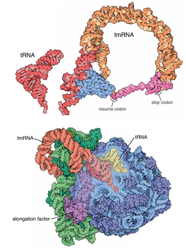
The final step of the central dogma is the most complex, where the information in RNA is translated to build proteins in the ribosome.  Ribosomes occasionally get stalled when faulty messenger RNA molecules are read, for Instance, for a messenger RNA that has broken and is thus is missing its stop codon. The ribosome uses an incredible peace of bio-engineering to rescue a stalled situation when they get stuck at the end of the truncated chain. They use a mechanism called Trans-translation. It is a ubiquitous bacterial mechanism for ribosome rescue in the event of translation stalling. This mechanism is a key component of multiple quality control pathways in bacteria that ensure proteins are synthesized with high fidelity in spite of challenges such as transcription errors, mRNA damage, and translational frame shifting. Trans-Translation is performed by a ribonucleoprotein complex. A strangely shaped RNA molecule mimics both a transfer RNA and a messenger RNA, restarting the process and cleaning up the mess. A proteolysis-inducing tag is added at the unfinished polypeptide, and facilitates the degradation of the aberrant messenger RNA. Trans-translation relies on two main factors: small stable 10S RNA (ssrA), an aminoacylated transfer-messenger RNA (tmRNA) with properties both of a tRNA and an mRNA; and SmpB (small protein B)

My comment:  This is awesome. Wow!! How could random non-guided, non-intelligent mechanisms have foresight, and the knowledge of this problem of truncated messenger RNA's that do not have a stop codon, and a sophisticated mechanism to deal with it? This is a masterfully crafted salvage mechanism in order to prevent the cell to produce toxic polymer strands that would accumulate, and in the end, destroy the cell. This is an engineering marvel of extraordinary sophistication.

Error check and repair during messenger RNA translation in the ribosome: by chance, or design ?  Transf10
Transfer-messenger RNA.
Transfer-messenger RNA (top ) includes a portion that mimics a transfer RNA ( red ) and a portion that mimics a messenger RNA ( magenta ), complete with a stop codon. It binds to stalled ribosomes ( bottom ), resuming synthesis using its own short message. Amazingly, this message encodes a small tag that is added to the end of the truncated protein, signaling to the cell that the protein is faulty and needs to be destroyed

Transfer-messenger RNA
https://en.wikipedia.org/wiki/Transfer-messenger_RNA

Transfer-messenger RNA (abbreviated tmRNA ) is a bacterial RNA molecule with dual tRNA-like and messenger RNA-like properties. The tmRNA forms a ribonucleoprotein complex (tmRNP) together with 
- Small Protein B (SmpB), 
- Elongation Factor Tu (EF-Tu), and 
- ribosomal protein S1. 
In trans-translation, tmRNA and its associated proteins bind to bacterial ribosomes which have stalled in the middle of protein biosynthesis, for example when reaching the end of a messenger RNA which has lost its stop codon. The tmRNA is remarkably versatile: it recycles the stalled ribosome, adds a proteolysis-inducing tag to the unfinished polypeptide, and facilitates the degradation of the aberrant messenger RNA

Biology of trans-Translation
The trans-translation mechanism is a key component of multiple quality control pathways in bacteria that ensure proteins are synthesized with high fidelity in spite of challenges such as transcription errors, mRNA damage, and translational frameshifting. trans-Translation is performed by a ribonucleoprotein complex composed of tmRNA, a specialized RNA with properties of both a tRNA and an mRNA, and the small protein SmpB. tmRNA-SmpB interacts with translational complexes stalled at the 3prime end of anmRNA to release the stalled ribosomes and target the nascent polypeptides and mRNAs for degradation. In addition to quality control pathways, some genetic regulatory circuits use transtranslation to control gene expression. Diverse bacteria require transtranslation when they execute large changes in their genetic programs, including responding to stress, pathogenesis, and differentiation. 1

Genes encoding tmRNA and SmpB are present throughout the bacterial kingdom.

Error check and repair during messenger RNA translation in the ribosome: by chance, or design ?  Trans-10
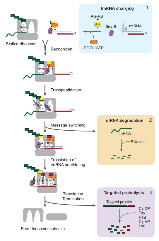
trans-Translation removes all components of stalled translation complexes. 
tmRNA binds to SmpB and is aminoacylated by alanyl-tRNA synthetase (AlaRS). EF-Tu in the GTP state binds to alanyl-tmRNA, activating the complex for ribosome interaction. The alanyl-tmRNA/SmpB/EF-Tu complex recognizes ribosomes at the 3' end of an mRNA and enters the A-site as though it were a tRNA. The nascent polypeptide is transferred to tmRNA, and the tmRNA tag reading frame replaces the mRNA in the decoding center. The mRNA is rapidly degraded. Translation resumes, using tmRNA as a message, resulting in addition of the tmRNA-encoded peptide tag to the C terminus of the nascent polypeptide. Translation terminates at a stop codon in tmRNA, releasing the ribosomal subunits and the tagged protein. Multiple proteases recognize the tmRNA tag sequence and rapidly degrade the protein (box 3).


1. https://sci-hub.st/https://www.annualreviews.org/doi/10.1146/annurev.micro.62.081307.162948
2. https://sci-hub.st/https://www.nature.com/articles/nrm3457?proof=t

https://reasonandscience.catsboard.com

4Error check and repair during messenger RNA translation in the ribosome: by chance, or design ?  Empty Ribosome Rescue Pathways in Bacteria Wed Nov 10, 2021 8:49 pm

Otangelo


Admin

Claudia Müller  Ribosome Rescue Pathways in Bacteria 18 March 2021 1

Ribosomes that become stalled on truncated or damaged mRNAs during protein synthesis must be rescued for the cell to survive. Bacteria have evolved a diverse array of rescue pathways to remove the stalled ribosomes from the aberrant mRNA and return them to the free pool of actively translating ribosomes.

My comment: Ribosomes are life-essential and had to be fully developed, and in operation, when life started. That includes the diverse rescue arrays that remove the stalled aberrant mRNA's.
That means evolution is an inadequate explanation. There was no evolution prior to DNA replication.

In addition, some of these pathways target the damaged mRNA and the incomplete nascent polypeptide chain for degradation.

My comment: in addition, if these degradation mechanisms were not fully in place either right from the beginning, the cell would be soon full of aberrant mRNA's, which would be toxic to the cell, and the cell would die.

Bacteria have evolved a diverse array of mechanisms to rescue no-go and non-stop ribosomal complexes. This is critical for survival in bacteria since otherwise ribosomes and tRNAs become sequestered from the pool of free translational components and the capacity of the cell to produce proteins rapidly diminishes

1. https://www.frontiersin.org/articles/10.3389/fmicb.2021.652980/full

https://reasonandscience.catsboard.com

Otangelo


Admin

How Cells Keep Right-Handed Amino Acids Out
https://evolutionnews.org/2015/04/how_cells_keep/


Michael T. Englander: The ribosome can discriminate the chirality of amino acids within its peptidyl-transferase center April 27, 2015
https://www.pnas.org/doi/10.1073/pnas.1424712112

https://reasonandscience.catsboard.com

Otangelo


Admin

Questions to ChatGTP about the origin of the error check and repair mechanisms of the Ribosome

https://reasonandscience.catsboard.com/t2984-error-check-and-repair-during-messenger-rna-translation-in-the-ribosome-by-chance-or-design#9839

Question: How many error check and repair systems are in the ribosome ?
Response ChatGTP: The ribosome has several error check and repair systems that help to ensure the fidelity of protein synthesis. These systems include proofreading and editing mechanisms that can detect and correct errors in the process of translation. Here are some of the key error check and repair systems in the ribosome:

1. Codon-anticodon base pairing: The ribosome relies on the precise base pairing between codons on the mRNA and anticodons on tRNA to ensure that the correct amino acid is incorporated into the growing protein chain. This process is highly selective and helps to prevent errors in translation.

2. Ribosome-associated proofreading: The ribosome has a proofreading mechanism that can detect errors in codon-anticodon base pairing. If an incorrect amino acid is added to the growing protein chain, the ribosome can stall and allow time for the incorrect tRNA to dissociate and be replaced with the correct tRNA.

3. Peptidyl transferase center (PTC): The PTC is the active site of the ribosome where peptide bond formation occurs. The PTC has a highly selective mechanism that helps to prevent errors in peptide bond formation, ensuring that only correct amino acids are incorporated into the protein.

4. Quality control mechanisms: In addition to proofreading and editing mechanisms, the ribosome also has quality control mechanisms that can recognize and correct errors in protein synthesis. For example, the ribosome-associated quality control (RQC) system can recognize and degrade proteins that are stalled during translation due to errors in codon-anticodon pairing or other factors.

5. Overall, the ribosome has multiple error check and repair systems that help to ensure the accuracy of protein synthesis. These mechanisms are critical for the proper functioning of cells and organisms, and they play an important role in maintaining the fidelity of the genetic code.

Question: Are aminoacyl tRNA synthetases also under error check and repair?
Response ChatGTP:  Yes, aminoacyl tRNA synthetases are also under error check and repair mechanisms.

Aminoacyl tRNA synthetases (aaRS) are enzymes that are responsible for attaching the correct amino acid to its corresponding transfer RNA (tRNA) molecule during protein synthesis. The accuracy of this process is essential for maintaining the fidelity of the genetic code and preventing errors in protein synthesis.

To ensure the accuracy of aminoacylation, aaRSs have evolved several mechanisms to detect and correct errors. One such mechanism is the "proofreading" activity, which involves the hydrolysis of incorrectly charged tRNAs by the aaRS enzyme. This ensures that only the correct amino acid is attached to the tRNA, and any errors are corrected.

In addition to proofreading, other error correction mechanisms have been identified, including editing by separate domains within the aaRS enzymes, as well as mechanisms that involve additional proteins that help to detect and correct errors.

Overall, the accuracy of aminoacylation by aaRSs is critical for the proper functioning of the genetic code, and error check and repair mechanisms have evolved to ensure the fidelity of this process.

Question: How accurate is protein synthesis by the ribosome ? how often is a wrong amino acid incorporated in the nascent polymer strand?
Response ChatGTP: Protein synthesis by the ribosome is highly accurate, with an error rate estimated to be around 1 in 10,000 to 1 in 100,000 amino acids incorporated.

The accuracy of protein synthesis is ensured by several mechanisms, including the proofreading activity of aminoacyl-tRNA synthetases (which attach the correct amino acid to the corresponding tRNA), the fidelity of codon-anticodon base pairing between the mRNA and tRNA, and the accuracy of peptide bond formation by the ribosome.

Despite these error-checking mechanisms, mistakes can still occur during protein synthesis. The incorporation of a wrong amino acid can result from various factors, including errors in tRNA charging by aminoacyl-tRNA synthetases, errors in codon recognition by tRNA, or mistakes in the peptide bond formation by the ribosome.

However, the frequency of these errors is relatively low, with studies suggesting that the error rate is typically less than 0.1% for most amino acids. Additionally, the ribosome has additional proofreading mechanisms that can detect and correct some of these errors, further reducing the frequency of errors in protein synthesis.

Overall, while errors in protein synthesis can occur, the ribosome's accuracy is quite high, and the fidelity of the process is critical for the proper functioning of the cell.

Question:  You informed that those error check and repairs evolved. But the proper funcion of the ribosome had to be guaranteed for life to start. That means, the origin of these error check and repair mechanisms  had to be fully functional when life started. That means, evolution could not have plaid a role in its origin.
Response ChatGTP: It is true that the proper functioning of the ribosome is essential for life, and that the error-checking and repair mechanisms that ensure the fidelity of protein synthesis had to be present and functional at the time life began. However, it is important to note that the evolution of these mechanisms did not necessarily have to occur after the origin of life.

It is widely accepted that the origin of life involved a complex series of chemical and physical processes that eventually gave rise to the first living organisms. The exact nature of these processes is still not fully understood, but it is likely that they involved the gradual emergence and refinement of the molecular machinery that underlies the fundamental processes of life.

In this context, it is possible that the error-checking and repair mechanisms that are present in modern organisms arose through gradual evolution from simpler systems that were present at the time life began. While the exact details of this evolutionary process are not known, it is thought that the high accuracy of modern protein synthesis is the result of a long history of evolutionary refinement and optimization of the molecular machinery involved.

In summary, while the error-checking and repair mechanisms that ensure the fidelity of protein synthesis had to be present and functional at the time life began, it is still possible that they arose through the process of evolution, which may have played a role in the emergence and refinement of the molecular machinery that underlies the fundamental processes of life.

Question: But a high degree of fidelity had to be guaranteed right from the beginning. And its origin cannot be explained by evolution, since the origin of life has nothing to do with evolution.
Response ChatGTP:  While the origin of life and the subsequent evolution of living organisms are distinct processes, it is important to recognize that they are closely related and are not completely independent of each other. The emergence of the first living organisms from non-living matter was likely a gradual process that involved a series of chemical and physical events that eventually gave rise to the first self-replicating systems. Once self-replication was established, evolution by natural selection could have taken place.

Regarding the origin of the error-checking and repair mechanisms that ensure the fidelity of protein synthesis, it is possible that these mechanisms were present in the first self-replicating systems, as they would have been essential for the accurate transmission of genetic information from one generation to the next. The exact nature of these mechanisms is not known, but it is likely that they evolved gradually through a series of genetic and biochemical changes that increased the fidelity of protein synthesis over time.

Therefore, while the origin of life and the evolution of living organisms are distinct processes, they are not completely independent, and it is possible that the error-checking and repair mechanisms that ensure the fidelity of protein synthesis arose through a combination of both processes.

https://reasonandscience.catsboard.com

Otangelo


Admin

Error check and repair during protein synthesis

Overview

Prokaryotic Protein Synthesis Quality Control:

Pre-translation Quality Control: Enzymes such as RNA helicases, methyltransferases, and GTPases ensure mRNA and tRNA readiness for translation.
Error Detection during Translation: The kinetic proofreading model indicates that ribosomes slow down when encountering tRNA-mRNA mismatches, allowing for error identification.
Error Correction during Translation: Aminoacyl-tRNA synthetases (aaRSs) have editing sites that rectify wrongly attached amino acids on tRNAs.
Discard and Recycling: Faulty proteins are marked for degradation by proteases like Lon and ClpXP. Ribosomal components are then reused.
Post-translation Quality Control: Chaperones aid in protein folding. Misfolded proteins are directed for degradation by specific proteolytic systems.

Eukaryotic Protein Synthesis Quality Control:

Pre-translation Quality Control: Before translation, tRNAs and mRNAs undergo extensive processing and modifications to ensure accuracy.
Error Detection during Translation: Mismatches between tRNAs and mRNA codons are recognized by eukaryotic ribosomes, ensuring the right amino acid placement.
Error Correction during Translation: Eukaryotic aaRSs possess editing sites that ensure tRNA charging precision, rectifying any errors.
Discard and Recycling: The ubiquitin-proteasome system tags and degrades erroneous proteins. Ribosomal components are then recycled.
Post-translation Quality Control: Chaperones and the ER oversee protein folding. Misfolded proteins are either retrotranslocated for degradation or tackled by autophagy which deals with larger protein aggregates.

Fundamental differences in these processes between prokaryotes, and eukaryotes

The fundamental processes of protein synthesis quality control are similar in many ways between prokaryotes and eukaryotes, given the essential nature of translation. However, due to the inherent differences in cellular complexity and compartmentalization, there are notable distinctions between the two:

Cellular Compartmentalization

Prokaryotes: Lack membrane-bound organelles. Translation and transcription occur simultaneously in the cytoplasm.
Eukaryotes: Have distinct cellular compartments. Transcription occurs in the nucleus, and the mature mRNA is then exported to the cytoplasm for translation. This separation allows for additional regulation and mRNA processing steps.

Ribosome Composition and Assembly

Prokaryotes: Ribosomes are smaller (70S) and assemble in the cytoplasm. They consist of 50S and 30S subunits.
Eukaryotes: Ribosomes are larger (80S) and consist of 60S and 40S subunits. Their assembly begins in the nucleolus of the nucleus with the involvement of small nucleolar RNAs (snoRNAs) and numerous assembly factors.

mRNA Processing

Prokaryotes: mRNA is generally not processed extensively, though some modifications can occur.
Eukaryotes: mRNA undergoes extensive processing, including capping, splicing, and polyadenylation. This processed mRNA is then transported from the nucleus to the cytoplasm for translation.

tRNA Modification

Both prokaryotic and eukaryotic tRNAs undergo post-transcriptional modifications, but eukaryotes tend to have more diverse and numerous modifications, which can impact tRNA stability, localization, and function.

Protein Folding and Quality Control

Prokaryotes: Misfolded proteins can be refolded with the help of chaperones or degraded by proteases like Lon and ClpXP.
Eukaryotes: Have the endoplasmic reticulum (ER) where many proteins are folded. Misfolded proteins in the ER can be targeted for degradation via the ER-associated degradation (ERAD) pathway. Additionally, eukaryotes utilize the ubiquitin-proteasome system for protein degradation.
Translation Regulation:

Prokaryotes: Regulation often occurs at the level of transcription, but mechanisms like operons allow for coordinated regulation of protein synthesis.
Eukaryotes: Have more complex regulatory mechanisms, including mRNA localization, sequestration, and regulated degradation. Translation initiation is a particularly important regulatory step in eukaryotes, with numerous initiation factors playing roles.

Response to Stalled Ribosomes

Both prokaryotes and eukaryotes have mechanisms to rescue stalled ribosomes, but the factors involved can differ. For example, while prokaryotes use tmRNA and associated proteins for ribosome rescue, eukaryotes utilize the Dom34-Hbs1 complex.

Post-translational Modifications

Prokaryotes: Proteins can undergo modifications like acetylation, methylation, and glycosylation, but the diversity and complexity of modifications are limited compared to eukaryotes.
Eukaryotes: Exhibit a wider variety of post-translational modifications, including phosphorylation, ubiquitination, and complex glycosylation patterns. These modifications can regulate protein activity, localization, or interactions.

While the core principles of protein synthesis are conserved, the differences in cellular organization and complexity require distinct mechanisms of quality control in prokaryotes and eukaryotes.

1. Pre-translation Quality Control

This stage focuses on preparing the translation components, monitoring errors, executing repairs, discarding or recycling flawed elements, and assuring overall fidelity.

Prokaryotic-Exclusive Mechanisms

Quality Control for 30S Ribosomal Subunit:

RNA helicases, like DeaD/CsdA in E. coli: Adjusts rRNA for proper folding.
16S rRNA methyltransferases, such as RsmA/KsgA: Serves as checkpoints by methylating specific rRNA segments, monitoring correct formation.
GTPases, exemplified by RsgA/YjeQ: Oversees proper protein assembly on the 30S subunit.
Endonucleases, notably RNase R: Directs incorrectly formed 16S rRNA precursors to degradation.

Quality Control for 50S Ribosomal Subunit:

Era GTPase: Validates 50S subunit completion.
Ribosomal RNA methyltransferases (RlmN and RlmO): Modifies 23S rRNA for proper folding.
Riboflavin synthase (RimP): Facilitates correct integration of specific ribosomal proteins.
Modulators like PrmC: Verifies interaction with the 50S subunit.
GTPases, e.g., RbgA (RrbA in E. coli): Acts as checkpoints during 50S assembly.
HelD RNA helicase: Aids in 50S subunit maturation.
Endonucleases like RNase III: Helps in 23S and 5S rRNAs maturation.
L16 ribosomal protein: Assures proper ribosomal protein incorporation.
GTPases such as EngA, EngB (Der), and ObgE: Validates 50S subunit formation.

Quality Control for 70S Ribosome Assembly:

IF3 (Initiation Factor 3): Halts premature 30S and 50S subunit union.
RsfS (or YbeB in E. coli): Restricts immature 30S units from binding with 50S units.
RimM: Enhances 70S ribosome assembly efficiency.
RbfA (Ribosome-binding factor A): Validates 30S and 50S subunit linkage.
ERA GTPase: Directs 70S ribosome assembly, guiding subunit interaction.
GTPases including EngA, Der, ObgE, and RbgA: Uphold 70S ribosome's integrity and functionality.

Ribosome Subunit Association Control

Anti-Association Factors in Ribosome Assembly: Entities in prokaryotic cells that inhibit early ribosomal subunit union.
IF3 (Initiation Factor 3): Blocks the 30S unit from prematurely binding with the 50S subunit, ensuring mature units only.
RsfS (or YbeB in E. coli): Restricts 30S units, ensuring mature units are used in ribosome formation.

mRNA and tRNA Interaction with the Ribosome

Shine-Dalgarno Interaction:
The Shine-Dalgarno (SD) sequence in bacterial mRNAs initiates translation by pairing with the anti-Shine-Dalgarno (aSD) sequence in the 16S rRNA of the 30S ribosomal subunit. This ensures the accurate positioning of the start codon for protein synthesis. Bacterial tRNAs possess anti-SD sequences, preventing inappropriate binding to the ribosome's mRNA site.

tRNA Charging and Accuracy

Aminoacyl-tRNA Synthetases (aaRSs):
Essential enzymes that charge tRNAs by attaching the correct amino acid. They ensure specificity by recognizing both the amino acid and its corresponding tRNA.
Editing Sites of aaRSs:
Ensure fidelity by removing any wrongly attached amino acids from tRNAs before they exit the enzyme.
tRNA Elements:
Structural features in tRNAs, like anticodons and acceptor stems, are essential for their recognition and accurate charging by aaRSs.

Eukaryotic Pre-translation Quality Control

Anti-Association Factors in Translation:
Prevent premature interactions, ensuring only well-formed components participate in translation.

Quality Control in Eukaryotic Translation

TRAMP Complex:
Assists in detecting and targeting wrongly modified rRNAs for degradation, ensuring only properly modified rRNAs participate in protein synthesis.
NOP58 and NOP56:
These proteins, crucial for rRNA methylation, maintain the functional integrity of rRNA, contributing to accurate translation.
Tsr2:
Detects improper rRNA modifications, which can impact subsequent processing and export steps, ensuring only correctly modified rRNA participates in translation.
Hen1:
Protects certain rRNA molecules by adding methyl caps, safeguarding them from degradation and ensuring their participation in protein synthesis.
Rrp8:
Contributes to the methylation of adenine bases in the ribosomal subunit, ensuring the ribosome's proper structure and function.

Quality Control and Error Management in Translation

Quality Assurance Mechanisms:
These mechanisms are paramount in overseeing, rectifying, or eliminating errors during protein synthesis in eukaryotic cells. A sophisticated array of proteins and RNAs enable these mechanisms to ensure translation fidelity.

Ribosome-associated Quality Control (RQC):
RQC Complex: Recognized when ribosomes are hindered in translation, this complex employs proteins like Ltn1 and Rqc2. Ltn1's role is to ubiquitinate the emerging peptide chain, tagging it for degradation.
Hel2 and ZNF598: Serving as E3 ubiquitin ligases, these proteins discern ribosomes that are halted due to certain disruptions, assisting in their tagging and guiding them to either degradation or recycling.

No-Go Decay (NGD):
Endonucleolytic cleavage: If ribosomes are obstructed by challenges like rare codons or secondary mRNA structures, a cleavage near the stall point is activated, initiating the aberrant mRNA's degradation.
Dom34 and Hbs1: Identifying halted ribosomes, these proteins foster their dissociation, thus aiding ribosome recycling.

Non-stop Decay (NSD):
Ski Complex: Triggered when ribosomes translate an mRNA missing a stop codon till its end, the Ski complex, along with the exosome, facilitates the mRNA degradation from its 3’ end.
Pelota and Hbs1: In yeast, they perform roles similar to Dom34 and Hbs1, ensuring the ribosome's detachment from non-stop mRNAs.
Ubiquitin-Proteasome System (UPS): This system is essential for the degradation of flawed polypeptides. E3 ligases, notably Ltn1 from the RQC complex, ubiquitinate these defective peptides, directing them towards proteasomal degradation.

Chaperone-assisted Ribosome Biogenesis (CARB):
ZNF622 and Jivaja: Operating as ribosome-linked chaperones, they ascertain the appropriate folding of nascent polypeptides as they emerge from the ribosome.

mRNA Surveillance Pathways:
Nonsense-Mediated Decay (NMD): Specializing in the identification and degradation of mRNAs that exhibit premature stop codons. UPF1, UPF2, and UPF3 are its key components.
Staufen-Mediated Decay (SMD): Engages mRNAs with Staufen1-binding sites in their 3' UTRs, subsequently leading to their degradation.
p58^IPK: This molecular chaperone gets associated with the ribosome during stress situations, warranting the suitable folding and operation of emerging polypeptides.

Heat Shock Proteins (HSPs):
Hsp70: This protein aids in refolding incorrectly folded nascent chains, and if a chain cannot be repaired, it targets it for degradation.
Hsp90: Working in tandem with Hsp70 and other auxiliary proteins, Hsp90 ensures the nascent polypeptides fold correctly.
GCN2 Kinase: During amino acid shortages, it identifies uncharged tRNAs, leading to the phosphorylation of eIF2α. This reduces overall protein synthesis but elevates the translation of specific mRNAs, for example, ATF4 in mammals.

Integrated Stress Response (ISR): Under conditions where translation is compromised, like during amino acid starvation or after viral infection, eIF2α gets phosphorylated, initiating a program to restore cellular homeostasis.

Ensuring the fidelity and efficiency of translation is critical for cellular function and health. The mechanisms and players described above form an interconnected network of quality control in eukaryotic cells, ensuring that translation proceeds with the utmost accuracy, and any aberrations are promptly managed.

Ensuring the precise import of ribosomal proteins from the cytoplasm to the nucleus and nucleolus is a fundamental aspect of cell biology. The mechanisms and players mentioned above facilitate this intricate process, ensuring the fidelity of ribosome biogenesis in eukaryotic cells.

mRNA Cap Structure and Translation Regulation: Quality Control and Surveillance

The 5′ cap structure of eukaryotic mRNAs is fundamental in ensuring accurate and efficient translation of proteins. Quality control mechanisms act as checkpoints to verify that mRNAs with the correct cap structures are prioritized for translation, while those with defects are targeted for degradation or other regulatory processes. Here are the key players specifically involved in monitoring, error-checking, repair, discarding, and recycling of mRNA cap structures during the translation process:

eIF4E: This is the cap-binding protein that forms part of the eIF4F complex. Proper recognition of the cap by eIF4E ensures that only appropriately capped mRNAs are selected for translation initiation. Any anomalies in this recognition can lead to translational repression.
Decapping Enzymes - DCP1 and DCP2: These are the primary enzymes responsible for removing the 5' cap from mRNAs. If an mRNA has an inappropriate cap structure or other defects, these enzymes can decap the mRNA, marking it for degradation and preventing its translation.
5' to 3' Exoribonucleases - Xrn1: After the mRNA has been decapped, Xrn1 ensures the defective mRNA is completely degraded from the 5' end. This prevents accumulation of faulty mRNAs in the cell.
CBC (Cap-Binding Complex): This complex, consisting of CBP80 and CBP20, binds to the cap structure post-transcription. While it assists in various mRNA processes, its binding acts as an initial quality control check, identifying potential anomalies in the cap structure.
Pan2-Pan3 and Ccr4-Not Complexes: Proper capping can influence mRNA stability through these deadenylase complexes. If an mRNA's cap structure is deemed aberrant, it might be subjected to rapid deadenylation and subsequent degradation.
NMD (Nonsense-Mediated Decay): Primarily responsible for detecting mRNAs with premature stop codons, NMD's function can be indirectly influenced by cap-binding proteins, and therefore, by the cap structure itself. Aberrant cap structures can influence NMD pathways, leading to mRNA degradation.
eIF4E Transporter (4E-T): By sequestering eIF4E, 4E-T ensures that only mRNAs with correct cap structures can bind eIF4E and proceed to translation. This acts as a checkpoint to ensure the quality of mRNAs entering the translation process.
Phosphorylation of eIF4E: The kinases, particularly Mnk1/2, can phosphorylate eIF4E. This phosphorylation event can modify eIF4E's affinity for the cap structure, adding another layer of regulation and quality control.

Through these meticulous surveillance systems, cells ensure that only mRNAs with proper cap structures are used in translation, maintaining cellular integrity and function.


Last edited by Otangelo on Fri Oct 27, 2023 7:39 am; edited 15 times in total

https://reasonandscience.catsboard.com

Otangelo


Admin

Pre-translation Quality Control Extant in Both, Prokaryotes and Eukaryotes

tRNA Processing and Surveillance

RTD Pathway: Degrades hypomodified or mutated mature tRNAs, preventing their incorporation in translation.
TRAMP Complex: Recognizes and degrades aberrant tRNA molecules in the nucleus.
La Protein: Protects newly transcribed pre-tRNA molecules from degradation.

tRNA Aminoacylation Quality Control

Editing Sites of aaRSs: Remove incorrectly attached amino acids from tRNAs.
Post-transfer Editing: Corrects some mischarged tRNAs by hydrolytic editing.
YbaK and ProXp-ala: Correct mischarged tRNAs when the aminoacyl-tRNA synthetase fails.
D-Tyr-tRNATyr Deacylase (DTD): Removes the D-form of tyrosine.

tRNA Anticodon Loop Modifications and Surveillance

AlkB Homologs: Reverse certain methylation modifications in the anticodon loop.
Anaerobic Modifications: Ensures correct decoding under anaerobic conditions.

tRNA Modification Surveillance

Rapid tRNA Decay (RTD): Targets hypomodified tRNAs.
Alkylation Repair Enzymes: Repair alkylated tRNAs.
NUFIP: Recognizes certain unmodified tRNAs and promotes their degradation.
Metazoan CCA-adding Enzymes: Discriminate between properly modified and hypomodified tRNAs.

The rigorous surveillance of rRNA modifications is vital for maintaining the accuracy of translation.

snoRNA Surveillance: Ensures snoRNAs guide modifications correctly.
RNA Exosome Complex: Degrades aberrant rRNA molecules that have been improperly modified.
Nucleolar Quality Control: Mis-modified rRNAs are retained in the nucleolus for repair or degradation.
Nop53p: Ensures only aptly modified rRNA advances in the ribosome assembly process.
TRAMP Complex: Targets aberrantly modified rRNA for degradation.
Rrp5: Monitors rRNA modifications, ensuring rRNA is correctly modified before it's incorporated into ribosomes.
Surveillance and Recycling Pathways: Catch and recycle or degrade mis-modified rRNA that escapes initial checks.

mRNA Surveillance via Ribosome Profiling: Quality Control and Monitoring Players

Ribosome-Associated Quality Control (RQC) System: Detects and responds to stalled ribosomes on mRNAs.
Nonsense-Mediated Decay (NMD) Pathway: Degrades mRNAs with premature stop codons.
Pelota-Hbs1: Triggers pathways that recycle the ribosome and degrade problematic mRNA.
Ltn1 (Listerin) E3 Ubiquitin Ligase: Ubiquitinates nascent polypeptides from stalled ribosomes for degradation.
No-Go Decay (NGD) Pathway: Targets mRNAs causing ribosome stalls.
Collided Ribosomes and Hel2 E3 Ligase: Marks collided ribosomes for quality control processes.
ZNF598: Initiates quality control processes for problematic translation events.
Nonstop Decay (NSD) Pathway: Ensures the rapid degradation of problematic mRNAs.
Ribosome Recycling Factors: Disassemble ribosomes from mRNAs after translation.

Regulation of Ribosomal RNA Transcription: Quality Control, Error-Check, Repair, and Recycling Mechanisms

Epigenetic Regulation: Ensures only correctly marked genes are transcribed.
Nucleolar Surveillance: Monitors rRNA transcription for aberrations.
Cellular Signaling Pathways: Adjust the rate of rRNA transcription in response to cellular conditions.
Non-Coding RNAs (ncRNAs): Ensure only properly modified and processed rRNAs proceed further.
Quality Control Mechanisms: Detect and rectify errors during the rRNA transcription process.
RNA Decay Pathways: Target and degrade aberrant rRNA molecules.
RNA Polymerase I (Pol I): Identifies and corrects mistakes during transcription.
UTP Complexes: Ensure proper early rRNA transcription steps.
Ribosomal DNA (rDNA) Replication: Surveillance mechanisms during replication catch and repair errors.


1. Prokaryotic Pre-translation Quality Control

Overview of the proteins, factors, and pathways involved in prokaryotic pre-translation quality control

1. 30S Ribosomal Subunit Assembly Quality Control
Proteins/Enzymes:
DeaD/CsdA: RNA helicase for rRNA structure modification.
RsmA/KsgA: 16S rRNA methyltransferases for checkpoint functions.
RsgA/YjeQ: GTPases for monitoring 30S subunit protein assembly.
RNase R: Endonuclease targeting improperly formed 16S rRNA precursors.

2. 50S Ribosomal Subunit Assembly Quality Control
Proteins/Enzymes:
Era: GTPase for 50S subunit maturation.
RlmN and RlmO: 23S rRNA methyltransferases.
RimP: Riboflavin synthase for late-stage 50S assembly.
RbgA/RrbA: GTPase for 50S assembly.
RNase III: Endonuclease for 23S and 5S rRNAs maturation.

3. 70S Ribosome Assembly Quality Control
Proteins/Enzymes:
IF3: Prevents premature 30S and 50S subunit association.
RsfS/YbeB: Regulates 70S assembly.
RimM: Aids in 70S ribosome assembly.

4. Ribosome Subunit Association Control
Proteins/Enzymes:
IF3: Primary anti-association factor.

5. mRNA and tRNA Interaction with the Ribosome:
Features:
Anti-Shine-Dalgarno Sequence: Prevents tRNA binding to ribosome mRNA binding site.
16S rRNA: Contains anti-Shine-Dalgarno sequence for mRNA positioning.

6. tRNA Charging and Accuracy
Proteins/Enzymes:
Aminoacyl-tRNA synthetases (aaRSs): For tRNA charging.
Editing Sites of aaRSs: Remove wrong amino acids.

Eukaryotic Pre-translation Quality Control

Overview of the proteins, factors, and pathways involved in eukaryotic pre-translation quality control

1. Ribosome Biogenesis and Surveillance
Surveillance Factors:
U3 snoRNP: Early steps of 18S rRNA processing.
Nop53p: Checkpoint protein for 5.8S and 25S rRNAs.

2. Endonucleolytic Cleavage
Enzymes:
RNase MRP: Targets pre-rRNA in the ITS1 region.
Las1: Involved in cleavage at C2 site in ITS2.

3. Exosome Complex
Components:
RRP44/Dis3, RRP6: Targets aberrant RNA for degradation.
Dom34-Hbs1: Targets stalled 80S ribosomes, also aids in ribosome recycling.

4. SSU Processome
Function:
Handles biogenesis of the small 18S rRNA subunit.

5. Nucleolar Surveillance
Proteins:
Rrp5: Linked to initial pre-rRNA cleavage steps.
Nsa2: Sensor for the 27S pre-rRNA.

7. ESCs (Eukaryotic-Specific Elements)
Checkpoint Proteins:
Ltv1, Rrp12, Tsr1: Specific for the small subunit.

9. mRNA Cap Structure and Translation Regulation
Key Players:
eIF4E: Recognizes cap structures.
Decapping Enzymes - DCP1 and DCP2: Removes 5' cap from mRNAs.
NMD (Nonsense-Mediated Decay): Degrades mRNAs with premature stop codons.

10. Ribosome-associated Quality Control (RQC) and Other Mechanisms
Major Players:
RQC Complex, Hel2, ZNF598: Recognize and manage stalled ribosomes.
No-Go Decay (NGD) - Dom34, Hbs1: Handle stalled ribosomes.
Non-stop Decay (NSD) - Ski Complex, Pelota, Hbs1: Manage ribosomes translating without a stop codon.
Ubiquitin-Proteasome System (UPS): Manages aberrant polypeptides.

Prokaryotic and Eukaryotic Pre-translation Quality Control Mechanisms

To ensure proper and efficient translation, cells have a myriad of quality control mechanisms. These mechanisms function at various stages of the translation process, from the initial processing of tRNAs and mRNAs to the final events of protein synthesis. 

1. tRNA Processing and Surveillance
Proteins/Enzymes/Pathways:
RTD Pathway: Eliminates hypomodified or mutated mature tRNAs in yeast.
TRAMP Complex: Collaborates with the RNA exosome for aberrant tRNA degradation in the nucleus.
La Protein: Shields pre-tRNA molecules, promoting correct maturation.

2. tRNA Aminoacylation Quality Control
Proteins/Enzymes/Pathways:
Editing Sites of aaRSs: Excise wrongly attached amino acids from tRNAs.
Post-transfer Editing: Rectifies mischarged tRNAs at a distinct active site.
YbaK and ProXp-ala: Rectify mischarged tRNAs when the aminoacyl-tRNA synthetase falters.
D-Tyr-tRNATyr Deacylase (DTD): Excises the D-form of tyrosine.
ATP/AMP Ratio Sensing: Verifies energy adequacy for translation.

3. tRNA Anticodon Loop Modifications and Surveillance
Proteins/Enzymes/Pathways:
AlkB Homologs: Counteract specific methylation modifications in the anticodon loop.
Anaerobic Modifications: Assure correct decoding under anaerobic circumstances.
tRNA Modifying Enzymes: Respond to environmental stresses, affecting modifications.

4. tRNA Modification Surveillance
Proteins/Enzymes/Pathways:
Rapid tRNA Decay (RTD): Targets hypomodified tRNAs for degradation.
Alkylation Repair Enzymes: Rectify alkylated tRNAs.
NUFIP: Identifies certain unmodified tRNAs.
ELAC2: Degrades incorrectly processed or modified tRNAs.
tRNA Nuclear Export: Permits only suitably processed and modified tRNAs to exit the nucleus.
Discriminator Base Surveillance: Initiates tRNA degradation upon detection of inappropriate modifications.

5. rRNA Modification Surveillance
Proteins/Enzymes/Pathways:
snoRNA Surveillance: Ensures accurate snoRNA guide modifications.
RNA Exosome Complex: Targets wrongly modified rRNA molecules for degradation.
TRAMP Complex: Designates aberrantly modified rRNA for degradation.

6. mRNA Surveillance via Ribosome Profiling
Proteins/Enzymes/Pathways:
Ribosome Positioning Analysis Tools: Map ribosome sites on mRNAs.
Ribosome-Associated Quality Control (RQC) System: Pinpoints and addresses stalled ribosomes.
RNA Helicases: Identify and unravel secondary mRNA structures.
Nonsense-Mediated Decay (NMD) Pathway: Degrades mRNAs containing premature stop codons.
Pelota-Hbs1: Recognizes ribosomes halted at mRNA termini.
Ltn1 (Listerin) E3 Ubiquitin Ligase: Tags emerging polypeptides from stalled ribosomes.
Cdc48/Npl4/Ufd1: Extracts the tagged nascent chain from the halted ribosome.

Proteins and Factors Involved in Prokaryotic Pre-translation Quality Control:

30S Ribosomal Subunit Assembly Quality Control: 4 proteins (DeaD/CsdA, RsmA/KsgA, RsgA/YjeQ, RNase R)
50S Ribosomal Subunit Assembly Quality Control: 5 proteins (Era, RlmN, RlmO, RimP, RbgA/RrbA, RNase III)
70S Ribosome Assembly Quality Control: 3 proteins (IF3, RsfS/YbeB, RimM)
Ribosome Subunit Association Control: 1 protein (IF3)
mRNA and tRNA Interaction with the Ribosome: 1 feature (16S rRNA)
tRNA Charging and Accuracy: 2 proteins (Aminoacyl-tRNA synthetases, Editing Sites of aaRSs)
Total for Prokaryotic: 16 proteins/factors


Proteins and Factors Involved in Eukaryotic Pre-translation Quality Control:

Ribosome Biogenesis and Surveillance: 2 factors (U3 snoRNP, Nop53p)
Endonucleolytic Cleavage: 2 enzymes (RNase MRP, Las1)
Exosome Complex: 3 components (RRP44/Dis3, RRP6, Dom34-Hbs1)
SSU Processome: 1 function (Handles biogenesis of the small 18S rRNA subunit)
Nucleolar Surveillance: 2 proteins (Rrp5, Nsa2)
ESCs (Eukaryotic-Specific Elements): 3 checkpoint proteins (Ltv1, Rrp12, Tsr1)
mRNA Cap Structure and Translation Regulation: 3 key players (eIF4E, DCP1, DCP2)
Ribosome-associated Quality Control (RQC) and Other Mechanisms: 7 players (RQC Complex, Hel2, ZNF598, Dom34, Hbs1, Ski Complex, Pelota, Hbs1, Ubiquitin-Proteasome System)
Total for Eukaryotic: 22 proteins/factors/complexes


Proteins and Factors Involved in Prokaryotic and Eukaryotic Pre-translation Quality Control Mechanisms:

tRNA Processing and Surveillance: 5 proteins/pathways (RTD Pathway, TRAMP Complex, La Protein)
tRNA Aminoacylation Quality Control: 6 proteins/enzymes/pathways (Editing Sites of aaRSs, Post-transfer Editing, YbaK, ProXp-ala, D-Tyr-tRNATyr Deacylase, ATP/AMP Ratio Sensing)
tRNA Anticodon Loop Modifications and Surveillance: 4 proteins/enzymes/pathways (AlkB Homologs, Anaerobic Modifications, tRNA Modifying Enzymes)
tRNA Modification Surveillance: 6 proteins/enzymes/pathways (Rapid tRNA Decay, Alkylation Repair Enzymes, NUFIP, ELAC2, tRNA Nuclear Export, Discriminator Base Surveillance)
rRNA Modification Surveillance: 3 proteins/enzymes/pathways (snoRNA Surveillance, RNA Exosome Complex, TRAMP Complex)
mRNA Surveillance via Ribosome Profiling: 9 proteins/enzymes/pathways (Ribosome Positioning Analysis Tools, RQC System, RNA Helicases, NMD Pathway, Pelota-Hbs1, Ltn1, Cdc48/Npl4/Ufd1)
Total for Prokaryotic and Eukaryotic Combined: 29 proteins/factors/pathways

Number of Mechanisms in Prokaryotic and Eukaryotic Cells Pre-translation Quality Control

Number of distinct Prokaryotic Pre-translation Quality Control Mechanisms:

1. 30S Ribosomal Subunit Assembly Quality Control
2. 50S Ribosomal Subunit Assembly Quality Control
3. 70S Ribosome Assembly Quality Control
4. Ribosome Subunit Association Control
5. mRNA and tRNA Interaction with the Ribosome
6. tRNA Charging and Accuracy

Total for Prokaryotic: 6 mechanisms

Number of distinct Eukaryotic Pre-translation Quality Control Mechanisms:

1. Ribosome Biogenesis and Surveillance
2. Endonucleolytic Cleavage
3. Exosome Complex
4. SSU Processome
5. Nucleolar Surveillance
6. ESCs (Eukaryotic-Specific Elements)
7. mRNA Cap Structure and Translation Regulation
8. Ribosome-associated Quality Control (RQC) and Other Mechanisms

Total for Eukaryotic: 8 mechanisms

Pre-translation Quality Control Extant in Both, Prokaryotes and Eukaryotes:

1. tRNA Processing and Surveillance
2. tRNA Aminoacylation Quality Control
3. tRNA Anticodon Loop Modifications and Surveillance
4. tRNA Modification Surveillance
5. rRNA Modification Surveillance
6. mRNA Surveillance via Ribosome Profiling
7. Regulation of Ribosomal RNA Transcription

Total for Both: 7 mechanisms

In summary:
Prokaryotic cells: 6 distinct mechanisms
Eukaryotic cells: 8 distinct mechanisms
Shared between both: 7 distinct mechanisms
Overall, there are a total of 21 distinct mechanisms mentioned.


Last edited by Otangelo on Fri Oct 27, 2023 8:06 am; edited 10 times in total

https://reasonandscience.catsboard.com

Otangelo


Admin

2. Error Detection during Translation

Mechanisms that identify mistakes or issues during the actual protein synthesis process.

Prokaryotic-Exclusive Mechanisms

Quality Control, Error-Check, Repair, Discard, and Recycling during Prokaryotic Translation

Ribosome Stalling and Rescue:
tmRNA: Recognizes stalled ribosomes and facilitates their rescue.
SmpB: Works with tmRNA to rescue stalled ribosomes.
ArfA and ArfB (YaeJ): Ribosome rescue proteins active when tmRNA is absent or non-functional.

Proteolytic Systems for Truncated Peptides:
Lon Protease: Degrades polypeptides tagged by tmRNA.
ClpXP Protease: Another protease system for degrading tagged peptides.

RNA Quality Control for Faulty mRNAs:
RNase R: Exoribonuclease that degrades faulty mRNA.
PNPase: Another ribonuclease involved in faulty mRNA degradation.
RNase II: Degradation of defective mRNA.

Translation Error-Check and Repair:
EF-Tu: Ensures accurate aminoacyl-tRNA delivery to prevent mismatches.
RelA and SpoT: Detect amino acid starvation and trigger the stringent response to reduce errors.

Ribosome Collision and Quality Control:
HflX: GTPase involved in dissociating collided or stalled ribosomes.
RsfA: Involved in preventing elongation in specific contexts to avoid errors.

Other Quality Control and Regulatory Factors:
RqcH and RqcP: Address stalled translation events.
YbeY: Ribosome quality control via its endonuclease activity.
MazEF: Toxin-antitoxin system; regulates translation under stress.

Chaperones for Folding and Protein Quality:
DnaK/DnaJ/GrpE: Chaperone system to aid in protein folding, especially for those emerging from the ribosome.
GroEL/GroES: Major chaperone system assisting newly synthesized polypeptides.

tmRNA-Mediated Ribosome Rescue: Rescues ribosomes stalled on mRNAs lacking a stop codon.
Key Players: tmRNA, SmpB protein.
Pathway: tmRNA with SmpB acts as both tRNA and mRNA, adding a peptide tag to the nascent chain for proteolysis.

Trans-Translation: Addresses ribosomal stalling.
Key Players: tmRNA, SmpB.
Pathway: Similar to tmRNA-mediated ribosome rescue, but can also result in mRNA cleavage.

Lon and Clp Proteases: Degrades misfolded or damaged proteins.
Key Players: Lon protease, ClpXP, ClpAP.
Pathway: Recognizes and degrades irregular peptides to maintain protein homeostasis.


Eukaryotic-Exclusive Mechanisms

Quality Control, Error-Check, Repair, Discard, and Recycling Players in Protein Synthesis

Nonsense-Mediated Decay (NMD): Recognizes and degrades mRNAs with premature termination codons (PTCs).
Key Players: UPF1, UPF2, UPF3, SMG1-7.
Pathway: UPF proteins recognize PTCs in conjunction with EJC, activating SMG1 kinase, which triggers rapid mRNA decay.

No-Go Decay (NGD): Addresses mRNAs that lead to ribosomal stalling during translation.
Key Players: Dom34 (Pelota in humans), Hbs1.
Pathway: Dom34 and Hbs1 recognize and dissociate stalled ribosomes, resulting in endonucleolytic cleavage of the mRNA.

Non-Stop Decay (NSD): Targets mRNAs missing a stop codon, causing ribosomes to translate into the poly-A tail.
Key Players: Ski7 in yeast (homologous mechanism in humans involves Hbs1 and Pelota).
Pathway: Ski7 identifies ribosomes stalled at the 3' end, subsequently recruiting the exosome complex for mRNA degradation.

Ribosome-Associated Quality Control (RQC): Targets nascent polypeptide chains from problematic mRNAs for degradation.
Key Players: LTN1 (Listerin in mammals), NEMF (Rqc2 in yeast), TCF25.
Pathway: After mRNA cleavage events like NGD, LTN1 ubiquitinates the incomplete polypeptide for proteasomal degradation.

mRNA Surveillance: Ensures only well spliced and processed mRNAs are translated.
Key Players: Exon Junction Complex components like eIF4A3, MAGOH, Y14, and MLN51.
Pathway: EJCs downstream of exon-exon junctions act as markers for mRNA processing events, influencing mRNA localization, nuclear export, and translation.

Endoplasmic Reticulum (ER)-Associated Degradation (ERAD): Degrades misfolded proteins in the ER.
Key Players: EDEM, HERP, SEL1L, and HRD1.
Pathway: Misfolded proteins are recognized, ubiquitinated, and retrotranslocated to the cytosol for proteasomal degradation.

Chaperone-Assisted Protein Quality Control: Aids proper protein folding and degrades misfolded proteins.
Key Players: HSP70, HSP90, CHIP.
Pathway: Chaperones bind to nascent/misfolded proteins, aiding in folding. If refolding fails, CHIP ubiquitinates the protein for proteasomal degradation.

Polysome Surveillance: Ensures efficient functioning of polysomes and addresses defects that might hinder translation.

Translation Fidelity Checkpoints: Confirms correct decoding of mRNA sequences and the appropriate incorporation of amino acids into the growing polypeptide chain.

Ribosome Function Monitoring: Monitors accurate matching of tRNAs to mRNA codons, ensuring that the correct amino acid-tRNA conjugates are selected.

Shared Error Detection Mechanisms during Translation in Prokaryotic and Eukaryotic Cells

Chaperone-assisted protein quality control:

Prokaryotes (specifically, bacteria):
Proteins: DnaK, DnaJ, and GrpE (HSP70 system), GroEL, GroES
Pathway: Chaperones recognize and bind to unfolded or misfolded proteins, aiding in their refolding. Persistent misfolds lead to tagging for proteolytic degradation.
Eukaryotes:
Proteins: HSP70, HSP90, BiP (in the ER)
Pathway: Similar chaperone-mediated refolding. Additionally, co-chaperones like CHIP can tag misfolded proteins for proteasomal degradation.

Proteolytic systems

Prokaryotes:
Proteins: Lon protease, ClpXP protease
Pathway: Recognize and degrade misfolded or damaged proteins, maintaining protein homeostasis.
Eukaryotes:
Proteins: The 26S proteasome system (with ubiquitin tagging)
Pathway: Misfolded proteins are tagged by ubiquitin and degraded by the 26S proteasome.

Ribosome stalling and rescue

Prokaryotes:
Proteins: tmRNA, SmpB, ArfA, ArfB
Pathway: tmRNA-SmpB acts to rescue stalled ribosomes by acting as both tRNA and mRNA, tagging the nascent chain for proteolysis.
Eukaryotes:
Proteins: Dom34 (Pelota in humans), Hbs1
Pathway: Dom34 and Hbs1 recognize stalled ribosomes, leading to mRNA cleavage and dissociation of the ribosome.

RNA quality control

Prokaryotes:
Proteins: RNase R, PNPase, RNase II
Pathway: Degradation of faulty or aberrant mRNA molecules.
Eukaryotes:
Proteins: The exosome complex, Xrn1
Pathway: Degradation of aberrant mRNA molecules, especially those with premature termination codons (related to NMD).

Translation fidelity checkpoints

Prokaryotes:
Proteins: EF-Tu
Pathway: EF-Tu ensures accurate aminoacyl-tRNA delivery, preventing mismatches during translation.
Eukaryotes:
Proteins: eEF1A (functional analog of EF-Tu), several aminoacyl-tRNA synthetases
Pathway: eEF1A ensures proper aminoacyl-tRNA delivery to the ribosome, and aminoacyl-tRNA synthetases ensure correct amino acid-tRNA charging.
It's crucial to understand that while these general mechanisms are shared, the specifics—like the exact proteins and sometimes the steps within the pathways—might differ between prokaryotes and eukaryotes.

Chiral checkpoints during protein biosynthesis

"Chiral" is a term derived from the Greek word "χείρ" (cheir) which means "hand." In chemistry, chirality refers to the property of a molecule or an object that cannot be superimposed on its mirror image, just like how our left and right hands are mirror images of each other but are not superimposable. Molecules or objects that have this non-superimposable mirror image property are called "chiral." In the context of molecules, chirality often arises due to the presence of an asymmetric carbon atom, also known as a chiral center or stereocenter. This carbon is typically bonded to four different atoms or groups. The two non-superimposable mirror-image forms of a chiral molecule are called enantiomers. For instance, many biological molecules, such as amino acids and sugars, are chiral. In living organisms, usually, only one of the enantiomers is utilized. For example, in proteins, only L-amino acids (left-handed) are used, while D-sugars (right-handed) are used in nucleic acids. The chirality of a molecule can significantly influence its chemical behavior and interactions, especially in biological systems.

Chiral discrimination is pivotal and life-essential for several fundamental reasons:

Structural Integrity of Proteins: The three-dimensional structure of proteins is determined by the sequence and chirality of the amino acids they comprise. A switch from L- to D-amino acids would result in a drastically different three-dimensional conformation. The unique shapes and folds of proteins, guided by the specific sequence and chirality of amino acids, allow them to perform their specific functions.
Biochemical Specificity: Enzymes, which are a subset of proteins, act as biological catalysts and often recognize their substrates with high specificity. The "lock and key" model, where the enzyme (lock) perfectly fits with its substrate (key), is heavily dependent on the unique chiral shape of both the enzyme and substrate. The wrong chirality could prevent this interaction, making the enzyme non-functional.
Signal Transduction: Many signaling molecules are chiral, and their interactions with proteins (like receptors) are chirality-dependent. Incorrect chirality can hinder proper signal transmission or even result in erroneous signaling.
Genetic Code Consistency: The genetic code is standardized to correspond to L-amino acids. If D-amino acids were incorporated into proteins, it would represent a deviation from the genetic blueprint, potentially leading to non-functional or harmful proteins.

Life on Earth has standardized on L-amino acids (and D-sugars for nucleic acids). Switching chirality would be incompatible with the vast majority of life processes. D-amino acids are not typically used in eukaryotic cells for protein synthesis. If they were to be incorporated, they could be recognized as foreign or even toxic, potentially eliciting an immune response or leading to cellular dysfunction.  The ribosome plays a pivotal role in scrutinizing the handedness or chirality of the amino acids it incorporates into proteins. Given that only L-amino acids are utilized in biological protein synthesis, the ribosome acts as a "chiral checkpoint," rejecting D-amino acids and ensuring that only L-isomers are integrated. Chiral checkpoints during protein biosynthesis are essential mechanisms that ensure the correct incorporation of amino acids into proteins. In biological systems, only L-amino acids are utilized for protein synthesis, and the ribosome serves as a critical "chiral checkpoint" to enforce this specificity. This quality control mechanism prevents the incorporation of D-amino acids, which are not biologically relevant for protein synthesis, and ensures that only L-isomers are integrated into growing polypeptide chains. The ribosome achieves this chiral discrimination by recognizing and selectively binding to L-amino acids while rejecting D-amino acids. This selective binding process is crucial for maintaining the structural and functional integrity of proteins in living organisms. While the ribosome's role as a chiral checkpoint is well-established in biology, the exact molecular details of how it discriminates between L- and D-amino acids may vary among different organisms. Nevertheless, this mechanism is fundamental for the fidelity of protein synthesis, ensuring that the proteins produced in cells are composed of the correct building blocks with the appropriate chirality.

Ribosomal A-site: This is the active site of the ribosome where aminoacyl-tRNAs are accepted. The unique structural configuration of the A-site primarily allows binding and acceptance of L-aminoacyl-tRNA, while excluding D-aminoacyl-tRNA.
Aminoacyl-tRNA Synthetases: These enzymes are responsible for attaching amino acids to their corresponding tRNA molecules. They have high specificity for L-amino acids, ensuring that tRNAs are charged with the correct isomer.
Elongation Factor Tu (EF-Tu) in Bacteria or Elongation Factor 1A (EF-1A) in Eukaryotes: These factors deliver aminoacyl-tRNAs to the ribosome. They help ensure that only properly charged tRNAs (with L-amino acids) are presented to the ribosome.
Peptidyl Transferase Center (PTC): Located in the large subunit of the ribosome, the PTC facilitates peptide bond formation. Its structure is geared to preferentially accommodate L-amino acids, contributing to chiral discrimination.
Ribosomal RNA (rRNA): Forms the core of the ribosome and plays a role in catalyzing peptide bond formation. Specific nucleotides in the rRNA can interact with the ester linkage of L-aminoacyl-tRNA, ensuring the proper orientation and acceptance of L-amino acids.
tRNA Molecules: The L-shape of tRNA molecules and their specific anticodon loops ensure the correct reading of mRNA codons and, by extension, the delivery of the appropriate L-amino acid to the growing polypeptide chain.
Post-Translational Modification Enzymes: While not directly part of the ribosomal machinery, some enzymes can modify D-amino acids in specific proteins after synthesis. However, these are exceptions and not the rule in general protein synthesis.
Quality Control Factors: Some factors can be recognized when a ribosome stalls due to anomalies, including if a D-amino acid were to somehow be incorporated. They can trigger mechanisms like ribosome recycling or mRNA decay in response.

These components work in tandem to ensure the fidelity of protein synthesis, especially concerning the chirality of the amino acids being incorporated. The exact nuances of their roles and interactions might differ across organisms, but their collective function ensures the robustness and accuracy of the protein synthesis machinery. The orchestration of chiral discrimination during protein synthesis is an intricate and highly coordinated process involving multiple molecular players as outlined. The interdependence and complexity of these mechanisms raise questions about their initial creation and emergence.  Each component in the ribosomal machinery and its auxiliary factors has a specific role, and their functions are interconnected. For instance, aminoacyl-tRNA synthetases charge tRNAs with the appropriate L-amino acid, and then EF-Tu or EF-1A ensures that only these properly charged tRNAs are delivered to the ribosome. This sequential and cooperative functioning suggests that the individual components are not just independently operating units but are parts of an integrated system. The emergence of such a complex integrated system poses a timing conundrum. The components not only need to exist simultaneously but also need to function correctly right from the onset. A ribosome that does not discriminate chirality or an aminoacyl-tRNA synthetase that charges tRNAs indiscriminately would produce non-functional proteins, jeopardizing cell viability. The ribosome and the associated protein synthesis machinery are central to cellular function and life. Without precise chiral discrimination, cells would produce misfolded or non-functional proteins, leading to cellular chaos. Given that this machinery is essential for life, it presents a "chicken or the egg" dilemma: which component came first, and how could it function without the others? The high specificity and precision of this system are striking. Mistakes in chirality could have dire consequences, and the machinery has multiple checkpoints to ensure accuracy. Such precision implies that random or unguided processes would have had a minimal margin of error to produce a functional system. Considering the above points,  the emergence of such a system through unguided means poses significant is warranted to cause significant skepticism. The synchrony, specificity, and complexity of the chiral discrimination machinery indicate the requirement of foresight and capability of coordination that is difficult to reconcile with step-by-step, unguided processes, especially given the essential nature of the machinery for life. Thus, the origin of foundational and interdependent systems, such as the ribosomal machinery and its chiral discrimination, points overwhelmingly to design. The notion here is that the presence of such a coordinated and complex system, foundational for life, is plausibly explained as a designed setup rather than by an unguided, stepwise emergence by nonintelligent means.

Error check and repair during messenger RNA translation in the ribosome: by chance, or design ?  41467_13

mRNA translation regulation via tRNA modifications

Across both domains of life, tRNAs undergo a myriad of post-transcriptional modifications. With nearly 100 known epigenetic tRNA modifications, these chemical alterations play pivotal roles in modulating mRNA translation, affecting translational speed, accuracy, and overall protein output. mRNA translation regulation via tRNA modifications is a critical process that occurs in both prokaryotic and eukaryotic cells. Post-transcriptional modifications of tRNA molecules are widespread, with nearly 100 known epigenetic tRNA modifications. These chemical alterations are essential for fine-tuning and regulating mRNA translation, ultimately influencing translational speed, accuracy, and the overall output of proteins. The diversity of tRNA modifications allows cells to control various aspects of translation. These modifications can impact codon-anticodon interactions, the fidelity of translation, and the efficiency of ribosome movement along the mRNA template. As a result, tRNA modifications contribute to the precise regulation of protein synthesis, ensuring that the correct proteins are produced under specific cellular conditions. The intricate network of tRNA modifications underscores the complexity of mRNA translation regulation. Cells have evolved these mechanisms to adapt to various environmental cues and cellular requirements, allowing them to modulate protein synthesis with precision and efficiency. Whether in prokaryotic or eukaryotic cells, the role of tRNA modifications in mRNA translation regulation is a fundamental aspect of cellular biology.

Proofreading and editing processes

Central to the fidelity of protein synthesis is the ribosome's ability to ensure the faithful translation of genetic information from mRNA into protein. Through a combination of proofreading and editing steps, cells ensure that errors in amino acid incorporation are kept to a minimum. Proofreading and editing processes are crucial for maintaining the fidelity of protein synthesis in both prokaryotic and eukaryotic cells. These processes help ensure that genetic information encoded in mRNA is accurately translated into proteins, minimizing errors in amino acid incorporation. The ribosome, which serves as the central machinery for translation, plays a key role in these proofreading and editing mechanisms. In prokaryotic and eukaryotic cells, several mechanisms contribute to proofreading and editing during translation:

Aminoacyl-tRNA Synthetase Proofreading: Aminoacyl-tRNA synthetases are enzymes responsible for attaching amino acids to their corresponding tRNA molecules. These enzymes have proofreading capabilities to ensure that the correct amino acid is selected and attached to the tRNA. If an incorrect amino acid is attached, the enzyme can hydrolyze the incorrect aminoacyl-tRNA bond.
Ribosomal Proofreading: During translation, the ribosome monitors the accuracy of codon-anticodon interactions. If a mismatch occurs, the ribosome can stall and undergo a conformational change, allowing for the release of the incorrect aminoacyl-tRNA. This process, known as "kinetic proofreading," enhances the accuracy of translation.
tRNA Proofreading: Some tRNA molecules have editing sites that can hydrolyze mischarged amino acids, correcting any errors in amino acid selection.
Peptidyl Transferase Center (PTC) Proofreading: The PTC of the ribosome plays a role in monitoring the formation of peptide bonds between amino acids. It can detect and correct errors in peptide bond formation.
Ribosome-Associated Factors: Various ribosome-associated factors, such as EF-G in prokaryotes and eEF2 in eukaryotes, participate in proofreading mechanisms by facilitating the translocation of tRNA molecules and ensuring the fidelity of translation.

These proofreading and editing processes collectively contribute to the high accuracy of protein synthesis, ensuring that the final protein product accurately reflects the genetic information encoded in the mRNA. By minimizing errors in amino acid incorporation, cells can produce functional and correctly folded proteins essential for various cellular processes.

Chiral Discrimination by Ribosome

Reflecting the universal preference for L-amino acids in protein synthesis, ribosomes across all life forms have mechanisms to exclusively recognize and incorporate the L-isomer of amino acids, ensuring chiral purity in synthesized proteins. Chiral discrimination by ribosomes is a universal mechanism that ensures the incorporation of L-amino acids exclusively during protein synthesis, maintaining the chiral purity of synthesized proteins across all life forms. This mechanism reflects the preference for L-amino acids in biological systems and contributes to the fidelity and functionality of proteins in cells.


Last edited by Otangelo on Wed Oct 25, 2023 10:50 am; edited 9 times in total

https://reasonandscience.catsboard.com

Otangelo


Admin

3. Error Correction during Translation

Once an error is detected, these mechanisms work to correct it.

Prokaryotic-Exclusive Mechanisms

Ribosome Stalling and Rescue:
tmRNA: Recognizes stalled ribosomes and facilitates their rescue by adding a peptide tag to truncated proteins for proteolysis.
SmpB: Works in tandem with tmRNA, ensuring its proper positioning on the stalled ribosome.
ArfA and ArfB (YaeJ): Ribosome rescue proteins that are active when tmRNA is absent or non-functional. They aid in releasing stalled ribosomes from mRNAs.

Peptidyl-tRNA Hydrolysis:
RF-2: A factor that can promote hydrolysis of peptidyl-tRNA on stalled ribosomes in specific sequence contexts.
PrfH: An alternative peptide release factor that acts under specific conditions.

Proteolytic Systems for Truncated Peptides:
Lon Protease: Targets polypeptides tagged by tmRNA for degradation.
ClpXP Protease: Recognizes and degrades truncated peptides and misfolded proteins.
FtsH (HflB) Protease: This membrane-bound protease can degrade certain ribosomal proteins and other misfolded proteins.

Ribosome Quality Control by rRNA Modifications:
RsmA, RsmB, RsmD, RsmE, and RsmG: Methyltransferases that modify specific residues in 16S rRNA, which can influence the accuracy of translation.

Trans-translation Mediated by tmRNA:
AlaRS: The alanyl-tRNA synthetase charges tmRNA, preparing it for its role in trans-translation.

Ribosome Recycling:
RRF (Ribosome Recycling Factor): Works in conjunction with EF-G (Elongation Factor G) to dissociate the ribosomal subunits post-translation, making them available for a new round of protein synthesis.
IF3 (Initiation Factor 3): Prevents the premature association of ribosomal subunits and ensures proper start codon recognition.

Decoding Center Accuracy:
16S rRNA: Its helix 44 plays a role in monitoring the accuracy of codon-anticodon base pairing, ensuring fidelity in translation.
RpsD and RpsE: Ribosomal proteins that contribute to the fidelity of tRNA selection.

Misincorporation and Ribosome Rescue:
MnmE and MnmG: Proteins involved in the modification of the wobble position of tRNAs, affecting translational fidelity.
YjjK and YqcB: Proteins that recognize and rectify certain amino acid misincorporations.

RNA Degradosome Components (for mRNA quality):
RNase E: Initiates the decay of many RNAs, playing a role in rRNA processing and mRNA decay.
Pnp (Polynucleotide Phosphorylase): Modulates the decay of several RNA species, ensuring RNA quality.
RhlB and Enolase: Additional components of the degradosome that aid in the effective degradation of faulty RNAs.

Tsp Protease: Recognizes and degrades proteins with aberrant C-termini.

Quality Control of tRNAs:
CCA-adding enzyme: Ensures that tRNAs have the correct CCA tail for amino acid attachment.
tRNA nucleotidyltransferase: Adds nucleotides to the 3' end of tRNA, crucial for proper aminoacylation.
RNase P: Processes precursor tRNA molecules to produce mature tRNAs.

Dealing with Damaged rRNAs and tRNAs:
RNase R: Degrades defective or damaged rRNAs and tRNAs.
PNPase (polynucleotide phosphorylase): Involved in RNA degradation; can degrade aberrant rRNA and tRNA molecules.

Recycling of Ribosomal Components:
RRF (ribosome recycling factor): Collaborates with elongation factor EF-G to disassociate post-termination ribosomal complexes, enabling ribosome recycling.

HflX: A GTPase that might be involved in ribosome splitting and recycling under stress conditions.

Ribosome Assembly and Integrity Check:
RsgA (YjeQ): A GTPase ensuring proper 30S subunit maturation and assembly.
EngA (Der): Another GTPase playing a role in 50S ribosomal subunit biogenesis and quality control.

Ribosome Hibernation Promotion

Ribosome hibernation is a fascinating strategy that certain bacteria employ to survive in adverse environmental conditions. During this process, the bacterial ribosomes are converted into translationally inactive 100S complexes, which can be rapidly reactivated when conditions become favorable. Mechanisms and Players in Ribosome Hibernation:

Hibernation-promoting factor (HPF): Found in many bacteria, this protein binds to the 30S ribosomal subunit, stabilizing the dimerization of 70S ribosomes into inactive 100S complexes.
Ribosome modulation factor (RMF): Present in some bacteria like E. coli, RMF binds to the 50S subunit, facilitating the formation of the inactive 90S dimer. This can then further dimerize with the help of HPF to form the 100S complex.
YfiA (pY): Found in certain bacteria, YfiA inhibits translation by binding to the 30S subunit, preventing association with the 50S subunit.
Short hibernation-promoting factor (sHPF): In some Gram-negative bacteria, a shorter version of HPF has been identified. While its mechanism is similar to HPF, it's evolved differently and seems to be more associated with certain pathogenic bacteria.
RelA and (p)ppGpp: Although not directly involved in ribosome hibernation, the stringent response, mediated by RelA and the alarmone (p)ppGpp, can reduce the overall translation activity in the cell. This can indirectly promote ribosome hibernation as a part of the general response to stress.
Ribosome-Associated Quality Control (RQC): In some cases, the hibernation process might be linked with RQC. If a ribosome gets stalled during translation, specific factors can target it for disassembly or hibernation.

The process of ribosome hibernation, and its subsequent reactivation, is a remarkable example of bacterial adaptability. By temporarily putting their protein synthesis machinery on hold, bacteria can endure conditions that might be detrimental to active ribosomes. Once conditions improve, these hibernating ribosomes can be swiftly reactivated, allowing the bacteria to quickly resume growth and proliferation. This capability provides a distinct survival advantage in fluctuating environments.

Hibernation in the Macro World

In the animal kingdom, numerous creatures undergo hibernation, a state of inactivity and metabolic depression, to survive prolonged periods of extreme cold or scarcity of food. For instance, bears will lower their heart rate, decrease their metabolic rate, and recycle waste products during winter months. This isn't merely a passive process. It's an intricate physiological adaptation that involves a multitude of finely regulated processes: Before hibernation, animals accumulate fat reserves, which will serve as their primary energy source during hibernation. During hibernation, body temperature drops to save energy. Some hibernators can recycle waste products, such as urea, to prevent toxin accumulation. Even after months of inactivity, hibernating animals can wake up without significant muscle loss. On a much smaller scale, certain bacteria transition ribosomes into a dormant "hibernation" state using hibernation-promoting factors. This strategy conserves cellular resources and allows bacteria to swiftly resume translation when conditions become favorable again.

Convergent Mechanism - A Parallel in Design

What's truly intriguing is the convergence of this mechanism across vastly different scales and domains of life. While the molecular and physiological processes in ribosome hibernation and animal hibernation are distinct, the underlying principle is the same: both entail a strategic metabolic downturn to conserve energy and endure unfavorable conditions, followed by a rapid reactivation when conditions improve. Such parallel strategies, seen both at the macroscopic level in mammals and at the microscopic level in bacteria, can be seen as indicative of a design principle. In the realm of human-made designs, recurring solutions and patterns are employed to solve similar challenges, even in very different contexts. Why would this principle not apply to the natural world? The existence of hibernation in both macro and micro realms suggests a designer who implemented similar solutions across different scales and domains of life, demonstrating foresight, efficiency, and an elegant sense of continuity. Convergence in biological systems, especially when it serves a similar functional purpose across diverse organisms, can be perceived as more than mere coincidence. It can be viewed as evidence of a purposeful design, implemented by an entity with an understanding of both the vastness and intricacies of life.

Error check and repair during messenger RNA translation in the ribosome: by chance, or design ?  G51610

Eukaryotic-Exclusive Mechanisms

Degradation of Faulty mRNAs:
RNase II and RNase R: Exoribonucleases involved in degrading problematic mRNAs.
PNPase: Polynucleotide Phosphorylase, involved in degradation of aberrant rRNA and mRNA.

Ribosomal Recycling:
RRF (Ribosome Recycling Factor): Facilitates the disassembly of ribosomal subunits post-translation.

Error Correction in Aminoacylation:
Editing domains of Aminoacyl-tRNA synthetases: Sites within the enzymes that hydrolyze incorrectly charged tRNAs, ensuring fidelity.
YbaK: Acts as a prolyl-tRNA editing protein, removing incorrectly attached amino acids from tRNA^Pro.

E-site Regulation:
Elongation factors (EF-Tu and EF-G): Ensure the smooth progression and exit of tRNAs through ribosomal sites, particularly the E-site.

Degradation of Misfolded Proteins:
DegP (HtrA): A protease involved in degrading misfolded proteins in the periplasm.
ClpB and DnaK/DnaJ/GrpE chaperone system: Facilitates the refolding of misfolded proteins or their targeting for degradation.

Ribosomal Surveillance:
RsfA: Monitors ribosome integrity and directs damaged ribosomes for degradation.
Rne and Rng: RNases that participate in quality control, cleaving problematic RNAs.

Recognition of Stalled Ribosomes and Nascent Chain Issues

RQC complex: Recognizes stalled ribosomes and engages with downstream factors for resolution.
DnaK and DnaJ (in prokaryotes): Analogous to Hsp70 and Hsp40 in eukaryotes, these chaperones recognize and bind to misfolded proteins to aid in their refolding or disposal.

Disaggregation and Refolding of Problematic Polypeptides

Hsp100/Clp family: Disaggregases that work to solubilize aggregated proteins, rendering them accessible for refolding chaperones.

Targeting for Degradation

ATP-dependent proteases (in prokaryotes): Recognize misfolded or problematic polypeptides and degrade them without requiring prior tagging.

Stress Response Triggered by Translation Errors

Heat shock response: In response to increased protein misfolding or stress conditions, the cell increases the expression of chaperones and proteases to manage the misfolded proteins.

By employing these quality control mechanisms, prokaryotic cells actively monitor the translation process, ensuring the production of functional proteins and dealing efficiently with errors when they arise.

Mechanisms Shared by Both Prokaryotic and Eukaryotic Cells

Aminoacyl-tRNA Synthetases (AARSs) with Editing Domains: In both types of cells, aminoacyl-tRNA synthetases play a pivotal role in ensuring the accuracy of protein synthesis by charging tRNAs with their corresponding amino acids. The editing domains of AARSs provide a second layer of quality control by hydrolyzing incorrectly charged tRNAs.
Elongation Factors: Both prokaryotes (EF-Tu, EF-G) and eukaryotes use elongation factors to mediate the entry and exit of tRNAs from the ribosome, ensuring smooth translation progression.
Ribosome Structure and Function: The fundamental structure and function of ribosomes are conserved across both domains. While there are differences in size and complexity, the core mechanisms, such as the rRNA-mediated peptide bond formation, are conserved.
Chaperones: Molecular chaperones are present in both prokaryotes (like DnaK/DnaJ in bacteria) and eukaryotes (like Hsp70/Hsp40 in eukaryotes). They bind to nascent or misfolded proteins to either aid in their correct folding or target them for degradation.
ATP-dependent Proteases: Both cellular domains have ATP-dependent proteases that recognize and degrade misfolded or damaged proteins.
tRNA Modifications: Various modifications at the tRNA's anticodon loop, especially at the wobble position, are essential for the fidelity and efficiency of translation. Both prokaryotes and eukaryotes modify their tRNAs to ensure correct codon-anticodon base pairing.
Heat Shock Response: Both types of cells can undergo a heat shock response. When exposed to stressful conditions, such as elevated temperatures, they upregulate the production of specific proteins, like chaperones and proteases, to manage and refold misfolded proteins.
Ribosome Recycling: Both prokaryotes and eukaryotes have mechanisms to dissociate ribosomes from mRNA after translation is complete, allowing them to be recycled for new rounds of protein synthesis.
Quality Control of mRNA: Both cell types have various mechanisms to ensure the integrity of their mRNA. For instance, exoribonucleases like RNase II and RNase R degrade problematic mRNAs in both domains.

While prokaryotes and eukaryotes have specific error correction mechanisms tailored to their unique cellular complexities, they also share a foundational set of tools that ensure the fidelity of protein synthesis. The presence of these  mechanisms underscores the critical importance of accurately translating genetic information into functional proteins in all forms of life.


Last edited by Otangelo on Wed Oct 25, 2023 10:42 am; edited 9 times in total

https://reasonandscience.catsboard.com

Otangelo


Admin

4. Discard and Recycling

Prokaryotic-Exclusive Mechanisms

Trans-translation: A process where tmRNA acts as both tRNA and mRNA, aiding in ribosome rescue and polypeptide tagging for degradation.

Degradation of Problematic mRNA:
mRNA Degradation Mechanisms: Target problematic mRNAs, which can cause ribosome stalling, for destruction to maintain translation fidelity.

Ribosome Recycling:
Ribosome Splitting Mechanisms: After rescue processes, ribosomal subunits are separated and prepared for subsequent rounds of translation.
Hibernation Promoting Factor (HPF): Assists in rendering ribosomes inactive by promoting dimerization.
Ribosome Modulation Factor (RMF): Facilitates the initial formation of 90S dimers in certain bacteria.
Proteins like YhbH: Play a role in splitting hibernated ribosome dimers back into active 70S ribosomes.
RRF: Collaborates in dissociating stalled ribosomal complexes.
EF-G: Assists RRF in ribosome dissociation and recycling.
RNase R: Exonuclease responsible for degrading defective mRNAs causing ribosomal stalls.

Eukaryotic-Exclusive Mechanisms

Ribosome Biogenesis Stress Response:
p53: A pivotal player in the cellular response to ribosomal stress, leading to cell cycle arrest.
Nucleolar surveillance: Ensures improperly processed rRNAs are rapidly degraded.
MDM2: An inhibitor of p53, which when bound by certain ribosomal proteins leads to p53 activation.
c-Myc: Modulates ribosome biogenesis under stress conditions.

Pathways for Ribosome and mRNA Quality Control:
No-Go Decay (NGD): Degrades mRNAs that cause ribosome stalling.
Rli1/ABCE1: Facilitates ribosomal subunit separation post-translation.

Degradation Systems:
Proteasome: Degrades damaged or unnecessary ribosomal proteins.
LC3/Atg8: Central players in the formation of autophagosomes.
Atg1/ULK1 complex: Initiates autophagy in response to cellular cues.
RACK1: Ribosomal protein with roles in autophagy and signaling.

Ribosome Degradation Pathways:
Ribophagy: Specialized autophagy that targets ribosomes.
Endoplasmic Reticulum (ER) stress: Activates autophagy leading to ribosome degradation.

Ribosome Stalling and Decay:
Dom34 (Pelota): Recognizes stalled ribosomes and facilitates their dissociation.
Hbs1: Works with Dom34 to release stalled ribosomes.
Upf1: RNA helicase that binds to stalled ribosomes, initiating the No-Go Decay process.
Xrn1: An exonuclease that degrades cleaved mRNA fragments from the No-Go Decay pathway.

Ribosome Collisions and Quality Control:
ZNF598: Recognizes and marks collided ribosomes by ubiquitination.
Hel2: Works alongside ZNF598 in the detection of ribosome collisions.
Rqc2: A key factor in ribosome-associated quality control, handling collided or stalled ribosomes.

Proteolytic Systems for Truncated Peptides:
Listerin (Ltn1): An E3 ubiquitin ligase that tags nascent polypeptides emerging from collided or stalled ribosomes for degradation.
RQC complex: Ribosome-associated quality control complex involved in handling and degrading problematic nascent polypeptides.

Degradation and Recycling Pathways:
Cdc48 (VCP/p97): An ATPase that extracts ubiquitinated proteins from ribosomes for degradation.

mRNA Quality Control and Decay:
Nonsense-Mediated Decay (NMD): Detects and degrades mRNAs with premature stop codons.
Upf1, Upf2, and Upf3: Core factors of the NMD pathway.
No-Go Decay (NGD): Recognizes ribosome stalling on problematic mRNAs and triggers their degradation.
Dom34 (Pelota) and Hbs1: Proteins that recognize stalled ribosomes during NGD.

Ribosome Recycling and Translation Termination:
eRF1 and eRF3: Not only play a role in translation termination but also aid in the dissociation of ribosomal subunits.
ABCE1: Powers the dissociation of ribosomal subunits post-translation termination.

Discarding Defective mRNAs:
Xrn1: Degrades mRNAs in a 5' to 3' manner following decapping.
Exosome Complex: A multi-protein machinery that degrades mRNAs in the 3' to 5' direction.

Shared Mechanisms in Prokaryotes and Eukaryotes

Ribosome Assembly:
Small Subunit Processome: Complexes that help in the processing and assembly of ribosomal RNA in both domains.

RNA Modifications:
Pseudouridine Synthases and Methyltransferases: Modify ribosomal RNAs in both prokaryotes and eukaryotes, aiding in ribosome structure and function.

Translation Initiation:
Initiation Factors (IFs/eIFs): Aid in the initiation of protein synthesis, though their number and specific functions may differ between the two systems.

Ribosomal RNAs:
16S (in Prokaryotes) and 18S (in Eukaryotes) rRNA: Form the core of the small ribosomal subunit and play crucial roles in mRNA decoding.
23S (in Prokaryotes) and 28S (in Eukaryotes) rRNA: Form the core of the large ribosomal subunit and catalyze peptide bond formation.

Ribosomal Proteins:
Ribosomal Protein Families: Many ribosomal proteins are evolutionarily conserved, playing structural and functional roles in the ribosome, though they may have different names or additional roles in the different domains.

It's worth noting that while some mechanisms are fundamentally similar, the specifics of how they operate, their regulatory controls, and their interactions with other cellular components can differ considerably between prokaryotes and eukaryotes.


Last edited by Otangelo on Wed Oct 25, 2023 10:39 am; edited 5 times in total

https://reasonandscience.catsboard.com

Otangelo


Admin

5. Post-translation Quality Control

Post-translational modifications and quality checks are critical for ensuring that proteins are functional, properly folded, and localized, and that any misfolded or improperly synthesized proteins are recognized and degraded.

Post-translation Quality Control in Prokaryotic Cells

Mismatch Recognition:
Aminoacyl-tRNA synthetases are responsible for editing mischarged tRNAs to ensure accurate amino acid-tRNA pairing.

Ribosome Rescue and Quality Control:
tmRNA-SmpB complex: Rescues stalled ribosomes.
ArfA and ArfB: Free ribosomes from truncated mRNAs.
RqcH and RqcP: Handle ribosomes that stall during translation.
HflX: An ATPase that assists ribosome quality control under stress conditions.

Proteolytic Systems:
Lon and Clp Proteases: Degrade proteins tagged for degradation.
ClpAP Protease: Collaborates in degrading specific substrates and stalled peptide chains.

Ribosome Recycling:
RRF: Essential for recycling ribosomes post-translation.
EF-G: Assists RRF in dissociating ribosomal subunits for subsequent rounds of translation.

Error Correction and Surveillance:
RNase R: Degrades aberrant mRNA associated with stalled ribosomes.
PNPase: Participates in the decay of mRNA fragments.
AlaXp: A tRNA deacylase that removes mischarged alanine from tRNAs, preventing mistranslation.
YbeY: Endoribonuclease involved in ribosome quality control during stress conditions.

Eukaryotic-Exclusive Mechanisms

Eukaryotic cells, with their intricate cellular machinery, employ various players to monitor, correct, or discard errors during translation. This ensures that proteins are correctly synthesized, functional, and free from aberrations that might jeopardize cellular integrity.

Quality Control Mechanisms:

Mismatch Recognition: Aminoacyl-tRNA synthetases ensure the proper loading of amino acids onto tRNAs. Errors in this process are recognized and corrected by the editing activity of these enzymes.
Ribosome Proofreading: During translation, the ribosome itself can recognize and correct mismatches between the mRNA codon and the tRNA anticodon, ensuring the incorporation of the correct amino acid.
Molecular Chaperones: These proteins assist in protein folding. If a protein fails to fold correctly, chaperones can either help refold the protein or target it for degradation.
Error-Check and Repair:

Trans-translation: In prokaryotes, when a ribosome stalls during translation due to mRNA truncation or other abnormalities, the tmRNA-SmpB complex rescues the stalled ribosome, adds a peptide tag to the nascent polypeptide, and targets it for proteolytic degradation.
Ribosome Rescue Systems: Other than trans-translation, prokaryotes employ ribosome rescue factors like ArfA and ArfB to free ribosomes from truncated mRNAs.
Discard and Recycling:

Lon and Clp Proteases: Proteins tagged for degradation in prokaryotes, either due to errors or as part of regulatory mechanisms, are degraded by ATP-dependent proteases such as Lon and Clp.
Peptidyl-tRNA Hydrolase: In cases where translation is aborted, peptidyl-tRNA molecules can accumulate. Peptidyl-tRNA hydrolase releases the peptide from the tRNA, allowing the tRNA to be recycled.
Recycling of Ribosomal Components: Following the completion or termination of translation, ribosome recycling factors (e.g., RRF in bacteria) dissociate the ribosomal subunits, allowing them to participate in subsequent rounds of translation.
Stress Responses Influencing Quality Control:

ppGpp and Stringent Response: In response to amino acid starvation, bacterial cells synthesize ppGpp, which acts as an alarmone. The stringent response modulates ribosome biogenesis and redirects resources to survival pathways, ensuring that translation under these conditions is highly accurate.

Misfolded Protein Response:
DnaK/DnaJ/GrpE Chaperones: Assist in the refolding or degradation of misfolded proteins.
GroEL/GroES Chaperones: Facilitate the correct folding of nascent polypeptides.

mRNA Surveillance:
RNase R: Degrades the aberrant mRNA that is associated with stalled ribosomes.
PNPase: Participates in the decay of mRNA fragments.

Translation Fidelity:
Elongation Factors (EF-Tu, EF-Ts, and EF-G): Ensure accurate aminoacyl-tRNA delivery and translocation of tRNA and mRNA.
Release Factors (RF1, RF2, and RF3): Recognize stop codons and promote termination of translation.

Aminoacyl-tRNA Proofreading:
Editing Domains of Aminoacyl-tRNA Synthetases: Ensure the correct amino acid is attached to the appropriate tRNA.

Ribosome Quality Control:
RQC (Ribosome Quality Control Complex): Recognizes and targets incomplete polypeptides for degradation when ribosomes stall during translation.

Recycling of Ribosomal Components:
RRF (Ribosome Recycling Factor): Aids in the disassembly of post-termination ribosomal complexes, making ribosomal subunits available for new rounds of translation.

Monitoring and Recycling Mechanisms in Eukaryotic Translation

Ribosome Stalling and Rescue:
Pelota and Hbs1: Recognize stalled ribosomes and initiate their disassembly.
Dom34: Eukaryotic homolog of Pelota, aiding in ribosome rescue.

Proteolytic Systems for Truncated Peptides:
Ltn1 (Listerin): E3 ubiquitin ligase that tags stalled translation products for degradation.
RQC (Ribosome-associated Quality Control Complex): Recognizes stalled translation events and targets them for degradation.

mRNA Surveillance:
Upf Proteins (Upf1, Upf2, and Upf3): Core factors in the NMD pathway, ensuring degradation of mRNAs with premature termination codons.

Chaperone Systems for Protein Folding:
Hsp70 and Hsp90: Facilitate the correct folding of nascent polypeptides and target misfolded proteins for degradation.

Translation Fidelity:
eEF1A and eEF2: Eukaryotic elongation factors that ensure accurate tRNA delivery and ribosome translocation.

Aminoacyl-tRNA Proofreading:
Editing Domains of Aminoacyl-tRNA Synthetases: Correct mischarged tRNAs, ensuring translation accuracy.

Ribosome Quality Control:
RACK1: A component of the 40S ribosomal subunit, involved in ribosome-associated quality control.

Recycling of Ribosomal Components:
eRF1 and eRF3: Eukaryotic release factors that promote termination and ribosomal subunit recycling.


ClpAP Protease: Collaborates in degrading specific substrates and stalled peptide chains.

Ribosome Quality Control:
RqcH and RqcP: Involved in ribosome-associated quality control, adding amino acid tags to nascent chains on stalled ribosomes.
HflX: ATPase that can split apart 70S ribosomes during stress conditions, playing a role in ribosome quality control.

Recycling and Disassembly:
RRF (Ribosome Recycling Factor): Essential for the recycling of the ribosomes after the termination of translation.
EF-G (Elongation Factor G): Assists RRF in dissociating the ribosomal subunits, preparing them for the next round of translation.

Errors and Mistranslation:
AlaXp: A tRNA deacylase that removes mischarged alanine from tRNAs to prevent mistranslation.
YbeY: Endoribonuclease that plays a role in 70S ribosome quality control, particularly during stress conditions.

Implications:
Translational Accuracy: Ensuring the fidelity of protein synthesis is crucial for cellular health. Mechanisms that check, repair, or degrade errors maintain the accuracy of translation.
Protein Homeostasis: Effective ribosome monitoring mechanisms ensure that only properly synthesized proteins are functional in the cell, maintaining proteostasis.
Cellular Fitness: In prokaryotes, where rapid growth and adaptation to environmental changes are crucial, the efficiency and fidelity of translation play a significant role in cellular fitness and survival.

Ribosome Stalling and Rescue:
Dom34 (Pelota) and Hbs1: Recognize stalled ribosomes in eukaryotes and facilitate their dissociation.

Ribosome Quality Control:
Ltn1 (Listerin): E3 ubiquitin ligase that tags incomplete polypeptides on stalled ribosomes for degradation.
RQC (Ribosome-associated Quality Control) Complex: Recognizes and targets stalled translation complexes, facilitating degradation of aberrant nascent chains.
NEMF (Rqc2): A component of the RQC complex that aids in tagging stalled peptides.

Recycling and Disassembly:
ABCE1 (Rli1): An ATPase involved in ribosomal subunit recycling post-translation.
eRF1 and eRF3: Termination factors that, in conjunction with ABCE1, help recycle ribosomes.

Proteolytic Systems for Truncated Peptides:
Proteasome: Major protein degradation machinery that disposes of polypeptides tagged for degradation.

Errors and Mistranslation:
EF-Tu and eEF1A: Elongation factors that play roles in ensuring aminoacyl-tRNAs match codons correctly, preventing mistranslation.

Implications:
Translational Accuracy: Ensuring the fidelity of protein synthesis is critical for maintaining cellular homeostasis and preventing diseases.
Protein Quality Control: Efficient ribosome monitoring and quality control systems ensure that aberrant or misfolded proteins are swiftly identified and degraded.
Cellular Health and Disease: Disruptions in these quality control mechanisms can lead to diseases, emphasizing their importance in maintaining cellular health.
ClpAP Protease: Collaborates in degrading specific substrates and stalled peptide chains.

Ribosome Quality Control:
RqcH and RqcP: Involved in ribosome-associated quality control, adding amino acid tags to nascent chains on stalled ribosomes.
HflX: ATPase that can split apart 70S ribosomes during stress conditions, playing a role in ribosome quality control.

Recycling and Disassembly:
RRF (Ribosome Recycling Factor): Essential for the recycling of the ribosomes after the termination of translation.
EF-G (Elongation Factor G): Assists RRF in dissociating the ribosomal subunits, preparing them for the next round of translation.

Errors and Mistranslation:
AlaXp: A tRNA deacylase that removes mischarged alanine from tRNAs to prevent mistranslation.
YbeY: Endoribonuclease that plays a role in 70S ribosome quality control, particularly during stress conditions.

Stalled Ribosome Sensing and Ribosome Rescue:
RQC Complex: Recognizes stalled ribosomes and facilitates their recycling. Includes proteins like Ltn1, which ubiquitinates the nascent polypeptide, marking it for degradation.
Dom34/Hbs1: Acts as a ribosome rescue factor in eukaryotes, similar to tmRNA in bacteria.
Pelota: Works in conjunction with Hbs1 to dissociate stalled ribosomes.
ZNF598: Recognizes ribosomes stalled on mRNAs and plays a role in initiating ribosome-associated quality control.
Rli1/ABCE1: Assists in ribosome recycling and dissociation after a stall.

Proteolytic Systems for Truncated and Misfolded Peptides:
Proteasome: Degrades ubiquitinated nascent polypeptides that arise from translation errors or ribosomal stalling.
Cdc48 (VCP/p97 in mammals): Helps extract ubiquitinated polypeptides from the ribosome and directs them to the proteasome for degradation.

mRNA Decay Mechanisms:
No-Go Decay: Targets mRNAs that stall ribosomes. The stalled mRNA is cleaved and subsequently degraded.
Non-Stop Decay: Targets mRNAs that lack stop codons, leading to ribosome stalling and mRNA degradation.

Peptide Release and Termination Factors:
eRF1: Recognizes stop codons in eukaryotes, promoting the release of the newly synthesized polypeptide.
eRF3: Works with eRF1 to ensure accurate termination and prevent readthrough of stop codons.
ASC-1 Complex: Recognizes stalled ribosomes, particularly at sequences that lack a stop codon, and promotes degradation of the problematic nascent chain.

The precise coordination and interplay among these players ensure that any errors during protein synthesis are swiftly dealt with, maintaining cellular health and homeostasis.

Post-translation Quality Control shared by both, Prokaryotic and Eukaryotic Cells

Post-translational modifications and quality checks are critical for ensuring that proteins are functional, properly folded, and localized, and that any misfolded or improperly synthesized proteins are recognized and degraded.

Ribosome Stalling and Rescue:
tmRNA: Recognizes stalled ribosomes and facilitates their rescue.
SmpB: Works with tmRNA to rescue stalled ribosomes.
ArfA and ArfB (YaeJ): Ribosome rescue proteins active when tmRNA is absent or non-functional.
Pelota (Hbs1): Eukaryotic counterpart of tmRNA that recognizes stalled ribosomes.
eRF3 and eRF1: Eukaryotic factors responsible for recognizing and terminating translation at stop codons.

Proteolytic Systems for Truncated Peptides:
Lon Protease: Degrades polypeptides tagged by tmRNA.
ClpXP Protease: Another protease system for degrading tagged peptides.
Proteasome: Eukaryotic multiprotein complex responsible for degrading misfolded proteins and those tagged for destruction by ubiquitination.

Protein Refolding Mechanisms:
DnaK (Hsp70) and DnaJ: Prokaryotic chaperone system for protein refolding.
Hsp90: Eukaryotic chaperone that assists with protein folding and stability.

Recognition and Degradation of Misfolded Proteins:
SecYEG: Prokaryotic protein-conducting channel that assists with protein insertion into membranes.
BiP (Grp78):Eukaryotic endoplasmic reticulum chaperone that recognizes misfolded proteins.
EDEM: Eukaryotic degradation-enhancing alpha-mannosidase-like protein involved in the recognition and retrotranslocation of misfolded proteins.

Endoplasmic Reticulum Associated Degradation (ERAD):
Derlin: Eukaryotic component of the retrotranslocation channel that helps in transporting misfolded proteins from the ER to the cytosol for degradation.

Ribosome-Associated Translation Pausing

The process of translation, while often efficient and swift, isn't always continuous. At times, ribosomes pause at certain points on the mRNA during translation. Such pauses, far from being glitches, play vital roles in ensuring proper protein synthesis and function. Certain sequences in the mRNA can fold into stable secondary structures, such as hairpins, that temporarily impede ribosome progression until they're resolved. Infrequent codons, which correspond to low-abundance tRNAs, can cause the ribosome to slow down or pause, waiting for the appropriate tRNA to arrive. Specific sequences or motifs in the nascent polypeptide can cause pauses, especially if they interact with the ribosomal exit tunnel. Factors like the Signal Recognition Particle (SRP) or certain chaperones can bind to the emerging polypeptide or the ribosome itself, causing a pause in translation. By pausing translation, the ribosome allows time for the already synthesized portion of the protein to begin folding correctly. This can be especially important for large proteins or those with complex tertiary structures. Proteins destined for membranes often have hydrophobic sequences. Pausing allows the Signal Recognition Particle (SRP) to recognize and bind these sequences, ensuring the ribosome targets the endoplasmic reticulum (in eukaryotes) or the plasma membrane (in bacteria) for co-translational insertion. For proteins that are part of multi-subunit complexes, pausing can allow time for other subunits or cofactors to associate with the nascent chain, aiding in the correct assembly of the complex. Extended pauses, especially those caused by problematic mRNA sequences or damaged ribosomes, can be signals for quality control mechanisms. These mechanisms can then rescue the ribosome and degrade the faulty mRNA or nascent protein. Pausing can provide the necessary temporal window for enzymes to carry out modifications on the nascent polypeptide, such as phosphorylation or glycosylation. Ribosome-associated translation pausing is a strategic mechanism to ensure the fidelity and efficiency of protein synthesis. By coordinating the rate of translation with other cellular processes, pausing plays an indispensable role in the life of a protein, from its synthesis to its final function.

References

1. Mohler K, Ibba M. (2017). Translational fidelity and mistranslation in the cellular response to stress. Nat Microbiol, 2:17117. Link.
2. Gomez MAR, Ibba M. (2020). Aminoacyl-tRNA synthetases. RNA, 26(8 ):910-936. Link.
3. Cvetesic, N., Perona, J., & Gruic‐Sovulj, I. (2012) Kinetic Partitioning in Class I Aminoacyl-tRNA Synthetases:The Journal of Biological Chemistry, 287, 25381 - 25394. Link.  
4. Richards J, Sundermeier T, Svetlanov A, Karzai AW. (2008). Quality Control of Bacterial mRNA Decoding and Decay. Biochim Biophys Acta, 1779(9):574-582. Link. 
5. Kumar Kuncha, S., Kruparani, S.P., & Sankaranarayanan, R. (2019). Chiral checkpoints during protein biosynthesis. J Biol Chem, 294(45):16535-16548. Link. (Discussion on the role of chiral checkpoints in protein biosynthesis.)
6. Kozak M. (2005). Regulation of translation via mRNA structure in prokaryotes and eukaryotes. Gene, 361:13-37. Link.
7. Quality Control Mechanisms During Ribosome Maturation. Trends Cell Biol, 23(5):242-250. Link. (Description or summary of the content is not provided.)
8. Lund E, Dahlberg JE. (1998). Proofreading and aminoacylation of tRNAs before export from the nucleus. Science, 282(5396):2082-2085. Link. (Description or summary of the content is not provided.)
9. Kimura, S., & Waldor, M.K. (2019). The RNA degradosome promotes tRNA quality control through clearance of hypomodified tRNA. Proc Natl Acad Sci U S A, 116(4):1394-1403. Link. (Discussion on the role of the RNA degradosome in tRNA quality control and its involvement in the clearance of hypomodified tRNA.)
10. Ibba, M., & Söll, D. (1999). Quality control mechanisms during translation. Science, 286(5446):1893-1897. Link. (Exploration of the mechanisms that ensure quality control during the process of translation.)
11. Drummond, D. A., & Wilke, C. O. (2009). The evolutionary consequences of erroneous protein synthesis. Nat Rev Genet, 10(10):715-724. Link. (A study on the potential evolutionary impacts of mistakes in protein synthesis.)
12. Michael T. Englander. (2015). The ribosome can discriminate the chirality of amino acids within its peptidyl-transferase center. PNAS. Link. (The study highlights the ribosome's ability to differentiate between the chiral forms of amino acids during the translation process.)
13. Marc Graille, Bertrand Séraphin. (2012). Surveillance pathways rescuing eukaryotic ribosomes lost in translation. Nat Rev Mol Cell Biol, 13(11), 727-35. Link. (The study discusses the mechanisms in eukaryotic cells that ensure ribosomes do not get stalled or lost during the translation process.)


Last edited by Otangelo on Wed Oct 25, 2023 11:01 am; edited 9 times in total

https://reasonandscience.catsboard.com

Otangelo


Admin

Error Check and Repair During Prokaryotic Ribosome Biogenesis and Maturation

Overview
1. rRNA Synthesis: In prokaryotes, rRNA genes are organized in operons and transcribed as a single precursor rRNA. This precursor undergoes further processing to yield the mature 16S, 23S, and 5S rRNAs. 
2. tRNA Processing: tRNAs are transcribed as precursors that undergo cleavage, base modification, and CCA sequence addition at their 3' ends.
3. rRNA Modification: The precursor rRNA undergoes modifications such as methylation and pseudouridylation. 
4. Ribosomal Protein Synthesis: Ribosomal proteins in prokaryotes are synthesized by ribosomes in the cytoplasm and then associated with the rRNAs to form ribosomal subunits. 
5. Small Subunit (30S) Assembly: The 16S rRNA and its associated ribosomal proteins come together to form the small subunit.
6. Large Subunit (50S) Assembly: The 23S and 5S rRNAs, with their associated ribosomal proteins, form the large subunit. 
7. 70S Ribosome Assembly: The small (30S) and large (50S) subunits come together to form the functional 70S ribosome, ready for translation. 
8. Quality Control and Recycling: Prokaryotes have mechanisms to ensure only properly assembled ribosomes participate in translation. Misassembled subunits are either repaired or degraded. 
9. Ribosome Function: The 70S ribosome facilitates the translation of mRNA into proteins in prokaryotes.
10. Regulation of Ribosome Biogenesis: Ribosome production in prokaryotes is regulated in response to nutritional and environmental cues.

1. rRNA Synthesis 

In prokaryotes, rRNA genes are organized in operons and transcribed as a single precursor rRNA. This precursor undergoes further processing to yield the mature 16S, 23S, and 5S rRNAs. During the rRNA synthesis phase of prokaryotic ribosome biogenesis and maturation, bacteria have in place a number of mechanisms to ensure the fidelity and functionality of these vital RNA components.

rRNA Synthesis and Maturation:
RNase III: This ribonuclease plays a role in the initial cleavage of the precursor rRNA (pre-rRNA), allowing for subsequent processing steps to yield mature rRNA molecules. Errors in processing can lead to degradation by other RNases.
rRNA methyltransferases: These enzymes modify rRNAs by adding methyl groups. Methylation not only confers functional modifications but can also act as a quality control mechanism. Incorrectly methylated rRNAs might be targeted for degradation.

Error Surveillance and Discard Mechanisms:
Decay pathways: In bacteria, decay pathways target aberrant rRNA for degradation. This is less well-defined than in eukaryotes but involves general ribonucleases, such as RNase R, RNase II, and PNPase.
Small RNA-mediated targeting: In some cases, small RNAs can target aberrant rRNA molecules, guiding ribonucleases to degrade them.

Repair Mechanisms:
In prokaryotes, there isn't a "repair" mechanism for rRNAs in the same way that DNA repair systems exist. Instead, aberrant rRNAs are typically degraded and replaced. The synthesis of rRNAs is a frequent event in rapidly growing cells, so there's always a supply of new, correctly processed rRNAs to replace any that are degraded.

Recycling Mechanisms:
RNase-mediated degradation: Aberrant rRNA molecules, or those from old/damaged ribosomes, are typically degraded into their constituent nucleotides by ribonucleases. These nucleotides can then be recycled by the cell to synthesize new RNA molecules.
Ribosome-associated quality control: While this is more defined in eukaryotes, there are indications that prokaryotes possess mechanisms to recognize malfunctioning ribosomes and target them for disassembly and recycling of their components.

2. tRNA Processing 

tRNAs are transcribed as precursors that undergo cleavage, base modification, and CCA sequence addition at their 3' ends.

rRNA Synthesis and Maturation:
RNase III: This ribonuclease plays a role in the initial cleavage of the precursor rRNA (pre-rRNA), allowing for subsequent processing steps to yield mature rRNA molecules. Errors in processing can lead to degradation by other RNases.
rRNA methyltransferases: These enzymes modify rRNAs by adding methyl groups. Methylation not only confers functional modifications but can also act as a quality control mechanism. Incorrectly methylated rRNAs might be targeted for degradation.

Error Surveillance and Discard Mechanisms:
Decay pathways: In bacteria, decay pathways target aberrant rRNA for degradation. This is less well-defined than in eukaryotes but involves general ribonucleases, such as RNase R, RNase II, and PNPase.
Small RNA-mediated targeting: In some cases, small RNAs can target aberrant rRNA molecules, guiding ribonucleases to degrade them.

Repair Mechanisms:
In prokaryotes, there isn't a "repair" mechanism for rRNAs in the same way that DNA repair systems exist. Instead, aberrant rRNAs are typically degraded and replaced. The synthesis of rRNAs is a frequent event in rapidly growing cells, so there's always a supply of new, correctly processed rRNAs to replace any that are degraded.

Recycling Mechanisms:
RNase-mediated degradation: Aberrant rRNA molecules, or those from old/damaged ribosomes, are typically degraded into their constituent nucleotides by ribonucleases. These nucleotides can then be recycled by the cell to synthesize new RNA molecules.
Ribosome-associated quality control: While this is more defined in eukaryotes, there are indications that prokaryotes possess mechanisms to recognize malfunctioning ribosomes and target them for disassembly and recycling of their components.

3. rRNA Modification 

rRNA Modification and Maturation:
Methylation: rRNA molecules are frequently modified by the addition of methyl groups, which can be important for the function of the mature ribosome. In prokaryotes, this methylation is guided by specific methyltransferase enzymes.
Pseudouridylation: Conversion of uridine to pseudouridine in rRNA can be catalyzed by specific enzymes or guided by small RNAs. This modification can influence the structure and function of the mature ribosome.

Error Surveillance and Discard Mechanisms for rRNA Modifications:
snoRNA-guided surveillance: In eukaryotes, small nucleolar RNAs (snoRNAs) guide the modification of rRNAs. These snoRNAs can prevent incorrect modifications based on mismatches or errors in base pairing. While prokaryotes don't have snoRNAs in the traditional sense, they might have similar RNA-guided mechanisms to ensure correct modification.

Repair Mechanisms for rRNA Modifications:
In prokaryotes, similarly to the synthesis phase, there isn't a traditional "repair" mechanism for rRNAs. Incorrectly modified rRNAs are typically degraded and replaced. Constant synthesis of rRNAs in growing cells ensures a steady supply of correctly modified molecules.

Recycling Mechanisms for rRNA Modifications:
RNase-mediated degradation: Incorrectly modified rRNA molecules, like their unmodified counterparts, are typically degraded into their constituent nucleotides by ribonucleases. This allows for the recycling of nucleotides for new RNA synthesis.
Ribosome-associated quality control: Malfunctioning ribosomes, which can arise from incorrectly modified rRNAs, may be recognized and disassembled by certain cellular mechanisms, allowing for recycling of ribosomal components.

4. Ribosomal Protein Synthesis 

Ribosomal proteins in prokaryotes are synthesized by ribosomes in the cytoplasm and then associated with the rRNAs to form ribosomal subunits.

Ribosomal Protein Synthesis and Assembly:
Protein Synthesis: Ribosomal proteins are synthesized by ribosomes in the cytoplasm. These proteins are encoded by ribosomal protein genes and are essential for the function and structure of ribosomes.
Ribosomal Protein Binding: Once synthesized, ribosomal proteins bind to the rRNAs at specific sites, ensuring proper ribosome structure and function. Proper binding is crucial for the subsequent steps of ribosome biogenesis.

Error Surveillance and Discard Mechanisms for Ribosomal Protein Synthesis:
Chaperone proteins: Molecular chaperones assist in the folding and assembly of ribosomal proteins. If a ribosomal protein is misfolded or improperly incorporated, chaperones can aid in its refolding or target it for degradation.
Proteases: Proteolytic enzymes target misfolded or damaged ribosomal proteins for degradation, ensuring that only functional proteins are incorporated into ribosomal subunits.

Repair Mechanisms for Ribosomal Protein Synthesis:
In prokaryotes, misfolded or damaged ribosomal proteins aren't traditionally "repaired." Instead, such proteins are typically degraded and replaced with newly synthesized ones. The continual synthesis of ribosomal proteins in growing cells ensures there's always a fresh supply of functional proteins.

Recycling Mechanisms for Ribosomal Protein Synthesis:
Protease-mediated degradation: Ribosomal proteins that aren't incorporated into ribosomes or that are part of old/damaged ribosomes can be degraded into their constituent amino acids by proteases. These amino acids can then be recycled by the cell for new protein synthesis.
Ribosome-associated quality control: Ribosomes with malfunctioning proteins can be recognized by the cellular machinery, leading to their disassembly and the recycling of ribosomal components.

5. Small Subunit (30S) Assembly 

Small Subunit (30S) Assembly:
16S rRNA Incorporation: The 16S rRNA, being a primary component of the 30S subunit, plays a central role in the structure and function of the small subunit. Proper processing and modifications of the 16S rRNA are crucial for its effective incorporation into the 30S subunit.
Ribosomal Protein Binding: Multiple ribosomal proteins specifically associate with the 16S rRNA to form the complete 30S subunit. Each protein has its specific binding site, ensuring proper assembly and functionality of the 30S subunit.

Error Surveillance and Discard Mechanisms for Small Subunit Assembly:
Nop53p Binding: In the context of quality control, Nop53p can bind to improperly modified rRNAs, preventing their incorporation into ribosomes and directing them for degradation.
RsgA: In prokaryotes, RsgA is a GTPase that associates with the 30S subunit. It can recognize and interact with immature 30S subunits, facilitating their maturation or, in the case of errors, their disassembly.

Repair Mechanisms for Small Subunit Assembly:
For the assembly of the 30S subunit, there isn't a conventional "repair" mechanism. If an assembly error occurs or if an rRNA is improperly modified, the affected molecules are typically degraded and replaced. The continuous synthesis and processing of rRNAs and ribosomal proteins in active cells ensure the regular formation of functional 30S subunits.

Recycling Mechanisms for Small Subunit Assembly:
Disassembly factors: Some proteins and factors can recognize faulty 30S subunits and facilitate their disassembly. The constituent components (rRNAs and ribosomal proteins) can then be recycled or degraded, depending on their condition.
RNase-mediated degradation: Improperly assembled or damaged 30S subunits can be targeted by ribonucleases, leading to the degradation of their rRNA components. The released nucleotides can then be reused by the cell for new RNA synthesis.

6. Large Subunit (50S) Assembly

The 23S and 5S rRNAs, with their associated ribosomal proteins, form the large subunit.

Large Subunit (50S) Assembly:
23S and 5S rRNA Incorporation: Both the 23S and 5S rRNAs are vital components of the 50S subunit. Their correct processing, modification, and subsequent incorporation into the large subunit are essential for its structure and function.
Ribosomal Protein Binding: Numerous ribosomal proteins specifically bind to the 23S and 5S rRNAs, ensuring the formation of a functional 50S subunit. Each of these proteins has a defined binding site, playing a role in the assembly and operational integrity of the 50S subunit.

Error Surveillance and Discard Mechanisms for Large Subunit Assembly:
Assembly chaperones: Several proteins, often termed assembly chaperones, aid in the correct assembly of the 50S subunit. They can recognize and stabilize intermediate states of assembly and can also prevent the incorporation of improperly processed or modified rRNAs.
Ribosome-modifying enzymes: Enzymes like RimM and RimP play roles in the maturation of the 50S subunit. Their malfunction or aberrant activity can lead to errors in 50S assembly.

Repair Mechanisms for Large Subunit Assembly:
As with the 30S subunit, traditional "repair" mechanisms aren't typically employed for the 50S subunit. Instead, any errors or improper modifications in the rRNA or associated proteins typically result in degradation and replacement. Given the constant synthesis and processing of ribosomal components in active cells, there's a steady formation of correct 50S subunits.

Recycling Mechanisms for Large Subunit Assembly:
Disassembly factors: Certain factors can recognize defective 50S subunits and promote their disassembly, allowing the recycling of their components or directing them towards degradation pathways.
RNase-mediated degradation: If improperly assembled or damaged, the 50S subunits might be targeted by ribonucleases. This degradation process breaks down the rRNA constituents, and the resulting nucleotides can be recycled by the cell for synthesizing new RNA molecules.

7. 70S Ribosome Assembly

The small (30S) and large (50S) subunits come together to form the functional 70S ribosome, ready for translation.

Subunit Association: The joining of the 30S and 50S subunits is a coordinated process, largely driven by complementary RNA structures and binding sites between the two subunits. This results in a functional 70S ribosome with its A, P, and E sites correctly aligned and ready for translation.

Error Surveillance and Discard Mechanisms for 70S Assembly:
Initiation Factors: Prokaryotic initiation factors, especially IF3, play a role in ensuring the correct assembly of the 70S ribosome. IF3 prevents the premature association of the 30S and 50S subunits, ensuring that only correctly formed subunits come together.
Mismatch Detection: The cell has mechanisms to detect mismatches or incorrect alignments between the 30S and 50S subunits. Aberrantly assembled ribosomes might be less functional or entirely nonfunctional, leading to their quick recognition and degradation.

Repair Mechanisms for 70S Assembly:
Like in the earlier stages, "repair" in the context of ribosome assembly generally involves degradation and replacement rather than direct repair. Aberrant 70S ribosomes are typically targeted for disassembly and degradation.

Recycling Mechanisms for 70S Assembly:
Ribosome Recycling Factor (RRF) and EF-G: After each round of translation, the 70S ribosome must be recycled. RRF and EF-G facilitate the dissociation of the 70S ribosome into its 30S and 50S subunits, allowing them to participate in new rounds of translation or be subjected to quality control mechanisms.
Ribosome-associated quality control: As with earlier steps, malfunctioning ribosomes can be recognized by specific cellular factors. These factors promote the disassembly of the faulty ribosome, with the components either being recycled or degraded.

8. Quality Control and Recycling

Quality Assurance of Assembled Ribosomes: Before participating in translation, assembled ribosomes are subjected to a "proofreading" step. Properly formed ribosomes will be efficient in this quality assurance step and move on to active translation. In contrast, misassembled or damaged ribosomes will be detained and not permitted to engage in protein synthesis.

Error Surveillance and Discard Mechanisms for Quality Control:
Stalled Ribosome Detection: Cellular mechanisms can detect ribosomes that are stalled during translation, indicating a possible assembly or functional error.
Trans-translation System: In bacteria, the trans-translation system, involving tmRNA (transfer-messenger RNA), tags proteins from stalled ribosomes for degradation and helps recycle the ribosomal components. This serves as a dual mechanism to deal with both the faulty protein being synthesized and the problematic ribosome.
Alternative Ribosome Rescue Systems: Besides the trans-translation system, prokaryotes have other ribosome rescue systems like the ArfA and ArfB (alternative ribosome-rescue factor A and B) systems. These recognize and rescue stalled ribosomes, ensuring continued translation efficiency.

Repair Mechanisms for Quality Control:
As with previous steps, there isn't a direct "repair" mechanism for faulty ribosomes. Instead, prokaryotes tend to degrade and replace faulty ribosomal units. The rapid rate of ribosome synthesis in prokaryotes ensures a steady supply of functional ribosomes.

Recycling Mechanisms for Quality Control:
Ribosome Recycling Factor (RRF): After each round of translation, especially after encountering stalls or errors, ribosomes need to be recycled. RRF, along with other factors like EF-G, disassembles the ribosome, prepping it for another round of translation or subjecting it to further quality control checks.
Degradation Pathways: Misassembled or damaged ribosomal components are targeted for degradation, breaking them down into their basic components, which can be reused by the cell. Specific ribonucleases play a role in this degradation process, ensuring efficient recycling of cellular resources.

9. Ribosome Function

The 70S ribosome facilitates the translation of mRNA into proteins in prokaryotes.

Ribosome Function:

Translation Initiation: The ribosome recognizes and binds to the Shine-Dalgarno sequence on mRNA, positioning it correctly for the start codon to be translated. Initiation factors (IFs) in prokaryotes help facilitate this process, ensuring proper start codon recognition and tRNA placement.

Error Surveillance and Discard Mechanisms during Translation:
Mismatch Detection: The ribosome has a built-in mechanism to detect tRNA-mRNA mismatches. Incorrect tRNAs are rejected, minimizing translation errors.
Stalled Ribosome Detection: Mechanisms exist to recognize ribosomes that stall during translation due to problematic mRNA sequences, or the absence of the appropriate tRNA. Stalled ribosomes can be recycled or directed to ribosome rescue systems.

Repair Mechanisms during Translation:
The ribosome does not "repair" mismatches directly. Instead, it tends to reject incorrectly paired tRNAs, preventing the incorporation of the wrong amino acid. If a mismatched amino acid is incorporated, post-translational repair mechanisms, like protein chaperones or proteolysis systems, might come into play to correct or degrade the faulty protein.

Recycling Mechanisms during Translation:
Ribosome Recycling: After translation termination, ribosomes are recycled to start new rounds of translation. Ribosome Recycling Factor (RRF) and elongation factor EF-G facilitate the dissociation of the two ribosomal subunits, making them available for new rounds of translation.
mRNA and tRNA Recycling: After translation, mRNAs and tRNAs are released from the ribosome. While tRNAs can be recharged with amino acids for subsequent rounds of translation, mRNAs can be bound by new ribosomes for repeated translation or be subjected to degradation.

10. Regulation of Ribosome Biogenesis

Ribosome production in prokaryotes is regulated in response to nutritional and environmental cues.

Regulation of Ribosome Biogenesis:

Stringent Response: An adaptive process where bacterial cells respond to amino acid starvation or other stresses. The production of the alarmone, guanosine tetraphosphate (ppGpp), decreases rRNA synthesis, thereby conserving resources and ensuring that ribosome biogenesis is scaled back during unfavorable conditions.

Error Surveillance and Discard Mechanisms:
tmRNA System: A unique trans-translation system in bacteria that rescues stalled ribosomes. When a ribosome stalls due to an mRNA lacking a stop codon, tmRNA binds and tags the nascent polypeptide for proteolytic degradation, ensuring that potentially harmful truncated proteins are not accumulated.
Rho-dependent Termination: Rho factor can terminate transcription of certain genes prematurely, preventing the full synthesis of potentially erroneous rRNAs or mRNAs.

Repair Mechanisms:
In the context of ribosome biogenesis in prokaryotes, repair typically involves degradation and replacement rather than direct correction. If erroneous rRNA molecules or ribosomal proteins are synthesized, they are typically detected by quality control mechanisms and degraded, followed by the synthesis of new, correct components.

Recycling Mechanisms:
RNase III, RNase E, and PNPase: These enzymes are not only involved in the maturation of rRNAs but also play roles in degrading aberrant or excess rRNAs. Degradation products, such as nucleotides, can be recycled by the cell for other processes or for the synthesis of new RNA molecules.
ppGpp: Apart from its role in the stringent response, ppGpp also has roles in regulating the stability of certain RNAs, ensuring that only functional rRNAs and mRNAs are stable while erroneous or unneeded ones are quickly degraded.

List of the key proteins and factors
involved in error monitoring, repair, discard, and recycling during prokaryotic ribosome biosynthesis:

RNase III - rRNA processing and discard
RNase E - rRNA processing and discard
PNPase - rRNA discard
RsgA - 30S subunit quality control
tmRNA - ribosome rescue and discard
ArfA - ribosome rescue
ArfB - ribosome rescue
IF3 - 70S assembly quality control
RRF - ribosome recycling
EF-G - ribosome recycling
Rho - transcriptional termination
Ribonucleases - rRNA and ribosome degradation and recycling
ppGpp - ribosome biogenesis regulation during stringent response
Ribosome methyltransferases - rRNA modification for 16S, 23S, and 5S
Ribosome-associated chaperones - protein folding quality control (e.g., DnaK and GroEL)
Ribosome-associated proteases - protein quality control (e.g., Lon and ClpXP)
Ribosome assembly factors - assembly quality control for both 30S and 50S subunits
RelA - ppGpp synthesis during amino acid starvation
SpoT - ppGpp synthesis and hydrolysis under nutritional stress
RNA helicases - aid in rRNA folding and ribosome assembly (e.g., DeaD and SrmB)
RimM - 16S rRNA processing and 30S assembly
Era - GTPase involved in 16S rRNA processing and 30S assembly
EngA and YihA - GTPases involved in 50S assembly


Prokaryotic Ribosome Biosynthesis

1. rRNA Synthesis:
Proteins/Enzymes:
RNase III
rRNA methyltransferases
RNase P
Signaling Pathways:
Decay pathways involving RNase R, RNase II, PNPase
Small RNA-mediated targeting

2. tRNA Processing:
Proteins/Enzymes:
RNase III
tRNA methyltransferases
CCA-adding enzyme
tRNA splicing endonuclease
Signaling Pathways:
Decay pathways involving RNase R, RNase II, PNPase

3. rRNA Modification:
Proteins/Enzymes:
rRNA methyltransferases
Pseudouridine synthases
Signaling Pathways:
snoRNA-guided surveillance
Ribosome-associated quality control

4. Ribosomal Protein Synthesis:
Proteins/Enzymes:
Ribosomal protein genes
Signaling Pathways:
Error surveillance involving chaperone proteins and proteases
Ribosome-associated quality control

5. Small Subunit (30S) Assembly:
Proteins/Enzymes:
16S rRNA processing enzymes
Ribosomal proteins specific to 30S
Signaling Pathways:
Nop53p binding
RsgA-mediated checks
Disassembly factors
RNase-mediated degradation

6. Large Subunit (50S) Assembly:
Proteins/Enzymes:
23S and 5S rRNA processing enzymes
Ribosomal proteins specific to 50S
Assembly chaperones
RimM and RimP
Signaling Pathways:
Disassembly factors
RNase-mediated degradation

7. 70S Ribosome Assembly:
Proteins/Enzymes:
Initiation factors like IF3
Ribosome Recycling Factor (RRF)
EF-G
Signaling Pathways:
Mismatch detection
Ribosome-associated quality control

8. Quality Control and Recycling:
Proteins/Enzymes:
tmRNA
ArfA and ArfB
Ribosome Recycling Factor (RRF)
EF-G
Signaling Pathways:
Trans-translation system
Alternative ribosome rescue systems

9. Ribosome Function:
Proteins/Enzymes:
tRNA synthetases
Ribosome Recycling Factor (RRF)
EF-G
Signaling Pathways:
Mismatch detection
Ribosome-associated quality control

10. Regulation of Ribosome Biogenesis:
Proteins/Enzymes:
RelA and SpoT (for ppGpp synthesis)
tmRNA
Rho factor
Signaling Pathways:
Stringent response
tmRNA system
Rho-dependent termination

Signaling Pathways involved

1. Decay pathways involving RNase R, RNase II, PNPase
2. Small RNA-mediated targeting
3. snoRNA-guided surveillance
4. Ribosome-associated quality control
5. Nop53p binding
6. RsgA-mediated checks
7. Disassembly factors
8. RNase-mediated degradation
9. Mismatch detection
10. Trans-translation system
11. Alternative ribosome rescue systems
12. Stringent response
13. tmRNA system
14. Rho-dependent termination

Distinct Processes and Pathways for Error Check, Repair, Discard, and Recycling

1. Error Check:
  a. Mismatch detection during ribosome function
  b. Quality control mechanisms in rRNA synthesis, ribosomal protein synthesis, and both 30S and 50S subunit assembly
  c. RsgA-mediated checks during small subunit assembly
  d. Rho-dependent termination during ribosome biogenesis regulation

2. Repair:
  a. Ribosome-associated quality control mechanisms during rRNA modification and 70S assembly
  b. Chaperone proteins assisting in ribosomal protein synthesis
  c. Post-translational repair mechanisms during ribosome function

3. Discard:
  a. tmRNA system during ribosome biogenesis regulation
  b. Disassembly factors during both 30S and 50S subunit assembly
  c. Ribosome Recycling Factor (RRF) and EF-G dissociating 70S ribosome after translation

4. Recycling:
  a. RNase-mediated degradation pathways during rRNA synthesis, rRNA modification, both 30S and 50S assembly
  b. Ribosome Recycling Factor (RRF) and EF-G recycling 70S ribosome after translation
  c. tRNA recharging and mRNA degradation or reuse after ribosome function
  d. Trans-translation system and alternative ribosome rescue systems during quality control
  e. RNase III, RNase E, and PNPase in ribosome biogenesis regulation

There are 14 specific processes or pathways for error checking, 3 for repair, 3 for discard, and 5 for recycling.

The prokaryotic ribosome, a sophisticated molecular machine, is vital for cellular life, playing a central role in protein synthesis. Its formation is a marvel of precision and reliability, ensuring accurate protein synthesis essential for cellular survival. Ribosome biosynthesis and maturation is a multi-step process, where even a minor error can disrupt the cellular machinery. The biogenesis begins with rRNA synthesis. The rRNA undergoes meticulous modifications and processing. Enzymes like RNase III, rRNA methyltransferases, and RNase P ensure the fidelity of this process. Degradation pathways involving RNase R, RNase II, and PNPase act as fail-safes, degrading misprocessed or damaged rRNA, showcasing the commitment to quality assurance. tRNA molecules are equally vital for protein synthesis. Enzymes like RNase III, tRNA methyltransferases, CCA-adding enzymes, and tRNA splicing endonuclease ensure their accurate processing. Specific decay pathways guarantee the removal of any aberrant tRNA.  rRNA modification involves precise enzymatic activity, including rRNA methyltransferases and pseudouridine synthases. The snoRNA-guided surveillance and ribosome-associated quality control are vital checkpoints, ensuring only properly modified rRNA gets incorporated into the ribosome. The synthesis of ribosomal proteins involves a detailed error surveillance system. Chaperone proteins and proteases are present to ensure only correctly folded proteins integrate into the ribosomal structure. The small (30S) and large (50S) ribosomal subunits undergo separate assembly processes, necessitating their unique sets of proteins and enzymes. Signaling pathways, like Nop53p binding and RsgA-mediated checks, oversee these assemblies, further fortifying the precision implemented in the cell's biosynthesis processes Once the individual subunits are ready, they assemble into the 70S ribosome. IF3, a meticulous gatekeeper, ensures only proper assemblies occur. Ribosome Recycling Factor (RRF) and EF-G play pivotal roles in quality control. tmRNA, ArfA, and ArfB lead the quality control front. They rescue and recycle stalled ribosomes, ensuring efficient utilization and preventing wastage. The trans-translation system and alternative ribosome rescue systems demonstrate the dedication to optimizing resource use. The regulation of ribosome biogenesis is crucial. RelA and SpoT, responsible for ppGpp synthesis, along with Rho factor, play essential roles. Pathways like stringent response, tmRNA system, and Rho-dependent termination further showcase the intricacies involved.

The prokaryotic ribosome's production process, with its multifaceted checkpoints and repair mechanisms, is a testament to a sophisticatedly implemented design, close to perfection. Such a coordinated, intricate system challenges the notion of a haphazard, unguided evolution. The nuanced processes, from rRNA synthesis to ribosomal assembly, illustrate an overwhelming depth of precision. The concept of unguided evolution struggles to explain such elaborate mechanisms working in unison. The meticulous order, from the precise function of individual proteins to the synchronization of signaling pathways, pushes the boundaries of random occurrences. The staggering intricacy of this molecular symphony suggests a more profound underpinning intelligence, orchestrating every note to perfection.


Last edited by Otangelo on Wed Oct 25, 2023 1:33 am; edited 8 times in total

https://reasonandscience.catsboard.com

Otangelo


Admin

Error Check and Repair During Eukaryotic Ribosome Biogenesis and Maturation

Overview
1. rRNA Synthesis: The synthesis of ribosomal RNA (rRNA) is initiated in the nucleolus. RNA polymerase I transcribes a long precursor rRNA, which will give rise to the mature 18S, 5.8S, and 28S rRNAs. 
2. tRNA Processing: tRNAs are transcribed as precursors by RNA polymerase III in the nucleus. These precursors undergo several maturation steps. 
3. rRNA Modification: The precursor rRNA undergoes various modifications facilitated by snoRNPs. 
4. Ribosomal Protein Synthesis: Ribosomal proteins are synthesized in the cytoplasm and then imported into the nucleus for assembly. 
5. Small Subunit (40S in eukaryotes) Assembly: The assembly process involves multiple maturation factors and chaperones. 
6. Large Subunit (60S in eukaryotes) Assembly: The 5.8S, 28S, and 5S rRNAs assemble to form the large subunit with numerous maturation factors. 
7. 80S Ribosome Assembly: The small and large subunits are exported from the nucleolus to the cytoplasm, where they form the functional 80S ribosome. 
8. Quality Control and Recycling: Several mechanisms ensure that only fully assembled ribosomes participate in translation. 
9. Ribosome Function: The fully assembled 80S ribosome facilitates the translation of mRNA into proteins. 
10. Regulation of Ribosome Biogenesis: The production of ribosomes is tightly regulated in response to cellular conditions. 

1. rRNA Synthesis and Initial Processing

Ensuring the accuracy and integrity of rRNA modifications is essential for proper cellular function. The synthesis and processing of rRNA in eukaryotic cells include a range of complex mechanisms, which involve surveillance, modification, degradation, and recycling processes to ensure the correct processing and modification of rRNAs before their incorporation into ribosomes.

Error Checking:
Nucleolar surveillance: This system continuously monitors the nucleolus, targeting improperly modified rRNAs for degradation.
TRAMP complex (in eukaryotes): This complex flags incorrectly processed and modified rRNAs for degradation by the exosome.

Repair Mechanisms:
Specialized mechanisms exist for correcting errors, such as isomerases that rectify incorrect modifications and demethylases that remove improper methyl groups. If repair is not feasible, the defective rRNA is degraded to prevent its use in functional ribosomes.

Discard and Recycling Mechanisms:
Exosome (in eukaryotes): This complex degrades misprocessed, damaged, or wrongly modified rRNAs.
Proteasome (in eukaryotes): It targets aberrant ribosomal proteins associated with improperly modified rRNAs for degradation.
Rrp6 (in eukaryotes): An exoribonuclease that collaborates with the exosome to degrade rRNAs.

Modification and Processing Factors:
Nop58 and fibrillarin (FBL): These components are involved in the methylation of pre-rRNA, an essential step in rRNA maturation.
Box C/D snoRNPs: Small nucleolar ribonucleoproteins critical for the 2'-O-methylation of pre-rRNAs, important for the chemical modification of rRNA.
Box H/ACA snoRNPs: Involved in the pseudouridylation of pre-rRNAs, another key modification in rRNA processing.

rRNA Recycling:
The degradation mechanisms ensure that components of decommissioned rRNAs are recycled back into the cellular pool. Rrp6 and the exosome play a vital role in this recycling process, contributing to the efficient use of cellular resources.

Through these complex and integrated systems, eukaryotic cells maintain the integrity of their ribosomes and ensure the fidelity of protein synthesis, crucial for their survival and proper functioning.

2. tRNA Processing

Ensuring the precision and reliability of tRNA molecules is pivotal for accurate protein synthesis and overall cellular function. The synthesis and subsequent processing of tRNA in eukaryotic cells utilize intricate surveillance, modification, degradation, and recycling pathways. These systems ensure that only tRNAs that are correctly processed and modified are recruited for translation.

Error Checking:
Aminoacyl-tRNA synthetases (AARSs): These enzymes are integral to the "proofreading" process, attaching the right amino acid to its corresponding tRNA. Some AARSs possess editing sites to remove incorrectly attached amino acids.
tRNA modification enzymes: These enzymes guarantee the correct modification of specific nucleotides within tRNA molecules. Such modifications can significantly influence translation accuracy.

Repair Mechanisms:
tRNA nucleotidyltransferases: These enzymes add nucleotides to the 3' ends of truncated tRNAs, repairing them.
tRNA ligases: These enzymes mend tRNAs cleaved in the anticodon loop, restoring their function.

Discard and Degradation Mechanisms:
Nuclear surveillance: In eukaryotes, this system rapidly degrades tRNA transcripts that are misprocessed or prematurely terminated within the nucleus.
Cytoplasmic surveillance: It identifies and degrades tRNAs that are either improperly processed, misfolded, or not correctly aminoacylated.
Rrp44/Dis3 and the TRAMP complex: They collaborate to identify and degrade malfunctioning tRNAs in eukaryotic cells.
RTD (Rapid tRNA Decay) pathway: This pathway is responsible for detecting and degrading tRNAs harboring mutations, ensuring they don't enter the translation process.

tRNA Recycling:
The degradation mechanisms breakdown malfunctioning tRNAs into their constituent nucleotides. These can then be recycled within the cellular milieu. Key players in this recycling process include the TRAMP complex and the exosome, ensuring that tRNA components are reutilized efficiently, reducing cellular wastage.

By orchestrating these intricate surveillance, repair, and degradation systems, eukaryotic cells ensure the fidelity of tRNAs. This, in turn, guarantees accurate and efficient protein synthesis, which is indispensable for cell survival and optimal function.

3. rRNA Modification in Eukaryotes

Surveillance Mechanisms in the Nucleolus:
The nucleolus, an intricate region within the eukaryotic cell nucleus, serves as the epicenter for ribosome biogenesis. Throughout this complex process, pre-rRNAs undergo comprehensive processing, intricate modifications, and assembly alongside ribosomal proteins. The cell enforces stringent surveillance mechanisms in the nucleolus to ensure that only correctly processed and assembled ribosomal units proceed to the later stages of ribosome maturation. Any flawed rRNAs or ribonucleoprotein complexes are swiftly recognized and subjected to degradation.

Exosome complex:
A critical multi-protein assembly that orchestrates the 3' to 5' degradation of RNA in eukaryotes.
Primary Function: It targets improperly processed and aberrant rRNA precursors for degradation, ensuring the fidelity of ribosome biogenesis.
Key Components:
RRP6/EXOSC10: An integral ribonuclease of the exosome complex that is particularly active within the nucleolus.
Dis3: Another pivotal ribonuclease within the exosome complex.
Core exosome constituents: A collection of proteins, notable among which are Csl4, Rrp4, and Rrp40.

DOM34-Hbs1 complex:
Functionally and structurally reminiscent of the eukaryotic release factors.
Primary Function: Specializes in detecting and earmarking stalled 60S preribosomes for targeted degradation.

UTP-A, UTP-B, and UTP-C subcomplexes:
These are integral components of the small subunit (SSU) processome in eukaryotic cells.
Primary Function: They contribute to the early processing stages of the 18S rRNA. Abnormalities in these complexes can lead to the accumulation of defective 18S precursors.

Nop53:
Distinguished as a factor in ribosome biogenesis.
Primary Function: Crucial for directing the exosome to preribosomes, marking malfunctioning 60S subunits for degradation.

Mtr4:
This RNA helicase is intrinsic to the eukaryotic degradation machinery.
Primary Function: Facilitates the exosome in degrading misprocessed rRNAs.

Rrp5:
Identified as a multifunctional ribosome biogenesis factor.
Primary Function: Participates in the processing of both the 18S and 25S rRNAs. In instances of binding to aberrant rRNAs, it signals these molecules for degradation.

Together, these molecular watchdogs ensure the utmost fidelity in ribosome biogenesis by eliminating rRNA molecules that are misprocessed, incorrectly modified, or improperly assembled, thereby preventing their incorporation into functional ribosomes.


4. Ribosomal Protein Synthesis in Eukaryotes

Quality Control and Surveillance:
Given the pivotal role of ribosomal proteins (RPs) in orchestrating precise and efficient protein synthesis within eukaryotic cells, there are specialized mechanisms designed to monitor and rectify any discrepancies arising during RP synthesis and assembly.

Ubiquitin-Proteasome System (UPS): This system identifies and marks unassembled or aberrantly folded ribosomal proteins for degradation within the eukaryotic cytoplasm.
ASC1 Complex: Operates within the eukaryotic nucleus to detect and rectify defects in ribosomal protein synthesis and assembly. It ensures that only properly assembled ribosomal subunits make their way to the cytoplasm.

Stabilization Mechanisms:
Although RPs in eukaryotes lack direct repair mechanisms akin to nucleic acids, the cell facilitates their appropriate folding and stabilization with specialized proteins termed molecular chaperones.

Molecular Chaperones: These proteins are pivotal in guiding the proper folding and stabilization of ribosomal proteins within eukaryotic cells. They can assist in refolding proteins that might have initially misfolded.

Elimination Mechanisms:
In eukaryotic cells, ribosomal proteins that don't find their place within ribosomes or are misfolded are swiftly identified and directed towards degradation pathways. This preemptive action curtails their accumulation and potential detrimental consequences.

Proteasome: Within the cytoplasm, ribosomal proteins failing to integrate properly into ribosomes are marked with ubiquitin and channeled for degradation by the proteasome.
Ltn1 E3 Ligase: Engages in targeting nascent ribosomal proteins that misfold during their synthesis, earmarking them for degradation.

Resource Reutilization Mechanisms:
While the ribosomal proteins themselves are not recycled, the amino acids retrieved from their degradation are salvaged and repurposed for synthesizing new proteins within eukaryotic cells.

By leveraging these intricate systems, eukaryotic cells ensure the highest fidelity in ribosomal protein synthesis. Any deviations or errors are promptly addressed. This rigorous oversight is vital for preserving the structural and functional integrity of ribosomes, which, in turn, underpins the overall proteome stability of the cell.


5. Formation of the Small Ribosomal Subunit in Eukaryotes

The construction of the small ribosomal subunit (SSU) within eukaryotic cells unfolds through a series of orchestrated events. It's pivotal to maintain the integrity of SSU assembly, and cells have evolved intricate strategies to pinpoint and rectify discrepancies during this process.

UTP-A, UTP-B, and UTP-C subcomplexes: As integral constituents of the SSU processome in eukaryotes, these subcomplexes drive the primary processing stages of the 18S rRNA. Disruptions or mutations within these components may culminate in the buildup of flawed 18S precursors, thereby activating cellular surveillance pathways.
Nucleolar Quality Control (NoQC): This mechanism zeroes in on malassembled SSU units, earmarking them for degradation and hindering their migration from the nucleolus to the nucleoplasm.

Remediation Strategies:
Direct mending of malassembled SSUs presents a daunting challenge. Eukaryotic cells generally lean towards disassembling and degrading these faulty units, followed by instigating a fresh assembly cycle.

Elimination Protocols:
Any small subunit derivatives or intermediates displaying assembly defects are swiftly identified and channeled towards degradation pathways. This ensures that only SSUs meeting assembly criteria partake in protein synthesis.

Exosome complex: Acting as the frontline RNA catabolic machinery, the exosome complex is primed to degrade rRNA molecules from the small subunit that display processing or assembly irregularities.
DOM34-Hbs1: With a structural and functional semblance to eukaryotic release factors, this duo targets SSU preribosomes caught in assembly stalls, setting them up for degradation.

Reutilization Pathways:
Molecules and components retrieved from dismantled SSU intermediates, encompassing ribosomal proteins and assorted assembly co-factors, are channeled back into successive SSU assembly cycles.

Molecular Chaperones: These custodians facilitate the refolding and recycling of ribosomal proteins, ensuring their apt integration in upcoming SSU assembly endeavors.

The synergistic interplay between these oversight, elimination, and recycling pathways guarantees the flawless assembly of the eukaryotic small ribosomal subunit, which is foundational for protein synthesis initiation. Disruptions or anomalies in its formation can reverberate throughout the cell, potentially compromising proteome quality and cellular vitality.


6. Formation of the Large Ribosomal Subunit in Eukaryotes

Quality Assurance and Monitoring:
In eukaryotic cells, the construction of the large ribosomal subunit (LSU) unfolds through intricate steps, demanding precise coordination. To uphold the sanctity of LSU assembly, the cell has evolved various strategies that vigilantly detect and rectify discrepancies throughout this sequence.

Rix1-Ipi1-Ipi3 complex: This complex, central to extracting the ITS2 spacer from the 27S pre-rRNA, is pivotal for LSU maturation. Disruptions within this system can culminate in the accumulation of misshapen precursors, thereby mobilizing cellular quality control pathways.
Nog2: A GTPase dedicated to LSU assembly. It orchestrates the expulsion of assembly factors from advanced pre-60S ribosomal entities. Malfunctions in Nog2 can derail the assembly track, instigating cellular oversight mechanisms.

Rectification Strategies:
Direct mending of faulty LSUs proves challenging. Instead, cells typically earmark these misassembled units for degradation, ensuring that only immaculately processed LSUs partake in protein synthesis.

Elimination Pathways:
Assembly anomalies, once detected, result in the rapid targeting and degradation of the aberrant LSUs or any defective assembly intermediates.

Exosome complex: As the primary catabolic machinery for RNA, it's primed to dismantle LSU rRNA molecules exhibiting assembly or processing errors.
Rea1: This AAA-ATPase, instrumental in evicting select assembly factors, acts as a safeguard against premature ribosomal subunit interactions and ensures the degradation of misassembled entities.

Reclamation Protocols:
Molecules and components retrieved from disassembled LSU intermediates, including ribosomal proteins and a range of assembly co-factors, are recycled for subsequent LSU assembly cycles.

Molecular Chaperones: These molecular custodians are pivotal in refolding and reinvigorating ribosomal proteins, ensuring their seamless integration in ensuing LSU assembly endeavors.

By leveraging these rigorous quality assurance, elimination, and recycling strategies, cells ensure the unblemished assembly of the large ribosomal subunit. This entity is foundational for protein synthesis' elongation and termination phases. Discrepancies in its formation can propagate ripple effects, potentially undermining proteome quality and cellular efficiency.


7. Eukaryotic Ribosome Biogenesis and Function

Nucleolar Assembly and Ribosome Genesis
Eukaryotic nuclei house the nucleolus – a dedicated hub for ribosomal RNA (rRNA) transcription, maturation, and eventual assembly with ribosomal proteins to birth ribosomal subunits. This distinct compartment, an evolutionary marvel absent in prokaryotes, offers an orchestrated milieu for ribosome genesis.

Transit of Ribosomal Particles
Nuclear Pore Complex (NPC): These mammoth protein assemblies punctuate the nuclear envelope, acting as molecular conduits, sanctioning regulated molecular exchange between the nucleoplasm and cytoplasm. Crucially, they serve as the primary conduits for ribosomal subunit egress.
Exportins: Falling within a family of transport mediators, these shuttle proteins can discern and latch onto ribosomal subunits, escorting them through the nuclear pore complex to the cytoplasmic realm.
Accessory Cohorts: Functioning in harmony with exportins, these proteins optimize the recognition, binding, and transport choreography, facilitating the ribosomal subunits' seamless NPC traversal.

Guardrails during Transit
Specificity of Binding: The discerning nature of exportins and their accessory partners ensures that only impeccably assembled ribosomal subunits are earmarked for export, thwarting the egress of misassembled or incomplete variants.
Guard Stations: Molecular sentinels stationed at the nuclear pore complex furnish additional layers of quality control, cross-checking the structural and functional integrity of ribosomal subunits primed for export.
Mid-Transit Repairs: If the transit reveals minor ribosomal glitches, a cadre of chaperone molecules can intervene, rectifying these hitches even as the subunits journey towards the cytoplasm.
Retention and Renaissance: Subunits that fall short of the assembly gold standard are incarcerated within the nucleoplasm. Their fate can either be disassembly and component recycling for fresh ribosomal construction or routing towards degradation, thereby preserving cytoplasmic translational fidelity.

Actors in Deconstruction
Rrp5: While pivotal in the maiden steps of pre-rRNA tailoring, its influence extends to ribosome assembly. Any aberration can see Rrp5 stymie the very inception of ribosomal genesis.
Dom34 (in yeast) / Pelota (in mammals): These molecular sensors can discern and disengage ribosomal traffic jams, priming them for component salvage.
Hbs1: Complementing the efforts of Dom34/Pelota, it aids in the identification and deconstruction of bottlenecked ribosomes.
Revival Agents: These stewards shepherd the components of dismantled ribosomes back into the assembly line:
Rli1/ABCE1: This energy-savvy protein ensures post-translation ribosomal dispersal, readying them for another translational round.
RRP (Ribosome Recycling Protein): In bacterial realms, this ally, in tandem with elongation factor G (EF-G), disbands post-termination ribosomal assemblies. Eukaryotic equivalents, while not direct RRP replicas, replicate its core functionalities.

Ribosomal refinement in eukaryotes is an intricate dance with a cast of molecular players, some of whose roles are yet to be illuminated in full. This list captures the more established contributors.

Finishing School for Ribosomes
In eukaryotes, a medley of maturation agents shepherd the ribosome's twilight assembly stages, ensuring only translation-ready entities participate in protein synthesis.

Integrated Stress Response (ISR): This cellular sentinel adjusts the translational tempo in eukaryotic cells, dialing down general protein synthesis while selectively amplifying specific gene expressions, all in response to environmental stressors.
Cap-Dependent Translation Initiation: The 5' cap adornment on most eukaryotic mRNAs serves as a molecular beacon for initiation agents, driving ribosomal recruitment and subsequent translation – a marked departure from the multifaceted initiation strategies seen in bacteria.

8. Quality Control and Recycling

tmRNA System
Trans-translation system that rescues stalled ribosomes and tags aberrant proteins for degradation.

The tmRNA system (trans-translation) is a mechanism in bacteria that rescues stalled ribosomes, releases them from truncated mRNAs, and targets the incompletely synthesized polypeptides for degradation. This system plays a crucial role in ensuring quality control in protein synthesis.

tmRNA (SSR-Encoded RNA or SsrA): This is the central component of the system. It has properties of both tRNA and mRNA. It binds to the A-site of the stalled ribosome when a regular tRNA cannot.
SmpB (Small Protein B): SmpB binds to tmRNA and facilitates its interaction with the ribosome. It stabilizes the interaction of tmRNA with the stalled ribosome and ensures proper positioning of the tmRNA.
EF-Tu (Elongation Factor Tu): This protein binds to tmRNA in a complex with GTP and delivers the tmRNA to the ribosome.
RF (Release Factors): Particularly RF2 in many bacteria, these are involved in the termination of the trans-translation process.
Alanyl-tRNA Synthetase: This enzyme charges the tRNA-like domain of tmRNA with alanine, allowing it to initially enter the ribosome's A-site.
Proteases: After the aberrant protein is tagged by the tmRNA system, various proteases, such as Lon and ClpXP, recognize the tag and degrade the faulty protein.
Ribosomal Proteins: While not directly enzymes, certain ribosomal proteins like uS11 are known to interact with tmRNA and are essential for efficient trans-translation.

This system is a vital part of the bacterial protein quality control machinery, ensuring that stalled translation events do not become detrimental to the cell and that erroneous proteins do not accumulate.

Exosome Complex
Degrades aberrant rRNA molecules.The exosome complex is a multi-subunit ribonucleolytic complex responsible for the 3' to 5' degradation of various RNA molecules in eukaryotic cells, including aberrant rRNA molecules. This complex plays an essential role in RNA quality control, processing, and turnover.

Exosome Complex Components
Core Exosome Subunits (Non-catalytic)
Rrp40 (EXOSC3): A core component of the exosome complex.
Rrp41 (EXOSC4): Pairs with Rrp45 to form a heterodimer in the complex.
Rrp42 (EXOSC7): Another core subunit.
Rrp43 (EXOSC8): Forms a heterodimer with Rrp46.
Rrp45 (EXOSC9): Interacts with Rrp41.
Rrp46 (EXOSC5): Pairs with Rrp43.
Csl4 (EXOSC1): Recognizes substrates for the exosome.
Mtr3 (EXOSC6): Core subunit essential for complex integrity.

Catalytic Subunits
Rrp6 (EXOSC10): A 3' to 5' exoribonuclease, predominantly nuclear and plays a role especially in the nucleolus.
Dis3 (also known as Rrp44): A ribonuclease with both exo- and endonuclease activities.

Cofactors and Associated Factors
Mtr4: An RNA helicase that assists the exosome in substrate recognition and degradation, especially in the nucleolus.
Note: While the core exosome complex is non-catalytic and serves primarily a structural role, the catalytic activities are conferred by Rrp6 and Dis3. These catalytic subunits are responsible for the degradation of aberrant rRNA molecules and other RNA substrates. The function and efficiency of the exosome complex are also modulated by various cofactors and associated factors, such as Mtr4.

9. Ribosome Function in Eukaryotes

Dom34/Hbs1 Ribosome Rescue System
Recognizes and rescues stalled ribosomes. The Dom34/Hbs1 system (known as the Dom34-Pelota/Hbs1 system in eukaryotes) is involved in ribosome rescue, particularly when the ribosome becomes stalled on mRNA during translation. This mechanism ensures the smooth functioning and quality of the cellular translation machinery in eukaryotes.

Dom34-Pelota/Hbs1 Ribosome Rescue System Components

Main Components
Dom34 (Pelota in higher eukaryotes): Recognizes the A-site of stalled ribosomes and has a central role in ribosome splitting.
Hbs1: A GTPase that cooperates with Dom34/Pelota, facilitating ribosome disassembly.

Associated Factors
ABCE1 (Rli1 in yeast): An ATP-binding cassette (ABC) protein crucial for disassembling the large and small ribosomal subunits after Dom34 and Hbs1 act.

Note: The Dom34/Hbs1 system is not solitary in its function. When ribosomes stall, multiple pathways in eukaryotes like the NGD (No-Go Decay), NMD (Nonsense-Mediated Decay), and NSD (Nonstop Decay) can be activated to resolve the stall. This system collaborates with these pathways to maintain cellular translation quality and efficiency.

NMD (Nonsense Mediated Decay)
Identifies and degrades mRNAs with premature stop codons, thus preventing their translation by ribosomes. Nonsense-Mediated Decay (NMD) is a surveillance mechanism in eukaryotes that spots and degrades mRNAs containing premature termination codons (PTCs), stopping the synthesis of potentially harmful truncated proteins.

Nonsense-Mediated Decay (NMD) Pathway Components

Core Components
UPF1 (Up-frameshift protein 1): A central RNA helicase phosphorylated during NMD, crucial for targeting mRNA for decay.
UPF2: Teams up with UPF1 and UPF3 to form an essential complex for NMD.
UPF3 (and its variant UPF3b): Collaborates with UPF1 and UPF2.

NMD Enhancers
SMG1 (Suppressor with morphological effect on genitalia): A kinase that phosphorylates UPF1.
SMG5, SMG6, and SMG7: Bind to phosphorylated UPF1 aiding in mRNA degradation. SMG6 notably can directly cleave mRNA due to its endonuclease activity.

Decapping and Decay Factors
DCP1/DCP2: Play roles in mRNA decapping.
XRN1: A 5' to 3' exonuclease that degrades mRNA post-decapping.

Note: The intricacies of the NMD pathway can vary across different eukaryotic organisms. The components mentioned are generally conserved in eukaryotes. Once targeted by NMD, an mRNA can either undergo decapping followed by exonucleolytic decay or endonucleolytic cleavage succeeded by exonucleolytic decay.

10. Regulation of Ribosome Biogenesis

The production of ribosomes is tightly regulated in response to cellular conditions, ensuring the maintenance of a pool of functional ribosomes while avoiding erroneous synthesis. Here are the different mechanisms and components involved:

TOR (Target of Rapamycin) Pathway:
A central regulator of cellular growth in response to nutrient availability, stress, and other factors. TOR also interacts with various other pathways, like the S6 kinase pathway, to modulate ribosomal protein synthesis. In nutrient-rich conditions, the TOR pathway is active, promoting ribosome production. However, in stress conditions or nutrient scarcity, the TOR pathway is inhibited, reducing ribosome production.

Myc:
A transcription factor that stimulates the expression of ribosomal RNA and ribosomal protein genes. When overexpressed, Myc can lead to increased ribosome biogenesis, thus supporting its role in promoting cellular growth and proliferation.

Stringent Response:
A bacterial adaptive response that adjusts rRNA synthesis during nutrient starvation or other stress conditions, ensuring that erroneous synthesis doesn't occur. This mechanism permits bacteria to survive and adapt in fluctuating environments.

ppGpp and pppGpp (alarmone molecules): The stringent response's key signaling molecules. Their levels rise under stress, affecting various cellular processes, including ribosome biogenesis.
RelA: Synthesized upon ribosomal stalling due to amino acid deprivation, leading to increased ppGpp synthesis.
SpoT: A more versatile enzyme than RelA, SpoT can both produce and degrade ppGpp, responding to multiple stress conditions, including fatty acid starvation and iron limitation.
RNA Polymerase: In the presence of elevated ppGpp levels, RNA polymerase alters its transcriptional preferences, leading to rRNA and tRNA gene repression while activating stress survival genes.
DksA: DksA acts synergistically with ppGpp, resulting in amplified changes to the transcriptional profile, ensuring the cell's survival under various stress conditions.
Lon protease: Involved in degrading certain regulatory proteins, thereby modulating the stringent response.

Rb Proteins:
Retinoblastoma proteins can also regulate ribosome biogenesis by repressing the transcription of ribosomal RNA genes. The phosphorylation status of Rb determines its activity in this regulatory pathway.

Nucleolar Stress:
Upon ribosomal dysfunction or stress, there can be an accumulation of ribosomal proteins in the nucleolus. This "nucleolar stress" can activate p53, a tumor suppressor protein, leading to cell cycle arrest or apoptosis, further highlighting the tight regulation of ribosome biogenesis.

These components and pathways illustrate the intricate regulatory networks ensuring ribosome biogenesis is in sync with the cell's environmental and metabolic state. Proper regulation is essential as imbalances in ribosome production can lead to various diseases, including cancer.

No-Go Decay

the NGD pathway can be organized into stages of recognition, ribosomal splitting and mRNA cleavage, mRNA decay, and decapping and further degradation. This sequential organization ensures that any mRNA causing ribosomal stalling is rapidly and efficiently detected and degraded to maintain cellular translation quality and efficiency.

No-Go Decay
Targets mRNAs that cause ribosomal stalling for degradation. No-Go Decay (NGD) is a quality control pathway that identifies and resolves mRNAs that stall ribosomes during translation. These stalls can arise due to structural elements within the mRNA or damaged mRNA.

No-Go Decay (NGD) Pathway Components: 

Ribosome Stalling Recognition and Cleavage
Dom34 (Pelota in humans): Recognizes stalled ribosomes and, along with Hbs1, facilitates the splitting of the 80S ribosome.
Hbs1: GTPase partner of Dom34.

Endonucleolytic Cleavage
Rli1/ABCE1: ATP-binding cassette protein that plays a role in ribosome recycling after mRNA cleavage.
Xrn1: 5' to 3' exonuclease that degrades the downstream cleavage product.
Ski complex (Ski2, Ski3, Ski8, and Ski7): Facilitates the 3' to 5' degradation of the upstream cleavage product by the exosome.

Decapping and Degradation:
DCP1/DCP2: Involved in the removal of the mRNA cap structure, promoting further decay.
Lsm1-7 complex: Binds to the 3' end of mRNA, stimulating decapping and protecting mRNA from 3' to 5' degradation.

Note: While the NGD process is conserved in eukaryotes, the specifics of the pathway and the proteins involved can vary slightly among different organisms. The primary focus of the NGD mechanism is to ensure that stalled translation events are rapidly and effectively resolved, protecting the cell from potential harmful consequences of aberrant translation.

List of the key proteins and factors
involved in error monitoring, repair, discard, and recycling during eukaryotic ribosome biosynthesis:

Exosome complex - rRNA processing, degradation, and discard of aberrant rRNA species
Mtr4 - RNA helicase involved in exosome-mediated rRNA processing
Las1 - Involved in processing of 27S pre-rRNA
Nop53 - Facilitates rRNA processing and ribosome assembly
Nsa2 - Involved in 60S ribosomal subunit biogenesis
Rix1-Ipi1-Ipi3 complex - Involvement in the maturation of the LSU and removal of the ITS2 spacer
Nog2 - GTPase, involved in 60S ribosome biogenesis and quality control
Rea1 - AAA-ATPase, involved in removal and recycling of assembly factors
Tif6 - Prevents premature association of 40S and 60S subunits
Efg1 and Sdo1 - GTPase and GTPase activator, respectively, required for 60S subunit biogenesis
Rrp5 - Has roles in both early and late stages of 40S and 60S subunit biogenesis
U3 snoRNP - Involved in early cleavage steps in 18S rRNA processing
Nob1 - Endonuclease involved in final maturation of 18S rRNA
Dom34 (in yeast) or Pelota (in mammals) - Recognizes and rescues stalled ribosomes
Hbs1 - Works with Dom34/Pelota in stalled ribosome recognition and disassembly
Rli1/ABCE1 - Ribosome recycling factor after translation termination
Nuclear Pore Complex (NPC) - Allows export of ribosomal subunits from nucleus to cytoplasm
Exportins - Transport proteins for ribosomal subunits through the NPC
Molecular chaperones - Assist in refolding of ribosomal proteins for reuse (e.g., Hsp90, Hsp70)
RACK1 - Involved in translation initiation and ribosome assembly
ISR pathway components - Adjust the rate of translation in response to stress signals
Ribosome assembly GTPases - Several GTPases including Nog1, Nug1, Nug2, regulate steps in ribosome assembly

This list provides an overview of the eukaryotic proteins and factors involved in ribosome biosynthesis, specifically with respect to error monitoring, repair, discard, and recycling. Note that eukaryotic ribosome biogenesis is complex, involving a multitude of proteins, many of which are localized to the nucleolus. Some may have been omitted for brevity, and the list largely covers well-characterized proteins.


Eukaryotic Ribosome Biosynthesis

1. rRNA Synthesis:
Proteins/Enzymes:
RNA polymerase I
NOP58 and fibrillarin (for rRNA modifications)
RNase MRP (for cleavage)
Signaling Pathways:
TOR (Target of Rapamycin) pathway
Decay pathways involving the exosome

2. tRNA Processing:
Proteins/Enzymes:
RNA polymerase III
RNase P
CCA-adding enzyme
tRNA splicing endonuclease
Signaling Pathways:
Decay pathways involving the exosome

3. rRNA Modification:
Proteins/Enzymes:
Small nucleolar RNAs (snoRNAs)
C/D and H/ACA box proteins
Signaling Pathways:
snoRNA-guided modification

4. Ribosomal Protein Synthesis:
Proteins/Enzymes:
RNA polymerase II
Ribosomal protein genes
Signaling Pathways:
NMD (Nonsense-mediated mRNA decay)

5. Small Subunit (40S) Assembly:
Proteins/Enzymes:
18S rRNA processing enzymes
U3 snoRNP
Ribosomal proteins specific to 40S
Signaling Pathways:
TOR (Target of Rapamycin) pathway
Pre-rRNA processing events

6. Large Subunit (60S) Assembly:
Proteins/Enzymes:
28S, 5.8S, and 5S rRNA processing enzymes
Ribosomal proteins specific to 60S
NMD3, Lsg1, and Mak5
Signaling Pathways:
Pre-rRNA processing events

7. 80S Ribosome Assembly:
Proteins/Enzymes:
eIFs (eukaryotic initiation factors)
Ribosome Recycling Factor (eRRF)
eEF2
Signaling Pathways:
CAP binding protein complex regulation

8. Quality Control and Recycling:
Proteins/Enzymes:
Pelota (Dom34 in yeast)
Hbs1
ABCE1 (Rli1 in yeast)
Signaling Pathways:
Ribosome-associated quality control
No-Go decay
Non-stop decay

9. Ribosome Function:
Proteins/Enzymes:
tRNA synthetases
eRRF
eEF2
Signaling Pathways:
CAP binding protein complex regulation

10. Regulation of Ribosome Biogenesis:
Proteins/Enzymes:
TOR kinases
RNA polymerase I transcription factors
RNA polymerase III transcription factors
Signaling Pathways:
mTORC1 pathway
Cell cycle checkpoints

Signaling Pathways involved

TOR (Target of Rapamycin) pathway
Decay pathways involving the exosome
snoRNA-guided modification
NMD (Nonsense-mediated mRNA decay)
Pre-rRNA processing events
CAP binding protein complex regulation
Ribosome-associated quality control
No-Go decay
Non-stop decay
mTORC1 pathway
Cell cycle checkpoints

This is a summarized overview of 11 eukaryotic ribosome biosynthesis signaling pathways involved. Note that ribosome biogenesis in eukaryotes, especially in higher organisms, is intricate, with many other proteins and pathways at play. The list captures key components but may not be exhaustive.

Distinct Processes and Pathways for Error Check, Repair, Discard, and Recycling in Eukaryotes

Error Check:
 a. CAP binding protein complex regulation during ribosome function to ensure the accuracy of mRNA translation initiation
 b. Quality control mechanisms during rRNA synthesis, ribosomal protein synthesis, and both 40S and 60S subunit assembly
 c. snoRNA-guided surveillance for rRNA modification accuracy
 d. Nonsense-mediated mRNA decay (NMD) to detect and degrade mRNAs containing premature stop codons

Repair:
 a. Ribosome-associated quality control mechanisms during rRNA modification and 80S assembly
 b. Chaperone proteins, such as Hsp70 and Hsp90, assisting in ribosomal protein synthesis and correct folding
 c. tRNA charging by aminoacyl-tRNA synthetases ensuring correct amino acid-tRNA matching

Discard:
 a. No-Go decay to degrade mRNAs that cause ribosome stalling
 b. Non-stop decay to target mRNAs lacking stop codons
 c. Disassembly factors during both 40S and 60S subunit assembly, including the exosome complex for rRNA degradation

Recycling:
 a. Exosome-mediated degradation pathways for rRNA and mRNA surveillance
 b. ABCE1 (Rli1 in yeast) recycling 80S ribosome after translation termination
 c. tRNA recharging for reuse in protein synthesis
 d. mRNA cap binding protein complex facilitating recycling of mRNA for subsequent rounds of translation
 e. eRRF and eEF2 in dissociating 80S ribosome post-translation

There are 4 specific processes or pathways for error checking, 3 for repair, 3 for discard, and 5 for recycling in eukaryotic ribosome biosynthesis and function. Total 15. This overview captures key components, but the intricacy of eukaryotic ribosome biogenesis means there may be more mechanisms at play that haven't been elaborated here.


The eukaryotic ribosome, a masterwork of cellular engineering, stands at the heart of eukaryotic life, orchestrating the intricate ballet of protein synthesis. Assembled with exquisite precision, it underpins the intricate fabric of cellular function, making sure every protein is sculpted to perfection. This monumental task starts with rRNA synthesis within the nucleolus. Here, a plethora of enzymes, including RNase MRP, snoRNPs, and exonucleases, work in concert to ensure the rRNA is processed with a precision that leaves no room for error. Should any discrepancies arise, surveillance pathways, including the exosome complex, swing into action, ensuring that any aberrant rRNA is swiftly dealt with. Just as in prokaryotes, tRNAs in eukaryotes are pivotal, acting as the liaison between mRNA and proteins. Their maturation is a testament to the cell's commitment to excellence, with enzymes such as RNase P, CCA-adding enzyme, and tRNA ligase ensuring their perfect formation. Quality control is paramount; any misshapen tRNAs are promptly recognized and discarded, ensuring the sanctity of protein synthesis. rRNA modification in eukaryotes is an elaborate dance of enzymes and guide RNAs. With the help of snoRNAs and complexes like the C/D and H/ACA snoRNPs, rRNAs undergo intricate modifications, tailoring them for their eventual role. This process is overseen by pathways like the snoRNA-guided surveillance, ensuring that only the best rRNAs make the cut. When it comes to ribosomal protein synthesis, eukaryotes employ a vast array of chaperones and cochaperones. These molecular guardians, like the Hsp70 and Hsp90 families, ensure that every ribosomal protein is folded with unparalleled precision, ready to be incorporated into the burgeoning ribosome. The assembly of the small (40S) and large (60S) subunits is a spectacle in its own right. With dedicated assembly factors and maturation enzymes, they're sculpted with a finesse that's unmatched. Pathways like the Nop53p-binding pathway are ever-watchful, ensuring that each subunit is crafted to perfection. Once assembled, they come together to form the 80S ribosome, a marvel of molecular architecture. Quality control doesn't end with assembly. ABCE1, eRRF, and eEF2 play crucial roles in ensuring the ribosome functions seamlessly. When things go awry, pathways like No-Go decay and Non-stop decay ensure the cell remains in harmony, rectifying stalls and errors in the translational process. Regulating all these processes are the master regulators of ribosome biogenesis: TOR and the S6 kinase pathways. They fine-tune the synthesis of ribosomes, ensuring the cell maintains a perfect balance. The eukaryotic ribosome, with its multilayered checks, balances, and fail-safes, is an ode to cellular mastery. The sheer intricacy of its biogenesis, from the nucleolus to the cytoplasm, raises profound questions about the nature of evolutionary processes. Can mere chance account for such precision? The deep-seated order, from individual protein functions to the harmonized pathways, challenges simplistic explanations. Such a wondrous interplay of systems seems to beckon towards a grander design, a symphony with every note, every pause, meticulously crafted for perfection.

https://reasonandscience.catsboard.com

Otangelo


Admin

1. Prokaryotic Pre-translation Quality Control

Overview of the proteins, factors, and pathways involved in prokaryotic pre-translation quality control

1. 30S Ribosomal Subunit Assembly Quality Control
Proteins/Enzymes:
DeaD/CsdA: RNA helicase for rRNA structure modification.
RsmA/KsgA: 16S rRNA methyltransferases for checkpoint functions.
RsgA/YjeQ: GTPases for monitoring 30S subunit protein assembly.
RNase R: Endonuclease targeting improperly formed 16S rRNA precursors.

2. 50S Ribosomal Subunit Assembly Quality Control
Proteins/Enzymes:
Era: GTPase for 50S subunit maturation.
RlmN and RlmO: 23S rRNA methyltransferases.
RimP: Riboflavin synthase for late-stage 50S assembly.
PrmC: Peptide chain release factor modulator.
RbgA/RrbA: GTPase for 50S assembly.
HelD: RNA helicase for 50S subunit maturation.
RNase III: Endonuclease for 23S and 5S rRNAs maturation.
L16: Ribosomal protein for proper protein incorporation.
EngA, EngB (Der), ObgE: GTPases for 50S subunit biogenesis.

3. 70S Ribosome Assembly Quality Control
Proteins/Enzymes:
IF3: Prevents premature 30S and 50S subunit association.
RsfS/YbeB: Regulates 70S assembly.
RimM: Aids in 70S ribosome assembly.
RbfA: Ensures 30S and 50S subunit association.
ERA: Guides 70S ribosome assembly.
EngA, Der, ObgE, RbgA: Contribute to 70S ribosome function.

4. Ribosome Subunit Association Control
Proteins/Enzymes:
IF3: Primary anti-association factor.
RsfS/YbeB: Negative regulator of ribosome assembly.

5. mRNA and tRNA Interaction with the Ribosome:
Features:
Anti-Shine-Dalgarno Sequence: Prevents tRNA binding to ribosome mRNA binding site.
16S rRNA: Contains anti-Shine-Dalgarno sequence for mRNA positioning.
tRNAs with aSD-like sequences: Compete with mRNA's SD sequence.

6. tRNA Charging and Accuracy
Proteins/Enzymes:
Aminoacyl-tRNA synthetases (aaRSs): For tRNA charging.
Editing Sites of aaRSs: Remove wrong amino acids.
ATP: Powers the reaction.
tRNA elements: Ensure correct aaRS interaction.
Post-transcriptional modifications: Affect aaRS recognition.

Eukaryotic Pre-translation Quality Control


Overview of the proteins, factors, and pathways involved in eukaryotic pre-translation quality control

1. Ribosome Biogenesis and Surveillance
Surveillance Factors:
U3 snoRNP: Early steps of 18S rRNA processing.
Nop53p: Checkpoint protein for 5.8S and 25S rRNAs.

2. Endonucleolytic Cleavage
Enzymes:
RNase MRP: Targets pre-rRNA in the ITS1 region.
Las1: Involved in cleavage at C2 site in ITS2.

3. Exosome Complex
Components:
RRP44/Dis3, RRP6: Targets aberrant RNA for degradation.
Dom34-Hbs1: Targets stalled 80S ribosomes, also aids in ribosome recycling.

4. SSU Processome
Function:
Handles biogenesis of the small 18S rRNA subunit.

5. Nucleolar Surveillance
Proteins:
Rrp5: Linked to initial pre-rRNA cleavage steps.
Nsa2: Sensor for the 27S pre-rRNA.
Exportin (Xpo1/Crm1): Manages nuclear export of ribosomal subunits.

6. GTPases
Proteins:
Nog1: Critical for large subunit biogenesis.

7. ESCs (Eukaryotic-Specific Elements)
Checkpoint Proteins:
Ltv1, Rrp12, Tsr1: Specific for the small subunit.

8. TRAMP Complex and Other Proteins
Components & Proteins:
NOP58, NOP56: Essential for rRNA methylation.
Tsr2: Senses improper rRNA modifications.
Hen1: Adds protective methyl caps to rRNA.
Rrp8: Involved in rRNA methylation.

9. mRNA Cap Structure and Translation Regulation
Key Players:
eIF4E: Recognizes cap structures.
Decapping Enzymes - DCP1 and DCP2: Removes 5' cap from mRNAs.
5' to 3' Exoribonucleases - Xrn1: Degrades mRNA after decapping.
CBC (Cap-Binding Complex): Initial quality control for cap structures.
Pan2-Pan3 and Ccr4-Not Complexes: Influence mRNA stability based on capping.
NMD (Nonsense-Mediated Decay): Degrades mRNAs with premature stop codons.
4E-T (eIF4E Transporter): Sequesters eIF4E, influencing translation.
Phosphorylation of eIF4E: Modifies affinity for the cap structure.

10. Ribosome-associated Quality Control (RQC) and Other Mechanisms
Major Players:
RQC Complex, Hel2, ZNF598: Recognize and manage stalled ribosomes.
No-Go Decay (NGD) - Dom34, Hbs1: Handle stalled ribosomes.
Non-stop Decay (NSD) - Ski Complex, Pelota, Hbs1: Manage ribosomes translating without a stop codon.
Ubiquitin-Proteasome System (UPS): Manages aberrant polypeptides.
Chaperone-assisted Ribosome Biogenesis (CARB) - ZNF622, Jivaja: Ensure correct folding of nascent polypeptides.
mRNA Surveillance Pathways - NMD, Staufen-Mediated Decay (SMD): Recognize and manage defective mRNAs.
Heat Shock Proteins (HSPs) - Hsp70, Hsp90: Assist in protein folding.
GCN2 Kinase: Detects uncharged tRNAs, modulating translation.
Integrated Stress Response (ISR): Responds to compromised translation.

Prokaryotic  and Eukaryotic Pre-translation Quality Control 

Overview of the proteins, factors, and pathways involved in eukaryotic pre-translation quality control

1. tRNA Processing and Surveillance
Proteins/Enzymes/Pathways:
RTD Pathway: Targets hypomodified or mutated mature tRNAs in yeast.
TRAMP Complex: Works with the RNA exosome to degrade aberrant tRNA molecules in the nucleus.
La Protein: Protects pre-tRNA molecules to ensure proper maturation.

2. tRNA Aminoacylation Quality Control
Proteins/Enzymes/Pathways:
Editing Sites of aaRSs: Removes incorrectly attached amino acids from tRNAs.
Post-transfer Editing: Corrects mischarged tRNAs at a separate active site.
YbaK and ProXp-ala: Corrects mischarged tRNAs when the aminoacyl-tRNA synthetase fails.
D-Tyr-tRNATyr Deacylase (DTD): Removes the D-form of tyrosine.
ATP/AMP Ratio Sensing: Ensures energy sufficiency for translation.

3. tRNA Anticodon Loop Modifications and Surveillance
Proteins/Enzymes/Pathways:
AlkB Homologs: Reverses certain methylation modifications in the anticodon loop.
Anaerobic Modifications: Ensures correct decoding under anaerobic conditions.
tRNA Modifying Enzymes and Stress: Influenced by environmental stresses.

4. tRNA Modification Surveillance
Proteins/Enzymes/Pathways:
Rapid tRNA Decay (RTD): Targets hypomodified tRNAs.
Alkylation Repair Enzymes: Repair alkylated tRNAs.
NUFIP: Recognizes certain unmodified tRNAs.
Metazoan CCA-adding Enzymes: Discriminates between properly modified and hypomodified tRNAs.
ELAC2: Degrades improperly processed or modified tRNAs.
tRNA Nuclear Export: Allows only correctly processed and modified tRNAs to move from the nucleus.
Discriminator Base Surveillance: Triggers tRNA degradation for aberrant modifications.

5. rRNA Modification Surveillance
Proteins/Enzymes/Pathways:
snoRNA Surveillance: Ensures correct snoRNA guide modifications.
RNA Exosome Complex: Degrades improperly modified rRNA molecules.
Nucleolar Quality Control: Retains mis-modified rRNAs for degradation or repair.
Nop53p, Grc3, Las1: Monitor and ensure correct rRNA modifications.
TRAMP Complex: Targets aberrantly modified rRNA for degradation.
Rrp5: Monitors rRNA modifications before it's incorporated into ribosomes.

6. mRNA Surveillance via Ribosome Profiling
Proteins/Enzymes/Pathways:
Ribosome Positioning Analysis Tools: Maps ribosome positions on mRNAs.
Ribosome-Associated Quality Control (RQC) System: Detects and responds to stalled ribosomes.
RNA Helicases: Detects and resolves secondary structures in mRNAs.
Nonsense-Mediated Decay (NMD) Pathway: Degrades mRNAs with premature stop codons.
Pelota-Hbs1: Detects ribosomes stalled at the end of mRNAs.
Ltn1 (Listerin) E3 Ubiquitin Ligase: Ubiquitinates nascent polypeptides from stalled ribosomes.
Cdc48/Npl4/Ufd1: Extracts the ubiquitinated nascent chain from the stalled ribosome.

7. Regulation of Ribosomal RNA Transcription
Proteins/Enzymes/Pathways:
Epigenetic Regulation: Controls the accessibility of rRNA genes.
Nucleolar Surveillance: Monitors rRNA transcription.
Cellular Signaling Pathways: Adjusts the rate of rRNA transcription.
Non-Coding RNAs (ncRNAs): Vital for rRNA modification and processing.


Total number of Proteins in Prokaryotic Pre-translation Quality Control

30S Ribosomal Subunit Assembly Quality Control: 4 proteins
50S Ribosomal Subunit Assembly Quality Control: 9 proteins
70S Ribosome Assembly Quality Control: 7 proteins
Ribosome Subunit Association Control: 2 proteins
mRNA and tRNA Interaction with the Ribosome: 3 features (not necessarily proteins)
tRNA Charging and Accuracy: 5 proteins
Total for Prokaryotic: 30 proteins/features

Total Number of Proteins in Eukaryotic Pre-translation Quality Control

Ribosome Biogenesis and Surveillance: 3 proteins
Endonucleolytic Cleavage: 2 proteins
Exosome Complex: 3 proteins
SSU Processome: 1 function (not necessarily a protein)
Nucleolar Surveillance: 3 proteins
GTPases: 1 protein
ESCs (Eukaryotic-Specific Elements): 3 proteins
TRAMP Complex and Other Proteins: 4 proteins
mRNA Cap Structure and Translation Regulation: 9 proteins
Ribosome-associated Quality Control (RQC) and Other Mechanisms: 11 proteins
Total for Eukaryotic: 39 proteins

Total Number of of Proteins in Prokaryotic and Eukaryotic Pre-translation Quality Control 


tRNA Processing and Surveillance: 3 proteins
tRNA Aminoacylation Quality Control: 5 proteins
tRNA Anticodon Loop Modifications and Surveillance: 3 proteins
tRNA Modification Surveillance: 7 proteins
rRNA Modification Surveillance: 8 proteins
mRNA Surveillance via Ribosome Profiling: 7 proteins
Regulation of Ribosomal RNA Transcription: 3 proteins/features
Total for Both: 36 proteins/features

In summary:
Prokaryotic cells have pre-translation quality control mechanisms involving 30 distinct proteins/features.
Eukaryotic cells utilize pre-translation quality control mechanisms with 39 distinct proteins.
Shared between both are pre-translation quality control mechanisms involving 36 distinct proteins or features.

Overall, there are a total of 105 distinct proteins/features involved in pre-translation quality control across both prokaryotic and eukaryotic cells.

Some entries are functions or features, which may not necessarily correspond to a single protein. The actual number of proteins might be higher if some functions or features involve multiple proteins.

Number of distinct Prokaryotic Pre-translation Quality Control Mechanisms:

30S Ribosomal Subunit Assembly Quality Control
50S Ribosomal Subunit Assembly Quality Control
70S Ribosome Assembly Quality Control
Ribosome Subunit Association Control
mRNA and tRNA Interaction with the Ribosome
tRNA Charging and Accuracy
Total for Prokaryotic: 6 mechanisms

Number of distinct Eukaryotic Pre-translation Quality Control Mechanisms:

Ribosome Biogenesis and Surveillance
Endonucleolytic Cleavage
Exosome Complex
SSU Processome
Nucleolar Surveillance
GTPases
ESCs (Eukaryotic-Specific Elements)
TRAMP Complex and Other Proteins
mRNA Cap Structure and Translation Regulation
Ribosome-associated Quality Control (RQC) and Other Mechanisms
Total for Eukaryotic: 10 mechanisms

Pre-translation Quality Control Extant in Both, Prokaryotes and Eukaryotes:

tRNA Processing and Surveillance
tRNA Aminoacylation Quality Control
tRNA Anticodon Loop Modifications and Surveillance
tRNA Modification Surveillance
rRNA Modification Surveillance
mRNA Surveillance via Ribosome Profiling
Regulation of Ribosomal RNA Transcription
Total for Both: 7 mechanisms

In summary:
Prokaryotic cells: 6 distinct mechanisms
Eukaryotic cells: 10 distinct mechanisms
Shared between both: 7 distinct mechanisms
Overall, there are a total of 23 distinct mechanisms mentioned.


Last edited by Otangelo on Thu Oct 26, 2023 3:14 pm; edited 7 times in total

https://reasonandscience.catsboard.com

Otangelo


Admin

2. Error Detection during Translation

Mechanisms that identify mistakes or issues during the actual protein synthesis process.

Prokaryotic-Exclusive Mechanisms

Overview of the quality control, error-check, repair, discard, and recycling mechanisms during prokaryotic translation.

1. Ribosome Stalling and Rescue
Key Players:
tmRNA: Recognizes stalled ribosomes and facilitates their rescue.
SmpB: Works with tmRNA to rescue stalled ribosomes.
ArfA and ArfB (YaeJ): Ribosome rescue proteins active when tmRNA is absent or non-functional.

2. Proteolytic Systems for Truncated Peptides
Key Players:
Lon Protease: Degrades polypeptides tagged by tmRNA.
ClpXP Protease: Another protease system for degrading tagged peptides.

3. RNA Quality Control for Faulty mRNAs
Key Players:
RNase R: Exoribonuclease that degrades faulty mRNA.
PNPase: Another ribonuclease involved in faulty mRNA degradation.
RNase II: Degradation of defective mRNA.

4. Translation Error-Check and Repair
Key Players:
EF-Tu: Ensures accurate aminoacyl-tRNA delivery to prevent mismatches.
RelA and SpoT: Detect amino acid starvation and trigger the stringent response to reduce errors.

5. Ribosome Collision and Quality Control
Key Players:
HflX: GTPase involved in dissociating collided or stalled ribosomes.
RsfA: Involved in preventing elongation in specific contexts to avoid errors.

6. Other Quality Control and Regulatory Factors
Key Players:
RqcH and RqcP: Address stalled translation events.
YbeY: Ribosome quality control via its endonuclease activity.
MazEF: Toxin-antitoxin system; regulates translation under stress.

7. Chaperones for Folding and Protein Quality
Key Players:
DnaK/DnaJ/GrpE: Chaperone system to aid in protein folding, especially for those emerging from the ribosome.
GroEL/GroES: Major chaperone system assisting newly synthesized polypeptides.

8. tmRNA-Mediated Ribosome Rescue
Key Players:
tmRNA, SmpB protein.
Pathway: tmRNA with SmpB acts as both tRNA and mRNA, adding a peptide tag to the nascent chain for proteolysis.

9. Trans-Translation
Key Players:
tmRNA, SmpB.
Pathway: Similar to tmRNA-mediated ribosome rescue, but can also result in mRNA cleavage.

10. Lon and Clp Proteases
Key Players:
Lon protease, ClpXP, ClpAP.
Pathway: Recognizes and degrades irregular peptides to maintain protein homeostasis.

Eukaryotic-Exclusive Mechanisms

Overview of the quality control, error-check, repair, discard, and recycling mechanisms in protein synthesis in eukaryotes.

1. Nonsense-Mediated Decay (NMD)
Key Players:
UPF1, UPF2, UPF3, SMG1-7.
Pathway: UPF proteins recognize PTCs in conjunction with EJC, activating SMG1 kinase, which triggers rapid mRNA decay.

2. No-Go Decay (NGD)
Key Players:
Dom34 (Pelota in humans), Hbs1.
Pathway: Dom34 and Hbs1 recognize and dissociate stalled ribosomes, resulting in endonucleolytic cleavage of the mRNA.

3. Non-Stop Decay (NSD)
Key Players:
Ski7 in yeast (homologous mechanism in humans involves Hbs1 and Pelota).
Pathway: Ski7 identifies ribosomes stalled at the 3' end, subsequently recruiting the exosome complex for mRNA degradation.

4. Ribosome-Associated Quality Control (RQC)
Key Players:
LTN1 (Listerin in mammals), NEMF (Rqc2 in yeast), TCF25.
Pathway: After mRNA cleavage events like NGD, LTN1 ubiquitinates the incomplete polypeptide for proteasomal degradation.

5. mRNA Surveillance
Key Players:
Exon Junction Complex components like eIF4A3, MAGOH, Y14, and MLN51.
Pathway: EJCs downstream of exon-exon junctions act as markers for mRNA processing events, influencing mRNA localization, nuclear export, and translation.

6. Endoplasmic Reticulum (ER)-Associated Degradation (ERAD)
Key Players:
EDEM, HERP, SEL1L, and HRD1.
Pathway: Misfolded proteins are recognized, ubiquitinated, and retrotranslocated to the cytosol for proteasomal degradation.

7. Chaperone-Assisted Protein Quality Control
Key Players:
HSP70, HSP90, CHIP.
Pathway: Chaperones bind to nascent/misfolded proteins, aiding in folding. If refolding fails, CHIP ubiquitinates the protein for proteasomal degradation.

8. Polysome Surveillance
Description: Ensures efficient functioning of polysomes and addresses defects that might hinder translation.

9. Translation Fidelity Checkpoints
Description: Confirms correct decoding of mRNA sequences and the appropriate incorporation of amino acids into the growing polypeptide chain.

10. Ribosome Function Monitoring
Description: Monitors accurate matching of tRNAs to mRNA codons, ensuring that the correct amino acid-tRNA conjugates are selected.

Shared Error Detection Mechanisms during Translation in Prokaryotic and Eukaryotic Cells

1. Chaperone-assisted protein quality control:
Prokaryotes (specifically, bacteria):
Proteins: DnaK, DnaJ, and GrpE (HSP70 system), GroEL, GroES
Pathway: Chaperones recognize and bind to unfolded or misfolded proteins, aiding in their refolding. Persistent misfolds lead to tagging for proteolytic degradation.

Eukaryotes:
Proteins: HSP70, HSP90, BiP (in the ER)
Pathway: Similar chaperone-mediated refolding. Additionally, co-chaperones like CHIP can tag misfolded proteins for proteasomal degradation.

2. Proteolytic systems
Prokaryotes:
Proteins: Lon protease, ClpXP protease
Pathway: Recognize and degrade misfolded or damaged proteins, maintaining protein homeostasis.

Eukaryotes:
Proteins: The 26S proteasome system (with ubiquitin tagging)
Pathway: Misfolded proteins are tagged by ubiquitin and degraded by the 26S proteasome.

3. Ribosome stalling and rescue
Prokaryotes:
Proteins: tmRNA, SmpB, ArfA, ArfB
Pathway: tmRNA-SmpB acts to rescue stalled ribosomes by acting as both tRNA and mRNA, tagging the nascent chain for proteolysis.

Eukaryotes:
Proteins: Dom34 (Pelota in humans), Hbs1
Pathway: Dom34 and Hbs1 recognize stalled ribosomes, leading to mRNA cleavage and dissociation of the ribosome.

4. RNA quality control
Prokaryotes:
Proteins: RNase R, PNPase, RNase II
Pathway: Degradation of faulty or aberrant mRNA molecules.

Eukaryotes:
Proteins: The exosome complex, Xrn1
Pathway: Degradation of aberrant mRNA molecules, especially those with premature termination codons (related to NMD).

5. Translation fidelity checkpoints
Prokaryotes:
Proteins: EF-Tu
Pathway: EF-Tu ensures accurate aminoacyl-tRNA delivery, preventing mismatches during translation.

Eukaryotes:
Proteins: eEF1A (functional analog of EF-Tu), several aminoacyl-tRNA synthetases
Pathway: eEF1A ensures proper aminoacyl-tRNA delivery to the ribosome, and aminoacyl-tRNA synthetases ensure correct amino acid-tRNA charging.

While these general mechanisms are shared, the specifics—like the exact proteins and sometimes the steps within the pathways—might differ between prokaryotes and eukaryotes.

Prokaryotic Error Detection during Translation:

Ribosome Stalling and Rescue: 4 proteins (tmRNA, SmpB, ArfA, ArfB)
Proteolytic Systems for Truncated Peptides: 3 proteins (Lon Protease, ClpXP Protease, ClpAP)
RNA Quality Control for Faulty mRNAs: 3 proteins (RNase R, PNPase, RNase II)
Translation Error-Check and Repair: 3 proteins (EF-Tu, RelA, SpoT)
Ribosome Collision and Quality Control: 2 proteins (HflX, RsfA)
Other Quality Control and Regulatory Factors: 4 proteins (RqcH, RqcP, YbeY, MazEF)
Chaperones for Folding and Protein Quality: 4 proteins (DnaK, DnaJ, GrpE, GroEL/GroES)
tmRNA-Mediated Ribosome Rescue: 2 proteins (tmRNA, SmpB)
Trans-Translation: 2 proteins (tmRNA, SmpB)
Lon and Clp Proteases: 3 proteins (Lon protease, ClpXP, ClpAP)
Total for Prokaryotic: 32 proteins


Eukaryotic Error Detection during Translation:

Nonsense-Mediated Decay (NMD): 8 proteins (UPF1, UPF2, UPF3, SMG1-7)
No-Go Decay (NGD): 2 proteins (Dom34/Pelota, Hbs1)
Non-Stop Decay (NSD): 1 protein (Ski7/Hbs1 and Pelota)
Ribosome-Associated Quality Control (RQC): 3 proteins (LTN1, NEMF, TCF25)
mRNA Surveillance: 4 proteins (eIF4A3, MAGOH, Y14, MLN51)
Endoplasmic Reticulum (ER)-Associated Degradation (ERAD): 4 proteins (EDEM, HERP, SEL1L, HRD1)
Chaperone-Assisted Protein Quality Control: 3 proteins (HSP70, HSP90, CHIP)
Polysome Surveillance: No specific proteins listed
Translation Fidelity Checkpoints: No specific proteins listed
Ribosome Function Monitoring: No specific proteins listed
Total for Eukaryotic: 25 proteins


Error Detection Mechanisms in Translation Extant in Both, Prokaryotes and Eukaryotes:

Chaperone-assisted protein quality control: Prokaryotes - 2 proteins (DnaK, DnaJ/GrpE, GroEL/GroES), Eukaryotes - 3 proteins (HSP70, HSP90, BiP)
Proteolytic systems: Prokaryotes - 2 proteins (Lon protease, ClpXP protease), Eukaryotes - 1 protein system (26S proteasome with ubiquitin tagging)
Ribosome stalling and rescue: Prokaryotes - 4 proteins (tmRNA, SmpB, ArfA, ArfB), Eukaryotes - 2 proteins (Dom34/Pelota, Hbs1)
RNA quality control: Prokaryotes - 3 proteins (RNase R, PNPase, RNase II), Eukaryotes - 2 protein systems (exosome complex, Xrn1)
Translation fidelity checkpoints: Prokaryotes - 1 protein (EF-Tu), Eukaryotes - 1 protein system and several aminoacyl-tRNA synthetases (eEF1A)
Total for Both: 23 proteins/features

In summary:
Prokaryotic cells have mechanisms involving 32 distinct proteins.
Eukaryotic cells utilize mechanisms with 25 distinct proteins.
Shared between both are mechanisms involving 23 distinct proteins or features.

Overall, there are a total of 80 distinct proteins or features involved in error detection during translation across both prokaryotic and eukaryotic cells.

Prokaryotic-Exclusive Mechanisms:

Overview of the quality control, error-check, repair, discard, and recycling mechanisms during prokaryotic translation.

1. Ribosome Stalling and Rescue
Key Players: tmRNA, SmpB, ArfA, ArfB

2. Proteolytic Systems for Truncated Peptides
Key Players: Lon Protease, ClpXP Protease

3. RNA Quality Control for Faulty mRNAs
Key Players: RNase R, PNPase, RNase II

4. Translation Error-Check and Repair
Key Players: EF-Tu, RelA, SpoT

5. Ribosome Collision and Quality Control
Key Players: HflX, RsfA

6. Other Quality Control and Regulatory Factors
Key Players: RqcH, RqcP, YbeY, MazEF

7. Chaperones for Folding and Protein Quality
Key Players: DnaK/DnaJ/GrpE, GroEL/GroES

8. tmRNA-Mediated Ribosome Rescue
Key Players: tmRNA, SmpB

9. Trans-Translation
Key Players: tmRNA, SmpB

10. Lon and Clp Proteases
Key Players: Lon protease, ClpXP, ClpAP
Prokaryotic cells: 6 distinct mechanisms


Eukaryotic-Exclusive Mechanisms:

Overview of the quality control, error-check, repair, discard, and recycling mechanisms in protein synthesis in eukaryotes.

1. Nonsense-Mediated Decay (NMD)
Key Players: UPF1, UPF2, UPF3, SMG1-7

2. No-Go Decay (NGD)
Key Players: Dom34, Hbs1

3. Non-Stop Decay (NSD)
Key Players: Ski7, Hbs1, Pelota

4. Ribosome-Associated Quality Control (RQC)
Key Players: LTN1, NEMF, TCF25

5. mRNA Surveillance
Key Players: eIF4A3, MAGOH, Y14, MLN51

6. Endoplasmic Reticulum (ER)-Associated Degradation (ERAD)
Key Players: EDEM, HERP, SEL1L, HRD1

7. Chaperone-Assisted Protein Quality Control
Key Players: HSP70, HSP90, CHIP

8. Polysome Surveillance
Description: Ensures efficient functioning of polysomes.

9. Translation Fidelity Checkpoints
Description: Ensures accurate decoding of mRNA sequences.

10. Ribosome Function Monitoring
Description: Monitors accurate tRNA to mRNA codon matching.
Eukaryotic cells: 10 distinct mechanisms

Shared Error Detection Mechanisms in Prokaryotic and Eukaryotic Cells:

1. Chaperone-assisted protein quality control:
Prokaryotes: DnaK, DnaJ, GrpE, GroEL, GroES
Eukaryotes: HSP70, HSP90, BiP

2. Proteolytic systems
Prokaryotes: Lon protease, ClpXP protease
Eukaryotes: 26S proteasome system

3. Ribosome stalling and rescue
Prokaryotes: tmRNA, SmpB, ArfA, ArfB
Eukaryotes: Dom34, Hbs1

4. RNA quality control
Prokaryotes: RNase R, PNPase, RNase II
Eukaryotes: Exosome complex, Xrn1

5. Translation fidelity checkpoints
Prokaryotes: EF-Tu
Eukaryotes: eEF1A, aminoacyl-tRNA synthetases
Shared between both: 7 distinct mechanisms

In summary:
Prokaryotic cells: 6 distinct mechanisms
Eukaryotic cells: 10 distinct mechanisms
Shared between both: 7 distinct mechanisms
Overall, there are a total of 23 distinct mechanisms mentioned.


Last edited by Otangelo on Thu Oct 26, 2023 3:55 pm; edited 2 times in total

https://reasonandscience.catsboard.com

Otangelo


Admin

3. Error Correction during Translation

Mechanisms that identify and rectify mistakes during the protein synthesis process, ensuring high-fidelity translation and preventing the accumulation of defective proteins.

Prokaryotic-Exclusive Mechanisms

Overview of the intricate systems in prokaryotic organisms that maintain the accuracy of translation.

1. Ribosome Stalling and Rescue
Key Players:
tmRNA: Detects and rescues stalled ribosomes, tagging truncated proteins for degradation.
SmpB: Collaborates with tmRNA, ensuring its optimal positioning on the stalled ribosome.
ArfA and ArfB (YaeJ): When tmRNA is unavailable or dysfunctional, these proteins step in to liberate stalled ribosomes from mRNAs.

2. Peptidyl-tRNA Hydrolysis
Key Players:
RF-2: Promotes peptidyl-tRNA hydrolysis on stalled ribosomes under specific sequence contexts.
PrfH: An alternative peptide release factor acting under specific conditions.

3. Proteolytic Systems for Truncated Peptides
Key Players:
Lon Protease: Targets and degrades tmRNA-tagged polypeptides.
ClpXP and FtsH (HflB) Protease: Dismantle truncated peptides and misfolded proteins.

4. Ribosome Quality Control via rRNA Modifications
Key Players:
RsmA, RsmB, RsmD, RsmE, and RsmG: Methyltransferases that modify 16S rRNA residues, influencing translation accuracy.

5. Trans-Translation Mediated by tmRNA
Key Players:
AlaRS: Charges tmRNA, prepping it for its role in trans-translation.

6. Ribosome Recycling
Key Players:
RRF (Ribosome Recycling Factor): Works alongside EF-G to dissociate post-translation ribosomal subunits, recycling them for subsequent translation cycles.
IF3: Ensures proper start codon recognition and prevents premature ribosomal subunit association.

7. Decoding Center Precision
Key Players:
16S rRNA's helix 44: Monitors codon-anticodon base pairing accuracy.
RpsD and RpsE: Enhance the fidelity of tRNA selection.

8. Misincorporation and Ribosome Rescue
Key Players:
MnmE and MnmG: Modify the wobble position of tRNAs, ensuring translational precision.
YjjK and YqcB: Identify and rectify specific amino acid misincorporations.

9. RNA Quality Control
Key Players:
RNase E: Initiates rRNA processing and mRNA decay.
Pnp (Polynucleotide Phosphorylase): Regulates RNA decay, ensuring quality.
RhlB and Enolase: Aid in the efficient degradation of defective RNAs.

10. tRNA Quality Control
Key Players:
CCA-adding enzyme: Confirms tRNAs have the correct CCA tail.
tRNA nucleotidyltransferase: Adds nucleotides to tRNA's 3' end, crucial for aminoacylation.
RNase P: Converts precursor tRNA molecules into mature tRNAs.

11. Management of Damaged rRNAs and tRNAs
Key Players:
RNase R: Dismantles damaged or faulty rRNAs and tRNAs.
PNPase: Degrades aberrant rRNA and tRNA molecules.

12. Ribosome Component Recycling
Key Players:
RRF: Disassembles post-termination ribosomal complexes in collaboration with EF-G.
HflX: Possibly involved in ribosome dissociation under stress conditions.

13. Ribosome Assembly and Quality Assurance
Key Players:
RsgA (YjeQ) and EngA (Der): GTPases ensuring proper ribosomal subunit maturation and integrity checks.

In conclusion, prokaryotes possess a sophisticated array of mechanisms to ensure the accuracy of translation. These systems not only detect and correct errors but also recycle ribosomal components, ensuring efficient protein synthesis.


Eukaryotic-Exclusive Mechanisms

Overview of the intricate systems in eukaryotic organisms that maintain the fidelity and functionality of the translational process.

1. Degradation of Faulty mRNAs
Key Players:
RNase II and RNase R: Exoribonucleases responsible for breaking down problematic mRNAs.
PNPase: Involved in the degradation of aberrant rRNA and mRNA molecules.

2. Ribosomal Recycling
Key Players:
RRF (Ribosome Recycling Factor): Assists in the disassembly of ribosomal subunits post-translation, prepping them for subsequent rounds of protein synthesis.

3. Error Correction in Aminoacylation
Key Players:
Editing domains of Aminoacyl-tRNA synthetases: Regions within the enzymes that dismantle incorrectly charged tRNAs to maintain accuracy.
YbaK: Acts as an editing protein for prolyl-tRNA, detaching misattached amino acids from tRNA^Pro.

4. E-site Regulation
Key Players:
Elongation factors (EF-Tu and EF-G): Oversee the seamless movement and exit of tRNAs through ribosomal sites, emphasizing the E-site.

5. Degradation of Misfolded Proteins
Key Players:
DegP (HtrA): Responsible for dismantling misfolded proteins in the periplasm.
ClpB and DnaK/DnaJ/GrpE chaperone system: Aid in the refolding of misfolded proteins or direct them towards degradation.

6. Ribosomal Surveillance
Key Players:
RsfA: Monitors the integrity of ribosomes and directs any damaged ones towards degradation.
Rne and Rng: RNases involved in quality control by cleaving troublesome RNAs.

7. Recognition of Stalled Ribosomes & Nascent Chain Issues
Key Players:
RQC complex: Identifies stalled ribosomes and coordinates with downstream factors for resolution.
DnaK and DnaJ (in prokaryotes): Similar to Hsp70 and Hsp40 in eukaryotes, these chaperones identify and bind misfolded proteins to facilitate their refolding or elimination.

8. Disaggregation and Refolding of Problematic Polypeptides
Key Players:
Hsp100/Clp family: Disaggregases that solubilize protein aggregates, making them available for refolding chaperones.

9. Targeting for Degradation
Key Players:
ATP-dependent proteases (in prokaryotes): Recognize and break down misfolded or problematic polypeptides without prior tagging.

10. Stress Response Triggered by Translation Errors
Key Players:
Heat shock response: Increases the expression of chaperones and proteases in reaction to elevated protein misfolding or stress conditions.

By leveraging these quality control mechanisms, eukaryotic cells rigorously oversee the translation process, ensuring the synthesis of functional proteins and adeptly handling errors when they occur.

A comprehensive look at the shared mechanisms between prokaryotic and eukaryotic organisms that work cohesively to maintain the fidelity of the protein synthesis process.

Mechanisms Shared by Both Prokaryotic and Eukaryotic Cells

1. Aminoacyl-tRNA Synthetases (AARSs) with Editing Domains
Key Features:
AARSs are crucial in both cell types, ensuring tRNAs are correctly charged with their corresponding amino acids. The editing domains further enhance fidelity by removing incorrectly charged amino acids from tRNAs.

2. Elongation Factors
Key Features:
Both prokaryotes (EF-Tu, EF-G) and eukaryotes utilize elongation factors to guide the entrance and exit of tRNAs from the ribosome, ensuring a seamless progression of translation.

3. Ribosome Structure and Function
Key Features:
The fundamental architecture and function of ribosomes remain consistent across both domains. Differences in size and composition exist, but core operations, like rRNA-driven peptide bond formation, are universally present.

4. Chaperones
Key Features:
Molecular chaperones, such as DnaK/DnaJ in prokaryotes and Hsp70/Hsp40 in eukaryotes, bind to emerging or misfolded proteins. Their role is to assist in correct folding or to mark proteins for degradation.

5. ATP-dependent Proteases
Key Features:
These proteases, present in both cellular domains, recognize and break down proteins that are misfolded or damaged, ensuring cellular protein quality.

6. tRNA Modifications
Key Features:
Modifications, especially in the tRNA's anticodon loop, are crucial for translation fidelity. Both cell types modify tRNAs to enable precise codon-anticodon base pairing.

7. Heat Shock Response
Key Features:
Upon exposure to stressors, like high temperatures, both prokaryotic and eukaryotic cells upregulate specific proteins, including chaperones and proteases, to manage and refold misfolded proteins.

8. Ribosome Recycling
Key Features:
Post-translation, mechanisms in both cell types dissociate ribosomes from mRNAs, preparing them for subsequent rounds of protein synthesis.

9. Quality Control of mRNA
Key Features:
Both prokaryotes and eukaryotes utilize mechanisms like exoribonucleases (RNase II and RNase R) to maintain mRNA integrity by degrading problematic sequences.

These shared mechanisms highlight the importance of maintaining accurate protein synthesis. Both prokaryotes and eukaryotes have these tools to ensure their survival, emphasizing the universality and critical nature of accurate translation in all life forms.

Prokaryotic Error Correction during Translation:

Ribosome Stalling and Rescue: 4 proteins (tmRNA, SmpB, ArfA, ArfB)
Peptidyl-tRNA Hydrolysis: 2 proteins (RF-2, PrfH)
Proteolytic Systems for Truncated Peptides: 3 proteins (Lon Protease, ClpXP Protease, FtsH (HflB) Protease)
Ribosome Quality Control via rRNA Modifications: 5 proteins (RsmA, RsmB, RsmD, RsmE, RsmG)
Trans-Translation Mediated by tmRNA: 1 protein (AlaRS)
Ribosome Recycling: 3 proteins (RRF, EF-G, IF3)
Decoding Center Precision: 3 proteins (16S rRNA's helix 44, RpsD, RpsE)
Misincorporation and Ribosome Rescue: 4 proteins (MnmE, MnmG, YjjK, YqcB)
RNA Quality Control: 3 proteins (RNase E, Pnp, RhlB and Enolase)
tRNA Quality Control: 3 proteins (CCA-adding enzyme, tRNA nucleotidyltransferase, RNase P)
Management of Damaged rRNAs and tRNAs: 2 proteins (RNase R, PNPase)
Ribosome Component Recycling: 2 proteins (RRF, HflX)
Ribosome Assembly and Quality Assurance: 2 proteins (RsgA (YjeQ), EngA (Der))

Total for Prokaryotic: 39 proteins

Eukaryotic Error Correction during Translation:

Degradation of Faulty mRNAs: 3 proteins (RNase II, RNase R, PNPase)
Ribosomal Recycling: 1 protein (RRF)
Error Correction in Aminoacylation: 2 proteins (Editing domains of Aminoacyl-tRNA synthetases, YbaK)
E-site Regulation: 2 proteins (EF-Tu, EF-G)
Degradation of Misfolded Proteins: 5 proteins (DegP, ClpB, DnaK, DnaJ, GrpE)
Ribosomal Surveillance: 3 proteins (RsfA, Rne, Rng)
Recognition of Stalled Ribosomes & Nascent Chain Issues: 3 proteins (RQC complex, DnaK, DnaJ)
Disaggregation and Refolding of Problematic Polypeptides: 1 protein (Hsp100/Clp family)
Targeting for Degradation: 1 protein (ATP-dependent proteases)
Stress Response Triggered by Translation Errors: 1 protein (Heat shock response)

Total for Eukaryotic: 22 proteins

Mechanisms Shared by Both Prokaryotic and Eukaryotic Cells:

Aminoacyl-tRNA Synthetases (AARSs) with Editing Domains: 2 proteins (AARSs, YbaK)
Elongation Factors: 2 proteins (EF-Tu, EF-G)
Ribosome Structure and Function: 1 protein (rRNA)
Chaperones: 4 proteins (DnaK, DnaJ, Hsp70, Hsp40)
ATP-dependent Proteases: 1 protein (ATP-dependent protease)
tRNA Modifications: 1 protein (tRNA's anticodon loop)
Heat Shock Response: 1 protein (Heat shock protein)
Ribosome Recycling: 1 protein (RRF)
Quality Control of mRNA: 2 proteins (RNase II, RNase R)

Total for Shared Mechanisms: 15 proteins

In summary:
Prokaryotic cells have mechanisms involving 39 distinct proteins.
Eukaryotic cells utilize mechanisms with 22 distinct proteins.
Shared between both are mechanisms involving 15 distinct proteins or features.

Prokaryotic-Exclusive Mechanisms:

Overview of the quality control, error-check, repair, discard, and recycling mechanisms during prokaryotic translation.

1. Ribosome Stalling and Rescue
Key Players: tmRNA, SmpB, ArfA, ArfB

2. Proteolytic Systems for Truncated Peptides
Key Players: Lon Protease, ClpXP Protease

3.RNAs
Key Players: RNase R, PNPase, RNase II

4. Translation Error-Check and Repair
Key Players: EF-Tu, RelA, SpoT

5. Ribosome Collision and Quality Control
Key Players: HflX, RsfA

6. Other Quality Control and Regulatory Factors
Key Players: RqcH, RqcP, YbeY, MazEF

7. Chaperones for Folding and Protein Quality
Key Players: DnaK/DnaJ/GrpE, GroEL/GroES

8. tmRNA-Mediated Ribosome Rescue
Key Players: tmRNA, SmpB

9. Trans-Translation
Key Players: tmRNA, SmpB

10. Lon and Clp Proteases
Key Players: Lon protease, ClpXP, ClpAP

Prokaryotic cells: 10 distinct mechanisms

Eukaryotic-Exclusive Mechanisms:

Overview of the quality control, error-check, repair, discard, and recycling mechanisms in protein synthesis in eukaryotes.

1. Nonsense-Mediated Decay (NMD)
Key Players: UPF1, UPF2, UPF3, SMG1-7

2. No-Go Decay (NGD)
Key Players: Dom34, Hbs1

3. Non-Stop Decay (NSD)
Key Players: Ski7, Hbs1, Pelota

4. Ribosome-Associated Quality Control (RQC)
Key Players: LTN1, NEMF, TCF25

5. mRNA Surveillance
Key Players: eIF4A3, MAGOH, Y14, MLN51

6. Endoplasmic Reticulum (ER)-Associated Degradation (ERAD)
Key Players: EDEM, HERP, SEL1L, HRD1

7. Chaperone-Assisted Protein Quality Control
Key Players: HSP70, HSP90, CHIP

8. Polysome Surveillance
Description: Ensures efficient functioning of polysomes.

9. Translation Fidelity Checkpoints
Description: Ensures accurate decoding of mRNA sequences.

10. Ribosome Function Monitoring
Description: Monitors accurate tRNA to mRNA codon matching.

Eukaryotic cells: 10 distinct mechanisms

Shared Error Detection Mechanisms in Prokaryotic and Eukaryotic Cells:

1. Chaperone-assisted protein quality control:
Prokaryotes: DnaK, DnaJ, GrpE, GroEL, GroES
Eukaryotes: HSP70, HSP90, BiP

2. Proteolytic systems
Prokaryotes: Lon protease, ClpXP protease
Eukaryotes: 26S proteasome system

3. Ribosome stalling and rescue
Prokaryotes: tmRNA, SmpB, ArfA, ArfB
Eukaryotes: Dom34, Hbs1

4. RNA quality control
Prokaryotes: RNase R, PNPase, RNase II
Eukaryotes: Exosome complex, Xrn1

5. Translation fidelity checkpoints
Prokaryotes: EF-Tu
Eukaryotes: eEF1A, aminoacyl-tRNA synthetases

Shared between both: 5 distinct mechanisms

In summary:
Prokaryotic cells: 10 distinct mechanisms
Eukaryotic cells: 10 distinct mechanisms
Shared between both: 5 distinct mechanisms
Overall, there are a total of 25 distinct mechanisms mentioned.


Last edited by Otangelo on Thu Oct 26, 2023 3:15 pm; edited 2 times in total

https://reasonandscience.catsboard.com

Otangelo


Admin

4. Discard and Recycling

Prokaryotic-Exclusive Mechanisms

Overview of the discard and recycling mechanisms in prokaryotic translation.

1. Trans-translation
Description: A process where tmRNA acts as both tRNA and mRNA, aiding in ribosome rescue and polypeptide tagging for degradation.
Key Players:
tmRNA: Recognizes and rescues stalled ribosomes.
SmpB: Collaborates with tmRNA in the rescue process.

2. Degradation of Problematic mRNA
Description: Target problematic mRNAs causing ribosome stalling for destruction to ensure translation fidelity.
Key Players:
RNase R: Exoribonuclease that degrades faulty mRNA.
PNPase & RNase II: Other ribonucleases involved in degrading defective mRNA.

3. Ribosome Recycling
Description: After rescue processes, ribosomes are split and readied for subsequent translation cycles.
Key Players:
Hibernation Promoting Factor (HPF): Promotes ribosome inactivity by aiding dimerization.
Ribosome Modulation Factor (RMF): Facilitates 90S dimer formation in specific bacteria.
Proteins like YhbH: Involved in converting hibernated ribosome dimers back to active 70S ribosomes.
RRF & EF-G: Work together in ribosome dissociation and recycling.

4. Proteolytic Systems for Truncated Peptides
Key Players:
Lon Protease: Degrades peptides tagged by tmRNA.
ClpXP Protease & ClpAP: Additional protease systems for degrading tagged peptides.

5. Chaperones for Protein Folding and Quality
Key Players:
DnaK/DnaJ/GrpE: Chaperones that assist in protein folding, especially for those emerging from the ribosome.
GroEL/GroES: Another major chaperone system assisting newly synthesized polypeptides.

6. Other Quality Control and Regulatory Factors
Key Players:
RqcH & RqcP: Address stalled translation events.
YbeY: Acts in ribosome quality control with its endonuclease activity.
MazEF: A toxin-antitoxin system regulating translation under stress conditions.


Eukaryotic-Exclusive Mechanisms

Overview of the discard, recycling, and quality control mechanisms in eukaryotic translation.

1. Ribosome Biogenesis Stress Response
Key Players:
p53: Central in the cellular response to ribosomal stress, instigating cell cycle arrest.
Nucleolar surveillance: Rapidly degrades improperly processed rRNAs.
MDM2: Binds certain ribosomal proteins, leading to p53 activation.
c-Myc: Regulates ribosome biogenesis under stress.

2. Pathways for Ribosome and mRNA Quality Control
Key Players:
No-Go Decay (NGD): Degrades mRNAs causing ribosomal stalls.
Rli1/ABCE1: Splits ribosomal subunits after translation.

3. Degradation Systems
Key Players:
Proteasome: Breaks down damaged or unnecessary ribosomal proteins.
LC3/Atg8: Essential in autophagosome formation.
Atg1/ULK1 complex: Initiates autophagy based on cellular signals.
RACK1: Involves in autophagy and cellular signaling.

4. Ribosome Degradation Pathways
Key Players:
Ribophagy: Targets ribosomes for specialized autophagy.
ER stress: Activates autophagy leading to ribosome degradation.

5. Ribosome Stalling and Decay
Key Players:
Dom34 (Pelota): Dissociates stalled ribosomes.
Hbs1: Assists Dom34 in releasing stalled ribosomes.
Upf1: Starts the No-Go Decay process when bound to stalled ribosomes.
Xrn1: Degrades mRNA fragments from the No-Go Decay pathway.

6. Ribosome Collisions and Quality Control
Key Players:
ZNF598: Marks collided ribosomes via ubiquitination.
Hel2: Works with ZNF598 detecting ribosome collisions.
Rqc2: Handles collided or stalled ribosomes in ribosome-associated quality control.

7. Proteolytic Systems for Truncated Peptides
Key Players:
Listerin (Ltn1): Tags nascent chains from stalled ribosomes for degradation.
RQC complex: Degrades problematic nascent polypeptides.

8. Degradation and Recycling Pathways
Key Players:
Cdc48 (VCP/p97): Extracts ubiquitinated proteins from ribosomes.

9. mRNA Quality Control and Decay
Key Players:
Nonsense-Mediated Decay (NMD): Detects and degrades mRNAs with premature stop codons.
Upf1, Upf2, Upf3: Central factors of the NMD pathway.
No-Go Decay (NGD): Targets mRNAs causing ribosome stalling.
Dom34 (Pelota) & Hbs1: Recognize stalled ribosomes during NGD.

10. Ribosome Recycling and Translation Termination
Key Players:
eRF1 & eRF3: Assist in both translation termination and ribosomal subunit separation.
ABCE1: Powers the separation of ribosomal subunits after translation termination.

11. Discarding Defective mRNAs
Key Players:
Xrn1: Degrades mRNAs post-decapping.
Exosome Complex: Degrades mRNAs from the 3' end.


Shared Mechanisms in Prokaryotes and Eukaryotes

A summary of the mechanisms that are conserved between prokaryotic and eukaryotic cells, emphasizing the fundamental similarities in their translation processes despite the vast distance and differences in complexity.

1. Ribosome Assembly
Key Players:
Small Subunit Processome: Complexes assisting in ribosomal RNA processing and assembly across both domains.

2. RNA Modifications
Key Players:
Pseudouridine Synthases and Methyltransferases: Modify ribosomal RNAs in both systems, enhancing ribosome structure and function.

3. Translation Initiation
Key Players:
Initiation Factors: Both prokaryotes (IFs) and eukaryotes (eIFs) employ these factors to aid in initiating protein synthesis. The exact number and specific functionalities might vary between the domains.

4. Ribosomal RNAs
Key Players:
16S rRNA (Prokaryotes) & 18S rRNA (Eukaryotes): They constitute the core of the small ribosomal subunit and are pivotal in mRNA decoding.
23S rRNA (Prokaryotes) & 28S rRNA (Eukaryotes): Integral to the large ribosomal subunit, these RNAs catalyze the formation of peptide bonds.

5. Ribosomal Proteins
Key Players:
Ribosomal Protein Families: Numerous ribosomal proteins are evolutionarily conserved between the domains. These proteins have foundational structural and functional roles in the ribosome. Notably, while they might share ancestral roles, they could possess different names or additional functionalities in each domain.

In conclusion, while prokaryotic and eukaryotic cells have distinct complexities and functionalities, they share foundational mechanisms in the realm of protein synthesis. These shared mechanisms underscore the continuity and the universal nature of the translation process across life forms.

Prokaryotic-Exclusive Mechanisms for Discard and Recycling

Trans-translation: 2 proteins (tmRNA, SmpB)
Degradation of Problematic mRNA: 3 proteins (RNase R, PNPase, RNase II)
Ribosome Recycling: 6 proteins (Hibernation Promoting Factor, Ribosome Modulation Factor, YhbH, RRF, EF-G)
Proteolytic Systems for Truncated Peptides: 4 proteins (Lon Protease, ClpXP Protease, ClpAP)
Chaperones for Protein Folding and Quality: 5 proteins (DnaK, DnaJ, GrpE, GroEL, GroES)
Other Quality Control and Regulatory Factors: 5 proteins (RqcH, RqcP, YbeY, MazEF - consisting of MazE & MazF)

Total for Prokaryotic: 25 proteins

Eukaryotic-Exclusive Mechanisms for Discard and Recycling

Ribosome Biogenesis Stress Response: 4 proteins (p53, Nucleolar surveillance, MDM2, c-Myc)
Pathways for Ribosome and mRNA Quality Control: 2 proteins (No-Go Decay, Rli1/ABCE1)
Degradation Systems: 4 proteins (Proteasome, LC3/Atg8, Atg1/ULK1 complex, RACK1)
Ribosome Degradation Pathways: 2 processes (Ribophagy, ER stress)
Ribosome Stalling and Decay: 4 proteins (Dom34, Hbs1, Upf1, Xrn1)
Ribosome Collisions and Quality Control: 3 proteins (ZNF598, Hel2, Rqc2)
Proteolytic Systems for Truncated Peptides: 2 proteins (Listerin, RQC complex)
Degradation and Recycling Pathways: 1 protein (Cdc48)
mRNA Quality Control and Decay: 5 proteins (Nonsense-Mediated Decay, Upf1, Upf2, Upf3, No-Go Decay)
Ribosome Recycling and Translation Termination: 3 proteins (eRF1, eRF3, ABCE1)
Discarding Defective mRNAs: 2 proteins (Xrn1, Exosome Complex)

Total for Eukaryotic: 32 proteins

Shared Translation Mechanisms across Domains for Discard and Recycling

Ribosome Assembly: 1 complex (Small Subunit Processome)
RNA Modifications: 2 groups of enzymes (Pseudouridine Synthases, Methyltransferases)
Translation Initiation: 1 group of factors (Initiation Factors)
Ribosomal RNAs: 4 RNAs (16S rRNA, 18S rRNA, 23S rRNA, 28S rRNA)
Ribosomal Proteins: 1 group (Ribosomal Protein Families)

Total Shared Mechanisms: 9 key players/groups

In summary:
Prokaryotic cells have mechanisms involving 25 distinct proteins.
Eukaryotic cells utilize mechanisms with 32 distinct proteins.
Shared between both are mechanisms involving 9 distinct proteins or features.

Overall, there are a total of 66 distinct proteins or features involved in discard and recycling processes across both prokaryotic and eukaryotic cells.

Prokaryotic-Exclusive Mechanisms:

Overview of the discard and recycling mechanisms during prokaryotic translation.

1. Trans-translation
Key Players: tmRNA, SmpB

2. Degradation of Problematic mRNA
Key Players: RNase R, PNPase, RNase II

3. Ribosome Recycling
Key Players: Hibernation Promoting Factor, Ribosome Modulation Factor, YhbH, RRF, EF-G

4. Proteolytic Systems for Truncated Peptides
Key Players: Lon Protease, ClpXP Protease, ClpAP

5. Chaperones for Protein Folding and Quality
Key Players: DnaK, DnaJ, GrpE, GroEL, GroES

6. Other Quality Control and Regulatory Factors
Key Players: RqcH, RqcP, YbeY, MazEF

Prokaryotic cells: 6 distinct mechanisms

Eukaryotic-Exclusive Mechanisms:

Overview of the discard, recycling, and quality control mechanisms in eukaryotic translation.

1. Ribosome Biogenesis Stress Response
Key Players: p53, Nucleolar surveillance, MDM2, c-Myc

2. Pathways for Ribosome and mRNA Quality Control
Key Players: No-Go Decay, Rli1/ABCE1

3. Degradation Systems
Key Players: Proteasome, LC3/Atg8, Atg1/ULK1 complex, RACK1

4. Ribosome Degradation Pathways
Key Players: Ribophagy, ER stress

5. Ribosome Stalling and Decay
Key Players: Dom34, Hbs1, Upf1, Xrn1

6. Ribosome Collisions and Quality Control
Key Players: ZNF598, Hel2, Rqc2

7. Proteolytic Systems for Truncated Peptides
Key Players: Listerin, RQC complex

8. Degradation and Recycling Pathways
Key Players: Cdc48

9. mRNA Quality Control and Decay
Key Players: Nonsense-Mediated Decay, Upf1, Upf2, Upf3, No-Go Decay

10. Ribosome Recycling and Translation Termination
Key Players: eRF1, eRF3, ABCE1

11. Discarding Defective mRNAs
Key Players: Xrn1, Exosome Complex

Eukaryotic cells: 11 distinct mechanisms

Shared Error Detection Mechanisms in Prokaryotic and Eukaryotic Cells:

1. Ribosome Assembly
Key Players: Small Subunit Processome

2. RNA Modifications
Key Players: Pseudouridine Synthases, Methyltransferases

3. Translation Initiation
Key Players: Initiation Factors

4. Ribosomal RNAs
Key Players: 16S rRNA, 18S rRNA, 23S rRNA, 28S rRNA

5. Ribosomal Proteins
Key Players: Ribosomal Protein Families

Shared between both: 5 distinct mechanisms

In summary:
Prokaryotic cells: 6 distinct mechanisms
Eukaryotic cells: 11 distinct mechanisms
Shared between both: 5 distinct mechanisms
Overall, there are a total of 22 distinct mechanisms mentioned.

https://reasonandscience.catsboard.com

Otangelo


Admin

5. Post-translation Quality Control

Post-translation Quality Control in Prokaryotic Cells

1. Mismatch Recognition
Description: Processes to ensure accurate amino acid-tRNA pairing.
Key Players:
Aminoacyl-tRNA synthetases: Edit mischarged tRNAs for correct pairing.

2. Ribosome Rescue and Quality Control
Description: Addressing and managing ribosomes that experience stalling during translation.
Key Players:
tmRNA-SmpB complex: Acts to rescue stalled ribosomes.
ArfA and ArfB: Work to release ribosomes from truncated mRNAs.
RqcH and RqcP: Engage with ribosomes that experience stalling during the translation process.
HflX: Functions as an ATPase aiding ribosome quality control, especially under stress conditions.

3. Proteolytic Systems
Description: Systems designed for the degradation of proteins that have been tagged for removal.
Key Players:
Lon and Clp Proteases: Engage in the degradation of proteins tagged for removal.
ClpAP Protease: Collaborates to degrade specific substrates and peptide chains that experience stalling.

4. Ribosome Recycling
Description: Mechanisms in place for preparing ribosomes for subsequent rounds of translation post-termination.
Key Players:
RRF: Plays an essential role in the recycling of ribosomes.
EF-G: Assists RRF in the dissociation of ribosomal subunits.

5. Error Correction and Surveillance
Description: Oversight mechanisms ensuring the removal of faulty mRNAs and prevention of mistranslation.
Key Players:
RNase R: Targets and degrades aberrant mRNA associated with stalled ribosomes.
PNPase: Engages in the decay of fragmented mRNAs.
AlaXp: Acts as a tRNA deacylase, removing mischarged alanine from tRNAs to prevent mistranslation.
YbeY: Functions as an endoribonuclease involved in ribosome quality control, especially during stress conditions.

These systems and components work in harmony to ensure the accuracy and efficacy of protein synthesis in prokaryotic cells.

Eukaryotic-Exclusive Mechanisms: Post-translation Quality Control

Overview of the post-translation quality control mechanisms exclusive to eukaryotic cells.

1. Quality Control Mechanisms
Description: Processes ensuring the accurate synthesis of proteins, free from errors that might affect cellular function.
Key Players:
Aminoacyl-tRNA synthetases: Recognizes and corrects errors in amino acid loading onto tRNAs.
Ribosome: Performs proofreading between the mRNA codon and tRNA anticodon.
Molecular Chaperones: Assist in protein folding and degradation.

2. Ribosome Stalling and Rescue
Description: Mechanisms to address ribosomes that stall during translation.
Key Players:
Pelota and Hbs1: Recognize stalled ribosomes.
Dom34: Aids in ribosome rescue.

3. Proteolytic Systems for Truncated Peptides
Description: Systems that target stalled translation products for degradation.
Key Players:
Ltn1 (Listerin): Tags stalled products for degradation.
RQC Complex: Targets stalled translation events.

4. mRNA Surveillance
Description: Mechanisms ensuring degradation of faulty mRNAs.
Key Players:
Upf Proteins: Core factors in the NMD pathway.

5. Chaperone Systems for Protein Folding
Description: Systems that assist in the correct folding of proteins.
Key Players:
Hsp70 and Hsp90: Chaperones for nascent polypeptides.

6. Translation Fidelity
Description: Ensuring accurate tRNA delivery and ribosome movement.
Key Players:
eEF1A and eEF2: Eukaryotic elongation factors.

7. Aminoacyl-tRNA Proofreading
Description: Correcting mischarged tRNAs.
Key Players:
Editing Domains of Aminoacyl-tRNA Synthetases.

8. Ribosome Quality Control
Description: Targeting incomplete polypeptides for degradation.
Key Players:
RACK1: Involved in ribosome-associated quality control.
RQC Complex: Handles stalled ribosomes.

9. Recycling of Ribosomal Components
Description: Promoting termination and recycling of ribosomal subunits.
Key Players:

eRF1 and eRF3: Eukaryotic release factors.
The intricate cellular machinery of eukaryotic cells, combined with the interplay of these players, ensures the swift resolution of errors during protein synthesis, thereby maintaining cellular health and homeostasis.

Post-translation Quality Control Shared by Both Prokaryotic and Eukaryotic Cells

Post-translational processes are pivotal in ensuring proteins are correctly formed, folded, localized, and functional. They also ascertain that misfolded or improperly synthesized proteins are identified and degraded.

1. Ribosome Stalling and Rescue
Description: Systems in place for identifying and rescuing ribosomes that have stalled during translation.
Key Players:
tmRNA: Recognizes and facilitates the rescue of stalled ribosomes.
SmpB: Works in tandem with tmRNA for the rescue.
ArfA and ArfB (YaeJ): Act as ribosome rescue proteins, especially in the absence or dysfunction of tmRNA.
Pelota (Hbs1): The eukaryotic counterpart to tmRNA for stalled ribosome recognition.
eRF3 and eRF1: Eukaryotic entities that recognize stop codons and terminate translation accordingly.

2. Proteolytic Systems for Truncated Peptides
Description: Mechanisms for degrading peptides that have been tagged.
Key Players:
Lon Protease: Degrades peptides identified by tmRNA.
ClpXP Protease: Another system for the degradation of tagged peptides.
Proteasome: A eukaryotic complex responsible for degrading misfolded proteins and those marked for destruction via ubiquitination.

3. Protein Refolding Mechanisms
Description: Systems that assist proteins in refolding to their correct conformations.
Key Players:
DnaK (Hsp70) and DnaJ: Prokaryotic chaperones assisting in protein refolding.
Hsp90: A eukaryotic chaperone that aids in protein folding and stabilization.

4. Recognition and Degradation of Misfolded Proteins
Description: Systems identifying misfolded proteins and guiding them towards degradation.
Key Players:
SecYEG: A prokaryotic channel aiding in protein insertion into membranes.
BiP (Grp78): Eukaryotic ER chaperone that detects misfolded proteins.
EDEM: Eukaryotic protein that recognizes and retrotranslocates misfolded proteins.

5. Endoplasmic Reticulum Associated Degradation (ERAD)
Description: Processes within the eukaryotic endoplasmic reticulum for the degradation of misfolded proteins.
Key Players:
Derlin: Part of the eukaryotic retrotranslocation channel, assisting in transporting misfolded proteins from the ER to the cytosol for subsequent degradation.
The harmonious functioning of these systems ensures the integrity and efficiency of protein synthesis across both prokaryotic and eukaryotic cells.

Total Number of Proteins in Prokaryotic Post-translation Quality Control

Mismatch Recognition: 1 protein (Aminoacyl-tRNA synthetases)
Ribosome Rescue and Quality Control: 5 proteins (tmRNA-SmpB complex, ArfA, ArfB, RqcH, RqcP)
Proteolytic Systems: 3 proteins (Lon and Clp Proteases, ClpAP Protease)
Ribosome Recycling: 2 proteins (RRF, EF-G)
Error Correction and Surveillance: 4 proteins (RNase R, PNPase, AlaXp, YbeY)
Total for Prokaryotic Post-translation: 15 proteins/features

Total number of Proteins in Eukaryotic-Exclusive Post-translation Quality Control

Quality Control Mechanisms: 3 proteins/features (Aminoacyl-tRNA synthetases, Ribosome, Molecular Chaperones)
Ribosome Stalling and Rescue: 3 proteins (Pelota and Hbs1, Dom34)
Proteolytic Systems for Truncated Peptides: 2 proteins (Ltn1 (Listerin), RQC Complex)
mRNA Surveillance: 1 protein (Upf Proteins)
Chaperone Systems for Protein Folding: 2 proteins (Hsp70, Hsp90)
Translation Fidelity: 2 proteins (eEF1A, eEF2)
Aminoacyl-tRNA Proofreading: 1 feature (Editing Domains of Aminoacyl-tRNA Synthetases)
Ribosome Quality Control: 2 proteins (RACK1, RQC Complex)
Recycling of Ribosomal Components: 2 proteins (eRF1, eRF3)
Total for Eukaryotic-Exclusive Post-translation: 18 proteins/features

Total number of Proteins in Post-translation Quality Control Shared by Both Prokaryotic and Eukaryotic Cells

Ribosome Stalling and Rescue: 7 proteins/features (tmRNA, SmpB, ArfA, ArfB (YaeJ), Pelota (Hbs1), eRF3, eRF1)
Proteolytic Systems for Truncated Peptides: 3 proteins (Lon Protease, ClpXP Protease, Proteasome)
Protein Refolding Mechanisms: 3 proteins (DnaK (Hsp70), DnaJ, Hsp90)
Recognition and Degradation of Misfolded Proteins: 3 proteins (SecYEG, BiP (Grp78), EDEM)
Endoplasmic Reticulum Associated Degradation (ERAD): 1 protein (Derlin)
Total for Shared Post-translation: 17 proteins/features

In summary:
Prokaryotic cells have post-translation quality control mechanisms involving 15 distinct proteins/features.
Eukaryotic cells utilize post-translation quality control mechanisms with 18 distinct proteins/features
Shared between both are post-translation quality control mechanisms involving 17 distinct proteins or features.

Number of distinct Prokaryotic Post-translation Quality Control Mechanisms:

Mismatch Recognition
Ribosome Rescue and Quality Control
Proteolytic Systems
Ribosome Recycling
Error Correction and Surveillance
Total for Prokaryotic: 5 mechanisms

Number of distinct Eukaryotic Post-translation Quality Control Mechanisms:

Quality Control Mechanisms
Ribosome Stalling and Rescue
Proteolytic Systems for Truncated Peptides
mRNA Surveillance
Chaperone Systems for Protein Folding
Translation Fidelity
Aminoacyl-tRNA Proofreading
Ribosome Quality Control
Recycling of Ribosomal Components
Total for Eukaryotic: 9 mechanisms

Post-translation Quality Control Extant in Both, Prokaryotes and Eukaryotes:

Ribosome Stalling and Rescue
Proteolytic Systems for Truncated Peptides
Protein Refolding Mechanisms
Recognition and Degradation of Misfolded Proteins
Endoplasmic Reticulum Associated Degradation (ERAD)
Total for Both: 5 mechanisms

In summary:
Prokaryotic cells: 5 distinct mechanisms
Eukaryotic cells: 9 distinct mechanisms
Shared between both: 5 distinct mechanisms
Overall, there are a total of 19 distinct mechanisms mentioned.

https://reasonandscience.catsboard.com

Sponsored content


Back to top  Message [Page 1 of 1]

Permissions in this forum:
You cannot reply to topics in this forum