ElShamah - Reason & Science: Defending ID and the Christian Worldview
Would you like to react to this message? Create an account in a few clicks or log in to continue.
ElShamah - Reason & Science: Defending ID and the Christian Worldview

Otangelo Grasso: This is my library, where I collect information and present arguments developed by myself that lead, in my view, to the Christian faith, creationism, and Intelligent Design as the best explanation for the origin of the physical world.


You are not connected. Please login or register

How intracellular Calcium signaling,  gradient and its role as a universal intracellular regulator points to design

Go down  Message [Page 1 of 1]

Otangelo


Admin

How  intracellular Calcium signaling,  gradient and its role as a universal intracellular regulator points to design

https://reasonandscience.catsboard.com/t2448-howintracellular-calcium-signaling-gradient-and-its-role-as-a-universal-intracellular-regulator-points-to-design

In view of the importance of calcium (Ca2+) as a universal intracellular regulator, its essential role in cell signaling and communication in many biological intra and extra cellular processes,  it is surprising how little it is mentioned in the origins ( evolution/ID) debate. Most discussions about the origin of life start with RNA worlds versus metabolism first scenarios, panspermia, hydrothermal vent theory etc. The origin of life cannot be elucidated, without taking into consideration and explaining how the calcium signaling machinery and cell homeostasis appeared. 

The Calcium gradient :
The ability of cells to maintain a large gradient of calcium across their outer membrane is universal. All biological cells have a low cytosolic (liquid found inside Cells ) calcium concentration, can and must keep this even when the free calcium outside is up to 20,000 times higher concentrated!  The first forms of life required an effective Ca2+ homeostatic system, which maintained intracellular Ca2+ at comfortably low concentrations—somewhere around 100 nanomolar, this being ∼10,000–20,000 times lower than that in the extracellular milieu.  Damage the ability of the plasma membrane to maintain this gradient and calcium will flood into the cell, precipitating calcium phosphate, damaging the ATP-generating machinery, and kill the cell. At millimolar concentrations, calcium competes with Mg2+ ( magnesium), binds to DNA and RNA, and clogs it up. Ca2+ binds to nucleotides, so they do not work properly. And crucially Ca2+, at too high concentrations, precipitates carbonate, phosphate, and sulfate. So if a primeval cell was to work, it had to get rid of Ca2+, lowering it at least to submillimolar levels, if not submicromolar. In fact, without control of intracellular Ca2+,  life would never have got off the ground! Control of intracellular Ca2+ had to be a crucial step in allowing the original cells to survive and replicate, even before RNA or DNA synthesis could begin in earnest. The evidence we have from molecular biology, together with the toxic nature of prolonged high Ca2+ levels inside cells, argues strongly that primeval cells must have had  Ca2+ pumps to keep their free intracellular Ca2+ low, setting the scene for the ‘calcium pressure’ across then plasma membrane to be exploited to act as the source for cell activation.

In order to maintain such a low cytosolic calcium concentration, Ca2+ ions thus have to be transported against a steep concentration gradient. In addition, the positively charged molecules are often transported against a very negative membrane potential, contributing to a large electrochemical gradient for Ca2+ ions.   The concentration is tightly regulated by Ca 2+ -binding proteins, Ca 2+ pumps and other transporters. This gradient has to be maintained by the continuous exclusion of Ca2+ from the cell. The removal of Ca2+ by active extrusion requires energy to pump the Ca2+ against the electrochemical gradient. The metabolic apparatus that serves this function involves Ca2+ protein-based and non-proteinaceous channels, Ca2+ antiporters (Ca2+/2H+, Ca2+/Na+), and ATP-dependent Ca2+ pumps.

The making of a power gradient ( which is a thermodynamically uphill process )  is always an engineering achievement, and a lot of knowledge,  planning, and intelligence is required for setup. Hydroelectric dams are highly complex, and always the result of years of planning by the most skilled, educated and knowledge engineers  of large companies. As for many human inventions, the engineering solutions discovered by man are employed in nature at least since life began in a far more elaborate and sophisticated way. So inanimate chemistry had the innate drive of trials and errors to produce a cell membrane, and amongst tons of other things, a Ca+ gradient through highly complex Calcium channels to keep a 10 000-fold higher concentration of calcium outside the cell than inside the cytosol in order to create a environment suited for a protocell to keep its vital functions and not to die ? Why would chemical elements do that? Did they have the innate drive and goal to become alive and keep an  ambiance prerequisite, homeostasis of various elements, to permit life ?

Calcium signaling:
Metabolism of ATP required intracellular free Ca(2+) to be set at exceedingly low concentrations, which in turn provided the background for the role of Ca(2+) as a universal signalling molecule. Furthermore, Ca(2+) is a universal carrier of biological information, and  one of the most extensively employed signal transduction mechanisms: it controls cell life from its origin at fertilization to its end in the process of programmed cell death. Ca(2+) is a conventional diffusible  messenger released inside cells by the interaction of first messengers with plasma membrane receptors. Perhaps the most distinctive property of the Ca(2+) signal is its ambivalence: while essential to the correct functioning of cells, Ca(2+) becomes an agent that mediates cell distress, or even (toxic) cell death, if its concentration and movements inside cells are not carefully tuned.   A prolonged high level of intracellular free Ca2+ irreversibly damages mitochondria and can cause chromatin condensation, precipitation of phosphate and protein and activation of degradative enzymes such as proteases, nucleases and phospholipases

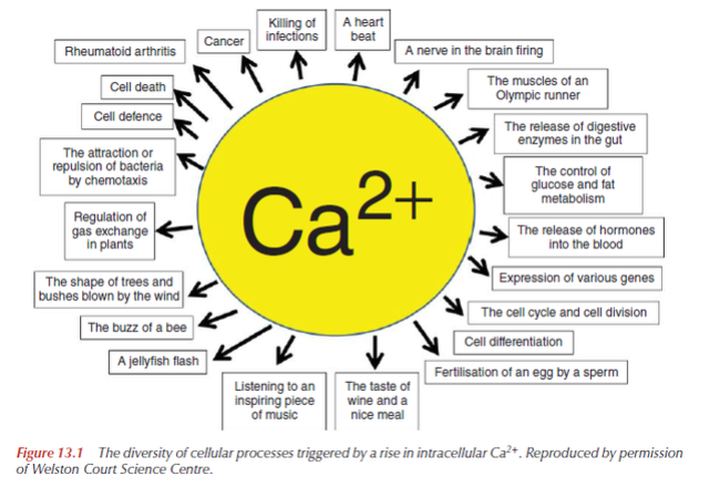
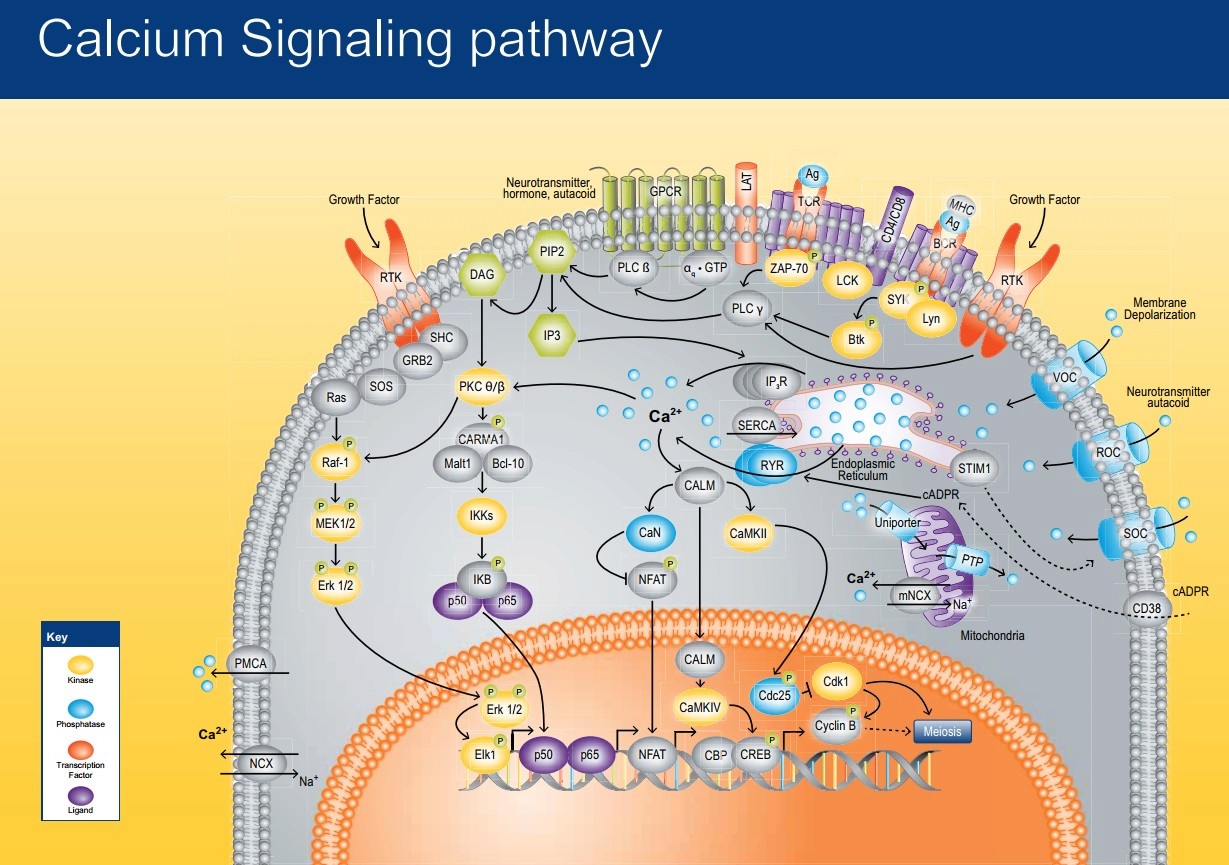
Calcium ions (Ca2+) serve as a universal signal to modulate almost every aspect of cellular function in all cells. Cells in the three domains of life all have a number of universalities, including intracellular Ca2+.   Calcium carries messages to virtually all important functions of cells. Ca2+ signaling pathway plays a key messenger role in regulating many cellular processes including fertilization, contraction, exocytosis, transcription, apoptosis, and learning and memory. Ca 2+ controls the most important cell functions in all eukaryotic organisms. Fertilization, muscle contraction, secretion, several phases of metabolism, gene transcription, apoptotic death, etc. are finely orchestrated by the functional versatility of Ca 2+ signaling and its exquisite spatial and temporal regulationMost likely its unique coordination chemistry has been a decisive factor as it makes its binding by complex molecules particularly easy, even in the presence of large excesses of other cations, e.g. magnesium. Its free concentration within cells can thus be maintained at the very low levels demanded by the signaling function. A large cadre of proteins exists to bind or transport calcium. They all contribute to buffer it within cells, but a number of them also decode its message for the benefit of the target. The most important of these "calcium sensors" are the EF-hand proteins.

Given the central role of intracellular calcium signaling in the living world, a better understanding of the constitution of this calcium-signaling toolkit, and the proteins that comprise it, is crucial to our global understanding of what was required for cells to emerge. These scientific studies highlight the high conservation of the calcium toolkit from prokaryotes to metazoa and the increasing complexity of the proteins that make it up.   The necessity of exporting Ca2+ from cells is a direct consequence of the ambivalent nature of the Ca2+ signal. Ca2+ is essential to cells: it presides over the origin of new life at fertilization and assists cells when their vital cycle has come to an end. Between origin and end, however, Ca2+ guides cells in most of what they must do to fulfill the tasks assigned to them. The balance of Ca2+ between cells and the outside ambient must be regulated with utmost precision: any escape that would somehow alter the balance by letting internal Ca2+ increase over the optimal level spells doom for cells.

Controlled environment is the essence of life.  This cellular separation from the surround pretty much builds around a simple and effective principle of divide et impera, i.e., divide the world into external environment and internal space and govern everything which goes into or out of the living cell/organism.   Ca2+ permits binding reactions that are ~ 100 times faster than Mg2+( magnesium ). 

The maintenance of the stability of cells, osmotic, electrical and chemical, is life essential, and requires the cell to reject certain elements  as ions, namely Na+ ( Sodium ), Ca2+ ( Calcium ) and cl ( Chlorine ), while retaining K+ ( potassium ions ) and Mg2+ ( magnesium )  . The levels of these simple ions are related to the cell's activities, both to metabolism and to the functioning of DNA.  This requirement to reject Ca2+ in the initial stages of life is the pre-requisite of all its advanced functions. To maintain steady states of flow, cells have numerous signaling (circuit) systems employing carriers and messengers, amongst which co-enzymes are of major importance. To describe cellular homeostasis , in total about twenty elements need to be regulated.  

To get this is already a major feat. How did inanimated matter achieve this without guiding intelligence ?

Inseparable tandem: evolution chooses ATP and Ca2+ to control life, death and cellular signalling.
From the very dawn of biological evolution, ATP was selected as a multipurpose energy-storing molecule. Had a adequate energy supply in the cell not to be established prior when life began ? And so, had the origin of energy supply not have to be setup without having evolution at hand as driving force, since dna replication was not setup yet ?  Yet, 
 low  Ca2+ concentration in the cell is a prerequisite  for ATP metabolism. 

That creates one more remarkable catch22 situation, since for a Ca2+ gradient, membrane channels are required, which are only made using ATP as energy source for its biosynthesis. But the production of ATP requires a existing calcium gradient !! The proposed naturalistic explanations, like Donnan potential without requiring Ca 2+ -ATPases and antiporters are fantasious at best. 

ATP effects are mediated by an extended family of purinoceptors often linked to Ca(2+) signalling. Similar to atmospheric oxygen, Ca(2+) must have been reverted from a deleterious agent to a most useful (Intra- and extracellular) signaling molecule. Invention of intracellular trafficking further increased the role for Ca(2+) homeostasis that became critical for regulation of cell survival and cell death. Several mutually interdependent effects of Ca(2+) and ATP have been exploited in evolution, thus turning an originally unholy alliance into a fascinating success story.

The Regulation of a Cell’s Ca 2+ Signaling Toolkit: The Ca 2+ Homeostasome
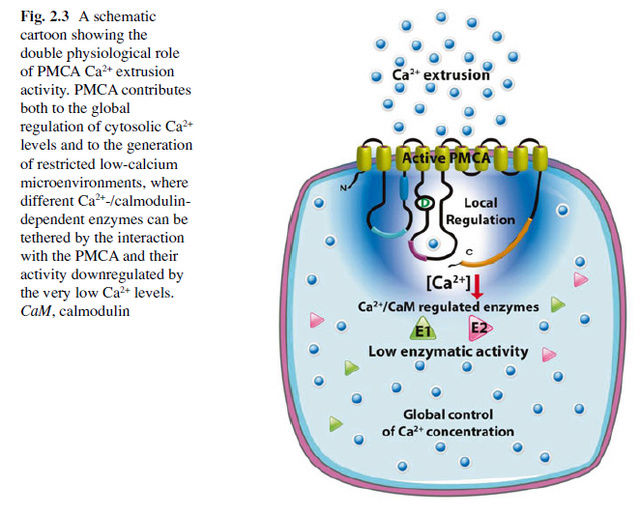
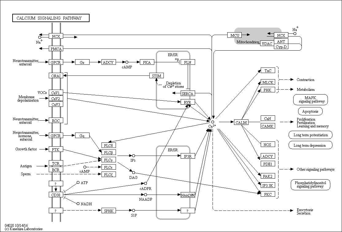
The Ca 2+ ion serves as a ubiquitous second messenger in eukaryotic cells and changes in the intracellular Ca 2+ concentration regulate many responses within a cell, but also communication between cells. In order to make use of such an apparently simple signal, i.e. a change in the intracellular Ca 2+ concentration, cells are equipped with sophisticated machinery to precisely regulate the shape (amplitude, duration) of Ca 2+ signals in a localization-specific manner. To ascertain such a precise regulation, cells rely on the components of the Ca 2+ signaling toolkit. This embraces Ca 2+ entry systems including Ca 2+ channels in the plasma membrane and organellar membranes, and Ca 2+ extrusion/uptake systems including Ca 2+ -ATPases (Ca 2+ pumps) and Na + /Ca 2+ exchangers. The Ca 2+ -signaling components orchestrate their activity as to ascertain the high accuracy of intracellular Ca 2+ signaling. The total of the molecules that build the network of Ca 2+ signaling components, and that are involved in their own regulation as to maintain physiological Ca 2+ homeostasis resulting in phenotypic stability is named the Ca 2+ homeostasome.

Ca2+ triggers life at fertilization and controls the development and differentiation of cells into specialized types. It mediates the subsequent activity of these cells and, finally, is invariably involved in cell death. To coordinate all of these functions, Ca2+ signals need to be flexible yet precisely regulated. This incredible versatility arises through the use of a Ca2+- signaling ‘toolkit’, whereby the ion can act in the various contexts of space, time and amplitude. Different cell types then select combinations of Ca2+ signals with the precise parameters to fit their physiology.

The concentration of Ca2+ increases during perturbation of stimuli, which get recognized by calcium binding proteins or sensor proteins. These proteins further transfer the signal downstream to start phosphorylation cascade that ultimately leads to the regulation of gene expression 

The modulation in Ca2+ concentration across the cell membrane is basically mediated by three classes of transporters-

1. Ca2+-ATPases (PMCAs)
2. Ca2+ permeable channels, or in other words : Ca2+ selective channels
3. Ca2+/cation antiporters (CaCAs), or in other words :  Ca2+/H+ and Na+/Ca2+ exchangers 

which only function in combination of each other 

This is a interdependent system !

The Plasma Membrane Calcium ATPase
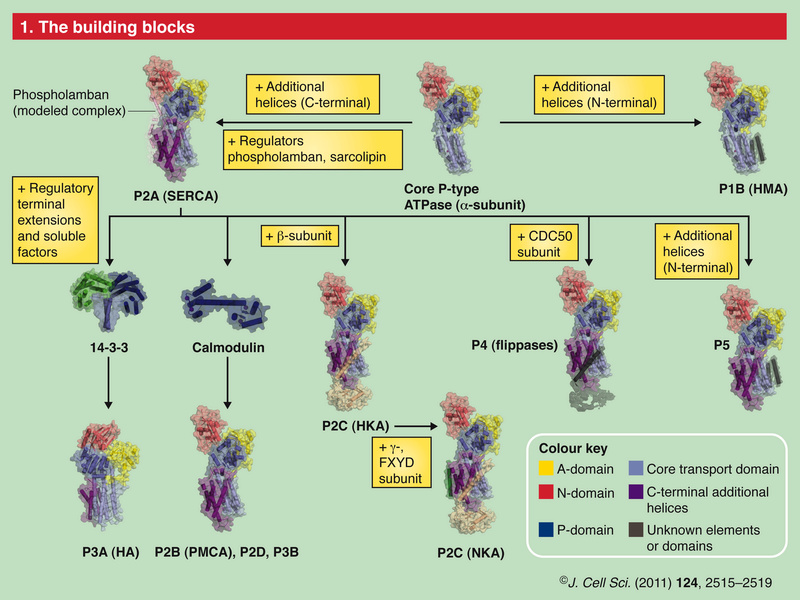
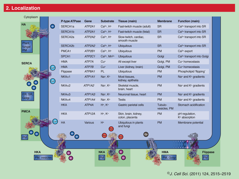
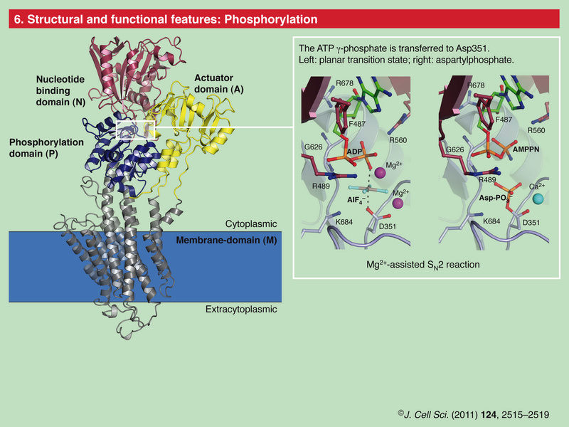
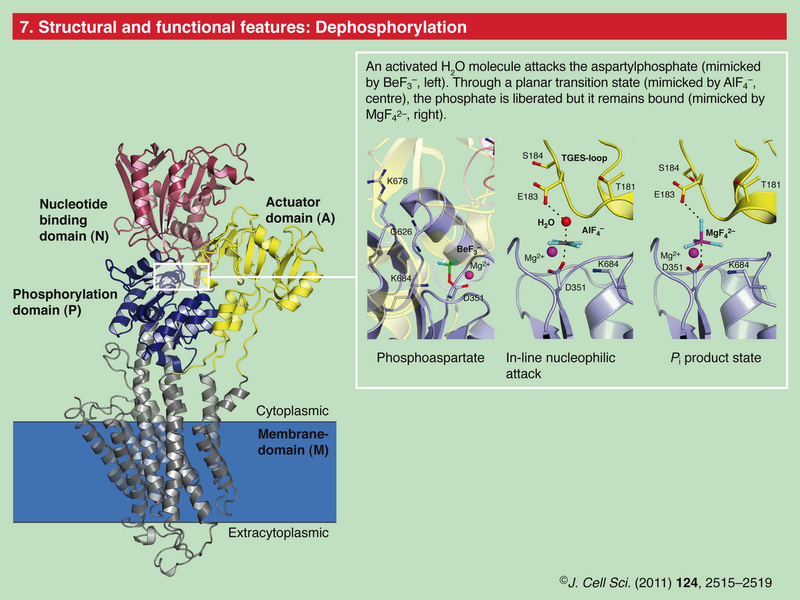
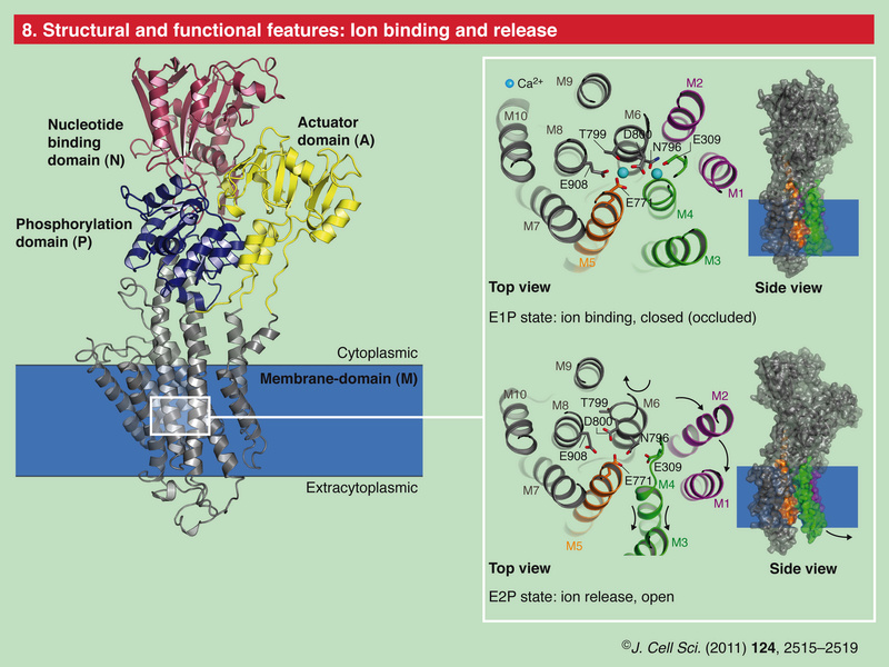
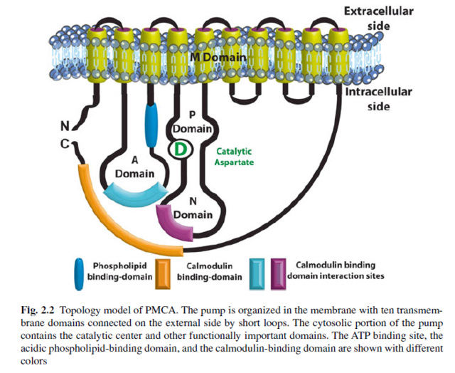
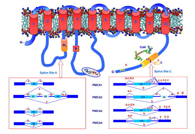
The plasma membrane Ca2+ ATPase (PMCA) is a transport protein in the plasma membrane of cells and functions to remove calcium (Ca2+) from the cell by using the energy stored in ATP.  Thus, it is necessary for cells to employ ion pumps to remove the Ca2+. The PMCA ATPase pump and the sodium-calcium exchanger (NCX) are together the main regulators of intracellular Ca2+ concentrations.   Perhaps the most important structural property that sets the PMCA pump apart from all other members of the superfamily is the presence of a long cytosolic C-terminal tail, which has an essential role in the regulation of the activity of the enzyme: it is the locus of interaction of regulatory partners, chief among them calmodulin, and is the structure responsible for the mechanism of autoinhibition, which is a distinctive properties of the PMCA pump.   Calmodulin interacts with its binding domain removing it from the docking sites next to the active center, freeing the pump from autoinhibition. 

Voltage-Dependent Channels at the Plasma Membrane
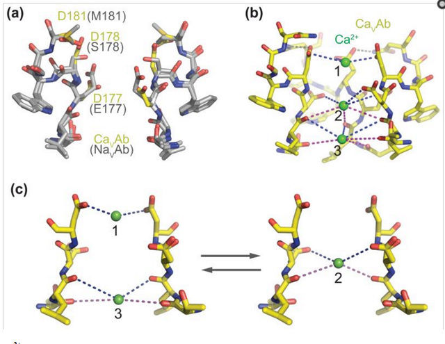
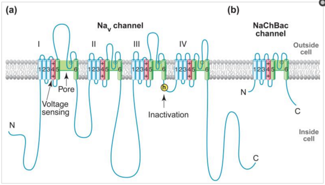
It is assumed that DACCs contribute to the short transient influx of Ca2+ in response to various stimuli, including chilling and microbe interaction. Voltage-gated NaV channels (NaVs) initiate action potentials in excitable cells and are crucial for electrical signaling from bacteria to man. Voltage-gated CaV channels (CaVs) are activated by depolarization during action potentials, and Ca2+ entry through them initiates synaptic transmission, muscle contraction, hormone secretion, and many other biochemical and physiological processes. These channels are thought to share similar voltage-dependent activation and inactivation processes, whose structural basis is fundamental for electrical signaling. Moreover, how these channels can rapidly and selectively conduct Na+ or Ca2+ ions in response to changes of the electrical membrane potential is a crucial question in biology.

Had the right electrical potential not have to be regulated, programmed and setup right from the beginning to bear function for survival of the first cells?

The Ca2+/Cation antiporter (CaCA) superfamily
Electrochemical potential driven Ca2+ transporters are mostly low-affinity Ca2+ transport systems that use the energy stored in the electrochemical gradient of ions.  Cation transport is a critical process in all organisms and is essential for mineral nutrition, ion stress tolerance, and signal transduction.

The PMCAs are effective at maintaining low internal [Ca2+] over long durations, whereas NCX and NCKX Ca2+/Cation antiporters can make the rapid adjustments needed during generation of action potentials in neurons.

Types of Intracellular Ca2+ Signal
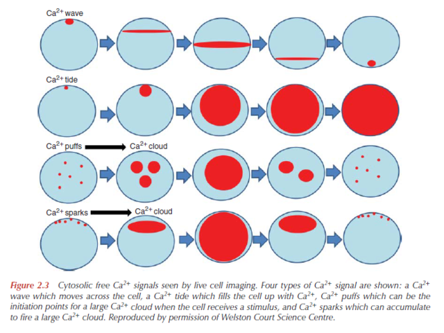
When a Ca2+channel opens, a highly concentrated plume of Ca2+ forms around its mouth and then dissipates rapidly by diffusion after the channel has closed. Such localized signals, which can originate from channels in the plasma membrane or on the internal stores, represent the elementary events — the basic building blocks of Ca2+ signaling.  The spatiotemporal properties of these elementary events, such as Ca2+ sparks and Ca2+ puffs, differ depending on the nature and location of the channels. 

By characterizing these signals, we can discover how the Ca2+- signaling repertoire is elaborated. Essentially, these elementary signals have two functions. They can either activate highly localized cellular processes in the immediate vicinity of the channels (Fig. 1a) or, by recruiting channels throughout the cell, they can activate processes at a global level (Fig. 1b, c). The subcellular location of Ca2+ channels is crucial for targeting elementary signals to different cellular processes. In smooth muscle, for example, Ca2+ sparks that arise locally, near the plasma membrane, activate potassium (K+ ) channels (Fig. 1a), causing the muscle to relax. But when elementary release events deeper in the cell are coordinated to create a global Ca2+ signal, the muscle contracts.

This is a striking example of how spatial organization enables Ca2+ to activate opposing cellular responses in the same cell.

This raises the question how the spatial organisation emerged. Trial and error? Had not both, the location of the channels that coordinate the elementary release events deeper in the cell, and a global Ca2+ signal, which induces the muscle to contract , and  the location of the channels that activate the signals that activates potassium, causing the muscles to relax, to be at the right place together, in order to get both required movements at the same time ? one movement without the other would make no sense. That is, a muscle would be able to contract, but not relax, and vice-versa. Isn't that clear evidence that the whole mechanism had to be setup right from the beginning, with every channel at the right place?  

Time
One of the paradoxes surrounding Ca2+ is that it is a signal for both life and death — although elevations in Ca2+ are necessary for it to act as a signal, prolonged increases in the concentration of Ca2+ can be lethal. Cells
avoid death either by using low-amplitude Ca2+ signals or, more usually, by delivering the signals as brief ‘transients’. These principles apply to both elementary and global signals.

That raises another interesting question: How was the coordination setup the first time? trial and error? If trials would not produce the right concentration, but a prolonged one, the cell dies. Did the cell die every time the dosage was not finely tuned to low amplitude?  Isn't that a clear indication that gradual step-wise evolutionary development of the mechanism would not be possible, but only an all or nothing setup ? 

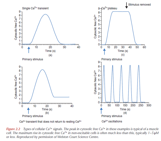
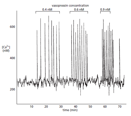
Single transients are used to activate certain cellular processes, such as secretion of cellular material in membrane-bound vesicles, or muscle contraction. However, when information has to be relayed over longer time periods, cells use repetitive signals known as Ca2+ oscillations. Both the elementary events and the global signals can oscillate, but they have widely different periods. For example, whereas the period of elementary Ca2+ sparks in arterial smooth muscle is 0.1–0.5 seconds, it is 10–60 seconds for global waves in liver cells, 1–35 minutes for Ca2+ waves in human eggs after fertilization, and 10–20 hours for the spontaneous Ca2+ transients that control cell division. 

The duration of the signals had to be preprogrammed, and follows the information transmission rules of encoding, code sending, and decoding, which can only be set up by intelligence. 

Cells use frequency modulation (FM) to vary the intensity and nature of the physiological output. For instance, arteries can be made to dilate by increasing the frequency of Ca2+ sparks, which cause the smooth muscle lining the arteries to relax. And by varying the frequency of global Ca2+ signals, different genes can be activated. To use FM signaling, cells have decoders that respond to the frequency and longevity of the Ca2 signals. Probably the best-known example is an enzyme called calmodulin-dependent protein kinase II, which is found in both animal and plant cells and which regulates other enzymes that rely on Ca2+. It works by ‘counting’ Ca2+ transients and varying its activity accordingly.

How did it "learn" that feat ? Had this mechanism not to be pre-programmed ? Since when can mindless matter learn to count and understand informational signals ? 

The enzyme is composed of many identical subunits, and these are activated to varying degrees depending on the frequency of the Ca2+ oscillations.

Amplitude
Information can also be encoded in the amplitude of Ca2+ signals. Such amplitudemodulated (AM) signalling is generally considered to be less reliable than that based on frequency, owing to the difficulties of detecting small Ca2+ changes above the background level. However, it has been shown that cells can interpret modest changes in the concentration of Ca2+. For example, different genes can be activated by varying the amplitude of Ca2+ signals. 

Fertilization and development
In mammals, life begins at fertilization when the sperm interacts with the egg to trigger a Ca2+ oscillation that persists for several hours. This prolonged period of repetitive Ca2+ pulses triggers the developmental program by stimulating the enzymatic machinery involved in the cell-division cycle. There are no further changes in Ca2+ until the one-cell embryo is ready to divide when a spontaneous Ca2+ transient triggers cleavage to form two daughter cells. There are indications that this orderly program may be controlled by two distinct oscillators — Ca2+ signals and oscillating levels of proteins involved in the cell-division cycle. The Ca2+ oscillator seems to be the main mechanism because it persists when dissociated from the cell-cycle oscillator. Just what drives this Ca2+ oscillator, with a period of 10–20 hours, is a mystery, but it may depend on periodic increases in the level of a diffusible intracellular messenger called inositol 1,4,5-trisphosphate (Ins(1,4,5)P3) with each division. In many cells, hormones and growth factors activate the enzyme phospholipase C, which catalyzes the production of InsP3 from the membrane lipid phosphatidylinositol 4,5- bisphosphate (Box 1). As embryos grow and groups of cells differentiate to perform specialized functions, Ca2+ signalling contributes to body polarity and pattern formation.For instance, in amphibians and zebrafish, it is thought to help specify which cells will form structures at the top (dorsal) or the bottom (ventral) part of the embryo. As cells differentiate to perform different functions, they select out those components of the Ca2+-signalling tool kit that best fit their remit. This versatility is emphasized by growing evidence that Ca2+ controls cellular processes as diverse as cell proliferation and the neuronal plasticity that is responsible for learning and memory. But at any moment, any of these orderly signaling events can be switched to activate a program that leads to cell death — a big challenge for the future is to understand how Ca2+ suddenly transforms from a signal for life to a signal of death.

This is highly relevant, as it shows that Ca2+ signaling makes part of the toolkit of body form formation, and if body form has to change, it depends on the change of Ca2+ signaling as well. Another epigenetic mechanism that refutes macro-evolution. There is a big challenge if naturalistic origins are proposed for the phenomena in question, but when the design is proposed, it's perfectly explainable that the designer pre - programmed the life cycle of cells, that is cell cycle, fertilization, cell differentiation, spatiotemporal patterning,  timing and amplitude of the signals, and regulation. 

Cell death
Very high concentrations of Ca2+ can lead to the disintegration of cells (necrosis) through the activity of Ca2+-sensitive protein-digesting enzymes. Calcium has also been implicated in the more orderly program of cell death known as apoptosis. Apoptosis is important during both normal development (the formation of tissue patterns, for example) and pathological conditions such as AIDS, Alzheimer’s disease, and cancer. A protein that is mutated in cancerous cells, called Bcl-2, prevents the cell death that would normally limit the survival and proliferation of cancer cells. Bcl-2 mediates some of its antiapoptotic action by modifying the way in which organelles such as the endoplasmic reticulum and mitochondria (where respiration occurs) handle Ca 2+


How intracellular Calcium signaling,  gradient and its role as a universal intracellular regulator points to design Calciu10


Last edited by Admin on Thu Apr 12, 2018 3:04 am; edited 46 times in total

https://reasonandscience.catsboard.com

Otangelo


Admin

Role of Calcium Ions in the Cell and Bacterial Calcium Metabolism 1

http://reasonandscience.heavenforum.org/t2448-role-of-calcium-ions-in-the-cell-and-bacterial-calcium-metabolism

The make of a power gradient is always a engineering achievement, and a lot of knowledge,  planning and intelligence is required for setup. Hydroelectric dams are highly complex, and  always the result of years of  planning by the most skilled, educated and knowledged  engineers  of large companies. As for many human inventions, the engineering solutions discovered by man are employd in nature at least since life began in a far more elaborated and sophisticated way.

In view of the importance of Ca2+ as a universal intracellular regulator, it is surprising how little of it is mentioned and debated when it comes to elucidate how life began.

Why Calcium? How Calcium became the best communicator.

To adapt to changing environments, cells must signal, and signaling requires messengers whose concentration varies with time. Filling this role, calcium ions (Ca2+) and phosphate ions have come to rule cell signaling.  19 An oversupply of certain elements, even essential ones, may subject an organism to stress. Accordingly, cellular systems need counteractive measures to overcome the threat imposed by their habitat. In order to illustrate the kind of defense strategies adopted by living cells  for managing hazardous compounds, the case of Ca2+ is presented. This element is an especially sensitive indicator because the cell has to maintain a critical level of it at all times and any changes, up or down, will severely impair the cell and eventually kill it. 18  The critical chemical step at the start of life was not to make lots of DNA or RNA, but must have been to get rid of Ca2+. 3    Calcium is a divalent cation (Ca2+), ubiquitous in natural waters. It has a variable degree of hydration of 6 to 8 water molecules that can be exchanged very rapidly. This makes calcium the fastest binding agent of all the available divalent ions in the environment. Mg2+ reacts 103 times slower.  

That shows that calcium regulation had to be full setup right from the start, when life began.
How intracellular Calcium signaling,  gradient and its role as a universal intracellular regulator points to design Cellul10

The book Intracellular Calcium, page 581 confirms this notion :
Our knowledge of the biological chemistry of Ca2+ argues strongly that Ca2+ must also have played a key part in the biochemistry of the first, primeval cells. Given the prevalence of calcium in the Earth’s crust, these primeval cells will have been surrounded by at least tens of micromolar free Ca2+, if not millimolar. This is high enough to mess up many reactions inside contemporary cells. At millimolar concentrations, Ca2+ competes with Mg2+, binds to DNA and RNA, and clogs it up. Ca2+ binds to nucleotides, so they do not work properly. And crucially Ca2+, at microto millimolar concentrations, precipitates carbonate, phosphate and sulphate. So if a primeval cell was to work, it had to get rid of Ca2+, lowering it at least to submillimolar levels, if not submicromolar. In fact, without control of intracellular Ca2+, I have argued life would never have got off the ground or even been able to get going in the ocean! Control of intracellular Ca2+ had to be a crucial step in allowing the original cells to survive and replicate, even
before RNA or DNA synthesis could begin in earnest.


There are three ways this can be done across a semipermeable membrane:

1. A membrane potential, positive inside, instead of the negative one in all contemporary cells, so that Ca2+ is at its equilibrium potential.
2. A Ca2+ exchanger, a Ca2+ anion symport (e.g. phosphate) or a Ca2+/cation antiport, (e.g. Na+ or H+).
3. A Ca2+ pump.

Most discussions about evolution start with RNA versus DNA worlds and the origins of the organelles in eukaryotes. Those discussing the evolution of the Ca2+ signalling system usually focus on the need for a Ca2+ pump, then Ca2+ channels and then Ca2+ target sites such as the EF-hand . But, it is much more likely that the primeval cells found a way of getting rid of intracellular Ca2+ without the need for a pump. The only way that this can be done is to generate a membrane potential, positive inside instead of the negative potential of contemporary cells.

For 3800 million years it has been essential for all cells, whether they were animal, plant or microbe, whether they were Bacteria, Archaea or Eukaryota, to maintain a free Ca2+ in the soluble part of the cell, the cytosol, very low, in the micromolar to submicromolar range. Without this low intracellular free Ca2+, particularly in the presence of millimolar Ca2+ outside the cell, damaging inorganic and organic Ca2+ precipitates would form, and the essential biochemical processes of energy metabolism, DNA, RNA, protein, lipid and carbohydrate synthesis and degradation could not take place.

Membrane Potential
All contemporary cells have a membrane potential, negative inside. So, without a Ca2+ pump, Ca2+ would concentrate inside. At equilibrium, a cell with a large, negative membrane potential would have a cytosolic free Ca2+ concentration as high as tens of millimolar or even 1M! Hence contemporary cells have to have a Ca2+ pump, the Ca2+-MgATPase and/or the Na+/Ca2+ exchanger, in order to prevent Ca2+ concentrating inside the cell. Pumps on internal organelles can remove Ca2+ from the cytosol, but without an efflux mechanism on the plasma membrane, Ca2+ would eventually reach its electrochemical equilibrium.
Most bacterial and eukaryotic cells have a cytosolic K+ at least as high as 150mM, with a cytosolic Na+ concentration of around 5–10mM, 1mM Mg2+ and submicromolar free Ca2+. The K+/Na+ gradient across the plasma membrane is maintained by the sodium pump, which exchanges three Na+ out for two K+ in. Thus, this pump is slightly electrogenic, contributing 5–10mV to the membrane potential, as shown by the small drop in membrane potential when the Na+ pump inhibitor ouabain is present. However, the bulk of the resting membrane potential in contemporary eukaryotic cells is due to the selective permeability of the plasma membrane to K+ ( Potassium ) compared with Na+( Sodium ), the membrane potential being close to the equilibrium potential for K+, typically –70mV in muscle, –60 to –90mV in nerve and –30mV in liver, all negative inside. But the primeval cell would not have evolved this far.

So, in the absence of pumps, how could a cell get rid of Ca2+? 
The answer must be that it had a membrane potential, positive inside, maintained by a Donnan potential . This can be generated by a gradient of permeant anions, where there is higher concentration inside than out. This will occur if there is a higher concentration of impermeant anions outside the cell than inside. Alternatively, a positive membrane potential inside could be generated by cations if there is a higher concentration of impermeant cations inside than outside, causing the permeant cations to be higher outside than in. 

After much speculation, at the end, the authors confess: 

These arguments emphasise the importance of relating the electrophysiology to the biochemistry of Ca2+, if we are to understand how the Ca2+ signalling system evolved and how it works in cells today. Once a ‘Ca2+ pressure’ had been established, and proteins started to be produced in earnest, the scene was set for the Ca2+ signalling proteins to appear. First would have been the Ca2+ pumps and exchangers, taking over from the Donnan potential, allowing a membrane potential, negative inside, to develop. Only when there were fully active plasma membrane pumps and exchangers was it possible to maintain the cytosolic free Ca2+ low in the presence of high concentrations outside a cell with a negative membrane potential. These early molecular processes for Ca2+ may have taken hundreds or even a billion years to develop effectively. By the time of Cambrian explosion, some 590 million years ago, the majority of Ca2+-regulated cellular events must have been in place – nerve action potentials and nerve terminal secretion, all forms ofmuscle contraction, vesicular secretion, invertebrate vision, gamete fusion, plant regulation, defence mechanism, and cell death by apoptosis.

As so often, there is a hudge gap between the unsatisfying just so speculation of how the Ca2+ emerged, and the extant solution and its complexity. If the Donnal potential was sufficient, why did cells evolve highly sophisticated, diversified Ca2+ pumps, extremely complex as exposed below, where Calcium ATPases require Calmodulin in a interdependent manner for proper function ?  As so often, the authors have no answer by detailled chronological narrative of the events that triggered the production of highly complex and regulation requiring membrane pumps. The pseudo-scientific assertions are remarkable. 

Calcium signaling is one of the most extensively employed signal transduction mechanisms in life. 10 Calcium ions (Ca2+) serve as a universal signal to modulate almost every aspect of cellular function in all cells. Cells in the three domains all have a number of universalities, including intracellular Ca2+. 3  Calcium carries messages to virtually all important functions of cells. Ca2+ signaling pathway plays a key second messenger role in regulating many cellular processes in virtually all types of animal cells including fertilization, contraction, exocytosis, transcription, apoptosis, and learning and memory. 11 Most likely its unique coordination chemistry has been a decisive factor as it makes its binding by complex molecules particularly easy, even in the presence of large excesses of other cations, e.g. magnesium. Its free concentration within cells can thus be maintained at the very low levels demanded by the signaling function. A large cadre of proteins exist to bind or transport calcium. They all contribute to buffer it within cells, but a number of them also decode its message for the benefit of the target. The most important of these "calcium sensors" are the EF-hand proteins.

In bacteria, Ca2+ is implicated in a wide variety of cellular processes, including the cell cycle and cell division. 17  The growing number of proteins containing various Ca2+-binding motifs supports the importance of Ca2+, which controls various protein functions by affecting protein stability, enzymatic activity or signal transduction.  Calcium is an ambivalent messenger: while essential to the correct functioning of cell processes if not carefully controlled spatially and temporally within cells it generates variously severe cell dysfunctions, and even cell death.   A prolonged high level of intracellular free Ca2+ irreversibly damages mitochondria and can cause chromatin condensation, precipitation of phosphate and protein and activation of degradative enzymes such as proteases, nucleases and phospholipases 18

The molecular mechanisms of the multiple effects of Ca2+ range from Ca2+ playing a role as a structural component stabilizing lipopolyscharide layer and cell wall to Ca2+ playing a role in structural stability of proteins or protein complexes ] to Ca2+ effecting transcription of two component systems, regulatory proteins, andproteinphosphorylation, whichmayhave eittherpositive or negative regulatory outcomes . When a cell is exposed to external Ca2+, in addition to Ca2+ uptake and its possible effect on the production of other intracellular messengers (ex, cAMP), it may be recognized by two component regulatory systems whose primary role is adjusting bacterial physiology to environmental conditions. 

Had the control not have to be setup right from the beginning ? 

Given the central role of intracellular calcium signaling in the living world, a better understanding of the constitution of this calcium-signaling toolkit, and the proteins that comprise it, is crucial to our global understanding of what was required for cells to emerge. 8 These scientific studies  highlight the high conservation of the calcium toolkit from prokaryotes to metazoa and the increasing complexity of the proteins that make it up.   The necessity of exporting Ca2+ from cells is a direct consequence of the ambivalent nature of the Ca2+ signal.14 Ca2+ is essential to cells: it presides over the origin of new life at fertilization and assists cells when their vital cycle has come to an end. Between origin and end, however, Ca2+ guides cells in most of what they must do to fulfil the tasks assigned to them. The balance of Ca2+ between cells and the outside ambient must be regulated with outmost precision: any escape that would somehow alter the balance by letting internal Ca2+ increase over the optimal level spells doom for cells.

Controlled environment is the essence of life.  This cellular separation from the surround  pretty much builds around a simple and effective principle of divide et impera, i.e., divide the world into external environment and internal space and govern everything which goes into or out of the living cell/organism.   Ca2+ permits binding reactions that are ~ 100 times faster than Mg2+( manganese ). Low Ca2+ in the cytosol of primeval cells is also compatible with energetics based around ATP and the usage of DNA/RNA for genetic encoding, because both cannot tolerate high Ca2+ concentrations; at the levels above 10 μMolar  of Ca2+, this ion induces the precipitation of phosphates, causes aggregation of proteins and nucleic acids and disrupts lipid membranes . A Ca2+ homeostatic system assures  a transmembrane Ca2+ gradient, which lies at the very base of Ca2+ signalling. 

The maintenance of the stability of cells, osmotic, electrical and chemical, is life essential, and requires the cell to reject certain elements  as ions, namely Na+ ( Sodium ), Ca2+ ( Calcium ) and cl ( Chlorine ), while retaining K+ ( potassium ions ) and Mg2+ ( magnesium )  . The levels of these simple ions are related to the cell's activities, both to metabolism and to the functioning of DNA.  This requirement to reject Ca2+ in the initial stages of life is the pre-requisite of all its advanced functions. To maintain steady states of flow, cells have numerous signalling (circuit) systems employing carriers and messengers, amongst which co-enzymes are of major importance. To describe cellular homeostasis , in total about twenty elements need to be regulated.  6 To get this is already a major feat. How did inanimated matter achieve this without guiding intelligence ? 


Origin of the calcium signaling machinery
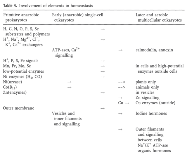
A complex Ca2+ signaling ‘toolkit’ might have evolved before the emergence of multicellular animals. 12 The basic principles of Ca2+ regulation emerged early in prokaryotes. 13
How intracellular Calcium signaling,  gradient and its role as a universal intracellular regulator points to design Elemen10

How intracellular Calcium signaling,  gradient and its role as a universal intracellular regulator points to design Elemen11
Figure 3

Calcium (Ca2+) represents an important cytosolic signalling molecule as it can affect almost all cellular processes.  7  The flux of calcium across the plasma membrane and endomembranes, i.e. membranes demarcating internal organelles, critically relies on the operation of various calcium channels within the membranes. It is a critical element in all organisms; it is an essential nutrient which also has a conserved signaling role. Transporters that mediate the movement of this ion are therefore particularly important. These Ca2+ transporters impact upon cell division, growth, development, and adaptation to environmental conditions.  2 Ca2+ is a faster binding agent than any other available divalent ion from the environment. It reacts 103 times faster than Mg2+ ( manganese ) . 4 The calcium ion is arguably the most widely employed and important intracellular messenger in physiological and pathophysiological cellular processes. 5

Calcium plays a vital role in membrane integrity, catalysis, in the electrical properties of cells and, crucially, as a unique regulator of events inside cells. The ability of cells to maintain a large gradient of Ca2+ across their outer membrane is universal. All animal, plant and microbial cells have a low cytosolic free Ca2+ in the submicromolar range, and can keep this even when the free Ca2+ outside is as high as 1–10 mMolar ! Damage the ability of the plasma membrane to maintain this gradient and Ca2+ will flood into the cell, precipitating calcium phosphate, damaging the ATP-generating machinery, and  even kill the cell. The evidence we have from molecular biology, together with the toxic nature of prolonged high Ca2+ levels inside cells, argues strongly that primeval cells must have had  Ca2+ pumps to keep their free intracellular Ca2+ low, setting the scene for the ‘calcium pressure’ across then plasma membrane to be exploited to act as the source for cell activation.

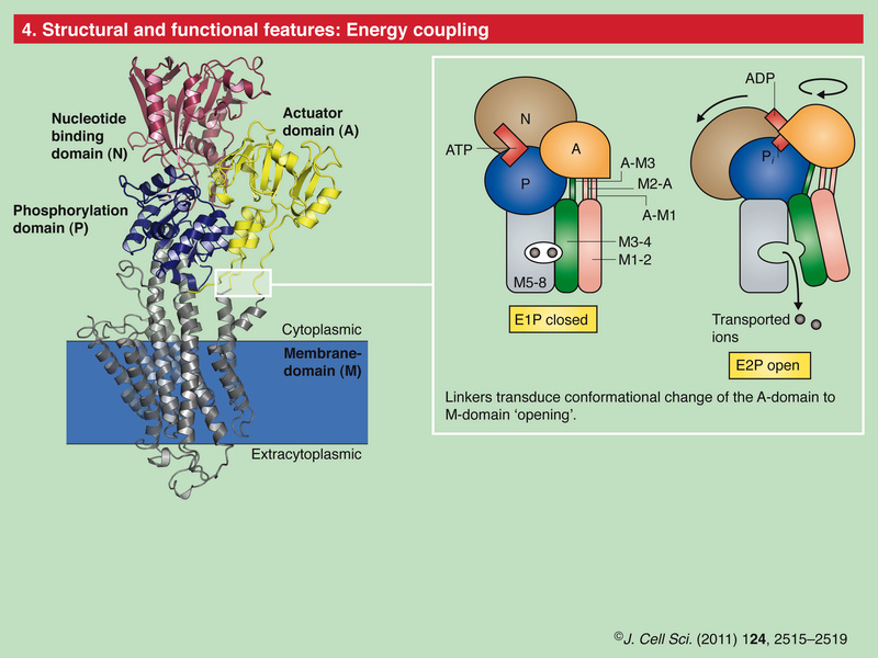
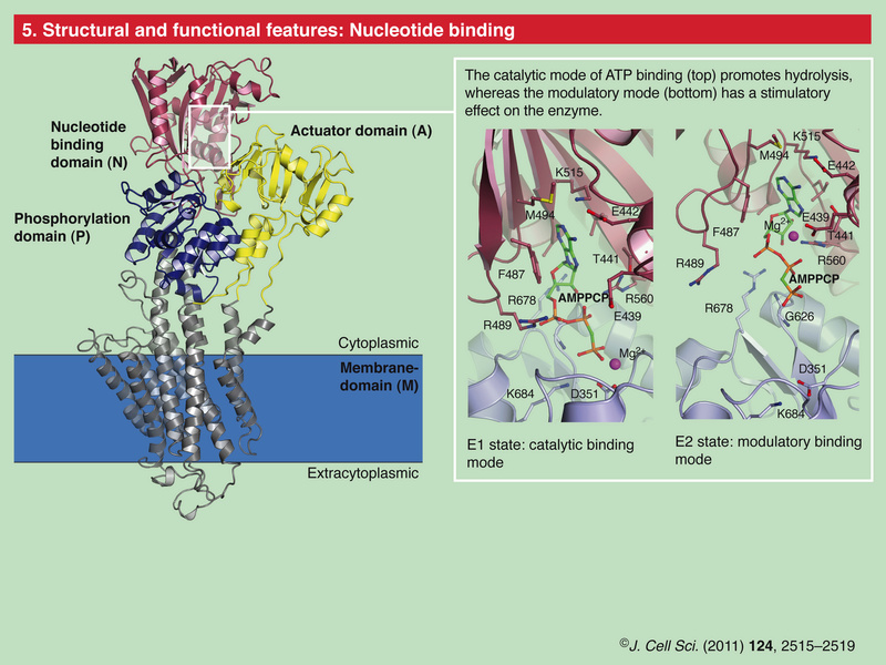
In order to maintain such a low cytosolic Ca2+ concentration, Ca2+ ions thus have to be transported against a steep concentration gradient. In addition, the positively charged molecules are often transported against a very negative membrane potential, contributing to a large electrochemical gradient for Ca2+ ions. 16 This function is carried out by (ACA)-encoded P-type ATPases. These ATP-driven pumps are members of a large protein family that are characterized by a phosphorylated (P) intermediate state of the protein during the pump cycle.  Plasma membrane Ca2+ -ATPases belong to the group of P-type 2B proteins that share high homology to the plasma membrane calcium ATPase (PMCA) pumps from animals . . The 2B class of P-type ATPases is characterized by the presence of an N-terminus that acts as an auto-inhibitory domain. This N-terminal auto-inhibitory domain is released from the catalytic side of the ATPase after binding calmodulin in the presence of Ca2+. 

So inanimate chemistry had the innate drive of trials and errors to produce a cell membrane, and amongst tons of other things, a Ca+ gradient through highly complex Calcium channels to keep a 10 000-fold higher concentration of calcium outside the cell than inside the cytosol in order to create a environment suited for a protocell to keep its vital functions and not to die ? Why would chemical elements do that ? Did they have the innate drive and goal to become alive and keep a  ambience prerequisite, homeostasis of various elements, to permit  life ?

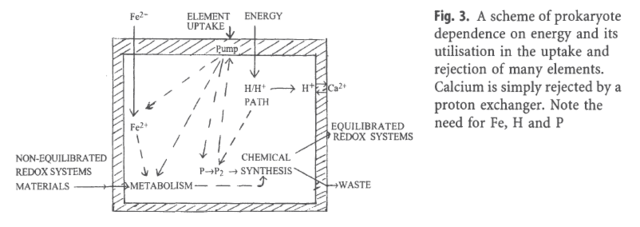
The origin of life required two processes that dominated:
(1) the generation of a proton gradient and 
(2) linking this gradient to ATP production in part and in part to uptake of essential chemicals and rejection of others. The generation of a proton gradient required especially appropriate amounts of iron (Fe2+), levels for electron transfer and the ATP production depended on controlling H+, Mg2+ and phosphate in the cytoplasm. 

The four elements had to be  linked together at once through complex formation, Mg2 (magnesium )+ and H+ ( hydrogen ) with phosphates, while chemical redox reactions linked Fe ( Iron ) and H ( Hydrogen ), and there were also feed-back connections of all four required through metabolism of carbon compounds and genetic controls. Thus we can consider that a network of carriers of the fundamental building blocks of biological polymers and of energy, the co-enzymes, fed and controlled (through feedback) inter-communicating pathways of metabolism were required,  so as to establish primitive homeostasis which at requisite energy input also set a low homeostatic ca+ (calcium) concentration. The whole required structural and catalytic units, proteins, which had to be supplied or restricted by feed-back to DNA and RNA coded production machineries. Each of the major pathways of the four elements, Fe, H, P, Mg, as well as of C, N, 0, S and Se, had  therefore to be linked through genetic regulation as well as through chemical controls to one another and then to all other cell activities. The earliest cells must have relied on an internal homeostasis of at least H+, Fe2+, Mg2+ and phosphates apart from systems related to C/N/O/H/S/Se metabolism.

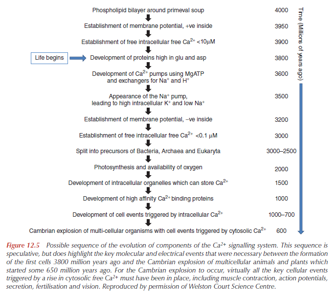
How do mainstream scientific papers explain how this gradient emerged ?

How intracellular Calcium signaling,  gradient and its role as a universal intracellular regulator points to design Evolut10

From the book, Intracellular Calcium, page 588

Calcium signalling and calcium channels: Evolution and general principles
Washout of Ca2+ ions from the Earth’s crust, in combination with a decreased alkalinisation of the ancient ocean, led to a continuous increase in Ca2+ concentration in the sea water, which in turn initiated the evolution of a Ca2+ homeostatic system that kept cytosolic Ca2+ at a low level. The molecules governing such homeostasis seem to evolve rather early in the genealogical tree as the most primitive bacteria were already in possession of Ca2+ pumps and Ca2+ exchangers. An increase in environmental Ca2+ concentration in combination with an evolving Ca2+ homeostatic system assured the build-up of a transmembrane Ca2+ gradient, which lies at the very base of Ca2+ signalling. This gradient soon was utilised by prokaryotes to develop Ca2+ permeable channels, which formed a pathway for a transmembrane Ca2+ influx and, thus, made Ca2+ signalling possible.

The evidence we have from molecular biology, together with the toxic nature of prolonged high Ca2+ levels inside cells, argues strongly that primeval cells must have had to develop Ca2+ pumps to keep their free intracellular Ca2+ low, setting the scene for the ‘calcium pressure’ across then plasma membrane to be exploited to act as the source for cell activation. A key chemical property of Ca2+ is that it comes on and off proteins fast, in milliseconds.

There are five main variations in the molecular processes responsible for Ca2+ signalling, which origin must be explained:

1. The type of Ca2+ signal, how big it is, how long it lasts and where in the cell it occurs.
2. The amino acid sequences, together with the gene sequences, of the components that produce the Ca2+ signal and those that are its targets.
3. The biochemical and electrical characteristics of these components (e.g. binding constants, kinetics, affinities, specificities, conductances).
4. The level of expression of the components.
5. The number of cells expressing particular components.

There are at least six types of Ca2+ pumps in the plasma membrane of cells:

four Ca2+-MgATPases, 
two Na+/Ca2+ exchangers and other
Ca2+/H+ exchangers. 
Three types of Ca2+ pumps in the Endoplasmic Reticulum (ER) (SERCA1, 2 and 3)
three types of each receptor (IP3 and ryanodine) in the ER that cause Ca2+ to be released into the cytosol.
And there are a plethora of Ca2+ channels in the plasma membrane opened by voltage, ligands outside and inside cells, and events inside the ER.

Questions : Is the process analogue or digital and how does calcium determine this?  Whether a cell fires or not can be determined by calcium reaching its target or not, by the level the Ca2+ reached, or by the
thermodynamic and morphological properties of the Ca2+ target. Yet there are analogue processes superimposed on the digital ones. 

We have here a classic example of pseudo scientific just so stories, full of assertions, but no scientific evidence that evolution explains the feat. P-type Ca2+ pumps require Calmodulin (CaM) activators. They work in a interdependent way. Since Calcium carries messages to virtually all important functions of cells, the setup had to emerge prior dna replication began , and evolution could not have been a driving force. How was the strict regulation setup ? No regulation, no homeostasis, no calcium intracellular signaling, no life. 

Intracellular calcium homeostasis and signaling
Ca(2+) is a universal carrier of biological information: it controls cell life from its origin at fertilization to its end in the process of programmed cell death. Ca(2+) is a conventional diffusible second messenger released inside cells by the interaction of first messengers with plasma membrane receptors. However, it can also penetrate directly into cells to deliver information without the intermediation of first or second messengers. Even more distinctively, Ca(2+) can act as a first messenger, by interacting with a plasma membrane receptor to set in motion intracellular signaling pathways that involve Ca(2+) itself. Perhaps the most distinctive property of the Ca(2+) signal is its ambivalence: while essential to the correct functioning of cells, Ca(2+) becomes an agent that mediates cell distress, or even (toxic) cell death, if its concentration and movements inside cells are not carefully tuned. Ca(2+) is controlled by reversible complexation to specific proteins, which could be pure Ca(2+) buffers, or which, in addition to buffering Ca(2+), also decode its signal to pass it on to targets. The most important actors in the buffering of cell Ca(2+) are proteins that transport it across the plasma membrane and the membrane of the organelles: some have high Ca(2+) affinity and low transport capacity (e.g., Ca(2+) pumps), others have opposite properties (e.g., the Ca(2+) uptake system of mitochondria). Between the initial event of fertilization, and the terminal event of programmed cell death, the Ca(2+) signal regulates the most important activities of the cell, from the expression of genes, to heart and muscle contraction and other motility processes, to diverse metabolic pathways involved in the generation of cell fuels.

Inseparable tandem: evolution chooses ATP and Ca2+ to control life, death and cellular signalling.
From the very dawn of biological evolution, ATP was selected as a multipurpose energy-storing molecule. Had a adequate energy supply in the cell not to be established prior when life began ? And so, had the origin of energy supply not have to be setup without having evolution at hand as driving force, since dna replication was not setup yet ? Metabolism of ATP required intracellular free Ca(2+) to be set at exceedingly low concentrations, which in turn provided the background for the role of Ca(2+) as a universal signalling molecule. The early-eukaryote life forms also evolved functional compartmentalization and vesicle trafficking, which used Ca(2+) as a universal signalling ion; similarly, Ca(2+) is needed for regulation of ciliary and flagellar beat, amoeboid movement, intracellular transport, as well as of numerous metabolic processes. Thus, during evolution, exploitation of atmospheric oxygen and increasingly efficient ATP production via oxidative phosphorylation by bacterial endosymbionts were a first step for the emergence of complex eukaryotic cells. Simultaneously, Ca(2+) started to be exploited for short-range signalling, despite restrictions by the preset phosphate-based energy metabolism, when both phosphates and Ca(2+) interfere with each other because of the low solubility of calcium phosphates. The need to keep cytosolic Ca(2+) low forced cells to restrict Ca(2+) signals in space and time and to develop energetically favourable Ca(2+) signalling and Ca(2+) microdomains. These steps in tandem dominated further evolution. The ATP molecule (often released by Ca(2+)-regulated exocytosis) rapidly grew to be the universal chemical messenger for intercellular communication; ATP effects are mediated by an extended family of purinoceptors often linked to Ca(2+) signalling. Similar to atmospheric oxygen, Ca(2+) must have been reverted from a deleterious agent to a most useful (intra- and extracellular) signalling molecule. Invention of intracellular trafficking further increased the role for Ca(2+) homeostasis that became critical for regulation of cell survival and cell death. Several mutually interdependent effects of Ca(2+) and ATP have been exploited in evolution, thus turning an originally unholy alliance into a fascinating success story.This article is part of the themed issue 'Evolution brings Ca(2+) and ATP together to control life and death'.

Interdependece is evidence AGAINST evolution. Its remarkable how the authors implicitly overlook this obvious fact. Interdependence means, one molecule has no use without the other. But how could and would evolution produce it, if it had no use in the organism by its own ? 

The Regulation of a Cell’s Ca 2+ Signaling Toolkit: The Ca 2+ Homeostasome 9
The Ca 2+ ion serves as a ubiquitous second messenger in eukaryotic cells and changes in the intracellular Ca 2+ concentration regulate many responses within a cell, but also communication between cells. In order to make use of such an apparently simple signal, i.e. a change in the intracellular Ca 2+ concentration, cells are equipped with sophisticated machinery to precisely regulate the shape (amplitude, duration) of Ca 2+ signals in a localization-specific manner. To ascertain such a precise regulation, cells rely on the components of the Ca 2+ signaling toolkit. This embraces Ca 2+ entry systems including Ca 2+ channels in the plasma membrane and organellar membranes, and Ca 2+ extrusion/uptake systems including Ca 2+ -ATPases (Ca 2+ pumps) and Na + /Ca 2+ exchangers. The Ca 2+ -signaling components not only orchestrate their activity as to ascertain the high accuracy of intracellular Ca 2+ signaling, but they are also implicated in the regulation of their own expression. The total of the molecules that build the network of Ca 2+ signaling components, and that are involved in their own regulation as to maintain physiological Ca 2+ homeostasis resulting in phenotypic stability is named the Ca 2+ homeostasome.

The cytosolic free Ca 2+ concentration is tightly regulated by Ca 2+ -binding proteins, Ca 2+ pumps and other transporters to maintain low [Ca 2+ ] i levels of about 100nM in order to avoid toxic calcium phosphate precipitation within the cell. 15

A bimodular mechanism of calcium control in eukaryotes

How Resting Cells Maintain Their Ca2+ Balance
In order to maintain the very large electrochemical gradient of Ca2+ across the plasma membrane, with a submicromolar cytosolic free Ca2+, in the presence of an extracellular free Ca2+ in the millimolar range, there has therefore to be a mechanism that continuously ‘pumps’ Ca2+ out of the cell. Without it, with a membrane potential on tens of millivolts, negative inside, the cytosolic free Ca2+ would eventually rise to hundreds of millimolar or even molar concentrations. Such a rise in free Ca2+ could not be prevented by internal stores such as the ER or mitochondria. As the Ca2+ has to be pumped out against a large electrochemical gradient, Ca2+ transport has to be coupled to another process with a ‘positive’ electrochemical gradient. Cells therefore exploit two sources of potential energy to pump Ca2+ out of cells against the electrochemical gradient across the plasma membrane:

1. ATP hydrolysis.
2. Another ion gradient, such as Na+ or H+.

The first of these to be discovered was the Ca2+ pump in red cells (Schatzmann, 1966, 1975). Even red cells can maintain a submicromolar cytosolic free Ca2+ in the presence of millimolar extracellular Ca2+ (Campbell and Dormer, 1975, 1978).

The universal need for Ca2+ by all cells falls into four distinct functional categories:

1. Structural (in both hard and soft tissues).
2. Electrical.
3. Cofactor.
4. Intracellular signal.

1. Structural
Ca2+ bound to inorganic anions, proteins and phospholipids is vital in maintaining the soft structures of most cells. Ca2+ bound to proteins holds cells in organs together and, on the outside of cells, helps to maintain the stability of the plasma membrane, together with its semipermeable properties.

But Ca2+ also plays a structural role inside the cell. Although Mg2+ is the main cation bound to the negatively charged phosphate groups inDNA and RNA, Ca2+ also appears to bind. For example, Ca2+ bound to DNA may play an important role in the structure the chromosome in bacteria. Nucleotides such as ATP, GTP, CTP and TTP are all mostly bound to a divalent cation inside the cell. With a free Mg2+ estimated at 1–2mM, this is the favoured cation. And the form that reacts with virtually all kinases and pumps is ATPMg2– . However, in secretory vesicles Ca2+ bound to nucleotides and other molecules plays a role in maintaining the structural role of the vesicle. It also enables the contents to dissolve very quickly when released from the vesicle after it fuses with the plasma membrane.

2.Electrical
Electrophysiology of Intracellular Ca2+
Electrophysiology is the study of the electrical properties of cells. These electrical properties depend on the movement of ions, rather than electrons, across lipid bilayers. A crucial feature of Ca2+ as an intracellular signal is how it interacts with these electrical properties. Across the outer membrane of the cell (i.e. the plasma membrane) the electrochemical gradient is made up of the membrane potential, which is negative inside, thereby attracting Ca2+ in, and the huge concentration difference of Ca2+, some 10000- to 100 000-fold between the outside and inside.

In a house, electricity is carried by electrons along a wire. In contrast, much of the electrical activity in living systems is carried by ions: K+, Na+, Ca2+ and Cl– . Since Ca2+ is a positively charged ion, it will move towards a negative potential. Ca2+ will itself generate an electrical potential if it moves across a semipermeable membrane down a concentration gradient. Many cells have  specific Ca2+ channels to exploit this electrical activity. All cells maintain an electrical potential difference across their outer membrane. This is usually of the order of 40–100mV, negative inside, depending on cell type, though red blood cells have a membrane potential less than this. The membrane potential occurs because of the selective permeability of the plasma membrane to particular cations and anions. Pure phospholipids bilayers are poorly permeable to ions. But as soon as proteins are inserted across the bilayer, ions are able to diffuse across. But we now know that the selective permeability of biological membranes to particular ions is caused by ion channels, which are usually, but not always, proteins. In 1902, Bernstein suggested that K+ ions ( potassium ) were responsible for the resting potential of cells and that the action potential of electrically excitable cells might be due to loss in the selective permeability of the cell membrane to potassium. It is true that the resting membrane potential of most cells is caused by K+ ions. However, thanks to pioneering studies using giant nerve cells, such as those from the squid Loligo forbesi, Curtis and Cole at Woods Hole in the United States  and Hodgkin and Huxley at Plymouth in the United Kingdom showed that the action potential in many nerve axons starts as a result of a sudden increase in the permeability to Na+ ( Sodium ). This rapidly depolarises the cell, which then repolarises as a result of increased permeability to K+. This is the so-called ‘sodium theory’ of the action potential, and resulted in Alan Hodgkin and Andrew Huxley being awarded the Nobel Prize in 1963. Soon after their pioneering work it was realised that many excitable cells also have Ca2+ channels that are sensitive to the potential across the plasma membrane. Thus, the contraction of a barnaclemuscle, to hold the plates shut when the tide goes out, is provoked by the transmitter glutamate initiating an action potential dependent on Ca2+ ions moving into the cell. Interestingly, our own heart beat depends on the electrical activity of Na+, Ca2+ and K+. The electrical activity of Ca2+ often may lead to, or contribute significantly to, the rise in cytosolic free Ca2+ which is responsible for the cell event (e.g. a heart cell beat). However, the movement of Ca2+ through ion channels does not inevitably lead to a global increase in cytosolic free Ca2+. Thus, the electrical role of Ca2+ can be considered distinct from its role as an intracellular regulator, even though these two functions interact closely with each other.

3. Cofactor
Ca2+ regulates the activity of many extracellular proteins in the blood and the gut. This role is one of a cofactor and is different from the role of Ca2+ inside cells as an intracellular regulator. In the case of Ca2+ as an intracellular regulator it is a change in free Ca2+ inside the cell which triggers a cell event. But with enzymes and other proteins outside cells, there is no change in free Ca2+ associated with their activation. Only by removing Ca2+ artificially (e.g. using a chelator) can the effect of Ca2+ be prevented. Collect a fewmillilitres of blood in glass tube, tilt it to and fro, and within a fewminutes a clot will form. A scheme to explain this was proposed by Morawitz in 1903. The key protein, thrombin, is released by platelets and damaged tissue. Thrombin provokes blood plasma to clot, as first shown by Kühne in 1864. But if the Ca2+ is removed from the plasma by addition of citrate, first isolated by the Swedish chemist Scheele in 1784, then the clot will not form. Prevention of a blood clot, using a Ca2+ chelator or heparin, is standard medical practice if clinical analysis is to be carried out on a plasma sample (i.e. the total blood fluid minus all the cells). Ca2+ is essential for the prothrombin to thrombin reaction via cleavage of a small peptide and for several other reactions in the blood-clotting cascade. The binding sites for Ca2+ have been identified and shown in three dimensions by X-ray crystallography.Yet Ca2+ is not a ‘physiological’ regulator in the blood-clotting process. Yes, it is an essential cofactor, but changes in blood Ca2+ are not responsible for initiating or propagating the blood-clotting cascade. Nor are changes in free Ca2+ in blood thought to significantly regulate the rate of clot formation. Similarly, an essential role for Ca2+ was discovered in the classical pathway of the complement cascade. This pathway was discovered by Jules Bordet (1870–1961).When complement is activated the first component complex, C1, attaches to a cell, it then binds other components leading to formation of the membrane attack complex (C5b6789n). This complex causes the cell to burst, unless the cell can remove the potentially lethal complex and its associated pore. This can easily be demonstrated using sheep erythrocytes sensitised with an antibody. Addition of serum to these antibody-coated cells results in attachment of complement factor C1q to the antibody, which requires Ca2+. This is followed by binding of C1r and s which activate a proteolytic cascade forming ultimately C5b678 with multiple C9s. It is this that forms a pore causing the erythrocyte to explode. Cell lysis can be quantified by measuring release of haemoglobin into the extracellular fluid. But, as with the blood-clotting cascade, Ca2+ is not a ‘physiological’ regulator of the complement cascade. It is an essential cofactor for this so-called ‘classical’ pathway, but Ca2+ does not initiate it nor are changes in blood Ca2+ thought to regulate it significantly. Several other biological process, proteins and enzymes also have an absolute requirement for Ca2+ (Table 1.9). Examples in animals include the enzyme

4. Intracellular Regulator
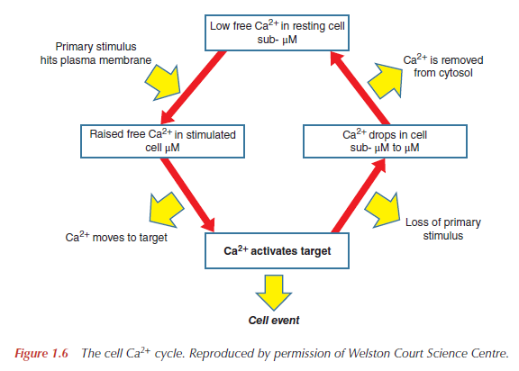
The key feature of Ca2+ as a universal intracellular regulator in animal, plant and microbial cells is that a change in free Ca2+, somewhere inside the cell, must occur prior to a cell event and that this change in free Ca2+ is responsible for initiating the cell event (Figure below).
How intracellular Calcium signaling,  gradient and its role as a universal intracellular regulator points to design The_ce10

It is this which distinguishes the ‘active’ role for Ca2+ as an intracellular regulator from its ‘passive’ role as a cofactor of many proteins. The selection of Ca2+ as a universal intracellular regulator  has depended on two critical features. First, all cells have  mechanisms to maintain a very low cytosolic free Ca2+, with the consequent large gradient of Ca2+ across the plasma membrane, as well as between the inside of organelles such as the ER and the cytosol. These gradients, typically 10 000-fold across the plasma membrane, generate a Ca2+ pressure, which is exploited by physiological and pharmacological stimuli to provoke a cellular event. Secondly, the chemistry of Ca2+ makes it ideal as an intracellular regulator. Oxygen ligands provide high-affinity Ca2+-binding sites which enable Ca2+ to bind, and come off, proteins quickly at micromolar concentrations of Ca2+, in the presence of millimolar Mg2+. Binding of Mg2+ to proteins occurs, but this is not able to produce the necessary structural change to initiate a cellular event nor is it possible to generate the necessary gradient of Mg2+ across membranes that is possible with Ca2+. Cations such as Zn2+, Mn2+, Fe2+/Fe3+ or Cu+/Cu2+ have not been selected because they come off proteins too slowly to allow a bee to buzz or an Olympic athlete to run 100m in less than 10 s. Furthermore, Ca2+ has only one ionisation state, unlike Fe2+/Fe3+ or Cu+/Cu2+, which are involved in redox reactions. It is the special electrical and chemical properties of Ca2+ that have allowed Natural Selection biological cells to generate the electrochemical gradients and intracellular targets which are required to exploit these in such a wide range of biological processes. 

Life evolved emerged in an environment containing many different cations, including Ca2+.  The role that Ca2+ came to play in the early cells may have been conserved during subsequent evolution. The attributes of specific cations suited them to specific roles. Cations of related elements often are associated in pairs, with one needed for intracellular nutrition and the other not used intracellularly. Unlike Mg2+, which functions in many intracellular roles and must be transported inward and carefully regulated, Ca2+ frequently functions extracellularly, rather than intracellularly, in biological processes and is maintained at low intracellular levels by efflux transport pathways. Many of the functions that Ca2+ performs in eukaryotes may therefore be expected to be present in prokaryotes. The common evolutionary origin of the prokaryotes and eukaryotes and the many examples of evolutionary conservation of structure and function that have been shown to exist between them, support the concept that such evolutionary conservation should extend to the role of Ca2+.

Extensive investigations on Ca2+ in eukaryotic cells have shown its important role in signal transduction, as a signal transmitter in membrane depolarization events, as an intracellular second messenger and as an effector of actin–myosin contraction. Recently, however, there has been an increased interest in the role of Ca2+ in prokaryotes and a number of investigations have reported data demonstrating a clear Ca2+ contribution to structure and regulatory functions of the prokaryotic cell. The evidence in favor of Ca2+ as a mediator of regulatory phenomena in prokaryotes continues to accumulate. There is evidence that calcium is involved in a number of bacterial processes such as maintenance of cell structure, motility, cell division, gene expression, and cell differentiation processes such as sporulation, heterocyst formation, and fruiting body development.

Since Ca2+ plays a pivotal role in numerous biological processes in both prokaryotes and eukaryotes, its intracellular concentration must be strictly regulated and maintained to constant values. The free intracellular Ca2+ concentration in bacteria is tightly regulated ranging from 100 to 300 nM, against large changes in external Ca2+concentrations. This is also necessary to avoid toxic effects of free Ca2+ excess and irreversible damage to the cells, such as formation of calcium salts relatively insoluble. Thus the maintenance of low intracellular concentrations of calcium is essential both for the survival of an organism and for calcium to function as a secondary messenger.The ability to control Ca2+ content, or at least intracellular free Ca2+ concentration, may thus be a fundamental attribute of all cells. A wide array of mechanisms participate in attaining such a homeostatic condition. Calcium regulation in bacterial cells comprises influxes and effluxes dependent on active and passive transport mechanisms. Due to the fact that normal intracellular calcium concentrations are usually up to 103 times lower than extracellular concentrations, passive transport usually accounts for calcium influx. A net Ca2+ concentration gradient across the cell membrane is formed which by present-day values would be of the order of 10000 - 100000.  This gradient has to be maintained by continuous exclusion of Ca2+ from the cell. The removal of Ca2+ by active extrusion requires energy to pump the Ca2+ against the electrochemical gradient. The metabolic apparatus that serves this function involves Ca2+ protein-based and non-proteinaceous channels, Ca2+ antiporters (Ca2+/2H+, Ca2+/Na+), and ATP-dependent Ca2+ pumps. ATP-driven calcium efflux appears to play a pivotal role in microbial calcium regulation. Another way to remove Ca2+ is to bind it into a harmless form by Ca2+ chelating materials.

Evolution of calcium homeostasis: From birth of the first cell to an omnipresent signalling system
Specific Ca2+ homeostatic system appeared very early in the history of the cell, as a survival system preventing Ca2+-mediated cell damage. This homeostatic system produced a steep (∼20,000 times) concentration gradient between extracellular and intracellular compartments, which has both survival importance (even relatively short increases in cytosolic Ca2+ concentrations higher then 100 nM are incompatible with life) and signalling function. Evolution Cells utilise  this gradient together with an ability of Ca2+ to interact with many biological molecules to create the most widespread and versatile signalling system, controlling the majority of cellular processes and executing complex routines of intercellular communications.

The first cell, therefore, was defined by a membrane, which contained ion conducting pores and ion pump(s) combined with some source of energy. Failure of any of these components would swamp the cytosol with ocean water and eliminate life forever. “.. Control over Ca2+ ions was the most crucial because Ca2+ ions interact eagerly with biological molecules due to numerous specific properties (flexible coordination chemistry, high affinity for carboxylate oxygen, which is the most frequent motif in amino acids, rapid binding kinetics, etc. At high concentrations, however, Ca2+ causes aggregation of proteins and nucleic acids, affects the integrity of lipid membranes and initiates the precipitation of phosphates. High intracellular Ca2+ concentration therefore is incompatible with life; at all phylogenetic stages, from the most ancient bacteria to the most specialised eukaryotic cells, an increase in cytosolic Ca2+ is always cytotoxic. As a result, the first forms of life, however primitive, required an effective Ca2+ homeostatic system, which maintained intracellular Ca2+ at comfortably low concentrations—somewhere around 100 nM, this being ∼10,000–20,000 times lower than that in the extracellular milieu. Indeed, even the most primitive bacteria are endowed with plasmalemmal Ca2+ pumps.  

The ancient roots of calcium signalling evolutionary tree
Molecular cascades of calcium homeostasis and signalling (Ca2+ pumps, channels, cation exchangers, and Ca2+-binding proteins) emerged in prokaryotes and further developed at the unicellular stage of eukaryote evolution. With progressive evolution, mechanisms of signalling became diversified reflecting multiplication and specialisation of Ca2+-regulated cellular activities. Recent genomic analysis of organisms from different systematic positions, combined with proteomic and functional probing invigorated expansion in our understanding of the evolution of Ca2+ signalling. Particularly impressive is the consistent role of Ca2+-ATPases/pumps, calmodulin and calcineurin from very early stages of eukaryotic evolution, although with interspecies differences. Deviations in Ca2+ handling and signalling are observed between vertebrates and flowering plants as well as between protists at the basis of the two systematic categories, Unikonta (for example choanoflagellates) and Bikonta (for example ciliates). Only the B-subunit of calcineurin, for instance, is maintained to regulate highly diversified protein kinases for stress defence in flowering plants, whereas the complete dimeric protein, in vertebrates up to humans, regulates gene transcription, immune-defence and plasticity of the brain. Calmodulin is similarly maintained
throughout evolution, but in plants a calmoldulin-like domain is integrated into protein kinase molecules. The eukaryotic cell has inherited and invented many mechanisms to exploit the advantages of signalling by Ca2+, and there is considerable overall similarity in basic processes of Ca2+ regulation and signalling during evolution, although some details may vary.

The huge Ca2+ concentration gradient seemingly accompanies every life form from the very first day of its existence. However, its maintenance demands substantial energy consumption. Thus it would have been surprising if evolution had not led to adaptation of the Ca2+ homeostatic system, whose initial function was to protect the cell against massive and incessant “Ca2+ pressure”, for more positive ends. And so it was: the components of mechanism initially designed for survival set the stage for the appearance of the Ca2+ signalling system that triumphantly advanced, through many phylogenetic stages to the most ubiquitous and versatile regulatory machinery cells have ever had in their possession

Calcium binding proteins and calcium signaling in prokaryotes

Ca2+ triggers life at fertilization, and controls the development and differentiation of cells into specialized types. It mediates the subsequent activity of these cells and, finally, is invariably involved in cell death. To coordinate all of these functions, Ca2+ signals need to be flexible yet precisely regulated. This incredible versatility arises through the use of a Ca2+- signalling ‘tool kit’, whereby the ion can act in the various contexts of space, time and amplitude. Different cell types then select combinations of Ca2+ signals with the precise parameters to fit their physiology. 1

Evolution of Ca2+-Binding Sites 2
An essential step in the evolution of Ca2+ signalling was the appearance of Ca2+-binding sites in target proteins. At least four types of high-affinity Ca2+-binding site have been identified, and which satisfy the eight, or sometimes seven, oxygen coordination required for micromolar affinity for Ca2+ in the presence of millimolar Mg2+. These are:

1. EF-hand (type I), first found in parvalbumin, and its modifications in copines and the S-100 family.
2. C2, first found at the C-terminus of synaptotagmin.
3. Types II, III and AB, found in annexin

1. Molecular Biomineralization: Aquatic Organisms Forming Extraordinary Materials, page 127
2. Protein Phylogenetic Analysis of Ca2+/cation Antiporters and Insights into their Evolution in Plants
3. Intracellular Calcium, page 22
4. The evolution of calcium biochemistry
5. Calcium and evolutionary aspects of aging , page 1
6. Calcium homeostasis, page 6
7. Calcium signalling and calcium channels: Evolution and general principles
8. Evolution of the Calcium-Based Intracellular Signaling System
9. Calcium Signaling, ADVANCES IN EXPERIMENTAL MEDICINE AND BIOLOGY page 15
10. https://www.ncbi.nlm.nih.gov/pubmed/25063443
11. https://designmatrix.wordpress.com/2009/01/31/the-calcium-toolkit/
12. https://www.ncbi.nlm.nih.gov/pubmed/18385221
13. https://www.ncbi.nlm.nih.gov/pmc/articles/PMC4355082/
14. https://www.researchgate.net/publication/7657470_Exporting_calcium_from_cells
15. https://academic.oup.com/jxb/article/67/13/3997/1749466/Calcium-impacts-carbon-and-nitrogen-balance-in-the
16. http://onlinelibrary.wiley.com/doi/10.1111/j.1365-3040.2009.02075.x/pdf
17. The functions of Ca2+ in bacteria: a role for EF-hand proteins?
18. Calcium in the Early Evolution of Living Systems: A Biohistorical Approach
19. http://www.sciencedirect.com/science/article/pii/S0092867407015310


Last edited by Admin on Mon Mar 13, 2017 9:53 am; edited 24 times in total

https://reasonandscience.catsboard.com

Otangelo


Admin

Calcium signaling in prokaryotic and eukaryotic cells

Ca2+ homeostasis and signalling in prokaryotes 7
All prokaryotic organisms living today have a low (80 – 100 nMolar) cytosolic free Ca2+ concentration ([Ca2+]i) -  and several systems for Ca2+ extrusion that include plasmalemmal Ca2+ pumps (which are structurally similar to eukaryotic P-type Ca2+ pumps), as well as Ca2+/H+ and Na+/Ca2+ exchangers. The prokaryotic cells also have intracellular Ca2+ signals, reflecting the activation of transmembrane Ca2+ fluxes through Ca2+ selective channels. These channels are, indeed, widespread in prokaryotic organisms, being arguably the most ancient ion channels.

The concentration of Ca2+ increases during perturbation of stimuli, which get recognized by calcium binding proteins or sensor proteins. These proteins further transfer the signal downstream to start phosphorylation cascade that ultimately lead to the regulation of gene expression 1

The modulation in Ca2+ concentration across the cell membrane is basically mediated by three classes of transporters-

1. Ca2+-ATPases (PMCAs),or in other words : plasmalemmal Ca2+ pumps (which are structurally similar to eukaryotic P-type Ca2+ pumps)
2. Ca2+ permeable channels, or in other words : Ca2+ selective channels
3. Ca2+/cation antiporters (CaCAs), or in other words :  Ca2+/H+ and Na+/Ca2+ exchangers 

which function in combination of each other This is a interdependent system !

Evolution of the Calcium-Based Intracellular Signaling System 8 
Early life may have emerged in the ocean or in local parts of it under alkaline conditions that favoured relatively low (in a 100 snM range) Ca2+ concentrations. At this early stage Ca2+ permeation into ancestral cells, Ca2+ handling and Ca2+ influence on energetic (and in particular the requirement of low free Ca2+ for ATP metabolism ) made Ca2+ ions critical for life and for signalling processes. 

Some bacteria express primary and secondary active transporters including P-type Ca2+-transport ATPases of which several resemble the Sarcoplasmic and Endoplasmic Reticulum Ca2+-ATPase (SERCA) of eukaryotes. These pumps, together with 

Mechanosensitive channels
Ca2+-activated channels
3. cation exchangers, such as Ca2+/H+ and Ca2+/Na+ exchangers
an array of Ca2+-binding proteins (CaBP)
and a battery of Ca2+-activated enzymes

which all are present in bacteria  formed the primordial Ca2+ homeostatic and Ca2+ signalling system.

Wow. How could that feat have been reached randomly ?

1. The Plasma Membrane Calcium ATPase 6

Ca2+ ATPases are mostly high-affinity Ca2+ pumps that export the cation from the cytosol to the extracellular environment by using the energy stored in ATP. Two types of ATPases (P- and F-) have been shown to play role in ATP-dependent Ca2+ flux in bacteria and archaea. 9  P-type ATPases form a transient phosphorylated intermediate upon hydrolyzing ATP and thus released energy is used to translocate cations across the membrane. Although most identified P-type ATPases in prokaryotes are putative and their in-vivo functions are poorly characterized, four bacterial P-type ATPases were purified and shown to either translocate Ca2+ or perform Ca2+- dependent phosphorylation.

The wealth of regulatory mechanisms singles it out from all other members of the P-type ion pumps superfamily. The cytosolic C-terminal tail of the protein contains a binding domain for calmodulin, which binds to sites near the active site and maintains the enzyme autoinhibited in the resting state. Calmodulin removes the C-terminal domain from these docking sites, relieving the inhibition. Other pump regulators are the acidic phospholipids of the inner leaflet of the membrane, which are in principle sufficient for 50 % of maximal pump activity. Perhaps the most important structural property that sets the PMCA pump apart from all other members of the superfamily is the presence of a long cytosolic C-terminal tail, which has an essential role in the regulation of the activity of the enzyme: it is the locus of interaction of regulatory partners, chief among them calmodulin, and is the structure responsible for the mechanism of autoinhibition, which is a distinctive properties of the PMCA pump. Another distinctive property is the wealth of regulatory mechanisms, which act with different and still incompletely understood mechanisms. Traditionally, the most important among them have been calmodulin and acidic phospholipids, but others have recently emerged, e.g., protein partners that may interact with the pump in a spatially confined cell environment. The tertiary structure of the PMCA pump is not available, but molecular modeling work on the SERCA pump template (Fig. below ) 

How intracellular Calcium signaling,  gradient and its role as a universal intracellular regulator points to design Pmca_p10

Figure above shows a topology of ten transmembrane domains and three main cytosolic protrusions that correspond to the A, N, and P cytosolic domains of the SERCA pump. It also shows that, as in the case of the SERCA pump, a large conformational change occurs when the pump binds Ca 2+ : the change affects both the transmembrane helices and the cytosolic portion, but is most evident in the latter, which becomes far more “open” in the presence of Ca 2+ . The PMCA pump has long been known to have a unique, unstructured C-terminal cytosolic tail of about 150 residues that contains a canonical calmodulin-binding domain (a second, lower-affinity domain that binds calmodulin and has recently been identified downstream of the first in some splicing variants of the pump). The C-terminal tail also contains consensus sites for two activatory kinases: that for PKA is isoform specific and is located downstream of the calmodulin-binding domain, whereas PKC has two target sites that are not isoform specific, a Thr within the calmodulin-binding domain and a Ser further downstream. One early finding on the PMCA pump which was made before its purification is its activation by acidic phospholipids. 
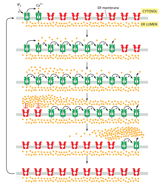
Eukaryotic P-type Ca2+ pumps  PMCA and  SERCA types

Plasma membrane Ca2+ ATPase
The plasma membrane Ca2+ ATPase (PMCA) is a transport protein in the plasma membrane of cells and functions to remove calcium (Ca2+) from the cell. PMCA function is vital for regulating the amount of Ca2+ within all eukaryotic cells. There is a very large transmembrane electrochemical gradient of Ca2+ driving the entry of the ion into cells, yet it is very important that they maintain low concentrations of Ca2+ for proper cell signalling. Thus, it is necessary for cells to employ ion pumps to remove the Ca2+. The PMCA and the sodium calcium exchanger (NCX) are together the main regulators of intracellular Ca2+ concentrations. Since it transports Ca2+ into the extracellular space, the PMCA is also an important regulator of the calcium concentration in the extracellular space.

The Plasma Membrane Ca2+ ATPase and the Plasma Membrane Sodium Calcium Exchanger Cooperate in the Regulation of Cell Calcium
Calcium is an ambivalent signal: it is essential for the correct functioning of cell life, but may also become dangerous to it. The plasma membrane Ca2+ ATPase (PMCA) and the plasma membrane Na+/Ca2+ exchanger (NCX) are the two mechanisms responsible for Ca2+ extrusion. The NCX has low Ca2+ affinity but high capacity for Ca2+ transport, whereas the PMCA has a high Ca2+ affinity but low transport capacity for it. Thus, traditionally, the PMCA pump has been attributed a housekeeping role in maintaining cytosolic Ca2+, and the NCX the dynamic role of counteracting large cytosolic Ca2+ variations (especially in excitable cells). This view of the roles of the two Ca2+ extrusion systems has been recently revised, as the specific functional properties of the numerous PMCA isoforms and splicing variants suggests that they may have evolved to cover both the basal Ca2+ regulation (in the 100 nM range) and the Ca2+ transients generated by cell stimulation (in the μM range).

The Plasma Membrane Ca 2+ ATPases: Isoform Specifi city and Functional Versatility  7
Plasma membrane Ca 2+ ATPases are single polypeptides of about 1100–1250 amino-acid residues with a molecular mass of 125–140 kDa. They contain ten membrane spanning segments and their N- and C-terminals are both on the cytosolic side. The bulk of their mass is also in the cytoplasm and contains three major intracellular domains: the A (actuator), N (nucleotide-binding), and P (catalytic phosphorylation) domains. Four basic isoforms are encoded by four distinct genes, and their transcripts originated a huge number of alternative splicing variants that in most cases are also translated in the corresponding protein variants. Emerging evidence underlines that PMCA pumps, in addition to maintain resting cytosolic Ca 2+ levels against a steep concentration gradient (i.e., nM versus mM), play a local control in specifi c sub-plasma membrane domains by tethering Ca 2+ -/calmodulindependent enzymes and reducing their activity, i.e., by decreasing Ca 2+ concentration in the microenvironment where they are confi ned. This aspect of pump activity confers to PMCA pump a key role as signal transducer and justifi es the existence of so many PMCA variants that could be specialized in tuning the activity of different partners with different Ca 2+ sensitivity.

Ca 2+ controls the most important cell functions in all eukaryotic organisms. Fertilization, muscle contraction, secretion, several phases of metabolism, gene transcription, apoptotic death, etc. are finely orchestrated by the functional versatility of Ca 2+ signaling and its exquisite spatial and temporal regulation. The specificity of cellular Ca 2+ signals depends on the coordinated interplay between numerous soluble Ca 2+ -binding proteins and membrane Ca 2+ transporters which differ both in their mechanism and sensitivity for Ca 2+ handling, in their distribution in the intracellular compartments and in their regulation. Ca 2+ -transporting proteins include ion channels, pumps, and exchangers that drive Ca 2+ ions across the plasma membrane and across the membranes of intracellular organelles. Three differently located Ca 2+ ATPase types (pumps) have been described in animal cells: the sarcoplasmic/endoplasmic Ca 2+ ATPase (SERCA pump) located in the membranes of endo(sarco) plasmic reticulum (including the nuclear envelope), the secretory pathway Ca 2+ ATPase (SPCA pump) in those of the Golgi network, and the plasma membrane Ca 2+ ATPase (PMCA pump) in the plasma membrane. Animal Ca 2+ pump types belong to the family of P-type ATPases. The name comes from their mechanism for Ca 2+ transport: the energy from ATP hydrolysis is conserved in the form of a phosphorylated enzyme intermediate (hence P-type) where ATP phosphorylation of an invariant aspartate residue in a highly conserved sequence S D KTGT[L/I/V/M][T/I/S] allows the translocation of Ca 2+ across the membrane. Structural works on the SERCA pump and the solution of its threedimensional structure have better elucidated the reaction cycle of P-type ATPases. The polypeptide chain of the pump folds in four main domains: one transmembrane domain M (composed by ten transmembrane helices) and three cytosolic domains— the actuator domain A and the phosphorylation domain P (both connected with the M domain) and the nucleotide-binding domain N, which is connected to the domain P. Upon binding of Ca 2+ and its translocation, a series of structural changes involving both the protruding cytoplasmic portion and the transmembrane domain results in the “opening” of the “compact” structure of the cytosolic portion (Fig. 2.1 ).

How intracellular Calcium signaling,  gradient and its role as a universal intracellular regulator points to design Pmca_p11


The mechanism of action is the same for all the Ca 2+ pumps, with the difference that SERCA pump transports two Ca 2+ ions instead of one and that SPCA is also able to transport Mn 2+ in addition to Ca 2+ . Despite of the common mechanism for Ca 2+ transport, the existence of a multitude of variants for each of the three Ca 2+ pumps, either encoded by different genes or generated by alternative splicing mechanisms, suggests that the cell needs to differentiate their action in Ca 2+ extrusion, possibly by activating the proper Ca 2+ pump in a precise moment or cell district. 

P-type ATPases at a glance
P-type ATPases are a large family of integral membrane transporters that are of vital importance in all kingdoms of life. In eponymous distinction from the other main classes of transport ATPase – the F0F1 (F-), the vacuolar (V-) and the ATP-binding cassette (ABC-) type – the P-type ATPases form a phosphorylated (P-) intermediate state during their ion transport cycle. Members of this family generate and maintain crucial (electro-) chemical gradients across cellular membranes, by translocating cations, heavy metals and lipids.

The Ca2+-ATPases of the sarco(endo)plasmic reticulum (SERCA), the plasma membrane (PMCA), and the secretory pathway (SPCA) are crucial for muscle function, Ca2+ signaling and Ca2+ transport into secretory vesicles.

How intracellular Calcium signaling,  gradient and its role as a universal intracellular regulator points to design Panel_16
How intracellular Calcium signaling,  gradient and its role as a universal intracellular regulator points to design Panel_15
How intracellular Calcium signaling,  gradient and its role as a universal intracellular regulator points to design Panel_14
How intracellular Calcium signaling,  gradient and its role as a universal intracellular regulator points to design Panel_17
How intracellular Calcium signaling,  gradient and its role as a universal intracellular regulator points to design Panel_18
How intracellular Calcium signaling,  gradient and its role as a universal intracellular regulator points to design Panel_19
How intracellular Calcium signaling,  gradient and its role as a universal intracellular regulator points to design Panel_20
How intracellular Calcium signaling,  gradient and its role as a universal intracellular regulator points to design Panel_21

General Properties of the Plasma Membrane Ca 2+ Pumps
The PMCA pump has high Ca 2+ affinity and low transport capacity, with a 1:1 Ca 2+ / ATP stoichiometry. It was cloned in 1988, and its sequence revealed the same essential membrane organization and topology properties of the SERCA pump . Later, molecular modeling work based on the structure of the SERCA pump predicts the same general features, with ten transmembrane domains and the large cytosolic  headpiece divided into the three main cytosolic A, N, and P domains (Fig. 2.2 ).

How intracellular Calcium signaling,  gradient and its role as a universal intracellular regulator points to design Topolo10

The catalytic phosphorylation site (SDKTGLT) and other important consensus domains are conserved, but the existence of two prominent domains makes the PMCA pump different with respect to the other two Ca 2+ ATPases. Specifically, a 40-residue-long domain responsible for the binding of activatory phospholipids is present in the first cytosolic loop between transmembrane domains 2 and 3, and a 120-amino-acidlong tail protruding from transmembrane domain 10 and containing the domain that binds calmodulin, i.e., the natural activator of the pump, is present in the C-terminal region.

The Calmodulin Binding Domain of the Plasma Membrane Ca2’ Pump Interacts Both with Calmodulin and with Another Part of the Pump*

 Under nonactivated conditions, the C-terminal tail of the pump is proposed to interact with two sites in the first and second cytosolic loops of the enzyme to maintain the pump auto-inhibited. Calmodulin interacts with its binding domain removing it from the docking sites next to the active center, freeing the pump from autoinhibition. A second calmodulin-binding domain has recently been identified in some splicing variants of the pump, and it has been suggested that, together with the original calmodulin-binding domain, it permits the regulation of the pump both in the nanomolar range and in the micromolar range of Ca 2+ concentration, according to a bi-modular mechanism of control. In addition to calmodulin binding, the PMCA pump has other mechanisms of activation. Among them are the ability of the calmodulin-binding domain to bind also acidic phospholipids, the presence of Ca 2+ -binding motifs upstream and downstream of the calmodulin-binding domain, an oligomerization (polymerization) process involving the calmodulin-binding sequence in the C-terminal tail of the pump, the cleavage by calpain, and phosphorylation by protein kinase C and protein kinase A (the latter only occurs in one of the isoforms). The cleavage by calpain occurs immediately upstream of the C-terminal calmodulin-binding domain  and activates the pump irreversibly, making it calmodulin insensitive. This irreversible mechanism of activation could become significant in conditions of pathological Ca 2+ overload that would demand increased Ca 2+ exporting ability. PKC and PKA consensus sequences have been found in the C-terminal tail of the pump, and regulation of PMCA by PKC has been reported in a variety of cell types. The physiological relevance of the mechanism of phosphorylation is still unclear; however, a number of studies suggest that it could affect various PMCA isoforms and splicing variants in different ways according to their C-terminal sequence characteristics.

Isoforms of the PMCA Pump
In mammals four basic genes ( ATP2B1 – ATP2B4 ) exist, and their transcripts undergo a complex alternative splicing process that increases the total number of isoforms to about 30.
The spliceosome , the splicing code, and pre - mRNA processing in eukaryotic cells 

The plethora of PMCA isoforms: Alternative splicing and differential expression

How intracellular Calcium signaling,  gradient and its role as a universal intracellular regulator points to design Splici10
Fig. 1. 
Topology domains and splicing variants of the human PMCA isoforms. The 10 transmembrane domains of the pump are numbered and indicated by red boxes. Splice sites “A” (first cytosolic loop) and “C” (C-terminal tail) are indicated by red arrows. Splice site “C” lies within the calmodulin-binding domain (yellow cylinder; defined by the structural model of CaM = calmodulin). The exon structure of the different regions affected by alternative splicing is shown for each of the 4 different PMCA genes. Constitutively spliced exons are indicated as dark blue boxes, alternatively inserted exons are shown in light blue; the resulting splice variants are labeled by their lower case symbols, the positions of the translation stop codons for each splice form are indicated by the corresponding capital letters. In PMCA3, splice variant “e” results from a read-through of the 154-nt exon into the following intron (indicated as small open box). The sizes of alternatively spliced exons are given as nucleotide numbers. PL = phospholipid binding domain; P = location of the aspartyl-phosphate formation; PDZ = PSD-95/Dlg/ZO-1 domain. The figure was taken from Fig. 1 of Ref. [24] with permission from the publishers.

The four gene products (isoforms 1–4) differ in tissue distribution and calmodulin affinity. Pumps 1 and 4 are ubiquitous and have lower calmodulin affinity ( K d ~ 30–50 nM), pumps 2 and 3 have higher calmodulin sensitivity ( K d ~ 2–8 nM), and their expression is restricted to some tissues: PMCA2 is expressed prominently in the nervous system and in the mammary gland, PMCA3 in the nervous and muscle system. All the four PMCA transcripts undergo alternative splicing at two sites (site A and site C), thus originating a large number of variants which differ for distribution, interaction with different proteins, and calmodulin affinity.

Why So Many PMCA Variants?
Emerging evidence suggests that in addition to their function as calcium transporters, PMCAs also participate in the regulation of calcium-dependent signal transduction pathways via the interaction with partner proteins. The existence of so many PMCA pump isoforms, including the splice variants, could be rationalized by the finding that they are selectively recruited to plasma membrane compartments/domains by the interaction with specific proteins and that, through a local control of Ca 2+ concentration, they may regulate the activity of enzymes recruited in functional complexes. Thus, the meaning of the interaction is double: by one side, specific interactors engage PMCA to sub-plasma membrane domains, and by the other, the Ca 2+ -ejection properties of PMCA, by maintaining intracellular calcium low in cellular microdomains where the tethering with calcium-dependent signaling proteins occurs, negatively modulate Ca 2+ -sensitive transduction pathways (Fig. 2.3 ).

How intracellular Calcium signaling,  gradient and its role as a universal intracellular regulator points to design Pmca_c10

In agreement with this interpretation, different regulatory interactions have been identified. The identification of some protein partners has, however, only partially reflected differences among isoforms. The preferential site of interaction is the PDZ-binding domain in the C-terminal tail of the b variants. PMCA2 and PMCA4 have been shown to interact with several members of the MAGUK (membraneassociated guanylate kinases, or SAP) family of protein kinases which contain PDZ domains and are associated with the cortical actin cytoskeleton. 

Selection, recruitment, control, regulatory interactions are all actions usually associated with intelligent interveening. For positive interaction or proteins with partner proteins, had they not to be fully setup and functional and fine-tuned one to the other for their beginning of interaction ?  

The data from several groups largely support the view that the PMCA pumps are not uniquely in place to keep resting cytosolic Ca 2+ concentration and counteract Ca 2+ transients generated by cell stimuli and thus turn off activatory signals, but that they can themselves regulate the activity of specifi c enzymatic complexes by locally controlling Ca 2+ environments.

2. Voltage-Dependent Channels at the Plasma Membrane

Voltage-dependent Ca2+-permeable channels have been classified as depolarization-activated Ca2+-permeable channels (DACCs) and hyperpolarization-activated Ca2+-permeable channels (HACCs). Although the properties of DACCs and HACCs are well studied electrophysiologically, the molecular identity of these channels is still unknown. It is assumed that DACCs contribute to the short transient influx of Ca2+ in response to various stimuli, including chilling and microbe interaction . 

Plant annexins are ubiquitous, soluble proteins capable of Ca2+-dependent and Ca2+-independent binding to endomembranes  and the plasma membrane. Surprisingly, a recent study indicated that cytosolic annexins create a Ca2+ influx pathway directly, particularly during stress responses involving acidosis in Zea mays. This suggests that annexins modulate or even represent a part of voltage-gated Ca2+-permeable channels. However, further characterization of plant annexins and especially the molecular identification of voltage-gated Ca2+ channels are required to enable more insights of the contribution of annexins and voltage-gated channels to Ca2+ signaling.

We do not know when or how Ca2+ was used for the first time as a regulatory ion, although we know that this happened very early. Indeed, as already mentioned, a Ca2+ gradient exists in the most primitive prokaryotes (whose cytosolic free Ca2+ is set at about 80–100 nM. . It is not surprising therefore, that the first ion channel which penetrated the plasmalemma with an aqueous pore was, most likely, Ca2+ selective. This is corroborated by the fact that Ca2+ channels are phylogenetically the oldest and, in contrast to other channels, can be found in many prokaryotes. These two polymers form a complex which has all the hallmarks of the Ca2+ channel: it is voltage-activated, it has a characteristic selectivity for divalent cations, it demonstrates classic open-closed state channel behaviour when studied under voltage-clamp and it is inhibited by the transition metal cations La3+, Co2+ and Cd2+.

Prokaryotic voltage-gated Ca2+ channels remain, in a sense, somewhat primitive, as they are constructed from only one domain comprising six transmembrane -helix segments S1–S6

Progress in the structural understanding of voltage-gated calcium channel (CaV) function and modulation
Voltage-gated calcium channels (CaVs) are large, transmembrane multiprotein complexes that couple membrane depolarization to cellular calcium entry. These channels are central to cardiac action potential propagation, neurotransmitter and hormone release, muscle contraction and calcium-dependent gene transcription. Over the past six years, the advent of high-resolution structural studies of CaV components from different isoforms and CaV modulators has begun to reveal the architecture that underlies the exceptionally rich feedback modulation that controls CaV action. These descriptions of CaV molecular anatomy have provided new, structure-based insights into the mechanisms by which particular channel elements affect voltage-dependent inactivation (VDI), calcium-dependent inactivation (CDI) and calcium-dependent facilitation (CDF). 

Structural basis for Ca2+ selectivity of a voltage-gated calcium channel
Voltage-gated calcium (CaV) channels catalyse rapid, highly selective influx of Ca2+ into cells despite a 70-fold higher extracellular concentration of Na+ ( Sodium ). How CaV channels solve this fundamental biophysical problem remains unclear. Here we report physiological and crystallographic analyses of a calcium selectivity filter constructed in the homotetrameric bacterial NaV channel NaVAb. Our results reveal interactions of hydrated Ca2+ with two high-affinity Ca2+-binding sites followed by a third lower-affinity site that would coordinate Ca2+ as it moves inward. At the selectivity filter entry, Site 1 is formed by four carboxyl side chains, which have a critical role in determining Ca2+ selectivity. Four carboxyls plus four backbone carbonyls form Site 2, which is targeted by the blocking cations Cd21 and Mn21, with single occupancy. The lower-affinity Site 3 is formed by four backbone carbonyls alone, which mediate exit into the central cavity. This pore architecture suggests a conduction pathway involving transitions between two main states with one or two hydrated Ca2+ ions bound in the selectivity filter and supports a ‘knock-off’ mechanism of ion permeation through a stepwisebinding process. The multi-ion selectivity filter of our CaVAb model establishes a structural framework for understanding the mechanisms of ion selectivity and conductance by vertebrate CaV channels.

Deciphering Voltage-gated Na+ and Ca2+ Channels By Studying Prokaryotic Ancestors

From NaVAb to CaVAb: structural insight into Ca2+ selectivity
CaV channels have  a selectivity mechanism that allows them to discriminate Ca2+ over the similar-sized Na+, even though the latter is far more abundant in the extracellular solution. How can CaV channels achieve this high ion selectivity while maintaining high-throughput conductance? At the sequence level, CaV channels are closely related to NaV channels, whose ion selectivity can be altered to favor Ca2+ with simple mutations in the selectivity filter. Inspired by early work converting NaChBac to a Ca2+-selective form with such mutations, crystallographic analysis of a similarly engineered NaVAb mutant, known as CaVAb, has provided valuable structural insights into Ca2+ selectivity and permeation in CaV channels.

CaVAb was created by substituting three amino acid residues in the ion selectivity filter of NaVAb (including the HFS site Glu) with Asp (Figure 6a; TLESWSM to TLDDWSD).

How intracellular Calcium signaling,  gradient and its role as a universal intracellular regulator points to design Side_v10
Figure 6. Ca2+ Selectivity and Permeation by CaVAb
(a) Side view of the superimposed selectivity filters of CaVAb and NaVAb. The side chains of the three residues of NaVAb mutated to generate CaVAb are colored in yellow in the CaVAb structure. The backbone structure of the selectivity filters of the two channels are nearly identical. 
(b) Side view of the CaVAb selectivity filter with three Ca2+ (green spheres) binding sites and their coordinating oxygen atoms. The distances between Ca2+ and the oxygen atoms indicated with dash lines range from 4.0 Å to 5.0 Å. 
(c) A proposed mechanism of Ca2+ permeation by CaVAb based on a combination of the “knock-out” and “stepwise permeation” models. The selectivity filter oscillates between two proposed ionic occupancy states where Ca2+ ions either bind to position 2, the high-affinity binding site, or position 1 and 3, which bind the ion at lower affinities.

These substitutions confer Ca2+ selectivity with a permeability ratio of PCa:PNa~400:1, equivalent to mammalian CaV channels. The crystal structure of CaVAb revealed little conformational change in the backbone geometry of the selectivity filter, indicating that ion selectivity is exclusively dictated by the amino acid side chains (Figure 6a)

The selectivity filter was essential to make a calcium gradient and concentration differentiation possible, and the calcium channel functional. Was it not essential that the right order of the amino acid side chains were at the right place right from the start ? A trial and error of random unguided events would require a incalculable number or trials. Why would there be physical pressure to start these trials at all ?!!  

Previous experimental and theoretical studies of mammalian CaV channels have postulated that CaV channels select Ca2+ over Na+ on the basis of affinity and that the high flux of Ca2+ is mediated by a “knock-off” mechanism, in which a second Ca2+ entering the pore “knocks off” a previous resident Ca2+ by charge repulsion. However, this concept was inconsistent with the single high-affinity Ca2+-binding site suggested by careful analyses of mutations and cation block. With Ca2+ included in the crystallization medium, the CaVAb structure clearly revealed three ion-binding sites lining up along the axis of the selectivity filter (Figure 6b). Leading from the entry to the exit of the ion conduction pathway, these three sites are coordinated by a quartet of Asp residues (Site 1), a box of four Asp side chains and four backbone carbonyls of Leu residues (Site 2), and a quartet of backbone carbonyls of Thr residues alone (Site 3). The distances between all three ion-binding sites and their surrounding oxygen atoms suggest that Ca2+ is bound and conducted in hydrated form, in agreement with the 6 Å diameter estimated for the selectivity filter in mammalian CaV channels. Favorable crystal structures revealed waters of hydration surrounding Ca2+ ions. By titrating Ca2+ concentration, the central site of CaVAb was identified as the high affinity site, whereas the third site close to the central cavity showed lowest affinity. Consistent with studies of mammalian CaV channels, divalent cations such as Cd2+ and Mn2+ block CaVAb by occupying only the central high-affinity site .

Voltage-gated sodium (NaV) and calcium (CaV) channels are involved in electrical signaling, contraction, secretion, synaptic transmission, and other physiological processes activated in response to depolarization. Despite their physiological importance, the structures of these closely related proteins have remained elusive because of their size and complexity. Bacterial NaV channels have structures analogous to a single domain of eukaryotic NaV and CaV channels.  New insights into their voltage-dependent activation and inactivation, ion conductance, and ion selectivity provide realistic structural models for the function of these complex membrane proteins at the atomic level.

Voltage-gated NaV channels (NaVs) initiate action potentials in excitable cells and are crucial for electrical signaling from bacteria to man. Voltage-gated CaV channels (CaVs) are activated by depolarization during action potentials, and Ca2+ entry through them initiates synaptic transmission, muscle contraction, hormone secretion, and many other biochemical and physiological processes. These channels are thought to share similar voltage-dependent activation and inactivation processes, whose structural basis is fundamental for electrical signaling. Moreover, how these channels can rapidly and selectively conduct Na+ or Ca2+ ions in response to changes of the electrical membrane potential is a crucial question in biology.

Mammalian NaV channels are complexes of a large α subunit of 260 kDa and smaller β subunits of 30-40 kDa. cDNA encoding the pore-forming α subunits is sufficient for expression of functional NaV channels, whereas the β subunits enhance expression, modulate NaV channel gating, and serve as cell adhesion molecules. NaV channel α subunits are polypeptides of approximately 2000 amino acid residues organized into four homologous domains, each containing six transmembrane segments (Figure 1a). Each homologous domain consists of two functional modules: a voltage-sensing module (VSM) composed of the S1-S4 segments, and a pore-forming module (PM) composed of the S5 and S6 segments and the P loop between them (Figure 1a).

How intracellular Calcium signaling,  gradient and its role as a universal intracellular regulator points to design Two-di10
Figure 1 NaV Channel Structure
(a) Two-dimensional schematic map of NaV channel structure and function. The α subunit of NaV1.2 channels is illustrated as a transmembrane folding diagram in which cylinders represent transmembrane alpha helices and lines represent connecting amino acid sequences in proportion to their length. The roman numerals indicate the four homologous domains and the Arabic numerals are used to label the six transmembrane helices. The S4 helices are colored in red with “+” signs indicating gating charges. The S5-S5 helices are colored in green and the small white circles indicate key residues in the selectivity filter with “+” and “−“ signs indicating their charge states. The yellow circle with an “h” indicates inactivation gate. 
(b) Schematic map of the bacterial NaChBac channel, which contains the minimal functional elements of a single homologous domain in a mammalian NaV channel.


1. Molecular Biomineralization: Aquatic Organisms Forming Extraordinary Materials, page 127

2. Protein Phylogenetic Analysis of Ca2+/cation Antiporters and Insights into their Evolution in Plants

3. Intracellular Calcium, page 22


3. The Ca2+/Cation antiporter (CaCA) superfamily

Electrochemical potential driven Ca2+ transporters are mostly low-affinity Ca2+ transport systems that use the energy stored in the electrochemical gradient of ions. Depending on the gradient, exchangers can operate in both directions (uptake and export). Ca2+/H+ and Ca2+/Na+ antiporters have been identified in a number of bacterial genera and were thought to serve as a major mechanism for Ca2+ transport in prokaryotes.

The CaCA superfamily  proteins are reported in diverse group of organisms from bacteria to higher plants and animals as well. These Calcium:cation antiporter proteins usually facilitate the efflux of Ca2+ against concentration gradient across the membrane, and influx of monovalent cations like H+, Na+, or K+ in exchange. Cation transport is a critical process in all organisms and is essential for mineral nutrition, ion stress tolerance, and signal transduction. 2

How intracellular Calcium signaling,  gradient and its role as a universal intracellular regulator points to design Cation10

Protein Phylogenetic Analysis of Ca2+/cation Antiporters and Insights into their Evolution in Plants

Control of ion concentrations is critical to cellular function. Such ion homeostasis is dependent on transporters, including ion-coupled transporters like the 

YRBG Putative Na+/Ca2+ exchanger
H+/Cation exchanger (CAX), 
K+ exchanger (NCKX), 
cation/Ca2+ exchanger (CCX) and 
Na+/Ca2+ exchanger (NCX), 

This superfamily has five major branches, tentatively named after characterized representative members:

(i) YRBG, comprising largely bacterial members;
(ii) CAX (Ca2+/anion exchanger), comprising mostly plant and yeast members;
(iii) NCX (SLC8)
(iv) NCKX (SLC24), both comprising almost exclusively vertebrate members;  
(v) CCX (Ca2+/cation exchanger) which contains the partially characterized molecule previously called NCKX6 or NCLX (Na+/Ca2+–Li+ exchanger).

How intracellular Calcium signaling,  gradient and its role as a universal intracellular regulator points to design Caca_g11

which are members of the CaCA superfamily. CAX and the prokaryotic-specific YRBG-type exchangers are abundant in bacteria, while NCX, NCKX and CCX genes are abundant in animals.  . These proteins serve as essential components in Ca2+ cycling systems. CaCA proteins promote Ca2+ efflux across membranes, normally against its concentration gradient, by using a counter-electrochemical gradient of other ions such as H+, Na+, or K+ to energize the process. Animal proteins principally use Na+ gradients as the driving force while plant and bacterial exchangers exclusively utilize H+

H+/Cation exchanger (CAX)
Structural basis for the counter-transport mechanism of a H+/Ca2+ exchanger.
Ca(2+)/cation antiporters catalyze the exchange of Ca(2+) with various cations across biological membranes to regulate cytosolic calcium levels. The recently reported structure of a prokaryotic Na(+)/Ca(2+) exchanger (NCX_Mj) revealed its overall architecture in an outward-facing state. Here, we report the crystal structure of a H(+)/Ca(2+) exchanger from Archaeoglobus fulgidus (CAX_Af) in the two representatives of the inward-facing conformation at 2.3 Å resolution. The structures suggested Ca(2+) or H(+) binds to the cation-binding site mutually exclusively. Structural comparison of CAX_Af with NCX_Mj revealed that the first and sixth transmembrane helices alternately create hydrophilic cavities on the intra- and extracellular sides. The structures and functional analyses provide insight into the mechanism of how the inward- to outward-facing state transition is triggered by the Ca(2+) and H(+) binding.
How intracellular Calcium signaling,  gradient and its role as a universal intracellular regulator points to design H_ca2_10

CAX-Af in the archaean Archaeglobus fulgidus  and Yfke in the Gram-positive bacterium Bacillus subtilis. 5 Two of the transmembrane helices in these putative antiporters form a hydrophilic cavity, providing the pathway for exchange. Ca2+ binding is typically via glutamate residues. These three-dimensional structures shed light on the evolution of the CaCA family of Ca2+ transporters and exchangers. However, what is still required is a correlation of exchange with cytosolic free Ca2+ measured in the live cell and its loss in knock-out mutants. Cation exchange in vesicles is encouraging but not sufficient proof that it occurs in the live cell. Even bacterial proteins 30–50% similar to Ca2+ signalling proteins in eukaryotes have turned out to be red herrings. Furthermore, where there is good evidence for a role of Ca2+ in a particular bacterial species, often the data has been obtained fromjust one group. In contrast, important data on Ca2+ signalling in animal and plant cells has been repeated by dozens of groups. Similarly, the failure to followup potentially important proteins which could regulate Ca2+-dependent processes with measurements of free Ca2+ on live cells, and molecular genetic andmutational studies, to establish their precise functional role has seriously held
back the field.

Isolation and Functional Characterization of Ca2+/H+ Antiporters from Cyanobacteria
CAXs have in general 10–14 transmembrane (TM)-spanning domain with about 400 amino acid residues. CAXs contain a central hydrophilic motif rich in acidic amino acid residues that bisect the polypeptide into two approximately equal segments. Four Arabidopsis CAXs (AtCAX1–4) were identified by their ability to sequester Ca2+ into yeast vacuoles in Saccharomyces cerevisiae mutants deleted of the vacuolar Ca2+-ATPase and Ca2+/H+ antiporter (ScVCX1). It was shown that AtCAX1, AtCAX3, and AtCAX4 specifically transport Ca2+, whereas AtCAX2 transports Ca2+, Mn2+, and Cd2+ . In these vacuolar type AtCAXs, the presence of an N-terminal autoinhibition domain and a 9-amino-acid region required for Ca2+ transport (Ca2+ domain) has been reported. By contrast, little is known about H+-coupled Ca2+ efflux antiporters. Hitherto, a plasma membrane Ca2+/H+ antiporter gene (chaA) has only been isolated from Escherichia coli. In ChaA, neither an N-terminal autoinhibition domain nor a 9-amino-acid region was reported. ChaA has been shown to catalyze both Na+/H+ and Ca2+/H+ exchange reactions at alkaline pH. Essentially nothing is known about molecular properties of Ca2+/H+ antiporters from other organisms, especially those on plasma membranes.

YRBG Putative Na+/Ca2+ exchanger
Characterization and Purification of a Na+ /Ca2+ Exchanger from an Archaebacterium*
The Internal Repeats in the Na+/Ca2+Exchanger-related Escherichia coli Protein YrbG Have Opposite Membrane Topologies*
How intracellular Calcium signaling,  gradient and its role as a universal intracellular regulator points to design C-term10
Figure 1
A, LFASTA alignment of the N- and C-terminal halves of YrbG. The predicted transmembrane segments areunderlined, and the conserved α-motifs areboxed. Note that each α-motif consists of two parts separated by a short, less well conserved loop. B, proposed topology for YrbG. The two homologous halves are indicated byblack and hatched transmembrane segments, respectively, and the two α-motifs are shown by thick lines. The number of Lys + Arg residues is indicated in eachloop. Each fusion is indicated by its number; the exact position of each fusion joint is given in the legend to Fig.2.

We have determined the topology of theEscherichia coli inner membrane protein YrbG, a putative Na+/Ca2+ exchanger with homology to a family of eukaryotic ion exchangers. Our results show that the two homologous halves of YrbG both have five transmembrane segments but opposite membrane orientations. This has implications for our understanding of the function of Na+/Ca2+ exchangers and provides an example of “divergent” evolution of membrane protein topology.


Structural basis for alternating access of a eukaryotic calcium/proton exchanger 3

Early evolution of Ca2+ signalling
https://www.ncbi.nlm.nih.gov/pmc/articles/PMC4037395/#R8

1. https://www.ncbi.nlm.nih.gov/pmc/articles/PMC5124604/
2. https://www.ncbi.nlm.nih.gov/pmc/articles/PMC4735048/
3. http://www.nature.com/nature/journal/v499/n7456/abs/nature12233.html
4. https://www.researchgate.net/publication/225074667_Protein_Phylogenetic_Analysis_of_Ca2cation_Antiporters_and_Insights_into_their_Evolution_in_Plants
5. Intracellular Calcium , page 441
6. Regulation of Ca2+- ATPases,VATPases and F-ATPases, page 3
7. Sajal Chakraborti Naranjan S. Dhalla ,  Regulation of Ca2+- ATPases,VATPases and F-ATPases, page 13
8. Evolution of the Calcium-Based Intracellular Signaling System
9. Calcium binding proteins and calcium signaling in prokaryotes

Further readings:
TYPES OF ION CHANNELS KNOWN
Ca2+/Calmodulin-Dependent Protein Kinases Mediate Many Responses to Ca2+ Signals


Last edited by Admin on Mon Mar 13, 2017 8:52 am; edited 32 times in total

https://reasonandscience.catsboard.com

4How intracellular Calcium signaling,  gradient and its role as a universal intracellular regulator points to design Empty Types of Intracellular Ca2+ Signal Tue Mar 07, 2017 3:21 pm

Otangelo


Admin

Types of Intracellular Ca2+ Signal

When a Ca2+channel opens, a highly concentrated plume of Ca2+ forms around its mouth and then dissipates rapidly by diffusion after the channel has closed. Such localized signals, which can originate from channels in the plasma membrane or on the internal stores, represent the elementary events — the basic building blocks of Ca2+ signalling 1  The spatio-temporal properties of these elementary events, such as Ca2+ sparks and Ca2+ puffs, differ depending on the nature and location of the channels. g (Fig. 1a) 

By characterizing these signals, we can discover how the Ca2+- signalling repertoire is elaborated. Essentially, these elementary signals have two functions. They can either activate highly localized cellular processes in the immediate vicinity of the channels (Fig. 1a) or, by recruiting channels throughout the cell, they can activate processes at a global level (Fig. 1b, c). The subcellular location of Ca2+ channels is crucial for targeting elementary signals to different cellular processes. In smooth muscle, for example, Ca2+ sparks that arise locally, near the plasma membrane, activate potassium (K+ ) channels (Fig. 1a), causing the muscle to relax. But when elementary release events deeper in the cell are coordinated to create a global Ca2+ signal, the muscle contracts.

This is a striking example of how spatial organization enables Ca2+ to activate opposing cellular responses in the same cell.

This raises the question how the spacial organisation emerged. Trial and error ? Had not both, the location of the channels that coordinate the elementary release events deeper in the cell , and a global Ca2+ signal, which induces the muscle to contract , and  the location of the channels that activate the signals that activates potassium, causing the muscles to relax, to be at the right place together, in order to get both required movements at the same time ? one movement  without the other would make no sense. That is, a muscle would be able to contract, but not relax, and vice-versa. Isn't that clear evidence that the whole mechanism had to be setup right from the beginning, with every channel at the right place ?  

Time
One of the paradoxes surrounding Ca2+ is that it is a signal for both life and death — although elevations in Ca2+ are necessary for it to act as a signal, prolonged increases in the concentration of Ca2+ can be lethal. Cells
avoid death either by using low-amplitude Ca2+ signals or, more usually, by delivering the signals as brief ‘transients’. These principles apply to both elementary and global signals.

That raises another interesting question : How was the coordination setup the first time ? trial and error ? If trials would not produce the right concentration, but a prolonged one, the cell dies. Did the cell die every time the dosage was not finely tuned to low amplitude ?  Isn't that a clear indication that gradual step-wise evolutionary development of the mechanism would not be possible, but only a all or nothing setup ?

Single transients are used to activate certain cellular processes, such as secretion of cellular material in membrane bound vesicles, or muscle contraction. However, when information has to be relayed over longer time periods, cells use repetitive signals known as Ca2+ oscillations. Both the elementary events and the global signals can oscillate, but they have widely different periods. For example, whereas the period of elementary Ca2+ sparks in arterial smooth muscle is 0.1–0.5 seconds, it is 10–60 seconds for global waves in liver cells, 1–35 minutes for Ca2+ waves in human eggs after fertilization, and 10–20 hours for the spontaneous Ca2+ transients that control cell division.

The duration of the signals had to be preprogrammed, and follows the information transmission rules of encoding, code sending, and decoding , which can only be setup by intelligence.

Cells use frequency modulation (FM) to vary the intensity and nature of the physiological output. For instance, arteries can be made to dilate by increasing the frequency of Ca2+ sparks, which cause the smooth muscle lining the arteries to relax . And by varying the frequency of global Ca2+ signals, different genes can be activated. To use FM signalling, cells have decoders that respond to the frequency and longevity of the Ca2 signals. Probably the best-known example is an enzyme called calmodulin-dependent protein kinase II, which is found in both animal and plant cells and which regulates other enzymes that rely on Ca2+. It works by ‘counting’ Ca2+ transients  and varying its activity accordingly.

How did it "learn" that feat ? Had this mechanism not to be pre-programmed ? Since when can mindless matter learn to count and understand informational signals ? 

The enzyme is composed of many identical subunits, and these are activated to varying degrees depending on the frequency of the Ca2+ oscillations.

Amplitude
Information can also be encoded in the amplitude of Ca2+ signals. Such amplitudemodulated (AM) signalling is generally considered to be less reliable than that based on frequency, owing to the difficulties of detecting small Ca2+ changes above the background level. However, it has been shown that cells can interpret modest changes in the concentration of Ca2+. For example, different genes can be activated by varying the amplitude of Ca2+ signals.

Fertilization and development
In mammals, life begins at fertilization when the sperm interacts with the egg to trigger a Ca2+ oscillation that persists for several hours. This prolonged period of repetitive Ca2+ pulses triggers the developmental programme by stimulating the enzymatic machinery involved in the cell-division cycle. There are no further changes in Ca2+ until the one-cell embryo is ready to divide, when a spontaneous Ca2+ transient triggers cleavage to form two daughter cells. There are indications that this orderly programme may be controlled by two distinct oscillators — Ca2+ signals and oscillating levels of proteins involved in the cell-division cycle. The Ca2+ oscillator seems to be the main mechanism, because it persists when dissociated from the cell-cycle oscillator. Just what drives this Ca2+ oscillator, with a period of 10–20 hours, is a mystery, but it may depend on periodic increases in the level of a diffusible intracellular messenger called inositol 1,4,5-trisphosphate (Ins(1,4,5)P3) with each division. In many cells, hormones and growth factors activate the enzyme phospholipase C, which catalyses the production of InsP3 from the membrane lipid phosphatidylinositol 4,5- bisphosphate (Box 1). As embryos grow and groups of cells differentiate to perform specialized functions, Ca2+ signalling contributes to body polarity and pattern formation.For instance, in amphibians and zebrafish it is thought to help specify which cells will form structures at the top (dorsal) or the bottom (ventral) part of the embryo. As cells differentiate to perform different functions, they select out those components of the Ca2+-signalling tool kit that best fit their remit. This versatility is emphasized by growing evidence that
Ca2+ controls cellular processes as diverse as cell proliferation and the neuronal plasticity that is responsible for learning and memory. But at any moment, any of these orderly signalling events can be switched to activate a programme that leads to cell death — a big challenge for the future is to understand how Ca2+ suddenly transforms from a signal for life to a signal of death.

This is highly relevant, as it shows that Ca2+ signaling makes part of the toolkit of bodyform formation, and if body form has to change, it depends on the change of Ca2+ signaling  as well. Another epigenetic mechanism that refutes macro-evolution. There is a big challenge if naturalistic origins are proposed for the phenomena in question, but when design is proposed, its perfectly explainable that the designer pre - programmed the life cycle of cells, that is cell cycle, fertilization, cell differentiation, spacio-temporal patterning,  timing and amplitude of the signals, and regulation. 

Cell death
Very high concentrations of Ca2+ can lead to the disintegration of cells (necrosis) through the activity of Ca2+-sensitive protein-digesting enzymes. Calcium has also been implicated in the more orderly programme of cell
death known as apoptosis. Apoptosis is important during both normal development (the formation of tissue patterns, for example) and pathological conditions such as AIDS, Alzheimer’s disease and cancer. A protein
that is mutated in cancerous cells, called Bcl-2, prevents the cell death that would normally limit the survival and proliferation of cancer cells. Bcl-2 mediates some of its antiapoptotic action by modifying the way in
which organelles such as the endoplasmic reticulum and mitochondria (where respiration occurs) handle Ca 2+ (Box 1).

How intracellular Calcium signaling,  gradient and its role as a universal intracellular regulator points to design Ca2_si10

How intracellular Calcium signaling,  gradient and its role as a universal intracellular regulator points to design Ca2_si11


For sites of elementary Ca2+ release to produce global responses, the individual channels must communicate with each other, to set up Ca2+ waves (Fig. 1b). If cells are connected, such intracellular waves can spread into neighbouring cells and become intercellular waves to coordinate cellular responses within a tissue (Fig. 1c).

In virtually all cell events provoked by a rise in intracellular Ca2+, there is movement of Ca2+ in and out of organelles inside the cell, as well as movement of Ca2+ from outside into the cell, and then out again. There are essentially three categories of cells that use a Ca2+ signal to provoke a cellular event:

• Type 1: the main source of Ca2+ for the global rise in cytosolic free Ca2+ is from outside the cell.
• Type 2: the main source of Ca2+ for the global rise in cytosolic free Ca2+ is from inside the cell.
• Type 3: the source of Ca2+ for the global rise in cytosolic free Ca2+ is a sum of Ca2+ from both outside and inside the cell.

Cytosolic free Ca2+ signals vary enormously between cell types, the type of signal matching the physiology of the cell event. There are four main types of cellular Ca2+ signal (Figure 2.2).

How intracellular Calcium signaling,  gradient and its role as a universal intracellular regulator points to design Types_10

The cytosolic free Ca2+ can rise and fall quickly, it can plateau for some time before returning to the level of a resting cell, it can oscillate, and it can appear as small puffs or sparks. Transient cytosolic free Ca2+ signals can be a very short spike of milliseconds duration (e.g. in a nerve terminal) or they can last just a few seconds or so (e.g. in the beating heart). In these cases, the cytosolic free Ca2+ returns to the basal level after each stimulus. In contrast, longer rises in cytosolic free Ca2+ occur in other cells, where the Ca2+ signal can remain at a plateau level, well above that in the resting cell, for many seconds or even minutes. A further interesting feature of the global cellular Ca2+ signal in some cells is that it oscillates. This was first seen in the nerves of the marine sea slug Aplysia, where the action potential generates a series of spikes followed by a resting period. This occurs because the action potential opens voltage-sensitive Ca2+ channels in the plasma membrane. Each spike leads to a gradual rise in cytosolic free Ca2+. This Ca2+ activates K+ channels, which repolarises the cell, the voltage-gated Ca2+ channels close and the pacemaker cell stops firing. Depolarisation then initiates the firing process to start again. Another type of Ca2+ oscillation was first discovered in hepatocytes, using aequorin to monitor the cytosolic free Ca2+ in individual cells. In the presence of the hormonal primary stimulus (e.g. vasopressin), the cytosolic free Ca2+ oscillated for many minutes. The mechanism and reason for this has still not been fully elucidated, but it would seem obvious that it is a way of maintaining a long-term cell event, such as glycogen breakdown through activation of phosphorylase, without depleting the cell of Ca2+. It has been suggested that cells which produce oscillations in cytosolic free Ca2+ have a frequency detector (i.e. the cell response depends on the frequency of the Ca2+ signals). Attractive as this idea might sound, there is little direct evidence for it at present. Imaging of cytosolic free Ca2+ in live cells, using first the photoprotein aequorin  and now more often using fluorescent dyes such as fluo-3 , has shown that in fact many Ca2+ signals are localised within the cell, particularly when observing them during a timecourse of cell stimulation. Four main types of spatial Ca2+ rise have been identified (Figure 2.3):

How intracellular Calcium signaling,  gradient and its role as a universal intracellular regulator points to design Cytoso10

1. Tiny localised clouds – sparklets, sparks, puffs, scintilla, also called syntilla , and blinks.
2. Large rises in microdomains.
3. Tides that move to fill up the cell.
4. Waves that move through the cell.

Tiny localised clouds are usually insufficient in themselves to activate the Ca2+ target proteins, unless there are sufficient small events which sum up to produce a large Ca2+ signal. They have been given various names by the workers who discovered them in a particular cell. Sparklets arise as a result of opening of Ca2+ channels in the plasma membrane, whereas sparks, puffs and scintilla arise as result of a small localised release of Ca2+ from the sarcoplasmic reticulum (SR) or ER. Thus, a sparklet was found as a rise in cytosolic free Ca2+ as a result of opening of voltage-operated Ca2+ channels (VOCs) in ventricular heart cells

1. Calcium—A life and death signal


Last edited by Admin on Sun Mar 12, 2017 7:06 pm; edited 7 times in total

https://reasonandscience.catsboard.com

5How intracellular Calcium signaling,  gradient and its role as a universal intracellular regulator points to design Empty Calcium signaling pathway Sat Mar 11, 2017 3:39 am

Otangelo


Admin

Calcium signaling pathway 1

Ca2+ that enters the cell from the outside is a principal source of signal Ca2+. Entry of Ca2+ is driven by the presence of a large electrochemical gradient across the plasma membrane. Cells use this external source of signal Ca2+ by activating various entry channels with widely different properties. The voltage-operated channels (VOCs) are found in excitable cells and generate the rapid Ca2+ fluxes that control fast cellular processes. There are many other Ca2+-entry channels, such as the receptor-operated channels (ROCs), for example the NMDA (N-methyl-D-aspartate) receptors (NMDARs) that respond to glutamate. There also are second-messenger-operated channels (SMOCs) and store-operated channels (SOCs). The other principal source of Ca2+ for signalling is the internal stores that are located primarily in the endoplasmic/sarcoplasmic reticulum (ER/SR), in which inositol-1,4,5-trisphosphate receptors (IP3Rs) or ryanodine receptors (RYRs) regulate the release of Ca2+. The principal activator of these channels is Ca2+ itself and this process of Ca2+-induced Ca2+ release is central to the mechanism of Ca2+ signalling. Various second messengers or modulators also control the release of Ca2+. IP3, which is generated by pathways using different isoforms of phospholipase C (PLCbeta, delta, epsilon, gamma and zeta), regulates the IP3Rs. Cyclic ADP-ribose (cADPR) releases Ca2+ via RYRs. Nicotinic acid adenine dinucleotide phosphate (NAADP) may activate a distinct Ca2+ release mechanism on separate acidic Ca2+ stores. Ca2+ release via the NAADP-sensitive mechanism may also feedback onto either RYRs or IP3Rs. cADPR and NAADP are generated by CD38. This enzyme might be sensitive to the cellular metabolism, as ATP and NADH inhibit it. The influx of Ca2+ from the environment or release from internal stores causes a very rapid and dramatic increase in cytoplasmic calcium concentration, which has been widely exploited for signal transduction. Some proteins, such as troponin C (TnC) involved in muscle contraction, directly bind to and sense Ca2+. However, in other cases Ca2+ is sensed through intermediate calcium sensors such as calmodulin (CALM).

Calcium signaling pathway - Reference pathway
How intracellular Calcium signaling,  gradient and its role as a universal intracellular regulator points to design Calciu11

How intracellular Calcium signaling,  gradient and its role as a universal intracellular regulator points to design Ca2_si13
...................How intracellular Calcium signaling,  gradient and its role as a universal intracellular regulator points to design Ca2_si14


Evolution of the Calcium-Based Intracellular Signaling System
The calcium-signaling toolkit is made up of different multidomain proteins . In response to an extracellular stimulus the concentration of cytosolic free calcium ions increases from its resting level of around 100 nMolar to in the region of 1 mMolar (  1 micro (µ) is equal 1000 nano (n) )  . The increase in the concentration of cytosolic free calcium is typically fuelled by a combination of calcium influx through calcium-permeable channels and release of calcium from intracellular stores, such as the endoplasmic reticulum. This latter route typically involves the participation of other intermediary molecules (such as inositol,1,4,5 trisphosphate) that couple the perception of the extracellular stimulus (at the plasma membrane) to the intracellular stores. The intracellular environment contains a myriad of proteins that are able to bind nanomolar concentrations of calcium. Their properties change after binding calcium and these changes are responsible for coupling the increase in calcium (the calcium signal) to downstream reactions that culminate in the response to the primary stimulus. The cell also contains mechanisms to “switch-off” the calcium signal and these center on removing the calcium from the cytosol. The suite of proteins responsible for generating the intracellular calcium signal, responding to it and finally switching it off, have been termed the “calcium toolkit” by Berridge et al. (2003). Although the eukaryotic calcium-based intracellular signaling system has been the subject of intense investigation for the past 40 years, we know rather less about calcium signaling in prokaryotes. However, the fact that it has been implicated in the control of cell division, virulence, and biofilm formation suggests that calcium and more particularly calcium-based signaling is important in these organisms.  Given the central role of intracellular calcium signaling in the living world, a better understanding of the evolution of this calcium-signaling toolkit, and the proteins that comprise it, is crucial to our global understanding of how cells emerged. Some  highlight the high conservation of the calcium toolkit from prokaryotes to metazoa and the increasing complexity of the proteins that make it up. The proteins that comprise the calcium-signaling toolkit are composed of modular domains. These domains, limited in number,  produce  the functionally diverse repertoire of proteins in a genome.  

How intracellular Calcium signaling,  gradient and its role as a universal intracellular regulator points to design Compon10

Last Eukaryote Common Ancestor
The recent study of the ciliated protozoan Paramecium showed that calcium signaling was already present in organisms at the unikonts–bikonts split (Plattner 2015). Our results showed the LECA was indeed potentially already able to generate and decode calcium signals as the domain architecture content of LECA included representatives from all of the main components of calcium signaling, including organelle specific Ca+-binding architectures from ancient endosymbiosis events.  Reconstruction of the domain architecture content of the LECA reveals the presence of representatives from all of the main components of calcium signaling.

Calcium Signals: The Lead Currency of Plant Information Processing 2

Ca2+ signals are core transducers and regulators in many adaptation and developmental processes of plants. Ca2+ signals are represented by stimulus-specific signatures that result from the concerted action of channels, pumps, and carriers that shape temporally and spatially defined Ca2+ elevations. Cellular Ca2+ signals are decoded and transmitted by a toolkit of Ca2+ binding proteins that relay this information into downstream responses. Major transduction routes of Ca2+ signaling involve Ca2+-regulated kinases mediating phosphorylation events that orchestrate downstream responses or comprise regulation of gene expression via Ca2+-regulated transcription factors and Ca2+-responsive promoter elements. Here, we review some of the remarkable progress that has been made in recent years, especially in identifying critical components functioning in Ca2+ signal transduction, both at the single-cell and multicellular level. Despite impressive progress in our understanding of the processing of Ca2+ signals during the past years, the elucidation of the exact mechanistic principles that underlie the specific recognition and conversion of the cellular Ca2+ currency into defined changes in protein–protein interaction, protein phosphorylation, and gene expression and thereby establish the specificity in stimulus response coupling remain to be explored.

Calcium (Ca2+) likely represents the most versatile ion in eukaryotic organisms. It is involved in nearly all aspects of plant development and participates in many regulatory processes. Because of its flexibility in exhibiting different coordination numbers and complex geometries, Ca2+ can easily form complexes with proteins, membranes, and organic acids. On the one hand, this feature renders Ca2+ a toxic cellular compound at higher concentrations because it would readily form insoluble complexes with phosphate (as present in ATP), but on the other hand, the required tight spatial and temporal control of cellular Ca2+ concentration may have paved the way for the evolutionary emergence of Ca2+ signaling.

Defined changes of cytosolic Ca2+ concentration are triggered by cellular second messengers, such as NAADP, IP3, IP6, Sphingosine-1-Phospate, and cADPR  and it is evident that the identity and intensity of a specific stimulus impulse results in stimulus-specific and dynamic alterations of cytosolic Ca2+ concentration.  This heterogeneity of increases in cytosolic-free Ca2+ ion concentration in terms of duration, amplitude, frequency, and spatial distribution lead A.M. Hetherington and coworkers to formulate the concept of “Ca2+ signatures”. Signal information would be encoded by a specific Ca2+ signature that is defined by precise control of spatial, temporal, and concentration parameters of alterations in cytosolic Ca2+ concentration. 2

The spectrum of stimuli that evoke such Ca2+ elevations and their stimulus-specific characteristics has been cataloged and critically discussed in a number of informative reviews.  Subsequent research suggested that while the shape and spatio-temporal distribution of Ca2+ elevations could be of critical importance for stimulus response coupling an additional level of regulation and specificity is achieved by Ca2+ binding proteins that function as signal sensor proteins.  These proteins decode and relay the information encoded by Ca2+ signatures into specific protein–protein interactions, defined phosphorylation cascades, or transcriptional responses.

FUNCTIONS OF CA2+ SIGNALING
Ca2+ is involved in various responses to abiotic and biotic stimuli, including light, high and low temperature, touch, salt and drought, osmotic stress, plant hormones, fungal elicitors, and nodulation factors. These stimuli induce a distinct spatio-temporal pattern of changes in cytosolic-free Ca2+ concentration ([Ca2+]cyt). Single-cell systems, such as guard cells, growing pollen tubes, or root hairs, represent excellent models to investigate primary and autonomous Ca2+ responses. However, the final response of the plant to external stimuli is manifested by regulation of complex growth processes in distinct tissues and organs. Concurrently to the diversity of stimulus-specific Ca2+ signatures at the single-cell level, differentiation gives rise to another layer of cell type–specific Ca2+ responses in tissues or organs. This additional level of complexity may contribute to more diversity in local or systemic responses. Therefore, research on plant Ca2+signaling has taken advantage of single-cell model systems but in parallel moves forward to elucidate Ca2+ dynamics in the tissue context and in the whole organism. Consequently, here, we review and discuss how Ca2+ signatures contribute to signaling processes at the single-cell level and the multicellular level.

GENERATION OF CA2+ SIGNALS
A Ca2+ signal is defined by the balanced activation of Ca2+ channels at different cellular membranes, which is followed by the subsequent inactivation of channels and activation of efflux transporters to terminate Ca2+ influx and to rebalance the cellular Ca2+ homeostasis. Both processes are strictly regulated and define the physiological outcome of Ca2+ signaling.

Influx of Ca2+
Distinct ion channel types capable of mediating Ca2+ fluxes coexist in different cell types and tissues. According to their activation mechanism, these channels can be classified as voltage-dependent, voltage-independent/ligand-dependent, and stretch-activated Ca2+ channels. Depending on their specific activation properties, Ca2+ channels can shape the parameters of Ca2+ influx and the resulting Ca2+ signature. This enables the plant to translate a wide range of different signals into distinct Ca2+ signatures. Moreover, variability in the specific abundance of the different channel types likely reflects the special needs of a cell type or tissue . A schematic summary of the channels and transporters that are discussed in this review is illustrated in Figure 2.

How intracellular Calcium signaling,  gradient and its role as a universal intracellular regulator points to design Ca2_in10

Figure 2. Overview of Ca2+ Transport Systems in an Arabidopsis Cell.
Shown are Ca2+ influx/efflux pathways that have been identified at the molecular level. See text for further details. CNGC, cyclic nucleotide channel; GLR, glutamate receptor; TPC1, two pore channel 1; CAS, Ca2+-sensing receptor; ACA, autoinhibited calcium ATPase; ECA, ER type calcium ATPase; HMA1, heavy metal ATPase1; CAX, cation exchanger.

Efflux of Ca2+
Much of the research on Ca2+ signaling has focused on advancing our understanding of the generation of Ca2+ elevations by Ca2+-releasing channels. However, to represent a distinct signal, the regulation of Ca2+ efflux that not only terminates but also shapes the Ca2+ signature is as important as the Ca2+-releasing events. During the past years, we have witnessed significant insights into the regulation of Ca2+ extrusion. However, in the future, it will be most important to reveal the interconnected regulation of Ca2+ release and extrusion that is required to generate defined signals.

Extrusion of Ca2+ from the cytosol is achieved by P-type Ca2+-ATPases and by the Ca2+/proton antiporter systems. While antiporters mediate a high-affinity low turnover efflux, ATPases mediate a low-affinity high-capacity efflux. Therefore, it is assumed that antiporters reduce the Ca2+ concentration back to a few micromolar after signal mediated influx, while Ca2+-ATPases are important to maintain the low resting concentration of Ca2+. The coordinative regulation of the cellular extrusion systems still is not fully understood. Transport activity is clearly activated after influx of Ca2+, as in response to different hormones, salt stress, or mechanical stimulation.


1. http://www.genome.jp/dbget-bin/www_bget?path:map04020
2. https://www.ncbi.nlm.nih.gov/pmc/articles/PMC2861448/


Last edited by Admin on Mon Mar 13, 2017 10:02 am; edited 2 times in total

https://reasonandscience.catsboard.com

Otangelo


Admin

Open questions :

Overall, the acquired so far knowledge on Ca2+ transport in prokaryotes is still limited and raises several important points.  Prokaryotes contain multiple mechanisms for Ca2+ translocation as well as multiple homologs of Ca2+ transporters. Is there a centralized regulatory network and how does it relate to Ca2+ signaling?

It is likely that Ca2+ transporters play a dual role by protecting cells from Ca2+ toxicity as well as maintaining steep Ca2+ gradients across cellular membrane required for signaling. What is the mechanistic and regulatory relationship between Ca2+ transporters and Ca2+ signaling?

Overall, different aspects of Ca2+signaling role in prokaryotic physiology have been demonstrated, however direct experimental evidence of intracellular signaling events connecting [Ca2+]i transients,their amplitude and rate to the regulatory outcomes are still to be discovered. Characterization of intracellular Ca2+ signaling has been challenged by technical difficulties of monitoring intracellular Ca2+.

Although we know a massive amount about Ca2+ as a universal regulator in eukaryotic cells, how it is regulated and how it works, there is still a lot of detail to work out. It has been proposed that the type of oscillation in cytosolic free Ca2+, and that within an organelle, provides a signal where the cell recognises both the frequency and amplitude of the Ca2+ signal. There is then an analogue-to-digital converter which tells the cell when it should fire. Attractive as this hypothesis is, much more data is required to support it, particularly in situ. In fact, there is still much to learn about intracellular free Ca2+ signals in intact organs and organisms, as opposed to cells in tissue culture. There are also uncertainties about the interaction of intracellular Ca2+ with other signals, such as cyclic ADP ribose, NAADP and sphingosine 1-phosphate. Precisely how Ca2+ signals are transmitted from cell to cell is also still not fully understood via gap junctions and other mechanisms. Transcription factor pathways can be activated by a rise in cytosolic and/or nuclear Ca2+. An important one is the calcineurin–NFAT pathway. But interactions of intracellular Ca2+ with other gene activation pathways, such as those involving mitogen-activated protein kinase (MAPK), c-fos, jun, src and so on, are not so well defined. Although there is much evidence for rises in cytosolic free Ca2+ being involved in several events during the cell cycle, it is still not clear how a cell doubles its contents required for cell division. There is a special Mg2+ TRP channel which opens to enable a cell to double its Mg2+, but how the ER doubles its Ca2+ is not known. This problem applies also to other ions such as K+, Na+ and phosphate.

Although much less is known about the role of intracellular Ca2+ in bacteria, it is clearly important for bacteria, like all eukaryotic cells, to maintain a low free Ca2+ internally. But why this is and precisely how this low intracellular free Ca2+ is maintained, and whether the Ca2+ pressure is used to generate Ca2+ signals in bacteria, is still not universally clear.

A major problem, of which we still understand little, is how the Ca2+ signalling system started and how it evolved?

What is a species and how does a new species really appear? In fact, Darwin and Wallace never really  addressed fully this last question. But we now know that a mouse cannot mate with an elephant, not because of size, but because the DNA just will not mix. Yet, different orchid species can form fertile  hybrids and even the Galapagos finches can breed with each other to produce fertile offspring.We need a new concept to understand the pathway of Rubicons that ultimately leads to the appearance of a new  species and how intracellular Ca2+ fits into this.

The vast array of cellular processes controlled by intracellular Ca2+ argues strongly that the molecular variations in the Ca2+ signalling system must have been crucial to the evolution of all species, but we
know virtually nothing of how this occurred and nor can we see precisely how such small variations could have had a selective advantage.

So, in summary there is much still to learn about:
1. The precise details of how a cell generates a Ca2+ wave, tide or oscillation.
2. The precise details of how a Ca2+ signal in a eukaryotic cell triggers a cell event.
3. Intracellular Ca2+ as a regulator in bacteria and archaeans.
4. How intracellular Ca2+ mediates events in plants and fungi.
5. How intracellular Ca2+ integrates with other signalling networks in whole organs and organisms.
6. The precise role of intracellular Ca2+ in many diseases and pathological processes.
7. How the intracellular Ca2+ signalling system evolved and what role it played in key steps in the evolution of the three main cell types: Bacteria, Archaea and Eukaryota, and in particular the molecular biodiversity in the Ca2+ signalling system.


Last edited by Admin on Sun Mar 12, 2017 10:16 pm; edited 2 times in total

https://reasonandscience.catsboard.com

Otangelo


Admin

Ca2+ Functions as a Ubiquitous Intracellular Mediator

Calcium in the Early Evolution of Living Systems: A Biohistorical Approach 
How intracellular Calcium signaling,  gradient and its role as a universal intracellular regulator points to design Ca210

Many extracellular signals, and not just those that work via G proteins, trigger an increase in cytosolic Ca2+ concentration. In muscle cells, Ca2+ triggers contraction, and in many secretory cells, including nerve cells, it triggers secretion. Ca2+ has numerous other functions in a variety of cell types. Ca2+ is such an effective signaling mediator because its concentration in the cytosol is normally very low (~10–7 M), whereas its concentration in the extracellular fluid (~10–3 M) and in the lumen of the ER [and sarcoplasmic reticulum (SR) in muscle] is high. Thus, there is a large gradient tending to drive Ca2+ into the cytosol across both the plasma membrane and the ER or SR membrane. When a signal transiently opens Ca2+ channels in these membranes, Ca2+ rushes into the cytosol, and the resulting 10–20-fold increase in the local Ca2+ concentration activates Ca2+ responsive proteins in the cell. Some stimuli, including membrane depolarization, membrane stretch, and certain extracellular signals, activate Ca2+ channels in the plasma membrane, resulting in Ca2+ influx from outside the cell. Other signals, including the GPCR-mediated signals described earlier, act primarily through IP3 receptors to stimulate Ca2+ release from intracellular stores in the ER (see Figure 15–29). The ER membrane also contains a second type of regulated Ca2+ channel called the ryanodine receptor (so called because it is sensitive to the plant alkaloid ryanodine), which opens in response to rising Ca2+ levels and thereby amplifies the Ca2+ signal, as we describe shortly. Several mechanisms rapidly terminate the Ca2+ signal and are also responsible for keeping the concentration of Ca2+ in the cytosol low in resting cells. Most importantly, there are Ca2+-pumps in the plasma membrane and the ER membrane that use the energy of ATP hydrolysis to pump Ca2+ out of the cytosol. Cells such as muscle and nerve cells, which make extensive use of Ca2+ signaling, have an additional Ca2+ transporter (a Na+-driven Ca2+ exchanger) in their plasma membrane that couples the efflux of Ca2+ to the influx of Na+.

Feedback Generates Ca2+ Waves and Oscillations

The IP3 receptors and ryanodine receptors of the ER membrane have an important feature: they are both stimulated by low to moderate cytoplasmic Ca2+ concentrations. This Ca2+-induced calcium release (CICR) results in positive feedback, which has a major impact on the properties of the Ca2+ signal. The importance of this feedback is seen clearly in studies with Ca2+-sensitive fluorescent indicators, such as aequorin or fura-2 , which allow researchers to monitor cytosolic Ca2+ in individual cells under a microscope.When cells carrying a Ca2+ indicator are treated with a small amount of an extracellular signal molecule that stimulates IP3 production, tiny bursts of Ca2+ are seen in one or more discrete regions of the cell. These Ca2+ puffs or sparks reflect the local opening of small groups of IP3-gated Ca2+-release channels in the ER. Because various Ca2+-binding proteins act as Ca2+ buffers and restrict the diffusion of Ca2+, the signal often remains localized to the site where the Ca2+ enters the cytosol. If the extracellular signal is sufficiently strong and persistent, however, the local Ca2+ concentration can reach a sufficient level to activate nearby IP3 receptors and ryanodine receptors, resulting in a regenerative wave of Ca2+ release that moves through the cytosol (Figure 15–31), much like an action potential in an axon.

How intracellular Calcium signaling,  gradient and its role as a universal intracellular regulator points to design Dfgf11

Positive and negative feedback produce Ca2+ waves and oscillations. This diagram shows IP3 receptors and ryanodine receptors on a portion of the ER membrane: active receptors are in green; inactive receptors are in red. When a small amount of cytosolic IP3 activates a cluster of IP3 receptors at one site on the ER membrane (top), the local release of Ca2+ promotes the opening of nearby IP3 and ryanodine receptors, resulting in more Ca2+ release. This positive feedback (indicated by positive signs) produces a regenerative wave of Ca2+ release that spreads across the cell. These waves of Ca2+ release move more quickly across the cell than would be possible by simple diffusion. Also, unlike a diffusing burst of Ca2+ ions, which will become more dilute as it spreads, the regenerative wave produces a high Ca2+ concentration across the entire cell. Eventually, the local Ca2+ concentration inactivates IP3 receptors and ryanodine receptors (middle; indicated by red negative signs), shutting down the Ca2+ release. Ca2+-pumps reduce the local cytosolic Ca2+ concentration to its normal low levels. The result is a Ca2+ spike: positive feedback drives a rapid rise in cytosolic Ca2+, and negative feedback sends it back downagain. The Ca2+ channels remain refractory to further stimulation for some period of time, delaying the generation of another Ca2+ spike (bottom). Eventually, however, the negative feedback wears off, allowing IP3 to trigger another Ca2+ wave. The end result is repeated Ca2+ oscillations . Under some conditions, these oscillations can be seen as repeating narrow narrow waves of Ca2+ moving across the cell.


Another important property of IP3 receptors and ryanodine receptors is that they are inhibited, after some delay, by high Ca2+ concentrations (a form of negative feedback). Thus, the rise in Ca2+ in a stimulated cell leads to inhibition of Ca2+ release; because Ca2+ pumps remove the cytosolic Ca2+, the Ca2+ concentration falls . The decline in Ca2+ eventually relieves the negative feedback, allowing cytosolic Ca2+ to rise again. As in other cases of delayed negative feedback (see Figure 15–18), the result is an oscillation in the Ca2+ concentration. These oscillations persist for as long as receptors are activated at the cell surface, and their frequency reflects the strength of the extracellular stimulus

How intracellular Calcium signaling,  gradient and its role as a universal intracellular regulator points to design Dfgfsd10

Picture above. Vasopressin-induced Ca2+oscillations in a liver cell. The cell was loaded with the Ca2+-sensitive protein aequorin and then exposed to increasing concentrations of the peptide signal molecule vasopressin, which activates a GPCR and thereby PLCβ . Note that the frequency of the Ca2+ spikes increases with an increasing concentration of vasopressin but that the amplitude of the spikes is not affected. Each spike lasts about 7 seconds.

The frequency, amplitude, and breadth of oscillations can also be modulated by other signaling mechanisms, such as phosphorylation, which influence the Ca2+ sensitivity of Ca2+ channels or affect other components in the signaling system. The frequency of Ca2+ oscillations can be translated into a frequency-dependent cell response. In some cases, the frequency-dependent response itself is also oscillatory: in hormone-secreting pituitary cells, for example, stimulation by an extracellular signal induces repeated Ca2+ spikes, each of which is associated with a burst of hormone secretion. In other cases, the frequency-dependent response is non-oscillatory: in some types of cells, for instance, one frequency of Ca2+ spikes activates the transcription of one set of genes, while a higher frequency activates the transcription of a different set. How do cells sense the frequency of Ca2+ spikes and change their response accordingly? The mechanism presumably depends on Ca2+-sensitive proteins that change their activity as a function of Ca2+-spike frequency.
A protein kinase that acts as a molecular memory device seems to have this remarkable property, as we discuss next.

The evolution of calcium biochemistry
http://www.sciencedirect.com/science/article/pii/S0167488906002540

https://reasonandscience.catsboard.com

Otangelo


Admin

Frequency decoding of calcium oscillations
https://sci-hub.tw/https://www.sciencedirect.com/science/article/pii/S0304416513005163

Calcium (Ca2+) oscillations are ubiquitous signals present in all cells that provide efficient means to transmit intracellular biological information. Either spontaneously or upon receptor-ligand binding, the otherwise stable cytosolic Ca2+ concentration starts to oscillate. The resulting specific oscillatory pattern is interpreted by intracellular downstream effectors that subsequently activate different cellular processes. This signal transduction can occur through frequency modulation (FM) or amplitude modulation (AM), much similar to a radio signal. The decoding of the oscillatory signal is typically performed by enzymes with multiple Ca2+ binding residues that diversely can regulate its total phosphorylation, thereby activating cellular program. To date, 

NFAT, Nuclear factor of activated T-cells (NFAT)
NF-κB, (nuclear factor kappa-light-chain-enhancer of activated B cells)
CaMKII, Ca 2+ /calmodulin-dependent protein kinase II
MAPK,   mitogen-activated protein kinase (MAPK or MAP kinase)
calpain, a protein belonging to the family of calcium-dependent, non-lysosomal cysteine proteases (proteolytic enzymes) 

have been reported to have frequency decoding properties. A limited number of cellular frequency decoding molecules of Ca2+ oscillations have yet been reported. Interestingly, their responsiveness to Ca2+ oscillatory frequencies shows little overlap, suggesting their specific roles in cells. Frequency modulation of Ca2+ oscillations provides an efficient means to differentiate biological responses in the cell, both in health and in disease. Thus, it is crucial to identify and characterize all cellular frequency decoding molecules to understand how cells control important cell programs.

The key question in the field of calcium (Ca2+) signaling is by what means this simple ion can regulate such a wide spectrum of cellular processes, including fertilization, proliferation, differentiation, muscle contraction, learning and cell death. The answer to this question most certainly lies in the huge spatial and temporal diversity of the signal, since a Ca2+ response can exhibit infinite patterns.  Through an intricate concert of action between several Ca2+ transporters in the cell the cytosolic Ca2+ concentration can start to oscillate, much like a radio signal. Specific information can thereby be efficiently encoded in the signal and transmitted through the cell without harming the cell itself. Already at the beginning of life, when the sperm injects phospholipase C-ζ into the egg, a slow Ca2+ oscillatory wave is traveling the egg that triggers fertilization . Transmitting information using oscillating waves can occur by means of amplitude (AM) or frequency modulation (FM). Ca2+ oscillations are never totally homogenous, but rather have intrinsic variations in oscillation parameters such as frequency and amplitude. 

Information encoding 
Cells are constantly exposed to extracellular stimuli, e.g., 

mitogens, A mitogen is a peptide or small protein that induces a cell to begin cell division: mitosis.
cytokines, Cytokines are a broad and loose category of small proteins (~5–20 kDa) important in cell signaling.
hormones A hormone (from the Greek, "setting in motion") is any member of a class of signaling molecules, produced by glands in multicellular organisms
neurotransmitters, Neurotransmitters are endogenous chemicals acting as signaling molecules that enable neurotransmission.

that translate into changes in the cytosolic Ca2+ concentration. In addition, environmental cues causing temperature and mechanical stress can result in activation of Ca2+ signals. During development, the otherwise stable cytosolic Ca2+ level starts to oscillate spontaneously due to largely unknown reasons. Depending on the stimuli, that differentially affect Ca2+ channels and pumps as well as Ca2+ binding proteins, a unique Ca2+ signal is induced. The process of building up a unique signal that can be associated with a specific stimulus is called information encoding.

https://reasonandscience.catsboard.com

Otangelo


Admin

“Evolution of the Calcium-Based Intracellular Signaling System” published in Genome Biology and Evolution in 2016 by Oats, et al
https://www.ncbi.nlm.nih.gov/pmc/articles/

https://www.ncbi.nlm.nih.gov/pmc/articles/PMC4987107/?fbclid=IwZXh0bgNhZW0CMTAAAR12Z4POGdaYM11kodc-Tvlzl7aYCWXpMaoiaklP6_QtuzKD7zdo19UZIeQ_aem_j5rcjCOjIadHTOz7nz-htQ

https://reasonandscience.catsboard.com

Sponsored content


Back to top  Message [Page 1 of 1]

Permissions in this forum:
You cannot reply to topics in this forum