ElShamah - Reason & Science: Defending ID and the Christian Worldview
Would you like to react to this message? Create an account in a few clicks or log in to continue.
ElShamah - Reason & Science: Defending ID and the Christian Worldview

Welcome to my library—a curated collection of research and original arguments exploring why I believe Christianity, creationism, and Intelligent Design offer the most compelling explanations for our origins. Otangelo Grasso


You are not connected. Please login or register

Understanding Ontogenetic Depth: Naming Versus Measuring

Go down  Message [Page 1 of 1]

Otangelo


Admin

Understanding Ontogenetic Depth: Naming Versus Measuring 1

http://reasonandscience.heavenforum.org/t2308-understanding-ontogenetic-depth-naming-versus-measuring

I was supposed to do this a year ago -- well, long before that, too -- but a glacier passed me on the interstate, and then I ran out of gas, got so depressed that I threw my notes into a box, and...oh, never mind. Let's get started.
After the second entry in this series (part II), we'll open up the comments section for your responses. Complete citations and additional reading will also be compiled at the end of part II.
1. Introduction: Why A Biological Distance That's Currently Impossible to Measure, Ontogenetic Depth, Nevertheless Really Matters to Evolutionary Theory


The theory of natural selection provides a mechanistic, causal account of how living things came to look as if they had been designed for a purpose.

-- Richard Dawkins (1982, 45)
Natural selection tells us absolutely nothing about underlying mechanisms of genomic changes, or their consequences on developmental changes which lead to evolutionary innovations. In short, it is silent about the emergence of complex forms.
-- George L.G. Miklos (1993, 24)
How can a theory of evolution that purports to explain how creatures with trillions of cells arose from multicellular beginnings be taken seriously if all it tells us is that differential rates of destruction can alter the genetic composition of populations?
-- Wallace Arthur (2004, 36)
A strong conclusion is that the evolutionary process generating the genomic programs responsible for the developmental formulation of basic eumetazoan body plans was in many ways very different from the evolutionary changes that can be observed at the species level in modern animals.
-- Eric Davidson and Douglas Erwin (2010, 1)


Those statements tell you where this ontogenetic depth (OD) blog series is going. Here's my thesis: The theory of evolution by natural selection does not explain the origin of animal form, because natural selection cannot account for origin de novo of the developmental stages required to construct (i.e., evolve) animals. The concept of OD helps us to understand why.
But first I have to eat some Corvus brachyrhynchos, partly served up 7 years ago by P.Z. Myers, but mostly stumbled on by me shortly thereafter, when I tried to make the first formulation of OD work. I am grateful to Myers for his original ammonia-capsule-under-the-nose critique, and for keeping the pressure on since then, including his annual April 7 lampoon, "Paul Nelson Day."
As I shall explain in part II, however, I don't think Myers grasps the severity of the problem posed by ontogenetic depth. As evidence, I'll describe a long conversation he and I had at the Society for Developmental Biology 2004 meeting, in Calgary, at a session where I was presenting a poster. I'm quite sure Myers remembers the conversation and a little picture he drew for me on a piece of scrap paper.
But that's for later. To start, I want to show why OD is easy to understand -- in one sense, anyway -- but, at the moment, nigh-well impossible to measure. Mmm, yummy crow feathers. The punch line after the feathers, however, may be counterintuitive to readers, but here it is anyway: not being able to give an exact value to a distance doesn't mean one can ignore it in constructing theories or explaining transitions. For the macroevolution of animals de novo to occur via natural selection, ontogenetic depth must be traversed. This is an inescapable, and currently unsolved, problem. And every improvement in measuring OD will only make the problem more challenging.

2. A Useful Distinction: Naming versus Measuring

Right now, the skies are clear in Chicago. Looking into the southeastern sky, I can see the sun, some distance away in space.
Now, let's suppose that we're looking at the sun 500 years ago, before Kepler, Newton, Huygens, and Cassini. We know that the sun is some measurable distance from the Earth, and we can give that distance a name. Call it the Earth-to-Sun distance, or, as it now known, the Astronomical Unit (AU; see Figure 1).


Understanding Ontogenetic Depth: Naming Versus Measuring Earth%20to%20sun%20distance
Figure 1
So how far away, exactly, is the Sun from the Earth? Five hundred years ago, we didn't have telescopes, radar, satellites, or any of the other instruments currently used to measure the AU. But we did have geometry, and could grasp the concept of parallax. It should be fairly easy, using naked-eye observations of the Sun and the planets, to estimate the exact value of the AU.
Not really. As the history of astronomy shows, measurements (or estimates) of the AU, attempted since antiquity, didn't settle on anything close to the modern value until the late 18th century. The data required to perform the triangulation depicted in Figure 2, accurately, by employing the Transit of Venus, were simply not available, or were known too imprecisely to be useful.


Understanding Ontogenetic Depth: Naming Versus Measuring Measuring%20AU
Figure 2
Does that mean the concept of the AU is unintelligible? Of course not. The difficulties we face in measuring a distance do not entail that the distance itself is unreal or unimportant. The difficulties just mean that the distance may be tricky to specify in exact terms.
3. Ontogenetic Depth: Naming versus Measuring
Segue to ontogenetic depth. Here is my original formulation (2003, 459):


Ontogenetic depth estimates the distancein terms of cell division and differentiationbetween a unicellular condition and the macroscopic adult metazoan capable of reproduction.


The words in bold define, or name, a distance. The words in italics, by contrast, represent an attempt (and a poor one at that; thanks, Prof. Myers, for showing as much) to measure that distance. If the reader can grasp this distinction, namely, between defining or naming a distance, versus measuring it, much of the confusion surrounding ontogenetic depth dissolves away. The important point to remember is this: the distance (OD) is real, and must be traversed by any candidate evolutionary process, whether we can measure OD precisely or not.
Consider a parallel to defining versus measuring the AU. Figure 3 shows OD as the distance between a fertilized C. elegans egg, and the reproductively capable adult phenotype. We can think of this like looking at the Sun, and knowing it is some distance [finite interval] away in space.


Understanding Ontogenetic Depth: Naming Versus Measuring Naming%20distance%20egg
Figure 3
Figure 4, by contrast, posits that cell division and differentiation (i.e., cell types) provide the correct units for measuring -- i.e., giving an exact value -- to OD:
Understanding Ontogenetic Depth: Naming Versus Measuring Measuring%20distance%20egg
Figure 4
See the difference? Anyone would agree that there's a distance between egg and adult in C. elegans. The question is how best to measure that distance, in units (or metrics) that allow comparison with other animal groups. In his original critique, Myers gave some reasons for thinking that the metrics of cell type and cell division wouldn't work. But he undersold the difficulty, as I'll illustrate next.
Here's the cell lineage (see Figure 5) of Toy Organism Alpha, with 16 cells of 5 types. Four rounds of cell division separate any cell in the adult from the starting point, the fertilized egg. A simple metric of OD for this toy organism would multiply the average number of cell divisions, between the egg and any cell in the adult (4), times the total number of cell types (5, with the red cell as the germ line producing gametes), to yield an OD of 20.
Understanding Ontogenetic Depth: Naming Versus Measuring Toy%20Organism%20Alpha
Figure 5
But compare Toy Organism Alpha to Toy Organism Beta. While they have the same OD -- if we use the simple metric of 4 rounds of cell division X 5 cell types = 20 -- their respective adult forms are very different. Beta has a large internal cavity, and places its "gonad" (the red cell) within the wall of that cavity, whereas Alpha's gonad is exterior. The positions of other cells differ as well.
Understanding Ontogenetic Depth: Naming Versus Measuring Toy%20Organism%20Beta
Figure 6
If reproductive capability depends (at least in part) on precise form, as it does for any real animal, then measuring ontogenetic depth will require specifying the three-dimensional positions and relationships of individual cells or tissues. This positional information cannot be captured either by cell division (lineages) or cell type, and providing a metric for it will be enormously more difficult.
"So why bother about a distance that cannot yet be measured?" the reader is probably asking. "What's the point?"
4. The Theory of Evolution by Natural Selection Itself Requires That We Explain Ontogenetic Depth
That's why. As I'll explain in Part II, using the process of natural selection in evolutionary explanation places strict evidential demands on the investigator. Evolving the goal-directed structures of metazoan development de novo raises challenges that natural selection -- because the process lacks foresight -- may in principle be incapable of explaining.

http://www.evolutionnews.org/2011/04/understanding_ontogenetic_dept045531.html


Last edited by Admin on Sun 27 Mar 2016 - 13:41; edited 5 times in total

https://reasonandscience.catsboard.com

Otangelo


Admin

Understanding Ontogenetic Depth, Part II: Natural Selection Is a Harsh Mistress

Longer post today. Bear with me. [For a little background, be sure to read Understanding Ontogenetic Depth, Part I.]
1. What Does "Traversing Ontogenetic Depth" Mean?
Let's look again at Toy Organism Alpha. Figure 1 depicts a complete cell lineage -- "complete" in the sense that the starting point is a single cell, and the end point, a reproductively capable adult. Adults, by definition, can produce the specialized cells (gametes), which, when fertilized, start the whole process of cell division and differentiation again.


Once upon an evolutionary time, this lineage did not exist. (Alpha is only an illustration, of course; I mean cell lineages such as are found in real animals, which are vastly more complicated.) It had to be constructed, incrementally, by some undirected process. Under textbook neo-Darwinian theory, that process was descent with modification via the natural selection of randomly-arising variation. I'll refer to this theory by the shorthand "natural selection."Traversing ontogenetic depth means simply that any candidate evolutionary process (natural selection, genetic drift, self-organization, whatever) must build across -- "traverse" -- a distance of increasing complexity, from an aboriginal single-celled state, to multicellularity, to a genuine dividing and differentiating lineage. That distance cannot be crossed in a single increment of change.
Now, before we consider what natural selection requires, let's strip down Alpha's cell lineage to just the first two rounds of cell division, yielding four cell types:


Figure 2
This will provide our model for the problem of traversing ontogenetic depth via natural selection. Recall that the process of natural selection (Endler 1986) has three jointly necessary and sufficient conditions, which may be expressed by the following conditional:

If, within a species or population, the individuals
a. vary in some attribute or trait q -- the conditions of variation;
b. leave different numbers of offspring in consistent relation to the presence or absence of trait q -- the condition of selection differences;
c. transmit the trait q faithfully between parents and offspring -- the condition of heredity;
then the frequency of trait q will differ predictably between the population of all parents and the population of all offspring.


Could this process have constructed the mini-lineage in Figure 2? Let's see. We're going to try to traverse ontogenetic depth, step-by-step, via an undirected process. We can set aside for the moment condition (b), selective differences, and focus just on conditions (a) and (c), variation and heredity.
The starting point will be a single cell -- call it Cell Zero -- which divides:


But that won't work for building the lineage, unless the daughter cells remain physically connected. So Cell Zero needs the instruction, (1), "divide and stick together." Obviously, this instruction must be in place before Cell Zero divides.


Now we have two daughter cells, but they're identical. Unless we provide another instruction to Cell Zero, however, we're going to produce a clonal mass of identical cells: no differentiation will occur, which we need to construct the lineage. Cell Zero therefore needs another instruction, (2), "daughter cells differ," which again must be in place before Zero divides.


Then the two daughters divide, yielding four cells of four different types. Have we successfully constructed the lineage via natural selection?
No; to this point, natural selection has caused nothing, because one of its necessary conditions, heredity, has not been satisfied. This organism (of four differentiated cells) must leave progeny, to which it transmits its distinctive variations. Condition (c) of the process of natural selection, heredity, requires that variations arising in a parent be transmitted to offspring. Heredity thus entails that at least one of four cells keep track of the instruction set for the lineage as a whole, if the cycle of differentiation is going to repeat (i.e., be maintained) in the next generation.
Cell Zero therefore needs yet another instruction, (3), in place before it divides: "One cell line must keep track of the whole instruction set, and become the starting point for another cycle of differentiation." This is the functional reason animals need something like a germ line (i.e., cells which produce gametes): those cells will be the instruction-minders, or instruction-carriers, for the organism as a whole.


Is natural selection operating yet? Nope: thus far, we're building an egg, namely, Cell Zero, by loading it with the functionally necessary instructions that must be in place before it divides. Reproduction of the whole lineage, which will enable selective differences (condition b) to arise, is causally well downstream. Whatever instructions must be present in Cell Zero, therefore, have to be there BEFORE natural selection can operate.


We've just traversed a relatively short interval of ontogenetic depth (OD). And the process of natural selection, harsh mistress that she is, stands entirely to one side, indifferently watching us do the work of building an egg. Not until we have produced a lineage capable of replicating itself with fidelity can natural selection begin to operate.
2. Try This Yourself, With a Pencil and a Sheet of Paper: P.Z. Myers Already Gave Me His Solution, Which Doesn't Work
There's nothing especially biological about all this. Figures 3 through 7 depict a decision tree, and what would be required, as instructions in the starting node (Cell Zero) to specify that tree and cycle it through successive generations. You can build such trees yourself, to any degree of complexity your patience will tolerate. Try to find the minimal set of instructions required in the starting node to generate the differentiated end-nodes, and to repeat the process from any one of those end-nodes. Keep in mind that, if you want the whole lineage to repeat ("reproduce") with fidelity, any decision affecting any end-node must be front-loaded into the starting node.
In late July 2004, at the Society for Developmental Biology annual meeting, held at the University of Calgary, P.Z. Myers and I talked about this very problem, at length. Myers stopped by my poster (which he pronounced wretched, by the way) and we found ourselves discussing what the "Urbilaterian" -- the putative ancestor of the bilaterally symmetrical animal phyla -- might have resembled. (In my poster, I argued that no one had successfully described Urbilateria because it is impossible to do, an argument I endorse even more strongly today.) Since Urbilateria, if it existed, would have been a metazoan and undergone a developmental process of its own, Myers and I wrangled about how that process would have evolved de novo -- just the problem we've been sketching above.
Myers told me I had greatly exaggerated what was a boringly simple puzzle. To illustrate his point, he drew me a picture (which I still have somewhere, I think), that looked like this:


"It's no real problem, Paul," Myers said. "Start with a eukaryote that becomes colonial. The cells of the colony start specializing, maybe a central cavity forms, and then some of the cells repress their gene expression and become a kind of germ line for the rest of the colony, which is evolving into a true animal. No big deal."
I have to admit when Myers told this story, he expressed it with such aplomb that, at the time, I could only smile. But as I contemplated his drawing, I realized what he had sketched could not possibly work in any realistic evolutionary scenario.
Indeed, it then struck me that I had seen Myers' scenario many times before. Evolutionary developmental biologist Lewis Wolpert -- whom no one, even in his wildest delirium, would ever mistake for an ID theorist -- had long critiqued the scenario on functional grounds, using what he called "the continuity principle." (1994) The continuity principle requires that any change occurring in an evolutionary transformation be biologically possible, that is, viable and stably heritable in the next generation.
"Since embryonic development requires the formation of a multicellular organism from a single cell," Wolpert observes (1994, 79), "the origin of the egg is a central and sadly neglected problem." The functional challenge to be solved is the origin of heritable differentiation:

The key to all development is the generation of differences between the cells, that is, making them non-equivalent [see Figures 1 and 2, above]. Only if the cells are different can the organism be patterned so that there are organized changes in shape, and cells at specific sites differentiate into different cell types. How could this have evolved? (Wolpert 1994, 80)

Look again at Figure 8, and think about what would need to happen between the "colonial" stage and the origin of the germ line (the red cell at the third stage). What mechanism is coordinating gene expression among all the members of the colony, such that only one cell lineage will evolve to carry the complete instruction set required to specify the form of the whole? How are mutations -- occurring in all individual cells of the colony -- transmitted to the next generation? If individual cells continue to reproduce via normal fission, or budding, notes Wolpert, "cell lineages [will be] mutating in all sorts of directions in genetic space." (2002, 745) Given such genetic chaos, he argues, "we consider it practically impossible" for the collection of cells to "yet retain the ability to evolve into viable new forms."
To modify irreversibly the global form of any animal, Wolpert contends, requires mutations affecting a single key cell -- the egg. Mutations affecting somatic cells -- especially when every cell, during the transition from colonial organism to true metazoan, is possibly either "somatic" or "germ line" -- cannot be coordinated:

There is no way that the genes in the huge number of cells...can change at the same time, and mutations in individual cells mean that they no longer share the behavioural rules of the majority. It is only through a coherent developmental programme, with all cells possessing the same genes, that organisms can evolve, and this requires an egg. (2002, 745)

At the moment, the problem of the evolutionary origin of cellular differentiation stands open, for the reasons outlined above. "How cell types of multicellular organisms came to be differentiated," noted Carl Schlichting (2003, 98), "is still an open issue." The problem, I think, cannot be solved within a neo-Darwinian (natural selection) framework, given that selection as a causal process cannot fix variants whose selective differences (condition b of selection) lie in the future. Building de novo the distantly end-directed pathways that characterize animal development, however, seems to require fixing just such variants.
To recapitulate my thesis, then:

The theory of evolution by natural selection does not explain the origin of animal form, because natural selection cannot account for origin de novo of the developmental stages required to construct (i.e., evolve) animals. The concept of ontogenetic depth helps us to understand why.

There's a lot more to say, but this outlet doesn't favor the long form. Let me end, however, with one final point, which I'm hoping P.Z. Myers will address.
3. How Does Early Development in Animals Evolve?
It is unclear how ontogenetic architectures early in metazoan history could have differed fundamentally from present-day systems. The problem may be summarized as follows:

-- There are striking differences in the early development in animals, even within classes and orders.
-- Assuming that these animals are descended from a common ancestor, these divergences suggest that early development evolves relatively easily.
-- Evolution by natural selection requires heritable variation.
-- But heritable variations in early development, in major features such as cleavage patterns, are not observed.


Extant species provide many case studies in the puzzle of modifying early development. Remarkable examples of early ontogenetic divergence abound (e.g., in nematodes; Felix 1999; Schierenberg 2001). These divergences have now become a commonplace of the evo-devo literature, and are taken as prima facieevidence that early development evolves dramatically. "It is clear that a casualty of [these comparative data]," argues Davidson (1990, 384), "is the 19th century concept that early development must be an evolutionarily conserved process" -- a concept, interestingly enough, which Davidson himself advocated in 1971: "One can imagine modest alterations or additions to the early parts of the developmental program," he wrote at the time, "but it would be very unlikely that such programs could be supplanted." (1971, 131)
There exists a striking paucity of experimental evidence showing heritable variation in what Wimsatt and Schank (1988) call the "deeply-entrenched" features of metazoan development. Why is such deep variation so hard to find? Again, we should ask, what does the logic of natural selection require?
A. Variation in a trait q
B. Fitness differences in consistent relation to the presence of trait q, and
C. Heritability of trait q.
The experimental literature on model systems such as Drosophila describes many mutations in early developmental characters and patterns. With rare exceptions, however, such mutations are not heritable, in the sense that the phenotypes exhibited do not survive as stably-breeding lines. As a result, some have argued that we should not expect mutagenesis to reveal the basis of adaptive variation: "The take home message," argues Nagy (1998, 820), "is that mutagenesis in model systems does not undo evolution or reveal, in any direct fashion, the basis of evolutionary change."
But if the experimental literature does not provide evidence of heritable deep variation, how do we know that such changes are even possible? "Comparative embryology abounds," argue Wray and McClay (1989, 811), "with empirical evidence of evolutionary modification of early development." The theory of common descent, of course, underwrites the assumption. Because the animals are believed to have descended from a common ancestor, therefore it must be possible for early development to vary heritably. "So the dilemma is easily solved," argues Thomson (1992, 112). "Because early stages have changed, they must be capable of change."
But what if the evidence from model systems continues to suggest that fundamental variation in early ontogeny is not heritable?


References
Arthur, Wallace. 2004. Biased Embyos and Evolution. Cambridge: Cambridge University Press.
Britten, R. and Davidson, E. 1971. Repetitive and non-repetitive DNA sequences and a speculation on the origins of evolutionary novelty. Quarterly Review of Biology 46:111-38.
Davidson, Eric. 1990. How embryos work: a comparative view of diverse modes of cell fate specification. Development 108:365 389.
Davidson, Eric and Erwin, Douglas. 2010. An Integrated View of Precambrian Eumetazoan Evolution. Cold Spring Harb Symp Quant Biol 74:1-17.
Dawkins, Richard. 1982. Replicators and Vehicles. In Current Problems in Sociobiology, eds. Kings College Sociobiology Group. Cambridge: Cambridge University Press.
Endler, John. 1986. Natural Selection in the Wild. Princeton: Princeton University Press.
Felix, Anne-Marie. 1999. Evolution of Developmental Mechanisms in Nematodes. Journal of Experimental Zoology (Mol Dev Evol) 285:3-18.
Miklos, G.L.G. 1993. Emergence of organizational complexities during metazoan evolution: perspectives from molecular biology, palaeontology and neo-Darwinism. Mem. Ass. Australas. Palaeontols 15:7-41.
Nagy, Liza. 1998. Changing Patterns of Gene Regulation in the Evolution of Arthropod Morphology. American Zoologist 38:818-828.
Nelson, P. and Ross, M. 2003. Understanding the Cambrian Explosion by Estimating Ontogenetic Depth. [abstract] Developmental Biology 259:459-60.
Schierenberg, Einhard. 2001. Three sons of fortune: early embryogenesis, evolution and ecology of nematodes. BioEssays 23:841-847.
Schlichting, C.D. 2003. Origins of differentiation via phenotypic plasticity.Evolution & Development 5:98-105.
Thomson, Keith S. 1992. Macroevolution: The Morphological Problem. American Zoologist 32:106-112.
Wimsatt, William and Schank, Jeffery. 1988. Two constraints on the evolution of complex adaptations and the means for their avoidance. Pp. 231-273. In Nitecki, M.H. (ed.) Evolutionary Progress. Chicago: University of Chicago Press.
Wolpert, L. 1994. The evolutionary origin of development: cycles, patterning, privilege and continuity. Development [Supplement] 70:79-84.
Wolpert, L and Szathm�ry, E. 2002. Evolution and the egg. Nature 420:745.
Wray, Gregory and McClay, David R. 1989. Molecular Heterochronies and Heterotopies in Early Echinoid Development. Evolution 43(4):803 813.


http://www.evolutionnews.org/2011/04/understanding_ontogenetic_dept_1045581.html


Last edited by Otangelo on Wed 2 Nov 2022 - 3:51; edited 1 time in total

https://reasonandscience.catsboard.com

Otangelo


Admin

From the comments section : 

how the information required in the DNA/RNA came into existence in the first place so that multi-cellular organisms could go through their complex development from egg to reproducing adult form. The animal embryo already contained this information to start with, bacteria did not now or ever have that information. How could they? Bacteria do not need that information and there is no selection pressure to accumulate and retain the genetic information that can produce some future multicelluar gamete-containing organism. 

https://reasonandscience.catsboard.com

Otangelo


Admin

Ontogenetic Depth and the Origin of Animals


ISCID Online Biology Chat Discussion Paper
5 February 2003

Paul A. Nelson Senior Fellow
Discovery Institute
www.discovery.org/crsc


1. THE PROBLEM

No scientist sets out (consciously, anyway) to become the butt of jokes in the future. Thus, when we now read Ernst Haeckel's statement that a cell is a "simple little lump of albuminous combination of carbon," we smile to ourselves - perhaps saving the passage  fortha humorous Powerpoint interlude - but we may also add, "Well, actually, in the late 19 century - could Haeckel or anyone else have foreseen just how complicated the cell would turn out to be?" The guy got it wrong.

The deeper point, of course, is that one cannot explain the origin of something when one does not understand what that thing really is. Haeckel failed to explain the origin of cells because he profoundly misunderstood, or mischaracterized, his explanatory target. As  the historian and philosopher of science Harmke Kamminga (1986) has observed, "At the heart of the origin-of-life problem lies a fundamental question: What is it that we are  trying to explain the origin of?" In 2003, we know that the ultimate target of abiogenesis research - the object whose origin we are trying to explain - is not an "albuminous combination of carbon." Therefore any historical explanation that aims to generate "simple lumps," instead of a real cell, will miss the mark by a long distance.

The same problem of accurately characterizing the explanatory target arises later in the  history of life, with the origin of the bilaterian animals. The origin of the animals has remained a puzzle in historical biology from Darwin's time to the present. As with any scientific problem, understanding what needs to be explained stands as the first task. The motivating question can be framed as follows: What sort of biological event does the  geological first appearance of forms such as arthropods (e.g., Anomalocaris) or molluscs  (e.g., Scenella) represent?

Intuitively most workers respond that these events represent a significant increase in biological complexity. As McShea (1996, 477) notes, however, although "the notion that complexity increases in evolution is widely accepted...the best-known evidence is highly impressionistic." Various measures have been proposed to quantify complexity increases in evolution, notable among them genome size (Britten and Davidson 1969), gene number, and cell type (Valentine 1994).

But genome size is vulnerable to the so-called "C-value paradox," i.e., the lack of correlation between genome size (measured as DNA content) and apparent morphological complexity. Gene number estimates can vary widely (see, e.g., Ewing and Green 2000 versus Liang et al. 2000, whose estimates for gene number in humans differ by a factor of 4), and cell type counts may be skewed by the use of intensively studied model taxa, possibly leading to higher counts (McShea 1996, 483). These difficulties suggest that a more comprehensive measure, relating more of the data of interest - body plans, organ systems, cell and tissue types, etc. - may be needed. Valentine (1994, 406) notes that "the ultimate measure of body-plan complexity would presumably be one that reflects the information required to specify the entire body, involving both gene number and the organization of gene expression." We suggest that a measure of ontogenetic depth may bring together many (if not most) of the key biological parameters, and help investigators focus on what really needs to be explained in such events as the Cambrian Explosion.


2. A PROPOSAL

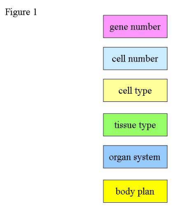
Consider Figure 1, which shows several of the salient biological levels employed in assessing the complexity increases exhibited by the Cambrian Explosion.

Understanding Ontogenetic Depth: Naming Versus Measuring Pn1

Gene number

is the sum of all functional sequences in a taxon's genome (whether those loci are classical protein-coding genes or regulatory sequences). Cell number is the total count of discrete cells, of any type, possessed by an adult organism capable of reproduction. Cell type describes the total number of histologically differentiated cellular morphologies (e.g., gut epithelium, nerve, muscle, blood cell). Tissue type describes the organization of cell types into functional units such as sheets or epithelia, connective materials, skeletal parts, and so on. Organ systems are the higher-level anatomical relationships responsible for major organismal functions (e.g., sensory, locomotory, digestive, reproductive), while Body plans represent the major architectural features characteristic of groups such as Arthropoda, Mollusca, Brachiopoda, and the other bilaterian phyla.

It might seem that the natural way to illuminate the relationship between these levels would begin "bottom up," with the genes. We argue, however, that for the problem of the origin of the phyla, the concept of an ontogenetic network best integrates these levels (see Figure 2).

Understanding Ontogenetic Depth: Naming Versus Measuring Pn12

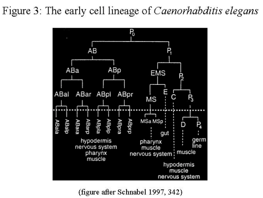
An example of one aspect of an ontogenetic network can be seen in Figure 3, depicting the beginning of the cell lineage of the nematode Caenorhabditis
elegans.

Understanding Ontogenetic Depth: Naming Versus Measuring Pn123

Ontogenetic networks in all animals commence with a single cell, the fertilized egg. Then an unfolding arborescence of developmental decisions begins, whose  complexity and overall architecture varies by taxon. In all animals, however, a point in the adult phenotype arrives when reproduction - the generation of gametes capable of  fertilization - is possible. This distance, from the egg to the adult capable of  reproduction, is what we term ontogenetic depth (see Figure 4).

Understanding Ontogenetic Depth: Naming Versus Measuring Pn1234

Somewhat more formally, ontogenetic depth may be defined as the distance, in terms of cell division and differentiation, between a unicellular condition and a macroscopic adult metazoan able  to reproduce itself (i.e., generate gametes).

The ontogenetic depth of a handful of extant animals (from the model systems of  developmental biology) is known with precision. In the nematode Caenorhabditis elegans, for instance, a relatively small animal only 1.5 mm in length, 7 to 9 rounds of cell division lie between the fertilized egg and any cell in the adult: 959 somatic cells in  the hermaphrodite (with a variable number of germ cells), and 1031 cells in the male(with its distinctive tale). For larger metazoans, of course, such as the dipteran  Drosophila melanogaster, ontogenetic depth is much greater, as total cell number, degree of cellular differentiation, and time to reproductive capability increase accordingly. The value of ontogenetic depth as a complexity metric lies in its relationship to all the parameters listed in Figures 1 and 2.

Of course, the ontogenetic depth of any extinct organism cannot be determined with complete exactitude. However, it should be possible, using modern analogues for fossil taxa - e.g., the extant monoplacophoran Neopilina for the extinct mollusc Scenella - to obtain good estimates on the ontogenetic depth requirements of many Cambrian forms.  This is research we are now conducting. It is likely that reasonable estimates of the
ontogenetic networks, and depth, required to specify such organisms as Anomalocaris or Opabinia, will require no less complexity than that of modern animals.


3. OK, SO WHAT? THE MARCHING BAND PROBLEM

To a skeptic, the concept of ontogenetic depth may seem to be little more than a  roundabout way of expressing the already-familiar problem of how animals originally  evolved from unicellular or colonial ancestors. We think, however, that focusing on ontogenetic depth helps to illuminate the central challenge that standard evolutionary theory faces when confronted with phenomena such as the geological first appearance of  forms such like Anomalocaris. One of us (Nelson 1999) has called this challenge "the  marching band problem."

The cells of an adult metazoan are specialized for particular functional roles (as gametes, nerves, gut epithelia, skin, skeleton or exoskeleton, sensory organs, and so on). "The  production of [these] differentiated cell types," writes Carl Schlichting (2003), "is a  hallmark of multicellular organisms." The production process is an ontogenetic network,  commencing with the fertilized egg. "A function [one might say the function] of developmental processes," notes Strathmann (2000), "is putting the right kind of cells in  the right places at the right times. The criterion for 'right" is survival and reproduction."

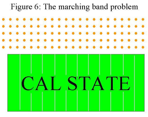
One can conceive this process of differentiation (or cellular specialization) very much on the model of an American university marching band (see Figure 5, where a 140-member marching band is depicted as orange dots, arrayed at the sideline of a football field). 

Understanding Ontogenetic Depth: Naming Versus Measuring Pn12345

In one sense, of course, any marching band is strongly disanalogous to a developing animal. A nematode or fruit fly commences its existence as a single cell (the fertilized egg), and will then construct its cell populations during development, whereas the marching band begins its maneuvers with all of its members already present.

But in another sense - the one that we'll focus on - the two processes share many  parallels. The band will move, through a series of intermediate maneuvers, toward its functional endpoint - say, spelling "CAL STATE" on the field (see Figure 6).

Understanding Ontogenetic Depth: Naming Versus Measuring Pn123456

 In its development, an animal also moves from the fertilized egg, through a series of  intermediate "maneuvers," towards its functional endpoint, namely, an organism capable of reproduction. The latter process, of course, is vastly more complex: "This temporally ordered sequence of morphological heterogeneities that we call development," writes Arthur (1997), "generates adult tissue patterns that, in some taxa, can be highly complex, involving very precise and repeatable arrangements of billions, even trillions, of cells." Now, if the band is going to spell "CAL STATE" successfully, it should be intuitively obvious that the members must have their instructions in place before they venture onto the field. The trumpet player, for instance, standing in the front row on the sideline, who will eventually become the tip of the serif at the bottom of the "L" (see Figure 7), must know how to execute the series of turns and motions that will carry him to his endpoint on the field.

Understanding Ontogenetic Depth: Naming Versus Measuring Pn1234567

The same is the case with a developing organism. "Development is possible," writes Arthur (2000), "only if cells 'know' what to do in all these respects," i.e., assign their planes of division, tendencies to move, directions and rates of movement, modes of  differentiation into particular cell types, and cell death (apoptosis). "So the key question," Arthur continues, "becomes 'how do they know?', and the whole of  developmental biology could be regarded as an attempt to answer this question." If the  question "How do cells know?" is to be answered by developmental biology, its sister (and far more difficult) question "How did cells learn what they know?" must be addressed by evolutionary (or historical) biology.

And here serious, and currently unanswered, questions arise. "How cell types of multicellular organisms came to be differentiated," notes Schlichting (2003), "is still an  open issue...the origins of differentiation remain unclear." Given that the origin of  animals - organisms defined by differentiated structures - is thought by most scientists to  have been a problem solved, at least in outline, by Charles Darwin, this is not a minor difficulty. Some authors have recently noted this explicitly; e.g., Davidson 2001:

...classical Darwinian evolution could not have provided an explanation, in a  mechanistically relevant way, of how the diverse forms of animal life actually arose during evolution, because it matured before molecular biology provided  explanations of the developmental process. To be very brief, the evolutionary theory that grew up before the advent of regulatory molecular biology dealt with the problem of the origin of novel organismal structures in two ways. The first  has been to treat the mechanisms generating novel morphological structures as a  black box. New forms were considered to arise "because" the environment  changed. But while changes in Precambrian or Ordovician weather, continental shifts, or temperature may have contributed crucial selective forces, they do not generate heads or appendicular forms; only genes do that.

Or, we might say, genes plus (the three-dimensional localization of their protein products,  et cetera - nucleic acid alone an organism never made). Davidson goes on to argue that  "stepwise, adaptive changes in protein sequence...is probably largely irrelevant to the evolution of any salient features of animal morphology," but we will focus on a more general difficulty, involving the process of natural selection itself, and its (probable) impotence for constructing ontogenetic networks.

Suppose we interrupt a marching band midway through its maneuvers, at some stage before "CAL STATE" appears on the field. Suppose, furthermore, that we cause this  interruption at a marching band competition where "success" is defined (at least in part)  by actually reaching the endpoint where the name of the band's home institution is  spelled. It should again be intuitively obvious that the functional reason for the band's  intermediate maneuvers is not the maneuvers themselves, but rather the distant endpoint that those maneuvers enable or bring about.

Now look again at Figure 3, showing the early cell lineage of C. elegans. One cannot  interrupt this canonical cell division pattern and obtain a viable organism. Viability, and,  in particular, reproductive capability - the only outcome "visible" to natural selection - lie in the distance, after several rounds of cell division and differentiation.

How then did natural selection construct the ontogenetic network of C. elegans? Figure 8  represents this problem in schematic form, using a very shallow network to make the  point. Reproductive capability arises only in the square on the right, when its five cells are in place.

Understanding Ontogenetic Depth: Naming Versus Measuring Pn12345678

REFERENCES
 
Arthur, Wallace. 1997. The Origin of Animal Body Plans: A Study in Evolutionary Developmental Biology. Cambridge: Cambridge University Press.
 
Britten, Roy and Eric Davidson. 1969. Gene Regulation for Higher Cells: A Theory. Science 165:349-357.
 
Davidson, Eric. 2001. Genomic Regulatory Systems: Development and Evolution. New York: Academic Press.
 
Ewing, Brent and Phil Green. 2000. Analysis of expressed sequence tags indicates 35,000 human genes. Nature Genetics 25:232-234.
 
Kamminga, Harmke. Protoplasm and the Gene. In A.G. Cairns-Smith and H. Hartman, eds., Clay Minerals and the Origin of Life. Cambridge: Cambridge University Press, pp. 1-10. 

Liang, Feng et al. 2000. Gene Index analysis of the human genome estimates
approximately 120,000 genes. Nature Genetics 25:239-240.
 
McShea, Daniel. 1996. Metazoan Complexity and Evolution: Is There a Trend? Evolution 50:477-492.
 
Nelson, Paul. 1999. Generative Entrenchment and Body Plans. Lecture presented at the
International Symposium on the Origins of Animal Body Plans and Their Fossil Records,
eds. J.Y. Chen, P.K. Chien, D.J. Bottjer, G.X. Li, and F. Gao, Early Life Research Center, Kunming, People's Republic of China, 21-25 June.
 
Schlichting, Carl D. 2003. Origins of differentiation via phenotypic plasticity. Evolution and Development 5:98-105.
 
Schnabel, Ralf. 1997. Why does a nematode have an invariant cell lineage? Seminars in Cell & Developmental Biology 8:341-349.
 
Strathmann, Richard. 2000. Functional design in the evolution of embryos and larvae. Seminars in Cell and Developmental Biology 11:395-402.
 
Valentine, James W. 1994. The Cambrian Explosion. In S. Bengston, ed., Early Life on Earth (New York: Columbia University Press), Nobel Symposium No. 84; pp. 401-411.
 
Valentine, James et al. 1994. Morphological Complexity Increase in Metazoans. Paleobiology 20:131-142. 

https://reasonandscience.catsboard.com

Otangelo


Admin

Ontogeny, Phylogeny and the Origin of Biological Information 1

http://reasonandscience.heavenforum.org/t2308-origin-of-development-and-ontogeny#4757

1. Introduction


In 1984 I presented the semi-meiotic hypothesis of organic evolution. This was followed by three other papers which exposed the complete failure of virtually every aspect of the Darwinian model (Davison 1984 1987 1993 1998).  Incorporating the conclusions of Otto Schindewolf (1896-1971), Richard B. Goldschmidt (1878-1958), William Bateson (1861-1926), Robert Broom (1866-1951), Pierre Grassé (1895-1985) and Leo S. Berg (1876-1950), the semi-meiotic hypothesis can be summarized as follows.  Macroevolution is largely finished. Sexual reproduction is incapable of supporting trans-specific (macroevolutionary) change.  Accordingly, all significant change was produced presexually involving the first meiotic division. The essential feature of these changes was due not to micromutations in the genes themselves, but rather to the way in which those genes express their effects which is dependent upon their arrangement within the structure of the chromosome (position effect). 

So how did the right program arise to permit the cells to find their right position, and assigning the right kind of cells at the right place , and the right quantity, and replacing cells during development, and cells with no use to be destroid ? 

 The first meiotic division is ideally suited to this form of reproduction as it can preserve the original karyotype and at the same time produce structural novelties instantaneously in homozygous form (Davison 1998).  Thus, this model provides a rational explanation for the absence of intermediates both in contemporary and in fossil species.
Such a mechanism does not depend on the production of new information but rather on information already present in the genome i.e. preformed.  This perspective remains in complete accord with the remarkable resemblances we find underlying disparate species, from the ubiquity of ATP to the universal 9 plus 2 nature of the structure of cilia and flagella wherever they are found in the living world. It should also be noted that Schindewolf (1993), Goldschmidt (1940), Berg (1969) and Grassé (1973) all subscribed to preadaptation (preformation) during evolutionary change.

These considerations bring me to the subject of this paper. First, what is the nature and source of the preformed information? Second, how was it made manifest?  In considering these two questions I came, as have others, to the realization that there is an intimate relationship between ontogeny and evolution (phylogeny).

The Russian ichthyologist and zoogeographer Leo S. Berg was perhaps the first to indicate that evolution and the development of the individual were closely related.  In a clear demonstration of his rejection of Darwinism, he wrote:

       The laws of the organic world are the same whether we are
       dealing with the development of an individual (ontogeny) or
       that of a paleontological series (phylogeny).  Neither in
       the one nor in the other is there room for chance.

                               Nomogenesis (1969), page 134

Note that Berg insisted that chance plays no role in either process. Whatever an organism becomes is determined entirely by the orderly unfolding of events that take place during its embryonic development. I accept Berg's judgment and elaborate on it to see where that perspective takes us with respect to our understanding of the two as yet unresolved problems of ontogeny and phylogeny.


2. Epigenesis and preformation

These two terms originated from observations on the nature of the developmental process.  It is obvious that all of the information necessary to produce a unique human being is contained (preformed) in a single cell, the fertilized egg, a mere few microns in diameter. On the other hand, the development of the individual is largely epigenetic.  I will deal first with epigenesis and then with preformation.  Epigenesis refers to the necessity for development to proceed in a definite serial fashion.  For example, neurulation or the formation of the central nervous system cannot proceed until gastrulation or the formation of the primitive digestive system is complete.  Following that, the optic cup can be formed.  Only after that can lens induction take place.  These ontogenetic events have interesting phylogenetic (evolutionary) parallels.


One of the most significant events in vertebrate evolution was the invention of bone.  Since bone is significantly more dense than the cartilage it replaced, bony vertebrates would have been relegated to the bottom of the sea if it were not for the early development of the swim bladder, an outgrowth of the primitive gut.  This hydrostatic organ made it possible for the animal to maintain neutral buoyancy. The swim bladder is homologous with the vertebrate lung which in turn made possible the invasion of the land and the evolution of the higher vertebrate taxa (amphibians, reptiles, birds and mammals). Accordingly, the invention of bone and the swim bladder both served to preadapt subsequent vertebrates for life on land.  It is significant that the Chondrichthyes (sharks skates, rays and chimeras) have remained cartilaginous and accordingly less specialized which may account for the ongoing successful presence of this very primitive group.

When something is lost during evolution it is rarely replaced. Examples are the loss of limbs in reptiles and amphibians, or digits in many tetrapod vertebrates.  Returning to the swim bladder example, the darters, tiny members of the perch family of fishes, have lost the swim bladder, a loss which allowed them to successfully invade swiftly flowing freshwater streams.  The Darwinian view would be that this was an adaptation to the stream environment.  An alternative view would be that, having lost the swim bladder, the darters sought out or perhaps simply stumbled into the stream environment where they would have a clear advantage over buoyant competitors.  Also the Darwinian interpretation would suggest that the swim bladder was gradually reduced, yet there is no evidence whatsoever for that assumption.  It would seem that many evolutionary specializations
occur instantly without intermediate forms.  Thus, just as ontogeny goes forward in epigenetic fashion, so has evolution.  The two processes also share irreversibility as a cardinal feature.

It is when we come to preformation that things really become interesting.  I now suggest that just as ontogeny and evolution share epigenetic features, so also may they share the characteristics we associate with preformation.  In short, I propose that the information for virtually all of evolution may have been present from very early in the onset of that process.  I realize that this idea may seem ludicrous at first glance, yet it remains compatible with an enormous number of otherwise enigmatic observations from comparative biology. It also avoids postulating Lamarckian devices for which no evidence has been forthcoming.

Before presenting this material it is necessary to grapple with the question of the origin of the information itself.  If, as I suggest, the information was preformed, someone or something had to put it
there.  I am not the first to confront this problem.  I quote Pierre Grassé:

       According to Darwinian doctrine and Crick's central dogma, DNA
       is not only the depository and distributor of the information
       but its sole creator.  I do not believe this to be true.

                               (Grassé's emphasis)
                               Evolution of Living Organisms
                               (1973), page 224

Oddly, Grassé does not pursue the problem further.  I think we have an obligation to do so.  It is my understanding that information does not arise de novo but must have a source.  While that source has yet to be identified, its existence is suggested by a host of examples from both the plant and animal kingdoms.
Understanding Ontogenetic Depth: Naming Versus Measuring Diplod11

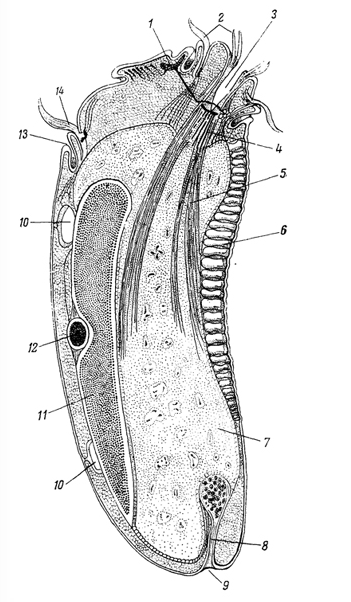
Figure 1.  Ris.65. Longitudinal section through the optical infusorian Diplodinium (Epidinium) ecaudatum. (According to Sharp, 1914). ecaudatum. (According to Sharp, 1914). 1 - It has a kind of "brain" (motorium); 2 - adoral cirri; 3 - mouth; 4 - conductive filaments of the pharynx; 5 - fibrils pharynx; 6 - skeletal plate; 7 - endoplasm; 8 - "hindgut"; 9 - poroshitsa; 10 - contractile vacuole; 11 - Ma;Mi-12; 13 - dorsal lip; 14 - dorsal cirri area.


Works American Protistology dedicated to "neuromotor apparatus" ciliates were subjected to severe criticism by many researchers in a number of countries. Bretschneider (Bretschneider, 1934), detail

The unicellular Amoeba is structurally one of the simplest of the Protozoa, yet in that same phylum can be found creatures exhibiting strikingly advanced features.  One such animal is Diplodinium (Epidinium) ecaudatum (Figure 1).  This ciliate exists in huge numbers as symbionts in the stomachs of cattle (Sharp 1914).  It has a kind of "brain" (motorium), "circumoesophageal neural connectives" resembling those of annelids and arthropods, clearly differentiated "muscles", a kind of "segmented skeleton" (skeletal laminae), a "mouth", "esophagus", "rectum" and "anus" all elaborated within the confines of a single cell.  Such a creature not only demonstrates that the information is already present for these structures, but also casts considerable doubt on the notion that multicellularity is a prerequisite for the division of labor.  It is no wonder that Libby Hyman described the protozoa as the acellular rather than the unicellular animals.  Why this creature should have such an array of advanced features remains a complete mystery.  Could it be there to provide us with a clue to the nature of the evolutionary process?

The relict worm-like Peripatus with its curious combination of annelid and arthropod characters could not be placed in either phylum, so it has been placed in a phylum of its own, the Onycophora.

While we associate the placenta with the higher mammals, this structure also is found in certain sharks.  Berg (1969) discussed this and related phenomena as examples of what he called physiological acceleration.  He noted that Peripatus also nourishes its embryos with a kind of placenta.  Thus Peripatus exhibits characters of three taxa:  the annelids, the arthropods and the placental mammals.

The primitive chordate Amphioxus (Branchiostoma lanceolatus), while it has all three chordate features (dorsal hollow nervous system, gill slits and notochord), has a kidney consisting of solenocytes of the protonephridial (flame cell) type found in the Platyhelminthes, the Aschelminthes and the polychaete Annelida.  On the other hand, certain oligochaete Annelida (earthworms) have a tubular kidney system more like that of vertebrates.  The limbless amphibia (Apoda) have large yolk-laden eggs suspended by albuminous chalazae closely resembling the situation in the cleidoic (shelled) eggs of reptiles and birds. They lack only the shell.  Such bizarre forms as the egg-laying monotreme mammals (Platypus and Echidna) can suddenly seem acceptable if one simply assumes that those combinations of characters were available when those forms evolved.  When it comes to the possible
combinations of features one can almost say -- Anything goes!

While we associate imaginal discs with the complete metamorphosis of the higher insects (Diptera, Hymenoptera, Homoptera and Lepidoptera), imaginal discs are also involved in the metamorphosis of certain marine worms (Nemertea), with the formation of the adult or imago taking place within the body of the Pilidium larva which then splits open to release the adult form much as does the pupa of a moth or butterfly.  Considerations like these and many others prompted Leo Berg (1969) to conclude:  "Evolution is in a great measure an unfolding of preexisting rudiments."

The same view was independently offered by Pierre Grassé (1973): "The existence of internal factors affecting evolution has to be accepted by any objective mind."

The very word evolution derives from the Latin evolvo meaning to unfold as the pages of a book.  Needless to say, a book has, by definition, already been written. The plant kingdom abounds with examples in support of preformation. Distantly related plant species often produce similar, if not  identical, chemical scents of anise, lemon, orange, pineapple, cinnamon and a host of other flavorings so valuable to our cuisine. There is even a chocolate scented member of the mint family!

The behavioral and structural reciprocities that often exist between plants and their insect pollinators indicate that they may have been sharing the same informational pool when those evolutionary events took place.

What does that mean ? What " informational pool " is the author writing about ? How about we put the intelligent designer as the source of that pool ? makes sense ? 

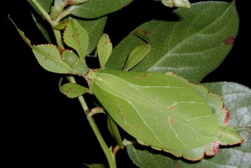
 One of the most remarkable of such interactions was described by Berg (1969).  Certain stick and leaf insects (Phasmodea) resemble the plants that they inhabit 
Understanding Ontogenetic Depth: Naming Versus Measuring Diplod10

even to the extent that the eggs of the insects closely resemble both internally and externally the seeds of the plants (Umbelliferae) they inhabit and, like the seeds, are scattered on the ground where they may remain for up to two years. The tunic of the Urochordata (sea squirts) is composed of cellulose, otherwise a polymer associated with the plant kingdom.


Rather than assuming independent inventions of these remarkable parallels, it seems to me more reasonable to postulate these events resulted from the activation (derepression) of an enormous yet clearly limited stockpile of potentialities which were available when those events took place.

These considerations cast a whole new perspective on what has been called convergent evolution.  The morphological similarities that exist, especially in the skeletal systems, between placental wolves and bears and their marsupial counterparts, to my mind, defy any explanation based on the accidental evolution of these similarities through chance events.  Thus, what has been described as convergent evolution is not that at all but the expression from preformed sources of virtually identical morphologies.  In this connection it is useful to recall the words of R.C. Punnett:

       Natural selection is a real factor in connection with mimicry,
       but its function is to conserve and render preponderant an
       already existing likeness, not to build up that likeness
       through the accumulation of small variations, as is so
       generally assumed.
                               (my emphasis)
                               Mimicry in Butterflies
                               (1915), page 152

One is compelled to ask -- What is the source of the already existing likeness?

How such transformations were effected remains, of course, a complete mystery.  Unfortunately, since macroevolution seems no longer to be in progress, we may never be able to resolve that issue.

However intangible, the issue remains -- What is the origin of the preexisting information?


3. Has evolution been guided?

Questions such as this can probably never be resolved.  Nevertheless, one can still offer observations which relate to such metaphysical considerations.  Some of our greatest minds have been physicists. Galileo, Newton, Pascal, Faraday and, in particular, Einstein all have referred to God one way or another.  Richard P. Feynman (1998) has compared scientific discovery to a religious experience.  I think it is fair to say that we are still ignorant of the source of bright ideas, insights and acts of creative genius.

It is interesting to compare our interpretation of the laws that govern the inanimate world with those that may be operative in the animate world.  Leo Berg, as the complete title of his book indicates, believed that all of both evolution and development was the result of laws, a perspective with which I have (admittedly reluctantly) been forced to agree.  If not chance, it seems to me that the only alternative is to agree with Berg.  The existence of laws presumes a law maker or makers.  That in turn suggests purpose.  Don't representatives in Congress have some purpose in mind when they enact legislation?

No one denies the validity of Galileo's equation which relates the distance that a body falls to time, or Newton's laws of motion, or Einstein's equation relating energy and mass.  Why then must one reject, as the Darwinians do, the suggestion that comparable laws exist or have existed controlling the living world?  Everyone accepts gravitation and the equations associated with it, yet no one yet understands the cause of gravity.  Accordingly, neither in religion nor in science does acceptance demand understanding.

Nevertheless, the Darwinians continue to insist that evolution is the result only of chance events.  Stephen J. Gould has recently compared evolution to a drunk reeling back and forth between the bar room wall and the gutter (Gould 1996 page 149).  He has also described intelligence as an evolutionary accident.  I will only comment that it was some accident!

Isn't it conceivable that there may really be no conflict between religion and evolutionary science?  Isn't it possible that both might be correct?  I have come to believe just that.  Blaise Pascal offered the following commentary as well as the solution:


       Men despise religion.  They hate it and are afraid it may
       be true.  The cure for this is first to show that religion is
       not contrary to reason, but worthy of reverence and respect.

Finally, let me close with the following appropriate quotations from
my favorite intellectual, Albert Einstein:

       I want to know God's thoughts ... the rest are details.
       
       The most beautiful thing we can experience is the mysterious.
       It is the source of all true Art and Science.

       Science without religion is lame.  Religion without science
       is blind.

       I shall never believe that God plays dice with the world.

I rest my case.


4. Conclusion

The greatest impediment to progress in the field of evolution resides in the stubborn refusal of the Darwinians to entertain any possibility that the living world might be subject to laws such as govern virtually every aspect of the inanimate world.  I find it fascinating that it is the physicists who postulate God while biologists typically remain atheists or agnostics.  I offer the following explanation for that curious situation.  It is the physicists who have been able to discover the several laws that serve to describe and thereby explain their domain of the inanimate world.  Thus they have been forced to come to grips with the reality that laws must have an origin.  By way of contrast, most biological scientists have yet to acknowledge those laws that so clearly operate in the living world, and so they reject them as being unnatural or mystical and accordingly unacceptable. I see no reason to make such distinctions.  Laws are laws whether or not we like them or understand them.  Accordingly, I have found the acceptance of the existence of preformed laws and information
to be a necessary and liberating asset in the ongoing search for ultimate truth concerning the two greatest mysteries in all of biological science:  ontogeny and phylogeny.  They are obviously intimately related just as Leo Berg so clearly recognized long ago. I now firmly believe that in solving the one we will solve the other.


References

BERG, L. (1969) Nomogenesis; or, Evolution Determined by Law.
M.I.T. Press, Cambridge.  (Original Russian edition 1922.)

DAVISON, J.A. (1984) Semi-meiosis as an evolutionary mechanism.
J. Theor. Biol., 111: 725-735.

DAVISON, J.A. (1987) Semi-meiosis and evolution: a response.
J. Theor. Biol., 126: 379-381.

DAVISON, J.A. (1993) The blind alley: Its significance for
evolutionary theory.  Rivista di Biologia (Biology Forum),
86: 101-110.

DAVISON, J.A. (1998) Evolution as a self-limiting process.
Rivista di Biologia (Biology Forum), 91: 199-220.

FEYNMAN, R.P. (1998) The Meaning of It All: Thoughts of a
Citizen Scientist.  Addison-Wesley, Reading, Massachusetts.

GOLDSCHMIDT, R.B. (1940) The Material Basis of Evolution.
Yale University Press, New Haven.

GOULD, S.J. (1996) Full House: The Spread of Excellence from Plato
to Darwin.  Harmony Books, New York.

GRASSÉ, P. (1977) Evolution of Living Organisms: Evidence
for a New Theory of Transformation.  Academic Press, New York.
(Original French edition 1973.)

PUNNETT, R.C. (1915) Mimicry in Butterflies.
University Press, Cambridge.

SCHINDEWOLF, O. (1993) Basic questions in paleontology.
University of Chicago Press, Chicago.  (Original German edition 1950.)

SHARP, R.G. (1914) Diplodinium ecaudatum, with an account of its
neuromotor apparatus.  University of California Publications in
Zoology, 13: 43-123.

1) http://www.uvm.edu/~jdavison/ontogeny.html


Last edited by Admin on Thu 18 Feb 2016 - 2:29; edited 3 times in total

https://reasonandscience.catsboard.com

Otangelo


Admin

IV. Conductive fibrils and the question of the nervous system in PROSTEYSHIX. Total protozoology. 1

IV. Conductive fibrils and the question of the nervous system in PROSTEYSHIX
The question about the presence of Protozoa system of special conductive fibers, which by their function to a certain extent meet the nerve fibers Metazoa and facilitate co-ordination among the various organelles in Protozoa, originated in a concrete form at the beginning of this century. Even before that, Schubert (Schuberg, 1890) gave a detailed description of the system at the Mion ciliatesStentor, which is very quick to respond to various stimuli sharp decline in the body. Nerssgeymer (Neresheimer, 1903) applied in the processing of new methods trumpeter painting, coloring and Mallory helped him open a new, hitherto unknown system of fibrils, having, according to Neresgeymera, the nature of the conducting nerve fibers, called them neyrofanami. Like mionemam, neyrofany costs in the form of the bouquet from the rear end of the body Stentor to the front and are approximately parallel mionemam, differing from them by their attitude to the dyes. However op showed that the substances acting on the nervous system paralyzing higher animals, causing a similar effect and blower, stopping its motion. In addition, it was observed that neyrofany find sometimes thin side branches that come into contact with mionemami.
All this has led Neresgeymera convinced that the most difficult arranged by representatives of Protozoa differentiated system of some kind of nerve fibrils, or neyrofanov.
The further development of this issue study played an important role Sharp (Sharp, 1914) on the organization Diplodinium (Epidinium) ecaudatum (Fig. 65), the representative of ciliates detachment Enlodiniomorpha, living in the rumen of ruminants. These ciliates have a very complex structure (Fig. 65). The body is provided with a spiral perioral cirri and another dorsal cirri arc, which is covered by a special plasma fold - dorsal lip; all the rest of the body wearing thick cuticle. The front of the body a few pole jutting forward in a so-called parietal ridge. Applying the Epidinium color Mallory, Sharpe found at the base of the parietal ridge over the presence of the pharynx area particularly differentiated plasma from sending itself to all sides
fiber system, which are directed primarily to the movement of organelles (cirri and mionemam). On this basis, Sharp calls the above section motor center, or "motoriumom" (motorium). Only motorium and micronucleus in the body ciliates are stained according to Mallory in a bright red color. The set motoriuma with beams radiating from him fibers obtained by Sharpe called "neuromotor apparatus." As described by Sharpe, the fibers that innervate the apparatus cirri perioral spiral and dorsal arch and form a gentle peripharyngeal ring, sending the fibrils to the pharynx mionemam. Finally, from the fiber bundle is motoriuma the crown ledge, which has, according to Sharpe, sensitive nature. This work Sharpe laid the foundation to the direction in the study of the conductive system of ciliates, which subsequently developed mainly in the United States. The main feature of the majority of American jobs is to recognize all neuromotor apparatus as the physiological and morphological entity, which is controlled by specifically localized nerve center, t. E. Motoriumom. Correspondingly to these articles by American authors always strenuously sought the nerve center, at least observed the pictures and did not give sufficient reasons for this. Meanwhile, in effect, called designated motoriuma formation are rather any kind of local condensations of fibrils or even artifacts. In addition, the well-known question is extremely varied shape and location motoriuma in different ciliates. Motorium placed it at the bottom of the throat, above the pharynx, then at the bottom of the front sucker (from Haptophrya); it is a ring, then tape the wrong lump; Sometimes, in addition to the main motoriuma, has indicated additional centers (Entodiscus, on Paursu (Powers, 1933)).


Understanding Ontogenetic Depth: Naming Versus Measuring B5014p97-a1

Ris.65. Longitudinal section through the optical infusorian Diplodinium (Epidinium) ecaudatum. (According to Sharp, 1914). ecaudatum. (According to Sharp, 1914). 1 - "motorium"; 2 - adoral cirri; 3 - mouth; 4 - conductive filaments of the pharynx; 5 - fibrils pharynx; 6 - skeletal plate; 7 - endoplasm; 8 - "hindgut"; 9 - poroshitsa; 10 - contractile vacuole; 11 - Ma; Mi-12; 13 - dorsal lip; 14 - dorsal cirri area.

Works American Protistology dedicated to "neuromotor apparatus" ciliates were subjected to severe criticism by many researchers in a number of countries. Bretschneider (Bretschneider, 1934), detail
studied the fibrillar structure of different Ophryoscolecidae, has not confirmed the existence of data Sharpe they motoriuma. He discovered a very complex system of fibrils Bretschneider regards primarily as a support unit. Similarly, AA Strelkov (1939) in an extensive study on morphology and systematicsEntodiniomorpha of Equidae, has not found any motoriuma or other elements of the neuromotor apparatus. There were a variety of unrelated systems fibrils together, carrying, according to small arms, support function. A negative attitude to the ideas of the American authors of the neuromotor apparatus and researchers such fibrillar structures ciliates as Ten-Cate (Ten Kate, 1927, 1928), LB Levinson (1941).
However, in recent years, presenting Sharpe and other American writers of the neuromotor apparatus ciliates Sem. Ophryoscolecidae in a slightly different form received support in the work of Fernandez-Galiano (Fernandez-Galiano, 1949) and. . Hyapo-Timote (Noirot-Timothee, 1960) Last in Epidinium ecaudatumdistinguishes the front end of the body are three main groups of fibrils: peripharyngeal ring (anneau perioesophagien), anterior dorsal arc (arc dorsal anterieur) and posterior dorsal arc (arc dorsal posterieur), including major motorium, with numerous radiating from them to the periphery of the fibrils (Fig. 66). About the function of all of the fibrillar apparatus noir Timote speaks very cautiously, not denying, however, its possible conducting and co-ordinating role. It should be noted that the described noir Timote fibril system does not coincide with that described by Sharpe in the same object, and in determining motorium Sharpe noir Timote - is a completely different formation (see Figure 65 and 66.). It is obvious that the issue needs further study.


Understanding Ontogenetic Depth: Naming Versus Measuring B5014p98-a1

Fig. 60. argentophilic fibril system in Epidinium ecaudatum (Ophryscolecidae). (By Noirot-Timothee, 1960). And - the front part of the body ciliates on the right side; At - the same with the left hand, adp - dorsal anterior arc; adp - posterior dorsal arch; ap - peripharyngeal ring; fi - fibril I; fb - basal fibril; foe - fibril esophagus; fp - peristomalnye fibrils; Ma - macronucleus; mot - "motorium"; rv - the ventral branch; tc - fibers underlying the ciliary apparatus.

Another trend in the study of fibrillar ciliates apparatus associated with the study of surface subpellikulyarnyh structures identified by methods of impregnation with salts of silver or gold on the dried or fixed wet preparations. These works were started almost simultaneously and independently of each other in the 1925- 1926 biennium. Klein and gel. Simultaneously with them the same questions and the same with the help of a modified method of silver plating began to vigorously explore Shatton, Lions and their employees.
Let us dwell on a brief consideration of the main relevant to this; the problem of work.
At the very beginning of this century Shuberg (Schuberg, 1905) found a thin fibrils lying in ectoplasm and connect with each other grain basal one row of eyelashes. Klein developed by applying them


Understanding Ontogenetic Depth: Naming Versus Measuring B5014p99-a1

Fig. 67. argentophilic line system in different ciliates. (According to Klein, 1926, 1927). A - Urocentrum turbo: a - the apical pole, d - the distal pole, p - basic plate tsitostoma; In - the apical pole of Paramecium aurelia: a - argentophilic hotline, s - argentophilic indirect communication lines, t - trichocysts, the k - additional grain, bo - fibrils, binding basal bodies, trichocysts and additional grains, b - basal body; S - area of ​​the body Stentor igneus: b - basal bodies, s - argentophilic line; D - Glaucoma scintillans.

"Dry" method of silvering, since 1926 in a large series of works described in representatives of the various groups of ciliates in the presence of an extremely complex system of ectoplasm CHERNYAEV silver nitrate (argentophilic) fibrils. This system is a closed network that includes all basal bodies eyelashes and trichocysts. Argentophile fibril system configuration can be very different (Fig. 67).
Sometimes it is a very fine-meshed network or series of meridian lines going with a few cross beams and so on. N. Often, in different parts of the body position of these ciliates argentophilic lines may be different (Fig. 67). Klein distinguishes among these two types of fibrils. Some of them are directly linked to each other basal grain eyelashes and trichocysts, is, in Klein, "direct connection system" (direkt verbindende System), the other in close connection with the basal grains are not, and are connected with them only indirectly, through "direct communication system ". Unlike the latter Klein calls "indirect communication system" system (indirekt verbindende System). Very clearly these differences appear, for example, Paramecium, where fibrils "direct communications" form longitudinal rows, and "indirect system," located under the hexagonal protrusions pellicle forming so characteristic of Parameciumpellikulyarnuyu surface sculpture. Klein distinguishes between simple and complex fibrils. Where there is a very dense fine-meshed network, it is composed of very fine elementary fibrils. In other cases (for example, of Paramecium)network consists of thicker fibrils and in these cases they are composed of several elementary fibrils. The latter, according to Klein, come and cilia, forming part of its centerline. Thus, fibrils, and the most basal eyelashes grains included in a single system argentophile fibrils.


Understanding Ontogenetic Depth: Naming Versus Measuring B5014p100-a1

Fig. 68. Euploies area of ​​the body on the ventral side. (According to Klein, 1926). The formation of a new peristomalnogo field (N) by dividing. Impregnation with silver salts.

Klein attributed argentophilic fibril system is very diverse and important functions. In his later works, especially in the great work in 1943, he calls her neyroformativnoy system (neuroformatives System, Neuroformium.) K functions of the system, according to Klein, refers primarily conduct the excitation wave and coordination eyelash heartbeat. However, besides this "nervous" function, Klein ascribes to it a number of other extremely important functions. The network loops occur basal grain and they form cilia. Thus, by dividing the system neyroformativnaya ciliates it is a material through which are newly formed in the ciliary apparatus. Bookmark and development of new peristome by dividing ciliates, according to Klein, also begins with the transformation argentophilic fibrils. At the site of the future of peristome forms a dense network (Fig. 68), by which the formation of the membrane is carried out, and so membranell. N. With their basal apparatus.
A different interpretation subpellikulyarnyh impregnated with silver nitrate structures ciliates found in the works of the gel (Gelei, 1932) and his son (Gelei, 1937). The use of very thin material fixation methods (rather than dried), followed by impregnation with salts of silver and gold, as well as some special dye (toluidine blue) allowed I. Klein Geleyu section describes system
argentophilic fibrils on two entirely different in nature category structures. According to I. Geleyu, there subpellikulyarnye fibrils bind together basal bodies eyelashes. These fibrils are neyronemt, and their function Carrying out of irritation and coordination of the ciliary apparatus. These fibrils correspond to "direct connection system" Klein. As for the "indirect systems", it is, for Geleyu, has no relation to the conductive unit and in most cases is not even a fibrillar structure. Pellikulyarnye structure (in particular, Paramecium rollers hexagonal meshes) impregnated with silver salts. All these structures are only a reference, rather than conducting irritation value.
I. Looks gel experienced a further complication due to the fact that his son G. gel (Gelei, 1937) was found in three species of Paramecium another, third, fibril system lying at the level of the basal cells and some of their deeper. This fibrillar network consists of longitudinal, transverse and diagonal fibers held together in close connection to the ground mating fibrils. Meshes of the network is smaller and more numerous outdoor network meshes and are more wrong. This third network is available throughout the periphery of the body and spread to the walls of the vestibule peristome and pharynx. Contact the third network could not be found with the other two authors. Third network function remains unclear.


Understanding Ontogenetic Depth: Naming Versus Measuring B5014p101-a1

Fig. 69. fibrillar system Paratecium niultimicronucleatum. (According to Lund, 1933). A - schematic pharyngoesophageal network of fibrils and fibril extending from it in endoplasm; In - circuit "motoriuma". 1 - "motorium"; 2 - fibrils extending from it in endoplasm; 3 - front and rear ends of the thickened portion of the fibrils network; 4, 5 - longitudinal and circumferential pharyngeal fibrils; 6 -fibrilly extending from the pharynx to the endoplasm; 7 -peristomalnye fibrils.

A further addition to the study of fibrillar structures Paramecium as a reference made ​​famous Lund (Lund is, 1933), which is the same method of silvering extended the study of these structures on the pharyngeal apparatus shoes (Fig. 69). There accordingly complexity pharyngeal function and its ciliary apparatus Lund found extreme complexity of fibrillar networks, fibers which form several portions or separate systems, and a pellicle go in different directions. Of fundamental importance in Lund has to determine their main and additional motoriumov in the walls of the pharynx (Fig. 69, B ).
However, Ivan Gel, also studied the fibrillar structure peristome and throat of Paramecium (Gelei, 1934) and in general confirmed the observation of Lund, significantly diverges from the last only one point - Gels motoriumov not found.
With regard to surface structures Paramecium observations are similar to those of Lund J. gel. He believes fibrillar conductive formations only fibrils that connect the basal grain. With regard to the polygon network ( "indirect communication system" Klein), the Lund sees it as a simple sculpture pellicle.
Finally, since 1930, but especially since 1935-1936, the Shatton and Lions have made ​​to the question before us is extremely important addition, pointing out that under the rows of cilia in ciliates are not just a series of basal bodies, but the double rows of grains of different values. One row corresponds generally centrioles flagella Mastigophora and capable, like the centrioles, to reproduce; these grains Shatton (Chatton, 1937) calls even to the centrosome, and earlier called Basal. Each centrosome formed only by division of pre-existing centrosomes. Along the number of centrosomes is a parallel series of pips, on each of which sits cilium. These correspond to the grain appears, additional gel grain and Klein; Shatton and Lviv (Chatton et Lwoff, 1930) give the name of this grain parabazosomy. Parabazosoma has centrosomal origin, but is incapable of self-reproduction and disappears as soon as the corresponding dies cilium.
Understanding Ontogenetic Depth: Naming Versus Measuring B5014p102-a1
Ris.70. Argentophilic surface network of the front half Polykrikos schwartzi body. (On Chatton et Hovasse, 1934).

The totality of the centrosome and parabazosom Shatton and Lions are asked to name the term "infratsiliatura". The question of infratsiliature we consider in more detail in the following chapter on organelles of motion. Here we note that the basic position of the French authors of the doctrine of infratsiliature is the idea of ​​its continuity. Basal (centrosome) originate from their own kind only by dividing and can not be formed de novo. Even in those cases when there is a reduction of the ciliary apparatus (for example, encystation) infratsiliatura preserved and for its account in the future recovery of the cilia occurs. Thus, unlike Klein's views, and Shatton Lions deny the possibility of tumors basal unit and eyelashes themselves in loops argentophilic fibrils (lines) (Silberliniensystem). infratsiliatury elements are usually connected to each other by means of fibrils - kinetodesm that match play neyronemam gel and probably conductive part. As for the other elements of "Silberliniensystem" Klein, the Shatton, Lviv, Brashon and other French authors emphatically deny their role in the formation of relationships and consider mezhresnichnyh structures, completely independent from mezhresnichnyh ties. The entire network of fibrils argentophilic Shatton (Chatton, 1937) brings together under the title "argyria" as opposed to aggregate and kinetodesm infratsiliature, which he gives the common name of "throw".
It is interesting that Shatton with the students opened the same argentophilic network and Dinoflagellata (for example, Polykrikos; Figure 70.), And a number of Sporozoa (shizogregariny, coccidia, sarkosporidii; Figure 71.). These findings, if Argir flagellates and Sporozoa really homologous to that of ciliates are important in the sense that they show complete independence from argiroma kinetoma. Regarding the physiological significance argiroma Shatton with certainty is not expressed, but is inclined to recognize him primarily a support function.
In recent years, a lot has been made widely unfolded Electron-microscopic studies (Metz, Pitelka a Westfall, 1953.; Sedar a Porter, 1955, etc...) In the knowledge subpellikulyarnyh fibrillar structures ciliates. According to these studies, in ectoplasm have cilia bases arranged with cylindrical basal bodies, or Basal. The dual nature of the basal unit of eyelashes, which insisted Shatton and Lviv, Electron-microscopic studies have not yet confirmed. From Basal leaves thin fibril - kinetodesma, which is then combined, but do not merge with other kinetodesmami extending from the adjacent basal cells of the same number of lashes. Thus a beam of fibrils, in which the individual elementary fibrils tightly in contact with each other. Formed some semblance of synapses. Each elementary fibril length is small. It stretches throughout several neighboring eyelash of this series, and then thinning, fading away (Fig. 72). With this bundle of fibrils comprises only a small number of elementary fibrils. The entire beam is generally consistent neyronemam or kinetodesmam, visible under the light microscope.


Understanding Ontogenetic Depth: Naming Versus Measuring B5014p103-a1

Fig. 71. Argir in Sporozoa. (On Chatton, 1937). A - Klossia helicina, gamont; In - Barrouxia ornata, merozoite; C - Lanhesterella ranarum, merozoite.

Other studies (Randall a Jackson, 1958;.. Yagiu a Shigenaka, 1959;. Ehret a Powers, 1959) give reason to believe that the fibrils kinetodesmy are long and stretch if not along the entire body, over a large area, with a each fibril kinetodesmy can connect many fibrils extending from several kinetosom one row. Links kinetodesm fibrils of the type many authors did not observe synapses. Different ciliates kinetodesma may consist of a different number of fibrils: fromColpidium coplodn of 2 or 4,
in Paramecium caudatum - 5 or 6 (. Ehret a Powers, 1959), in Condylostorna spatiosum (Yagiu a Shigenaka, 1959.) - a few dozen, while Stentor (a Randall the Jackson, 1958.) - about 500. It is very likely that kinetodesmalnyh nature of relations among different ciliates is different, and requires further comparative research to establish some general patterns in terms of communication with the fibrils kinetodesm kinetosome.
With regard to the network of fibrils, forming the "indirect communication system" Klein, results elektronnomikroskopichsskih research is contradictory. Metz Pitelka and Westphal (Metz, Pitelka a. Westfall, 1953) describe inParamecium under the hexagonal bulges


Understanding Ontogenetic Depth: Naming Versus Measuring B5014p104-a1

Fig. 72. The structure of the scheme and the pellicle kinetodesmalnyh connections in Paramecium by electron microscopy. (According to Grell, 1956a). Bk - basal body; Bf - basal fibril; Lf - longitudinal fibrils.

pellicle presence of peripheral pellikulyarnoy lattice, the terms of the fibril network. However Porter and Sedar (Sedar a. Porter, 1955) from the same subject is not pellikulyarnoy grating and believe that the network structure of the impression surface pellicle. Interestingly, Cedar and Porter with Electron studyParamecium found at the level of the basal cells fibrillar another closed network, which, in the opinion of these authors, corresponds to the third network subpellikulyarnyh fibrils, which has been described by G. gel.
To assess the functional significance and the role of various fibrillar systems along with morphological facts necessary to take into account the physiology and biochemistry of the data. Observations on living ciliates (Paramecium, Spirostomum, Stentor), as well as the application of the methods of instant fixation eyelashes (Párducz, 1954) show that the activity of the cilia strictly coordinated with each other. In Paramecium beating cilia occurs in waves from the front end to the rear. The cilia, which are at the same level with respect to the longitudinal axis, simultaneously hit. Cilia are located at different levels in the same longitudinal row hit with some delay with respect to each other. Availability metahronizma above indicates the presence of co-ordination of individual elements of the ciliary apparatus, but does not say anything more about the mechanism of coordination. some external
effects can cause a change in the direction of movement of the cilia - their reversion. This phenomenon occurs, for example, under the action of electric current, as well as monovalent cations. At the same time the activity of the ciliary cover ciliates rebuilt as a coordinated whole.
In recent years, it managed to prove that the chemical dynamics of ciliated protozoa movement participates acetylcholine-cholinesterase system (Seaman a. Houlighan, 1951). However, the method was proved by fractional centrifugation (Seaman, 1951a, 1951b, 1951s) to Tetrahymena, that the system and localized to the pellicle ectoplasm with its elements (basal grain ectoplasmic fibrillar structure). As is known, the system acetylcholine-cholinesterase plays a crucial role in the transmission of nerve impulses from the nerve to the working body (eg muscle fibers) or from one neuron through a synapse to another. The opening of this system in ciliates is therefore of great interest, as it shows the similarity of the biochemical processes that underlie the basic neural processes. It has been shown that anticholinesterase agents (eserine, diisopropyl fluorophosphate, etc..) Inhibit the movement of ciliates. X. S. Koshtoyants and HH Kokin (1957) found that for the summation of stimuli in the processRaramecium is very important system acetylcholine-cholinesterase.
The presented material on the preceding pages shows how much more controversial and unresolved are many questions related to the study of fibrillar apparatus ciliates. The biggest differences is the problem of "nervous system"Protozoa (primarily from ciliates). Maybe one of the reasons many disputes is the lack of the necessary contact to resolve the problem between the morphological (including the electron microscope), physiological and biochemical studies.
If we try to understand what is now the undisputed or probable on the question of "nervous system" of ciliates, then it seems possible to formulate the following provisions: 1) there is a strict coordination in the activities of the individual elements (of eyelashes, membranell etc.) locomotor apparatus.. ciliates; 2) there is similar to the nervous system, multicellular mechanism of excitation transfer (acetylcholine-cholinesterase system); 3) wave excitation takes place in the ectoplasm. It seems likely that the substrate material is passing impulse fibrils that connect the basal unit of eyelashes (neyronemy gel "Direct Communication" Klein kinetodesmy Shattona and Lviv). Participation of "indirect communications" Klein in conducting field seems unlikely. It plays, apparently static support-part.


1) http://obschaja-protozoologija.odn.org.ua/B5014Part20-96.html

https://reasonandscience.catsboard.com

Sponsored content


Back to top  Message [Page 1 of 1]

Permissions in this forum:
You cannot reply to topics in this forum