ElShamah - Reason & Science: Defending ID and the Christian Worldview
Would you like to react to this message? Create an account in a few clicks or log in to continue.
ElShamah - Reason & Science: Defending ID and the Christian Worldview

Otangelo Grasso: This is my personal virtual library, where i collect information, which leads in my view to the Christian faith, creationism, and Intelligent Design as the best explanation of the origin of the physical Universe, life, biodiversity


You are not connected. Please login or register

Origin and development of bones ( Osteogenesis)

Go down  Message [Page 1 of 1]

Otangelo


Admin

Origin and development of bones  ( Osteogenesis)

https://reasonandscience.catsboard.com/t2296-origin-and-development-of-bones-osteogenesis

It seems perfectly apt to say that bones are well-designed: indeed, to describe them in any other way seems pedantic. 3

Osteoblasts, 1 cells with single nuclei that synthesize bone., arise from mesenchymal stem cells. Mesenchymal stem cells are found in large numbers in the periosteum, the fibrous-like layer on the outside surface of bones, and in the bone marrow. During cellular differentiation of osteoblasts, the developing progenitor cells express the regulatory transcription factor Cbfa1/Runx2, which is also active in chondrocytes. A second important transcription factor required for osteoblastic differentiation is osterix.[5] Osteoprogenitors differentiate under the influence of growth factors, although isolated mesenchymal stem cells in tissue culture form osteoblasts under permissive conditions that include vitamin C and substrates for alkaline phosphatase, a key enzyme that provides high concentrations of phosphate at the site of mineral deposition.[6]
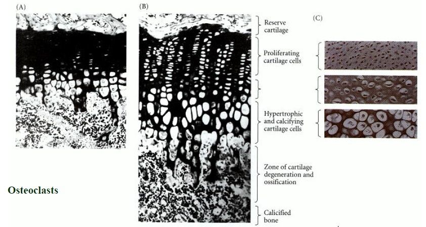
In the living organism, bone development is very complex; in most cases it follows the formation of a first skeleton of cartilage made by chondrocytes, which is then removed and replaced by bone, made by osteoblasts. Key growth factors in skeletal differentiation include bone morphogenetic proteins (BMPs), which determine to a major extent where bone differentiation occurs and where joint spaces are left between bones. The system of cartilage replacement by bone in the living organism has a complex regulatory system. It includes the bone morphogenetic proteins, in particular BMP2, that also regulate early patterning of the skeleton. Other growth factors that are important include transforming growth factor beta (TGF-β), which is part of a superfamily of proteins that include BMPs, which possess common signaling elements in the TGF beta signaling pathway. TGF-β is particularly important in cartilage differentiation, which in most cases precedes osteoblast-mediated bone formation. An additional family of essential bone regulatory factors is the fibroblast growth factors (FGFs), which determine where skeletal elements occur in relation to the skin.

bones in adults that contain red marrow serve an essential function in hemopoiesis (blood cell formation). Bones also serve as crucial reservoirs for calcium, phosphate, and other essential minerals. Almost all (99%) of the calcium in the body is stored in bones, from which the body draws its daily calcium needs.

Bone building: perfect protein 2
Bones are an amazing example of design, present in all vertebrates. They have a huge advantage over man-made girders, in that they are constantly rebuilding and redesigning themselves to cope with changing stress directions.1
This involves a fine balance of the activity of bone-depositing cells (osteoblasts) and bone resorbing cells (osteoclasts). It’s been recently shown that thyroid-stimulating hormone (TSH), best known for what its name says—stimulating the production of hormones in the thyroid gland—has an important role. It oversees both types of cells—without it, bones have osteoporosis in some parts (too little bone, so very weak), and are too dense in other patches.2 So both are essential. 

Osteocalcin and hydroxyapatite

The strength of bones mainly comes from the hexagonal mineral hydroxyapatite (HA, formula Ca5(PO4)3OH).3 But this must be built up in the right patterns. In vertebrate bones, this is built up with a special protein called osteocalcin (OC). It is a small protein, 49 amino acids long (5.8 kDa), and is ‘highly conserved’, meaning that its sequence is almost identical among vertebrates. Human OC has the sequence Tyr Leu Tyr Gln Trp Leu Gly Ala Pro Val Pro Tyr Pro Asp Pro Leu Gla Pro Arg Arg Gla Val Cys Gla Leu Asn Pro Asp Cys Asp Glu Leu Ala Asp His Ile Gly Phe Gln Glu Ala Tyr Arg Arg Phe Tyr Gly Pro Val.4

Like all proteins, the instructions for OC are in the DNA,5 but there is more to its manufacture than simply decoding / translating the code and synthesizing the OC on a ribosome. Firstly, the transcription (DNA→mRNA) is regulated by 1,25-dihydroxy-Vitamin D3, one reason that Vitamin D is so important for healthy bones. It is then first decoded (translated) as a preproosteocalcin, which is 98 amino acids long. This comprises three parts: a 23-residue signal protein that is cleaved during translation,5 a 26-residue target propeptide, and the 49-residue mature protein.6
Even this does not complete the process; this requires another vitamin—K. Vitamin K1 or phylloquinone, best known for its vital role in the blood clotting cascade, is an essential co-factor in -carboxylation. That is, the specific glutamyl residues (Glu, from the amino acid glutamic acid) at positions 17, 21 and 24 have a second carboxyl group (–COOH) added to form -carboxyglutamyl residues (Gla). This changes the structure and stabilizes the -helical portion of the protein.6
Even now, the OC protein is fairly shapeless. But when OC meets calcium ions, it folds to a special structure.8 The two carboxyl groups on the -carboxyglutamyl residues chelate1 to the Ca2+ ions, as shown by Fourier-Transform Infrared spectroscopy (FTIR).9 There was no spectral change when Ca2+ was added to decarboxylated OC (i.e. as it would be before converted by Vitamin K), showing that there is no binding without the carboxylation.9 Amazingly (for uniformitarians), enough osteocalcin to produce an immune reaction was found in bones of an Iguanodon ‘dated’ to 120 Ma,10 yet proteins could not last for millions of years. And the fact that it’s a bone protein shows it can’t be contamination from outside.

Osteocalcin’s crystal structure

Now, pig OC’s crystal structure has been discovered, using a type of X-ray diffraction called the iterative single anomalous scattering method. This provides new insights into how finely designed it must be to work.7 The active site has a negatively charged region that binds the positively charged Ca2+ ions. Five Ca2+ ions are coordinated by three special Gla residues and an Asp at position 30. But not in just any old way—five calcium ions are bound in the same arrangement as in the exposed face of a HA crystal, parallel to the c axis. So the OC can dock on the HA and add the calcium, and thus grow the crystal, making the bone grow in the area needed.
To do this, OC’s building blocks, the amino acids, must be in a very precise sequence. For example, there is a tightly packed core involving the hydrophobic residues Leu 16, Leu 32, Phe 38, Ala 41, Tyr 42, Phe 45 and Tyr 46. There is also hydrogen bonding to stabilize the connection between different α-helices, Asn 26 in the helix α1–α2 linker and Tyr 46 in α3. The helices α1 and α2 form a V-shaped arrangement stabilized by a disulphide bridge between Cys 23 and Cys 29.
Thus the very precise sequence of OC, as well as the metabolism to form the essential -carboxyglutamyl residues, seems to be yet another example of irreducible complexity, a hallmark of design.11 

This means this is yet another component that must be exactly right for the alleged transition from invertebrate to vertebrate. So it is not surprising that proponents of evolution have no fossil evidence for how the transition occurred—this protein alone shows it could never have happened. 2

Summary

Bones are dynamic supports, constantly rebuilding to cope with changing stresses. Bone shape is a delicate balance of bone deposition and resorption.Bone strength is largely from the mineral hydroxyapatite (HA).One vital component for bone growth is the small, highly conserved protein osteocalcin (OC).Large fragments have been found in dinosaur bones ‘dated’ over 100 million years old, although measured rates of breakdown mean that nothing should have survived that long.Vitamin K is essential to modify three amino acid residues of OC, otherwise it can’t bind calcium at all.Recent discovery of OC’s crystal structure shows that it binds calcium in exactly the right geometry to add to a certain crystal face of HA.

Thus bone construction is irreducibly complex.

Which explains why there is not only no fossil intermediate between invertebrate and vertebrate, but why it could not exist (and mainly because God didn’t create one!).

What is required for bone formation (osteogenesis) ?
 
Osteoblasts

 cells with single nuclei that synthesize bone

Mesenchymal stem cells                              

Multipotent stromal cells that can differentiate into a variety of cell types

Regulatory transcription factor Cbfa1/Runx2                       


osterix


bone morphogenetic proteins (BMPs)

determine to a major extent where bone differentiation occurs and where joint spaces are left between bones

alkaline phosphatase

provides high concentrations of phosphate at the site of mineral deposition

chondrocytes

formation of a first skeleton of cartilage

transforming growth factor beta (TGF-β)

 important in cartilage differentiation,

fibroblast growth factors (FGFs)

osteocalcin (OC)

determine where skeletal elements occur in relation to the skin

3-residue signal protein

1,25-dihydroxy-Vitamin D32

regulates transcription (DNA→mRNA)

26-residue target propeptide


49-residue mature protein


 Vitamin K1 or phylloquinone

essential co-factor in -carboxylation

calcium ions

osteopontin

paracrine factors
induce the nearby mesodermal cells to express two transcription factors, Pax1 and Scleraxis
transcription factors, Pax1 and Scleraxis

N-cadherin and N-Cam
important in the initiation of the condensation of the committed mesenchyme cells into compact nodules and differentiate into chondrocytes, the cartilage cells.
collagen X

fibronectin

enzymes

 active in the generation of calcium and phosphate ions and initiate the mineralization process within the cartilaginous matrix

Estrogen Receptors

1) https://en.wikipedia.org/wiki/Osteoblast
2) http://creation.com/bone-building-perfect-protein-osteocalcin
3. http://www.esf.edu/efb/turner/publication%20pdfs/turner%20stellenbosch%20complexity.pdf


Last edited by Admin on Tue Sep 11, 2018 3:04 am; edited 13 times in total

https://reasonandscience.catsboard.com

2Origin and development of bones ( Osteogenesis) Empty Osteogenesis: The Development of Bones Tue Feb 09, 2016 10:59 am

Otangelo


Admin

Osteogenesis: The Development of Bones 1

There are three distinct lineages that generate the skeleton. The somites generate the axial skeleton, the lateral plate mesoderm generates the limb skeleton, and the cranial neural crest gives rise to the branchial arch and craniofacial bones and cartilage.* There are two major modes of bone formation, or osteogenesis, and both involve the transformation of a preexisting mesenchymal tissue into bone tissue. The direct conversion of mesenchymal tissue into bone is called intramembranous ossification. This process occurs primarily in the bones of the skull. In other cases, the mesenchymal cells differentiate into cartilage, and this cartilage is later replaced by bone. The process by which a cartilage intermediate is formed and replaced by bone cells is called endochondral ossification.

Intramembranous ossification
Intramembranous ossification is the characteristic way in which the flat bones of the skull and the turtle shell are formed. During intramembranous ossification in the skull, neural crestderived mesenchymal cells proliferate and condense into compact nodules. (Thus, intramembranous ossification is not occurring from sclerotome-derived cells.) Some of these cells develop into capillaries; others change their shape to become osteoblasts, committed bone precursor cells (Figure 14.11A).

Origin and development of bones ( Osteogenesis) Osteob10

The osteoblasts secrete a collagen-proteoglycan matrix that is able to bind calcium salts. Through this binding, the prebone (osteoid) matrix becomes calcified. In most cases, osteoblasts are separated from the region of calcification by a layer of the osteoid matrix they secrete. Occasionally, though, osteoblasts become trapped in the calcified matrix and become osteocytes bone cells. As calcification proceeds, bony spicules radiate out from the region where ossification began (Figure 14.11B). Furthermore, the entire region of calcified spicules becomes surrounded by compact mesenchymal cells that form the periosteum (a membrane that surrounds the bone). The cells on the inner surface of the periosteum also become osteoblasts and deposit osteoid matrix parallel to that of the existing spicules. In this manner, many layers of bone are formed.

The mechanism of intramembranous ossification involves bone morphogenetic proteins ( BMPs ) and the activation of a transcription factor called CBFA1. Bone morphogenetic proteins (probably BMP2, BMP4, and BMP7) from the head epidermis are thought to instruct the neural crest-derived mesenchymal cells to become bone cells directly (Hall 1988). The BMPs activate the Cbfa1 gene in the mesenchymal cells. Just as the myogenic bHLH family of transcription factors is competent to transform primitive mesenchyme cells (or just about any other cell) into muscle-forming myoblasts, the CBFA1 transcription factor appears to be able to transform mesenchyme cells into osteoblasts. Ducy and her colleagues (1997) found that the mRNA for mouse CBFA1 is severely restricted to the mesenchymal condensations that form bone, and is limited to the osteoblast lineage. The protein appears to activate the genes for osteocalcin, osteopontin, and other bone-specific extracellular matrix proteins.

Origin and development of bones ( Osteogenesis) Endoch10
(Figure 14.13)

Endochondral ossification involves the formation of cartilage tissue from aggregated mesenchymal cells, and the subsequent replacement of cartilage tissue by bone. The process of endochondral ossification can be divided into five stages (Figure 14.13). First, the mesenchymal cells are commited to become cartilage cells. This committment is caused by paracrine factors that induce the nearby mesodermal cells to express two transcription factors, Pax1 and Scleraxis. These transcription factors are thought to activate cartilage-specific genes. Thus, Scleraxis is expressed in the mesenchyme from the sclerotome, in the facial mesenchyme that forms cartilaginous precursors to bone, and in the limb mesenchyme (Figure 14.14).

During the second phase of endochondral ossification, the committed mesenchyme cells condense into compact nodules and differentiate into chondrocytes, the cartilage cells. Ncadherin appears to be important in the initiation of these condensations, and N-CAM seems to be critical for maintaining them (Oberlender and Tuan 1994; Hall and Miyake 1995). In humans, the SOX9 gene, which encodes a DNA-binding protein, is expressed in the precartilaginous condensations. Mutations of the SOX9 gene cause camptomelic dysplasia, a rare disorder of skeletal development that results in deformities of most of the bones of the body. Most affected babies die from respiratory failure due to poorly formed tracheal and rib cartilage.

During the third phase of endochondral ossification, the chondrocytes proliferate rapidly to form the model for the bone. As they divide, the chondrocytes secrete a cartilage-specific extracellular matrix.

In the fourth phase, the chondrocytes stop dividing and increase their volume dramatically, becoming hypertrophic chondrocytes. These large chondrocytes alter the matrix they produce (by adding collagen X and more fibronectin) to enable it to become mineralized by calcium carbonate.

The fifth phase involves the invasion of the cartilage model by blood vessels. The hypertrophic chondrocytes die by apoptosis. This space will become bone marrow. As the cartilage cells die, a group of cells that have surrounded the cartilage model differentiate into osteoblasts. The ostoblasts begin forming bone matrix on the partially degraded cartilage. Eventually, all the cartilage is replaced by bone. Thus, the cartilage tissue serves as a model for the bone that follows. The skeletal components of the vertebral column, the pelvis, and the limbs are first formed of cartilage and later become bone.

The replacement of chondrocytes by bone cells is dependent on the mineralization of the extracellular matrix. This is clearly illustrated in the developing skeleton of the chick embryo, which utilizes the calcium carbonate of the eggshell as its calcium source. During development, the circulatory system of the chick embryo translocates about 120 mg of calcium from the shell to the skeleton. When chick embryos are removed from their shells at day 3 and grown in shell-less cultures (in plastic wrap) for the duration of their development, much of the cartilaginous skeleton fails to mature into bony tissue. A number of events lead to the hypertrophy and mineralization of the chondrocytes, including an initial switch from aerobic to anaerobic respiration, which alters their cell metabolism and mitochondrial energy potential. Hypertrophic chondrocytes secrete numerous small membrane-bound vesicles into the extracellular matrix. These vesicles contain enzymes that are active in the generation of calcium and phosphate ions and initiate the mineralization process within the cartilaginous matrix. The hypertrophic chondrocytes, their metabolism and mitochondrial membranes altered, then die by apoptosis.

In the long bones of many mammals (including humans), endochondral ossification spreads outward in both directions from the center of the bone . If all of our cartilage were turned into bone before birth, we would not grow any larger, and our bones would be only as large as the original cartilaginous model. However, as the ossification front nears the ends of the cartilage model, the chondrocytes near the ossification front proliferate prior to undergoing hypertrophy, pushing out the cartilaginous ends of the bone. These cartilaginous areas at the ends of the long bones are called epiphyseal growth plates. These plates contain three regions: a region of chondrocyte proliferation, a region of mature chondrocytes, and a region of hypertrophic chondrocytes (Figure 14.16)

Origin and development of bones ( Osteogenesis) Endoch11
(Figure 14.16)


As the inner cartilage hypertrophies and the ossification front extends farther outward, the remaining cartilage in the epiphyseal growth plate proliferates. As long as the epiphyseal growth plates are able to produce chondrocytes, the bone continues to grow. As new bone material is added peripherally from the internal surface of the periosteum,
there is a hollowing out of the internal region to form the bone marrow cavity. This destruction of bone tissue is due to osteoclasts, multinucleated cells that enter the bone through the blood vessels. Osteoclasts are probably derived from the same precursors as macrophage blood cells, and they dissolve both the inorganic and the protein portions of the bone matrix. Each osteoclast extends numerous cellular processes into the matrix and pumps out hydrogen ions onto the surrounding material, thereby acidifying and solubilizing it (Figure 14.17).

Origin and development of bones ( Osteogenesis) Endoch12
(Figure 14.17)

The blood vessels also import the blood-forming cells that will reside in the marrow for the duration of the organism's life. The number and activity of osteoclasts must be tightly regulated. If there are too many active osteoclasts, too much bone will be dissolved, and osteoporosis will result. Conversely, if not enough osteoblasts are produced, the bones are not hollowed out for the marrow, and osteopetrosis results. 


Is the regulation not information dependent ? So had the information not have to arise together with osteogenesis in a coordinated , interdependent manner ? 

Control of Cartilage Maturation at the Growth Plate
Recent discoveries of human and murine mutations resulting in abnormal skeletal development have provided remarkable insights into how the differentiation, proliferation, and patterning of chondrocytes are regulated.

Fibroblast Growth Factor Receptors
The proliferation of the epiphyseal growth plate cells  can be halted by the presence of fibroblast growth factors. These factors appear to instruct the cartilage precursors to differentiate rather than to divide. In humans, mutations of the receptors for fibroblast growth factors can cause these receptors to become activated prematurely. Such mutations give rise to the major types of human dwarfism. Achondroplasia is a dominant condition caused by mutations in the transmembrane region of fibroblast growth factor receptor 3 (FGFR3). Roughly 95% of achondroplastic dwarfs have the same mutation of FGFR3, a base pair substitution that converts glycine to arginine at position 380 in the transmembrane region of the protein. In addition, mutations in the extracellular portion of the FGFR3 protein or in the tyrosine kinase intracellular domain may result in thanatophoric dysplasia, a lethal form of dwarfism that resembles homozygous achondroplasia

Insulin-like Growth Factors
The epiphyseal growth plate cells are very responsive to hormones, and their proliferation is stimulated by growth hormone and insulin-like growth factors. Nilsson and colleagues (1986) showed that growth hormone stimulates the production of insulin-like growth factor I (IGF-I) in the epiphyseal chondrocytes, and that these chondrocytes respond to it by proliferating. When they added growth hormone to the tibial growth plates of young mice who could not manufacture their own growth hormone (because their pituitaries had been removed), it stimulated the formation of IGF-I in the chondrocytes of the proliferative zone. The combination of growth hormone and IGF-I appears to provide an extremely strong mitotic signal. It appears that IGF-I is essential for the normal growth spurt at puberty. The pygmies of the Ituri Forest of Zaire have normal growth hormone and IGF-I levels until puberty. However, at puberty, their IGF-I levels fall to about one-third that of other adolescents.

Estrogen Receptors
The pubertal growth spurt and the subsequent cessation of growth are induced by sex hormones. At the end of puberty, high levels of estrogen or testosterone cause the remaining epiphyseal plate cartilage to undergo hypertrophy. These cartilage cells grow, die, and are replaced by bone. Without any further cartilage formation, growth of these bones ceases, a process known as growth plate closure. In conditions of precocious puberty, there is an initial growth spurt (making the individual taller than his or her peers), followed by the cessation of epiphyseal cell division (allowing that person's peers to catch up and surpass his or her height). In males, it was not thought that estrogen played any role in these events. However, in 1994, Smith and colleagues published the case history of a man whose growth was still linear despite his undergoing a normal puberty. His epiphyseal plates had not matured, and he still had proliferating chondrocytes at 28 years of age. His "bone age" the amount of ephiphyseal cartilage he retained was roughly half his chronological age. This person was found to lack any functional estrogen receptor. At present, at least three human males have been reported who either cannot make estrogens or who lack the estrogen receptor. All three are close to 7 feet tall and are still growing . Therefore, estrogen plays a role in epiphyseal maturation in males as well as in females. Thyroid hormone and parathyroid-related hormone are also important in regulating the maturation and hypertrophy program of the epiphyseal growth plate (Ballock and Reddi 1994). Thus, children with hypothyroidism are prone to developing growth plate disorders.

Extracellular Matrix Proteins
The extracellular matrix of the cartilage is also critical for the proper differentiation and organization of growth plate chondrocytes. Mutations that affect type XI collagen or the sulfation of cartilage proteoglycans can cause severe skeletal abnormalities. Mice with deficiencies of type XI collagen die at birth with abnormalities of limb, mandible, rib, and tracheal cartilage. Failure to add sulfate groups to cartilage proteoglycans causes diastrophic dysplasia, a human dwarfism characterized by severe curvature of the spine, clubfoot, and deformed earlobes


1) Developmental Biology, 6th edition http://www.ncbi.nlm.nih.gov/books/NBK9983/

https://reasonandscience.catsboard.com

Otangelo


Admin

SKELETAL SYSTEM


Evolutionists defend their theory using the fossil record, often ascribing enormous significance to miniscule bone fragments. Tiny variations in bone structure often result in claims for an entirely new species of “fossil man.” But what knowledge do we possess about skeletal bone in the first place? The story often told by evolutionists is that bony structures first evolved in fish known as Agnathans. Most evolutionists contend that these jawless fish were the first vertebrates—supposedly living 500 million years ago. One such scenario alleges: “The most efficient way to swim was to wriggle from side to side. This style of motion was made more effective by having hard parts inside the body. These hard parts began as fluid-filled spaces which later accumulated minerals to take the form of bones” (Stewart, 2005). These fluid-filled spaces just “accumulated minerals to take the form of bones”? Certainly sounds easy enough. But this simplistic (textbook-like) description in no way mirrors the complex protein cascade known to occur. Today we recognize that the formation and maintenance of bone tissue is an enormously complex process that entails at least four specific types of bone cells. In addition, skeletal tissue requires various minerals, vitamins, hormones, and input from other systems of the body, such as oxygen, water, and nutrients from the circulatory and digestive systems. Its interdependence with other systems places the skeletal system in an interesting chicken-or-egg scenario. Without the circulatory system and digestive system in place, bone formation could not take place. However, those systems require the rigid framework of the skeletal system for support, protection, and locomotion. A loss of any of these vital components would result in physiological abnormalities of the skeletal system and negatively affect other body systems.
For instance, a lack of Vitamin D results in a condition called “rickets.” As Howard Glicksman noted:

Vitamin D has many functions within the body but the most important...is its ability to tell the digestive system to absorb calcium. If the body doesn’t efficiently absorb calcium then there won’t be enough raw material for the bone cells to use when they try to form bone. How do we know this? Well, modern medicine is very familiar with various forms of Vitamin D deficiency syndromes which can cause severe disability and even death. Therefore, by extrapolation we’ve come to realize that if there were a total absence of Vitamin D activity in the body, we would not be able to survive (2003).

Bones are dependent on Vitamin D, which is obtained from both food and sunlight, which allows calcium to be absorbed. The irony is that Vitamin D is not very soluble in blood serum, so a protein transporter is needed. This transporter protein is manufactured in the liver, and allows Vitamin D to be carried to the intestinal tract, allowing intestinal cells to absorb calcium, which is used in the manufacture of bones. However, before it can be used in the absorption of calcium, Vitamin D must become activated by enzymes in the liver and kidney. This precise pathway must be followed in a critical step-by-step fashion in order for calcium to be present for the formation of bones. Did the liver create the protein transporter first or the enzyme to help activate the Vitamin D? As Glicksman observed:

What good would it be for the liver to be able to start the activation process of Vitamin D if it hadn’t first produced the Vitamin D transport protein so that the Vitamin D could come to the liver in the first place? And what good would it be if the Vitamin D transport protein was able to transport Vitamin D, but the liver couldn’t start the activation process? And when did the kidney develop its ability to apply the final activating step without which Vitamin D activity in the body would be so reduced that intestinal absorption of calcium would be seriously hampered to the point of certain death? (2003).

So how were those “early fish” able to orchestrate such a precise pathway in just the right manner? Why would they go through the trouble in the first place? Would it even be statistically possible to grow living bone through a series of random chance events? The answer is a resounding no!

Skeletal Frame


The human body is composed of 206 bones ranging in size from long bones such as the femur of the leg (see Figure 1), to tiny bones such as those found within the inner ear (Van de Graaff and Fox, 1989, p. 205). At birth, the human skeleton contains approximately 270 bones, 64 of which fuse together as ossification takes place during normal growth. The word “skeleton” is derived from the Greek word skeletos, which means “dried up” (see Oxford Companion..., 2001, p. 622). Because of that “dried up” appearance, many people consider bone to be simple inorganic deposits of calcium and phosphorus. Yet, there is an active living component involved in bone as well. As David Cannatella noted: “Bone is a composite of inorganic calcium phosphate crystals (hydroxyapatite) and organic collagen fibers. The mineral content of a bone like the mammalian femur is about 67%. The mineral gives rigidity and the collagen resists tension” (2001). The living portion of bone is able to manufacture blood cells from within its marrow. A close inspection of the functions of bones quickly reveals they are far from just a dead deposit of minerals.

Functions of Bone

Body Movement—Probably one of the first attributes that comes to mind when one thinks of the skeletal system is its contribution to human locomotion. Biped mobility is uncommon in the animal world, and the human body appears to have been made for its upright stance (see Figure 2). As Van de Graaff and Fox noted: “Bones serve as anchoring attachments for most skeletal muscles. In this capacity, the bones act as levers with the joints functioning as pivots when muscles contract to cause body movement” (1989, p. 207). Miller and Goode went one step further in ascribing admiration to the complex lever action. They remarked:

When our muscles move us about, they do it by working a series of articulated levers that make a most efficient use of every ounce of muscular motive power. The levers are the bones of the body’s framework, fitted together with the neatness of jigsaw pieces and hinged by joints that must win the admiration of any mechanic (1960, p. 25, emp. added).

In addition to lever action, consider the various joint actions the skeletal system facilitates. The human body contains gliding, hinge, pivot, condyloid, saddle, and ball-and-socket joints (Oxford Companion..., p. 413), which allow a full range of movements. Also, the coccyx flexes anteriorly act as a shock absorber (Van de Graaff and Fox, p. 234). Levers, hinged joints, and shock absorbers? These sound like engineering feats. The question should be raised: Who was the ultimate Engineer who designed these actions?

Support
—Consider how ineffective other bodily systems would be if they could not rely on the rigid framework of the body’s skeletal system. Wayne Jackson compared the human skeletal system to the “interior framework of a house” (2000, p. 21). Imagine trying to hang drywall or build a sturdy roof without the proper framework. In his book Body by Design, Alan Gillen observed: “Our skeletal frames are more than just scaffolding that holds us erect; they serve as the structures upon which we hang all that we are. Our bones are the anchors to which muscles attach, and they act as the levers and fulcrums for our daily activities” (2001, p. 41). Most of the density and strength of the skeletal system results from the inorganic components. Consider that the skeletal system must be able to bear the weight of the entire body (even during periods of stress or locomotion). These attributes are due to the composite matrix that makes up bone.

Protection—In order to provide protection (as well as support and locomotion) bones must possess the strength of steel. Nevertheless, the weight of steel would be a huge drawback in locomotion. Therefore, bone must be extremely strong, yet, lightweight. The National Space Biomedical Research Institute noted: “The collagen fibers and calcium salts together make bone almost as strong as steel, but much lighter. Unlike steel, bone can repair itself when broken with the help of bone-forming cells (osteoblasts) and bone digesting cells (osteoclasts)” (“Muscles and Bones,” 2000, emp. added). Able to repair itself? Does this property sound like something that originated from a lifeless, Big Bang explosion? Most definitely not! As the Center for Disease Control explained:

When your body makes new bone tissue, it first lays down a framework of collagen. Then, tiny crystals of calcium from your blood spread throughout the collagen framework. The hard crystals fill in all the nooks and crannies. Calcium and collagen work together to make bones strong and flexible (see “Healthy Bones...,” n.d.).
Laying a framework indicates organization and purposeful activity. This complex process is accomplished by the composite integration of both organic and inorganic tissues—something evolution cannot explain.
The brain, the central processing unit of the body, is housed in a protective covering known as the cranium. Soft organs such as the lungs, heart, liver, and spleen, are housed within the safe confines of the rib cage. The pelvic viscera also are housed in a safe cavity surrounded by the bony pelvic girdle (Moore, 1992, p. 243). An unbiased observer recognizes this special protection as the product of careful design.

Hemopoiesis—At birth, humans produce red blood cells in the spleen and liver. Interestingly, this production of blood cells shifts to bones as humans develop and mature. The platelets, along with red and white blood cells, all are synthesized in the red blood marrow of bones (see Figure 3). This is one reason many diseases are currently being fought with bone marrow transplants—in hopes that this new tissue will be able to combat specific conditions. However, the production of these cells is not haphazard on some “assembly line.” As Bruce Alberts and his colleagues noted: “Thus blood cell formation (hemopoiesis) necessarily involves complex controls in which the production of each type of blood cell is regulated individually to meet changing needs” (Alberts, et al., 1994, p. 1164, emp. added). Those “controls” indicate a feedback loop with an elaborate series of steps involved in the production of blood cells—a scheme which is better explained by the Creation model.
In discussing the ability of bone marrow to make blood cells, Van de Graaff and Fox observed: “...[A]s bones mature, the bone marrow assumes the performance of this formidable task. It is estimated that an average of one million red blood cells are produced every second by the bone marrow” (p. 207). So exactly how can evolutionists explain the origins of the circulatory system, which is dependent on bones for the manufacture of blood cells, while the skeletal system could not have come into existence without nutrients from the circulatory system?! As Van de Graaff and Fox noted: “The development of bone from embryonic to adult size depends on the orderly processes of mitotic divisions, growth, and the structural remodeling determined by genetics, hormonal secretions, and nutritional supply” (p. 214, emp. added). The only logical conclusion is that the design from these two complex systems required a Designer and Creator.
Mineral Storage—Another important function of the skeletal system is the storage of inorganic material (minerals) that are essential for other bodily functions. When humans ingest calcium and phosphorus, approximately 90% of those minerals are stored in bones and teeth. These minerals give bones their rigidity. However, these minerals pose a serious problem for evolutionists. The vertebral skeleton is composed of calcium phosphate rather than calcium carbonate that is found in invertebrates (Ruben and Bennett, 1987, 41[6]:1187). How did vertebrates evolve this different composition of mineral storage?
Multiple functions, and yet the skeletal system performs each of these tasks seamlessly on a daily basis. As Brand and Yancey observed: “Perhaps an engineer will someday develop a substance as strong and light and efficient as bone, but what engineer could devise a substance that, like bone, can grow continuously, lubricate itself, require no shutdown time and repair itself when damage occurs?” (1980, p. 91).

Bone Cells

In addition to multiple functions, there are also four different bone cell types that have been identified: osteoblasts, osteocytes, osteoclasts, and undifferentiated bone mesenchymal cells (see “Bone Morphology,” 2006). Glicksman described the complexity and dependency of these four cell types:

The bone-forming cell is able to put down a firm mesh consisting of protein in which it then deposits crystals containing calcium phosphate. The bone breakdown cell is able to undue [sic] what the bone-forming cell accomplishes. They are constantly working together to develop and remodel the bone. In addition, there are two other bone cells that help the bone-forming and bone breakdown cells survive by separating them from the bone marrow and the body’s circulation and help them obtain water, nutrients and oxygen from the bloodstream. Remember, every cell in the body requires these basic components to live. So the bone cells themselves are dependent on the systems in the body that provide them with these vital necessities for life. Systems such as the gastrointestinal tract for absorbing water, calcium and the raw materials for protein mesh formation and energy production, the lungs and red blood cells for bringing in needed oxygen, and finally, the heart and the circulation for bringing all of these necessary components for survival and function to the site for bone formation” (2003, italics in orig.).

Did the osteoblasts (bone forming cells) evolve first, or the osteoclasts (bone erosion cells)? Both are needed for bone formation and maintenance.

http://www.apologeticspress.org/ApPubPage.aspx?pub=1&issue=581&article=625


Last edited by Otangelo on Thu Jan 20, 2022 6:11 pm; edited 1 time in total

https://reasonandscience.catsboard.com

Otangelo


Admin

http://www.astrobio.net/also-in-news/skeletons-developed-chemistry-oceans-changed-study-shows/

http://www.astrobio.net/origin-and-evolution-of-life/skeletons-in-the-pre-cambrian-closet/

https://reasonandscience.catsboard.com

Sponsored content


Back to top  Message [Page 1 of 1]

Permissions in this forum:
You cannot reply to topics in this forum