ElShamah - Reason & Science: Defending ID and the Christian Worldview
Would you like to react to this message? Create an account in a few clicks or log in to continue.
ElShamah - Reason & Science: Defending ID and the Christian Worldview

Welcome to my library—a curated collection of research and original arguments exploring why I believe Christianity, creationism, and Intelligent Design offer the most compelling explanations for our origins. Otangelo Grasso


You are not connected. Please login or register

Cell Membranes, origins through natural mechanisms, or design ?

Go down  Message [Page 1 of 1]

Otangelo


Admin

Cell Membranes, origins through natural mechanisms, or design ?  

https://reasonandscience.catsboard.com/t2128-cell-membranes-origins-through-natural-mechanisms-or-design

Steven A. Benner: Paradoxes in the Origin of Life 2014
Lipids that provide tidy compartments under the close supervision of a graduate student (supporting a protocellfirst model for origins) are quite non-robust with respect to small environmental perturbations, such as a change in the salt concentration, the introduction of organic solvents, or a change in temperature.
https://www.sci-hub.ee/10.1007/s11084-014-9379-0

Commentary: In the protocell-first scenario, simple self-assembled structures like lipid bilayers are proposed as the precursors to the complex cellular membranes of living organisms. These protocells are thought to encapsulate biochemical components necessary for life, thereby creating a microenvironment conducive to biochemical reactions. However, the sensitivity of these lipid membranes to factors such as salt concentration, the presence of organic solvents, and temperature variations raises questions about their viability as stable precursors to life in the variable conditions of early Earth. For instance, a slight increase in salt concentration disrupts the integrity of the lipid bilayer, leading to the leakage of essential biomolecules. Similarly, temperature changes could affect the fluidity and permeability of the membrane, while organic solvents could dissolve the lipids, destroying the compartmentalization essential for the protocell's function. These vulnerabilities suggest that if protocells were indeed the stepping stones towards the first living cells, they would require a remarkably stable and benign environment to maintain their integrity and facilitate the transition to more complex life forms. This requirement appears to be at odds with what is known about the early Earth's environment, which was likely subject to frequent and drastic changes due to volcanic activity, meteorite impacts, and variations in temperature and chemistry. The fragility of protocells to environmental perturbations thus serves as a significant piece of evidence against the notion that such structures could spontaneously give rise to life without some form of external stabilization or guidance. This challenges the spontaneous origin of life hypothesis by underscoring the need for more robust mechanisms or conditions that could have sustained the initial steps towards life in the face of the Earth's dynamic early environment.


Some popularisers of abiogenesis like to draw diagrams showing a simple hollow sphere of lipid (a ‘vesicle’) that can form under certain conditions in a test-tube. 2 However, such a ‘membrane’ could never lead to a living cell because the cell needs to get things through the cell membrane, in both directions. Such transport into and out of the cell entails very complex protein-lipid complexes known as transport channels, which operate like electro-mechanical pumps. They are specific to the various chemicals that must pass into and out of the cell (a pump that is designed to move water will not necessarily be suitable for pumping oil). Many of these pumps use energy compounds such as ATP to actively drive the movement against the natural gradient. Even when movement is with the gradient, from high to low concentration, it is still facilitated by carrier proteins.

The cell membrane also enables a cell to maintain a stable pH, necessary for enzyme activity, and favourable concentrations of various minerals (such as not too much sodium). This requires transport channels (‘pumps’) that specifically move hydrogen ions (protons) under the control of the cell. These pumps are highly selective.10

Transport across membranes is so important that “20–30% of all genes in most genomes encode membrane proteins”.11 The smallest known genome of a free-living organism, that of the parasite Mycoplasma genitalium, codes for 26 transporters12 amongst its 482 protein-coding genes.

A pure lipid membrane would not allow even the passive movement of the positively-charged ions of mineral nutrients such as calcium, potassium, magnesium, iron, manganese, etc., or the negatively-charged ions such as phosphate, sulfate, etc., into the cell, and they are all essential for life. A pure-lipid membrane would repel such charged ions, which dissolve in water, not lipid. Indeed, a simple fat membrane would prevent the movement of water itself (try mixing a lipid like olive oil with water)!

Membrane transporters would appear to be essential for a viable living cell.

The complexity of the lipidome is often not fully appreciated. Eukaryotic cells contain phospholipids, sphingolipids, and glycolipids with a wide variation in the hydrophilic headgroups as well as diverse fatty acid compositions. Add to this complexity other lipids such as steroids and fatty acid derived cellular products, and the complexity of the lipidome equals or exceeds that of the proteome.  1

According to this website : The Interdependency of Lipid Membranes and Membrane Proteins
The cell membrane contains various types of proteins, including ion channel proteins, proton pumps, G proteins, and enzymes. These membrane proteins function cooperatively to allow ions to penetrate the lipid bilayer. The interdependency of lipid membranes and membrane proteins suggests that lipid bilayers and membrane proteins co-evolved together with membrane bioenergetics.

The nonsense of this assertion is evident. How could the membrane proteins co-evolve, if they had to be manufactured by the machinery , protected by the cell membrane ?

The cell membrane contains various types of proteins, including ion channel proteins, proton pumps, G proteins, and enzymes. These membrane proteins function cooperatively to allow ions to penetrate the lipid bilayer.

The ER and Golgi apparatus together constitute the endomembrane compartment in the cytoplasm of eukaryotic cells. The endomembrane compartment is a major site of lipid synthesis, and the ER is where not only lipids are synthesized, but membrane-bound proteins and secretory proteins are also made.

So in order to make cell membranes, the Endoplasmic Recticulum is required. But also the Golgi Apparatus, the peroxysome, and the mitochondria. But these only function, if protected and encapsulated in the cell membrane.  What came first, the cell membrane, or the endoplasmic recticulum ? This is one of many other catch22 situations in the cell, which indicate that the cell could not emerge in a stepwise gradual manner, as proponents of natural mechanisms want to make us believe.

Not only is the cell membrane intricate and complex (and certainly not random), but it has tuning parameters such as the degree to which the phospholipid tails are saturated. It is another example of a sophisticated biological design about which evolutionists can only speculate. Random mutations must have luckily assembled molecular mechanisms which sense environmental challenges and respond to them by altering the phospholipid population in the membrane in just the right way. Such designs are tremendously helpful so of course they would have been preserved by natural selection. It is yet another example of how silly evolutionary theory is in light of scientific facts.

Membrane Structure
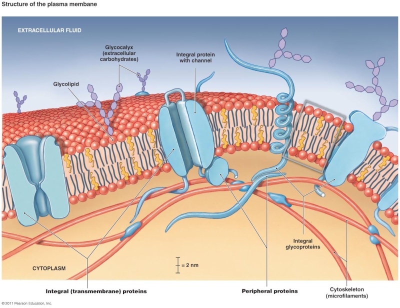
Membranes are the outer boundary of individual cells and of certain organelles. Plasma membranes are the selectively permeable outermost structures of cells that separate the interior of the cell from the environment. Certain molecules are permitted to enter and exit the cell through transport across the plasma membrane. Cell membranes contain lipids and proteins that form their structure and also facilitate cellular function. For example, cell adhesion and cell signaling are cellular processes initiated by the plasma membrane. Plasma membranes also serve as attachment points for intracellular cytoskeletal proteins and for components of the extracellular matrix outside of cells.

Lipids
In most cell membranes, lipids are the most abundant type of macromolecule present. Plasma and organelle membranes contain between 40% and 80% lipid. These lipids provide both the basic structure and the framework of the membrane and also regulate its function. Three types of lipids are found in cell membranes: phospholipids, cholesterol, and glycolipids.

1. Phospholipids: The most abundant of the membrane lipids are the phospholipids. They are polar, ionic compounds that are amphipathic in nature. That is, they have both hydrophilic and hydrophobic components. The hydrophilic or polar portion is in the “head group”. Within the head group is the phosphate and an alcohol that is attached to it. The alcohol can be serine, ethanolamine, inositol, or choline. Names of phospholipids then include phosphatidylserine, phosphatidylethanolamine, phosphatidylinositol, and phosphatidylcholine. While all these phospholipids contain a molecule called glycerol, the membrane phospholipid sphingomyelin has the alcohol choline in its head group and contains sphingosine instead of glycerol.

The hydrophobic portion of the phospholipid is a long, hydrocarbon (structure of carbons and hydrogens) fatty acid tail. While the polar head groups of the outer leafl et extend outward toward the environment, the fatty acid tails extend inward. Fatty acids may be saturated, containing the maximum number of hydrogen atoms bound to carbon atoms, or unsaturated with one or more carbonto- carbon double bonds. The length of the fatty acid chains and their degree of saturation impact the membrane structure. The fatty acid chains normally undergo motions such as fl exion (bending or fl exing), rotation, and lateral movement . Whenever a carbon-to-carbon double bond exists, there is a kink in the chain, reducing some types of motions and preventing the fatty acids from packing tightly together. Phospholipids in plasma membranes of healthy cells do not migrate or fl ip-fl op from one leafl et to the other. (However, during the process of programmed cell death, enzymes catalyze the movement of phosphatidylserine from the inner leaflet to the outer leaflet

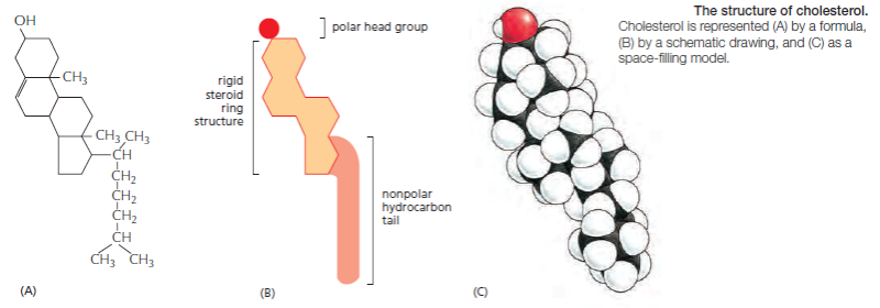
Cholesterol: Another major component of cell membranes is cholesterol. An amphipathic molecule, cholesterol contains a polar hydroxyl group as well as a hydrophobic steroid ring and attached hydrocarbon . Cholesterol is dispersed throughout cell membranes, intercalating between phospholipids. Its polar hydroxyl group is near the polar head groups of the phospholipids while the steroid ring and hydrocarbon tails of cholesterol are oriented parallel to those of the phospholipids. Cholesterol fits into the spaces created by the kinks of the unsaturated fatty acid tails, decreasing the ability of the fatty acids to undergo motion and therefore causing stiffening and strengthening of the membrane.

Glycolipids: Lipids with attached carbohydrate (sugars), glycolipids are found in cell membranes in lower concentration than phospholipids and cholesterol. The carbohydrate portion is always oriented toward the outside of the cell, projecting into the environment. Glycolipids help to form the carbohydrate coat observed on cells and are involved in cell-to-cell interactions. They are a source of blood group antigens and also can act as receptors for toxins including those from cholera and tetanus.

Proteins
While lipids form the main structure of the membrane, proteins are largely responsible for many biological functions of the membrane. For example, some membrane proteins function in transport of materials into and out of cells. Others serve as receptors for hormones or growth factors. The types of proteins within a plasma membrane vary depending on the cell type. However, all membrane proteins are associated with membrane in one of three main ways.

Membrane associations of proteins: While some proteins span the membrane with structures that cross from one side to the other, others are anchored to membrane lipids and still others are only peripherally associated with the cytosolic side of a plasma membrane

Lipid-anchored proteins: Members of the second category of membrane proteins are lipid-anchored proteins that are attached covalently to a portion of a lipid without entering the core portion of the bilayer of the membrane. Both transmembrane and lipid-anchored proteins are integral membrane proteins since they can only be removed from a membrane by disrupting the entire membrane structure.

Peripheral membrane proteins: Proteins in the third category are peripheral membrane proteins. These proteins are located on the cytosolic side of the membrane and are only indirectly attached to the lipid of the membrane; they bind to other proteins that are attached to the lipids. Cytoskeletal proteins, such as those involved in the spectrin membrane skeleton of erythrocytes, are examples of peripheral membrane proteins

Membrane protein functions: Membrane proteins enable cells to function as members of a tissue . For example, cell adhesion molecules are proteins that extend to the surface of cells and enable cell-to-cell contact . Other membrane proteins function as ion channels and transport proteins to enable molecules to enter and exit a cell. Membrane proteins that are ligand receptors enable cells to respond to hormones and other signaling molecules. The preceding examples of membrane proteins are of integral, transmembrane proteins whose structures span the bilayer. Lipid- anchored membrane proteins include the G proteins, which are named for their ability to bind to guanosine triphosphate (GTP) and participate in cell signaling in response to certain hormones .Peripheral membrane proteins include cytoskeletal proteins that attach to the membrane and regulate its shape and stabilize its structure. Some other peripheral membrane proteins are also involved in cell signaling and include enzymes attached to the inner membrane leafl et that are activated after a hormone binds to a protein receptor

Cell membranes are crucial to the life of the cell. The plasma membrane encloses the cell, defines its boundaries, and maintains the essential differences between the cytosol and the extracellular environment. Inside eukaryotic cells, the membranes of the nucleus, endoplasmic reticulum, Golgi apparatus, mitochondria, and other membrane-enclosed organelles maintain the characteristic differences between the contents of each organelle and the cytosol. Ion gradients across membranes, established by the activities of specialized membrane proteins, can be used to synthesize ATP, to drive the transport of selected solutes across the membrane, or, as in nerve and muscle cells, to produce and transmit electrical signals. In all cells, the plasma membrane also contains proteins that act as sensors of external signals, allowing the cell to change its behavior in response to environmental cues, including signals from other cells; these protein sensors, or receptors, transfer information—rather than molecules—across the membrane. Despite their differing functions, all biological membranes have a common general structure: each is a very thin film of lipid and protein molecules, held together mainly by noncovalent interactions. Cell membranes are dynamic, fluid structures, and most of their molecules move about in the plane of the membrane. The lipid molecules are arranged as a continuous double layer about 5 nm thick.

Cell Membranes, origins through natural mechanisms, or design ? Cellme10

This Lipid bilayer provides the basic fluid structure of the membrane and serves as a relatively impermeable barrier to the passage of most water-soluble molecules. Most membrane proteins span the lipid bilayer and mediate nearly all of the other functions of the membrane, including the transport of specific molecules across it, and the catalysis of membrane-associated reactions such as ATP synthesis. In the plasma membrane, some transmembrane proteins serve as structural links that connect the cytoskeleton through the lipid bilayer to either the extracellular matrix or an adjacent cell, while others serve as receptors to detect and transduce chemical signals in the cell’s environment. It takes many kinds of membrane proteins to enable a cell to function and interact with its environment, and it is estimated that about 30% of the proteins encoded in an animal’s genome are membrane proteins. In this chapter, we consider the structure and organization of the two main constituents of biological membranes—the lipids and the proteins. Although we focus mainly on the plasma membrane, most concepts discussed apply to the various internal membranes of eukaryotic cells as well.

The Lipid Bilayer
The lipid bilayer provides the basic structure for all cell membranes. It is easily seen by electron microscopy, and its bilayer structure is attributable exclusively to the special properties of the lipid molecules, which assemble spontaneously into bilayers even under simple artificial conditions. In this section, we discuss the different types of lipid molecules found in cell membranes and the general properties of lipid bilayers.

Phosphoglycerides, Sphingolipids, and Sterols Are the Major Lipids in Cell Membranes
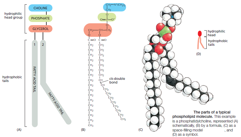
Lipid molecules constitute about 50% of the mass of most animal cell membranes, nearly all of the remainder being protein. There are approximately 5 × 10⁶ lipid molecules in a 1 μm × 1 μm area of lipid bilayer, or about 109 lipid molecules in the plasma membrane of a small animal cell. All of the lipid molecules in cell membranes are amphiphilic—that is, they have a hydrophilic (“water-loving”) or polar end and a hydrophobic (“water-fearing”) or nonpolar end. The most abundant membrane lipids are the phospholipids. These have a polar head group containing a phosphate group and two hydrophobic hydrocarbon tails. In animal, plant, and bacterial cells, the tails are usually fatty acids, and they can differ in length (they normally contain between 14 and 24 carbon atoms). One tail typically has one or more cis-double bonds (that is, it is unsaturated), while the other tail does not (that is, it is saturated).
As shown in Figure the figure below:
Cell Membranes, origins through natural mechanisms, or design ? Jkljqz10

each cis-double bond creates a kink in the tail. Differences in the length and saturation of the fatty acid tails influence how phospholipid molecules pack against one another, thereby affecting the fluidity of the membrane, as we discuss later. The main phospholipids in most animal cell membranes are the phosphoglycerides, which have a three-carbon glycerol backbone Two long-chain fatty acids are linked through ester bonds to adjacent carbon atoms of the glycerol, and the third carbon atom of the glycerol is attached to a phosphate group, which in turn is linked to one of several types of head group. By combining several different fatty acids and head groups, cells make many different phosphoglycerides. Phosphatidylethanolamine, phosphatidylserine, and phosphatidylcholine are the most abundant ones in mammalian cell membranes. In addition to phospholipids, the lipid bilayers in many cell membranes contain glycolipids and cholesterol. Glycolipids resemble sphingolipids, but, insteadof a phosphate-linked head group, they have sugars attached. We discuss glycolipids later. Eukaryotic plasma membranes contain especially large amounts of cholesterol—up to one molecule for every phospholipid molecule. Cholesterol is a sterol. It contains a rigid ring structure, to which is attached a single polar hydroxyl group and a short nonpolar hydrocarbon chain
Cell Membranes, origins through natural mechanisms, or design ? Jkljqz11

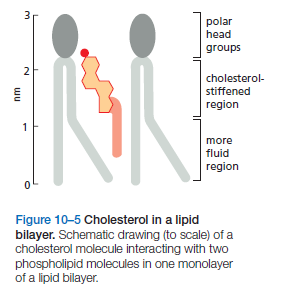
The cholesterol molecules orient themselves in the bilayer with their hydroxyl group close to the polar head groups of adjacent phospholipid molecules
Cell Membranes, origins through natural mechanisms, or design ? Jkljqz12

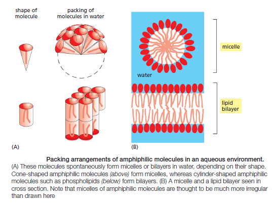
Phospholipids Spontaneously Form Bilayers
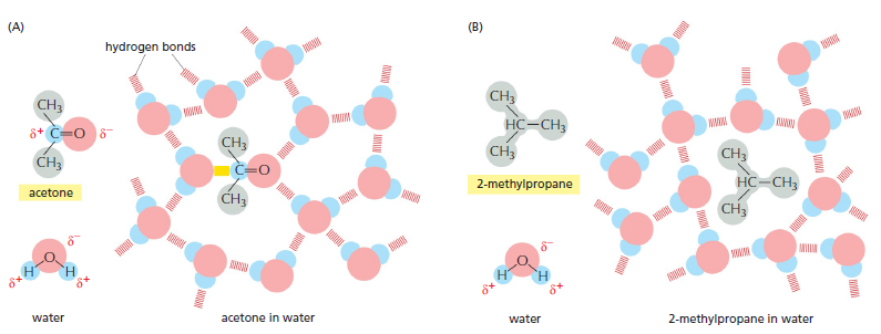
The shape and amphiphilic nature of the phospholipid molecules cause them to form bilayers spontaneously in aqueous environments. hydrophilic molecules dissolve readily in water because they contain charged groups or uncharged polar groups that can form either favorable electrostatic interactions or hydrogen bonds with water molecules
Cell Membranes, origins through natural mechanisms, or design ? Jkljqz13

Figure above How hydrophilic and hydrophobic molecules interact differently with water. (A) Because acetone is polar, it can form hydrogen bonds (red) and favorable electrostatic interactions (yellow) with water molecules, which are also polar. Thus, acetone readily dissolves in water. (B) By contrast, 2-methyl propane is entirely hydrophobic. Because it cannot form favorable interactions with water, it forces adjacent water molecules to reorganize into icelike cage structures, which increases the free energy. This compound is therefore virtually insoluble in water. The symbol δ– indicates a partial negative charge, and δ+ indicates a partial positive charge. Polar atoms are shown in color and nonpolar groups are shown in gray.

Hydrophobic molecules, by contrast, are insoluble in water because all, or almost all, of their atoms are uncharged and nonpolar and therefore cannot form energetically favorable interactions with water molecules. If dispersed in water, they force the adjacent water molecules to reorganize into icelike cages that surround the hydrophobic molecule . Because these cage structures are more ordered than the surrounding water, their formation increases the free energy. This free-energy cost is minimized, however, if the hydrophobic molecules (or the hydrophobic portions of amphiphilic molecules) cluster together so that the smallest number of water molecules is affected. When amphiphilic molecules are exposed to an aqueous environment, they behave as you would expect from the above discussion. They spontaneously aggregate to bury their hydrophobic tails in the interior, where they are shielded from the water, and they expose their hydrophilic heads to water. Depending on their shape, they can do this in either of two ways: they can form spherical micelles, with the tails inward, or they can form double-layered sheets, or bilayers, with the hydrophobic tails sandwiched between the hydrophilic head groups.
Cell Membranes, origins through natural mechanisms, or design ? Sdfsdf12

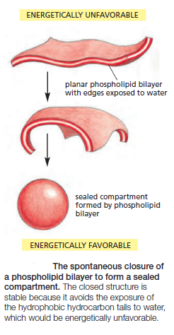
The same forces that drive phospholipids to form bilayers also provide a self-sealing property. A small tear in the bilayer creates a free edge with water; because this is energetically unfavorable, the lipids tend to rearrange spontaneously to eliminate the free edge. (In eukaryotic plasma membranes, the fusion of intracellular vesicles repairs larger tears.) The prohibition of free edges has a profound consequence: the only way for a bilayer to avoid having edges is by closing in on itself and forming a sealed compartment
Cell Membranes, origins through natural mechanisms, or design ? Sdfsdf13

This remarkable behavior, fundamental to the creation of a living cell, follows directly from the shape and amphiphilic nature of the phospholipid molecule. A lipid bilayer also has other characteristics that make it an ideal structure for cell membranes. One of the most important of these is its fluidity, which is crucial to many membrane functions 


The Lipid Bilayer Is a Two-dimensional Fluid

The Fluidity of a Lipid Bilayer Depends on Its Composition
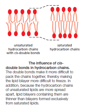
The fluidity of cell membranes has to be precisely regulated. Certain membrane transport processes and enzyme activities, for example, cease when the bilayer viscosity is experimentally increased beyond a threshold level. The fluidity of a lipid bilayer depends on both its composition and its temperature, as is readily demonstrated in studies of synthetic lipid bilayers. A synthetic bilayer made from a single type of phospholipid changes from a liquid state to a two-dimensional rigid crystalline (or gel) state at a characteristic temperature. This change of state is called a phase transition, and the temperature at which it occurs is lower (that is, the membrane becomes more difficult to freeze) if the hydrocarbon chains are short or have double bonds. A shorter chain length reduces the tendency of the hydrocarbon tails to interact with one another, in both the same and opposite monolayer, and cis-double bonds produce kinks in the chains that make them more difficult to pack together, so that the membrane remains fluid at lower temperatures
Cell Membranes, origins through natural mechanisms, or design ? Sdfsdf14

Bacteria, yeasts, and other organisms whose temperature fluctuates with that of their environment adjust the fatty acid composition of their membrane lipids to maintain a relatively constant fluidity. As the temperature falls, for instance, the cells of those organisms synthesize fatty acids with more cis-double bonds, thereby avoiding the decrease in bilayer fluidity that would otherwise result from the temperature drop. Cholesterol modulates the properties of lipid bilayers. When mixed with phospholipids, it enhances the permeability-barrier properties of the lipid bilayer. Cholesterol inserts into the bilayer with its hydroxyl group close to the polar head groups of the phospholipids, so that its rigid, platelike steroid rings interact with—and partly immobilize—those regions of the hydrocarbon chains closest to the polar head groups

By decreasing the mobility of the first few CH2 groups of the chains of the phospholipid molecules, cholesterol makes the lipid bilayer less deformable in this region and thereby decreases the permeability of the bilayer to small water-soluble molecules. Although cholesterol tightens the packing of the lipids in a bilayer, it does not make membranes any less fluid. At the high concentrations found in most eukaryotic plasma membranes, cholesterol also prevents the hydrocarbon chains from coming together and crystallizing.

The lipid composition of a typical eukaryotic cell membrane is much more complex than originally thought. These membranes contain a bewildering variety of perhaps 500–2000 different lipid species with even the simple plasma membrane of a red blood cell containing well over 150. While some of this complexity reflects the combinatorial variation in head groups, hydrocarbon chain lengths, and desaturation of the major phospholipid classes, some membranes also contain many structurally distinct minor lipids, at least some of which have important functions. The inositol phospholipids, for example, are present in small quantities in animal cell membranes and have crucial functions in guiding membrane traffic and in cell signaling

Their local synthesis and destruction are regulated by a large number of enzymes, which create both small intracellular signaling molecules and lipid docking sites on membranes that recruit specific proteins from the cytosol

Despite Their Fluidity, Lipid Bilayers Can Form Domains of Different Compositions
Because a lipid bilayer is a two-dimensional fluid, we might expect most types of lipid molecules in it to be well mixed and randomly distributed in their own monolayer. The van der Waals attractive forces between neighboring hydrocarbon tails are not selective enough to hold groups of phospholipid molecules together. With certain lipid mixtures in artificial bilayers, however, one can observe phase segregations in which specific lipids come together in separate domains

Cell Membranes, origins through natural mechanisms, or design ? Figure11

A model of a raft domain. Weak protein–protein, protein–lipid, and lipid–lipid interactions reinforce one another to partition the interacting components into raft domains. Cholesterol, sphingolipids, glycolipids, glycosylphosphatidylinositol (GPI)-anchored proteins, and some transmembrane proteins are enriched in these domains. Note that because of
their composition, raft domains have an increased membrane thickness.We discuss glycolipids, GPI-anchored proteins, and oligosaccharide linkers later.

The Asymmetry of the Lipid Bilayer Is Functionally Important
The lipid compositions of the two monolayers of the lipid bilayer in many membranes are strikingly different. In the human red blood cell (erythrocyte) membrane, for example, almost all of the phospholipid molecules that have choline—( CH3)3N+CH2CH2OH—in their head group (phosphatidylcholine and sphingomyelin) are in the outer monolayer, whereas almost all that contain a terminal primary amino group (phosphatidylethanolamine and phosphatidylserine) are in the inner monolayer

Cell Membranes, origins through natural mechanisms, or design ? Sem_ty46

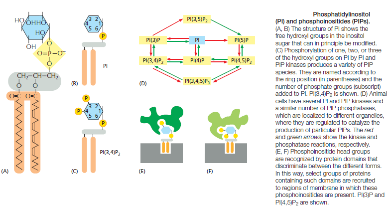
Because the negatively charged phosphatidylserine is located in the inner monolayer, there is a significant difference in charge between the two halves of the bilayer. Lipid asymmetry is functionally important, especially in converting extracellular signals into intracellular ones . Many cytosolic proteins bind to specific lipid head groups found in the cytosolic monolayer of the lipid bilayer. The enzyme protein kinase C (PKC), for example, which is activated in response to various extracellular signals, binds to the cytosolic face of the plasma membrane, where phosphatidylserine is concentrated, and requires this negatively charged phospholipid for its activity. In other cases, specific lipid head groups must first be modified to create protein-binding sites at a particular time and place. One example is phosphatidylinositol (PI), one of the minor phospholipids that are concentrated in the cytosolic monolayer of cell membranes
Cell Membranes, origins through natural mechanisms, or design ? Yyuu10

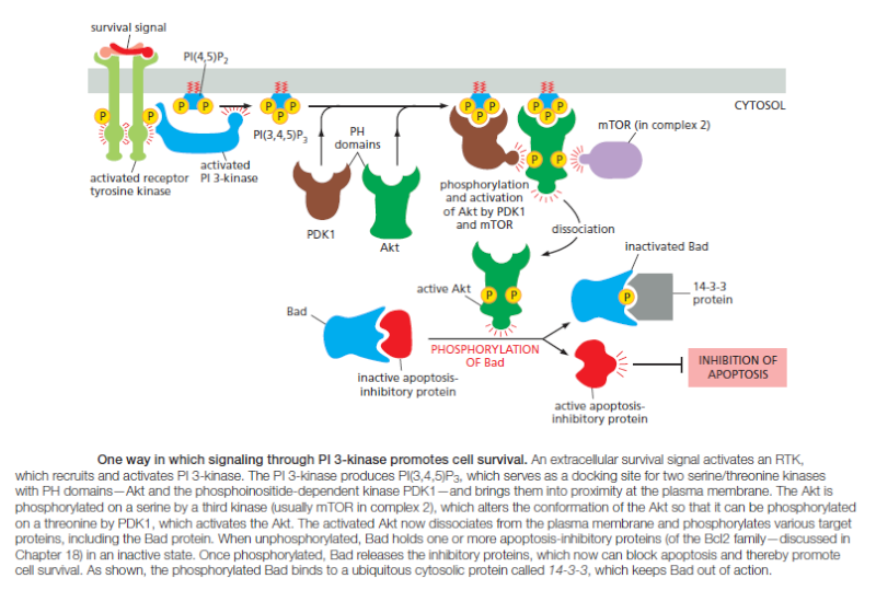
Various lipid kinases can add phosphate groups at distinct positions on the inositol ring, creating binding sites that recruit specific proteins from the cytosol to the membrane. An important example of such a lipid kinase is phosphoinositide 3-kinase (PI 3-kinase), which is activated in response to extracellular signals and helps to recruit specific intracellular signaling proteins to the cytosolic face of the plasma membrane
Cell Membranes, origins through natural mechanisms, or design ? Qweraa10

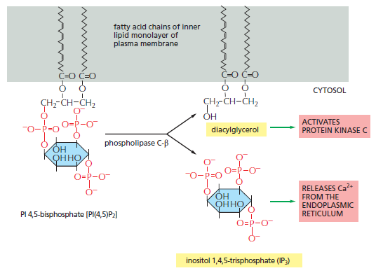
Similar lipid kinases phosphorylate inositol phospholipids in intracellular membranes and thereby help to recruit proteins that guide membrane transport. Phospholipids in the plasma membrane are used in yet another way to convert extracellular signals into intracellular ones. The plasma membrane contains various phospholipases that are activated by extracellular signals to cleave specific phospholipid molecules, generating fragments of these molecules that act as short-lived intracellular mediators. Phospholipase C, for example, cleaves an inositol phospholipid in the cytosolic monolayer of the plasma membrane to generate two fragments, one of which remains in the membrane and helps activate protein kinase C, while the other is released into the cytosol and stimulates the release of Ca2+ from the endoplasmic reticulum
Cell Membranes, origins through natural mechanisms, or design ? Qwersd10

The hydrolysis of PI(4,5) P2 by phospholipase C-β. Two second messengers are produced directly from the hydrolysis of PI(4,5)P2: inositol 1,4,5-trisphosphate (IP3), which diffuses
through the cytosol and releases Ca2+ from the endoplasmic reticulum, and diacylglycerol, which remains in the membrane and helps to activate protein kinase C (PKC see below).
Cell Membranes, origins through natural mechanisms, or design ? Qwersd11

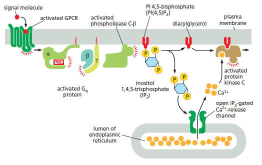
How GPCRs increase cytosolic Ca2+ and activate protein kinase C. The activated GPCR stimulates the plasma-membrane-bound phospholipase C-β (PLCβ) via a G protein called Gq. The α subunit and βγ complex of Gq are both involved in this activation. Two second messengers are produced when PI(4,5)P2 is hydrolyzed by activated PLCβ. Inositol 1,4,5-trisphosphate (IP3) diffuses through the cytosol and releases Ca2+ from the ER by binding to and opening IP3-gated Ca2+-release channels (IP3 receptors) in the ER membrane. The large electrochemical gradient for Ca2+ across this membrane causes Ca2+ to escape into the cytosol when the release channels are opened. Diacylglycerol remains in the plasma membrane and, together with phosphatidylserine (not shown) and Ca2+, helps to activate protein kinase C (PKC), which is recruited from the cytosol to the cytosolic face of the plasma membrane. Of the 10 or more distinct isoforms of PKC in humans, at least 4 are activated by diacylglycerol. There are several classes of phospholipase C: these include the β class, which is activated by GPCRs; as we see later, the γ class is activated by a class of enzymecoupled receptors called receptor tyrosine
kinases (RTKs).

Once activated, PKC phosphorylates target proteins that vary depending on the cell type. The principles are the same as discussed earlier for PKA, although most of the target proteins are different. Diacylglycerol can be further cleaved to release arachidonic acid, which can either act as a signal in its own right or be used in the synthesis of other small lipid signal molecules called eicosanoids. Most vertebrate cell types make eicosanoids, including prostaglandins, which have many biological activities. They participate in pain and inflammatory responses, for example, and many anti-inflammatory drugs (such as aspirin, ibuprofen, and cortisone) act in part by inhibiting their synthesis.


Ca2+ Functions as a Ubiquitous Intracellular Mediator
Many extracellular signals, and not just those that work via G proteins, trigger an increase in cytosolic Ca2+ concentration. In muscle cells, Ca2+ triggers contraction, and in many secretory cells, including nerve cells, it triggers secretion. Ca2+ has numerous other functions in a variety of cell types. Ca2+ is such an effective signaling mediator because its concentration in the cytosol is normally very low (~10–7 M), whereas its concentration in the extracellular fluid (~10–3 M) and in the lumen of the ER [and sarcoplasmic reticulum (SR) in muscle] is high. Thus, there is a large gradient tending to drive Ca2+ into the cytosol across both the plasma membrane and the ER or SR membrane. When a signal transiently opens Ca2+ channels in these membranes, Ca2+ rushes into the cytosol, and the resulting 10–20-fold increase in the local Ca2+ concentration activates Ca2+ responsive proteins in the cell. Some stimuli, including membrane depolarization, membrane stretch, and certain extracellular signals, activate Ca2+ channels in the plasma membrane, resulting in Ca2+ influx from outside the cell. Other signals, including the GPCR-mediated signals described earlier, act primarily through IP3 receptors to stimulate Ca2+ release from intracellular stores in the ER (see Figure 15–29). The ER membrane also contains a second type of regulated Ca2+ channel called the ryanodine receptor (so called because it is sensitive to the plant alkaloid ryanodine), which opens in response to rising Ca2+ levels and thereby amplifies the Ca2+ signal, as we describe shortly. Several mechanisms rapidly terminate the Ca2+ signal and are also responsible for keeping the concentration of Ca2+ in the cytosol low in resting cells. Most importantly, there are Ca2+-pumps in the plasma membrane and the ER membrane that use the energy of ATP hydrolysis to pump Ca2+ out of the cytosol. Cells such as muscle and nerve cells, which make extensive use of Ca2+ signaling, have an additional Ca2+ transporter (a Na+-driven Ca2+ exchanger) in their plasma membrane that couples the efflux of Ca2+ to the influx of Na+.

Feedback Generates Ca2+ Waves and Oscillations
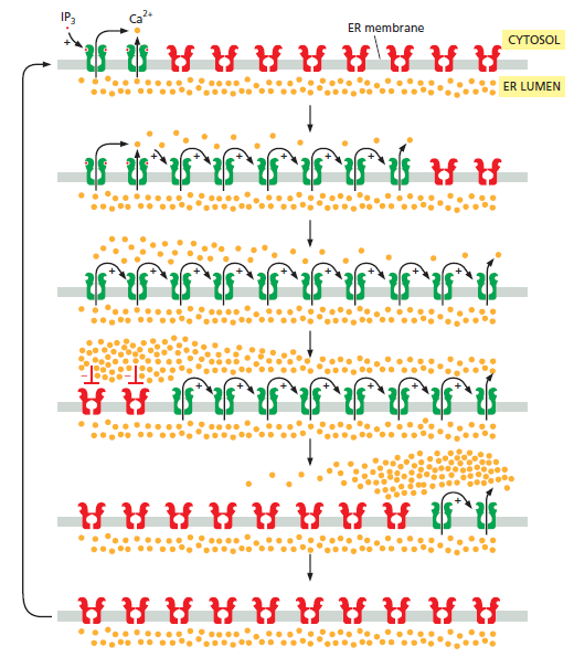
The IP3 receptors and ryanodine receptors of the ER membrane have an important feature: they are both stimulated by low to moderate cytoplasmic Ca2+ concentrations. This Ca2+-induced calcium release (CICR) results in positive feedback, which has a major impact on the properties of the Ca2+ signal. The importance of this feedback is seen clearly in studies with Ca2+-sensitive fluorescent indicators, such as aequorin or fura-2 , which allow researchers to monitor cytosolic Ca2+ in individual cells under a microscope.When cells carrying a Ca2+ indicator are treated with a small amount of an extracellular signal molecule that stimulates IP3 production, tiny bursts of Ca2+ are seen in one or more discrete regions of the cell. These Ca2+ puffs or sparks reflect the local opening of small groups of IP3-gated Ca2+-release channels in the ER. Because various Ca2+-binding proteins act as Ca2+ buffers and restrict the diffusion of Ca2+, the signal often remains localized to the site where the Ca2+ enters the cytosol. If the extracellular signal is sufficiently strong and persistent, however, the local Ca2+ concentration can reach a sufficient level to activate nearby IP3 receptors and ryanodine receptors, resulting in a regenerative wave of Ca2+ release that moves through the cytosol (Figure 15–31), much like an action potential in an axon.
Cell Membranes, origins through natural mechanisms, or design ? Dfgf11

Positive and negative feedback produce Ca2+ waves and oscillations. This diagram shows IP3 receptors and ryanodine receptors on a portion of the ER membrane: active receptors are in green; inactive receptors are in red. When a small amount of cytosolic IP3 activates a cluster of IP3 receptors at one site on the ER membrane (top), the local release of Ca2+ promotes the opening of nearby IP3 and ryanodine receptors, resulting in more Ca2+ release. This positive feedback (indicated by positive signs) produces a regenerative wave of Ca2+ release that spreads across the cell. These waves of Ca2+ release move more quickly across the cell than would be possible by simple diffusion. Also, unlike a diffusing burst of Ca2+ ions, which will become more dilute as it spreads, the regenerative wave produces a high Ca2+ concentration across the entire cell. Eventually, the local Ca2+ concentration inactivates IP3 receptors and ryanodine receptors (middle; indicated by red negative signs), shutting down the Ca2+ release. Ca2+-pumps reduce the local cytosolic Ca2+ concentration to its normal low levels. The result is a Ca2+ spike: positive feedback drives a rapid rise in cytosolic Ca2+, and negative feedback sends it back downagain. The Ca2+ channels remain refractory to further stimulation for some period of time, delaying the generation of another Ca2+ spike (bottom). Eventually, however, the negative feedback wears off, allowing IP3 to trigger another Ca2+ wave. The end result is repeated Ca2+ oscillations . Under some conditions, these oscillations can be seen as repeating narrow narrow waves of Ca2+ moving across the cell.


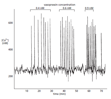
Another important property of IP3 receptors and ryanodine receptors is that they are inhibited, after some delay, by high Ca2+ concentrations (a form of negative feedback). Thus, the rise in Ca2+ in a stimulated cell leads to inhibition of Ca2+ release; because Ca2+ pumps remove the cytosolic Ca2+, the Ca2+ concentration falls . The decline in Ca2+ eventually relieves the negative feedback, allowing cytosolic Ca2+ to rise again. As in other cases of delayed negative feedback (see Figure 15–18), the result is an oscillation in the Ca2+ concentration. These oscillations persist for as long as receptors are activated at the cell surface, and their frequency reflects the strength of the extracellular stimulus
Cell Membranes, origins through natural mechanisms, or design ? Dfgfsd10

Picture above. Vasopressin-induced Ca2+oscillations in a liver cell. The cell was loaded with the Ca2+-sensitive protein aequorin and then exposed to increasing concentrations of the peptide signal molecule vasopressin, which activates a GPCR and thereby PLCβ . Note that the frequency of the Ca2+ spikes increases with an increasing concentration of vasopressin but that the amplitude of the spikes is not affected. Each spike lasts about 7 seconds.

The frequency, amplitude, and breadth of oscillations can also be modulated by other signaling mechanisms, such as phosphorylation, which influence the Ca2+ sensitivity of Ca2+ channels or affect other components in the signaling system. The frequency of Ca2+ oscillations can be translated into a frequency-dependent cell response. In some cases, the frequency-dependent response itself is also oscillatory: in hormone-secreting pituitary cells, for example, stimulation by an extracellular signal induces repeated Ca2+ spikes, each of which is associated with a burst of hormone secretion. In other cases, the frequency-dependent response is non-oscillatory: in some types of cells, for instance, one frequency of Ca2+ spikes activates the transcription of one set of genes, while a higher frequency activates the transcription of a different set. How do cells sense the frequency of Ca2+ spikes and change their response accordingly? The mechanism presumably depends on Ca2+-sensitive proteins that change their activity as a function of Ca2+-spike frequency.
A protein kinase that acts as a molecular memory device seems to have this remarkable property, as we discuss next.

Ca2+/Calmodulin-Dependent Protein Kinases Mediate Many Responses to Ca2+ Signals
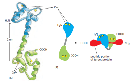
Various Ca2+-binding proteins help to relay the cytosolic Ca2+ signal. The most important is calmodulin, which is found in all eukaryotic cells and can constitute as much as 1% of a cell's total protein mass. Calmodulin functions as a multipurpose intracellular Ca2+ receptor, governing many Ca2+-regulated processes. It consists of a highly conserved, single polypeptide chain with four high-affinity Ca2+-binding sites
Cell Membranes, origins through natural mechanisms, or design ? Erttr10

The structure of Ca2+/calmodulin. (A) The molecule has a dumbbell shape, with two globular ends, which can bind to many target proteins. The globular ends are connected by a long, exposed α helix, which allows the protein to adopt a number of different conformations, depending on the target protein it interacts with. Each globular head has two Ca2+- binding sites (Movie 15.6). (B) Shown is the major structural change that occurs in Ca2+/calmodulin when it binds to a target protein (in this example, a peptide that consists of the Ca2+/calmodulin-binding domain of a Ca2+/calmodulin-dependent protein kinase). Note that the Ca2+/calmodulin has “jack-knifed” to surround the peptide. When it binds to other targets, it can adopt different conformations.

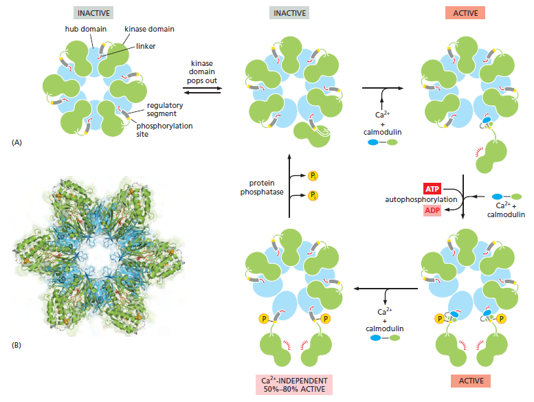
When activated by Ca2+ binding, it undergoes a conformational change. Because two or more Ca2+ ions must bind before calmodulin adopts its active conformation, the protein displays a sigmoidal response to increasing concentrations of Ca2+.The allosteric activation of calmodulin by Ca2+ is analogous to the activation of PKA by cyclic AMP, except that Ca2+/calmodulin has no enzymatic activity itself but instead acts by binding to and activating other proteins. In some cases, calmodulin serves as a permanent regulatory subunit of an enzyme complex, but usually the binding of Ca2+ instead enables calmodulin to bind to various target proteins in the cell to alter their activity. When an activated molecule of Ca2+/calmodulin binds to its target protein, the calmodulin further changes its conformation, the nature of which depends on the specific target protein (Figure B above). Among the many targets calmodulin regulates are enzymes and membrane transport proteins. As one example, Ca2+/calmodulin binds to and activates the plasma membrane Ca2+-pump that uses ATP hydrolysis to pump Ca2+ out of cells. Thus, whenever the concentration of Ca2+ in the cytosol rises, the pump is activated, which helps to return the cytosolic Ca2+ level to resting levels. Many effects of Ca2+, however, are more indirect and are mediated by protein phosphorylations catalyzed by a family of protein kinases called Ca2+/calmodulin- dependent kinases (CaM-kinases). Some CaM-kinases phosphorylate transcription regulators, such as the CREB protein, and in this way activate or inhibit the transcription of specific genes. One of the best-studied CaM-kinases is CaM-kinase II, which is found in most animal cells but is especially enriched in the nervous system. It constitutes up to 2% of the total protein mass in some regions of the brain, and it is highly concentrated in synapses. CaM-kinase II has several remarkable properties. To begin
with, it has a spectacular quaternary structure: twelve copies of the enzyme are assembled into a stacked pair of rings, with kinase domains on the outside linked to a central hub
Cell Membranes, origins through natural mechanisms, or design ? Dsffsd10

The stepwise activation of CaM-kinase II. (A) Each CaM-kinase II protein has two major domains: an aminoterminal kinase domain (green) and a carboxyl-terminal hub domain (blue), linked by a regulatory segment. Six CaM-kinase II proteins are assembled into a giant ring in which the hub domains interact tightly to produce a central structure that is surrounded by kinase domains. The complete enzyme contains two stacked rings, for a total of 12 kinase proteins, but only one ring is shown here for clarity. When the enzyme is inactive, the ring exists in a dynamic equilibrium between two states. The first (upper left) is a compact state, in which the kinase domains interact with the hub, so that the regulatory segment is buried in the kinase active site and thereby blocks catalytic activity. In the second inactive state (upper middle), a kinase domain has popped out and is linked to the central hub by its regulatory segment, which continues to inhibit the kinase but is now accessible to Ca2+/calmodulin. If present, Ca2+/calmodulin will bind the regulatory segment and prevent it from inhibiting the kinase, thereby locking the kinase in an active state (upper right). If the adjacent kinase subunit also pops out from the hub, it will also be activated by Ca2+/calmodulin, and the two kinases will then phosphorylate each other on their regulatory segments (lower right). This autophosphorylation further activates the enzyme. It also prolongs the activity of the enzyme in two ways. First, it traps the bound Ca2+/calmodulin so that it does not dissociate from the enzyme until cytosolic Ca2+ levels return to basal values for at least 10 seconds (not shown). Second, it converts the enzyme to a Ca2+-independent form, so that the kinase remains active even after the Ca2+/calmodulin dissociates from it (lower left). This activity continues until the action of a protein phosphatase overrides the autophosphorylation activity of CaM-kinase II. (B) This structural model of the enzyme is based on x-ray crystallographic analysis. The remarkable dodecameric structure of the enzyme allows it to achieve a broad range of intermediate activity states in response to different Ca2+ oscillation frequencies: higher frequencies tend to cause more subunits in the enzyme to reach the phosphorylated active state. The behavior of CaM-kinase II is also controlled by the length of the linker segment between the kinase and hub domains. The linker is longer in some isoforms of the enzyme; in these isoforms, the kinase domains tend to pop out of the ring more frequently, making it more sensitive to Ca2+. These and other mechanisms allow the cell to tailor the responsiveness of the enzyme to the needs of different types of neurons.

This structure helps the enzyme function as a molecular memory device, switching to an active state when exposed to Ca2+/ calmodulin and then remaining active even after the Ca2+ signal has decayed. This is because adjacent kinase subunits can phosphorylate each other (a process called autophosphorylation) when Ca2+/calmodulin activates them  Once a kinase subunit is autophosphorylated, it remains active even in the absence of Ca2+, thereby prolonging the duration of the kinase activity beyond that of the initial activating Ca2+ signal. The enzyme maintains this activity until a protein phosphatase removes the autophosphorylation and shuts the kinase off. CaM-kinase II activation can thereby serve as a memory trace of a prior Ca2+ pulse, and it seems to have a role in some types of memory and learning in the vertebrate nervous system. Mutant mice that lack a brain-specific form of the enzyme have specific defects in their ability to remember where things are. Another remarkable property of CaM-kinase II is that the enzyme can use its intrinsic memory mechanism to decode the frequency of Ca2+ oscillations. This property is thought to be especially important at a nerve cell synapse, where changes in intracellular Ca2+ levels in a postsynaptic cell as a result of neural activity can lead to long-term changes in the subsequent effectiveness of that synapse When CaM-kinase II is exposed to both a protein phosphatase and repetitive pulses of Ca2+/calmodulin at different frequencies that mimic those observed in stimulated cells, the enzyme’s activity increases steeply as a function of pulse frequency
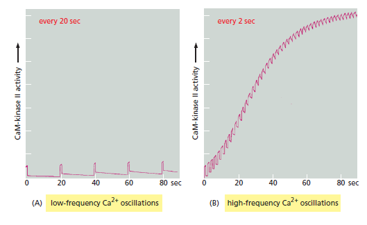
Cell Membranes, origins through natural mechanisms, or design ? Wertwr10

CaM-kinase II as a frequency decoder of Ca2+ oscillations. (A) At low frequencies of Ca2+ spikes, the enzyme becomes inactive after each spike, as the autophosphorylation induced by Ca2+/calmodulin binding does not maintain the enzyme’s activity long enough for the enzyme to remain active until the next Ca2+ spike arrives. (B) At higher spike frequencies, however, the enzyme fails to inactivate completely between Ca2+ spikes, so its activity ratchets up with each spike. If the spike frequency is high enough, this progressive increase in enzyme activity will continue until the enzyme is autophosphorylated on all subunits and is therefore maximally activated. Although not shown, once enough of its subunits are autophosphorylated, the enzyme can be maintained in a highly active state even with a relatively low frequency of Ca2+ spikes (a form of cell memory). The binding of Ca2+/ calmodulin to the enzyme is enhanced by the CaM-kinase II autophosphorylation (an additional form of positive feedback), helping to generate a more switchlike response to repeated Ca2+ spikes.

Some G Proteins Directly Regulate Ion Channels
G proteins do not act exclusively by regulating the activity of membrane-bound enzymes that alter the concentration of cyclic AMP or Ca2+ in the cytosol. The α subunit of one type of G protein (called G12), for example, activates a guanine nucleotide exchange factor (GEF) that activates a monomeric GTPase of the Rho family (discussed later and in Chapter 16), which regulates the actin cytoskeleton. In some other cases, G proteins directly activate or inactivate ion channels in the plasma membrane of the target cell, thereby altering the ion permeability— and hence the electrical excitability—of the membrane. As an example, acetylcholine released by the vagus nerve reduces the heart rate (see Figure 15–5B). This effect is mediated by a special class of acetylcholine receptors that activate the Gi protein discussed earlier. Once activated, the α subunit of Gi inhibits adenylyl cyclase (as described previously), while the βγ subunits bind to K+ channels in the heart muscle cell plasma membrane and open them. The opening of these K+ channels makes it harder to depolarize the cell and thereby contributes to the inhibitory effect of acetylcholine on the heart. (These acetylcholine receptors, which can be activated by the fungal alkaloid muscarine, are called muscarinic acetylcholine receptors to distinguish them from the very different nicotinic acetylcholine receptors, which are ion-channel-coupled receptors on skeletal muscle and nerve cells that can be activated by the binding of nicotine, as well as by acetylcholine.) Other G proteins regulate the activity of ion channels less directly, either by stimulating channel phosphorylation (by PKA, PKC, or CaM-kinase, for example) or by causing the production or destruction of cyclic nucleotides that directly activate or inactivate ion channels. These cyclic-nucleotide-gated ion channels have a crucial role in both smell (olfaction) and vision, as we now discuss.
1. https://www.ncbi.nlm.nih.gov/pmc/articles/PMC2579957/
2. http://creation.com/origin-of-life


Last edited by Otangelo on Sat Mar 02, 2024 3:24 pm; edited 19 times in total

https://reasonandscience.catsboard.com

Otangelo


Admin

As we saw in the previous post the cell membrane has been found to be increasingly sophisticated. Not only is the protein-punctuated sandwich structure incredibly complicated, but organisms actively fine-tune its design as conditions change. For instance, the number and location of carbon-carbon double bonds, in the phospholipid tails in the lipid bilayer, are astonishingly modified in real-time to deal with temperature changes. A recent paper agreed that “the membrane is a biological device of a staggering complexity”: 1

Complex Patterns Have Been Discovered in the Cell Membrane 1
The membrane that surrounds and encapsulates living cells is truly amazing. This lipid bilayer-protein mixture, which is only a few nanometers thick, has complex chemical, structural and electrical properties which are finely-tuned and crucial for the cell’s operation. And the various proteins perform a great diversity of tasks, ranging from transmitting information to transporting molecules, across the membrane. As usual the inexorable march of science continues to reveal the sophistication and elegance of the membrane design. A recent study, for instance, revealed that the membrane proteins are arranged in complex patterns, ranging from what has been described as “patches” to “networks.” Furthermore, the all-important lipid composition of the membrane strongly influences these patterns as well as the protein sequences themselves. This work reveals far greater structure and organization in the cell membrane than was previously understood. And it continues to reveal how unlikely it is that such designs spontaneously arose. For more interesting details on the cell membrane design, here is a post from a year ago on the fine-tuning of the membrane’s fluidity:
You learned about DNA and proteins in your high school biology class, but you may not remember much about the cell’s membrane which is based on a dynamic, fluctuating sandwich structure. This cellular envelope controls what chemicals enter and exit the cell, partly due to molecular machines such as channels and pumps in the membrane, and partly due to the sandwich structure itself. This sandwich structure is a barrier to certain types of chemicals. But the membrane permeability and the operation of the molecular machines depend on the details of the sandwich structure. And as recent research has been finding, contra evolutionary expectations, organisms actively maintain and fine-tune the sandwich structure in response to environmental challenges.
The cell membrane’s sandwich structure consists of two layers facing away from each other. Each layer is made up of an array of phospholipid molecules lined up next to each other. Phospholipid molecules consist of a water-loving (or hydrophilic) head, a phosphate group, and two oily (or hydrophobic) hydrocarbon tails.
The two layers face away from each other in a tail-to-tail arrangement. This creates a very oily, low dielectric, interior of the sandwich. While small oily molecules are able to pass through this membrane structure, it is difficult for any water-loving molecule, including water itself, to pass through the barrier. So when this sandwich structure forms a closed sphere surrounding the cell, the inside compartment is separated and protected from the outer aqueous environment. But the sandwich structure is not a simple, rigid, structure. It fluctuates, and this influences the membrane performance.
At lower temperatures the sandwich structure has a more rigid, gel, phase and at higher temperatures it has a less rigid, fluid phase. As with the freezing and melting of water there is a distinct phase change, between the gel and fluid phases of the sandwich structure, which occurs over a rather narrow temperature range. This melting point temperature is strongly influenced by the degree of attraction between the adjacent phospholipid tails. Depending on the temperature and this degree of attraction, the sandwich structure may be like a gel or like a fluid, and this is important because the phase influences the membrane permeability and the membrane’s molecular machines.
Within the membrane sandwich structure, the phospholipid tails are attracted to each other via the weakest chemical force, van der Waals interactions. Unlike the stronger chemical bonds, van der Waals interactions do not arise from the trading or sharing of electrons. So how do these interactions work?
Consider two neighboring atoms. As the electrons quickly move about, uneven charge distributions can occur across the atom. One side of an atom may temporarily be positively charged, and the other side negatively charged. Such charges influence the neighboring atom. For instance, a negative charge will tend to repel the electrons of the neighboring atom causing an attractive, uneven charge distribution in that atom. The two atoms can then continue with synchronized, fluctuating charge distributions. But all of this depends greatly on the distance between the two neighboring atoms. If they are too far apart (or too close together), the entire interaction, weak as it is, becomes insignificant.
The distance between adjacent phospholipid tails depends on their shape. If the tails have two hydrogen atoms for each carbon atom, then there are no double bonds. Such saturated chains have a consistent, linear, shape and they pack tightly together at the van der Waals preferred distance.
Unsaturated chains, on the other hand, have double bonds which cause structural kinks, loose packing and therefore weaker van der Waals interactions. And the particular location of the double bond is important, as some locations disrupt the van der Waals interactions more than others.
So all of this means that the number and location of hydrogen atoms in the phospholipid tails is an important tuning parameter (there are other tuning strategies as well), determining the phase of the sandwich structure and, in turn, the cell’s membrane performance. This is particularly important for organisms that are subject to greater temperature variations, such as poikilotherms. Such temperature variations can cause unwelcome phase changes in the membrane’s sandwich structure.
Physiological response to temperature change
Years ago it was thought that the various protein machines in the cell’s membrane were more or less randomly distributed. It is yet another example of the influence of evolutionary thinking on biology. If the biological world is a fluke, then aren’t biological designs, such as the cell’s membrane architecture, random?
Now we know better. The cell membrane architecture is anything but random. In fact, the attention to detail is enormous. This includes the phase of the sandwich structure and its tuning mechanisms, such as the degree to which the phospholipid tails are saturated. Here are quotes from representative research papers discussing how organisms monitor and control their membrane fluidity, particularly in response to temperature variations:
E. coli incorporates increasing proportions of saturated and long-chain fatty acids into phospholipids as growth temperature is increased. It was found that this compositional variation results in the biosynthesis of phospholipids that have identical viscosities at the temperature of growth of the cells. 2[/justify]
Numerous studies have shown that fluidity is an important factor in the function of biological membranes. Changes in fluidity affect the activity of membrane-bound enzymes and the activity of transporters, as well as the permeability of membranes to nonelectrolytes, water, and cations. Given that temperature has profound effects on membrane fluidity, it is not surprising that poikilotherms adjust the composition of their membranes in ways that defend fluidity in the face of changes in body temperature. …

Although the ways in which membrane composition is altered in response to temperature are not always consistent among species, tissues, cells, or even organelles, a few important trends have emerged. One prominent response to a decrease in the body temperature of poikilotherms is an increase in the percentage of unsaturated fatty acids that make up the phospholipids. … Phospholipids with saturated fatty acids pack readily into bilayers, whereas phospholipids with unsaturated (and therefore, kinked) acyl chains tend to disrupt hydrophobic interactions among acyl chains of adjacent phospholipids. An increase in the proportion of unsaturated fatty acids thus results in an increase in membrane disorder and fluidity, which tends to oppose the ordering effect of a drop in temperature.
The phospholipid composition of plasma membranes from the kidney of rainbow trout, Salmo gairdneri, was determined over a period of 21 days as fish were acclimating between temperatures of 5 and 20 degrees C. Proportions of phosphatidylethanolamine (PE) were significantly higher (29.03 vs. 23.26%) in membranes of 5 degrees C- than 20 degrees C-acclimated trout 
[justify]Our observations suggest that a physical parallel to the changes of lipid composition is the maintenance of an optimal lipid order in the hydrophobic core of the cytoplasmic membranes. It can be interpreted as a tendency of Bacillus subtilis to keep the lateral pressure in its membranes at an optimal value, independent of the temperature of cultivation. 

Not only is the cell membrane intricate and complex (and certainly not random), but it has tuning parameters such as the degree to which the phospholipid tails are saturated. It is another example of a sophisticated biological design about which proponents of evolution can only speculate. Random mutations must have luckily assembled molecular mechanisms which sense environmental challenges and respond to them by altering the phospholipid population in the membrane in just the right way. Such designs are tremendously helpful so of course they would have been preserved by natural selection. It is yet another example of how silly evolutionary theory is in light of scientific facts.


1) http://darwins-god.blogspot.com.br/2012/06/complex-patterns-have-been-discovered.html
2) http://www.ncbi.nlm.nih.gov/pmc/articles/PMC388039/


Last edited by Admin on Thu Mar 21, 2019 3:23 am; edited 8 times in total

https://reasonandscience.catsboard.com

Otangelo


Admin

Cell Membrane Lipids: More than Fat Chance 3

Joost C.M. Holthuis and Anant K. Menon provide a fairly detailed survey of the fascinating world of lipids: the building blocks of membranes. The paper, "Lipid landscapes and pipelines in membrane homeostasis," discusses, with illustrations, a symphony of processes and players that maintain the integrity of the cell and its organelles.

If you've been told that lipids (fatty acids) in cells self-organize into membranes, you've heard a half-truth. Yes, lipids will spontaneously form layers and bilayers due to their hydrophobic chains and hydrophilic heads, but membranes need to do much more than wall in the cell or its organelles. They also have to control which proteins and other molecules are allowed to pass in or out. Some organelles need looser or tighter membranes. And all need to respond to signals with special sensors.

Here are a few highlights from this paper. The high level of design is obvious:

Variety. There are many different kinds of lipids. "The organelles along the secretory pathway have major differences in lipid composition that help to shape their specialized tasks," the authors say. For instance, phospholipids (the most abundant) form narrower membranes than sphingolipids. Due to the properties of specific lipids, some membranes will curve into a cone, some will curve into a cylinder, and others into an inverted cone shape. Specific lipids influence the fluidity, thickness and packing density of the membranes they comprise.

Electrostatics. Some membranes carry a charge, due to the composition of the lipid molecules in the "head" of the molecules. The charge is functional: for instance, "The endoplasmic reticulum (ER) has a thin bilayer, loose lipid packing and neutral cytoplasmic surface charge adapted for its biogenic function," while "The plasma membrane (PM) has a thick bilayer, tight lipid packing and negative cytoplasmic surface charge adapted for its barrier function." The charged lipids are spaced within the membrane to provide just the right amount of charge; this charge density is tightly controlled.

Physical properties. Membranes can be stiffened or loosened in several ways. One is by using saturated or unsaturated fats. Saturated lipids bind more tightly together, forming stiffer and less permeable membranes. Sterols inserted between the lipids can also affect the thickness and packing density of the membrane; "Thickness is promoted by acyl-chain length and sterols, which order and stretch the acyl chains." The properties are carefully regulated, because defects can be disastrous. For instance, "an imbalance between saturated and unsaturated phospholipids readily affects ER biogenic activity, inducing a stress response that can trigger cell death."
Manufacture. The production of lipids and sterols is complex! The authors show a flowchart of chemical pathways for the manufacture of cellular lipids. It reveals a multitude of proteins, enzymes, and cofactors that take part in regulated, rate-limited production lines. Some of the steps occur in the cytosol, many in the ER, some in the mitochondria. Final packaging and delivery of sphingolipids takes place in the Golgi body. "Thus, the Golgi defines a demarcation line between two broad membrane territories with distinct physical and functional features."

Sensing. Even in "simple" bacteria, membranes are studded with sensors that can produce downstream effects. For instance, "Thermosensor DesK is a histidine kinase acting at the top of a regulatory cascade controlling the synthesis of unsaturated fatty acids in Bacillus subtilis." A drop in temperature triggers a cascade of effects that includes feedback to the genes, regulating lipid manufacture. There are stress sensors, packing defect sensors, sterol excess sensors, curvature sensors, and more.
Pipelines. Newly manufactured lipids from the ER need to be delivered to where they're needed. "Newly synthesized lipids are exported from the ER as components of secretory vesicles, or through pipelines operated by cytoplasmic lipid transfer proteins (LTPs)," the authors say. "The latter mechanism is crucial for supplying ER lipids to mitochondria and other organelles that are not connected by vesicular trafficking but rely on lipid import for proper function." It's notable that delivery continues unabated when vesicular traffic is shut off. This led the authors to focus on the lipid pipelines, which they determined are the "key to lipid homeostasis" (dynamic equilibrium). Their cartoon drawing shows a dizzying array of donor and acceptor machines that take part in the complex delivery system.

Cross-communication. Membranes talk to each other. The ER membrane is in touch with the plasma membrane through a series of machines and chemical pathways, so that the manufacturing plant knows what the remote site needs for repairs and growth. After discussing some details, the authors say, "Thus, a number of pipelines intersect to ensure that sphingolipid precursors reach the trans-Golgi in synchrony with sterol arrival, allowing a fundamental transition in the lipid landscape that divides the secretory pathway in early and late membrane territories."

Health and balance. In their final subsection, the authors discuss what happens when things go wrong. "Numerous links between lipid imbalances and human pathologies underscore the importance of membrane lipid homeostasis," they begin, then they "focus on two examples that highlight the physical principles of lipid organization in early and late membrane territories in the context of liver disease." The results of failure can be severe. Obesity, diabetes, cholestasis are just a few consequences when the normally balanced system has a breakdown.

It should be obvious that membrane construction and maintenance is highly complex and finely tuned. Some proponents of naturalism focus only on the self-organizational properties of lipids, thinking that simple membranes might have surrounded RNA molecules or the first proteins during the origin of life to protect them from dilution or damage. As we have seen, though, even in bacteria the level of complexity is high. The first cell could not just "wall in" its lucky molecules. Cells need to take in nutrients and excrete waste. A beginning cell membrane could not, furthermore, just "leak" to pass material in and out by osmosis. A cell needs active transport to work against concentration gradients. Without the sensors, pathways, and gateways provided with the lipid membranes, to say nothing of a genetic information system controlling them, a simple membrane would be a death trap.

In their concluding "Outlook" subsection, the authors mention a surprise that they think is a portent for more discoveries:

Surprisingly, some recently identified lipid composition sensors have turned out to be dual function proteins, with their second function mediating seemingly unrelated processes such as protein quality control and vesicular trafficking. This reinforces the concept that membrane lipid homeostasis is integral to a wide range of cellular processes. Because the dual roles of these proteins were not predicted, it seems likely that other examples of 'moonlighting' lipid composition sensors will be discovered.

Indeed, much remains to be discovered about the world of lipid membranes. It would seem that intelligent design has an opening here, because the authors had only a quick just-so story to give in favor of Darwinian evolutionary theory:

The lipid composition of cellular organelles is tailored to suit their specialized tasks. A fundamental transition in the lipid landscape divides the secretory pathway in early and late membrane territories, allowing an adaptation from biogenic to barrier functions. Defending the contrasting features of these territories against erosion by vesicular traffic poses a major logistical problem. To this end, cells evolved a network of lipid composition sensors and pipelines along which lipids are moved by non-vesicular mechanisms. (Emphasis added.)

How did that happen, by fat chance? Since they could say no more about evolution, and the bulk of the paper considered the "elegant" systems at work in membranes, it appears that intelligent design is best positioned to understand the players and processes that remain to be elucidated. The expectation of design is a good motivator for research.


The Evolution of Membranes 1

The Interdependency of Lipid Membranes and Membrane Proteins
The cell membrane contains various types of proteins, including ion channel proteins, proton pumps, G proteins, and enzymes. These membrane proteins function cooperatively to allow ions to penetrate the lipid bilayer. The interdependency of lipid membranes and membrane proteins suggests that lipid bilayers and membrane proteins co-evolved together with membrane bioenergetics.

The nonsense of this assertion is evident. How could the membrane proteins co-evolve, if they had to be manufactured in the machinery , protected by the cell membrane ?

Several hypotheses of the origin of cellular membranes exist:

Evolution subsequently took place in vesicles, which were formed by the accumulation of abiogenically formed amphiphilic molecules. The vesicles then transformed into envelopes, likely reminiscent of viral envelopes. Proto-cells evolved from the folding of vesicles, upon which the first life forms existed.

The ER and Golgi apparatus together constitute the endomembrane compartment in the cytoplasm of eukaryotic cells. The endomembrane compartment is a major site of lipid synthesis, and the ER is where not only lipids are synthesized, but membrane-bound proteins and secretory proteins are also made. 2

in order to make cell membranes, the endoplasmic recticulum is required. But the ER only functions, if protected and encapsulated in the cell membrane.
What came first, the cell membrane, or the endoplasmic recticulum ?


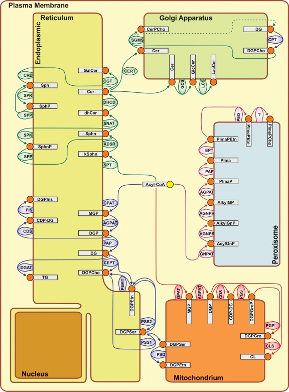
The phospholipid biosynthetic enzymes that produce membrane lipid products have been assigned to the different organelles in the picture below, according to where the majority of protein or activity for each has been measured.  :

Cell Membranes, origins through natural mechanisms, or design ? Jlr50s12

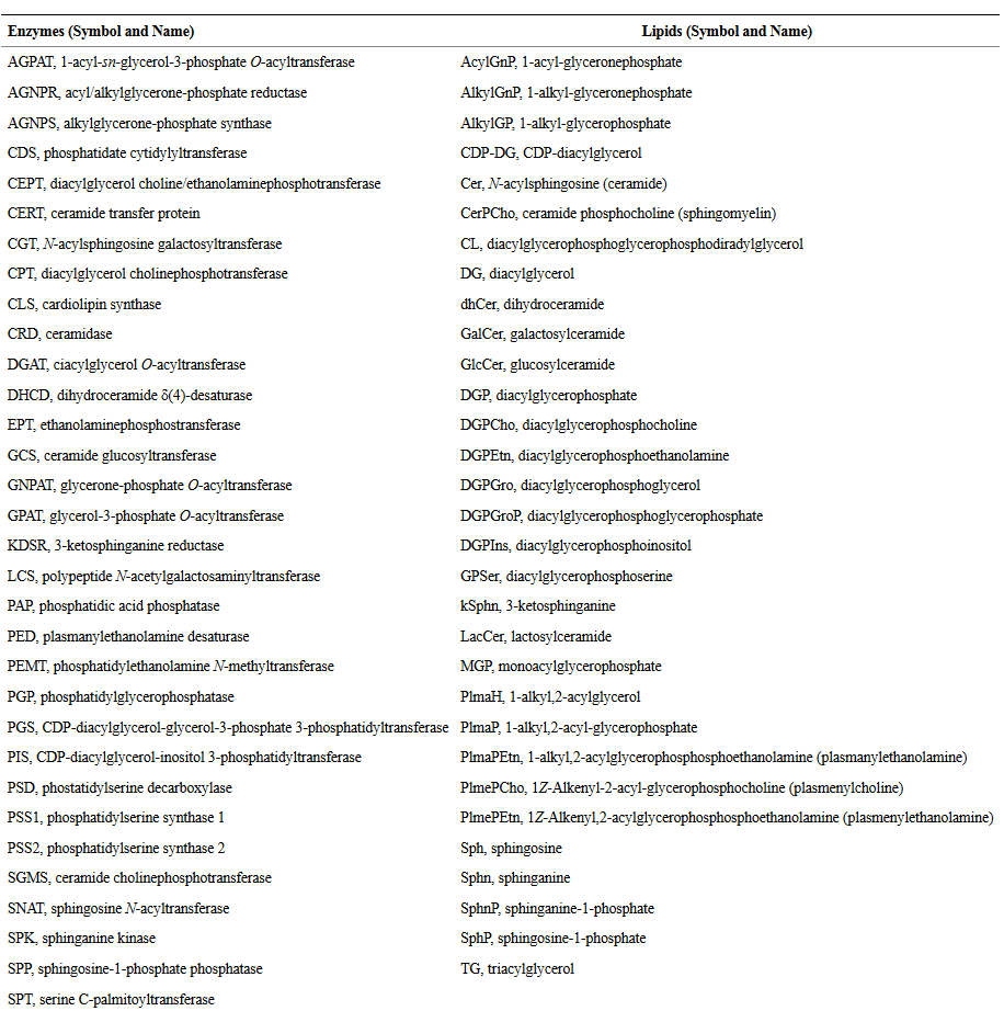
Above : Compartmentalization of phospholipid biosynthetic activities. The abbreviations are listed below:

Cell Membranes, origins through natural mechanisms, or design ? Pubmed11

Enzymes that synthesize unique glycerophospholipids, called plasmalogens, are included. Water soluble intermediates are not shown in the scheme, with the exception of fatty acyl-CoA, which is used as substrate by acyltransferase enzymes for the synthesis of both glycerolipids and sphingolipids in the ER, cardiolipin (CL) and phosphatidylglycerol (DGPGro) in mitochondria, and for the synthesis of plasmalogens (PlmePEtn and PlmePCho) in the peroxisomes. The family of acyltransferases is quite extended, and only a few isoforms are directly involved in the de novo synthesis of membrane lipids, while others are involved in the remodeling of acyl chains of the different lipid classes (5, 6). The acronyms for the different lipids are those proposed by the Lipid Maps project (20), and the corresponding nomenclature for the lipid biosynthetic enzymes are defined in Table 1. Different isoforms of the lipid biosynthetic enzymes are not pointed out unless they are known to have alternate substrate specificities. Rather, the goal is to obtain a holistic view of the overall process of membrane lipid biogenesis.


1) https://en.wikibooks.org/wiki/Structural_Biochemistry/The_Evolution_of_Membranes
2) http://www.ncbi.nlm.nih.gov/pmc/articles/PMC2674712/
3) http://www.evolutionnews.org/2014/06/cell_membrane_l087171.html


Last edited by Admin on Thu Mar 21, 2019 3:24 am; edited 5 times in total

https://reasonandscience.catsboard.com

Otangelo


Admin

Mitochondrial membrane biogenesis: phospholipids and proteins go hand in hand 1

Mitochondrial membrane biogenesis requires the import and synthesis of proteins as well as phospholipids.The biochemical approach of Kutik et al. (2008) uncovered an unexpected role of the mitochondrial translocator assembly and maintenance protein, Tam41, in the biosynthesis of cardiolipin (CL), the signature phospholipid of mitochondria. The genetic analyses of Osman et al. (2009) led to the discovery of a new class of mitochondrial proteins that coordinately regulate CL and phosphatidylethanolamine, another key mitochondrial phospholipid. These elegant studies highlight overlapping functions and interdependent roles of mitochondrial phospholipid biosynthesis and protein import and assembly

The mitochondrial inner membrane has a unique composition of proteins and phospholipids, whose interdependence is crucial for mitochondrial function.

It is highly enriched in proteins specific to this membrane, the majority of which are encoded by the nuclear genome and imported from the cytosol. The lipid component contains the major classes of phospholipids found in all cell membranes, including phosphatidylcholine (PC), phosphatidylethanolamine (PE), phosphatidylinositol (PI), phosphatidylserine
(PS), and phosphatidic acid (PA), as well as phosphatidylglycerol (PG) and cardiolipin (CL), which are located predominantly if not exclusively in the mitochondria.   PE, PG, and CL are synthesized “ in house, ” whereas the others must be imported.

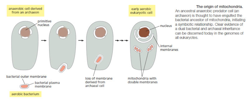
The endosymbiotic theory helds that  mitochondria appear to originate only from other mitochondria. 2 They contain their own DNA, which is circular as is true with bacteria, along with their own transcriptional and translational machinery. Mitochondrial ribosomes and transfer RNA molecules are similar to those of bacteria, as are components of their membrane.These and related observations led Dr. Lynn Margulis, in the 1970s, to propose an extracellular origin for mitochondria.

If the highly enriched  proteins specific to this membrane, the majority  are encoded by the nuclear genome and imported from the cytosol, then the endosymbiotic theory is nonsense, what i suspected for long.

Cell Membranes, origins through natural mechanisms, or design ? Jlr50s11

1) http://jcb.rupress.org/content/184/4/469.full.pdf
2) http://www.ruf.rice.edu/~bioslabs/studies/mitochondria/mitorigin.html

https://reasonandscience.catsboard.com

Otangelo


Admin

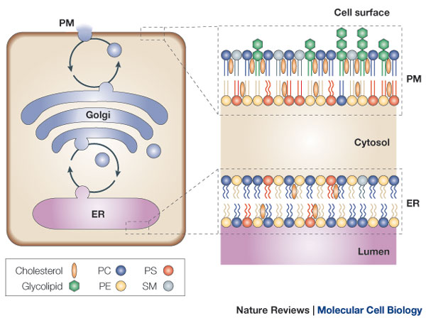
Lipid traffic: floppy drives and a superhighway  1

Understanding how membrane lipids achieve their non-random distribution in cells is a key challenge in cell biology at present. In addition to being sorted into vesicles that can cross distances of up to one metre, there are other mechanisms that mediate the transport of lipids within a range of a few nanometres. These include transbilayer flip–flop mechanisms and transfer across narrow gaps between the endoplasmic reticulum and other organelles, with the endoplasmic reticulum functioning as a superhighway along which lipids can rapidly diffuse.

Cell Membranes, origins through natural mechanisms, or design ? Nrm15910

Membrane lipids show non-random distributions between and within organelles that are connected by vesicular pathways31, 98, 99, 100. The plasma membrane (labelled PM in the figure) is rich in sterols, sphingolipids and saturated glycerolipids (Box 1), which, due to their high packing densities, promote bilayer rigidity and impermeability. For example, the rat liver plasma-membrane composition is: 30–40 mol% cholesterol; 10–15 mol% sphingomyelin (SM) and glycosphingolipids; 25 mol% phosphatidylcholine (PC); 15 mol% phosphatidylethanolamine (PE); 5 mol% phosphatidylserine (PS); and 5 mol% phosphatidylinositol (PI). In addition, the plasma membrane has an asymmetric lipid arrangement with the aminophospholipids concentrated in the cytosolic leaflet and the sphingolipids concentrated in the exoplasmic leaflet. The membrane topology of cholesterol is not known, but its location is probably determined by its high affinity for sphingolipids and saturated glycerolipids22, 23, 24. The endoplasmic reticulum (ER) membrane, on the other hand, shows a symmetric lipid distribution and primarily contains unsaturated glycerolipids that make the membrane flexible, and therefore facilitate the incorporation of newly synthesized proteins. For example, the composition of the ER membranes of rat liver is: 5 mol% cholesterol; 50–60 mol% PC; 25 mol% PE; and 10 mol% PI. Vesicular traffic between the ER and the plasma membrane passes through the Golgi, a multi-cisternal organelle in which lipid sorting must occur to prevent the randomization of the subcellular lipid distribution.


1) file:///E:/Downloads/5.Holthuis2005%20pp209-220.pdf

https://reasonandscience.catsboard.com

6Cell Membranes, origins through natural mechanisms, or design ? Empty Origin of the cell membrane Tue Dec 27, 2016 6:21 pm

Otangelo


Admin

Origin of the cell membrane

https://reasonandscience.catsboard.com/t2128-cell-membranes-origins-through-natural-mechanisms-or-design#5066

A simple primitive cell, or protocell, would consist of two key components: a protocell membrane that defines a spatially localized compartment, and an informational polymer that allows for the replication and inheritance of functional information.  1

The emergence of the first cells on the early Earth was the culmination of a long history of prior chemical and geophysical processes.

Question: How do they know that? They don't know. That's just one of the typical baseless assertions without a shred of evidence.

Modern cell membranes are composed of complex mixtures of amphiphilic molecules such as phospholipids, sterols, and many other lipids as well as diverse proteins that perform transport and enzymatic functions. Phospholipid membranes are stable under a wide range of temperature, pH, and salt concentration conditions. Such membranes are extremely good permeability barriers, so that modern cells have complete control over the uptake of nutrients and the export of wastes through the specialized channel, pump and pore proteins embedded in their membranes. A great deal of complex biochemical machinery is also required to mediate the growth and division of the cell membrane during the cell cycle.

Question: Had this complex machinery for growth and division not have to be fully functional with the first cell, otherwise replication would not be able to occur, and life would not perpetuate  ??

Fatty acids are attractive as the fundamental building block of prebiotic membranes in that they are chemically simpler than phospholipids. Fatty acids with a saturated acyl chain are extremely stable compounds and therefore might have accumulated to significant levels, even given a relatively slow or episodic synthesis.

An early RNA replicase probably would not have a built-in way of differentiating between a replicase or non-replicase sequence, and as a result, will make a copy of any RNA that happens to be close by.  Without some means of separating the replicases from the non-replicases, the population of replicases is unlikely to grow and prosper.  This issue can be resolved if the replicases are placed within a compartment, such as a vesicle, which can physically separate the replicases from other RNAs. 2

In addition, a membrane may have played an important role in the early cell's ability to store energy in the form of a chemical gradient. In modern eukaryotic cells, the mitochondria, often called the "cellular powerhouse" uses an internal chemical gradient to create energy-storing molecules known as ATP.

FORMING FATTY ACIDS ON THE EARLY EARTH

How might fatty acids have formed on the early Earth? Some scientists have proposed that hydrothermal vents may have been sites where prebiotically important molecules, including fatty acids, were formed.  Research has shown that some minerals can catalyze the stepwise formation of hydrocarbon tails of fatty acids from hydrogen and carbon monoxide gases -- gases that may have been released from hydrothermal vents. Fatty acids of various lengths are eventually released into the surrounding water.

It seems likely that primitive cells incorporated lipid-like molecules from the environment as a nutrient, rather than undertaking the much more complex process of synthesizing complex lipids by an enzyme-catalyzed process.

Based on what that scenario seems likely is a mystery to me......

http://www.csj.jp/journals/bcsj/bc-cont/b12may/85_20110349.html

Submarine hydrothermal systems (SHSs) have been thought of as a suitable environment for the origin of life subsequent to the abiotic synthesis of organic molecules. However, it has been pointed out that bioorganic molecules, such as amino acids, are easily degraded at a high temperature, and thus not likely to survive for the next step of chemical evolution in a SHS environment.


http://physwww.mcmaster.ca/~higgsp/3D03/MillerHighTemp.pdf

The problem with monomers is bad enough,but it is worse with polymers,e.g.,RNA and DNA (Lindahl1993),whose stability in the absence of efficient repair enzymes is too low to maintain genetic integrity iyperthermophiles. RNA and DNA are clearly too unstable to exist in a hot prebiotic environment.The existence of an RNA world with ribose appears to be incompatible with the idea of a hot origin of life.

Although such materials might have been synthesized near hydrothermal vents in the early seas, the assembly of such materials is quite problematic. Conditions requiring high concentrations, exact pH and temperature, plus the absence of high sodium and small amounts of certain metal ions, prevents the assembly of such components within the earth's early oceans. Conditions that might concentrate fatty acids to sufficient levels to form membranes would also concentrate solutes that disrupt the formation of those membranes. Encapsulation of a proto-cell replicator and metabolic system would be quite problematic, since the conditions that would encourage such activity would likely lead to conditions that would disrupt the primitive membrane completely. Primitive membranes must be able to transport nutrients and wastes, although passive transport systems would readily reach equilibrium and active transport systems would not be expected to be produced immediately upon encapsulation. Energy acquisition is problematic, since fatty acids membranes cannot generate a proton gradient. Membranes composed of unsaturated fatty acids or phospholipids can generate proton gradients, but would not be expected to have existed in early earth environments. Virtually all studies that have examined membrane growth and division have used unsaturated fatty acid membranes, which would not have been present on the early earth. Because of this problem, these studies have questionable relevance to the origin of life on earth. 4)

While a wide range of amphiphilic compounds that could serve as lipid components for primitive biological membranes self-assemble into bilayers, this self-assembly process requires “just right” conditions and “just right” molecular components. It is unlikely that such conditions would exist or persist for long time frames on early Earth. 5

1) http://www.ncbi.nlm.nih.gov/pmc/articles/PMC2926753/
2) http://exploringorigins.org/fattyacids.html
3) http://www.ucmp.berkeley.edu/education/events/deamer1.html
4) http://www.godandscience.org/evolution/origin_membranes.html#n07
5) http://www.reasons.org/articles/biotic-borders-cell-membranes-under-scrutiny

https://reasonandscience.catsboard.com

Otangelo


Admin

Acetyl-CoA as a Precursor: Acetyl-CoA is the starting point for fatty acid synthesis in many organisms, including chemolithoautotrophs. It can be derived from various metabolic pathways, including the TCA cycle.

Transfer of Acetyl Group: The acetyl group from acetyl-CoA is indeed transferred to a carrier protein, typically acyl carrier protein (ACP), initiating the fatty acid synthesis pathway.

Elongation: Fatty acids are synthesized by consecutively adding two-carbon units. This iterative process involves the enzymes of the fatty acid synthase complex.

Combining with Glycerol-3-Phosphate: The synthesized fatty acids are esterified with glycerol-3-phosphate to form phosphatidic acid. The glycerol-3-phosphate is derived from dihydroxyacetone phosphate, which is part of the glycolytic pathway (or gluconeogenesis in the case of autotrophs that might not be taking up sugars).

Formation of Various Phospholipids: From phosphatidic acid, as you mentioned, you can indeed get various phospholipids. The head group can be modified to produce phospholipids like phosphatidylcholine, phosphatidylethanolamine, phosphatidylserine, and others.

https://reasonandscience.catsboard.com

Otangelo


Admin

December 14, 2023 From lipids to life: Cracking the puzzle about the origin of life

The quest to understand life’s origin has long intrigued scientists seeking to unravel the mysteries of existence. Traditional approaches to this question have their own limitations. Professor Doron Lancet and his team from the Weizmann Institute of Science, Israel, offer a fresh perspective and a more feasible pathway for understanding how life emerged on Earth. Their work introduces a paradigm shift, emphasising the spontaneous assemblage of variegated simple lipid molecules in ancient oceans as the likely seed of life. Their research redefines the emergence of reproduction, offering new insights and opening avenues for exploration in the field of the origin of life. The quest to unravel the mysteries surrounding life’s origin continues to captivate scientists. In fact, the central question revolves around how an abiotically formed mixture of non-living chemicals (no-life) gave rise to the early self-reproducing and evolving living cells, called protocells (life). In simpler terms, the question is: what bridged no-life to life? Alexander Oparin’s book, The Origin of Life, pioneered a scientific approach to these questions; yet, 100 years later, scientists are still grappling with the profound question of how life, with its remarkable ability to replicate and evolve, emerged from the chaotic mix of chemical compounds on our early planet. The abiogenesis conundrum The central debate in this field of abiogenesis is the study of how life could have arisen spontaneously and naturally from non-living substances. The popular answer revolves around two major complementary lines of study: one focuses on the prebiotic synthesis of life-like small molecules, and the other centres on the prebiotic synthesis of RNA, a complex molecule believed to have the capacity for self-replication. However, there is currently an unbridged chasm between a mixture of numerous scattered organic compounds, large and small, and the formation of the first reproducing protocells.

To resolve this problem, we need spontaneously formed containers. It is widely accepted that key molecules capable of forming such containers are lipid molecules. Such amphiphilic assemblies can gather specific molecules from the complex mix of chemicals around them. But there is another challenging aspect to this: figuring out how these early assemblies could make copies of themselves, in the case of vesicles, including both the outer lipid shell and the chemical content inside. Two decades ago, Professor Doron Lancet from the Weizmann Institute of Science, Israel, and colleagues began the unravelling of this enigma. The lipid salvation In living cells today, there are four main classes of small organic molecules: amino acids (building blocks of proteins), nucleobases (building blocks of DNA and RNA), sugars, and lipids. It is only lipids that can readily form labile (easily breakable) multi-molecular ensembles, such as the membranes surrounding cells and protocells as well as the much smaller micelle particles, which are known to be simple efficacious chemical factories, competing with present-life complex proteins. Primordial lipid ensembles thus provide both compartmentalisation and chemical specificity that might lead to self-reproduction capacities. The reason lipids can perform such marvels is their amphiphilic nature; they consist of two parts: water attracting heads and water repelling tails. This laid the foundation for Lancet’s ‘lipid first’ alternative for the famous ‘RNA first’ argument. The role of lipid micelles A novel perspective in the research on the origin of life has thus ushered in a paradigm shift. Rather than exclusively focusing on the study of how certain molecules might have formed before life existed, scientists are now exploring how simple lipid molecules, copiously present in ancient oceans, could have autonomously come together.

In a recent paper (Kahana, A, Lancet, D, 2021), the researchers point out that it is the modest nanoscopic micelles that had numerous advantages as early protocells, despite the fact that they did not have an inner water volume (Figure 1). Within these tiny protocellular structures, networks of molecules can collaboratively function, akin to a team, because all molecules are crowded in a miniscule volume, initiating a critical step towards the emergence of life. Scientists are now exploring how simple lipid molecules, copiously present in ancient oceans, could have autonomously come together. Importantly, these lipid micelles are far from random assemblies; they possess an innate capacity for self-organisation. However, this organisation is not in terms of spatial position or order of amino acids as in a protein. Instead, the organisation is expressed in terms of composition. In a simplified example, imagine an environment in which all types of lipids have the same concentration. Upon micelle growth driven by molecule accretion, the network dynamics are capable of biasing the inner composition, with some being in high amounts and others being small or rejected entirely. This behaviour is analogous to highly specific membrane transport mechanisms controlling the content of present-day cells. Figure 1: Nanoscopic micelles: Seeking early protocellular simplicity and efficacy (Kahana, A, Lancet, D, 2021). The truly surprising aspect is that not only do lipid micelles have capacity to self-organise, but they can also maintain a constant composition upon growth. This means that these micelles have a built-in system to ensure that their lipid composition would remain stable as they get bigger. This is called ‘homeostatic growth’, another capability of reproducing living cells. When these entities split into two, the offspring are very similar to each other, just like when living cells reproduce. One of the most important findings of the research is that the catalytic networks within lipid micelles (a team of molecules working together, where certain molecules speed up the entry of some others) might have enabled self-reproduction, meaning micelles could reproduce themselves by a mechanism analogous to metabolism in living cells (Figure 2) (Lancet, D, Zidovetzki, R, Markovitch, O, 2018).

The Graded Autocatalysis Replication Domain (GARD) model Over the span of 20 years, Lancet and his team developed the Graded Autocatalysis Replication Domain (GARD) model – a computational chemistry approach that simulates the chemical reactions and processes that could have occurred in the prebiotic era. The GARD model builds upon the earlier foundations of the Collectively Autocatalytic Set (CAS) model (Kauffman, SA, 1986), which predicts that catalytic networks may give rise to self-reproduction. GARD adds quantitative layers to CAS, providing a chemical kinetics understanding of how complex mutual catalysis can lead to self-reproduction. The GARD model, supported by computational simulations and experimental evidence, suggests that growing and splitting lipid assemblies can reach a dynamic state where their chemical composition is self-reproduced. The team highlights that the transition from non-life to life involved chemical entities that possessed catalytic properties, which can lead to self-reproduction. These capabilities are considered crucial steps in the emergence of life because they set the stage for the development of more complex, organised, and self-sustaining systems, all having catalysis as their foundation, eventually leading to what we recognise as living organisms. Figure 2: Micellar self-reproduction driven by homeostatic compositional preservation happens rarely in privileged micelle configurations, called composomes (Lancet, D, Zidovetzki, R, Markovitch, O, 2018). The GARD model represents a crucial advancement in the study of life’s origin. Its simulations predict the dynamic behaviour as described above, which leads to a reproduction point, termed ‘composome’ (Figure 2). GARD demonstrates the feasibility of self-replication much before the advent of utterly complex molecules such as DNA and RNA, as well as catalysis ahead of complex proteins. This opens new avenues for exploring the emergence of life-like behaviour at very early stages. Experimental findings published over the last few years, as summarised (Kahana, A, Lancet, D, 2021), strengthen the foundation of the GARD model and emphasise the plausibility of early emergence of life-like networks. They bridge the gap between primordial soup and early protocells as described in empirical observations, bringing us closer to understanding the origins of life on Earth. Significantly, GARD simulations also predict that the reproducing point would be reached much more readily than expected, because that point is a dynamic attractor, a state toward which a network tends to progress (Kahana, A, Segev, L, Lancet, D, 2023). The plausibility of reproducing micelles is enhanced by the fact that the oceans of planet Earth can harbour 10³³ micelles (Figure 3) (Lancet, D, Zidovetzki, R, Markovitch, O, 2018). From micelles to complex protocells Once self-reproduction begins, mutations and natural selection emerge as a possibility. The most important implication of this transition is thus the likelihood of early Darwinian evolution (Segré, D, Lancet, D, 2000). This evolutionary potential opens up prospects for the gradual development of bacterium-like, Last Universal Common Ancestors (LUCA), passing through a myriad of protocellular quasi-species over hundreds of millions of years. In this evolutionary vein, the team’s research not only delves into the early stages of how life began, but also suggests the potential for a shift from simple micellar protocells to more complex vesicular counterparts. Figure 3: Planetary micellar ‘library’ enhances self-reproduction probability. This does not happen with the larger vesicles: they are all the same composition, as statistics dictates for plausible lipid repertoire size(Lancet, D, Zidovetzki, R, Markovitch, O, 2018). They point out that it is possible to envisage the chemical steps that would lead to new functionalities, including metabolism-like synthesis of new metabolites, vesicular membrane transporters, as well as RNA and its translation device (Kahana, A, Lancet, D, 2021). Charting new horizons The GARD model, as described by Lancet, provides the dynamic journey from simple lipid micelles to complex protocells, offering new insights into how life may have originated and evolved on Earth. It highlights the significance of locally available compounds coming together to form complex, life-like entities. Lancet and his team not only provide a novel understanding but also open up new avenues for research in the domain.

Personal Response What do you believe are the most promising implications of your research on the origins of life for our understanding of life’s emergence on Earth and potentially, elsewhere in the Universe? Our research provides a rigorous chemical framework that constitutes a complete path from simple chemistry to bacterial complexity. We demonstrated that spontaneously formed amphiphile assemblies, initially nanoscopic micelles, will naturally undergo chemical catalytic modifications, leading to self-reproduction. This generates a population with some mutations (quasi-species) that can undergo natural selection and therefore, portray early Darwinian evolution. Along this path, micelles can evolve into more elaborate vesicular protocells that can endogenously synthesise new and larger molecules, leading to increasing protocellular complexity. Given half a billion years of evolution, it is not unlikely that a bacterium-like Last Universal Common Ancestor (LUCA) will emerge. As catalytic reproducers appear highly prevalent on an entire planet, and a network reproduction state is a dynamic attractor, the likelihood of extraterrestrial life seems much higher than anticipated.

Unresolved Challenges in Early Micelle-Based Protocellular Structures

1. Self-Organisation Without Spatial Order
The self-organisation observed in micelle-based protocells is expressed in their composition, not in a spatial or structural sense like in modern cells. While the micelles' lipid composition adjusts dynamically, it is unclear how such sophisticated compositional control could emerge unguided.

Conceptual problem: Lack of Spatial Order in Organization
- No mechanism to explain how molecular networks can function cooperatively without spatial coordination
- Difficulty explaining how compositional biases emerge in the absence of external regulation or enzymatic catalysis.

2. Homeostatic Growth in Primitive Micelles
The ability of lipid micelles to maintain a constant composition during growth, termed 'homeostatic growth,' is a trait usually associated with living cells. This phenomenon requires a robust system that can stabilize and monitor internal lipid content during size expansion, a process not clearly understood in prebiotic conditions.

Conceptual problem: Spontaneous Emergence of Homeostatic Control
- No known prebiotic mechanism to explain how primitive micelles can regulate and maintain stable compositions during growth
- Homeostatic growth typically requires feedback systems absent in early environments.

3. Catalytic Networks in Lipid Micelles
Lipid micelles appear capable of forming catalytic networks where certain molecules assist in the transport or catalysis of others, mimicking metabolic activities. This coordinated network suggests a high degree of functional complexity, difficult to explain without guided interactions.

Conceptual problem: Emergence of Catalytic Complexity
- No natural unguided pathway explains how molecules could spontaneously form highly organized catalytic networks
- Without proteins or ribozymes, there is no clear method for efficient catalytic activity within micelles.

4. Spontaneous Formation of Amphipathic Lipids
Lipid micelles depend on amphipathic molecules (lipids with hydrophilic heads and hydrophobic tails) for their structural integrity. The synthesis of such molecules is a multi-step process, traditionally reliant on enzymatic catalysis. In prebiotic environments, where no enzymes existed, it is unclear how these molecules could form.

Conceptual problem: Prebiotic Synthesis of Lipids
- The multi-step process of lipid formation lacks plausible prebiotic catalysts
- Environmental conditions necessary for spontaneous lipid formation remain speculative, with no direct evidence of sustained favorable conditions.

5. Absence of Selective Permeability in Micelles
Selective permeability is a key feature of living cells, enabling them to control the flow of substances in and out. However, early micelle structures would have lacked proteins such as transporters or channels, raising the question of how these micelles could support basic proto-cellular functions without these crucial mechanisms.

Conceptual problem: Lack of Permeability Control
- Primitive membranes likely lacked the selectivity required to differentiate between nutrient intake and waste removal
- No known primitive mechanism explains how micelles could develop selective permeability without proteins.

6. Energy Requirements for Micelle Stability and Growth
In modern cells, processes such as membrane growth and lipid synthesis are energy-intensive and depend on molecules like ATP. The lack of prebiotic energy equivalents challenges the possibility of maintaining micelle stability and supporting growth mechanisms.

Conceptual problem: Energy Source for Lipid Dynamics
- Lack of ATP or similar high-energy molecules in early Earth environments complicates explanations for the energy-intensive processes involved in micelle growth
- Without external energy sources, the stability and persistence of lipid micelles are difficult to justify.

7. Environmental Instability and Lipid Degradation
Lipid micelles are vulnerable to environmental degradation, particularly from UV radiation and oxidation, which would have been prevalent in early Earth conditions. The absence of protective mechanisms in these primitive structures further exacerbates the problem of maintaining lipid integrity long enough for them to participate in protocellular processes.

Conceptual problem: Stability of Lipids in Harsh Environments
- Early Earth’s conditions, such as radiation and fluctuating temperatures, would likely degrade lipids before they could contribute to protocell formation
- No protective systems existed in early micelles to shield lipids from environmental degradation.

8. Self-Reproduction in Micelles without Prebiotic Machinery
The Kahana and Lancet (2021) paper suggests that lipid micelles may have had the ability to self-reproduce, which would require the coordination of complex molecular networks similar to metabolic systems in living cells. However, the mechanisms driving this self-reproduction in the absence of biological machinery remain unknown.

Conceptual problem: Reproduction Without Metabolic Networks
- Reproduction of micelles in a manner analogous to cellular metabolism lacks a clear, unguided pathway
- Without enzymes or ribozymes, it is unclear how molecular interactions could replicate the complexity of metabolic processes necessary for self-reproduction.

9. Prebiotic Bias Towards Specific Lipid Compositions
The concept of lipid micelles developing compositional biases through accretion mechanisms akin to modern membrane transport systems poses a significant challenge. Prebiotic environments likely had a uniform distribution of lipid types, making it difficult to explain how specific lipids could have been favored in the absence of a selective mechanism.

Conceptual problem: Emergence of Lipid Compositional Bias
- The bias in lipid composition suggests a level of selectivity typically seen in cellular transport systems, which would not have been available prebiotically
- No clear mechanism exists to explain how micelles could have developed compositional diversity spontaneously.

10. Interdependence of Lipid Networks and Other Biochemical Systems
For micelles to function as protocells, they would need to interact with genetic material or other biomolecules, such as peptides or sugars, to establish the cooperative networks necessary for life. The simultaneous emergence of these interdependent systems presents a formidable challenge without invoking guided or designed processes.

Conceptual problem: Co-Emergence of Lipids and Biochemical Networks
- Lipid micelles alone cannot explain the full complexity required for life without the concurrent emergence of other biomolecules
- No natural process has been identified that could account for the coordinated emergence of lipid and other biomolecular systems.

11. Prebiotic Membrane Chirality Selection
Modern membranes exhibit chirality, which is essential for their function. However, prebiotic synthesis of lipids would likely produce racemic mixtures, meaning an equal proportion of right- and left-handed molecules, which would compromise membrane function.

Conceptual problem: Lack of Mechanism for Chirality Selection
- Prebiotic environments would not naturally select for one chiral form over another, yet functional membranes require specific chirality
- No known mechanism explains how primitive micelles could have developed the necessary chiral purity for functional membranes.

12. Integration with Other Molecular Systems
Even if lipid micelles could form under early Earth conditions, their integration with other systems, such as genetic material and proteins, is required for the full development of proto-cellular life. The simultaneous emergence of these diverse systems presents an unresolved problem, as no known natural mechanism can explain their coemergence.

Conceptual problem: Lack of Mechanism for Integrated Systems
- The integration of lipid micelles with other molecular systems would require simultaneous, coordinated development, which remains unexplained
- Without genetic material or primitive proteins, it is unclear how lipid micelles alone could have achieved the complexity necessary for life.

https://researchoutreach.org/articles/lipids-life-cracking-puzzle-origin-life/


https://reasonandscience.catsboard.com

Sponsored content


Back to top  Message [Page 1 of 1]

Permissions in this forum:
You cannot reply to topics in this forum