ElShamah - Reason & Science: Defending ID and the Christian Worldview
Would you like to react to this message? Create an account in a few clicks or log in to continue.
ElShamah - Reason & Science: Defending ID and the Christian Worldview

Otangelo Grasso: This is my library, where I collect information and present arguments developed by myself that lead, in my view, to the Christian faith, creationism, and Intelligent Design as the best explanation for the origin of the physical world.


You are not connected. Please login or register

Can the origin of feathers be explained through evolution ?

Go down  Message [Page 1 of 1]

Otangelo


Admin

Can the origin of feathers be explained through evolution ?

https://reasonandscience.catsboard.com/t3184-can-the-origin-of-feathers-be-explained-through-evolution

The Dinosaur/Bird Connection: Dapper Dino & Otangelo Grasso
https://www.youtube.com/watch?v=DOXDm55LdSE&t=2435s

See as well:
No evidence for the evolution of birds, feathers, and flight
https://reasonandscience.catsboard.com/t1694-no-evidence-for-the-evolution-of-birds-feathers-and-flight

Feathers and Plumages - a general description

Can the origin of feathers be explained through evolution ?  Feathe61
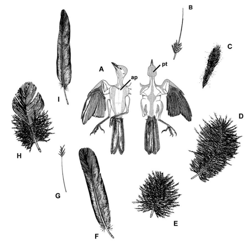
Distribution of feather and main feather types. 
(a) Distribution of pterylae (pt) and apteria (ap) in the common blackbird (Turdus merula) (modified after Bergmann 1987). 
(b) Bristle feather. 
(c) Powder down. 
(d) Semiplume. 
(e) Down feather. 
(f) Rectrices. 
(g) Filoplume. 
(h) Pennaceous body feather. 
(i) Primary remiges. 
(b, c) 2


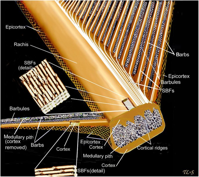
Although feathers come in an incredible diversity of forms, they are all composed of the protein beta-keratin and made up of the same basic parts, arranged in a branching structure. In the most complex feathers, the calamus extends into a central rachis which branches into barbs, and then into barbules with small hooks that interlock with nearby barbules. 10

Feathers exhibit amazing regenerative behavior both for physiological needs and in response to trauma. Structurally, feathers are the most sophisticated ectodermal organ whose shapes and color patterns show tremendous variation both among body regions of the same bird as well as between different species 1


Can the origin of feathers be explained through evolution ?  Types_13
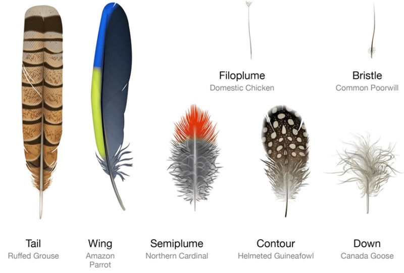
Feathers fall into one of seven broad categories based on their structure and location on the bird’s body.
Wing feathers -The wing feathers specialized for flight are characterized by uniform windproof surfaces on either side of the central shaft that are created by an interlocking microstructure. Also called remiges, these feathers are asymmetric with a shorter, less flexible leading edge that prevents mid-air twisting.
Tail feathers -Most tail feathers, or rectrices, feature an interlocking microstructure similar to wing feathers. Arranged in a fan shape, these feathers support precision steering in flight. In some birds like peacocks, tail feathers have evolved into showy ornaments.
Contour feathers -Contour feathers are what you see covering the bird’s body. Arranged in an overlapping pattern like shingles, the waterproof tips are exposed to the elements and the fluffy bases are tucked close to the body. Sometimes brilliantly colored or uniformly drab, contour feathers can also help the bird show off or stay camouflaged. Feathers on the wing, called coverts, shape it into an efficient airfoil by smoothing over the region where the flight feathers attach to the bone.
Semiplume - Mostly hidden beneath other feathers on the body, semiplumes have a developed central rachis but no hooks on the barbules, creating a fluffy insulating structure.
Down –Similar to semiplumes with a loosely branching structure but little or no central rachis, down feathers are relatively short and positioned closest to the body where they trap body heat.
Filoplume –Short simple feathers with few barbs, filoplumes function like mammal whiskers to sense the position of the contour feathers.
Bristle –Bristles are the simplest feathers, with a stiff rachis that usually lacks barb branches. Most commonly found on the head, bristles may protect the bird’s eyes and face.

Can the origin of feathers be explained through evolution ?  Feathe35
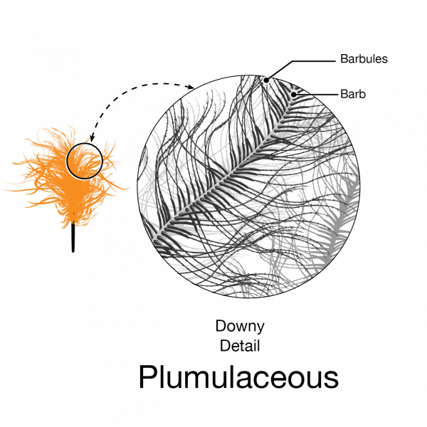
[ltr]Downy feathers look fluffy because they have a loosely arranged plumulaceous microstructure with flexible barbs and relatively long barbules that trap air close to the bird’s warm body. Pennaceous feathers are stiff and mostly flat, a big difference that comes from a small alteration in structure; microscopic hooks on the barbules that interlock to form a wind and waterproof barrier that allows birds to fly and stay dry. Many feathers have both fluffy plumulaceous regions and more structured pennaceous regions. [/ltr]
Can the origin of feathers be explained through evolution ?  Feathe37
[ltr]Pennaceous feathers are stiff and mostly flat, a big difference that comes from a small innovation in structure; microscopic hooks on the barbules that interlock to form a wind and waterproof barrier that allows birds to fly and stay dry.[/ltr]
Can the origin of feathers be explained through evolution ?  Feathe36
Advance to watch the feather open and close. Pennaceous feathers are stiff and mostly flat, a big difference that comes from a small innovation in structure; microscopic hooks on the barbules that interlock to form a wind and waterproof barrier that allows birds to fly and stay dry. 

Can the origin of feathers be explained through evolution ?  Differ10
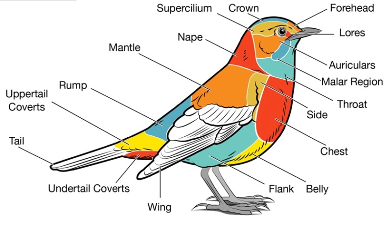
[ltr]The external bird is labeled according to the commonly used feather groups. Experienced birders often use these terms when referring to specific field marks.[/ltr]

Can the origin of feathers be explained through evolution ?  Wing_f10
Advance to step through the feather types that become visible as the bird moves its wing. The primary and secondary wing feathers, or remiges, are visible from both sides of the wing. There are also specialized groups of coverts of both sides, which smooth the wing by covering the bases of the remiges, Scapulars cover the bird’s shoulder. The alula is a set of feathers that jut up from the top of the wing that helps birds with precision landings.

Can the origin of feathers be explained through evolution ?  Develo11
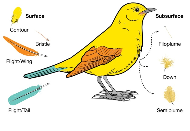
When you look at a bird, the most recognizable feathers are the flight feathers on the wing and tail. Covering the rest of the body are contour feathers that overlap like shingles to shed water. On some birds you will also see bristles near the beak and eyes. What you can’t see unless you look beneath the contour feathers are the fluffy down and semiplumes that are tucked close to the body to trap heat.  Filoplumes are tiny whisker-like feathers that are often hard to locate tucked in amongst the down.

Can the origin of feathers be explained through evolution ?  Develo10
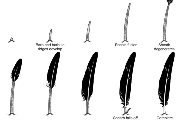
As feathers mature, their tips get pushed away from a small outgrowth of skin, called the papilla, where the newest parts of the feather form. The feather’s structure develops as proteins are laid down around the surface of this bump of skin. It’s here that the branching patterns form by smaller branches fusing at the base to make thicker ones — barbules fuse into barbs and barbs fuse into a rachis. As the feather grows, it stays curled in a tubular shape around the papilla until it is pushed away from the growth area. A protective sheath maintains the feather’s cylindrical shape until it starts to disintegrate near the tip, allowing the mature part of the feather to unfurl.
Can the origin of feathers be explained through evolution ?  Bird_b11
Downy feathers look fluffy because they have a loosely arranged plumulaceous microstructure with flexible barbs and relatively long barbules that trap air close to the bird’s warm body.


Structural basics of feathers 
Asked to draw a feather, even young children will illustrate three basic features. First, there is the shaft, the thin but stiff, central, rod‐like portion. Second, the vanes extend in opposite directions from the shaft, forming two broad, flat, and more pliable regions of the feather. Third, on close inspection, the vanes—rather than being smooth and solid like a simple sheet of plastic—have a comb‐like substructure. To round out this general description, a biologist would likely direct attention to a few more details of a typical feather. 

Can the origin of feathers be explained through evolution ?  Feathe38

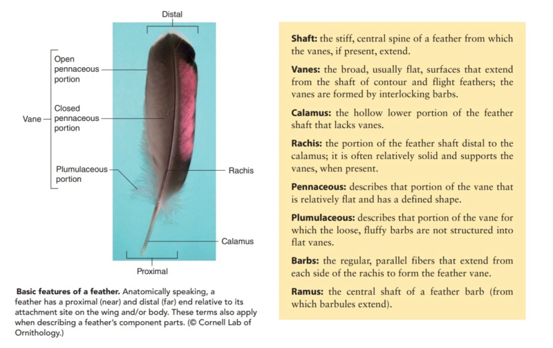
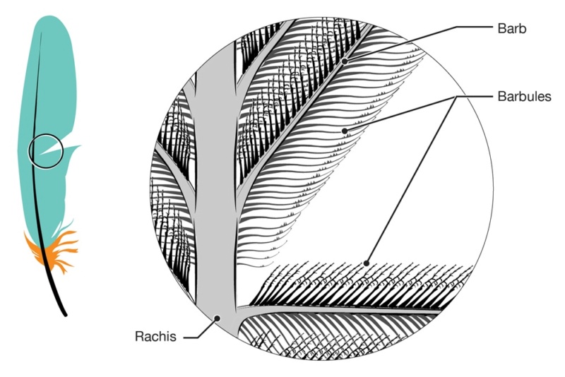
The shaft can be subdivided into two regions, the more medial (or close to the body) portion of the shaft is vaneless and defines the area called the calamus. The calamus is relatively short and vane‐free primarily because it is the portion of the feather that perforates the bird’s skin, thereby anchoring the feather to the body. The rest of the shaft is distal (further from the body) relative to the calamus, and has the vanes extending from it. This region is called the rachis. Given that the rachis frequently comprises as much as 90–95% of the length of  the shaft, the terms rachis and shaft are often used interchangeably. When most people envision a typical feather, they tend to think of long wing or tail feathers rather than a small fluffy tuft of down—which is also a feather. This contrast draws attention to another fundamental structural feature of feathers, or, more specifically, of feather vanes. A typical wing or tail feather is pennaceous, a feather that has vanes with a relatively smooth, two‐dimensional (flat) surface, and with a distinct shape or outline. A down feather is plumulaceous, a feather with barbs that are so fluffy as to be relatively formless. Plumulaceous feathers are incapable of holding any but the most delicate rounded form, and their overall shape changes drastically in response to the slightest breeze. In order to understand what underlies this difference in vane form (or lack thereof), one needs to understand in more detail the anatomical underpinnings of the firmer, pennaceous feather vane. Pennaceous vanes are a flexible, breathable, intricate mesh made of hundreds of tiny interlocking fibers. On close inspection, one can see that these vanes are comprised of a dense row of hundreds of long, slender, but stiff filaments, called barbs, branching off from the rachis one after the other, somewhat like teeth on a comb (Fig.A).

Can the origin of feathers be explained through evolution ?  Feathe39
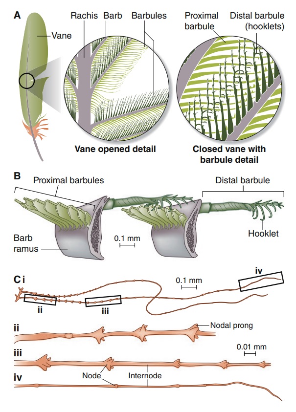
Feather vane and barbule anatomy. 
(A) Detail of feather vanes with interacting proximal and distal barbules. 
(B) Detail showing hooklets interact with the barbules on the adjacent barb. 
(C) Two plumulaceous barbules (i) of a Rock Pigeon (Columba livia), with three regions from the proximal to distal tip (ii–iv) enlarged below. In (ii), the base and proximal nodes are relatively large compared with the central region of the barbule (iii). Distal barbule tips (iv) have substantially reduced nodes. 

At an even finer scale, the barbs themselves have a set of smaller structures, barely visible to the naked eye. That is, along the barbs the overall feather structure—the larger central shaft with many smaller fibers branching off on opposite sides—repeats itself at a smaller scale: each barb Distal consists of a central shaft called the ramus that has its own rows of slender branches, called barbules, extending from either side. Barbules are generally not symmetrical off the two sides of the ramus, and we refer to the barbules that extend away from the bird’s body (or away from the base of the feather) as the distal barbules, and those that extend toward the body as the proximal barbules. The two sides typically have complementary form and function; the distal barbules often have hook‐shaped structures along their ventral (or under) surfaces, called barbicels or hooklets (Fig.B). The hooklets grab and hold the dorsal surface (or top) of the proximal barbules of the adjacent barb. Thus, although the barbs are long, thin, and can physically disconnect from one another, they are held parallel to neighboring barbs along their entire length by the hooklets on their distal barbules. In this way, barbules generate and maintain the continuous structure of the vanes—causing the feather to appear like one, smooth, continuous surface. The fibers that create plumulaceous vanes are also barbs, but with a different structure. Barbs on plumulaceous feathers have proportionally longer and less stiff rami, and their barbules are typically reduced either to fewer, thinner, simple hairs (no hooklets), or even short nodules off the rami (Fig.C). In comparison to the densely packed, stiff, and hooked fibers of pennaceous vanes, the lack of barbule structure liberates the long thin rami to move independently from one another, and creates the loose, tufted form of a plumulaceous vane. All of these feather structures are made of the same material: keratin. Keratin is a proteinaceous connective tissue (the same basic material of human fingernails) produced inside specialized cells called keratinocytes. Among all the species in the animal kingdom, there are a diverse set of genes that produce keratin, and consequently diverse forms of keratin proteins exist. Within vertebrate animals, keratins belong to one of two fundamentally different structural forms (Fig.A below). 

Can the origin of feathers be explained through evolution ?  Feathe40
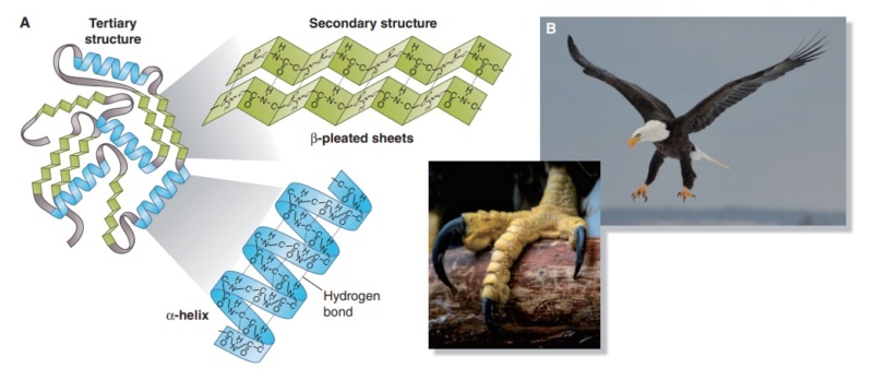
Structure and use of keratin. 
(A) The different molecular structures of alpha- and beta-keratins create distinctive folding (tertiary) structures and properties. 
(B) Beta-keratins are unique to birds and reptiles and are the main structural component of feathers and claws, as in the talons of this Bald Eagle (Haliaeetus leucocephalus).

The alpha-keratins are found in all vertebrates (including ourselves), are relatively flexible, and make up much of mammalian skin, hair, and nails. Beta‐keratins are unique to reptiles and birds, tend to be tougher, and are found in scales, claws, and feathers (Fig. B above). The specific beta‐keratin found in feathers is a form unique to living birds. The diversity of beta‐keratins within birds is still largely unexplored and—given the importance of the material properties of keratin for feather function, and of feather function for birds in general.

How Do Birds Use their Feathers?
https://askabiologist.asu.edu/content/23-functions-feathers

Feathers make birds unique animals. How they are used by birds can be unique too. If you think to yourself, you can probably come up with maybe a half dozen to a dozen ways feathers are used by birds. To be sure, you will have missed a few feather functions. Let's go through 23 ways birds can use their feathers.

birds flying

Flying
Flight feathers are very strong and stiff feathers that are found on the wings of birds.

Help Keep Warm
Downy feathers as well as semiplume feathers are able to trap pockets of air close to the bird's body to help keep it warm. How much body heat they keep can be adjusted by arranging their feathers to trap more or less air. If you see birds fluffing their feathers in the cold, that is their way of adding extra air to trap body heat and stay warmer.

Control Body Temperature
To keep body temperature steady, birds can either expose their heads and feet to the air or water to cool down, or tuck them into their feathers to help keep warm.

Protect From Wind, Moisture, and Sun
The strong and ridged contour feathers shield birds from wind. The tough material they are made from, beta-keratin, is water and wear resistant. Darker-colored feathers might also provide protection from the sun. Feathers also work to keep water out, keeping birds dry in the rain. The interlocking feather barbs and a special coating that is either oily or waxy create a shield that water runs off of.

Swimming and Diving
Some birds use their half-spread out wings in a flying motion to swim in water. Penguins have developed their wings into stiff, flat flippers that make penguins great swimmers.

Floating
Using the trapped air in downy feathers, water birds like ducks can float on water as well as add protection from cold water.

Snowshoeing
One of the more unusual feather uses is snowshoeing. Grouse, chicken-like birds that live in snow-covered areas, have feather-covered feet in the winter that increase the size of the foot just like snowshoes. This keeps the birds from sinking into the snow.

Tobogganing
Why walk if you can slide, or in the case of penguins, toboggan. The Antarctic birds flop down on the smooth feathers of their bellies and use their flipper-like wings together with their feet to move themselves, toboggan-like, across snow and ice.

Bracing
When not flying, many birds use their tail feathers as supports when on the ground or climbing the sides of trees such as is seen with woodpeckers.

Feeling
Feathers do not have nerves, but they do stimulate nerves that surround where the feather attaches to the bird. Birds can adjust the position of their feathers and posture depending on the stimulation of those nerves.

Hearing
Some predators, especially owls, have their face feathers arranged like two dishes (facial discs) to collect and channel sounds into their ears so they can more accurately locate prey in the dark (parabolic reflector).

looking fierce

Making Sounds
We think of bird sounds either as songs or calls, but using their feathers, some birds are able to make many different sounds like humming, drumming, and whistling.

Muffling Sounds
Birds that hunt at night like owls are able to use their wings to muffle their own sounds as they approach their prey. You can think of them as an early stealth fighter plane.

Foraging (Looking for Food)
Some birds, like herons that hunt for fish in the water of lakes and streams, will sometimes use their feathers to form an umbrella over their heads. This might make it easier for them to see fish in the water.

Helping to Keep a Steady Supply of Food
Hummingbirds help to pollinate flowers when foraging for sweet nectar when the feathers around their heads pick up pollen from a flower. As they continue looking for more nectar, the pollen is then transferred to other flowers.

Eating
Special long feathers called rictal bristles are found around the mouths of some insect-eating birds. These may either act like a funnel to catch the insect in the air, or they may protect the eyes while catching an insect. Other birds use feathers on the side of their mouths to select fruits.

getting food
Keeping Clean
Some birds, like herons, have small feathers called powder down that they crush with their beak and feet to rub into the normal feathers and keep them conditioned. This powder down may also help control feather parasites like mites.

Aiding Digestion
Some fish-eating birds also eat their own feathers to line their digestive area. This helps to protect the bird from sharp fish bones.

Constructing Nests
Many birds (especially water birds) line their nests with bird feathers. This helps to keep their eggs warm and also provides a soft padding. Some birds like parakeets actually use the feathers located on their bottom and lower back to move grass and leaves to their nest.

colored feathers
Transporting Water
When raising eggs and baby chicks, many adult birds will soak the feathers on their belly before returning to the nest. They can then use the water to keep the eggs from drying out and to give their chicks a drink. Some birds that live in the desert (like the sandgrouse) have special belly feathers that are very good at holding water. This adaptation lets them nest further away from water holes, to avoid the higher numbers of predators found in areas near water holes.

Escaping From Predators
When birds are attacked or frightened they can drop some of their tail feathers. This is called fright molt. This sometimes helps the bird get away, leaving the attacker with only a mouth or foot full of feathers.

Sending Visual Signals
Feather colors and patterns are used to send signals to mates and rivals. This is likely the largest and most used function of feathers.

Camouflage


1. https://onlinelibrary.wiley.com/doi/10.1111/dgd.12024
2. https://sci-hub.ren/10.1007/978-3-030-27223-4


Last edited by Otangelo on Thu Jan 13, 2022 2:13 pm; edited 63 times in total

https://reasonandscience.catsboard.com

Otangelo


Admin

A.C. McINTOSH Evidence of design in bird feathers and avian respiration 2009 1

Many have taken the view that design is only an illusion in living systems [1], arguing that such ‘apparent design’ and accompanying complexity can be explained by the neo-Darwinian paradigm. However, such thinking fails to realize that functional systems, in order to operate as working machines, must have all the required parts in place in order to be effective. If one part is missing, then the whole system is useless. The inference of design is the most natural step when presented with evidence such as in this paper, that is evidence concerning avian feathers and respiration. The counterargument has often been made that functional complexity can appear from simpler systems that evolve over time. However, there has never been a recorded observation of this happening experimentally in the laboratory (where the precursor information or machinery is not already present in embryonic form). Though it is true that a design action is also not observed in the laboratory, nevertheless the inference to original design and intelligence is a perfectly valid alternative from direct analogy to designs within the man-made world. Even though specified functional complexity has not been experimentally observed to develop from simpler systems, this has not deterred the stridency with which such views are put forward by some of evolution’s proponents. In particular, the arguments seeking to align such thinking with the principles of thermodynamics lead to, at best, speculative ideas to explain how these fundamental structures and information emerge. The powerful fundamental arguments from thermodynamics actually favor the straightforward view that such organizational structures cannot appear without pre-existing functional complexity being there, to begin with. Therefore, the main intention in this paper is to draw attention to machinery which can readily be understood by most readers with some scientific background and to appreciate some of the features which defy an explanation by slow gradual changes, as required by neo-Darwinian evolution.

It needs to be stated clearly that origins science, though it clearly has philosophical implications, is nevertheless a genuine scientific debate. It is not the prerogative of science to make statements beyond the remit of the study of natural systems, since, by its very nature, science can only be the study of the material world. Consequently, to try and assert that natural systems can only ever have come about by a preexisting natural system without intelligence is an unproven assumption, and must immediately be recognized as such. To take only natural causes as one’s starting point seems innocuous enough, for some would say that does it not helpfully separate ‘religious’ questions from ‘scientific’ ones? Surely ‘here is the way forward’ say a large group of scientists, most of whom have no predisposition to be either for or against any particular philosophical view of reality. They just wish to pursue science. However, what is a useful and pragmatic way forward, for taxonomic purposes, of describing rich, living biological systems, becomes totally inappropriate when looking at the origins of such systems, since to deny the possibility of the involvement of external intelligence is effectively an assumption in the religious category. Science can study the effect on the natural world of systems of pre-existing material, but it cannot preclude the possibility of intelligence extraneous to that very matter and energy being involved in its formation. To say otherwise is effectively wedding science to a narrow philosophical foundation. We quote here the important statement of a great thinker and evolutionist – Stephen Jay Gould, who often spoke against the position that there is necessarily an intelligence behind the design observed in nature:

Moreover, ‘fact’ does not mean ‘absolute certainty.’ The final proof of logic and mathematics flow deductively from stated premises and achieve certainty because they are not about the empirical world. ... In science, ‘fact’ can only mean ‘confirmed to such a degree that it would be perverse to withhold provisional assent. I suppose that apples might start to rise tomorrow, but the possibility does not merit equal time in physics classrooms.

Gould is right that logic and mathematics flow from stated premises, and it is that very point that we seek to emphasise here. Once one opens the possibility that intelligence is involved, the evidence leads very naturally to the conclusion of design, not by going against the known empirical laws (such as gravity in the analogy of Gould), but precisely the reverse. We must keep to the ‘nullius in verba’ motto of the Royal Society (‘on the words of no one’), and not preclude from the outset where the evidence may lead.

Types of feathers 
Feathers are of different types depending on location. The major types of feathers are illustrated in Fig. 1, which shows that there are at least five different types. 

Can the origin of feathers be explained through evolution ?  Types_10

Flight feathers are mainly the primary feathers on the outer part of the wing. These and the tail feathers are together called remiges. The primary feathers are larger in size relative to the other feathers, and asymmetric in shape. As the name implies, they bear the greatest aerodynamic load, and without them, a bird will not be able to fly. These are often the feathers cut in order to train a bird. They will, of course, regrow, but a bird is helpless without them. The secondary feathers are needed to complete the inner part of the wing and maintain stability. Those closest to the body of the bird are called the tertiary feathers. The base of the flight feathers are also covered by smaller feathers called covert feathers. These also thicken the leading edge of the wing such that an aerofoil shape is maintained in cross section by the extended wing. All the above are called pennaceous feathers as they have closely connected barbs and form aerodynamic surfaces. There are other feathers which are called plumulaceous, or downy, feathers. These have only a rudimentary rachis and a jumbled tuft of barbs with long barbules to provide an excellent thermal insulation. The details of a feather are shown in Fig. 2. Not shown in Fig. 1 are the tail feathers (retrices) which have a symmetrical shape but are designed for use as air brakes and also control the direction of flight.

One of the most easily overlooked feathers are those forming the alula (or alular) grouping – essentially a set of finger feathers on the leading edge of each wing of a bird. These feathers are crucial because, for low speed flight, they act as a leading edge slat and keep the boundary layer attached across the upper surface of the wing that is formed from the main primary, secondary and tertiary pennaceous feathers. The alula group of feathers are attached to a projecting digit coming from the humerus bone. This digit is called the pollex and acts rather like the human thumb. The number of alula feathers attached to the pollex depends on the species. The humming bird has two, the cuckoo has five or six, and there can be as many as seven. Without these small feathers, the control of flight would be extremely difficult at low speed.

Hook and ridge barbule arrangement of feathers Feathers are made of keratin, a protein also used to make hair and fingernails. There are differences in the exact type of keratin used. Feather keratin occurs in a ‘β-sheet’ configuration, which differs from the α-helices that generally occur in mammalian keratins. The β keratin of bird feathers is rather like a stretched spring in consistency. The fact that scales of reptiles are also made of keratin is used by some to propose that dinosaurs are the precursors to birds. However, it should be noted that there are significant hurdles to transform one type of keratin to the other. The feather grows from a follicle, and from the central rachis come barbs which give the vane of the feather. The details of the sophistication involved in the barb system of the pennaceous feather become clear under a microscope. In Fig. 3, barbules can be seen coming from each barb. They are only visible at the micro level, but have a structure that is essential for feathers to work as aerodynamic surfaces. The barbules in one direction are ridge-like, while the barbules in the opposing direction have hooks. Consequently, the hooks of the barbule in one direction grip the ridge of the opposing barbule. Figure 4 shows further details of this remarkable arrangement.

Can the origin of feathers be explained through evolution ?  Types_11
Keratin sheath of feathers 
Feathers grow from follicles and are made from multilayered keratinocyte sheets. As already noted, the feather keratin is in a β-sheet configuration and develops within the follicle which supports, in the initial stages of growth, a cone arrangement made from a rachidial ridge (which becomes the rachis in the fully developed feather) and the barbs curled round with a longer circumference at the base of the cone and shorter barbs forming the tip. All this is enclosed in a keratin sheath, the lining of which is connected to the follicle as a single layer. As the plumage appears, at each feather follicle, the cone becomes a sheath and then gives way to the feather as it emerges from the vertex. These sheaths become tube-like and are present for any new feather. This will be the case for adults, as feathers are replaced in moulting, but are more visible and noticeable in a fledgling (see Fig. 5) since all the feathers are then developing together and emerge from the vertex of their coned follicles into individual separate tubes of keratin. 

Can the origin of feathers be explained through evolution ?  Types_12

These tubes run the length of each feather, thus protecting the delicate feather barbs as they develop in the  embryo within the egg. The final emergence of the feathers in a fledgling can take a few weeks for these to unfold and the keratin sheath to break away – see Fig. 5.

Design features of feathers 
There is multifunctioning and multioptimisation in feather construction. There are the features which are immediately apparent such as aerodynamic loading and the material construction of rachis and barbs to sustain this. However, there are also more subtle features such as the arrangement of hooks and barbules primarily for keeping the feather together, such that they prevent air from going through them during the downstroke but allowing some air to pass through in the upstroke, thus maximising the efficiency of energy use in wing flap. The keratin itself has an extremely high specific strength, and the shape of the filament cross sections used in rachis construction moves from near circular near the root to a curved and ribbed rectangular shape away from the root for structural efficiency under bending and potentially buckling loads. The evidence is consistent with the design thesis both from the fossils found of flight in the past, and in the multifunctional nature of wings today

Fossil evidence 
It is evident that the hook and ridge system is a key feature of the barbule system connecting the barbs of a feather. How this came about has been the subject of a number of speculative conjectures in the scientific literature. Laudable indeed have been the attempts to find the evolutionary engine to provide specific function, but the attempt is not impressive, since the array of simpler structures is difficult to imagine, let alone find in the fossil record. Prum takes the view that the forerunner of the pennaceous feather could have been a conical papilla similar to a hair arising out of a cylindrical follicle within the skin. It is then proposed that the papilla became a tuft of barbs (unbranched filaments), and then each of these filaments eventually branched into the barbules. And then, finally, it is maintained that the branched filaments became organised around a central stem (rachis) to produce the hook and ridge structure of present-day feathers. However, the real issue is not addressed by any of these studies. By definition, the Darwinian evolutionary ‘mechanisms’ (which Dawkins summarised as ‘non-random survival of randomly varying hereditary instructions for building embryos …’) have no sense of overall future gain other than the immediate next step. These authors look for evidence that true feathers developed first in small non-flying dinosaurs before the advent of flight, possibly as a means of increasing insulation for the warm-blooded species that were emerging. Though attempts have been made to suggest that the Liaoning shales in Northeast China provide evidence of early feathers on dinosaurs, the hard evidence of clear examples of an intermediate intricate barbule system (hook and ridge) in the vanes has not yet been produced. Xu et al. refer to structures made from filaments of skin in fossils of Sinornithosaurus millenii, a non-avian theropod dinosaur in sediments that are classically dated as about 125 million years old. Though there is evidence of a downy structure, flight feathers were not apparent. What is actually known from the hard fossil evidence (rather than speculation) is that there certainly were now extinct creatures which also had feathers. Archaeopteryx clearly had fully developed flight feathers and the species Microraptor gui shows every evidence of being simply another perching extinct bird, though the feathers are not as distinct as those in Archaeopteryx. A better example is the early Cretaceous Hongshanornis longicresta from the lower Jehol group in the Yixian formation in Northeast China. This example does have barbed feathers and thus falls again into the category of an extinct bird. Thus, the actual evidence shows that one either has extinct fully developed feathers (Archaeopteryx, Hongshanornis, possibly Microraptor gui) or small reptilian dinosaurs such as Sinornithosaurus millenii. A clear example of an in-between transitional stage is missing. It should also be noted that to maintain aerodynamic versatility, pennaceous feathers have to be maintained airworthy. This is done by a bird obtaining oil from the uropygial (preening) gland at the base of its spine. The ability to reach this gland is a feat of twisting which a bird performs with ease. However, it raises serious issues concerning the supposed evolution of feathers, since it is necessary for the feather construction (barbule ridge and hook system) to arise concurrently with the preening gland and the ability to manoeuvre the neck a full 180 degrees. None of the fossil evidence shows any evidence of such transitions.

The details of the sophistication involved in the barb system of the pennaceous feather become clear under a microscope. In Fig. 3, barbules can be seen coming from each barb. 

Can the origin of feathers be explained through evolution ?  Feathe11
Figure 3: The hooked and ridged structure of barbules in a pennaceous feather.

They are only visible at the micro level, but have a structure that is essential for feathers to work as aerodynamic surfaces. The barbules in one direction are ridge-like, while the barbules in the opposing direction have hooks. Consequently, the hooks of the barbule in one direction grip the ridge of the opposing barbule.

Evolutionary arguments of feather morphogenesis 
Alongside the paleontological studies involving the search for clear transitional fossil evidence, there have been attempts to analyse molecular mechanisms in supposed feather-branching morphogenesis. Yu et al. delivered exogenous genes to regenerate flight follicles in chickens and identified a critical protein necessary in feather branching. They suggest that this identifies molecular pathways underlying possible transformations of feathers from cylindrical epithelia to hierarchical branched structures. Two alternative routes are discussed. The first is by suggesting that the rachis evolved, then the barbs and finally the barbules. The other view of Wu et al.  is that barbs appeared first from supposed integument evolution, followed by a fusing of the barbs to form a rachis. However, in all these investigations, it is still speculation governing such evolutionary hypotheses, since a critical protein has yet to be identified in the formation of feathers. The reality is that there is a fully formed structure of ridged and hooked barbules in all pennaceous feathers and these are found with precise function and position in the wings of birds. So, the rigorous examination of the evidence points rather towards functional complexity coming from intelligence – to suggest that this came about only through the workings of natural selection and random mutations is, in the view of this author, not consistent with the evidence. One of the points which is important is that it is not sufficient to simply have barbules to appear from the barbs but that opposing barbules must have opposite characteristics – that is, hooks on one side of the barb and ridges on the other so that adjacent barbs become attached by hooked barbules from one barb attaching themselves to ridged barbules from the next barb (Fig. 4). It may well be that as Yu et al. suggested, a critical protein is indeed present in such living systems (birds) which have feathers in order to form feather branching, but that does not solve the arrangement issue concerning lefthanded and right-handed barbules. It is that vital network of barbules which is necessarily a function of the encoded information (software) in the genes. Functional information is vital to such systems. 

Functional information 
Some authors assert that possible modes of functionality increase with the rise in ‘Shannon information’ (Shannon information equates with the uncertainty of states of an ensemble of microsystems) and that natural selection then selects out the functioning alternative valid for that environment. They appeal in particular to autocatalytic systems, self-organising systems and pattern formation to form primal replicators from which functional complexity then emerges as natural selection sifts the ensemble of alternatives to single out the replicator with functional advantage. Ball developed these ideas and put greater detail into the arguments by showing convincingly that pattern formation arises from the autocatalytic feedback chemical systems. The Turing patterns in the chlorite–iodide–malonic acid reaction are an example of dissipative structures in reaction–diffusion equations. These type of non-linear systems are connected to the patterns that emerge, such as giraffe and zebra pelts. Most are of the view that he is very likely correct and this author agrees. There can  be no doubt that the work of Murray and co-workers of many years  has done much to elucidate the role of reaction-diffusion mechanisms for formation of patterns in living systems. The well-known Turing reaction-diffusion equation predicts accurately the distribution of surface markings in animals, and the target patterns of the Belousov–Zhabotinskii chemical reaction (involving cyclic AMP, that is, cyclic adenosine monophosphate) are well simulated by the Field–Noyes mathematical model. Furthermore, the periodic patterns of feather germs can also be predicted using similar mathematical principles. However, correct and enlightening as these models are, it is important to recognise that this is not the same as functional information, where coded instructions are involved, first, in the precise ordered arrangement of nucleotides in DNA, and, secondly, in the multifunctioning construction of items from these codes such as hooked and ridged feather barbules. This is a subject of a separate paper by the author where the argument is made that all living systems have coded machinery which sits on high free energy bonds, all of which have to be in place for the system to work. That is, the natural tendency is for the linkages needed for those coding systems (e.g. nucleotide bonds) to decay, and not to be sustained without prior information within the system. Thermodynamically, the very material on which the coded information sits is acting against the natural law which, were it not for the information in the system, would have it fall apart. This strongly suggests that the information, far from being thought as material, is in fact non-material (like the coded instructions of software on a computer) and itself constrains the matter and energy of the nucleotide bonds to perform as they do in DNA. This is certainly the view of other authors and a very cogent statement of this position comes at the end of the paper (conclusions) by Abel and Trevors:

S.C. BURGESS MULTI-FUNCTIONING AND MULTI-OPTIMISATION IN FEATHERS 2007 2

Natural organisms often have multiple functions and multi-optimisation in a single component or mechanism. In addition, natural organisms are highly integrated assemblies. In contrast, human design has traditionally avoided multi-functioning in single components because of the difficulties this presents in the design process. Only in recent years has there been a trend towards multi-functioning in engineering components. The advantage of multi-functioning is that extremely high levels of performance can be achieved. This paper gives examples of multi-functioning and multi-optimisation in a bird flight feather and a bird display feather. In each case, the advantages of multi-functioning are explained and analogies with man-made design are given.

INTRODUCTION 
One of the interesting characteristics of natural organisms from a design point of view is the existence of multiple functions and multi-optimisation in a single component or mechanism. In addition, natural organisms are highly integrated assemblies with several sub-systems being closely integrated together. In contrast, human design has traditionally avoided multi-functioning in single components because of the difficulties this presents in the design process. Multi-functioning and multi-optimisation are very challenging because there are more constraints in the design process and therefore fewer possible solutions. In practice, multi-functioning leads to a need for very sophisticated design solutions. When designing an engineering device, it has traditionally been recommended to design each component for one main function in order to make the behaviour of the device easier to understand and predict. For example, in material selection methodology it has traditionally been assumed that components generally have one main function. Another reason for avoiding multi-functioning in the past is the lack of multidisciplinary design teams and a lack of suitable technology. Observations of past engineering devices show that they do indeed generally possess limited multi-functioning and integration of parts. Only in recent years have engineers adopted a design philosophy of integrating different functions together in single components and mechanisms. For example, cars are becoming highly integrated, with computing hardware and software being closely integrated with mechanical sub-systems such as engines and braking systems. Multi-functioning and integration have obvious benefits. The number of components in a device can be dramatically reduced and this can lead to compactness and low mass. Compactness and low mass can lead to many improved aspects of mechanical performance such as energy and space efficiency and speed of operation. Low part count can also lead to high levels of reliability and easier maintenance. Integration and multi-functioning are very common in nature. Leonardo da Vinci was one of the first scientists to appreciate how the natural world contained optimal design. After studying many aspects of the natural world, 


Leonardo concluded: ‘Although human genius through various inventions makes instruments corresponding to the same ends, it will never discover an invention more beautiful, nor more ready nor more economical than does nature, because in her inventions nothing is lacking, and nothing is superfluous 3

D’Arcy Thompson (1860–1948) was one of the first modern scientists to systematically study optimum design in nature. In 1917 he published his classic work On Growth and Form. More recently, there has been a growing interest in optimum design in nature and its possible application to engineering design. It is very useful to study multi-functioning and multi-optimisation in nature because lessons can be learnt about how to achieve these desirable attributes.

This paper gives examples of multi-functioning and multi-optimisation in a bird flight feather and a bird display feather. In each case, the advantages of multiple functions are explained and analogies with man-made design are given.

MULTI-FUNCTIONING IN BIRD FLIGHT FEATHERS 
The structure of a flight feather is shown in Fig. 1. 

Can the origin of feathers be explained through evolution ?  Feathe13
Figure 1: Structure of a flight feather. 

There is a hierarchy of structures. The main feather stem comes first, then the barbs and finally the barbules. The stem has a massed array of barbs on each side that form the basic feather shape. Each barb itself has two sets of barbules. The barbules on one side have a set of hooks whilst the barbules on the other side are plain. Therefore, the hooked barbules can interlock with the plain barbules on the adjacent barb. The flight feathers of birds can be considered to have three major functions: an aerodynamic function, a fail-safe function and a lightweight structural function. These functions are summarised in a function-means tree in Fig. 2. 

Can the origin of feathers be explained through evolution ?  Feathe14
Figure 2: Function-means tree for a bird flight feather. 

A function-means tree summarises the functions and solutions of a device at different levels of detail and shows where multi-functioning takes place. The function means tree in Fig. 2 shows that certain features of the feather are optimal for more than one function. In particular, the hierarchical structure is optimal for all three functions and hence is a very important feature. Despite having three complex functions, the feather is a single integrated structure. 
Optimal aerodynamic layout The overall asymmetric feather profile is optimal from an aerodynamic point of view because the barbs are very short on the leading edge and are therefore protected against buckling from the airflow. Another important aerodynamic feature is a one-way airflow mechanism at each barbule joint. The hooks and barbules are arranged so that they prevent air from going through them when the wing is pushed downwards, but they allow some air to pass through them when the wing is being pulled upwards.

This feature enables the bird to maximise the efficiency of flapping by making the wing mainly push the air down. 

Optimal fail-safe mechanism 
The flight feather of birds contains a localised fail-safe mechanism in the hook structure of the barbules. If the barbs are overloaded then they will unzip from adjacent barbs before any serious damage is done to the feather structure. Once unzipped, the barbs can be re-zipped together by the simple action of the bird passing its beak through the feather. The large number of separate zipping mechanisms ensures that the feather will unzip very close to the point of overload, thus causing minimum damage to the feather. The optimal fail-safe feature of the barbule connections leads to a high level of reliability in the wings of a bird. 

Optimal structural layout 
The hierarchical layout of the flight feather is optimal from a structural point of view because the feather transfers loads from a surface to a point. A hierarchical tree structure is generally the optimal solution for generating an efficient flow of forces (or heat or fluid) between a point source and a volume or surface. The hierarchical structure of the feather is extremely important because it enables localised hooking mechanisms and fail-safe mechanisms as well as an optimum flow of forces. As well as having an optimal hierarchical layout, the flight feather also has optimal material and shape properties. The feather consists of thin-walled keratin sections filled with lightweight foam.

CONCLUSIONS 
One of the main reasons for the exceptional performance and sophistication of bird feathers is the feature of multi-functioning. The design of bird feathers demonstrates that multi-functioning and multi-optimisation can produce large benefits in performance. Not surprisingly, there has been a recent trend in engineering design to move towards multi-functioning in single components and structures. The hierarchical structure of feathers is one of the key features that enable multiple functions to be carried out in one integrated structure. The hierarchical structure enables fine-tuning of shapes and layout and enables a very large number of localised sub-mechanisms to exist. Hierarchical structures have been found to be important in other natural systems such as trees and many other systems in nature. Multi-functioning and multi-optimisation are very challenging because there are more constraints in the design process and hence fewer possible solutions. In addition, the design team must have wide cross-discipline knowledge to know what is feasible and optimal. Nature can be a rich source of ideas and inspiration that can help to achieve multi-functioning in engineering. Multi-functioning in nature can be studied using methods such as function-means trees. Function means trees are a good way of analysing multi-functioning because they help clarify aspects of the solution that are optimal for more than one function. Function-means trees are also good for considering functions of different disciplines such as industrial design (aesthetics) and engineering.


1. https://www.witpress.com/elibrary/dne-volumes/4/2/399
2. https://www.witpress.com/elibrary/dne-volumes/1/1/190
3. http://web.mit.edu/4.464/www/Selection-Vol20No1.pdf
da Vinci L. Manuscript RL 19115v; KrP 114r located in the Royal Library, Windsor Castle, Windsor, England, ca. 1500.


Last edited by Otangelo on Sat Nov 06, 2021 6:05 am; edited 11 times in total

https://reasonandscience.catsboard.com

Otangelo


Admin

Keratin: Structure, and mechanical properties

Approximately 88–90% of feathers are composed of the protein keratin, which requires a high level of sulfur-containing amino acids cystine and methionine for its production 11

ED YONG An insider’s look at the feather, a marvel of bioengineering DECEMBER 15, 2009 

The feather is an extraordinary biological invention and the key to the success of modern birds. It has to be light and flexible to give birds fine control over their airborne movements, but tough and strong enough to withstand the massive forces generated by high-speed flight. It achieves this through a complicated internal structure that we are only just beginning to fully understand, with the aid of unlikely research assistants – fungi.
At a microscopic level, feathers are made of a protein called beta-keratin. The same protein also forms the beaks and claws of birds, and the scales and shells of reptiles. It’s close (but less rigid) relative, alpha-keratin, makes up the nails, claws and hairs of mammals. Zoom out, and we see that feathers have a central shaft called the rachis with two vanes on either side. Each vane is composed of barbs that branch off the rachis. Even thinner barbules branch off from the barbs, and are held together by small hooks that give the feather its shape.

What’s much less clear is how the keratin fibres and filaments are organised into the rachis, barbs and barbules. To work that out, scientists would typically slice the rachis in cross-sections and look at it under an electron microscope. But feathers don’t give up their secrets so easily. Their fibres are stuck together with a chemical glue that makes them virtually impossible to separate. Imagine gluing a bundle of matches together and cutting them cross-ways. You could see the fibres that make up the component matches, but if they were glued together tightly enough, you wouldn’t be able to tell where one match started and another began. So it is with feathers and their keratin.
Theagarten Lingham-Soliar from the University of Kwazulu-Natal solved the problem by recruiting fungi as research assistants. He used four species, which like to grow on keratin, to digest the complex molecules that glue individual filaments together. The process was very slow. Even after a year, the feathers seemed in pretty good shape and it was only after 18 months that they had broken down enough to be studied under the microscope
The wait was worth it. For the first time, the microscope revealed how feathers are organised. For a start, they contain the thickest keratin fibres ever recorded, with a 6-micrometre diameter that’s ten times greater than their next thickest rivals. The long fibres make the rachis strong and stiff, and each is made up of even smaller ‘megafibrils’ and ‘fibrils’. Most of the keratin fibres are aligned along the shaft of the rachis but some are wrapped around them. These wraparound fibres prevent their long-ways comrades from buckling. Without them, the slender rachis might turn into a bulging barrel.

The keratin fibres were lined with small lumps or nodes that stick up from the main axis at regular intervals. Each node is capped with hooks or a ring, and those of neighbouring fibres are staggered. They act like the bricks of a wall, linking together to prevent cracks from spreading and resisting forces that would fracture the rachis. They also help to anchor the fibres into the glue that binds them together, much like steel rebars used in the construction of high-rise buildings.

But the most surprising feature of the fibres is that they’re almost identical to the structures of barbules, and very similar to the downy plumage of baby chicks. This has implications for the evolution of feathers. You might think that the central feature – the rachis – came first, followed by the structures that branch off it. But the fact that the rachis itself is made of barbule-like fibres suggests that the barbules came first. Only later did they unite to form a solid rachis.

Can the origin of feathers be explained through evolution ?  Kerati18
Photographic images and micrographs showing a bird’s feather structure and a keratin β-sheet as the major composition component of the feather

Bin Wang: Structure, mechanical properties, occurrence in biological organisms, and efforts at bioinspiration  4 July 2015 3

A ubiquitous biological material, keratin represents a group of insoluble, usually high-sulfur content and filament-forming proteins, constituting the bulk of epidermal appendages such as hair, nails, claws, turtle scutes, horns, whale baleen, beaks, and feathers. These keratinous materials are formed by cells filled with keratin and are considered ‘dead tissues’. Nevertheless, they are among the toughest biological materials, serving as a wide variety of interesting functions, e.g. scales to armor body, horns to combat aggressors, hagfish slime as defense against predators, nails and claws to increase prehension, hair and fur to protect against the environment. The vivid inspiring examples can offer useful solutions to design new structural and functional materials. Keratins can be classified as a- and b-types. Both show a characteristic filament-matrix structure: 7 nm diameter intermediate filaments for a-keratin, and 3 nm diameter filaments for b-keratin. Both are embedded in an amorphous keratin matrix. The molecular unit of intermediate filaments is a coiled-coil heterodimer and that of b-keratin filament is a pleated sheet. The mechanical response of a-keratin has been extensively studied and shows linear Hookean, yield and post-yield regions, and in some cases, a high reversible elastic deformation. Thus, they can be also be considered ‘biopolymers’. On the other hand, b-keratin has not been investigated as comprehensively. Keratinous materials are strain-rate sensitive, and the effect of hydration is significant. Keratinous materials exhibit a complex hierarchical structure: polypeptide chains and filament-matrix structures at the nanoscale, organization of keratinized cells into lamellar, tubular–intertubular, fiber or layered structures at the microscale, and solid, compact sheaths over porous core, sandwich or threads at the macroscale. These produce a wide range of mechanical properties: the Young’s modulus ranges a  from 10 MPa in stratum corneum to about 2.5 GPa in feathers, and the tensile strength varies from 2 MPa in stratum corneum to 530 MPa in dry hagfish slime threads. Therefore, they are able to serve various functions including diffusion barrier, buffering external attack, energy-absorption, impact-resistance, piercing opponents, withstanding repeated stress and aerodynamic forces, and resisting buckling and penetration. A fascinating part of the new frontier of materials study is the development of bioinspired materials and designs. A comprehensive understanding of the biochemistry, structure and mechanical properties of keratins and keratinous materials is of great importance for keratin-based bioinspired materials and designs. Current bioinspired efforts including the manufacturing of quill-inspired aluminum composites, animal horn-inspired SiC composites, and feather-inspired interlayered composites are presented and novel avenues for research are discussed. The first inroads into molecular-based biomimicry are being currently made, and it is hoped that this approach will yield novel biopolymers through recombinant DNA and self-assembly. We also identify areas of research where knowledge development is still needed to elucidate structures and deformation/failure mechanisms.

Nature presents a plethora of unique materials and have become a continuing source of inspiration for engineers. Biomimetics, the science of imitating nature, is thus an exciting field where the refinements are investigated and the biological solutions are applied to develop new materials. The study of biological materials, Biological Materials Science, indispensably paves the way for inventing novel materials by providing principles and mechanisms obtained from natural designs. The more traditional approach is being complemented by molecular biomimetics, which shows a bright potential. Many biological materials are composites based on biopolymers and some minerals. This combination yields materials with outstanding properties and functionalities, considering the mainly weak constituents (primarily C, O, N, H, Ca, P and S). Others process nanoscale fibrils with high tensile strength. Wood may have a strength per unit weight comparable to that of the strongest steels; spider silk has a higher specific strength and modulus than steels; shell, bone, and antler have a toughness an order of magnitude greater than their mineral constituents (e.g. calcite, hydroxyapatite). The secret for achieving this is usually the hierarchically organized structure incorporating biopolymers and minerals. Keratin represents the most abundant structural proteins in epithelial cells, and together with collagen, is the most important biopolymer in animals.  Keratin is among the toughest biological materials, possessing both high toughness and high modulus, although it is solely composed of polymeric constituents, and seldom contains minerals. Keratinous materials, formed by specifically organized keratinized cells filled with mainly fibrous proteins (keratins), are natural polymeric composites that exhibit a complex hierarchical structure ranging from nanoscale to centimeter scale: polypeptide chain structure, filament-matrix structure, lamellar structure, sandwich structure. They compose the hard integuments of animals, e.g. epidermis, wool, quills, horns of mammals, as well as feathers, claws and beaks of birds and reptiles, and effectively serve a variety of functions, such as for protection and defense, predation and as armor. Therefore, a thorough understanding of the relationships between the units that make up a functional keratinous material would expectantly provide useful knowledge in designing new materials. Keratinous materials have started to trigger great interest in recent years, and the nascent research area of bioinspiration is gaining increasing attention. 

Keratin represents the most abundant structural proteins in epithelial cells, and together with collagen, is the most important biopolymer in animals. According to the Ashby map, shown in Fig. 1, keratin is among the toughest biological materials, possessing both high toughness and high modulus, although it is solely composed of polymeric constituents, and seldom contains minerals

Can the origin of feathers be explained through evolution ?  Kerati10
Fig. 1. Materials property chart for biological materials: toughness versus Young’s modulus

Filament-matrix structure at nanoscale 
Both a- and b-keratinous materials show a fine filament-matrix structure at the nanoscale. Here the ‘filament’, for a-keratins, denotes the ‘intermediate filament (IF)’ which represents the structural feature seen by transmission electron microscopy and shows an intermediate size (7–10 nm in diameter) between two other major classes of filamentous structures: microfilaments (actin, 7 nm) and microtubules (24 nm). For b-keratins, the ‘filament’ is called ‘beta-keratin filament’ and has a diameter of 3–4 nm. Fig. 3 presents transmission electron micrographs of the filament-matrix structure for typical a-keratinous (IFs in hair, Fig. 3a) and b-keratinous materials (beta-keratin filaments in feather rachis, Fig. 3b). 

Can the origin of feathers be explained through evolution ?  Kerati12
Figure 3. Transmission electron micrographs of typical keratinous materials with clear filament-matrix structure: 
(a) cross section of a human hair (a-keratin), stained with osmium tetroxide, showing 7 nm diameter intermediate filaments embedded in a darker matrix; 
(b) cross section of a seagull feather rachis (b-keratin), stained with potassium permanganate, showing the 3.5 nm diameter b-keratin filaments differentiated by the densely stained matrix

The filaments are ordered components composed by tightly bonded polypeptide chains and are considered as crystalline portions. The a-keratin has specialized constituent proteins: several kinds of low-sulfur proteins compose the IFs while the matrix consists of high-sulfur and high-glycine–tyrosine proteins. For b-keratin, there are no different types of proteins; the filament and matrix are incorporated into one single protein. Finally, the molecular mass of a-keratin ranges from 40 to 68 kDa, which is much larger than that of b-keratin, 10–22 kDa.

Molecular structure and formation of the filaments 
The differences of molecular structure and formation of the filaments are the most important features that distinguish a- and b-keratins, shown in Figs. 4 and 5. 

Can the origin of feathers be explained through evolution ?  Kerati13
Fig. 4. Intermediate filament structure of a-keratin: 
(a) ball-and-stick model of the polypeptide chain, and a-helix showing the location of the hydrogen bonds (red ellipse) and the 0.51 nm pitch of the helix; 
(b) schematic drawing of the intermediate filament formation (reproduced based on): a-helix chains twist to form the dimers, which assemble to form the protofilament. Four protofilaments organize into the intermediate filament.

The a-keratin proteins are organized as coiled coils. The a-helix conformation for the polypeptide chains was first postulated independently by Pauling and Crick [41,42], shortly after Pauling, Corey, and Branson [43] identified the structure as consisting of two helically wound chains of polypeptides. 

Can the origin of feathers be explained through evolution ?  Kerati14
Fig. 5. Structure of the beta-keratin filaments: 
(a) ball-and-stick model of the polypeptide chain, and illustration of the pleated beta-sheet); 
(b) schematic drawing of the formation of beta-keratin filament: one polypeptide chain folds to form four b-strands which twist to form the distorted b-sheet. Two sheets assemble to form a beta-keratin filament.

Naturally occurring a-helices found in proteins are all right-handed. The helical structure is stabilized by the hydrogen bonds (red circled line in Fig. 4a,) inside the helix chain, causing the chain to twist and exhibit a helical shape. Fig. 4b shows the IF formation process: two isolated right-handed a-helix chains form a left-handed coiled-coil, the dimer (45 nm long), by disulfide cross links; then dimers aggregate end-to-end and stagger side-by-side via disulfide bonds to form a protofilament (about 2 nm diameter); two protofilaments laterally associate into a protofibril; four protofibrils combine into a circular or helical IF with a diameter of 7 nm. It is clear that the IF is based on coiled-coil structure. Then, the IFs pack into a supercoiled conformation, and link with the matrix proteins. The sulfur-rich amorphous keratin matrix consists of protein chains that have a high amount of cysteine residues or high amounts of glycine, tyrosine and phenylalanine residues. Although there has not been a high-resolution characterization of keratin IFs, recent studies have reported the crystal structure within the heterodimeric coiled-coil region. Keratins are expected to share structural homology with vimentin, an IF protein, and the crystal structure of vimentin in the literature can provide useful information to the understanding of keratin structure. In addition to keratin, fibrin and myosin also form IFs. For b-keratin, the pleated-sheet (Fig. 5a) consists of laterally packed b-strands which can be parallel or antiparallel (more stable), and the chains are held together by intermolecular hydrogen bonds (red circled line in Fig. 5a). The pleated sheet structure is stabilized by two factors: the hydrogen bonds between beta strands contribute to forming a sheet and the planarity of the peptide bond forces a b-sheet to be pleated. The formation of beta-keratin filament involves (Fig. 5b): the central region of one polypeptide chain folds to form four lateral beta-strands which then link through hydrogen bonding, resulting in a pleated sheet; then, the sheet distorts to lie in a left-handed helical ruled surface; each residue (marked by red circle in Fig. 5b) is represented by a sphere in the model (red dot in Fig. 5b); two pleated sheets are related by a horizontal diad, superpose and run in opposite directions, forming the filament with a diameter of 4 nm (a pitch length of 9.5 nm and four turns per unit). The terminal parts (not shown in Fig. 5b) of the peptide chains wind around the b-keratin filaments and form the matrix. Therefore, keratins can be considered as a polymer/polymer composite of crystalline filaments embedded in an amorphous matrix.

Biochemical and molecular analysis 
The systematic protein biochemical analyses of human cells and tissues revealed the diversity of human keratin polypeptides; these proteins were separated into type I (acidic) and type II (basic to neutral) keratins. A new consensus nomenclature for mammalian keratin genes and proteins to accommodate functional genes and pseudogenes was developed, and it classifies the 54 functional keratin genes as epithelial and hair keratins (28 type I keratin genes with 17 epithelial and 11 hair keratins, and 26 type II keratin genes with 20 epithelial and 6 hair keratins). a-keratin can only constitute its filamentous state through the coiled coil assembly and, heteropolymeric pair formation of type I and type II (1:1) protein molecules. This gives the name, heterodimer (same as the dimer in Fig. 4b), which is the monomeric unit of the keratin IF (shown in Fig. 6a,);

Can the origin of feathers be explained through evolution ?  Kerati20
Fig. 6. Detailed structure of: 
(a) molecular unit of a intermediate filament: the heterodimer. The non-helical N- and C-terminal domains bond with other intermediate filaments and matrix; the central region (about 46 nm in length) has the a-helical coiled coil segments (1A, 1B, 2A, 2B). There are short links (L1, L12 and L2) and a ‘stutter’ in middle segment; 
(b) molecular unit of b-keratin filament: the upper illustrates the distorted sheet and the lower is a schematic representation of a molecule with central domain and N- and C-terminal domains. The central domain (about 34 residues in length) consists of b-forming residues; the N- and C-terminal domains vary among species.

it consists of two chains. Each one contains a central alpha-helical rod (about 46 nm in length) with non-helical C- and N-terminal regions. The central rod region contains non-helical links at L1, L12, L2 and a stutter. The C- and N-terminal domains are involved in bonding with other IF molecules and matrix. For b-keratin, the unit molecule of the filaments also consists of three domains: the central domain with residues forming b-sheet and the N- and C-terminal domains (seen in Fig. 6b, the lower schematic) with different lengths and compositions depending on specific keratinous tissues. The central domain has been the focus in the literature for the molecular structure of b-keratin filament. It is the central part of one polypeptide chain folding several times that forms a pleated sheet structure, the region within two dotted lines shown in Fig. 6b. The other two parts of the chain form the N- and C-terminal domains.

For the b-keratin, the length of central region is about 2.3 nm and the diameter about 2 nm. The keratin assembly for a-keratin involves the organization of dimers into IFs, the terminal domains link with other molecules and matrix proteins, and the terminal domains and matrix proteins wind around IFs to form keratin. While for b-keratin, the pleated sheets arrange into filaments, C- and N-terminal domains compose the matrix and wind central domain, forming the keratin.

Feathers 
How birds fly has fascinated humans ever since very early days; even Leonardo da Vinci wrote a paper examining flight behavior of birds and proposing the mechanisms. Among the distinct characteristics enabling birds to fly, the feathers are the most essential component and make the most contribution, a unique feature that distinguishes them from other animals. Typically, a feather is composed of a central shaft and laterally attached vanes on the two sides (Fig. 48a, adapted from ). 

Can the origin of feathers be explained through evolution ?  Morpho10
Fig. 48. Morphology of a flight feather: (a) components including calamus, rachis and asymmetrical vanes; (b) organization of flight feathers – the overlapping zone between two remiges – that gives an airfoil shape.

The shaft can be subdivided into the calamus and the rachis, and consists of a hollow tube called cortex and a foam core called medulla. The proximal part of the feather shaft, the calamus, anchors the feather into the bird’s skin (embedded in the feather follicle) and has a cylindrical shape, and the rachis with a more angular shape (above the skin) supports barbs and barbules which are connected via hooks and bow radiates, forming the vanes that are light, flexible and resistant to damage. The flight feathers are long and asymmetrically shaped (seen in Fig. 48a) on the wings (remiges) but have bilateral symmetry on the tail (rectrices). In addition, the asymmetry and organization of the remiges give the feathers an airfoil shape: the leading edge of a feather is narrower than the trailing edge (Fig. 48b); (2) the feathers are aligned on the wing, partially overlapping with each other, as the trailing edge of a feather is covered by the leading edge of next feather (Fig. 48b). The demands of flight cause the feather shaft (the major structural support of the feather) to bend, similar to a cantilever; thus the shaft must be lightweight, sufficiently stiff and resistant to wear-induced damage, since it can be replaced only periodically during molting. The general design of a feather shaft resembles a sandwich-structured composite: a dense keratin cortex surrounds a spongy keratin medulla, which maximizes strength and resists flexure and rupture while minimizing weight. Fig. 49 shows the cross sections of flight feather shaft from California gull along the shaft axis from the calamus to feather tip, indicating the geometry change from circular to rectangular. 

Can the origin of feathers be explained through evolution ?  Struct17
Fig. 49. Structure of the feather from California gull: a feather (upper) and scanning electron micrographs of transverse crosssections of the cortex along the feather shaft length (the dorsal, ventral and sidewalls of cortex are indicated). It is a hollow cylinder filled with struts at calamus, reduced struts and increasing foam-like medullae at middle rachis, and all foam-like medullae at distal rachis.

The dorsal and ventral walls of the cortex are thicker compared with the lateral walls. There are membranous struts inside the cortex at the calamus, reduced struts and increasing foam-like medullae at middle rachis, and all foam-like medullae at distal rachis. It has been generally agreed that the feather rachis has three major components, which are illustrated in Fig. 50: 

Can the origin of feathers be explained through evolution ?  Fibrou10
Fig. 50. Fibrous structural components of a feather from seagull: (a) schematic drawing of the feather rachis; (b) circumferential fibers in thin outer layer (orientation indicated by double-headed arrow); (c) longitudinal fibers (orientation indicated by double-headed arrow) composing the thick bulk inner layer; (d) crossed-lamellae composing the entire sidewalls of cortex; (e) closed-cell foam-like medullae of the feather core; (f) the fibrous structure of the medulla; (g) schematic of the transverse cross section of the feather cortex, showing the circumferential and longitudinal fibers composing the dorsal and ventral cortex and the crossed-lamellae composing sidewalls.

(i) the superficial layers consist of fibers that wound circumferentially around the rachis (Fig. 50a and b, orientation indicated by double-headed arrow); (ii) a thick layer, through the thickness of the cortex, composed of the fibers (6 lm in diameter reported by [239]) aligned in parallel to the rachis axis (Fig. 50a and c, orientation indicated by double-headed arrow); (iii) the feather central core composed of closed-cell foam-like medullae, and the cell (about 20–30 lm in diameter) walls exhibit a porous and fibrous structure (Fig. 50e and f) with curved fibrils piling up with spaces, further down in the structural hierarchy.

An interesting finding is that the entire lateral walls of rachis and barbs reveal a crossed-lamellar (300–600 nm thick) structure, as shown in Fig. 50d. The lateral walls of cortex in the rachis and barbs consist of oppositely oriented in alternate layers of crossed-fibers (about 100–800 nm in diameter)

Biosynthesis of keratins 
Present knowledge suggests that differentiation from the germ cells into a particular cell type (here keratinocytes) involves the programmed sequential restriction and activation of different sets of genes. Keratins are synthesized and regulated by messenger ribonucleic acid (mRNA) inside keratinocytes. A general scheme for the cytodifferentiation of keratinocytes is shown in Fig.below. 

Can the origin of feathers be explained through evolution ?  Kerati21

After the cell undergoes a critical mitosis, one or both daughter cells are switched to keratin production. Synthesis of stable keratin mRNA begins, followed by the synthesis of the keratin proteins. As the keratinocyte approaches maturity, the production of RNA and other cellular proteins stops and the nucleus starts degradation. 

My comment: Beginning and stopping Keratin production, and cell degradation are all preprogrammed processes, which have to be instructed beforehand, which points to design.  

The cell begins keratin stabilization and finally dies, filled with keratin. It has been suggested that keratin synthesis (red rectangle in Fig.above) occurs at the surface of the fibrils (bundles of filaments) inside the cell. The newly synthesized proteins from the mRNA–polysome complex aggregate with the preexisting filaments while still attached to the polyribosome. The polyribosomes are held in close proximity to the fibril until the chain is completed and released. During this period other chains grow on the polyribosome, thus providing further sites of aggregation with the fibril and so the process continues. In addition, there is also post-synthetic chemical modification of keratins, which is keratin stabilization by the formation of disulfide linkages . It is of interest to consider how the newly-synthesized protein chains organize into the final filament-matrix structure. In a-keratinous materials, evidence suggests that the IF is formed by the orderly aggregation of low-sulfur proteins. The high-sulfur proteins forming the matrix do not appear to be important for the formation of IFs, and their synthesis reaches a maximum at a later stage in the maturation of the keratinocyte. 

The syntheses of a- and b-keratins appear to follow different courses, which are related to the different structural organizations. 

Formation of keratinous materials
Keratinous materials are formed by intracellularly synthesized keratins through epidermal cells which build up at the outermost layer of skin. Keratinization replaces the cytoplasmic content by filamentous proteins, and is part of the cellular differentiation that transforms living and functional cells into cornified, structurally stable dead cells. The formation of keratinous materials involving keratin development and ultrastructural changes is illustrated here for stratum corneum in mammalian epidermis (representing a-keratin) and feathers (representing b-keratin). The reptilian epidermis is quite distinct. The mammalian epidermis consists of four distinguishable layers of cells (shown in Fig. a and b)

Can the origin of feathers be explained through evolution ?  Kerati22
(a) Diagram of cross section of stained bovine skin that shows the epidermal layers. 
(b) Micrograph of a bovine hoof  and a schematic diagram of the epidermal cells showing the structural changes during keratinization. Bundles of filaments (F) have developed in the cytoplasm of basal cells. In stratum granulosum, aggregated keratohyalin granules are visible. In the last stage, the plasma membranes thicken (TPM) and the major cytoplasmic components disappear except for the fibrils [87]; 
(c) transmission electron micrograph of the border between the basal layer of the epidermal cells (E) and the dermis (D) that shows the basement membrane (BM) and the filaments as bundles (F) in the cytoplasm of basal cells.

stratum basale, stratum spinosum, stratum granulosum and stratum corneum. Cells in the first three layers are differentiating keratinocytes while the outermost stratum corneum is composed of dead keratin-filled corneocytes. The stratum basale is about one cell thick and rests on the basement membrane (BM in Fig.c), which separates the basal layer from the dermis and follows the contours of the finger-like process of the epidermal cells (seen in Fig.a and c). Fig. b shows a micrograph and schematic of the epidermal cells illustrating the keratin development and structural changes. In the stratum basale, cells begin to proliferate and the cytoplasm of cells contains fine filaments (F in Fig. c), which measure about 5 nm in diameter and are of indeterminate length. These filaments frequently occur in bundles or fibrils. Cells move outward and differentiate. In the stratum spinosum, keratin synthesis proceeds at a high rate. The cells are star-shape and there is a dramatic increase in the cytoplasmic content of fibrils , which were reported to be 7–8 nm in diameter. The stratum granulosum layer indicates the border between differentiation and cornification processes, in which cells have undergone a change in shape so that their dimensions parallel to the skin surface are much greater than those in the direction of growth. The salient feature is the appearance and accumulation in the cytoplasm of keratohyalin granules (a protein structure involved in keratinization). It was reported that at high magnification, filaments (filaments of the final keratin in the stratum corneum) are observed to pass through keratohyalin granules. As the cells proceed outward to the stratum corneum, an abrupt transition takes place involving complete filling of the cytoplasm with keratin and the removal of the nucleus, the keratohyalin granules and all of the cytoplasmic organelles. The cells are flattened and dense with filament-amorphous matrix structure (the matrix was reported to be derived from keratohyalin, finalizing the keratinization process. For the formation of feathers, it has been reported that the events occurring along the time line include: 

Can the origin of feathers be explained through evolution ?  Barb_r10
Fig. 9. Schematic illustrating the development from an immature feather (pin feather) to an adult feather with electron micrographs showing the keratin development. 
(a) A pin filament from an embryonic chick with longitudinal barb ridges that are at the earliest stages of the barbs and barbules; (b) separated barb ridges originating from the germinal collar; (c) barbs with barbules formed from barb ridges attached to the calamus; (d) a new germ is formed at the germinal collar (the base of calamus) for the second generation of the feather. At this time, the barb ridges develop into barbs and merge to form the rachis; (e) a feather showing the calamus, rachis and vanes (formed by barbs and barbules), and the follicle in the skin where the calamus resides; (f) elongated barbule cells (bl) in the chick after 13 days incubation. Keratin bundles (k) are assembled into long filaments [89]; (g) keratin bundles (K) among the cytoplasm and lipid material (L) of a differentiating cell of chick wing feathers. Arrows indicate 10 nm thick filaments; (h) detail of large keratin bundles (arrows point to 10 nm dense filaments); (i) mature cell showing filaments (arrows) among the electron-pale and amorphous matrix. [90].

(i) the initiation of a pin feather (the developing feather rising from epidermis, Fig. 9a), 
(ii) elongation of the pin feather, 
(iii) production, differentiation, and maturation of cells comprising calamus, rachis, barbs and barbules (feather components shown in Fig. 9), and 
(iv) regression of dermal core (proliferating part at the basal of feather) during final calamus maturation. Fig. 9a–c illustrates the developing process from a pinfeather to a down feather. In the germinal layer (Germinal Collar in Fig. 9b) of the follicle, mitotic activity produces densely-packed, polygonal immature feather keratinocytes that contribute to a pin feather visible above skin. The pin feather of an embryonic chick shows longitudinal barb ridges (Fig. 9b) that consist of several kinds of cells that later develop into separated barbs with opposite branching barbules (forming the vanes). The continuing production of keratinocytes pushes previously formed differentiating and mature tissues to move outward. The sheath (formed by outermost epidermal cells encasing the growing feather), feather and dermal tissues are generated proximally. Along with the feather growing, the sheath and feather tissues differentiate, mature, die, and dehydrate as they move distally. Once a feather reaches length appropriate for a specific body location and/or species, proximal cell proliferation diminishes drastically, and the epidermal tissues no longer move distally but remain stationary and mature in situ. During these periods, different kinds of cells undergo the keratinization process in different time courses.

Fig. 9d and e shows the regeneration of feather (developing feather during molt): at the base of the calamus (germinal collar) a new germ is formed for the second generation of feather; the barb ridges develop into barbs and merge to form rachis, and gradually a pennaceous feather grow from the follicle with calamus, rachis and barbs. It has been reported that keratin fibrils about 3 nm in diameter appear in the cytoplasm and extend the length of the barb ridge cell from 13-day chick embryo. Fig. 9f shows long and parallel keratin bundles (kl) in elongated barbule cells (bl) in the chick at about 13 days incubation. As the embryo ages, the size of the filament bundles increases. Finally, the fibrils cease growing, coalesce and dehydrate while other cytoplasmic organelles are resorbed from the cell. The cytoplasmic keratin bundles (K) and lipid material (L) from a differentiating cell in chick wing feather cortex are shown in Fig. 9g. Fig. 9j and k shows the detailed view of the keratin bundles and filament-matrix structure in a mature cell. It is interesting to note that during the formation of feathers which are exclusively made of (b-) keratins, studies indicated the presence of a-keratins in developing feather: a small amount of a-keratins of intermediate filament type forms the early keratin clumps in barb and barbule cells. These initial nuclei are rapidly coated/degraded and replaced by large amounts of feather keratins, which turn the keratin bundles into corneous materials where no signs of a-intermediate filaments are seen. The formation of hard epidermis on the carapace (dorsal shell) and plastron (ventral shell) of turtle embryos and juvenile turtles has been studied. Fig. 10 schematically illustrates the epidermis development during this process interpreting the transition from a- to b-keratins, which takes place in the embryonic stage. 

Can the origin of feathers be explained through evolution ?  Barb_r11
Fig. 10. Schematic of the keratinization process in the shell epidermis of embryonic turtle: 
in middle embryonic stage, peridermis and basal layer present and later suprabasal layer appears; in advanced embryos, embryonic epidermis (EE) forms which will disappear during hatching; in late embryonic stage, a-keratin layer forms from embryonic epidermis, and later b-keratin layer begins to form under a-layer; at the end of embryonic stage, a-keratin layer and compact b-keratinized layer form; during hatching, the embryonic epidermis sheds on b-keratinized layer. Arrowheads indicate the shedding line

Keratin development begins with differentiation of cells in the peridermis (a superficial layer of early developing epidermis) and the basal layer in middle embryonic stage, followed by the formation of embryonic epidermis consisting of 3–6 cell layers. In advanced embryos, the lower two layers of embryonic epidermis start depositing a-keratin bundles forming the a-layer. This precedes the formation of the b-layers from the basal epidermis of the shell. As a-layer and compact b-layer form toward the end of the embryonic stage, a splitting zone is formed beneath the a-layer. During hatching the embryonic epidermis including the a-layer sheds with only the b-layer remaining. The morphologies of developing keratinized layers during the formation of the shell is shown in Fig. 11. 

Can the origin of feathers be explained through evolution ?  Barb_r12
Fig. 11. Keratin layers of the developing carapace and plastron of a turtle (Emydura macquarii): (a) epidermis of the carapace showing a shedding layer (arrow) beneath the embryonic layers (e). The arrowheads show b-keratin cells. (b) At the end of embryonic stage for carapace, a thick b-layer (k) is observed. The arrow indicates the a-layer which sheds with embryonic layers. (c) The plastron at the end of embryonic stage. A b-keratinized layer (k) forms, beneath the epidermis. Arrowheads point to b-cells. b, basal layer.

At a later embryonic stage, the epidermis layer (e in Fig. 65a) forms, beneath which b-keratin cells are developing (arrowheads in Fig. 11a). At the end of embryonic stage, the carapace shows a thick b-layer under both the embryonic epidermis layers and the darker a-layer; while the plastron (Fig. 11c) shows the layered structure (from outside to inside): embryonic layers (e), a-layer (arrow), b-layer (k), differentiating b-cells (arrowheads), and basal layer (b). The carapace shows the large-scale synthesis of b-keratin in the b-layer and the start of shedding of embryonic layer at the end of embryonic stage.


Required Nutrients for Keratinization Amino acids. 
The amino acids Cys, His, and Met play key roles in establishing the structural integrity  of the keratinocyte. The formation of disulfide bonds between Cys residues was an integral step in the final stage of keratinization and in cornification and establishment of the cellular envelope, providing cell-wall rigidity and high resistance against a variety of proteolytic enzymes. Cultured explants preferentially incorporated 35S-Cys into partially keratinized epidermal lamina as opposed to the uptake of 35S-Met, thus supporting the requirement for Cys in formation of the keratin-rich cornified hoof wall. 

R.D. Bruce Fraser The structural basis of the filament-matrix texture in the avian/reptilian group of hard b-keratins  1 October 2010 6

Avian hard keratin has a filament-matrix texture in which the filaments contain a helical array of twisted b-sheets and the matrix has unusually high concentrations of cysteine, glycine, and tyrosine. X-ray diffraction studies have established that similar filaments exist in the hard keratins of crocodiles, turtles, tuataras, lizards and snakes. Here, the relationship between amino acid sequence and the filament-matrix texture is explored in a wide variety of avian and reptilian hard keratins. Universally, the molecules contain three distinct domains: a central domain rich in b-favoring residues associated with the filament framework, and N- and C-terminal domains associated with the matrix and with crosslinking via disulfide bonds. A variety of structural probes were employed to identify the b-framework of the filaments and a common pattern 34 residues in length was found in all cases. In addition, detailed analyses of the sequences in the two ‘‘matrix’’ domains revealed profound differences between the Archosaurs (birds, crocodiles and turtles), where the N-terminal domains were very similar, and the Squamates (snakes and lizards) where the N-terminal domains varied widely in length and composition, in some cases exhibiting a subdomain structure, and segments of highly homologous sequence. The C-terminal domains in both branches varied widely in composition but almost all exhibit a subdomain structure characterized by a terminal sequence rich in cysteine and arginine residues. A revised model for the molecular organization in avian and reptilian hard keratins is presented and similarities and differences in the matrix domains are noted.

The pioneering X-ray studies of Astbury and coworkers at Leeds  established that the dominant secondary structure in the hard keratins of birds and reptiles was the b-form rather than the a-form found in the hard keratins of mammals. Later X-ray studies of feather keratin by Bear and coworkers, using higher resolution cameras, showed that it consisted of a well ordered assembly of repeating units, which were later identified with the protein obtained when feather was solubilized by oxidizing the disulfide bridges responsible for its insolubility using organic peracids. An unusual feature of the X-ray pattern obtained by Bear and Rugo (1951) was the strong resemblance to that expected from a two-dimensional grid of repeating units. An explanation for this was provided by the electron microscope studies of Filshie and Rogers (1962) which showed that feather keratin had a filament-matrix texture and that the 3 nm filaments tended to assemble into sheets (Fig. 1).

Can the origin of feathers be explained through evolution ?  Kerati16
Figure 1: Cross section of feather keratin showing filaments ca. 3 nm in diameter (mf) embedded in a matrix (from Filshie and Rogers, 1962). The filaments tend to aggregate in sheets (l), which leads to the unusual X-ray diffraction pattern reported earlier by Bear and Rugo (1951). Electron micrograph reproduced by courtesy of Professor G.E. Rogers.

The X-ray diffraction pattern obtained by Bear and Rugo indicates that the filaments in a sheet are in lateral register. Filaments around 3 nm in diameter have also been observed in chicken scale keratin (Stewart, 1977) and in the hard keratin (b-layer) of snake scale. A filament-matrix texture is also found in mammalian keratins but the diameter of the filament (7 nm) is considerably greater than that found in feather (3 nm). Another difference is that in feather the filament and matrix are incorporated into a single protein whereas the filament in mammalian keratins contain several proteins, and the matrix consists of a variety of proteins belonging to two distinct groups, one sulfur-rich and the other glycine-tyrosine rich. The protein composition of the matrix in mammalian keratins is variable and depends on species, appendage, diet and health. When the amino acid sequence of a feather keratin protein became available  it proved possible to identify five contiguous segments of the molecule, eight residues in length, that were rich in b-favoring residues, separated by turn-favoring residues and it was suggested that this b-rich domain constituted the framework of the filament. Earlier X-ray studies had shown that the observed pattern was consistent with a helical structure with four repeating units per turn and a pitch length of 9.6 nm, and that the repeating unit consisted of a pair of twisted b-sheets related by a perpendicular diad. The axial length of the repeating unit was 9.6/4 = 2.4 nm and the simplest interpretation of the sequence data was that the polypeptide chain in an individual sheet was folded regularly so that it looped up and down between the end of the sheets. 

Thus the molecule contained three distinct domains; a central domain that constituted the b-sheet framework of the filament and N-terminal and C-terminal domains that constituted the matrix. When the sequence of a reptilian claw protein (Varanus gouldii) became available a detailed analysis revealed that a similar pattern of domains was present in the reptilian keratin and that the central domain had a strong resemblance to that present in emu feather. The fact that the molecular weight was much greater implied that the proportion of matrix was correspondingly greater. Valuable information on the sequences of reptilian keratins has been gathered in recent years by Alibardi and co-workers. They have studied the sequences in considerable detail and noted a high degree of homology in a 20 residue segment, corresponding to residues 33– 52 in the original model for the b-framework of emu feather keratin (Table 1). All these reptilian sequences contained a central domain with at least four b-strands, similar to that identified as constituting the b-framework of the filament in emu feather. The X-ray diffraction data obtained so far from avian and reptilian hard keratins does not permit the calculation of electron density maps showing the path of the polypeptide chain through the molecule and so trial-and-error methods must be used to progressively develop better models. The most recent of these involved the mapping of part of the sequence of emu feather keratin onto the early model for the three-dimensional distribution of the b-chains in the feather keratin filament (Fraser et al., 1971) and the result showed many of the features expected for a material with a filament-matrix texture.

Can the origin of feathers be explained through evolution ?  The_am10
Table 1 The amino acid sequence of emu feather 
(a) with the original assignment for the b-framework (f). In the b-turns (t1–t4) t1 and t4 are part of the b-sheet. The emu sequence is compared in columns b–e with partially homologous sections from four reptilian hard keratins representing each of the main branches in Fig. 2 except Tuatara. Sequences a, b, and c are from the Archosaurs and d and e from the Squamates.

n, residue number in emu feather keratin. 
a, emu feather (Dromaius novae-hollandiae) 
b, crocodile 1 (Crocodylus niloticus)
c, turtle 1 (Pseudemys nelsonii)
d, lizard 1 (Podarcis sicula)
e, snake 5 (Elaphe guttata)
f, original assignment of the b-framework of the filament 
b, inner residues of the b-strands. t1, t4, end residues of the b-strands. t2, t3, outer residues of the hairpin turns.

Further progress in refining the model for the filament is critically dependent on gaining a better understanding of the nature of the b-sheet (number of strands, lengths of strands, nature of loops and the presence of b-bulges etc.).  Very little is known about the conformation of the polypeptide chain in the matrix or the relationship between the composition of the matrix and the properties of the particular epidermal appendage. In addition to the study of the b-sheets we report an examination of the compositions of the matrix domains and the results of a search for evidence of structural regularities. Of particular interest is the small extent to which the sequence in the domain associated with the filament framework has changed, in contrast to the many and varied changes in the N-terminal and C-terminal matrix domains associated with the development of different avian and reptilian appendages.

All the reptilian proteins are longer than the feather proteins. The incidences of complete structural homology all occur in the original allocation for the b-sheet region (residues 24–63). . The hairpin turn between strands 3 and 4 and much of strand 4 clearly tolerate only very conservative replacements.  The hydropathy of strand 1 and the turn residues between strands 1 and 2 is highly variable and this correlates with the variability in composition in the range 27–34 (Table 1) although all the proteins have a glutamine residue in position 29. This presumably has a special function, possibly the inhibition of hydrogen bonding at the exposed edge of the sheet. Strands 3 and 4 and the turn residues between them appear to be internal and this may be correlated with the high structural homology between keratins from different branches

In the Archosaurs the counts for residues having special importance in regulating conformation (cysteine, proline, serine, glycine, phenylalanine and tyrosine) show little inter-species variation in the N-terminal and filament framework domains but considerable differences in the C-terminal domain, especially in the cysteine, serine and tyrosine residue contents (Table 4a). We conclude that it is in this domain that the development of special physical properties has taken place. Of particular interest are the very high counts of glycine and tyrosine residues in certain cases as, for example, in chicken scale and chicken claw. A parallel situation exists in mammalian hard keratins where there is high content of glycine–tyrosine rich proteins in the matrix of appendages such as claws and quills. All these materials have great strength and hardness and in the extreme case of echidna quill, which has a 30% content of glycine-tyrosine rich proteins, the appendage is so hard that it exhibits a glass-like fracture when crushed.

Can the origin of feathers be explained through evolution ?  Kerati17

Can the origin of feathers be explained through evolution ?  Archos10
Fig. 10. Summary: three distinct domains can be recognized in all the avian and reptilian keratins so far sequenced. 
A central domain around 34 residues in length that is very similar in all sequences, containing a high proportion of b-favoring residues, and N-terminal and C-terminal domains that vary in length and composition depending upon both species and appendage. The composition of the N-terminal domain is markedly different between the hard keratins of the Archosaurs (birds, crocodiles and turtles) and the Squamates (lizards and snakes). In the Archosaurs the lengths are very similar (22–26 residues) and the compositions are also similar (Table 4a). In the Squamates the length of the N-terminal domain is very variable and two distinct sub-domains can be recognized (Table 4b). The lengths of the C-terminal domains vary widely in both Archosaurs and Squamates and both exhibit sub-domains that can be differentiated on the basis of composition (Table 5a). The central filament framework domain is thought to correspond to a b-sheet with three internal strands and two somewhat shorter edge strands (see circle in lower part of diagram in which the residues in the multiply folded chain are represented either by the symbol b for residues in the strands and d for residues in the turns). There are several indications that the lengths of the strands, and the number of residues involved in the turns, are not quite as regular as envisaged in the diagram.

Summary and conclusions 
In the present contribution we have combined information obtained from various structural probes to give an integrated picture of the filament-matrix structure in avian and reptilian keratins (Fig. 10). A universal feature is the division of the molecule into three distinct domains: a central domain consisting of five contiguous b-strands folded to give an antiparallel-chain b-sheet with a right-handed twist, and N-terminal and C-terminal domains with compositions that vary between appendages but may contain segments of repetitive secondary structure. In the b-sheet domain the first strand, which is linked to the N-terminal domain, possibly has a variable length and, as judged from the hydropathy data, is close to the surface of the molecule. In some of the keratins examined cysteine residues occur at each end of strand 1 in exposed positions and so are able to form disulfide bonds with cysteine residues in the matrix domains, or with cysteine residues in other molecules. Strands 2, 3 and 4 are all buried in the interior and strands 3 and 4 are the most highly conserved. Strand 5 is located on the outer part of filament and is linked to the C-terminal domain. The regularly spaced turns in the b-sheet are rich in proline and glycine and the four turn residues between strands 3 and 4 are highly conserved (L/I-P-G-P).

Matthew J Greenwold† Research article Genomic organization and molecular phylogenies of the beta (β) keratin multigene family in the chicken (Gallus gallus) and zebra finch (Taeniopygia guttata): implications for feather evolution 2010 

The skin of terrestrial vertebrates  prevents water loss and to provide a barrier between the organism and its environment. In reptiles and birds, skin appendages such as claws, scales, beaks and feathers develop, and provide novel functions.

My comment: What has to be explained, is not only the origin of numerous types of feathers in birds, but as well claws, scales, beaks, which had to emerge in parallel. That includes the higher order and form. While beaks, claws and scales use all b-keratin, the higher structures and forms composed of keratin is completely different. Feathers vary in size and type. That structure depends on complex multifaceted development programs and mechanisms, that need to work with timing, and in a joint venture toghther, regulating the development and morphogenesis from the stemcell, to the embryo, to the chick, and finally, the adult. The right gene expression sequence is essential.  

These diverse epidermal structures are composed of beta (β) keratins, whose genes have been isolated from all major groups of reptiles including squamates, crocodilians, and chelonians. To date β-keratin sequences are known for three crocodilian species, which are highly similar to avian β-keratins. Initial analysis of the chicken genome demonstrated that there are ~150 avian β-keratins with a tandem array of 30 being located on microchromosome 27. Recently, the feather subfamily of βkeratins has been located on multiple chromosomes in the chicken genome, yet the claw, feather-like, and scale β-keratins are restricted to microchromosome 25. Glenn et al. isolated multiple copies of feather β-keratins from eight orders of the class aves, but they were unable to amplify sequences in the Passeriforme order, which makes up over fifty percent of all living birds.

Expression of Chicken β-keratins In the chicken four subfamilies (claw, feather, feather-like and scale) of β-keratin genes have been named in accordance with tissue specific expression and sequence heterogeneity. However, during development of the epidermis and its appendages more than one subfamily may be expressed in a specific tissue. For example, the feather-like gene is not only expressed in feathers, but also in embryonic scales, and claw genes are expressed in embryonic feathers. Furthermore the scaleless (sc/sc) mutant chicken, which does not undergo scale and feather development, expresses β-keratins from all four subfamilies in its embryonic epidermis. This embryonic epidermis is generated by the initial stem cell population of the embryonic ectoderm. As appendage morphogenesis and epidermal differentiation progress in normal birds, new epidermal stem cell lineages (germinative basal cell populations) differentiate and the expression of the β-keratin subfamilies becomes more restricted to specific appendages. Interestingly, the four subfamilies of β-keratin genes form a cluster on microchromosome 25 (GGA25), and form monophyletic groups. In the case of the chicken, members of the feather subfamily are located on 6 different chromosomes in addition to GGA25. Although we have a genomic map of the β-keratins in the chicken, we are far from understanding how the individual genes in these specific β-keratin subfamilies are utilized to build all the epidermal appendages such as the beak, spur, egg tooth, lingual nail or the numerous types of feathers. In addition to the four subfamilies of β-keratins, two novel β-keratins have also been identified in separate experimental approaches; one from serially cultured chicken keratinocytes and another from jun-transformed quail fibroblasts.

Structure of the β-keratin Protein 
Feather β-keratins are fibrous proteins that have four repeating units of two β-sheets that form a helical structure. This structure is surrounded by a matrix that makes up the filament-matrix texture that is seen in the structure of feathers. A 32 amino acid segment, of the total 97 amino acids that comprise the feather β-keratin coding region, makes up the 2-3 nm filament and that the remaining residues comprise the matrix (Figure 1).

Can the origin of feathers be explained through evolution ?  Amino_10
Figure 1 Amino Acid Alignment of β-keratins showing the 32aa Filament Segment of Feathers: Alignment of the two most diverged feather β-keratins from each chromosome of G. gallus and T. guttata. Annotation of the sequences includes the three letter abbreviation of the species, the chromosome number, type of β-keratin, and the number indicating position in the 5' to 3' direction on each chromosome. The 31 amino acids in the box comprise those described as the 32-residue segment constituting the filament framework of feather β-keratins. Both the G. gallus and T. guttata β-keratins possess a deletion in position 3 of the 32-residue segment.

This is in contrast to the alpha (α)-keratins (intermediate filaments), which have a coiled coil α-helix structure and have associated amorphous proteins. Based on sequence similarity, this 32 amino acid residue has also been identified in the β-keratins of scales and claws from reptiles and birds in addition to the chicken, suggesting that it is an important region and should be under intense purifying selection.


a Young's modulus Can the origin of feathers be explained through evolution ?  4232c9de2ee3eec0a9c0a19b15ab92daa6223f9b, the Young modulus, or the modulus of elasticity in tension or compression (i.e., negative tension), is a mechanical property that measures the tensile or compressive stiffness of a solid material when the force is applied lengthwise.


Keith Pennock Did Complex Flight Feathers “Emerge”? February 2, 2017
https://evolutionnews.org/2017/02/did_complex_fli/

Michael Denton On the Diversification of Fur, Feathers, and Scales, the Mystery Remains July 6, 2016
https://evolutionnews.org/2016/07/on_the_diversif/

1. https://www.discovermagazine.com/planet-earth/an-insiders-look-at-the-feather-a-marvel-of-bioengineering
2. https://royalsocietypublishing.org/doi/10.1098/rspb.2009.1980
3. https://escholarship.org/content/qt5sb7q6jp/qt5sb7q6jp_noSplash_c1c37856499ca1edc205ed93c911ff99.pdf
4. https://link.springer.com/article/10.1007%2Fs11837-012-0302-8
5. https://www.witpress.com/elibrary/dne-volumes/1/1/190
6. https://www.sciencedirect.com/science/article/abs/pii/S1047847710002893
7. https://sci-hub.fallingwaterdesignbuild.com/10.1186/1471-2148-10-148
8. http://onlinelibrary.wiley.com/doi/10.1002/cplx.20365/abstract
9. https://www.biorxiv.org/content/10.1101/2020.02.21.960435v2.full
10. https://academy.allaboutbirds.org/feathers-article/
11. https://www.frontiersin.org/articles/10.3389/fphys.2019.01609/full


Last edited by Otangelo on Thu Nov 04, 2021 12:27 pm; edited 10 times in total

https://reasonandscience.catsboard.com

Otangelo


Admin

R.D. Bruce Fraser Molecular packing in the feather keratin filament  2 February 2008 7

In the β-keratin of the emu feather, only 32 amino acids form the central rod domain, 23 amino acids form the head domain and 47 amino acids form the tail domain. Amino acid sequences from a range of avian and reptilian keratins were collected and a 32-residue segment corresponding to the filament framework could be identified in every case, supporting the notion that there is a common plan for the filament framework in all of these materials. 6

Can the origin of feathers be explained through evolution ?  B-kera10
(a) A model for the 32-residue asymmetric unit of the framework of the filament in feather keratin-like materials. It contains a repeating hairpin motif and the sequence can be mapped on to this framework in two different ways, leading to the two distinct structures, A and B, shown in (b) and (c). For clarity the b-sheet is shown undistorted by its natural right-handed twist. In Structure A, the first hairpin turn, comprising residues 7, 8, 9, and 10, loops to the right whilst in Structure B it loops to the left. Based on the analysis of hydropathy and Pb given by Fraser and Parry (1996) the upper surface in this view must be directed towards the inside of the dimer. The spatial coordinates of residues i + 1 and i + 2 in the four residue hairpin turns will depend on the types of turn present. These are not known at present.

Roger H. Sawyer The Expression of Beta (β) Keratins in the Epidermal Appendages of Reptiles and Birds1 August 2000 5

A current focus of our laboratory is the investigation of the diversity of DNA sequences leading to the keratin proteins, especially the feather β keratins. These sequences clearly demonstrate that all of the keratins are closely related.

Can the origin of feathers be explained through evolution ?  I0003-10
This figure shows the alignment of feather β keratins for a variety of birds including: direct protein sequences from emu (O'Donnell 1973), pigeon (Arai et al. 1986), and duck (Arai et al. 1986), and protein sequences inferred from DNA sequences for chickens (Presland et al. 1986a), quail (unpublished data, J.O.F.) and woodstork (unpublished data, J.O.F.). These sequences are very similar to other keratins from chickens including claw, scale, feather, and feather-like β keratins (Presland et al.,1989a, b; Whitbread et al., 1991; Wilton et al., 1985; Rogers, 1985). The similarity even extends to 20 amino acids directly determined from alligator claw β keratin (unpublished data, Y. I. and R.B.S.)


Richard B. Preslandt Avian Keratin Genes  II. Chromosomal Arrangement and Close Linkage of Three Gene  Families 9 May 1989 2

The 100 kb (1 kb = lo3 base-pairs) of DNA represented in these clones contains a cluster of 18 feather keratin genes spanning 53 kb of DNA. The feather keratin genes are spaced about 3 kb apart and at least 11 of them have the same transcriptional orientation. We have shown here that, as originally suggested by Lockett et al. (1979), most of the genes encoding chick feather /3-keratins are clustered at one chromosomal locus. Tn total, the mapped region covers a continuous segment of DNA 100 kb in length and contains 18 feather keratin genes that are closely linked to two other keratin families: the feather-like genes and the claw keratin genes. The general arrangement of the feather keratin genes previously reported in the clone XFKl (Molloy et al., 1982) is maintained throughout the entire feather gene cluster isolated in this study. The tandem arrangement of most, and probably all. of the feather keratin genes and their regular spacing along the DNA indicates that the feather keratin gene family arose by a series of tandem duplications of an ancestral feather keratin gene.

MATTHEW J. GREENWOLD Linking the Molecular Evolution of Avian Beta (b) Keratins to the Evolution of Feathers 2011 3

Our results demonstrate that the evolutionary origin of feathers does not coincide with the molecular evolution of the feather b-keratins found in modern birds. More likely, during the Late Jurassic, the epidermal structures that appeared on organisms in the lineage leading to birds, including early forms of feathers, were constructed of avian b-keratins other than those found in the feathers of modern birds. Recent biophysical studies of the b-keratins in feathers support the view that the appearance of the subfamily of feather b-keratins altered the biophysical nature of the feather establishing its role in powered flight.

My comment:  The question arises then, of courses, how modern birds adquired their b-keratins.

The amino acid sequence (31–32 residues) of the central filament region of b-keratins is highly conserved throughout all reptiles and birds suggesting that this domain has changed little in the 285 Ma of evolution

D. J. Tomlinson Formation of Keratins in the Bovine Claw: Roles of Hormones, Minerals, and Vitamins in Functional Claw Integrity 2004 1 

Keratin proteins comprise a major portion of the protective matrix of the skin, hair, horn, beak, and feathers of mammals and fowl. Formation of keratin proteins is part of a systematic process of cellular differentiation that transforms living, highly functional epidermal cells into cornified, i.e., dead, structurally stable cells with no metabolic activity. 

My comment: This process goes against and opposed to the evolutionary motto: Survival of the fittest. Together with apoptosis, programmed cell death, this is as well a programmed process to form cellular structures that function not as living, but dead material. Cells, producing a product ( Keratin ) that has a higher end function, and then die, indicates a goal oriented process, and so, design. Another remarkable feat is that these cells are produced where they are needed, and not anywhere in the body, which again indicates the requirement of preprogramming. 

The dermal layer of the skin and its ability to provide much needed nutrients, along with hormonal exposure, modulates and controls cell differentiation in the epidermis, including keratin formation. It is this process of keratinization and the programmed cell death (cornification) of epidermal cells that dairy cattle rely on for formation of healthy skin, hair, and horn. When nutrient supply to keratin-forming cells is compromised or completely interrupted, inferior keratinized tissue, i.e., horn, is produced, which may lead to increased susceptibility to claw disorders and ultimately to lameness. Trace minerals and vitamins play important roles in production and maintenance of healthy keratinized tissues. Increasing the bioavailability of trace minerals improves their utilization and thus can help improve the integrity of keratinized tissues. 

Keratin is a fibrous (keratin filaments) or amorphous (intermediate filament associated protein, [IFAP]), proteinaceous material that is produced by epidermal cells in the integument (epidermal layer) or outer covering of the body . The primary role of keratin is make the skin, hair, and horn a pliable, insoluble, and unreactive barrier against the natural environment. The fibrous structural proteins of the epidermis are many and varied and are collectively termed keratin proteins. Keratin is often misunderstood as a single substance even though it is composed of a complex mixture of proteins. 

Keratin formation. 
To the casual observer, the keratinization process is purely a degenerative process, i.e., keratin simply arises through drying of degenerated and dead epithelial cells. Histological, biochemical, and molecular biological investigations, however, have clearly shown that keratin is formed through highly specific cell processes that aim to produce proteins with certain chemical and physical properties. The presence of the following substances in keratinizing cells serves as a positive indicator of intense cellular activity: ribonucleic and deoxyribonucleic acid, ascorbic acid, free aldehyde groups, alkaline phosphatase, lipids, glycogen, and glutathione. The dermis, located just beneath the epidermis, also referred to as corium is termed the “real” living layer of the skin. It forms the supportive connective tissue layer for the epidermis, containing blood vessels and nerves. The principal fibrous proteins of the dermis are collagen and elastin. From this layer, nutrients and hormones are provided to the stratum basal, or germinal layer, for the production of epidermal cells (Figure 1). The germinal layer is located on the laminar or papillary surface of the dermis and is the site of mitotic cell division . All distal layers of the epidermis are derived from these cells by a process of proliferation and differentiation. It is the dermal layer that directs the differentiation processes within the epidermis. Early works by Larson et al. (1956) demonstrated that in the early stages of acute laminitis in the equine hoof, histopathological changes occur only in the epidermis, whereas the capillaries and the connective tissue remained intact during the initial phase. Hendry et al. (1997) made histological examinations of laminitic dairy cows and found marked changes in the microvasculature of the dermal laminae. Mu¨lling and Lischer (2002) reported that changes in the dermal vascular system during an acute laminitic insult may lead to disruption in cell differentiation and result in the production of inferior horn. The epidermis consists of 4 distinguishable layers of cells—the stratum basal, the stratum spinosum, the stratum granulosum exclusively in regions where soft horn is produced, and the stratum corneum (Figure 1). 


J. McKittrick The Structure, Functions, and Mechanical Properties of Keratin 03 April 2012 4

Keratin is one of the most important structural proteins in nature and is widely found in the integument in vertebrates. It is classified into two types: a-helices and b-pleated sheets. Keratinized materials can be considered as fiber-reinforced composites consisting of crystalline intermediate filaments embedded in an amorphous protein matrix. They have a wide variety of morphologies and properties depending on different functions. Here, we review selected keratin-based materials, such as skin, hair, wool, quill, horn, hoof, feather, and beak, focusing on the structure–mechanical property-function relationships and finally give some insights on bioinspired composite design based on keratinized materials.

Keratin is a structural protein found in the integument (outer covering) in vertebrates. It is, after collagen, the most important biopolymer encountered in animals. Keratinized materials have a variety of morphologies that depend on the function. These range from a simple waterproof layer (turtle shell) to a structurally robust, impact-resistant material (horn). Keratin is both mechanically efficient in tension (wool) and compression (hooves). Similarities and differences are found with collagen, which is the other major structural protein in animals (bones, teeth, and connective tissue). Both have a-helix polypeptide chains that have a well-defined amino acid sequence. Both contain a high amount of the smaller amino acid residues, glycine and alanine, which makes the a-helical structure possible. In keratin, two polypeptide chains (a-keratin) twist together to form a coiled coil, whereas in collagen, three a-helices (tropocollagen) twist together and assemble to form the collagen fibril. One major distinction is that the keratinocytes (keratin-producing cells) die after producing keratin; thus, keratin is a ‘‘dead’’ tissue that is not vascularized, as opposed to collagen that forms in the extracellular matrix. 

My comment: This is pretty interesting. If evolution is all about survival of the fittest, why would/should it produce cells, that produce a protein

For this reason, the most keratinized materials form polygonal tiles (tens of microns in diameter) that overlap laterally and are stacked on top of each other to form a relatively dense layer. Another distinction is that keratin can be considered as a composite material consisting of a short fiber (crystalline keratin)-reinforced polymer (amorphous keratin).1 The crystalline component is insoluble in water, but the amorphous parts can absorb water and swell. Table II compares some mechanical properties of keratin and other biological fibrous materials. Keratin generally has a higher Young’s modulus than collagen, yet it has tremendous strains to failure, indicating that keratin should have high toughness values. Keratin has a large amount of cysteine residues, which have a thiol group (-SH), producing a strong, covalent disulfide bond that cross links the polypeptide chains together and also cross links the matrix molecules. This process is similar to what occurs during the vulcanization of rubber. Keratins can be classified as ‘‘hard’’ or ‘‘soft’’–softer keratin has less sulfur and therefore fewer cross links. Soft keratin is almost exclusively as the outermost layer of the skin (epidermis).

Structure
The basic macromolecules that form keratin are polypeptide chains. These chains can either curl into helices (the a-conformation) or bond side-by side into pleated sheets (the b-conformation). Mammals have approximately 30 a-keratin variants that are the primary constituents of hair, nails, hooves, horns, quills, and the epidermal layer of the skin. In reptiles and birds, the claws, scales, feathers, and beaks are b-keratin, which is tougher than the a form, and it is configured into a b-pleated sheet arrangement. The setae of the gecko foot, which provide the strong attachment of the feet to surfaces, are also composed of b-keratin. Figure 1a shows the molecular structure of a keratin.

Can the origin of feathers be explained through evolution ?  Bakera11
Fig. 1. (a) Molecular structure of a keratin: (left to right): 
(i) space-filling ball model. 
(ii) Two keratin polypeptides form a dimeric coiled coil. 
(iii) Protofilaments form from two staggered rows of tail-to-head associated coiled coils. 
(iv) Protofilaments bimerize to form a protofibril, eight of which form an intermediate filament. 
(b) TEM micrograph of a-keratin intermediate filament from a sheep horn. The strongly diffracting core of crystalline keratin is surrounded by an amorphous matrix. 
(c) b-Pleated sheet configuration. Hydrogen bonding holds the protein chains together. R groups extend to opposite sides of the sheet are in register on adjacent chains 

Three distinct regions can be identified: the crystalline fibrils (helices), the terminal domains of the filaments, and the matrix. Isolated a-helix chains form a dimer (coiled coil) with sulfur cross links, which then assemble to form protofilaments. These protofilaments have nonhelical N- and C-termini that are rich in cysteine residues and cross link with the matrix. The protofilaments polymerize to form the basic structural unit, the intermediate filament (IF), with a diameter of 7 nm and a spacing of 10 nm apart. The IFs can be acidic (type I) or basic (type II). The IFs are embedded in an amorphous keratin matrix of two types of proteins, high sulfur, which has more cysteinyl residues, and high-glycine-tyrosine proteins that have high contents of glycyl residues. The matrix has been modeled as an isotropic elastomer. A transmission electron microscope (TEM) micrograph of ram horn keratin is shown in Fig. 1b—the dark strand is the crystalline IF, which is surrounded by the lighter amorphous matrix. The alignment of the IFs influences the mechanical properties. For example, the tensile strength of human hair (200 MPa) is an order of magnitude greater than that of human nail25 because of the higher order alignment of the keratin IFs in hair. The volume fractions of the matrix (amorphous) and crystalline fibers vary significantly in different materials. For example, the volume fractions of the matrix are 0.37, 0.42, and 0.54 for porcupine quills, wool, and human hair, respectively, which roughly correlates with a decrease in Young’s modulus. The molecular structure of b keratin with a pleated structure is illustrated in Fig. 1c. The pleated sheets are composed of antiparallel chains. Positioned side by side, two or more protein strands (b strand) link through hydrogen bonding.The linked b strands form small rigid planar surfaces that are slightly bent with respect to each other, forming a pleated sheet arrangement. If the a-form is stretched, then it will transform to the b-form, which is reversible up to approximately 30% strain. In this article, we provide a broad and introductory presentation of the structure and mechanical properties of various keratinous materials. It is divided into functional sections: protection and covering, defense and aggression, motion, and finally some thoughts on bioinspired materials and structures based on keratinized materials. 

Feathers 
Feathers are the most complex integumentary appendages on all vertebrates. They serve a variety of functions that includes flight, camouflage, courtship, thermal insulation, and water resistance. Feathers form from follicles in the epidermis that are periodically replaced by molting. The two main types of feathers are contour and down. The contour feathers cover the entire body with the insulating down feathers beneath them. Most studies on feathers have focused on two types of contour feathers—the remiges (wing) and the retrices (tail). Feathers are comprised of b keratin and melanin (which provides color). The feather has a hierarchical construction based on a primary shaft, or rachis consisting of a cortex that encloses a cellular core, composed of uniformly sized cells of 20 lm in diameter. The rachis supports barbs, which are secondary keratinous features that form the herringbone pattern of the vane (Fig. 14). 

Can the origin of feathers be explained through evolution ?  Sem_of10
Fig. 14. SEM of the surface microstructure of the cortex and (a) the cross section of a distal section of rachis. The (b) dorsal and (c) ventral cortical rachis is smooth at the microscale, whereas the (d) lateral cortical rachis keratin is fibrous and textured with ridges separated by 10– 20 lm. The cortex encloses (e) a medullary core constructed of cells ranging from 20 lm to 30 lm in diameter67

Similarly, the barbs support tertiary features, including barbules. The bulk of the cortex is constructed of fibers that measure 6 lm in diameter, which are aligned predominantly along the length of the shaft. These fibers are comprised of fibrils measuring 300– 500 nm in diameter. The most superficial layer (cuticle) of the cortex is distinguishable from the bulk of the cortex in that it consists of circumferentially oriented fibers. The feather can be described as a paradigm of a sandwich-structured composite,70 and the cortex itself is a hierarchical, bilaminate, fiber-reinforced composite. Some attempts to identify interspecies variations in Young’s modulus of rachis keratin sampled from the dorsal surface of the cortex have been reported in the literature. Bonser and Purslow tested cortex strips of the rachis on three outermost wing feathers sampled from eight species of birds. They reported that the interspecies variations in mechanical properties were low. The mean Young’s modulus of the feather cortex was found to be 2.5 GPa, and with few exceptions, the interspecific differences were not statistically significant. The mass of the species studied represents a range of almost three orders of magnitude (0.06–10 kg); therefore, the authors reported that the stiffness of the cortex does not vary with mass of the bird. Previously, MacLeod  tested the segments of intact rachis (rachis segments in which the medullary core had not been separated from the cortex) from three species of landfowl and from a Herring Gull.  Significant differences were identified as a function of position along the length of the rachis. The distal (furthest from body) region of the feather is more mature than the proximal region (closest to body), and morphology is substantially different along the length, in terms of size, cross-sectional geometry, and thickness of the cuticle. This was reported in cortical rachis along a single wing feather of a Mute Swan, which is one of the most massive of the flying species of birds. The Young’s modulus from the proximal end to the distal tip, based on tension testing of dorsal (top surface of feather rachis) cortex strips, was found to increase linearly, from 1.8 GPa to 3.8 GPa. This trend was reported to be absent in the rachis of the flightless ostrich. Bostandzhiyan et al. reported failure strengths of dorsal section of cortex collected from a goose to be 188–240 MPa at the calamus and 74 MPa at a more distal section, whereas Weiss and Kirchner reported an inverse trend for the tail coverts of a wild-type peacock, a generally cursorial (running) species. Therefore, for birds capable of flight, temporal effects and fiber alignment gradients from the proximal to distal end may contribute to an increase of at least 100% in stiffness or a decrease in failure strength by more than 200%. Bodde et al. investigated the tensile properties of the tail feathers of the Toco Toucan. The dorsal and ventral surfaces of the cortex are both significantly stiffer and stronger than the lateral surface. The distal end of the feather was found to be more stiff and weaker than those sampled from the proximal and middle regions. Distinctive fracture patterns correspond to the failure in the superficial cuticle layer and the bulk of the rachis cortex. In the cuticle, where supramolecular keratinous fibers are oriented tangentially, evidence of ductile tearing was observed. In the bulk cortex, where the fibers are bundled and oriented longitudinally, patterns suggestive of nearperiodic aggregation and brittle failure were observed. BIOINSPIRED STRUCTURES The study of structural biological materials shed insights into how are organisms assemble tough, lightweight structures. The design concept of the porcupine quill has synthetic structure parallels in many fields, such as in aviation, offshore oil platforms, and scaffolds in the medical field. Karam and Gibson suggested the structure of the hedgehog spine is optimally designed to resist buckling loads. 

Good bonding is needed between the fiber and the matrix in a polymer composite, which is accomplished well in keratin, through chemical bonding by sulfur cross links between the fiber and the matrix. The mechanical properties of keratin, like most biological materials, are extremely sensitive to the amount of hydration, with stiffness and strength decreasing accompanied by an increase in toughness with increasing hydration. The volume fraction and orientation of the IFs also influence the mechanical properties.

Keratin outperforms the best standard steel and aluminium sections by about 5 times. This means that the keratin circular section weighs less than a quarter of that of the steel and aluminium sections for a given bending strength requirement. This result demonstrates that the keratin feather is extremely efficient from a structural strength point of view, despite the fact that the feather performs three complex functions. 5

Alpha-keratin, which forms a helical strand (such as is found in hair) and is very flexible, and beta-keratin, which forms a pleated and folded sheet and is more rigid. All avian epidermal appendages, including feathers, are made from a family of beta-keratin molecules called phi-keratins. Phi-keratins have the special ability to self-assemble into filaments (not all members of the beta-keratin family have this ability). Genes direct the types of phi-keratins produced and the timing and sequence in which they are made; however it is the physical properties of the phi-keratins that determine how they may assemble from the molecular level to filaments to feather fibers]"] (hierarchical organization). By taking advantage of the physical ways in which components may assemble, fewer genes are necessary to form complex structures such as feathers 4

1. https://pubmed.ncbi.nlm.nih.gov/15259213/
2. https://sci-hub.ren/10.1016/0022-2836(89)90594
3. https://sci-hub.ren/10.1002/jez.b.21436
4. https://journals.tdl.org/watchbird/index.php/watchbird/article/view/1842
5. https://academic.oup.com/icb/article/40/4/530/101506
6. https://sci-hub.ren/10.1016/j.jsb.2008.01.011
7. https://sci-hub.ren/10.1016/j.jsb.2008.01.011


Last edited by Otangelo on Thu Nov 04, 2021 11:44 am; edited 21 times in total

https://reasonandscience.catsboard.com

5Can the origin of feathers be explained through evolution ?  Empty Syncytial barbule fibres (SBFs) Sun Oct 24, 2021 3:04 pm

Otangelo


Admin

Syncytial barbule fibres (SBFs) 

Theagarten Lingham-Soliar Selective biodegradation of keratin matrix in feather rachis reveals classic bioengineering 16 December 2009 2

Flight necessitates that the feather rachis is extremely tough and light.

Can the origin of feathers be explained through evolution ?  Parts-11

Yet, the crucial filamentous hierarchy of the rachis is unknown—study hindered by the tight chemical bonding between the filaments and matrix.  It revealed the thickest keratin filaments known to date (factor >10), approximately 6 µm thick, extending predominantly axially but with a small outer circumferential component. Near-periodic thickened nodes of the fibres are staggered with those in adjacent fibres in two- and three-dimensional planes, creating a fibre–matrix texture with high attributes for crack stopping and resistance to transverse cutting. Close association of the fibre layer with the underlying ‘spongy’ medulloid pith indicates the potential for higher buckling loads and greater elastic recoil. Strikingly, the fibres are similar in dimensions and form to the free filaments of the feather vane and plumulaceous and embryonic down, the syncitial barbules, but, identified for the first time in 140+ years of study in a new location—as a major structural component of the rachis. Syncitial barbules have a robust central rachis, definitively characterizing the avian lineage of keratin.

Introduction
The feather is an extraordinary device and among the most prominent of a series of adaptations that facilitates flight in birds. The main structural support is the rachis, which is symmetrically located in contour feathers but nearer the leading edge (asymmetrical) in flight feathers. The cortex of the feather comprises the bulk of the material of the rachis and has been shown to account for most of its tensile strength. It is constructed of compact β keratin, the keratin of reptiles and birds (sauropsids), a light, rigid material. However, besides the fine microfibrils and fibrils of the rachis, we know little of the gross keratin fibrous structure of the rachis and consequently of how it contributes to extreme mechanical strength and flexibility.

Absence of structural data in feather keratin is undoubtedly a consequence of the tight bond between the polymeric filaments of keratin and the amorphous polymer matrix. Attempts to section and freeze-fracture the rachidial cortex in feathers to reveal higher structural organization have proved unsuccessful (analogous to physically trying to recover individual matchsticks after they had been super-glued together into a bundle—sectioning would merely show the internal fibrils of the matchsticks).

The objective of our study was to obtain information on the filamentous structure of the feather rachis. We believed this would be a key to answers on feather biomechanics, namely how structure may contribute to extreme strength. However, in order to accomplish this, we had to find a means to get around the almost inextricable bonding between the filamentous and matrix texture of feather keratin. 

Morphology and structure of β-keratogenic tissues of the feather rachis
Fungi preferentially degraded the amorphous keratin matrix. Selective biodegradation of the keratin matrix occurred in samples of feather rachis (indistinguishable between plumulaceous and flight feathers) through the entire depth of the rachidial cortex and left the ‘fibres’ cleanly exposed. SEM reveals for the first time densely packed, predominantly axially oriented filaments with an average diameter of approximately 6 µm, the thickest by far recorded in the structural elaboration of any form of keratin by a magnitude greater than 10 (figures 1 and 2 ). We also show for the first time SEM images of identical filaments circumferentially oriented in superficial layers of the cortex, approximately 15 per cent of the total depth of filaments in the cortex (figure 3). From a morphological and biomechanical perspective, both the axial and circumferential filaments are designated as fibres (a specific term not to be used generally to mean filaments), which can be separated into thick fibrils or megafibrils down to the finest fibrils including the protofibrils. Macrofibrils, next down in the hierarchy of feather keratin, range in diameter from approximately 300–500 nm (figure 2; electronic supplementary material, figure S2b) (comparable to the thickest filaments noted in mammalian keratin;and below them are fibrils approximately 100 nm thick (electronic supplementary material, figure S2c). Other authors mentioned here describe the finer fibrils.

Can the origin of feathers be explained through evolution ?  Rspb2010
Figure 1. SEM of fibres (syncitial barbules) in the cortex of feather rachis of Gallus gallus exposed after fungal biodegradation of matrix (resin embedded and etched). All fibres show regularly spaced syncitial nodes that extend in the proximo-distal direction of the rachis (vertical arrow on the right). The syncitial nodes show variations in morphology, terminating in hooks (arrow; details in figure 2a) or a ring (arrowhead), while others are intermediate between the two. Fibres are densely packed through the cortex (curved arrow) and indicate that the nodes are staggered in arrangement on two- and three-dimensional planes.

Can the origin of feathers be explained through evolution ?  Rspb2011

Figure 2. SEM of feather rachidial (cortex) fibres (syncitial barbules).
(a,b) Biodegraded fibres of Gallus gallus (resin embedded and etched).
(a) A detail showing fibres, syncitial nodes and macrofibrils; arrows and arrowheads show hooked and ringed terminations of nodes, respectively.
(b) Group of fibres seen in cross section; the thicker cross sections indicate proximity to the syncitial nodes; arrows show macrofibrils.
(c)–(e) Non-biodegraded rachidial fibres.
(c) Gallus gallus, showing ringed nodes (arrow).
(d) Fibres of Falco tinnunculus showing two morphologies of syncitial nodes, hook (arrows) and ring (arrowhead), and component macrofibrils.
(e) Fibres of Falco peregrinus. The fibre surface is partially stripped, showing the component macrofibrils (diameter approx. 400–500 nm); part of the syncitial node remains (double-headed arrow).
(f) Fibres of taxidermy specimen of Falco biarmicus  with matrix degraded and somewhat superficially degraded but intact fibres. (a,c,d) Scale bar, 5 µm; (b) 1 µm; (e,f) 2 µm.

Can the origin of feathers be explained through evolution ?  Rspb2012
Figure 3. SEM of biodegraded feather rachis of Gallus gallus (resin embedded and etched).
Circumferential fibres (syncitial barbules), identical to the longitudinal fibres (see below in the figure), wound round the outer ‘layers’ of the rachidial cortex. The matrix is partially degraded and shows the honeycomb-like structure in which the fibres are embedded in life (small arrows). Detail shows fibres, comparable to steel rebars in concrete (see text). Arrow shows long axis of rachis. Scale bar, 10 µm.

The fine detail exposed by fungal biodegradation (seen graphically in figure 4a) has enabled observation of the most striking and highly unexpected feature of the fibres—nearly periodic nodes at intervals of approximately 70 µm along the entire length of each fibre (figure 1). Each node terminates in hooks or as a ring (figures 1 and 2a,d, arrows and arrowheads, respectively). In both features, they resemble structures observed in embryonic and plumulaceous down filaments (figure 4b). The fibres also compare closely with down filaments in thickness, in subdivision of filaments into megafibrils (figures 2 and 4b (inset) and electronic supplementary material, figure S2a,b) and in spacing of the nodes along the filaments (electronic supplementary material, table S2). The nodes in adjacent fibres are staggered in both two- and three-dimensional planes.

Can the origin of feathers be explained through evolution ?  Rspb2013
Figure 4. SEM of fibres (syncitial barbules).
(aGallus gallus. Rachidial (cortex) fibres and fungal hyphae and spores (detail in the electronic supplementary material, figure S1). (b) Free syncitial barbules (similar to rachidial cortex fibres) from the downy part of a pennaceous feather of Falco peregrinus. Arrows show both ringed and hooked terminations of the syncitial nodes. Inset shows the megafibrils of the fibres (cf. figure 4a and electronic supplementary material, figure S2). (a) Scale bar, 2 µm and (b) 10 µm.

The exquisite detail of the fibres seen after biodegradation of the matrix can perhaps be better appreciated when compared with a partially degraded part of the rachis (figure 3) where a substantial amount of the matrix was not biodegraded, presenting a graphic view of the fibre–matrix texture for the first time. Calculations from SEM images of ‘holes’ left in the matrix by the fibres (figure 3) reveal that they occupy 68.5 per cent of the keratin fibre–matrix texture, similar to the microfibrillar proportion suggested by chemical analysis of α keratin. In figure 5a, we present a schematic view of the fibres, in figure 5b the probable structures biodegraded in the amorphous matrix and in figure 5c approximate thickness of the structural components of the rachis.

Can the origin of feathers be explained through evolution ?  Beta_k10
Figure 5. A schematic view of the three major structural components of the feather rachis.
(a) (i) superficial layers of *fibres, the ultimate size-class in the hierarchy of feather keratin filaments (approx. 6 µm diameter), wound circumferentially round the rachis. (ii) The majority of the fibres extending parallel to the rachidial axis and through the depth of the cortex. Part of the section is peeled back to show why the fibres and even megafibrils are not usually recognized in histological sectioning, but rather only fibrils lower down the hierarchy (based on the electronic supplementary material, figure S2c). Any longitudinal section along the line of the arrows or at any point along the height of the fibre other than at the fibre surface (arrowheads) will fail to show the fibre. (iii) It shows the medulloid pith comprising gas-filled polyhedral structures . 
(b) Schematic cross section of fibres and biodegraded ‘matrix’: (i) fibres; (ii) residual cytosol of keratinocytes presumably housing effete organelles and perhaps cytoskeletal elements—all degraded along with corneous envelope; (iii) interdigitating plasma membrane of the original keratinocytes with associated corneous envelope proteins. 
(c) A schematic three-dimensional cross section of the rachis showing approximate thickness (based on SEMs) of the three keratin layers comprising, (i) circumferential and (ii) longitudinal fibres of the cortex and (iii) polyhedra of medulloid pith. Asterisk denotes homologous with syncitial barbules.

A fibre diameter of approximately 6 µm was found to be rather constant in a number of bird species investigated.
The medulloid cells of the rachidial pith are distributed internal to the rachidial cortex (figure 5a,c) and comprise large, central gas-filled vacuoles. We show  that the multicellular mass of the medulloid pith still holds together and is tightly integrated with the cortex after 3 years biodegradation.
To summarize the results, the fibres of the feather rachis (i) are the thickest keratin filaments known (±6 µm); (ii) possess significantly thickened nodes (>25%) which (iii) occur near periodically, (iv) and are staggered in adjacent fibres; (v) occur predominantly axially but; (vi) include a small but important outer circumferential component; and (vii) are closely juxtaposed with the medulloid pith.

Discussion

(a) Morphology of β-keratogenic tissues of the feather

Classical histological studies, recently enhanced by TEM analysis of barb differentiation, recognized three distinct cellular morphologies of β-keratogenic tissues of the feather (Alibardi 2007a,b): (i) a very flattened, proximo-distally (with reference to the feather) elongated form that characterizes the outer cortex (epicortex) of the rachis and of the barb rami (epitheloid cells)—not considered any further here; (ii) a cylindrical form, elongated proximo-distally, which characterizes the barbules and down feathers—syncitial barbule cells—the main focus here, and (iii) a polyhedral form, whose axes lack obvious spatial relation to feather axes, which characterizes the medulloid pith that is unique to the rachis and barb rami—mentioned here only functionally.

(i) Syncitial barbule cells of the feather rachis
The heretofore known location of syncitial barbule cells is as barbules attached to barbs, which function to maintain vane integrity (figure 5a); barbules forming the plumulaceous (downy) portions of contour feathers (figure 4b); and barbules in embryonic down, which function to keep the barbs apart. The new location of syncitial barbules—in the feather rachidial cortex—had not been identified in 140+ years of study —understandably because it is the only instance where they occur not as free cells but tightly bonded with the keratin matrix. We shall refer to syncitial barbules when applied to their occurrence in the rachis, as fibres, a term that will better explain their structural and mechanical qualities; in all other cases, the term syncitial barbule prevails, bearing in mind their homology. The biological roles of the fibres are emergent properties of (i) tissue organization; (ii) increased relative mass at successive disto-proximal levels; and (iii) juxtapositioning to the medulloid tissues to which they adhere. Structurally, the importance of the fibres is implicit given that they form the bulk (approx. two-thirds) of the physical and chemical makeup of the rachis, as β-keratin bundles, and that flexural stiffness has been found to be largely controlled by the morphology of the cortical region (figure 5).

(b) Biomechanical implications of the rachidial fibres (cortex)

Both the newly emergent feather and the mature feather must transmit muscular force to undertake aerodynamic activity. The rachis of the feather can be regarded as a fibrous composite material, consisting of long fibres (contributing stiffness and strength) bonded by an amorphous matrix. Increased mass of fibres of the rachis in the thicker dorsal wall (also enhanced by cortical ridges; figure 5a, electronic supplementary material, figure S5, arrows), and proximally, give more distal portions of the feather greater flexibility. Feather shafts may be expected to buckle at lower bending moments in vivo (because of low tapering of rachis, i.e. high ratio of rachis height to diameter) than those measured in four-point bending. However, tight integration of the rachidial cortex with the medulloid pith (compact keratin (fibre–matrix texture) approximately 100 times stiffer than the medullary foam; figure 5a,c; electronic supplementary material, figures S5 and S6) may function to delay the onset of buckling under compressive loading by transference of tensile stresses from the cortical layer and absorption of the energy by the medulloid pith.
At present, we have little understanding regarding the nature of loads on the feather rachis during flight, apart from some uniaxial strain gauge measurements in the pigeon . We consider that predominant axial orientation of the fibres maximizes flexural rigidity while minimizing wing inertia and drag . It is equally possible that it is an adaptation to allow torsion of the asymmetric feather vane , because a composite with unidirectional fibres tends to have a lower torsional stiffness. This raises the question of the apparent lack of obvious keratinous cross-links for resisting excessive torsion. Crucially, we show fibres wound 8–10 fibres deep (approx. 15% of cortical depth) around the circumference of the rachidial cortex (approx. 60–70° to the rachidial long axis) (figure 3), directions consistent with X-ray diffraction analyses of fibrils. They indicate the presence of an anisotropic fibrous structure. Despite the relatively thin circumferential layer, we believe that it would be significant enough to control the hoop and longitudinal stresses (comparable to a thin-walled pressure cylinder) and prevent ovalization of the rachis.
Three characteristics of the fibres are of especial significance: (i) the highly thickened fibres, further enhanced by near-periodic thickened nodes (>25%, electronic supplementary material, table S2), may explain why measurements of cutting energies are approximately three times higher transversely than axially ; (ii) syncitial nodes are staggered in both two- and three-dimensional planes (figure 1; electronic supplementary material, figure S1), comparable to a ‘brick and mortar’ structure, increasing resistance to fracture, specifically the propagation of a crack; and (iii) syncitial nodes function to prevent ‘pull-out’ of the fibres from the surrounding matrix and improve the transmission of forces, analogous in structure and function to steel rebars used in high-rise building construction ( figure 5a, inset).

(c) Developmental and evolutionary implications

Although we anticipated a higher structural hierarchy of keratin filaments of the feather rachis than previously known, we could not have conceptualized that the discovery would involve syncitial barbule cells. Existing knowledge is of a basic mode of avian keratinization, i.e. columnar syncitial cells used in key feather structures—barbules in feather venation, barbules in the downy portions of contour feathers and barbules in embryonic down. Our study completes the picture with barbules as a major component of the rachidial cortex and, probably, the most critical usage—construction of a robust feather shaft. This remarkable variation in the usage of the syncitial barbule cells in both the embryonic and mature feather suggests that the material properties of feather keratin are constrained in an evolutionary sense by a highly conserved molecular structure of β keratins, considered a plesiomorphic feature of the archosaurian ancestor of crocodilians and birds , but nevertheless capable of forming diverse structural elements.
The present study raises as many questions as are answered. Incumbent on selective biodegradation was anticipation of a high sulphur content of the matrix, as shown in mammalian α keratins . Selective biodegradation has certainly occurred and raises the question of the possible similarity of the β-keratin matrix of the feather with that of the α keratins of mammals, supporting recent proposals (reviewed in  that the β keratins of sauropsid hard-cornified tissues resemble the non-filamentous KFAPs of mammals (i.e. ‘matrix proteins’). Here, selective biodegradation of feather keratin suggests, as suspected  that the matrix and filamentous components of sauropsid hard cornified tissues have perhaps far less in common than previously thought and despite being tightly bonded together, retain distinctive chemical and molecular structures. Our use of microbial biodegradation as an investigative tool, although pioneering and considered ‘clever’ by two anonymous reviewers of this paper, which we gratefully acknowledge, was long overdue and, with fine-tuning, may be used to investigate other cornified tissue, whose microstructures are notoriously difficult to study.

The study raises perhaps the most controversial question with respect to the evolution and developmental biology of the feather. Biomechanical reasons for syncitial barbules being incorporated in the feather rachis seem clear but, from an evolutionary perspective, pivotally—when did it happen? In feather evolution, the classical model is that feathers evolved from reptilian scales—that a basic rachis would have formed first (with the potential for differentiation into other feather parts;, then barbs and finally barbules. An alternative hypothesis is that barbs form first during development, and the rachis, a specialized form of fused barbs, appeared later as an evolutionary novelty. This view has been closely linked with the contentious allegations of ‘protofeathers’ in the Chinese dinosaurs. The present discovery of barbules comprising the filamentous structure of the rachis adds a new key component to the controversial subject of feather evolution and raises important questions, which we hope will prove stimulating to both sides of the debate and, not least, in other aspects of feather structural and developmental biology.

Theagarten Lingham-Soliar Microstructural tissue-engineering in the rachis and barbs of bird feathers   27 March 2017

Abstract

Feathers do not have to be especially strong but they do need to be stiff and at the same time resilient and to have a high work of fracture. Syncitial barbule fibres are the highest size-class of continuous filaments in the cortex of the rachis of the feather. However, the rachis can be treated as a generalized cone of rapidly diminishing volume. This means that hundreds of syncitial barbule fibres of the rachis may have to be terminated before reaching the tip – creating potentially thousands of inherently fatal crack-like defects. Here I report a new microstructural architecture of the feather cortex in which most syncitial barbule fibres deviate to the right and left edges of the feather rachis from far within its borders and extend into the barbs, side branches of the rachis, as continuous filaments. This novel morphology adds significantly to knowledge of β-keratin self-assembly in the feather and helps solve the potential problem of fatal crack-like defects in the rachidial cortex. Furthermore, this new complexity, consistent with biology’s robust multi-functionality, solves two biomechanical problems at a stroke. Feather barbs deeply ‘rooted’ within the rachis are also able to better withstand the aerodynamic forces to which they are subjected.

My comment: The architecture has to be "just right", in order to avoid crack-like defects in the rachidial cortex. That of course, rises the question of how the structure emerged, and if a stepwise-evolutionary trajectory would/could be viable or lead to non-functional intermediate stages.

Introduction

Feathers of flying birds are subjected to extraordinary aerodynamic forces during flight. They are made of a remarkably hard material, keratin. During the first half of the last century pioneering X-ray studies indicated that the conformation of the polypeptide chain in the hard keratins of birds and reptiles is based on the β-pleated-sheet (β-form) rather than the coiled-coil α-helix (α-form) found in mammalian keratins.  β-keratin is composed of a framework of fine microfibrils approximately 30 angstroms (Å) in diameter and that these long protein filaments are surrounded by an amorphous protein matrix, each filament possessing a helical structure with four repeating units per turn. Ultrastructural studies next turned to the developmental biology of the feather including, perhaps the most controversial question, the origin and evolution of the feather
β-keratin is the toughest natural polymer known (toughness can be defined as the quantity of energy required to break or fracture a given cross-section of material–see below). So as to understand the nature of this toughness of feathers, my own work focused on the microstructure (micron level of investigation) of the feather rachis and barbs. The cortex makes up the upper and lower structural surfaces of the rachis and barbs and has been shown to account for most of its tensile stiffness (modulus of elasticity). That stiffness is achieved by the efficiency of the bond between the amorphous polymer matrix and the polymeric filaments of β-keratin. It is precisely the efficiency of this bond that has circumvented details of feather microstructure by conventional structure determination methods. This involved lab-based microbial hydrolysis of the amorphous polymer matrix of the rachidial cortex which in turn freed or delineated for the first time the highest fibre size-class of the feather rachis, the syncitial barbule fibres (SBFs hereafter). The SBFs are comprised of sub-fibres (hereafter microfibrils or fibrils.The SBFs are several magnitudes greater in size than the microfibrils previously identified in the cortex of the feather rachis. SBFs form long, continuous filaments of β-keratin, the majority of which are tightly assembled parallel to the longitudinal axis of the rachis, in 2- and 3-D planes. Subsequently, the same SBF hierarchy was revealed in the cortex of the dorsal and ventral walls of the barbs.


Perhaps one of the most intriguing questions in bird flight involves how toughness or a high work of fracture is achieved in the cortex of the rachis and barbs. Put another way, what are the material conditions that would help prevent (or delay) the feather from splitting or cracking down its length or across its hoop (circumference) during the stresses of flight.


My comment: Another pertinent question is how was the right material "discovered", that would avoid splitting and cracking during the stresses of flight? Trial and error? Did cells have foresight and the aim to produce such a material with that specific function? Is foresight not necessary ? An engineer first thinks about the long distant goal, and then goes to problem solving, which is a goal-directed process.  There are multiple independent points of architectural design that need to conform and work together to bear the same function. Only minds and intelligence can anticipate a problem, and has the know-how of problem solving. 

That question was intuitively first raised over 38 years ago by the notable aeronautical engineer, John Gordon although he declared it was a mystery at the time of writing. However, recent research on the cortical SBF structure of the rachis and barbs has allowed new light to be shed on the problem. But, as so often happens as we find new answers we also find more questions. I consider one such question here, which is also closely tied to bird flight.
Historically most of the fibres of the feather rachis (apart from a thin band or two aligned around the hoop) were considered to be aligned along the longitudinal axis. This contrasts with proposals in a recent study in which the authors suggest that the rachidial cortex is comprised of multiple laminae of differentially oriented fibres. This will be mentioned briefly in my discussion below.
The rachis has a general morphology of a tapering cone (Fig. 1a), while in actuality its precise cross-sectional shape is four-sided, square to rectangular. This generalized attenuated cone entails linearly decreasing volumes of the rachidial cortex from base to tip as shown definitively by X-ray diffraction analysis – volume greater proximally compared to distally (Fig. 1b). The question that arises, how is a system of continuous β-keratin SBFs extending in the proximo-distal direction of the rachidial cortex

Figure 1: Feather rachis as a cone.
Can the origin of feathers be explained through evolution ?  41598_2017_Article_BFsrep45162_Fig1_HTML
(a) Diagrammatic view of the rachis as a tapering cone showing potential terminations of SBFs (numbered 1–10) because of the linear decrease in cortex thickness in the proximo-distal direction. (b) Diagram of rachis of Pavo cristatus sub. alba, subjected to X-ray diffraction along its length, showing that both outer diameter and cortex thickness decrease linearly from the base to the tip, modified29.


One way would be for hundreds of SBF terminations in the proximo-distal direction of the rachis. However, I propose that if excess SBFs are simply terminated in the cortex along the rachis length it would result in notches or free ends that would locally concentrate the stress at each fibre tip. Simple mechanics shows that sudden failure in a material begins at a notch or crack that locally concentrates the stress. This is analogous to the scissor-snip a tailor makes before tearing a piece of fabric. In the 1920 s, Griffith showed that according to thermodynamic principles the magnitude of the stress concentration at a crack tip is dependent on the crack length (L) i.e. that the strain energy released in the area around the crack length (L2, proportional to the crack length) is available for propagating the crack. 

From this principle we see that there is a dangerous potential of numerous self-perpetuating cracks in the feather cortex. How then have birds  been able to respond to this threat ? The present hypothesis is that there must be a means to eliminate this potentially catastrophic condition in a structure critical to bird flight – i.e., crucially a structural mechanism to avoid an inherent condition of notches or cracks in the feather rachidial cortices. To this end the feather cortices and comprising SBFs are investigated.

Results and Descriptions Syncitial barbule fibres
Essential to our understanding of the present findings is a keen understanding of the β-keratin SBFs Figs 1–5 .  Structurally, the importance of SBFs is implicit given that they form the bulk of the physical and chemical makeup of the rachis and barbs as concentrated in their cortices. The cortex forms both the dorsal and ventral walls of the feather rachis and barbs (Figs 5 and 6). Each SBF is a complex fibre and presents a number of key morphological characters: (i) thickness of a single SBF ranges with a diameter of about 5–8 microns along its length which includes, (ii) a relatively slender stem, made up of fibrils (next in order ~0.5–1.0 micron diameter), with (iii) regularly-spaced thickened nodes, furnished with (iv) hooks and/or rings, which in entirety (v) form a continuous filamentous structure that is, (vi) tightly assembled with thousands of others within a matrix to make up the cortices of the rachis and barbs.

Figure 2: SEM of primary flight feathers Peeled longitudinal section (see Material and methods) from rachis cortex mid-length area.
Can the origin of feathers be explained through evolution ?  41598_2017_Article_BFsrep45162_Fig2_HTML
(a) and (b), sacred ibis, Threskiornis aethiopicus. (a) Section of rachis directly level with the barbs. Approximately one third of the SBFs on each side of the central region diverge toward the barbs.

(b) Detail of area demarcated in (a). Approximately 1313 SBFs on either side of the rachis diverge. The black eagle, Aquila verreauxii.
(c) SEM of SBFs diverging en masse (~ 2323) to the barbs on both sides of the rachis.
(d) Detail at barb rachis interface of SBFs entering the barbs from the rachis. White arrow represents the longitudinal axis in all sub-figures.

Figure 3: Sem. Primary flight feather. Rachis (~mid-length). All longitudinally peeled sections except (e).
Can the origin of feathers be explained through evolution ?  41598_2017_Article_BFsrep45162_Fig3_HTML
(a–c) SBFs deep in the cortex, adjacent to medullary pith. SBFs diverge from the right quarter of the rachis toward its edge where they reorient to the rachis long axis. (a) Scarlet ibis, Eudocimus ruber. (b) Marsh harrier, Circus ranivorus, (c) Pygmy falcon, Polihierax semitorquatus. (d) Blue and yellow macaw, Ara ararauna, left side of rachis (SBFs surface treated with solution of acetone to reveal fibrils). Hemicircles show approximate thickness of the SBFs. (e) Mute swan, Cygnus olor. (microtomed section).Traces of the syncitial barbules are evident although most are cut through. (f) Spur-winged goose, Plectropterus gambensis. SBFs near the surface of the cortex on either side of the rachis, comprising about half the rachis width in total. Arrow represents the longitudinal axis in all sub- figures.


Figure 4: SEM. Primary flight feather. Rachis cortex. Native feather of Falco tinnunculus, sectioned manually using a blade.
Can the origin of feathers be explained through evolution ?  41598_2017_Article_BFsrep45162_Fig4_HTML
(a) SBFs from the cortex in the mid-length of the rachis, on the left side just adjacent to and level with the barbs (evidenced by the barb basal sheaths/petioles). There are ~6 layers of SBFs all similarly oriented except along the left edge where the SBFs run straight (arrowheads). Numerous SBF nodes both with and without hooks can be seen. Most SBFs show inner fibrils the next level down in the fibre hierarchy. Note, some SBFs were dissected (as noted by SBF debris) or pulled out. Top left, shows ~3 SBF layers still tightly ‘glued’ together. (b) Fine detail from an area in (a). (c) A section just to the right of the area in (a), showing both hooked and ringed nodes of SBFs. Arrow represents the longitudinal axis in all images.


Figure 5: SEM. Gallus gallus, primary flight feather, mid-rachis, cortex fungal delineated showing hundreds of SBFs.
Can the origin of feathers be explained through evolution ?  41598_2017_Article_BFsrep45162_Fig5_HTML
Oblique (part transverse- longitudinal)-section representing a large part of the upper cortex, ± one third total depth and ± one quarter of cortex width (on the right side). On far right can be seen dorsal part of transected barb with SBFs exposed at cortical surface. Below barb are trans-sectioned barb sheaths/petioles. Inset (circle) shows SBF node with hooks. White arrow represents the longitudinal axis.


Figure 6: SEM of longitudinal and transverse sections of SBFs of the rachidial cortex.
Can the origin of feathers be explained through evolution ?  41598_2017_Article_BFsrep45162_Fig6_HTML
(a–c), flight feathers, left side of rachis. (a) Block of deep longitudinal section of partially fungal degraded (early) rachidial cortex of Gallus gallus with a thick part peeled back to reveal numerous layers (~20) of SBFs all oriented parallel to each other at a constant angle (section taken at ¾ rachis length and representing ± 2/3 cortical depth). Circled area enlarged in the inset. (b) Freeze-fractured partial vertical section of the rachidial cortex of native feather (mid-length) of Falco tinnunculus exposing from the surface ~10 layers of identically oriented SBFs along the longidutinal axis. Inset circle shows an oblique cross-section (cross-longitudinal) showing SBFs oriented identically (~8 layers). (c) Freeze-fractured section similar to (b) of Falco peregrinus showing ~10 layers of identically oriented SBFs along the longidutinal axis. Arrows in (a), (b) and (c) show longitudinal axis of rachis. (d) Tail feather of the rachidial cortex (ventral, distal) of the Toco toucan (Ramphastos toco) (freeze-fractured transverse section), modified, showing the SBFs similarly oriented in several layers (square, outlines a single SBF).


Figure 7: SEM. Primary flight feather of Polihierax semitorquatus. Cortex. Section longitudinally peeled at ~mid-rachis length. Right side.
Can the origin of feathers be explained through evolution ?  41598_2017_Article_BFsrep45162_Fig7_HTML
(a) The partial stripping of the SBFs by the peeling process shows in situ the dynamic nature of SBF cohesion and compaction and how bonding is achieved by just a thin layer of matrix. On the right, two SBFs have bonded almost into one. Hemi-circles define thickness of some SBFs. (b) Presence of a small infestation of fungi. The matrix is still intact and un-degraded showing that there is little additional thickness compared to a degraded SBF (c). (c) Close by (to the left of (a) fungi have completely stripped a SBF of its matrix, showing the typical structure.


Figure 8: Schematic view of the rachis and barbs.
Can the origin of feathers be explained through evolution ?  41598_2017_Article_BFsrep45162_Fig8_HTML
A diagrammatic representation of an uninterrupted architecture between the rachis and barbs comprising masses of cortical SBFs. For simplicity it is best visualized as single diverging SBFs (rachis to barbs) from a single layer of the cortex (see Fig. 1). The right side of the rachis only is figured in this context. 


The present results show a new structural organization of the SBFs of the rachidial cortex. Rather than the majority of these SBFs being organized strictly parallel to the long axis of the rachis, I report a new, distinctive architecture of the SBFs. SEMs show that the majority of the SBFs of the cortex deviate or divert, in close regularity, to the right and left margins of the rachis and enter the barbs. This was demonstrated in several regions along the rachis and at different depths (Figs 2, 3, 4, 5, 6 and and SI Figs 1, 2g, 5). SBFs that deviate to the left and right margins of the rachis involve ~50–65% of the total SBFs in the entire cortical width. Only in the central area of the cortex and in a thin region along the edges/surfaces of the rachis are the SBFs oriented with the rachidial longitudinal axis (Figs 2 and 3a,b; microstructural details in Figs 4, 5, 6 and SI Fig. 3). SBF angles range from 75.6 to 82.1° increasing proximo-distally (Table 1, SI Fig. 1a–c), which coincide with the increasing barb angles31,32. These angles are maintained, in numerous sections observed, in the layers at each of these points along the longitudinal axis (Figs 4a, 5 and 6a–d). Because of only gradual change in angle proximo-distally (SI Fig. 1a–c), and the constant angles of the SBFs in numerous layers at a number of points along the axis (Figs 4, 5, 6), they are not considered to affect the anisotrophy of the cortex18.
Table 1 Syncitial barbule fibre angles in the rachidial cortex on either side of the midline as they diverge to the barbs.


Evolution News Feather Design Is Better than Thought March 31, 2017 1

It’s actually more sophisticated than it sounds. In addition to the branching, there’s a glue-like substance that holds adjacent SBFs together. The glue has properties that “improved the tensile stiffness of the material by improving lateral slippage.” The SBFs are also held together longitudinally by hooks and rings.

Think about the design problem as the feather emerges from the follicle during development. How do some of the SBFs know to bend out into a barb? What teaches these growing fibers to cross over the longitudinal access in successive waves and branch out left and right into the barbs, leaving enough material behind to continue building the cortex all the way to the tip? What concentrates the glue where it is needed, in the right amount? What tells the barbs to grow barbules with hooks and channels that fit just right? Building a machine that could do this by extrusion would seem like an engineer’s nightmare.

Here I report a new microstructural architecture of the feather cortex in which most syncitial barbule fibres deviate to the right and left edges of the feather rachis from far within its borders and extend into the barbs, side branches of the rachis, as continuous filaments. This novel morphology adds significantly to knowledge of β-keratin self-assembly in the feather and helps solve the potential problem of fatal crack-like defects in the rachidial cortex. Furthermore, this new complexity, consistent with biology’s robust multi-functionality, solves two biomechanical problems at a stroke. Feather barbs deeply ‘rooted’ within the rachis are also able to better withstand the aerodynamic forces to which they are subjected.

1. https://evolutionnews.org/2017/03/feather-design-is-better-than-thought/
2. https://royalsocietypublishing.org/doi/10.1098/rspb.2009.1980


Last edited by Otangelo on Thu Nov 04, 2021 10:05 am; edited 16 times in total

https://reasonandscience.catsboard.com

6Can the origin of feathers be explained through evolution ?  Empty Development of feathers Wed Oct 27, 2021 10:01 am

Otangelo


Admin

Development of feathers

There are no genes for feathers. The information for their production resides in the timing for production of a set of unique protein molecules. This occurs only in feather follicles during well-defined periods of molt. The conversion of the protein into feathers involves a production-linelike series of events. 5


Chih-Min Lin Molecular signaling in feather morphogenesis 2006 Oct 17 4

Feather development One might imagine that the double‐branched feather structure of a typical feather emerges as the feather grows, in a way analogous to how a tree grows branches: by small tips sending shoots off of a growing branch stock. Instead, feathers have their own surprising way of growing into their hallmark branched form. 
Feather morphogenesis can be described in five successive phases: 

1. macro-patterning,
2. micro-patterning,
3. intra-bud morphogenesis
4. follicle morphogenesis
5. regenerative cycling


Feathers are derived from a series of interactions between the epithelium and mesenchyme. Epithelium develops from the surface ectoderm, which initially consists of a multipotent single layer. These epithelial cells receive signals from the dermis and signal back to the dermis to form specialized appendages. The continuous reciprocal interactions between the epithelium and mesenchyme lead to one level of morphogenesis after another, reaching more and more complex morphologies 4

1. Macro-patterning
Skin regional specificities are quite obvious on the body surface of chickens and humans, although not so in the mouse. The process of forming these different skin regions (in chicken, different feather tracts and scale regions) is called macro-patterning. This process requires the formation of dermis and fate specification of that dermal region. In chicken embryos, the distinct skin regions are established by the dense dermis underlying the epidermis. Chicken dorsal dermis first forms at embryonic day (E)3–5 (stage 20–26). Mesodermal segmentation forms the somites, which gives rise not only to vertebrae, but also to segmented dorsal dermis progenitors. These dermal cells form a dense gradient at E6 and gradually expand from the midline of the dorsal trunk to the lateral regions in the chicken. With the propagation of the morphogenetic wave, the dense dermis forms, becoming a competent tract field to form skin appendages.

The mechanism of the macro-patterning is not well understood. It is generally agreed that the formation of dermis requires cell immigration. Different regions of dense dermis in the body come from different origins. For instance, the head and neck dermis are derived from neural crest cells, while the dorsal trunk dermis is generated by the dermomyotome of the somites and the lateral and ventral body wall dermis comes from the lateral plate mesoderm. What could initiate the process? Classical work showed that initiation of dorsal dermis formation requires the induction of the dorsal neural tube. Wnt1 was shown to be able to substitute for the neural tube in carrying out this role. RCAS (replication-competent avian sarcoma virus) Wnt1 mis-expression in ovo induces Wnt11 expression, which decreases the expression of collagen type 2 and NCAM and induces cell migration.

It is generally agreed that densification is an important step in making the skin region competent to form skin appendages. To this end, cDermo-1 (also known as Twist 2) was found to be expressed in the subectodermal mesenchyme at a very early embryonic stage. Over-expression of cDermo-1 induces dense dermis formation that consequently induces ectopic feathers and scales and makes existing feathers grow longer. What may induce c-Dermo-1? This group showed that BMP2 protein could induce ectopic feather tract formation at early stages (stage 17–21), probably via cDermo-1. It should be noted that overexpression of BMP2 in the next phase inhibits individual feather primordium formation. This suggests that the signaling molecule can have different stage-dependent effects, as seen in other molecules. 

On the epithelial side, expression of β-catenin is a reliable marker for the competence of epithelia to form feather primordia. Stabilization of excess β-catenin leads to ectopic bud formation and even converts parts of scales to feather buds. In a temporal sequence, BMP 7 appears nearly as early as β-catenin, and its presence is considered to be associated with the formation of ectodermal organs. Over-expressing BMP 7 can also lead to feathery scales. Wnt 7a appears slightly later and also plays a critical role in feather formation.

Different feather tracts form with particular shapes, sizes and positioning on the chicken embryo surface, with apteric regions spaced between tracts. Wnt members may be implicated in dermis formation and/or tract formation.

Different signals are required for the specification of dorsal and ventral feather dermal progenitors. The different origins may lead them to express different morphogen profiles and to have different responses to morphogen gradient forming signals. Yet what sets up regional specificity remains unknown. In the limb bud, Lmx1 and engrailed-1 have been implicated in dorsal and ventral specification. Lmx1 can endow ventral foot dermis to form scutate scales, while En-1 blocks the competence of the dorsal dermis to form scutate scales. In the plantar skin, engrailed-1 may inhibit Shh and Wnt 7a, and Shh and Wnt 7a can convert scutate scales to reticulate scales or to glabrous skin.

We hypothesized that skin Hox codes may determine regional specificity of skin and skin appendages. Results from studies on CHOX C-8 and D-13 are consistent with this thinking. It was also found that one group of Hox proteins (Hox B4, A7 and C8) have a restricted expression on the chicken skin while expression of another group (Hox D4, D13, A11, C6) is unrestricted. The restricted group is expressed in temporal and spatial co-linearity with respect to their position in the Hox complex from E5–25 on the epidermis. Unrestricted group genes are expressed concomitantly. More data on the whole picture and more functional experiments will be required to continue this line of research.

Another interesting thought comes from the fossil record showing that the feathered dinosaurs Sinosauropteryx exhibited similar skin appendages distributed all over the body. There is minimal or no regional specificity. It is not until later in Caudipteryx that multiple skin regions start to form. Eventually, the evolution of skin regional specificity allows different types of feathers (downy, contour, flight, etc), scales (reticulate, scutate, etc) to form in different body regions, making the integument much more diverse in its functions.

Reciprocal epithelio-mesenchymal interactions between the prospective epidermis and the underlying dermis are the major driving forces in the development of skin appendages. Feather development is initiated by a still unknown signal from the dermis in feather-forming skin. The morphological response of the ectoderm to this signal is the formation of an epidermal placode, which signals back to the mesenchyme to induce dermal condensations. Together, epidermal and dermal components constitute the outgrowing feather bud. The bHLH transcription factor cDermo-1 is expressed in developing dermis and is the earliest known marker of prospective feather tracts. 

Can the origin of feathers be explained through evolution ?  Feathe44

Macro-patterning 
How does integument patterning occur? Regions of the skin first are separated into distinct tracts. Skin appendages within a tract will grow with characteristic arrangements, including their size, shape, length, orientation, etc. which may differ from those found in neighboring tracts. The chicken integument contains about 20 tracts separated by apteric (naked) regions. Fractionating the integument into domains promotes the formation of diverse structures with expanded functional capabilities distributed with regional specificity. How are different tracts formed? Tracts form by the migration of epidermal and dermal precursor cells. The precursors for different tracts are derived from different regions. In the spinal tract the dermis originates from the dermatome of the somite. The migration and survival of these dermal precursors involve Wnt1 signaling. Once in the spinal tract, these cells proliferate and form a dense dermis which signals to its overlying competent ectoderm to form a feather tract. NCAM expression is up regulated in the dense dermis. β-catenin expression marks the competent epithelium. Different Wnt members play distinct roles at these different hierarchical levels.  These molecules are not just early markers, but also confer essential properties to these cells 

Can the origin of feathers be explained through evolution ?  Feathe48

2. Micro-patterning
Following the formation of tract fields, a morphogenetic wave sweeps through each field, resulting in the previously homogenous field being converted into discrete buds separated by interbud spacing. This is also called micro-patterning. During micro-patterning in the spinal tract, the primary bud (the first bud to form in a tract) forms at the lumbar region at the level of hind limbs and propagates bidirectionally along the midline body axis to form the primary row (the first row to form in a tract). Then the morphogenetic wave spreads symmetrically and bilaterally from the midline. In the femoral tract, the primary bud starts at the posterior limb bud/trunk junction, then spreads toward the anterior limb bud to form the primary row at the junction between the hind limb bud and trunk. This is followed by the asymmetric lateral spread of feather buds toward the trunk with only one row toward the limb [14].
The formation of the primary row is likely to be regulated differently from the secondary rows because scaleless chickens can still form the primary rows in spinal, femoral and scapular tracts. Even the Ottawa naked mutant can form remnant feather buds in the primary row regions.  The morphogenetic wave of feather primordium formation is a global process imposed on the local periodic patterning process. What underlies the morphogenetic wave in vivo is unknown. We think it represents the acquisition of competence in both the epithelia and the mesenchyme, but the cellular/molecular basis is still under investigation.

The local periodic patterning process is characterized by the re-distribution of the superficial dense dermis to periodic dermal condensations. It has been implied that a molecular code may specify bud and interbud regions, particularly when some molecules are preferentially expressed in the bud, such as Shh, or in the interbud region, such as collagen I. We named this the de novo expression mode (Figure 2). However, to push the question to its origin, we must start at the point when the field is homogenous. Some molecules are expressed throughout the field and only later become restricted to the bud region, such as β-catenin and Wnt 7a, or to the interbud region, such as gremlin and Wnt 11. We named this the restrictive expression mode (Figure 2). 

Can the origin of feathers be explained through evolution ?  Feathe45
Figure 2 Highlight of the periodic patterning process. Medium blue indicates the basal state; dark blue indicates bud domains; light blue is inter-bud state; white is apteric region (inter-tract regions). The timing for restrictive or de novo mode of molecular expression are also shown.

Micropatterning of individual feather buds 
Once the tract has formed, when a threshold of dermal cell density in the tract field is reached, a self-organizing process forms periodically-arranged dermal condensations subdividing the originally homogenous feather field into bud domains and interbud domains. The message to form a feather primordium initiates from the dermis. Through epithelial - mesenchymal signaling, dermal condensations followed by epithelial placodes (area of thickening of the epithelium) are formed. The placement of dermal condensations determines the location of induced feather primordia. In each tract field, the feather primordia form in a relatively specific sequence, reflecting the progressive maturation of the dermis and responding epithelium. Feathers first form a primary row along a longitudinal line. Feathers then form spreading unilaterally (ie, femoral tract) or bilaterally (ie, dorsal tract) and propagate at a rate of roughly 6-8 hours per row. The buds in the lateral row usually appear at a level midway between two existing buds of the previous row. Subsequent rows repeat this alternating arrangement. Thus feather primordia appear in an orderly sequence up to the borders of each tract and form a pattern

Models of pattern formation 
Although repeated patterns are observed in feather formation, the mechanism underlying their formation remains controversial. These repeated structural patterns have inspired scientists to think about how such regular patterns arise. Here we just summarize these models. 

Code based models 
Some scientists thought embryonic patterning might be based upon positional information in the form chemical, physical and genetic characteristics. Cells within an organism presumably have similar genetic characteristics, but their physical and chemical attributes as well as the genes they express, would be influenced by morphogens. These extracellular factors would be distributed throughout the body to provide each spatial entity with a unique molecular signature. This could be accomplished if there were several diffusible morphogens arrayed as intersecting gradients. Cells could sense the molecular concentration gradients perhaps by discerning higher concentrations on one side than the other or by detecting changes in the concentration as they moved in any given direction. This would then guide these immature cells toward specific routes of differentiation. The discovery of graded and overlapping distributions of homeobox proteins supported the notion of a molecular code. Template based models Some wrote mathematical models to show that sequential formation is important and that the lateral rows are formed using the medial rows as templates. In the dorsal skin the initial feather primordia form along the midline. Then through a morphogenetic determination wave feathers sites are established row by row to the lateral margin. This might occur because as cells move they distort the surface of cells and the extracellular matrix over which they move. Since the extracellular matrix is in mechanical equilibrium, cell migration creates mechanical traction forces that extend beyond the regions in direct contact with the migrating cells. This causes locally low/high concentrations of adherent extracellular matrix. The cells will preferentially bind to the highest concentrations of extracellular matrix and hence an extracellular matrix gradient is interpreted as localized pockets of cells. This further deforms the extracellular matrix and places tension at distant sites which guides additional cells toward the cell cluster and toward the sites of future cell clusters posterior and lateral to the initial feather primordia. Hence existing periodic patterns with specific intervals can act as a template and direct the formation of future periodic patterns. 

Determination wave models 
These models are based on a signal released from an organizer region which propagates. The signals can be in the form of chemicals passed by diffusion or by direct contact from cell to cell. In some models the cell state oscillates in a constant state of flux, but becomes fixed by the determination signal. This can only be used to explain the one dimensional formation of somites. Many of these models incorporate the concept of a competence wave where there is a narrow response window in which tissues can respond to the determination wave.

Cell adhesive interactions
The most initial events may be driven simply by cell adhesion when the mildly adhesive cells reach a threshold density. We found that the homogenous mesenchymal cells then form many small cellular aggregates (10- 25 cells). These microaggregates can condense into larger aggregates, but the adhesions are reversible at this stage and the aggregates are unstable. As condensed aggregates get bigger, the above activator/inhibitor mechanism is initiated and the formation of the dermal condensations is consolidated. These condensations then can send the "1st dermal message" to the epidermis to form the placode (Fig. 5E)

Can the origin of feathers be explained through evolution ?  Feathe49
 
Several families of cell adhesion molecules have been localized in the developing chick skin. Although their exact functions during skin and feather development are not completely known, some intriguing information has emerged in recent investigations. NCAM and cadherins are cell - cell adhesion molecules which have been found to play important roles in feather patterning (Fig. 5A). Unstable mesenchymal cell aggregates initially form, but once aggregates of 10 - 20 cells appear they stabilize express higher levels of NCAM and attract neighboring cells and cell aggregates (Fig. 5B). Regions surrounding the aggregates stop expressing NCAM (Fig. 5C). Cell–substrate binding is also important for feather formation. Cell attachment to extracellular matrix often requires integrin binding to laminin or fibronectin, adhesion proteins found localized in extracellular matrix and through molecular cascades may trigger focal adhesions or contacts.

Digital Hormone Model
At the level of cell interactions, a set of rules different from strict genetics rules are in operation. While the information to form feathers is coded in the avian genome, the final arrangement is not directly coded in the genome. The genome specifies combinatorial protein profiles that are expressed on the cell membrane, as well as the extra-cellular micro-environment made of diffusible signaling molecules and matrix molecules. At this level, cells sense and interpret the environment and then make a response to it. It is not as precise as genetic control in which information flows from the top down and aberrant mutations will be eliminated. The cell interactions are now governed by physico-chemical rules that flow from the bottom up (local), stochastic, and probabilistic. The reaction finally reaches an equilibrium, and the pattern we observe is this equilibrated state, not due to coordinate information directly encoded in DNA. In invertebrates or other species, patterning may be under dominant genetic control. At least in feather bud formation and likely in many other higher levels of organization, the epigenetic rules appear to have more control. How can we describe cell behaviors at this level? Turing has proposed the reaction diffusion mechanism in which two factors interact locally and diffuse randomly to form distinct patterns. Giere and Meinhardt developed it further to interpret biological patterning. Our feather reconstitution data are consistent with these theories. At the beginning of reconstitution, cells are reset to a homogeneous state (Fig. 6A). 

Can the origin of feathers be explained through evolution ?  Feathe50

Whether a cell becomes part of a cell cluster or remains dissociated is due to competitive equilibrium. This equilibrium is modulated by adhesive properties of cell membranes (a sum of cell adhesion molecules on that cell), activity of activators/inhibitors (concentration of ligands and their antagonists, the amount of receptors on that cell), properties of extracellular matrix (diffusion rate of signaling molecules, adhesiveness for cell migration), and the dimension of the field. The sum of these factors in an equilibrated state produces small dots, large dots, or stripes. The system is self-organizing because the cells will start to reorganize themselves once dissociated and placed in culture (Fig. 4). The system is plastic, since the size and number of cell clusters can be changed by adjusting variables (ratio of activators/inhibitors, and available cell numbers, respectively). The system is not pre-coded since the final patterns of replicate samples are similar but non-identical. The system is random since in replicate samples different cells are incorporated into cell clusters. While we know these principles are involved, we do not have a model in which cells can follow a set of basic principles and self-organize into patterns. To have a model that closely simulates biological phenomena and has molecular activities/cellular events identifiable for its parameters, we have developed the Digital Hormone Model (DHM) (Fig. 6B). Here the hormone indicates the local extra-cellular signaling molecules. This model consists of the basic activator/ inhibitor concept, but also builds upon cell adhesion and cell density as essential elements. DHM consists of three basic components: 1) a self-reconfigurable set of cells, 2) a probabilistic function for individual cell behavior, and 3) a set of equations for hormone reaction, diffusion, and dissipation. 

3. Intra-bud morphogenesis
Bud formation establishes the bud and inter-bud domains. Each will start a new level of morphogenesis. Intra-bud morphogenesis entails setting up the anterior–posterior (A–P) and proximal–distal (P–D) axes, as well as the transformation from a bud to a follicular structure. Inter-bud morphogenesis involves the mesenchymal patterning and differentiation of the dermal sheath, inter-follicular muscle and dermis. We will not discuss inter-bud morphogenesis as very few molecular studies have been done on this. The Notch pathway is involved in feather orientation.

Sonic Hedgehog (Shh) is a secreted protein expressed in the epidermis that has previously been implicated in mitogenic and morphogenetic processes throughout feather development.
Other processes important in the intra-bud morphogenesis phase include the formation of the follicle and the achievement of the tubular organization.:

Intra-bud patterning We have discussed the initiation of feather buds from a flat piece of skin in the section on Micropatterning. Many studies have focused on the molecular signals involved in the initiation, proliferation and morphogenesis of feather primordia. However, the fundamental principles underlying the movement of epithelial and mesenchymal cells have not been clearly elucidated. How can they form a placode in a particular location? How are they guided to do so? Are these events independent or coupled with proliferation and differentiation? Are these cells recruited from the general vicinity of the forming feather primordia? How are the boundaries of recruitment zones between neighboring feather primordia established? Some studies in frog and fish revealed a process called "convergent extension", in which a tissue narrows along one axis and lengthens in a perpendicular axis. The process can be observed in different cell behaviors; cell migration, radial intercalation, or cell rearrangement and may play an important role in early feather bud formation. During epithelial placode formation, the epithelium is probably rearranged in a process resembling convergent extension. The feather forms as an elongated bud with epithelium surrounding the mesenchymal cells in a cone-like structure. We have evidence that this formation is interrupted when several Wnts, Noggin, Sprouty, and Dkk are overexpressed. What are the molecular mechanisms that may govern this process? Non-canonical Wnt pathway members may be directly involved. Overexpression studies showed that Wnt4 and Wnt5a disrupt convergent extension. Many Wnt pathway components were also shown to disrupt the process, including Dishevelled and frizzled. In addition, studies in Drosophila showed that convergent extension was regulated by a different set of transducers, including Strabismus (Stbm), Prickle, and JNK. Other molecules acting upon cell migration or intercalation may also promote convergent extension in developing systems. level of morphogenesis occurs within the primordia where proliferation generates new cell mass  for subsequent molding and morphogenesis. They then acquire anterior-posterior asymmetry (A-P). This AP asymmetry interaction information resides in the epithelium at E7 as determined from epithelial - mesenchymal recombination studies. However, the information is transferred later to the mesenchyme at E8 and it is possible to produce branched feather buds by recombining E and M in frame-shifted Therefore, to intensively study the contribution of convergent extension to feather morphogenesis, it must be approached from several different angles. Several molecular pathways thought to be involved  should be perturbed to provide more insight into this process. Changes in the cytoskeleton associated with cell rearrangements should be visualized. Alterations of the direction or rate of cell motility can be traced using time-lapse microscopy. When initially formed, the short buds are radially symmetric and proliferation takes place in the distal bud (Fig. 8A). 


Can the origin of feathers be explained through evolution ?  Feathe56
 
Through the experimental analysis of feather morphogenesis and the literature review of other integuments, we can conclude that there is both genetic and epigenetic control of integument pattern formation. The genetic control provides transcription and translational control of molecules. Specific sets of cell surface molecules and intra-cellular signaling are produced for particular cell types. The molecular information endows cells and their micro-environment with particular properties. Based on these properties, cells interact in accordance to physical-chemical rules, and there are competition, equilibrium, randomness, and stochastic events, at this cellular level. Epigenetic events appear to play important roles at the cellular level. The integument pattern we observe is the sum of these cell behaviors. Genetic control provides combinational molecular information that defines the properties of the cells but not the final pattern. Epigenetic control governs interactions among cells and their environment based on physical-chemical rules. Integument pattern is the sum of these two components of control and that is why they are usually similar but non-identical (Fig. 9).

Can the origin of feathers be explained through evolution ?  Feathe55

4. Follicle morphogenesis
Has a follicle structure, with mesenchymal pulp wrapped inside to form a tubular organization during development.
Has localized growth zone (LoGZ) of proliferating cells mainly positioned proximally, with a proximal–distal growth mode.
Forms hierarchical branches of rachis, barbs and barbules. Barbs can be bilaterally or radially symmetric.
At maturity, the two sides of the feather vane represent the previous basal and supra-basal layer respectively. The pulp is gone.
Has stem cells and dermal papilla in the follicle, and is able to go through a molting cycle physiologically and to regenerate after plucking.

My comment: All these steps already constitute enormous complexity that has not been fully elucidated and described in details, but indicates and illustrates the complexity that we are talking about.

The follicle forms through the invagination of epithelia surrounding the long bud. The tubular configuration is achieved through ‘loosening’ of the bud mesenchyme. The molecular mechanisms of theses processes are unknown.


The uniqueness of the feather follicle structure is that it allows for sustained growth and can go through molting and regeneration throughout life. To achieve this, it places its growth zone (transiently amplifying [TA] cells) deep in the follicle and new cells are added from the proximal end. Feather filaments grow in length, and the distal filament is more differentiated. Then it goes through episodic molting and regeneration.T

Feather stem cells are located within the follicle, in a collar bulge region, above the dermal papilla (Figure 3d). 

Can the origin of feathers be explained through evolution ?  Feathe46

This stem cell region is surrounded by the collar region, which is full of transiently amplifying (TA) cells. As cells move upward (in a distal direction), they start to differentiate to form barb ridges at the ramogenic zone. Barb ridge differentiation is shown schematically in Figure 3e.

The mesenchyme near the base of the long buds is packed tightly and becomes the dermal papilla. The rest of the mesenchyme is loosely packed and becomes the pulp, rich with blood vessels and extracellular matrix. As a result, the feather filament is a cylindrical or tubular structure with a mesenchymal core. This tubular organization is key to the ability of the feather follicle to organize morphogenetic events in multiple independent axes. In 3D, stem cells assume a ring configuration (Figure 3b). Interestingly, when we cut sections we found that the stem cell ring is horizontally placed in radially symmetric downy feathers, but tilted toward the anterior side (rachis side) in bilaterally symmetric feathers.

Follicles 
Feather development is a complex process that begins and ends with feather follicles, the specialized structures on the surface of the skin that generate feathers. If you have seen raw chicken, you have probably noticed feather follicles as the little bumps and/or pores on the skin surface. The key to understanding how a follicle makes a feather is to understand its anatomy. The skin of a bird (like our skin) consists of two major layers, the inner dermis and the outer epidermis. During development within the egg, the surface of the embryo’s skin is visibly dotted with little bumps called feather buds or papillae. Each papilla is formed when a small swelling of dermis, the dermal core, pushes into its overlying thickened epidermal skin (called the epidermal placode) (Fig. 4.05A). 

Can the origin of feathers be explained through evolution ?  Papill11
Feather growth from dermal papilla. 
(A) The early bud of a new feather, rising from both the epidermal placode (thickened epidermal skin not shown) and dermal skin layers, forms the papilla. 
(B) Epidermal cells at the base of the papilla grow rapidly. These cells eventually double‐back into the dermal layer, forming a double layer of invaginated epidermal skin—the feather follicle. 
(C) A cross‐section of the growing feather within the follicle reveals the arrangement of epidermal skin, creating the epidermal collar and its dermal core.

As development progresses in the embryo, each papilla transforms into a feather follicle when the epidermal cells at the base of the papilla start growing back into the dermis. The growth of this layer of skin at the perimeter of the papilla is so rapid that the epidermis doubles back on itself, invaginating into the dermis and thereby forming a double wall inside the skin (the internal wall surrounds the dermal core, the outer wall connects to the surface of the skin) (Fig.B). Thus, a more formal definition of a feather follicle is a double‐walled column of epidermis with a dermal core from which feathers are developed. Once developed in the embryo, follicles remain in the skin for the rest of the bird’s life. Complex cell growth occurs within the follicle each and every time a feather develops. The cells of the follicle’s inner epidermal layer are the locus of this growth, and they are referred to as the epidermal collar (Fig.C). Within the epidermal collar, keratinocytes multiply rapidly and transform themselves into cells full of the keratin that becomes the feather. When the development of an individual feather is completed, the cells cease to multiply within the follicle, and it goes dormant. However, the follicle, with its dermal core and epidermal collar, still exists, ready to restart the process when it is time to produce a new feather. New keratinocytes, through their rapid new growth, will then simply push their progenitors upward and outward from the collar, pushing out the old feather and replacing it with the next one. The same follicle can thereby produce several different feather types over a bird’s lifetime. Two aspects of follicle anatomy make the development of a feather a bit counterintuitive. The first is the fact that feather follicles are tubular structures. How does tissue extruded from a circular growing surface become a “flat” feather? imagine the growing feather within the follicle being pushed out from the skin as a hollow column of tissue shaped like a straw (Fig.A). 

Can the origin of feathers be explained through evolution ?  Feathe41
Stages of an unfurling feather. 
(A) As it emerges from the dermal papilla, a feather resembles a straw with a slit in one side (black dashed line). The internal and external surfaces have different curvatures. 
(B) As it unfurls, the tube interior forms the ventral (underside) surface of the feather. 
(C) When a feather has unfurled completely, its ventral surface is concave and its dorsal surface is convex.

Splitting open the straw along one side and uncurling it (Fig. B above ) then produces a long, flattened, but concave structure (Fig.C). Roughly speaking, feathers grow curled with the outside edges of their vanes touching. This sort of unfurling is the basis for how a flat feather emerges from its initial columnar shape. 

The next challenge is to figure out how the follicle, which remains fixed on the surface of the skin, can grow an intricately branched structure backwards, from its outer tips to its base. That is, rather than sprouting out barbs from the growing rachis as the rachis grows out (and barbules from the barbs as barbs grow out), feathers develop and emerge from their distal tips first. The branching structure is produced when the finest growing fibers migrate or shift inside the cylindrical follicle with time, to fuse together into larger fibers (like small streams merging into larger rivers) as they grow. Thus, feathers are not so much intricately branched structures like trees, but instead are intricately fused networks of fibers in which the smallest fibers first originate, then merge together, and finally emerge from inside the feather follicle (Fig. below)

Can the origin of feathers be explained through evolution ?  Pennac10
Pennaceous feather growth. Growth begins inside the epidermal placode (A) with the initiation of feather barb ridges (B). Inside the feather sheath, the dorsal barb ridges continue to grow down (C, D), eventually meeting the epidermal collar around the placode base. The ventral base of the growing feather forms another barb ridge (E, F), with helical growth leading to the formation of a central rachis ridge of the still sheathed feather (G, H, I). The sheath then deteriorates (J), permitting the barbs of the pennaceous feather to fully unfurl (K, L). 

With these two generalizations in mind—a feather is grown like a thin‐walled straw that becomes unfurled along its long axis, and its branched structure is created by intricate fusing of fine fibers—let us examine the developmental biology of a typical pennaceous feather in more detail.

Generating the branched feather form 
The epidermal collar is where everything happens to generate the branching structure of the feather. The geometric organization of the growing and fusing cells within the collar is what creates the classic feather shape of a central shaft (the rachis) with symmetrical side branches (the barbs). Because the position of the keratinocytes within the epidermal collar determines what part of the final feather the cells will become, it is important to understand the relationship between the position of cell growth within the follicle and the position of finished structures on the mature feather. Just as the whole bird itself has a back, a belly, a head and a tail, the epidermal collar also has an orientation, or sides. Most simply, when unfurled, cells on the inside of the collar (the inside surface of the straw) become the feather’s ventral (or bottom) surface, and those on the outside of the collar (the outside surface of the straw) become the feather’s dorsal (or upper) surface (Fig. below).

Can the origin of feathers be explained through evolution ?  Pennac11
Dorsal and ventral surfaces. 
These Scarlet Macaws (Ara macao) expose both sides of their primary feathers during flight. The dorsal side (top) of each primary is blue; the ventral side (bottom) is red.

Another basic axis of a feather is proximal/distal (or close to the bird’s body versus further away). As a feather grows, newer cells are generated under the older ones, such that the new growth forces the older growth outward, away from the body (much as our nails grow). Thus, the first cells grown correspond to the distal tip of the feather, and the last cells, the proximal base of the calamus. 

Can the origin of feathers be explained through evolution ?  Feathe57

The epidermal collar has other visibly differentiated regions that correspond to regions of the mature feather. One area is a distinct swelling or ridge of keratinocytes called the rachis ridge (Fig.A).

Can the origin of feathers be explained through evolution ?  Pennac12
Epidermal collar and barb ridge anatomy. 
(A) Cross‐ section of the epidermal collar and the rachis ridge, where the keratin generated by keratinocytes produce the rachis of a feather. 
(B) An enlargement of the barb ridges, which grow toward the rachis ridge.

As you might expect, cells grown within the rachis ridge produce the rachis. The tissue forming the remainder of the collar is composed of barb ridges: strips of active keratinocytes, packed together in bundles (one bundle of keratinocytes per barb ridge) (Fig.B above) and growing around in a semicircular route toward the rachis ridge. As the barb ridges grow they become the elongate, filamentous barbs that eventually converge upon and fuse with the continuously growing rachis (Fig. below). 

Can the origin of feathers be explained through evolution ?  Progre10
Progression of feather growth from the epidermal collar. 
Counterintuitively, distal feather barbs (feather tip) grow out from the epidermal collar before they merge with the central rachis ridge later on during the growth process.

New barb ridges continually form in the epidermal collar opposite the rachis ridge, and each barb ridge becomes exactly one barb. When the epidermal collar is viewed in cross‐section, it looks like a ring, with blocks of active tissue—the barb ridges—beading the perimeter, and with the rachis ridge forming an enlarged portion on one side of the ring (Fig.A Epidermal collar and barb ridge anatomy ). On a finer scale, the individual barbs are formed by a process similar to that occurring within the epidermal collar as a whole, with cell migration during growth leading to slender barbules that ultimately fuse with the rami of the barb (Fig.B). In summary, although the feather‐growing structure, the  follicle, is a hollow cylinder of tissue, the keratin‐ growing cells are distributed in strips within that cylinder. These cells migrate and fuse to one another in very specific ways, creating, when it unfurls, the feather’s doubly branched, relatively two‐dimensional structure.

My comment: It seems evident, that is is an ingeniously, finely orchestrated, and programmed process, where each individual cell must migrate to the right specific location to get, in the end, the right functional form. That requires foresight. 

One result of this constantly extruded ring of growth is that all fibers at a particular point along the mature feather—whether those of the rachis, the barbs, or the barbules—originated at the same time (Fig.above), and therefore represent a single moment of time during growth. For this reason, the different feather structures do not originate as a simple series. Instead, the distal tip of the rachis originates at the moment of initiation of each feather’s development, while barbs originate over and over again, often in pairs, starting at their tips opposite the rachis, and growing back to merge with the rachis as the rachis ridge grows. Often dozens of barbs will be growing at the same time, each at different points along their length, and each sequentially joining to the rachis. Barbs continually originate as long as the vane is growing. In a process similar to barb growth, barbules originate at their tips in hundreds of pairs as each barb grows, and this process continues until the last barbule on a barb fuses with the rami near its attachment to the rachis. As mentioned above, often the hardest part of this process is to visualize how the branching structure of the feather can emerge from a ring of tissue perpetually growing at its base. Yet consider the alternatives. If the feather follicle functioned like a simple pasta maker, squeezing out a hollow tube of keratin, the epidermal collar would simply produce keratinous macaroni or straws. Alternatively, if a feather was to branch into finer subdivisions as it grew from its base outward to its tips, like a plant, vessels and nutrients would be needed to support the tissues growing ever further out away from the body. Given the huge number of feathers on the bird, this would be a difficult undertaking. However, all the growth is in fact generated from one ring of multiplying tissue sitting near the surface of the skin. Thus, the only way for the branching shape to develop is for the many distal tips to grow inward and fuse into larger structures: barbules into barbs, and barbs into the rachi. The dermal core—the structure that originally precipitated the development of the feather follicle—occupies the space inside the epidermal collar for the duration of feather development. While a feather is developing, blood vessels extend into the base of the growing feather, providing nourishment for the growing cells. 

Question: Had the nourishment of blood through blood vessels not develop hand in hand in order to have the emergence of this development ? Is this therefore not an interdependent process, that could not have evolved in a stepwise, process, but only, if the case, all at once, together? If so, that requires foresight, and, as consequence, an intelligent agent. 

As cells multiply, and they are pushed further and further from the epidermal collar, they stop receiving nourishment from the dermal core, and ultimately they dehydrate and die, while younger cells are still growing at the feather’s base. When the feather is fully formed, the blood supply to the entire feather is cut off as the vessels are reabsorbed into the papilla through a  hole at the very end of the calamus. 

My comment: Cells that have settled after migration, do something that is contrary to the motto: survival of the fittest: They die. They are programmed to do so. Blood supply is cut-off. Again, something that blind evolutionary forces are not supposed to do.  Programming requires a programmer with specific goals. 

On  a large feather this hole often can be seen without magnification. When viewing a living bird, growing feathers can be recognized by the presence of oddly thick, plastic‐looking rods where feathers would otherwise be. These distinctive growing feathers are called pin feathers—newly grown feathers still enclosed within a bluish or grayish waxy covering called a sheath. Developing independently from the rest of the feather, the sheath is produced by the outer layer of the epidermal collar as the feather grows. Functionally, the sheath protects the nascent feather as it grows and then dries and dies. It also holds the feather in its curled, cylindrical form while it continues growing at its base. As the feather matures, and its cells dehydrate and die, the sheath also dries, splits, and falls away or is preened away by the bird. Although the general process of feather development described here proceeds by tightly programmed and regulated processes, external environmental factors can influence the development of feather structure. For example, brief nutritional deficiencies or short‐term stresses can lead to a sudden reduction of the cross‐sectional area of the  barb and barbule fibers, creating visible lines on the mature feather known as a fault bars.

A feather emerges from its sheath, a temporary structure that protects the growing feather. At the same time, the internal epidermal layer becomes partitioned into a series of compartments called barb ridges, which later grow to become the barbs of a feather.

Can the origin of feathers be explained through evolution ?  Bird_b10

A close look at feather growth reveals how these intricate structures form.
Each new feather grows from a small outgrowth of skin called the papilla.
As feathers mature, their tips get pushed away from the papilla, where the newest parts of the feather form. Like human hair, feathers are youngest at their base.
The feather’s structure develops as proteins are laid down around the surface of this bump of skin. It’s here that the branching patterns form by smaller branches fusing at the base to make thicker ones—barbules fuse into barbs and barbs fuse into a rachis.
As the feather grows, it stays curled in a tubular shape around the papilla until it is pushed away from the growth area.
A protective sheath maintains the feather’s cylindrical shape until it starts to disintegrate near the tip, allowing the mature part of the feather to unfurl.
The sheath falls off and the growth process is complete.

Question: How could this sequence of events have emerged step by step, through evolutionary innovations, where each intermediary stage would confer a survival advantage to the bird? 

Once the feather unfurls, its interlocking structure is fully formed. Throughout the year, the bird maintains its mature feathers through regular care, or preening. Whenever the barbules become disturbed, the bird uses its beak to carefully guide them back into place. By the following molt season, many of the bird’s feathers have experienced enough wear and tear that preening can no longer maintain their structure. Fortunately, during molt the bird grows a completely new set.

Question: Since feathers have a shorter lifetime than birds, and need regular replacement, had the process of the bird being able to grow new feathers to replace the old ones not have to be fully set up right from the beginning?

SBFs fan out toward the barbs in waves (Fig. 2a–c, SI Fig. 5b), diverting to the proximal barbs first, succeeded by the next wave further inward to the next barb along the rachis, and so on (Fig. 2b). In a deep cross-section of the dorsal cortex of Gallus gallus (fungal delineated), the more-or-less constant SBF angles are noted in 20 + layers of SBFs (approximately a third of the dorsal cortical depth at mid-rachis length) (Fig. 5). There is no marked change in SBF orientation apart from a few that are only slightly disoriented, possibly during the sectioning process. Although the matrix or ‘glue’ has been stripped in this specimen, the regularity of the SBF angles is maintained by the close-knit association of the SBFs and the fact that these SBFs apparently belong to long continuous chains (see Discussion). This close-knit association of the SBFs contributes to the stiffness of the cortex. Their unique morphology, which includes nodes with hooks and rings, plays a major part in the design strategy of keeping the filaments locked together, somewhat similar to the functioning of the free syncitial barbules of the feather venation. Significantly, a number of species of birds and flight types examined, from Gallus gallus to Falco peregrinus, all showed a similar SBF architecture of the cortex. Careful examination of the longitudinal section of the cortex of Falco tinnunculus (Fig. 4a) shows approximately 6 layers of SBFs that appear to follow the same orientation. This is confirmed in vertical sections along the long axis of the rachis of F. tinnunculus and F. peregrinus. They are at least 10 SBF layers deep and all show constant SBF orientations (Fig. 6b,c respectively). Not only do these constant orientations coincide with the SBF organization in G. gallus (Figs 5 and 6a) but they can also be observed in the rachidial cortex of the retrices of the Toco toucan, Ramphastos toco (Fig. 6d).

Discussion

Cracks can have catastrophic consequences in natural and man-made materials. The rachidial cortex of the feather is considered to be a brittle surface, as opposed to the epicortex, which is ductile. Griffith proposed that the much lower experimentally determined strengths of brittle solids such as ceramics, where there is little plasticity because of the difficulty of moving dislocations, were the result of the presence within the materials of a population of crack-like defects each of which was capable of concentrating the stress at its crack tip. Failure would occur when the stress local to the largest crack exceeded the theoretical fracture strength. As stated in the introduction, the potential for crack-like defects by abrupt fibre terminations is exceptionally high in the rachidial cortex unless there is an alternative way to deal with a reduction of SBFs in a cortex of diminishing volume proximo-distally. The results here have demonstrated a unique microstructural architecture of the feather rachidial cortex that averts such a potentially fatal flaw in the feather rachis by the diverting of hundreds of SBFs from the rachis to the barbs. A schematic drawing simplifies how this occurs (Fig. Cool.

The cortical matrix

We get a rare glimpse up close of SBFs with the matrix or ‘glue’ intact in the rachidial cortex of the pygmy falcon, Polihierax semitorquatus (Fig. 7a). The feather was freshly moulted or dislodged (see Material and methods). As a consequence of the preparation technique of peeling a thin section from the rachis surface, fibrils in the SBFs may be disturbed, hence not leaving the smooth surfaces seen in microbial delineation. However, the advantage of longitudinal peeled sections of a layer of SBFs spanning the entire rachidial width is that we see for the first time with matrix intact the closely packed nature of the SBFs in the rachidial cortex–whether at 90° or deviating at ~80° to the barbs (Fig. 2a–c, SI Figs 1a–c, 2g and SI Fig. 5). Interestingly, the matrix forms just a thin coating of a glue-like substance that has a possibly granular consistency (Fig. 7a,b). In some areas, SBFs with ‘glue’ intact (Fig. 7b) are in the process of being degraded by fungi, a parasite we know to attack feathers in both living birds34,35 and dead18,19,21. Close by (a matter of microns), a SBF has been completely exposed after hydrolysis of the matrix by fungi (Fig. 7c). The SBF shows the typical hooked structure of the node seen in numerous bird species (cf. Gallus gallus, Fig. 5, inset), further evidence of the conservative nature of the microstructure of the feather cortex in birds across numerous species investigated. It is clear that this extraordinary cortical microstructure of the feather has evolved and been perfected over the millions of years of bird evolution.

Discussion

Cracks can have catastrophic consequences in natural and man-made materials. The rachidial cortex of the feather is considered to be a brittle surface, as opposed to the epicortex, which is ductile. Griffith proposed that the much lower experimentally determined strengths of brittle solids such as ceramics, where there is little plasticity because of the difficulty of moving dislocations, were the result of the presence within the materials of a population of crack-like defects each of which was capable of concentrating the stress at its crack tip. Failure would occur when the stress local to the largest crack exceeded the theoretical fracture strength. As stated in the introduction, the potential for crack-like defects by abrupt fibre terminations is exceptionally high in the rachidial cortex unless there is an alternative way to deal with a reduction of SBFs in a cortex of diminishing volume proximo-distally. The results here have demonstrated unique microstructural architecture of the feather rachidial cortex that averts such a potentially fatal flaw in the feather rachis by the diverting of hundreds of SBFs from the rachis to the barbs. A schematic drawing simplifies how this occurs (Fig. Cool.

β-keratin or beta-keratin is a member of a structural protein family found in the epidermis of reptiles and birds. β-keratins were named so because they are components of epidermal stratum corneum rich in stacked β pleated sheets, in contrast to alpha-keratins, intermediate-filament proteins also found in stratum corneum and rich in alpha helices. Because the accurate use of the term keratin is limited to the alpha-keratins, the term "beta-keratins" in recent works is replaced by "corneous beta-proteins" or "keratin-associated beta-proteins." The scales, beaks, claws and feathers of birds contain β-keratin of the avian family. Phylogenetic studies of β-keratin sequences show that feather β-keratins evolved from scale β-keratins
https://en.wikipedia.org/wiki/Beta-keratin#cite_note-5

Can the origin of feathers be explained through evolution ?  Bakera10

Pennaceous feather Development May 11, 2009
https://www.youtube.com/watch?v=95ypGX5n9fo


27B. Down Feather Development: Gallus domestics [sound]
https://www.youtube.com/watch?v=_8ykumFZXFQ


1. https://www.mdpi.com/2073-4360/12/1/32/htm
2. https://sci-hub.ren/10.1038/nature01196
3. https://sci-hub.ren/https://ieeexplore.ieee.org/document/9240548
4. https://www.ncbi.nlm.nih.gov/pmc/articles/PMC4406286/
5. https://journals.tdl.org/watchbird/index.php/watchbird/article/view/1842


Last edited by Otangelo on Thu Nov 04, 2021 10:35 am; edited 45 times in total

https://reasonandscience.catsboard.com

Otangelo


Admin

Christian M. Laurent Nanomechanical properties of bird feather rachises: exploring naturally occurring fibre reinforced laminar composites 06 December 2014 1

Flight feathers are sufficiently light and strong enough to cope with the stresses of flight. The feather shaft (rachis) must resist these stresses and is fundamental to this mode of locomotion. Relatively little work has been done on rachis morphology, especially from a mechanical perspective and never at the nanoscale. Nano-indentation is a cornerstone technique in materials testing. Here we use this technique to make use of differentially oriented fibres and their resulting mechanical anisotropy. (the property of a material which allows it to change or assume different properties in different directions as opposed to isotropy.) The rachis is established as a multi-layered fibrous composite material with varying laminar properties in three feathers of birds with markedly different flight styles; the Mute Swan (Cygnus olor), the Bald Eagle (Haliaeetus leucocephalus) and the partridge (Perdix perdix). These birds were chosen not just because they are from different clades and have different flight styles, but because they have feathers large enough to gain meaningful results from nano-indentation. Results from our initial datasets indicate that the proportions and orientation of the laminae are not fixed and may vary either in order to cope with the stresses of flight particular to the bird or with phylogenetic lineage.

Introduction

Feathers are the most complex integumentary derivatives (the set of organs forming the outermost layer of an animal's body)  found in any vertebrate animal. Although these structures are now known from fossils comprising almost all major paravian theropod dinosaur lineages, the β-keratin protein that makes up the feathers of living birds has long been thought to be a unique synapomorphy of the avian lineage (Aves/Avialae depending on usage).  flight feathers need to  be light enough to facilitate flight and to be strong enough to sustain aerodynamic loadingOne of the key anatomical features of flight feathers is a more-or-less circular central core (figure 1a),the rachis.

Can the origin of feathers be explained through evolution ?  Feathe10
Figure 1.
(a) The sampling location on a whole feather.
(b) A traced cross section shows how replicate indentation maps were positioned on the cross sections of feather samples removed from C. olor, H. leucocephalus and P. perdix.
(c) The C. olor section was split again, length ways, to yield a further longitudinal section.

While we know that feather rachises consist of keratin micro-layers, or laminae, the exact number of these layers remains uncertain. Busson et al. reported three laminae (four including a superficial lipid membrane) in a peacock feather and, more recently, Lingham-Soliar et al. reported two functional laminae in chicken feathers. It is also not known whether these keratin micro-laminae are an artefact of development or whether they are a mechanism to cope with multi-directional stresses. While loading at the distal end of a feather is mostly in the dorso-ventral plane, the opposite is true at the proximal end where stress is multi-directional and rotatory and transmitted to the skeleton by the muscles and tendons of the postpatagium as well as by direct skeletal articulation. Layers are orientated in such a way as to increase the rachises' resistance to multi-directional stresses.

β-keratin has a fibrous, microtubular microstructure embedded in a substantia composed of an amorphous protein. Similar to other fibre composite materials, we therefore expect the mechanical response of the feather β-keratin composite to be very different when tested in different directions, which provides an opportunity to identify laminae on the basis of this mechanical variation.


Mingke Yu  The morphogenesis of feathers 21 NOVEMBER 2002 2

Feathers are highly ordered, hierarchical branched structures that confer birds with the ability of flight. Discoveries of fossilized dinosaurs in China bearing ‘feather-like’ structures have prompted interest in the origin and evolution of feathers. However, there is uncertainty about whether the irregularly branched integumentary fibres on dinosaurs such as Sinornithosaurus are truly feathers, and whether an integumentary appendage with a major central shaft and notched edges is a non-avian feather or a proto-feather. Here, we use a developmental approach to analyse molecular mechanisms in featherbranching morphogenesis. We have used the replication-competent avian sarcoma retrovirus to deliver exogenous genes to regenerating flight feather follicles of chickens. We show that the antagonistic balance between noggin and bone morphogenetic protein (BMP4) has a critical role in feather branching, with BMP4 promoting rachis formation and barb fusion, and noggin enhancing rachis and barb branching. Furthermore, we show that sonic hedgehog (Shh) is essential for inducing apoptosis of the marginal plate epithelia, which results in spaces between barbs. Our analyses identify the molecular pathways underlying the topological transformation of feathers from cylindrical epithelia to the hierarchical branched structures, and provide insights on the possible developmental mechanisms of feather forms.

With three levels of branching (that is, from rachis to barbs; from barbs to barbules; and from barbules to cilia or hooklets; Fig. 1a) 

Can the origin of feathers be explained through evolution ?  Feathe28
Figure 1 Feather-branching morphogenesis and gene expression. 
a, Diagram showing three branching levels. Level I, rachis (blue) branches into barbs (red). 
Ia, radially and Ib, bilaterally symmetric feathers. Level II, barbs branch into barbules (green); level III, barbules branch into cilia and hooklets (purple). 
b, Different types of chicken feather. 
c, Diagram of feather follicle structure. 
d–f, BMP4 (d), noggin (e) and BMP2 (f) expression patterns. The two dotted lines indicate the level of cross-sections shown in Supplementary Fig. 2. 
g, Diagram of feather barb ridge. 
h, i, BMP2 in barb ridges. BMP2 is expressed first in peripheral marginal plates (mp; h) then switches to barbule plates (bp; 
i). dp, dermal papilla. Scale bar, 100 mm.

feathers can develop into a variety of forms, including downy feathers, contour feathers, or flight feathers (Fig. 1b). As in hairs, the feather follicle is composed of a dermal papilla and epidermal collar (equivalent to the hair matrix, Fig. 1c–f). Through epithelial– mesenchymal interactions, the epithelial cells at the bottom of the follicle undergo active proliferation (proliferation zone, Fig. 1c). Immediately above this zone, the epithelial cells start to form the rachidial ridge and the barb ridges (ramogenic zone, Fig. 1c). In a more distal position along the follicle, the barb ridge epithelia actively proliferate and differentiate to form the marginal plates, barbule plates and axial plates (Fig. 1g). The barb ridges grow to form barbs, composed of the ramus and barbules, whereas the marginal and axial plate cells die to become the intervening space. Individual barbule plate cells undergo further cell shape changes to form the cilia and hooklets . The barb ridges fuse proximally to form the rachidial ridge, which eventually becomes the rachis. The cellular and molecular mechanisms of epithelial organ morphogenesis are beginning to be understood. Although branching morphogenesis has been studied in the lung and kidney, branching in the feather is unique owing to its exquisite order and non-randomness. Here, we studied the role of noggin–BMP interactions that underlie the fundamental morphogenetic mechanisms in this process. We first analysed the dynamic expressions of BMP2, BMP4 and noggin in remiges (flight feathers) of 15-day-old chicken embryos (E15) using in situ hybridization. BMP4 transcripts were detected in the dermal papilla and overlying pulp area (Fig. 1d). At a later time point, BMP4 was expressed in barbule plate cells. BMP2 was present in the marginal plate epithelia in early ramogenesis, but quickly switched to barbule plate epithelia (Fig. 1f, h, i). BMP4 expression in the mesenchyme appeared to form a gradient, tapering from the proximal to distal regions (Fig. 1d). Noggin transcripts were detected in the pulp cells, overlapping with BMP4 transcripts. Noggin was not expressed in the dermal papilla, but was expressed in the pulp regions adjacent to the epidermis. The expression level of noggin appeared to form a gradient from the proximal to distal pulp, with highest expression at the level of the ramogenic zone (Fig. 1e). A distinct feature of feathers is that they can regenerate repetitively after plucking.


Matthew P. Harris Molecular evidence for an activator–inhibitor mechanism in development of embryonic feather branching August 16, 2005 5

The developmental basis of morphological complexity remains a central question in developmental and evolutionary biology. Feathers provide a unique system to analyze the development of complex morphological novelties. Here, we describe the interactions between Sonic hedgehog (Shh) and bone morphogenetic protein 2 (Bmp2) signaling during feather barb ridge morphogenesis. We demonstrate that activator–inhibitor models of Shh and Bmp2 signaling in the tubular feather epithelium are sufficient to explain the initial formation of a meristic pattern of barb ridges and the observed variation in barb morphogenesis in chick natal down feathers. Empirical tests support the assumptions of the model that, within the feather ectoderm, Shh (activator) up-regulates its own transcription and that of Bmp2 (inhibitor), whereas Bmp2 signaling down-regulates Shh expression.

In forming feather buds, Shh and Bmp2 are necessary for barb ridge formation and differentiation

MATTHEW P. HARRIS Shh-Bmp2 Signaling Module and the Evolutionary Origin and Diversification of Feathers 2002 6

To examine the role of development in the origin of evolutionary novelties, we investigated the developmental mechanisms involved in the formation of a complex morphological noveltyFbranched feathers. We demonstrate that the anterior-posterior expression polarity of Sonic hedgehog (Shh) and Bone morphogenetic protein 2 (Bmp2) in the primordia of feathers, avian scales, and alligator scales is conserved and phylogenetically primitive to archosaurian integumentary appendages. In feather development, derived patterns of Shh-Bmp2 signaling are associated with the development of evolutionarily novel feather structures. Longitudinal Shh-Bmp2 expression domains in the marginal plate epithelium between barb ridges provide a prepattern of the barbs and rachis. Thus, control of Shh-Bmp2 signaling is a fundamental component of the mechanism determining feather form (i.e., plumulaceous vs. pennaceous structure). We show that Shh signaling is necessary for the formation and proper differentiation of a barb ridge and that it is mediated by Bmp signaling. BMP signaling is necessary and sufficient to negatively regulate Shh expression within forming feather germs and this epistatic relationship is conserved in scale morphogenesis. Ectopic SHH and BMP2 signaling leads to opposing effects on proliferation and differentiation within the feather germ, suggesting that the integrative signaling between Shh and Bmp2 is a means to regulate controlled growth and differentiation of forming skin appendages. We conclude that Shh and Bmp signaling is necessary for the formation of barb ridges in feathers and that Shh and Bmp2 signaling constitutes a functionally conserved developmental signaling module in archosaur epidermal appendage development. We propose a model in which branched feather form evolved by repeated, evolutionary re-utilization of a Shh-Bmp2 signaling module in new developmental contexts. 

Sung-Jan Lin Feather regeneration as a model for organogenesis 07 January 2013 4

What controls the regeneration of feathers? The development of feather follicles results from delicate molecular cross talk between the epithelium and mesenchyme. Even in an early stage of feather bud formation when dermal condensates appear, the mesenchymal cells in the developing feather bud or interbud dermis are still plastic and can grow into either new dermal condensates or interbud dermal fibroblasts after they are dissociated and reconstituted with embryonic epidermis. DP cell fate is not predetermined before feather morphogenesis and that embryonic dermal mesenchymal cells are true mesenchymal stem cells that can adopt either a bud or interbud fate through interactions with the developing epidermis. In this process, embryonic dermal mesenchymal cells first form dermal condensates, which are further specified to dermal papilla fate during later morphogenesis. The shapes of feathers can vary in the same body regions or among different body regions. As the birds age, the shapes of feathers regenerating from the same feather follicles can vary according to physiological needs.   Different regions of birds produce different types of feathers. This offers developmental biologists an opportunity to study cellular and molecular aspects of the feather cycle. Feathers begin to form in the initiation phase of the feather cycle (Fig. 2A). First, there is a re-epithelialization of the dermal papilla by activated papillary ectoderm. At this stage, the epithelial collar is small and in close proximity to the dermal papilla. Normal pulp lies distal to the dermal papilla. In the growing phase, the epithelial collar extends distally and barb ridges begin to form (Fig. 2A).

Can the origin of feathers be explained through evolution ?  Dgd12010
Figure 2 Molecular expression in regenerating feather follicles.
(A) Schematic diagram of the feather cycle consisting of Initiation, Growth and Resting phases (top panel). The lower panels show longitudinal sections of feathers at each stage stained for H&E, neural cell adhesion molecule (NCAM), Tenascin, fibroblast growth factor (FGF)10, Spry4 and Ker A. Positive staining is red for immunostaining (NCAM, Tenascin) and blue for in situ hybridization (FGF10, Spry4 and Ker A). br, barb ridge; cl, collar; cm, collar mesenchyme; dPulp, degenerating pulp; dp, dermal papilla; nPulp, newly formed pulp. Bar, 1000 μm. (B) Dynamics of molecular expression patterns in semiplume feathers in cross sections. In situ hybridization shows RNA expression pattern of Wnt3a, Shh and β-keratin in the cross sections of a semiplume feather. Wnt3a is expressed in the rachises of the feather and the after-feather (arrowheads). Shh is present in the marginal plate of barbs (arrowhead). β-keratin shows up in the differentiated region of the barbs and rachises. The 2nd and 3rd rows are magnified graphs from the boxed regions in the top left panel to highlight rachis and barb regions. Bar, 500 μm.

BMP signalling determines the balance of rachis and barb. Wnt3a signaling determines bilateral and radial symmetry. 

Sprouty signaling determines branching and proximal/distal morphology.
The ectopic expression of Spry4, a negative regulator of receptor tyrosine kinases, converts proximal regions of the feather follicle to more distal fates (Fig. 3C,D). In other words, the region of the collar epithelium abnormally produces barbs. Ectopic barbs can even be seen within the follicle sheath (equivalent to the hair follicle outer root sheath). The resultant feathers have an expanded vane with a greatly diminished calamus (the unbranched, bottom section of a feather). Feathers overexpressing Spry4 have an expanded pulp and reduced or missing dermal papilla. Since epithelial–dermal papilla interactions are essential for feather initiation of a new feather cycle, these feathers often fail to regenerate. Ectopic expression of FGF10 has the opposite effect (Fig. 3E). The dermal papilla is increased in size and the collar epithelium and adjacent mesenchyme are expanded. Branching morphogenesis and keratin differentiation are inhibited (Yue et al. ).

Can the origin of feathers be explained through evolution ?  Sprout10
Figure 3 Fibroblast growth factor (FGF)/Sprouty determines the proximal-distal feather morphology and the size of the dermal papilla.
(A, B) Schematic depiction of methods used for studying feather regeneration and morphogenesis. (A). Feathers are plucked to induce the initiation of a new feather. Virus carrying exogenous genes such as β-galactosidase is used to transduce cells of the newly formed feather. Red color indicates β-galactosidase staining. (B) Classical tissue recombination studies involving microdissection and transplantation of specific components of the follicle. The dermal papilla can be microdissected from the donor follicle and transplanted to the recipient follicle where the dermal papilla has been removed. (C–E) Examples using replication competent avian sarcoma virus (RCAS) sprouty to study the roles of signaling genes on feather morphogenesis. (C) Control feather transduced with RCAS Lac Z. (D) Regenerating feathers transduced with RCAS Spry 4 show miniaturization of the dermal papilla (DP), which also becomes Tenascin C negative (compared with Fig. 2A). The perturbed follicle also shows an expanded pulp, which is represented by empty space in the section. The follicles also show numerous ectopic branches forming within the follicle and also on the follicle sheath outside of the follicle. (E) Transduction with RCAS FGF10 induces proximal feather structures with a thickened keratinocyte collar and a diffuse dermal papilla which is positive for neural cell adhesion molecule (NCAM) and laminin. aCl, abnormal collar; aBr, abnormal barb ridge; aDp, abnormal dermal papilla; aPulp, abnormal pulp.


Meng-jie Chen Molecular Signaling and Nutritional Regulation in the Context of Poultry Feather Growth and Regeneration 21 January 2020 3

Feather follicle stem cells are the basis for driving feather development and are regulated by various molecular signaling pathways in the feather follicle microenvironment. To date, the roles of the Wnt, Bone Morphogenetic Protein (BMP), Notch, and Sonic Hedgehog (SHH) signaling pathways in the regulation of feather growth and regeneration are among the best understood. While these pathways regulate feather morphogenesis in different stages, their dysregulation results in a low feather growth rate, poor quality of plumage, and depilation. Additionally, exogenous nutrient intervention can affect the feather follicle cycle, promote the formation of the feather shaft and feather branches, preventing plumage abnormalities. This review focuses on our understanding of the signaling pathways involved in the transcriptional control of feather morphogenesis and explores the impact of nutritional factors on feather growth and regeneration in poultry. 

Feathers, which are unique epidermal structures originating from epidermal cells of the ectoderm, have a complex and fine structure. Feathers are not simply a flying tool, but also function as inulation, as well as aid in protection, swimming, temperature regulation, and a mode of communication. Poultry feathers have an extensive branching structure, and the development of feathers is the result of the proliferation and differentiation of feather follicle stem cells. Feather branching begins in the early stage of feather growth and consists of three levels: from rachis to barbs, from barbs to barbules, and from barbules to cilia or hooklets. These three levels of morphogenesis are combined to yield different types of feathers, which can be divided into symmetric down feathers, bilaterally symmetric contour feathers, and bilaterally asymmetric flight feathers. In addition, feather branching is strictly controlled by time and space. However, the molecular signal or cell fate determination mechanism involved in initiating feather branching remains an area to be further investigated.

Feather Follicle Development
Feather follicles are formed by the interaction of dermal cells and epithelial cells, which is the basis for the growth and development of poultry feathers (Figure 2A).

Can the origin of feathers be explained through evolution ?  Fphys-10
Figure 2. Poultry feather growth and development. 
(A) Diagram of the feather follicle structure. 
(B) Diagram of the feather barb ridge.

The dermis begins to form within in the developing plumage bearing skin due to rapidly proliferating mesenchymal cells on the 10th day of goose embryo development, and the process is completed by the 11th or 12th day. Then, dermal papillae are formed by the accumulation of columnar cells on the surface of the dermis, thus providing nutrients for feather growth. On the 13th or 14th days of goose embryo development, the dermal papilla grows thicker and forms a feather primordium together with the epithelial compartment. Then, the epidermis continues to bulge to form a feather bud, which further invaginates to form a primary feather follicle; secondary feather follicles are formed on the 18th day in the goose embryo.

For the chicken, feather buds are visible from the 5th to the 8th days of the embryonic stage, and feather buds began to gradually differentiate on the 9th day. Complete follicles and feathers are formed on the 17th day of hatching. In ducks, cell proliferation has been demonstrated to form feather buds at the epithelium on the 11th day of the embryonic stage. On the 15th day, the primary follicle forms, and the feather sheath fills in newly formed follicles. On the 20th day, the follicle and feather sheath are closely linked together to form a single layer, and feathers completely cover the body. Therefore, different types of poultry have different feather development patterns.

The growth of feathers is accompanied by the development of feather follicles. The dermal papilla grows upward to form a feather pulp, and endothelial cells invade and form capillary vessels, which transport nutrients within the dermal papilla to various parts of the feather. A proliferative zone exists at the bottom of the feather follicle, and a ramogenic zone lies above this area. In this zone, the rachidial and barb ridges are formed through epithelial-mesenchymal interactions. In a more distal position along the follicle, the barb ridge actively proliferates and differentiates to form the marginal plates, barbule plates and axial plates (Figure 2B). The marginal and axial plate cells later die, yielding the intervening space. Individual barbule plate cells undergo further cell shape changes to form cilia and hooklets. The barb ridges fuse proximally to form the rachidial ridge, which eventually becomes the rachis .

Feathers repetitively molt and regrow throughout the life of birds. Feathers can be regenerated naturally through molting or artificially by plucking. Chickens undergo more than 3–4 successions of feather growth and replacement to form adult plumage. The first feathers formed at the end of the embryonic stage are called downy feathers, the second generation is called juvenal feathers, the third is called youth feathers, and the fourth is the adult plumage. From this point, the feathers usually molt at regular intervals.

Molecular Signaling in Appendage/Feather Morphogenesis
The mammalian hair follicle and avian feather follicle are similar morphological structures and share many aspects of growth cycles, although they appear to have evolved independently. Due to dermal-epidermal cell interactions, feather follicles develop in the embryonic stage and undergo different cycles, including growth, resting and initiation phases. Mammalian hair follicles undergo four phases: anagen, catagen, telogen and exogen. Moreover, mature follicles also have a similar stem cell niche, inner root sheath (IRS), outer root sheath (ORS) and dermal papilla structures, but feather follicles have dermal pulp, while hairs do not. The feather follicle is ellipsoidal, while hair follicles are slender. The most important difference is that avian follicles produced branched to form different types of feathers. Mammalian hair development has been extensively studied in transgenic and knock-out mice and therefore the understanding of molecular signaling that controls the process is in some case more mature than that for avian feather development. Thus, in reviewing signaling pathways below, we will describe the conclusions for the hair follicle when work for the feather is incomplete.

Feather development regulation starts from the changes in the adjacent microenvironment sensed by the basal filamentous pseudopods, which dynamically regulate the proliferation and differentiation of feather follicle stem cells, thereby affecting the formation of feather follicles and the process of feather bifurcation. Previous studies have found that signaling pathways such as Wnt, SHH, Notch and BMP, including their ligands, receptors and signaling molecules, regulate the development and cycle of feather follicles (Figures 3, 4A).

Can the origin of feathers be explained through evolution ?  Signal11
Figure 3. Molecular signaling in poultry feather follicle and feather development. 
Canonical Wnt/β-catenin, SHH, and Notch positively regulate feather follicle development, while BMP and the non-canonical Wnt signaling pathway negatively regulate feather follicle development. 
(A) Canonical Wnt/β-catenin signaling pathway. 
(B) Non-canonical Wnt signaling pathway. 
(C) SHH signaling pathway. 
(D) Notch signaling pathway. 
(E) BMP signaling pathway.

Can the origin of feathers be explained through evolution ?  Signal13
Figure 4. Comparison of different signaling molecules involved in the regulation of feather follicle and hair follicle development.
(A) The molecular signaling that involved in poultry feather follicle development (E8 = day 8 of incubation, E17 = day 17 of incubation, and DOH = day of hatching. 
(B) Extra signaling molecules involved in hair development.

Wnt Signaling
Wnt signaling regulates feather follicle morphogenesis and feather growth by regulating the development of the dermis, feather bundles and feather buds. Wnts trigger three downstream signaling pathways: the classical Wnt/β-catenin signaling pathway and non-canonical signaling pathways (Wnt/Ca2+ pathway and planar cell polarity (PCP) pathway). The Wnt/β-catenin signaling pathway plays a key role in the regulation of feather follicle morphogenesis and skin remodeling.

Wnt/β-Catenin Signaling Pathway
TH3he classical Wnt pathway mainly includes the Wnt signaling protein, the membrane receptor FZD family, cytosolic β-catenin and the nuclear LEF/TCF transcription factor family. Wnt ligands bind to the Frizzled receptor via the low-density lipoprotein receptor LRP5/6 and transmit signals to Dsh. Activated Dsh reduces the activity of degradation complexes composed of APC, Axin, GSK-3β and PP2A; inhibits the degradation of β-catenin; and promotes its accumulation in the cytoplasm, as well as transfer to the nucleus. Finally, β-catenin binds to TCF/LEF1 to replace the transcriptional suppressor Groucho in the target gene promoter, thereby regulating the expression of downstream target genes (c-Myc, Cyclin D1, etc.). This pathway activates both the proliferation and differentiation of feather follicle stem cells.

Wnt ligands are necessary for feather follicle morphogenesis and feather growth. Wnt7a and Wnt11 are related to feather follicle initiation, in which Wnt7a is involved in the location of feather buds , and Wnt11 helps to determine the boundary of feather buds by regulating the interbud domain. Moreover, Wnt7a can elongate feather buds to promote the development of feather follicles.  Wnt1 and Wnt3a activate the classical Wnt signaling pathway and positively affect the formation of feathers. When the positive regulator is dominant, the bud is unusually thick. Similarly, the activation of c-Myc, a protein downstream of Wnt, also resulted in increased feather buds. This result suggests that Wnt plays a positive role in the development of feather follicles, possibly by regulating the expression of downstream c-Myc. Studies on chicken feathers have found that inhibition of Wnt3a transforms bilaterally symmetric feathers (contour feathers) into radially symmetric feathers (downy feathers). Wnt3a may also play an important role in feather branching.

Wnt ligands can regulate the activity of β-catenin, which is the central link of the Wnt/β-catenin signaling pathway. In the early stage, β-catenin is involved in the formation of the track. Subsequently, β-catenin and Wnts together regulate the entire feather follicle structure and the interbud domain, and increased β-catenin activity promotes better feather follicle growth. Furthermore, the absence of β-catenin in hair leads to hair follicle development stagnation and a decrease in the number of hair follicles, but this phenomenon needs to be further verified in feather follicles.

Cyclin D1 is a downstream target gene of β-catenin that can regulate the proliferation and differentiation of hair follicle stem cells. Therefore, as shown in Figure 4B, Cyclin D1 controls hair follicle development by regulating the proliferative activity of hair follicle stem cells and transiently amplifying cells.

Non-canonical Signaling Pathway
Similar to the canonical Wnt signaling pathway, non-canonical Wnt signaling pathways, including the Wnt/Ca2+ and PCP signaling pathways, require Wnt proteins to bind to a cysteine-rich domain at the amino terminus of the Frizzled receptor on the cell membrane but will not cause β-catenin accumulation. The Wnt/Ca2+ pathway is mainly activated by Wnt5, which promotes the production of calcium ions by phospholipase C (PLC) and further acts on protein kinase C (PKC) and calmodulin-dependent protein kinase II (CAMKII). PKC and CAMKII affect gene transcription by dephosphorylation of the nuclear factor of activated T cells (NF-AT).

PCP genes have been identified Drosophila as being important for establishing polarity in various processes, including feather follicle orientation. During the formation of chicken embryonic feather buds, PCP genes are potentially involved in polarity. To date, few studies have focused on exploring the mechanism by which the non-classical PCP pathway regulates follicle morphogenesis. In general, in PCP pathways, Wnt11 activates disheveled associated activator of morphogenesis-1 (DAAM1) and protein kinase B (PKB) through Dvl in the cytoplasm, while DAAM1 positively regulates Rho-associated protein kinase 2 (ROCK2) to affect cytoskeleton formation, and PKB activates c-Jun N-terminal kinase (JNK). These regulatory proteins affect the transcription of multiple genes. A previous study also found that Wnt11 can increase the interbud domain, but whether it works through only the PCP pathway needs to be clarified. Similarly, Wnt5 and Wnt11 negatively affect the development of poultry feather follicles through non-canonical Wnt signaling pathways. When the negative regulatory wnts dominates, the feather buds lengthen more rapidly, and the diameter of the feather was reduced. The ligands of the Wnt signaling pathway and their key proteins play a positive or negative regulatory role in the development of feather follicles and feather growth in poultry. However, the specific mechanism of the Wnt signaling pathway needs to be further studied, and research on mammalian hair may provide a good reference for future work.

SHH Signaling Pathway
Sonic Hedgehog (SHH), a member of the Hedgehog (Hh) signal protein family, is a necessary signal transduction pathway for feather follicle development. It mainly participates in mitosis and morphogenesis during dermal papilla maturation and feather bud development. SHH is an important factor for controlling the transition from the telogen to the growth stage of feather follicles.

The SHH signaling pathway is highly conserved in evolution, and its components include ligands [patched (ptc) and smo], Gli family members and downstream targets. Mechanically, the SHH precursor is activated by acyltransferase and then binds to the receptor Ptc on the cell membrane, dissociates the Ptc-Smo complex and releases Smo, thereby disrupting the inhibitory effect of Ptc on Smo activity. When free Smo enters the cytoplasm, it activates downstream Gli family zinc finger transcription factor to complex with protein kinase A (PKA), which moves into the nucleus and activates the transcription of downstream target genes.

SHH is mainly expressed in the epidermis of feather follicles during feather development and mediates the key interaction between epithelial and mesenchymal cells. When SHH was inhibited, feather buds became irregular and fused. Overexpression of exogenous SHH during feather development expanded feather bud formation. Li et al. found that in the normal process of chicken feather elongation, SHH-responsive mesenchymal cells displayed synchronized Ca2+ oscillations, and inhibition of the SHH signal changed the mesenchymal Ca2+ distribution and feather elongation. SHH and Wnt/β-catenin were shown to coactivate the expression of Connexin-43, establish a gap junction network, synchronize the distribution of Ca2+ among cells and coordinate the cell movement mode.

Studies have shown that the downregulation of SHH expression inhibits dermal papilla cell condensation and maturation, resulting in inhibition of hair follicle formation, as shown in Figure 4B. Knocking out the transcription factor SOX9 gene downstream of the SHH signaling pathway will reduce epidermal regeneration. However, exogenous SHH can increase the expression of Gli, activate dermal papilla cells and improve the ability of hair follicle formation. Whether SHH can activate the growth of feather follicle dermal papilla cells needs further verification.

Notch Signaling Pathway
Notch signaling can promote or inhibit cell proliferation, cell death, the acquisition of specific cell fates, and the activation of differentiation processes. These processes occur in cells throughout the entire process of organism development and in adult tissues that maintain self-renewal. The release of intracellular notch fragments depends on the proteolytic cleaveage of receptor proteins after ligand binding. After its release by proteolysis from a membrane tether, the Notch intracellular domain (NICD) translocates to the nucleus. There, the NICD associates with a DNA binding protein to assemble a transcription complex that activates downstream target genes. Importantly, Notch/Delta signaling plays a role in early feather pattern formation and feather growth.

A previous study reported that Notch 1 and Notch 2 mRNAs are expressed in the skin before the initiation of feather buds in a localized pattern. In the early stages of feather bud development, the ligand Delta 1 and Notch 1 are localized to the forming buds, while the expression of Notch 2 is excluded from the bud. Delta1 is expressed in the dermis, whereas Notch 1 expression is restricted in the epithelial placode. Therefore, the complementary expression of Delta in the dermis with Notch 1 in the epidermis suggests that this signaling promotes feather growth. In contrast, Notch 2 transcripts have been observed in the dermis adjacent to each shoot, indicating that Notch 2 activity inhibits feather growth. During the branching of the feather, Notch 1 is enriched in the prefeathered epithelium and is expressed in basal keratinocytes at low levels. After branching, Notch 1 is enriched in the barb plate. Thus, Notch and Delta 1 are expressed at the correct time and place to participate in the formation of the feather pattern. Once the initial buds form, the expression of Notch and its ligands is observed within each bud, whereas Delta 1 transcripts are downregulated. These results indicate that Notch and Delta are involved in the formation of the feather array, and Delta 1 exerts the following two effects in the early stage of feather formation: promoting feather growth by activating Notch 1 and inhibiting feather growth by activating Notch 2.

Previous studies have shown that Notch and Wnt signaling pathways interact to regulate hair follicle growth. Notch1 can activate Wnt5a expression. As shown in Figure 4B, Wnt5a regulates hair follicle differentiation by promoting Foxn1 gene expression, but its role in feather follicle development remains uncertain

BMP Signaling Pathway
Bone morphogenetic proteins (BMPs) belong to the TGFβ superfamily of ligands and play an important role in the development of feather follicles and feathers. BMP-induced signal transduction by the extracellular BMP ligand involves binding to the BMP receptor complex on the cell membrane, which allows the type II receptor to activate the type I receptor by phosphorylation. The activated type I receptor phosphorylates the serine residue at the R-Smad end of the regulatory receptor and binds to a Co-Smad to enter the nucleus and regulate the transcription of the target gene under the action of different DNA binding proteins.

In the process of feather follicle development, BMP mainly plays an inhibitory role. Drm/Gremlin inhibits BMP and limits the inhibitory effect of BMPs, allowing the adjacent row of feathers to form. However, the combination of BMP with other factors can relieve this inhibition to balance these proteins and thus regulate the growth of feather follicles and feathers. The derived SHH-BMP2 signaling pattern is related to the development of feather structure. The longitudinal SHH-BMP2 expression domain in the marginal plate epithelium between the barb ridges provides the anterior form of barbs and rachis. Therefore, SHH-BMP2 may be involved in the feather branching morphology. It was also confirmed that antagonizing BMP4 with Noggin (a BMP signal antagonist) controls feather branching. BMP4 promotes the formation of the rachise, while Noggin promotes the formation of barb ridges. In addition, the combination of Noggin and sonic hedgehog (SHH) has been shown to induce feathered skin.

Kobielak et al. (2007) found that BMP was stably expressed in the microenvironment of hair follicle stem cells, and knockout of the BMP receptor could lead to overactivation of hair follicle stem cells. BMP6 can inhibit the proliferation of hair follicle cells and the growth of hair follicles by maintaining the resting state of stem cells. However, this process requires further verification in feather follicles.

Other Signaling Pathways
Many members of the FGF family are involved in the regulation of feather follicle development. For example, FGF2 can induce the formation of dense dermal tissue in wild-type chickens, regulating the normal growth of feathers. The FGF2 can induce the formation of numerous feather buds. Studies have shown that cDermo-1 leads to the formation of dense dermal tissue because of its overexpression and induces continued feather growth. In contrast, EGFR inhibitors shorten the distance between buds and increase the number of feather buds. The EGF signal acts directly on the epidermis and functions independently of BMP signaling.

Conclusion
Feather follicle stem cell-driven development and regeneration are dependent on the regulation of different signals (e.g., Wnt, SHH, Notch, and BMP). These signals integrate to form a fine and dense gene network system, regulate the fate of stem cells in an orderly fashion, and interact in dermal and epidermal cells. The feather follicles are formed underneath and eventually bifurcate to form a complete feather structure. Nutrients are not only the material basis for feather follicle and feather development but also serve as mediators triggering signal transduction networks in the feather follicle stem cell microenvironment. Their deficiencies generally lead to severe feather loss or structural abnormalities that reduce the profits of rearing poultry. However, the intricate linkages among nutrient-mediated feather follicle development, regeneration processes and signaling pathways through various signaling molecules are unclear. Therefore, it is necessary to further understand the mechanism of action of nutrients upon the feather follicle stem cell microenvironment, to provide a theoretical basis for novel interventions that can enhance plumage coverage, during critical periods of the commercial poultry lifespan.


Ya-ChenLiang  Folding Keratin Gene Clusters during Skin Regional Specification  8 June 2020 1

Regional specification is critical for skin development and regeneration. The contribution of epigenetics in this process remains unknown. Here, using avian epidermis, we find two major strategies regulate β-keratin gene clusters. (1) Over the body, macro-regional specificities (scales, feathers, claws, etc.) established by typical enhancers control five subclusters located within the epidermal differentiation complex on chromosome 25;

My comment: Parallel to the emergence of b-keratins, there has to be explained the emergence of the epigenetic information, controlling the gene regulatory network, orchestration and expression of the right genes, at the right time, to get all the parts made of keratin.

(2) within a feather, micro-regional specificities are orchestrated by temporospatial chromatin looping of the feather β-keratin gene cluster on chromosome 27. Analyses suggest a three-factor model for regional specification: competence factors (e.g., AP1) make chromatin accessible, regional specifiers (e.g., Zic1) target specific genome regions, and chromatin regulators (e.g., CTCF and SATBs) establish looping configurations. Gene perturbations disrupt morphogenesis and histo-differentiation. This chicken skin paradigm advances our understanding of how regulation of big gene clusters can set up a two-dimensional body surface map.

My comment: It seems this is an interdependent system, where all functionalities must be fully developed and in operation, all at once. That is 1. competence factors to make chromatin accessible, 2. regional specifiers, and 3. Chromatin regulators. If one is missing, morphogenesis, and histo-differentiation is disruppted. That is an all or nothing business.

Can the origin of feathers be explained through evolution ?  Feathe16

Skin on the body surface forms regional specificity (e.g., hairs, glands, feathers, and scales) to provide diverse functions. Changes in skin appendages also occur in different life stages to adapt to the environment and fulfill physiological needs. During development, skin progenitors undergo global epigenetic programing and differentiate into different cell types, producing region-specific appendages in different body parts.  Skin appendages are formed whose characteristics define  animal classes. The most dramatic example are feathers
Skin appendage formation requires input from both epidermal and dermal components. Because chicken skin shows obvious regional differences and experimental accessibility, we focus on the chicken epidermis to study how region-specific epidermal genes are controlled. Among them, the Keratin (Krt) gene family constituting the outer layer of the skin is the largest and most representative region-specific gene family. α-keratin (α-krt) genes form intermediatefilaments, the major keratinocyte cytoskeleton. β-keratins (β-krt), also named corneous β-proteins, are small structural proteins, members of the epidermal differentiation complex to strengthen krt stiffness. α-krt genes are grouped into type I (acidic) and type II (basic to neutral) Krts arranged into distinct gene clusters located on different chromosomes. Type I and type II α-krt gene clusters are located on chromosomes (Chr) 17 and 12 in humans and Chr 11 and 15 in mice. Chickens have about 33 putative α-krt and 149 β-krt genes. There are two major β-krt gene clusters located on Chr 25 and 27. Interestingly, the chicken β-krt gene cluster located on Chr 25 (Chr25 β-krt cluster) is embedded within the chicken epidermal differentiation complex (EDC), but human, mouse, and chicken α-krt gene clusters are separated from EDC loci. Moreover, the Chr25 β-krt cluster is organized in five subclusters. Each subcluster (about 3–16 genes) is differentially enriched in keratinocytes of different skin regions (feathers, scales, claws, etc.); whereas, the chicken β-krt gene cluster located on Chr 27 (Chr27 β-krt cluster) contains 48 clustered genes, which are differentially expressed exclusively in feathers but with different expression patterns within feathers. Thus, correspondence between the body regional topographic map and genomic organization of β-krt clusters offers a wonderful opportunity to study the epigenetic mechanisms regulating skin regional specification.

Here, using the avian Krt system as a paradigm, we show that skin regional specification is established through two different epigenetic strategies. “Macro” regional skin specificity is regulated by expressing β-krts on Chr25 in different body regions (e.g., feathers versus scales) through the differential regulation of typical enhancers. “Micro” regional specificity is set up within the feather follicle by mechanisms that lead to differential higher-order looping configurations conferring differential Chr27 β-krt expression. In this cluster, we found previously unidentified consensus elements from 38 H3K27ac-marked regions that act as looping anchor candidates to bring together chromatin regulators (e.g., CCCTC-binding factor [CTCF] and Krüppel Like Factor 4 [KLF4]), competence factors (e.g., Activator Protein-1 [AP1]), and region-specific transcription factors (e.g., Zic1). Together they provide numerous combinatorial looping configuration possibilities to regulate β-krt expression patterns. This study provides the epigenetic basis of how regional specificity is set up via a three-dimensional (3D) chromatin looping of feather β-krt clusters and suggests fundamental principles of how complexities operate by the co-regulation of multiplex gene clusters. The findings here also provide a possible genomic explanation on how the large krt repertoire can be generated for the making of complex feather bio-architectures.

Two Distinct Epigenetic Modes of β-Keratin Gene Cluster Regulation Are Revealed by Transcriptional and Histone Modification Profiling of Avian Epidermis
To understand the transcriptional and epigenetic control of Krt clusters during embryonic skin specification, we examined region-specific expression patterns and quantified both gene expression and histone modifications of major Krt clusters at three stages of skin development. We used embryonic day 7 (E7) dorsal back epidermis, which shows no region-specific development, E9 dorsal back, and leg epidermis, which show early feather specification on the back but no specification on the legs, as well as E14 dorsal back and leg epidermis, which show completely specified feather and scale epidermis, respectively (Figure 1A). We performed RNA-seq and ChIP-seq with antibodies against histone H3 lysine 27 acetylation (H3K27ac), H3 lysine 4 mono-methylation (H3K4me1), and H3 lysine 4 tri-methylation (H3K4me3).

Can the origin of feathers be explained through evolution ?  1-s2_014
Figure 1. Distinct Transcription and Epigenetic Landscapes of Avian β-keratin Gene Clusters in Skin Regional Specification
(A) Schematic of different avian skin developmental stages used in the study. Embryonic tissue stages were taken from feather (green) and scale producing (blue) regions (E7, E9, and E14). Short vertical lines represent individual clustered genes.
(B) Schematic of two major avian β-krt clusters on Chr25 and Chr27. Chr25 β-krt cluster contains five subclusters located within the chicken epidermal differentiation complex (EDC). FK, feather Krt; FL, feather-like; SK, scale Krt; Ktn, keratinocyte Krt. Arrows under subclusters represent orientations of the subclusters based on the general orientation of clustered genes. →, sense; ←, antisense.
(C) y axis of RNA-seq tracks represents normalized read coverages of 1× sequencing depth (RPGC). y axis of H3K27ac tracks represents a linear scale fold enrichment (FE) of ChIP-enriched per input genomic DNA signal intensity. The yellow boxes represent super-enhancers (SE) analyzed by HOMER using ChIP-seq against H3K27ac and H3K4me1 marks.
(C′) Enlargement of the FK gene subcluster and a typical enhancer (TE; red triangles) at its 5′ end.
(D) Profiles of RNA-seq and H3K27ac marks on Chr27 β-krt cluster during embryonic skin patterning. Red triangles indicate H3K27ac peaks.
(D′) A closer look at the 5′-end of the Chr27 β-krt cluster.
(E) Summary of epigenetic landscapes and basic information of β-krt clusters.

The first surprise came when we compared the RNA-seq and ChIP-seq profiles from different stages. Although clusters on Chr25 and Chr27 contain the same sub-type of β-krt genes, Feather Keratin (FK) (Figure 1B), they are differentially regulated showing two different histone modification landscapes. In E14 feather-bearing skin, the Chr25 Feather Krt cluster located within the chicken EDC contains a single typical enhancer (TE) characterized by HOMER enhancer analysis (findPeak command—typical and—style super) using H3K27ac and H3K4me1 marks (Figures 1C, 1C′, S1A, and S1A′). These marks were gradually established 5′ to the FK subcluster start site from E7 to E14 as were two super-enhancers (SE) located at both ends of the whole EDC. In contrast, individual H3K27ac and H3K4me1 signals were found among FK genes on the Chr27 cluster in feather but not in scale-bearing regions (Figures 1D, 1D′, S1B, and S1B′). We further analyzed the genomic location of the 6-Kb typical enhancer on Chr25 (Figure S1C) and found it contains both promoter (H3K27ac and lower H3K4me1/H3K4me3 ratio) and putative enhancer (H3K27ac and higher H3K4me1/H3K4me3 ratio) characteristics (Andersson and Sandelin, 2020). No similarly enriched histone marks exist around other FK genes on the Chr25 cluster (dash-box from FK2 to FK5, Figure 1C′). The individual active chromatin marks on the Chr27 β-krt cluster were not expected to serve as promoters since their genomic locations are far (5–13Kb) from the FK gene transcription start sites (TSSs) (Figure S1D) (Andersson and Sandelin, 2020). Interestingly, through RNA-seq results, we found a highly expressed EDC gene called Scaffoldin (SCFN) in E14 scale compared to feather-bearing skin (Figures 1C and S1F, black box). However, its function in scale specification is as yet unknown.

Previous studies in the mouse β-globin cluster and Hox gene clusters demonstrated that 3D gene cluster organization is established based on long-range chromatin interactions at hubs with sequences enriched for acetylated histone marks. Our results (Figure 1E) imply a possibility that two fundamental epigenetic modes act to regulate multiplex β-krt clusters (two-mode): (1) a single enhancer-mediates the simultaneous transcription of specific β-krt subclusters (Figure S1G), and (2) intra-cluster higher-order chromatin looping controls differential β-krt gene expression within a cluster (Figure S1H).

Unexpected Finding that the 38 Putative Enhancers within β-Keratin Cluster on Chromosome 27 Contain CTCF and KLF4 Binding Motifs Embedded within Consensus Sequences
During development, undifferentiated epidermal progenitors undergo chromatin reorganization orchestrated by a group of chromatin-associated architectural proteins that bring distant genes close together so they can be co-regulated at correct places and times. Facilitating the formation of cell- and tissue-specific chromatin conformation requires chromatin regulators such as CTCF, KLF4, and AT-rich sequence binding proteins (SATBs).

The second surprise in our study was revealed when we performed multiple sequencing alignment of the 38 selected H3K27ac regions on Chr27 β-krt cluster using Clustal Omega. The results show almost every candidate anchor contained three consensus sequences (CS-1, CS-2, and CS-3) that were not present in the typical enhancer of the Chr25 FK subcluster (Figures 2A, S2A, and S2A′; Table S1). We then used MATCH, TRANSFAC, and HOMER to predict consistent transcription-factor-binding motifs within the 38 candidate anchors. Binding motifs of CTCF and KLF4 were significantly enriched within CS-1 (Figures 2A, S2A, and S2A′). Besides CTCF and KLF4, we also found SATBs as candidates for chromatin organization. We previously showed that SATB2 was differentially expressed in feathers compared to scales. SATBs serve as nuclear scaffolds that help form tissue-specific chromatin architectures . Moreover, SATB1 protein is essential for the specific spatial organization of the central domain of the EDC locus containing genes activated during terminal keratinocyte differentiation in the epidermis.

Temporal and regional control of β-krt clusters are epigenetically regulated with unique regulatory strategies. Macro-regional differences are controlled by typical and super-enhancers on Chr25. To venture into new eco-spaces, a large spectrum of intra-feather rigidities and flexibilities, representing micro-within-feather regional specificity, are required. The FK cluster on Chr27 requires chromatin-looping mechanisms to generate enormous combinatorial possibilities with a large spectrum of biophysical properties. The complexity of Krt cluster regulation is probably involved in diversifying tissue types within individuals as well as between species.


1. https://www.sciencedirect.com/science/article/pii/S1534580720303968
2. https://www.sciencedirect.com/science/article/pii/S0092867419312292
3. https://www.frontiersin.org/articles/10.3389/fphys.2019.01609/full
4. https://onlinelibrary.wiley.com/doi/10.1111/dgd.12024
5. https://www.pnas.org/content/102/33/11734
6. https://sci-hub.ren/10.1002/jez.10157


Last edited by Otangelo on Tue Nov 02, 2021 7:58 am; edited 13 times in total

https://reasonandscience.catsboard.com

Otangelo


Admin

Can the origin of feathers be explained through evolution ?  Ddddrr10

Christian Foth The Evolution of Feathers From Their Origin to the Present 2020 2

Feather Development 
The embryonic development of feathers starts with the formation of feather tracks in particular body regions, the pterylae, which usually develop down-like feathers, so-called neoptile or natal downs, as first feather generation . In these areas, the skin forms parallel rows of placodes, which are local thickening of dermis and epidermis. The placodes do not develop simultaneously, but show regional specifications, depending on the morphotype of later feather generation (e.g., pennaceous feather filoplume, etc.). After a certain developmental stage, placode formation stops so that their number, and thus also the number of potential feathers, remains constant over the remaining lifetime of the animal. In the next stage, each placode develops into to a feather bud with a distally located epidermal growth zone (Fig. 1.3a). The dermal core inside the feather bud forms the pulpa (Fig. 1.3b, d), which supplies the feather bud with nutrients via blood vessels, but additionally transfers pigment cells into the epidermis. The pulpa also expresses signal molecules, which play an important role in the morphogenesis of the epidermis. 

Can the origin of feathers be explained through evolution ?  Feathe62
Fig. 1.3 Development of feathers. 
(a) Feather bud anlage during embryonic development. 
(b) Cross section through the feather bud. 
(c) Detail of a barb ridge in cross section. 
(d) Longitudinal section through the feather follicle and the collar. 
(e) Anlage of a molted feather. 

ax axial artery, axp axial plate, b barbs, bp barbule plate, br barb ridges, co collar, de dermis, dp dermal papilla, ep epidermis, fo follicle, fs feather sheath, mp marginal plate, pu pulp, r rhachis, rc ramogenic column. (a, e) Modified after Starck (1982), (b, c) modified after Mickoleit (2004), (d) modified after Lillie and Wang (1941)

During growth, the dermal pulpa is produced continuously, but reabsorbed periodically, which goes hand in hand with pulp cap formation by the epidermis. Within the feather bud, the epidermis starts to differentiate into three main layers: the outer layer, the intermediate layer, and the basal layer. The outer layer is homologous with the second periderm of embryonic bird scales and forms the feather sheath, which protects the inside of the feather germ. The feather sheath formation is characterized by strong α-Keratin expression and subsequent apoptosis. The basal layer forms the pulp caps and the marginal plates (Fig. 1.3c) that separate the barb ridges (Fig. 1.3b–d) from each other and control the morphogenesis of the intermediate layer into barbs, barbules, rachis, and calamus. Finally, the intermediate layer is formed between the outer and basal layer due to cell proliferation, forming the barb ridges through a balloon-like expansion into the basal layer.

Within the barb ridge, cells differentiate into a ramogenic column, central axial plate, and two lateral barbule plates so that the axial plate ends up separating the two barbule plates medially (Fig. 1.3c) before disintegrating at the end of this developmental process. The ramogenic columns form the barbs. The barbule plates contain a single row of cells and differentiate into simplified plumulaceous barbules. Here, the innermost cells of the barbule plate become the base and fuse to the ramogenic column, while the more peripheral cells become the elongate distal cells of the pennulum. The process ends with the apoptosis of the cells of the marginal plate and axial plate and the keratinization of the cells of the barb ramus and barbule plate. After keratinization, the remaining cells die as well. As written above, barb ridges formation initially starts at the distal end of the feather bud and then moves in a proximal direction. This process goes hand in hand with the delocation of the growth zone to the base of the feather bud, forming the ring-shaped collar. At this point feather embryogenesis can form two different morphologies. The first morphology results from an early stop of barb ridge morphogenesis, resulting in a radially symmetric arrangement. Follicle formation is initiated, while calamus formation is often suppressed and the barbs are held together proximally by the feather sheath. When the second feather generation is formed during the first molting process, the barbs of the first feather generation are continuously connected to the distal barbs of the second generation . Alternatively, the barb ridges “move” during the proximal delocation of the growth zone in an anterior direction, anteriorly fuse with each other at their proximal end, and form the rhachis ridge. Thus, natal feathers gain a bilaterally symmetric arrangement of the barb ridges. In contrast to later feather generations, the initial number of barb ridges remains and no new barb ridges are formed. As a result, when barb ridge formation is finished the calamus formation is initiated by a stop of differentiation processes in the intermediate layer. As with the barbs, calamus formation, and thereby feather morphogenesis, ends with keratinization and final apoptosis. During embryogenesis the feather bud grows out, but simultaneously sinks into the skin, forming a follicle (Fig. 1.3a, d, e). The timing of the process is variable between different body regions, but also between species. In Anas platyrhynchos, Anser anser (both Anseriformes), Columba livia (Columbiformes), and Eudyptes chrysocome (Sphenisciformes), follicle formation starts after barb ridge formation. By contrast, in Struthio camelus (Struthioformes) the follicle is formed before barb ridge formation, while in Gallus gallus (Galliformes), follicle formation can happen before, after, or simultaneously with barb ridge formation. Independent from the timing of this process, the collar is finally placed under the skin and divided into two zones (Fig. 1.3d): a proliferation zone and the ramogenic zone. Due to follicle formation, the outer follicle wall, which surrounds the calamus, comes into contact with the dermal musculature (see above) allowing the movement of the final feather. Feather embryogenesis can be further varied through placement of the germ under the skin before barb ridge formation is initiated. In this case, all developmental processes rest until hatching. This process can happen regionally or across the entire body so that the chick appears to be partially or fully naked at hatching, as is the case in Coraciiformes, Cuculiformes, Piciformes, and various Passeriformes. Depending on the species, the hatchling develops an ontogenetically delayed neoptile plumage or skips this process entirely forming the second feather generation immediately. After the initial development, feather morphogenesis is periodically repeated throughout ontogeny, a process called molting. As part of this cycle, the old feather generation is shed (ecdysis) and the new feather generation is then formed (endysis) (Fig. 1.3e;). In contrast to embryogenesis, the development of later feather generations is initiated in the collar at the base of the follicle, in which the latter can produce different feather types throughout lifetime. However, barb ridge, barbule, rhachis, calamus, and feather sheath formation are basically similar to the embryogenic developmental process described above (except for follicle formation), but can produce very different morphologies of the barbules, barbs, rhachides, or calami by modifying the molecular pathways, which control the developmental processes.

The key innovation related to the origin of feathers was likely the evolution of a follicle with an internal, ring-shaped collar resulting from the secondary invagination of a tubular epidermal outgrowth

The Evolution and Development of Feathers 
According to analysis of this kind of linkage, a five-staged feather evolution-development (evo-devo) model was proposed by Prum (1999). In this model, 
the evolution of a feather follicle is characterized by the presence of a cylindrical, unbranched collar (stage I)
the inner layer of the collar then periodically differentiates into barb ridges along a horizontal plane (stage II)
Later on, the plane of a barb ridge formation becomes oblique, allowing the emergence of new barbs from one side of the collar (barb generative zone, or BGZ) and the fusion of barbs into rachis at the other side. This results in the helical displacement of barb ridges as they grow (stage III)
The next step is the further differentiation of barbule plates into structurally distinct distal and proximal barbules (stage IV)
accompanied by lateral displacement or duplication of the BGZ (stage V)

These innovations produce the bilaterally asymmetric remiges and after-feathers. Recent paleontological and neontological studies on feathers and feather-like integumentary structures associated with non-avian and avian theropods have greatly improved our understanding of the origin and early evolution of feathers. It seems that the five-staged feather evo-devo model is still adopted, even though an intermediate morphotype of the avian feather has been identified from amber. However, the most developmental criteria for modern feathers are not applicable to fossil records. In non-avian dinosaurs, eight morphotypes of feathers have been identified, but they could not be interpreted using extant feather morphogenesis . This suggests that the early evolution of feathers would have been more complicated than the morphogenic processes of extant feathers have predicted. The morphogenic processes of modern feathers start during embryogenesis in conjunction with other skin-associated appendages such as scales and glands. The inner layer of skin, called the dermis, serves as a center of signaling to induce stratification and further periotic patterning in the outer layer, known as the epidermis. The molecular and cellular mechanisms involved in the early stages of developing feathers (before the formation of cylindrical filaments) are conserved in a variety of feathers. In contrast, the emergence of evolutionary novelties—such as branching, stem cell niche formation, and the establishment of anterior-posterior (A-P), proximal-distal (P-D), and medial-lateral (M-L) axes—occur relatively late in development (Fig. 2.2). 

Can the origin of feathers be explained through evolution ?  Feathe63
Fig. 2.2 Stepwise evolution of feathers

In other words, more morphogenetic processes are gradually added in development to make the feather structure more complex. To date, the evo-devo model proposed by Prum in 1999 is generally consistent with newly discovered molecular and cellular mechanisms of feather morphogenesis. Several key mechanisms are illustrated in Figs. 2.2, 2.3, 2.4, 2.5, 2.6, and 2.7

Can the origin of feathers be explained through evolution ?  Feathe64


Can the origin of feathers be explained through evolution ?  Feathe65


Can the origin of feathers be explained through evolution ?  Feathe66


Can the origin of feathers be explained through evolution ?  Feathe67

Development of Feathers During Embryogenesis -  Tract Field Formation 
The morphogenetic processes of feathers start from macro-patterning. Even though the mechanism of the macro-patterning is notwell understood, regional specificity, such as different feather tracts and scales in the bird’s skin, is quite obvious (Fig. 2.3a). On the cellular level, the distinct skin regions are closely associated with the accumulation of dermis underlying the epidermis. For example, a region with relatively dense dermis becomes a competent tract field to form feathers. Molecular signals derived from the dorsal neural tube, such as Wnt-1, trigger the formation of feather tracts that are characterized by dense dermis. After that, the neural cell adhesion molecule (NCAM) and nuclear-enriched β-catenin are observed in the epithelium of the feather tract fields. Ectopic feather tracts can also be induced by bone morphogenetic protein 2 (BMP2), which up-regulates exogenous expression of cDermo-1 (Twist 2) and then leads to the formation of dense dermis (Fig. 2.3b). 

Feather Bud Induction 
Following the formation of feather tracts, micropatterning takes place within the homogenous tract field to demarcate bud and interbud regions. This periodic patterning can be generated by reaction diffusion (R-D) and interactions of molecules that promote [activators: fibroblast growth factors (FGFs)] and suppress (inhibitors: BMPs) feather bud formation (Fig. 2.3c). During the micro-patterning processing of bud induction, several genes identified as restrictive expression patterns in the bud domain (e.g., Wnt-7a, β-catenin) or interbud domains (e.g., GREM1 and Wnt-11) define the boundaries between neighboring feather buds.

Establishment of the A-P Axis and Elongation Along the P-D Axis 
After the formation of feather bud boundaries, further specification of bud and interbud regions occurs through the de novo activation of certain molecular pathways within specific regions. For example, sonic hedgehog (Shh) is preferentially expressed in the bud region and induces dermal condensation, whereas collagen I is preferentially expressed in the interbud. In summary, these genes expressed de novo are involved in intra-bud morphogenesis, including bud axis specification, growth, and differentiation. At the early stage, feather placodes are radially symmetric. Then, due to the de novo expression of Notch ligand (Delta-1, Serrate-1) and Notch-1 on the posterior and central region of the outgrowing feather bud, an A-P molecular asymmetry emerges (Fig. 2.3d), after which the symmetric short buds will develop into asymmetric elongated buds. The outgrowth of feather buds proceeds along the posterior direction on the body, accompanied by increased cell proliferation at the posterior bud epithelium and polarized dermal cell rearrangement (Fig. 2.3d). Wnt-7a is initially restrictively expressed at the boundary of feather buds. Later, the asymmetrical expression of Wnt-7a in the posterior-distal bud epidermis becomes an inducer of A-P asymmetry and the elongation of buds along the P-D axis. Wnt-7a induces β-catenin nuclear translocation, which activates non-muscle myosin IIB (NM IIB) and Serrate-1 (Notch ligand) expression. NM IIB enhances cell motility to enable polarized movements, while a positive feedback loop between Wnt and Notch signaling, as well as the lateral inhibition of Serrate-1 and Notch-1, help establish and maintain the spatial configuration of cell rearrangement zones. This ensures the elongation of feathers in a robust manner. In the anterior half of feather buds, Msx-1 and -2 are asymmetrically localized and involved in bud growth and differentiation . During feather bud elongation, the localized cell proliferative zone in the epithelium shifts from the posterior to the distal bud end and mediates the expansion of the feather epithelium to adapt to polarized dermal cell movements. Wnt-6, myb, and myc are concomitantly expressed at the shifting proliferation zone (Fig. 2.3d). Ectopic expression of Wnt-6 results in abnormal localized outgrowths.

Invagination of Feather Filaments and Morphogenesis of Feather Follicles 
Feather buds elongate into filamentous structures, while the epidermis that surrounds the bud invaginates into the dermis to form the feather follicles (Fig. 2.3e). This process involves the formation of a boundary between the inside and outside of the feather follicle (the future follicle sheath) through Ephrin/Eph signaling in the epidermis at the invagination stage. A matrix degrading protease (matrix metalloproteinase 2, MMP2) and its inhibitor, tissue inhibitor of metalloproteinase 2 (TIMP2), may also be essential for the invagination of feather buds. Both of them are initially expressed in the feather buds; however, a complementary pattern of expression with MMP2 present in marginal plates and TIMP2 in the barb ridges can be detected later. At the bottom of the feather follicle, aggregation of dermal cells near the base of the filament becomes the dermal papilla, which serves as the signaling center to trigger feather regeneration (Fig. 2.4a0 ). In contrast, the sparse population of dermal cells distributed at the more distal part of the feather follicle becomes the pulp, which provides nutrients and signals for the growth and differentiation of the distal epithelium (Fig. 2.4a0 ). All these processes together shape the feather follicle into a solid foundation so that the feather itself can further elongate along the proximal to distal direction (Fig. 2.4a).

Branching Morphogenesis of Adult Feathers 
Branching morphogenesis begins at the ramogenic zone with a periodic invagination of the multilayered epithelium towards the dermis to form the initial barb ridges (Fig. 2.5b–b00). Both the separation of the barb ridge into individual barbs and the further branching of barbs into barbules are caused by differential cell death. For instance, the invagination of the basal layer of the feather filament flanking each barb ridge becomes a marginal plate that will undergo apoptosis (Fig. 2.5b00).

My comment: This seems to compare to a sculptor that has a solid marmor block, and from there removes all unnecessary material, and sculptures a figure out of the block. Getting a harmonious, functional hole in the end of the process, requires foresight in how to instantiate the entire process from the beginning.

In contrast, the epithelial region located at the summit of the pulp is an actively growing zone, which continuously proliferates during the growth phase (Fig. 2.5c0 ). Furthermore, the barb ridges at the anterior end of the feather follicle fuse to form the rachis, which is the feather backbone (Fig. 2.5c). Each barb ridge also consists of centrally aligned axial plates and bilaterally positioned barbule plates (Fig. 2.5c0 ). Axial plate cells will eventually disappear to give space to opening barbules (Fig. 2.5d, d0 ).

My comment: Axial plate cells are entirely removed, while others remain as building blocks of the feathers. Useless material is removed, and useful material remains, but remain as dead material.

Meanwhile, the barbule plates will keratinize. At the follicular level, there is another round of apoptosis that includes the pulp epithelia lying internal to the filament cylinder, the feather sheath enclosing the filament cylinder, and the barb generative zone (BGZ) located opposite to the rachis in bilateral symmetric feathers. Thus, feather barbs open up after the branching pattern is sculpted by programmed cell deaths (Fig. 2.5d). Apart from the morphological changes, a few molecular pathways have been demonstrated to be involved in the branching morphogenesis. The barb ridge patterning may be regulated by Ephrin B1, which is expressed in the marginal plate epithelium. The formation of barbs is coordinated by an R-D mechanism in which Shh is an activator and BMP2 is an inhibitor. The striped expression pattern of Shh causes apoptosis in the marginal plates, resulting in periodically arranged barbs . Furthermore, BMP4 and its antagonist Noggin were verified to control the branching of feather epithelia. BMP4 promotes barb fusion and the formation of rachis, whereas Noggin enhances rachis and barb branching.

Molecular and Cellular Bases of Diverse Feather Form Formation - Radial and Bilateral Symmetric Feathers

During embryogenesis, the complex shape of feathers develops within the feather germ from the distal to proximal end. In the process of follicle morphogenesis, feather stem cells form a ringshaped collar located above the dermal papilla. Here, the configuration of the stem cells in the feather follicle is correlated with the morphological type of feather (Fig. 2.6). In downy feathers, which have a radially symmetric shape, the stem cells are arranged horizontal to the A-P axis, showing a homogenous expression of Wnt-3a (Fig. 2.6a0 ). However, a Wnt-3a gradient is responsible for the tilting of the stem cell ring. This tilting makes the distance between the stem cell ring and the ramogenic zone different at the anterior and posterior side of the feather follicle (marked as m, m1, and m2 in Fig. 2.6). Therefore, the A-P Wnt-3a gradient tilts the barb ridges toward the anterior side, leading to the formation of a rachis on one side but not the other, resulting in the conversion of radially symmetric feathers to bilaterally symmetric ones (Fig. 2.6). In a bilaterally symmetric feather, the topologies of rachis and BGZ (setting vane boundaries) are mainly regulated by GDF10 and GREM1, which are diffusible molecules derived from peripheral pulp. Their interactions with the epithelial BMP signaling and the anterior–posterior Wnt-3a gradient modulate the bilateral-symmetric vane configuration, such as the change of feather vane width (Fig. 2.7)

Bilateral Asymmetric Feathers 
The M-L (or bilateral) asymmetric shape of flight feathers (remiges) and tail feathers (rectrices) is a hallmark of flying abilities in modern birds and some feathered non-avian theropod dinosaurs. In the fossil record, the emergence of asymmetric flight feathers precedes that of the specialized bone-musculature for flapping, implying a pivotal role for this morphological feature during the early stages of flight evolution. Aerodynamically, the asymmetric flight feathers serve as mini airfoils that can generate lift. The co-localization of the center of gravity and the center of the lifting force enable more stable flight. These feathers also facilitate the unidirectional pass-through of air during flapping. Additionally, they can separate from each other to minimize wind resistance. In the molecular aspect, Li et al. (2017) recently discovered the asymmetric distribution of retinoic acid (RA)–related molecules in the pulp of developing chicken flight feathers. By comparing the gene expression profile of the more asymmetric primary remiges to the less asymmetric secondary remiges, Li et al. revealed localized expression of CYP26B1 (RA degradation enzyme) in the pulp adjacent to the lateral vanes (narrower) of primary remiges. Meanwhile, the RA binding protein CRABP1 has enriched expression in the pulp adjacent to the medial vanes (wider). The nuclear enrichment of CRABP1 protein in these pulp cells indicates that they may facilitate the nuclear transport of RA to activate downstream gene expression. As a consequence, an RA signaling gradient is present in the pulp of primary remiges along the M-L axis. More interestingly, the gradient exhibits position dependent variation of steepness in remiges along the wing. Remiges closer to the wing tip have higher CYP26B1 expression and less CRABP1, whereas those distant to the wing tip, such as the secondary remiges, barely express any CYP26B1 at all. The bilateral symmetric body plumes have symmetrically distributed CYP26B1 or nuclear CRABP1. Thus, the RA signaling landscape has close correlation with the levels of feather asymmetry. In functional perturbation experiments, enforced expression of dominant negative forms of RA receptors (to inhibit RA signaling) in plucked remige follicles significantly reduces the vane width of regenerated remiges. The narrower vanes are associated with increased BGZ width, sharper barb-rachis angles, and more elongated feather epithelial cell shapes. Further analysis indicates that RA signaling downregulates GREM1 expres​sion(Fig. 2.7). Because GREM1 is a major determinant of BGZ topology, elevation of RA signaling would inhibit the BGZ and hence expand the vane width during feather growth, whereas the inhibition of RA signaling has the opposite effect. The sharper barb-rachis angle may contribute to the further reduction of vane width. Mathematical modeling revealed a cause-and-effect relationship between elongated epithelial cell shapes and sharper helical growth angles, which is a component of the barb-rachis angle. However, the molecular mechanism of how RA signaling modulates epithelial cell shape remains unclear.

The contribution of barb-rachis angles and the asymmetric position of BGZ to asymmetric vane formation has been confirmed in an early morphology study. However, the helical growth angle was not found to contribute to vane asymmetry in that study. This difference may be explained by the following factors: (1) the authors did not remove the pulp and flatten the feather cylinder to image under a confocal microscope when they measured the helical growth angles, like we did; (2) the authors only measured the angles at two positions; (3) measurements in their study were taken in rectrices rather than remiges. Another interesting aspect of the barb-rachis angle is that the narrower lateral vanes of primary remiges in Mesozoic birds, such as Archaeopteryx and Confuciusornis, have shorter barbs but comparable barb-rachis angles to the wider medial vanes. Therefore, the utilization of barb-rachis angles to modulate asymmetry levels might be a mechanism that evolved after the Mesozoic period. 

Other Parameters in Feather Complexity 
Other parameters can add to the complexity of feather phenotypes. Here, we briefly discuss these topics without a detailed review. Pigmented stripes were already present in the feathers of feathered dinosaurs. With expanded vanes on the protruding feathers, they become the canvas for communication, thus adding one more dimension of complexity to feather phenotypes. Feather coloration can result from the chemical color produced by melanocytes or compounds from the food supply, as well as structural color generated by light refraction through regularly-spaced melanin. The melanocyte-based color patterns are further enriched by the renewal and regulation of feather melanocyte stem cells under sex hormone regulation. Another parameter is the feather texture, mainly based on the types and organization of feather keratins. Frizzed chicken feather phenotypes are caused by a mutation in keratin 75 (KRT75) that weakens the rachis medulla structure. With more than 30,000 feather primordia on an individual bird, each primordium can have its own pace in development and cycling. Such heterochronic control provides one more mechanism for functional adaptation. The most dramatic example is seen in highly altricial birds, such as the Zebra Finch. Here, the feather development is paused so that the hatchling is naked, allowing a stronger interaction between the chick and mother. The FGF pathway is shown to be involved in this heterochronic control.


Dongyang Cheng Contraction of basal filopodia controls periodic feather branching via Notch and FGF signaling 09 April 2018 1

Epithelial branching is a widely used mechanism to increase the surface area of an organ. Such a mechanism is exemplified in feather branching, which characterizes modern birds. In this process, the epithelial sheath at the base of the follicle organizes into periodic branches (Fig. 1).

Can the origin of feathers be explained through evolution ?  41467_11
Feather branching morphogenesis.
a A schematic diagram showing the developing feather follicle. The proximal follicle epithelium called the collar (Cl) is not branched. Above the collar, the feather branches along the circumference of the follicle, except in the rachis (Ra). The dashed box is enlarged in b. 
b Feather branching is marked by Shh in situ hybridization. 
c, d Cross sections of the developing feather follicle in the lower collar level (pre-branch) and the upper branched level. The rachis (Ra) region is not branched. Bar = 100 μm

Recently, the regularly branched feather structure was utilized as a model to dissect the pathological principles of tissue damage due to chemo- and radiation therapy, because any perturbations of feather development are recorded in the final feather morphology. 

Feather branching has been considered as a classical example of how periodic structures result from the reaction-diffusion mechanism during pattern formation. The involvement of the antagonistic molecule pairs such as BMP4/Noggin and BMP2/Shh has been proposed. Furthermore, a set of core signaling molecules, including BMP, Shh, Wnt and FGF, has been shown to regulate this process. However, it remains unclear at the cell level how the keratinocytes are organized into the periodic branches.

Here we report how cells accommodate the rapid formation of feather branches through the rearrangement of cell adhesion and changes in cell shape, and how molecular signaling controls the patterning of the periodic feather branches. We find that extensive filopodia present on basal keratinocytes before branching, which disappear after branch formation. These filopodia are regulated by the Rho family small GTPases RhoA and Cdc42, and help interpret the FGF signaling gradient in the feather follicle. FGF and Notch signaling regulate the branching process and further control the formation of the filopodia. Calculating the surface area before and after branching reveals a scaling effect resembling the “coastline paradox”, which was proposed by Benoit Mandelbrot in the 1960s to describe the fractal nature of the coastline. Thus counter-intuitively, the surface area increase during feather branching morphogenesis is actually prepared in advance. These results provide mechanistic insight into the epithelial branching process.

Filopodia in basal keratinocytes of the feather epithelium
We examined the ultrastructure of feather epithelium before and after branching (Fig. 2a, b;).

Transmission electron microscopy (TEM) analysis revealed extensive filopodia in basal keratinocytes in the pre-branch feather epithelium (Fig. 2c). Higher magnification views showed clear basal lamina along the filopodia, including the lamina densa and lamina lucida (Fig. 2c). Depending on the specific location in the feather follicle, these filopodia vary in size and length. On average, each basal cell extends 3–5 filopodia about 2–10 μm long as counted/measured from the TEM images, with no single filopodium showing dominance over the others. Filopodia from two neighboring cells may fuse together, with the cell membranes running side by side to separate the cells (Supplementary Fig. 1a). Upon branching, the filopodia disappear and a smooth basal lamina is formed. Still, adjacent basal keratinocytes form tight junctions (TJs) in the apical/basolateral border, and zone of adherens junctions (AJs) at the sites of lateral cell-cell contact (Fig. 2d and insert). Therefore, even with the extensive filopodia, the basal keratinocytes retain these classical adhesion structures.

1. https://www.nature.com/articles/s41467-018-03801-z
2. https://sci-hub.ren/10.1007/978-3-030-27223-4


Last edited by Otangelo on Wed Nov 03, 2021 10:40 am; edited 37 times in total

https://reasonandscience.catsboard.com

Otangelo


Admin

Wei-LingChang The Making of a Flight Feather: Bio-architectural Principles and Adaptation 27 November 2019 2

Flight in early birds required flight feathers whose architecture features hierarchical branches. While barb-based feather forms were investigated, feather shafts and vanes are understudied. Here, we take a multi-disciplinary approach to study their molecular control and bio-architectural organizations. In rachidial ridges, epidermal progenitors generate cortex and medullary keratinocytes, guided by Bmp and transforming growth factor β (TGF-β) signaling that convert rachides into adaptable bilayer composite beams. In barb ridges, epidermal progenitors generate cylindrical, plate-, or hooklet-shaped barbule cells that form fluffy branches or pennaceous vanes, mediated by asymmetric cell junction and keratin expression. Transcriptome analyses and functional studies show anterior-posterior Wnt2b signaling within the dermal papilla controls barbule cell fates with spatiotemporal collinearity. Quantitative bio-physical analyses of feathers from birds with different flight characteristics and feathers in Burmese amber reveal how multi-dimensional functionality can be achieved and may inspire future composite material designs.

Can the origin of feathers be explained through evolution ?  Feathe18

Introduction
During feather evolution, fluffy plumulaceous branches evolved for thermoregulation and pennaceous vanes for flight and display (Chen et al., 2015, Lin et al., 2013, Prum, 1999, Xu et al., 2014). Fossils of feathered dinosaurs and Mesozoic birds show diverse intermediate feather forms, highlighting the paths taken early in the evolution of avian flight (Benton et al., 2019, Xu et al., 2014). Through at least 150 million years of evolution, the coupling of function and forms optimized feathers for birds to adapt to diverse environments (Bartels, 2003, Chuong et al., 2003, Prum and Brush, 2002).

The  functions of feathers are based on the prototypic hierarchical branched architecture composed of rachis, barbs, and barbules (Figures 1A, S1A, and S1B).


Can the origin of feathers be explained through evolution ?  1-s2_026
Figure 1. The Cellular Mechanism Guiding the Making of a Feather
(A) Chicken feather schematic, with enlargement of the rachis, pennaceous barbule, and plumulaceous barbule (Lucas and Stettenheim, 1972).
(B) Growth phase feather follicle structure. Stem cell ring in the collar region (yellow stripe). Blue arrows indicate barb ridge orientation.
(C) Chicken flight feather rachis cross-section showing its composition. Cortex is divided into four regions (white dashed lines). Green line surrounds the medulla. Purple line outlines the rachis. Red arrows in (B) and (C) indicate rachis orientation.
(D) Rachis organization along the proximal-distal axis in flight, downy, and contour feathers. The rachis is parameterized along the z-axis (z), where z = 0 at SUR (superior umbilical region, junction of the calamus, and rachis) and z = 1.0Z at the distal tip of the rachis. Cortex is depicted in blue. Medulla cell organization is quantified by QMorF measurements. Vertical PS scale is for the main figures, and horizontal PS scale is for the insets. dc, dorsal cortex; lc, lateral cortex; m, medulla; vc, ventral cortex.

Feathers on a single bird show remarkable macro-region-specific (across the body axis) architectural phenotypes (i.e., flight feathers on the wing, contour feathers on the body, and pennaceous feathers on the tail). Within a feather, micro-region specificity along the proximal-distal axis enables a single contour feather to have a proximal plumulaceous, fluffy portion to maintain endothermy and a distal pennaceous vane for display and for flight. Yet, during morphogenesis, they are all derived from the interaction of feather stem cells with the dermal papilla (DP) niche (Figure 1B). Tissue transplantation studies show that the DP controls epidermal stem cell fate, implying different branch forms can be modulated based on molecular signals. To date, most morphogenesis studies have focused on barbs and the formation of feather symmetry. Few studies have examined the architectures of the central shaft and feather vane. Both structures are essential for flight. Here, we study how a lightweight, strong main shaft  is made and how fluffy barb branches can be weaved into a planar vane. Together, the remarkable bio-architectures enable diverse flight mode adaptations.

Can the origin of feathers be explained through evolution ?  1-s2_025
Figure S1. Topology of Feather Follicle and Cellular Organization of the Rachis, Related to Figure 1
(A) Three-dimensional view of a growth phase feather follicle (Lucas and Stettenheim, 1972).
(B) Feather branches before a feather opens. No barbules are present (Lucas and Stettenheim, 1972).
(C) A chicken flight feather in the growth phase. Serial sections in 1 cm intervals are prepared. Green dashed line indicates the shape of the rachis.
(D) H&E staining showing the development of rachis.
(dc, dorsal cortex: m, medulla; vc, ventral cortex)

The rachis, a non-uniform tapered beam made of a porous medullary core, and the surrounding dense cortex provide the backbone to support feather weight (Figure 1C, cross-section). The performance of this composite beam depends on its geometry and combinatorial constituents of the medulla and cortex. At the molecular level, a Wnt 3a gradient determines the position of the rachis. A curved rachis in frizzled chickens forms due to a mutated keratin 75 (K75) with a defective medulla that alters rachidial rigidity. Despite this progress, much remains to be learned about how the rachis becomes structured.

The feather vane serves an essential role, yet the molecular basis for transforming the 3-dimensional (3D) barb branches into a 2-dimensional (2D) planar vane is unknown. The barb ridge is the basic structure needed to form feather branches. Each barb branch is composed of a ramus and two rows of barbules (proximal and distal). As the axial plates disappear from the middle of the barb ridge, the proximal and distal rows of barbules separate and open. In plumulaceous barbs, the distal and proximal barbules have the same shape. In pennaceous barbs, the distal barbules bear a hooklet, a subcellular structure, which interlocks onto the proximal barbules of the immediately adjacent barb. This linking occurs via a Velcro-like mechanism to form the closed, coherent surface of the pennaceous feather vane. Basic barb ridge organization allows the flexibility to generate diverse barb branching forms, as seen in different feather types in current birds.

To study how feather architecture can be adaptive in diverse ecospaces, we analyze the rachis and barb branches of feathers. We used a newly developed quantitative morphology field analysis (QMorF) to evaluate the properties of the rachis. Examining different feathers from chickens and flight feathers from birds reveal the importance of medulla cell shape and normalized cortex thickness in endowing feather properties. Molecular studies suggest that transforming growth factor β (TGF-β) signaling is involved in assembling the rachis architecture. We examined cellular and molecular differences leading to the formation of pennaceous and plumulaceous branching. Tissue interactions, transcriptome analyses, and gene perturbation studies show that the DP stores positional information to control feather branch morphology. We further study feathers embedded in amber ~99 million years ago and found that an ancient feather vane can form by simple overlapping of barbules whereas modern birds use a Velcro-like mechanism.

Our results show that in modern birds, the rachis and barbules use distinct architectures for optimal functional performance in their unique eco-spaces. We aspire to learn similarly fundamental principles that govern feather bio-architectures, enabling birds to adapt to a wide range of eco-spaces. Understanding these principles may also inspire engineers to fabricate light and strong materials for other uses.

Structural Analysis of the Rachidial Cortex and Medulla
To analyze the structure of the rachidial cortex and medulla, we first examined whole mount and serial cross-sections (Figures S1C and S1D) at 1-cm intervals from a 4-week regenerating chicken flight feather that grew to approximately 50% of full length. These specimens demonstrated the gradual maturation of the rachis from young (level I) to older (level IV) developmental stages along the proximal-distal axis. At level I, we see the dorsal cortex start to differentiate but the medulla and ventral cortex have not yet formed. Ridges from the dorsal cortex can be observed at level II. The medulla cells also begin to differentiate at this stage. At level III, both the dorsal cortex and medulla are well differentiated but the ventral cortex remains immature. At level IV, all of the rachis components are mature.

To compare the characteristics of the downy, contour, and flight feather rachis collected when the feathers had reached their full adult size (Figure 1D). Specimens were analyzed at positions along the z axis of the rachis from the beginning of branching at the proximal end (0.0Z) to the distal end (1.0Z) using two methods to assess the internal rachis structure.  We cut physical cross-sections at different intervals along the proximal-distal axis and then analyzed the properties of the reconstructed rachis. Cortex strength, in general, is a function of its geometry and the material from which it is configured.

Topology of the Medulla
The medulla of the rachis is made of keratinocytes that vacuolized to form one pore per cell. Properties of the medulla are characterized by the size, elongation, and orientation of these pores, collectively. We developed QMorF to provide high-resolution measurements to analyze the spatial distribution of cell shapes and to assess changes in medulla cell structure along the proximal-distal axis (Figure S2C). This method converts multiple cross-sectional images to a digital reconstruction of the medulla with three parameters: pore size (PS) (Figures 1D and S2D), pore elongation (PEL) (Figure S2E), and pore orientation (PO) (Figure S2F).

Can the origin of feathers be explained through evolution ?  1-s2_023

Figure S2. Morphological Characterizations of the Feather Rachis and Evaluation by QMorF (Quantitative Morphological Field) Analyses, Related to Figure 2
(A) Synchrotron X-ray micro-CT of the contour feather rachis. The 3D reconstruction of a chicken contour feather rachis near the proximal end. Three orthogonal optical sections highlighted in magenta, yellow and cyan are shown on the right panel. X-ray tomography reveals that the rachis is a composite beam with a medulla core and surrounding cortex.
(B) The rachis shrinks with increasing numbers of X-ray scans. (Left) The bright-field image (BF) of a physically sectioned rachis shows the original size of the transverse cross-section. A graph of the transverse cross-sectional area versus increasing numbers of scans shows morphological changes associated with rachis shrinkage.
(C) The quantitative morphology field (QMorF) calculation. The fitted ellipse contains parameters of its major (2a), minor (2b) axes and the orientation (Θ) (upper panel). We stacked cross-sectional images aligning the s-by-s squares along the z axis for about 100 μm in total (marked by yellow edges). This is used to obtain mean values of pore size (PS), elongation (PEL), and orientation (PO). The schematic plot of how QMorF was measured, using PS as the example, is shown in the lower panel. QMorF results are the average of more than 8 high quality sections to ensure statistical quality.
(D)-(F) Comprehensive QMorF analyses of rachides from different body regions. The QMorF distributions of the PS, PEL, and PO of the rachidial medulla of chicken down, contour, and flight feathers at different positions along the z-axis are shown in panel (D), (E) and (F), respectively. The horizontal PS scale bar in panel D (top right), applies to the insets. The QMorF of PS, PEL (panel E) and PO (panel F) are able to illustrates distinct vacuole morphology in the rachis medulla. As shown by the third row of each panel (panels D–F), cell bands in the flight feather medulla with colocalized patterns in PS, PEL, and PO are the proposed products of cellular origami (Figure S2I). As the vacuoles represent dehydrated residues of the interconnected medullar keratinocytes, the patterned deformation of the vacuole network reflects coherently introduced mechanical stress during rachis development. Varying spatial-temporal QMorF patterns in the medulla uncovered complex region-specific variations in rachis morphogenesis.
(G) PS, PEL, and PO distributions in flight feather shafts of five different avian species. The mean value is labeled on top of each histogram and its location within the distribution is indicated by the black arrow. As demonstrated by the ostrich and chicken, the PS and PEL are narrowly distributed yet the PO is more random in the chicken than in the ostrich. These 3 parameters collectively characterize the cell band. The concise bi-laterally symmetric QMorF patterns in the ducks and eagles are composed of the larger PS, with less significant elongation and orientation.
(H) Pore size and elongation derived from different species’ rachidial medullas demonstrate various degrees of cellular origami, suggested by variations in the mean of PS and PEL.
(I) The cellular lattice exhibits cellular origami under the influence of the folding field. An ideal cellular lattice subjected to various folding fields results in the cellular origami and contributes the inhomogeneous morphological field of cells.

We analyzed three major types of chicken feathers: flight, down, and contour feathers  and found that the PS are small and relatively uniform in downy and contour feathers, but show a large range of variation in flight feathers (Figures 1D and S2D). In flight feathers, PS is small along the midline and at the periphery but is large in the center of each half of the rachis (darker colors indicate larger pore size). This finding suggests these cells are subjected to spatially varied compression/stretching during rachis morphogenesis. The PEL and PO reveal the spatially varied degree of cellular elongation and the direction of the anisotropic stress on the cell, respectively. The PEL was uniformly high in downy feathers, showing a center core in contour feathers, and was highest along cell bands and at the periphery of flight feathers. Cell bands indicate a collective of cells/pores sharing similar geometry that differ from adjacent ones. PEL was close to 1 in the center of the flight feathers (Figure S2E). The range of flight feather PO was much greater than for downy or contour feathers that may reflect the asymmetric feather configuration (Figure S2F). Cell bands are aligned from the dorsal cortex ridge toward the ventral cortex. Beneath the dorsal cortex, medulla cells are orientated perpendicular to the cortical ridge, but cell bands become aligned toward the midline in the ventral medulla. This heterogeneous, yet patterned, organization suggests uneven biomechanical landscapes in the rachis.


Geometry of the Cortex
We further explore the structure-function relationship of rachis architecture in the chicken flight feather cortex. We defined dorsal, ventral, and lateral regions based on their geometric and material characteristics (Figure S3A). We then determined the mean normalized cortex thickness (NCT), to resolve the distribution of dense keratin in these regions. The multiple ridges on the dorsal cortex and the 2 ventral cortex projections on either side of the groove are highlighted as peaks in the angular cortex thickness plot (Figure S3B). Regions showing larger mean NCT suggest a thicker cortex, which would provide greater material strength geometrically. To test the strength of the cortex, a section of rachis (0.3Z–0.4Z) was cut, and the cortex from each of the 4 regions was isolated and subject to a tensile test. The ultimate tensile strength (UTS, the point at which the feather cortex broke) and elastic modulus (E) were calculated (Figures S3C and S3D). Regional cortexes with a larger UTS and E imply their material properties are stronger and stiffer and vice versa.

Analyses of Molecular Expression during the Development of Barb Branches
To study the formation of the feather vane, we examine how a barb ridge gives rise to pennaceous or plumulaceous barbs (Figure 4A).

Can the origin of feathers be explained through evolution ?  1-s2_020
Figure 4. The Morphology of Pennaceous and Plumulaceous Feather Branches Are Marked by Distinct Distributions of Cell Adhesion Molecules and the Cytoskeleton
(A) Feather filament schematic drawing showing epithelial cell arrangements in barb ridge. Undifferentiated barbule primordial cells (blue) can become either pennaceous barbules (red) or plumulaceous barbules (green).
(B and C) Expression patterns of β-cat, K17, and DSG1 are shown in longitudinal and cross-sections in pennaceous (B) or plumulaceous (C) feather branches. White dashed lines outline one barb ridge unit. Bottom panels (B13–B16, and C13–C16) summarize the dynamic molecular expression in each barb region. Stained samples were pseudocolored red (β-cat) and purple (K17) using Photoshop. Images showing DSG1 staining kept the original green color. Expression patterns of these molecules are shown in the schematic diagram using the same colors. No expression is shown in black. Multiple molecule expression is represented with two or three adjacent colors. Scale bar, 15 μm (B)(D and E) Super resolution microscopy (SIM) imaging of β-cat and DSG1 immunostaining in pennaceous (D) and plumulaceous (E) feather barbules. Insets show lower magnification views. Bottom panels show schematic drawings of each molecule’s expression in one barbule cell. Arrows indicate feather branch components. AP, axial plate cell; β-cat, β-catenin; BP, barbule plate cell; Cross, cross section; DAPI, 4′,6-diamidino-2-phenylindole; DSG1, desmoglein 1; K17, keratin 17; Long, longitudinal section; MP, marginal plate cell; Pen, pennaceous barbule; Plu, plumulaceous barbule.

We use lateral longitudinal sections to examine the barbule structure and cross-sections at the ramogenic zone to discern the barb ridge composition. We explored molecular signaling during different stages of pennaceous and plumulaceous feather formation examining the expression patterns of cell adhesion molecules (α- and β-cat) connexin 43 (Cx43), DSG1, integrin α6 (Int α6), focal adhesion kinase (FAK), liver cell adhesion molecules (LCAM, chicken E cadherin), laminin, and keratin genes (K5 and K17). We only show β-cat (red), K17 (purple), and DSG1 (green) as examples in Figures 4B and 4C; others are shown in Figures S5A–S5C.

Can the origin of feathers be explained through evolution ?  1-s2_021
Figure S5. Molecular Characterization of Barb Ridge Morphogenesis in Pennaceous and Pulumulaceous Feathers, Related to Figure 4
(A-B) Distinct molecular expressions in barb ridges during pennaceous (A) and plumulaceous (B) barb morphogenesis. The expression patterns of α-catenin (α-cat), cytokeratin 5 (K5), connexin 43 (CX43), integrin α6 (Int α6), focal adhesion kinase (FAK), liver cell adhesion molecules (LCAM), and laminin are shown in longitudinal and cross-sections in pennaceous or in plumulaceous feathers. In cross-sections, we define early, middle, and late stages to illustrate the dynamic change of each molecule during development. 
(C) Summary of dynamic adhesion molecule distribution and keratinocyte assembly in pennaceous and plumulaceous feather barb branches. Bar: 15 μm.
B, barbule plate cells; A, axial plate cells; M, marginal plate cells; , signal can be detected in cells; x, signal is not or is weakly detected in cells.

Can the origin of feathers be explained through evolution ?  Colorb10

We found that the expression patterns of adhesion molecules differ significantly between pennaceous and plumulaceous branches during development (Figures 4B, 4C, S5A, and S5B). For example, the large barbule cells in pennaceous follicles express β-cat and DSG1 (Figure 4B, panel 16); while in plumulaceous follicles, β-cat is expressed in small cuboid-shaped barbule cells, and DSG1 shows punctate expression in two barbule cell rows (Figure 4C, panel 16). This expression pattern is fundamental and can be used as markers for barb ridges in plumulaceous and pennaceous branches. This expression pattern is also seen in feathers from peacocks and mallard ducks (Figures S6A–S6D).

Jiajun Zhang Biological Modeling of Feathers by Morphogenesis Simulation 2020 3

Feathers are sophisticated skin appendages on bird skin, with massive fiber curves (called barbs) branching out from a shaft. Each barb uses its hooklets (called barbules) to further interlock with each other and form two surfaces. We propose a biological modeling scheme that follows the natural feather development to procedurally reproduce common biological characteristics on outputs. Based on our investigations of biology studies, we chooes to generate pathlines of particles in a velocity field to emulate the helical growth of barb curves inside a cylindrical feather follicle, then apply forward kinematics to pathline curves to mimic the unfurling of a feather after its follicle sheath breaks off. Feathers, like hairs and furs, are one of the most noticeable skin appendages that can be found in nature. However, unlike a human hair that can be represented by a single strand, or animal furs that can be rendered in group as offset shells on a surface, an individual feather holds a highly complex structure with hierarchical branches, which is insufficient to be described by one simple geometric primitive or pattern. Simply speaking, a feather has a stiff shaft (called rachis) at the middle, to which hundreds of barbs attach themselves and adjacently interlock with each other (the hooklets along a barb are called barbules) one by one to form two macroscopic-level blades. Therefore, the morphogenetic factors that may have decisive effects on the final feather shape and pattern cannot be discussed under this scheme. 

Feather morphogenesis 
Morphogenesis is a biological term used to describe the formation of a certain organ at a cellular level. For feathers, this process takes place inside the cylindrical follicles on bird skin. When a feather is growing, stem cells (pink squares in Figure 2 A) inside the ring-shaped collar actively proliferate and migrate distally. 

Can the origin of feathers be explained through evolution ?  Feathe29
Overview of feather follicle structure. 
(A) Schematic drawing of developing follicle, which shows the helical arrangement of barb ridges (black and gray curves). 
(B) Zoom-in of follicle collar. The proximal ends of barb ridges start to form after cells reach the ramogenic zone. 
(C) Real horizontal cross-section of follicle at level of red line in B. Ra shows the locus and width of the rachidial ridge at the anterior polarity, the barb ridges lie on the circumference and continuously emerge from the barb generative zone BGZ located at the posterior polarity.

When the proliferated cells reach a thin horizontal area (ramogenic zone in Figure 2 B), they start to differentiate and rearrange, and the wave-like structures, barb ridges, start to emerge (Figure 2 C), each of which contains cells for future barb and barbules. Therefore, the tip of a feather is actually formed earlier, and we assume that the emergence order and initial location of each barb ridge have a decisive effect on the final tip shape. How cells are added has a great impact on the final feather shape. Due to the effect of chemical gradients, barb ridges elongate towards the anterior polarity (Figure 3) after their emergence, causing the helical growth and the fusion of barb ridges into a rachidial ridge (Ra in Figure 2 C) that becomes the future rachis. This is the reason why most feathers have such a branching structure. In this case, the first few new barb ridges initially emerge one by one

Can the origin of feathers be explained through evolution ?  Feathe30
Schematic drawing of anatomical orientation terms. 

Epithelial-mesenchymal interactions Some of the best-studied cases of induction involve the interactions of sheets of epithelial cells with adjacent mesenchymal cells. All organs consist of an epithelium and an associated mesenchyme, so these interactions are among the most important phenomena in nature.  Regional specificity of induction Using the induction of cutaneous (skin) structures as our examples, we will look at the properties of epithelial-mesenchymal interactions. The first of these properties is the regional specificity of induction. Skin is composed of two main tissues: an outer epidermis (an epithelial tissue derived from ectoderm) and a dermis (a mesenchymal tissue derived from mesoderm). The chick epidermis secretes proteins that signal the underlying dermal cells to form condensations, and the condensed dermal mesenchyme responds by secreting factors that cause the epidermis to form regionally specific cutaneous structures. These structures can be the broad feathers of the wing, the narrow feathers of the thigh, or the scales and claws of the feet 

Can the origin of feathers be explained through evolution ?  Feathe27
Feather induction in the chick. 
(A) In situ hybridization of a 10-day chick embryo shows Sonic hedgehog expression (dark spots) in the ectoderm of the developing feathers and scales. 
(B) When cells from different regions of the chick dermis (mesenchyme) are recombined with wing epidermis (epithelium), the type of cutaneous structure made by the epidermal epithelium is determined by the source of the mesenchyme

The dermal mesenchyme is responsible for the regional specificity of induction in the competent epidermal epithelium. Here, the mesenchyme plays an instructive role, calling into play different sets of genes in the responding epithelial cells. Genetic specificity of induction The second property of epithelial-mesenchymal interactions is the genetic specificity of induction. Whereas the mesenchyme may instruct the epithelium as to what sets of genes to activate, the responding epithelium can comply with these instructions only so far as its genome permits. 

The Ectodermal Appendages 
The ectodermal epidermis and the mesenchymal dermis interact inductively at specific sites to create the ectodermal appendages: hairs, scales, scutes (e.g., the coverings of turtle shells), teeth, sweat glands, mammary glands, or feathers, depending on the species and type of mesenchyme. The formation of these appendages requires a series of reciprocal inductive interactions between the mesenchyme and the ectodermal epithelium, resulting in the formation of epidermal placodes that are the precursors of the epithelia of these structures. Remarkably, the early development of structures as different as hair, teeth, and mammary glands follow the same patterns and appear to be governed by reciprocal induction using same paracrine factors. In all these ectodermal appendages, the first obvious sign of morphogenesis is a local epithelial thickening, the placode.

Ricardo K. Donato Keratin Associations with Synthetic, Biosynthetic and Natural Polymers: An Extensive Review 23 December 2019 1

Keratin’s Structure and Chemical Toolset


A keratin protein is defined by a primary structure based on amino acid chains. These chains vary in number and sequence of amino acids, polarity, charge and size. However, similarities exist in their structure independent of the species of animal or function . Small modifications in the keratin’s amino acid sequence cause significant properties’ modification, since these sequences determine the whole molecular structure and the nature of the bonds (e.g., covalent or ionic). Keratins were classified into two distinct groups considering their structure, function and regulation:
 i) “Hard” keratins forming ordered filaments embedded in a cysteine-rich proteins’ matrix, presenting a compact and hard structure; 
ii) “Soft” keratins forming loosely-packed bundles of filaments and with the function to grant elongation and stress release. The structural subunits of both epithelial and hair keratins, which differ in molecular weight and composition, were designated as types I (acidic) and II (neutral-basic), forming heterodimers that further polymerize into 10 nm intermediate filaments.
Within this context, the sulphur-containing amino acids, methionine and cysteine (Figure 2), present even greater influence due to their role in establishing intra- or intermolecular disulfide bonds. These disulfide bonds are formed by connecting two sulfhydryl functionalities of two amino acids (such as two cysteines) enzymatically via the enzyme sulfhydryl oxidase. The role of disulphide bonds is important in keratin’s structuration, due to the necessity of structural integrity.
Can the origin of feathers be explained through evolution ?  Polyme10

Figure 2. Sulphur-containing amino acids in keratin.

The mechanical properties of keratin-based materials were not comprehended for a long time; however, their bond lability has been revealed and many of the reinforcing mechanisms understood. Keratin polymerizes into intermediate filaments consisting of a central elongated α-helical domain, flanked by a globular head (N-terminal), and a tail (C-terminal) domain. The extensibilities of different types of intermediate filaments (including keratin) were determined by cleaving these filaments laterally with an AFM tip, finding a maximum breaking strain of 260%. This large extensibility was proposed to be made possible by a transition of the central α-helical coiled coil rod to an elongated β-strand structure, which was further demonstrated for keratin and hair fibres (matrix embedded keratin) under mechanical stress. Hard α-keratin is a tough composite material that forms structures such as wool, hair, hooves and claws in mammals. This composite consists of keratin microfibrils, (very similar in structure to the intermediate filament), embedded in a sulphur matrix. The breaking strain of hard, wet α-keratin fibres, such as hair and wool, is about 45% and their Young’s modulus is about 2000 MPa. Moreover, α to β-conversion has also been demonstrated to be reversible in hydrated, hard keratin, such as wool.
On the other hand, the amino acid chains of β-keratins, which are characteristic of hard-keratinized and hard-cornified modified epidermis in reptiles and birds, are shorter than those of α-keratins. For example, in the β-keratin of the emu feather, only 32 amino acids form the central rod domain, 23 amino acids form the head domain and 47 amino acids form the tail domain, while α-keratins can present hundreds of amino acid residues.
This broad chemical tool-set and structural variation allows the application of keratin with quite varied functions. For example, the flexible but resistant hair α-keratin allows for very effective multi-responsive smart materials, since it presents isolated shape-memory responses to oxidation/reduction, moisture, temperature and light (Figure 3), while the stiff and densely packed avian β-keratin can present tensile moduli and tensile strengths of approximately 3.6 GPa and 203 MPa, respectively, acting as an effective filler for polymer composites.
Can the origin of feathers be explained through evolution ?  Polyme12
Figure 3. Camel hairs under four different stimuli: Original (Ori.), deformed (Def.), fixed (Fix.), recovered (Rec.), induced by water, heat (85 °C), redox (NaHSO3/H2O2 solutions) and UV-light (254 nm) (top-left). Hierarchical structure, inter-molecule bonds and crystals formed within the hair keratin (right). Schematic representation of the oxidation/reduction effect forming reversible disulfide bonds (DBs) and exchange of DBs among macromolecules under UV-light. 

A.C. McINTOSH Evidence of design in bird feathers and avian respiration 2009 1

Hook and ridge barbule arrangement of feathers 
Feathers are made of keratin, a protein also used to make hair and fingernails. There are differences in the exact type of keratin used. Feather keratin occurs in a ‘β-sheet’ configuration, which differs from the α-helices that generally occur in mammalian keratins. The β keratin of bird feathers is rather like a stretched spring in consistency. The fact that scales of reptiles are also made of keratin is used by some to propose that dinosaurs are the precursors to birds. However, it should be noted that there are significant hurdles to transform one type of keratin to the other. The feather grows from a follicle, and from the central rachis come barbs which give the vane of the feather.


Gene expression

Maloyjo Joyra Regulatory Divergence among Beta-Keratin Genes during Bird Evolution August 8, 2016 2

While much research has been conducted on the duplication and functional diversification of β-keratin genes, little has been done on the regulatory network that controls the expression of different classes (claw, scale, feather and keratinocyte) of β-keratin genes and on the differential expression of feather β-keratin genes in different feather parts. A transcription factor (TF) binds to specific DNA sequences (TF binding sites, abbreviated as TFBSs) in the promoter of a gene, thereby controlling the expression timing and level of the gene. TF–TFBS pairs therefore are important elements of a gene regulatory network. 

While the Chr7 β-keratin gene still shares a TF with scale and claw β-keratin genes, the β-keratin genes in other chromosomes have recruited distinct TFs and diverged in regulation. 

My comment: Recruiting distinct transcription factors means in other words, these are novel transcription factors that express genes in new, different ways. That means, there was no gradative transition, which falsifies evolutionary claims. 

In total, we identified 81 pTFs, which regulate 91 β-keratin genes. Among the 81 pTFs, 56 TFs showed differential expression with their target genes. Finally, 26 TFs were identified as major regulators, each regulating multiple β-keratin genes. Our data showed that the up- and down-regulation of a β-keratin gene in a feather region correlates well with the up- and down-regulation of its putative TF genes. From these observations, we propose that the regulatory divergence among β-keratin genes is largely responsible for the diversification of feathers in different body parts of a bird.


L. Alibardi Cell structure of developing downfeathers in the zebrafinch with emphasis on barb ridge morphogenesis 25 April 2006
https://onlinelibrary.wiley.com/doi/10.1111/j.1469-7580.2006.00580.x

Can the origin of feathers be explained through evolution ?  Quill_10

Quill knobs
A Turkey Vulture’s (Cathartes aura) secondary flight feathers attach directly to the ulna (A) via bony protrusions called quill knobs (B), shown here without the attached feathers.


Lukas Jenni Determinants and constraints of feather growth April 24, 2020 1

Feathers assume many vital functions in birds (e.g. protective barrier, thermal insulation, flight). Full-grown feathers are dead structures and deteriorate with time. Therefore, feathers need to be periodically replaced, a process known as moult.

My comment: That means, a replacement program had to be fully set up with the origin of feathers.

1. https://journals.plos.org/plosone/article?id=10.1371/journal.pone.0231925
2. https://academic.oup.com/mbe/article/33/11/2769/2272039


Last edited by Otangelo on Tue Nov 02, 2021 5:24 pm; edited 7 times in total

https://reasonandscience.catsboard.com

10Can the origin of feathers be explained through evolution ?  Empty Can feathers be the product of evolution? Sun Oct 31, 2021 3:56 am

Otangelo


Admin

Can feathers be the product of evolution?

How did feathers evolve? - Carl Zimmer

Exactly how feathered dinosaurs took flight is still a bit of a mystery. But if a small-feathered dinosaur flapped its arms as it ran up an incline, its feathers would have provided extra lift to help it run faster. This accident of physics might have led to the evolution of longer dinosaur arms, which would let them run faster and even leap short distances through the air. Eventually, their arms stretched out into wings. Only then, perhaps 50 million years after the first wiry feathers evolved, did feathers lift those dinosaurs into the sky.

https://www.youtube.com/watch?v=hPLgfGX1I5Y


My comment: This video is a perfect example of evolutionary pseudo-scientific storytelling. Full of assertions based on guesswork and gaps of knowledge, that are filled with evolution. Evolutionism of the gaps, so to say.

Among living organisms, only birds have feathers. As we have begun to see, feathers (and their development) are so intricate and complex, they raise questions about how they evolved in the ancestors of modern birds. One modern hypothesis of the evolutionary steps that led to modern feather structure is summarized below:

Can the origin of feathers be explained through evolution ?  Evolut13
Hypothetical stages of feather evolution. 
(A) Developmental stages of feather growth from an anterior perspective of the epidermal collar. (B) One hypothesized transition of feather follicles. Ornithologist Richard Prum (1999) has proposed a developmental model for the evolution of modern feathers. He suggested that feather evolution likely occurred via a series of six stages, each representing increasingly more complex versions of the avian feather follicle and its resultant feather. The last evolutionary stage is the modern feather follicle and modern feather; the earlier stages represent their potential evolutionary predecessors. 


Stage I would have involved the origin of the simplest feather follicle, a hollow cylinder of undifferentiated, homogeneous tissue. No substructure within the collar, such as a rachis ridge or barb ridges, would be present— only a simple tube of keratin‐growing cells. We can imagine that an epidermal collar of this structure would grow a hollow column of keratin, or a tubular feather. 

My comment:  Even the structuring and ordering of a "simple" tube of kerating growing cells would be far from simple, but demand the precise instruction from various signaling networks, the b-keratin proteins, and the main question would be, what survival advantage would there be for such a structure.

The evolutionary novelty defining stage II would have been the creation of barb ridges. This would require differentiation of tissue within the epidermal collar into strips of keratinocytes growing cells separately from other strips within the collar. Such a follicle would create a tuft of unbranched barbs, much like the tuft of fluttering strips of paper on the end of a fringed party horn. With no rachis ridge formed, and thus with nothing for barb ridges to fuse with, no rachis would be formed, and the length of the feather would be limited to the length of its constituent barbs. Stage III involves two steps that must be taken before the next stage could happen, here generating the two levels of branching structures found in modern feathers: barbs branching from (fusing with) the rachis, and barbules branching from (fusing with) the barb. 

Thus in stage III, a rachis ridge would have become differentiated such that a branching structure could have been formed by barb ridges fusing with the rachis ridge. Additionally, the developmental substructure that produces the barbule must have formed within the barb ridges to create the branching structure of the barb. These two developmental advances result in two levels of branching hierarchy—barbs and barbules—on the feather itself. 

Stage IV involved the advance from the mere presence of  barbules to specialization of the barbule structure. In order to function the way they do in modern pennaceous feathers, the distal barbules must have the hooklets, or barbicles, that allow them to grab the proximal barbules of the neighboring barbs and create that special weave that closes the pennaceous vane. This is the hypothesized novelty of stage IV.

Finally, stage V is considered the “specialization stage,” various modifications of the general developmental model generate the diversity in feather morphology we see within and among modern birds.

Feathers were long thought to be essentially a frizzy or  frayed version of reptilian scales. However, evidence challenging this hypothesis comes from how feathers grow: rather than developing as plate‐like structures like reptilian scales, feathers are produced by feather follicles, which, as we have seen, are miniature tubular developmental organs distributed over the skin. Although feathers have some features in common with the reptilian scale (like being distributed regularly across the skin, grown from the base, and made of beta‐keratin), the columnar nature of the feather follicle itself is a unique feature. Thus, developmentally, from the creation of the follicle forward, all of the details of feather growth are also novel, with no direct parallels in other animals. In fact, the unique arrangement of the follicle means that a feather can be defined simply as any structure produced by a feather follicle. This definition may seem trivial given that, at this point in history, all birds and only birds have feathers, but it becomes important when looking back through the fossil record to the origins of feathers in the ancestors of birds. Over the past several decades, paleontologists working on exceptionally well‐preserved fossils from Liaoning Province in China have filled in many of the details about how various anatomical features of birds, including feathers. Numerous non‐avian theropod fossils have been found with clear imprints of so‐called “integumentary appendages” surrounding the body—skin‐covering structures like scales, feathers, and hair, presumably grown from the skin itself—forming pigmented halos around fossilized limbs, torsos, heads, necks, and tails (Fig.below). 

Can the origin of feathers be explained through evolution ?  Small_10
Proto-feathers from dinosaur fossils. 
Integument structures surrounding the body of this small theropod dinosaur from Liaoning Province, China are feather‐like in form and (likely) function. The close‐ups of the six labeled regions on the fossil reveal the filamentous morphology at different areas of the body. 

When it was verified that these appendages were composed of beta‐keratin, which is known to occur only in bird feathers and reptile scales, researchers became more comfortable referring to these thin, elongate structures as feathers. Together, these fossils contribute to a coherent picture of feather evolution occurring in various steps through many millions of years. 

My comment: What we see here, is actually that dinosaurs already had fully developed feathers, which says nothing about the evolutionary trajectory. Answers must be found on the biochemistry level, first, by elucidating how feathers develop, and after that, how this complex development could have emerged. Either by a slow, gradative evolutionary manner, or eventually, as an alternative explanation, design.

The greatest surprise has been how early the hypothesized evolutionary/developmental stages occurred relative to the origin of birds. Until recently, few researchers questioned the idea that feathers are, and have always been, limited to birds. In fact, feathers had long been considered a defining characteristic of birds, as hair is to mammals. But now, relatively simple filamentous feathers—those without the secondary and tertiary structure of barbs and barbules—have been found on some of the earliest fossil theropod dinosaurs that lived long before the first birds evolved. These fossils suggest that many theropod dinosaurs had primitive feathers covering their skin.

Can the origin of feathers be explained through evolution ?  Filame11
Filamentous dinosaur feathers.
(A) This early Beipiaosaurus species fossil provides evidence that filamentous feathers (red arrows) existed on dinosaurs long before the first birds evolved. 
(B) Close‐up of the filamentous feathers near the spine of the specimen, drawn in monochrome on the right to clearly depict their slender, elongated form.

Similar structures have also been found in even more distantly related dinosaur groups, introducing the possibility that proto‐feathers may have arisen very early in dinosaur evolution and been worn by many diverse dinosaur species. In addition to the surprisingly ancient origin of primitive feathers, feathers that appear completely modern in form also pre‐date the origin of birds. For example, Mark Norell et  al. (2002) published evidence of a modern pennaceous feather from a group of theropod dinosaurs

Can the origin of feathers be explained through evolution ?  Pennac13
Fossil with modern feather morphology. 
(A) The earliest known form of the modern, pennaceous feather comes from this small dromaeosaur skeleton. 
(B) A magnified view of the boxed area in A) reveals individual feather structures, including rachi and barbs.

Other dinosaurs have been found exhibiting a diversity of feather forms all over their bodies, resembling the diversity of feather forms found across the body of a single individual bird today, with distinctive down, body, and wing and/or tail feathers. One particularly exciting fossil discovery was that of a “four‐winged” dinosaur, Microraptor gui (Xu et al. 2003), in which elongated feathers appearing just like modern flight feathers are visible along the hindleg. More evidence of modern‐looking feathers in pre‐avian dinosaurs includes a stunning set of publications relating the visual appearance of pre‐birds and their feathers to those of modern birds. Researchers have found fossil evidence that, as in modern birds, ancient feathers had brown and black pigments inside them, including arrangements of these pigments that could indicate the presence of iridescent coloration. These discoveries have been used to reconstruct the plumage patterns of a handful of feathered dinosaurs. For example, one study reconstructed a striking coloration pattern that might have existed on the feathered dinosaur Anchiornis huxleyi

Can the origin of feathers be explained through evolution ?  Pennac14
Feathered dinosaur and its probable plumage coloration. 
(A) Jurassic troodontid (Anchiornis huxleyi) with the left forelimb in ventral view (inset) and the right in dorsal view. Selected samples (red dots on the fossil wing) show the location of melanosomes 
(B, C) used to determine the probable coloration of the dinosaur (D).

The original function of feathers, and their role in the origin of flight, has been a contentious issue for more than a century. These recent discoveries of feathers on non‐avian dinosaurs have overturned many of the older arguments about why feathers evolved in the first place. The very ancient origin of feathers, in what appear to be very simple, unbranched forms, argues against feathers originating for aerodynamic functions, as a pennaceous vane is required for feathers to aid in flight. Instead, the variety of feather forms seen across many dinosaur lineages indicates that feathers did not originally evolve for flight at all; instead, they seem to have diversified rapidly from whatever their original function was to become adapted to many functions. That is, as in modern birds, feathers likely performed a variety of functions ranging from protective covering to social advertising or camouflage, with their aerodynamic or flight functions only evolving later, closer in time to the evolutionary radiation of birds themselves.

My comment: There is no evidence in the fossil record of a gradative evolutionary path from the simple to the complex. Feathers appear fully developed right from the beginning.

Flight feathers, with their intricate microstructure, are impressive examples of biological engineering.  Based on the fossil record, the claim is that birds evolved from dinosaurs, some of which had feathers. But those first feathers had nothing to do with flight—they supposedly helped dinosaurs show off. Scientists frequently work out hypotheses in the attempt to explain how flight feathers could have evolved. The narrative goes that they probably began as simple tufts, and then gradually developed through stages of increasing complexity into interlocking structures capable of supporting flight.

Can the origin of feathers be explained through evolution ?  Evolut12

1. The earliest feather was a simple hollow tube.
2. The simple tube evolved into a cluster of barbs.
3. (a) The base of the barbs fused together to form a central rachisand (b) barbules branched from the barbs, as we see in modern-day down feathers.
4. The barbules evolved hooks that interlock to make flat vanes as in current contour feathers.
5. The feather structure evolved asymmetry with the aerodynamic properties of modern-day flight feathers.

Fossil evidence recently unearthed in China and Canada has confirmed that bird ancestors did indeed possess feathers from each of the steps in this proposed evolutionary pathway. Surprisingly, many theropod dinosaurs had simple stage 1 feathers covering their bodies. This “dino fuzz” even covered close relatives of the great Tyrannosaurus rex. These early feathers may have been insulating, or when colorful, may have helped dinosaurs show off or stay camouflaged. There is also intriguing evidence of more complex Stage 3 and 4 feathers from finds like Ornithomimus edmontonicus. As adults, these large, bulky creatures sported feathers arranged along wing-like structures, but no wing feather traces have been found among the juvenile specimens. This suggests that even as dinosaurs started to evolve wings, the wing feathers were likely used for courtship or territorial display, not flight.

My comment: This evidence can also be interpreted as common design. Consider that each of the proposed steps requires the emergence of a new development program, new information in regards of several new features. So each step is not a tiny macro, but a huge macro one, that would require thousands, if not hundreds of thousands of additional nucleotides encoding new information, instructing not only the addtional complexity of each feather, but as well the composition, that is where to locate each feather in the body, as well as what kind of feathers during development, and when to replace them.


Richard O. Prum THE EVOLUTIONARY ORIGIN AND DIVERSIFICATION OF FEATHERS  September 2002 3

FEATHERS are the most complex integumentary appendages found in vertebrates. They have complex branched structure, and grow from their bases by a unique mechanism (Figures 1 and 2).

Can the origin of feathers be explained through evolution ?  Feathe20
(A) The structure of a typical pennaceous contour feather with afterfeather. 
(B) Cross section of two adjacent feather barbs from the closed pennaceous portion of a feather vane (orientation as in the labeled barbs in A). Distal barbules are oriented toward the tip of the feather (extending right) and the proximal barbules are oriented toward the base of the feather (extending left). The hooked pennulae of the ends of the distal barbules extend over the obverse (upper) surface of the vane to interlock with the grooved dorsal flanges of the bases of the proximal barbules of the adjacent barbs to form the closed pennaceous vane. The distal barbules of open pennaceous feathers lack hooked pennulae. Both illustrations are from Lucas and Stettenheim. 

The evolutionary origin of feathers has been a persistent and intractable question for more than 140 years. Two important sources have contributed to the fundamental difficulty of studying this problem: the intellectual limitations of available models, and the lack of any antecedent fossil feather structures. Over the last few years, both problems have been addressed in ways that have fundamentally changed our conception of and answers to these evolutionary questions. Recent proposals of the developmental theory, and startling new paleontological discoveries of primitive feathers in nonavian theropod dinosaurs, have made it possible to make the first concrete conclusions about the evolutionary origin of feathers. The earliest known feathers appear in the fossil record in Archaeopteryx lithographica, known from the 140 million-year-old Solnhofen Limestone of Germany (de Beer 1954). The discovery of these spectacular fossils in the 1860s stunned scientists because of their mosaic of primitive reptilian and modern avian features, including essentially modern feathers. Most specimens of Archaeopteryx are preserved with impressions of the remiges and rectrices (flight feathers of the wings and tail, respectively) that exhibit asymmetrical, closed pennaceous vanes indicative of advanced flight capability. The closed pennaceous structure of the remiges and rectrices of Archaeopteryx demonstrates an entirely modern morphology, however, including differentiated distal and proximal barbules that interlock between neighboring barbs to create the planar vane of modern feathers. Thus, the oldest known fossil feathers give no more clues as to the ancestral morphology and ultimate origin of feathers than do the feathers of extant birds. (In this paper, the terms Aves, birds, and avian refer to members of the most inclusive clade including Archaeopteryx and modern birds. Research on the origin of feathers requires a backward extrapolation from the complex, entirely modern feathers of Archaeopteryx and modern birds to propose plausible ancestral feather morphologies. Unfortunately, the development of a heuristic theory of the origin of feathers has been limited by many of the same conceptual problems faced by macroevolutionary biology over the last century. Early workers attempted to reconstruct primitive feather morphologies based on variations in feather structures found among “primitive” lineages of extant birds. In absence of an explicit concept of phylogeny, these theories overlooked the fact that all modern birds share a common ancestor with Archaeopteryx that already had fully modern feathers. Therefore, extant variations were derived, secondarily simplified feather morphologies. Since 1950, many theories focused on constructing functional theories for the origin of feathers. These theories used speculations about the plausible function of ancestral feathers to predict their morphology, despite the paucity of evidence about the biology of avian ancestors. Functional theories of the origin of feathers have failed to establish a consensus on either the original function or original morphology of feathers. Over the last half of the 20th century, neoDarwinian approaches to the origin of feathers have hypothesized a microevolutionary and functional continuum between feathers and a hypothesized antecedent structure (usually an elongate scale). Feathers, however, are hierarchically complex assemblages of numerous novelties—the feather follicle, tubular feather germ, feather branched structure, interacting differentiated barbules—that have no homolog in any antecedent structures. Genuine evolutionary novelties are distinct from simple microevolutionary changes in that they are qualitatively or categorically different from any antecedent or homonomous structure . Consequently, Wagner has argued that macroevolutionary research on homology and the origins of evolutionary novelties should ask different questions that are focused on uncovering the mechanisms that generate morphological novelties. Traditional neo-Darwinian approaches to the origin of feathers have focused on creating theoretical continuity with antecedent structures, and as a consequence, few of these theories have adequately appreciated the many novel aspects of feather morphology and feather development, and none have formulated adequately detailed hypotheses about the origin and evolution of these morphological and developmental novelties. In contrast to neo-Darwinian approaches, and in congruence with a macroevolutionary concept of novelty, the developmental theory of feather origins is focused specifically on reconstructing the transition of developmental novelties required for the origin and diversification of feathers. Another conceptual problem has been the tendency to propose complex evolutionary scenarios as intellectual “package deals” that include correlated and interdependent hypotheses about the origins of birds, avian flight, and feathers. One package features birds as an early nondinosaurian lineage of archosaurs, the arboreal theory of the origin of flight, and the aerodynamic theory of the origin of feathers. An alternative package offers birds as a lineage of theropod dinosaurs, the cursorial theory of the origin of flight, and the thermal insulation theory of the origin of feathers. These two packages have been promoted inaccurately as reflecting “ornithological” and “paleontological” schools of thought, respectively. Clearly, the solutions to these complex questions are ultimately interrelated (i.e., there is only one history of life). But the fundamental problem with these combined scenarios is that they conflate the analysis of these complex issues and eliminate many plausible combinations. These macroevolutionary questions can only be productively and rigorously pursued independent from one another. By approaching the questions of phylogenetic relationships and evolutionary functional morphology independently, emergent historical patterns can be used to test hypotheses of morphological homology and evolutionary process. Perhaps unsurprisingly, the recent theoretical progress on the evolutionary origin of feathers has come from outside these entrenched scenarios. 

In this paper, we present a conceptual review of the previous theories of the origin of feathers and a synopsis of the recently proposed developmental theory of the origin of feathers. We review recent paleontological evidence that supports the theropod origin of feathers prior to the origin of birds or flight, and the congruence between these newly discovered theropod feather morphologies and the morphologies predicted by the developmental theory. In light of these findings, we discuss the homology of feathers and scales, plausible and implausible functional theories of the origin of feathers, feathers as evolutionary novelties, and the molecular basis of feather development and evolution. 

Conceptual Review of Previous Theories 
Previous theories of the origin of feathers can be categorized in terms of four conceptual approaches: analyses of the “primitive” feathers of extant birds, functional theories, scale-to-feather transformation theories, and theories based on the details of keratin biochemistry and keratinogenesis. These four approaches are not mutually exclusive, but have often been combined in different ways to advocate a variety of hypotheses. Here we review the conceptual bases for these theories. Many early predictions of the ancestral morphology of feathers were based on analyses of the variation in extant feather morphology and its distribution among modern birds. These attempts were unsuccessful for several reasons. First, researchers attempted comparative evolutionary analyses without historically explicit concepts of monophyly and phylogeny. This conceptual vacuum led to incongruous arguments that the feathers of specific extant groups represent the primitive morphology of all avian feathers. For example, ratites, penguins, and megapodes were each hypothesized at various times to be a basal lineage of birds that actually posses primitive feathers. These authors did not understand explicitly that an extant lineage within the monophyletic clade of modern birds could not be more closely related to the lineage in which feathers evolved than Archaeopteryx is, and that the phylogenetic distribution of feather variation in extant bird clades could not be used to identify primitive feather morphologies.

 Other researchers argued that the simpler structure of certain extant feather types, commonly either plumulaceous downs or open pennaceous contour feathers, supported the hypothesis that such morphologies were likely to be primitive. These hypotheses were countered by evidence that some of these modern feathers show obviously derived features. For example, some downs have differentiated distal and proximal barbules that indicate that they may be secondarily derived from pennaceous feathers in which the differentiated barbules interconnect to form a closed vane. Of course, both arguments incorrectly imagined that the ancestral morphology of all feathers could be found within extant feather diversity. The presence of differentiated barbules in some modern downs may constitute evidence that they are secondarily derived from pennaceous feathers, but this finding is irrelevant to the question of whether plumulaceous or pennaceous structure was primitive to the initial radiation of feather morphology. Theories of the origin of feathers based on nonphylogenetic analyses of extant feather diversity have confounded the evolution of feathers among extant birds with the initial origin and diversification of feathers preceding Archaeopteryx. A second conceptual current has been the development of functional theories of the evolutionary origin of feathers. These theories propose plausible initial functions for ancestral feathers, and then hypothesize a suitable ancestral morphology to fulfill that function. Plausible initial functions have been justified based on notions about the natural history and ecology of ancestral birds, and notions about what functional transitions in morphology are evolutionarily possible. Recent evidence of biologically convergent, analogous instances of the intermediate functional states proposed by the models have been used to support the plausibility of specific functional theories e.g., the hairy arms of Propithecus lemurs as an aerodynamic model of early feather evolution. The list of functional theories of feather origins includes the hypotheses that feathers evolved for flight, thermal insulation, heat shielding, water repellency, communication, and tactile sensation.

The conceptual basis of functional theories of the origin of feathers is weak because these theories rest upon hypotheses about the function of an ancestral structure whose morphology is unknown. Biologists can actively debate the “functional significance” of some well-known morphological structures from extant organisms, yet the ability to make inferences about the function of specific morphologies without direct experimental tests is limited. Functional theories of feather origins presume that current knowledge of the identity and natural history of the lineage in which feathers initially evolved, and the evolutionary mechanisms that shape morphology and function, are sufficient to permit a confident reconstruction of the ancestral morphology of feathers. Given that it can be difficult to understand the function of known structures, it is essentially impossible to confidently infer the ancestral function of feathers in absence of an independent hypothesis on their form. Alternative functional theories of the origin of feathers have been justified by restating the initial functional speculations in absence of supporting evidence from the organism’s biology. Over the past 20 years, it has been recognized that historical analysis in evolutionary biology requires an independent documentation of the pattern of evolutionary events before testing alternative functional hypotheses about the evolutionary process that explain those events. Based on nonphylogenetic, neo Darwinian evolutionary theory, functional theories of the origin of feathers have failed because they attempt to do exactly the opposite: they use presumed knowledge of adaptive evolutionary process to reconstruct historical pattern.

The details of feather development support an hypothesis of feather evolution that is independent of phylogenetic and functional assumptions. We have each independently proposed developmental theories of the origin of feathers. Prum analyzed the complex, hierarchical mechanisms by which feathers grow, and hypothesized a transition series of developmental novelties from the first feathers through modern feather morphological diversity. Brush used feather biochemistry and the hierarchy of feather development and structure to define the conditions for recognizing the original “protofeather,” and to propose a “phylogeny” of feather types. The implications of these two independent approaches are extensively congruent, and represent a conceptually new direction in the study of the origin of feathers.

The details of feather development provide a rich source of information about feather biology. Feathers are branched, filamentous keratin structures uniquely characterized by a tubular follicle which forms by a cylindrical invagination around the elongate feather papilla or short bud (Figure 3).

Can the origin of feathers be explained through evolution ?  Feathe31
Figure 3. Schematic Diagram of the Initial Development of a Feather Follicle 
(A) Development of the epidermal feather placode and dermal condensation. 
(B) Development of a short bud or feather papilla. 
(C) Formation of the feather follicle through the invagination of a cylinder of epidermal tissue around the base of the feather papilla. 
(D) Cross section of the feather follicle through the horizontal plane indicated by the dotted line in C. The invaginated tubular feather follicle is characterized by a series of tissue layers (from peripheral to central): the dermis of the follicle, the epidermis of the follicle (outer epidermal layer), the follicle cavity or lumen (the space between epidermal layers), the follicle collar (inner epidermal layer or ramogenic zone), and the dermal pulp (tissue at the center of the follicle). The tubular feather germ grows by proliferation and differentiation of keratinocytes in the follicle collar. 

Rather than growing from bifurcating tips like a plant, feathers form a branched structure from the base using two unique mechanisms. The branching of the barbs and rachis is a consequence of the helical growth of barb ridges around the tubular epidermal cylinder of the feather follicle and feather germ, and the fusion of the barb ridges to the rachis ridge on the anterior side of the follicle (Figure 2).

Can the origin of feathers be explained through evolution ?  Feathe26

In contrast, the branched structure of the barbules and barb rami involves the differentiation of keratinocytes within the peripheral barbule plates of the developing barb ridges into a paired series of cells that connect basally to the barb ramus.

By focusing on the evolution of the mechanisms of feather development, Prum proposed a detailed, testable model of the evolutionary origin of feathers that is independent of functional or phylogenetic assumptions. The model proposed a fivestage transition series in the history of feather diversity as a hypothesized sequence of novelties in feather development (Figure 4). 

Can the origin of feathers be explained through evolution ?  Feathe23

The model hypothesizes that the first feather (Stage I) originated with the first feather follicle—the cylindrical epidermal invagination around the initial feather papilla. Subsequent feather diversity evolved through a series of derived developmental novelties within the tubular intermediate epidermal  layer of the follicle, called the follicle collar, which generates the tubular feather germ. After the origin of the follicle came the differentiation of the follicle collar into barb ridges that generate the barbs (Stage II). The model proposes two alternative stages next—the origin of helical growth (Stage IIIa), or the origin of barbule plate differentiation (Stage IIIb). The model cannot differentiate between the two alternative orders for these events (i.e., IIIa before IIIb, or IIIb before IIIa), but following the evolution of both of these developmental novelties came the capacity to grow both kinds of branched structure typical of modern feathers (Stage IIIab). The origin of differentiated distal and proximal barbule plates followed next (Stage IV). Finally, additional developmental mechanisms evolved and created further diversity in feather structure (Stage Va–f). 

This hypothesized series of five developmental novelties predicts an explicit transition series in the morphologies of the feathers grown from these follicles (Figure 5; Prum 1999). Stage I follicles would produce an unbranched, hollow, tubular feather. The model predicts the keratinaceous composition and essentially tubular geometry of the primitive feather, but does not predict the shape, size, stiffness, or other structural qualities. A Stage II follicle would grow a tuft of barbs fused basally to a single calamus. A Stage IIIa follicle would grow a feather with a rachis (formed by the initial fusion of feather barbs on the anterior side of the follicle) and a series of fused barbs. Stage IIIb follicles would produce a tuft of barbs with branched barbules. Stage IIIab follicles would grow the first bipinnate (double-branched) feathers with a rachis, barbs, and barbules. In the absence of differentiated barbules, a Stage IIIab feather would be open pennaceous. Stage IV follicles would grow a pennaceous feather with a closed, coherent vane created by the interactions of the differentiated hooks and grooves on the distal and proximal barbules of neighboring barbs. Only after Stage IV could subsequent novelties in Stages Va–f yield additional feather diversity, including an asymmetrical vane (Stage Va) and the aftershaft (Stage Vb). The justification for the order of the evolution of the developmental novelties in the model comes from the observed causal hierarchy within feather growth mechanisms. For example, a feather with filamentous barbs (Stage II) is hypothesized to have evolved before the origin of the rachis (Stage IIIa) because the rachis of a pennaceous feather is initially created, or specified, by the fusion of barb ridges. Likewise, barbs (Stage II) are hypothesized to evolve before barbules (Stage IIIb) because barbules develop within layers of the preexisting barb ridges. A bipinnate feather with a rachis, barbs, and barbules (Stage IIIab) is hypothesized to have evolved before the differentiated barbules (Stage IV) because prior to the origin of the rachis, barbules could not have had distal or proximal orientation relative to the feather vane. Prum describes developmental justifications for additional stages.

Feathers originated and diversified in structure within the coelurosaurian theropod dinosaurs before the origin of birds or the origin of flight (Figure 6). 

Can the origin of feathers be explained through evolution ?  Feathe25
Figure 6. Phylogenetic Hypothesis for the Origin and Diversification of Feathers 
This historical hypothesis for the origin and diversification of feathers is based on recent paleontological discoveries and a proposed phylogeny of the theropod dinosaurs. Details of fossil evidence of feathers is displayed at the top: presence (P) or absence (A) of feathers, presence of feather branched structure (Y ), presence of feather diversity (Y ), and the most advanced stage of the developmental model of feather evolution exhibited by that taxon. Fossil evidence of feathers has been reported for eight nonavian theropods. Sinosauropteryx, Shuvuuia, Beipiaosaurus, Caudipteryx, Sinornithosaurus, Microraptor, and the unnamed dromaeosaur are depicted here as sister taxa to the clades that they have been demonstrated to belong in. The only specimen of the eighth taxon, Protarchaeopteryx, is too fragmentary to be phylogenetically assigned. Feathers are coded as unknown in the basal coelurosaur Compsognathus. The historical hypothesis for the evolution of feather and feather diversity is a parsimonious reconstruction based on the available data. Optimization of some events is equivocal because of missing data (?).

The hypothesis that feathers evolved by natural selection for flight is falsified, but numerous other proposed initial functions of feathers remain plausible.

Steve Hunter Feathers: What's flight got to do - got to do with it? 4

Feathers are extraordinary constructs. We shall take a look at feathers, a couple of alternatives that have been proposed as possible "almost feathers" and at the way feathers have been treated by cladistics. Before vetting proto-feathers, it is worthwhile to look at what a feather really is. Feathers come in many forms – natal down, asymmetric flight feathers, contour feathers, bristles, powder feathers and more, but the basic structure and development are consistent.

Feathers grow from, and are anchored in, follicles. A follicle is, at very least, an extremely unusual structure. In a paper published in 1999, Richard Prum went so far as to declare the infolded follicle to be "the defining developmental and morphological characteristic of feathers." In extant birds, the follicle holds the feather erect. Additionally, it provides a unique interaction between the dermal and epidermal layers, which produces a series of feathers (often different sorts) as the bird grows and sheds its feathers in molting. A mature feather is not living tissue. It is composed of structural protein in the beta keratin family. Alan Brush has made much of the uniqueness of the particular beta keratin and, on the basis of this uniqueness, has questioned the homology of reptilian scales and feathers. In 2003, Roger Sawyer and colleagues published the results of their work that convincingly answered those questions. The new research shows that not only is feather-type beta keratin present in embryonic bird scales (those layers are shed at hatching), but that a feather-type beta keratin is also expressed in the skin of embryonic alligators. It appears that feather-type beta keratin is pleisiomorphic for archosaurs. Feathers are highly derived scales. How evolution turned scales into feathers is the contentious question at hand. There are likely clues to this process in the way an individual feather grows, if we are clever enough to tease them loose. Although a feather resembles a tree (with a trunk sprouting branches and smaller branches sprouting from those branches), the way a feather grows is quite different from the way a tree grows. Feather growth is more akin to the story of the way a sculptor produces a figure from a block of marble - the statue of David was already in the rock, Michelangelo simply removed anything that wasn't David. Similarly, a feather does not start as a shaft which sprouts branches, it begins as a tapered cylinder and cell-death creates the structures.

A feather begins as a cluster of elongate epidermal cells, the feather placode. The dermal condensation forms below. The basilar layer of the epidermis differentiates to add the intermediate layer of the epidermis, from which most parts of the feather are derived. Initially, the dermal cells induce the differentiation of the epidermal cells and set growth rates and responses to hormones. Then the epidermis takes over. Apart from the ability to trigger feather growth, the dermis retains control over no other details of feather development. An elongate feather bud emerges creating what has been referred to as a "finger of dermis covered by a thimble of epidermis." At this point it generally resembles a reptilian scale. In cross section, the thimble part resembles a tire tube of intermediate epidermal cells with thin layers of basilar and outer epidermis inside and out, respectively.
There is more rapid proliferation of the epidermal cells anteriorly, so the bud bends back. In the distal third of the bud, cells of the intermediate epidermis cluster on the interior surface of the dorsal side to form two adjacent longitudinal ridges. This is the first manifestation of the barb ridges. The base of the feather bud begins to push down into the dermis as the follicle begins to form. The cells of the outer layer of the epidermis flatten and elongate. These cells will form the sheath. More barb ridges form on either side of the initial pair and they all lengthen to base of the feather bud.

Matthew J Greenwold† Research article Genomic organization and molecular phylogenies of the beta (β) keratin multigene family in the chicken (Gallus gallus) and zebra finch (Taeniopygia guttata): implications for feather evolution 2010 

Gene duplication 
Evolution of multigene families is believed to occur through gene duplication. Duplication is relatively common and occurs by several methods, including unequal crossing over, gene conversion, and transposition by genomic elements. Unequal crossing over and gene conversion are often linked to tandem duplication, which results in arrays of similar regions of DNA. Transpositions are the result of transposable elements and can result in tandem duplication or the duplication of genes to other loci in a genome or species. Lynch et al.  points out three possible outcomes of gene duplication: non-functionalization, in which one gene is silenced; neo-functionalization, where one of the copies acquires a new function; and sub-functionalization, in which case both copies become partially compromised. It has been proposed that the feather β-keratin subfamily evolved from the scale β-keratin subfamily through a deletion event followed by gene duplication, but other authors suggest that the feather genes are basal to the avian scale genes.

My comment: Is gene duplication a viable explanation for the origination of biological information and complexity?
although the process of gene duplication and subsequent random mutation has certainly contributed to the size and diversity of the genome, it is alone insufficient in explaining the origination of the highly complex information pertinent to the essential functioning of living organisms. 8

We hypothesize that the filament region should be under purifying selection for the proper formation of feathers. Conclusion Our results suggest the following scenario for the evolution of the β-keratin gene family (Figure 5).

Can the origin of feathers be explained through evolution ?  Feathe15
Figure 5 Proposed Evolution of the β-keratin Genomic Region. 
This figure illustrates a proposed scenario for the evolution of the β-keratin subfamilies in extant birds from their archosaurian ancestor. The vertical arrows indicate evolutionary time and the horizontal arrows in the boxes indicate possible unequal crossing over events. The subfamilies are colored with the following scheme: scale β-keratin genes = blue, claw β-keratin genes = yellow, feather β-keratin genes = green and feather-like β-keratin genes = magenta.

The genome of early archosaurians contained a cluster of β-keratin genes, closely related to the scale β-keratin genes seen in today's crocodilians and birds. 

My comment:  Maybe it has gone unnotices to the authors, that they describe a sequence of evolutionary steps starting from the Archosaurian to claws, scales, feather-like appendages, and feathers,
when the archosaurian should already have scales, claws and other kerating depending tissues and appendages. As can be seen in the picture below, the Riojasuchus, and Psittacosaurus, look VERY different, are fully developed, and supposedly share a common ancestor. Evidently, there is a deep explanatory gap here. 

Can the origin of feathers be explained through evolution ?  Archos11
Phylogenetic framework.
A phylogenetic tree was created based on the evolutionary relations among taxa as detailed in Galton & Upchurch (2004), Hailu & Dodson (2004), Holtz & Osmolska (2004), Norell & Makovicky (2004), Padian (2004), Tykoski & Rowe (2004), Upchurch et al (2004), Brusatte et al. (2010a), Nesbitt (2011), and Bhullar et al (2012). Bifurcation times were calibrated based on fossil dates from Benton and Donoghue (2007) using the equal method in the paleotree package. 9

Duplication and diversification lead to the subfamily known as claw, which provided additional building blocks for the evolution of archosaurian appendages; i. e., claws, beaks, spurs, etc. In fact, members of both the scale and claw subfamilies of β-keratin are present in developing claws, beaks, scales, and even feathers of birds. As the development and morphogenesis of the epidermal appendages diversified further, recombination in the β-keratin gene cluster provided the raw material for the evolution of new β-keratin genes, such as feather and feather-like, which would eventually provide the structural proteins for appendages with novel functions, such as the feather. In fact, our molecular phylogenies demonstrate that the avian claw genes evolved from the scale genes, and form a basal group to the feather-like and feather genes.

Can the origin of feathers be explained through evolution ?  Feathe19
Preserved evidence of archosaurian body covering
The earliest preserved scales, filaments, or feathers are from the late Jurassic; the earliest crown clade bird with feathers is from the Paleocene. Filamentous feather precursors may have originated nearly 100 million years before the origin of flight, but very few fossil deposits sample this period. Sexual dimorphism in plumage and color patterning in Late Jurassic and Early Cretaceous dinosaurs suggest that display functions played a key role in the early evolution of pinnate feathers 2

Because birds evolved from reptiles and the integument of present-day reptiles (and most extinct reptiles including most dinosaurs) is characterized by scales, early hypotheses concerning the evolution of feathers began with the assumption that feathers developed from scales, with scales elongating, then growing fringed edges and, ultimately, producing hooked and grooved barbules (Figure 6 below). The problem with that scenario is that scales are basically flat folds of the integument whereas feathers are tubular structures. A pennaceous feather becomes ‘flat’ only after emerging from a cylindrical sheath (Prum and Brush 2002). In addition, the type and distribution of protein (keratin) in feathers and scales differ (Sawyer et al. 2000). The only feature shared by feathers and scales is that they both begin development as a morphologically distinct placode – an epidermal thickening above a condensation, or congregation, of dermal cells (see Figure 8 below). Feathers, then, are not derived from scales, but, rather, are evolutionary novelties with numerous unique features, including the feather follicle, tubular feather germ (an elevated area of epidermal cells), and a complex branching structure (Prum and Brush 2002; Figure 7 below).

Matthew J. Greenwold: Molecular evolution and expression of archosaurian β-keratins: diversification and expansion of archosaurian β-keratins and the origin of feather β-keratins 2013 Jun 6 3
The archosauria consist of two living groups, crocodilians, and birds. Here we compare the structure, expression, and phylogeny of the beta (b)‐keratins in two crocodilian genomes and two avian genomes to gain a better understanding of the origin of the feather b‐keratins. Unlike squamates such as the green anole, a tree-dwelling species of anole lizard, with 40 b‐keratins in its genome, the chicken and zebra finch genomes have over 100 b‐keratin genes in their genomes, while the American alligator has 20 b‐ keratin genes, and the saltwater crocodile has 21 b‐keratin genes. The crocodilian b‐keratins are similar to those of birds and these structural proteins have a central filament domain and N‐ and C‐ termini, which contribute to the matrix material between the twisted b‐sheets, which form the 2– 3 nm filament. Overall the expression of alligator b‐keratin genes in the integument increases during development. Phylogenetic analysis demonstrates that a crocodilian b‐keratin clade forms a monophyletic group with the avian scale and feather b‐keratins, suggesting that avian scale and feather b‐keratins along with a subset of crocodilian b‐keratins evolved from a common ancestral gene/s. Overall, our analyses support the view that the epidermal appendages of basal archosaurs used a diverse array of b‐keratins, which evolved into crocodilian and avian specific clades. In birds, the scale and feather subfamilies appear to have evolved independently in the avian lineage from a subset of archosaurian claw b‐keratins. The expansion of the avian specific feather b‐keratin genes accompanied the diversification of birds and the evolution of feathers. 

Recently the sequencing of three crocodilian genomes (Alligator mississippiensis, Crocodylus porosus, and Gavial gharial) was announced (St. John et al., 2012). These crocodilian genomes will enable researchers to better understand the basal archosaurian state and how modern birds and crocodilians have evolved since the split of these phenotypically divergent lineages. Genomic comparisons can be used to understand the overall molecular evolution of organisms and they can be used, more specifically, to understand the molecular evolution of specific protein families when applied across multiple genomes. These comparisons may also provide information about the relationships between multigene families and the evolution of lineage‐specific phenotypes. The b‐keratin multigene family is found solely in reptiles and birds, and the sequencing of any reptilian or avian genome allows greater insight into the evolution of this family of structural proteins. Many of the cornified epidermal appendages of reptiles and birds (claws, scales, beaks, and feathers) are constructed of these proteins, which function in the cornification process (forming filaments) as do the epidermal a‐keratins, a subgroup of intermediate filaments. Recent studies suggest that b‐keratins should be renamed Keratin Associated Beta Proteins (KAbPs)b‐Keratins differ from a‐keratins in their molecular sequence, gene and protein structure, and their presence among vertebrates. Likewise the b‐ keratins differ from the mammalian Keratin Associated Proteins (KAPs) and KAPs are not found in the genomes of reptiles or birds. At the present time there is limited chemical, biophysical or ultrafine structural data on how the b‐keratins associate with the a‐keratins in reptiles or birds; as demonstrated for the a‐keratins and KAPs of mammals. Here we use the term b‐keratin as presently used by other research groups. a‐Keratins form a‐helical filaments of 8–10 nm, while b‐ keratins with a diameter of 2–3 nm have a filament–matrix structure where the central filamentous domain is 34 amino acids in length forming 5 b‐strands with intervening turns. The N and C‐terminal domains of b‐keratins form the matrix and their sequences and lengths vary from species to species and among appendages. Examining b‐keratin sequences from bird feathers, scale, and claws; Nile crocodile scale; turtle, lizard, and snake scales, Fraser and Parry found that the N‐ terminal domain of these b‐keratins is highly conserved in length and amino acid composition among archosaurs (turtles, crocodilians, and birds), but varies greatly among squamates. While specialized mammalian epidermal appendages require 8–10 nm a‐keratin filaments and independent matrix molecules (KAPs) to form the filament–matrix structure of corneous material, the corneous material of avian scales and feathers form beta fibrils (beta packets) composed of 2–3 nm filaments surrounded by a matrix composed of the N‐ and C‐termini of the b‐keratin molecule. Cornification, using only a‐ keratins, also occurs in some epidermal appendages of reptiles and birds, but the filament–matrix structure is unclear. The number of b‐keratins found in reptilian and avian genomes varies from 37 in the green sea turtle and 40 in the green anole to well over a hundred in both the chicken and zebra finch genomes. The b‐keratin diversity in crocodilians appears to be limited in both structure and number. In the chicken and zebra finch, each species has four major b‐keratin subfamilies (claw, scale, feather, and feather‐like), which are monophyletic and form a cluster on microchromosome 25. Feather b‐keratins are located at several chromosomal loci and can be subdivided into multiple phylogenetic clades, which are associated with the different genomic loci. Recently, Li et al. (2013) found that the b‐keratin copy number varied from 37 to 89 in three turtle genomes and that lineage specific expansions have occurred in turtles and birds. Due to the relatively few reptile genomes available, it is not known if a large number of b‐keratins exist in other squamates and crocodilians, but it is clear that this family of proteins has been subjected to multiple duplication events in the green anole lizard and birds. Expression of avian b‐keratins has been characterized for the four major subfamilies. For example, during embryogenesis, claw b‐ keratins are mainly expressed in the developing claws and beaks, scale b‐keratins are expressed mainly in the scutate scales and feather b‐keratins are expressed in both embryonic and adult feathers. The feather‐like subfamily is also expressed in embryonic and adult feathers. The expression of other unique b‐keratin genes has also been characterized, albeit, under unique in vitro conditions. The b‐ keratin isolated from jun‐transformed quail cells is similar in sequence to the feather‐like b‐keratins of the chicken and zebra finch, and is localized to a unique b‐keratin locus on chicken chromosome 6. Also, a b‐keratin isolated from cultured chick keratinocytes (keratinocyte b‐keratin) has been described that is located 30 to the four b‐keratin subfamilies on chicken microchromosome 25. It is unclear if these latter two b‐keratins are expressed in the avian epidermis, but it has been shown that the keratinocyte b‐keratin is highly similar to multiple expressed sequence tags (ESTs) from adult ovary and multi‐tissue (embryo to adult) EST libraries.

Crocodilian b‐keratins are expressed in claws and scales. Sawyer et al. (2000) identified a 20 amino acid domain of a b‐keratin isolated from the claw of the alligator and Dalla Valle et al. (2009a) identified two b‐keratin sequences from mRNA extracted from the dorsal and ventral skin of juvenile Nile crocodiles. Although it is known that phylogenetically the crocodilian b‐keratins are closely related to the claw and scale b‐keratins of birds, and that multiple b‐keratins with different molecular weights are expressed in the epidermis of crocodilians, little progress has been made in determining the complete repertoire of crocodilian b‐keratins and their specific expression profiles in different epidermal appendages. In this study we characterize the b‐keratin sequences in the genomes of the American alligator and saltwater crocodile, and analyze the expression of the b‐keratins in the alligator. The crocodilian b‐keratins are compared to the chicken and zebra finch genomic sequences in order to investigate the evolution of b‐keratins in these archosaurians and more specifically to analyze the evolutionary origin of the avian feather b‐keratin genes.

RESULTS 
Genome Searches: The number of b‐keratins found in American alligator and saltwater crocodile is less than that of the green anole, turtles, or birds. We found a total of 20 and 21 unique b‐keratins in the American alligator and saltwater crocodile, respectively. In the American alligator we found 12 genes in the first build and eight additional genes in the second build with six b‐keratins common to both builds. In the saltwater crocodile, we also found 12 b‐ keratin genes in the first build and nine additional genes in the second build with 11 overlapping genes between the two builds. Several loci in both the alligator and crocodile contain linked b‐ keratins with the crocodile genome having five linked genes on one scaffold. Interestingly, the 20 amino acid domain expressed in the alligator claw has 100% similarity to a 20 amino acid peptide domain in AMI_BK_P and G. This domain is located in the 34 amino acid domain that makes up the b‐keratin filament domain as described by Fraser and Parry (2011b). Furthermore, the highly conserved (L/IGPG) turn residues between b strands three and four are also located within the 20 amino acid peptide domain. Fraser and Parry (2008) suggest that this highly conserved and atypical site may represent the nucleating site and thereby play a role in determining the structure of the b‐sheet. This 20 amino acid domain is referred to as the “core box” by Alibardi and Toni (2008). While the crocodilian sequences published for the Nile crocodile (Cr‐gptrp‐1–3), marsh mugger and Orinoco crocodile  are more similar to the saltwater crocodile than to the American alligator, we were unable to find any exact matches to the crocodilian genomic sequences detailed in this study. All of the Nile crocodile, marsh mugger and Orinoco crocodile b‐keratins were most similar to CPO_BK_B (87–92% similarity) except for the Nile crocodile sequence, Cr‐gptrp‐2, that was 98% similar to CPO_BK_A. All of these crocodilian sequences have multiple GGX repeats in the C‐terminus. The GGYGGL amino acid motif is repeated five times in the CPO_BK_A and Orinoco crocodile b‐ keratin and six times in Cr‐gptrp‐1, whereas, CPO_BK_B only has several interspersed GGX repeats similar to Cr‐gptrp‐2. 

Although the C‐termini of some alligator b‐keratins have high glycine content (up to 42.3%) and interspersed GGX motifs, they lack the regularity seen in crocodilian species. Searching the crocodilian genomes has allowed greater insight into the diversity of avian b‐keratins through the identification and characterization of novel archosaurian b‐keratins. We found that two crocodilian b‐keratins (CPO_BK_Q and AMI_BK_C) closely resemble the chicken and zebra finch claw b‐keratins. Two other b‐keratins (AMI_BK_I and CPO_BK_O) resemble the b‐ keratins from cultured keratinocytes. However, these sequences contain a 26 and 22 amino acid insertion, respectively; in the C‐terminal domain. Using the genomic crocodilian b‐keratins described in this study as queries, we were able to locate highly similar b‐keratin genes containing the aforementioned insertions  in the chicken and zebra finch. Therefore, we reanalyzed the data set from Greenwold and Sawyer (2010), and identified an additional 22 b‐keratins in the chicken genome for a total of 133 b‐keratin sequences. This number is closer to earlier copy number estimates by the International Chicken Genome Sequencing Consortium (2004) and Alibardi and Toni (2008) of between 137 and 150 b‐ keratins in the chicken genome. We added 41 b‐keratins to the zebra finch genome for a total of 149 sequences . Among these new sequences are several genes similar to the b‐ keratin from cultured keratinocytes. Previously, only one keratinocyte b‐keratin had been identified in the chicken, and now 11 unique sequences have been identified in the chicken and 10 in the zebra finch, with at least one major indel. Also, we have identified six additional chicken scale b‐keratins to make a total of 10 scale b‐keratins in the chicken and five in the zebra finch. In the zebra finch, 31 of the 41 additional sequences are similar to feather b‐keratins.

Can the origin of feathers be explained through evolution ?  Phylog10

A long held view has been that avian scales (and feathers) developed from reptilian scales. However, it has been suggested that the scales of birds evolved separately and distinctly in the avian lineage and did not evolve from reptilian scales (Sawyer et al., 2005; Dhouailly, 2009). The development of avian scales, like that of feathers, involves the formation of a placode, which is absent in reptilian scale development. Also, studies on chicken mutants have shown that avian scutate scale development on the legs of chickens requires the repression of feather development leading to the hypothesis that avian scales are secondarily derived structures.


Last edited by Otangelo on Wed Nov 03, 2021 10:46 am; edited 37 times in total

https://reasonandscience.catsboard.com

Otangelo


Admin

Feathers are associated with structural, biochemical and functional modifications of the skin, including a lipid-rich corneous layer characterised by continuous shedding. During dinosaur evolution, the increase in metabolic rate towards a true endothermic physiology (as in modern birds) was associated with profound changes in integument structure relating to a subcutaneous hydraulic skeletal system, an intricate dermo-subcutaneous muscle system, and a lipid-rich corneous layer characterised by continuous shedding. 4

Recent studies have regrouped the β-keratins into four different, but overlapping phylogenetically distinct subfamilies (claw, feather, scale, and keratinocyte β-keratins), proposing that the featherlike and BKJ genes are basal genes within the feather β-keratin clade. All four β-keratin subfamilies (claw, feather, scale, and keratinocyte β-keratins) have been localized to a single locus in both the chicken and zebra finch; microchromosome 25. All four β-keratin subfamilies are highly expressed in developing scales, whereas the feather and keratinocyte β-keratins are highly expressed in the developing feather. 5

Keratins within preserved integumentary structures can be identified in nonavian dinosaurs and birds. Our data support the hypothesis that feather β-keratins are coexpressed and preserved with more basal α-keratins in the Anchiornis fossil integumentary materials. Several genomic studies indicate that feather β-keratin genes were probably present in the genome and expressed in the epidermis of scaled archosaurians and lepidosaurs before the emergence of feathers.  The formation of feather placodes and the origin of the axial rachis and hierarchical branching of barbs and barbules, as well as different feather types with different functions, required increasing specialization of the feather β-keratin genes 6

Lorenzo Alibardi Review: cornification, morphogenesis and evolution of feathers  19 August 2016 7

Major proteins of feathers IF keratins (Fig. 1a) seem to play a lesser role than other proteins in the formation of feather barbs and barbules, but are more abundant in the calamus and rachis .

Can the origin of feathers be explained through evolution ?  Polyme13
Fig.1 Molecular modeling showing the different protein structure and polymerization mechanism present in IF keratins 
(a) as compared to corneous betaprotein (b). a The 4 alpha-helix of the central rod of IF keratins joined by non-alpha linker segments (a), form pairs (b), than further associate into a tretamer (c), octamer (d) forming long proto-filaments (e) which association form IF in the hollowed or compact model (f), and later form the tonofilaments (g). b the N-, C-, and the central 4 antiparallel strands of a CbetaP is shown (a). Only the beta-region of one protein is shown (not the N- and C-regions) (b) and it interacts with another beta-region to form a dimer (c), and six dimers interact (d) to form a linear growing filament made of piled dimers (e). The β-filament grows linearly by the progressive addition of dimers with a 45 °C rotation at each addition (f). The N- and C- lateral chains of each dimer form the interfilament matrix material (gray, g)

In fact, most of the corneous material, particularly in barbules, is composed of small proteins different from IF keratins, traditionally identified as feather keratins. These small corneous betaproteins (CBetaPs), which genes are localized in the EDC,  form most of the mature corneous material of the complex feather. 

My comment: That means, that this differentiation needs an explanation. 

Since other studies have shown that these proteins represent the specific corneous beta-proteins of feathers  and that their genes are located within the EDC of both birds and reptiles, in the remaining part of this review, we will use the term “Corneous beta-proteins” (CbetaPs) instead of the term “beta-keratins,” and the term “Feather Corneous beta-proteins” (FCbetaPs) instead of the term “Feather keratins.” The central 34 amino acid-long beta-sheet region of CbetaPs includes a highly conserved 20 amino acid region, representing the core box in all sauropsid beta-proteins. The beta-region is considered to be the site of polymerization of beta-protein monomers to form long filaments that pack into the dense corneous material of scales, claws, and beaks  Fig. 1b. The high degree of similarity among the beta-region in sauropsid CbetaPs suggests that this beta-region was already present in the sauropsid ancestor. The beta-sheet region has maintained a high similarity in various reptilian and avian groups, reflecting its essential role in the formation of the beta-keratin filaments. 

My comment: The odds to have the right sequence of 34 amino acids, which are the core sequence of beta-keratins, is 1 to 20^34, or 1 to 10^44. An anstronomically large number. There are about 10^22 stars in the universe. That puts the emergence of beta-keratins already into the realm of close to impossible.

While in IF keratins and in the KAPs/CPs so far known, no central beta-sheet regions are present, the beta-region of CbetaPs determines the formation of filamentous polymers with a completely different mechanism from than that of IF keratins (Fig. 1a, b). In IF keratin filaments, the interactions between the central alpha-helix region and the lateral regions of two monomers (types I and II,) give rise to a dimer, then to a tetramer and eventually, to a proto-filament that associates into eight peripheral protofilaments (in epidermal keratins), or into seven peripheral proto-filaments surrounding one central proto-filament (in hair keratins). This organization gives rise to the IFs of 10–12 nm in diameter, which associate with other filaments forming bundles or tonofilaments (Fig. 1a). In contrast to IF keratins, in CbetaPs, the central region of 34 amino acids initially gives rise to a homo-dimer (the N- and C-regions remain outside this nucleation region) due to specific polar and apolar bonds within the beta-regions of two monomers (Fig. 1b). Next, the dimers pile up into a linear filament of 3 nm due to hydrogen and van der Waals bonds. Each dimer is bonded over the previous dimer with a rotational angle of about 45° along a right-handed axis so that, when four dimers are repeated along the axis, the position of the beta-sheets is conserved (same red-colored beta-sheets shown in Fig. 1b, e;). As mentioned earlier, the N- and C-regions of the monomers and dimers remain peripheral to the elongated axial filaments, forming the inter-fibrillar- or matrix-material. These lateral N- and C-regions are specific for each sauropsid group, lepidosaurians, turtles, and archosaurians, and were likely added and/or changed in the individual lineages of derived reptiles to fulfill specific roles in their epidermis.

Corneous proteins of feathers In the chicken genome, at least 36 genes for scale, claw, and beak CbetaPs are present, and the longer proteins (130–150 amino acids) are located mainly in chromosome 25 , see numerous examples in Fig. 2S). 

Can the origin of feathers be explained through evolution ?  Polyme14
Fig 2. Schematic drawing showing the gene organization of IF keratins 
(a, b) and CbetaPs (c, d) with an indicative map distribution of the main proteins in a feather (e). a Chromosome locus for the alpha-keratin type I cluster (the locus for type II can be in the same or in different chromosomes according to the species) with only few genes indicated with their transcriptional directions 5 –3 (arrows). b An example of a gene containing eight coding exons (black) separated by introns (In, pale). c Chromosomal locus containing the EDC, where between the S100A9/ loricrin (LOR) and proline-rich protein/cornulin/S100A11 (PRP, CRNN) genes are localized those for the different types of CbetaPs (CβPs cluster comprising in the order from left to right in 5 to 3 direction, claw, feather, beak CPs). d The gene structure for CbetaPs, consisting in one intron (In, pale blue) and two exons (black) with the coding region (Cr) only within the second exon. e The expression/localization so far determined for IF keratins and different CbetaPs with their specific chromosomes (indicated as feather keratins by Ng et al. 2012, 2015; Greenwold et al. 2014; Wu et al. 2015)

Although expression data for most of these proteins are not known, the presence of glycine-rich regions after the core box suggests that these CbetaPs are expressed in non-feather appendages as indicated by Ng et al. (2015) and Wu et al. (2015). Conversely, feather corneous beta-proteins (FCbetaPs or feather keratins) show a reduction or complete loss of these glycine-rich regions  Fig. 3S).

Can the origin of feathers be explained through evolution ?  Morpho11
Fig.3 Drawing featuring the morphogenesis of a downfeather as observed through an ideally transparent sheath. 
Inside the upper part of the feather germ (arrow, a) the epidermis forms barb ridges all along the epithelium. The purple color indicates that a number of cells are present at a certain time. In a following period more cells are added (indicated in blue color) while old barb ridges have grown longer (b). In each barb ridge, the cells organize in a central ramus and lateral barbule columns (b1). The cells shown in this sectioned barb ridge are well differentiated and the union between all the chains of barbules cells and the ramus is completed so that the basic feather branching is visible. The next cells produced in the collar and barb ridges (in red color) further elongate and separate barb ridges (c) which form barbule branching starting from their apical part (d) while new cells are added at the base of long barbs (green color, d). d1 Details in the folding of the inner epidermis of the feather filament (yellow) and the organization of subperiderm cells (green) into barb and barbule cells (b1). The cornification of barb and barbule cells and the degeneration of supportive cells (arrowheads, b2) transform the barb ridge into a ramus and lateral barbules (b2). When no other cells are added to barb ridges at the end of the growth phase, the embryonic epidermis located at the base of the downfeather remains linear as subperiderm layer (green) which cells eventually cornify and give rise to the basal calamus (d2). Stem cells remain at the base of the follicle (e) while the mature and separated barbs (e, 1–7 in this example) contain numerous barbules. B subperiderm or pre-barb/barbule cells, BA mature branched barbs, BL branched barbules, BR barb ridges (1BR first barb ridge), BRGCO barb ridge germinal collar, DP dermal papilla, DS degenerating sheath, FF forming feather follicle, FO follicle, G germinal epithelium, GCO germinal epithelium of the collar, GSBR growing separated barb ridges, M marginal plate cells, RA ramus, S sheath, stopBRG stop in forming barb ridges, V barb vane ridge cells (supportive cells)

During embryogenesis in the alligator, chick, and zebra finch, the subperiderm layer (the third layer of keratinocytes produced in the embryo accumulates CbetaPs, including a protein possessing an epitope characteristic for feathers. This observation shows that the embryonic archosaurian epidermis is capable of producing FCbetaPs in addition to other CbetaPs. The interpretation of this observation is that  the glycine-rich region might have been added to the short precursor of both feather and scale proteins to evolve scale and claw beta-proteins. Therefore, the specific genes for scale, claw, beak, and feather beta-proteins are activated in different areas of the skin of a bird during its life according to their specific morphogenetic program. Proteomic data indicate that a large heterogeneity of feather beta-proteins is present in the different parts of feathers.

In feathers, beta-keratins rapidly replace the few alphakeratin filaments present at the beginning of differentiation of feather cells so that mature feather cells, especially in barbs and barbules, show little, if any, alpha-keratin content or x-ray alpha-pattern. The initial, electrondense small bundles made of few 8–10 nm-thick IF keratin filaments synthesized in barbule and barb cells are rapidly coated by the deposition of large amount of the smaller FCbetaPs, forming large bundles or packets of amorphous and more electron-pale material, traditionally indicated as feather keratin but actually representing corneous bundles containing several proteins in addition to the prevalent FCbetaPs. When observed at high magnification, this corneous material shows an irregular filament pattern of 3–4 nm in diameter. In contrast to the proteins of scales, claws, and beaks, feather beta-proteins have a deletion of 52 amino acids in the C-terminal region. Feather beta-proteins are formed by only 97–105 amino acids, and this shortening initially suggested that feather proteins derived from scale proteins during evolution. Most glycines form loops that somehow interfere with the linear framework of the filament of beta-keratin, and the elimination of the glycine-rich regions and the smaller dimension make feather proteins capable to form the elongated bundles of corneous material present in barb and barbule cells. The absence of the glycine-rich sequences  probably allows a more direct interaction of monomers that facilitate the formation of organized bundles of feather filaments with respect to the more irregular orientation of the bundles of scales, claws, and beak filaments. Although these molecular data support the derivation of feathers from scales, other embryological observations  and phylogenetic analyses  suggest the possibility that the short and specialized CbetaPs of feathers evolved from a common sauropsid ancestor independent from scale proteins. It was proposed that the common progenitor protein present in a basic reptilian ancestor could be derived from the assemblage of smaller peptides, some containing the beta-pleated region and others the external regions. 

After this gene was duplicated, one copy might have been maintained or refined the glycine-rich tail region (scales, claw, and beak proteins) while in the other copy, the region coding for the glycine-rich region was deleted giving rise to the smaller feather proteins. Past and the recent molecular studies have indicated that feathers are composed not only of FCbetaPs (feather keratins) but also of IF keratins and non-beta corneous proteins of the EDC. The genes for IF keratins are relatively few, less than 40 in the chick genome, and only few of these IF keratins are found in feathers. These genes contain 8–9 coding exons separated by introns. Conversely, many more CbetaPs are found in feathers, encoded by numerous genes most located in the EDC. Each gene is made of 2 exons separated by a single intron, with only part of the second exon encoding. The initial screening of the chick genome  showed that most of claw, scale, beak, and feather keratin genes (at least 107 in the chick genome, completely or partially lacking the glycinerich tail region), are located in chromosome 27 (67 genes), and in chromosome 25 (24 genes), while a few feather genes are present in chromosomes 1 (1 gene), 2 (7 genes), 5 (1 gene), 6 (3 non-feather genes), 7 (1 gene), and 10 (3 genes). Similar figures were also found in the chick and other birds. The location of so many feather protein genes on chromosome 27 indicates an intense process of gene duplication and mutation that might have occurred during bird evolution. This process was probably related to the increase in feather types during ontogenesis and in adult life, such as in natal downfeathers, juvenile feathers, adult feathers of different types (e.g., plumulaceous feathers, pennaceous feathers, filoplumes, and bristles). 

My comment:  Since birds use various types of feathers, as well during development, had the emergence of these different feather types not have to emerge together?

The analysis of most beta-keratin genes in the chick genome and of the related proteome shows that most (74 %) of these structural proteins are represented by feather keratins, while relatively few genes are coding for scale, claw, and beak beta-proteins. The numerous variants probably represent specialized FCbetaPs utilized in different types of feathers formed during the development and post-natal life of the chick, but specific expression studies are still incomplete. Variations in FCbetaPs are also present in modern bird orders, suggesting that the evolution of genes coding for these proteins is still active. The map of IF keratins and CbetaPs distribution derived from different studies indicates that some proteins contribute to the progressive maturation and hardening of barbule and barb cells, while other proteins are likely more suited for cells of the calamus or the rachis in plumulaceous (incoherent) or pennaceous (laminar) feathers. 

My comment:  This seems to indicate again that since birds use various types of feathers, as well during development,  the emergence of these different feather types had to emerge together.

In general, few similar IF keratins are present in different regions of feather where only their amount varies. IF keratins are more plentiful in the rachis and calamus compared to the barb and barbules. Conversely, the FCbetaPs are much more numerous and more specialized in their localization within feathers, indicating that these proteins play a major role in the biomechanical properties and morphology of the different regions in a feather. For instance, 13 FCbetaPs genes located in chromosome 25 code for beta-proteins accumulated in softer contour feathers while 13 other genes present in chromosome 2 encode beta-proteins forming the stiffer flight feathers. In scales, claws, and beaks, the corneous material is made of glycine-rich, stiff, inflexible, and hydrophobic CbetaPs, which aggregate as irregularly orientated microfibrils and impart resistance and hardness. In feathers, the ordered axial orientation of the corneous bundles made of FCbetaPs produce flexibility and resistance to mechanical tensions and vibrations generated during flight. Other proteins of the EDC devoid of a beta-sheet region, such as the HRP (fast protein) and cysteine-rich proteins  are synthesized during barbules and barbs formation. The specific influence of the unique protein associations located in different regions of feathers on the mechanical performance remains to be more fully analyzed and evaluated. For example, the Young’s modulus (an index of stiffness of an elastic material) increases from the base to the tip of the rachis, and also barbs and barbules have a different consistency and likely molecular organization of the FCbetaPs. The complex ramification of feathers and the various feather morphotypes that replace feathers during the lifetime of a bird require not only numerous types of beta-proteins but also the presence of morphogenetic process capable of giving rise to the final branching of barbs, namely the formation of barb ridges.

Development of natal downs and barb ridge morphogenesis 
 Ultrastructural studies conducted on developing juvenile and adult regenerating feathers in the chick, zebra finch, quail, and ostrich, described the ultrastructural details of the main differentiating cells that form natal downfeathers, juvenile and adult feathers, such as barb and barbule cells, supportive cells such as those of sheath, calamus, rachis, dermal papilla, pulp, and pulp cups. The latter are a series of corneous chambers formed at the base of the calamus during the last period of feather growth. The translation of these cytological images into three-dimensional representations of the cell distribution within feathers have indicated that it is the morphogenetic origin of the threedimensional cell structure of barb ridges and its variations (during the Mesozoic) that have allowed the evolution of the large varieties of feathers. Natal downfeathers develop from small conical germs that elongate into filaments surrounded by a thin sheath where the inner epidermis folds into barb ridges. Inside the sheath, which contains embryonic periderm granules, barb ridges or true barbs form axial parallel lines visible from near the tip of the feather filaments to its base. Barbules represent the thinnest branched filaments stemming from barbs like the branches of a plant branch or ramus (ramus/rami are also the synonyms for barb/barbs). Eventually, the sheath covering the feather filaments breaks open and the separated barbs open up in a downfeather that deploys from a short calamus or rachis. After the embryonic feather is shed and replaced by a juvenile feather, the emerging barbs seen underneath the breaking sheath mainly appear attached to an axial rachis (Fig. 1Sg). Detailed studies on natal downfeathers show that barb ridges (Fig. 4Sa) are initially absent at the filament tip where a circular epithelium is present (Fig. 4Sb). 

Can the origin of feathers be explained through evolution ?  Chick_10
Fig. 4 Schematic presentation (a), light (b, c) and electron microscopy (d–g) of chick feather follicle. 
a Base of the follicle which indicated the bulge where stem cells are located. The curved arrows on the blue-colored collar indicate possible migration pathways of stem cells. b Toluidine blue stain of collar epithelium, bulge, and dermal papilla, oriented as in the previous figure (a). Bar 25 μm. c Detail on the bulge region (dashes separate the collar from the dermal papilla) showing the 5BrdU-labeled cells (arrow). The orientation is as in the previous figures (a, b). Bar 10 μm. d Low electron microscope magnification of small euchromatic cells (asterisks) present in the collar epithelium. Bar 1 μm. e 5BrdU labeled nucleus of a stem cell localized in the bulge region (silver intensification of the goldimmunolabeled nucleus). Bar 0.5 μm. f Detail of a small cell with irregular surface (arrows) located in the collar epithelium. Bar 1 μm. g A dermal fibroblast inserting cytoplasmic elongation (asterisks) between two barb ridges. Arrows indicate melanosomes. Bar 1.5 μm. br barb ridge, bu bulge, cl collar epithelium, de dermal cell, dp dermal papilla, f follicle, it intermediate differentiating epithelium, nu nucleus, pu pulp, rm ramogenic collar, sh sheath

After the folding of this epithelium, apical barb ridges contain barb and barbule cells and supportive cells, the latter divided into cells of the marginal plates (cylindrical cells), and cells located beneath the sheath. Cytological and ultrastructural studies have described the differentiation and redistribution of supportive cells among barb and barbule cells within feather filaments. In the very apical barb ridges, large barb cells are formed and mature into corneous barbs while no or few barbules are formed (Fig. 4Sc, e). The resulting structure at maturity is an un-branched barb with no or only short barbules. Toward the basis of the maturing downfeather filament, barb ridges also form barbule cells that in cross-section appear organized as two rows of dark cells forming the barbule plates (Fig. 4Sf–h). Barbule plates represent cross-sectioned barbules at different developmental stages that are seen in a single plane of section, and comprise younger barbule cells, which are external and close to the sheath and more mature cells which are more internal and close to the ramus. The more internal barbule cells of barbule plates merge with the ramus that represents the branching point of barbules in the mature feather (see the threedimensional reconstruction of a barb ridge in Fig. 3,b1). The final branching of feathers occurs through the degeneration of supportive cells which leaves empty spaces among the barbules (Figs. 4Sm 3b2). This process can be best appreciated in longitudinal sections where the barbules appear separated from one another by paler supportive cells (Fig. 4Si–l). The degeneration process occurs after the penetration of the supportive cells among the chains of barbule cells, as this was observed by detailed ultrastructural studies. The molecular mechanism responsible for the segregation between barbule and supportive cells is unknown, although different adherens and tight junctions are involved. The basal retraction of blood vessels and of the mesenchyme inside the feather filament as well as the terminal differentiation of the supportive cells (lipidization and necrosis), of barb cells (lipidization, necrosis and cornification), and of barbule cells (cornification), determines the typical branching of mature feathers (Figs. 4Sm, 3b2).

The main features of downfeather morphogenesis are schematically shown in a simplified form in Fig. 3 which summarizes a generalized image of the collar at the base of the feather filament, viewed across a sheath assumed to be transparent for explanative purposes. Within barb ridges, cells of the embryonic epidermis are displaced to form two barbule plates and a central ramus area from which the branched shape of barbs is formed (Fig. 3a–b1, d1). The branched appearance of barb ridges results from the insertion of supportive cells located among barbule cells (violet color in Fig. 3b1, d1) which degenerate leaving separate barbules. Barb ridges are delimited from the central connective tissue of the feather filament (the pulp) by epithelial cells that form the marginal plates (yellow color in Fig. 3b1, d1, 2;). At the base of the developing natal downfeather a cylindrical epithelium (the collar) sinks inside the dermis forming a follicle that stores stem cells for the regeneration of the following feather  Fig. 3d, e). After barbs have reached a specific length, the germinal collar stops forming barb ridges so that a circular epithelium reappears while the downfeather enters in a resting stage (telogen) before being replaced from the next feather (termed juvenile feather). Figure 3d, e represents a simple, idealized type of downfeather in which all barb ridges remain separated and terminate on the calamus. In the case of other natal downfeathers in which barb ridges merge at their base, a short rachis is formed instead. The calamus derives from the epithelium of the cylindrical collar after barb ridges are no longer produced. The calamus follows the end of formation of barb ridges in the follicle so that the subperiderm layer (the layer accumulating CFbetaPs, green cells in 3b1, d1) remains confined in the upper part of the collar. The epithelium of the collar surrounding the retracting pulp at the base of the follicle of downfeathers has not been studied in detail. The accumulation of FCbetaPs (feather keratins) and another EDC protein rich in cysteine are detected in the subperiderm layer of the embryonic epidermis in alligator and avian epidermis (Fig. 5Sa, b, f, g), but these proteins are predominant in developing feathers (Fig. 5Sc–e, h;).

Can the origin of feathers be explained through evolution ?  Morpho13
Fig. 5 Morphogenetic process taking place during the formation of downfeathers 
(a–a1), barb ridge with main areas of signaling expression indicated by arrows (b, see text), and pennaceous feather with numbered rami (c–c3, 1–10). Different colors indicate groups of cells generated at progressive periods of feather growth and conserved in their growth into elongating barb ridges and the rachis (purple first, then blue, orange, and green, see the text). a–a1 The barb ridges growing parallel one to another from a horizontal ring-shaped stem cell plane remain separated. b Detail of a barb ridge which indicated the higher expression regions (arrows) of different gene products (Shh, NCAM etc., see text). c The beginning of barb fusion after the stem cell plane has become tilted (only the foreground barb ridges are shown). c1 Frontal view of a growing feather where a rachis and few barbs are seen with indicated the point of high (+) and low (−) expression of different signaling molecules (see text). c2 More barb ridges in a growing feather observed from the side. c3 Further stage where barb ridges are still curved inside the sheath. The collar (the upper part containing the proliferating cells derived from stem cells in the bulge (amplifying cells) is shown in yellow; the lower or papillary part is in brown) contains the tilted ringshaped plane (green) where stem cells are located. bl barbules, br barb ridges, BMP bone morphogenetic protein, dp dermal papilla, f follicle, hrp horizontal ramogenic plane, ir initial rachis formation, L-CAM liver cell adhesion molecule, mp marginal plates (made of cylindrical supportive cells), N-CAM neural-cell adhesion molecule, PC lower part of the collar indicated as papillary collar, r rachis, ra ramus, s sheath, sc stem cell plane (bulge), Shh sonic hedgehog, trp tilted ramogenic plane, v barb vane ridge cells (supportive cells), TA upper part of the collar occupied by transit amplifying cells that are the cells in rapid proliferation derived from the stem cells plane, vl ventral locus, Wnt wingless integrated gene

The latter studies have hypothesized that the small FCbetaPs and the cysteine-rich protein present in the subperiderm also continues to be synthesized in barb and barbule cells of avian feathers that are believed to be an elaboration of the subperiderm layer after the evolution of barb ridges.

Feather regeneration and rachis formation 
The re-activation of stem cells located in the bulge area of the feather follicle before molting, gives rise to amplifying cells from which new barb ridges and the sheath are formed  Fig. 4a, b. This is a modified recapitulation of the embryonic process of morphogenesis, as it is indicated not only from the renewed synthesis of FCbetaPs, but also from the re-formation of (embryonic) periderm granules in the sheath and supportive cells of regenerating feathers. The passage from a circular collar to a folded epithelium where new barb ridges are produced indicates that the threedimensional shape of different feathers derives from the induction of barb ridges. The labeling with 5-Bromo-2 deoxyuridine (a metabolite incorporated into newly formed DNA, indicating cell duplication), has identified stem cells as long-labeling retaining cells present in the collar, especially located in the bulge region but also in the dermal papilla ( Fig. 4a–c). These small cells possess few organelles and free ribosomes; their nuclei are mainly euchromatic while their cell surface possess microvilli or folds indicating cell motility (Fig. 4d–f). The re-activation of stem cells derives from poorly known signaling factors likely released from the fibroblasts of the dermal papilla, and these stem cells proliferate and eventually give rise to pennaceous feathers of various types. The histological and ultrastructural study of the follicle in regenerating feathers has shown that the pattern of barb ridge formation in the ramogenic collar, the region where barb ridges are formed, gives rise to different feather morphotypes (Figs. 5, 6, 7, 8, and 9, 6S). The regenerating feather undergoes a growth phase, termed anagen, that is followed by a telogen (resting), and catagen phase (destructive), when the old feather is eliminated from the follicle and replaced by a new feather. Like in natal downfeathers, the fibroblasts contacting the collar epithelium in the ramogenic region of regenerating feathers penetrate between barb ridges, establishing close contacts with epithelial cells  Figs. 6S, 7Sa–g. In contrast to natal downfeathers, barb ridges in regenerating feathers show a more or less pronounced gradation in development, starting from a ventral (posterior) locus where barb ridges appear more immature in cross-section, to a dorsal (anterior) locus where they mature and a rachis is formed (Fig. 6Sa–e). 

Can the origin of feathers be explained through evolution ?  Schema13
Fig. 6 Schematic drawing illustrating the modality of branching in a symmetric contour feather with a broader vane in the central portion of the feather. 
Different colors indicate groups of cells generated at progressive periods of feather growth and conserved in the elongating barb ridges and the rachis (purple first, then blue, orange, and green, see the text). a The ventral locus where new barb ridges of the same size are generated (arrow) and the dorsal side where the rachidial ridge is formed after the fusion of the first two barb ridges (arrowhead). b The initial rachis is now evident. c, d The following stages of feather growth. The generation of more barb ridges in following stages (dotted arrow between d and e) give rise to a mature feather which apex remains free from the degenerating sheath (dashes, e). The feather vane have hooklets that overlap with other barbules to form a close vane (detail in e1). BA barbs, BL barbule, BR barb ridge, CA calamus, DG degenerating sheath, DP dermal papilla, DS degenerating sheath (dashes) EV emerging vane, FO follicle, GBR growing barb ridges, GCO germinal collar, GNF germ of the next feather, HK hooklets, IU inferior umbilicus basal-most part of the calamus where the feather will detach from the next generated feather), MP marginal plate, R rachis, RA ramus, RCO ramogenic collar, RR rachidial ridge, S sheath, V barb vane ridge cell (supportive cell)

Detailed histological and ultrastructural studies, have indicated that barb ridges vary in length and size in relation to the final length of barbs and barbules and that the apparent position of the ventral locus along the perimeter of the follicle is related to the formation of symmetric and asymmetric feathers. The production of numerous barb ridges in the follicle gives rise to pennaceous feathers with numerous barbs and barbules attached to a rachis (Figs. 5, 6Sa–e), whereas a decrease in the number of barb ridges in the follicle determines the formation of feathers with few and spaced barbs and barbules, such as in bristles and in filoplumes (Figs. 6Sf, g, 6, 7, 8, and 9). These observations have lead to the drafting of drawings that translate the two-dimensional pattern of barb ridge formation into a three-dimensional representation of various morphotypes of feathers ( Figs. 6, 7, 8, and 9). These drawings represent a generalization of the hundreds of different feather morphotypes that can be generated in different birds, but give an indication on the morphogenetic process that can produce the branching pattern in other morphotypes. In both natal and adult downfeathers and pennaceous feathers, barb ridges grow perpendicular to the ramogenic plane by the addition of new cells from the collar (Figs. 3a–d, 6a, a1). Like in natal downfeathers also in regenerating feathers barb ridges have a similar three-dimensional organization, despite their larger size and number of barbules that elongate the barbule plates ( compare Figs. 3b1 with 5b). When the direction of growth of barb ridges occurs in a tilted plane with the ring-shaped bulge (the disc shown in Fig. 5c), this geometrical variation determines the fusion of barb ridges in a point of the collar (considered anterior or dorsal) where barb ridges terminate their identity as separated columns of cells and form the rachis (Fig. 5c–c3). The axial growth of the rachidial ridge lifts up the branched region between rachis and barbs so that a planar feather vane is eventually formed when, later, the feather sheath brakes open ( Fig. 5c2–3). The molecular mechanism that determine the tilt of the ramogenic plane, connected with the position of stem cells in the bulge plane, is not known.

The dermal papilla and its role in feather morphogenesis 
The formation of feathers with varied shapes and dimension depends on the activation of the epidermis of the collar under the influence of the dermal papilla. The dermal papilla is necessary for the regeneration of feathers, the formation and position of a rachis, the orientation of feathers, and, therefore, the specification of the feather morphotypes such as symmetric or asymmetric contour feathers, bristles, filoplumes, and so forth . It is unknown whether cells of the dermal papilla form specific three-dimensional connections with epithelial cells in some regions of the collar. The modulation of the proliferative activity and barb ridge production in the ramogenic collar, in conjunction with the variation in diameter and size of the dermal papilla during anagen, affects the size and length of barb ridges. The growth of barb ridges by the addition of new cells from the collar in a horizontal plane forms separate barbs (Figs. 3a–d, 5a–a1). The dermal influence primarily affects the size, length, and number of barb ridges produced from the ventral side (or locus) of the feather. The dermis also influences other processes in feather morphogenesis, such as providing mechanical and nutritional support to the differentiating and maturing barb and barbule cells. In anagen, the dermal papilla is formed by a condensation of small fibroblasts located between the lowermost cylindrical (papillar) collar, and their density may vary in different periods of activity. During anagen in the ramogenic zone where barb ridges are forming, dermal cells penetrate between the barb ridges and extend their length in order to contact most of the epithelial cells of the marginal plate (Fig. 7S a, g). Three main types of cell connections are formed between fibroblasts and epithelial cells of barb ridges: anchoring junctions, intracellular microvilli, and pinocytotic vescicles, that are formed on the apposing cell membranes ( Fig. 7S). 

Can the origin of feathers be explained through evolution ?  Schema14
Fig. 7 Schematic representation of stages of morphogenesis in an asymmetric contour feather according to the cellular hypothesis (a–a2, see text) and the topographical hypothesis (b–b2, see text). Different color codes represent groups of cells generated in the collar at progressive periods of growth (anagen) and their following location in the growing feather (first cells formed are colored in purple, then in blue, orange, and green). a The arrowhead indicates the site of formation of the rachidial ridge which is opposite to the ventral locus. a1 A cross-section of the follicle showing that the curved arrow on the right of the ventral locus has the same length than the curved arrow on the left. a1– 2, c, d Illustrated progressive phases of growth. Note in 1L–7L (L longer barb ridge) that the size of right barb ridges is bigger and the ramus is longer then the size of the ramus on the left barb ridges indicated as 1S–7S (S shorter barb ridge). The relative growth of the rachis and asymmetric barb ridges in the following stages (dotted arrow) gives rise to the mature, asymmetric feather (e). b A cross-section of the follicle showing that the curved arrow on the right of the ventral locus is longer than the curved arrow on the left. The curved arrows describe the length of the apparent “helicoidal growth” of barb ridges. b1–2, c, d The next three stages of barb ridge formation are shown. Note in 1L–7L that right barb ridges are longer (L longer barb ridges) than left barb ridges indicated as 1S–7S (S shorter barb ridges). The relative growth of the rachis and asymmetric barb ridges in the following stages (dotted arrow) produces the mature, asymmetric feather (e). BL barbula, BR barb ridge, CA calamus, DP dermal papilla, DS degenerating sheath (dotted), EV emerging vane, FO follicle, GCO germinal collar, GLR growing longer ramus, GRF germ of the next, regenerating feather, GSR growing shorter ramus, IU inferior umbilicus, LBA longer barbs, LRA longer ramus in the barb, LBR longer barb ridges, R rachis, RCO ramogenic collar, RR rachidial ridge, S sheath, SBA shorter barbs, SBR shorter barb ridges, SRA shorter ramus in the barb

Anchoring filaments were also seen between dermal cells and the matrix epithelium of the hair bulb, so that fibroblasts in the feather papilla can communicate mechanical tension in addition to chemical signaling to the epithelium of the marginal plates. Direct cell-cell contacts between mesenchymal and epithelial cells of the collar are seen in the dermal papilla and in the rim of the papilla cells that extend into the ramogenic zone. In the latter, where barb ridges are formed, the density of dermal cells along the epithelium and the dermal-epidermal contacts decreases while mesenchymal cells stretch along marginal plates. The dermis from the ramogenic zone is composed of loose cells of the pulp that appear as typical fibroblasts with moderately developed rough endoplasmic reticulum and surrounded by loosely arranged collagen fibrils. The numerous anchoring filaments noticed in the ramogenic region between epidermal cells of barb ridges and mesenchymal cells suggest that dermal-epidermal interactions here reflect a mechanical connection.

Feather evolution and final considerations 
Previous hypotheses on feather evolution favor the tubular origin of feathers from conical skin appendages indicated as proto-feathers. The present, general hypothesis illustrated in Fig. 10 is based on the histological and ultrastructural analyses of barb ridge morphogenesis in both downfeathers and regenerating feathers, and emphasizes the role of supportive cells on the morphogenesis of barb ridges as a key feature for the evolution of feathers, especially for the origin of the barbule ramification

Can the origin of feathers be explained through evolution ?  Feathe43
Fig. 10 Schematic representation of feather evolution from a scaled integument of basal archosaurians 
(1). The embryonic epidermis (2) can activate genes for scale (blue), beak (purple), or claw (pink) beta-proteins in different regions (3–5). From a tuberculate scale (6), a cone-shaped appendage without dermal core (7) became more elongated and was later colonized by a vascular mesenchymal core (MC in yellow, Cool. One or more barb ridges (BR in red) were formed producing separate barbs that gave rise to downfeathers (9). The further elaboration of a barb ridge (seen frontally in 10 and 13 and 11 and 14 in cross-section) was due to the different three-dimensional association between supportive cells (SC) and barb-barbule cells (in green), and produced naked (12) and other forms of barb ramification (10–22). The hypothetic alternate (19) and raceme (22) branching is absent in modern barbs, which are mostly symmetric and planar branched (15). When barb ridges merged according to different patterns into a rachis (cross-sections in 23, 26, 27, 29, 30, 32), different types of pennaceous feathers evolved. Among others, only the asymmetric type (25) allowed flight in conjunction to other characteristics that evolved in birds. BA basal epidermis, BE suprabasal beta-keratinocytes, P1 outer periderm, P2 inner periderm, RC rachis, SP subperiderm layer

These ultrastructural and immunocytochemical studies have indicated that supportive and barb/barbule cells derive from the expansion of the second and of the third embryonic layer, respectively, formed in the feather filament . The present hypothesis considers that the sequence of development of modern feathers recapitulates their progressive steps in evolution, from simple to complex, therefore from feather filaments to downy feathers, and later to pennaceous feathers. Previous hypotheses considered successive stages of feather evolution in terms of a progressive complexity of the branching pattern (stages 2, 3, 4 ). The present hypothesis instead indicates that barbule branching is not necessarily a successive stage of un-branched barbs but a variation in the pattern of the branching of barb ridges that might have occurred at the same time during evolution. This means that branched stages with numerous barbules (e.g., stages 3–5 ) are not necessarily successive to less branched stages made only of barbs (stages 2–3 ). In fact, the formation of branched or un-branched barbs depends on the interactions between supportive and barb/barbule cells, and the pattern of interactions gives rise to different shapes of barb ridges and later of barb ramification. In the Mesozoic archosaurians like theropods, skin derivatives varied from flat to tuberculate scales but also short filaments have been found in the fossil record. We hypothesize here that the number of CbetaPs necessary to make this relatively limited variety of appendages was probably not higher than 20–40, as in lizard, or 20–21 as in crocodilians, or 30–40 beta-proteins present in chick scales, claws, and beaks (Figs. 1S, 10, n. 1–5). The plastic genome of avian archosaurs likely underwent dramatic changes when hair-like scale derivations or filamentous appendages evolved in the avian lineage in association with other anatomical features of birds that allowed these archosaurians to evolve homeothermy and later flight . Over 100 small FCbetaPs of 97–105 amino acids (feather beta-keratins) were produced in the Epidermal Differentiation Complex locus (EDC), in conjunction with the evolution of genes orchestrating the morphogenesis of barb ridges (Fig. 10, n. 8–9). These genes were probably present in the scaled archosaurian epidermis before feather emerged, but only the shorter feather beta-proteins, devoid of the glycine-rich tail (Fig. 2S, 3S), were used in these filamentous appendages. After the process of barb ridge morphogenesis evolved through the interaction between supportive cells and barb-barbule cells, different cell arrangements and barbule branchings were possibly generated and required more types of FCbetaPs. With the origin of a calamus, axial rachis, hook barbules, and juvenile and adult feather types (Fig. 10, n. 10–32), the number of genes coding for the specialized beta-proteins of feathers further increased (Fig. 3S). Also other corneous proteins, different from both alpha-keratins and beta-proteins, evolved within the EDC of birds, but their role in feather cornfication is not known yet. Feathers evolved as peculiar branched horny appendages from skin tubercles of archosaurian reptiles that covered the integument of theropods and early birds, probably in relation to homeothermy and/or behavioral display. The archosaurian scaled integument in which finger tips were modified into claws, initially expressed genes coding for scale, claw, and beak beta-proteins, and these proteins contained long-tail regions rich in glycine-repeats and tyrosines (scale-claws KAbetaPs, Fig. 10, n. 1–5). Perhaps other genes produced also shorter beta-proteins devoid of the tail region and containing a feather epitope, that were used in corneocytes of coniform scales or in elongated cells of piliform appendages (proto-feathers, Fig. 10, n. 6–7). The piliform appendages became hollowed, and were colonized by the mesenchyme and blood vessels so that a new morphogenetic process forming one or more epidermal folds or barb ridges were established (Fig. 10, n. 8–9). It was the innovation and elaboration of the morphogenesis of barb ridges and the origin and redistribution of supportive and feather cells that produced diverse types of barbule branching, from rami with no branches (naked) to complex branches like those found in Mesozoic and modern feathers (Fig. 10, n. 10–22). However, the association between barbule and supportive cells might have created other, uncommon forms of barbule ramifications, such as the alternate (Fig. 10, n. 19) or racemic (Fig. 10, n. 22) barbs, that were likely eliminated by natural selection in favor of the classical types. Therefore, although naked barbs are the simplest structures, also branched barbs could be generated at the same time in evolution by the different association between supportive and barb/barbule cells within barb ridges. Only later, after the ramogenic plane moved from a horizontal to a tilted orientation, barb ridges merged to originate a rachis (Fig. 10, n. 23). Some specialized feathers became planar and were utilized for contour, display, insulation, mechanical protection, and a few types (asymmetric pennaceous) with a planar vane were eventually utilized for flight (Fig. 10, n. 24–32). Final remarks and future directions The next studies on feather formation should analyze the specific gene regulation for the rapid production of FCbetaPs (feather keratins), and their polymerization in association with other proteins of the EDC present in feather cells. The knowledge of the timing and tissue expression of numerous FCbetaP genes would allow to correlate the feather shape with specific proteins that accumulate in the rachis, calamus, and barbs, the basis for understanding the different biomechanical properties of feathers. However, the mechanism of translation of the messages originated from the dermal papilla and sent to the collar epidermis that activates the specific pattern of barb ridges formation responsible for producing the enormous variety of feather types, remains the main challenge to be elucidated in feather biology.

4. https://www.nature.com/articles/s41467-018-04443-x
5. https://scholarcommons.sc.edu/cgi/viewcontent.cgi?article=5353&context=etd
6. https://www.ncbi.nlm.nih.gov/pmc/articles/PMC6386655/
7. https://sci-hub.ren/10.1007/s00709-016-1019-2


Last edited by Otangelo on Tue Nov 02, 2021 5:21 pm; edited 14 times in total

https://reasonandscience.catsboard.com

Otangelo


Admin

Richard O. Prum WHICH CAME FIRST, THE FEATHER OR THE BIRD? 2003 6

The origin of feathers is a specific instance of the much more general question of the origin of evolutionary novelties—structures that have no clear antecedents in ancestral animals and no clear related structures (homologs) in contemporary relatives. Although evolutionary theory provides a robust explanation for the appearance of minor variations in the size and shape of creatures and their component parts, it does not yet give as much guidance for understanding the emergence of entirely new structures, including digits, limbs, eyes, and feathers. Progress in solving the particularly puzzling origin of feathers has also been hampered by what now appear to be false leads, such as the assumption that the primitive feather evolved by elongation and division of the reptilian scale, and speculations that feathers evolved for a specific function, such as flight. A lack of primitive fossil feathers hindered progress as well. For many years the earliest bird fossil has been  Archaeopteryx lithographica,  which lived in the Late Jurassic period (about 148 million years ago). But  Archaeopteryx offers no new insights on how feathers evolved because its own feathers are nearly indistinguishable from those of today’s birds. Contributions from several fields have put these traditional problems to rest. First, biologists have begun to find fresh evidence for the idea that developmental processes—the complex mechanisms by which an individual organism grows to its full size and form—can be a window into the evolution of a species’ anatomy. This idea has been reborn as the field of evolutionary developmental biology, or “evo-devo.” It has given us a powerful tool for probing the origin of feathers. Second, paleontologists have unearthed a trove of feathered dinosaurs in China. These animals have a diversity of primitive feathers that are not as highly evolved as those of today’s birds or even  Archaeopteryx.  They are critical clues to the structure, function, and evolution of modern birds’ intricate appendages. Together these advances have produced an extremely detailed and revolutionary picture: feathers originated and diversified in carnivorous, bipedal theropod dinosaurs before the origin of birds or the origin of flight.

THE TOTALLY TUBULAR FEATHER  
The surprising picture was pieced together thanks in large measure to a new appreciation of exactly what a feather is and how it develops in modern birds. Like hair, nails, and scales, feathers are integumentary appendages—skin organs that form by the controlled proliferation of cells in the epidermis, or outer skin layer, that produce the keratin proteins. A typical feather features a main shaft, called the rachis. Fused to the rachis are a series of branches or barbs. In a fractal-like reflection of the branched rachis and barbs, the barbs themselves are also branched: a series of paired filaments called barbules are fused to the main shaft of the barb, the ramus. At the base of the feather, the rachis expands to form the hollow tubular calamus, or quill, which inserts into a follicle in the skin. A bird’s feathers are replaced periodically during its life through molt—the growth of new feathers from the same follicles. Variations in the shape and microscopic structure of the barbs, barbules, and rachis create an astounding range of feathers. But despite this diversity, most feathers fall into two structural classes. A typical pennaceous feather has a prominent rachis and barbs that create a planar vane. The barbs in the vane are locked together by pairs of specialized barbules. The barbules that extend toward the tip of the feather have a series of tiny hooklets that interlock with grooves in the neighboring barbules. Pennaceous feathers cover the bodies of birds, and their tightly closed vanes create the aerodynamic surfaces of the wings and tail. In dramatic contrast to pennaceous feathers, a plumulaceous, or downy, feather has only a rudimentary rachis and a jumbled tuft of barbs with long barbules. The long, tangled barbules give these feathers their marvelous properties of light-weight thermal insulation and comfortable loft. Feathers can have a pennaceous vane and a plumulaceous base. In essence, all feathers are variations on a tube produced by proliferating epidermis with the nourishing dermal pulp in the center. And even though a feather is branched like a tree, it grows from its base like a hair. How do feathers accomplish this? Feather growth begins with a thickening of the epidermis called the placode, which elongates into a tube—the feather germ. Proliferation of cells in a ring around the feather germ creates a cylindrical depression, the follicle, at its base. The growth of keratin cells, or keratinocytes, in the epidermis of the follicle—the follicle “collar”—forces older cells up and out, eventually generating the entire feather in an elaborate choreography that is one of the wonders of nature. As part of that choreography, the follicle collar divides into a series of longitudinal ridges—barb ridges—that create the separate barbs. In a pennaceous feather, the barbs grow helically around the tubular feather germ and fuse on one side to form the rachis. Simultaneously, new barb ridges form on the other side of the tube. In a plumulaceous feather, barb ridges grow straight without any helical movement. In both types of feather, the barbules that extend from the barb ramus grow from a single layer of cells, called the barbule plate, on the periphery of the barb ridge.

EVO-DEVO COMES TO THE FEATHER
we think the process of feather development can be mined to reveal the probable nature of the primitive structures that were the evolutionary precursors of feathers. Our developmental theory proposes that feathers evolved through a series of transitional stages, each marked by a developmental evolutionary novelty, a new mechanism of growth. Advances at one stage provided the basis for the next innovation

Can the origin of feathers be explained through evolution ?  Feathe60

In 1999 we proposed the following evolutionary scheme.
Stage 1 was the tubular elongation of the placode from a feather germ and follicle. This yielded the first feather—an unbranched, hollow cylinder. 
Then, in stage 2, the follicle collar, a ring of epidermal tissue, differentiated (specialized): the inner layer became the longitudinal barb ridges, and the outer layer became a protective sheath. This stage produced a tuft of barbs fused to the hollow cylinder, or calamus. The model has two alternatives for the next stage—either the origin of helical growth of barb ridges and formation of the rachis (stage 3a) or the origin of the barbules (3b). The ambiguity about which came first arises because feather development does not indicate clearly which event occurred before the other. 
A stage 3 follicle would produce a feather with a rachis and a series of simple barbs. A stage 3b follicles would generate a tuft of barbs with branched barbules. Regardless of which stage came first, the evolution of both these features, stage 3a+b, would yield the first double-branched feathers, exhibiting a rachis, barbs, and barbules. Because barbules were still undifferentiated at this stage, a feather would be open pennaceous—that is, its vane would not form a tight, coherent surface in which the barbules are locked together. In stage 4 the capacity to grow differentiated barbules evolved. 
This advance enabled a stage 4 follicle to produce hooklets at the ends of barbules that could attach to the grooved barbules of the adjacent barbs and create a pennaceous feather with a closed vane. Only after stage 4 could additional feather variations evolve, including the many specializations seen at 
stage 5, such as the asymmetrical vane of a flight feather.

THE SUPPORTING CAST
Inspiration for the theory came from the hierarchical nature of feather development itself. The model hypothesizes, for example, that a simple tubular feather preceded the evolution of barbs because barbs are created by the differentiation of the tube into barb ridges. Likewise, a plumulaceous tuft of barbs evolved before the pennaceous feather with a rachis because the rachis is formed by the fusion of barb ridges. Similar logic underlies each of the hypothesized stages of the developmental model. Support for the theory comes in part from the diversity of feathers among modern birds, which sport feathers representing every stage of the model. Obviously, these feathers are recent, evolutionarily derived simplifications that merely revert back to the stages that arise during evolution because complex feather diversity (through stage 5) must have evolved before  Archaeopteryx.  These modern feathers demonstrate that all the hypothesized stages are within the developmental capacity of feather follicles. Thus, the developmental theory of feather evolution does not require any purely theoretical structures to explain the origin of all feather diversity. Support also comes from exciting molecular findings that have confirmed the first three stages of the evo-devo model. Technological advances allow us to peer inside cells and identify whether specific genes are expressed (turned on so that they can give rise to the products they encode). Several laboratories have combined these methods with experimental techniques that investigate the functions of the proteins made when their genes are expressed during feather development. Matthew Harris, now at Harvard Medical School, John F. Fallon of the University of Wisconsin–Madison and one of us (Prum) have studied two important pattern formation genes— sonic hedgehog  ( Shh ) and bone morphogenetic protein 2 ( Bmp2 ). These genes play a crucial role in the growth of vertebrate limbs, digits, and integumentary appendages such as hair, teeth and nails. We found that  Shh and Bmp2 proteins work as a modular pair of signaling molecules that, like a general-purpose electronic component, is reused repeatedly throughout feather development. The Shh protein induces cell proliferation, and the Bmp2 protein regulates the extent of proliferation and fosters cell differentiation. The expression of Shh and Bmp2 begins in the feather placode, where the pair of proteins is produced in a polarized anterior-posterior pattern. Next, Shh and Bmp2 are both expressed at the tip of the tubular feather germ during its initial elongation and, following that, in the epithelium that separates the forming barb ridges, establishing a pattern for the growth of the ridges. Then, in pennaceous feathers, the Shh and Bmp2 signaling lays down a pattern for helical growth of barb ridges and rachis formation, whereas in plumulaceous feathers the Shh and Bmp2 signals create a simpler pattern of barb growth. Each stage in the development of a feather has a distinct pattern of Shh and Bmp2 signaling. Again and again, the two proteins perform critical tasks as the feather unfolds to its final form. 

These molecular data confirm that feather development is composed of a series of hierarchical stages in which subsequent events are mechanistically dependent on earlier stages. For example, the evolution of longitudinal stripes in Shh-Bmp2 expression is contingent on the prior development of an elongate tubular feather germ. Likewise, the variations in Shh-Bmp2 patterning during pennaceous feather growth are contingent on the prior establishment of the longitudinal stripes. Thus, the molecular data are beautifully consistent with the scenario that feathers evolved from an elongate hollow tube (stage 1), to a downy tuft of barbs (stage 2), to a pennaceous structure (stage 3a).

THE STARS OF THE DRAMA
Conceptual theories have spurred our thinking, and state-of-the-art laboratory techniques have enabled us to eavesdrop on the cell as it gives life and shape to a feather. But plain old-fashioned detective work in fossil-rich quarries in northern China has turned up the most spectacular evidence for the developmental theory. Chinese, American and Canadian paleontologists in Liaoning Province have unearthed a startling trove of fossils in the Early Cretaceous Yi xian Formation (128 million to 124 million years old). Excellent conditions in the formation have preserved an array of ancient organisms, including the earliest placental mammal, the earliest flowering plant, an explosion of ancient birds, and a diversity of theropod dinosaur fossils with sharp integumentary details. Various dinosaur fossils clearly show fully modern feathers and a diversity of primitive feather structures. The conclusions are inescapable: feathers originated and evolved their essentially modern structure in a lineage of terrestrial, bipedal, carnivorous dinosaurs before the appearance of birds or flight.The first feathered dinosaur found there, in 1997, was a chick-en-sized coelurosaur ( Sinosauropteryx ); it had small tubular and perhaps branched structures emerging from its skin.

Next the paleontologists discovered a turkey-sized oviraptoran dinosaur ( Caudipteryx ) that had beautifully preserved, modern-looking pennaceous feathers on the tip of its tail and forelimbs. Some skeptics have claimed that Caudipteryx was merely an early flightless bird, but many phylogenetic analyses place it among the oviraptoran theropod dinosaurs. Subsequent discoveries at Liaoning have revealed pennaceous feathers on specimens of dromaeosaurs, the theropods that are hypothesized to be most closely related to birds but that clearly are not birds. In all, investigators found fossil feathers from more than a dozen nonavian theropod dinosaurs, among them the ostrich-sized therizinosaur  Beipiaosaurus and a variety of dromaeosaurs, including  Microraptor and  Sinornithosaurus. The heterogeneity of the feathers found on these dinosaurs is striking and provides strong direct support for the developmental theory.

My comment: All that this evidence provides is the conclusion, that many dinosaur species had feathers. But an explanation of a stepwise evolutionary trajectory from dinos without feathers, to dinos with feathers, based on the fossil evidence, is glaringly lacking.

The most primitive feathers known—those of  Sinosauropteryx —are the simplest tubular structures and are remarkably like the predicted stage 1 of the developmental model.  Sinosauropteryx, Sinornithosaurus and some other nonavian theropod specimens show open tufted structures that lack a rachis and are strikingly congruent with stage 2 of the model. There are also pennaceous feathers that obviously had differentiated barbules and coherent planar vanes, as in stage 4 of the model. These fossils open a new chapter in the history of vertebrate skin. We now know that feathers first appeared in a group of theropod dinosaurs and diversified into essentially modern structural variety within other lineages of theropods before the origin of birds. Among the numerous feather-bearing dinosaurs, birds represent one particular group that evolved the ability to fly using the feathers of its specialized forelimbs and tail.  Caudipteryx, Protopteryx, and dromaeosaurs display a prominent “fan” of feathers at the tip of the tail, indicating that even some aspects of the plum-age of modern birds evolved in theropods.

The consequence of these amazing fossil finds has been a simultaneous redefinition of what it means to be a bird and a reconsideration of the biology and life history of the theropod dinosaurs. Birds—modern birds and the group that includes all species descended from the most recent common ancestor of  Archaeopteryx —used to be recognized as the flying, feathered vertebrates. Now we must acknowledge that birds are a group of the feathered theropod dinosaurs that evolved the capacity of powered flight. New fossil discoveries have continued to close the gap between birds and dinosaurs and ultimately make it more difficult even to define birds. Conversely, many of the most charismatic and culturally iconic dinosaurs, such as  Tyrannosaurus and  Velociraptor,  are very likely to have had feathered skin but were not birds.

DINOSAUR OR BIRD? THE GAP NARROWS
The distinctions between birds and dinosaurs continue to diminish with every new discovery. In 2003 Xing Xu and Zhonghe Zhou of the Institute of Vertebrate Paleontology and Paleoanthropology at the Chinese Academy of Sciences described some remarkable new specimens of  Microraptor gui,  a dromaeosaur in the group of theropods that are most closely related to birds. The creatures have asymmetrical feathers on both their arms and legs. In living birds, feathers with asymmetrical vanes function in flight. Microraptor had four wings—two on its arms and two on its legs—that apparently had an aerodynamic function. Xu and his colleagues hypothesize that  Microraptor was an advanced glider, and because  Microraptor is in the group that is most closely related to birds, they further propose that the two-winged powered flight of birds evolved through a similar four-winged gliding ancestor. The debate on the origin of bird flight has focused on two competing hypotheses: flight evolved from the trees through a gliding stage, or flight evolved from the ground through a powered running stage. The trees-down theory gets some support from the discovery of a functional glider in the theropod dinosaurs most closely related to birds. Many questions remain, of course, including how  Microraptor  actually used its four wings
For thousands of years, humans have believed that feathers and feather-powered flight were unique to birds. But we have learned that feathers evolved and diversified in theropod dinosaurs before the origin of birds and discovered that even some aspects of avian flight may not be unique to birds. Both of the historical claims to the status of the birds as a special class of vertebrates—feathers and flight—have evaporated. Although this realization may disappoint some people, the disappearance of large gaps in our knowledge about the tree of life represents a great success for evolutionary biology.

A FRESH LOOK
Thanks to the dividends provided by relatively recent findings, researchers can now reassess the various earlier hypotheses about the origin of feathers. The new evidence from developmental biology is particularly damaging to the classical theory that feathers evolved from elongate scales. According to this scenario, scales evolved into feathers by first elongating, then growing fringed edges, and finally producing hooked and grooved barbules. As we have seen, however, feathers are tubes; the two planar sides of the vane—the front and the back—are created by the inside and outside of the tube only after the feather unfolds from its cylindrical sheath. In contrast, the two planar sides of a scale develop from the top and bottom of the initial epidermal outgrowth that forms the scale. The fresh evidence also puts to rest the popular and enduring theory that feathers evolved primarily or originally for flight. Only highly evolved feather shapes—namely the asymmetrical feather with a closed vane, which did not occur until stage 5—could have been used for flight. Proposing that feathers evolved for flight now appears to be like hypothesizing that fingers evolved to play the piano. Rather feathers were “exapted” for their aerodynamic function only after the evolution of substantial developmental and structural complexity. They evolved for some other purpose and were then exploited for a different use. Numerous other proposed early functions of feathers remain plausible, including insulation, water repellency, courtship, camouflage, and defense. Even with the wealth of new paleontological data, though, it seems unlikely that we will ever gain sufficient insight into the biology and natural history of the specific lineage in which feathers evolved to distinguish among these hypotheses. Instead, our theory underscores that feathers evolved by a series of developmental innovations, each of which may have evolved for a different original function. We do know, however, that feathers emerged only after a tubular feather germ and follicle formed in the skin of some species. Hence, the first feather evolved because the first tubular appendage that grew out of the skin provided some kind of survival advantage. Creationists and other evolutionary skeptics have long pointed to feathers as a favorite example of the insufficiency of evolutionary theory. There were no transitional forms between scales and feathers, they have argued. Further, they asked why natural selection for flight would first divide an elongate scale and then evolve an elaborate new mechanism to weave it back together. Now, in an ironic about-face, feathers offer a sterling example of how we can best study the origin of an evolutionary novelty: focus on understanding those features that are truly new and examine how they form during development in modern organisms. This new paradigm in evolutionary biology is certain to penetrate many more mysteries. Let our minds take wing.

Can the origin of feathers be explained through evolution ?  How_fe10


Can the origin of feathers be explained through evolution ?  How_fe11


Feather follicles are unique epidermal structures invaginating into the dermis. Structurally, they are similar to hair follicles, but they are of different evolutionary origin . Compared to hair follicles, feather follicles are more complex. Follicles in different tracts can generate feathers with different forms, sizes and colors. The most distinct feature of feathers is that they are highly branched, and follow a hierarchical order. The rachis branches into barbs. The rami (the shaft of the barbs) branch into barbules and barbules branch into hooklets. However, during development, these branches are sculpted from a feather filament cylinder with differential cell proliferation and death. The classical descriptive work was done in Avian Integument , However, much of the molecular basis and cellular mechanism remains unknown. Feather follicles can be a good “Rosetta stone” for various studies, such as epithelial-mesenchymal interactions, epithelial branching morphogenesis, cell cycling, pigmentation pattern, etc. Most importantly, the study of feather morphogenesis provides valuable insights to the study of feather evolution, since the evolutionary biologists have to build their theories on fossils and speculations. In the past few years, some research about the early stage of chicken feather follicle development has generated valuable information about epithelial and mesenchymal interactions and pattern formation . Most recently, we have started to study the post hatch chicken feather follicles. These works can help us test various models for the origin and evolution of feathers . The information generated has made the feather follicle a rich area of cell biology research that inspires many new perspectives. 4

Paleontological discoveries over the past 15 years indicate a more ancient origin; filamentous feather precursors are now known to be present in many lineages of nonavian dinosaurs, and pennaceous feathers clearly arose prior to the origin of flight 5

RANDALL B. WIDELITZ Molecular Biology of Feather Morphogenesis: A Testable Model for Evo-Devo Research 2003 Aug 15 1

Through a process of induction, the mesenchyme signals to the epithelium to begin to build a feather bud. A thickened epithelial placode and mesenchymal condensation forms at the site of feather bud initiation. Feather primordia grow to form symmetric and later asymmetric feather buds Latent molecular asymmetries precede the organization of the feather asymmetries. As the feather buds elongate, they invaginate into the skin and form feather follicles Feather filaments grow from the feather follicles. Branches emerge from the feather filaments as they continue to grow and differentiate.

This process is highly interactive and through a series of stimulatory and inhibitory signals, the feather primordia are arranged so each feather primordium is surrounded by a hexagonal array of other feather primordia. The highly ordered periodic patterns do not depend on pre-determined codes designating cells to follow specific fates. Rather, they rely upon an interplay between the physico-chemical properties of interacting cells and their environment. Hence, a homogenous cell population becomes patterned through an equilibrium of cell properties including cell adhesion/repulsion, proliferation/differentiation, signaling, migration and apoptosis. Feather patterns, like fingerprints, are formed through random interactions that are constrained by rules of organization, rather than by directing individual cells to specific locations and to specific fates.

Once the feather tract is established, individual feather primordia are laid out in specific arrangements, leading to distinct patterns, which are based on positional information and the well-coordinated x,y axis values. Sequential formation is important and the lateral rows are formed using the medial rows as templates. Gene regulating enhancer elements turn on specific inducing molecules and thus form the feather primordia. Feather patterning is the product of inherent positional codes across the feather field.

Jing Yang Dynamic transcriptome profiling towards understanding the morphogenesis and development of diverse feather in domestic duck  24 May 2018 2

Recent studies of feather morphogenesis have concentrated on the molecular mechanisms underlying placode induction and feather bud formation. Little attention has been paid to the regulatory networks that pattern and define the morphogenesis of the elaborate feather structure. Only a handful of signal transduction molecules, cell junctions, feather keratins and other few genes have been reported to possibly involved in the formation of diverse feathers. Previous studies revealed that BMP and SHH signaling are involved in regulating the formation and balance among the rachis and barbs. A feather morphogenesis model suggests that plumulaceous feather structure evolved by the establishment of activator-inhibitor interactions between SHH and BMP2 signaling in the basal epithelium of the feather germ. In addition, the mis-expressed of BMP4 would enhance the rachis formation and barbs fuse. When noggin (a BMP antagonist) is mis-expressed, the rachis is split and increased barb branching ensues. Perturbing the gradient of WNT3A converts bilaterally symmetric feathers into radially symmetrical feathers. The complex branching pattern of feathers may derive from the establishment of specific cell junctions among barb/barbules cells. Gap junctions serve in cell communication while tight junctions stabilize the complex branching of keratinized feather cells. Feathers consist mainly of flexible corneous materials made of α- and β-keratin multigene families. Ng et al. suggested that feather keratins on chromosome 2 of Gallus gallus may have significant effects on the formation of stiff feather structures, and feather keratin on chromosome 25 may be required for softer textures. Furthermore, the crest phenotype is caused by a cis-acting regulatory mutation underlying the ectopic expression of HOXC8.

Conclusions  The morphogenesis and development of avian feathers are regulated by a complicated process, including a series signal transduction molecules, growth factors and transcription factors, several types of cell connections, abundant members from HOX and feather keratin families.

Can the origin of feathers be explained through evolution ?  Gene-a12
Gene-act-network analyses of candidate genes involved in the feather development.
a Gene-act-network of candidate genes from abdomen skin and follicle transcriptomes;
b Gene-act-network of candidate genes from wing skin and follicle transcriptomes; Different colours of genes indicate they belong to different clusters

Parts, development and formation of feathers, depends on:
- Gene regulagory network:
- slow-cycling long-term label-retaining cells (LRCs)
- transient amplifying cells
- Transcription factors: cDermo-1
- BMP2 protein
- β-catenin
- Lmx1 and engrailed-1
- skin Hox codes
- Sonic Hedgehog (Shh) protein
- follistatin
- Biosynthesis of keratins
- mRNA–polysome complex
- Post-synthetic chemical modification of keratins, which is keratin stabilization by the formation of disulfide linkages
- Filament-matrix structure at nanoscale 
- Ephrin B1
- GDF10 and GREM1
- retinoic acid (RA)–related molecules
- CYP26B1 (RA degradation enzyme)
- FGF pathway
- Histidine-rich proteins (HRPs)
- Molecular structure and formation of the filaments
- Keratohyalin granules
- b-keratin dimers
- b-keratin protofilaments
- b-keratin protofibrils
- b-keratin intermediate filaments
- Central keratin domain 34 residues very similar in all sequences
- Sulfhydryl oxidase enzyme
- Feather Keratin Genes FK
- Adherens and tight junctions
- Rachis
- Formation of the rachis tapering cone
- Calamus
- Barbs
- Barbules
- Barbule hooks
- Syncitial barbule fibres (SBF)
- SBF matrix glue
- Medullary pith
- Epicortex
- Ensheathed feather
- Neck of follice
- Ensheathed filoplume
- Rachis
- Rachis medulla
- Epigenetic programing and differentiation to form different cell types, producing region-specific appendages in different body parts
- Epidermal differentiation complex (EDC)
- Growth factors and their receptors
- Cell adhesion molecules and their ligands
- Signal transduction molecules and transcription factors
- Signal transduction molecules
- Transcription factors
- Cell connections
- Cell junctions
- Gap junctions
- Tight junctions
- HOXC8
- Bone morphogenetic protein (BMP4)
- Canonical Wnt/β-catenin signaling pathway (  (WNT5A, WNT5B, WNT6, WNT10A, WNT11, WNT16), and its receptors of frizzled protein (FZD1, FZD2, FZD3, FZD5, FZD7, FZD8, FZD10) and DKK protein (DKK1, DKK2), CTNNB1, AXIN2, BAMBI, WIF1, TCF7L1, LEF1 )
- Wnt signaling protein
- Non-canonical Wnt signaling pathway.
- SHH signaling pathway.
- Notch signaling pathway.
- BMP signaling pathway
- Sprouty signaling
- Gene β-keratin clusters
- Pennaceous feather growth
- FGF signaling pathway
- Folding of the ventral surface of the dorsal cortex to produce different numbers and sizes of cortical ridges
- Medulla growing to a certain size.
- Cortex extending laterally and ventrally to enclose the medulla
- Cells in the medulla become vacuolated and some are organized into cell bands
- Molecular signaling within the follicle niche
- Expression of information in the dermal cells within the Dermal Papilla Complex
- Temporal and spatial regulation of cellular processes, including localized cell proliferation, migration, adhesion, death and differentiation.

Epidermis of skin:
- Corneous layer
- Germinative layer
- Barbule
- Ramus
- Follicular cavity
- Pulp
- Pulp epithelium
- Blood vessels
- Barb ridge
- Sheath
- Feather muscle
- Connection of muscle to follice wall
- Epidermis of follice
- Axial artery
- Dermal papilla
- Epidermal collar


1. https://www.ncbi.nlm.nih.gov/pmc/articles/PMC4382008/
2. https://bmcgenomics.biomedcentral.com/articles/10.1186/s12864-018-4778-7
3.
4. https://www.ncbi.nlm.nih.gov/pmc/articles/PMC4380223/
5. https://academic.oup.com/mbe/article/32/1/23/2925601
6. https://www.docdroid.net/Q6DBh2u/prum2014-pdf


Last edited by Otangelo on Wed Nov 03, 2021 9:43 pm; edited 8 times in total

https://reasonandscience.catsboard.com

Otangelo


Admin

Keith Pennock - Evolutionnews:  Did Complex Flight Feathers “Emerge”? February 2, 2017

Adaptation of feathered dinosaurs and Mesozoic birds to new ecological niches was potentiated by rapid diversification of feather vane shapes. The molecular mechanism driving this spectacular process remains unclear. Here, through morphology analysis, transcriptome profiling, functional perturbations and mathematical simulations, we find that mesenchyme-derived GDF10 and GREM1 are major controllers for the topologies of rachidial and barb generative zones (setting vane boundaries), respectively, by tuning the periodic-branching programme of epithelial progenitors…. Incremental changes of RA gradient slopes establish a continuum of asymmetric flight feathers along the wing, while switch-like modulation of RA signalling confers distinct vane shapes between feather tracts. Therefore, the co-option of anisotropic signalling modules introduced new dimensions of feather shape diversification. Major novel functions of feathers that evolved include endothermy, communication, aerodynamic flight and so on. These are achieved through stepwise retrofitting of the original feather forms.

Yikes! “Aerodynamic flight” just evolved? The authors casually toss in three miracles as an afterthought: “major novel functions of feathers that evolved.” Let’s be clear: warm-bloodedness, communication and powered flight are not afterthoughts. Nor do they arise by “stepwise retrofitting” of feather forms. As Paul Nelson aptly says in Flight: The Genius of Birds, “You don’t just partly fly, because flight requires not just having a pair of wings, but having your entire biology coordinated towards that function.”

Following is the paper in question: 

Ang Li Diverse feather shape evolution enabled by coupling anisotropic signalling modules with self-organizing branching programme 20 January 2017 8

Over the last two decades, spectacular palaeontological discoveries, mainly from China, have revolutionized our understanding in the origin and evolution of feathers. Major novel functions of feathers that evolved include endothermy, communication, aerodynamic flight and so on. These are achieved through stepwise retrofitting of the original feather forms.

The three major transformative events that occurred during feather shape evolution are: 

(i) singular cylindrical filaments to periodically branched feathers; 
(ii) radially symmetric feathers to bilaterally symmetric feathers by developing mirror-imaged vanes separated by a central shaft (rachis) and 
(iii) symmetric or asymmetric alterations of vane shapes, including the innovation of feathers specialized for flight. Previous comparative analysis of flight feather (remige) shapes in a variety of birds indicates a strong association between the level of vane asymmetry and flying ability. 

These feathers serve as mini-airfoils that can generate lift. The co-localization of the centre of gravity and the centre of the lifting force in these feathers make the birds more stable in the air. These feathers also facilitate unidirectional pass-through of air during flapping. Additionally, they can separate from each other to minimize wind resistance. Besides these major transformative events, other morphologic features that emerged during evolution include the deep follicles containing stem cells for cyclic regeneration, the hooklets and curved flanges in barbules and the solid cortex and air-filled pith in rachis and ramus. Together, these features enhanced feather mechanical strength, reduced weight, improved air-trapping efficiency and ensured renewability of feathers after damage.

In the past, efforts have been made to unveil the patterning rules and molecular circuitries generating different feather forms. For the previously mentioned transformative 

event  (i), BMP and its antagonist, NOGGIN, were shown to regulate branching periodicity. An activator/inhibitor periodic-branching (PB) model was further used to explain how branching morphogenesis occurs autonomously by interactions of diffusible morphogens in the epithelium. For 
event (ii), feather stem cells were found to exhibit a ring configuration, horizontally placed in downy feathers but tilted downward anteriorly (rachis side) in bilaterally symmetric feathers19. An anterior–posterior WNT3A gradient was shown to convert radial to bilateral feather symmetry. Flattening of the gradient converted bilaterally to radially symmetric feathers20. Yet for 
event (iii), it remains unclear how feather vane shapes are altered in different body regions (for example, symmetric body plumes vs asymmetric remiges along the wing), at different growth phases (for example, primary remiges of large flying birds have naturally occurring emarginated notches, meaning different vane widths at different phases of feather growth). Understanding of feather polymorphism at different physiological developmental stages (for example, natal down and adult plumes) and across different genders (for example, sail-shaped remiges occur in male but not female mandarin ducks) is also lacking. We believe studying the complex feather vane shapes in Aves provides great opportunities to understand how systematic and environmental information are sensed and interpreted by skin appendage stem cells.

Discussion
Organ shaping is a fundamental issue in development and critical in tissue engineering. In many cases organ shapes are influenced by signals arising both within and outside the organ. We believe the diverse feather vane shapes in modern birds provide a great opportunity to decipher the principles of morphogenesis and understand how stem cells can alter their behaviours in response to different environmental information.

Here we established a multi-module regulatory model revealing that the feather mesenchyme provides micro-environmental signals to tune the self-organized branching programme of feather epithelial progenitors. First, branching of the feather epithelial cylinder requires interactions between activators and inhibitors. BMP signalling appears to be the major inhibitor. SHH, NOGGIN and SPRY4 have been considered as the candidate activators. However, there has not been any evidence showing enforced expression of SHH can induce ectopic feather branching. NOGGIN and SPRY4 could enhance feather branching but their spatial distribution does not fit the prediction from the PB model. Therefore, the molecular identity of the activator may remain to be revealed. Second, GDF10 and GREM1 acted on BMP signalling to tune the branching process, leading to the establishment of Rachis and BGZ topology, respectively. Third, a WNT gradient coordinated the position of the rachis and BGZ through interactions with GDF10 and GREM1, which established the bilateral-symmetric vane configuration. Fourth, the anisotropic RA landscape, shaped by differential levels of CYP26B1, RALDH3 and CRABP1 over different body regions and time, introduced a new dimension of vane shape variations through crosstalk with GREM1 to adjust the BGZ topology (and potentially GDF10 to adjust rachis topology) and barb-rachis angles.


Did dinosaurs have feathers?
Yes! When the first perfectly preserved specimens of feathered dinosaurs were found in China in the 1990s, it was proved beyond doubt that these ancient animals were the ancestors of modern-day birds. Since then, more and more species of dinosaur have been revealed to have been covered in feather-like structures. But how common these structures were, and how many different groups were feathered, is still being debated today. 1

Ryan M. Carney Evidence corroborates identity of isolated fossil feather as a wing covert of Archaeopteryx 30 September 2020 2
The historic fossil feather from the Jurassic Solnhofen has played a pivotal but controversial role in our evolutionary understanding of dinosaurs and birds. Recently, a study confirmed the diagnostic morphology of the feather’s original calamus, but nonetheless challenged the proposed identity as an Archaeopteryx covert. However, there are errors in the results and interpretations presented. Here we show that the feather is most likely an upper major primary covert, based on its long calamus (23.3% total length) and eight other anatomical attributes.

Can the origin of feathers be explained through evolution ?  Fossil10
The isolated nature of this feather means we can never know for certain who it belonged to, but researchers from USF are convinced that the Archaeopteryx is the "most empirical and parsimonious" explanation. 4

Thomas G. Kaye Archaeopteryx feather sheaths reveal sequential center-out flight-related molting strategy 08 December 2020 3

Modern flying birds molt in a variety of ways with the goal of replacing old and worn feathers that inhibit flight performance. Pennaraptorans are the group of theropod dinosaurs that generically have vaned feathers and include birds7. They also comprise oviraptorosaurians and the closest bird relatives, the dromaeosaurids and troodontids. The feather outlines on the right wing shows that the feathers were grown out and the feather sheaths were starting the process of being shed

Can the origin of feathers be explained through evolution ?  42003_10
Arrow delimiting the full length of a molted feather in WDC-CSG-100.
This figure shows that primaries #5 and #7 were nearly full length. Cornified feather sheaths on primaries #5 and #7 are visible under LSF in Fig. 3a. The numbering on the feathers follows Mayr et al.13. The image is in grayscale and under white raking light to make the feathers easier to see. Scale bar is 2 cm.

Daniel J. Field Melanin Concentration Gradients in Modern and Fossil Feathers 27.03.2013 5

Can the origin of feathers be explained through evolution ?  Previe10
A ∼55 million year old fossil contour feather with a dark distal tip grading into a lighter base was recovered from the Fur Formation in Denmark.

Natasha S. Vitek Exceptional three-dimensional preservation and coloration of an originally iridescent fossil feather from the Middle Eocene Messel Oil Shale   12 February 2013  6

Can the origin of feathers be explained through evolution ?  Sem_tz21

Zixiao Yang Pterosaur integumentary structures with complex feather-like branching 2019  10

Pterosaurs were the first vertebrates to achieve true flapping flight, but in the absence of living representatives, many questions concerning their biology and lifestyle remain unresolved. Pycnofibres—the integumentary coverings of pterosaurs—are particularly enigmatic: although many reconstructions depict fur-like coverings composed of pycnofibres, their affinities and function are not fully understood. Here, we report the preservation in two anurognathid pterosaur specimens of morphologically diverse pycnofibres that show diagnostic features of feathers, including non-vaned grouped filaments and bilaterally branched filaments, hitherto considered unique to maniraptoran dinosaurs, and preserved melanosomes with diverse geometries. These findings could imply that feathers had deep evolutionary origins in ancestral archosaurs, or that these structures arose independently in pterosaurs. The presence of feather-like structures suggests that anurognathids, and potentially other pterosaurs, possessed a dense filamentous covering that probably functioned in thermoregulation, tactile sensing, signalling and aerodynamics.

Can the origin of feathers be explained through evolution ?  Integu10
Integumentary filamentous structures in CAGS–Z070. 
a, Overview, showing extensive preservation of soft tissues. b–p, Details of the integumentary filaments in the regions indicated in a on the head and neck (b–d, i and j), forelimb (f and g), wing (l and m) and tail (o and p), and illustrated reconstructions of the filaments (type 1 filament (e), type 2 filament (h), type 3 filament (k) and type 4 filament (n)). Scale bars: 20 mm in a, 10 mm in b, 500 µm in c and i, 100 µm in d, 1 mm in f, l, m and p, 200 µm in g and j, and 5 mm in o.

Can the origin of feathers be explained through evolution ?  Evolut14
Evolutionary relationships of pterosaurs and dinosaurs showing the single evolutionary origin of feathers in the common ancestor of both groups and multiple losses within the dinosaurs. The red branches represent lineages with feathers, the blue branches represent lineages with only scales, and the gray branches represent lineages without skin fossils. 11


Can the origin of feathers be explained through evolution ?  Dinosa10
Feather diversity in the Barremian (Early Cretaceous) of Las Hoyas, Spain  12
Fig. 1. Fossil feathers from Las Hoyas described in this study. A, ADRM PL 110. B, LH 23740. C, LH 15980. D, LH 2162. E, LH 27135. F, LH 3000. G, LH 26460. H, LH 26209. I, LH 23279. J, LH 29781. K, LH14224. Arrows point to tonal patches that could indicate differences in colour pattern. Scale bars: 10 mm. Fig. 1. Plumes fossiles de Las Hoyas décrites dans cette étude. A, ADRM PL 110. B, LH 23740. C, LH 15980. D, LH 2162. E, LH 27135. F, LH 3000. G, LH 26460. H, LH 26209. I, LH 23279. J, LH 29781. K, LH 14224. Les flèches indiquent la position des taches pouvant correspondre à des différences de couleur ou de ton. Barres d’échelle : 10 mm.

wangwc PNAS: Jurassic Feathered Dinosaurs providing direct evidence for the molecular evolution of feathers 2019-02-01

Can the origin of feathers be explained through evolution ?  20190312
Pan and colleagues  showed that the flight feathers of Chinese Mesozoic birds such as Eoconfuciusornis and Yanornis, as well as a Oligocene bird feather are mainly composed of β-keratins, as in modern birds. Among the studied materials, the Oligocene bird feather is collected from the Lunpola Basin in Tibet by Dr. Wu Feixiang during the Tibet Scientific expendition.

Michael J. Benton The Early Origin of Feathers 2019-11-23 13

Early Origin of Feathers 
It is shocking to realise that feathers originated long before birds because feathers have generally been regarded as the key avian innovation. However, thousands of astonishing fossils from China have shown that many nonavian dinosaurs (see Glossary) also had feathers, including feather types not found in birds today. These discoveries extended the origin of feathers minimally back to ~175 million years ago (Ma), 25 million years (Myr) before the first generally acknowledged bird, Archaeopteryx. However, this is just a start. Discoveries of feathers in ornithischian dinosaurs hinted that feathers are a character of dinosaurs as a whole, although this has been disputed. A startling new discovery showed that even pterosaurs had four kinds of feather, apparently homologous in form with those of dinosaurs, their closest relatives. Could it be that feathers in fact arose ~250 Ma, during the Early Triassic, when life was recovering from the devastating end-Permian mass extinction? This would place the origin of feathers at a time of arms races between archosaurs and synapsids, when their postures became erect, metabolic rates were speeding up, and they became capable of sustained activity. These new fossils provide a novel perspective on the drivers of early feather evolution, and they open macroevolutionary questions about their function: insulation first, then display and flight? These fossil discoveries tie with a developing consensus on the genomic regulation of feather development. The Wnt, Eda-Edar, BMP, and Shh developmental pathways in vertebrates are shared by the denticles of sharks, the mineralised scales of bony fish, the epidermal scales of reptiles, the hair of mammals, and feathers of birds. Furthermore, genomic work shows that lizard scales, bird feathers, and mammal hairs are the default, and can be suppressed by additional genomic regulators to stop them developing on the eyes or the soles of the feet, for example. The absence of feathers in large sauropod dinosaurs and armoured dinosaurs could be explained by suppression. 

Among dinosaurs, all seven feather types have been identified, and more. Palaeontologists were surprised when they found feathers in some fossil specimens  that did not match the modern forms. The conclusion is evident: feathers can adopt a range of forms, mostly showing branching barbs, but not always in the simplest monofilaments.

Can the origin of feathers be explained through evolution ?  Some_d10
Diversity of Fossil Feathers. 
Some dinosaurs had feathers not seen in modern birds. For example, some theropods and extinct birds had ribbon-like feathers with expanded tips, seen in the oviraptorosaurian theropod Similicaudipteryx (A), an unnamed maniraptoran (B), an enantiornithine bird (C), and a confuciusornithid bird (D). The diversity of feather types seen in theropod dinosaurs (E–L) includes some morphologies (E, I, and L) not seen in modern birds. Images courtesy Xu Xing


Can the origin of feathers be explained through evolution ?  Develo12
Developmental Pathways of Denticles, Scales, Hair, and Feathers, and Their Genomic Regulation.
Absence of Wingless-integrated (Wnt) activation
prevents placode initiation of all types of appendage, in all species. The ectodysplasin A [Eda-Eda receptor (r)] pathway is activated downstream of Wnt signalling. The
placodes express the receptor Edar, while the interplacodal epidermis expresses Eda. Edar triggers, among others, fibroblast growth factor (FGF) and Sonic hedgehog
(Shh) signals, which are required for the formation of the dermal condensation and the growth of the placode, respectively. Thus, the anatomical starting points of all
these structures are shared, as are the genome regulatory pathways and basic biochemistry, across various major groups of vertebrate. According to clade, those
interactions produce odontodes (skin denticles), scales, feathers, or hairs in sharks, lizards, birds, and mammals, respectively.

Can the origin of feathers be explained through evolution ?  Develo13
Major Genomic Events Underlying the Origin of Feathers. The simplified phylogeny of vertebrates shows key points at which regulatory genes concerned with the formation of keratins and corneous β-proteins (CBPs) in patterned dermal structures emerged. Keratin regulation had already emerged with jawed vertebrates, the gnathostomes, 421 million years ago (Ma) and control of the distinction between plantar and regular dermis with the tetrapods or amniotes, 340–320 Ma. Key components for the generation of CBPs emerged with the origin of reptiles, especially at the origin of archosaurs, over 250 Ma. Abbreviation: MW, molecular weight.

Can the origin of feathers be explained through evolution ?  41598_10
Lithic Preservation of Early Cretaceous rachis-dominated feathers from the Yixian Formation. (a)
Confuciusornis pair with and without rectrices. (b) Paired rectrices of an indeterminate enantiornithine displaying limited 3D preservation. (c) Junornis; an enantiornithine with proportionately long rachis-dominated rectrices. Abbreviations: ms, medial stripe; r, rachis. 14

Can the origin of feathers be explained through evolution ?  41598_11
Generalized overview of the morphogenesis of a modern chicken tail feather and proposed morphogenesis of extinct rachis-dominated feather.
(a–c) Cross sectional views of the developing epidermal feather tube of a modern chicken tail feather. Sections exhibit increasing maturity and cornification as the location moves distally. (d) Illustrated cut section of a mature barb from the deployed pennaceous vane of a modern chicken, modified from24. (d–g) Cross sectional views of the proposed developmental progression in the epidermal feather tube of an extinct rachis-dominated feather. (h) Illustrated cut section of a mature barb from the deployed pennaceous vane of rachis-dominated feather in amber.


Matthew J Greenwold Genomic organization and molecular phylogenies of the beta (β) keratin multigene family in the chicken (Gallus gallus) and zebra finch (Taeniopygia guttata): implications for feather evolution 2010 May 18 1

The epidermal appendages of reptiles and birds are constructed of beta (β) keratins. The molecular phylogeny of these keratins is important to understanding the evolutionary origin of these appendages, especially feathers. Knowing that the crocodilian β-keratin genes are closely related to those of birds, the published genomes of the chicken and zebra finch provide an opportunity not only to compare the genomic organization of their β-keratins, but to study their molecular evolution in archosaurians.

Phylogenetic analyses demonstrate that evolution of archosaurian epidermal appendages in the lineage leading to birds was accompanied by duplication and divergence of an ancestral β-keratin gene cluster. As morphological diversification of epidermal appendages occurred and the β-keratin multigene family expanded, novel β-keratin genes were selected for novel functions within appendages such as feathers.

My comment: The question is how ancestral β-keratin gene clusters emerged in the first place !!

Chen Siang Ng Genomic Organization, Transcriptomic Analysis, and Functional Characterization of Avian α- and β-Keratins in Diverse Feather Forms 24 August 2014 2
Feathers are hallmark avian integument appendages, although they were also present on theropods. They are composed of flexible corneous materials made of α- and β-keratins, but their genomic organization and their functional roles in feathers have not been well studied. First, we made an exhaustive search of α- and β-keratin genes in the new chicken genome assembly (Galgal4). Then, using transcriptomic analysis, we studied α- and β-keratin gene expression patterns in five types of feather epidermis. The expression patterns of β-keratin genes were different in different feather types, whereas those of α-keratin genes were less variable. In addition, we obtained extensive α- and β-keratin mRNA in situ hybridization data, showing that α-keratins and β-keratins are preferentially expressed in different parts of the feather components. Together, our data suggest that feather morphological and structural diversity can largely be attributed to differential combinations of α- and β-keratin genes in different intrafeather regions and/or feather types from different body parts. The expression profiles provide new insights into the  origin and diversification of feathers. Finally, functional analysis using mutant chicken keratin forms based on those found in the human α-keratin mutation database led to abnormal phenotypes. 

Introduction
For birds, feathers play a crucial role in heat retention, mate attraction, protection, flight, etc. Feathers can have such diverse functions because they form different structures to adapt to functional needs in different body parts or at different times of their life. There are specific feather types in different body regions, and there are different branching morphologies in different parts of the same feather. The feather is a unique morphological innovation which might have originated from modifications of reptilian scales and evolved in nonavian dinosaurs and basal birds. 

The major components of feathers are α- and β-keratins, which are encoded by multigene families. For instance, the independent origin of hair and nails in mammals and baleen in whales might have been led by the expansion of α-keratin genes ( Vandebergh and Bossuyt 2012 ). 

In birds, five β-keratin gene subfamilies (claw, feather, feather-like, keratinocyte, and scale) have been classified by sequence heterogeneity and tissue-specific expression. Previous genome-wide comparative analyses in zebra finch and chicken identified several clusters of β-keratin genes; the largest two are on chromosomes 25 (Chr25) and 27 (Chr27). New β-keratin genes in the expanded β-keratin multigene family might have been selected for novel functions in evolved skin appendages such as the feather of birds and the plastron and carapace of turtles. 

Although the expansion and radiation of the avian β-keratin genes could have contributed to the evolution of feathers and the diversification of birds, little work has been carried out to characterize their expression profiles in different feather parts and types. Coordinated expression of the acidic and basic keratins, which are encoded by the Type I and Type II α-keratin gene clusters, is also essential for skin appendage development. Characterization of the genomic organization is helpful for understanding the regulation of α- and β-keratin genes. Knowledge of the timing and tissue expression of copious α- and β-keratin genes would allow us to associate feather shape with the specific keratins produced to form the ramus, barbules, rachis, and calamus in various feather types.

The availability of transcriptomic analysis tools and avian whole-genome sequences provides an excellent opportunity to study gene expression patterns that potentially account for morphological variations. In this study, we aim to identify α- and β-keratin genes involved in the formation of different types of feathers at different developmental stages. We search for and annotate the α- and β-keratin sequences in the new chicken genome assembly, and analyze the expression profiles of the α- and β-keratins during the development of different feather types by RNA-seq and by in situ hybridization. Finally, we conduct functional analysis using mutant chicken α-keratin forms based on those found in the human α-keratin mutation database.

Chen Siang Ng Genetic and Molecular Basis of Feather Diversity in Birds  10, October 2018 7

Can the origin of feathers be explained through evolution ?  Evy18011
Different types of feather in a chicken.
(A) Downy feather, contour feather, and flight feather.
(B) Developing and mature embryonic and adult chicken feathers. The branches in downy feathers only include the ramus and barbules, whereas most adult chicken feathers are bilaterally symmetric and include a rachis, ramus, and barbules.

Many questions remain to be answered. For instance, how is the regional specificity of developing feathers generated? How are α- and β-keratin genes regulated in different types or structures of feather? How do feather follicles modulate the development of different regions to generate various feather morphotypes? Both transcriptomic and epigenomic approaches are needed to answer these questions

Brian Mariani EVOLUTION OF FEATHERS AND BIRDS – CREATION PERSPECTIVE  Jul 9, 2014 9

“Feathers may look simple, but they’re really very complicated. Each one can have more than a million tiny parts.”[ix] “The precise position of each feather is monitored by sensory receptors and controlled individually by tiny muscles to change shape and position in response to varying air pressure.  Feathers are stronger by weight than any man-made substitute.”[x] They are so strong because each feather is made up of a shaft with two vanes. Each vane has, on average, 400 barbs extending out from the shaft of the feather. Each barb has an average of 800 barbules that have many hooklets that interconnect each barb. This interconnecting structure acts like Velcro and is therefore extremely strong, flexible and very light-weight.[xi]

Jerry Bergman The evolution of feathers: a major problem for Darwinism
https://creation.com/the-evolution-of-feathers-a-major-problem-for-darwinism

CARL ZIMMER Your Inner Feather NOVEMBER 20, 2014
https://www.nationalgeographic.com/science/article/your-inner-feather

Yanhong Pan The molecular evolution of feathers with direct evidence from fossils February 19, 2019
https://www.pnas.org/content/116/8/3018


1. https://www.nhm.ac.uk/discover/news/2020/march/the-first-dinosaurs-probably-didn-t-have-feathers.html
2. https://www.nature.com/articles/s41598-020-65336-y#Fig2
3. https://www.nature.com/articles/s42003-020-01467-2
4. https://www.sciencealert.com/study-argues-first-fossilised-feather-really-does-belong-on-the-archaeopteryx-wing
5. https://journals.plos.org/plosone/article?id=10.1371/journal.pone.0059451
6. https://link.springer.com/article/10.1007/s12542-013-0173-5
7. https://academic.oup.com/gbe/article/10/10/2572/5086307
8. https://www.nature.com/articles/ncomms14139
9. https://www.discovercreation.org/blog/2014/07/09/evolution-of-feathers-and-birds-creation-perspective/#_edn9
10. https://sci-hub.ren/10.1038/s41559-018-0728-7
11. https://eartharchives.org/articles/exquisitely-preserved-fossils-prove-pterosaurs-grew-bird-like-feathers/index.html
12. https://sci-hub.ren/10.1016/j.crpv.2011.02.002
13. https://sci-hub.ren/https://www.cell.com/trends/ecology-evolution/fulltext/S0169-5347(19)30140-5
14. https://www.nature.com/articles/s41598-019-54429-y
15. http://www.pantpe.ac.cn/site-en/info/2068


Last edited by Otangelo on Thu Nov 04, 2021 4:50 pm; edited 21 times in total

https://reasonandscience.catsboard.com

Otangelo


Admin

Xing Xu An integrative approach to understanding bird origins 12 DECEMBER 2014 1

Can the origin of feathers be explained through evolution ?  Bird_e10
Fig. 2. Selected species on a temporally calibrated archosauromorphan phylogeny showing the evolution of major characteristics along the birdline.
The phylogeny is a combination of two recently published analyses: The basal part of the tree is derived from (55) and the upper part from (16). Skeletal silhouettes of several archosauromorphans show the general morphological features along the bird-line within this group, and they are the basal archosauromorphan Euparkeria, the basal crocodilomorph Sphenosuchus, the basal theropod Coelophysis, the basal coelurosaur Protoceratosaurus, the basal paravian Anchiornis, the basal avialan Archaeopteryx, the basal pygostylian Sapeornis, the basal ornithuromorphan Yanornis, and the crown group bird Columba (bottom to top). Acronyms in the figure: ACVP, advanced costosternal ventilator pump; AET, arm elongation and thickening; AFC, arm flapping capability; AL, aerial locomotion; AOO, active ovary and oviduct; BL, bipedal locomotion; BMR, basal metabolic rate; CASPE, cervical air sacs posterior extension; CCIASE, cranial and caudal intrathoracic air ascs elaboration; CE, cerebral expansion; ECFSC, egg clutch free of sediment cover; EM, extreme miniaturization; ER, egg rotation; ES, egg size; FF, filamentous feathers; FPB, fusion of pelvic bones; GR, growth rate; IA, increased asymmetry; ICI, improved contact incubation; IL, iterative laying; KBL, knee-based locomotion; KS, kinetic skull; LFC, laterally folding capability; LP, low porosity; MO, monoautochronic ovulation; PC, paternal care; PPO, pubis posterior orientation; PSP, plowshareshaped pygostyle; RLP, rodlike pygostyle; RMILTAY, rapid maturity in less than a year; SAE, slightly asymmetrical egg; SBT, short bony tail; SP, skeletal pneumatization; TFH, three-fingered hand; TL, third (external) layer; UB, unidirectional breathing; US, unornamented surface; VABRE, visually associated brain regions elaboration; VF, vaned feathers.

Can the origin of feathers be explained through evolution ?  Bird_e11
Fig. 5. The morphogenesis and evolution of feathers in dinosaurs. 
(A) Monofilamentous feathers in Tianyulong. (B) Broad monofilamentous feathers in Beipiaosaurus. Radially branched feathers in Sinosauropteryx (C), Sinornithosaurus (D), and Anchiornis (E). Bilaterally branched feathers in Dilong (F) and Sinornithosaurus (G). (H) Wing flight feathers with symmetrical vanes in Anchiornis. (I) Pedal flight feathers with asymmetrical vanes in Microraptor. (J) Rachis-dominant tail feathers in Confuciusornis. (K) Proximally ribbonlike tail feathers in Similicaudipteryx. (L) Phylogenetic distribution of major feather morphotypes (monofilamentous, radially branched, bilaterally branched, symmetrical flight, and asymmetrical flight feathers) among dinosaurs. (M) Major novel morphogenetic events and molecular pathways during feather evolution.These major feather morphotypes can be explained by selective usage of the five novel “molecular circuits” discussed in the text. Red arrows flank a feather.

CBC Radio: The first feathers for flight? A 160 million year old dinosaur might have had them Feb 01, 2019 3
Alpha and beta keratin proteins
Researchers found that the fossil feathers of Anchiornis did have the modified b-keratin protein that was found in modern bird feathers.

Can the origin of feathers be explained through evolution ?  Anchio10

Yanhong Pan The molecular evolution of feathers with direct evidence from fossils January 28, 2019 2

Our data support the hypothesis that feather β-keratins are coexpressed and preserved with more basal α-keratins in the Anchiornis fossil integumentary materials, but that α-keratins take the predominant position, which is clearly different from comparable flight feathers in extant birds. These molecular data are supported by ultrastructural data showing the presence of thick filaments resembling α-keratins in the Anchiornis feathers. Because mature feathers of extant birds are dominated by feather β-keratins, the coexpression of α-keratins and feather β-keratins, in combination with the ultrastructural patterns shown here, suggests that feathers of Anchiornis may represent an evolutionary transition between more ancestral integumentary appendages and extant bird feathers. Subsequent predominance of feather β-keratins in mature feathers of extant birds have been shown to greatly affect the mechanical properties, increasing resilience and plasticity (13, 14). Thus, these modifications may have evolved in tandem with the evolution of powered flight.


Yanhong Pan Molecular evidence of keratin and melanosomes in feathers of the Early Cretaceous bird Eoconfuciusornis November 21, 2016 4

Can the origin of feathers be explained through evolution ?  F1_lar11
New specimen of Eoconfuciusornis (STM 7-144) collected from the Early Cretaceous lake deposits in Hebei, northern China, and SEM images of associated feather material, compared with SEM images of a black feather from the extant chicken G. gallus. (A) Photograph of the new fossil specimen, indicating sample locations (boxes; not to scale). (B–E) SEM images of a black feather from G. gallus. (B) Low-magnification image. (C–E) High-magnification images of the boxed areas in B. (F–J) SEM images of the feather samples 111 (crural feathers) and 202 (interpreted as the tertials of the left wing) from Eoconfuciusornis. (F) Low-magnification image. (G) High-magnification image of the boxed area in F. (H) Melanosomes are shown located immediately below the structured thin layer, which is indicated with yellow arrowheads. (I and J) High-magnification images of the boxed areas in H. These bodies are embedded within the feathers and not observed on the surface of the feathers. (Scales are as indicated.)

Can the origin of feathers be explained through evolution ?  F2_lar13
TEM images of the crural feathers sampled in Eoconfuciusornis STM 7-144 (A and B, sample 111) compared with that of a black feather from G. gallus (C and D). (A) Low-magnification image. (B) High-magnification image of the boxed area in A. (C) Low-magnification image. (D) High-magnification image of the boxed area in C. Both melanosomes and beta-keratin filaments are more densely distributed in the fossil sample. (Scales are as indicated.)

The multiple independent analyses used in this study provide strong evidence for the retention of original and phylogenetically significant protein components in Eoconfuciusornis STM 7-144, and are consistent with the identification of melanosomes as well as keratin filaments in these ancient (∼130-Ma) tissues.

1. https://sci-hub.ren/10.1126/science.1253293
2. https://www.pnas.org/content/116/8/3018
3. https://www.cbc.ca/radio/quirks/feb-2-2019-rocking-good-sleep-music-and-body-language-the-first-feathers-for-flight-and-more-1.5000349/the-first-feathers-for-flight-a-160-million-year-old-dinosaur-might-have-had-them-1.5000364
4. https://www.pnas.org/content/113/49/E7900


Last edited by Otangelo on Thu Nov 04, 2021 3:38 pm; edited 9 times in total

https://reasonandscience.catsboard.com

Otangelo


Admin

Each feather on a bird’s body is a finely tuned structure that serves an important role in the bird’s activities. Feathers allow birds to fly, but they also help them show off, blend in, stay warm, and keep dry. Some feathers evolved as specialized airfoil for efficient flight. Others have been shaped into extreme ornamental forms that create impressive displays but may even hinder mobility. Often we can readily tell how a feather functions, but sometimes the role of a feather is mysterious and we need a scientific study to fill in the picture.

The primary and secondary wing feathers, or remiges, permit birds to take to the skies. Unlike other feathers, remiges are anchored to bone with strong ligaments ligament band of tissue that connects a bone to another bone, piece of cartilage, or feather so they can withstand the demands of flight and be precisely positioned. The primaries are longest of the flight feathers. They occupy the outer half of the wing, can be controlled and rotated like rigid fingers, and provide most of the bird’s forward thrust. While secondaries cannot be controlled as extensively, they provide most of the lift by overlapping to form an efficient airfoil. Tail feathers, or rectrices, are also classified as flight feathers. They are essential for steering, but only the two most central feathers attach to bone.

Can the origin of feathers be explained through evolution ?  Wood_d10
Some feathers are so highly modified for display that they almost don’t look like feathers at all. For example, the iridescent spiral from a King Bird-of-Paradise (Cicinnurus regius) tail functions as an ornament in the male’s courtship display. Structurally, the feather is bizarre, with a bare rachis that ends in a tight spiral of barbs and barbules arranged only on one side of the rachis to form an eye-catching brilliant medallion.

Modified contour feathers on the head are also commonly used in courtship displays. For example, the male Wood Duck’s (Aix sponsa) crest forms a colorful fan that completely changes its head shape. During this transformation, the bird elevates thousands of tiny feathers in unison by manipulating muscles just under the skin.

Question: How did feathers that are formed exclusively for courtship display evolve ? Evidently, there had to be an ancestor that did not have these feathers, nor attracting the females in that way. So if it worked without the beauty they have today, why at all would evolutionary pressures produce this additional attraction?
To me, it seems far more plausible to believe, that there is a super intelligent creator with the sense of beauty, who created birds in all their beauty, fully formed, from the get go.

That birds can sing, we all know. But how about, if male birds use their wings to generate court sounds & songs ? Only a few species on earth is known capable of this feat.

Male Club-winged Manakins (Machaeropterus deliciosus)  are known to produce a unique mechanical sound with their extremely modified secondary feathers. Their feathers work like a musical instrument
They use a highly modified feather structure to play a powerful one-note tune. To attract females, they have this unique feature  in the bird world. So how do they do it? Club-winged Manakins sing with their wings by rubbing specialized feathers together. One of these feathers is club-shaped with ridges along its edge. The adjacent feather is slender, and bent at a 45-degree angle. This bent feather acts as a pick, while its ridged counterpart acts as a comb to produce a one-note song. This method of producing sound is called stridulation and also occurs in insects, such as crickets.

The claim is that the behavior evolved through a series of small steps, including short wing clicks and backwards hopping, into one of the most unusual displays in the animal world.

Manakins have the capacity to make wing sounds several different times, and even within a single species a male can use as many as five distinctive mechanisms to produce his wing sonations
The Club‐winged Manakin (Machaeropterus deliciosus) of the northern Andes is a superlative since he can generate sonation with his wings. Although they look like relatively normal birds, with one flick of his wings above his back, a male can do something no other bird can do: make a short but sustained, harmonically rich, violin‐like “ting” sound with his wings. To make this sonation, male manakins flip their wings above their back into a unique posture, and then vibrate them in that position at a rate of 107 Hertz—faster even than hummingbirds beat their wings. Studies using high‐ speed video have shown that the manakin’s wings are not just vibrating, but knocking along their inner edges. Club‐winged Manakins are named for their set of unique secondary feathers, found on the inside of the wing, in which the rachi are enlarged. One pair of these enlarged feathers (the sixth secondaries) has a series of ridges, and the neighboring feathers (the fifth secondaries) have kinked ends that causes the tip of its rachis to lie on top of the ridges of the sixth secondary. During sound production, when the male knocks these modified feathers together above his back, the knocking causes the fifth feather’s “pick” rachis to slide over the sixth feather’s “washboard” rachis, and this vibrates the club‐shaped feathers. The club‐shaped feathers are “tuned” to vibrate at exactly the frequency at which they are being stimulated, which is also the frequency of the “ting” sound produced during courtship. In other words, the secondary feathers of male Club‐winged Manakins are modified to act as a carefully tuned instrument. Yet the story of the Club‐winged Manakin’s sonations does not end here. The resonating feathers stimulate the whole wing to vibrate as a unit, like a membrane. Further, this species is unique among birds in having massive, solid wing bones that are adaptations to this species’ unique form of sonation. These birds also have specialized muscles that allow them to vibrate their wings at such extremely fast rates. Although some of these adaptations are unique to the Club‐winged Manakin, other closely related manakin species also produce wing sounds in similar but less extreme ways.

https://academic.oup.com/auk/article/117/2/465/5561675
According to Prum's phylogenetic hypotheses, mechanical sounds in displays have been derived independently four to six times, with subsequent diversification of mechanical sound repertoires in several clades of the Pipridae

Really ?

Maybe God created these awesome features to put ashame these  evolutionary fantasy story-tellers....

https://www.youtube.com/watch?v=7FHSQQMnOko

https://reasonandscience.catsboard.com

Otangelo


Admin

Mingang Xu How a Bird Gets Its Feathers: Insights from Chromatin Looping  June 8, 2020 1

Chicken feathers and scales are characterized by expression of a large family of b-keratin genes, which form two major clusters on Chromosome (Chr) 25 and 27. The Chr25 cluster is organized into five sub-clusters, each containing 3–16 genes, which are enriched in specific skin regions, for instance, in skin bearing feathers, scales, or claws, and thus provide a system for examining macroregional variation. By contrast, the Chr27 cluster contains 48 b-keratin genes that are expressed exclusively in feathers, but with distinct expression patterns within each feather, exemplifying microregional variation.

Can the origin of feathers be explained through evolution ?  Kerati23
Region-specific factors, including HOX family members, provide positional information in the dermis. This is conveyed to epithelial cells via secreted signals including Wnt pathway inhibitors and activators, resulting in formation of scale (low Wnt) or feather (high Wnt) placodes and expression of region-specific transcription factor complexes that activate scale- or feather-specific keratin gene expression in epithelial cells.


Lara Busby Sonic hedgehog specifies flight feather positional information in avian wings 06 MAY 2020 2

Signals operating during early limb development specify the position and identity of feathers. Here, we show that Sonic hedgehog (Shh) signalling in the embryonic chick wing bud specifies positional information required for the formation of adult flight feathers in a defined spatial and temporal sequence that reflects their different identities. We also reveal that Shh signalling is interpreted into specific patterns of Sim1 and Zic transcription factor expression, providing evidence of a putative gene regulatory network operating in flight feather patterning. Our data suggest that flight feather specification involved the co-option of the pre-existing digit patterning mechanism and therefore uncovers an embryonic process that played a fundamental step in the evolution of avian flight.

Although much is known about the molecular pathways involved in the induction, positioning and morphogenesis of feathers, little is known about how different types of feathers are specified. Classical tissue recombination experiments in chickens provide evidence that signals acting at the earliest stages of wing bud development . Thus, grafts of prospective chick thigh mesoderm made to the wing result in the formation of feathers characteristic of those found in the leg. These findings show that the cells of the morphologically indistinct wing bud mesoderm, which give rise to the dermis, have non-equivalence (have a different intrinsic character), and thus carry positional information that determines feather identity in the overlying ectoderm.

An important signal known to operate at HH20 is Sonic hedgehog (Shh) – a protein produced by a transient signalling centre called the polarising region (also known as the zone of polarising activity or ZPA), which is located in mesoderm at the posterior margin of the limb bud. Shh is implicated in the specification of antero-posterior positional values (thumb to little finger, digits 1, 2 and 3) in chick limb bud cells derived from the lateral plate mesoderm in a concentration-dependent manner between HH18 and HH22. Shh is also involved in stimulating proliferative growth along the antero-posterior axis. Grafts of Shh-expressing polarising region cells made to the anterior margin of host HH20 wing buds (day 3.5) duplicate the antero-posterior axis to produce digit patterns such as 3, 2, 1, 1, 2 and 3 Fig. 1A. Other tissues that are not derived from the lateral plate mesoderm, including the nerves and muscles, are duplicated as a secondary consequence, and thus show equivalence  i.e. progenitor cells are not intrinsically different in character and do not carry positional information. The pattern of feather buds is also duplicated across the antero-posterior axis (Fig. 1A,B). [Secondary flight feather buds are marked in Arabic numerals, primaries in Roman numerals and alulars are yet to form.] Therefore, the fact that, like the digit skeleton and other connective tissues, the dermis originates from multipotent lateral plate mesoderm progenitor cells (Pearse et al., 2007) raises the possibility that it is also specified with positional values in response to Shh signalling, and that this could determine feather identity. Alternatively, feathers could be specified independently of antero-posterior polarity or by other signals.

William K. W. Ho Feather arrays are patterned by interacting signalling and cell density waves February 21, 2019 4

Feathers are arranged in clusters called tracts, apparent as well-defined feather-bearing areas of skin at specific body sites. Each tract is a contiguous set of feathers initially laid out with a regular spacing between neighbouring feathers. This typically results in every feather within the developing tract having six nearest neighbours arranged about it, the entire pattern forming a lattice of hexagons. Such a hexagonal arrangement of neighbours is the most efficient packing of a 2-dimensional space, arising through the closest possible positioning of elements when each projects a circular inhibitory influence preventing the placing of new elements nearby.

In the embryonic skin, the initiation of feather formation begins with the condensation of dermal mesenchymal cells beneath an epidermal placode

Can the origin of feathers be explained through evolution ?  Pbio_310
Fig 1. Molecular and cellular interactions in feather patterning.
(A) Sections of embryonic skin showing cell distribution before and after feather primordium formation. Cell nuclei are stained purple. Black dashed lines indicate the epithelial-mesenchymal boundary. Schematic below. Scale bar: 100 μm. 
(B) Time series of feather primordium formation in chicken embryos from E6.5 visualised by detection of CTNNB1 expression by RNA in situ hybridisation. Arrowheads indicate the discrete tracts that are visible in this orientation (red: dorsal, green: humeral, blue: femoral, purple: alar). Scale bar: 2.5 mm. 
(C) Skin explants from E6.5 CAG-GFP embryos cultured for 0 or 24 hours. Cell condensates appear as green spots. Cell aggregation occurs only when FGF sources are localised to beads (apparent here as nonfluorescent black dots). Treatment with 1 μg/ml FGF9 or 150 ng/ml Latrunculin A, an inhibitor of cell movement, abolishes skin patterning. Scale bar: 1 mm. (D) and (E) qRT-PCR assessing FGF20 expression in E6.5 skin explants cultured in the presence of (D) 500 ng/ml BMP4 or (E) 10 μM LDN193189 (an inhibitor of BMP signalling) for 5 hours. Filled circles indicate individual data points; red bars indicate the mean. Statistical significance from control was calculated using a Student t test (*p < 0.05, ***p < 0.001). (F) and (G) Skin explants from E6.5 CAG-GFP embryos cultured for 18 hours with BSA- or FGF9-coated beads (blue coloured) in the presence or absence of 150 ng/ml Latrunculin A in the culture medium. Detection is of 
(F) BMP4 or (G) FGF20 expres​sion(purple-coloured signal). Cell aggregation at FGF beads is accompanied by induction of FGF20 and BMP4 expression. Insets show enlargement of area around a single bead and corresponding dotted bars for scale. Scale bars: 1 mm. 
(H) qRT-PCR determination of FGF20 expression levels in E6.5 skin explants freshly dissected from embryos and explants cultured for 2 or 4 hours either on filters (taut) or free-floating in culture medium (compressed). Statistical significance from respective controls was calculated using a Student t test (*p < 0.05, **p < 0.01). (I) Skin from E6.5 CAG-GFP embryos was cultured for 24 hours and imaged to detect GFP, and then CTNNB1, AXIN2, EDAR, EDA, FGF20, and BMP4 expression was detected in the same sample by in situ hybridisation. Regions of high cell density (i.e., intense GFP signal) completely overlap focal expression of molecular markers of primordium formation. Scale bar: 500 μm. (J) Schematic of the molecular and cellular processes involved in feather induction. Green arrows indicate stimulation and red bars inhibition. The numerical values for D, E, and H can be found in S1 Data. BMP, bone morphogenetic protein; BSA, bovine serum albumin; E, embryonic day; FGF, fibroblast growth factor; GFP, green fluorescent protein; qRT-PCR, quantitative reverse transcription PCR.

An integrated cell signalling, cell aggregation, and mechanical process breaks symmetry to achieve periodic patterning of feathers
We first aimed to determine the mechanism underlying the local spatial patterning of feathers. Based on genetic evidence for their importance in feather patterning, we began by investigating the relationship between FGFs and BMPs, and their control of cell aggregation, in developing chicken dorsal skin. To define the role of FGF, we treated skin explant cultures with FGF9 (a member of the FGF9/16/20 subfamily with more stable activity than FGF20)  to assess its effect on cell behaviour and feather patterning. We used embryonic skin from the CAG-GFP transgenic line, which expresses green fluorescent protein (GFP) in all cells, to reveal feather primordia as sites of high cell density. Skin explants were cultured and imaged after perturbation of FGF signalling. Treatment of explants with FGF protein yielded differing results depending on the protein’s spatial availability. Local exposure via delivery from an FGF-soaked bead resulted in mesenchymal cell aggregation at the FGF source and disruption to the endogenous condensate pattern, whereas ubiquitous exposure through FGF incorporation into the culture medium inhibited all primordium formation (Fig 1C). Inhibition of FGF signalling, via treatment of explants with SU5402, further demonstrated the importance of FGF signalling in condensate formation (S1B Fig). These findings suggest that FGF protein acts as a chemoattractant inducing mesenchymal cell clustering at its local sources, but ubiquitous exposure blinds cells to endogenous sources. Consistent with a requirement for cell movement in feather condensate formation, Latrunculin A, an inhibitor of actin polymerisation [45] and thus cell movement, also suppresses feather pattern formation (Fig 1C).

Application of BMP4 or other BMP family members reduced the number of feather primordium rows formed in cultured explants in a dose-dependent manner (S1C Fig and S1D Fig). Pharmacological inhibition of BMP signalling using LDN193189 resulted in broad areas of skin assuming primordium identity, with loss of the interprimordium interval (S1E Fig and S1F Fig). Quantitative reverse transcription PCR (qRT-PCR) analysis detected suppression of FGF20 expression after 5 hours of BMP4 treatment, whereas inhibition of BMP signalling led to rapid elevation of FGF20 levels, showing that BMP rapidly negatively regulates FGF20 expres​sion(Fig 1D and Fig 1E). Cotreatment of explants with FGF-soaked beads and ubiquitous BMP4 inhibited endogenous primordium formation but did not prevent cell aggregation at applied FGF protein sources (S1G Fig). Thus, BMP signalling inhibits FGF production but does not abolish cell coalescence at FGF sources.


1. https://www.cell.com/developmental-cell/pdf/S1534-5807(20)30397-X.pdf
2. https://journals.biologists.com/dev/article/147/9/dev188821/223183/Sonic-hedgehog-specifies-flight-feather-positional
3. https://www.sciencedirect.com/science/article/pii/S001216061930274X#bib51
4. https://journals.plos.org/plosbiology/article?id=10.1371/journal.pbio.3000132

https://reasonandscience.catsboard.com

Otangelo


Admin

Flight feathers are mainly the primary feathers on the outer part of the wing. These and the tail feathers are together called remiges. The primary feathers are larger in size relative to the other feathers, and asymmetric in shape. As the name implies, they bear the greatest aerodynamic load, and without them, a bird will not be able to fly. These are often the feathers cut in order to train a bird. They will, of course, regrow, but a bird is helpless without them. The secondary feathers are needed to complete the inner part of the wing and maintain stability. Those closest to the body of the bird are called the tertiary feathers. The base of the flight feathers are also covered by smaller feathers called covert feathers. These also thicken the leading edge of the wing such that an aerofoil shape is maintained in cross section by the extended wing. All the above are called pennaceous feathers as they have closely connected barbs and form aerodynamic surfaces. There are other feathers which are called plumulaceous, or downy, feathers. These have only a rudimentary rachis and a jumbled tuft of barbs with long barbules to provide an excellent thermal insulation. The details of a feather are shown in Fig. 2. Not shown in Fig. 1 are the tail feathers (retrices) which have a symmetrical shape but are designed for use as air brakes and also control the direction of flight.

One of the most easily overlooked feathers are those forming the alula (or alular) grouping – essentially a set of finger feathers on the leading edge of each wing of a bird. These feathers are crucial because, for low speed flight, they act as a leading edge slat and keep the boundary layer attached across the upper surface of the wing that is formed from the main primary, secondary and tertiary pennaceous feathers. The alula group of feathers are attached to a projecting digit coming from the humerus bone. This digit is called the pollex and acts rather like the human thumb. The number of alula feathers attached to the pollex depends on the species. The humming bird has two, the cuckoo has five or six, and there can be as many as seven. Without these small feathers, the control of flight would be extremely difficult at low speed.

Hook and ridge barbule arrangement of feathers Feathers are made of keratin, a protein also used to make hair and fingernails. There are differences in the exact type of keratin used. Feather keratin occurs in a ‘β-sheet’ configuration, which differs from the α-helices that generally occur in mammalian keratins. The β keratin of bird feathers is rather like a stretched spring in consistency. The fact that scales of reptiles are also made of keratin is used by some to propose that dinosaurs are the precursors to birds. However, it should be noted that there are significant hurdles to transform one type of keratin to the other. The feather grows from a follicle, and from the central rachis come barbs which give the vane of the feather. The details of the sophistication involved in the barb system of the pennaceous feather become clear under a microscope. In Fig. 3, barbules can be seen coming from each barb. They are only visible at the micro level, but have a structure that is essential for feathers to work as aerodynamic surfaces. The barbules in one direction are ridge-like, while the barbules in the opposing direction have hooks. Consequently, the hooks of the barbule in one direction grip the ridge of the opposing barbule. Figure 4 shows further details of this remarkable arrangement.

Keratin sheath of feathers 
Feathers grow from follicles and are made from multilayered keratinocyte sheets. As already noted, the feather keratin is in a β-sheet configuration and develops within the follicle which supports, in the initial stages of growth, a cone arrangement made from a rachidial ridge (which becomes the rachis in the fully developed feather) and the barbs curled round with a longer circumference at the base of the cone and shorter barbs forming the tip. All this is enclosed in a keratin sheath, the lining of which is connected to the follicle as a single layer. As the plumage appears, at each feather follicle, the cone becomes a sheath and then gives way to the feather as it emerges from the vertex. These sheaths become tube-like and are present for any new feather. This will be the case for adults, as feathers are replaced in moulting, but are more visible and noticeable in a fledgling (see Fig. 5) since all the feathers are then developing together and emerge from the vertex of their coned follicles into individual separate tubes of keratin. 

These tubes run the length of each feather, thus protecting the delicate feather barbs as they develop in the  embryo within the egg. The final emergence of the feathers in a fledgling can take a few weeks for these to unfold and the keratin sheath to break away – see Fig. 5.

Design features of feathers 
There is multifunctioning and multioptimisation in feather construction. There are the features which are immediately apparent such as aerodynamic loading and the material construction of rachis and barbs to sustain this. However, there are also more subtle features such as the arrangement of hooks and barbules primarily for keeping the feather together, such that they prevent air from going through them during the downstroke but allowing some air to pass through in the upstroke, thus maximising the efficiency of energy use in wing flap. The keratin itself has an extremely high specific strength, and the shape of the filament cross sections used in rachis construction moves from near circular near the root to a curved and ribbed rectangular shape away from the root for structural efficiency under bending and potentially buckling loads. The evidence is consistent with the design thesis both from the fossils found of flight in the past, and in the multifunctional nature of wings today

Fossil evidence 
It is evident that the hook and ridge system is a key feature of the barbule system connecting the barbs of a feather. How this came about has been the subject of a number of speculative conjectures in the scientific literature. Laudable indeed have been the attempts to find the evolutionary engine to provide specific function, but the attempt is not impressive, since the array of simpler structures is difficult to imagine, let alone find in the fossil record. Prum takes the view that the forerunner of the pennaceous feather could have been a conical papilla similar to a hair arising out of a cylindrical follicle within the skin. It is then proposed that the papilla became a tuft of barbs (unbranched filaments), and then each of these filaments eventually branched into the barbules. And then, finally, it is maintained that the branched filaments became organised around a central stem (rachis) to produce the hook and ridge structure of present-day feathers. However, the real issue is not addressed by any of these studies. By definition, the Darwinian evolutionary ‘mechanisms’ (which Dawkins summarised as ‘non-random survival of randomly varying hereditary instructions for building embryos …’) have no sense of overall future gain other than the immediate next step. These authors look for evidence that true feathers developed first in small non-flying dinosaurs before the advent of flight, possibly as a means of increasing insulation for the warm-blooded species that were emerging. Though attempts have been made to suggest that the Liaoning shales in Northeast China provide evidence of early feathers on dinosaurs, the hard evidence of clear examples of an intermediate intricate barbule system (hook and ridge) in the vanes has not yet been produced. Xu et al. refer to structures made from filaments of skin in fossils of Sinornithosaurus millenii, a non-avian theropod dinosaur in sediments that are classically dated as about 125 million years old. Though there is evidence of a downy structure, flight feathers were not apparent. What is actually known from the hard fossil evidence (rather than speculation) is that there certainly were now extinct creatures which also had feathers. Archaeopteryx clearly had fully developed flight feathers and the species Microraptor gui shows every evidence of being simply another perching extinct bird, though the feathers are not as distinct as those in Archaeopteryx. A better example is the early Cretaceous Hongshanornis longicresta from the lower Jehol group in the Yixian formation in Northeast China. This example does have barbed feathers and thus falls again into the category of an extinct bird. Thus, the actual evidence shows that one either has extinct fully developed feathers (Archaeopteryx, Hongshanornis, possibly Microraptor gui) or small reptilian  

The details of the sophistication involved in the barb system of the pennaceous feather become clear under a microscope. In Fig. 3, barbules can be seen coming from each barb. 


Figure 3: The hooked and ridged structure of barbules in a pennaceous feather.

They are only visible at the micro level, but have a structure that is essential for feathers to work as aerodynamic surfaces. The barbules in one direction are ridge-like, while the barbules in the opposing direction have hooks. Consequently, the hooks of the barbule in one direction grip the ridge of the opposing barbule.

Evolutionary arguments of feather morphogenesis 
Alongside the paleontological studies involving the search for clear transitional fossil evidence, there have been attempts to analyse molecular mechanisms in supposed feather-branching morphogenesis. Yu et al. delivered exogenous genes to regenerate flight follicles in chickens and identified a critical protein necessary in feather branching. They suggest that this identifies molecular pathways underlying possible transformations of feathers from cylindrical epithelia to hierarchical branched structures. Two alternative routes are discussed. The first is by suggesting that the rachis evolved, then the barbs and finally the barbules. The other view of Wu et al.  is that barbs appeared first from supposed integument evolution, followed by a fusing of the barbs to form a rachis. However, in all these investigations, it is still speculation governing such evolutionary hypotheses, since a critical protein has yet to be identified in the formation of feathers. The reality is that there is a fully formed structure of ridged and hooked barbules in all pennaceous feathers and these are found with precise function and position in the wings of birds. So, the rigorous examination of the evidence points rather towards functional complexity coming from intelligence – to suggest that this came about only through the workings of natural selection and random mutations is, in the view of this author, not consistent with the evidence. One of the points which is important is that it is not sufficient to simply have barbules to appear from the barbs but that opposing barbules must have opposite characteristics – that is, hooks on one side of the barb and ridges on the other so that adjacent barbs become attached by hooked barbules from one barb attaching themselves to ridged barbules from the next barb (Fig. 4). It may well be that as Yu et al. suggested, a critical protein is indeed present in such living systems (birds) which have feathers in order to form feather branching, but that does not solve the arrangement issue concerning lefthanded and right-handed barbules. It is that vital network of barbules which is necessarily a function of the encoded information (software) in the genes. Functional information is vital to such systems. 

Functional information 
Some authors assert that possible modes of functionality increase with the rise in ‘Shannon information’ (Shannon information equates with the uncertainty of states of an ensemble of microsystems) and that natural selection then selects out the functioning alternative valid for that environment. They appeal in particular to autocatalytic systems, self-organising systems and pattern formation to form primal replicators from which functional complexity then emerges as natural selection sifts the ensemble of alternatives to single out the replicator with functional advantage. Ball developed these ideas and put greater detail into the arguments by showing convincingly that pattern formation arises from the autocatalytic feedback chemical systems. The Turing patterns in the chlorite–iodide–malonic acid reaction are an example of dissipative structures in reaction–diffusion equations. These type of non-linear systems are connected to the patterns that emerge, such as giraffe and zebra pelts. Most are of the view that he is very likely correct and this author agrees. There can  be no doubt that the work of Murray and co-workers of many years  has done much to elucidate the role of reaction-diffusion mechanisms for formation of patterns in living systems. The well-known Turing reaction-diffusion equation predicts accurately the distribution of surface markings in animals, and the target patterns of the Belousov–Zhabotinskii chemical reaction (involving cyclic AMP, that is, cyclic adenosine monophosphate) are well simulated by the Field–Noyes mathematical model. Furthermore, the periodic patterns of feather germs can also be predicted using similar mathematical principles. However, correct and enlightening as these models are, it is important to recognise that this is not the same as functional information, where coded instructions are involved, first, in the precise ordered arrangement of nucleotides in DNA, and, secondly, in the multifunctioning construction of items from these codes such as hooked and ridged feather barbules. This is a subject of a separate paper by the author where the argument is made that all living systems have coded machinery which sits on high free energy bonds, all of which have to be in place for the system to work. That is, the natural tendency is for the linkages needed for those coding systems (e.g. nucleotide bonds) to decay, and not to be sustained without prior information within the system. Thermodynamically, the very material on which the coded information sits is acting against the natural law which, were it not for the information in the system, would have it fall apart. This strongly suggests that the information, far from being thought as material, is in fact non-material (like the coded instructions of software on a computer) and itself constrains the matter and energy of the nucleotide bonds to perform as they do in DNA. This is certainly the view of other authors and a very cogent statement of this position comes at the end of the paper (conclusions) by Abel and Trevors:

S.C. BURGESS MULTI-FUNCTIONING AND MULTI-OPTIMISATION IN FEATHERS 2007 2

Natural organisms often have multiple functions and multi-optimisation in a single component or mechanism. In addition, natural organisms are highly integrated assemblies. In contrast, human design has traditionally avoided multi-functioning in single components because of the difficulties this presents in the design process. Only in recent years has there been a trend towards multi-functioning in engineering components. The advantage of multi-functioning is that extremely high levels of performance can be achieved. This paper gives examples of multi-functioning and multi-optimisation in a bird flight feather and a bird display feather. In each case, the advantages of multi-functioning are explained and analogies with man-made design are given.

INTRODUCTION 
One of the interesting characteristics of natural organisms from a design point of view is the existence of multiple functions and multi-optimisation in a single component or mechanism. In addition, natural organisms are highly integrated assemblies with several sub-systems being closely integrated together. In contrast, human design has traditionally avoided multi-functioning in single components because of the difficulties this presents in the design process. Multi-functioning and multi-optimisation are very challenging because there are more constraints in the design process and therefore fewer possible solutions. In practice, multi-functioning leads to a need for very sophisticated design solutions. When designing an engineering device, it has traditionally been recommended to design each component for one main function in order to make the behaviour of the device easier to understand and predict. For example, in material selection methodology it has traditionally been assumed that components generally have one main function. Another reason for avoiding multi-functioning in the past is the lack of multidisciplinary design teams and a lack of suitable technology. Observations of past engineering devices show that they do indeed generally possess limited multi-functioning and integration of parts. Only in recent years have engineers adopted a design philosophy of integrating different functions together in single components and mechanisms. For example, cars are becoming highly integrated, with computing hardware and software being closely integrated with mechanical sub-systems such as engines and braking systems. Multi-functioning and integration have obvious benefits. The number of components in a device can be dramatically reduced and this can lead to compactness and low mass. Compactness and low mass can lead to many improved aspects of mechanical performance such as energy and space efficiency and speed of operation. Low part count can also lead to high levels of reliability and easier maintenance. Integration and multi-functioning are very common in nature. Leonardo da Vinci was one of the first scientists to appreciate how the natural world contained optimal design. After studying many aspects of the natural world, 


Leonardo concluded: ‘Although human genius through various inventions makes instruments corresponding to the same ends, it will never discover an invention more beautiful, nor more ready nor more economical than does nature, because in her inventions nothing is lacking, and nothing is superfluous 3

D’Arcy Thompson (1860–1948) was one of the first modern scientists to systematically study optimum design in nature. In 1917 he published his classic work On Growth and Form. More recently, there has been a growing interest in optimum design in nature and its possible application to engineering design. It is very useful to study multi-functioning and multi-optimisation in nature because lessons can be learnt about how to achieve these desirable attributes.

This paper gives examples of multi-functioning and multi-optimisation in a bird flight feather and a bird display feather. In each case, the advantages of multiple functions are explained and analogies with man-made design are given.

MULTI-FUNCTIONING IN BIRD FLIGHT FEATHERS 
The structure of a flight feather is shown in Fig. 1. 


Figure 1: Structure of a flight feather. 

There is a hierarchy of structures. The main feather stem comes first, then the barbs and finally the barbules. The stem has a massed array of barbs on each side that form the basic feather shape. Each barb itself has two sets of barbules. The barbules on one side have a set of hooks whilst the barbules on the other side are plain. Therefore, the hooked barbules can interlock with the plain barbules on the adjacent barb. The flight feathers of birds can be considered to have three major functions: an aerodynamic function, a fail-safe function and a lightweight structural function. These functions are summarised in a function-means tree in Fig. 2. 


Figure 2: Function-means tree for a bird flight feather. 

A function-means tree summarises the functions and solutions of a device at different levels of detail and shows where multi-functioning takes place. The function means tree in Fig. 2 shows that certain features of the feather are optimal for more than one function. In particular, the hierarchical structure is optimal for all three functions and hence is a very important feature. Despite having three complex functions, the feather is a single integrated structure. 
Optimal aerodynamic layout The overall asymmetric feather profile is optimal from an aerodynamic point of view because the barbs are very short on the leading edge and are therefore protected against buckling from the airflow. Another important aerodynamic feature is a one-way airflow mechanism at each barbule joint. The hooks and barbules are arranged so that they prevent air from going through them when the wing is pushed downwards, but they allow some air to pass through them when the wing is being pulled upwards.

This feature enables the bird to maximise the efficiency of flapping by making the wing mainly push the air down. 

Optimal fail-safe mechanism 
The flight feather of birds contains a localised fail-safe mechanism in the hook structure of the barbules. If the barbs are overloaded then they will unzip from adjacent barbs before any serious damage is done to the feather structure. Once unzipped, the barbs can be re-zipped together by the simple action of the bird passing its beak through the feather. The large number of separate zipping mechanisms ensures that the feather will unzip very close to the point of overload, thus causing minimum damage to the feather. The optimal fail-safe feature of the barbule connections leads to a high level of reliability in the wings of a bird. 

Optimal structural layout 
The hierarchical layout of the flight feather is optimal from a structural point of view because the feather transfers loads from a surface to a point. A hierarchical tree structure is generally the optimal solution for generating an efficient flow of forces (or heat or fluid) between a point source and a volume or surface. The hierarchical structure of the feather is extremely important because it enables localised hooking mechanisms and fail-safe mechanisms as well as an optimum flow of forces. As well as having an optimal hierarchical layout, the flight feather also has optimal material and shape properties. The feather consists of thin-walled keratin sections filled with lightweight foam.

CONCLUSIONS 
One of the main reasons for the exceptional performance and sophistication of bird feathers is the feature of multi-functioning. The design of bird feathers demonstrates that multi-functioning and multi-optimisation can produce large benefits in performance. Not surprisingly, there has been a recent trend in engineering design to move towards multi-functioning in single components and structures. The hierarchical structure of feathers is one of the key features that enable multiple functions to be carried out in one integrated structure. The hierarchical structure enables fine-tuning of shapes and layout and enables a very large number of localised sub-mechanisms to exist. Hierarchical structures have been found to be important in other natural systems such as trees and many other systems in nature. Multi-functioning and multi-optimisation are very challenging because there are more constraints in the design process and hence fewer possible solutions. In addition, the design team must have wide cross-discipline knowledge to know what is feasible and optimal. Nature can be a rich source of ideas and inspiration that can help to achieve multi-functioning in engineering. Multi-functioning in nature can be studied using methods such as function-means trees. Function means trees are a good way of analysing multi-functioning because they help clarify aspects of the solution that are optimal for more than one function. Function-means trees are also good for considering functions of different disciplines such as industrial design (aesthetics) and engineering.

https://reasonandscience.catsboard.com

Otangelo


Admin

Repairable cascaded slide-lock system endows bird feathers with tear-resistance and superdurability
https://www.pnas.org/content/115/40/10046

Unzipping bird feathers
https://royalsocietypublishing.org/doi/10.1098/rsif.2013.0988

Extreme lightweight structures: avian feathers and bones
https://sci-hub.ren/10.1016/j.mattod.2017.02.004

https://reasonandscience.catsboard.com

Sponsored content


Back to top  Message [Page 1 of 1]

Permissions in this forum:
You cannot reply to topics in this forum