

Vestibule Gyro and Accelerometer Triads, by evolution, or design?
https://reasonandscience.catsboard.com/t3131-vestibule-gyro-and-accelerometer-triads-by-evolution-or-design

The Vestibular System
https://www.sumanasinc.com/webcontent/animations/content/vestibular.html
The inner ear houses the vestibular apparatus. It consists of the three semicircular canals, the utricle, and the saccule. Together they provide sensory information to the brain to help the body maintain its position relative to gravity and control eye movement with head motion.

2
1. Anterior semicircular canal
2. Ampulla (superior canal)
3. Ampulla (lateral canal)
4. Sacculus
5. Cochlear duct
6. Helicotrema
7. Lateral (horizontal) canal
8. Posterior canal
9. Ampulla (posterior canal)
10. Oval window
11. Round window
12. Vestibular duct (scala vestibuli)
13. Tympanic duct (scala tympani)
14. Utricule
The presence of sensory and response systems is a universal attribute of life as we know it. All living organisms on Earth have the ability to sense and respond appropriately to changes in their internal and external environment. Organisms, including humans, must sense accurately before they can react, thus ensuring survival. If our senses are not providing us with reliable information, we may take an action that is inappropriate for the circumstances and this could lead to injury or death. 3
The vestibular system
One of the most powerful senses is the vestibular sense, provided by the vestibular system. It is our ability to sense body movement combined with our ability to maintain balance (postural equilibrium). The human body has a remarkable ability to sense and determine the direction and speed in which it is moving and maintain balance (postural equilibrium). The 1. Otolith organs are what allows an organism, including humans, to perceive linear acceleration, both horizontally and vertically (gravity). Although they are both located within the vestibular apparatus of your inner ear, are interconnected, and operate using similar physical principles, the sensory mechanisms which allow you to detect linear acceleration (otolith organs) are structurally and functionally different than 2. the ampulla, which allows you to detect angular acceleration (semicircular canals).
My comment: Consider that both abilities are of utmost importance, and if one is missing, an essential ability is lacking. And as will be outlined next, both abilities require separate, complex, and sophisticated systems composed of various interlocked, interdependent parts.
On the right, the 1. otolith, required to sense acceleration, both horizontally and vertically (gravity). On the left, the 2. crista ampullaris which is the sensory organ of body movement, like rotation, responsible for the body to maintain balance.
Humans have the ability to walk a tight rope, do repeated pirouettes in a ballet performance, combine twists and turn when diving, or perform triple toe loops while ice skating... all (usually) without losing balance and while keeping track of the relative position of arms and legs with respect to the rest of the body. If you give it careful thought, you will realize how amazing this is!!
How does the human body sense and control the movement so precisely? How does it maintain balance while we put ourselves through a wide variety of spinning and tumbling activities that are inherently "unbalancing"? When we are in motion, how do we know in what direction and at what speed we are moving? How do these important body senses change or adapt when we fly in an aircraft?
Maintaining postural equilibrium, sensing movement, and maintaining an awareness of the relative location of our body parts requires the precise integration of several of the body's sensory and response systems including visual, vestibular, somatosensory (touch, pressure, and stretch receptors in our skin, muscles, and joints), and auditory. Acting together, these body systems constantly gather and interpret sensory information from all over the body and usually allow us to act on that information in an appropriate and helpful way.
My comment: Such an integrated interdependent system cannot be the product of slow evolutionary gradual processes. It is an all-or-nothing business. It has to be conceptualized from the get-go, right from the start, and be implemented fully operational from the beginning. These are essential key functions of our body. If one of the interacting parts/systems is missing, the consequences are catastrophic. If we could not balance our body, we could never walk upright.
1. Saccule and Utricle - the otolith organs sense linear acceleration and are affected by gravity.
The vestibular system, which is key to our senses of balance, motion, and body position, is comprised of three semicircular canals connected to two membranous sacs called the saccule and utricle. The saccule and utricle are referred to collectively as "the otolith organs".

This figure shows a close-up of the inner ear. The utricle is contained within a swelling adjacent to the semicircular canals, and the saccule is close to the cochlea. The black dots surrounding the utricle and saccule are the dark cells. The saccule and untricle sense linear acceleration and are affected by gravity. They also provide us with information concerning changes in head position (tilt).
Because of the way they are situated within the vestibular apparatus, the saccule is more sensitive to vertical acceleration (like riding in an elevator) and the utricle is more sensitive to horizontal acceleration (riding in a car). Both the saccule and the utricle contain a thickened patch of specialized cells called a macula that consists of sensory hair cells interspersed with "supporting" cells. In vertebrates the utricular maculae in the inner ear contain an otolithic membrane and otoconia (particles of calcium carbonate) that bend hair cells in the direction of gravity. This response to gravitational pull helps animals maintain their sense of balance.


Extremely oversimplified schematic of the orientation of the otoliths, meant to simply give one a general idea as to their orientation. The utricle is approximately in the horizontal plane, and the saccule in the sagittal plane. 4

This is a much more accurate schematic of the otoliths, showing their orientation in 3 dimensions. The utricle is not entirely horizontal, and the saccule is also somewhat tilted. A set of hair cells are coupled to a mass of stones. When the stones accelerate, with respect to the hairs, they exert a shearing force on the hairs. This force is detected by the hair cells and sent to the brain via branches of the vestibular nerve. The utricle sends input to the brain via the superior division of the nerve, and the saccule, (mainly) via the inferior division. There is considerably more complexity to the organization of the utricle and saccule, including different types of hair cells and detail to the sensory macule (patch of sensory cells) that are omitted here.
The free hair-like tufts extending from the hair cells are embedded in a gelatinous otolithic membrane which supports small piles of calcium carbonate crystals on its surface. Collectively, these calcium carbonate crystals are called otoliths.
Otoconia
Our sense of balance is dependent on small crystals in the inner ear called otoconia. These crystals are embedded within a fibrous extracellular matrix that couples the force of gravity to the cilia of sensory cells, which in turn send signals to the nervous system. Otoconia represent essential functional components of the otolith organs located on top of the sensory epithelium of the utricle and the saccule. Because of their inertial mass, otoconia provide the mechanical stimulus, which leads to a deflection of vestibular hair cells to enable the otolith system to detect linear accelerations and head tilts relative to the gravity vector, which is the reference in space 5
The importance of otoconia is clearly demonstrated by the impact of balance disorders upon the elderly population that involve otoconia degeneration, as well as by canalithiasis and cupulolithiasis, in which otoconia are dislocated. 8
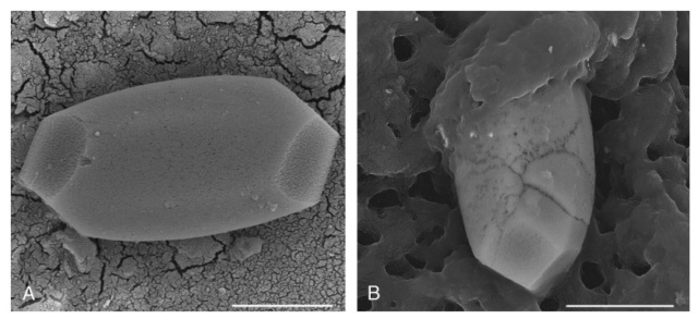
A, Single intact vital human otoconium. The more regular surface structures of the rhombohedral faces differ significantly from the surface of the cylindrical body indicating different areas of the composite structures,
B, Vital human otoconium affected by degeneration. Pores are deepened and fissures occur.
Otoconia serve a purely mechanical purpose, that is, to provide a mass load required for optimal gravity reception. Gravity receptors are in principle bioaccelerometers consisting of three essential elements: a mass load, a sensing element, and the elastic connection between the two.
Biosynthesis of otoconia
In some lower systems, the mass load is imported from the environment through an open endolymphatic duct. This is the case in crustaceans where grains of sand are deposited on the gelatinous membrane. In most vertebrates, the mass load has to be produced in vivo, which for several reasons is a formidable engineering job. 9 In mammals, complex interactions are required between organic and inorganic processes, further complicated by the constraint that the assembly must be accomplished in the extracellular space, in endolymph, a fluid with highly unusual chemical makeup. In fact, the whole process is occurring in an organ designed for refined mechanoreception and not for the mundane task of mass production of high-density particles. Recent experimental findings provide a glimpse into how nature deals with this particular engineering challenge. The strategy appears to be to sweep through the massive construction project rapidly and leave the scene prior to onset of function. In a clear division of labor, the less specialized structures seem to supply the bulk of building materials, freeing the developing sensory elements to concentrate on the more refined tasks.
In human otoconia, calcite nanocrystals together with the organic minority component are organized to form different arrangements of nanocomposite structures. There are 2 different volume densities within the uniform outer shape leading to an inner architecture consisting of 3 + 3 dense branches and a less dense belly region.
The biosynthesis of otoconia occurs during fetal development when core proteins secreted by vestibular epithelial cells form a proteinaceous matrix that sequesters calcium carbonate. [url=https://science.sciencemag.org/content/329/5988/twil#:~:text=The biosynthesis of otoconia occurs,matrix that sequesters calcium]6[/url] Otoconia development is a process that requires the spatiotemporal orchestration of complex biochemical events 7 . A distinct collection of proteins in the macula plus the unique ionic microenvironment of the endolymph near its epithelium likely contributes to the site-specific calcification of otoconia.

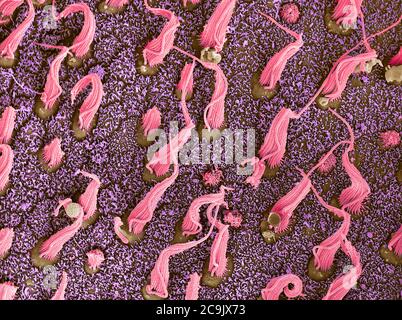
Scanning electron microscopy of mouse and leopard frog otoconia as examples of how aragonitic and calcitic crystals are organized. (A) Mouse utricular otoconia, (B) mouse saccular otoconia, (C) frog utricular otoconia, and (D) frog saccular otoconia. Panels A–C have a calcitic crystalline morphology and panel D an aragonitic crystalline morphology. (C and D are taken from Ref. Pote and Ross, 1993, with publisher's permission). Scale bars in panels A and C = 10 μm, in panels B and D= 5 μm.

Proteins are critical players in sequestering Ca2+ for otoconia formation and in determining otoconia crystalline morphology, growth, and stability 8
Regeneration and Repair of Otoconia
Otoconia, newly formed during the developmental phase, are highly susceptible to demineralization. When this is the case, dormant embryonic genes are upregulated, resulting in a regenerative cascade, mimicking the early events of otoconial morphogenesis.
The otoliths increase the mass of the otolithic membrane and give it more inertia. On Earth, when the head is tilted to the left or right, forward or back, the otoliths tend to move along the gravity gradient (downwards). Even a slight movement of the otolithic membrane is enough to bend hair cells and send sensory information to the brain. A similar inertia and gravity-dependent process occurs when you accelerate linearly -- up or down, forward or backward.
Body movements undertaken in our every day "Earth-normal" environment usually do not upset our sense of balance or body orientation. However, we have all experienced dizziness and difficulty walking after spinning around in a circle. How does the unique gravitational condition encountered in space flight affect an astronaut's sense of body orientation, movement, and balance?

The underlying physiology and functioning of the otolith organs are remarkably similar to those of the semicircular canals. Both systems depend upon inertia and the mechanical deflection of hair cells to initiate nerve impulses that are sent to the brain and interpreted as body movement. The brain then reflexively initiates appropriate "corrective" actions within the nervous, visual, and muscular systems to ensure that situational awareness and balance are maintained.
During forward acceleration, inertia causes the utricle's otolithic membrane and its associated otoliths to lag behind the portion of the utricle that is firmly attached to the head. This in turn causes the hair cells, whose hair-like extensions are embedded within the otolith membrane, to be deflected backwards. This backward deflection stimulates sensory nerves to fire and this provides the brain with information on the direction and speed of acceleration. A similar process occurs within the saccule when you are in an elevator and it either begins to rise or descend rapidly.
2. The ampulla
Parts of the vestibular nerve penetrate the base of each ampulla and terminate in a tuft of specialized sensory hair cells. The hair cells are arranged in a mound-like structure called the ampullary crest. Rising above the ampullary crest is the cupula, consisting of the hair-like extensions of the hair cells surrounded by a gelatinous material arranged into a wedge-shaped structure. This structure consisting of the ampullary crest and the cupula is called a crista.


When the endolymph moves (or the cupula moves and the fluid remains stationary), the gelatinous tip of the cupula and the hair cell extensions embedded within it are displaced to one side or the other. When the embedded hair cells bend, they send a signal via the vestibular nerve to the brain where the information is evaluated and appropriate action is initiated.
The Semicircular Canals
What's the difference between angular and linear acceleration? Linear acceleration is a change in velocity (speed increasing or decreasing over time) without a change of direction (straight line). Angular acceleration is a change in both velocity and direction at the same time. For example, imagine you are in a stopped car. The driver of the car steps on the accelerator and you accelerate straight ahead. The driver steps on the brake pedal and you decelerate to a stop. Then the driver puts the car in reverse and you accelerate straight backwards, and then the driver slams on the brakes once again. You have just experienced linear acceleration and deceleration in both forward and backward directions. Your movement was along a straight line and your otolith organs helped you sense these linear accelerations and decelerations.
Imagine yourself on a roller coaster. You start out accelerating straight ahead, just like in the car. Suddenly, the track dips almost straight down and you "pitch" forward. Then the nose of your car (and you) comes almost straight up. You have just experienced downward and upward pitch. The roller coaster, while staying perfectly flat on the track, now takes a severe left turn followed by a right turn. You have just "yawed" to the left and right. Now comes the really fun part. Your roller coaster and the track do a complete 360-degree roll, first to the left and then to the right. Makes you dizzy just thinking about it, right? You have just experienced the three planes of angular acceleration; pitch, yaw, and roll. An aircraft, a spaceship, or any vehicle operating in three-dimensional space can accelerate in these three planes of rotation and often along more than one plane at the same time. Your semicircular canals enable you to sense these angular accelerations.
The semicircular canals are oriented along three planes of movement with each plane at right angles to the other two.

Observe how the Lateral, posterior, and anterior canal are in perfect angles to each other.

Although they are both located within the vestibular apparatus of your inner ear, are interconnected, and operate using similar physical principles, the sensory mechanisms which allow you to detect linear acceleration (otolith organs) are structurally and functionally different than those which allow you to detect angular acceleration (semicircular canals).
The mechanics of how the semicircular canals actually function to "sense" angular acceleration may be more easily understood by reviewing the physics of inertia. The Law of Inertia states that "a body at rest remains at rest unless acted upon by an unbalanced force." This is important because angular acceleration and deceleration primarily affect the semicircular canals and entirely depend on the relative movement of endolymph with respect to the cupula.
The mechanics of how the semicircular canals actually function to "sense" angular acceleration may be more easily understood by reviewing the physics of inertia. The Law of Inertia states that "a body at rest remains at rest unless acted upon by an unbalanced force." This is important because angular acceleration and deceleration primarily affect the semicircular canals and entirely depend on the relative movement of endolymph with respect to the cupula.
This means that if you were to begin accelerating along one of the three planes of rotation (pitch, roll, or yaw), structural components of the corresponding semicircular canal would begin moving immediately since they are attached to the rest of your head. However, the endolymph within that particular semicircular canal would tend to "remain at rest" due to inertia. It would lag behind the structural components, deflecting the cupula and generating a nerve impulse to the brain.
Initially, the membranous tubular and cellular structures move but the fluid does not. Thus, there is relative movement between the fluid and the rest of the semicircular canal. Eventually, due to friction and the drag it induces, the fluid begins to move at the same speed as the components within which it is contained. When this occurs, the cupula is not deflected and, even though your body is continuing to angularly accelerate, the acceleration is not "sensed". You incorrectly perceive that you are stationary.
Now, let's stop your angular acceleration suddenly. What happens? The moving fluid now has momentum and so it continues to move until friction and drag bring it to a stop. In other words, fixed structures of your semicircular canal stop immediately (since they are still attached to your head which is still attached to your body) but the endolymph fluid continues to move in the direction of the previous movement. The Law of Inertia also states that a body in motion will continue in motion in a straight line unless acted upon by an unbalanced force. Now, the cupula and the embedded hair cells are bent in the opposite direction. This causes you to incorrectly sense that you are accelerating in the direction opposite to your previous acceleration, even though you are completely stopped!
The body of organisms uses devices to detect physical phenomena so it knows what is going on outside and inside through sensory transducers, which sense a physical phenomenon and convert it into useful information. The sensation of balance comes from the vestibular apparatus in the ears that tells the body its position relative to motion and the force of gravity and also helps to stabilize the retinal image. 1
Not only all parts of the vestibule need to be present, but also the nerve which sends the information to the brain, and converts the information into experience the sensation of balance.
The semicircular canals are a set of three membranous tubes embedded within a bony structure of the same shape. The central cavity of each canal is filled with a fluid called endolymph. Each endolymph-filled canal has an enlarged area near its base called an ampulla.
The vestibular system also helps you maintain a fixed gaze on a stationary or moving external object while you are undergoing complex head and body movements. Look at the clock on the wall. Now move your head sideways or up and down, or even in a circle. Your eyes stay fixed on the clock. With slow movement, the eyes are kept stationary by visual mechanisms only. As the speed of movement increases, the vestibular system takes over the image stabilization process.
Like the three sides in each corner of a box, the three fluid-filled semicircular canals are oriented at right angles to each other. The lateral canal is positioned like the bottom of the box, but is oriented about thirty degrees above the horizontal plane. The other two sides, the superior and posterior canals, are oriented vertically and at right angles to each other like the front or back and either side of the box. In this way they are set up to provide the brain with three-dimensional information about the angular acceleration caused by head motion.
The lateral canal is most sensitive to spinning or rotating the head from right to left or left to right, whereas the vertically oriented canals are most sensitive to nodding the head up or down or flexing the head to the right or left shoulder and back again. Head motion in any direction naturally moves the fluid within the semicircular canals and stimulates hair cells that are sensory receptors. The messages sent from the semicircular canals are sent through the vestibular branch of the same nerve that carries information on hearing to the brain (vestibulo-cochlear nerve) where they are processed and analyzed so the body can maintain its balance.
However another important function the information from angular head motion provides is to help stabilize the retinal image. Think about it. When you are in motion, unless you focus on something, everything moves across your visual field at the same speed as you do. If you were unable to have controlled eye movements when your head moves in any direction, everything you look at would always be blurry. Imagine our earliest ancestors running up and down over hill and dale trying to find food or avoid becoming food, without being able to focus on anything? So how does the body do it?
It’s called the vestibulo-ocular reflex. Look into a mirror and focus on your eyes as you rotate your head from side to side, up and down and then in any direction. Notice how your eyes automatically move in the opposite direction of your head so you can keep them in focus. This is also known as the doll’s eye reflex and is often used by physicians (along with the corneal and pupillary light reflex) to assess brainstem function. It is the sensory information supplied by the semicircular canals about angular head motion that allows the brain to reflexively move the eyes in the opposite direction to maintain the retinal image so we can focus on things no matter how fast or in what direction we move.
The utricle and the saccule also contain fluid but in addition have tiny calcium crystals that overlie the hair cells within the sensory membrane. In response to linear motion, the weight of the calcium crystals shears across the hair cells, resulting in depolarization and excitation. The utricle and the saccule provide information on linear acceleration and gravity. The sensory neurons of the utricle are oriented in the horizontal plane and provide information on the body’s back to front and side to side motion whereas the ones in the saccule are oriented in the vertical plane and give information on its up and down motion (like in an elevator) and the position of the head with respect to gravity.
As with the semicircular canals, the sensory information from the utricles and saccules travels in the vestibular branch of the vestibulo-cochlear nerve to the brain. It is within the various parts of the brain that this information is processed and integrated with other sensory data. This allows the body to maintain its balance and along with it, its survival capacity within nature.
Each part of the vestibular apparatus had to be present and working properly, while the brain had to know what to do with the information it received to maintain the body in balance and able to keep its eyes in focus.
The vestibule has 3 circular loops that are oriented in the x-y-z axis, a perfect 90 degrees from each other. The fluid in the loops moves as we move, activating small hair-like sensors that track angular acceleration. Like when you make a sharp turn in your car. Plus it also contains 3 linear accelerometers that are also at 90 degrees. Linear acceleration is that feeling you get when you accelerate in your car or ride in an elevator. That's the 6 degrees of freedom required to track a body as it moves through space. If you know the acceleration in each axis you can calculate the velocity in each axis. Then you can use velocity to calculate the distance traveled in each axis. Then using geometry you can then calculate your exact location from where you began. Believe it or not, all these calculations are being processed in our cerebellum and used to plot our movement on grid cells in an area called the entorhinal cortex.
Processing the information in the entorhinal cortex
Researchers have discovered a sophisticated neural computer, buried deep in the cerebellum, that performs inertial navigation calculations to figure out a person's movement through space. "These calculations are no mean feat, emphasized the researchers..11
The vestibular system in the inner ear provides the primary source of input to the brain about the body's movement and orientation in space. However, the vestibular sensors in the inner ear yield information about head position only. Also, the vestibular system's detection of head acceleration cannot distinguish between the effect of movement and that of gravitational force.
Dora Angelaki and colleagues based their brain studies on the predictions of a theoretical mathematical model postulating that the brain could compute inertial motion by combining rotational signals from the semicircular canal in the inner ear with gravity signals. They concentrated their search for the brain's inertial navigation system on particular types of neurons, called Purkinje cells, in a region of the cerebellum known to receive signals from the vestibular system. This region is known as the posterior cerebellar vermis, a narrow, worm-like structure between the brain's hemispheres.
In their experiments, the researchers measured the electrical activity of these Purkinje cells in monkeys as the animals' heads were maneuvered through a precise series of rotations and accelerations. After analyzing the electrical signals measured from the Purkinje cells during these movements, the researchers concluded that the specialized Purkinje cells were, indeed, computing earth-referenced motion from head-centered vestibular information.
The researchers concluded that the output of the Purkinje cells indicates an "elegant solution" to the computational problems involved in inertial navigation. 11
Purkinje Cells in Posterior Cerebellar Vermis Encode Motion in an Inertial Reference Frame June 21, 2007 12
The ability to orient and navigate through the terrestrial environment represents a computational challenge common to all vertebrates. It arises because motion sensors in the inner ear, the otolith organs, and the semicircular canals transduce self-motion in an egocentric reference frame. As a result, vestibular afferent information reaching the brain is inappropriate for coding our own motion and orientation relative to the outside world. Here we show that cerebellar cortical neuron activity in vermal lobules 9 and 10 reflects the critical computations of transforming head-centered vestibular afferent information into earth-referenced selfmotion and spatial orientation signals. Unlike vestibular and deep cerebellar nuclei neurons, where a mixture of responses was observed, Purkinje cells represent a homogeneous population that encodes inertial motion. They carry the earth-horizontal component of a spatially transformed and temporally integrated rotation signal from the semicircular canals, which is critical for computing head attitude, thus isolating inertial linear accelerations during navigation.
Important sensory information about our motion and orientation relative to the world arises from labyrinthine receptors in the inner ear. However, interpretation of signals from the peripheral vestibular sensors, the semicircular canals and the otolith organs, faces two interdependent problems
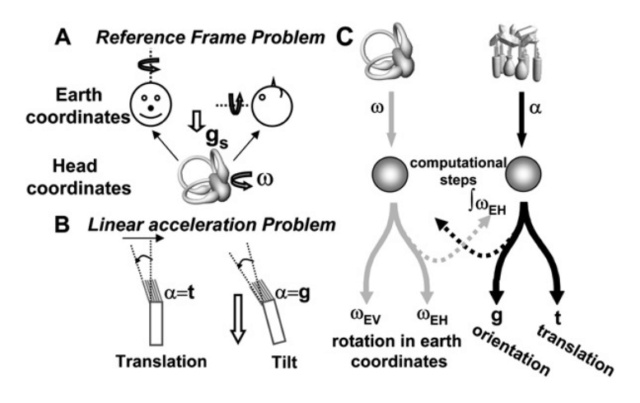
Schematics Illustrating the ‘‘Reference Frame’’ and ‘‘Linear Acceleration’’ Problems, along with the Proposed Mathematical Solution
(A) The ‘‘Reference frame’’ problem is illustrated by an example of two yaw rotations that are identical in head coordinates but differ in earth-centered coordinates. Yaw rotations in upright and supine orientations differ relative to the direction of gravity (gs, defining here the earth reference frame), yet elicit identical semicircular canal afferent responses that encode rotation, u, in head-centered coordinates.
(B) The ‘‘Linear acceleration’’ problem is described by schematizing that hair cells and otolith afferents encode net linear acceleration, a, thus respond identically to either translational, t, or gravitational, g, components.
(C) Proposed computational solution, schematized as two steps (for details about the underlying mathematics, see Supplemental Data). To solve the ‘‘Reference frame’’ problem, neural estimates of g must be used by the brain to decompose the head-fixed canal activation, u, into earth-vertical (uEV, parallel to gravity) and earth-horizontal (uEH, perpendicular to gravity) components. To solve the ‘‘Linear acceleration’’ problem, a change in angular orientation can be computed by temporal integration of uEH. This signal (!uEH) can then be combined with net linear acceleration from otolith afferents to extract translation.
The first, referred to as the ‘‘reference frame problem,’’ arises because vestibular sensors are physically fixed in the head. The second, referred to as the ‘‘linear acceleration problem,’’ is due to a sensory ambiguity that arises because of physical laws (Einstein’s equivalence principle). The brain thus faces the task of computing inertial motion and spatial orientation using multimodal integration. Inertial motion is explicitly coded by single Purkinje cells in the cortex of the nodulus (lobule 10) and uvula (lobule 9; collectively referred to as NU), areas that receive direct primary vestibular afferent inputs and are heavily interconnected with the VN
Vestibular Primary Afferent Cerebellum Projections: Mossy Fibers 13
The peripheral vestibular apparatus consists of five end organs; three semicircular canals are orthogonally oriented and sense angular rotation in the horizontal, pitch, and roll planes. Two otoliths sense linear acceleration of gravity during roll-tilt (utricle) and pitch (saccule). Vestibular primary afferents that originate from each of the five ipsilateral vestibular end organs combine in the vestibular nerve. As it approaches the brainstem, the vestibular nerve divides into two fiber bundles containing axons of unequal thickness. The thicker axons enter the medulla passing into the vestibular complex where they terminate on the medial, descending, and superior vestibular nuclei (MVN, DVN, and SVN) as well as the parasolitary nucleus
1. https://evolutionnews.org/2016/08/a_sense_of_bala/
2. http://www.cochlea.eu/en/ear/inner-ear
3. https://www.nasa.gov/audience/forstudents/9-12/features/F_Human_Vestibular_System_in_Space.html
4. https://dizziness-and-balance.com/anatomy/ear/otoliths.html
5. https://sci-hub.ren/10.1097/mao.0000000000000206
6. https://pubmed.ncbi.nlm.nih.gov/16357260/
7. https://www.jci.org/articles/view/42601
8. https://sci-hub.ren/10.1016/j.brainres.2006.02.083
9. https://sci-hub.ren/10.1111/j.1749-6632.2001.tb03743.x
http://library.lol/main/F97969AB6DC48376306E5CAEB4F1AE27
10. https://www.thehighestofthemountains.com/evolution.php
11. https://www.sciencedaily.com/releases/2007/06/070620122014.htm
12. https://sci-hub.ren/10.1016/j.neuron.2007.06.003
13. https://sci-hub.ren/https://link.springer.com/referenceworkentry/10.1007%2F978-94-007-1333-8_18
https://reasonandscience.catsboard.com/t3131-vestibule-gyro-and-accelerometer-triads-by-evolution-or-design

The Vestibular System
https://www.sumanasinc.com/webcontent/animations/content/vestibular.html
The inner ear houses the vestibular apparatus. It consists of the three semicircular canals, the utricle, and the saccule. Together they provide sensory information to the brain to help the body maintain its position relative to gravity and control eye movement with head motion.
2
1. Anterior semicircular canal
2. Ampulla (superior canal)
3. Ampulla (lateral canal)
4. Sacculus
5. Cochlear duct
6. Helicotrema
7. Lateral (horizontal) canal
8. Posterior canal
9. Ampulla (posterior canal)
10. Oval window
11. Round window
12. Vestibular duct (scala vestibuli)
13. Tympanic duct (scala tympani)
14. Utricule
The presence of sensory and response systems is a universal attribute of life as we know it. All living organisms on Earth have the ability to sense and respond appropriately to changes in their internal and external environment. Organisms, including humans, must sense accurately before they can react, thus ensuring survival. If our senses are not providing us with reliable information, we may take an action that is inappropriate for the circumstances and this could lead to injury or death. 3
The vestibular system
One of the most powerful senses is the vestibular sense, provided by the vestibular system. It is our ability to sense body movement combined with our ability to maintain balance (postural equilibrium). The human body has a remarkable ability to sense and determine the direction and speed in which it is moving and maintain balance (postural equilibrium). The 1. Otolith organs are what allows an organism, including humans, to perceive linear acceleration, both horizontally and vertically (gravity). Although they are both located within the vestibular apparatus of your inner ear, are interconnected, and operate using similar physical principles, the sensory mechanisms which allow you to detect linear acceleration (otolith organs) are structurally and functionally different than 2. the ampulla, which allows you to detect angular acceleration (semicircular canals).
My comment: Consider that both abilities are of utmost importance, and if one is missing, an essential ability is lacking. And as will be outlined next, both abilities require separate, complex, and sophisticated systems composed of various interlocked, interdependent parts.
On the right, the 1. otolith, required to sense acceleration, both horizontally and vertically (gravity). On the left, the 2. crista ampullaris which is the sensory organ of body movement, like rotation, responsible for the body to maintain balance.
Humans have the ability to walk a tight rope, do repeated pirouettes in a ballet performance, combine twists and turn when diving, or perform triple toe loops while ice skating... all (usually) without losing balance and while keeping track of the relative position of arms and legs with respect to the rest of the body. If you give it careful thought, you will realize how amazing this is!!
How does the human body sense and control the movement so precisely? How does it maintain balance while we put ourselves through a wide variety of spinning and tumbling activities that are inherently "unbalancing"? When we are in motion, how do we know in what direction and at what speed we are moving? How do these important body senses change or adapt when we fly in an aircraft?
Maintaining postural equilibrium, sensing movement, and maintaining an awareness of the relative location of our body parts requires the precise integration of several of the body's sensory and response systems including visual, vestibular, somatosensory (touch, pressure, and stretch receptors in our skin, muscles, and joints), and auditory. Acting together, these body systems constantly gather and interpret sensory information from all over the body and usually allow us to act on that information in an appropriate and helpful way.
My comment: Such an integrated interdependent system cannot be the product of slow evolutionary gradual processes. It is an all-or-nothing business. It has to be conceptualized from the get-go, right from the start, and be implemented fully operational from the beginning. These are essential key functions of our body. If one of the interacting parts/systems is missing, the consequences are catastrophic. If we could not balance our body, we could never walk upright.
1. Saccule and Utricle - the otolith organs sense linear acceleration and are affected by gravity.
The vestibular system, which is key to our senses of balance, motion, and body position, is comprised of three semicircular canals connected to two membranous sacs called the saccule and utricle. The saccule and utricle are referred to collectively as "the otolith organs".
This figure shows a close-up of the inner ear. The utricle is contained within a swelling adjacent to the semicircular canals, and the saccule is close to the cochlea. The black dots surrounding the utricle and saccule are the dark cells. The saccule and untricle sense linear acceleration and are affected by gravity. They also provide us with information concerning changes in head position (tilt).
Because of the way they are situated within the vestibular apparatus, the saccule is more sensitive to vertical acceleration (like riding in an elevator) and the utricle is more sensitive to horizontal acceleration (riding in a car). Both the saccule and the utricle contain a thickened patch of specialized cells called a macula that consists of sensory hair cells interspersed with "supporting" cells. In vertebrates the utricular maculae in the inner ear contain an otolithic membrane and otoconia (particles of calcium carbonate) that bend hair cells in the direction of gravity. This response to gravitational pull helps animals maintain their sense of balance.
Extremely oversimplified schematic of the orientation of the otoliths, meant to simply give one a general idea as to their orientation. The utricle is approximately in the horizontal plane, and the saccule in the sagittal plane. 4

This is a much more accurate schematic of the otoliths, showing their orientation in 3 dimensions. The utricle is not entirely horizontal, and the saccule is also somewhat tilted. A set of hair cells are coupled to a mass of stones. When the stones accelerate, with respect to the hairs, they exert a shearing force on the hairs. This force is detected by the hair cells and sent to the brain via branches of the vestibular nerve. The utricle sends input to the brain via the superior division of the nerve, and the saccule, (mainly) via the inferior division. There is considerably more complexity to the organization of the utricle and saccule, including different types of hair cells and detail to the sensory macule (patch of sensory cells) that are omitted here.
The free hair-like tufts extending from the hair cells are embedded in a gelatinous otolithic membrane which supports small piles of calcium carbonate crystals on its surface. Collectively, these calcium carbonate crystals are called otoliths.
Otoconia
Our sense of balance is dependent on small crystals in the inner ear called otoconia. These crystals are embedded within a fibrous extracellular matrix that couples the force of gravity to the cilia of sensory cells, which in turn send signals to the nervous system. Otoconia represent essential functional components of the otolith organs located on top of the sensory epithelium of the utricle and the saccule. Because of their inertial mass, otoconia provide the mechanical stimulus, which leads to a deflection of vestibular hair cells to enable the otolith system to detect linear accelerations and head tilts relative to the gravity vector, which is the reference in space 5
The importance of otoconia is clearly demonstrated by the impact of balance disorders upon the elderly population that involve otoconia degeneration, as well as by canalithiasis and cupulolithiasis, in which otoconia are dislocated. 8

A, Single intact vital human otoconium. The more regular surface structures of the rhombohedral faces differ significantly from the surface of the cylindrical body indicating different areas of the composite structures,
B, Vital human otoconium affected by degeneration. Pores are deepened and fissures occur.
Otoconia serve a purely mechanical purpose, that is, to provide a mass load required for optimal gravity reception. Gravity receptors are in principle bioaccelerometers consisting of three essential elements: a mass load, a sensing element, and the elastic connection between the two.
Biosynthesis of otoconia
In some lower systems, the mass load is imported from the environment through an open endolymphatic duct. This is the case in crustaceans where grains of sand are deposited on the gelatinous membrane. In most vertebrates, the mass load has to be produced in vivo, which for several reasons is a formidable engineering job. 9 In mammals, complex interactions are required between organic and inorganic processes, further complicated by the constraint that the assembly must be accomplished in the extracellular space, in endolymph, a fluid with highly unusual chemical makeup. In fact, the whole process is occurring in an organ designed for refined mechanoreception and not for the mundane task of mass production of high-density particles. Recent experimental findings provide a glimpse into how nature deals with this particular engineering challenge. The strategy appears to be to sweep through the massive construction project rapidly and leave the scene prior to onset of function. In a clear division of labor, the less specialized structures seem to supply the bulk of building materials, freeing the developing sensory elements to concentrate on the more refined tasks.
In human otoconia, calcite nanocrystals together with the organic minority component are organized to form different arrangements of nanocomposite structures. There are 2 different volume densities within the uniform outer shape leading to an inner architecture consisting of 3 + 3 dense branches and a less dense belly region.
The biosynthesis of otoconia occurs during fetal development when core proteins secreted by vestibular epithelial cells form a proteinaceous matrix that sequesters calcium carbonate. [url=https://science.sciencemag.org/content/329/5988/twil#:~:text=The biosynthesis of otoconia occurs,matrix that sequesters calcium]6[/url] Otoconia development is a process that requires the spatiotemporal orchestration of complex biochemical events 7 . A distinct collection of proteins in the macula plus the unique ionic microenvironment of the endolymph near its epithelium likely contributes to the site-specific calcification of otoconia.

Scanning electron microscopy of mouse and leopard frog otoconia as examples of how aragonitic and calcitic crystals are organized. (A) Mouse utricular otoconia, (B) mouse saccular otoconia, (C) frog utricular otoconia, and (D) frog saccular otoconia. Panels A–C have a calcitic crystalline morphology and panel D an aragonitic crystalline morphology. (C and D are taken from Ref. Pote and Ross, 1993, with publisher's permission). Scale bars in panels A and C = 10 μm, in panels B and D= 5 μm.

Proteins are critical players in sequestering Ca2+ for otoconia formation and in determining otoconia crystalline morphology, growth, and stability 8
Regeneration and Repair of Otoconia
Otoconia, newly formed during the developmental phase, are highly susceptible to demineralization. When this is the case, dormant embryonic genes are upregulated, resulting in a regenerative cascade, mimicking the early events of otoconial morphogenesis.
The otoliths increase the mass of the otolithic membrane and give it more inertia. On Earth, when the head is tilted to the left or right, forward or back, the otoliths tend to move along the gravity gradient (downwards). Even a slight movement of the otolithic membrane is enough to bend hair cells and send sensory information to the brain. A similar inertia and gravity-dependent process occurs when you accelerate linearly -- up or down, forward or backward.
Body movements undertaken in our every day "Earth-normal" environment usually do not upset our sense of balance or body orientation. However, we have all experienced dizziness and difficulty walking after spinning around in a circle. How does the unique gravitational condition encountered in space flight affect an astronaut's sense of body orientation, movement, and balance?
The underlying physiology and functioning of the otolith organs are remarkably similar to those of the semicircular canals. Both systems depend upon inertia and the mechanical deflection of hair cells to initiate nerve impulses that are sent to the brain and interpreted as body movement. The brain then reflexively initiates appropriate "corrective" actions within the nervous, visual, and muscular systems to ensure that situational awareness and balance are maintained.
During forward acceleration, inertia causes the utricle's otolithic membrane and its associated otoliths to lag behind the portion of the utricle that is firmly attached to the head. This in turn causes the hair cells, whose hair-like extensions are embedded within the otolith membrane, to be deflected backwards. This backward deflection stimulates sensory nerves to fire and this provides the brain with information on the direction and speed of acceleration. A similar process occurs within the saccule when you are in an elevator and it either begins to rise or descend rapidly.
2. The ampulla
Parts of the vestibular nerve penetrate the base of each ampulla and terminate in a tuft of specialized sensory hair cells. The hair cells are arranged in a mound-like structure called the ampullary crest. Rising above the ampullary crest is the cupula, consisting of the hair-like extensions of the hair cells surrounded by a gelatinous material arranged into a wedge-shaped structure. This structure consisting of the ampullary crest and the cupula is called a crista.
When the endolymph moves (or the cupula moves and the fluid remains stationary), the gelatinous tip of the cupula and the hair cell extensions embedded within it are displaced to one side or the other. When the embedded hair cells bend, they send a signal via the vestibular nerve to the brain where the information is evaluated and appropriate action is initiated.
The Semicircular Canals
What's the difference between angular and linear acceleration? Linear acceleration is a change in velocity (speed increasing or decreasing over time) without a change of direction (straight line). Angular acceleration is a change in both velocity and direction at the same time. For example, imagine you are in a stopped car. The driver of the car steps on the accelerator and you accelerate straight ahead. The driver steps on the brake pedal and you decelerate to a stop. Then the driver puts the car in reverse and you accelerate straight backwards, and then the driver slams on the brakes once again. You have just experienced linear acceleration and deceleration in both forward and backward directions. Your movement was along a straight line and your otolith organs helped you sense these linear accelerations and decelerations.
Imagine yourself on a roller coaster. You start out accelerating straight ahead, just like in the car. Suddenly, the track dips almost straight down and you "pitch" forward. Then the nose of your car (and you) comes almost straight up. You have just experienced downward and upward pitch. The roller coaster, while staying perfectly flat on the track, now takes a severe left turn followed by a right turn. You have just "yawed" to the left and right. Now comes the really fun part. Your roller coaster and the track do a complete 360-degree roll, first to the left and then to the right. Makes you dizzy just thinking about it, right? You have just experienced the three planes of angular acceleration; pitch, yaw, and roll. An aircraft, a spaceship, or any vehicle operating in three-dimensional space can accelerate in these three planes of rotation and often along more than one plane at the same time. Your semicircular canals enable you to sense these angular accelerations.
The semicircular canals are oriented along three planes of movement with each plane at right angles to the other two.
Observe how the Lateral, posterior, and anterior canal are in perfect angles to each other.

Although they are both located within the vestibular apparatus of your inner ear, are interconnected, and operate using similar physical principles, the sensory mechanisms which allow you to detect linear acceleration (otolith organs) are structurally and functionally different than those which allow you to detect angular acceleration (semicircular canals).
Do you know what the odds are that two perfect triads can evolve by chance to cover the x-y-z axes? Actually it's zero. 10
Because if each accelerometer is not perfectly aligned to each axis, the sensor does not work, it's useless. Even Darwin himself said that discoveries like this would prove his theory to be false. The vestibule had to be built correctly from the beginning and we are not even considering all the circuits needed to calculate navigation data from the sensor. How do you suppose they evolved? Researchers have even pinpointed where those calculations take place to track our movement.
Do you know how much electronics it takes to process this kind of data? Here is a photo of a complete navigation system that I was the lead engineer on, so I know what is involved. This video from a neuroscience site shows how the vestibule works.
From an engineering standpoint, we are high-tech biological machines, with more processing power than 34,500 Intel i7 CPUs. All the instructions to wire up 80 billions neurons and assemble the body must be stored in our DNA which we know contains about 3 gigabytes of data.
The mechanics of how the semicircular canals actually function to "sense" angular acceleration may be more easily understood by reviewing the physics of inertia. The Law of Inertia states that "a body at rest remains at rest unless acted upon by an unbalanced force." This is important because angular acceleration and deceleration primarily affect the semicircular canals and entirely depend on the relative movement of endolymph with respect to the cupula.
The mechanics of how the semicircular canals actually function to "sense" angular acceleration may be more easily understood by reviewing the physics of inertia. The Law of Inertia states that "a body at rest remains at rest unless acted upon by an unbalanced force." This is important because angular acceleration and deceleration primarily affect the semicircular canals and entirely depend on the relative movement of endolymph with respect to the cupula.
This means that if you were to begin accelerating along one of the three planes of rotation (pitch, roll, or yaw), structural components of the corresponding semicircular canal would begin moving immediately since they are attached to the rest of your head. However, the endolymph within that particular semicircular canal would tend to "remain at rest" due to inertia. It would lag behind the structural components, deflecting the cupula and generating a nerve impulse to the brain.
Initially, the membranous tubular and cellular structures move but the fluid does not. Thus, there is relative movement between the fluid and the rest of the semicircular canal. Eventually, due to friction and the drag it induces, the fluid begins to move at the same speed as the components within which it is contained. When this occurs, the cupula is not deflected and, even though your body is continuing to angularly accelerate, the acceleration is not "sensed". You incorrectly perceive that you are stationary.
Now, let's stop your angular acceleration suddenly. What happens? The moving fluid now has momentum and so it continues to move until friction and drag bring it to a stop. In other words, fixed structures of your semicircular canal stop immediately (since they are still attached to your head which is still attached to your body) but the endolymph fluid continues to move in the direction of the previous movement. The Law of Inertia also states that a body in motion will continue in motion in a straight line unless acted upon by an unbalanced force. Now, the cupula and the embedded hair cells are bent in the opposite direction. This causes you to incorrectly sense that you are accelerating in the direction opposite to your previous acceleration, even though you are completely stopped!
The body of organisms uses devices to detect physical phenomena so it knows what is going on outside and inside through sensory transducers, which sense a physical phenomenon and convert it into useful information. The sensation of balance comes from the vestibular apparatus in the ears that tells the body its position relative to motion and the force of gravity and also helps to stabilize the retinal image. 1
Not only all parts of the vestibule need to be present, but also the nerve which sends the information to the brain, and converts the information into experience the sensation of balance.
The semicircular canals are a set of three membranous tubes embedded within a bony structure of the same shape. The central cavity of each canal is filled with a fluid called endolymph. Each endolymph-filled canal has an enlarged area near its base called an ampulla.
The vestibular system also helps you maintain a fixed gaze on a stationary or moving external object while you are undergoing complex head and body movements. Look at the clock on the wall. Now move your head sideways or up and down, or even in a circle. Your eyes stay fixed on the clock. With slow movement, the eyes are kept stationary by visual mechanisms only. As the speed of movement increases, the vestibular system takes over the image stabilization process.
Like the three sides in each corner of a box, the three fluid-filled semicircular canals are oriented at right angles to each other. The lateral canal is positioned like the bottom of the box, but is oriented about thirty degrees above the horizontal plane. The other two sides, the superior and posterior canals, are oriented vertically and at right angles to each other like the front or back and either side of the box. In this way they are set up to provide the brain with three-dimensional information about the angular acceleration caused by head motion.
The lateral canal is most sensitive to spinning or rotating the head from right to left or left to right, whereas the vertically oriented canals are most sensitive to nodding the head up or down or flexing the head to the right or left shoulder and back again. Head motion in any direction naturally moves the fluid within the semicircular canals and stimulates hair cells that are sensory receptors. The messages sent from the semicircular canals are sent through the vestibular branch of the same nerve that carries information on hearing to the brain (vestibulo-cochlear nerve) where they are processed and analyzed so the body can maintain its balance.
However another important function the information from angular head motion provides is to help stabilize the retinal image. Think about it. When you are in motion, unless you focus on something, everything moves across your visual field at the same speed as you do. If you were unable to have controlled eye movements when your head moves in any direction, everything you look at would always be blurry. Imagine our earliest ancestors running up and down over hill and dale trying to find food or avoid becoming food, without being able to focus on anything? So how does the body do it?
It’s called the vestibulo-ocular reflex. Look into a mirror and focus on your eyes as you rotate your head from side to side, up and down and then in any direction. Notice how your eyes automatically move in the opposite direction of your head so you can keep them in focus. This is also known as the doll’s eye reflex and is often used by physicians (along with the corneal and pupillary light reflex) to assess brainstem function. It is the sensory information supplied by the semicircular canals about angular head motion that allows the brain to reflexively move the eyes in the opposite direction to maintain the retinal image so we can focus on things no matter how fast or in what direction we move.
The utricle and the saccule also contain fluid but in addition have tiny calcium crystals that overlie the hair cells within the sensory membrane. In response to linear motion, the weight of the calcium crystals shears across the hair cells, resulting in depolarization and excitation. The utricle and the saccule provide information on linear acceleration and gravity. The sensory neurons of the utricle are oriented in the horizontal plane and provide information on the body’s back to front and side to side motion whereas the ones in the saccule are oriented in the vertical plane and give information on its up and down motion (like in an elevator) and the position of the head with respect to gravity.
As with the semicircular canals, the sensory information from the utricles and saccules travels in the vestibular branch of the vestibulo-cochlear nerve to the brain. It is within the various parts of the brain that this information is processed and integrated with other sensory data. This allows the body to maintain its balance and along with it, its survival capacity within nature.
Each part of the vestibular apparatus had to be present and working properly, while the brain had to know what to do with the information it received to maintain the body in balance and able to keep its eyes in focus.
The vestibule has 3 circular loops that are oriented in the x-y-z axis, a perfect 90 degrees from each other. The fluid in the loops moves as we move, activating small hair-like sensors that track angular acceleration. Like when you make a sharp turn in your car. Plus it also contains 3 linear accelerometers that are also at 90 degrees. Linear acceleration is that feeling you get when you accelerate in your car or ride in an elevator. That's the 6 degrees of freedom required to track a body as it moves through space. If you know the acceleration in each axis you can calculate the velocity in each axis. Then you can use velocity to calculate the distance traveled in each axis. Then using geometry you can then calculate your exact location from where you began. Believe it or not, all these calculations are being processed in our cerebellum and used to plot our movement on grid cells in an area called the entorhinal cortex.
Processing the information in the entorhinal cortex
Researchers have discovered a sophisticated neural computer, buried deep in the cerebellum, that performs inertial navigation calculations to figure out a person's movement through space. "These calculations are no mean feat, emphasized the researchers..11
The vestibular system in the inner ear provides the primary source of input to the brain about the body's movement and orientation in space. However, the vestibular sensors in the inner ear yield information about head position only. Also, the vestibular system's detection of head acceleration cannot distinguish between the effect of movement and that of gravitational force.
Dora Angelaki and colleagues based their brain studies on the predictions of a theoretical mathematical model postulating that the brain could compute inertial motion by combining rotational signals from the semicircular canal in the inner ear with gravity signals. They concentrated their search for the brain's inertial navigation system on particular types of neurons, called Purkinje cells, in a region of the cerebellum known to receive signals from the vestibular system. This region is known as the posterior cerebellar vermis, a narrow, worm-like structure between the brain's hemispheres.
In their experiments, the researchers measured the electrical activity of these Purkinje cells in monkeys as the animals' heads were maneuvered through a precise series of rotations and accelerations. After analyzing the electrical signals measured from the Purkinje cells during these movements, the researchers concluded that the specialized Purkinje cells were, indeed, computing earth-referenced motion from head-centered vestibular information.
The researchers concluded that the output of the Purkinje cells indicates an "elegant solution" to the computational problems involved in inertial navigation. 11
Purkinje Cells in Posterior Cerebellar Vermis Encode Motion in an Inertial Reference Frame June 21, 2007 12
The ability to orient and navigate through the terrestrial environment represents a computational challenge common to all vertebrates. It arises because motion sensors in the inner ear, the otolith organs, and the semicircular canals transduce self-motion in an egocentric reference frame. As a result, vestibular afferent information reaching the brain is inappropriate for coding our own motion and orientation relative to the outside world. Here we show that cerebellar cortical neuron activity in vermal lobules 9 and 10 reflects the critical computations of transforming head-centered vestibular afferent information into earth-referenced selfmotion and spatial orientation signals. Unlike vestibular and deep cerebellar nuclei neurons, where a mixture of responses was observed, Purkinje cells represent a homogeneous population that encodes inertial motion. They carry the earth-horizontal component of a spatially transformed and temporally integrated rotation signal from the semicircular canals, which is critical for computing head attitude, thus isolating inertial linear accelerations during navigation.
Important sensory information about our motion and orientation relative to the world arises from labyrinthine receptors in the inner ear. However, interpretation of signals from the peripheral vestibular sensors, the semicircular canals and the otolith organs, faces two interdependent problems

Schematics Illustrating the ‘‘Reference Frame’’ and ‘‘Linear Acceleration’’ Problems, along with the Proposed Mathematical Solution
(A) The ‘‘Reference frame’’ problem is illustrated by an example of two yaw rotations that are identical in head coordinates but differ in earth-centered coordinates. Yaw rotations in upright and supine orientations differ relative to the direction of gravity (gs, defining here the earth reference frame), yet elicit identical semicircular canal afferent responses that encode rotation, u, in head-centered coordinates.
(B) The ‘‘Linear acceleration’’ problem is described by schematizing that hair cells and otolith afferents encode net linear acceleration, a, thus respond identically to either translational, t, or gravitational, g, components.
(C) Proposed computational solution, schematized as two steps (for details about the underlying mathematics, see Supplemental Data). To solve the ‘‘Reference frame’’ problem, neural estimates of g must be used by the brain to decompose the head-fixed canal activation, u, into earth-vertical (uEV, parallel to gravity) and earth-horizontal (uEH, perpendicular to gravity) components. To solve the ‘‘Linear acceleration’’ problem, a change in angular orientation can be computed by temporal integration of uEH. This signal (!uEH) can then be combined with net linear acceleration from otolith afferents to extract translation.
The first, referred to as the ‘‘reference frame problem,’’ arises because vestibular sensors are physically fixed in the head. The second, referred to as the ‘‘linear acceleration problem,’’ is due to a sensory ambiguity that arises because of physical laws (Einstein’s equivalence principle). The brain thus faces the task of computing inertial motion and spatial orientation using multimodal integration. Inertial motion is explicitly coded by single Purkinje cells in the cortex of the nodulus (lobule 10) and uvula (lobule 9; collectively referred to as NU), areas that receive direct primary vestibular afferent inputs and are heavily interconnected with the VN
Vestibular Primary Afferent Cerebellum Projections: Mossy Fibers 13
The peripheral vestibular apparatus consists of five end organs; three semicircular canals are orthogonally oriented and sense angular rotation in the horizontal, pitch, and roll planes. Two otoliths sense linear acceleration of gravity during roll-tilt (utricle) and pitch (saccule). Vestibular primary afferents that originate from each of the five ipsilateral vestibular end organs combine in the vestibular nerve. As it approaches the brainstem, the vestibular nerve divides into two fiber bundles containing axons of unequal thickness. The thicker axons enter the medulla passing into the vestibular complex where they terminate on the medial, descending, and superior vestibular nuclei (MVN, DVN, and SVN) as well as the parasolitary nucleus
1. https://evolutionnews.org/2016/08/a_sense_of_bala/
2. http://www.cochlea.eu/en/ear/inner-ear
3. https://www.nasa.gov/audience/forstudents/9-12/features/F_Human_Vestibular_System_in_Space.html
4. https://dizziness-and-balance.com/anatomy/ear/otoliths.html
5. https://sci-hub.ren/10.1097/mao.0000000000000206
6. https://pubmed.ncbi.nlm.nih.gov/16357260/
7. https://www.jci.org/articles/view/42601
8. https://sci-hub.ren/10.1016/j.brainres.2006.02.083
9. https://sci-hub.ren/10.1111/j.1749-6632.2001.tb03743.x
http://library.lol/main/F97969AB6DC48376306E5CAEB4F1AE27
10. https://www.thehighestofthemountains.com/evolution.php
11. https://www.sciencedaily.com/releases/2007/06/070620122014.htm
12. https://sci-hub.ren/10.1016/j.neuron.2007.06.003
13. https://sci-hub.ren/https://link.springer.com/referenceworkentry/10.1007%2F978-94-007-1333-8_18
Last edited by Otangelo on Wed Oct 26, 2022 7:11 pm; edited 2 times in total
