The fantastic design of unicellular euplote protists
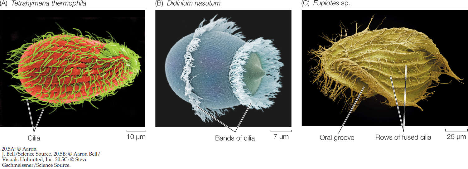
Ciliates comprise a diverse and ecologically important phylum of unicellular protists. One of the most specious and best-defined genera is Euplotes 3 Ciliates (unicellular protists of the phylum Ciliophora) are ubiquitous, abundant and eye-catching components of aquatic environments. In some systems of biological classification, the Protozoa are defined as a diverse group of unicellular eukaryotic organisms.[1] Historically, protozoa were defined as single-celled animals or organisms with animal-like behaviors, such as motility and predation. The group was regarded as the zoological counterpart to the "protophyta", which were considered to be plant-like, as they are capable of photosynthesis 1
Eukaryotic cells typically stabilize their plasma membranes with a more-or-less tightly associated layer of cytoskeletal proteins, forming a complex often referred to as the `membrane skeleton'. A classic example has been the spectrin network linked (via ankyrins and several actin-binding proteins) to integral membrane glycoproteins at the cytoplasmic face of the erythrocyte plasmalemma 4


2


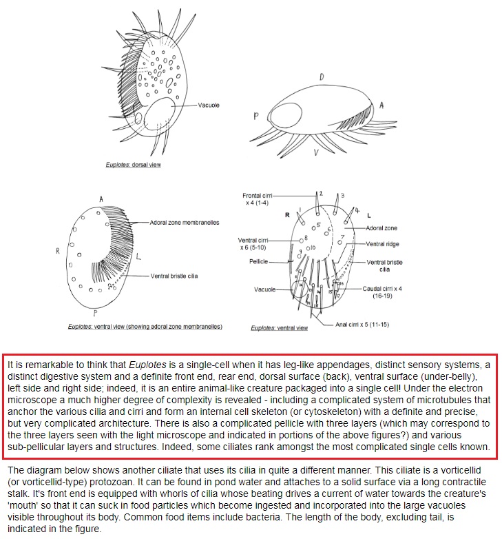
Cilia on the ventral surface of the hypotrich ciliate Euplotes are clustered into polykinetids or compound ciliary organelles, such as cirri or oral membranelles, used in locomotion and prey capture. A single polykinetid may contain more than 150 individual cilia; these emerge from basal bodies held in a closely spaced array within a scaffold or framework structure that has been referred to as a basal-body "cage". Cage structures were isolated free of cilia and basal bodies; the predominant component of such cages was found on polyacrylamide gels to be a 45-kDa polypeptide. Antisera were raised against this protein band and used for immunolocalizations at the light and electron microscope levels. Indirect immunofluorescence revealed the 45-kDa polypeptide to be localized exclusively to the bases of the ventral polykinetids. Immunogold staining of thin sections of intact cells further localized this reactivity to filaments of a double-layered dense lattice that appears to link adjoining basal bodies into ordered arrays within each polykinetid. Scanning electron microscopy of isolated cages reveals the lower or "basal" cage layer to be a fine lacey meshwork supporting the basal bodies at their proximal ends; adjoining basal bodies are held at their characteristic spacing by filaments of an upper or "medial" cage layer. The isolated cage thus resembles a miniature test-tube rack, able to accommodate varying arrangements of basal-body rows, depending on the particular type of polykinetid. Because of its clear and specific localization to the basal-body cages in Euplotes, we have termed this novel 45-kDa protein "cagein". 5
1. https://en.wikipedia.org/wiki/Protozoa
2. http://cronodon.com/BioTech/Cells_motility_2.html
3. http://journals.plos.org/plosone/article?id=10.1371/journal.pone.0165442
4. http://jcs.biologists.org/content/116/7/1291
5. https://www.researchgate.net/publication/233326174_Structure_and_Protein_Composition_of_a_Basal-Body_Scaffold_Cage_in_the_Hypotrich_Ciliate_Euplotes
Ciliates comprise a diverse and ecologically important phylum of unicellular protists. One of the most specious and best-defined genera is Euplotes 3 Ciliates (unicellular protists of the phylum Ciliophora) are ubiquitous, abundant and eye-catching components of aquatic environments. In some systems of biological classification, the Protozoa are defined as a diverse group of unicellular eukaryotic organisms.[1] Historically, protozoa were defined as single-celled animals or organisms with animal-like behaviors, such as motility and predation. The group was regarded as the zoological counterpart to the "protophyta", which were considered to be plant-like, as they are capable of photosynthesis 1
Eukaryotic cells typically stabilize their plasma membranes with a more-or-less tightly associated layer of cytoskeletal proteins, forming a complex often referred to as the `membrane skeleton'. A classic example has been the spectrin network linked (via ankyrins and several actin-binding proteins) to integral membrane glycoproteins at the cytoplasmic face of the erythrocyte plasmalemma 4


2


Cilia on the ventral surface of the hypotrich ciliate Euplotes are clustered into polykinetids or compound ciliary organelles, such as cirri or oral membranelles, used in locomotion and prey capture. A single polykinetid may contain more than 150 individual cilia; these emerge from basal bodies held in a closely spaced array within a scaffold or framework structure that has been referred to as a basal-body "cage". Cage structures were isolated free of cilia and basal bodies; the predominant component of such cages was found on polyacrylamide gels to be a 45-kDa polypeptide. Antisera were raised against this protein band and used for immunolocalizations at the light and electron microscope levels. Indirect immunofluorescence revealed the 45-kDa polypeptide to be localized exclusively to the bases of the ventral polykinetids. Immunogold staining of thin sections of intact cells further localized this reactivity to filaments of a double-layered dense lattice that appears to link adjoining basal bodies into ordered arrays within each polykinetid. Scanning electron microscopy of isolated cages reveals the lower or "basal" cage layer to be a fine lacey meshwork supporting the basal bodies at their proximal ends; adjoining basal bodies are held at their characteristic spacing by filaments of an upper or "medial" cage layer. The isolated cage thus resembles a miniature test-tube rack, able to accommodate varying arrangements of basal-body rows, depending on the particular type of polykinetid. Because of its clear and specific localization to the basal-body cages in Euplotes, we have termed this novel 45-kDa protein "cagein". 5
1. https://en.wikipedia.org/wiki/Protozoa
2. http://cronodon.com/BioTech/Cells_motility_2.html
3. http://journals.plos.org/plosone/article?id=10.1371/journal.pone.0165442
4. http://jcs.biologists.org/content/116/7/1291
5. https://www.researchgate.net/publication/233326174_Structure_and_Protein_Composition_of_a_Basal-Body_Scaffold_Cage_in_the_Hypotrich_Ciliate_Euplotes

