ElShamah - Reason & Science: Defending ID and the Christian Worldview
Would you like to react to this message? Create an account in a few clicks or log in to continue.
ElShamah - Reason & Science: Defending ID and the Christian Worldview

Otangelo Grasso: This is my personal virtual library, where i collect information, which leads in my view to the Christian faith, creationism, and Intelligent Design as the best explanation of the origin of the physical Universe, life, biodiversity


You are not connected. Please login or register

Evolution: Where Do Complex Organisms Come From?

Go to page : Previous  1, 2, 3, 4  Next

Go down  Message [Page 2 of 4]

Otangelo


Admin

Key Developmental Processes Shaping Organismal Form and Function

Developmental biology encompasses a wide range of processes that dictate the growth, form, and function of organisms from conception to maturity.

Cell Differentiation: This is where cells evolve and become specialized in their function.
Morphogenesis: The mechanism by which the structure of an organism develops.
Growth: Refers to the increase in cell number and size, allowing the organism to develop in size and complexity.

Developmental processes are foundational in shaping the life of organisms. They determine how cells renew and differentiate, ensuring that each organism is not only formed correctly but is also functionally adept. These processes have extensive implications in medicine, evolutionary biology, and agriculture. This extensive list represents a comprehensive overview of key developmental processes that are essential for the formation and function of an organism.  The following list encompasses processes ranging from the molecular to the organ level, each vital for the proper development, structure, and function of an organism. These processes, often interlinked, collectively orchestrate the intricate dance of development from a single cell to a multicellular organism.

The dichotomy between structural and regulatory codes is a suitable tool to start the systematic study of codes. Barbieri assumes the existence of three types of organic codes, namely manufacturing, signaling, and regulatory codes.

Codes are pervasive in nature, woven into the fabric of life, ensuring that organisms function, communicate, and adapt. While the word "code" might immediately conjure images of binary digits or cryptographic sequences, the biological realm showcases its own elegant systems of information storage and processing. Let's unravel these intricate codes.

Manufacturing Codes: At the heart of life lies a manufacturing system, a protocol that defines how the blueprints of life are read and realized. Picture a vast assembly line where raw materials are meticulously transformed into complex products. This is what the manufacturing code oversees. It's a set of instructions determining how basic molecular building blocks are assembled into the machinery that powers life. Whether it's the synthesis of proteins vital for structure and function or the replication of the genetic material itself, this code ensures the flawless production of life's necessities.

Signaling Codes: Imagine a vast network where information flows seamlessly, messages sent and received with precision. This is the realm of signaling codes. Every organism, no matter how simple or complex, exists in a dynamic environment. To thrive, it must sense and respond to myriad signals, be they external cues or internal messages. The signaling code encompasses the myriad ways cells communicate, both with each other and with their surroundings, ensuring harmony and coordination in the face of constant change.

Regulatory Codes: Regulatory codes act as the grand orchestrators, ensuring that the myriad processes within a cell or organism are coordinated in time and space. Think of them as conductors of a grand symphony, ensuring each instrument plays its part at the right moment. They determine when a gene should be activated or silenced, when a cell should grow or pause, and how resources are allocated. The regulatory code ensures that each process dovetails seamlessly with others, fostering a harmonious interplay that sustains life.

Epigenetic Codes: While genetics lays down the foundational script of life, epigenetics offers a layer of finesse, allowing organisms to modulate their genetic instructions based on experience and environment. It's akin to annotations on a manuscript, guiding how the original script is to be interpreted under different circumstances. Without altering the fundamental script, the epigenetic code offers a dynamic interface, allowing organisms to adapt and respond with a level of plasticity that is both astounding and essential.

Together, these codes offer a glimpse into the intricate ballet of life, a dance of molecules choreographed with precision and purpose. Delving into each code, we uncover the myriad ways nature encodes, processes, and utilizes information, showcasing the elegance and complexity inherent in the living world. As we continue our exploration, the wonder of life's codes becomes ever more evident, reminding us of the marvels that underpin our existence.
 
First, I will individual questions specifically to each of them will be answered, and in the following, I will ask these questions related to each of them:

The appearance of X (1-47) in the evolutionary timeline
De Novo Genetic Information necessary to instantiate X (1-47)
Manufacturing codes and languages that would have to emerge and be employed to instantiate X (1-47)
Epigenetic Regulatory Mechanisms necessary to be instantiated for X (1-47)
Signaling Pathways necessary to create, and maintain X (1-47)
Regulatory codes necessary for maintenance and operation of X (1-47)
Is there scientific evidence supporting the idea that X (1-49) was brought about by the process of evolution?
Irreducibility and Interdependence of the systems to instantiate and operate X (1-47)
Once is instantiated and operational, what other intra and extracellular systems is X (1-47) interdependent with?

Elucidating these questions will provide the reader a comprehensive understanding of the intricacies involved in the formation, operation, and emergence of the most relevant biological processes or components (1-47) that convey biological form and architecture. 


Appearance of (1-47) in the evolutionary timeline: This will map out when and in which organisms these processes or components supposedly first appeared according to the evolutionary timeline. This historical context can provide insights into the environmental, genetic, or ecological factors that is claimed to have driven their emergence.
De Novo Genetic Information necessary to instantiate (1-47): Understanding the new genetic instructions required for these processes will shed light on the complexity and uniqueness of their emergence and the kind of required innovations they represent.
Manufacturing codes and languages that would have to emerge and be employed to instantiate 1-47: This looks into the specialized 'codes' or mechanisms that these processes require. It emphasizes the complexity of biological systems and how they can't function without a precise set of instructions.
Epigenetic Regulatory Mechanisms necessary to be instantiated for X (1-47): Exploring the non-DNA sequence-based regulations emphasizes the layers of control and fine-tuning in biological systems, showing that it's not just the genes themselves but also their regulation that's crucial.
Signaling Pathways necessary to create, and maintain X (1-47): This emphasizes the interconnectedness of biological processes. Signaling pathways are like communication networks, ensuring that processes are coordinated.
Regulatory codes necessary for maintenance and operation of X (1-47): Delves into the continuous requirements for these processes to function properly over an organism's lifespan, showing the persistence of complexity beyond just the initial emergence.
Is there scientific evidence supporting the idea that X (1-47) was brought about by the process of evolution? This question seeks to tie the aforementioned complexities back to the current scientific understanding, emphasizing empirical grounding and challenging or affirming existing paradigms.
Irreducibility and Interdependence of the systems to instantiate and operate X (1-47): Exploring these concepts touches upon the evidence that some systems are so complex that they seem to require all their parts to be present and functioning from the outset, posing questions about incremental evolutionary paths.
Once X is instantiated and operational, what other intra and extracellular systems is X (1-47) interdependent with? This emphasizes the holistic nature of biology, where processes are not isolated but are parts of larger networks, influencing and being influenced by myriad other systems.


Following are the key developmental processes shaping organismal form and function with a brief description of each process, The list is in alphabetic order

1. Angiogenesis and Vasculogenesis: Formation of new blood vessels from pre-existing ones (angiogenesis) and de novo vessel formation (vasculogenesis).
2. Apoptosis: Programmed cell death essential for removing unwanted cells.
3. Cell-Cycle Regulation: Controls the progression of cells through the stages of growth and division.
4. Cell-cell adhesion and the ECM: Refers to how cells stick to each other and to the extracellular matrix, essential for tissue formation.
5. Cell-Cell Communication: Cells communicate to coordinate their actions.
6. Cell Fate Determination and Lineage Specification (Cell differentiation): Process by which cells become specialized in their function.
7. Cell Migration and Chemotaxis: Movement of cells, guided by certain chemical gradients.
8. Cell Polarity and Asymmetry: Defines distinct cellular 'sides' or 'ends', crucial for many cell functions.
9. Cellular Pluripotency: Cells can give rise to multiple cell types.
10. Cellular Senescence: State of stable cell cycle arrest.
11. Centrosomes: Organize microtubules and provide structure to cells.
12. Chromatin Dynamics: How DNA and proteins are organized in the nucleus.
13. Cytokinesis: Physical process of cell division.
14. Cytoskeletal Arrays: Framework of the cell, involved in cell shape, movement, and division.
15. DNA Methylation: Addition of methyl groups to DNA, often involved in gene silencing.
16. Egg-Polarity Genes: Determine the axes of the egg and subsequently the organism.
17. Epigenetic Codes: Changes in gene function without changing DNA sequence.
18. Gene Regulation Network: Interactions between genes, controlling when and where genes are expressed.
19. Germ Cell Formation and Migration: Development and movement of reproductive cells.
20. Germ Layer Formation (Gastrulation): Development of primary tissue layers in embryos.
21. Histone PTMs: Modifications to histone proteins affecting DNA accessibility.
22. Homeobox and Hox Genes: Control the body plan of an embryo along the head-tail axis.
23. Hormones: Chemical messengers coordinating bodily functions.
24. Immune System Development: Formation and maturation of immune cells.
25. Ion Channels and Electromagnetic Fields: Channels allowing ions to flow in/out of cells; electromagnetic fields can influence development.
26. Membrane Targets: Processes focusing on cell membrane components.
27. MicroRNA Regulation: Small RNAs regulating gene expression post-transcriptionally.
28. Morphogen Gradients: Concentration gradients of substances determining tissue development.
29. Neural Crest Cells Migration: Movement of cells contributing to diverse structures, including peripheral nerves.
30. Neural plate folding and convergence: Formation of the neural tube in early development.
31. Neuronal Pruning and Synaptogenesis: Refinement of neural connections and formation of synapses.
32. Neurulation and Neural Tube Formation: Development of the neural tube, precursor to the CNS.
33. Noncoding RNA from Junk DNA: RNA molecules not coding for protein but having various functions.
34. Oogenesis: Egg cell (oocyte) formation.
35. Oocyte Maturation and Fertilization: Development of mature egg and its fusion with sperm.
36. Pattern Formation: Processes determining organized spatial arrangement of cells/tissues.
37. Photoreceptor development: Formation of cells detecting light in the eye.
38. Regional specification: Defining distinct regions within developing tissues.
39. Segmentation and Somitogenesis: Division of body into segments and formation of somites in embryos.
40. Signaling Pathways: Series of molecular events relaying extracellular signals to intracellular targets.
41. Spatiotemporal gene expression: Time and place-specific gene expression.
42. Spermatogenesis: The process of sperm cell formation and maturation.
43. Stem Cell Regulation and Differentiation: Control of stem cell fate and their development into specialized cells.
44. Symbiotic Relationships and Microbiota Influence: Interactions with microbial partners and their influence on host development.
45. Syncytium formation: Multinucleated cell formation, especially important in muscle tissues.
46. Transposons and Retrotransposons: Mobile genetic elements, sometimes influencing gene regulation.
47. Tissue Induction and Organogenesis: Formation of tissues and organs from undifferentiated cells.

The processes and systems listed are essential to the complex orchestration of development and physiology in multicellular organisms. Many of these are interwoven and interdependent to ensure accurate and timely development, function, and maintenance. 

Angiogenesis and Vasculogenesis are closely linked to Tissue Induction and Organogenesis. As tissues and organs form, they require a blood supply for nutrient and oxygen delivery.
Apoptosis works in tandem with Cell-Cycle Regulation. As cells progress through the cycle, faulty or unwanted cells undergo programmed death to maintain tissue homeostasis.
Cell-Cell Adhesion and the ECM are fundamental for Cell Migration and Chemotaxis, allowing cells to navigate the 3D environment of the body.
Cell Fate Determination and Lineage Specification are influenced by Cell-Cell Communication and Morphogen Gradients, which provide cues for cells to differentiate into specific lineages.
Cell Polarity and Asymmetry are critical for processes like Cytokinesis and work closely with the Cytoskeletal Arrays to ensure accurate cell division.
Cellular Pluripotency ties into Stem Cell Regulation and Differentiation, with pluripotent cells being a subset of stem cells that can give rise to almost all cell types.
Chromatin Dynamics, particularly Histone PTMs and DNA Methylation, play roles in Epigenetic Codes and influence Gene Regulation Network.
Egg-Polarity Genes are involved in Oogenesis and also influence the Oocyte Maturation and Fertilization processes.
Homeobox and Hox Genes work in synchrony with Pattern Formation and Segmentation and Somitogenesis to define body plans.
Hormones can impact various processes, including Immune System Development and Oogenesis.
MicroRNA Regulation, Noncoding RNA from Junk DNA, and Transposons and Retrotransposons provide additional layers of post-transcriptional control on the Gene Regulation Network.
Neural Crest Cells Migration is a part of Neurulation and Neural Tube Formation and directly affects Neural plate folding and convergence.
Neuronal Pruning and Synaptogenesis are essential aspects of Photoreceptor development and general neural development.
Spermatogenesis and Oogenesis interplay during Oocyte Maturation and Fertilization to initiate the next generation's development.
Signaling Pathways are pervasive and influence many of the aforementioned processes, from Cell-Cycle Regulation to Pattern Formation to Stem Cell Regulation and Differentiation.
Symbiotic Relationships and Microbiota Influence can indirectly impact processes like Immune System Development.

It's worth noting that this list only scratches the surface. The interconnectedness of these systems is immensely complex, with each one potentially influencing or being influenced by multiple others. They collectively underscore the intricacy and delicate choreography inherent to biology.

In biology, at least 47 complex systems, and over 230 structural and regulatory codes, genetic and epigenetic languages play key roles in developmental processes shaping organismal form and function. They are so complex and intertwined, that they seem to require all their parts to be present and functioning from the outset, posing big question marks about incremental evolutionary paths being an adequate explanation of their origins. In Biology, it is not only the complex biomolecular machines, systems,  and languages that are irreducibly complex, and work in an interdependent manner together, but once instantiated, they are often part of a larger, higher-order system. Irreducibly complex and interdependent systems are often required to convey and produce a complex function-bearing product, like a machine. Often, that machine alone bears no function on its own, unless it is interconnected with other machines to convey a final higher-order system with a specific function. An example is a car, that has an engine, which is interconnected and interdependent with the Transmission System, Exhaust System, cooling System, etc.

Premise 1. Life is embroidered with a profound amalgamation of genetic and epigenetic codes. With at least 33 distinct genetic variations and over 230 intertwined manufacturing, signaling, and operational codes, together with hundreds, and in prokaryotes probably thousands of intricate signaling networks, these codes collectively choreograph the exquisite dance of multicellular organisms, sculpting the vast biodiversity, nuanced form, and majestic architecture we observe.
Premise 2. This monumental wealth of information isn't random. It's meticulously orchestrated, often resembling the digital semiotic languages characterized by syntax, semantics, and pragmatics. Every protein, each metabolic trajectory, and every biomechanical construct operates with precision, abiding by principles finely tuned for specific functions.
Premise 3.  While the foundations of life are tangible, the essence of information transcends the physical. It's conceptual, operating in realms beyond the reach of spontaneous and aimless physical phenomena. Arguing that such processes can birth semiotic codes is akin to believing a rainbow might pen a sonnet or winds could draft architectural wonders. The directive, purposeful nature inherent in biological programming suggests intention, foresight, and specific goals.
Conclusion: The staggering complexity and deliberate design seen in organismal architecture and biodiversity beckon consideration beyond mere chance. It suggests that these wonders are not just products of random evolution but the handiwork of a deliberate and intelligent design.

Interdependence between Intrinsic and  Extrinsic Irreducibly Complex Systems

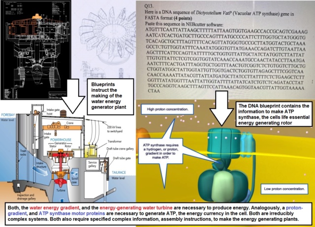
The concept of irreducible complexity posits that certain systems are so intricate in their interdependent components that removing any one of them would cause the system to cease functioning. This principle can be applied to both intrinsic components (those that are an inherent part of the system) and extrinsic components (external factors or conditions that the system relies upon).  The principle of interdependence is evident in many systems, both artificial and biological. Let me give an example, examining and comparing both hydroelectric turbines and ATP synthase ( molecular turbines, that generate ATP, the energy currency in the cell:

Hydroelectric energy production 

The hydroelectric turbine is intrinsically irreducibly complex

As water flows over the blades of a turbine, it causes the turbine to turn, converting the water's kinetic energy into mechanical energy.  It requires several intrinsic components that make the turbine functional:

Turbine Blades (Runner): These are the components that capture the kinetic energy of water. They are designed in specific shapes and configurations to optimize the conversion of water's kinetic energy into rotational mechanical energy. Without the blades, water would simply flow through without generating rotation.
Shaft: The shaft is connected to the turbine blades and translates the rotational movement of the blades into the generator. Without the shaft, the rotation of the blades couldn't be transferred.
Generator: This is where the mechanical energy (from the rotating shaft) is converted into electrical energy. The generator contains magnets and coils of wire. As the shaft rotates, it induces a flow of electric current in the wires.
Wicket Gates: These are adjustable gates that control the flow of water onto the turbine blades. Without effective wicket gates, the turbine could be overwhelmed by too much water or underutilized with too little.

But the turbine alone will bear no function. It requires a set of extrinsic parts, that together, in a joint venture, will achieve the final goal, which is to generate usable energy. These parts are:

Extrinsic irreducible complexity

Dam: A dam holds back water, creating a reservoir or a lake. This stored water has potential energy due to its height. Without the dam, there's no potential energy from water height.
Water: The moving water's kinetic energy, or the stored potential energy in the dam's reservoir, is what gets converted into mechanical energy by the turbines. Without water, there's no kinetic energy. 
Penstock: This is a conduit that brings water from the reservoir to the turbine. It plays a role in regulating the flow and pressure of the water hitting the turbine blades. Without the penstock, there's no controlled flow of water.
Generator: The turbine is connected to a generator. As the turbine spins, so does the generator, converting mechanical energy into electrical energy. Without the generator, there is electrical energy. 

ATP energy production in the cell

ATP synthase is intrinsically irreducibly complex

ATP synthase is an intricate molecular machine that plays a pivotal role in cellular respiration, generating ATP (adenosine triphosphate) from ADP (adenosine diphosphate) and inorganic phosphate. It harnesses the energy stored in a proton gradient, usually across the inner mitochondrial membrane in eukaryotes, to catalyze this reaction. For this complex enzyme to function, several critical components must be in place.

F1 Subunit (Catalytic Core)
α and β subunits: These subunits form the catalytic core where ATP synthesis or hydrolysis takes place. The β subunit is where ATP is synthesized from ADP and inorganic phosphate.
γ, δ, and ε subunits: These form the central stalk that rotates within the α/β core. The rotation of the γ subunit drives the conformational changes in the β subunit required for ATP synthesis.

Fo Subunit (Proton Channel)
c-Ring (c subunits): This ring is in the membrane and rotates as protons flow through the Fo portion of ATP synthase. The rotation of the c-ring is directly linked to the rotation of the γ subunit in the F1 portion.
a Subunit: This subunit forms a channel allowing protons to flow through the Fo complex. It interacts with the c-ring, enabling the translocation of protons to drive the rotation of the c-ring.
b and δ subunits: These form a peripheral stalk, holding the α/β core stationary while the central stalk and c-ring rotate.

Stator (Peripheral Stalk)
This part of the complex ensures that while the central stalk and c-ring rotate, the catalytic α/β core remains stationary. It is vital for the enzyme's ability to synthesize ATP efficiently.

Proton Channel
The Fo portion, specifically the interaction between the a and c subunits, allows for the passage of protons. This flow of protons is what drives the rotation of the c-ring and, subsequently, the γ subunit inside the F1 portion.

For ATP synthase to function effectively, all of these components must be present and interact in a coordinated manner. The absence or malfunction of any of these parts would disrupt the enzyme's ability to synthesize ATP, making ATP synthase a prime example of a molecular system exhibiting irreducible complexity.

Extrinsic irreducible complexity

ATP Synthase Enzyme: This complex enzyme has a rotating component. As protons flow through the enzyme, they cause this component to rotate.
Proton Gradient: ATP synthase operates in cell membranes, especially the inner mitochondrial membrane. Here, there's a gradient of protons (H+ ions) across the membrane, known as the proton motive force.
Flow of Protons: As protons flow back across the membrane, they pass through ATP synthase.

Just like a dam creates potential energy in water for turbines, the proton gradient sets up potential energy for ATP synthase. The flow of protons through ATP synthase can be likened to the flow of water through turbines. Without the proton gradient, ATP synthase wouldn't function, just as without water, a turbine won't spin. Both systems exemplify how specific conditions and components are crucial for energy conversion processes. The structures, be it a dam or a cellular membrane, and the flow (of water or protons) are not just beneficial but essential for the proper functioning of the respective systems. This interconnectedness underscores the intricacy of both engineered and biological systems and emphasizes the importance of each component in the overall process. While intrinsic irreducible complexity deals with the essentiality of components within a system, extrinsic irreducible complexity pertains to the vital external conditions or components the system relies upon. Both concepts highlight the exquisite level of coordination and specialization present in biological systems, emphasizing their delicate balance and interdependence.

Evidence of Design in Irreducibility and Interdependence

Both hydroelectric turbines and ATP synthase exemplify systems that wouldn't function without their respective intrinsic and extrinsic components. The seamless interplay between these components suggests a high level of precision and specificity.  Just as an engineer carefully calibrates the components of a hydroelectric turbine to maximize efficiency, the components of ATP synthase are finely tuned to optimize ATP production. Each part has a unique and specific function, which, if altered, can compromise the system's overall effectiveness.  In the hydroelectric turbine, the wicket gates control water flow, while in ATP synthase, the proton gradient drives rotation. These mechanisms don't just randomly occur; they reflect a level of intelligence in their design and function. The interdependence of intrinsic and extrinsic components in both systems indicates that for these systems to function, multiple conditions must be met simultaneously.  For instance, ATP synthase would be pointless without a proton gradient, and similarly, a hydroelectric turbine would be of no use without water flow. The existence of both components and conditions together speaks to foresight in their design. Just as a watch requires all its parts to tell time or a car needs every component to drive, the hydroelectric turbine and ATP synthase showcase the principle of interdependence. Such interdependence is a hallmark of design, where every component and condition is essential for the system's function. While science continues to explore and uncover the intricacies of natural systems, the principles of intrinsic and extrinsic irreducible complexity, coupled with the interdependence of these systems, lend compelling support to the idea of intentional design using advanced engineering.  Such meticulous calibration and specificity,  mechanisms to ensure optimal performance, and simultaneous occurrence and harmonious integration hint both at foreplanning and design.

Major Premise: Systems that exhibit both intrinsic and extrinsic irreducible complexity, with interdependence among their parts, demonstrate characteristics that are typically associated with deliberate design and engineering.
Minor Premise: ATP synthase and hydroelectric turbines are systems that exhibit both intrinsic and extrinsic irreducible complexity with a high degree of interdependence among their parts.
Conclusion: Therefore, ATP synthase and hydroelectric turbines demonstrate characteristics that are typically associated with deliberate design and engineering.

Evolution: Where Do Complex Organisms Come From? - Page 2 Sem_tz32


Manufacturing, signaling, and regulatory codes

The concept of "organic codes" provides a framework for understanding the many ways biological systems store, transmit and interpret information. Organic codes can be broadly categorized into manufacturing, signaling, and regulatory codes.

R.Prinz (2023): Barbieri assumes the existence of three types of organic codes, namely manufacturing, signaling, and regulatory codes (Barbieri 2015) 1 

Manufacturing Codes

These are direct transcription-translation systems where a sequence of units in a polymer (like DNA or RNA) is converted into another sequence in a different polymer (like protein).
The canonical example is the genetic code, where triplets of nucleotides (codons) in mRNA are translated into specific amino acids in proteins.
It's a 'manufacturing' process because a physical entity (a protein) is being produced based on the information contained in another entity (the mRNA).

Signaling Codes

These codes are involved in the transmission of signals between cells or within cells. The codes here translate specific molecular signals into cellular responses. This translation often happens through signaling pathways.
Examples include hormone-receptor interactions. When a hormone binds to its specific receptor on a cell, it triggers a series of intracellular events, leading to a particular cellular response.

Regulatory Codes

These codes are responsible for controlling and coordinating the activities of manufacturing and signaling codes. An example would be the binding of transcription factors to DNA. The sequence specificity of this binding determines whether a particular gene will be turned on or off. Regulatory codes often interface with feedback mechanisms to adjust the activities of cells and maintain homeostasis.

Where do epigenetic codes fit in?

Epigenetic codes deal with heritable changes in gene function that don't involve changes in the DNA sequence itself. Instead, they involve chemical modifications to the DNA or to histone proteins with which DNA is associated. These modifications can influence gene expression by changing the structure of chromatin – the complex of DNA and proteins – and by regulating the accessibility of genes to the cellular machinery. Epigenetic codes primarily fall under regulatory codes. This is because epigenetic modifications like DNA methylation, histone acetylation, and histone methylation play crucial roles in regulating gene expression. For example, methylated DNA often corresponds to gene silencing, while acetylated histones usually correlate with gene activation. However, there's also a signaling aspect to epigenetics. Certain stimuli or signals can lead to changes in the epigenetic landscape of a cell. For instance, external stressors or developmental cues might trigger a cascade of intracellular events leading to specific epigenetic modifications. In this context, epigenetic codes can also interface with signaling codes. The categorization of organic codes into manufacturing, signaling, and regulatory codes provides a structured way to understand the myriad informational processes in biology. Epigenetic codes, given their role in regulating gene expression in response to both intrinsic and extrinsic signals, predominantly belong to the regulatory category but also intersect with signaling codes.

1. Angiogenesis and Vasculogenesis

Angiogenesis and vasculogenesis are essential processes involved in the formation and maintenance of blood vessels within biological systems. Angiogenesis refers to the formation of new blood vessels from preexisting ones. It plays a crucial role in various biological contexts, such as wound healing, tissue regeneration, and development. Angiogenesis produces a network of blood vessels that supply nutrients and oxygen to tissues, remove waste products, and facilitate the exchange of molecules between the bloodstream and surrounding cells. This process is particularly important in embryonic development, tissue repair, and growth, as well as in conditions like cancer where new blood vessels support tumor growth and metastasis. Vasculogenesis is the process by which new blood vessels are formed de novo from endothelial progenitor cells. It is particularly significant during embryonic development when the cardiovascular system is initially established. Vasculogenesis contributes to the formation of the primary vascular plexus, which serves as a scaffold for further vascular remodeling and the eventual development of a mature vascular network. Defects in vasculogenesis can lead to severe developmental abnormalities. Angiogenesis and vasculogenesis are fundamental for the survival and function of complex multicellular organisms. They enable efficient transport of nutrients, oxygen, hormones, and immune cells throughout the body. These processes are vital for tissue growth, repair, and regeneration, as well as for maintaining proper physiological functions. Additionally, angiogenesis plays a role in various diseases, such as cancer, where excessive or aberrant blood vessel formation supports tumor growth. Understanding the molecular mechanisms underlying angiogenesis and vasculogenesis has implications for developing therapeutic strategies for conditions involving abnormal blood vessel formation or inadequate blood supply to tissues.

How do angiogenesis and vasculogenesis contribute to the establishment of blood vessel networks during embryonic development?

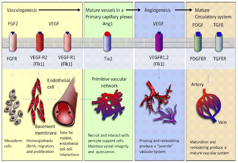
Angiogenesis and vasculogenesis are essential processes that contribute to the establishment of blood vessel networks during embryonic development. These processes involve the formation, expansion, and remodeling of blood vessels, which are crucial for supplying nutrients, oxygen, and other essential molecules to developing tissues and organs. Here's how angiogenesis and vasculogenesis work together to create functional vascular systems:

Vasculogenesis

Formation of Blood Islands: Vasculogenesis begins with the formation of blood islands, which are clusters of angioblasts (precursor cells) that differentiate into endothelial cells, the building blocks of blood vessels.
Angioblast Migration and Aggregation: Angioblasts migrate to specific areas in the developing embryo, guided by chemical signals and gradients. They aggregate to form endothelial cords or tubes, which serve as the initial structures of blood vessels.
Vascular Lumen Formation: The endothelial cords undergo lumenization, during which they develop a central channel, or lumen. This lumen becomes the pathway for blood flow.
Hemangioblast Differentiation: Some angioblasts differentiate into hemangioblasts, which give rise to both endothelial cells and blood cells. This connection between blood vessel formation and blood cell production is crucial for the functional development of the circulatory system.

Angiogenesis

Sprouting Angiogenesis: In regions where tissues require increased blood supply, endothelial cells from existing vessels start to sprout out in response to pro-angiogenic signals, such as growth factors like VEGF (vascular endothelial growth factor).
Migration and Proliferation: Sprouting endothelial cells migrate towards the source of pro-angiogenic signals and proliferate to form new endothelial sprouts.
Lumen Formation: Similar to vasculogenesis, the endothelial sprouts undergo lumenization to form functional tubular structures.
Anastomosis: The newly formed sprouts elongate and connect with adjacent sprouts, leading to the establishment of interconnected networks of blood vessels. This anastomosis process creates a functional circulation system.
Stabilization: Pericytes and smooth muscle cells are recruited to the developing vessels to provide structural support and stability to the new blood vessels.

Appearance of  angiogenesis and vasculogenesis in the evolutionary timeline

The appearance of angiogenesis and vasculogenesis in the evolutionary timeline is a complex topic that involves speculation and ongoing research. While the exact timing is not definitively known, scientists have proposed hypotheses about when these processes might have emerged based on comparative studies and the fossil record. Angiogenesis and vasculogenesis are fundamental processes for the development and maintenance of blood vessels in organisms. Angiogenesis involves the formation of new blood vessels from preexisting ones, while vasculogenesis refers to the de novo formation of blood vessels from endothelial precursor cells.

Early Life Forms (Prokaryotes): These simple organisms did not possess complex vascular systems or mechanisms like angiogenesis and vasculogenesis. Their nutrient exchange and waste removal were likely facilitated by direct diffusion through cell membranes due to their small size and relatively simple structure.
Simple Eukaryotes: As organisms supposedly evolved into more complex eukaryotic forms, some multicellular organisms would have started to develop basic mechanisms to transport nutrients and waste products. However, true vascular systems had not yet emerged, and mechanisms like diffusion and simple tissue organization served these early organisms.
Invertebrates: The appearance of more complex invertebrates would have marked the transition toward more developed circulatory systems. Many invertebrates have open circulatory systems where blood is pumped into body cavities, allowing for nutrient exchange. These systems would have involved rudimentary precursor processes that could be considered primitive forms of angiogenesis.
Vertebrates: The emergence of vertebrates would have brought about more sophisticated circulatory systems, including closed systems with dedicated blood vessels. While the exact point at which angiogenesis and vasculogenesis evolved in vertebrate history is unclear, they would have played essential roles in the development of more advanced vascular systems.
Early Vertebrates: Over time, vertebrates would have evolved into increasingly complex cardiovascular systems. The emergence of angiogenesis and vasculogenesis would have accompanied the need for more efficient nutrient delivery, waste removal, and tissue repair. These processes would have allowed vertebrates to sustain larger and more metabolically active bodies.

De Novo Genetic Information, necessary to instantiate angiogenesis and vasculogenesis

The hypothetical emergence of angiogenesis, the process of forming new blood vessels from scratch in the development of organisms that didn't previously possess a vascular system, would have required the addition of specific genetic information to enable this complex process. While the exact genetic changes are speculative, here are some potential additions or modifications to genetic information that could have been involved in the evolution of angiogenesis:

Angiogenic Factors: The development of angiogenesis from scratch would have necessitated the evolution of genes encoding angiogenic factors, such as rudimentary versions of vascular endothelial growth factors (VEGFs). These factors would act as signaling molecules to initiate vessel formation.
Receptor Proteins: The emergence of angiogenesis would have required the development of new receptor proteins on hypothetical endothelial-like precursor cells. These receptors would allow cells to detect and respond to angiogenic factors, initiating the signaling cascades necessary for vessel formation.
Cell Adhesion Molecules: As cells begin to organize into blood vessels, new genetic information could have been necessary to generate primitive cell adhesion molecules. These molecules would facilitate cell-cell interactions and the formation of vessel-like structures.
Matrix Remodeling Enzymes: The initial formation of blood vessels from scratch would involve breaking down and restructuring extracellular matrix. Genes encoding matrix remodeling enzymes could have evolved to allow cells to create pathways for vessel growth.
Transcription Factors: The evolution of angiogenesis would require new transcription factors that activate gene expression programs specific to vessel formation. These factors would regulate the expression of genes involved in cell migration, proliferation, and differentiation.
Guidance Proteins: To direct the movement of primitive endothelial-like cells toward the formation of blood vessels, hypothetical guidance proteins could have evolved to provide directional cues.
Signaling Molecules and Pathways: New genetic information would be needed to establish the initial signaling pathways that trigger cellular responses, such as migration, proliferation, and differentiation, during angiogenesis.
Cytoskeletal Regulators: The development of angiogenesis from scratch would require genes that control cytoskeletal dynamics, enabling cell movement, migration, and organization into vessel-like structures.
Apoptosis and Survival Regulators: As cells form blood vessels, mechanisms for balancing cell survival and cell death would be crucial. Genes involved in apoptosis and survival pathways might have been required.
Differentiation and Specification Genes: The evolution of angiogenesis would involve the development of genes that specify and differentiate precursor cells into endothelial-like cells with distinct roles in vessel formation.
Cell-Cell Communication Molecules: New genetic information could have been necessary to allow cells to communicate and coordinate their behaviors during the formation of blood vessels.
Cell Signaling Pathways for Coordination: To ensure proper coordination among endothelial-like cells, genetic information might have evolved to create rudimentary cell signaling pathways for communication and synchronization.

Epigenetic Regulatory Mechanisms necessary to be instantiated 

The hypothetical emergence of angiogenesis "from scratch," meaning the evolution of blood vessel formation in organisms that previously lacked such a system, would have required the establishment of new epigenetic regulations to coordinate the complex cellular changes necessary for vessel development. 

Epigenetic Priming for Vascular Precursors: Establishment of epigenetic marks to prime cells for vascular differentiation, creating a permissive environment for angiogenesis-related genes.  
Histone Modifications for Vessel Formation: Specific histone modifications, such as acetylation, to activate genes involved in endothelial differentiation and tube formation.  
DNA Methylation Dynamics: Establishment of specific DNA methylation patterns to regulate the expression of angiogenesis-essential genes, adjusting their activation during vessel formation.  
Non-Coding RNA-Mediated Regulation: Emergence of non-coding RNAs to interact with chromatin-modifying complexes, regulating genes associated with vessel development.  
Imprinting and Allelic Regulation: Establishment of allele-specific epigenetic marks to guide cellular roles during vessel formation.  
Epigenetic Inheritance of Vascular Patterns: Ability to inherit epigenetic information related to vessel formation, passing regulatory marks necessary for angiogenesis to subsequent generations.  
Temporal Epigenetic Regulations: Evolution of epigenetic mechanisms acting as molecular "clocks" for the precise timing of vessel-related processes.  
Suppression of Anti-Angiogenic Factors: New epigenetic regulations to suppress genes encoding anti-angiogenic factors, ensuring uninterrupted vessel development.  
Chromatin Remodeling for Vessel Assembly: Epigenetic mechanisms regulating chromatin remodeling to facilitate cell-cell interactions and tube formation during vessel assembly.  
Epigenetic Regulation of Vascular Maturation: Epigenetic marks guiding the maturation and stabilization of newly formed vessels, including recruitment of support cells.  
Epigenetic Sensing of Environmental Cues: Epigenetic mechanisms enabling cells to sense and respond to environmental signals for adaptive vessel formation.

Signaling Pathways necessary to create, and maintain angiogenesis and vasculogenesis

If we're considering the emergence of angiogenesis "from scratch," i.e., the initial development of the process in organisms that didn't previously have any vascular system, potential signaling pathways that would have had to be involved in the hypothetical evolution of angiogenesis: are: 

Basic Growth Factor and Receptor Pathways: Emergence of fundamental growth factor pathways and receptor systems for cell proliferation, migration, and differentiation, involving simple signaling cascades.  
Chemotaxis Pathways: Development of rudimentary chemotactic signaling to direct primitive endothelial-like cells towards factor gradients promoting movement.  
Adhesion Pathways: Formation of signaling pathways related to cell adhesion molecules to support endothelial cell movement and organization into early blood vessels.  
Cytoskeletal Remodeling Pathways: Creation of signaling pathways for cytoskeletal alterations, crucial for cell shape and movement.  
Cell-Cell Communication Pathways: Development of basic pathways for communication between primitive endothelial cells during early vessel formation.  
Apoptosis and Survival Pathways: Establishment of pathways to regulate the balance between cell death and survival, vital for vessel formation.  
Initial Extracellular Matrix Signaling: Emergence of signaling pathways to respond to the extracellular matrix, guiding the formation of vessel-like structures.  
Evolution of Ligand-Receptor Pairs: Development of new ligand-receptor pairs for sensing and responding to vessel formation cues.

The emergence of angiogenesis, the process of forming new blood vessels from preexisting ones, would have involved the establishment and modification of specific signaling pathways to coordinate the complex cellular changes required for vessel formation. While the exact signaling pathways are speculative, here are some potential pathways that could have been involved in the emergence of angiogenesis:

VEGF Signaling Pathway: Vascular endothelial growth factor (VEGF) is a central regulator of angiogenesis. The VEGF signaling pathway involves the binding of VEGF to its receptors (VEGFRs) on endothelial cells. This activates downstream signaling cascades that promote endothelial cell proliferation, migration, and tube formation.
FGF Signaling Pathway: Fibroblast growth factors (FGFs) also play a role in angiogenesis. The FGF signaling pathway, similar to the VEGF pathway, involves FGF ligands binding to their receptors, which triggers signaling events that contribute to endothelial cell proliferation and migration.
Notch Signaling Pathway: The Notch pathway is involved in cell-cell communication and could have played a role in coordinating endothelial cell differentiation and tip cell selection during vessel sprouting.
Wnt Signaling Pathway: The Wnt pathway has diverse roles in development, and its components could have been involved in angiogenesis by influencing endothelial cell behavior and vessel branching patterns.
TGF-β Signaling Pathway: Transforming growth factor-beta (TGF-β) family members could have been implicated in angiogenesis by regulating endothelial cell differentiation and extracellular matrix remodeling.
PDGF Signaling Pathway: Platelet-derived growth factor (PDGF) signaling might have been involved in recruiting pericytes and smooth muscle cells to stabilize and mature newly formed vessels.
ECM Signaling Pathways: Extracellular matrix (ECM) components, including integrins and focal adhesion kinase (FAK), could have participated in transmitting signals that guide endothelial cell migration and vessel assembly.
MAPK Signaling Pathway: Mitogen-activated protein kinase (MAPK) pathways could have been essential for transmitting signals that regulate cell proliferation, survival, and migration during angiogenesis.
PI3K/AKT Signaling Pathway: The phosphatidylinositol 3-kinase (PI3K)/AKT pathway could have been involved in promoting endothelial cell survival, migration, and angiogenesis-related cellular responses.
Rho GTPase Signaling Pathway: Rho GTPases, such as Rho, Rac, and Cdc42, could have participated in regulating cytoskeletal dynamics and cell migration during angiogenesis.
Chemokine Signaling: Chemokines and their receptors might have guided endothelial cell migration and positioning during vessel sprouting.
Hedgehog Signaling Pathway: Hedgehog signaling could have been implicated in regulating vascular patterning and endothelial cell differentiation.
Endothelial-Specific Signaling Pathways: Signaling pathways specifically active in endothelial cells could have evolved to control angiogenesis-related processes like proliferation, migration, and tube formation.

Regulatory codes necessary for maintenance and operation

The hypothetical emergence of blood vessels and angiogenesis would have likely involved the establishment of regulatory codes and languages to coordinate the development, maintenance, and operation of the vascular system. While the exact details are speculative, here are potential regulatory codes and languages that could have been instantiated:

Transcriptional Regulatory Code: The evolution of blood vessels would require a transcriptional regulatory code involving specific DNA sequences, transcription factors, and regulatory elements that control the expression of genes involved in vessel development, maintenance, and function.
Cis-Regulatory Elements: Enhancers, promoters, and other cis-regulatory elements would need to evolve to ensure proper spatiotemporal expression of angiogenesis-related genes.
Epigenetic Regulatory Language: Epigenetic modifications such as DNA methylation and histone modifications could form an epigenetic regulatory language that guides the activation and repression of genes essential for vascular development and maintenance.
Signaling Pathway Crosstalk: Complex signaling pathways involved in angiogenesis and vascular function would need to communicate and coordinate their activities through a regulatory language that ensures proper cellular responses.
Cell-Cell Communication Codes: As blood vessels involve multiple cell types, a communication code involving cell surface receptors, ligands, and their interactions would be necessary to coordinate cellular behaviors and functions.
Extracellular Matrix (ECM) Signaling: A code involving interactions between cells and the extracellular matrix would regulate processes such as cell adhesion, migration, and signaling.
Vascular Patterning Code: The establishment of hierarchical vessel networks would require a code that guides the formation and branching patterns of blood vessels to ensure efficient distribution of nutrients and oxygen.
Stability and Maturation Code: Blood vessel stabilization and maturation would require a regulatory code involving communication between endothelial cells and support cells (pericytes and smooth muscle cells) to ensure structural integrity.
Oxygen and Nutrient Sensing Code: Blood vessels need to adapt to changing oxygen and nutrient levels. A regulatory code might govern the response of vessels to these fluctuations, ensuring appropriate vessel dilation and constriction.
Immune-Endothelial Communication: Blood vessels interact with the immune system. A code would be needed to regulate the communication between endothelial cells and immune cells, enabling immune surveillance and inflammation regulation.
Inflammatory Response Code: Inflammatory responses and repair processes would require a regulatory code to activate and control specific genes involved in tissue repair and vessel remodeling.
Vascular Tone and Homeostasis Code: Blood pressure and vessel tone need to be tightly regulated. A code would be necessary to balance vasoconstriction and vasodilation to maintain blood flow and homeostasis.
Angiogenic Switch Code: The transition from quiescent vessels to angiogenesis activation would require a code that senses environmental cues and triggers angiogenic responses.
Vascular Regression Code: Vessels need to regress when not needed. A regulatory code would be necessary to initiate vessel regression and tissue remodeling.
Wound Healing and Regeneration Code: Blood vessels play a role in tissue repair. A code would be involved in coordinating vessel-related processes during wound healing and tissue regeneration.

These regulatory codes and languages would have had to emerge to ensure the development, maintenance, and operation of blood vessels. The precise details would depend on the specific context and the genetic and molecular mechanisms that contributed to the emergence of angiogenesis and vascular systems.

Evolution: Where Do Complex Organisms Come From? - Page 2 Image318


Last edited by Otangelo on Sat Feb 24, 2024 9:27 am; edited 66 times in total

https://reasonandscience.catsboard.com

27Evolution: Where Do Complex Organisms Come From? - Page 2 Empty Apoptosis Wed Aug 23, 2023 7:05 pm

Otangelo


Admin

What molecular cues and signaling pathways regulate the growth and branching of blood vessels?

The growth and branching of blood vessels, known as angiogenesis, is regulated by a complex interplay of molecular cues and signaling pathways that guide the formation of new blood vessels in response to various physiological and pathological stimuli. Several key molecular cues and signaling pathways are involved in this process:

VEGF (Vascular Endothelial Growth Factor) Family: VEGF is a central regulator of angiogenesis. It binds to its receptors (VEGFRs) on endothelial cells and promotes their proliferation, migration, and survival. The VEGF family includes various isoforms, such as VEGF-A, VEGF-B, VEGF-C, and VEGF-D, each with specific roles in angiogenesis and lymphangiogenesis.
FGF (Fibroblast Growth Factor) Family: FGFs, particularly FGF-2 and FGF-9, stimulate endothelial cell proliferation and migration. They also induce the release of proteases that help remodel the extracellular matrix to create paths for new vessel growth.
Notch Signaling: Notch signaling is crucial for maintaining proper vascular sprouting and branching. Delta-like ligands (DLLs) on the tip cells of growing vessels interact with Notch receptors on neighboring stalk cells, promoting stalk cell fate and inhibiting tip cell behaviors.
Ephrin-Eph Signaling: Ephrins and their Eph receptors play a role in guiding angiogenic sprouting and vessel branching. The interactions between Ephs on endothelial cells and ephrins on adjacent cells help establish vessel boundaries and prevent excessive vessel growth.
Angiopoietins and Tie Receptors: Angiopoietin-1 (Ang1) and Angiopoietin-2 (Ang2) interact with Tie receptors on endothelial cells to regulate vessel stabilization and remodeling. Ang1 promotes vessel maturation, while Ang2 destabilizes vessels, preparing them for sprouting.
PDGF (Platelet-Derived Growth Factor): PDGF recruits pericytes and smooth muscle cells to stabilize newly formed blood vessels. Pericytes help to reinforce vessel walls and prevent leakage.
Semaphorins and Plexins: Semaphorins guide vessel growth by repelling or attracting endothelial cells. They interact with Plexin receptors to influence angiogenic processes.
Hedgehog Signaling: Hedgehog signaling plays a role in angiogenesis by influencing endothelial cell behavior and vessel branching during development and tissue repair.
Integrins and Cell-ECM Interactions: Integrin-mediated interactions between endothelial cells and the extracellular matrix guide vessel migration, alignment, and branching.
Hypoxia and HIF (Hypoxia-Inducible Factor) Pathway: Low oxygen levels induce the expression of HIF, which upregulates the production of angiogenic factors like VEGF in response to tissue hypoxia.
Plasminogen Activation System: Plasminogen activators and plasmin play a role in angiogenesis by degrading the extracellular matrix and facilitating endothelial cell migration during vessel sprouting.

These signaling pathways and molecular cues interact in a highly coordinated manner to regulate various aspects of blood vessel growth and branching. They respond to tissue-specific needs, developmental cues, and pathological conditions, ensuring the proper formation of functional blood vessel networks that support tissue growth, repair, and homeostasis.


What mechanisms for angiogenesis and vasculogenesis had to be instantiated to ensure proper oxygen and nutrient supply to developing tissues?

Hypoxia-Induced Signaling: Oxygen deficiency triggers the upregulation of hypoxia-inducible factors (HIFs), which stimulate the production of angiogenic factors like VEGF-A. This response initiates angiogenesis by promoting endothelial cell proliferation, migration, and vessel sprouting to supply oxygen and nutrients to hypoxic tissues.
Extracellular Matrix Remodeling: During angiogenesis, proteases are activated to degrade the extracellular matrix, creating paths for endothelial cell migration and vessel sprouting. This remodeling allows vessels to extend into tissues where oxygen and nutrients are needed.
Chemoattractant Gradients: Pro-angiogenic factors, such as VEGF, form gradients that guide endothelial cell migration towards areas of higher concentration. This directional migration ensures that new vessels form in regions with the greatest need for oxygen and nutrients.
Tip Cell and Stalk Cell Differentiation: Developing blood vessels exhibit tip cells at the leading edge and stalk cells behind them. Tip cells guide vessel growth, while stalk cells proliferate and form the vessel trunk. Proper balance between tip and stalk cell behaviors ensures efficient vessel sprouting and branching.
Pericyte and Smooth Muscle Cell Recruitment: During vessel maturation, pericytes and smooth muscle cells are recruited to stabilize vessel walls and regulate blood flow. This prevents vessel collapse and optimizes oxygen and nutrient delivery to tissues.
Branching and Anastomosis: Angiogenic vessels branch and form anastomoses (connections) to create an interconnected network. This network ensures redundancy and efficient distribution of oxygen and nutrients throughout tissues.
Remodeling and Maturation: Angiogenic vessels undergo maturation and remodeling processes, including endothelial cell-cell junction formation and basement membrane deposition. These processes enhance vessel stability and functionality, improving oxygen and nutrient delivery.
Angiopoietin-Tie Signaling: Angiopoietins and their Tie receptors influence vessel stabilization and maturation. Ang1 promotes pericyte recruitment and vessel maturation, optimizing nutrient and oxygen transport.
Functional Adaptation: Developing tissues release angiogenic factors in response to local metabolic demands. This adaptive response ensures that new vessels form where they are most needed for adequate oxygen and nutrient supply.
Vessel Diameter Regulation: Vessel diameter and permeability are regulated to match tissue demands. Small-diameter vessels provide higher oxygen exchange rates, while larger vessels accommodate greater blood flow and nutrient delivery.

The instantiation of these mechanisms ensures that developing tissues receive sufficient oxygen and nutrients by facilitating the establishment of an intricate vascular network. Properly regulated angiogenesis and vasculogenesis enable tissues to grow and develop while maintaining physiological homeostasis.


Differences between Angiogenesis and Vasculogenesis 

Vasculogenesis and angiogenesis are two distinct but interconnected processes involved in the formation and development of blood vessels within an organism. While they share similarities, they differ in their timing, mechanisms, and contexts. 

Vasculogenesis

Vasculogenesis is the process by which new blood vessels are formed de novo (from scratch) during embryonic development. It primarily occurs during the early stages of embryogenesis when the embryo is a cluster of undifferentiated cells. Here are the key differences:

Timing: Vasculogenesis occurs very early in embryonic development, often before the formation of major organs. It involves the initial assembly of the primary vascular network.
Cell Origin: During vasculogenesis, endothelial precursor cells, called angioblasts, differentiate from mesodermal cells. These angioblasts aggregate and coalesce to form the primitive blood vessels.
Organogenesis: Vasculogenesis is closely associated with organogenesis. As organs begin to develop, the primary vascular network formed by vasculogenesis provides the foundation for the subsequent growth and development of organs.
Vessel Formation: In vasculogenesis, angioblasts aggregate to form blood vessel-like structures, which then coalesce to create the primary capillary plexus. This process involves both endothelial cell differentiation and organization into vessel-like structures.

Angiogenesis

Angiogenesis is the process of forming new blood vessels from preexisting ones. It is a more refined and specialized process that occurs during various developmental stages and in response to specific needs in the body. Here are the key differences:

Timing: Angiogenesis can occur during both embryonic development and postnatal stages. It plays a role in tissue growth, wound healing, and other processes throughout an organism's life.
Cell Origin: During angiogenesis, existing endothelial cells in preexisting blood vessels are activated to proliferate, migrate, and remodel, forming new blood vessels. It involves the expansion and remodeling of existing vascular networks.
Tissue Growth and Repair: Angiogenesis is essential for tissue growth, repair, and regeneration. It allows for the expansion of the vascular network to supply nutrients and oxygen to growing tissues or to aid in healing wounded tissues.
Initiation: Angiogenesis is initiated in response to specific signals, such as hypoxia (low oxygen levels) or growth factors released during tissue injury. These signals activate endothelial cells in existing vessels to sprout, migrate, and form new vessels.

Is there scientific evidence supporting the idea that these biological systems were brought about by the process of evolution?

Functional Interdependence: Angiogenesis and vasculogenesis involve multiple interdependent steps, including cell migration, proliferation, signaling, and ECM remodeling. It's challenging to envision how each step could evolve independently, as many components are needed for the process to be functional.
Complex Signaling Networks: The successful formation of blood vessels requires precise coordination of multiple signaling pathways. These pathways involve intricate interactions between growth factors, cell adhesion molecules, and transcription factors. For angiogenesis and vasculogenesis to work, these pathways need to be in place and functional simultaneously.
Required Structures: Blood vessels require a certain structural complexity to function properly. The vessels need to be able to transport blood efficiently, withstand mechanical forces, and maintain barrier properties. These structural requirements make it unlikely that partially developed vessels or intermediate stages would provide any selective advantage.
Energy and Resource Costs: Developing and maintaining blood vessels is metabolically costly. Evolution requires that intermediate stages offer some selective advantage to the organism. If the intermediate stages lack functionality and only incur energy and resource costs, they are less likely to be favored by natural selection.
Regulation and Control: The processes of angiogenesis and vasculogenesis need to be tightly regulated. Failure to regulate these processes properly can lead to diseases such as cancer, where blood vessel growth is uncontrolled. The precise regulation required for these processes to function accurately raises questions about how this regulation could evolve incrementally.
Cell-Cell Interactions: The formation of blood vessels involves intricate cell-cell interactions and communication. Cells need to respond to signals from neighboring cells and the microenvironment. Evolutionary intermediates that lack the ability to properly communicate and interact might not provide any adaptive advantage.
Emergence of Blood Cells: Blood vessels require specialized blood cells (endothelial cells and blood-forming cells) to function. The evolution of these cell types alongside the vessels raises questions about their origin and development in a stepwise manner.

Irreducibility and Interdependence of the systems to instantiate and operate Angiogenesis and Vasculogenesis 

The processes involved in the emergence of angiogenesis are highly interdependent:

Angiogenic Factors and Receptor Proteins: Both are needed to initiate signaling for vessel formation.
Cell Adhesion Molecules and Matrix Remodeling Enzymes: They work together to organize cells and create pathways for growth.
Transcription Factors and Differentiation Genes: Transcription factors activate differentiation genes, guiding cell specialization.
Signaling Pathways and Guidance Proteins: Signaling cues guide cells via guidance proteins, requiring functional pathways.
Cytoskeletal Regulators and Cell Signaling: Proper cell migration relies on regulated signaling and coordinated cytoskeletal changes.
Apoptosis and Survival Regulators: Balancing cell numbers during vessel formation involves coordinated apoptosis and survival.
Cell-Cell Communication and Signaling Pathways: Communication depends on functional signaling, enabling coordinated cell behaviors.
Cell Signaling Pathways and Cytoskeletal Regulation: Effective cell migration requires synchronized signaling and cytoskeletal changes.
Epigenetic Priming and Histone Modifications: Epigenetic priming sets the stage for histone modifications to activate vessel-related genes. Both are essential for creating a conducive chromatin environment.
DNA Methylation Dynamics and Non-Coding RNA Regulation: DNA methylation patterns and non-coding RNAs work together to regulate gene expression. Non-coding RNAs could influence DNA methylation patterns, fine-tuning gene activation during vessel formation.
Imprinting and Allelic Regulation: Imprinting relies on specific epigenetic marks that guide cell roles. Allelic regulation ensures proper vessel assembly by assigning distinct roles based on epigenetic marks.
Epigenetic Inheritance and Temporal Regulation: Epigenetic inheritance and temporal regulation are linked. The ability to inherit epigenetic information aids the precise timing of vessel-related processes.
Suppression of Anti-Angiogenic Factors and Chromatin Remodeling: Suppressing anti-angiogenic genes and proper chromatin remodeling go hand in hand. Both are necessary for unobstructed vessel formation.
Epigenetic Regulation of Maturation and Sensing of Cues: The regulation of vascular maturation and sensing cues is interconnected. Epigenetic marks guide vessel maturation while sensing cues allow adaptive vessel formation.
Basic Growth Factor and Receptor Pathways: These pathways rely on each other to stimulate cell behaviors, such as proliferation and migration, essential for vessel formation.
Chemotaxis and Adhesion Pathways: Chemotaxis guides cell movement, while adhesion pathways facilitate cell organization. Both are necessary for coordinated vessel formation.
Cytoskeletal Remodeling and Cell-Cell Communication: Cytoskeletal changes are directed by signaling pathways and influence cell movement. Effective cell-cell communication ensures coordinated behavior during angiogenesis.
Apoptosis and Survival Pathways: Balancing apoptosis and survival is crucial for proper vessel formation. Both pathways work together to determine cell numbers and roles.
Extracellular Matrix Signaling and Ligand-Receptor Pairs: Cells respond to extracellular cues via signaling pathways, which also involve ligand-receptor interactions. These processes work in tandem to guide cell behavior during vessel formation.
VEGF and FGF Signaling: Both pathways promote endothelial cell proliferation and migration. Their combined action is essential for vessel formation.
Notch and Wnt Signaling: These pathways likely cooperated to coordinate endothelial cell differentiation and vessel sprouting, ensuring proper branching patterns.
TGF-β and PDGF Signaling: TGF-β could have influenced endothelial cell differentiation and extracellular matrix remodeling, while PDGF might have recruited supporting cells. Both pathways contribute to vessel stabilization.
ECM, MAPK, and Rho GTPase Signaling: ECM signaling, MAPK pathways, and Rho GTPases could have worked together to guide cell migration, cytoskeletal dynamics, and vessel assembly.
PI3K/AKT and Chemokine Signaling: PI3K/AKT pathway likely collaborated with chemokine signaling to regulate endothelial cell survival, migration, and positioning during vessel sprouting.
Hedgehog and Endothelial-Specific Signaling: Hedgehog signaling and endothelial-specific pathways could have coordinated to regulate vascular patterning, differentiation, proliferation, and tube formation.
Transcriptional Regulatory Code and Epigenetic Regulatory Language: Transcriptional regulation relies on epigenetic modifications for proper gene expression control.
Cis-Regulatory Elements and Signaling Pathway Crosstalk: Cis-regulatory elements ensure spatiotemporal gene expression, influenced by signaling pathway crosstalk.
Cell-Cell Communication Codes and Immune-Endothelial Communication: Effective immune-endothelial communication requires coordinated cell-cell communication codes.
Extracellular Matrix (ECM) Signaling and Vascular Patterning Code: ECM interactions influence vascular patterning, which involves signaling pathways guiding vessel formation.
Stability and Maturation Code and Inflammatory Response Code: Blood vessel maturation is related to inflammation regulation. Proper maturation relies on coordinated immune responses.
Oxygen and Nutrient Sensing Code and Vascular Tone and Homeostasis Code: Both codes contribute to maintaining vessel tone and blood flow in response to changing environmental cues.
Angiogenic Switch Code and Vascular Regression Code: Angiogenesis activation and regression are interconnected processes that require coordinated switches in response to cues.
Wound Healing and Regeneration Code and Vascular Tone and Homeostasis Code: Blood vessels play a role in wound healing and tissue repair, which impacts vessel tone and homeostasis.


The intricacies of creating, developing, and operating complex biological processes like angiogenesis and vasculogenesis highlight a deep interdependence among the manufacturing, signaling, and regulatory codes and languages involved. This complexity raises questions about the feasibility of a stepwise, gradual evolution and instead suggests a purposeful design. The behaviors of cells, including migration, proliferation, and differentiation, are essential for the formation of blood vessels. Without the manufacturing code, cellular actions would lack direction and purpose, rendering vessel formation impossible. Signaling pathways guide cellular behaviors by conveying information between cells. These cues instruct cells on where to migrate, when to proliferate, and how to differentiate. Without the signaling codes, cells would lack the guidance needed to perform their specific tasks. The regulatory codes orchestrate gene expression, determining which genes are activated or suppressed. This precise control ensures that the appropriate genes are turned on to support angiogenesis and vasculogenesis. Without the regulatory codes, genes required for these processes would lack the necessary regulation. Signaling pathways provide cues for cellular behaviors (manufacturing), ensuring that cells migrate, proliferate, and differentiate in a coordinated manner. Without proper signaling, cellular actions would lack direction and purpose.  The regulatory codes interpret signaling cues and guide gene expression. Signaling pathways activate transcription factors that influence gene expression levels. Without functional signaling cues, regulatory mechanisms would have no meaningful signals to interpret. Cellular behaviors rely on gene expression controlled by the regulatory code. The manufacturing code's instructions are carried out through the expression of specific genes. Without proper gene regulation, cells wouldn't perform the behaviors required for vessel formation.

From this perspective, the intricate interdependence of these codes suggests a coordinated design, rather than a gradual evolution. The simultaneous emergence of these interconnected systems seems implausible through incremental changes. Instead, it points toward a purposeful design where these components were created all at once, fully operational, to achieve the complex task of forming functional blood vessels. The interconnected nature of the manufacturing, signaling, and regulatory aspects implies that these systems were instantiated together, working in harmony to achieve the intricate process of angiogenesis and vasculogenesis. This kind of interdependence and complexity aligns with the concept of intelligent design, where the coordinated emergence of these mechanisms suggests a guiding intelligence that foresaw the necessary components and their interactions from the outset.

What are veins and arteries interdependent with?

Once veins and arteries are operational, they are interconnected with various intracellular and extracellular systems to ensure proper blood circulation, nutrient delivery, waste removal, and overall physiological function. 

Intracellular Systems

Cardiac Muscle Contraction: The heartbeat generated by the cardiac muscle drives blood through the arteries and veins. The function of the circulatory system is closely tied to the heart's pumping action.
Blood Cell Production: The circulatory system relies on the bone marrow to produce red blood cells, white blood cells, and platelets. Arteries and veins transport these cells to various parts of the body.
Hemostasis and Coagulation: The circulatory system interacts with the clotting cascade to prevent excessive bleeding from damaged blood vessels. Platelets, clotting factors, and endothelial cells play roles in this process.
Oxygen and Carbon Dioxide Exchange: Oxygen from inhaled air diffuses into the bloodstream in the lungs and binds to hemoglobin. Carbon dioxide produced by cells is carried by the blood back to the lungs for exhalation.

Extracellular Systems

Respiratory System: The circulatory system collaborates with the respiratory system to ensure oxygen uptake and carbon dioxide removal. Oxygenated blood is transported to cells, and deoxygenated blood is returned to the lungs for oxygen exchange.
Lymphatic System: The lymphatic system helps maintain fluid balance by collecting excess fluid and proteins leaked from blood vessels. Lymphatic vessels return this fluid to the bloodstream, preventing swelling.
Endocrine System: Hormones released by endocrine glands regulate blood pressure, heart rate, and electrolyte balance, all of which impact the function of veins and arteries.
Digestive System: The circulatory system transports nutrients absorbed from the digestive tract to cells throughout the body. Veins and arteries play a role in nutrient distribution.
Nervous System: Autonomic nerves control the diameter of blood vessels, affecting blood pressure and blood flow regulation. Sympathetic and parasympathetic signals influence vessel constriction and dilation.
Immune System: Immune cells are transported via the bloodstream to sites of infection or injury. Blood vessels also play a role in immune cell trafficking and inflammation.
Excretory System: Blood vessels transport waste products to the kidneys for filtration and elimination from the body.
Temperature Regulation: Blood flow to the skin's surface helps regulate body temperature. Arterioles in the skin can dilate to release excess heat or constrict to conserve heat.

The interdependence of veins and arteries with these systems underscores the role of the circulatory system in maintaining overall homeostasis and the proper functioning of various bodily processes. The integration of these systems ensures the coordination of functions necessary for survival and optimal physiological performance.

Premise 1: The interdependence of the circulatory system (veins and arteries) with various intracellular and extracellular systems is crucial for maintaining overall homeostasis and optimal physiological function.
These interconnected systems, including cardiac muscle contraction, blood cell production, hemostasis and coagulation, oxygen and carbon dioxide exchange, respiratory, lymphatic, endocrine, digestive, nervous, immune, excretory systems, and temperature regulation, all rely on each other to perform their functions.
Premise 2: Such intricate interdependence and collaboration among these diverse systems indicate a high level of complexity, precision, and mutual adaptation that appears designed for a holistic and well-coordinated physiological setup.
Conclusion: The profound interdependence and mutual adaptation observed among the circulatory system and the various interconnected systems strongly suggest a carefully designed and integrated setup. The simultaneous emergence of these systems, each playing a crucial role in the function and maintenance of the organism, implies a coordinated plan rather than a gradual, step-by-step process. The functional harmony and coherence across these interdependent systems hint at a deliberate design, ensuring the survival and optimal performance of the organism.

Premise 1: The emergence of blood vessels and angiogenesis involves a multitude of interconnected processes, including angiogenic factors, cell adhesion molecules, transcription factors, signaling pathways, cytoskeletal regulators, and more.
Premise 2: These processes are interdependent, requiring simultaneous and coordinated activation to ensure proper vessel formation, maintenance, and operation.
Conclusion: The intricate interdependence among these processes strongly suggests that they cannot have evolved step by step, as isolated mechanisms or codes, but must have been designed and instantiated together from scratch to function harmoniously and achieve the complex goal of creating, developing, and operating a functional vascular system.

1. Prinz, R. (2023). biological codes: a field guide for code hunters. biological Theory. https://doi.org/10.1007/s13752-023-00444-2


2. Apoptosis

Apoptosis is a fundamental biological process of programmed cell death that plays a crucial role in shaping tissues and organs during development, maintaining tissue homeostasis, and eliminating damaged or surplus cells. Unlike necrosis, which is a chaotic and uncontrolled form of cell death, apoptosis follows a tightly regulated sequence of events. During apoptosis, cells undergo controlled dismantling in response to specific signals. These signals can originate from internal factors like DNA damage or external cues such as growth factor withdrawal. The process involves a series of distinct stages:

Initiation: Apoptosis can be triggered by various stimuli, either intrinsic (e.g., DNA damage) or extrinsic (e.g., binding of death ligands to cell surface receptors).
Signaling Pathways: Activation of signaling pathways leads to the expression or activation of specific genes and proteins that orchestrate apoptosis.
Caspase Activation: Caspases, a family of protease enzymes, play a central role in apoptosis. Initiator caspases are activated first, triggering a cascade that culminates in the activation of executioner caspases.
Cellular Changes: Apoptotic cells undergo characteristic changes, including cell shrinkage, chromatin condensation, and nuclear fragmentation.
Plasma Membrane Alterations: Phospholipids flip from the inner to the outer leaflet of the cell membrane, signaling to phagocytes that the cell is undergoing apoptosis.
Blebbing: The cell membrane forms bulges called blebs, which are eventually shed as apoptotic bodies containing cellular debris.
Phagocytosis: Phagocytes recognize apoptotic bodies through "eat me" signals on their surfaces. The phagocytes engulf and digest these bodies, preventing inflammation and tissue damage.
Resolution: Apoptotic cells are efficiently cleared, leading to minimal impact on surrounding tissues.

What is the role of apoptosis in sculpting and refining tissues during development?

Tissue Patterning and Shape Formation: Apoptosis is involved in eliminating excess cells and shaping developing tissues and organs. By selectively removing specific cells, apoptosis helps establish and refine the proper structure and shape of organs. This is particularly important in processes like limb development, where apoptosis is responsible for creating spaces between digits (interdigital spaces) and sculpting the final shape of the limb.
Digit Formation: In vertebrate limb development, apoptosis removes cells from the areas between developing digits, allowing them to separate. This process is essential for forming individual fingers or toes. The removal of cells in the interdigital regions is orchestrated by signaling pathways that activate apoptosis in a precisely coordinated manner.
Nervous System Development: Apoptosis plays a role in shaping the nervous system. During neural development, there is an initial overproduction of neurons, and apoptosis eliminates excess neurons that do not establish proper connections or synapses. This helps refine neural circuits and optimize their functionality.
Organ Development and Homeostasis: Apoptosis is involved in eliminating unwanted or abnormal cells during organ development. It helps shape organs by selectively removing cells that would otherwise disrupt the proper structure or function of the organ. Additionally, apoptosis continues to operate in adulthood to maintain tissue homeostasis by removing damaged or aged cells.
Immune System Formation: In the immune system, apoptosis is involved in shaping lymphoid organs and eliminating self-reactive immune cells. Immature immune cells undergo selection processes that involve apoptosis to ensure that only functional and non-self-reactive cells mature and become part of the immune repertoire.
Cell Number Control: Apoptosis helps regulate cell numbers in various tissues to achieve the appropriate balance between cell proliferation and cell death. This balance is essential for maintaining the overall integrity and functionality of tissues and organs.
Preventing Abnormal Development: Apoptosis acts as a quality control mechanism by eliminating cells with developmental defects, DNA damage, or other abnormalities. This prevents the propagation of genetic or cellular errors that could lead to malformation or disease.

Apoptosis serves as a precise and controlled mechanism for eliminating cells that are no longer needed or that could disrupt proper tissue and organ development. By sculpting and refining tissues, apoptosis contributes to the establishment of functional and well-organized structures in the developing organism.

How do the regulatory mechanisms of apoptosis ensure the precise elimination of specific cell populations?

The regulatory mechanisms of apoptosis ensure the precise elimination of specific cell populations through a series of tightly controlled steps and molecular interactions. These mechanisms allow cells to be targeted for elimination while minimizing the risk of collateral damage to neighboring cells. Here's how these regulatory mechanisms work:

Caspase Activation: Caspases are a group of protease enzymes that play a central role in apoptosis. They are initially present as inactive procaspases. Activation of caspases is a key step in initiating apoptosis. Activation can occur through two main pathways: the extrinsic pathway, which is initiated by death receptors on the cell surface, and the intrinsic pathway, which is activated by intracellular stress signals.
Bcl-2 Family Proteins: The Bcl-2 family of proteins includes both pro-apoptotic and anti-apoptotic members. These proteins regulate the permeability of the mitochondrial membrane and the release of cytochrome c, a trigger for caspase activation. Pro-apoptotic members promote apoptosis by inducing mitochondrial membrane permeabilization, while anti-apoptotic members inhibit apoptosis by preventing cytochrome c release.
Caspase Cascade: Activated caspases initiate a cascade of proteolytic events that lead to cell dismantling. Initiator caspases, such as caspase-8 and caspase-9, cleave and activate effector caspases, such as caspase-3. Effector caspases cleave a wide range of cellular substrates, including structural and functional proteins, resulting in cell disassembly.
Cellular Engulfment: Phagocytic cells, such as macrophages, play a role in engulfing apoptotic cells. This process, called phagocytosis or efferocytosis, prevents the release of cellular contents that could trigger inflammation and damage neighboring cells. Engulfment is facilitated by "eat-me" signals displayed on the surface of apoptotic cells and recognized by phagocytes.
Death Receptors and Ligands: In the extrinsic pathway, death receptors on the cell surface, such as Fas (CD95) and TNF receptor, bind to specific ligands. This binding triggers a signaling cascade that leads to caspase activation and apoptosis. This pathway is particularly important for immune system regulation and defense against infected or abnormal cells.
Survival Factors and Apoptotic Signals: Cells receive survival signals from their environment through growth factors and other molecules. These signals activate intracellular pathways that promote cell survival by inhibiting apoptosis. Conversely, absence of survival signals or exposure to apoptotic signals can tip the balance towards apoptosis.
Checkpoint Mechanisms: Cells have built-in checkpoints to ensure that apoptosis is activated only when appropriate. For example, DNA damage triggers the activation of p53, a tumor suppressor protein. p53 can induce cell cycle arrest to allow DNA repair, but if the damage is severe, it can activate apoptosis to prevent propagation of genetic errors.
Tissue-Specific Regulation: The regulation of apoptosis can be tissue-specific. Some cells are more sensitive to apoptosis signals due to their expression of certain proteins or the presence of specific receptors. This allows for precise elimination of particular cell populations during development, differentiation, and tissue homeostasis.

Collectively, these regulatory mechanisms ensure that apoptosis is a controlled and highly orchestrated process. The interplay between pro-apoptotic and anti-apoptotic signals, along with checks and balances, allows cells to be targeted for elimination with high specificity, contributing to the precise elimination of specific cell populations during development and tissue maintenance.

How were apoptotic pathways instantiated to balance tissue growth and removal in different organisms?

The instantiation of apoptotic pathways to balance tissue growth and removal in different organisms is a complex process that involves both conserved and organism-specific mechanisms. While the exact details can vary among species, certain core principles guide the establishment of apoptotic pathways to achieve proper tissue homeostasis:

Conserved Core Components: Many of the core components of apoptotic pathways are evolutionarily conserved across different organisms. For instance, caspases, Bcl-2 family proteins, and death receptors are present in a wide range of species. These conserved components provide a foundation for the initiation and execution of apoptosis.
Diversification of Pathways: Different organisms may have evolved specific apoptotic pathways tailored to their physiological and developmental needs. For example, the extrinsic pathway involving death receptors is more prominent in mammals, particularly in immune regulation. In contrast, the intrinsic pathway, centered around mitochondria, is a more universal mechanism present in various organisms.
Tissue-Specific Regulation: Apoptosis is often regulated in a tissue-specific manner. The expression of certain pro-apoptotic and anti-apoptotic factors can vary between tissues, allowing for selective control of apoptosis in different parts of the body. This tissue-specific regulation is crucial for achieving the proper balance between tissue growth and removal.
Adaptation to Developmental Stages: Apoptosis is involved in various developmental stages, from embryogenesis to adulthood. Different organisms have adapted their apoptotic pathways to suit the requirements of each developmental phase. For example, apoptosis during embryogenesis shapes tissue formation and organ development, while apoptosis in adult organisms contributes to tissue maintenance and removal of damaged cells.
Integration with Signaling Networks: Apoptotic pathways are integrated with other signaling networks in the cell. For instance, growth factors, survival signals, and DNA damage responses influence the balance between survival and apoptosis. The integration of apoptotic pathways with these networks allows organisms to respond dynamically to changing environmental and cellular conditions.
Fine-Tuning and Feedback: Apoptotic pathways often incorporate feedback loops and fine-tuning mechanisms. Regulatory feedback loops can enhance the precision of apoptotic responses and prevent excessive cell death. This ensures that tissue removal occurs only when needed and prevents the loss of essential cell populations.
Genetic and Functional Redundancy: Some organisms possess genetic redundancy, where multiple genes encode functionally similar proteins. This redundancy provides backup mechanisms to ensure that apoptotic pathways can function effectively even if some components are compromised.

Overall, the instantiation of apoptotic pathways in different organisms involves a combination of conserved elements and adaptive mechanisms.  The diverse strategies employed by different species reflect the complexity and flexibility of apoptotic regulation across the tree of life.


Evolution: Where Do Complex Organisms Come From? - Page 2 3912

Apoptosis is critical for normal development, tissue remodeling, immune response regulation, and maintaining tissue integrity. Dysregulation of apoptosis is associated with various diseases, including cancer (where excessive cell survival occurs) and neurodegenerative disorders (where excessive cell death occurs). The complex and highly orchestrated nature of apoptosis suggests that it serves as a fine-tuned mechanism that is essential for the proper functioning of multicellular organisms. The emergence of apoptosis, a complex process involving cellular self-destruction, would have required the establishment of specific manufacturing codes and languages to orchestrate the production, assembly, and regulation of the components involved.

Appearance of apoptosis in the evolutionary timeline  

The emergence of apoptosis, a highly regulated form of programmed cell death, is believed to have played a crucial role in shaping the development and complexity of multicellular organisms. While the exact timing of when apoptosis first appeared in the evolutionary timeline is not definitively known, researchers have proposed theories based on comparative studies and evidence from various organisms.

Early Single-Celled Organisms (Prokaryotes): It is unlikely that apoptosis, as understood in multicellular organisms, would have existed in early single-celled organisms like prokaryotes. The cellular complexity and mechanisms necessary for programmed cell death as seen in higher organisms would not have been present.
Emergence of Eukaryotes: With the supposed evolution of eukaryotic cells, which possess more complex internal structures and organelles, the potential for controlled cell death mechanisms might have increased. However, at this stage, any form of cell death would have been more similar to necrosis, a less regulated process compared to apoptosis.
Multicellular Organisms: The transition to multicellularity would have introduced new challenges related to cell differentiation, tissue development, and maintaining proper cell numbers. Apoptosis might have started to emerge as a potential mechanism to eliminate excess or damaged cells, refine tissue structures, and aid in proper development. Rudimentary forms of programmed cell death would have been present even in early multicellular organisms.
Invertebrates: As organisms became more complex, particularly within the animal kingdom, it is speculated that apoptosis would have become more sophisticated. Invertebrates would have employed some form of programmed cell death to assist in shaping tissues, organs, and structures during development. However, the molecular mechanisms and regulation would have been less intricate than in more advanced organisms.
Vertebrates: With the appearance of vertebrates and the evolution of intricate organ systems, it's suggested that apoptosis would have become more refined and tightly regulated. This process would have played critical roles in various aspects including organogenesis, immune system development, tissue repair, and the removal of potentially harmful or unnecessary cells.
Evolution of Adaptive Immunity: The emergence of the adaptive immune system in vertebrates would have supposedly introduced a necessity for precise cell death mechanisms to eliminate unwanted immune cells and prevent autoimmune responses. Apoptosis might have played a central role in maintaining the balance of the immune system.

De Novo Genetic Information to instantiate apoptosis

The emergence of apoptosis, a complex and regulated process of programmed cell death, would have required the addition of specific genetic information to enable its functions.

Apoptosis Initiation Genes: New genetic elements encoding proteins responsible for initiating apoptosis would need to evolve. These proteins, often activated in response to cellular stress or signals, would trigger the apoptotic cascade.
Caspase Genes: Caspases are a family of protease enzymes crucial for executing apoptosis. The evolution of apoptosis would require the emergence of genes encoding various types of caspases, each with distinct roles in apoptosis progression.
Cell Death Regulatory Proteins: Proteins that regulate the balance between pro-apoptotic and anti-apoptotic signals would need to evolve. These proteins would control the decision-making process of whether a cell undergoes apoptosis.
Apoptotic Signaling Pathways: New genetic information would be necessary to establish the intricate signaling pathways that transmit pro-apoptotic and anti-apoptotic signals, leading to the activation of caspases and subsequent cell death.
DNA Fragmentation Genes: During apoptosis, DNA is fragmented into smaller pieces. Genes responsible for this fragmentation process would need to evolve, ensuring the controlled degradation of the cell's genetic material.
Cellular Membrane Alteration Genes: Changes in the cell's membrane structure and properties are common in apoptosis. Genes encoding proteins responsible for altering the membrane would be essential.
Phagocytosis Recognition Genes: In multicellular organisms, phagocytes engulf apoptotic cells. Genes encoding surface molecules on apoptotic cells that signal for their recognition and engulfment by phagocytes would need to emerge.
Regulation of Apoptosis Timing: The evolution of apoptosis would require genetic mechanisms to regulate the timing of cell death, ensuring that it occurs at the right stage of development or in response to appropriate signals.
Apoptosis in Single-Celled Organisms: In prokaryotes or unicellular eukaryotes, apoptosis-like mechanisms could involve genes that trigger self-destruction under certain conditions, such as nutrient depletion or environmental stress.

The emergence of apoptosis would require the simultaneous addition of multiple genes and regulatory elements to form a functional and controlled process of programmed cell death. The complexity and coordination involved in apoptosis suggest that such genetic information would need to be instantiated and integrated into existing genetic systems to achieve its crucial functions.

Manufacturing codes and languages employed to instantiate apoptosis

Exempting genetic codes and languages, the emergence of apoptosis would have required the establishment of intricate manufacturing codes and languages within cells. These codes and languages encompass a series of coordinated biochemical processes that lead to the controlled dismantling and elimination of cells. 

Cellular Signaling Network: Cells would need to develop a sophisticated network of signaling pathways that sense both internal and external cues. These pathways would translate specific triggers into appropriate responses that initiate apoptosis.
Protein Activation and Inactivation: Intricate protein interactions would be established, involving activation and inactivation of key enzymes and factors. This would require the development of mechanisms to ensure precise timing and regulation.
Enzyme Cascades: Manufacturing codes would involve creating enzyme cascades, like the caspase cascade in apoptosis. These cascades amplify signals, ensuring a rapid and coordinated response throughout the cell.
Protein Modifications: The emergence of new manufacturing codes would facilitate various protein modifications, including phosphorylation, ubiquitination, and proteolytic cleavage. These modifications would control protein functions within the apoptosis pathway.
Membrane Remodeling: Manufacturing languages would govern the reorganization of cellular membranes during apoptosis, leading to characteristic changes such as blebbing and externalization of phospholipids.
Cellular Morphological Changes: The process of apoptosis involves specific changes in cell shape and structure. Manufacturing codes would be needed to coordinate these changes, such as cell shrinkage and chromatin condensation.
Phagocytosis Coordination: Apoptotic bodies, the remnants of apoptotic cells, must be recognized and engulfed by phagocytes to prevent inflammation. Manufacturing languages would regulate the signals that facilitate this recognition and engulfment.
Removal of Cellular Debris: Efficient removal of cellular debris resulting from apoptosis would necessitate manufacturing codes for the disassembly and recycling of cellular components.
Tissue Repair and Homeostasis: Manufacturing languages would ensure that apoptosis contributes to tissue repair and homeostasis, avoiding excessive cell loss and enabling the replacement of damaged cells.

Integration with Other Processes: The codes for apoptosis would need to integrate with other cellular processes, such as inflammation and cell survival pathways, to maintain balanced responses and prevent excessive tissue damage. The development of these manufacturing codes and languages would require a comprehensive understanding of cell biology, biochemistry, and cellular interactions. The interplay and synchronization of these processes would be vital to ensure proper apoptosis execution while avoiding unintended consequences. The emergence of apoptosis would represent a finely tuned system that contributes to the overall health and functionality of multicellular organisms.

Epigenetic Regulatory Mechanisms necessary to be instantiated for Apoptosis

To instantiate the development of apoptosis from scratch, several epigenetic regulatory mechanisms would need to be created and subsequently employed. These mechanisms involve intricate interactions between different cellular systems to ensure proper regulation and functioning of apoptosis.

DNA Methylation: Epigenetic marks involving DNA methylation could be established to regulate the expression of genes related to apoptosis. Methylation of promoter regions could silence or activate specific apoptotic genes.
Histone Modifications: Various histone modifications, such as acetylation, methylation, and phosphorylation, would need to be established to influence the accessibility of chromatin regions associated with apoptotic genes.
Non-Coding RNAs: Non-coding RNAs, like microRNAs and long non-coding RNAs, could emerge to fine-tune the expression of apoptotic genes by interacting with mRNA transcripts or chromatin-modifying complexes.

Systems Involved in Instantiating and Employing Apoptotic Regulation

Transcriptional Machinery: The core transcriptional machinery, including RNA polymerases and transcription factors, would need to be in place to enable the transcription of apoptotic genes based on the epigenetic marks.
Chromatin Remodeling Complexes: Complexes responsible for chromatin remodeling would play a crucial role in modulating the accessibility of apoptotic gene promoters and enhancers, guided by epigenetic modifications.
RNA Processing Machinery: The emergence of a functional RNA processing machinery would be necessary for the production and regulation of non-coding RNAs that control apoptotic gene expression.

Systems Collaborating to Maintain Apoptotic Regulation

DNA Repair and Replication Systems: These systems would collaborate to ensure the faithful inheritance of epigenetic marks during cell division, maintaining the proper epigenetic regulation of apoptotic genes.
Cell Signaling Pathways: Cellular signaling pathways that respond to various cues, such as stress or developmental signals, would work in conjunction with epigenetic mechanisms to initiate or suppress apoptosis as needed.
Apoptotic Pathways: Once initiated, the apoptotic pathways themselves would engage in a feedback loop to reinforce or inhibit apoptotic signals, further shaping the outcome of the process.

The instantiation and employment of epigenetic regulation for apoptosis would require a coordinated effort between multiple cellular systems. The DNA methylation, histone modification, and non-coding RNA systems would work together to establish the proper epigenetic marks on apoptotic genes. Subsequently, transcriptional machinery, chromatin remodeling complexes, and RNA processing machinery would collaborate to translate these marks into appropriate gene expression patterns. Maintenance of this regulation would involve systems that ensure accurate epigenetic inheritance during cell division and the integration of signals from various cellular pathways to determine whether apoptosis should be initiated or inhibited. Overall, these interdependent systems would contribute to the proper development and functioning of apoptosis.

Signaling Pathways necessary to create, and maintain apoptosis

The emergence of apoptosis from scratch would require the creation and subsequent involvement of specific signaling pathways that coordinate and regulate the process of programmed cell death. These signaling pathways would be interconnected, and interdependent, and would crosstalk with each other and with other biological systems to ensure proper apoptotic regulation. 

Intrinsic Apoptotic Pathway

Activation Trigger: Cellular stress, DNA damage, or other internal factors.
Pathway: Mitochondrial outer membrane permeabilization (MOMP) releases cytochrome c, activating caspases.
Crosstalk: Interacts with anti-apoptotic Bcl-2 family proteins that counteract MOMP.
Connection to Other Systems: Responds to DNA damage and stress signals, integrates with cell cycle checkpoints, and engages DNA repair systems.

Extrinsic Apoptotic Pathway

Activation Trigger: External signals, such as binding of death ligands (e.g., Fas ligand) to death receptors (e.g., Fas receptor).
Pathway: Ligand-receptor binding activates caspase-8, initiating downstream caspase cascade.
Crosstalk: Inhibitory proteins like FLIP can block caspase-8 activation.
Connection to Other Systems: Interacts with immune responses and inflammation pathways, and integrates signals from death ligands.

Caspase Activation Pathway

Activation Trigger: Initiators such as caspase-8 (extrinsic) or caspase-9 (intrinsic) are activated.
Pathway: Initiators cleave and activate effector caspases (caspase-3, -6, -7) leading to cell dismantling.
Crosstalk: Inhibitors like XIAP can directly block effector caspases.
Connection to Other Systems: Links to DNA repair systems and cellular stress responses.

PI3K/AKT Survival Pathway

Activation Trigger: Growth factors and survival signals.
Pathway: Activation of PI3K and AKT promotes cell survival and inhibits apoptosis by phosphorylating pro-apoptotic factors.
Crosstalk: Counteracted by PTEN, which opposes PI3K.
Connection to Other Systems: Integrates with cell growth, proliferation, and nutrient sensing pathways.

p53 Signaling Pathway

Activation Trigger: DNA damage or stress signals.
Pathway: Activation of p53 leads to transcription of pro-apoptotic genes (e.g., PUMA, Bax).
Crosstalk: Interaction with MDM2 regulates p53 stability.
Connection to Other Systems: Coordinates DNA repair mechanisms, cell cycle arrest, and apoptosis in response to genotoxic stress.

NF-κB Pathway

Activation Trigger: Inflammation, immune responses, and stress signals.
Pathway: Activation of NF-κB promotes cell survival and inhibits apoptosis by regulating the expression of anti-apoptotic genes.
Crosstalk: IKK complex phosphorylation controls NF-κB activation.
Connection to Other Systems: Links apoptosis to inflammation and immune responses.

These signaling pathways are interconnected, often converging and diverging to fine-tune the apoptotic response based on cellular conditions. They are interdependent, with some pathways directly regulating others to maintain proper cell fate decisions. Crosstalk between pathways allows cells to integrate various signals, ensuring a balanced and appropriate apoptotic response. Additionally, these pathways interact with broader biological systems, including immune responses, DNA repair, and cell cycle control, to create a sophisticated network that orchestrates the emergence and regulation of apoptosis.

Regulatory codes necessary for maintenance and operation of apoptosis

The emergence, maintenance, and operation of apoptosis would require the instantiation of regulatory codes and languages that ensure proper coordination, execution, and control of the process. These regulatory codes and languages would involve intricate interactions between various molecules and cellular components to regulate apoptosis. 

Transcriptional Regulatory Code: The activation of specific genes associated with apoptosis, including pro-apoptotic and anti-apoptotic factors, would require a transcriptional regulatory code. Transcription factors such as p53, NF-κB, and STATs could bind to specific DNA sequences, promoting or inhibiting the expression of apoptotic genes.
Post-Translational Modification Code: Phosphorylation, acetylation, ubiquitination, and other post-translational modifications would be involved in modulating the activity of apoptotic regulators. For instance, phosphorylation of Bcl-2 family proteins can affect their pro-apoptotic or anti-apoptotic functions.
Protein Interaction Networks: Regulatory codes would govern the interactions between proteins involved in apoptosis. These interactions would dictate the formation of protein complexes, such as apoptosome assembly, which activate caspase cascades.
Inhibitory and Activating Signals: The language of signaling molecules, including growth factors, cytokines, and ligands, would determine whether apoptosis is initiated or inhibited. These signals would engage receptors and trigger downstream signaling events.
Feedback Loops: Regulatory codes could involve feedback loops that sense cellular stress, DNA damage, or other triggers and modulate the apoptotic response accordingly. These loops could fine-tune the process to ensure an appropriate response.
Epigenetic Regulation: Epigenetic marks, such as DNA methylation and histone modifications, might be established to regulate the accessibility of apoptotic genes. Epigenetic changes could be involved in maintaining proper balance between pro-apoptotic and anti-apoptotic factors.
Protein Stability Control: Regulatory codes would control the stability of key apoptosis regulators. For instance, proteins like p53 are tightly regulated through degradation mechanisms.
Feedback Inhibition: Inhibitory factors, such as caspase inhibitors (cIAPs) and anti-apoptotic Bcl-2 proteins, would be part of the regulatory language to prevent excessive or premature apoptosis.
Cell-Cell Communication: Apoptosis can be influenced by signals from neighboring cells. Communication between cells through death ligands and their receptors (e.g., Fas/FasL) would be an important aspect of the regulatory code.
Feedback to Other Cellular Processes: The apoptotic regulatory code would interact with other cellular processes like DNA repair, cell cycle control, and immune responses to ensure proper integration of signals and responses.

These regulatory codes and languages would be intertwined, forming a complex network that governs the initiation, execution, and modulation of apoptosis. The balance and coordination of these codes would be crucial to ensure the appropriate elimination of cells and maintenance of tissue homeostasis.

Is there scientific evidence supporting the idea that apoptosis is brought about by the process of evolution?

Apoptosis, or programmed cell death, is a complex and highly regulated process that plays a crucial role in maintaining tissue homeostasis, development, and immune responses. 

Functional Complexity: Apoptosis involves a series of intricate steps, including cellular signaling, organelle fragmentation, DNA degradation, and cell engulfment by neighboring cells or phagocytes. These steps require multiple interacting components to function properly.
Regulation and Signaling: Apoptosis is tightly regulated to ensure that it occurs when necessary and doesn't harm the organism. It requires a sophisticated signaling network involving pro-apoptotic and anti-apoptotic factors. This network must be fully operational to prevent accidental cell death or survival.
Cellular Communication: Apoptosis often involves communication between cells to signal the need for cell death. The evolution of the ability to send and receive such signals, and the corresponding cellular responses, is complex.
Selective Advantage: For an evolutionary process to proceed, intermediate stages must offer some selective advantage to the organism. The early stages of apoptosis, without the complete set of regulatory mechanisms, could potentially lead to harmful outcomes, such as uncontrolled cell death or immune system dysfunction.
Interdependence of Components: Apoptosis requires the coordinated activity of various proteins, enzymes, and regulatory factors. The evolution of these components in a stepwise manner might not offer any functional advantage until the entire system is in place.
Conservation and Complexity: Apoptosis is a highly conserved process found across diverse organisms. This suggests that the components and mechanisms involved are fundamental to life. The complex nature of apoptosis raises questions about how these components could have evolved incrementally.
Cellular Consequences: Incomplete or partially functional apoptotic pathways could have detrimental effects on an organism. If intermediate stages led to excessive cell death or impaired cell survival, they might be disadvantageous and not favored by natural selection.
Integration with Other Systems: Apoptosis is interconnected with various cellular processes, including cell proliferation, immune responses, and tissue development. Evolution would need to consider how apoptosis fits into these existing systems.


Last edited by Otangelo on Thu Sep 14, 2023 5:19 pm; edited 23 times in total

https://reasonandscience.catsboard.com

28Evolution: Where Do Complex Organisms Come From? - Page 2 Empty Cell-Cycle Regulation Thu Aug 24, 2023 11:09 am

Otangelo


Admin

Irreducibility and Interdependence of the systems to instantiate and operate apoptosis

The emergence, development, and operation of apoptosis involve a highly intricate and interdependent web of manufacturing, signaling, and regulatory codes and languages. These codes are irreducible and cannot function independently; they rely on each other to achieve a coherent and functional apoptotic process. Communication between these codes is crucial for normal cell operation, ensuring that apoptosis is triggered, executed, and controlled accurately. It becomes apparent that these interdependencies point to a simultaneous and purposeful instantiation of these codes.

Manufacturing Codes and Languages: The manufacturing codes are responsible for producing the intricate molecular machinery required for apoptosis, including the components of the apoptosome, caspases, and their regulators. These codes are interdependent, as without the manufacturing of these specific components, apoptosis cannot be executed effectively. The coordination between the manufacturing codes ensures that the required proteins and structures are produced accurately and in the right quantities.

Signaling Pathways: Signaling pathways, like those involving death receptors and their ligands, guide the initiation and propagation of apoptotic signals. These pathways communicate with each other through crosstalk, amplifying or dampening the apoptotic response based on cellular context. For example, interactions between the extrinsic and intrinsic pathways ensure a balanced response. Without these interconnected signaling pathways, the decision to initiate apoptosis or not would lack proper integration and coordination.

Regulatory Codes and Languages: Regulatory codes control the activation, inhibition, and modulation of apoptosis. These codes are interconnected, forming a delicate balance between pro-apoptotic and anti-apoptotic factors. The communication between these codes is crucial to fine-tune the apoptotic response and prevent unintended cell death or survival. The regulatory codes communicate with the manufacturing and signaling codes to ensure that the execution of apoptosis is well-timed and controlled.

Communication among these codes is essential to ensure proper cell operation. For instance, the manufacturing of apoptotic components must be closely regulated to prevent premature apoptosis or cell survival. Signaling pathways communicate the cellular status to initiate apoptosis only when appropriate. Regulatory codes ensure that apoptosis proceeds correctly, preventing aberrant outcomes. This intricate interplay is unlikely to have evolved in a stepwise manner, as individual components would lack functionality without the presence of the others. An incomplete manufacturing code would lead to missing apoptotic components, rendering signaling and regulatory codes meaningless. Similarly, signaling pathways without regulatory controls would lead to uncontrolled cell death or survival. These codes were likely instantiated all at once, fully operational, to ensure the coordinated and purposeful execution of apoptosis. The precise interdependencies, communication, and crosstalk among these codes point to a carefully orchestrated system that couldn't have emerged gradually through evolution. The interdependence of manufacturing, signaling, and regulatory codes strongly suggests that they were designed to work together harmoniously from the outset.

Once apoptosis is operational, what other intra and extracellular systems is it interdependent with?

Apoptosis, as a highly regulated process of programmed cell death, is interconnected with various intracellular and extracellular systems to ensure proper functioning, tissue homeostasis, and the overall health of the organism.

Intracellular Systems

Cell Cycle Control: Apoptosis interacts with the cell cycle machinery. In cases of irreparable DNA damage or cell stress, apoptosis prevents the replication and division of damaged cells.
DNA Repair Pathways: If DNA damage can be repaired, apoptosis might be averted. However, if repair mechanisms fail, apoptosis eliminates cells with potentially harmful mutations.
Cell Signaling Pathways: Apoptosis interacts with various intracellular signaling pathways, such as growth factor pathways, stress response pathways (e.g., p53), and immune signaling pathways, to integrate signals that determine whether a cell should undergo apoptosis.
Mitochondrial Function: Apoptosis involves the mitochondrial pathway, where mitochondria release pro-apoptotic factors. The health and integrity of mitochondria impact the sensitivity of cells to apoptosis.
Endoplasmic Reticulum Stress: Disruption of protein folding in the endoplasmic reticulum can trigger apoptosis, ensuring that misfolded proteins don't accumulate.
Cell Adhesion and ECM: Cells undergoing apoptosis often detach from neighboring cells and the extracellular matrix to facilitate their removal by phagocytic cells.

Extracellular Systems

Immune Response: Apoptosis plays a role in immune regulation. Dead or dying cells release signals that attract immune cells to remove cellular debris and prevent inflammation.
Phagocytosis: Apoptotic cells release "eat me" signals that attract phagocytic cells (macrophages and dendritic cells), which engulf and clear the dying cells.
Inflammation: Failure to properly clear apoptotic cells can lead to secondary necrosis, where cellular contents spill out and trigger inflammation. Timely apoptosis prevents this.
Tissue Development and Homeostasis: Apoptosis is essential for sculpting tissues during development, eliminating excess or unwanted cells. It also maintains tissue homeostasis by removing damaged or aged cells.
Cancer and Tumor Suppression: Dysregulated apoptosis can contribute to cancer development. Apoptosis acts as a fail-safe mechanism to eliminate cells with potential oncogenic mutations.
Vascular System: Apoptosis can play a role in vascular regression during development and disease. In angiogenesis, for example, excess blood vessels are pruned through apoptosis.
Neurodevelopment: Apoptosis is involved in sculpting the developing nervous system by eliminating excess neurons and establishing proper connections.

The interdependence of apoptosis with these systems highlights its role in maintaining tissue integrity, preventing disease, and contributing to the overall function and health of multicellular organisms.

1. The intricate interdependence of apoptosis with various intracellular and extracellular systems, including cell cycle control, DNA repair, signaling pathways, mitochondrial function, immune response, phagocytosis, tissue development, and others, is crucial for maintaining tissue integrity, proper immune regulation, and overall health in multicellular organisms.
2. These interdependent systems must function harmoniously and collaboratively from the outset to ensure that apoptosis serves its vital roles, including eliminating damaged cells, preventing inflammation, sculpting tissues, and maintaining homeostasis.
The seamless integration of apoptosis with these interconnected systems suggests a coherent and intentional design that facilitates the coordinated functioning of diverse cellular and physiological processes.
Conclusion: The interdependence of apoptosis with various intracellular and extracellular systems, each contributing to the health and function of the organism, strongly implies a purposeful and intricately designed setup. The simultaneous and interlocked emergence of these systems underscores a level of complexity and coordination that appears to go beyond gradual step-by-step evolution, pointing to an orchestrated design that ensures the holistic functioning and well-being of multicellular organisms.

Premise 1: The emergence, development, and operation of apoptosis involve a highly intricate and interdependent web of manufacturing, signaling, and regulatory codes and languages. These codes are irreducible and cannot function independently; they rely on each other to achieve a coherent and functional apoptotic process.
Premise 2: Communication between these codes is crucial for normal cell operation, ensuring accurate triggering, execution, and control of apoptosis.
Conclusion: Therefore, the intricate interdependencies, irreducibility, and seamless communication among manufacturing, signaling, and regulatory codes indicate a purposeful and simultaneous instantiation of these codes to orchestrate apoptosis, pointing towards intelligent design rather than a stepwise evolutionary process.


3. Cell-Cycle Regulation

Cell-cycle regulation refers to the intricate processes that control the progression of a cell through its life cycle, from its formation to division into two daughter cells. This regulation ensures that cells divide when necessary for growth, development, and tissue repair, while preventing uncontrolled division that could lead to diseases like cancer. The cell cycle consists of various phases, including interphase (G1, S, G2) and mitosis (or meiosis), each with distinct activities and checkpoints. Cell-cycle regulation involves a complex interplay of molecular signals, checkpoints, and regulatory molecules that work together to ensure the accurate duplication and division of genetic material.

Evolution: Where Do Complex Organisms Come From? - Page 2 1-4

The cell cycle is a highly regulated process that governs cell division and plays a critical role in maintaining the health and integrity of an organism's tissues. Checkpoints are key control points within the cell cycle that ensure proper progression and prevent errors that could lead to genetic instability or diseases like cancer. Let's delve into the explanation of the cell cycle checkpoints and their regulation in more detail:

G1 Checkpoint

The G1 checkpoint, also known as the restriction point, occurs at the end of the cell's first growth phase (G1 phase). At this point, the cell assesses whether conditions are favorable for cell division. Several critical factors are evaluated:
Cell Size: The cell ensures it has reached a sufficient size to support two daughter cells upon division.
Nutrient Availability: Adequate nutrients must be available to provide energy and building blocks for the growing daughter cells.
DNA Integrity: The cell checks for DNA damage that could lead to mutations in the daughter cells. If DNA damage is detected, the cell may activate repair mechanisms before proceeding.

If all conditions are met, the cell continues into the S phase for DNA replication. If any conditions are not met, or if external signals indicate unfavorable conditions, the cell may enter a quiescent state (G0 phase) or undergo apoptosis to prevent the transmission of damaged DNA.

G2 Checkpoint

The G2 checkpoint occurs at the end of the cell's second growth phase (G2 phase) just before entering mitosis. Here, the cell ensures that DNA replication has been accurately completed and that there are no errors or damage in the DNA. This checkpoint prevents cells with damaged DNA from progressing into mitosis, which could lead to the transmission of genetic mutations to daughter cells.

M Checkpoint (Metaphase Checkpoint)

The M checkpoint, also known as the spindle checkpoint, takes place during metaphase of mitosis. At this point, the cell checks if all chromosomes are properly aligned at the metaphase plate and attached to the mitotic spindle fibers. This ensures that each daughter cell will receive an equal and complete set of chromosomes during cell division. Cyclins and cyclin-dependent kinases (CDKs) are central players in the regulation of the cell cycle. Cyclins are proteins that cyclically rise and fall in concentration as the cell progresses through the cycle. CDKs are enzymes that are always present but become activated when bound to specific cyclins. The cyclin-CDK complexes phosphorylate target proteins, triggering the events that drive the cell cycle forward. Cancer arises when the delicate balance of cell cycle regulation is disrupted. Mutations in genes that control checkpoints or components of the cell cycle machinery can lead to uncontrolled cell division. These mutations can result in cells evading checkpoints, resisting apoptosis, and ultimately forming tumors. Understanding the intricate web of molecular interactions and control mechanisms involved in cell cycle regulation is crucial for developing targeted therapies against diseases like cancer. By deciphering the roles of molecules like cyclins, CDKs, and other regulators at these checkpoints, researchers aim to restore proper control over cell division and prevent the proliferation of abnormal cells.

Key Components of Cell-Cycle Regulation

Checkpoints: These are critical control points where the cell assesses its readiness to proceed to the next phase. Checkpoints ensure that DNA replication is accurate and that damaged DNA is repaired before division.
Cyclins and Cyclin-Dependent Kinases (CDKs): These are protein complexes that drive the cell cycle by phosphorylating specific target proteins. Cyclin levels fluctuate during the cell cycle, activating CDKs at different stages.
Tumor Suppressor Genes: Genes like p53 play a role in monitoring DNA integrity and initiating repair or cell-death pathways if DNA damage is extensive.
DNA Replication and Repair: Cell-cycle regulation coordinates DNA replication during the S phase and ensures that any damage is repaired before cell division.

Importance in Biological Systems

Cell-cycle regulation is of utmost importance in biological systems due to its role in maintaining tissue homeostasis, and development, and preventing harmful consequences like uncontrolled cell growth. Proper cell-cycle regulation ensures:

Organism Growth: Cell division allows organisms to grow and develop from a single fertilized cell into complex multicellular structures.
Tissue Repair: Cell division is crucial for replacing damaged or dead cells, allowing tissues to recover after injuries or wear and tear.
Genome Integrity: Cell-cycle checkpoints ensure that DNA replication and division are accurate, preventing the inheritance of mutations.
Prevention of Cancer: Dysregulation of cell-cycle control mechanisms can lead to uncontrolled cell division and the formation of tumors, contributing to cancer development.
Development and Differentiation: Cell-cycle regulation is involved in controlling the timing and rate of cell differentiation during development.

How is the cell cycle regulated to ensure proper timing of DNA replication and cell division?

The regulation of the cell cycle is a complex process that ensures the proper timing of DNA replication and cell division. It involves a series of checkpoints, regulatory proteins, and feedback mechanisms that work together to maintain genomic integrity and prevent aberrant cell division. Here's an overview of how the cell cycle is regulated:

Checkpoints: Checkpoints are critical control points that monitor the progress of the cell cycle and halt it if necessary. The main checkpoints are the G1 checkpoint (restriction point), the G2 checkpoint, and the mitotic (M) checkpoint. These checkpoints assess whether the cell has met the required conditions for progression.
Cyclin-Dependent Kinases (CDKs): CDKs are a family of protein kinases that play a central role in cell cycle regulation. Their activity is tightly controlled by binding to specific regulatory proteins called cyclins. Different cyclin-CDK complexes are active at different stages of the cell cycle and promote the transition from one phase to the next.
Cyclin Levels and Degradation: Cyclin levels fluctuate throughout the cell cycle, rising and falling in a coordinated manner. Cyclins are synthesized at specific stages and then degraded by the ubiquitin-proteasome system. The rise and fall of cyclin levels help regulate CDK activity and ensure proper cell cycle progression.
Inhibitory Proteins: CDK activity is further regulated by inhibitory proteins called CDK inhibitors (CKIs). CKIs bind to cyclin-CDK complexes and prevent their activation. This provides another layer of control over cell cycle progression.
Retinoblastoma Protein (Rb): The Rb protein acts as a gatekeeper at the G1 checkpoint. When Rb is phosphorylated by G1 cyclin-CDK complexes, it releases transcription factors that promote the expression of genes required for DNA replication and cell cycle progression.
DNA Damage Response: Cells have mechanisms to detect DNA damage or replication errors. Checkpoints are activated if DNA damage is detected, pausing the cell cycle to allow for DNA repair before cell division proceeds. p53 is a key protein involved in initiating cell cycle arrest in response to DNA damage.
Mitotic Spindle Checkpoint: The mitotic checkpoint, also known as the spindle assembly checkpoint, ensures accurate chromosome segregation during cell division. It monitors the attachment of spindle fibers to chromosomes and prevents anaphase (chromatid separation) until all chromosomes are properly aligned.
Feedback Loops: Regulatory feedback loops maintain proper cell cycle progression. For example, the activation of cyclin-CDK complexes triggers the degradation of specific proteins, including cyclins themselves and CKIs. This feedback loop ensures that the cell cycle proceeds only when appropriate signals are present.
External Signaling: Growth factors, hormones, and external signals from the cellular environment can influence cell cycle progression. These signals activate intracellular signaling pathways that converge on CDKs, cyclins, and checkpoint proteins.
Cell Size and Nutrient Availability: Cell cycle progression can also be influenced by cell size and nutrient availability. Cells need to reach a certain size and have access to essential nutrients before committing to DNA replication and division.

The interplay of these regulatory mechanisms ensures that cells undergo DNA replication and division at the right time, under appropriate conditions, and with proper checkpoints to monitor and maintain genomic stability. Dysfunction in cell cycle regulation can lead to various diseases, including cancer, highlighting the importance of these regulatory mechanisms in maintaining cellular health.

What are the molecular checkpoints that monitor cell cycle progression and prevent errors?

Molecular checkpoints are crucial regulatory mechanisms that monitor cell cycle progression and prevent errors. These checkpoints are specialized control points that ensure the accurate and timely completion of each phase of the cell cycle. They assess the integrity of the DNA, the successful completion of previous stages, and the readiness for the next phase. 

G1 Checkpoint (Restriction Point): This checkpoint occurs at the end of the G1 phase and ensures that conditions are favorable for the cell to enter the S phase and initiate DNA replication. Key factors assessed include the size of the cell, nutrient availability, growth factor signaling, and the absence of DNA damage. The retinoblastoma protein (Rb) plays a central role in this checkpoint by regulating the expression of genes involved in DNA synthesis.
G2 Checkpoint: This checkpoint occurs at the end of the G2 phase and monitors the completion of DNA replication and DNA damage repair. The cell assesses whether DNA replication has occurred accurately and whether the genome is intact before proceeding to mitosis. If DNA damage is detected, the cell cycle is paused to allow for repair before entering the next phase.
Mitotic (M) Checkpoint: Also known as the spindle assembly checkpoint, this checkpoint occurs during mitosis (M phase) and ensures proper chromosome alignment on the mitotic spindle before chromosome segregation (anaphase) occurs. It monitors the attachment of spindle fibers to kinetochores, specialized protein complexes on chromosomes. If all chromosomes are not properly aligned, the checkpoint delays anaphase to prevent uneven distribution of genetic material.
DNA Damage Checkpoints: Throughout the cell cycle, cells have mechanisms to detect DNA damage or replication errors. These checkpoints are activated in response to DNA damage and halt cell cycle progression. The protein p53 plays a central role in initiating cell cycle arrest and allowing time for DNA repair. If the damage is too severe to repair, the cell may undergo apoptosis to prevent the propagation of mutations.

These molecular checkpoints involve a complex network of signaling pathways, protein interactions, and regulatory factors. They ensure the fidelity of DNA replication, accurate chromosome segregation, and maintenance of genomic stability. Dysregulation or failure of these checkpoints can lead to cell cycle defects, genomic instability, and the development of diseases such as cancer. The checkpoints act as critical safeguards to prevent the propagation of errors and to maintain the integrity of cellular processes.

Appearance of Cell-Cycle Regulation in the evolutionary timeline  

Early Single-Celled Organisms (Prokaryotes): The earliest life forms, prokaryotic organisms, lacked a defined cell nucleus and complex cell-cycle regulation mechanisms. These organisms primarily relied on simple binary fission for reproduction, without the intricate cell-cycle checkpoints seen in more complex organisms.
Emergence of Eukaryotes: With the emergence of eukaryotic cells, which have a true nucleus and membrane-bound organelles, the necessity for more sophisticated cell-cycle regulation emerged. The eukaryotic cell cycle was necessary to coordinate processes like DNA replication and division more accurately.
Multicellular Organisms: The emergence of multicellularity meant new challenges related to coordinating cell division, tissue development, and differentiation. Cell-cycle regulation was essential to ensure proper growth, development, and maintenance of multicellular organisms.
Cell Differentiation and Development: As organisms supposedly evolved, specialized cell types would have emerged to perform specific functions. Cell-cycle regulation plays a role in coordinating cell differentiation, ensuring that cells divide at the right time and in the right manner to contribute to proper tissue development.
Complex Organisms: With the emergence of complex multicellular organisms, such as plants and animals, cell cycle regulation would have become more intricate. Checkpoints and regulatory mechanisms had to be instantiated to prevent errors during DNA replication, monitor genome integrity, and prevent uncontrolled cell division.
Specialization and Tissue Maintenance: In organisms with specialized tissues and organs, cell-cycle regulation would have become essential for tissue maintenance, repair, and regeneration. Different cell types within these tissues coordinate their cell cycles to support overall tissue function.
Cancer and Disease: While cell-cycle regulation is crucial for normal development and tissue maintenance, dysregulation of these processes can lead to diseases like cancer. The emergence of cell-cycle control mechanisms is ongoing, as organisms adapt to maintain the delicate balance between cell division and differentiation.

The appearance and development of cell-cycle regulation in the evolutionary timeline would have paralleled the increasing complexity of organisms. As organisms supposedly evolved from simple prokaryotic cells to complex multicellular entities, the need for precise control over cell division and differentiation became more pronounced, leading to the establishment of sophisticated cell-cycle regulation mechanisms.

De Novo Genetic Information necessary to instantiate Cell-Cycle Regulation 

Creating cell-cycle regulation from scratch would involve adding new genetic information to the existing genome in a precise manner. 

Emergence of CDK Genes: New genes encoding Cyclin-Dependent Kinases (CDKs) would need to be added. These genes would provide the foundation for controlling the timing of cell-cycle phases. Their sequences should allow for the synthesis of functional CDK proteins.
Cyclin Genes: Genes encoding different types of cyclins would be added. Cyclins bind to CDKs and activate them at specific points in the cell cycle. Each cyclin's gene sequence must correspond to its binding partner CDK and be regulated appropriately.
Cell-Cycle Checkpoint Genes: New genes coding for checkpoint proteins like p53 would be introduced. These genes would contain sequences that enable the sensing of DNA damage and the ability to halt the cell cycle if needed.
Tumor Suppressor and Oncogene Genes: Genes for tumor suppressors and oncogenes would be inserted. Tumor suppressors regulate cell division and prevent uncontrolled growth, while oncogenes promote it. These genes would require specific sequences for their respective roles.
DNA Replication and Repair Genes: New genes involved in DNA replication and repair would be added. These genes should contain sequences that enable accurate DNA synthesis and repair mechanisms during the cell cycle.
Cell-Cycle Inhibitor Genes: Genes encoding inhibitors of CDKs would be integrated. These genes need sequences that allow them to interact with CDKs and modulate their activity, ensuring proper control of cell-cycle progression.
Epigenetic Regulator Genes: Genes encoding epigenetic regulators like histone modifiers and DNA methylases would be introduced. These genes would require sequences that guide the modification of chromatin and gene accessibility.
Transcription Factor Genes: Genes for transcription factors that regulate cell-cycle-related genes would be added. These genes should contain sequences that enable them to bind to specific promoter regions and regulate gene expression.
Signaling Pathway Genes: Genes for ligands, receptors, and downstream effectors of signaling pathways would be inserted. These genes should have sequences that allow for the transmission of external signals affecting cell-cycle progression.
Differentiation Regulator Genes: New genes that control cell differentiation would be integrated. These genes would need sequences that dictate the timing and differentiation paths of different cell types.

The new genetic information would need to be inserted at the appropriate genomic loci and integrated into existing regulatory networks. The order and placement of these genes would be crucial to ensure coordinated and regulated cell-cycle progression. Each genetic element's sequence should enable proper protein synthesis and interactions, allowing for the accurate and controlled execution of the cell cycle. This precise integration and coordination of multiple genetic elements point to a complex and purposeful design that would have been required for the development of cell-cycle regulation.

Manufacturing codes and languages employed to instantiate Cell-Cycle Regulation 

Creating cell-cycle regulation involves the establishment of intricate manufacturing codes and languages to produce the necessary proteins and molecules that control the cell cycle. These codes and languages work together to ensure proper timing, coordination, and regulation of the cell cycle stages.

Synthesis of CDK Proteins: The manufacturing codes would need to specify the synthesis of Cyclin-Dependent Kinase (CDK) proteins. These proteins are crucial for driving cell-cycle transitions.
Production of Cyclins: The codes would guide the production of various cyclin proteins that activate CDKs at specific points in the cell cycle. Each cyclin type would be produced at the appropriate time.
Assembly of CDK-Cyclin Complexes: Specific codes would dictate the assembly of CDK and cyclin proteins into functional complexes. These complexes activate CDKs, initiating downstream events.
Cell-Cycle Checkpoint Proteins: Codes would specify the synthesis of proteins involved in cell-cycle checkpoints, such as proteins monitoring DNA integrity and ensuring accurate progression.
Transcription Factors: Manufacturing instructions would guide the synthesis of transcription factors that regulate the expression of cell-cycle-related genes.
Cell-Cycle Inhibitors: Codes would describe the production of cell-cycle inhibitors that prevent CDKs from becoming overactive, ensuring controlled progression.
Protein Degradation Machinery: Manufacturing languages would establish the synthesis of proteins involved in tagging cell-cycle regulators for degradation, enabling timely transitions.
Epigenetic Modifiers: Codes would describe the production of epigenetic modifiers that influence chromatin structure and gene accessibility during the cell cycle.
Signaling Molecules: The codes would specify the production of signaling molecules that communicate external cues affecting cell-cycle progression.

These manufacturing processes would be interdependent and orchestrated to ensure precise timing and coordination of the cell cycle. The emerging cell-cycle regulation would rely on the correct execution of each step to achieve accurate cell-cycle transitions. Communication between these manufacturing processes would be essential to prevent errors, ensure proper assembly of complexes, and regulate gene expression. This interdependence and coordination among manufacturing processes point to a complex and purposeful system that must have been designed to achieve functional cell-cycle regulation.

Epigenetic Regulatory Mechanisms necessary to be instantiated for Cell-Cycle Regulation 

Epigenetic regulation plays a critical role in the development and maintenance of cell-cycle regulation. It involves modifications to DNA and histones that influence gene expression and accessibility. To instantiate the development of cell-cycle regulation, various epigenetic mechanisms would need to be created and employed:

DNA Methylation: The creation of DNA methylation patterns would help establish stable gene expression profiles during different cell-cycle stages. Methyl groups added to specific DNA regions could repress or activate genes involved in the cell cycle.
Histone Modifications: Different histone modifications, such as acetylation, methylation, and phosphorylation, would be created to mark genes associated with cell-cycle regulation. These marks would influence chromatin structure and accessibility.
Chromatin Remodeling Complexes: Epigenetic instructions would involve the production of chromatin remodeling complexes that alter DNA-histone interactions, making gene promoters more accessible or repressed during specific phases of the cell cycle.
Non-Coding RNAs (ncRNAs): The creation of various ncRNAs, such as microRNAs, could be employed to fine-tune cell-cycle gene expression. These ncRNAs would target mRNAs and regulate their translation or stability.
Epigenetic Inheritance: The establishment of mechanisms for epigenetic inheritance would ensure that daughter cells inherit the proper epigenetic marks associated with specific cell-cycle stages. This would help maintain accurate cell-cycle progression.

Systems Employed to Instantiate and Maintain Epigenetic Cell-Cycle Regulation

DNA Replication Machinery: The DNA replication system would be involved in maintaining epigenetic marks during DNA replication, ensuring their accurate transfer to daughter cells.
Transcription Factors: Transcription factors produced by the transcription machinery would interact with epigenetic marks to activate or repress cell-cycle genes.
Chromatin Remodeling Complexes: These complexes, created based on manufacturing codes, would function in tandem with the epigenetic marks to modulate gene accessibility and expression.
Epigenetic Modifiers: The epigenetic machinery would include enzymes that add or remove epigenetic marks, establishing a dynamic balance during cell-cycle transitions.
Cell-Cycle Checkpoint Proteins: These proteins would monitor proper epigenetic marks and chromatin structure to ensure accurate progression through the cell cycle.
Signal Transduction Pathways: External cues, sensed by the signaling system, could influence epigenetic modifiers and transcription factors to adapt cell-cycle regulation in response to changing conditions.
Cell-Cycle Inhibitors: Cell-cycle inhibitors would interact with epigenetic marks and transcriptional regulation to maintain balance and prevent overactivation of cell-cycle genes.
Epigenetic Readers and Writers: These proteins would interpret and create epigenetic marks, respectively, ensuring their proper placement and interpretation during cell-cycle regulation.

The joint venture of these systems would be necessary to establish and maintain the intricate epigenetic regulation of the cell cycle. Their interconnectedness highlights the complexity and precision required to achieve proper cell-cycle regulation, suggesting a coordinated design rather than a gradual evolutionary process.

Signaling Pathways necessary to create, and maintain  Cell-Cycle Regulation

Growth Factor Signaling: Growth factors would trigger signaling pathways that promote cell division and initiate the cell cycle. These pathways would activate receptors and downstream effectors to stimulate cell-cycle entry.
DNA Damage Response: DNA damage sensing pathways would monitor the genome's integrity. If damage is detected, signaling pathways would halt the cell cycle, allowing time for repair before cell-cycle progression.
Checkpoint Signaling: Checkpoint pathways, including the G1, S, and G2 checkpoints, would ensure that each phase of the cell cycle is completed correctly before moving to the next phase. Signaling molecules would assess conditions and halt or proceed with the cell cycle accordingly.
Cyclin-CDK Signaling: Cyclins and cyclin-dependent kinases (CDKs) would form an interconnected network of signaling pathways that regulate progression through the cell cycle phases. Cyclins activate CDKs, which in turn phosphorylate target proteins to drive cell-cycle transitions.
p53 Signaling: The p53 pathway would monitor cellular stress and DNA damage, leading to cell-cycle arrest or apoptosis if abnormalities are detected. This pathway would be interconnected with DNA damage response pathways.
Nutrient and Energy Sensing Pathways: Pathways that sense nutrient availability and energy levels would influence cell-cycle progression. Adequate resources would promote cell division, while nutrient scarcity could delay the cell cycle.

Interconnections, Interdependencies, and Crosstalk

Growth Factor and Cyclin-CDK Pathways: Growth factors would stimulate cyclin expression, which activates CDKs. This interaction ensures that the cell cycle is initiated only when conditions are favorable for cell division.
Checkpoint and DNA Damage Response Pathways: DNA damage response pathways would interact with checkpoint pathways to pause the cell cycle and allow DNA repair. This cooperation prevents damaged DNA from propagating through cell divisions.
p53 and Checkpoint Pathways: The p53 pathway would activate checkpoint responses if DNA damage is severe. p53-dependent cell-cycle arrest provides time for DNA repair before cell-cycle progression.
Nutrient Sensing and Growth Factor Pathways: Nutrient-sensing pathways could interact with growth factor pathways to ensure that cells only divide when there are sufficient resources available for proper growth and replication.
Cyclin-CDK and Checkpoint Pathways: Cyclin-CDK complexes regulate the timing of cell-cycle transitions. Checkpoint pathways could halt cell-cycle progression if cyclin-CDK activity is abnormal.
Cell-Cycle Regulation and Differentiation Pathways: Cell-cycle progression might be interconnected with pathways that regulate cell differentiation, ensuring that dividing cells differentiate appropriately.
Cell-Cycle Regulation and Metabolism: Cell-cycle progression would be influenced by metabolic pathways, as energy availability is critical for cell division. Metabolic cues could modulate the pace of cell-cycle transitions.

Crosstalk with Other Biological Systems

Apoptosis and Cell-Cycle Regulation: Apoptosis pathways might intersect with cell-cycle regulation to eliminate cells with irreparable DNA damage or those that fail cell-cycle checkpoints.
Epigenetic Regulation and Cell-Cycle Control: Epigenetic marks can influence gene expression during the cell cycle, and signaling pathways could crosstalk with epigenetic modifiers to fine-tune cell-cycle transitions.
Cell-Cycle Regulation and Immune Response: In immune cells, cell-cycle regulation could crosstalk with immune signaling pathways, enabling the expansion of immune cell populations during infection.

The interconnectedness, interdependencies, and crosstalk among these signaling pathways highlight their complexity and coordination. These intricate interactions suggest a purposeful design rather than a gradual evolutionary process, as simultaneous instantiation of these pathways would be necessary for the proper and balanced regulation of the cell cycle.

Regulatory Codes and Languages in the Maintenance and Operation of Cell-Cycle Regulation

Transcriptional Regulatory Code: This code governs the expression of genes involved in cell-cycle progression, DNA replication, and checkpoints. Transcription factors and enhancers work in concert to activate or repress target genes at specific cell-cycle phases.
Epigenetic Regulatory Language: Epigenetic modifications, such as histone acetylation and DNA methylation, create a regulatory language that marks genes for activation or repression during different cell-cycle stages. This language helps maintain proper gene expression patterns.
Checkpoint Signaling Code: This code orchestrates cell-cycle checkpoints that halt or proceed cell-cycle progression based on cellular conditions. Signaling molecules communicate whether the cell is ready to advance to the next phase.
Cyclin-CDK Regulatory Code: The intricate regulatory network of cyclins and CDKs constitutes a code that determines the timing and order of cell-cycle transitions. Cyclin-CDK complexes are activated and inhibited at specific stages.
Nutrient and Energy Sensing Code: This code integrates signals related to nutrient availability and energy levels. It determines whether the cell has enough resources to safely initiate cell-cycle progression.
DNA Damage Response Code: This code monitors DNA integrity and activates pathways that initiate cell-cycle arrest or repair in response to DNA damage. It ensures that cells with compromised genomes do not progress through the cycle.
p53-Mediated Code: The p53 pathway is a central player in maintaining cell-cycle integrity. Its code ensures that damaged or stressed cells undergo cell-cycle arrest, DNA repair, or apoptosis.
Ubiquitin-Proteasome Language: The ubiquitin-proteasome system marks specific proteins for degradation, including those involved in cell-cycle progression. This language ensures the timely removal of regulatory factors to maintain balance.
Metabolic Regulation Code: Metabolic cues influence cell-cycle progression by regulating the availability of resources needed for growth and division. This code connects cellular metabolism with cell-cycle control.
Differentiation and Cell Fate Code: Regulatory codes that guide cell fate decisions intersect with the cell-cycle regulation. In some cases, differentiation may be linked to specific cell-cycle phases.
Temporal Coordination Code: Timing is critical in the cell cycle. The temporal coordination code ensures that cell-cycle events occur in the correct sequence and duration.

Is there scientific evidence supporting the idea that Cell-Cycle Regulation were brought about by the process of evolution?

The complexity and interdependence of the involved mechanisms present challenges to traditional gradual step-by-step evolution. Here are some points to consider:

The step-by-step evolution of cell cycle regulation faces significant challenges due to the intricate interdependence and complexity inherent in the process. The emergence of cell cycle regulation necessitates the simultaneous instantiation of multiple components, codes, and mechanisms that must work in concert right from the beginning. The concept of gradual, incremental evolution encounters hurdles that question its feasibility:

Coordinated Codes and Mechanisms: Cell cycle regulation requires a precise orchestration of genetic codes, protein interactions, and signaling pathways. The initiation of cell replication, DNA duplication, and accurate distribution of genetic material during mitosis demand a seamless integration of codes and mechanisms. The simultaneous presence of various codes and languages, without which the system would bear no function, suggests a cohesive design rather than a stepwise evolutionary process.
Functional Interdependence: The components involved in cell cycle regulation are functionally interdependent. Genes coding for regulatory proteins, checkpoints, and cell cycle phases must be present and operational together. Attempting to evolve one aspect without the others would likely result in non-functional, detrimental states. 
Information-Rich Complexity: The information necessary for cell cycle regulation is encoded in the DNA, specifying not only the proteins and their functions but also the timing and sequence of events. The intricate genetic codes and interlocking mechanisms imply that the information required for the entire process had to be present from the outset. This level of complexity challenges the notion that the system could have emerged step by step through random mutations and selection.
Lack of Selective Advantage: Intermediate stages of cell cycle regulation, with incomplete codes or mechanisms, would likely confer no selective advantage to an organism. The system would only become advantageous when fully operational. 
Regulatory Networks and Feedback: The precision of cell cycle regulation involves intricate feedback loops, checkpoints, and surveillance mechanisms. These mechanisms serve to ensure accurate DNA replication, prevent errors, and maintain genomic stability. The simultaneous emergence of these regulatory networks, operating seamlessly, is more aligned with a designed setup than a gradual evolution.

Interplay and Interdependencies

The transcriptional regulatory code interacts with epigenetic marks to ensure proper gene expression patterns that guide cell-cycle progression.
Checkpoint signaling code communicates with cyclin-CDK regulatory code to regulate cell-cycle transitions and ensure fidelity.
DNA damage response code intersects with checkpoint and p53-mediated codes to prevent damaged cells from proliferating.
Nutrient and energy sensing code communicates with metabolic regulation code to integrate cellular resources and cell-cycle progression.
Differentiation and cell fate code might crosstalk with the cell-cycle regulatory code to coordinate cell division with differentiation events.

These regulatory codes and languages work in harmony to orchestrate the intricate dance of cell-cycle regulation, ensuring controlled and balanced cell division while maintaining genome stability and proper cellular functions. The complex interactions and interdependencies within these codes suggest an integrated system designed to facilitate proper cell-cycle control and coordination.

Irreducibility and Interdependence of the systems to instantiate and operate Cell-Cycle Regulation

The emergence, development, and operation of Cell-Cycle Regulation involve an intricate interplay of manufacturing, signaling, and regulatory codes and languages, all of which are irreducible, interdependent, and essential for normal cell function. These codes and languages communicate and crosstalk to ensure proper cell-cycle control, making it implausible for them to have evolved stepwise over time. This complexity strongly suggests a purposeful design.

Manufacturing Codes and Languages: The manufacturing codes produce the myriad of proteins, enzymes, and complexes required for cell-cycle regulation, including cyclins, CDKs, checkpoint proteins, and more. These codes are interdependent, as one cannot function without the other. Without the proper manufacturing of these components, cell-cycle checkpoints, transitions, and controls would be compromised.
Signaling Pathways: Signaling pathways communicate critical information about the cell's environment and readiness for cell-cycle progression. These pathways crosstalk with each other to ensure accurate decision-making. For instance, nutrient sensing pathways interact with DNA damage response pathways to coordinate cell-cycle arrest in case of damage. Communication between these pathways is essential to prevent erroneous cell-cycle progression that could lead to DNA mutations or uncontrolled division.
Regulatory Codes and Languages: Regulatory codes orchestrate the activation, inhibition, and coordination of cell-cycle events. These codes communicate with manufacturing and signaling components to maintain balance. For instance, the DNA damage response code collaborates with checkpoint signaling codes to arrest the cell cycle and initiate repair processes. This interdependence ensures that cell-cycle regulation is accurately executed.

The interdependence and communication between these codes are vital for normal cell operation. Without the manufacturing of necessary components, signaling pathways would lack the molecular tools to transmit accurate information. In turn, regulatory codes would be ineffective in orchestrating proper cell-cycle events. If any of these codes were to operate in isolation, cell-cycle control would be compromised, leading to detrimental outcomes like uncontrolled proliferation or inadequate repair mechanisms. The complexity and coordinated functioning of these codes point to a holistic, integrated system. The intricate interplay of manufacturing, signaling, and regulatory codes is not amenable to gradual, stepwise evolution. An incomplete system lacking any of these elements would bear no function, rendering cell-cycle regulation dysfunctional and potentially leading to cell death or uncontrollable division.
This intricately interdependent web of codes suggests a purposeful design where all components were instantiated and coordinated from the beginning. The simultaneous emergence of manufacturing, signaling, and regulatory codes was necessary to ensure the accurate and balanced operation of cell-cycle regulation, underscoring the implausibility of their gradual evolution.

1. Cell-cycle regulation relies on manufacturing, signaling, and regulatory codes to coordinate cell progression.
2. These codes are interdependent, as they require each other for proper cell-cycle control.
3. The manufacturing codes produce vital components like cyclins and CDKs for cell-cycle regulation.
4. Signaling pathways convey essential information for cell-cycle progression and response to damage.
5. Regulatory codes orchestrate cell-cycle events to ensure accurate transitions and prevent errors.
6. Interdependence among manufacturing, signaling, and regulatory codes rules out stepwise evolution.
7. The complex interplay suggests purposeful instantiation of these codes for balanced cell-cycle control.
8. Simultaneous code emergence aligns with a design-based explanation for accurate cell-cycle execution.

How did the intricate cell-cycle regulatory mechanisms emerge to ensure accurate cell division during development?

The intricate cell-cycle regulatory mechanisms that ensure accurate cell division during development evidence purposeful design and intentional creation. The complexity and precision of these mechanisms, along with their interdependence and coordination, suggest a deliberate plan rather than a random, stepwise process. The emergence of such intricate regulatory mechanisms points to the need for various components to be in place from the outset. For accurate cell division to occur, a multitude of factors must work together seamlessly, including:

Molecular Machinery: The cell cycle involves a highly orchestrated series of events, with proteins, enzymes, and checkpoints interacting in a specific order. These components need to be present in the right proportions and properly configured to ensure precise timing and coordination.
Error Detection and Correction: Cell cycle checkpoints and DNA repair mechanisms play a critical role in identifying and repairing errors in DNA replication and chromosome segregation. The existence of these error-detection and correction systems implies a preconceived plan to maintain genomic stability.
Feedback Loops: The cell cycle includes various feedback loops that allow the cell to monitor its progress and adjust accordingly. These loops ensure that cell division only proceeds when conditions are optimal and mistakes are minimized.
Timing and Synchronization: The timing of cell cycle phases and transitions is essential for proper development and tissue formation. The mechanisms that synchronize cell division within a developing organism's context demonstrate a level of coordination that suggests intentional design.
Integration with Developmental Processes: The cell cycle is intricately intertwined with other developmental processes. For instance, the timing of cell division must align with tissue growth and differentiation. This coordination implies a comprehensive design plan that considers the overall development of the organism.

Given the irreducible complexity of the cell cycle and its integration with other biological systems, an evolutionary stepwise process becomes highly implausible. Intermediate stages lacking key components or regulatory mechanisms would likely be non-functional and disadvantageous, making their selection unlikely. Instead, an intelligent designer could have instantiated the entire cell-cycle regulatory network, complete with its intricate checks and balances, from the beginning to ensure accurate cell division during development. The interdependence, precision, and functionality of these mechanisms provide compelling evidence that they were intentionally created to ensure the accurate and controlled cell division necessary for proper development.

Once cell-cycle regulation is operational, what other intra and extracellular systems is it interdependent with?

Once cell-cycle regulation is operational, it becomes interdependent with various intra and extracellular systems to ensure proper cellular growth, development, and maintenance. 

Intracellular Systems

DNA Replication and Repair: The cell cycle includes phases for DNA replication and repair. DNA replication is tightly coordinated with the cell cycle to ensure accurate duplication of genetic material, while DNA repair mechanisms fix any damage that might occur during replication.
Cell Signaling Pathways: Cell-cycle progression is influenced by various signaling pathways, including growth factor signaling and checkpoints that monitor cell health. Dysregulation of these pathways can lead to cell cycle disruptions and diseases like cancer.
Metabolism and Energy Production: The cell cycle requires energy for various processes, such as DNA replication and cell division. Metabolic pathways supply the energy needed to drive these events.
Cell Differentiation and Development: The cell cycle is closely linked to cell differentiation and tissue development. The timing of cell cycle phases affects when and how cells differentiate into specialized cell types during embryonic development and tissue repair.

Extracellular Systems

Tissue Homeostasis and Repair: Proper cell-cycle regulation is essential for maintaining tissue homeostasis and efficient tissue repair. Uncontrolled cell division or disruptions in the cell cycle can lead to tissue dysfunction or diseases.
Immune System: Cell-cycle regulation interacts with the immune response. Immune cells proliferate and differentiate in response to infections, and cell-cycle checkpoints play a role in preventing abnormal cell growth that could lead to cancer.
Extracellular Matrix (ECM): The ECM provides structural support and cues to cells, influencing cell-cycle progression and behavior. Cell adhesion to the ECM can impact cell cycle regulation and vice versa.
Hormonal Regulation: Hormones released by endocrine glands can influence the cell cycle, affecting growth and proliferation rates. For example, growth hormone influences cell division.
Nutrient Availability: Nutrient availability and metabolic conditions influence cell-cycle progression. Cells monitor nutrient levels to ensure there are sufficient resources for division.
Oxygen and Nutrient Delivery: Proper cell-cycle regulation depends on the availability of oxygen and nutrients delivered by the circulatory system. Oxygen and nutrients are necessary for energy production during the cell cycle.
Apoptosis and Cell Death: The cell cycle and apoptosis are intricately connected. Apoptosis eliminates cells that are damaged or no longer needed, preventing the proliferation of defective cells.
Nervous System: Neuronal development and function are interconnected with the cell cycle, especially during brain development. Neurons must coordinate their cell cycles for proper brain formation.

These interconnected systems demonstrate how cell-cycle regulation is not isolated but rather deeply integrated into the broader physiological context of the organism. The proper functioning of cell-cycle regulation is essential for maintaining health, growth, and development across various biological systems.

1. The functional interdependence between cell-cycle regulation and various intracellular and extracellular systems, including DNA replication and repair, cell signaling, metabolism, tissue homeostasis, immune response, hormonal regulation, and more, is crucial for maintaining health, growth, and development in organisms.
2. These interdependent systems rely on intricate codes, languages, pathways, and mechanisms that must work harmoniously to ensure proper cellular functioning, differentiation, and maintenance.
3. The simultaneous emergence of these interconnected systems, each contributing to the coordination and regulation of cell-cycle processes, implies a coherent and integrated design that facilitates the optimal functioning of biological systems.
Conclusion: The complex web of interdependence among cell-cycle regulation and numerous other systems underscores a level of coordination and integration that suggests a purposeful design rather than a random accumulation of parts over time. The immediate functionality and seamless interaction between these systems point toward a designed setup that ensures the overall health, growth, and development of organisms.


Last edited by Otangelo on Thu Sep 14, 2023 5:19 pm; edited 20 times in total

https://reasonandscience.catsboard.com

Otangelo


Admin

4. Cell-cell adhesion and the Extra Cellular Matrix (ECM)


Cell-cell adhesion and the extracellular matrix (ECM) are fundamental components that play indispensable roles in upholding the structural integrity, proper function, and effective communication within tissues and organisms. These mechanisms encompass a intricate network of physical connections and interactions that occur between cells themselves and their surrounding microenvironment, exerting a profound influence on a wide array of biological processes. Cell-cell adhesion involves the establishment of robust connections between neighboring cells. These adhesion mechanisms are crucial for creating and maintaining tissue architecture, as they form the basis for the structural organization of multicellular organisms. Tight junctions, adherens junctions, desmosomes, and gap junctions are examples of cell-cell adhesion structures that not only anchor cells together but also enable the exchange of ions, nutrients, and signaling molecules. These connections are essential for proper tissue function, as they facilitate coordinated responses and allow cells to act as a synchronized unit. There are different types of cell-cell adhesion, including:

Tight Junctions: These create a barrier between cells, preventing substances from passing through the gaps between cells. They are essential in maintaining the integrity of epithelial and endothelial layers.
Desmosomes: Desmosomes provide mechanical strength to tissues, particularly in tissues subjected to mechanical stress, like skin and heart muscles. They consist of proteins that link the cytoskeletons of adjacent cells.
Gap Junctions: These allow direct communication between cells by forming channels that allow small molecules and ions to pass. They are crucial for coordinated cell activities, especially in excitable tissues like the heart.

Evolution: Where Do Complex Organisms Come From? - Page 2 Epithe10

Adherens junctions play a crucial role in linking actin filaments between adjacent cells. The provided diagram illustrates adherens junctions depicted as red rectangles, effectively connecting actin filaments represented by red lines. In polarized epithelial cells, this interaction leads to the creation of contractile bundles comprising actin and myosin filaments in proximity to the apical surface. This unique arrangement gives rise to a distinct structure known as the adhesion belt, evident from the depicted arrows. Additionally, within these cells, there are other types of junctions known as desmosomes, indicated by larger blue rectangles, and hemi-desmosomes, depicted as smaller blue rectangles. These specialized junctions serve to link intermediate filaments, shown as blue lines, that extend between adjacent cells. This interconnected network of junctions not only contributes to the mechanical stability of tissues but also facilitates coordinated cell movements and supports the overall integrity of the epithelial layer. 1

Evolution: Where Do Complex Organisms Come From? - Page 2 Cardia10
Adherens junctions, while often associated with epithelial cells, also hold vital roles in cardiac cells and various non-epithelial cell types. These specialized junctions are essential for maintaining tissue integrity, facilitating communication, and enabling coordinated actions in diverse cellular contexts. In cardiac cells, adherens junctions are particularly significant for the proper functioning of the heart. Cardiac tissue is composed of cardiomyocytes, which are the contractile cells responsible for generating the heart's rhythmic contractions. Adherens junctions in cardiac cells link adjacent cardiomyocytes through a protein called cadherin, specifically cardiac cadherin or N-cadherin. These junctions not only physically anchor cardiomyocytes together but also play a crucial role in transmitting mechanical forces during contraction. The intercalated discs in cardiac tissue represent specialized sites where adherens junctions are prominent. These discs consist of three main components: adherens junctions, desmosomes, and gap junctions. Adherens junctions provide mechanical stability by firmly attaching adjacent cardiomyocytes, which is vital for synchronized contractions. Additionally, they facilitate the transmission of signals between cells, enabling coordinated electrical impulses that regulate heartbeats. Beyond the realm of epithelial and cardiac cells, adherens junctions have been identified in various non-epithelial cell types as well. Neurons in the nervous system, for instance, utilize adherens junctions to establish connections at synapses, ensuring efficient communication between nerve cells. In vascular endothelial cells, these junctions contribute to the integrity of blood vessels and play a role in controlling vascular permeability.

Evolution: Where Do Complex Organisms Come From? - Page 2 Desmos10
Desmosome junctions play a pivotal role in connecting intermediate filaments to the plasma membrane, contributing to the structural integrity and cohesion of tissues subjected to mechanical stress. These specialized junctions consist of a complex arrangement of molecular components that collectively ensure robust adhesion between adjacent cells. At the core of desmosomes are desmosomal cadherins, namely desmogleins and desmocollins. These transmembrane proteins span the plasma membrane and interact with their counterparts on neighboring cells, forming strong adhesive bonds. The extracellular domains of desmosomal cadherins create the adhesive interface that holds cells together. Inside the cell, desmosomal components further reinforce the connection. Cytoplasmic proteins like plakoglobin and plakophilins link the desmosomal cadherins to the network of intermediate filaments. Plakoglobin acts as an adaptor, bridging the cadherins to the intracellular machinery. Plakophilins, on the other hand, contribute to the stabilization of desmosomes by aiding in the interaction between desmosomal cadherins and intermediate filaments. The pivotal link between desmosomal cadherins and intermediate filaments is established by desmoplakin. This protein spans the cytoplasm and binds to the cytoplasmic domains of desmosomal cadherins on one end, while its other end associates with intermediate filaments. Desmoplakin serves as a molecular bridge, effectively tethering the cell-cell adhesion complex to the cell's internal structural framework. Collectively, desmosomes provide robust mechanical coupling between cells, especially in tissues subjected to stretching or shear forces, like the epidermis, cardiac muscle, and tissues lining internal cavities. This unique molecular assembly not only reinforces tissue integrity but also enables cells to withstand mechanical stress, ensuring the coherence and functionality of these specialized tissues.

Extracellular Matrix (ECM)

The ECM, on the other hand, is a complex network of proteins, glycoproteins, proteoglycans, and other molecules that provides a supportive scaffold for cells. This intricate matrix not only offers physical support but also participates in regulating various cellular activities. The ECM influences processes such as cell migration, differentiation, proliferation, and survival. It acts as a reservoir for growth factors, cytokines, and other signaling molecules, modulating cell behavior and orchestrating tissue development and repair. Furthermore, the ECM acts as a substrate for cell adhesion. Integrins, a family of transmembrane receptors, link the ECM to the cell's cytoskeleton, facilitating mechanical and biochemical communication between the two. This connection is vital for transmitting external cues into the cell and translating them into intracellular responses. As cells interact with the ECM, they can alter its composition through synthesis and degradation, thereby adapting to changing environmental conditions. Collectively, cell-cell adhesion and the ECM form a dynamic partnership that ensures tissue integrity and function. They not only create a structural framework but also regulate cellular behavior, enabling tissues to respond to physiological demands, developmental cues, and repair processes. Dysregulation of these mechanisms can lead to various diseases, underscoring the essential roles they play in maintaining the overall health and vitality of organisms. The ECM consists of various components:

Collagen: A fibrous protein that provides tensile strength to tissues like tendons, ligaments, and skin.
Elastin: A protein that imparts elasticity to tissues, such as blood vessels and lungs.
Proteoglycans: Large molecules that trap water, contributing to tissue hydration and resilience.
Fibronectin and Laminin: Adhesive proteins that facilitate cell attachment to the ECM and play a role in cell migration and differentiation.

Evolution: Where Do Complex Organisms Come From? - Page 2 4012
Illustration of Extracellular Matrix (ECM) molecules. The extracellular matrix (ECM) is primarily comprised of glycosaminoglycans (GAGs) like hyaluronic acid (HA) and proteoglycans. These components form covalent bonds with GAGs, giving rise to a diverse range of protein complexes. Within cell membranes, integrins act as receptors that can bind to various ECM molecules including collagens, fibronectins, growth factors, and laminins, among others. The key receptors for HA are CD44 and CD168. The ECM's dynamic nature is maintained through processes like degradation by matrix metalloproteinases (MMPs), ensuring a responsive environment. The interactions between ECM molecules and cell receptors, including integrins and CD44/CD168, not only establish the extracellular framework but also trigger signals that set off downstream cellular changes. This interplay between ECM composition, receptor engagement, and subsequent signaling not only creates supportive cellular scaffolds but also initiates cascades of molecular events within the cells. 2

Importance in Biological Systems

Tissue Integrity: Cell-cell adhesion maintains tissue cohesion, preventing cells from detaching and maintaining the structural integrity of tissues.
Cell Communication: Gap junctions enable direct communication between cells, allowing ions, small molecules, and signaling molecules to pass. This is crucial for synchronized activities in tissues like the heart.
Embryonic Development: Cell adhesion and ECM play pivotal roles in embryonic development by guiding cell migration, tissue formation, and organogenesis.
Wound Healing and Repair: Proper cell adhesion and ECM are essential for wound healing, as cells migrate to close wounds and restore tissue integrity.
Cancer and Metastasis: Dysregulation of cell adhesion and ECM can contribute to cancer progression by promoting uncontrolled cell growth and metastasis.
Cell Differentiation: ECM components and adhesion molecules influence cell differentiation and specialization during development.
Mechanical Support: The ECM provides mechanical support to tissues and helps them withstand various physical forces.
Cell Migration: Cell adhesion and ECM interactions guide cell movement during processes like immune responses and tissue repair.

Cell-cell adhesion and the extracellular matrix are vital components of biological systems, contributing to tissue integrity, communication, development, repair, and disease progression. These mechanisms ensure the proper functioning of cells within tissues and organs, highlighting their importance in maintaining overall organism health.

How do cell-cell adhesion and interactions with the ECM contribute to tissue organization and morphogenesis?

Cell-cell adhesion and interactions with the extracellular matrix (ECM) play pivotal roles in tissue organization and morphogenesis during development. These processes involve complex molecular mechanisms that are highly interdependent, orchestrated, and precisely regulated, which suggests purposeful design.

Cell-Cell Adhesion: Cell-cell adhesion involves the binding of cells to each other through specific adhesion molecules, such as cadherins. This adhesion is crucial for the formation and maintenance of tissue structures. Key roles of cell-cell adhesion include:
Tissue Integrity: Adhesion molecules ensure that cells remain tightly connected within tissues. This integrity is essential for the formation of coherent tissues and organs.
Cell Sorting and Compartmentalization: Differential adhesion between different cell types contributes to the sorting of cells into specific regions and compartments. This process is vital for creating organized tissue architectures.
Cell Communication: Cell-cell adhesion molecules are often linked to signaling pathways that regulate cell behavior, including proliferation, differentiation, and migration. This communication contributes to the coordinated development of tissues.
Morphogenesis: During tissue remodeling and morphogenesis, cell-cell adhesion plays a role in shaping tissues and generating tissue-specific structures.

Interactions with the Extracellular Matrix (ECM)

The ECM is a complex network of proteins and molecules that provides structural support and guidance cues for cells. The interactions between cells and the ECM are critical for tissue organization and morphogenesis:

Cell Migration and Guidance: The ECM provides tracks and signals that guide cell migration during tissue development. Integrins, which are cell surface receptors, mediate these interactions and allow cells to move along ECM fibers.
Cell Differentiation: Cells receive signals from the ECM that influence their differentiation into specific cell types. Different regions of the ECM can provide different cues, directing cells to adopt particular fates.
Tissue Architecture: The ECM contributes to the formation of tissue architecture by providing mechanical support and defining the three-dimensional structure of tissues and organs.
Cell Survival and Apoptosis: Signals from the ECM can influence cell survival or apoptosis, contributing to tissue sculpting during development.

The complex interplay between cell-cell adhesion, interactions with the ECM, and various signaling pathways ensures that cells within tissues are organized, communicate effectively, and contribute to the formation of functional tissues and organs. The precision and coordination required for these processes suggest a purposeful design, where different elements of the system were intricately integrated to achieve specific developmental outcomes. The simultaneous presence of cell adhesion molecules, ECM components, and signaling pathways from the beginning becomes an essential feature, as a stepwise evolutionary process without all the components would likely result in non-functional or misshapen tissues.

What are the molecular components that mediate cell-ECM and cell-cell interactions during development?

Cell-ECM and cell-cell interactions during development involve a complex array of molecular components that allow cells to communicate, adhere, and respond to their environment. These components include:

Cell-ECM Interactions

Integrins: Transmembrane receptors that mediate the attachment of cells to the ECM components like fibronectin, collagen, and laminin. Integrins link the ECM to the cell's cytoskeleton, enabling mechanical signaling and migration.
Fibronectin: A glycoprotein present in the ECM that provides binding sites for integrins. It plays a crucial role in cell adhesion, migration, and tissue morphogenesis.
Collagen: A major component of the ECM, collagen fibers provide structural support to tissues. Different collagen types interact with different integrins and contribute to tissue-specific functions.
Laminin: A protein found in the basement membrane that interacts with integrins to mediate cell-ECM adhesion. It plays a role in cell differentiation, migration, and tissue development.
Proteoglycans: Molecules consisting of a core protein and glycosaminoglycan chains. They contribute to the ECM's hydrated gel-like properties and help regulate cell behavior.

Cell-Cell Interactions

Cadherins: Calcium-dependent adhesion molecules that mediate homophilic interactions between cells. Different types of cadherins are expressed in various tissues and contribute to tissue-specific cell sorting and organization.
Desmosomes: Cell-cell junctions that link intermediate filaments in adjacent cells, providing strong mechanical adhesion. They are particularly important in tissues subjected to mechanical stress.
Tight Junctions: Membrane junctions that seal adjacent cells together, preventing the passage of molecules between cells. They are essential for maintaining the barrier functions of epithelial tissues.
Gap Junctions: Channels that allow small molecules and ions to pass between adjacent cells. They enable direct communication and coordination between cells in tissues like the nervous system and cardiac muscle.
Notch Signaling: A cell-to-cell signaling mechanism involved in cell fate determination and differentiation. It plays a crucial role in processes such as tissue patterning and organ development.

These molecular components work together to regulate cell adhesion, migration, differentiation, and tissue organization during development. The specificity of these interactions, the diversity of molecules involved, and their coordinated functioning all point to a sophisticated design that ensures proper tissue formation and function. The intricate nature of these components suggests that they needed to be present and functional from the beginning to achieve the desired developmental outcomes, making stepwise evolution without all the components less plausible.

Appearance of Cell-Cell Adhesion and ECM  in the evolutionary timeline

The appearance and development of Cell-Cell Adhesion and the Extracellular Matrix (ECM) in the evolutionary timeline coincide with the emergence of multicellular organisms and the emergence of more complex body structures. As organisms supposedly transitioned from single-celled life forms to multicellular entities, the need for mechanisms that facilitate cell cohesion, communication, and tissue organization became increasingly important.

Early Single-Celled Organisms: In the early stages of life on Earth, single-celled organisms would have predominated. These organisms did not require elaborate cell-cell adhesion mechanisms or an extensive ECM, as their functions were largely individualistic, and they often lived in isolation.
Emergence of Multicellularity: With the supposed evolution of multicellularity, cells would have begun to cooperate and specialize in various functions. The transition from loose collections of cells to coordinated tissues required the development of adhesion mechanisms to keep cells together and form cohesive structures.
Evolution of Simple Tissues: The first multicellular organisms would have formed simple tissues where cells directly interacted with each other. Basic cell-cell adhesion proteins would have emerged to ensure these cells remained connected and functioned collectively.
Importance in Complex Tissues: As organisms would have emerged to have more complex body structures, the need for stronger cell-cell adhesion and ECM would have become evident. Tissues and organs with specific functions would have required a well-organized structure to maintain proper function.
Diversification of Adhesion Molecules: Over time, adhesion molecules and junctions like tight junctions, desmosomes, and gap junctions would have originated to provide different forms of adhesion and communication between cells. These mechanisms would have allowed cells to work together and coordinate activities.
Development of Extracellular Matrix: The ECM would have emerged as an intricate network of proteins and carbohydrates secreted by cells. It would have provided mechanical support, anchorage, and signaling cues for cells within tissues. Components like collagen, elastin, proteoglycans, and adhesive proteins would have gradually emerged.
Tissue Specialization: As tissues would have specialized for various functions, cell-cell adhesion and ECM components would have adapted to suit the needs of different tissues. For instance, connective tissues would have required strong collagen fibers, while epithelial tissues needed tight junctions for barriers.
Complex Organisms and Systems: In more complex organisms, cell-cell adhesion and the ECM would have become integral to the functioning of various systems, including nervous, circulatory, and immune systems. These mechanisms would have facilitated interactions and responses within tissues and organs.
Evolution of Integrins and ECM Proteins: Integrins, cell surface receptors that connect cells to the ECM, would have evolved to provide a dynamic link between cells and their environment. ECM proteins would have diversified and became more specialized in different tissues.
Importance in Development and Disease: Cell-cell adhesion and the ECM play critical roles in embryonic development, tissue repair, and disease processes like cancer metastasis. Their functions are tightly intertwined with the overall health and functionality of organisms.

The appearance of Cell-Cell Adhesion and the Extracellular Matrix is closely linked to the evolution of multicellularity and the development of more complex body structures. These mechanisms would have been essential for maintaining tissue cohesion, enabling communication between cells, and facilitating the emergence of specialized tissues and organs. As organisms would have diversified and specialized, the complexity of cell-cell adhesion and ECM systems increased, contributing to the intricate functioning of complex organisms.

De Novo Genetic Information necessary to instantiate Cell-cell adhesion and the extracellular matrix (ECM)

Cell-Cell Adhesion Genetic Information

Cadherin Genes: Cadherins are crucial molecules for cell-cell adhesion. New genetic information would be needed to code for various cadherin proteins that mediate adhesion specificity. Different tissues might require different types of cadherins.
Cytoskeletal Proteins: Genes encoding cytoskeletal proteins such as actin and myosin might need additional regulatory elements to be expressed in specific cell types for the formation of contractile structures.
Junctional Proteins: Genes coding for proteins like catenins, which link cadherins to the cytoskeleton, and proteins involved in gap junctions and tight junctions, would need to be introduced or modified.

Extracellular Matrix Genetic Information

Glycoproteins and Proteoglycans: New genes would need to be added or modified to code for various glycoproteins (e.g., fibronectin, laminin) and proteoglycans that compose the ECM.
Enzymes: Genes coding for enzymes responsible for synthesizing and modifying ECM components would need to be introduced. For example, genes for enzymes that create cross-links between collagen molecules.
Integrins and Receptors: New genetic information would be required to code for integrins and other cell surface receptors that interact with ECM components.
Matrix Metalloproteinases (MMPs): Genes for MMPs and their regulators would be needed to allow for controlled degradation of the ECM.

Developing these complex processes would also involve genetic changes related to cell signaling, tissue-specific expression, and regulatory elements. Creating a biological system as intricate as cell-cell adhesion and the ECM from scratch is an immensely complex process, involving numerous genetic alterations, regulatory mechanisms, and interactions.

Manufacturing codes and languages employed to instantiate Cell-cell adhesion and the extracellular matrix (ECM)

Blueprints and Design Changes

Imagine an organism without cell-cell adhesion and ECM as a basic structure with individual cells loosely interacting. To develop these features, genetic changes analogous to design blueprints would need to occur.
New genetic information (analogous to revised blueprints) would specify the production of adhesion molecules, such as cadherins and ECM components like fibronectin or collagen.

Cellular Communication Update

In manufacturing, design changes often require updates across departments. Similarly, cells need to "communicate" these changes to coordinate their actions. Signaling pathways within cells would need to be instantiated or be repurposed to trigger the expression of adhesion proteins and ECM components.

Protein Synthesis and Assembly Instructions

In manufacturing, new parts are manufactured based on updated designs. Similarly, cells must synthesize new proteins according to the updated genetic instructions. Cells would need the "instructions" to fold proteins correctly and assemble them into functional adhesion molecules and ECM components.

Quality Control and Integration

In manufacturing, quality control ensures new parts fit seamlessly. In biology, mechanisms akin to quality control would need to ensure newly synthesized adhesion molecules properly interact. Cells would need to integrate these new components into their existing structure while maintaining cohesion and stability.

Fine-Tuning and Adaptation

Manufacturing processes often require adjustments for optimal performance. Similarly, biological systems would require fine-tuning and adaptation. 

Epigenetic Regulatory Mechanisms necessary to be instantiated for Cell-cell adhesion and the extracellular matrix (ECM)

The development of complex biological features like cell-cell adhesion and the extracellular matrix (ECM) involves intricate epigenetic regulations that control gene expression patterns. Epigenetic mechanisms play a crucial role in orchestrating the intricate processes required for these features to emerge and function effectively. 

Epigenetic Regulation for Development

DNA Methylation: The addition of methyl groups to DNA can affect gene expression. In the development of cell-cell adhesion and the ECM, specific genes encoding adhesion proteins and ECM components would have to be regulated by DNA methylation.
Histone Modification: Chemical modifications to histone proteins can alter chromatin structure and gene accessibility. Histone acetylation and methylation could be involved in controlling the expression of genes related to adhesion and ECM.
Non-coding RNAs: MicroRNAs and long non-coding RNAs (lncRNAs) regulate gene expression post-transcriptionally. They fine-tune the expression of genes involved in adhesion and ECM formation.

Epigenetic Regulatory Mechanisms necessary to be instantiated to create Cell-Cell Adhesion and the ECM

DNA Methylation System: Enzymes such as DNA methyltransferases are responsible for adding methyl groups to DNA. Demethylases can remove these marks. The balance between these enzymes would determine the DNA methylation pattern.
Histone Modification System: Histone acetyltransferases (HATs) and histone deacetylases (HDACs) regulate histone acetylation levels. Similarly, histone methyltransferases and demethylases control histone methylation. The interplay of these enzymes maintains proper chromatin structure.
RNA-Mediated Regulation System: Enzymes like Dicer process miRNAs, which target specific mRNAs for degradation or translational repression. LncRNAs can also interact with chromatin-modifying complexes, influencing gene expression.

Maintaining Balance and Operation - Collaborative Systems

Transcription Factor Networks: Transcription factors play a role in establishing cell-specific gene expression patterns. They work in conjunction with epigenetic regulators to ensure precise gene activation or repression.
Cell Signaling Pathways: Signaling pathways can influence epigenetic marks and gene expression. For instance, growth factors or environmental signals can activate cascades that modulate DNA methylation or histone modifications.
Cell-Cell Communication: Cells within tissues communicate to establish coordinated gene expression. In the context of ECM and adhesion, cells might signal each other to ensure proper adhesion protein expression and ECM production.
Cell Cycle Control: The cell cycle influences epigenetic regulation. Cell division provides opportunities for resetting epigenetic marks, allowing cells to re-establish appropriate gene expression patterns during development.
Environmental Influences: External factors like diet, stress, and exposure to toxins can impact epigenetic marks, potentially influencing the development of adhesion and ECM systems.

The collaboration of these systems ensures the precise activation and repression of genes required for cell-cell adhesion and ECM formation. Epigenetic mechanisms act as a dynamic regulatory layer, responding to cues from both the internal cellular environment and external signals to ensure proper development, function, and maintenance of these complex biological features.

Signaling Pathways necessary to create, and maintain Cell-Cell Adhesion and the ECM

The emergence of complex biological features like cell-cell adhesion and the extracellular matrix (ECM) involves intricate signaling pathways that coordinate various cellular processes. 

Wnt Signaling Pathway

Wnt signaling plays a critical role in tissue development, stem cell maintenance, and cell adhesion. It can influence the expression of cadherins and other adhesion molecules, affecting cell-cell interactions.
Interconnection: Wnt signaling crosstalks with other pathways, such as the Notch and Hedgehog pathways, enhancing regulatory complexity.

Transforming Growth Factor-Beta (TGF-β) Pathway

TGF-β is involved in various processes, including ECM synthesis and remodeling. It stimulates the expression of ECM components like collagen and fibronectin.
Interconnection: TGF-β signaling interacts with other pathways, like MAPK and BMP, to regulate diverse cellular functions.

Integrin-Mediated Signaling

Integrins connect ECM components to the cell's cytoskeleton and activate signaling pathways upon ligand binding. Integrin signaling influences cell adhesion, migration, and ECM remodeling.
Interconnection: Integrin signaling cross-communicates with growth factor pathways, modulating cellular responses.

Notch Signaling Pathway

Notch signaling is involved in cell fate determination and tissue development. It can influence cell adhesion through the regulation of cadherin expression.
Interconnection: Notch crosstalks with Wnt and other pathways to coordinate developmental decisions.

MAPK/ERK Pathway

MAPK/ERK pathway controls cell proliferation, differentiation, and migration. It can impact ECM synthesis and cell adhesion molecule expression.
Interconnection: MAPK/ERK crosstalks with integrin and growth factor pathways to modulate cell behavior.

Hedgehog Signaling Pathway

Hedgehog signaling regulates tissue patterning and development. It may influence ECM production and remodeling processes.
Interconnection: Hedgehog pathway crosstalks with Wnt and TGF-β pathways for coordinated effects.

PI3K/AKT Pathway

PI3K/AKT pathway controls cell survival, growth, and migration. It can impact integrin-mediated cell adhesion and ECM interactions.
Interconnection: PI3K/AKT crosstalks with multiple pathways, including growth factor pathways.

These pathways are highly interconnected and interdependent. Crosstalk between them allows for intricate regulation and response to various stimuli. Additionally, these pathways communicate with other biological systems, such as developmental pathways, immune responses, and cellular metabolism. The interconnectedness allows cells to integrate signals from different sources, ensuring coordinated responses and adaptive behaviors, ultimately contributing to the emergence, maintenance, and function of cell-cell adhesion and the ECM in complex organisms.

Regulatory codes necessary for maintenance and operation

The maintenance and operation of complex biological systems like cell-cell adhesion and the extracellular matrix (ECM) involve intricate regulatory codes and languages that ensure proper function, adaptation, and balance. 

Feedback Loops and Homeostasis

Just as in a control system, biological systems employ feedback loops to maintain stability. Regulatory mechanisms ensure that cell-cell adhesion and ECM components are produced in appropriate amounts. Cells might sense the density of adhesion molecules or ECM components and adjust their expression accordingly.

Signal Integration and Cross-Communication

Cells integrate various signals from their environment, translating them into appropriate responses. Regulatory pathways like MAPK, PI3K/AKT, and others act as interpreters, relaying information from growth factors, hormones, and mechanical cues to regulate adhesion and ECM-related gene expression.

Epigenetic Regulation for Memory and Plasticity

Epigenetic modifications, like DNA methylation and histone modifications, can serve as "memory" marks. These marks maintain stable gene expression patterns over time, ensuring that adhesion and ECM components are consistently produced in the right contexts.

Cell-cell communication and Quorum Sensing

Cells in tissues communicate with each other to synchronize behavior. Cells might employ mechanisms similar to quorum sensing to determine the presence of neighboring cells and adjust adhesion and ECM production accordingly.

Dynamic Remodeling and ECM Degradation

Just as a construction site adapts to changing needs, cells can modify the ECM based on requirements. Regulatory pathways control matrix metalloproteinases (MMPs) that degrade and remodel ECM components, ensuring dynamic adaptation.

Cell Differentiation and Specialization

Regulatory codes guide stem cells to differentiate into specific cell types, some of which contribute to cell adhesion and ECM production.Signaling pathways like Wnt, Notch, and BMP play roles in determining cell fate.

Tissue-Specific Regulation

Different tissues require different levels of adhesion and ECM components. Regulatory mechanisms ensure tissue-specific expression patterns, maintaining the uniqueness of each tissue type.

Feedback from Cellular Mechanics

Just as a machine's performance feedback affects its operation, cells can sense mechanical forces and adjust adhesion and ECM accordingly. Mechanotransduction pathways translate mechanical cues into biochemical responses.
These regulatory mechanisms collaborate in a language of molecular interactions to maintain the operation of cell-cell adhesion and the ECM. The intricate orchestration of these codes ensures that cells can adhere, communicate, adapt, and contribute to tissue integrity and function in the dynamic environment of a living organism.

How did the molecular machinery for cell-cell adhesion and ECM interactions emerge to support multicellular organisms?

The emergence of the molecular machinery for cell-cell adhesion and extracellular matrix (ECM) interactions is a complex process that likely involved the coordination of various molecular components. While the specific details of this process are still a subject of scientific investigation, there are several hypotheses and mechanisms that would explain the origin of these essential cellular processes:

Cooption of Pre-existing Molecules: It's claimed that some of the molecules involved in cell-cell adhesion and ECM interactions had pre-existing functions in single-celled organisms. These functions would have included interactions with the external environment or with other cells. Through genetic changes and adaptations, these molecules would have been coopted for cell-cell and cell-ECM interactions in multicellular contexts.
Gene Duplication and Divergence: Gene duplication events followed by divergence would have played a role in creating new molecules with adhesion-related functions. Over time, these duplicated genes would have evolved distinct roles in cell-cell and cell-ECM interactions.
Horizontal Gene Transfer: Genetic material can sometimes be transferred between different species or organisms. Horizontal gene transfer would have introduced novel genes or gene variants into multicellular organisms, providing the molecular basis for adhesion and ECM interactions.
Emergence of Protein Domains: Some protein domains have inherent adhesive properties. Through gene duplication, recombination, and mutation, these domains would have been integrated into larger proteins that facilitated adhesion and ECM interactions.
Evolution of Cell Signaling: Cell adhesion and ECM interactions are closely linked to cell signaling pathways. It is claimed that the evolution of these pathways would have enabled cells to communicate and respond to each other and their environment, promoting coordinated multicellular behavior.
Symbiotic Relationships: Multicellularity would have evolved from symbiotic relationships between different types of cells. These cells would need to adhere to each other and establish cooperative interactions for survival, leading to the development of adhesion and ECM-related mechanisms.

Once Cell-Cell Adhesion and the Extra Cellular Matrix (ECM) are operational, what other intra and extracellular systems is it interdependent with?

Once Cell-Cell Adhesion and the Extracellular Matrix (ECM) are instantiated and operational, they become interdependent with various intra and extracellular systems to ensure proper tissue structure, function, and communication. Here are some of the key systems with which Cell-Cell Adhesion and the ECM are interconnected:

Intracellular Systems

Cytoskeleton and Cell Shape: Cell-cell adhesion and ECM interactions influence cytoskeletal organization, which in turn affects cell shape, migration, and mechanical stability.
Cell Signaling Pathways: Interactions with Cell-Cell Adhesion and the ECM can activate signaling pathways that regulate cell survival, proliferation, differentiation, and migration.
Cellular Transport and Communication: Cell-cell adhesion and the ECM affect the localization of membrane proteins involved in cellular transport and communication, influencing nutrient uptake, waste removal, and intercellular signaling.
Gene Expression and Differentiation: Cell-cell adhesion and ECM interactions can impact gene expression patterns that drive cell differentiation and tissue-specific functions.

Extracellular Systems

Immune Response: Cell-cell adhesion and the ECM influence immune cell trafficking, recruitment, and interactions with target cells during immune responses.
Blood Circulation and Oxygen Delivery: The ECM provides structural support for blood vessels, while Cell-Cell Adhesion guides the organization of endothelial cells lining vessels, affecting blood flow and oxygen/nutrient delivery to tissues.
Extracellular Signaling Molecules: The ECM can store and release signaling molecules that regulate cell behavior, tissue repair, and immune responses.
Nervous System and Neurodevelopment: The ECM contributes to neural development and synapse formation, while Cell-Cell Adhesion guides neuronal migration and connectivity in the developing nervous system.
Hormonal Regulation: Cell-cell adhesion and ECM interactions can affect hormone receptor availability and signaling, influencing physiological responses.
Tissue Regeneration and Repair: Proper Cell-Cell Adhesion and ECM are crucial for tissue regeneration and wound healing, providing the structural support needed for new tissue growth.
Mechanical Integrity: The ECM provides mechanical support to tissues and organs, ensuring their structural integrity and protection.
Cell Differentiation and Organization: Cell-cell adhesion and the ECM play roles in organizing cells into functional tissues, allowing for cooperative interactions and specialized functions.

These interconnected systems highlight how Cell-Cell Adhesion and the ECM, once instantiated and operational, are integral components of the overall physiological framework. Their interactions with various cellular and extracellular processes contribute to tissue homeostasis, communication, and the proper functioning of diverse biological systems.

1. The intricate interdependence observed between Cell-Cell Adhesion, the Extracellular Matrix (ECM), and various intra and extracellular systems, including cytoskeleton, cell signaling, immune response, blood circulation, nervous system, and more, forms a tightly integrated network crucial for proper tissue structure, function, and communication.
2. These interdependent systems exhibit a level of coordinated complexity that suggests a designed setup rather than a random accumulation of components over time. The immediate functionality and seamless interaction among these systems imply a purposeful arrangement to achieve optimal biological function.
3. The simultaneous emergence and functional integration of Cell-Cell Adhesion, the ECM, and multiple interconnected systems highlight a coherent and intentional design that enables effective tissue organization, communication, and response to environmental cues.
Conclusion: The evident interconnectedness and functional reliance on Cell-Cell Adhesion, the ECM, and diverse biological systems provide strong indications of a designed framework. The intricate coordination, immediate functionality, and harmonious collaboration between these systems point toward an intelligently orchestrated setup that supports the intricate physiological requirements of organisms.

1. What are cell-cell adhesions?
2. M.Karlinski Unfolding the Folds: How the Biomechanics of the Extracellular Matrix contributes to Cortical Gyrification September 2018


Last edited by Otangelo on Fri Sep 01, 2023 6:57 pm; edited 18 times in total

https://reasonandscience.catsboard.com

30Evolution: Where Do Complex Organisms Come From? - Page 2 Empty 5. Cell-Cell Communication Thu Aug 24, 2023 9:38 pm

Otangelo


Admin

5. Cell-Cell Communication

Cell-cell communication refers to the process by which individual cells exchange information, signals, and molecules with one another to coordinate various physiological functions and behaviors within multicellular organisms. This communication is essential for the proper functioning and regulation of biological systems, allowing cells to respond to changes in their environment, maintain homeostasis, and carry out specialized tasks. Cell-cell communication involves a complex network of signaling pathways that allow cells to transmit and receive information. 

How do cells communicate with each other to coordinate developmental processes?

Cells communicate with each other to coordinate developmental processes through a variety of signaling mechanisms that allow them to exchange information and respond to changes in their environment. This communication is crucial for achieving precise and coordinated tissue and organ development. 

Direct Cell-Cell Contact: Cells can communicate through direct physical contact, facilitated by cell adhesion molecules and gap junctions. These interactions allow for the transfer of ions, small molecules, and even signaling proteins between adjacent cells.
Paracrine Signaling: In paracrine signaling, cells release signaling molecules (such as growth factors, cytokines, and chemokines) into their immediate environment. These molecules travel short distances to interact with nearby cells, influencing their behavior and differentiation.
Endocrine Signaling: In endocrine signaling, cells release hormones into the bloodstream. These hormones can travel long distances to reach target cells in various parts of the body, regulating processes like growth and metabolism.
Autocrine Signaling: Cells can also respond to signals they produce themselves. This autocrine signaling allows a cell to regulate its own behavior based on its current state and requirements.
Juxtacrine Signaling: Juxtacrine signaling involves interactions between cell surface molecules of adjacent cells. For example, the Notch signaling pathway involves the direct interaction between membrane-bound receptors and ligands on neighboring cells.
Synaptic Signaling: In the nervous system, neurons communicate with each other and with target cells (such as muscles) at synapses. Neurotransmitters are released from one neuron's axon terminal and bind to receptors on the target cell's membrane, transmitting signals.
Mechanical Signaling: Mechanical forces and physical cues can also play a role in cell communication. Cells can sense changes in their mechanical environment and respond accordingly, influencing processes like cell migration and tissue organization.
Intracellular Signaling Pathways: Once a signaling molecule binds to a cell's receptor, intracellular signaling pathways are activated. These pathways involve a series of biochemical reactions that transmit the signal from the cell surface to the nucleus, where changes in gene expression can occur.

The coordination of these communication mechanisms allows cells to make decisions about their fate, differentiation, migration, and proliferation based on the needs of the developing tissue. The complexity, specificity, and orchestrated nature of these communication processes raise questions about their origin and evolution. The fact that multiple cellular mechanisms had to emerge together and function harmoniously from the outset suggests a high degree of design and purpose in the development of multicellular organisms.

There are several mechanisms through which cells communicate, including:

1. Antibody-Mediated Signaling

Domain: Immune System
Context: Immune cells use antibodies to mark foreign cells or molecules for destruction or trigger immune responses.

2. Antigen Presentation

Domain: Immune System
Context: Immune cells present antigens from pathogens to activate other immune cells and mount specific immune responses.

3. Autocrine Signaling

Domain: General Cellular Regulation
Context: Cells use autocrine signaling to regulate their own activities, such as growth and differentiation.

4. Autophagy Signaling

Domain: Cellular Maintenance
Context: Cells initiate autophagy in response to nutrient scarcity or cellular stress, allowing recycling of components for energy.

5. Cell-Cell Adhesion Molecules

Domain: Cell Adhesion and Tissue Formation
Context: These molecules mediate physical connections between cells, influencing tissue structure and organization.

6. Chemotaxis

Domain: Cellular Movement and Navigation
Context: Cells move in response to gradients of signaling molecules, directing migration toward specific targets or environments.

7. Contact Inhibition

Domain: Cell Growth and Tissue Organization
Context: Cells stop dividing and migrating when they contact neighboring cells, ensuring proper tissue organization.

8. Contact-Dependent Signaling

Domain: Direct Cell Communication
Context: Cells communicate by direct interaction between transmembrane proteins, transmitting signals at cell-cell junctions.

9. Cytokine Signaling

Domain: Immune System and Beyond
Context: Immune cells release cytokines to regulate immune responses, inflammation, and various physiological processes.

10. DAMPs Signaling

Domain: Immune Response to Cellular Stress
Context: Cells release damage-associated molecules as danger signals, activating immune responses to cellular damage.

11. Direct Contact

Domain: Direct Cell Communication
Context: Cells exchange ions, molecules, and signals through gap junctions or physical contact at cell-cell interfaces.

12. Direct Transfer of Cellular Components

Domain: Cellular Maintenance and Repair
Context: Cells directly transfer organelles, molecules, or cellular components to neighboring cells to aid repair and maintenance.

13. Electrical Synapses

Domain: Neuronal and Cellular Communication
Context: Neurons and certain cells communicate through gap junctions, allowing fast electrical signaling.

14. Endocrine Signaling

Domain: Hormonal Regulation
Context: Specialized cells release hormones into the bloodstream, influencing distant target cells and regulating processes.

15. Exosome-Mediated Signaling

Domain: Intercellular Communication
Context: Cells release exosomes containing signaling molecules that influence the behavior of neighboring cells.

16. Gap Junctions

Domain: Direct Cell Communication
Context: Channels between adjacent cells allow direct exchange of ions, small molecules, and signaling molecules.

17. Hormone-Like Gaseous Signaling

Domain: Cellular Regulation
Context: Gases like nitric oxide and carbon monoxide act as signaling molecules, influencing various cellular processes.

18. Insulin Signaling

Domain: Metabolic Regulation
Context: Insulin controls glucose metabolism and cellular responses to nutrient availability, particularly in metabolic tissues.

19. Ion Channel-Mediated Signaling

Domain: Cellular Communication
Context: Cells release ions that influence neighboring cells' electrical properties, initiating signaling cascades.

20. Juxtacrine Signaling

Domain: Direct Cell Communication
Context: Ligands on one cell's surface interact with receptors on an adjacent cell's surface, transmitting signals.

21. Mechanosensory Signaling

Domain: Cellular Sensing and Response
Context: Cells detect mechanical forces and transmit signals, crucial for touch sensation and tissue response.

22. Metabolic Signaling

Domain: Cellular Regulation
Context: Cells communicate through metabolic intermediates or by sensing changes in nutrient availability.

23. Neuroendocrine Signaling

Domain: Nervous System and Hormonal Regulation
Context: Neurons release neurohormones into the bloodstream, affecting distant target cells' functions.

24. Neurotransmitter Signaling

Domain: Nervous System Communication
Context: Neurons communicate with other cells through synapses, releasing neurotransmitters that affect target cells' activity.

25. Notch Signaling

Domain: Development and Cell Differentiation
Context: Notch signaling regulates cell fate determination and differentiation during development.

26. Paracrine Signaling

Domain: Local Cellular Communication
Context: Cells release signaling molecules into the extracellular fluid, influencing nearby target cells.

27. Pheromone Signaling

Domain: Reproductive and Behavioral Communication
Context: Cells release chemical signals (pheromones) to communicate with other cells of the same species, often related to reproductive behaviors.

28. Phagocytosis Signaling

Domain: Immune Response and Cellular Interaction
Context: Phagocytic cells release signaling molecules to attract other phagocytes to sites of infection or debris.

29. Quorum Sensing

Domain: Bacterial Communication
Context: Bacteria use signaling molecules to coordinate group behaviors and regulate gene expression based on population density.

30. RNA-Mediated Signaling

Domain: Genetic Regulation and Communication
Context: Cells release RNA molecules that affect gene expression and behavior of neighboring cells.

31. Synaptic Signaling

Domain: Neuronal Communication
Context: Neurons communicate with other cells through synapses, releasing neurotransmitters that bind to receptors on target cells.

32. SAR (Systemic Acquired Resistance)

Domain: Plant Immune Response
Context: Plants transfer signaling molecules to induce a defense response in uninfected parts, protecting against pathogens.

33. Tunneling Nanotubes (TNTs)

Domain: Cellular Communication and Exchange
Context: Cellular extensions allow direct communication and transfer of cellular components between distant cells.

34. Virus-Mediated Signaling

Domain: Cellular Manipulation by Viruses
Context: Viruses exploit cellular signaling pathways to manipulate host cells and facilitate viral replication.

35. Wnt Signaling

Domain: Development, Regeneration, and Disease
Context: Wnt signaling is crucial for embryogenesis, tissue regeneration, and cancer development.

36. Vitamin D Signaling

Domain: Metabolic and Bone Health
Context: Cells respond to vitamin D, influencing gene expression, calcium absorption, and bone health.

Importance in Biological Systems

Cell-cell communication is of paramount importance in biological systems for several reasons:

Coordination and Regulation: Multicellular organisms are composed of various cell types that must work together to maintain proper physiological functions. Communication between cells enables coordinated responses to external stimuli and internal changes, ensuring the organism's survival.
Development and Differentiation: During embryonic development, cells communicate to determine their fate and differentiate into specific cell types. This communication ensures that the right cells are formed in the right places at the right times.
Immune Responses: Immune cells communicate to recognize and respond to pathogens or abnormal cells. Signaling between immune cells helps orchestrate complex defense mechanisms.
Tissue Repair and Homeostasis: Cell-cell communication is essential for tissue repair processes. Cells at the site of injury release signaling molecules that attract immune cells and initiate the healing process.
Nervous System Function: Neuronal communication enables the transmission of electrical and chemical signals throughout the nervous system, allowing for sensory perception, motor control, and cognitive functions.
Cell Growth and Death: Signaling pathways control cell growth, proliferation, and programmed cell death (apoptosis). Dysregulation of these pathways can lead to diseases like cancer.

Cell-cell communication is a fundamental aspect of biology that enables cells to interact, coordinate activities, and respond to their environment. This communication is essential for maintaining the overall health, development, and proper functioning of multicellular organisms.

Appearance of  Cell-Cell Communication in the evolutionary timeline  

The timeline provided is a simplified and hypothetical representation of the appearance of these systems during evolutionary history. It should be understood that the precise timing of these events can vary and is subject to scientific investigation.

Chemotaxis, Pheromone Signaling, Direct Contact, Contact Inhibition: These mechanisms would have evolved in early single-celled organisms as they developed the ability to respond to chemical cues in their environment.
Direct Transfer of Cellular Components: Primitive forms of horizontal gene transfer and cellular cooperation would have led to the exchange of genetic material between cells.
Ion Channel-Mediated Signaling: As cells evolved ion channels for basic cellular functions, these channels would have been co-opted for communication purposes.
Electrical Synapses: The evolution of electrical communication would have occurred as complex multicellularity emerged, possibly in early metazoans.
Gap Junctions: These would have evolved as a more sophisticated version of electrical synapses, enabling direct exchange of ions and small molecules between cells.
Cell-Cell Adhesion Molecules, Juxtacrine Signaling: As multicellularity would have become more advanced, cells would have required more precise ways to communicate and coordinate activities.
Contact-Dependent Signaling: With the development of multicellular tissues, cells needed mechanisms to signal to each other at points of direct contact.
Autocrine Signaling: With the rise of more specialized cell types, autocrine signaling would have emerged to regulate cellular activities within specific cell populations.
Paracrine Signaling, Cytokine Signaling: The need for cells to influence nearby cells would have led to the evolution of paracrine signaling, which includes cytokines in the immune system.
Endocrine Signaling: As multicellular organisms became more complex, the need for long-distance communication between cells would have led to the development of endocrine signaling systems.
Neurotransmitter Signaling, Synaptic Signaling: In organisms with nervous systems, neurons would have evolved to transmit signals rapidly over longer distances through neurotransmitters and synapses.
RNA-Mediated Signaling, Autophagy Signaling: More sophisticated cellular processes would have given rise to these mechanisms as organisms evolved greater regulatory complexity.
Wnt Signaling, Notch Signaling: As developmental processes would have become more intricate, these signaling pathways would have evolved to regulate cell fate and differentiation.
Vitamin D Signaling: The need to regulate calcium homeostasis and other metabolic processes would have led to the evolution of the vitamin D signaling pathway.
Mechanosensory Signaling: In multicellular organisms, cells needed to sense mechanical forces for proper tissue function and response to the environment.
Exosome-Mediated Signaling, Tunneling Nanotubes (TNTs): As cellular communication would have became more refined, mechanisms like exosomes and TNTs would have evolved to facilitate long-range signaling and material exchange.
Antigen Presentation: With the evolution of more advanced immune systems, the presentation of antigens to activate immune responses would have emerged.
Antibody-Mediated Signaling: As immune systems developed, antibodies would have evolved to mark cells for immune recognition and signaling.
Phagocytosis Signaling, DAMPs Signaling: The evolution of more complex immune responses would have given rise to these mechanisms to detect and respond to cellular damage and pathogens.
Quorum Sensing, Virus-Mediated Signaling: In microbial communities, the evolution of group behaviors and viral interactions would have led to these communication mechanisms.

Signaling pathways are a fundamental aspect of cell-cell communication in complex organisms, but it is claimed that they wouldn't necessarily need to exist fully formed from the very beginning. Evolution doesn't require all components to emerge simultaneously. Instead, it works gradually, with small changes accumulating over time. The development of cell-cell communication likely involved a stepwise process where simpler forms of signaling mechanisms evolved first, and then these mechanisms became more sophisticated and interconnected over generations. Early forms of communication might have been based on simple chemical signals or direct physical interactions between cells. As organisms evolved, more complex signaling pathways and networks emerged to enable more precise and coordinated communication between different cells and tissues. So, while signaling pathways are integral to cell-cell communication as we understand it in complex organisms, the evolution of these pathways wouldn't have been a catch-22 situation. Instead, it would have been a gradual and adaptive process, where even rudimentary forms of communication provided some selective advantage, and over time, more sophisticated pathways and mechanisms developed.

De Novo Genetic Information necessary to instantiate Cell-Cell Communication

The process of generating and introducing new genetic information for the instantiation of the mechanisms of cell-cell communication would involve a series of steps that build upon each other to create increasingly sophisticated communication systems. 

Emergence of Genetic Variation: In the early stages of life, single-celled organisms would have had limited genetic material. Mutation and genetic recombination through primitive horizontal gene transfer processes could have introduced genetic variation. These variations could lead to the development of rudimentary sensory receptors that could detect changes in the local environment.
Chemical Sensing and Signaling: Mutations in certain genes could lead to the emergence of basic chemoreceptors, allowing cells to detect and respond to chemical gradients in their environment. Over time, these receptors could become more specialized, responding to specific molecules and forming the basis of chemotaxis, where cells move in response to chemical cues.
Pheromone Signaling and Direct Contact: As populations of cells grew, the need for communication between individuals would arise. Cells might evolve to produce and release signaling molecules, similar to pheromones, to influence the behavior or physiology of neighboring cells. Additionally, direct physical contact between cells could initiate signaling through surface proteins and receptors.
Horizontal Gene Transfer and Cooperation: Primitive forms of horizontal gene transfer, such as plasmid exchange or transposon movements, could lead to the introduction of novel genetic elements related to communication. Some of these genetic elements might facilitate better cell-cell interactions, such as encoding for adhesive proteins that promote cell clustering.
Co-option of Cellular Components: As cells exchanged genetic material, they might acquire new genes related to ion channels or other cellular components. Some of these components could serve as the foundation for basic communication mechanisms, allowing cells to exchange ions and small molecules.
Emergence of Multicellularity: With the development of clusters of interconnected cells, more complex communication systems would become necessary. Cells might evolve more specialized adhesion molecules, allowing them to adhere in specific patterns and engage in juxtacrine signaling, where signaling molecules are directly transmitted through cell-cell contact.
Development of Simple Signaling Pathways: Over time, cells could evolve more elaborate intracellular signaling pathways, enabling them to transmit and receive more specific messages. This could involve the creation of receptor proteins that trigger cascades of events within the cell upon binding to specific signaling molecules.
Diversification of Signaling Modes: As multicellularity advanced, cells could develop various modes of communication, including autocrine signaling (signaling to themselves), paracrine signaling (signaling to nearby cells), and endocrine signaling (long-distance signaling through the bloodstream).
Emergence of Nervous Systems: In more complex multicellular organisms, nervous systems could evolve. Neurons would develop to transmit signals rapidly over longer distances through neurotransmitter release at synapses, enabling rapid and precise communication within the organism.
Evolution of Specialized Signaling Pathways: As organisms become more intricate and their development more regulated, specific signaling pathways such as Wnt, Notch, and Vitamin D signaling could evolve to coordinate cell fate determination, tissue differentiation, and metabolic processes.
Integration with Immune Responses: With the evolution of immune systems, signaling mechanisms related to antigen presentation, antibodies, phagocytosis signaling, and damage-associated molecular patterns (DAMPs) could arise, allowing cells to communicate and coordinate immune responses.
Refinement of Long-Range Signaling: More sophisticated mechanisms like exosome-mediated signaling and tunneling nanotubes (TNTs) could evolve to facilitate long-range communication and material exchange between cells in various parts of the organism.
Emergence of Complex Group Behaviors: In microbial communities, quorum sensing could evolve to enable coordination of behaviors based on population density, while viral interactions could lead to virus-mediated signaling.

Manufacturing codes and languages employed to instantiate Cell-Cell Communication

The transition from an organism without cell-cell communication to one with fully developed cell-cell communication would involve a complex interplay of genetic, molecular, and cellular processes. 

Genetic Code and Language: At the heart of this transition is the genetic code, a language encoded in DNA sequences that provides instructions for building and operating organisms. DNA carries the information needed to produce proteins, which are the workhorses of cellular communication. To evolve cell-cell communication, new genetic codes or sequences would need to emerge or be modified.
Gene Expression and Regulation:The genetic code provides the instructions for synthesizing proteins, including those involved in cell communication. Gene expression and regulation mechanisms control when and where specific genes are turned on or off. The creation of new genetic elements (promoters, enhancers, etc.) and the fine-tuning of existing ones would be necessary to establish proper cell communication pathways.
Protein Signaling Pathways: Proteins play a central role in cell communication. Signaling pathways involve a series of protein interactions that transmit information from one cell to another. New proteins with specific functions related to cell communication would need to evolve. These proteins might act as receptors on the cell surface, relays within the cell, or transcription factors that regulate gene expression in response to signals.
Cell Membrane Receptors: For cell communication to occur, receptors on the cell membrane must recognize external signals, such as hormones or other molecules. New receptors with binding sites for specific signaling molecules would need to arise through genetic mutations and selection.
Intracellular Signaling Networks: Inside the cell, signaling cascades transmit information from the receptors to target proteins, ultimately influencing cellular responses. Developing these networks would require the emergence of new protein interactions and modifications that relay signals accurately.
Evolutionary Selection: Throughout this process, natural selection would play a crucial role. Mutations in the DNA sequences of genes involved in cell communication might result in altered protein structures and functions. Those mutations that enhance communication and confer a survival advantage would be more likely to spread through a population over time.
Cell Differentiation and Specialization: As communication pathways develop, cells may differentiate and specialize into different types, each having specific functions in communication. This might lead to the formation of tissues, organs, and more complex organism structures.
Developmental Processes: In multicellular organisms, communication is essential during development. Signals between cells guide processes like cell migration, tissue formation, and organogenesis. The evolution of cell communication would also involve the creation and refinement of these developmental processes.

Epigenetic Regulatory Mechanisms necessary to be instantiated for Cell-Cell Communication

The development of cell-cell communication from scratch would indeed involve complex epigenetic regulation alongside genetic changes. 

DNA Methylation: DNA methylation involves the addition of a methyl group to the DNA molecule, often leading to gene silencing. This process would be crucial to differentiate cell types and ensure specific signaling pathways are established.
Histone Modifications: Histones are proteins around which DNA is wrapped, forming chromatin. Various chemical modifications of histones, such as acetylation, methylation, phosphorylation, and ubiquitination, can influence chromatin structure and gene expression. These modifications would be key to activating or suppressing genes involved in communication.
Non-coding RNAs: Non-coding RNAs, like microRNAs and long non-coding RNAs, can regulate gene expression post-transcriptionally. They can target messenger RNAs for degradation or prevent their translation into proteins, impacting the cell's response to signaling cues.
Chromatin Remodeling Complexes: These complexes alter the accessibility of DNA by repositioning or evicting nucleosomes. They are responsible for allowing or preventing transcription factors and other regulatory proteins from binding to specific DNA regions.
DNA Demethylation: In addition to DNA methylation, mechanisms for DNA demethylation would be required to activate genes that had been previously silenced. These mechanisms would involve removing methyl groups from specific DNA sites.
Transcription Factors: Transcription factors are proteins that bind to specific DNA sequences and control gene expression. Their activity is often influenced by epigenetic modifications and signaling pathways.
Signaling Pathways: Signaling pathways play a dual role in the epigenetic regulation of cell communication. On one hand, they can initiate cascades that lead to epigenetic changes. On the other hand, they can also be influenced by the epigenetic state of a cell, creating a feedback loop.
Feedback Loops: Epigenetic regulation and signaling pathways would often interact in feedback loops. For instance, a signaling pathway might initiate changes in epigenetic marks that further amplify or attenuate the signal.
Cellular Memory Mechanisms: Epigenetic marks can act as cellular memory, ensuring that once a cell has differentiated and acquired a specific function, it maintains that identity through subsequent divisions. This memory is essential for proper tissue and organ development.
Epigenetic Inheritance: Epigenetic changes can sometimes be inherited through multiple cell divisions or even across generations. This could play a role in maintaining consistent cell communication strategies within a lineage.

A joint venture between epigenetic regulation and signaling pathways is essential to establish and maintain cell-cell communication. Epigenetic mechanisms help ensure that the right genes are expressed in the right cells at the right times, while signaling pathways transmit external and internal cues that influence these epigenetic marks. This intricate interplay is central to the development and functioning of complex organisms with sophisticated cell communication systems.

Signaling Pathways necessary to create, and maintain Cell-Cell Communication

The emergence of cell-cell communication from scratch would require the development of signaling pathways that transmit information between cells and orchestrate various cellular processes. These pathways would need to be interconnected, interdependent, and capable of crosstalk to ensure effective communication and coordination within the organism. Here are some key signaling pathways that would have to be involved:

Hormone Signaling Pathways:Hormones are signaling molecules that are produced in one part of the body and affect cells in other parts. These pathways often involve receptors on the cell surface that, when activated, initiate a cascade of events leading to specific cellular responses. Different hormones could activate distinct pathways that coordinate various physiological functions.
Receptor Tyrosine Kinase (RTK) Pathway: RTKs are a family of cell surface receptors that, when bound by ligands, activate intracellular signaling cascades. These cascades are interconnected and can lead to various outcomes such as cell growth, differentiation, and survival.
G Protein-Coupled Receptor (GPCR) Pathway: GPCRs are another class of cell surface receptors that trigger intracellular responses when bound by ligands. They activate G proteins, which in turn activate or inhibit downstream signaling pathways, including those involving second messengers like cyclic AMP (cAMP) or calcium ions.
Wnt Signaling Pathway: The Wnt pathway is crucial for development and tissue homeostasis. It regulates processes like cell proliferation, differentiation, and migration. Dysregulation of this pathway is implicated in various diseases, including cancer.
Notch Signaling Pathway: The Notch pathway controls cell fate determination and differentiation. Cells communicate their developmental status through the interactions of Notch receptors and ligands, leading to a series of proteolytic events that affect gene expression.
MAPK/ERK Pathway: The Mitogen-Activated Protein Kinase (MAPK) pathway, particularly the extracellular signal-regulated kinase (ERK) branch, is involved in transmitting signals from the cell surface to the nucleus. It regulates processes like cell proliferation, differentiation, and survival.
JAK-STAT Pathway: The Janus kinase (JAK) and Signal Transducer and Activator of Transcription (STAT) pathway is activated by cytokines and growth factors. It plays a role in immune responses, cell growth, and differentiation.
TGF-β Signaling Pathway: Transforming Growth Factor-beta (TGF-β) pathways are involved in regulating cell proliferation, differentiation, and tissue homeostasis. They are interconnected with other pathways and can have both pro- and anti-tumorigenic effects.
Cross-Talk and Integration: Signaling pathways are interconnected through various mechanisms. Cross-talk allows different pathways to influence each other's activities, often at points of convergence. For example, pathways might converge on common signaling molecules like protein kinases, phosphatases, or second messengers.
Integration with Metabolic Pathways: Signaling pathways are often intertwined with metabolic pathways. Cellular energy status can affect signaling outputs, and signaling events can also influence metabolic responses.
Feedback Loops: Signaling pathways often involve feedback loops that regulate the intensity and duration of the signal. Negative feedback helps prevent excessive responses, while positive feedback can amplify signals in certain contexts.
Integration with Developmental Pathways: Signaling pathways play a central role in development. They guide processes like cell fate determination, tissue patterning, and organ formation, interacting with epigenetic regulation and gene expression.

In the evolution of cell-cell communication, these pathways would need to emerge and evolve, developing ligands, receptors, downstream effectors, and feedback mechanisms. Over time, their interconnectedness, interdependence, and crosstalk would become more refined, allowing for intricate coordination and regulation of cellular and physiological processes, eventually contributing to the emergence of complex multicellular organisms.

How did the mechanisms for cell-cell communication emerge to ensure proper coordination in complex multicellular organisms?

The emergence of mechanisms for cell-cell communication to ensure proper coordination in complex multicellular organisms is a remarkable feat that presents challenges for evolutionary explanations due to their interdependence and irreducible complexity. The intricate and interconnected nature of these communication systems suggests a purposeful design to achieve functional harmony and robust development.  It's clear that the simultaneous emergence of various communication mechanisms was essential for the successful functioning of multicellular organisms. Here are some considerations:

Simultaneous Emergence: Many different cell-cell communication mechanisms, such as paracrine, endocrine, autocrine, and juxtacrine signaling, would need to emerge simultaneously to establish effective communication networks. If any one of these mechanisms were missing or non-functional, it could disrupt the overall coordination of development.
Specificity and Complexity: Cell communication involves precise molecular recognition, signaling pathways, receptors, and ligands. The complexity of these components and their interactions suggests a well-designed system. For example, the specificity of growth factor-receptor interactions implies an intricate code that allows cells to distinguish between different signals.
Functional Integration: Communication mechanisms are interdependent and need to integrate seamlessly with other cellular processes, such as gene expression, cell division, and differentiation. This level of integration suggests a coherent plan rather than a stepwise evolutionary process.
Timing and Coordination: During development, cells need to communicate not only with their immediate neighbors but also with cells at a distance. This requires precise timing, synchrony, and the ability to adapt to changing conditions. Coordinated responses to environmental cues and signals indicate foresight in design.
Emergence of Receptors and Ligands: Receptor-ligand pairs, which are essential for many communication mechanisms, must emerge together for communication to be effective. This challenges gradualistic explanations, as intermediates lacking either receptors or ligands would have no functional advantage.
Origin of Signaling Pathways: Intracellular signaling pathways are required to transmit signals from the cell membrane to the nucleus, regulating gene expression. These pathways involve numerous components and regulatory steps that need to be in place simultaneously for proper communication.
Maintenance of Complexity: As multicellular organisms evolved, the complexity of communication systems would need to be maintained while new species and structures emerged. This implies the presence of mechanisms to prevent degradation or loss of communication functions.

The simultaneous emergence of multiple, interdependent cell-cell communication mechanisms with their specificity, complexity, and functional integration raises questions about how such a system could evolve gradually. An alternative perspective is that these mechanisms were designed to work together from the outset, ensuring the precise coordination necessary for the development and functioning of complex multicellular organisms.


Last edited by Otangelo on Sat Aug 26, 2023 12:04 pm; edited 14 times in total

https://reasonandscience.catsboard.com

Otangelo


Admin

Regulatory codes necessary for maintenance and operation of cell-cell communication 

The maintenance and operation of cell-cell communication involve complex regulatory codes and languages that ensure proper functioning and coordination of cellular processes. These regulatory elements are essential for transmitting, receiving, and interpreting signals within and between cells. While the exact details can be intricate and context-dependent, here are some key regulatory codes and languages that would be involved:

Promoters and Enhancers: These DNA sequences regulate the initiation of gene transcription. Promoters are regions where RNA polymerase binds to initiate transcription, while enhancers are distant regulatory elements that can enhance gene expression. In cell-cell communication, specific promoters and enhancers would be needed to activate genes involved in transmitting and responding to signals.
Transcription Factors (TFs): Transcription factors are proteins that bind to specific DNA sequences and regulate gene expression. Different TFs could be activated by signaling pathways, translating external signals into specific transcriptional responses. Combinations of TFs create a regulatory code that determines which genes are turned on or off in response to different signals.
Epigenetic Marks: Epigenetic modifications, such as DNA methylation and histone modifications, play a critical role in gene regulation. These marks can be heritable and influence the accessibility of DNA to transcription machinery, affecting the expression of genes involved in communication.
RNA Regulation: Non-coding RNAs, like microRNAs and long non-coding RNAs, can regulate gene expression post-transcriptionally. They can inhibit translation or promote mRNA degradation. These molecules might fine-tune the response to signaling pathways or play a role in coordinating communication-related genes.
Signal Response Elements: These DNA or RNA sequences are recognized by transcription factors or other regulatory proteins specifically in response to signaling pathways. They allow cells to respond to external signals by altering gene expression.
Feedback Loops: Regulatory codes often involve feedback loops, where the products of a pathway regulate their own synthesis. Feedback loops can help fine-tune the duration and intensity of signaling responses.
Post-Translational Modifications: After translation, proteins can undergo various modifications, such as phosphorylation, acetylation, ubiquitination, and more. These modifications can influence protein function, stability, and localization, affecting the cellular response to signals.
Protein Degradation: The ubiquitin-proteasome and lysosome systems regulate the degradation of proteins. Proper protein turnover is crucial for maintaining balanced signaling and preventing excessive responses.
Cellular Localization Signals: Proteins often contain signals that determine their subcellular localization. Proper localization is crucial for signaling molecules to interact with their targets.
Cell-Cell Adhesion Proteins: Proteins that facilitate cell-cell adhesion, such as cadherins, are essential for maintaining proper cell communication and tissue integrity. Their expression and interactions are tightly regulated.

These regulatory codes and languages collectively orchestrate the intricate dance of cell-cell communication, ensuring that signals are accurately transmitted, interpreted, and responded to in a coordinated manner. They play a vital role in maintaining the balance and functioning of the communication systems within organisms.

Is there scientific evidence supporting the idea that cell-cell communication systems were brought about by the process of evolution?

The emergence of cell-cell communication through an evolutionary step-by-step process raises significant challenges due to the complexity and interdependence of the required mechanisms, languages, codes, and proteins. The intricacies of these systems and their need to function collectively from the beginning suggest that gradual, isolated changes would be unlikely to produce functional outcomes.

Complexity and Interdependence: Cell-cell communication involves a multitude of interdependent components, including signaling pathways, receptors, ligands, transcription factors, and regulatory elements. Each of these elements relies on the presence and proper functioning of the others. The complexity and interdependence suggest that incremental changes to individual components would often result in non-functional intermediates, rendering them less likely to be selected.
Irreducible Complexity: The concept of irreducible complexity asserts that certain biological systems require all their components to be in place and functioning simultaneously for the system to be viable and provide any selective advantage. In the case of cell-cell communication, the absence of any key element – whether it's a receptor, signaling molecule, or regulatory code – would render the system non-functional. Intermediate stages lacking these elements would not confer a fitness advantage and would be unlikely to persist in the population.
Functionless Intermediates: In an evolutionary scenario, transitional stages are expected to provide some degree of functional advantage that contributes to an organism's survival or reproduction. However, with systems as complex as cell-cell communication, many intermediate stages would not possess any functional advantage. Without the complete network of components working together, the capacity to transmit, receive, and interpret signals would be compromised, rendering intermediate stages non-adaptive.
Coordinated Emergence: Cell-cell communication requires the simultaneous emergence of multiple interdependent components. For instance, the existence of a signaling molecule or ligand alone would be insufficient without the corresponding receptor on the target cell. This mutual dependence suggests that an all-at-once, coordinated emergence of the entire communication system would be more plausible than gradual, stepwise development.
Lack of Selection Pressure: In evolutionary theory, natural selection operates based on the fitness advantage conferred by advantageous traits. In the context of cell-cell communication, incremental changes to individual components might not provide discernible advantages until the entire system is functional. Without selective pressure to maintain or favor intermediate stages, they would be unlikely to persist in a population.
Information and Specificity: The specificity and accuracy of cell-cell communication rely on intricate coding and language systems that demand precise interactions. Random mutations would be unlikely to generate the necessary codes, languages, and specific protein interactions required for effective communication. Such specific arrangements of information are often considered more consistent with intentional design.

Irreducibility and Interdependence of the systems to instantiate and operate cell-cell communication

The intricate process of creating, developing, and operating cell-cell communication involves numerous irreducible and interdependent codes, languages, and mechanisms.  The complexity and mutual reliance of these systems suggest that they are best explained by a purposeful, coordinated implementation rather than a stepwise evolutionary process. 

Irreducible and Interdependent Elements: The codes, languages, signaling pathways, and regulatory mechanisms involved in cell-cell communication are intricately interwoven and reliant on each other. Key components include:
Signaling Pathways and Receptors: Signaling pathways, such as those involving RTKs and GPCRs, are interdependent with cell surface receptors. The absence of functional receptors would render signaling pathways ineffective.
Ligands and Receptors: Ligands, the signaling molecules, rely on specific receptors to transmit signals. Without the appropriate receptor, ligands would not be recognized and the signal would not be received.
Transcription Factors and Regulatory Elements: Transcription factors bind to specific regulatory elements (promoters/enhancers) to regulate gene expression. Without both transcription factors and their corresponding binding sites, genes would not be properly activated or repressed.
Feedback Loops and Response Elements: Feedback loops fine-tune the response to signals. These loops rely on proper activation of response elements, and their absence would lead to imbalanced signaling.
Cross-Talk and Communication Systems: Different signaling pathways and codes communicate with each other through cross-talk, creating a complex network of interactions:
Cross-Pathway Interactions: Signaling pathways often intersect at common signaling molecules, such as protein kinases or second messengers. These intersections enable pathways to influence each other's activities.
Integration with Developmental Pathways: Signaling pathways intersect with developmental pathways, ensuring coordinated growth and differentiation. Without proper integration, cellular development could be disrupted.
Integration with Metabolic Pathways: Signaling also communicates with metabolic pathways, allowing cells to adapt their energy usage based on external cues.
Functionless Intermediate Stages: The tightly intertwined nature of these codes, languages, and mechanisms presents a challenge to the idea of stepwise evolution. Many intermediate stages would lack functionality because they require the presence of multiple components to operate coherently:
Partial Signaling Pathways: An incomplete signaling pathway, lacking functional receptors or signaling molecules, would not convey meaningful information and would likely not provide a selective advantage.
Isolated Regulatory Elements: Transcription factors and regulatory elements without proper signaling inputs and corresponding response elements would not lead to coherent gene expression changes.
Non-Functional Ligands/Receptors: Isolated ligands or receptors without their counterparts would not contribute to effective communication.
Simultaneous Instantiation and Intelligent Design: The complex interplay and mutual dependence of these elements suggest a more plausible scenario of simultaneous instantiation. In other words, the interdependent codes, languages, mechanisms, and pathways required for functional cell-cell communication would be best explained by a coordinated design rather than a stepwise, trial-and-error evolutionary process.

In summary, the intricate interdependence, irreducibility, and cross-talk observed in the creation, development, and operation of cell-cell communication strongly suggest that these systems are best explained by the concept of intelligent design, where all necessary elements were orchestrated and instantiated simultaneously to ensure functional and coordinated cellular communication.

The various codes in the cell
https://reasonandscience.catsboard.com/t2213-the-various-codes-in-the-cell


Premise 1: In the process of creating, developing, and operating cell-cell communication, a complex web of irreducible and interdependent codes, languages, signaling pathways, and regulatory mechanisms is evident.
Premise 2: These codes and languages are semiotic in nature, signifying specific messages and functions that enable cells to transmit, receive, and interpret signals accurately.
Premise 3: The interdependence among signaling pathways, receptors, ligands, transcription factors, and regulatory elements is profound. One component without its counterpart would render the system non-functional, as in the case of ligands without receptors or transcription factors without proper response elements.
Conclusion 1: The intricate interdependence and semiotic nature of the codes and languages point to a designed system where all components were intentionally interlocked and orchestrated to function together.

Premise 4: The absence of intermediate stages that could provide a selective advantage, combined with the non-functionality of isolated components, challenges the plausibility of a gradual, stepwise evolution of these complex systems.
Conclusion 2: The lack of functional intermediates and the requirement for fully operational systems from the outset align more coherently with a scenario of simultaneous instantiation, which is consistent with a purposeful design approach.
Premise 5: Cross-talk, integration with other pathways, and the specificity of interactions further emphasize the holistic design nature of cell-cell communication systems.
Conclusion 3: The complexity, interdependence, irreducibility, and integrated functionality of cell-cell communication present a compelling case for intelligent design, where the simultaneous emergence of these elements would ensure a coherent, functioning communication network.


Last edited by Otangelo on Fri Sep 01, 2023 4:25 pm; edited 7 times in total

https://reasonandscience.catsboard.com

Otangelo


Admin

6. Cell Fate Determination and Lineage Specification

Cell fate determination and lineage specification are fundamental processes in developmental biology that govern how cells acquire specific identities and functions during the growth and differentiation of multicellular organisms. These processes are tightly regulated and involve intricate molecular mechanisms that ensure the formation of diverse cell types, tissues, and organs.

Cell Fate Determination

Cell fate determination refers to the process by which precursor cells commit to specific developmental pathways, leading to the formation of distinct cell types. It involves a series of decisions that cells make as they progress from a pluripotent or multipotent state to a more specialized state. Various internal and external cues contribute to these decisions, including signaling molecules, transcription factors, epigenetic modifications, and interactions with neighboring cells.

Lineage Specification

Lineage specification is a subset of cell fate determination that involves the restriction of cell potential, where a precursor cell's developmental options become progressively limited. As cells differentiate, they adopt specific lineage identities, committing to a particular developmental trajectory. This process ensures that different tissues and organs form with the correct cell types in the right proportions.

Importance in Biological Systems

Developmental Morphogenesis: Cell fate determination and lineage specification are essential for the proper formation of tissues and organs during embryonic development. Without these processes, the intricate structures and functional diversity of complex organisms would not be possible.
Tissue Homeostasis: Even after development, these processes continue to play a vital role in maintaining tissue integrity and function. In adult organisms, stem cells often contribute to tissue repair and regeneration by undergoing controlled cell fate determination and lineage specification.
Adaptation and Evolution: The flexibility of cell fate determination allows organisms to adapt to different environmental conditions.

What molecular factors govern cell fate determination and the specification of distinct cell lineages?

Cell fate determination and lineage specification are orchestrated by a complex interplay of molecular factors that regulate gene expression and developmental pathways. These factors work in harmony to guide cells down specific differentiation pathways during embryonic development. Some key molecular factors that govern cell fate determination and lineage specification include:

Transcription Factors: Transcription factors are proteins that bind to specific DNA sequences and control the expression of target genes. They can activate or repress gene expression, leading to the activation of lineage-specific genes and the repression of genes associated with other lineages.
Master Regulators: Master regulators are a subset of transcription factors that play a pivotal role in specifying particular cell lineages. For example, the transcription factor MyoD is a master regulator for muscle cell development, while Pax6 is a master regulator for eye development.
Signaling Pathways: Extracellular signaling molecules, such as growth factors and cytokines, interact with cell surface receptors to activate intracellular signaling pathways. These pathways can trigger changes in gene expression, leading to the determination of cell fate. Examples include the Wnt, Notch, and Hedgehog pathways.
Epigenetic Modifications: Epigenetic changes, such as DNA methylation and histone modifications, can lock cells into specific lineage pathways by altering chromatin structure and gene accessibility. Epigenetic marks serve as memory devices that maintain lineage-specific gene expression patterns through cell divisions.
MicroRNAs: MicroRNAs are small RNA molecules that regulate gene expression by binding to target mRNAs and inhibiting their translation or promoting their degradation. MicroRNAs can fine-tune gene expression and contribute to lineage-specific differentiation.
Cell-Cell Interactions: Interactions between neighboring cells and the microenvironment play a role in influencing cell fate decisions. Notch signaling, for example, is involved in determining cell fate based on neighboring cell interactions.
Temporal and Spatial Gradients: Gradients of signaling molecules across developing tissues provide positional information that helps cells adopt specific fates based on their location within the embryo.
Feedback Loops: Regulatory feedback loops involving multiple factors can stabilize and reinforce cell fate decisions. These loops can create self-sustaining patterns of gene expression.
Chromatin Accessibility: The accessibility of specific genes for transcription is influenced by the chromatin state. Regulatory elements, such as enhancers and promoters, must be accessible for transcription factors to bind and activate gene expression.
Cell-Intrinsic Factors: Intrinsic properties of individual cells, such as their initial gene expression profiles, can also contribute to lineage determination.

The combination of these molecular factors creates a complex regulatory network that ensures the precise determination of cell fates and the specification of distinct lineages during development. This intricate system operates through a web of interactions, feedback loops, and cross-talk, ensuring the robustness and reliability of the process.

How do cells acquire and maintain their specific identities during development?

Cells acquire and maintain their specific identities during development through a combination of intrinsic and extrinsic factors that establish and uphold their unique gene expression profiles. This process is guided by a series of tightly regulated molecular events that ensure cells adopt appropriate fates and functions within the developing organism. Here's how cells acquire and maintain their identities:

Cell Fate Determination

Initial Specification: Cells become specified to particular lineages early in development through interactions with neighboring cells and signaling molecules. This initial specification restricts the potential fate options of a cell.
Master Regulators: Master regulatory genes encode transcription factors that are critical for directing cells into specific lineages. These master regulators activate lineage-specific gene expression programs and suppress alternative fates.
Signal Gradients: Morphogen gradients provide spatial information that helps cells determine their position along a developmental axis. Cells interpret these gradients and activate specific gene expression patterns that correspond to their positions.
Epigenetic Modifications: Epigenetic marks, such as DNA methylation and histone modifications, stabilize and propagate lineage-specific gene expression patterns. These marks are maintained during cell divisions to ensure the fidelity of cell identity.

Cell Identity Maintenance

Positive Feedback Loops: Lineage-specific transcription factors can activate their own expression or that of other factors in the same lineage. This creates positive feedback loops that reinforce and stabilize cell identity over time.
Cell-Cell Interactions: Communication with neighboring cells can play a role in maintaining cell identity. Cells can receive signals that help reinforce their lineage-specific gene expression patterns.
Microenvironment: The extracellular matrix, neighboring cells, and signaling molecules in the microenvironment contribute to maintaining cell identity. Cells interact with these factors to ensure their continued expression of appropriate genes.
Transcriptional Memory: Regulatory elements, such as enhancers, can retain a memory of a cell's lineage identity through chromatin modifications. This ensures that specific genes remain accessible for transcription.
Epigenetic Inheritance: Epigenetic marks can be passed down from parent cells to daughter cells during cell division, maintaining consistent gene expression profiles.
Cell Division Patterns: Asymmetric cell divisions can lead to the generation of two daughter cells with distinct fates. This allows for the generation of cell diversity and maintenance of specific lineages.
Feedback from Function: The function and activity of a cell within its tissue can provide feedback that reinforces its identity. For example, a neuron's electrical activity may influence its gene expression profile.

Cells undergo a dynamic process of fate determination and identity maintenance, relying on intricate regulatory networks and precise molecular mechanisms. The interplay between intrinsic genetic programs, extracellular cues, and epigenetic modifications ensures that cells acquire, maintain, and faithfully pass on their specific identities during development.


Appearance of Cell Fate Determination and Lineage Specification in the evolutionary timeline

The appearance of cell fate determination and lineage specification in the evolutionary timeline is a complex and gradual process that has supposedly evolved over billions of years. While the exact details remain speculative due to the lack of direct observational evidence, scientists have proposed a general timeline based on comparative studies, molecular genetics, and fossil records. 

Early Single-Celled Organisms: In the early stages of life on Earth, organisms were supposedly only unicellular and lacked the complexity of differentiated tissues. Their activities were primarily governed by simple genetic and regulatory mechanisms that allowed for basic survival and reproduction.
Emergence of Multicellularity: Over time, some unicellular organisms would have begun to cooperate and form multicellular structures. This transition would have involved mechanisms to regulate cell adhesion, communication, and differentiation. Primitive forms of cell fate determination would have emerged as groups of cells started to specialize in certain functions within these multicellular structures.
Simple Multicellular Organisms: The evolution of multicellular organisms like sponges would have marked an important step. While these organisms lack specialized tissues, they exhibit some level of cell differentiation and coordination. Regulatory mechanisms that determine cell types and functions within these organisms would have began to evolve.
Tissue Formation in Early Metazoans: With the emergence of more complex animals, such as early metazoans (simple animals), the differentiation of specialized tissues would have become more pronounced. These organisms displayed the ability to form distinct cell types and tissues, indicating the presence of rudimentary cell fate determination and lineage specification mechanisms.
Bilateral Symmetry and Complexity: The evolution of bilateral symmetry in animals would have marked a significant milestone. This symmetry required greater coordination between cells and tissues, involving more sophisticated mechanisms for cell fate determination and differentiation.
Ectoderm, Endoderm, and Mesoderm Layers: In more complex animals like worms, the development of germ layers (ectoderm, endoderm, and mesoderm) would have allowed for even more specialized tissue types to evolve. These germ layers would have contributed to the formation of specific organs and systems.
Vertebrates and Further Complexity: Vertebrates (animals with a backbone) exhibit even greater complexity in terms of tissue specialization and organ development. Neural crest cells, for instance, play a pivotal role in the development of diverse structures including bones, nerves, and certain glands.
Chordates and Vertebrate Evolution: The evolution of chordates and vertebrates would have brought about the development of more intricate systems, such as the nervous system and the complex organs associated with vertebrates.
Diversification and Specialization: As animals diversified and adapted to various ecological niches, cell fate determination, and lineage specification mechanisms would have continued to evolve. This would have allowed for the development of a wide range of cell types and tissues suited for different functions.

De Novo Genetic Information necessary to instantiate Cell Fate Determination and Lineage Specification

De novo genetic information refers to new genetic material that arises through processes such as mutations, genetic recombination, or other genomic changes. In the context of cell fate determination and lineage specification, de novo genetic information can play a crucial role, but it's important to note that these processes are complex and involve various factors beyond just genetic information. Cell fate determination and lineage specification are fundamental processes in the development and differentiation of multicellular organisms. They involve the process by which a stem cell or precursor cell becomes specialized into a specific cell type with distinct functions and characteristics. While genetic information is a central player in these processes, it's not the only factor involved. 

Gene Expression and Regulation: The genetic information encoded in DNA provides the instructions for making proteins and other molecules. The expression of specific genes is tightly regulated and controlled. Different cell types express different sets of genes, and the timing and levels of gene expression are critical for proper cell fate determination. Transcription factors and other regulatory molecules influence which genes are turned on or off in a given cell, guiding its differentiation.
Cell Signaling: Cells communicate with each other through signaling pathways. External signals, such as growth factors or chemical signals from neighboring cells, can activate specific pathways that ultimately influence gene expression and cell behavior. These signaling pathways can induce changes in cell fate and lineage specification.
Cell-Cell Interactions: The environment in which a cell resides, including interactions with neighboring cells and the extracellular matrix, can influence its fate. Physical interactions and molecular signals from surrounding cells can guide a cell's differentiation.

Manufacturing codes and languages that would have to emerge and be employed to instantiate Cell Fate Determination and Lineage Specification

The development of an organism with a fully developed cell fate determination and lineage specification involves a complex interplay of various "manufacturing codes" and communication "languages" beyond genetic information. These codes and languages help cells communicate, interpret signals, and make decisions about their fate. 

Cell Signaling Networks: Cells communicate with each other using signaling molecules such as growth factors, cytokines, and hormones. The manufacturing code here involves the precise production, release, and reception of these molecules. Different cell types secrete specific signals that are recognized by target cells. Cells interpret the concentration and combination of these signals to make decisions about their differentiation and fate.
Receptor-Ligand Interactions: Cell surface receptors play a crucial role in receiving and transmitting signals. The language involves the specificity of receptors for different ligands. Receptors can be membrane-bound proteins or even within the cell. The interactions between receptors and their ligands trigger intracellular cascades that lead to changes in gene expression and cell behavior.
Cell-Cell Interactions: The manufacturing code involves the expression of adhesion molecules and receptors on cell surfaces that allow cells to physically interact with each other. These interactions are essential for processes like cell sorting and tissue formation. For instance, during embryonic development, certain cells guide others to specific locations through adhesion and repulsion mechanisms.
Extracellular Matrix (ECM) Composition: The ECM is a complex network of proteins and carbohydrates that provides structural support to cells and tissues. The manufacturing code here involves the synthesis and assembly of ECM components. The composition of the ECM can influence cell adhesion, migration, and differentiation.
Chemical Gradients: The establishment of chemical gradients is a manufacturing code that helps guide cell migration and differentiation. During embryogenesis, concentration gradients of signaling molecules provide spatial cues that direct cells to specific locations and drive their differentiation along particular lineages.
Cellular Response to Mechanical Forces: Mechanical forces play a role in cell fate determination. Cells can sense their mechanical environment through mechanoreceptors and respond by altering gene expression. The manufacturing code involves mechanotransduction pathways that convert mechanical signals into biochemical responses.
Microenvironmental Factors: The immediate environment around cells, known as the microenvironment, includes factors like oxygen levels, pH, and nutrient availability. Cells can differentiate in response to changes in these factors. For instance, stem cells in low-oxygen environments might differentiate into specific cell types to better adapt to their surroundings.
Temporal Regulation: The timing of signaling events and gene expression changes is crucial. The manufacturing code involves intricate regulatory mechanisms that dictate when certain signals are produced and when certain genes are expressed during development.
Feedback Loops: Regulatory networks often involve feedback loops where a cell's response to a signal can, in turn, influence the production of that signal. These loops contribute to the stability and robustness of cell fate determination processes.
Epigenetic Inheritance: Although not directly related to genetic information systems, epigenetic modifications (like DNA methylation and histone modifications) can be passed from parent cells to daughter cells during division, helping maintain cell identity and lineage specification.

These manufacturing codes and communication languages collectively guide cells through the intricate process of cell fate determination and lineage specification. They ensure that cells interpret their environment, respond to signals, and differentiate into the diverse array of cell types that make up a fully developed organism.

Epigenetic Regulatory Mechanisms necessary to be instantiated for Cell Fate Determination and Lineage Specification

A complex interplay of epigenetic regulations must be established and employed. This involves a network of interconnected systems working in harmony to ensure successful development. The process can be broken down into three key stages: establishment, employment, and maintenance of the regulatory mechanisms.

1. Establishment of Epigenetic Regulation

Epigenetic Modification Systems: DNA methylation, histone modification enzymes, and non-coding RNAs contribute to the establishment of specific epigenetic marks that guide cell fate determination.
Transcription Factor Networks: Transcription factors play a crucial role in initiating gene expression programs that drive cell differentiation.
Signaling Pathways: Intercellular signaling, such as Notch, Wnt, and BMP pathways, provide external cues that guide cell fate decisions.
Chromatin Remodeling Complexes: These complexes reshape the chromatin structure to allow or restrict access to certain genes, influencing cell fate.

2. Employment of Epigenetic Regulation

DNA Replication and Maintenance Systems: During cell division, the epigenetic marks need to be faithfully replicated to maintain cell identity across generations.
RNA Polymerases and Transcription Machinery: Proper transcriptional machinery is crucial for activating specific gene expression programs in differentiated cells.

3. Maintenance and Collaboration for Regulation

Cell-Cell Communication Systems: Cells within tissues communicate via various signaling molecules to coordinate their functions and maintain tissue integrity.
Immune and Inflammatory Responses: These systems ensure the removal of damaged or infected cells, preserving the health of the tissue.
Stem Cell Niche Environment: Stem cells and their progeny receive signals from their microenvironment, influencing their behavior and ensuring a balance between self-renewal and differentiation.
Epigenetic Maintenance Mechanisms: Enzymes involved in maintaining DNA methylation and histone modifications ensure the stability of cell identity throughout the cell's lifespan.

Signaling Pathways necessary to create, and maintain Cell Fate Determination and Lineage Specification

The emergence of Cell Fate Determination and Lineage Specification involves a complex interplay of signaling pathways that communicate essential information to guide cells through their developmental trajectories. These pathways are interconnected, interdependent, and often crosstalk with each other and with other biological systems. Here are some key signaling pathways involved:

Notch Signaling Pathway

Function: Mediates cell-cell communication, influencing cell fate decisions, differentiation, and development.
Interconnection: Cross-talks with Wnt, BMP, and Hedgehog pathways.
Interdependence: Depends on ligand-receptor interactions for activation.

Wnt Signaling Pathway

Function: Regulates cell fate, proliferation, and differentiation during embryogenesis and tissue homeostasis.
Interconnection: Interplays with Notch and BMP pathways.
Interdependence: Requires precise regulation to prevent abnormal growth and differentiation.

BMP (Bone Morphogenetic Protein) Signaling Pathway

Function: Controls various aspects of development, including cell differentiation, tissue patterning, and organogenesis.
Interconnection: Interacts with Wnt, Hedgehog, and TGF-β pathways.
Interdependence: Balancing with other pathways is crucial for proper tissue development.

Hedgehog Signaling Pathway

Function: Essential for tissue polarity, stem cell maintenance, and cell differentiation.
Interconnection: Cross-talks with Wnt and Notch pathways.
Interdependence: Proper regulation is vital, as aberrant activation can lead to developmental disorders and cancers.

TGF-β (Transforming Growth Factor Beta) Pathway

Function: Regulates cell growth, differentiation, and apoptosis in various cell types.
Interconnection: Interplays with BMP and other pathways.
Interdependence: Balance between promoting differentiation and inhibiting proliferation is critical.

FGF (Fibroblast Growth Factor) Signaling Pathway

Function: Controls cell migration, differentiation, and tissue development.
Interconnection: Crosstalks with MAPK and other pathways.
Interdependence: Precise timing and level of FGF signaling influence cell fate decisions.

These signaling pathways are interconnected through shared components and regulatory mechanisms. They often crosstalk to fine-tune cell fate determination and ensure proper tissue development. Additionally, these pathways communicate with other biological systems such as transcription factor networks, epigenetic modifiers, and cell-cell communication systems. The interconnected nature of these pathways and their interactions with other systems enable cells to interpret complex cues and make precise developmental decisions.

Regulatory codes necessary for  Cell Fate Determination and Lineage Specification

Cell Fate Determination and Lineage Specification are intricate processes that rely on a combination of regulatory codes and languages to ensure precise control over gene expression and cell behavior. 

Transcription Factor Binding Sites

Regulatory Code: Specific DNA sequences recognized by transcription factors.
Function: Transcription factors bind to these sites to activate or repress gene expression, guiding cell fate decisions.

Epigenetic Marks (DNA Methylation, Histone Modifications)

Regulatory Code: Chemical modifications on DNA and histones.
Function: Epigenetic marks determine chromatin accessibility and influence gene expression patterns during cell differentiation.

MicroRNAs and Non-Coding RNAs

Regulatory Code: Short RNA sequences that base-pair with target mRNAs.
Function: MicroRNAs regulate gene expression by inhibiting translation or promoting mRNA degradation, impacting cell fate determination.

Cell Signaling Ligands and Receptors

Regulatory Code: Specific ligands and their receptors.
Function: Activation of signaling pathways upon ligand-receptor binding transmits information that guides cell fate decisions.

Cell-Cell Adhesion Proteins

Regulatory Code: Specific adhesion molecules on cell surfaces.
Function: Cell adhesion is crucial for proper tissue organization and communication, influencing cell fate and differentiation.

Feedback Loops

Regulatory Code: Regulatory circuits involving proteins or molecules that influence each other's expression.
Function: Feedback loops fine-tune gene expression levels and ensure robustness in maintaining cell fate.

Transcriptional Enhancers and Silencers

Regulatory Code: Specific DNA regions that modulate gene expression from a distance.
Function: Enhancers activate or silencers repress gene expression, contributing to cell type-specific regulation.

Chromatin Remodeling Complexes

Regulatory Code: Protein complexes that modify chromatin structure.
Function: These complexes facilitate access to DNA, allowing transcription factors to bind and regulate gene expression.

Splicing Codes

Regulatory Code: RNA sequence elements that dictate alternative splicing patterns.
Function: Alternative splicing generates diverse mRNA isoforms, contributing to cell type-specific functions.

Protein-Protein Interaction Domains

Regulatory Code: Specific protein interaction domains.
Function: These domains mediate interactions between regulatory proteins, influencing cellular processes like signaling and transcription.

The interplay of these regulatory elements forms a complex language that cells use to interpret external cues, respond to their environment, and execute precise developmental programs. This regulatory network ensures the maintenance of cell fate determination and lineage specification by orchestrating gene expression, epigenetic modifications, and signaling pathways.

How did the genetic and molecular networks for cell fate determination emerge to generate diverse cell types?

The emergence of genetic and molecular networks for cell fate determination is a complex process that is claimed to have evolved over millions of years. While the exact details are still a subject of ongoing research and debate, several key principles are claimed to have emerged to generate diverse cell types:

Gene Duplication and Divergence: Early in evolutionary history, gene duplication events supposedly provided the raw genetic material necessary for the emergence of new functions. Duplicated genes would accumulate mutations that allowed them to diverge in function, leading to the development of novel regulatory pathways.
Co-option of Existing Pathways: Pre-existing genetic and molecular pathways are claimed to have been co-opted for new roles in cell fate determination. Regulatory proteins and signaling molecules that originally served other functions would have been repurposed to participate in cell fate specification.
Evolution of Transcription Factors: Transcription factors are proteins that bind to specific DNA sequences to regulate gene expression. The evolution of new transcription factors with unique DNA-binding domains would lead to the activation or suppression of specific target genes, enabling the specification of different cell lineages.
Gene Regulatory Networks (GRNs): Over time, genes that controlled cell fate decisions would have become integrated into larger gene regulatory networks. These networks involve interactions between transcription factors, enhancers, and other regulatory elements that collectively determine cell identity.
Evolution of Enhancers: Enhancers are DNA sequences that control the spatial and temporal expression of target genes. The evolution of new enhancers or modifications to existing ones would result in the expression of specific genes in particular cell types.
Evolutionary Constraints and Adaptations: The emergence of new cell types would have been driven by evolutionary pressures, such as the need to exploit new ecological niches or perform specialized functions. Cells that acquired beneficial mutations for these functions would be favored by natural selection.
Gene Duplication and Divergence: Early in evolutionary history, gene duplication events would have provided the raw genetic material necessary for the emergence of new functions. Duplicated genes would accumulate mutations that allowed them to diverge in function, leading to the development of novel regulatory pathways.
Horizontal Gene Transfer: Horizontal gene transfer, the transfer of genetic material between organisms, would have contributed to the acquisition of new genes and regulatory elements. This would have played a role in introducing novel mechanisms for cell fate determination.
Gradual Accumulation of Complexity: The genetic and molecular networks for cell fate determination would have evolved incrementally, with new elements being added to existing pathways over time. This gradual accumulation of complexity would have led to the generation of diverse cell types.

Overall, the emergence of genetic and molecular networks for cell fate determination would have been the result of a combination of evolutionary processes, including gene duplication, divergence, co-option, and adaptation. These processes, acting over millions of years, would have led to the development of intricate regulatory networks that govern the generation of diverse cell types in multicellular organisms.

Is there scientific evidence supporting the idea that Cell Fate Determination and Lineage Specification were brought about by the process of evolution?

The intricate process of Cell Fate Determination and Lineage Specification presents an exceptionally complex challenge for gradual evolution. The remarkable interplay of regulatory codes, languages, signaling pathways, and proteins required for these processes suggests that a step-by-step evolutionary pathway is highly implausible due to the inherent functional interdependence of these components. Consider the interplay between transcription factor binding sites, epigenetic marks, signaling pathways, and more. In the absence of any one of these elements, the system would lack function and fail to guide cells toward specific fates. For example, even if transcription factor binding sites were present without the corresponding signaling pathways, the transcription factors would have no meaningful cues to respond to, rendering their presence futile. The complexity goes beyond individual components. The coordination required between different regulatory elements, proteins, and signaling pathways is staggering. The emergence of such a coordinated system through small, incremental changes is highly improbable. In an evolutionary scenario, intermediate stages would lack function, as the codes, languages, and pathways would need to be operational together from the outset to drive meaningful cell fate determination. Furthermore, the sheer number of essential components that would need to be present simultaneously raises serious questions about the likelihood of a gradual, stepwise process. The development of a functional language system requires the concurrent presence of the elements that constitute that language. Waiting for each component to evolve independently and then spontaneously come together in a fully functional manner seems implausible given the astronomical odds against such a scenario.

Irreducibility and Interdependence of the systems to instantiate and operate Cell Fate Determination and Lineage Specification

The intricate processes of creating, developing, and operating Cell Fate Determination and Lineage Specification involve irreducible and interdependent manufacturing, signaling, and regulatory codes and languages. 

Irreducible and Interdependent Elements

Transcription Factor Binding Sites and Signaling Pathways: Transcription factors rely on specific DNA binding sites to regulate gene expression. Without the corresponding signaling pathways to activate these factors, the binding sites would be meaningless.
Epigenetic Marks and Regulatory Networks: Epigenetic modifications play a critical role in gene regulation. However, without the presence of regulatory transcription factors or signaling cues, these marks would not guide cell fate.
Cell Signaling and Adhesion Molecules: Signaling pathways communicate external cues to cells, directing their developmental fate. Cell adhesion molecules facilitate cell-cell interactions, which are crucial for proper tissue organization and function.

Cross-Talk and Communication Systems

Cross-Talk Between Signaling Pathways: Signaling pathways often cross-talk with each other, fine-tuning cellular responses. For example, the Notch, Wnt, and BMP pathways interact to coordinate cell fate decisions.
Integration of Epigenetic and Transcriptional Regulation: Epigenetic marks and transcription factors integrate to regulate gene expression. These systems communicate to ensure proper cell differentiation.
Feedback Loops and Self-Regulation: Feedback loops involve reciprocal interactions between regulatory components, allowing cells to self-regulate gene expression and maintain stability.

Complexity and Unlikelihood of Stepwise Evolution

The interconnectedness of these systems makes it implausible for them to evolve in a stepwise manner:

Functional Necessity: The interdependence between codes, languages, and pathways means that the absence of any one element would render the system non-functional. Intermediate stages lacking any of these components would not be selected for by natural processes.
Simultaneous Emergence: The emergence of functional Cell Fate Determination and Lineage Specification requires the simultaneous instantiation of these interdependent elements. Waiting for each component to evolve independently and then synchronize in a functional manner defies statistical probabilities.

These complexities suggest a purposeful, fully orchestrated creation rather than a gradual stepwise evolution.


Last edited by Otangelo on Fri Sep 01, 2023 6:58 pm; edited 3 times in total

https://reasonandscience.catsboard.com

33Evolution: Where Do Complex Organisms Come From? - Page 2 Empty Cell Migration and Chemotaxis Sat Aug 26, 2023 5:34 am

Otangelo


Admin

7. Cell Migration and Chemotaxis

Cell Migration and Chemotaxis Overview

Cell migration is a fundamental biological process where cells move from one location to another within an organism. Chemotaxis, a specific type of cell migration, involves the directed movement of cells in response to chemical gradients, typically towards higher or lower concentrations of signaling molecules.

During cell migration, cells undergo a series of coordinated steps:

Sensing: Cells detect external cues through receptors on their surface that bind to signaling molecules called chemoattractants or chemorepellents.
Polarization: Upon sensing a gradient, the cell establishes a front-rear polarity, extending pseudopodia (cellular projections) at the leading edge and retracting the trailing edge.
Actin Polymerization: Actin filaments within the cell's cytoskeleton polymerize at the leading edge, driving protrusion of the cell membrane in the direction of movement.
Adhesion and Traction: Cells adhere to the substrate through specialized adhesion structures, generating traction for movement.
Contractility: Actin-myosin interactions lead to contraction at the cell's rear, enabling the cell to move forward.
Release and Reattachment: The trailing edge detaches, and the cycle repeats as the cell moves.

Importance in Biological Systems

Cell migration and chemotaxis play crucial roles in various biological processes:

Development: During embryogenesis, cells migrate to their designated positions to form tissues and organs.
Immune Response: Immune cells migrate to sites of infection or injury guided by chemotactic signals, aiding in defense.
Tissue Repair: Cell migration is involved in wound healing and tissue regeneration.
Cancer Metastasis: Malignant cells migrate to distant locations, contributing to cancer spread.
Neural Connectivity: Neurons migrate during brain development to establish proper neural circuits.

The ability of cells to migrate and respond to chemical gradients is essential for proper development, tissue maintenance, immune responses, and disease processes. Chemotaxis allows cells to navigate complex environments and locate specific targets with precision, ensuring the proper functioning of various physiological processes.

How do cell migration and chemotaxis contribute to tissue morphogenesis and repair?

Cell migration and chemotaxis are essential processes that play crucial roles in tissue morphogenesis and repair. They enable cells to move within tissues and respond to chemical gradients, guiding their movement to specific locations. Here's how these processes contribute to tissue morphogenesis and repair:

Tissue Morphogenesis

During tissue development, cells need to move to specific positions to contribute to the formation of complex structures. Cell migration allows cells to reach their intended destinations and organize themselves into the correct spatial patterns.

Pattern Formation: Migrating cells can form distinct patterns that are critical for tissue organization. For example, during neural tube formation, neural crest cells migrate and contribute to the formation of various structures, including sensory organs and craniofacial tissues.
Boundary Formation: Migrating cells can establish boundaries between different tissue compartments. This helps create well-defined tissue structures with distinct functions. For instance, during limb development, migrating cells contribute to the formation of digit boundaries.

Tissue Repair

Cell migration and chemotaxis are crucial for repairing damaged tissues and restoring their normal function. After injury, cells need to move to the site of damage to initiate repair processes.

Wound Healing: In the context of wound healing, migrating cells from the surrounding tissue move into the wound area to close the gap and regenerate damaged tissue. Fibroblasts and epithelial cells are examples of cells that migrate to promote wound closure.
Immune Response: Immune cells, such as neutrophils and macrophages, use chemotaxis to migrate to sites of infection or tissue damage. They help clear debris, remove pathogens, and promote tissue healing.

Chemotaxis and Guidance

Chemotaxis is the directed movement of cells along chemical gradients. Cells respond to concentration gradients of signaling molecules, called chemoattractants or chemorepellents, by migrating towards or away from their source.

Axon Guidance: During nervous system development, axons of growing neurons migrate along specific pathways to establish neuronal connections. Chemotactic cues guide axon growth toward their target destinations.
Immune Cell Recruitment: Immune cells migrate towards sites of inflammation or infection in response to chemotactic signals released by damaged tissues. This allows immune cells to reach the site of action quickly.

Regeneration

In tissue regeneration, cell migration and chemotaxis are critical for restoring tissue function after injury or damage.

Stem Cell Homing: Stem cells can migrate to injured tissues and differentiate into specialized cell types needed for regeneration. For example, in bone marrow transplantation, hematopoietic stem cells migrate to the bone marrow niche to restore blood cell production.
Neuronal Regeneration: After nervous system injury, neuronal precursor cells can migrate to damaged areas to replace lost neurons and contribute to functional recovery.

Appearance of Cell Migration and Chemotaxis in the evolutionary timeline  

Cell migration and chemotaxis are ancient biological phenomena that would have emerged early in the evolutionary timeline. While the exact origins are challenging to pinpoint, these processes are observed across a wide range of organisms, from single-celled bacteria to complex multicellular organisms. 

Early Prokaryotes

Chemotaxis in Bacteria: Even simple, single-celled organisms like bacteria exhibit chemotactic behavior. They can move towards or away from certain chemicals in their environment, aiding their survival and resource acquisition.

Protists and Simple Eukaryotes

Emergence of Eukaryotic Cells: The evolution of eukaryotic cells allowed for more complex migration mechanisms due to the presence of cytoskeletal elements like actin and microtubules.
Simple Eukaryotic Movement: Early eukaryotic organisms, like amoebas and other protists, used cell migration for finding nutrients, escaping predators, and other basic functions.

Multicellular Organisms

Tissue Formation: As multicellularity evolved, cell migration became essential for shaping tissues and organs during development. This is especially evident in processes like gastrulation in embryos.
Immune Response: Cell migration is vital for immune cells to reach infection sites and participate in immune responses.

Complex Organisms

Tissue Repair and Regeneration: In more complex organisms, cell migration is involved in wound healing and tissue regeneration.
Neural Migration: In vertebrates, neural crest cells and neurons migrate extensively during development to form the nervous system.

Evolution of Chemotaxis Mechanisms

Diversification of Receptors: Over time, organisms evolved a variety of receptors that allowed them to sense different chemical cues in their environment, enabling more sophisticated chemotactic responses.
Fine-Tuning of Signaling Pathways: As organisms became more complex, their signaling pathways and downstream responses became more refined.

De Novo Genetic Information necessary to instantiate Cell Migration and Chemotaxis

Creating the complex mechanisms of Cell Migration and Chemotaxis from scratch by introducing new genetic information in the correct sequence is a hypothetical scenario that requires careful consideration. Keep in mind that this is a speculative exercise and not reflective of any known scientific process. 

Origin of Genetic Information

New genetic information encoding the proteins, receptors, signaling pathways, and regulatory elements required for cell migration and chemotaxis would need to emerge. This could involve random mutations, gene duplications, or horizontal gene transfer events.

Sequential Genetic Assembly

The genes encoding the necessary components would have to be assembled in a specific sequence to ensure functional interactions. Regulatory elements, such as promoters and enhancers, would need to be in place to control gene expression in response to signals.

Coding for Protein Machinery

Genes would encode the proteins involved in cell migration and chemotaxis, including receptors, signaling molecules, cytoskeletal elements (actin and microtubules), and adhesion molecules.The coding sequences must accurately reflect the protein structures and functions required for cellular movement.

Signal Detection and Transduction

Genes coding for receptors capable of sensing chemical gradients (chemoreceptors) would be introduced. These receptors would need to respond to specific ligands (chemoattractants or chemorepellents) by initiating signaling cascades.

Cytoskeletal Rearrangement

New genes would code for actin-binding proteins, microtubules, and motor proteins to enable dynamic cytoskeletal rearrangements required for cell movement and polarization.

Adhesion Mechanisms

Genes encoding adhesion molecules like integrins and cadherins would need to be in place. These molecules allow cells to anchor to substrates and communicate with other cells.

Signaling Pathways and Feedback Loops

Genetic information for signaling pathways such as MAPK, PI3K-AKT, and Rho GTPases would be introduced. Feedback loops involving regulatory elements and proteins would help fine-tune responses and ensure proper coordination of movement.

Cell-Polarity Genes

Genes responsible for establishing cell polarity and guiding the direction of movement would be necessary.

Chemoattractant and Chemorepellent Production

Genes encoding chemoattractant and chemorepellent molecules would need to be introduced. These molecules would establish the chemical gradients that cells respond to.

Regulatory Networks

Complex networks of regulatory genes and elements would ensure precise temporal and spatial control over the expression of migration-related genes. It's important to emphasize that the simultaneous emergence and correct integration of all these genetic components, in a functional and coordinated manner, is an enormous challenge from an evolutionary perspective. The intricate interplay between various components, the requirement for precise spatial and temporal regulation, and the need for functional systems from the outset raise questions about the plausibility of such a scenario occurring through a stepwise evolutionary process. This hypothetical scenario highlights the complexity and interdependence of genetic information required for cell migration and chemotaxis.

Manufacturing codes and languages employed to instantiate  Cell Migration and Chemotaxis

Transitioning from an organism without Cell Migration and Chemotaxis to one with fully developed mechanisms requires the establishment and utilization of various manufacturing codes and languages beyond genetic information. These non-genetic regulatory elements contribute to the complexity of creating functional cell migration and chemotaxis systems:

Post-Translational Modification Codes

Phosphorylation Codes: Specific amino acid residues (e.g., serine, threonine, tyrosine) in proteins are phosphorylated by kinases. This code regulates protein activity and interactions during migration.
Acetylation and Methylation Codes: Modifications like acetylation and methylation influence protein-protein interactions, affecting cellular functions including migration.

Secretion and Localization Codes

Signal Peptide Sequences: Proteins destined for secretion or membrane insertion carry signal peptides that guide their trafficking to the correct cellular compartment.
Sorting Motifs: Specific amino acid sequences direct proteins to particular cellular locations, enabling proper distribution of migration-related molecules.

Extracellular Matrix Interaction Codes

Extracellular Matrix (ECM) Binding Domains: Proteins involved in cell adhesion contain domains that interact with components of the ECM, aiding migration by providing attachment points.

Chemotactic Gradient Decoding Codes

Receptor Sensing Domains: Receptors capable of detecting chemotactic gradients possess specific sensing domains that recognize chemoattractant or chemorepellent molecules.
Signal Amplification Codes: Intracellular proteins amplify signals from receptors, enhancing cellular response to subtle changes in chemotactic cues.

Cytoskeletal Dynamics Codes

Actin-Binding Domains: Proteins involved in cell movement possess domains that bind to actin filaments, promoting cytoskeletal rearrangements.
Microtubule-Binding Sequences: For polarized movement, microtubule-binding proteins interact with microtubules to guide directional migration.

Adhesion and Traction Codes

Adhesion Motifs: Cell adhesion molecules contain specific motifs that allow them to bind to extracellular matrix components or other cells, facilitating migration and substrate attachment.
Integrin Activation Sequences: Integrins, key adhesion molecules, switch between active and inactive states through conformational changes controlled by regulatory sequences.

Signaling Network Activation Codes

Activation Loop Sequences: Kinases and other signaling molecules contain specific sequences that must be phosphorylated for full activation, ensuring proper signaling cascades.

Feedback Loop Integration Codes

Feedback Regulator Domains: Proteins involved in feedback loops possess domains that allow them to modulate upstream components, fine-tuning migration responses.

Chemoattractant Gradient Sensing Codes

Receptor Gradient Sensing Regions: Receptors sensitive to chemoattractants have regions that respond to concentration gradients, guiding directional movement.

Polarity Establishment Codes

Polarity Domain Sequences: Proteins involved in polarity establishment contain sequences that enable the cell to distinguish front from rear, crucial for directed migration.

These manufacturing codes and languages, in conjunction with genetic information, orchestrate the intricate processes of cell migration and chemotaxis. Their precise organization and interplay are essential for creating a functional system capable of responding to chemical cues and facilitating directed movement. The simultaneous emergence and coordination of these regulatory elements raise questions about the plausibility of their gradual evolution through a stepwise process.

Epigenetic Regulatory Mechanisms necessary to be instantiated for Cell Migration and Chemotaxis

The development of Cell Migration and Chemotaxis involves intricate epigenetic regulations that must be established and employed to ensure proper gene expression patterns and cellular responses. These epigenetic regulations contribute to the fine-tuning of the migration processes. 

DNA Methylation

Establishment: DNA methyltransferases introduce methyl groups to specific cytosine residues, modulating gene expression.
Function: DNA methylation patterns guide cell differentiation and migration-related gene expression.
Collaboration: Collaborates with histone modifications and transcription factors to influence gene accessibility.

Histone Modifications

Histone Acetylation and Methylation: Enzymes add or remove acetyl or methyl groups on histone tails, influencing chromatin structure and gene activity.
Function: Histone modifications help determine the accessibility of migration-related genes.
Collaboration: Works in conjunction with DNA methylation and transcription factors to regulate gene expression.

Non-Coding RNAs

MicroRNAs and Long Non-Coding RNAs: These molecules regulate gene expression post-transcriptionally.
Function: MicroRNAs can target mRNAs encoding migration-related proteins, influencing cellular responses.
Collaboration: Collaborates with other regulatory mechanisms to fine-tune gene expression.

Chromatin Remodeling Complexes

SWI/SNF Complexes: These complexes alter chromatin structure to make certain genes accessible for transcription.
Function: Chromatin remodeling allows migration-related genes to be activated when needed.
Collaboration: Works with histone modifications, DNA methylation, and transcription factors to control gene expression.

Transcription Factor Networks

Cell Migration-Specific Transcription Factors: Transcription factors activated by external cues control the expression of migration-related genes.
Function: These factors bind to enhancers and promoters of target genes, initiating migration processes.
Collaboration: Coordinate with epigenetic marks to establish cell type-specific migration programs.

Signaling Pathways

Intercellular Signaling Pathways: External signals, such as chemoattractants, activate intracellular signaling cascades that influence migration.
Function: Signaling pathways interact with transcription factors and epigenetic regulators to guide migration.
Collaboration: Integrates with transcription factors and epigenetic mechanisms to regulate gene expression.

Feedback Loops

Epigenetic Feedback Loops: Regulatory loops involving epigenetic marks and transcription factors help maintain stable gene expression patterns during migration.
Function: Ensure proper balance and responsiveness of migration-related genes.
Collaboration: Collaborates with other epigenetic and regulatory mechanisms to sustain appropriate gene expression.

Signaling Pathways necessary to create, and maintain Cell Migration and Chemotaxis

The emergence of Cell Migration and Chemotaxis involves the creation and subsequent involvement of several signaling pathways that coordinate cellular responses to external cues. These pathways are interconnected, interdependent, and often crosstalk with each other and with other biological systems. 

PI3K-AKT Pathway

Function: Promotes cell survival, growth, and migration by regulating cytoskeletal dynamics and cell polarity.
Interconnection: Crosstalks with MAPK pathway and integrates with Rho GTPases to coordinate cell migration.

MAPK Pathway (Mitogen-Activated Protein Kinase)

Function: Controls gene expression, proliferation, and migration in response to extracellular signals.
Interconnection: Interacts with PI3K-AKT pathway and integrates with other pathways for coordinated cellular responses.

Rho GTPase Signaling (e.g., Rho, Rac, Cdc42)

Function: Regulates actin cytoskeleton dynamics, cell adhesion, and migration by controlling cellular protrusions and contractions.
Interconnection: Interacts with PI3K-AKT and MAPK pathways, forming a complex network influencing migration.

Wnt Signaling Pathway

Function: Plays roles in embryonic development, cell polarity, and migration.
Interconnection: Crosstalks with other pathways like Hedgehog and Notch, coordinating cell fate and migration decisions.

Notch Signaling Pathway

Function: Controls cell fate decisions and tissue patterning during development.
Interconnection: Interplays with Wnt and other pathways, influencing migration and differentiation.

Chemokine Signaling Pathway

Function: Guides immune cell migration and directs cell movement during development.
Interconnection: Interacts with integrins, G protein-coupled receptors, and other pathways, ensuring precise cell migration.

Integrin Signaling Pathway

Function: Mediates cell adhesion to the extracellular matrix and guides migration by influencing cytoskeletal rearrangements.
Interconnection: Crosstalks with several pathways, including PI3K-AKT and MAPK, to coordinate migration-related responses.

Hedgehog Signaling Pathway

Function: Regulates tissue patterning, cell fate, and migration during embryonic development.
Interconnection: Cross-talks with Wnt and other pathways, ensuring proper migration and tissue organization.

G Protein-Coupled Receptor (GPCR) Signaling

Function: Initiates various cellular responses, including migration, by transducing extracellular signals.
Interconnection: GPCRs interact with multiple pathways, including chemokine and integrin signaling, to coordinate migration.

Neurotransmitter Signaling

Function: Neurons utilize neurotransmitters to guide cell migration during brain development.
Interconnection: Integrates with other signaling pathways, such as GPCR and Wnt pathways, for proper neuronal migration.

These signaling pathways form a complex web of interactions, enabling cells to interpret external cues and execute migration processes. The interconnections and crosstalk among these pathways ensure precise and coordinated responses during cell migration and chemotaxis. Additionally, these pathways communicate with other biological systems such as transcription factor networks, epigenetic regulators, and cell-cell communication systems, further integrating migration with broader physiological contexts.

Regulatory codes necessary for maintenance and operation Cell Migration and Chemotaxis

The maintenance and operation of Cell Migration and Chemotaxis involve a combination of regulatory codes and languages that ensure precise coordination, responsiveness, and control of cellular movement. These codes and languages contribute to the dynamic nature of migration and chemotaxis processes. Here are some regulatory elements that would be instantiated and involved:

Chemotactic Gradient Decoding Code

Receptor Sensing Domains: Cells express receptors with specific domains for sensing chemoattractants or chemorepellents.
Activation Signaling: Upon ligand binding, receptors transmit signals that trigger downstream cascades.

Signal Amplification Code

Second Messengers: Secondary messengers like cyclic AMP (cAMP) and calcium ions amplify receptor-mediated signals.
Kinase Cascades: Activation of kinases through phosphorylation amplifies signal strength, influencing cytoskeletal dynamics.

Cytoskeletal Rearrangement Code

Actin-Binding Proteins: Proteins with domains that bind to actin filaments promote actin polymerization, lamellipodia formation, and cell protrusion.
Rho GTPases: Rho, Rac, and Cdc42 regulate cytoskeletal dynamics, guiding directional movement.

Adhesion and Traction Code

Integrin Activation: Integrins switch between active and inactive conformations, allowing cells to adhere to the extracellular matrix.
Adhesion Signaling: Adhesion molecules transmit signals that influence cell movement and traction.

Feedback Regulation Language

Autoregulatory Loops: Proteins involved in migration form feedback loops, adjusting their activity to maintain balanced movement.
Coordinated Control: Feedback loops ensure proper spatiotemporal coordination of migration-related processes.

Polarity Establishment Language

Polarity Proteins: Migrating cells establish front-rear polarity using proteins like Par and Rho GTPases.
Spatial Control: Polarity proteins ensure directional migration by guiding cell protrusion and contraction.

Chemokine Communication Language

Chemokine-Receptor Interaction: Cells respond to chemokines by binding to specific receptors, initiating migration.
Receptor Activation: Chemokine-receptor interactions trigger downstream signaling, guiding cell movement.

Cell-Cell Communication Code

Gap Junctions: Direct communication through gap junctions allows migrating cells to coordinate movement within cell groups.
Autocrine and Paracrine Signaling: Cells release signaling molecules that influence the migration of neighboring cells.

Intercellular Signaling Language

Cross-Talk: Signaling pathways like PI3K-AKT and MAPK interconnect to ensure coordinated responses.
Integration: Migrating cells integrate signals from various pathways to make migration decisions.

Epigenetic Memory and Adaptation Language

Histone Modifications: Epigenetic marks remember migration-related gene expression patterns.
Transcriptional Memory: Certain genes retain accessibility for quick responses to future migration cues.

These regulatory codes and languages ensure the precise execution of cell migration and chemotaxis. Their intricate interplay guarantees that cells can navigate their environment, respond to gradients, establish polarity, and maintain balanced movement. The complexity of these mechanisms further underscores the challenges of explaining their simultaneous and gradual evolution through a stepwise process.


Last edited by Otangelo on Fri Sep 01, 2023 6:59 pm; edited 3 times in total

https://reasonandscience.catsboard.com

Otangelo


Admin

How did the mechanisms for cell migration and chemotaxis emerge to ensure proper cellular positioning and tissue integrity?

The mechanisms for cell migration and chemotaxis are claimed to have evolved over time to ensure proper cellular positioning and tissue integrity in multicellular organisms. 

Evolution of Receptor-Ligand Interactions: Cell migration and chemotaxis involve the recognition of signaling molecules (chemoattractants) by cell surface receptors. Over time, the evolution of diverse receptors and ligands would have allowed cells to respond to a wider range of signals. Mutations that conferred selective advantages, such as enhanced response to beneficial cues or avoidance of harmful ones, would have been favored through natural selection.
Adaptation to Environmental Niches: Cells supposedly evolved in various environments with distinct chemical gradients. Those cells that could sense and move toward or away from specific gradients would have had a survival advantage. This adaptation to diverse niches and chemical cues would have contributed to the emergence of chemotaxis as a widespread mechanism.
Co-Opting Existing Pathways: Some components of the cell migration and chemotaxis machinery would have originated from pre-existing cellular processes. For instance, the cytoskeletal elements that are crucial for cell migration, such as actin filaments, are involved in various cellular functions. Evolution would have co-opted these components for directed cell movement by modifying their regulation and interactions.
Gradual Complexity Building: The evolution of cell migration and chemotaxis would have involved the gradual accumulation of components that enhance cellular movement and response to chemical gradients. These components would have provided selective advantages in terms of finding nutrients, avoiding toxins, and positioning cells optimally within tissues.
Development of Cell-Cell Communication: As multicellular organisms would have evolved, communication between cells would have become essential for coordinated tissue development and repair. Chemical signals released by cells could have served as cues for guiding cell migration. Over time, the ability to detect and respond to these signals would have become more refined.
Genetic Diversification and Adaptation: Genetic mutations and recombination would have led to diversity in the traits related to cell migration and chemotaxis. Variations that improved the efficiency, accuracy, and fidelity of these processes would have been favored, contributing to the evolution of more sophisticated mechanisms.
Coordinated Evolution of Related Processes: Cell migration and chemotaxis are interconnected with other cellular processes, such as cytoskeletal dynamics and membrane remodeling. As these related processes supposedly evolved, the mechanisms of cell migration and chemotaxis would have co-evolved to integrate with them seamlessly.

Is there scientific evidence supporting the idea that Cell Migration and Chemotaxis were brought about by the process of evolution?

The intricate processes of Cell Migration and Chemotaxis present a profound challenge to a gradual, stepwise evolutionary explanation. The complexity, interdependence, and precision of these mechanisms suggest that they required simultaneous and purposeful instantiation, rather than piecemeal evolution.

Functional Interdependence

The components involved in cell migration and chemotaxis, including codes, languages, signaling pathways, and proteins, are profoundly interdependent. Each element relies on others to function meaningfully. For example, chemotactic receptors would have no purpose without downstream signaling pathways, and these pathways would lack guidance without the presence of chemoattractants or chemorepellents.

Complex Simultaneous Requirements

The numerous genes, codes, and molecules required for cell migration and chemotaxis must be present and functional at the same time. Waiting for each of these complex elements to evolve independently, and then synchronizing them in a functional manner, presents insurmountable odds.

Irreducible Complexity

The irreducible nature of these systems implies that intermediate stages lacking any component would be non-functional and non-selectable. Codes, languages, signaling pathways, and proteins need to be operational together from the outset to enable directed migration.

Lack of Gradual Functionality

Unlike simpler traits that could evolve incrementally, the mechanisms of cell migration and chemotaxis are unlikely to have had any selective advantage in their initial, incomplete stages. A receptor without its corresponding ligand or downstream signaling would not provide any fitness advantage, making gradual development improbable.

Coordinated Precision

Achieving the precision required for proper cell migration involves an intricate coordination of multiple components. The correct positioning of cells, polarity establishment, and dynamic cytoskeletal rearrangements demand an immediate, organized implementation rather than a gradual trial-and-error process.

Purposeful Directionality

Cell migration and chemotaxis require not only functioning components but also a directional purpose. The establishment of gradients and the guidance of cells toward specific locations imply an intended orientation, suggesting the involvement of foresight and planning.

The complex and interdependent nature of Cell Migration and Chemotaxis points toward a comprehensive and simultaneous instantiation of all necessary components. Intelligent design provides a compelling explanation for the emergence of these complex mechanisms, as it accounts for the intricacies, the precision, and the purposeful nature of their formation.

Irreducibility and Interdependence of the systems to instantiate and operate Cell Migration and Chemotaxis

The intricate orchestration of manufacturing, signaling, and regulatory codes and languages in the development and operation of Cell Migration and Chemotaxis is compelling evidence for their irreducible and interdependent nature. These interconnections signify a purposeful, simultaneous instantiation rather than a stepwise evolutionary process. 

Irreducible and Interdependent Components

Chemotactic Gradient Decoding and Signaling: Chemotaxis relies on receptors that decode chemoattractant or chemorepellent gradients. Without these receptors, downstream signaling pathways would lack activation cues, rendering directional movement impossible.
Signal Amplification and Cytoskeletal Rearrangement: Signaling pathways like PI3K-AKT and Rho GTPases amplify receptor signals to orchestrate actin polymerization and cytoskeletal rearrangements. Absence of either component would lead to incomplete migration responses.
Adhesion and Traction Coupling: Integrins and adhesion molecules play a vital role in cell adhesion and migration. Without functional adhesion molecules, cells would lack the ability to anchor to substrates, leading to ineffective migration.
Transcription Factors and Regulatory Networks: Cell migration requires precise gene expression coordination. Transcription factors, epigenetic marks, and regulatory networks ensure the correct expression of migration-related genes. The absence of any of these elements would disrupt proper cellular responses.

Interplay and Communication

Signaling Pathway Crosstalk: PI3K-AKT, MAPK, Rho GTPases, and other pathways interact and crosstalk to integrate responses. This collaboration is crucial for precise migration, where the absence of one pathway could lead to inadequate cellular directionality.
Epigenetic and Transcriptional Regulation: Epigenetic marks and transcription factors communicate to modulate gene expression. This cross-talk ensures timely activation of migration-related genes and balanced responses to environmental cues.
Feedback Loops and Autoregulation: Proteins involved in feedback loops collaborate to maintain optimal migration. This interdependence stabilizes migration processes, and loss of any component would disrupt cellular coordination.

Simultaneous Instantiation vs. Gradual Evolution

The intricate interconnectedness and interdependence of these components make it improbable for them to evolve gradually. In the context of stepwise evolution, intermediate stages would bear no function and would not be selected, as they require the full ensemble of components to operate meaningfully. The simultaneous emergence of all these elements suggests a purposeful design, as they must be fully operational from the beginning to allow cells to respond to external cues, establish polarity, and execute directed movement. The highly coordinated nature of Cell Migration and Chemotaxis, reliant on multiple interdependent mechanisms, serves as a compelling argument for the notion of intelligent design as the driving force behind their existence.

Once Cell Migration and Chemotaxis is instantiated and operational, what other intra and extracellular systems is it interdependent with?

Once Cell Migration and Chemotaxis are instantiated and operational, they become interdependent with various intracellular and extracellular systems that ensure proper functioning, integration, and coordination within the organism. 

Intracellular Interdependencies

Cytoskeletal Dynamics: The cytoskeleton, comprising actin filaments, microtubules, and intermediate filaments, is essential for cell migration. It's interconnected with migration-related signaling pathways and contributes to cell shape changes and movement.
Cell Adhesion Complexes: Adhesion molecules, such as integrins and cadherins, interact with the extracellular matrix and neighboring cells. They cooperate with migration processes by providing traction and anchoring points for cell movement.
Cell Polarity and Vesicle Trafficking: Polarity proteins and vesicle trafficking machinery are involved in establishing cell front-rear asymmetry and directional movement. They ensure the proper orientation of migrating cells.
Gene Expression and Transcriptional Regulation: Transcription factors, epigenetic marks, and regulatory networks play a critical role in coordinating gene expression during migration. They ensure the correct expression of genes related to adhesion, cytoskeletal dynamics, and chemotactic responses.
Metabolism and Energy Production: Energy-demanding processes like migration require efficient metabolism and energy production. Cellular metabolism must be tightly integrated with migration to provide the necessary resources for movement.

Extracellular Interdependencies

Extracellular Matrix (ECM): The ECM provides physical support for migrating cells and influences their movement through adhesion and guidance cues. The ECM composition and structure interact with migration processes to create migration-permissive environments.
Chemokines and Chemoattractants: Migrating cells respond to chemokines and chemoattractants, which are released by other cells or tissues. These signaling molecules create gradients that guide cell movement toward specific destinations.
Cell-Cell Interactions: Communication between migrating cells and neighboring cells influences migration processes. Cell-cell interactions can affect migration speeds, directionality, and coordination during collective migration.
Blood and Lymphatic Vessels: Immune cells and certain other cell types migrate through blood and lymphatic vessels. The interactions between migrating cells and vessel walls affect their movement, aiding in immune responses and tissue repair.
Immune System: Immune cells often utilize chemotaxis for migration to sites of infection or injury. The chemotactic responses of immune cells are closely intertwined with the immune system's overall function.
Tissue Architecture and Developmental Context: Tissue organization and developmental cues influence the migration of cells during embryogenesis, tissue repair, and organ development. These contexts provide spatial guidance for migrating cells.

The interdependence with these intra and extracellular systems underscores the complexity and integration of Cell Migration and Chemotaxis within the broader biological context. The successful operation of migration processes relies on the precise coordination of numerous factors both within and outside the cell.

1. Cell Migration and Chemotaxis exhibit intricate coordination, interdependence, and communication with various intra and extracellular systems.
2. These migration processes involve complex regulatory codes, languages, and signaling pathways that guide cells' responses to external cues and guide their movement.
3. Such precision and integration imply a purposeful design, as the simultaneous emergence of interdependent elements is necessary for their functionality.
Conclusion: The orchestrated interplay between Cell Migration and Chemotaxis and the intricate network of intra and extracellular systems suggests a designed setup. The simultaneous and fully operational instantiation of codes, languages, and pathways highlights the implausibility of gradual evolution and points towards an intelligently designed system where components were purposefully interlocked to ensure proper cell movement and response to environmental cues.


Last edited by Otangelo on Sat Aug 26, 2023 12:28 pm; edited 2 times in total

https://reasonandscience.catsboard.com

35Evolution: Where Do Complex Organisms Come From? - Page 2 Empty Cell Polarity and Asymmetry Sat Aug 26, 2023 7:46 am

Otangelo


Admin

8. Cell Polarity and Asymmetry

Cell Polarity and Asymmetry refer to the spatial organization and differentiation of cellular components within a single cell. It involves the establishment of distinct regions, often referred to as the front (leading edge) and the rear (trailing edge), which possess different molecular compositions and functions. This spatial organization plays a fundamental role in various biological processes, including cell migration, embryonic development, tissue homeostasis, and cellular responses to external cues.

Importance in Biological Systems

Cell Migration: Cell polarity and asymmetry are crucial for directed cell movement. During migration, cells establish front-rear polarity, with the front edge extending protrusions (like lamellipodia) and the rear retracting. This coordinated polarity guides cells to navigate their environment accurately.
Embryonic Development: In embryogenesis, cell polarity is essential for proper tissue formation and organ development. Asymmetrical divisions ensure the differentiation of distinct cell types, which collectively give rise to complex multicellular structures.
Neuronal Wiring: Cell polarity is central to nervous system development. Neurons develop distinct axons and dendrites with specialized functions. Proper neuronal polarity is essential for establishing functional neural circuits and synapses.
Epithelial Tissues: In epithelial cells lining organs and surfaces, apical-basal polarity determines the orientation of cellular structures. This polarity is crucial for functions like barrier formation, secretion, and absorption.
Cell Signaling: Some cellular signals are localized to specific regions due to polarity. For instance, during chemotaxis, cells respond to signaling molecules differently depending on their orientation.
Stem Cell Fate Determination: Asymmetric cell divisions in stem cells generate both differentiated and self-renewing daughter cells, contributing to tissue regeneration and maintenance.
Tissue Regeneration: In tissues with regenerative capacities, like the skin and intestines, cell polarity ensures the correct alignment of cells during repair.
Immune Responses: Immune cells need polarity for directed migration towards sites of infection or injury. It enables immune cells to respond quickly and precisely to threats.
Organ Function: Polarity in specialized cells contributes to the proper functioning of organs. For example, polarized cells in the kidney's tubular system enable selective filtration and reabsorption.

Cell polarity and asymmetry are central to diverse biological processes that require accurate spatial organization and cellular specialization. Their significance lies in enabling cells to function effectively within tissues, respond to external cues, and contribute to the overall functioning and development of complex multicellular organisms.

What are the molecular mechanisms that establish and maintain cell polarity and asymmetry?

The establishment and maintenance of cell polarity and asymmetry involve intricate molecular mechanisms that ensure specific cellular structures and molecules are distributed asymmetrically within a cell. While the exact details can vary across cell types and contexts, here are some fundamental molecular mechanisms that contribute to the establishment and maintenance of cell polarity and asymmetry:

Cytoskeletal Dynamics: The cytoskeleton, composed of actin filaments, microtubules, and intermediate filaments, plays a crucial role in maintaining cell polarity. Motor proteins and regulators of cytoskeletal dynamics help transport and anchor cellular components to specific locations within the cell.
Protein Localization and Vesicle Trafficking: Selective transport of proteins and vesicles to specific regions of the cell is essential for establishing and maintaining polarity. Molecular motors, such as kinesins and dyneins, move along cytoskeletal tracks to deliver cargo to the appropriate cellular domains.
Par Polar Complexes: Protein complexes known as Par complexes (PAR for partitioning-defective in asymmetric cells) are conserved regulators of cell polarity. These complexes establish asymmetry by segregating proteins unequally between different cell compartments.
Planar Cell Polarity (PCP) Pathway: In multicellular tissues, the PCP pathway helps establish polarity within the plane of the tissue. This pathway involves interactions between cells to ensure coordinated orientation and polarization.
Exocyst Complex: The exocyst complex is involved in vesicle targeting and fusion at specific plasma membrane domains, contributing to the asymmetric distribution of membrane components and proteins.
Membrane Lipid Composition: Lipid rafts and specific lipid-protein interactions contribute to membrane asymmetry. Lipid distribution influences membrane curvature and the recruitment of proteins to distinct membrane regions.
Feedback Loops and Positive Feedback: Positive feedback loops involving signaling molecules can amplify initial polarization cues, reinforcing the asymmetry over time.
Cyclic Nucleotide Signaling: In some cells, cyclic nucleotide gradients establish polarity. For example, cyclic AMP gradients contribute to the polarized growth of certain cells like neurons.
Post-translational Modifications: Phosphorylation, acetylation, and other post-translational modifications can influence protein localization and function, contributing to cell polarity.
Cell-Cell Communication: Communication between neighboring cells can influence their polarity. Contact with neighboring cells might provide spatial cues that guide the orientation of polarity.
Subcellular Organelles and Structures: Subcellular structures like the Golgi apparatus, endosomes, and the centrosome play roles in organizing cellular components and directing their distribution.
Morphogen Gradients: Localized concentrations of morphogens can influence cell polarity by providing directional cues for cell behavior.
Intrinsic Cellular Machinery: Asymmetric inheritance of organelles during cell division, such as the unequal distribution of mitochondria or endoplasmic reticulum, can contribute to cell asymmetry.

These molecular mechanisms are interconnected and interdependent, forming a complex network that regulates the establishment and maintenance of cell polarity and asymmetry. The coordinated function of these mechanisms allows cells to differentiate into distinct functional domains, enabling them to perform specialized roles within tissues and contribute to overall tissue organization and function.

How does cell polarity influence cell behavior and tissue organization during development?

Cell polarity plays a crucial role in orchestrating cell behavior and tissue organization during development. The establishment and maintenance of cell polarity enable cells to perform specialized functions, interact with neighboring cells, and contribute to the formation of complex tissues. Here's how cell polarity influences cell behavior and tissue organization:

Directed Migration and Chemotaxis: Polarized cells exhibit distinct front-rear orientations, enabling them to migrate directionally. This is essential for processes like embryonic development, wound healing, and immune responses. Cells can move towards or away from specific cues in their microenvironment through polarized structures such as lamellipodia and filopodia.
Cell-Cell Interactions: Polarized cells communicate with neighboring cells through specialized cell-cell junctions. These interactions are crucial for tissue morphogenesis, ensuring that cells are properly aligned and organized to function cooperatively.
Tissue Morphogenesis and Patterning: Cell polarity contributes to tissue morphogenesis by guiding cell movements and ensuring the proper arrangement of cells. This is particularly evident in processes like gastrulation and neurulation, where polarized cell behaviors lead to the formation of distinct tissue layers and structures.
Epithelial Barrier Function: Polarized epithelial cells form barriers that separate different tissue compartments. Proper cell polarity is essential for maintaining barrier integrity, preventing the unregulated passage of molecules and pathogens between tissues.
Apical-Basal Polarity: In epithelial tissues, apical-basal polarity determines cell surface domains with distinct functions. Apical surfaces face the external environment or a lumen, while basal surfaces interact with underlying tissues. This polarity ensures that cells perform specialized functions based on their position within the tissue.
Organelle Positioning and Function: Cell polarity influences the positioning and function of organelles within cells. For example, polarized neurons have distinct axons and dendrites, each with specific organelle distributions essential for their functions in signal transmission.
Stem Cell Niches: In stem cell niches, polarity cues direct stem cell behavior. Polarized signals from neighboring cells regulate stem cell self-renewal and differentiation, contributing to tissue maintenance and regeneration.
Symmetry Breaking: During development, cell polarity helps break symmetry and establish distinct body axes. Asymmetrical cell divisions and localized cues contribute to the formation of structures like the nervous system and appendages.
Complex Tissue Architecture: The coordinated polarity of multiple cells within a tissue contributes to its overall architecture. This is evident in tissues like the intestinal epithelium, where polarized cells collectively form structures optimized for nutrient absorption.
Cell Fate Specification: Polarity cues influence cell fate decisions. In some cases, different polarity complexes segregate unequally during cell division, leading to the inheritance of specific molecular information that guides cell fate.
Signaling and Transduction: Polarized cells can exhibit differential responses to extracellular signals based on their orientation. This is crucial for establishing gradients of signaling molecules, which guide tissue development and differentiation.
Synapse Formation: In neurons, polarity is crucial for synapse formation. Neuronal polarity allows axons to make specific connections with target cells, forming functional neural circuits.

Overall, cell polarity provides a framework for cells to spatially and functionally organize themselves within tissues. It guides cell behavior, influences tissue architecture, and is central to the precise orchestration of developmental processes that ultimately lead to the formation of complex multicellular organisms.

The appearance of Cell Polarity and Asymmetry in the evolutionary timeline

Here's a general outline of the hypothesized appearance of cell polarity and asymmetry based on current evolutionary theories:

Early Single-Celled Organisms:  In the earliest stages of life, unicellular organisms like bacteria would have exhibited simple forms of asymmetry based on the distribution of molecules within the cell. Early forms of membrane specialization would have laid the foundation for more complex polarity.
Emergence of Eukaryotes:  With the emergence of eukaryotic cells, increased complexity would have allowed for more pronounced cell polarity. The development of organelles like the nucleus and endomembrane system would have provided compartments within the cell, enabling different molecular distributions.
Development of Cytoskeleton: As eukaryotic cells would have evolved, the cytoskeleton would have emerged. Cytoskeletal elements like actin filaments and microtubules would have played a role in cellular organization and division, potentially contributing to cell asymmetry.
Colonial Cells and Aggregates: In colonial organisms and early multicellular aggregates, cells would have begun to display simple forms of asymmetry. Cells on the periphery would have exhibited different behaviors or functions compared to interior cells.
Differentiation in Multicellular Organisms:  With the supposed evolution of more complex multicellular organisms, cell differentiation and tissue formation would have become more sophisticated. Specialized cell types and tissues emerged, each with distinct roles and molecular compositions.
Epithelial Tissues and Organ Development:  The formation of epithelial tissues brought about apical-basal polarity, where cells had distinct top and bottom regions. This polarity would have allowed for functional compartmentalization in organs, such as the digestive tract and skin.
Neuronal Polarity and Complex Tissues:  In more advanced organisms, the development of nervous systems would have introduced neuronal polarity. Neurons extended axons and dendrites, establishing connections and specialized functions.
Fine-Tuned Cell Polarity:  Over time, as organisms would have evolved and became more complex, the mechanisms governing cell polarity and asymmetry would have become more refined and regulated. Cells would have developed more precise methods for establishing and maintaining polarity, crucial for complex processes like cell migration and tissue regeneration.

De Novo Genetic Information necessary to instantiate Cell Polarity and Asymmetry

Creating the mechanisms of Cell Polarity and Asymmetry from scratch would involve the introduction of new genetic information in a coordinated manner. Here's a hypothetical description of how this might occur:

Emergence of Molecular Components: New genetic information would need to encode the synthesis of molecular components specifically designed for cell polarity and asymmetry. These components might include proteins responsible for cytoskeletal organization, membrane trafficking, and cell surface receptors.
Polarity-Determining Genes: Genes encoding polarity-determining factors would need to arise. These genes would guide the establishment of cell front-rear asymmetry, allowing cells to develop distinct regions with different molecular compositions.
Regulatory Elements and Sequences: Alongside new genes, regulatory elements and sequences would need to emerge. These elements would control the timing, location, and level of gene expression, ensuring that the newly introduced genes are activated in a coordinated manner.
Spatial Localization Signals: Information would need to originate for the development of signals that instruct the cell to localize certain proteins or organelles to specific regions. These signals would be essential for creating and maintaining cellular asymmetry.
Feedback and Signaling Pathways: New genetic information would have to specify the development of feedback loops and signaling pathways that allow cells to sense and respond to their own asymmetry. This feedback would be crucial for refining and maintaining the polarity state.
Molecular Recognition Mechanisms: Genetic information would need to provide instructions for the creation of molecular recognition mechanisms that allow proteins to interact with specific partners and bind to specific locations on the cell membrane or within the cell.
Cellular Memory and Inheritance: Mechanisms for transmitting the established polarity information to daughter cells during cell division would need to originate. This ensures that the asymmetric organization is maintained over generations.
Environmental Sensing and Response: Genetic information would be required to enable cells to respond to external cues and adjust their polarity and asymmetry based on the surrounding environment. This might involve sensory receptors that trigger specific responses.
Cell-Cell Communication: New genetic information could establish mechanisms for cells to communicate with each other regarding their polarity states. This could facilitate coordinated behaviors in cell aggregates or tissues.
Integration of Molecular Systems: The introduced genetic information would need to seamlessly integrate with existing cellular processes, regulatory networks, and genetic material to ensure functional compatibility.

The creation of mechanisms for Cell Polarity and Asymmetry would involve originating novel genetic information that orchestrates the development of specialized components, regulatory elements, feedback loops, and signaling pathways. This process would require a precise arrangement of genetic instructions to achieve the desired spatial organization within cells.

Epigenetic Regulatory Mechanisms necessary to be instantiated for Cell Polarity and Asymmetry

Creating the development of Cell Polarity and Asymmetry from scratch would necessitate the establishment of epigenetic regulation, which governs the activation and expression of specific genes responsible for these processes. 

Epigenetic Regulation: DNA Methylation and Histone Modification: Epigenetic marks like DNA methylation and histone modifications would need to emerge to control the accessibility of genes involved in cell polarity and asymmetry. Methylation and modifications could inhibit or promote gene expression based on cellular needs.
Enhancers and Silencers: Regulatory regions like enhancers and silencers would need to evolve to direct the spatial and temporal expression of genes linked to polarity. These regions would activate or suppress gene activity as required during development.
Chromatin Remodeling: Epigenetic mechanisms for remodeling chromatin structure would have to arise. This would involve modifying the packing of DNA around histones to allow transcription factors and other regulatory proteins access to the gene promoters.
Collaborative Systems: Transcription Factors: Regulatory proteins (transcription factors) would need to emerge that can recognize and bind to specific enhancers and silencers. They would initiate or suppress gene transcription based on the cell's polarity state.
Signal Transduction Pathways: Cellular signaling pathways would collaborate with epigenetic regulation. Signals from the environment or neighboring cells would activate or suppress these pathways, influencing epigenetic marks and gene expression related to polarity.
Feedback Loops: Collaborative feedback loops between signaling pathways and epigenetic regulators would refine and stabilize the established polarity state. Asymmetry-related signaling events could reinforce epigenetic marks, leading to a balanced system.
Cellular Machinery: Cellular machinery for DNA methylation, histone modification, and chromatin remodeling would need to be present or evolve to execute the modifications required for polarity gene regulation.
Environmental Sensing: Cells would collaborate with the extracellular environment. External cues could influence epigenetic marks through signaling pathways, ensuring that the cell's polarity aligns with its surroundings.
Cell Division and Inheritance: Mechanisms to ensure that epigenetic marks are faithfully passed down to daughter cells during cell division would need to exist or develop. This would maintain the established polarity pattern over generations.
Cell-Cell Communication: Collaborative communication between cells within tissues would help coordinate the polarity state of neighboring cells. Shared cues could result in aligned polarity orientations for effective tissue function.

In this hypothetical scenario, the development of Cell Polarity and Asymmetry from scratch would require the emergence of epigenetic regulation and its integration with various cellular systems. Collaborative interactions between transcription factors, signaling pathways, chromatin remodeling mechanisms, and the extracellular environment would be essential to establish, maintain, and adjust the polarity state within cells.

Signaling Pathways necessary to create, and maintain Cell Polarity and Asymmetry

Creating the emergence of Cell Polarity and Asymmetry from scratch would involve the establishment of specific signaling pathways that coordinate and regulate the processes underlying polarity. 

Hypothetical Signaling Pathways

Polarity-Sensing Pathway (PSP): A signaling pathway could evolve that senses initial cellular differences and instructs the cell to establish polarity. This pathway might involve receptors that detect external cues or internal cellular asymmetry.
Cytoskeletal Remodeling Pathway (CRP): Another pathway could emerge to regulate cytoskeletal dynamics. It would activate factors like actin polymerization, microtubule stability, and motor proteins, enabling directional movement and shape changes.
Polarity Maintenance Pathway (PMP): Once polarity is established, a pathway could develop to maintain the state. This pathway would involve feedback loops that ensure continuous monitoring and adjustment of molecular distributions.

Interconnections and Crosstalk

PSP and CRP Crosstalk: The PSP and CRP might communicate to ensure that the cell translates polarity cues into physical responses. PSP could activate CRP by triggering actin polymerization in specific regions, aligning with the established polarity axis.
CRP and PMP Interdependency: The CRP and PMP pathways would collaborate closely. CRP would drive cytoskeletal changes, and PMP would monitor the effects and adjust molecular distributions accordingly.
PSP and PMP Regulation: PSP might influence PMP by providing initial cues for where to establish polarity. PMP, in turn, could refine the established polarity in response to feedback from CRP and external cues.

Connections with Other Biological Systems

Cell Migration and Chemotaxis: Signaling pathways involved in cell polarity could overlap with those governing cell migration and chemotaxis. The ability to establish a front-rear axis is essential for directional movement towards chemoattractants or away from chemorepellents.
Cell-Cell Communication: Polarity-related signaling pathways could integrate with cell-cell communication systems. Communication between neighboring cells might help align polarity orientations in collective migration or during tissue development.
Tissue Development and Organogenesis: Signaling pathways involved in polarity could contribute to the formation of tissues and organs. Coordinated cell polarization is essential for tissue integrity and organ functionality.
Stem Cell Differentiation: The same pathways governing polarity could play a role in the differentiation of stem cells into various cell types. Polarity establishment might guide cells towards specific fates during development.
Environmental Sensing: Polarity signaling could interact with systems that sense the extracellular environment. Cells could adjust their polarity in response to environmental cues, optimizing their functions within their surroundings.

In this speculative scenario, the evolution of signaling pathways for Cell Polarity and Asymmetry would involve complex interconnections and crosstalk between these pathways, ensuring coordinated cellular responses and integrating with other critical biological systems for optimal cell function and tissue development.

Regulatory codes necessary for maintenance and operation of Cell Polarity and Asymmetry

The establishment and maintenance of Cell Polarity and Asymmetry would require the instantiation of specific regulatory codes and languages that coordinate the processes involved. Here are some regulatory codes and languages that would be essential:

Transcriptional Regulatory Codes: Genes responsible for cell polarity and asymmetry would be controlled by transcriptional regulatory elements. Transcription factors would recognize specific DNA sequences and initiate or inhibit gene expression. These codes would dictate when and where polarity-related genes are active.
Post-Transcriptional Regulation: mRNA stability and translation control mechanisms would contribute to regulating the levels of polarity-related proteins. Regulatory elements in mRNA sequences would guide the recruitment of RNA-binding proteins and miRNAs to fine-tune protein production.
Protein Localization Signals: Cells would need to have signals that instruct proteins to localize to specific regions of the cell. These signals could be embedded within protein sequences and would guide the asymmetric distribution of proteins involved in polarity.
Kinase-Substrate Interaction Codes: Signaling pathways involved in polarity would rely on specific interactions between kinases and substrates. Phosphorylation events would be dictated by recognition motifs present in both the kinase and substrate, transmitting signals to regulate cell polarity.
Feedback Loop Control: Feedback loops would require specific regulatory codes to maintain balance. Regulatory elements might govern the intensity and duration of signaling events, allowing cells to adjust their polarity state as needed.
Epigenetic Marks for Maintenance: Epigenetic marks like DNA methylation and histone modifications could establish a memory of the established polarity state. These marks would be inherited during cell division, ensuring that daughter cells maintain their polarity orientation.
Spatial Sensing and Gradient Decoding: Cells would need codes to interpret spatial cues and gradients, allowing them to establish proper polarity axes. Molecular recognition events and differential responses to gradients would guide cell asymmetry.
Cell-Cell Communication Codes: In collective migration or tissue development, cells would communicate with each other to align their polarity orientations. These communication codes might involve specific signaling molecules or receptor-ligand interactions.
Environmental Response Codes: External cues and environmental factors would influence cell polarity. Cells would need codes to interpret these cues and adjust their polarity state accordingly.
Cytoskeletal Regulation Codes: Codes would guide the interaction between cytoskeletal components and regulatory proteins. This would enable the precise coordination of cytoskeletal dynamics required for cell movement and shape changes.

The instantiation of these regulatory codes and languages would allow cells to establish, maintain, and adjust their polarity and asymmetry in response to internal and external cues. These codes would ensure the accurate distribution of molecular components, facilitating proper cellular organization and function.

How did the genetic and regulatory mechanisms for cell polarity emerge to generate functional tissues with distinct orientations?

The emergence of genetic and regulatory mechanisms for cell polarity contributed to the generation of functional tissues with distinct orientations.  It's important to note that the following explanation is a conceptual overview and not a detailed historical account.

Emergence of Basic Cellular Structures: Early in the supposed evolution of multicellular organisms, basic cellular structures such as the cytoskeleton and cell membranes would have emerged. These structures would have provided a foundation for the development of more complex cellular behaviors.
Development of Asymmetrical Divisions: As multicellular organisms would have evolved, some cells would have begun to divide in an asymmetrical manner, producing daughter cells with distinct identities or functions. This asymmetry would have laid the groundwork for the establishment of cell polarity, as certain molecular components became localized in specific regions of the cell.
Emergence of Regulatory Proteins: Genetic mutations and duplications would have led to the emergence of new genes encoding regulatory proteins. These proteins would have had the ability to interact with the cytoskeleton, cell membranes, and other cellular components, allowing them to influence cell polarity.
Establishment of Signaling Pathways: Signaling pathways would have evolved to regulate the activation, localization, and interactions of regulatory proteins involved in cell polarity. These pathways would have sensed external cues or internal conditions and conveyed instructions to the cell on how to polarize.
Selection for Improved Functionality: Over time, cells that exhibited polarity and coordination of molecular components would have had a selective advantage. Polarized cells would have been more efficient at performing specialized functions, such as migrating towards or away from specific cues, forming tissues, or participating in developmental processes.
Diversification of Polarity Mechanisms: As multicellular organisms supposedly continued to evolve, the mechanisms and components involved in cell polarity would have diversified. Different cell types and tissues would have developed unique strategies for establishing polarity based on their specific functions and requirements.
Integration of Polarity with Development: The genetic and regulatory mechanisms governing cell polarity would have become integrated with broader developmental processes. For instance, cell polarity mechanisms would have been incorporated into processes like tissue morphogenesis, organ formation, and body axis establishment.
Expansion of Complexity: As organisms became more complex, so did the mechanisms for cell polarity. Additional layers of regulation, crosstalk between pathways, and coordination with other cellular processes would have emerged to ensure precise control and functionality.
Co-evolution with Tissue Architecture: Cell polarity mechanisms would have co-evolved with the architecture of tissues. Tissues required different orientations and polarities of cells to function collectively. This co-evolution would have allowed for the emergence of organs and structures with diverse functions.

The genetic and regulatory mechanisms for cell polarity would have emerged through a combination of genetic mutations, gene duplications, and the selection for cells that exhibited improved functionality through polarity. These mechanisms would have gradually evolved to generate functional tissues with distinct orientations, contributing to the complex organization of multicellular organisms. The process would have been iterative, with genetic variation providing the raw material for selection to act upon, leading to the development of intricate polarity systems that are now essential for the proper functioning of diverse organisms.

Is there scientific evidence supporting the idea that Cell Polarity and Asymmetry were brought about by the process of evolution?

The intricate complexity and interdependence of the mechanisms underlying Cell Polarity and Asymmetry pose significant challenges to the feasibility of their step-by-step evolutionary development. The simultaneous emergence of multiple interdependent components, each requiring precise codes, languages, and functions, presents a compelling argument against a gradual evolutionary progression. 

Complex Interdependence: The establishment of cell polarity and asymmetry involves a multitude of components, including specialized proteins, regulatory codes, signaling pathways, and cellular machinery. These elements must work together seamlessly from the outset to create functional spatial organization. The lack of any of these components would lead to dysfunction, rendering any intermediate stages non-viable.
Irreducible Complexity: The irreducible complexity of the systems required for cell polarity and asymmetry implies that they cannot be broken down into simpler parts without losing their functionality. Removing or altering a single component could disrupt the delicate balance necessary for polarity establishment, making it challenging for gradual changes to confer any selective advantage.
Absence of Intermediate Functionality: In an evolutionary scenario, intermediate stages must provide some functional advantage to be selected. However, it's difficult to conceive how partial cell polarity or asymmetry would confer a selective advantage in the absence of the entire system. Incomplete protein localization, uneven cytoskeletal dynamics, or impaired cell movement would likely be detrimental rather than advantageous.
Simultaneous Requirements: Cell polarity and asymmetry rely on the synchronized operation of multiple processes, including gene expression, protein localization, cytoskeletal dynamics, and signaling cascades. If these processes were to evolve incrementally, their individual steps would need to be coordinated from the beginning, which is highly implausible without a guiding blueprint.
Informational Challenge: The instantiation of regulatory codes, languages, and signaling pathways requires specific information encoded in DNA. The sudden emergence of this information, required for the simultaneous functionality of multiple systems, points toward a coherent and purposeful design rather than a gradual accumulation of random changes.
Selective Disadvantage of Intermediate Stages: Any intermediates in the evolution of cell polarity and asymmetry that do not contribute to full functionality could potentially hinder an organism's survival and reproduction. Natural selection would likely act against these intermediates, making their persistence and accumulation counterintuitive.

In light of these challenges, the intricacies of Cell Polarity and Asymmetry suggest that their emergence is better explained by a model of intelligent design, where the necessary components were orchestrated and instantiated together in a fully operational state. The interdependent nature of these mechanisms, along with the absence of plausible intermediate stages, raises compelling questions about the feasibility of their gradual evolutionary development.

Irreducibility and Interdependence of the systems to instantiate Cell Polarity and Asymmetry

The complexity of Cell Polarity and Asymmetry involves an intricate web of irreducible and interdependent manufacturing, signaling, and regulatory codes. Each component is essential, and their simultaneous existence is crucial for functional cell operation. The interdependence among these codes and languages precludes a stepwise evolutionary progression, favoring the idea of intelligent design.

Manufacturing and Assembly Codes: Irreducible complexity arises in the manufacturing of specialized proteins required for cell polarity. These proteins contribute to cytoskeletal organization, membrane trafficking, and protein localization. Without the complete set of proteins and their precise assembly, the cell's ability to establish asymmetry would be compromised.
Signaling Pathways and Crosstalk: Signaling pathways like the Polarity-Sensing Pathway (PSP) and Cytoskeletal Remodeling Pathway (CRP) rely on interconnected crosstalk. PSP provides cues for polarity, and CRP translates these cues into physical cytoskeletal changes. Disruption of either pathway would prevent proper polarity establishment.
Transcriptional and Post-Transcriptional Regulatory Codes: Regulatory codes controlling gene expression are integral. Transcription factors activate polarity-related genes, and post-transcriptional codes regulate mRNA stability and translation. The absence of either would lead to improper protein expression, hindering cell polarization.
Protein Localization Signals and Feedback Loops: Protein localization signals guide asymmetric protein distribution. Feedback loops, underpinned by regulatory codes, maintain polarity. Without functional protein localization signals, proteins wouldn't reach their designated locations, and without feedback loops, polarity wouldn't be sustained.
Epigenetic Marks and Maintenance Codes: Epigenetic marks play a role in maintaining polarity patterns across cell divisions. Without these marks, polarity information wouldn't be inherited, leading to loss of cell asymmetry over generations.
Kinase-Substrate Interaction Codes and Environmental Sensing: Kinase-substrate interactions are essential for signaling pathways. These codes translate external cues into intracellular responses. Without these interactions and the codes guiding them, cells wouldn't respond appropriately to their environment.

The communication systems among these codes are crucial for normal cell operation

Cross-Code Communication: Protein localization signals and kinase-substrate interactions involve the interplay of multiple codes. This cross-code communication ensures that proteins localize correctly and that signaling pathways are activated in response to specific cues.
Feedback Loop Communication: Feedback loops, reliant on transcriptional, translational, and post-translational codes, maintain cell polarity by continuously adjusting molecular distributions. These feedback loops communicate information about the cell's polarity state to ensure proper alignment.
The interdependence of these codes argues against their gradual evolution in a stepwise manner
Simultaneous Complexity: The complexity of these interdependent codes suggests a coordinated setup, as each mechanism requires the others to function. Evolution in a stepwise fashion would lead to non-functional intermediates and lack the selective advantage needed for natural selection to act.
Functional Coherence: The successful establishment of cell polarity hinges on all systems working seamlessly together. A gradual progression would require each step to be functionally coherent, which is implausible given the intricate dependencies.
Lack of Intermediate Advantage: In a stepwise scenario, partial codes and mechanisms would lack selective advantage on their own. Without full assembly and functionality, incomplete systems wouldn't confer any survival benefits to drive their evolution.

The complexity and interdependence of the manufacturing, signaling, and regulatory codes in Cell Polarity and Asymmetry support the notion that they were instantiated and created together in a fully operational state. The simultaneous existence of these codes and languages, each reliant on the others, is better explained by an intelligent design perspective.

Once Cell Polarity and Asymmetry are instantiated and operational, what other intra and extracellular systems is it interdependent with?

Once Cell Polarity and Asymmetry are instantiated and operational, they become interdependent with various intra and extracellular systems to ensure coordinated functioning within the organism. Some of these interdependent systems include:

Intracellular Interdependencies

Cytoskeletal Dynamics: The cytoskeleton, comprising actin filaments, microtubules, and intermediate filaments, is tightly interconnected with cell polarity. Proper cytoskeletal dynamics are essential for maintaining cell shape, directional movement, and the establishment of polarized regions.
Endomembrane System: The endomembrane system, including the endoplasmic reticulum, Golgi apparatus, and vesicles, plays a role in protein trafficking and membrane distribution. Interactions between the endomembrane system and polarity-regulating proteins are essential for localized protein transport and maintenance of polarized domains.
Cell Adhesion Complexes: Cell adhesion molecules, such as integrins and cadherins, facilitate cell-cell and cell-extracellular matrix interactions. These complexes cooperate with cell polarity by providing anchoring points and traction for directed cell movement.
Vesicle Trafficking: Intracellular vesicle trafficking pathways contribute to protein and lipid distribution within the cell. Proper vesicle transport is critical for delivering polarity-related proteins to specific cellular regions.
Membrane Lipid Composition: The lipid composition of cellular membranes influences membrane curvature, fluidity, and protein recruitment. Lipid domains and specific lipid-protein interactions contribute to the establishment and maintenance of cell polarity.

Extracellular Interdependencies

Extracellular Matrix (ECM): The ECM provides physical support and guidance cues for cell movement and orientation. Integrins and other cell adhesion molecules interact with the ECM to transmit signals that influence cell polarity and migration.
Chemotaxis and Chemoattractants: Cells often migrate in response to chemical gradients, such as those formed by chemoattractants. The ability of cells to sense and respond to these gradients is interconnected with their polarity and migration behavior.
Cell-Cell Interactions: In multicellular organisms, neighboring cells communicate and influence each other's behavior. Cell-cell interactions can impact cell polarity orientations, particularly during collective cell migration or tissue development.
Tissue Architecture: The overall architecture of tissues and organs provides spatial cues that influence cell polarity. Cells must align their polarity with the tissue's structure to maintain proper functionality.
Extracellular Signaling: Signaling molecules secreted by neighboring cells or distant tissues can affect cell polarity. These signals might influence the activation of polarity-related pathways and gene expression.
Blood and Lymphatic Circulation: Cells often migrate through blood vessels and lymphatic vessels during immune responses or tissue repair. Interactions between migrating cells and vessel walls can impact their movement and directionality.
Metabolic and Energy Balance: Energy production and cellular metabolism are closely linked to cell movement and migration. Proper metabolic pathways are necessary to provide the energy required for polarized cell movement.

The interdependence of Cell Polarity and Asymmetry with these various intra and extracellular systems highlights the intricate coordination required for normal cell function within the broader biological context. The successful operation of cell polarity depends on its integration with these interconnected systems to ensure proper cell movement, tissue organization, and overall organismal health.

Premise 1: Intra and extracellular systems, including cytoskeletal dynamics, vesicle trafficking, extracellular matrix interactions, and chemotaxis, operate based on intricate regulatory codes and languages.
Premise 2: These systems are interdependent with Cell Polarity and Asymmetry, which also rely on specific codes and languages for establishment and maintenance.
Premise 3: The functional integration of Cell Polarity and Asymmetry with these systems is essential for proper cellular movement, tissue organization, and organismal health.
Conclusion: The coherent emergence and interdependence of these systems, all guided by codes and languages, suggest a purposeful and designed setup. The simultaneous existence and intricate coordination of these systems from the beginning is better explained by an intentional design than by a random step-by-step evolutionary process.


Last edited by Otangelo on Sat Aug 26, 2023 12:50 pm; edited 3 times in total

https://reasonandscience.catsboard.com

36Evolution: Where Do Complex Organisms Come From? - Page 2 Empty Cellular Pluripotency Sat Aug 26, 2023 8:49 am

Otangelo


Admin

9. Cellular Pluripotency

Cellular Pluripotency refers to the unique state of certain cells that possess the remarkable ability to differentiate into a wide range of cell types found in the body. These cells, known as pluripotent stem cells, hold immense significance in developmental biology, regenerative medicine, and basic research. Pluripotent stem cells are a type of stem cell that can give rise to almost any cell type found in the body, excluding those in the placenta and extraembryonic tissues. These cells can self-renew, maintaining their undifferentiated state, while also having the potential to differentiate into specialized cells like neurons, muscle cells, and blood cells. The study of pluripotency provides insights into the early stages of embryonic development. Understanding how cells transition from pluripotency to various specialized lineages helps unravel the complex process of how organisms develop and grow. Cellular pluripotency challenges conventional notions of cellular fate, highlighting the immense flexibility and potential of certain cells.

What are the molecular factors that confer cellular pluripotency and the ability to differentiate into various cell types?

Cellular pluripotency and the ability to differentiate into various cell types are conferred by a combination of molecular factors that regulate gene expression, epigenetic states, and signaling pathways. These factors work together to maintain a balance between self-renewal and differentiation. While the exact mechanisms can vary among different organisms, the following are some key molecular factors that play crucial roles in conferring cellular pluripotency:

Evolution: Where Do Complex Organisms Come From? - Page 2 4611

Pluripotency is a defining characteristic of certain stem cells, particularly embryonic stem cells. It refers to the ability of these stem cells to differentiate into cell types belonging to all three germ layers: the ectoderm, mesoderm, and endoderm. These germ layers are the fundamental cell lineages that give rise to the various tissues and organs in a developing organism.

Embryonic Stem Cells (ESCs): Pluripotency is most commonly associated with embryonic stem cells, which are derived from the inner cell mass of the blastocyst stage of the developing embryo. At this early stage, the cells are undifferentiated and possess the potential to give rise to any cell type in the body. This remarkable capability makes ESCs invaluable tools for research and regenerative medicine.
Three Germ Layers: During early embryonic development, the embryo undergoes gastrulation, a process in which the three germ layers are established. Each germ layer has the potential to differentiate into specific cell types and tissues:

Ectoderm: Gives rise to the skin, nervous system, and other external structures.
Mesoderm: Forms muscle, bone, blood, and other connective tissues.
Endoderm: Develops into the gut, respiratory tract, and other internal organs.
Versatility: Pluripotent stem cells can differentiate into a wide range of cell types, allowing them to contribute to the formation of various tissues and organs in the body. This versatility is crucial for the proper development and growth of an organism.
Differentiation: The process of pluripotent stem cells becoming specialized cells is called differentiation. As the cells differentiate, they progressively lose their pluripotency and commit to specific lineages. This process is tightly regulated by genetic and epigenetic mechanisms.

Appearance of Cellular Pluripotency in the evolutionary timeline  

There are several hypotheses about how Cellular Pluripotency might have arisen.

Early Cellular Differentiation Mechanisms: One hypothesis is that early multicellular organisms would have possessed rudimentary mechanisms for cell differentiation. These mechanisms could have enabled certain cells to retain a level of plasticity, allowing them to differentiate into a variety of cell types as needed.
Emergence of Germ Layers: As multicellular organisms supposedly evolved, the emergence of germ layers (ectoderm, mesoderm, and endoderm) would have facilitated cellular specialization. Within these layers, certain cells would have retained pluripotency, allowing them to give rise to a range of cell types specific to their germ layer.
Evolution of Regulatory Networks: Over time, genetic and regulatory networks controlling cell fate determination would have become more complex and sophisticated. Regulatory genes that govern pluripotency-related factors would have emerged, allowing certain cells to maintain the potential for diverse differentiation outcomes.
Development of Developmental Pathways: Evolutionary developments in pathways such as Wnt, BMP, and Notch signaling would have contributed to the emergence of pluripotent cells. These pathways are known to play roles in cellular differentiation, and their evolution would have allowed for the preservation of pluripotency in certain cell populations.
Importance in Regeneration: Pluripotent cells would have provided an advantage in terms of regeneration and tissue repair. Organisms with cells capable of reverting to a pluripotent state would have been better equipped to regenerate lost or damaged tissues, leading to increased survival and reproductive success.
Benefit for Environmental Adaptation: Pluripotent cells would have facilitated adaptation to changing environments. Organisms with pluripotent cells would have been more flexible in responding to environmental challenges by producing specialized cell types suited to new conditions.

It's important to emphasize that the evolutionary emergence of pluripotency is complex and would have involved a combination of genetic, regulatory, and environmental factors. The gradual evolution of the molecular and genetic machinery necessary for maintaining pluripotency remains an area of active research and exploration. 

De Novo Genetic Information necessary to instantiate Cellular Pluripotency

Creating the mechanisms of Cellular Pluripotency de novo would involve the precise generation and integration of genetic information into the existing genetic material. While the exact details are speculative, here's a hypothetical process of introducing new genetic information to establish cellular pluripotency:

Emergence of Regulatory Elements: New genetic elements, such as enhancers and promoters, would need to originate. These elements would regulate the expression of pluripotency-associated genes and coordinate their activity.
Pluripotency Master Regulators: New genes encoding master regulators of pluripotency would need to emerge. These genes would encode transcription factors that play pivotal roles in maintaining pluripotency by controlling the expression of numerous downstream genes.
Epigenetic Machinery: Genes encoding enzymes involved in epigenetic modifications, such as DNA methylation and histone modifications, would need to evolve. These modifications are critical for maintaining the open chromatin state required for pluripotency.
Cell Signaling Pathways: Novel genes encoding components of signaling pathways (e.g., Wnt, BMP, FGF) that interact with the pluripotency network would need to arise. These pathways contribute to maintaining pluripotency and influencing cell fate decisions.
Cell-Cell Communication Genes: New genes related to cell-cell communication would be necessary. Pluripotent cells require signaling cues from their environment to maintain their undifferentiated state, so genes encoding receptors and ligands might evolve.
Genetic Network Development: The new genetic information would need to establish intricate networks of interactions. Cross-regulatory interactions between genes would need to evolve, ensuring the proper balance of pluripotency-related factors.
Chromatin Remodeling Factors: Genes encoding chromatin remodeling factors would be essential. These factors reshape the chromatin structure to allow access to key pluripotency genes and maintain the cellular identity.
Cell Division Control: Genes involved in regulating cell division rates and cycles would be important. Pluripotent cells must strike a balance between self-renewal and differentiation, which requires precise control over cell division.
DNA Repair and Replication: Genes encoding DNA repair and replication factors would need to be in place. The dynamic nature of pluripotency requires accurate DNA maintenance during cell division and replication.
Transcriptional Machinery: Components of the transcriptional machinery, including RNA polymerases and transcription factors, would need to evolve to interact with the new pluripotency-related genes.

The process of generating and integrating new genetic information to establish Cellular Pluripotency would involve the emergence of a complex network of genes, regulatory elements, and interactions. The simultaneous appearance of these components in a coordinated manner would be essential to ensure the functional establishment of pluripotency. It's important to note that this description is speculative and intended to highlight the complexity of creating the mechanisms for Cellular Pluripotency from scratch.

Manufacturing codes and languages that would have to emerge and be employed to instantiate Cellular Pluripotency

The establishment of Cellular Pluripotency involves a sophisticated interplay of various manufacturing codes and languages beyond genetic information. These codes and languages contribute to the development and maintenance of pluripotent cells within an organism. 

Epigenetic Codes: The epigenetic code involves modifications to DNA and histone proteins that determine how genes are expressed. To establish pluripotency, new epigenetic codes would need to be instantiated to create an open chromatin structure, allowing access to pluripotency-related genes. Epigenetic modifications would regulate the balance between self-renewal and differentiation.
Protein Folding and Modification Codes: The correct folding and modification of proteins are crucial for pluripotency-related factors to function properly. Codes governing protein folding, post-translational modifications, and quality control mechanisms would need to evolve to ensure the proper functioning of pluripotency-associated proteins.
Cell Signaling Languages: Pluripotency requires intricate cell signaling networks. New signaling languages would need to emerge, allowing cells to communicate with each other and their environment. These languages would guide cellular responses to external cues, influencing pluripotency maintenance and differentiation decisions.
Metabolic Codes: Cellular metabolism plays a vital role in maintaining pluripotency. Evolving metabolic codes would be necessary to ensure the energy and nutrient requirements of pluripotent cells are met while maintaining their unique state.
Cytoskeletal Dynamics: Codes governing cytoskeletal dynamics would need to be instantiated to enable cell shape changes, migration, and interactions with the microenvironment. These codes contribute to the physical properties of pluripotent cells and their ability to respond to signals.
Extracellular Matrix (ECM) Codes: The ECM provides cues for cell adhesion, migration, and differentiation. New codes governing the synthesis and arrangement of ECM components would need to emerge to support pluripotency-related functions.
Membrane Receptor Codes: To interact with external signals, new membrane receptor codes would be essential. These codes would enable cells to sense and respond to specific ligands that influence pluripotency-related pathways.
Chemotactic Language: Pluripotent cells often migrate in response to chemical gradients. A chemotactic language would need to evolve, allowing cells to follow specific cues and migrate toward or away from certain regions to maintain pluripotency.
Cell Adhesion Codes: Codes governing cell adhesion molecules would be required for interactions between pluripotent cells and neighboring cells or the extracellular matrix. These codes would support the maintenance of pluripotency and cell-cell communication.
Stem Cell Niche Codes: Pluripotent cells reside in specific niches within tissues. Niche codes would need to evolve to create environments that support the self-renewal and differentiation of pluripotent cells.

These manufacturing codes and languages, operating in concert with genetic information, would need to be simultaneously instantiated and integrated into the organism's biological systems. Their coordinated emergence would be essential for the successful development of Cellular Pluripotency, highlighting the intricate design required to establish and maintain this complex cellular state.

Epigenetic Regulatory Mechanisms necessary to be instantiated to create Cellular Pluripotency

Creating Cellular Pluripotency would involve intricate epigenetic regulation to control gene expression patterns and maintain the pluripotent state. The systems and processes required to instantiate and maintain this regulation would work collaboratively. 

Epigenetic Regulation: DNA Methylation Patterns: New DNA methylation patterns would need to emerge, allowing for the appropriate silencing and activation of genes associated with pluripotency and differentiation. These patterns would be established by DNA methyltransferases.
Histone Modifications: Specific histone modifications would need to be established, marking genes for either activation or repression. Histone acetyltransferases, methyltransferases, and other modifying enzymes would play a role in shaping the pluripotent epigenome.
Chromatin Accessibility: The establishment of an open chromatin structure in pluripotent cells would require changes in nucleosome positioning and chromatin remodeling. ATP-dependent chromatin remodelers and modifiers would be involved.
Non-Coding RNAs: Non-coding RNAs, such as microRNAs and long non-coding RNAs, would emerge to fine-tune gene expression and contribute to the maintenance of pluripotency-related pathways.
Collaborating Systems: Cell Signaling Pathways: Signaling pathways like Wnt, BMP, and FGF would interact with the epigenetic machinery to influence gene expression patterns and maintain pluripotency. They would provide external cues that guide epigenetic modifications.
Transcriptional Regulation: Transcription factors specific to pluripotency would collaborate with epigenetic regulators to control the expression of pluripotency-related genes. This collaboration would ensure the appropriate genes are activated or repressed.
Cell-Cell Communication: Intercellular communication would involve ligand-receptor interactions that transmit signals affecting epigenetic modifications. The collaboration between cells would contribute to maintaining a pluripotent environment.
Metabolic Regulation: Cellular metabolism and energy availability would influence epigenetic modifications. Metabolic pathways would interact with the epigenetic machinery to support pluripotency.
Quality Control Mechanisms: Cellular systems that ensure proper protein folding and quality control would collaborate with the epigenetic machinery. Correctly folded proteins would be essential for maintaining the pluripotent state.
Cell Division Control: The cell cycle and division control mechanisms would need to synchronize with epigenetic changes to ensure that pluripotent cells maintain their epigenetic identity during replication.
DNA Repair and Replication: Efficient DNA repair and replication processes would collaborate with the epigenetic machinery to preserve accurate epigenetic patterns during cell division.
Environmental Adaptation: The collaboration between epigenetic regulation and cellular responses to environmental cues would enable pluripotent cells to adapt to changing conditions while maintaining their identity.

The instantiation and maintenance of epigenetic regulation for Cellular Pluripotency would involve the coordinated interplay of these systems, ensuring that genes are expressed in a controlled manner to maintain the pluripotent state. The collaborative nature of these systems emphasizes the complexity and integrated design required for the development and functionality of pluripotent cells.

Signaling Pathways necessary to create, and maintain Cellular Pluripotency

The emergence of Cellular Pluripotency would involve the creation and orchestration of various signaling pathways that interact, collaborate, and crosstalk to establish and maintain the pluripotent state. 

Wnt Signaling Pathway: The Wnt pathway would be crucial for the activation of pluripotency-related genes. It could promote the expression of key transcription factors, such as Oct4, contributing to the establishment of pluripotency. The Wnt pathway might crosstalk with the FGF and BMP pathways to fine-tune pluripotency maintenance.
FGF Signaling Pathway: Fibroblast Growth Factor (FGF) signaling would play a role in sustaining pluripotency. It might collaborate with the Wnt pathway to maintain self-renewal and block differentiation. The FGF pathway could crosstalk with the TGF-β pathway to balance self-renewal and differentiation signals.
BMP Signaling Pathway: Bone Morphogenetic Protein (BMP) signaling might drive the differentiation of non-pluripotent cells. However, in the context of pluripotency, it could collaborate with the FGF and Wnt pathways to establish a balance between pluripotency and differentiation cues.
Notch Signaling Pathway: Notch signaling might be involved in cell-cell communication and lineage commitment. In pluripotency, it could interact with the FGF and Wnt pathways to influence differentiation decisions and maintain the pluripotent state.
Hedgehog Signaling Pathway: Hedgehog signaling could contribute to cell-fate determination and differentiation. In the pluripotent context, it might cross-interact with the Wnt and FGF pathways to influence lineage specification.
TGF-β Signaling Pathway: Transforming Growth Factor-beta (TGF-β) signaling could play a role in differentiation. In pluripotency, it might interact with the FGF pathway to balance self-renewal and differentiation signals.
PI3K/AKT/mTOR Pathway: This pathway could regulate cellular growth and survival. It might collaborate with the FGF and Wnt pathways to support the pluripotent state by regulating cell cycle progression and nutrient availability.
Cell Adhesion Pathways: Signaling pathways related to cell adhesion, such as integrin-mediated signaling, could crosstalk with the above pathways to influence cell migration, communication, and the maintenance of pluripotency.
Metabolic Signaling: Metabolic pathways could cross-interact with various signaling pathways to ensure energy availability for pluripotent cells. Collaborations between metabolic and pluripotency-related pathways would contribute to self-renewal and pluripotency maintenance.
Environmental Sensing: The interplay between signaling pathways and the cell's environment would allow pluripotent cells to sense external cues and adapt their behavior accordingly. Collaboration with the immune system, tissue architecture, and developmental cues would influence pluripotency maintenance.

The interconnections and collaborations among these signaling pathways would create a highly intricate network that governs the establishment and maintenance of Cellular Pluripotency. The crosstalk among these pathways would ensure that pluripotent cells respond appropriately to internal and external cues, striking a balance between self-renewal and differentiation while interacting with other biological systems within the organism.

Regulatory codes necessary for maintenance and operation of Cellular Pluripotency

The maintenance and operation of Cellular Pluripotency would involve a complex interplay of regulatory codes and languages that control gene expression, cell behavior, and interactions. Here are some of the key regulatory codes and languages that would need to be instantiated and involved:

Transcriptional Regulatory Code: This code involves the binding of transcription factors to specific DNA sequences to activate or repress gene expression. Transcription factors like Oct4, Sox2, and Nanog would need to establish a regulatory network to maintain pluripotency-associated gene expression.
Cell Signaling Languages: Cell signaling pathways, including Wnt, FGF, and BMP, would require specific signaling languages to transmit information within pluripotent cells. These languages would guide cellular responses to maintain self-renewal and differentiation balance.
Epigenetic Language: The epigenetic language involves DNA methylation patterns, histone modifications, and chromatin structure that determine gene accessibility. This language would need to ensure the maintenance of an open chromatin state around pluripotency genes while repressing lineage-specific genes.
Chemotactic Language: A chemotactic language would guide the migration of pluripotent cells to specific niches or environments within tissues, ensuring proper self-renewal and differentiation cues.
Cell-Cell Communication Codes: Cell-cell communication would involve ligand-receptor interactions that convey information about the cellular environment and influence pluripotency maintenance.
Metabolic Code: The metabolic code would coordinate the energy and nutrient requirements of pluripotent cells, ensuring proper functioning and self-renewal.
Stem Cell Niche Code: Pluripotent cells reside in specific niches within tissues. The niche code would establish a microenvironment that supports self-renewal and prevents premature differentiation.
Quality Control Mechanisms: Regulatory codes related to protein folding, quality control, and cellular homeostasis would ensure that pluripotency-associated proteins are properly folded and functional.
Cell Cycle Code: The cell cycle regulatory code would coordinate cell division and replication with pluripotency maintenance, ensuring accurate transmission of genetic and epigenetic information to daughter cells.
DNA Repair and Replication Code: The code governing DNA repair and replication would safeguard the integrity of the genome and epigenome during cell division.
Differentiation Repression Code: To prevent premature differentiation, pluripotent cells would need a code that represses lineage-specific genes and pathways while maintaining pluripotency-related factors.

These regulatory codes and languages would collaboratively orchestrate the complex processes that maintain and operate Cellular Pluripotency. Their coordinated functioning would ensure the self-renewal, plasticity, and controlled differentiation potential of pluripotent cells within biological systems.

Is there scientific evidence supporting the idea that Cellular Pluripotency was brought about by the process of evolution?

The emergence of Cellular Pluripotency presents a confluence of complex regulatory systems, precise codes, and intricate interdependencies that operate harmoniously to maintain this unique cellular state. An evolutionary step-by-step process seems highly unlikely due to several reasons:

Interdependent Codes and Mechanisms: The codes, languages, signaling pathways, and proteins required for Cellular Pluripotency are deeply interdependent. Each component relies on the others to function properly. Without the simultaneous presence and coordination of these elements, any intermediate stages would lack functionality and would likely be eliminated by natural selection.
Functional Intermediate Stages: In the context of pluripotency, intermediate stages that lack the full set of required codes, languages, and mechanisms would not confer a survival advantage. Without the ability to maintain pluripotency, cells would be prone to spontaneous differentiation or loss of critical cellular functions, leading to non-viable outcomes.
Precision and Complexity: The intricate nature of pluripotency-related systems necessitates a high level of precision in their instantiation. The probability of these complex systems evolving sequentially and independently is extremely low. The simultaneous emergence of multiple interdependent components is more consistent with the concept of an orchestrated design.
Informational Requirements: The establishment of pluripotency requires the instantiation of sophisticated regulatory codes and languages that guide cellular behavior. These codes contain a vast amount of complex and specified information that is highly unlikely to arise through random, gradual processes.
Cellular Identity and Function: The unique nature of pluripotent cells, their ability to self-renew, and their differentiation potential point to a holistic and integrated design. A stepwise evolutionary process would struggle to explain how such intricate functions emerged gradually while maintaining cellular identity and viability.
Functional Irreducibility: The concept of irreducible complexity applies to Cellular Pluripotency, where removing even one essential component would render the system non-functional. This challenges the idea that such a complex trait could have evolved incrementally.

Considering the interwoven complexity, the simultaneous requirement for multiple codes, languages, and mechanisms, and the functional irreducibility of Cellular Pluripotency, it seems more reasonable to posit that this intricate system was instantiated and designed all at once. This viewpoint suggests that an intelligent agency played a role in creating the precise orchestration of elements necessary for the emergence and maintenance of Cellular Pluripotency.

Irreducibility and Interdependence of the systems to instantiate and operate Cellular Pluripotency

The systems involved in creating, developing, and operating Cellular Pluripotency are characterized by irreducibility and intricate interdependence. These manufacturing, signaling, and regulatory codes and languages are so tightly interwoven that the idea of their stepwise evolution becomes implausible. 

Transcriptional Regulatory Code and Signaling Languages: The transcriptional regulatory network, comprised of transcription factors like Oct4, Sox2, and Nanog, communicates with cell signaling pathways such as Wnt, FGF, and BMP. These pathways activate or repress pluripotency-associated genes through complex language interactions. Without the simultaneous operation of both codes and languages, the precise gene expression patterns necessary for pluripotency would not emerge.
Epigenetic Language and Transcriptional Regulation: The epigenetic landscape, marked by DNA methylation, histone modifications, and chromatin structure, communicates with transcription factors. The epigenetic language determines gene accessibility, influencing which genes are activated or silenced. This interaction ensures proper gene expression profiles for pluripotency maintenance.
Cell Signaling Crosstalk: The cell signaling pathways—Wnt, FGF, BMP, etc.—crosstalk with each other to fine-tune pluripotency maintenance. These pathways communicate critical information about self-renewal, differentiation cues, and cell fate decisions. Disruption of any pathway or lack of crosstalk would disrupt the dynamic equilibrium required for pluripotency.
Feedback and Feedforward Loops: Regulatory codes, signaling languages, and pathways often include feedback and feedforward loops. These loops ensure the system's stability and responsiveness to internal and external cues. They also facilitate rapid adjustments to changing conditions, a feature unlikely to arise incrementally.
Functional Irreducibility: The irreducible nature of these systems means that removing any component or mechanism would render the whole system non-functional. In the absence of a complete set of codes, languages, and pathways, cellular pluripotency would not emerge. This functional irreducibility defies the notion of gradual, stepwise evolution.
Precision and Complexity: The precision and complexity required for these interdependent systems to arise simultaneously and function harmoniously point to an orchestrated design. A stepwise process would entail numerous non-functional intermediates, as one mechanism, language, or code system alone would lack utility without the others.

The intricate interdependence, precise communication, and irreducibility of these systems challenge the notion of a gradual evolution of Cellular Pluripotency. Instead, they align more coherently with the concept that these systems were instantaneously created and integrated, fully operational from the beginning, reflecting the work of designing intelligence.

Once Cellular Pluripotency is instantiated and operational, what other intra and extracellular systems is it interdependent with?

Once Cellular Pluripotency is instantiated and operational, it becomes interdependent with various intra and extracellular systems to ensure proper functioning and integration within the organism:

Intracellular Interdependencies

DNA Replication and Repair Systems: Pluripotent cells undergo frequent DNA replication and repair to maintain genomic integrity during self-renewal. The coordination between pluripotency and these systems prevents mutations and ensures accurate transmission of genetic information.
Epigenetic Maintenance Mechanisms: The proper maintenance of epigenetic marks, including DNA methylation and histone modifications, is critical for sustaining pluripotency-associated gene expression patterns. Epigenetic regulation and pluripotency are tightly interconnected.
Cell Cycle Regulation: The regulation of the cell cycle ensures that pluripotent cells maintain a balance between self-renewal and differentiation. Cell cycle checkpoints and regulatory factors coordinate with pluripotency-associated mechanisms.
Protein Quality Control: Pluripotent cells rely on proper protein folding and quality control mechanisms. Misfolded proteins or protein aggregates can disrupt pluripotency maintenance, making protein quality control systems essential.
Metabolic Pathways: The metabolic state of pluripotent cells influences their behavior. Energy production, nutrient utilization, and metabolic pathways are interdependent with pluripotency maintenance and differentiation.

Extracellular Interdependencies

Stem Cell Niches: Pluripotent cells reside within specialized niches that provide appropriate signals and microenvironment for their self-renewal. These niches contribute to the regulation of pluripotency and prevent premature differentiation.
Extracellular Matrix (ECM): The ECM provides physical support and signaling cues that influence pluripotent cell behavior. Integrin-mediated interactions with the ECM contribute to pluripotency maintenance and lineage commitment.
Cell-Cell Interactions: Communication between pluripotent cells and neighboring cells can influence pluripotency maintenance and differentiation decisions. Ligand-receptor interactions transmit signals that impact cell fate.
Immune System: Pluripotent cells interact with the immune system, as they can potentially elicit immune responses. Immune cells and cytokines in the microenvironment may influence pluripotency and differentiation.
Developmental and Tissue Context: Pluripotent cells play a role in embryonic development and tissue regeneration. Their behavior and differentiation potential are influenced by developmental cues and the specific tissue environment.
Extracellular Signaling Molecules: Secreted factors, such as growth factors and cytokines, influence pluripotency maintenance, self-renewal, and differentiation. The presence of appropriate signaling molecules is essential for proper pluripotency function.

The interdependence of Cellular Pluripotency with these intra and extracellular systems highlights its integration within the broader biological context. Successful pluripotency maintenance relies on the coordinated functioning of these interconnected systems, ensuring the cell's ability to self-renew and contribute to various developmental and regenerative processes.

1. Systems Based on Semiotic Codes: The systems governing Cellular Pluripotency rely on complex regulatory codes, epigenetic languages, and signaling pathways to orchestrate cellular behavior and maintain pluripotency.
2. Irreducible Interdependence: The irreducible and interdependent nature of these systems implies that they must have emerged together, fully functional, to ensure successful pluripotency. Disrupting any one system would compromise the delicate balance required for pluripotency maintenance.
3. Integrated Design: The simultaneous existence of interdependent systems, each contributing to the overall pluripotency functionality, aligns with the concept of a coordinated design rather than a gradual accumulation of components through evolutionary steps.


Last edited by Otangelo on Sat Aug 26, 2023 7:55 pm; edited 3 times in total

https://reasonandscience.catsboard.com

37Evolution: Where Do Complex Organisms Come From? - Page 2 Empty Cellular Senescence Sat Aug 26, 2023 10:12 am

Otangelo


Admin

10. Cellular Senescence

Cellular senescence is a biological phenomenon in which cells lose their ability to divide and function properly due to various factors, including DNA damage, stress, or reaching the maximum number of divisions (replicative senescence). Senescent cells undergo distinct changes in gene expression and morphology, often becoming larger and developing a characteristic senescence-associated secretory phenotype (SASP). This phenotype involves the secretion of various molecules, including inflammatory cytokines, growth factors, and proteases, which can impact neighboring cells and the tissue microenvironment.

Importance in Biological Systems

Cellular senescence plays a dual role in biological systems. On one hand, it serves as a mechanism to prevent damaged or potentially cancerous cells from proliferating, which helps maintain tissue integrity and suppress tumor formation. On the other hand, the accumulation of senescent cells over time contributes to aging and age-related diseases. Senescent cells can promote inflammation, tissue dysfunction, and impair the regenerative capacity of tissues. Cellular senescence has an important role in the developmental processes that shape organismal form and function. During embryonic development, senescence helps regulate tissue morphogenesis by eliminating cells that are not needed or have the potential to disrupt proper development. This process is called developmental senescence. It ensures that organs and tissues form correctly and that unnecessary structures are removed. Senescence also contributes to tissue repair and regeneration during development. For example, during limb development, some cells undergo senescence to guide the precise patterning of fingers or toes.

How does cellular senescence contribute to tissue aging and development?

The effects of cellular senescence can be both beneficial and detrimental, depending on the context and timing. Here's how cellular senescence influences tissue aging and development:

Tissue Aging

Accumulation of Senescent Cells: Over time, senescent cells can accumulate in tissues due to various factors such as exposure to stress, DNA damage, and chronic inflammation. These cells have lost their ability to divide and can persist in tissues, contributing to age-related changes.
Senescence-Associated Secretory Phenotype (SASP): Senescent cells undergo changes in gene expression, leading to the secretion of inflammatory cytokines, growth factors, and other molecules. This SASP can promote chronic inflammation, disrupt tissue homeostasis, and contribute to age-related diseases.
Tissue Dysfunction: The presence of senescent cells and their SASP can impair tissue function by promoting fibrosis (scar tissue formation), altering the extracellular matrix, and interfering with the regenerative capacity of stem cells. This can lead to decreased tissue functionality and an overall decline in organ performance.
Loss of Regenerative Potential: Senescent cells can negatively affect the regenerative capacity of tissues. They can impair the activity of nearby stem cells and hinder tissue repair and renewal, leading to decreased resilience against damage and aging.

Development

Tissue Patterning and Morphogenesis: During embryonic development, cellular senescence plays a role in tissue patterning and morphogenesis. Some cells undergo senescence to help sculpt and refine developing structures. This process ensures proper formation of organs and body parts.
Clearance of Unwanted Cells: Senescence can help eliminate cells that are no longer needed during development. This ensures the removal of structures or cell populations that could interfere with proper tissue formation.
Guidance of Tissue Regeneration: In certain developmental contexts, senescent cells can guide tissue regeneration. For instance, during limb development, senescent cells may contribute to precise finger or toe patterning.

What are the molecular triggers and pathways that lead to cellular senescence?

Cellular senescence is a complex process regulated by various molecular triggers and pathways. Several factors can induce senescence, including DNA damage, telomere attrition, oncogene activation, and oxidative stress. 

DNA Damage: DNA damage activates the DNA damage response (DDR) pathway, involving proteins like ATM, ATR, and p53. Activation of p53 can lead to cell cycle arrest and senescence through transcriptional regulation of genes involved in cell cycle control and senescence.
Telomere Attrition: Telomeres, protective caps at the ends of chromosomes, shorten with each cell division. Critically short telomeres trigger a DNA damage response, leading to p53 activation and cell cycle arrest.
Oncogene Activation: Certain oncogenes (genes that promote cell growth) can induce senescence when activated. The RAS oncogene pathway is known to contribute to senescence induction.
Oxidative Stress: High levels of reactive oxygen species (ROS) can cause cellular damage. ROS-induced DNA damage and activation of DDR can lead to senescence.
Epigenetic Changes: Alterations in epigenetic marks, such as DNA methylation and histone modifications, can drive senescence.
Senescence-associated heterochromatin foci (SAHF) form due to epigenetic changes and contribute to gene silencing.
Mitochondrial Dysfunction: Impaired mitochondrial function and increased ROS production can lead to senescence. Mitochondrial dysfunction contributes to the aging process and senescence.
Inflammatory Signaling: The senescence-associated secretory phenotype (SASP) involves the secretion of inflammatory cytokines, chemokines, and growth factors. SASP promotes inflammation and can affect neighboring cells and the tissue microenvironment.
Senescence-Associated Kinase Pathways: Various kinases, such as mTOR (mammalian target of rapamycin) and p38 MAPK (mitogen-activated protein kinase), are implicated in senescence regulation.
Autophagy Deficiency: Impaired autophagy (cellular recycling process) can lead to accumulation of damaged cellular components and senescence.
Non-Coding RNAs: MicroRNAs and long non-coding RNAs can regulate senescence-associated pathways and gene expression. The interplay of these triggers and pathways contributes to the intricate regulation of cellular senescence.

Depending on the context and combination of factors, cells can enter a state of senescence, which can have both protective and detrimental effects on the organism. Senescence serves as a mechanism to prevent damaged cells from proliferating and potentially becoming cancerous, but the accumulation of senescent cells can contribute to aging and age-related diseases.

Evolution: Where Do Complex Organisms Come From? - Page 2 4112
Hallmarks of Senescence Morphological Alterations. The molecular pathways of senescence result in morphological alterations. Senescent cells are enlarged and have an irregular shape; their nuclear integrity is compromised
due to the loss of laminB1, which also leads to the appearance of cytoplasmic chromatin fragments (CCFs); they have an increased lysosomal content, which is often detected as high b-galactosidase activity; they have large but dysfunctional mitochondria that produce high levels of reactive oxygen species (ROS); and their plasma membrane (PM) changes its composition (for instance, upregulating caveolin-1). 1

Appearance of Cellular Senescence in the evolutionary timeline 

Cellular senescence has likely emerged relatively early in the evolution of complex multicellular organisms. While the exact timing and origins of cellular senescence are not fully elucidated, evidence suggests that its emergence is intertwined with the evolution of multicellularity and complex life forms. Here's a general overview of how cellular senescence might have appeared in the evolutionary timeline:

Origin of Simple Multicellularity: Around 1 to 2 billion years ago, simple multicellular organisms, such as algae and early colonial organisms, would have started to evolve. As cells came together to form tissues, mechanisms to prevent uncontrolled growth and maintain tissue integrity would have emerged. These mechanisms would be considered rudimentary forms of what we now understand as cellular senescence.
Emergence of More Complex Multicellular Life: About 600 million years ago, more complex multicellular life forms would have began to evolve, including early metazoans (animals). With the development of more specialized cell types and tissues, the need to regulate cell division and prevent runaway growth would have become more critical.
Balancing Senescence and Regeneration: Throughout evolution, organisms would have needed to strike a balance between the protective effects of cellular senescence (preventing cancer and tissue overgrowth) and the necessity for tissue regeneration and repair. Cellular senescence would have evolved as a way to prevent damaged cells from propagating in multicellular organisms, contributing to the overall fitness of the organism.
Variability in Senescence Mechanisms: Different organisms would have developed variations of senescence mechanisms based on their specific needs and environmental pressures. Senescence would have evolved independently in different lineages, leading to diverse regulatory pathways and outcomes.
Evolution of Longevity and Aging: As organisms would have evolved, longer lifespans would have become advantageous for various reasons, such as increased reproductive opportunities and the development of more complex behaviors. Cellular senescence would have become intertwined with aging processes, contributing to age-related changes and the development of age-related diseases.

De Novo Genetic Information necessary to instantiate Cellular Senescence

Creating a hypothetical process for generating new genetic information and introducing it in the correct sequence to establish the mechanisms of cellular senescence involves conceptualizing a scenario where new functional elements are added to an existing genetic system. Here's a simplified outline of what such a process might entail:

DNA Sequences Encoding Senescence Factors: New DNA sequences would need to originate that encode for specific proteins and regulatory elements involved in cellular senescence.
These sequences should include instructions for the synthesis of proteins that regulate cell cycle arrest, senescence-associated secretory phenotype (SASP) components, and factors that induce the senescent state.
Regulatory Elements for Expression: Alongside the new DNA sequences, regulatory elements such as promoters, enhancers, and transcription factor binding sites would be necessary.
These regulatory elements would ensure that the genes involved in senescence are expressed at the appropriate times and in the correct cell types.
Protein Interaction Networks: New information would have to emerge to establish protein-protein interaction networks that allow the newly synthesized senescence-associated proteins to function together in a coordinated manner.
This would involve the de novo generation of functional protein domains, motifs, and interaction interfaces.
Signaling Pathways and Feedback Loops: Information would need to originate to establish signaling pathways that sense cellular stress, DNA damage, or other triggers of senescence.
Feedback loops and crosstalk mechanisms would be required to fine-tune the response and duration of the senescence process.
Epigenetic Changes and Chromatin Remodeling: The process would necessitate the emergence of mechanisms that induce epigenetic changes leading to altered gene expression patterns associated with senescence.
Histone modifications, DNA methylation patterns, and chromatin remodeling would need to be established.
Establishment of Senescence Phenotype: The correct sequence of events would need to be orchestrated, starting with the activation of senescence-inducing factors, followed by cell cycle arrest, morphological changes, and the secretion of SASP components.
Coordination with Developmental Programs: The hypothetical process should ensure that the induction of cellular senescence is coordinated with other developmental processes to maintain tissue integrity and overall functionality.
In this speculative scenario, the challenge lies in generating entirely new genetic information de novo, including sequences encoding proteins, regulatory elements, and complex functional networks. 

This description simplifies a highly complex and multifaceted biological process and emphasizes the requirement for coordinated and functional information to establish the mechanisms of cellular senescence without invoking evolutionary mechanisms. It's important to note that real biological systems are much more intricate and involve a dynamic interplay of genetics, epigenetics, and molecular networks.

Manufacturing codes and languages that would have to emerge and be employed to instantiate Cellular Senescence

Regulatory Sequences and Transcription Factors: New regulatory sequences, such as enhancers and silencers, would need to emerge de novo. Transcription factors specific to senescence-related genes would need to originate, recognizing these regulatory sequences and activating the genes involved in cellular senescence.
Protein Folding and Modification Codes: A new set of protein folding codes and chaperone molecules would need to be established to ensure that the newly synthesized senescence-associated proteins are correctly folded into their functional three-dimensional structures. Post-translational modification codes, like phosphorylation and acetylation, would need to be created to regulate protein activities.
Signaling Networks and Cascade Codes: Signaling cascades would have to be introduced, involving the de novo generation of receptor-ligand interactions and intracellular signaling codes. Feedback loops and response thresholds would need to be established to coordinate the cellular response to senescence triggers.
Secretory Pathway Codes: Manufacturing codes would be required to assemble the components of the secretory pathway that allow for the packaging and secretion of senescence-associated factors, including inflammatory cytokines and growth factors.
Epigenetic Modification Machinery: Enzymatic codes for DNA methylation and histone modification would need to be introduced to establish the specific epigenetic changes associated with cellular senescence.
Cellular Localization Signals: New signals and codes directing the correct localization of senescence-associated proteins within the cell would need to be instantiated.
Morphological Changes Coordination: Codes would be necessary to coordinate the morphological changes associated with cellular senescence, such as cell enlargement and alterations in organelle distribution.
Crosstalk and Feedback Mechanisms: Complex codes for crosstalk and feedback mechanisms would have to be introduced to ensure the proper coordination of cellular senescence with other cellular processes and environmental cues.
Timed Expression Codes: Codes for temporal regulation of gene expression and protein activity would need to be established to ensure that senescence-associated processes occur at the appropriate times during an organism's life cycle.

In this speculative scenario, the focus is on the non-genetic manufacturing aspects required to transition from an organism lacking cellular senescence to one with a fully developed senescence process. The challenge here lies in conceiving a hypothetical process where intricate codes and languages are instantiated and coordinated to create the mechanisms of cellular senescence, beyond the genetic information previously outlined. The actual biological processes involve a highly sophisticated interplay of molecular interactions, signaling networks, and cellular machineries.

Epigenetic Regulatory Mechanisms necessary to be instantiated for Cellular Senescence

The hypothetical development of cellular senescence from scratch would require intricate epigenetic regulations to establish and maintain the process. Several systems would need to collaborate to ensure the proper instantiation and balance of these regulations:

Epigenetic Regulations

DNA Methylation: A system of DNA methyltransferases would need to be created to introduce DNA methylation marks at specific regulatory regions. Methylation can silence genes involved in inhibiting senescence or activate genes promoting senescence.
Histone Modifications: Enzymatic complexes would be required to introduce histone modifications like H3K9me3 and H4K20me3 associated with gene silencing and heterochromatin formation. These modifications can impact gene expression profiles central to senescence.
Chromatin Remodeling: An ATP-dependent chromatin remodeling system would be necessary to restructure chromatin and provide accessibility to regulatory elements, influencing senescence-associated gene expression.
Non-Coding RNAs: Systems for producing and processing non-coding RNAs, such as microRNAs and long non-coding RNAs, would need to emerge to regulate gene expression involved in senescence.

Collaborating Systems

Cell Signaling Pathways: Intracellular signaling networks would operate in conjunction with epigenetic regulation to detect stress, DNA damage, or other senescence triggers. These pathways would integrate signals and activate downstream effectors that modulate epigenetic marks.
Cell Cycle Machinery: The cell cycle control machinery, including cyclins and cyclin-dependent kinases, would interact with epigenetic regulators to establish cell cycle arrest during senescence.
DNA Repair Systems: DNA repair mechanisms would work in collaboration with epigenetic regulators to ensure the accurate repair of DNA damage, which can influence the activation of senescence pathways.
Inflammatory Signaling: Systems for sensing and responding to inflammatory cues would interact with epigenetic regulators, shaping the senescence-associated secretory phenotype (SASP).

Feedback and Crosstalk

Feedback loops between epigenetic modifications, gene expression, and signaling pathways would ensure the proper progression and termination of cellular senescence. Crosstalk mechanisms between different epigenetic modifications and regulatory systems would help maintain the balance between cellular senescence and other cellular processes, such as cell proliferation and survival. In this speculative scenario, the establishment and operation of epigenetic regulations for cellular senescence involve the orchestration of multiple complex systems. The collaboration between these systems is essential for fine-tuning the activation, maintenance, and coordination of cellular senescence within the broader cellular context.

Signaling Pathways necessary to create, and maintain Cellular Senescence

The emergence of cellular senescence from scratch would require the establishment of intricate signaling pathways that are interconnected, interdependent, and capable of crosstalk with each other and other biological systems:

p53 Pathway

The p53 pathway would need to be established to sense DNA damage, cellular stress, or oncogenic activation. p53 activation leads to transcriptional upregulation of genes involved in cell cycle arrest and senescence. This pathway interacts with the DNA repair machinery to ensure accurate DNA damage response. Crosstalk with other pathways, such as the mTOR pathway, influences the decision between cell cycle arrest and continued proliferation.

mTOR Pathway

The mTOR pathway would need to integrate nutrient and growth factor signals. Inhibition of mTOR can induce senescence through effects on metabolism and autophagy. Interacts with the p53 pathway to modulate senescence outcome based on cellular context.

Rb Pathway

The Rb pathway would need to control the cell cycle by inhibiting E2F transcription factors. Rb pathway dysfunction can lead to senescence. Crosstalk with other pathways, including p53 and mTOR, influences the decision between senescence and proliferation.

MAPK Pathways

MAPK pathways, including ERK and p38, would need to integrate stress and growth factor signals. Activation of p38 MAPK can trigger senescence through regulation of p53 and other downstream effectors. Crosstalk between MAPK pathways and the p53 pathway can influence the decision to undergo senescence.

TGF-β Pathway

The TGF-β pathway would need to be established to regulate cell growth, differentiation, and senescence. Activation of TGF-β can induce cell cycle arrest and senescence. Interacts with other pathways to modulate senescence in response to external cues.

NF-κB Pathway

The NF-κB pathway would need to be involved in sensing inflammation and oxidative stress. NF-κB activation leads to the transcription of genes associated with the senescence-associated secretory phenotype (SASP).
Crosstalk with other pathways, such as p53 and MAPK, influences the coordination between inflammation and senescence.

Interconnectedness and Crosstalk

These pathways are interconnected, with molecules and signals shared between them. Crosstalk occurs through direct interactions, shared transcriptional targets, and feedback loops. The balance between these pathways determines the decision between senescence, proliferation, and other cellular responses.

Interdependence with Other Systems

Signaling pathways crosstalk not only with each other but also with other biological systems. They interact with epigenetic regulation, DNA repair, metabolic networks, and cellular machinery involved in senescence.
Together, these interactions ensure the integration of senescence signals with broader cellular functions and responses. In this speculative scenario, the emergence of cellular senescence requires the intricate establishment and coordination of signaling pathways that are interconnected, interdependent, and capable of crosstalk. These pathways collaboratively govern the decision-making processes that lead to the initiation, maintenance, and regulation of cellular senescence within the context of broader biological systems.

Regulatory codes necessary for maintenance and operation of Cellular Senescence

The establishment, maintenance, and operation of cellular senescence would necessitate the instantiation of intricate regulatory codes and languages that orchestrate the complex processes involved:

Epigenetic Regulatory Codes

DNA Methylation Patterns: Specific DNA methylation marks would need to be established at regulatory regions to silence or activate genes involved in cellular senescence.
Histone Modification Codes: Codes for histone modifications, such as H3K9me3 and H4K20me3, would need to be instantiated to influence gene expression profiles during senescence.
Chromatin Remodeling Instructions: Codes would be required to direct ATP-dependent chromatin remodeling complexes to modulate chromatin accessibility.

Transcriptional Regulatory Languages

Promoter and Enhancer Codes: Regulatory sequences like promoters and enhancers would need to be established to drive gene expression of senescence-related factors.
Transcription Factor Binding Instructions: Binding sites for transcription factors like those associated with p53 and other senescence-inducing proteins would need to be instantiated.

Signaling Integration and Interpretation

Signaling Code Interpretation: Cellular signaling networks would need codes to interpret inputs from DNA damage, oxidative stress, inflammation, and other triggers of senescence.
Activation and Inhibition Codes: Codes for activation and inhibition of specific signaling pathways would determine the overall cellular response to senescence-inducing cues.

Protein Interaction and Modification Instructions

Protein Interaction Domains: Specific protein interaction domains would be needed to enable the assembly of senescence-associated protein complexes.
Post-Translational Modification Codes: Codes for post-translational modifications like phosphorylation, acetylation, and ubiquitination would influence protein function and stability.

Feedback and Threshold Codes

Feedback Loop Codes: Codes for positive and negative feedback loops would help regulate the progression and termination of cellular senescence.
Threshold Detection Instructions: Codes would need to be established to detect certain thresholds of DNA damage or other senescence-inducing cues.

Temporal and Spatial Coordination Languages

Temporal Regulation Codes: Instructions for temporal regulation would ensure that cellular senescence is initiated and maintained at appropriate times during an organism's life cycle.
Spatial Localization Codes: Codes for spatial localization would direct senescence-related processes to specific cellular compartments.

Secretory Phenotype Code

SASP Regulation Codes: Codes would be necessary to regulate the expression and secretion of components of the senescence-associated secretory phenotype (SASP).

In this speculative scenario, the regulatory codes and languages required for the maintenance and operation of cellular senescence encompass diverse aspects of gene expression, signaling integration, protein interactions, and feedback mechanisms. The orchestration of these codes ensures the intricate and coordinated execution of cellular senescence within the broader cellular context.

How did the mechanisms for cellular senescence supposedly evolve to balance tissue homeostasis and longevity in different species?

The supposed evolution of mechanisms for cellular senescence and their role in balancing tissue homeostasis and longevity in different species is a complex topic that involves a combination of genetic, environmental, and selective pressures. While the exact evolutionary pathways are not fully elucidated, researchers propose several mechanisms and theories to explain how cellular senescence could have evolved to contribute to these balances:

Antagonistic Pleiotropy Theory: This theory suggests that certain genes or mechanisms that confer benefits early in life but have detrimental effects later in life would have been favored by natural selection. In the context of cellular senescence, genes that promote growth and development early in life would also contribute to cellular damage and senescence later in life. Such genes would be selected for their positive effects on reproduction and fitness, even though they lead to aging-related effects.
Trade-Offs between Growth and Longevity: Evolutionary trade-offs between allocating resources for growth and reproduction versus maintenance and longevity would influence the emergence of senescence mechanisms. Species that allocate more resources toward growth and reproduction would have shorter lifespans due to increased cellular damage and senescence, whereas species that prioritize maintenance would have longer lifespans.
Species-Specific Adaptations: Different species would have evolved unique strategies to cope with environmental challenges, predation pressures, and reproductive demands. Cellular senescence would have evolved differently in various species to adapt to their specific ecological niches and life history traits.
Genetic Diversity and Mutation Accumulation: Genetic diversity within and between species, as well as the accumulation of mutations over generations, would lead to variations in the efficiency and regulation of cellular senescence mechanisms. Some species would have evolved more effective cellular repair and maintenance mechanisms, leading to extended lifespans.
Evolutionary Constraints: Evolution is constrained by existing genetic and molecular networks. The emergence of cellular senescence would have been influenced by the pre-existing genetic architecture, limiting the range of possible evolutionary trajectories.
Social and Cooperative Behavior: In social species, where cooperative behavior and division of labor are prominent, cellular senescence would have evolved to ensure proper allocation of resources within the colony or group. This would impact individual longevity.
Environmental Factors and Evolutionary Pressure: Environmental factors, such as exposure to predators, pathogens, and resource availability, would shape the evolution of senescence mechanisms. Species facing high mortality rates due to external factors would evolve senescence mechanisms to ensure rapid turnover and reproduction.

In summary, the evolution of mechanisms for cellular senescence and their effects on tissue homeostasis and longevity involve a complex interplay of genetic, environmental, and selective factors. Different species would have adapted unique strategies to balance the advantages of cellular senescence in terms of tissue maintenance and growth with its potential costs in terms of aging and decreased longevity.

Is there scientific evidence supporting the idea that cellular senescence systems were brought about by the process of evolution?

The stepwise evolution of cellular senescence, as required by conventional evolutionary theory, faces insurmountable challenges due to the intricate interdependence of various components. The complexity and functional requirements of the mechanisms involved in cellular senescence raise significant doubts about the feasibility of a gradual evolutionary pathway.  Cellular senescence entails the integration of multiple levels of information, including regulatory codes, languages, signaling networks, and protein interactions. These components need to be operational from the beginning to create a functional system. Without the simultaneous existence and proper coordination of all these components, any intermediate stages would likely bear no selective advantage. Consider, for instance, the establishment of regulatory codes and languages. The instantiation of epigenetic marks, transcription factor binding sites, and promoter sequences requires a coordinated effort. Without functional signaling pathways to interpret cues and regulate gene expression, these codes would lack context and purpose. Similarly, the presence of protein interaction domains without the relevant proteins and their specific post-translational modifications would be inconsequential. The signaling pathways involved in cellular senescence present another challenge. Signaling networks are intricately intertwined, crosstalk between pathways is common, and the activation of one pathway often requires the presence and interaction of proteins from other pathways. This interdependence suggests that the full set of pathways needed for cellular senescence would need to be in place right from the start. Any intermediate stage lacking crucial components would likely lead to non-functional or detrimental outcomes, leaving no room for gradual selection. Furthermore, the concept of antagonistic pleiotropy, often invoked to explain the evolution of aging mechanisms, faces difficulties in explaining the complex orchestration of cellular senescence. While some genes may provide benefits early in life at the cost of later-life effects, the regulatory systems for senescence involve numerous genes, pathways, and processes that require simultaneous coordination. A gradual accumulation of such changes without an intelligent design process would likely lead to non-functional or maladaptive outcomes.

Irreducibility and Interdependence of the systems to instantiate and operate cellular senescence

Cellular senescence involves a complex interplay of manufacturing, signaling, and regulatory codes and languages that are not only irreducible but also intricately interdependent. The seamless operation of these components is essential for the functional establishment and maintenance of cellular senescence. 

Epigenetic Codes and Regulatory Languages

Irreducibility: Epigenetic codes, such as DNA methylation and histone modifications, work in conjunction with transcriptional regulatory languages. These codes determine whether genes are silenced or activated. Without these codes, genes involved in senescence, including those regulating cell cycle arrest and senescence-associated secretory phenotype (SASP), would not be properly regulated.
Interdependence: Regulatory languages, such as transcription factor binding sites and promoter sequences, communicate with epigenetic codes. The binding of transcription factors to specific sites on DNA is influenced by epigenetic marks. Without the correct epigenetic context, these regulatory languages would lack functionality and specificity.

Signaling Pathways and Crosstalk

Irreducibility: Signaling pathways, such as the p53, mTOR, and MAPK pathways, are essential for interpreting cellular cues that trigger senescence. These pathways interconnect and influence each other's activation states. Without all relevant pathways, cells would be unable to appropriately respond to senescence-inducing triggers.
Interdependence: Crosstalk between signaling pathways allows cells to integrate multiple inputs and determine the appropriate response. For example, the p53 pathway interacts with the mTOR and MAPK pathways to make decisions between cell cycle arrest and proliferation. Without this interplay, cells might be unable to navigate complex decisions.

Protein Interaction and Modification Codes

Irreducibility: Protein interaction domains, post-translational modification codes, and chaperone systems are integral for proper protein function during senescence. Proteins must be folded correctly, modified appropriately, and interact with other proteins to carry out senescence-related tasks. Without these codes, proteins involved in cell cycle arrest and SASP would lack functionality.
Interdependence: Protein modifications, such as phosphorylation and acetylation, communicate with protein interaction domains. Modifications can influence protein interactions, localization, and stability. Without proper modifications, protein networks critical for senescence would not function as intended.

Feedback and Communication Systems

Irreducibility: Feedback loops are essential for controlling the progression and termination of cellular senescence. These loops involve various codes and signaling pathways. Without feedback mechanisms, cells might enter into a state of permanent senescence or fail to properly terminate the process.
Interdependence: Communication between feedback loops and other systems, including epigenetic regulation and signaling pathways, ensures the balanced operation of cellular senescence. These interactions prevent uncontrolled cell cycle arrest or prolonged secretion of inflammatory factors.

Given the irreducible and interdependent nature of these manufacturing, signaling, and regulatory codes and languages, the stepwise evolution of cellular senescence becomes implausible. The intricate coordination and simultaneous existence of all these components are required for functional senescence mechanisms. Intermediate stages with incomplete or partial components would likely be non-functional and disadvantageous, thus rendering a gradual evolutionary pathway unlikely. Instead, an intelligent design perspective suggests that cellular senescence needed to be instantiated and created all at once, fully operational, to achieve the intricacies of its function within complex cellular systems.

Once cellular senescence is instantiated and operational, what other intra and extracellular systems is it interdependent with?

Once cellular senescence is instantiated and operational, it becomes interdependent with a range of intra and extracellular systems, as its effects extend beyond individual cells and influence various aspects of tissue and organismal function. Some of these interdependent systems include:

Tissue Homeostasis and Repair Mechanisms

Cellular senescence is interconnected with tissue homeostasis and repair processes. It helps prevent the proliferation of damaged or potentially harmful cells, maintaining tissue integrity.
The clearance of senescent cells by immune cells and other mechanisms is crucial for tissue regeneration and healing.

Immune System: Cellular senescence contributes to immune system regulation and inflammation. Senescent cells can secrete factors that recruit immune cells to clear them.
The immune response to senescent cells, including immune surveillance and immune-mediated clearance, is an important aspect of senescence control.
Inflammation and SASP: Senescence-associated secretory phenotype (SASP) involves the secretion of various factors, including cytokines and growth factors.
The SASP influences local and systemic inflammation, immune responses, tissue remodeling, and even the progression of age-related diseases.
DNA Repair and Maintenance Pathways: Cellular senescence is closely linked to DNA damage response pathways. It can be triggered by irreparable DNA damage, serving as a mechanism to prevent the propagation of damaged genetic material. DNA repair and maintenance systems influence the induction and regulation of senescence.
Metabolism and Nutrient Sensing Pathways: Metabolic pathways and nutrient sensing systems can regulate senescence. Nutrient deprivation or specific metabolic conditions can induce or delay the senescence process.
Senescent cells also exhibit metabolic changes that influence their secretory profile.
Aging and Longevity: Cellular senescence is connected to aging and longevity. Accumulation of senescent cells over time contributes to age-related tissue dysfunction. The balance between senescence, tissue repair, and regeneration influences overall organismal aging trajectories.
Cancer Suppression: Cellular senescence plays a role in cancer suppression by halting the proliferation of potentially tumorigenic cells. Tumor suppression mechanisms and DNA repair pathways intersect with the induction of senescence.
Extracellular Matrix Remodeling: Senescent cells can secrete enzymes that remodel the extracellular matrix, which can impact tissue architecture and function. Interactions between senescent cells and the extracellular matrix influence tissue structure and integrity.
Stem Cell Dynamics: Senescence can affect stem cell populations and their regenerative potential. The presence of senescent cells can alter the stem cell microenvironment and influence tissue regeneration.

In summary, cellular senescence is not an isolated process but is tightly interwoven with a network of intra and extracellular systems. Its effects on tissue homeostasis, inflammation, immune response, DNA repair, metabolism, aging, and more emphasize its complex interactions within the broader context of cellular and organismal biology. These interdependencies contribute to the overall function and impact of cellular senescence on various physiological processes.

Complex Biological Systems Require Purposeful Design

1. Complex biological systems, such as cellular senescence, exhibit intricate interactions involving semiotic codes, languages, and interdependent mechanisms. In our observations of the natural world, systems that involve intricate codes, languages, and dependencies are typically associated with intentional design.
2. Cellular Senescence Involves Semiotic Codes and Languages: Cellular senescence relies on regulatory codes, epigenetic languages, and signaling pathways that communicate critical information. These codes determine gene expression, coordinate responses, and regulate cell behavior.
3. Interdependence of Cellular Senescence and Other Systems: Cellular senescence demonstrates an interdependence with various systems, including tissue homeostasis, the immune response, inflammation, DNA repair, metabolism, aging, and more. These interconnected systems collectively contribute to the orchestration of cellular behavior and tissue function.
4. Simultaneous Emergence Points to Design: The simultaneous emergence of multiple interdependent systems necessary for cellular senescence, all functioning coherently, indicates purposeful design. Gradual, stepwise evolution would not account for the interlocking requirements of these systems.
5. Complexity Beyond Chance: The complexity of cellular senescence and its intricate interactions within diverse systems suggests a level of sophistication that surpasses what random chance or unguided processes could reasonably produce.
Design as a Reasonable Explanation: The evident design in the interdependent systems governing cellular senescence aligns with the idea of an intelligent designer orchestrating these mechanisms. The intricate coordination and interplay among these systems point to an intentional setup rather than a series of chance events. In conclusion, the interdependent nature of cellular senescence and its integration with other biological systems, all relying on semiotic codes, languages, and intricate coordination, provides compelling evidence for a purposeful design that goes beyond the realm of random processes. The complexity and coherence of these systems strongly suggest an intelligent designer as the best explanation for their existence and function.

Alejandra Hernandez-Segura: Hallmarks of Cellular Senescence  June 2018

https://reasonandscience.catsboard.com

38Evolution: Where Do Complex Organisms Come From? - Page 2 Empty Centrosomes Sat Aug 26, 2023 6:45 pm

Otangelo


Admin

11. Centrosomes

A centrosome is a small, specialized organelle found in the cytoplasm of animal cells. It plays a crucial role in organizing microtubules, which are essential components of the cytoskeleton. The centrosome consists of two centrioles, cylindrical structures made up of microtubules, surrounded by a protein-rich matrix known as the pericentriolar material (PCM).

Importance in Biological Systems

Microtubule Organization: Centrosomes are the primary microtubule-organizing centers in animal cells. Microtubules form the cytoskeleton, providing structural support, aiding in cell division, and facilitating intracellular transport.
Cell Division: Centrosomes play a key role in cell division, especially in mitosis and meiosis. They are responsible for organizing the mitotic spindle, a structure that separates chromosomes during cell division, ensuring the accurate distribution of genetic material to daughter cells.
Cell Migration: In some cells, centrosomes help orient the direction of cell movement by organizing microtubules in a way that guides the movement of the cell.
Ciliary and Flagellar Function: Centrioles found within centrosomes are crucial for forming cilia and flagella, which are hair-like structures that extend from the cell surface and are involved in cell motility and sensory functions.

Importance in Developmental Processes

In developmental processes shaping organismal form and function, centrosomes contribute to:

Embryonic Development: During embryogenesis, centrosomes are essential for the proper division of cells, ensuring the development of tissues and organs with correct cell numbers and arrangements.
Tissue Morphogenesis: Centrosomes play a role in the establishment of cell polarity, which is crucial for tissue organization and morphogenesis during development.
Cell Fate Determination: Asymmetric division of cells, which is facilitated by centrosome positioning and orientation, can influence cell fate decisions during development, leading to the generation of different cell types.
Organ Development: Centrosomes contribute to the development of organs by guiding cell divisions that generate the appropriate cell types, sizes, and spatial arrangements necessary for proper organ function.

In summary, centrosomes are vital cellular organelles with multifaceted roles in microtubule organization, cell division, cell migration, and more. In developmental processes, centrosomes are crucial for embryonic development, tissue morphogenesis, cell fate determination, and the development of functional organs. Their importance in both cellular and developmental contexts underscores their significance in shaping organismal form and function.

What is the role of centrosomes in organizing microtubules and ensuring accurate cell division?

The role of centrosomes in organizing microtubules and ensuring accurate cell division is essential for maintaining cell integrity, proper distribution of genetic material, and successful cell reproduction. 

Microtubule Organization

Centrosomes act as the primary microtubule-organizing centers within animal cells. Microtubules are dynamic tubular structures that form the cytoskeleton, providing structural support and serving as tracks for intracellular transport. Centrosomes play a pivotal role in regulating microtubule dynamics and organization:

Nucleation: The centrioles within the centrosome serve as nucleation sites for microtubule growth. They initiate the assembly of new microtubules from tubulin subunits.
Polarity: The centrosome establishes microtubule polarity, ensuring that microtubules have their plus ends (fast-growing ends) oriented outward and minus ends (slow-growing ends) anchored at the centrosome.
Organization: The centrosome coordinates the arrangement of microtubules, leading to the formation of complex structures like the mitotic spindle and the microtubule network throughout the cell.

Ensuring Accurate Cell Division

Centrosomes play a critical role in accurate cell division, particularly in mitosis and meiosis. They ensure the proper distribution of genetic material to daughter cells and prevent errors that could lead to genetic instability:

Mitotic Spindle Formation: During mitosis, centrosomes duplicate, and their centrioles contribute to the formation of the mitotic spindle. The mitotic spindle is responsible for segregating chromosomes into daughter cells.
Chromosome Segregation: The microtubules of the mitotic spindle attach to chromosomes at specific regions called kinetochores. Centrosomes position the spindle apparatus, ensuring that each chromosome aligns properly before segregation.
Anaphase Regulation: Centrosomes are involved in the regulation of anaphase, the stage of cell division where sister chromatids are separated. Proper microtubule attachment and tension at kinetochores are crucial for initiating anaphase.
Cytokinesis: After chromosome segregation, centrosomes also contribute to cytokinesis, the physical separation of the two daughter cells. They aid in organizing the microtubules that guide the contractile ring formation and cleavage of the cell.

Centrosomes are central to microtubule organization and accurate cell division. By serving as microtubule-organizing centers, they establish proper microtubule arrangements and ensure orderly cell division processes. Their roles in forming the mitotic spindle, regulating chromosome segregation, and contributing to cytokinesis collectively ensure the faithful distribution of genetic material and the generation of genetically identical daughter cells.

How do centrosomes contribute to cell polarity, migration, and intracellular trafficking?

Centrosomes play significant roles in cell polarity, migration, and intracellular trafficking by orchestrating microtubule organization and dynamics, which in turn influence these cellular processes:

Cell Polarity

Centrosomes contribute to cell polarity by establishing a spatial organization that guides cellular structures and processes:

Microtubule Organization: Centrosomes organize microtubules in specific orientations within the cell. Microtubules can extend from the centrosome toward the cell periphery, defining the direction of cellular extensions.
Polarized Microtubule Arrays: The organized microtubule arrays radiating from the centrosome influence the distribution of organelles, vesicles, and other cellular components. This spatial arrangement contributes to cell polarity by directing trafficking.
Centrosome Positioning: The centrosome's position can determine the direction of cellular activities. For instance, in neurons, the centrosome's location influences the growth of axons and dendrites.

Cell Migration

Centrosomes are involved in cell migration through their impact on microtubule dynamics and organization:

Microtubule Tracks: Microtubules emanating from the centrosome provide tracks along which molecular motors, such as dynein and kinesin, transport cellular materials.
Directional Guidance: Microtubules can be aligned along the axis of migration, providing directional cues for migrating cells. Centrosomes help orient microtubules in the desired direction.
Centrosomal Rearrangement: During migration, the centrosome can reposition itself to guide the movement of the cell. This repositioning influences the establishment of the leading edge and trailing edge of the migrating cell.

Intracellular Trafficking

Centrosomes play a role in intracellular trafficking by facilitating the movement of vesicles and organelles along microtubule tracks:

Molecular Motor Transport: Centrosome-generated microtubules act as tracks for molecular motor proteins, allowing them to move vesicles, organelles, and other cargo within the cell.
Cargo Sorting and Directionality: Microtubule-associated motors, guided by the centrosome-oriented microtubules, sort cargo and direct them to specific cellular destinations.
Organelle Positioning: The centrosome's involvement in positioning microtubules affects the distribution of organelles, impacting cellular functions such as secretion, endocytosis, and organelle positioning.

Centrosomes contribute to cell polarity, migration, and intracellular trafficking by organizing microtubules and facilitating their dynamic arrangements. The microtubule arrays established by centrosomes provide tracks for intracellular transport, guide cell migration, and influence cellular structures' organization. By influencing these processes, centrosomes contribute to various aspects of cell function, including polarization, movement, and the precise delivery of cellular cargo.

Evolution: Where Do Complex Organisms Come From? - Page 2 4211

Centriole and basal body structure
a  Schematic view of the centrosome. In each triplet, the most internal tubule is called the A-tubule; the one following it is the B-tubule; and this is followed by the most external one, the C-tubule. At its distal end, the centriole constitutes of doublets.  
b  Electron micrograph of the centrosome. The top inset indicates a cross-section of subdistal appendages; the bottom inset indicates a cross-section of the proximal part of the centriole. Note the triplet microtubules (MTs) . Scale bar: 0.2 μm.  
c Electron micrographs and schematic view of the flagella of green algae. There are different types of cilia and flagella, depending on the structure of the axoneme. The axoneme is a cylindrical array of nine doublet MTs that surround either zero MTs (called structure 9C0) or the two singlet MTs (structure 9C2), represented here. The two singlet MTs are called the central pair. Differences in the structure of axonemes might be reflected in their properties: for example, whether they are motile or not. The transition fibres extend from the distal end of the basal body to the cell membrane. It has been suggested that they can be part of a pore complex that controls the entry of molecules into the cilia. Scale bar: 0.25 μm. CW, cartwheel (one of the first structures to appear in a forming centriole). 1

Appearance of centrosomes in the evolutionary timeline  

The appearance of centrosomes in the evolutionary timeline is hypothesized based on our understanding of cell biology and evolutionary history. However, it's important to note that the exact timeline and evolutionary origins of centrosomes are still areas of ongoing research and debate. The following is a general overview of the hypothesized appearance of centrosomes in the evolutionary timeline:

Prokaryotic Cells (Early Life)

Centrosomes are not present in prokaryotic cells, which lack membrane-bound organelles. The earliest forms of life, such as bacteria, do not possess the complex structures found in eukaryotic cells, including centrosomes.

Emergence of Eukaryotic Cells (Around 1.5 - 2 Billion Years Ago)

Eukaryotic cells supposedly evolved from prokaryotic ancestors through endosymbiosis and the development of various organelles. Initially, eukaryotic cells would have had a simpler microtubule organizing center (MTOC) precursor instead of the well-defined centrosomes found in modern cells.

Development of Microtubule-Organizing Structures: Over time, as eukaryotic cells would have become more complex, specialized structures for organizing microtubules would have evolved. These structures would have played a role in microtubule nucleation and organization, paving the way for the eventual emergence of centrosomes.
Formation of Centrosomal Components (Early Eukaryotes): Centrosomes as we know them today would have emerged gradually through the aggregation of centrioles and pericentriolar material (PCM). Centrioles, cylindrical structures composed of microtubules, would have evolved from pre-existing microtubule organizing structures.
Refinement and Complexity (Continued Evolution): As eukaryotes diversified and supposedly evolved, the centrosomal structures would have become more specialized and complex. The emergence of centrosomes would have provided cells with enhanced capabilities for microtubule organization, accurate cell division, and intracellular transport. 

It's important to emphasize that the evolutionary timeline of centrosomes is a subject of ongoing research, and our understanding continues to evolve as new discoveries are made. The appearance of centrosomes supposedly involved a gradual process of refinement and adaptation, driven by the functional benefits they provided to cells in terms of microtubule organization, cell division, and intracellular trafficking.

De Novo Genetic Information necessary to instantiate centrosomes 

Creating the mechanisms of centrosomes de novo would involve the precise generation and introduction of new genetic information to enable their formation. The process would require the following genetic information and mechanisms:

Centriole Formation Genes: New genetic information encoding the structural components of centrioles, including tubulin and associated proteins. This information would be necessary to build the cylindrical centrioles, which are key components of centrosomes.
PCM Protein Encoding Genes: Genes encoding proteins specific to the pericentriolar material (PCM), the protein-rich matrix surrounding centrioles. This genetic information would guide the synthesis and assembly of PCM components.
Microtubule Nucleation Factors: New genetic information for proteins that facilitate microtubule nucleation from centrioles. These factors would ensure that microtubules are properly organized and oriented within the centrosome.
Centrosome Positioning and Anchoring Genes: Genes responsible for positioning the centrosome within the cell and anchoring it to specific cellular structures. This information would enable proper centrosome localization and functionality.
Microtubule Motor Protein Genes: Genes encoding motor proteins such as dynein and kinesin, which are essential for intracellular transport along microtubules. These proteins are crucial for centrosome-related functions like cell migration and organelle transport.
Cell Cycle Regulation Genes: Genetic information controlling the duplication and separation of centrioles during the cell cycle. This information would ensure accurate centriole duplication and centrosome division.
Mitotic Spindle Formation Factors: Genes encoding proteins involved in mitotic spindle formation, which is essential for accurate chromosome segregation during cell division. These factors would ensure the proper assembly and function of the mitotic spindle.
Protein-Protein Interaction Domains: Genetic information for domains that facilitate protein-protein interactions within the centrosome, enabling the assembly of complex structures and networks.
Cellular Localization Signals: Sequences guiding the proper localization of centrosomal proteins within the cell, ensuring that they are targeted to the centrosome for their functions.

In the hypothetical process of creating centrosomes de novo, all these elements of genetic information would need to originate and be introduced in the correct sequence to the existing genetic material. This precise orchestration of genetic information would enable the formation of functional centrosomes with the ability to organize microtubules, facilitate accurate cell division, and contribute to various cellular processes.

Manufacturing codes and languages that would have to emerge and be employed to instantiate centrosomes 

The establishment of centrosomes in an organism would necessitate the creation and instantiation of intricate manufacturing codes and languages to guide the construction and operation of these organelles. Beyond genetic information, several non-genetic elements are essential for the formation of centrosomes:

Protein Folding Codes: The manufacturing codes responsible for proper protein folding and assembly are crucial. These codes ensure that the various proteins required for centriole and PCM formation fold correctly, interact with each other, and contribute to the structural integrity of the centrosome.
Post-Translational Modification Instructions: Post-translational modifications such as phosphorylation, acetylation, and ubiquitination play a role in regulating protein function and interaction. Manufacturing codes would be necessary to orchestrate these modifications at specific sites within centrosomal proteins.
Localization Signals: Codes guiding the localization of centrosomal proteins to specific cellular regions are essential. These signals ensure that the centrosome components are transported and anchored correctly within the cell, enabling proper centrosome function.
Structural Assembly Instructions: Manufacturing codes would guide the step-by-step assembly of centrioles and the surrounding PCM. These instructions would specify the arrangement of protein subunits, their interactions, and the overall architecture of the centrosome.
Dynamic Regulation Codes: Centrosomes are dynamic structures that undergo changes throughout the cell cycle. Codes controlling the dynamic behavior, duplication, and division of centrosomes would be crucial for their proper functioning.
Binding Domain Information: Manufacturing codes would include binding domain information that facilitates interactions between centrosomal proteins. These codes ensure that the proteins necessary for centrosome formation and function can interact and collaborate effectively.
Spindle Assembly Codes: Instructive codes would be required to guide the formation of the mitotic spindle during cell division. These codes ensure that microtubules are organized properly to segregate chromosomes accurately.
Motility Codes: If the organism's cellular functions involve migration or motility, specific codes would be necessary to establish the orientation of microtubules, ensuring accurate cellular movement.
Quality Control Mechanisms: Codes governing quality control mechanisms would monitor the integrity of centrosomal components and detect and address any defects or errors that might arise during their assembly and functioning.

The manufacturing codes and languages necessary for transitioning from an organism without centrosomes to one with fully developed centrosomes would encompass protein folding, post-translational modifications, structural assembly, dynamic regulation, localization, interactions, and more. These codes would be meticulously orchestrated to ensure the proper construction, function, and coordination of centrosomes within the cell.

Epigenetic Regulatory Mechanisms necessary to be instantiated for centrosomes 

The development of centrosomes from scratch would require intricate epigenetic regulation to control gene expression, protein interactions, and structural assembly. Multiple systems would collaborate to maintain this regulation:

Chromatin Remodeling Complexes: Epigenetic regulation involves chromatin remodeling complexes that modify the accessibility of DNA for transcription. These complexes would need to be instantiated to control the expression of genes involved in centrosome formation.
DNA Methylation and Histone Modifications: DNA methylation and histone modifications are key mechanisms of epigenetic regulation. These systems would need to be created to modulate the expression of genes related to centriole and PCM components.
Non-Coding RNAs (ncRNAs): ncRNAs, such as microRNAs and long non-coding RNAs, play roles in regulating gene expression post-transcriptionally. Instantiating these systems would enable fine-tuning of centrosome-related gene expression.
Transcription Factors: Transcription factors bind to specific DNA sequences to regulate gene expression. The creation of diverse transcription factors would allow precise control over the expression of genes required for centrosome formation.
Epigenetic Memory Systems: Epigenetic memory mechanisms, such as histone modifications passed from one cell generation to the next, would need to be established to maintain centrosome-related gene expression patterns during cell division.
Protein Interaction Networks: Protein-protein interaction networks are crucial for assembling centrosomal components. Epigenetic regulation would need to establish the proper protein-protein interaction domains to ensure correct assembly.
Post-Translational Modifications: Instantiating systems for various post-translational modifications, such as phosphorylation and acetylation, would allow the fine-tuning of protein interactions and activities within the centrosome.
Cell Cycle Control Pathways: Cell cycle checkpoints and regulatory pathways must be established to synchronize centrosome duplication with the cell division cycle. Collaboration between epigenetic and cell cycle control systems ensures proper centrosome duplication.
Spindle Assembly Checkpoints: To ensure accurate chromosome segregation, spindle assembly checkpoints would need to be instantiated, collaborating with epigenetic systems to regulate centrosome-related gene expression during cell division.
Mitotic Exit Network: Collaborating with epigenetic mechanisms, this network would control the transition from mitosis to interphase, ensuring accurate centrosome duplication and function.
DNA Repair Pathways: Collaborative systems would repair any potential DNA damage affecting centrosome-related genes, contributing to the maintenance of centrosome integrity and function.

Epigenetic regulation for centrosome development would involve chromatin remodeling, DNA modifications, ncRNAs, transcription factors, and protein interaction networks. These systems would collaborate with cell cycle control, spindle assembly checkpoints, and other pathways to ensure precise gene expression, structural assembly, and functional balance of centrosomes.

Signaling Pathways necessary to create, and maintain centrosomes 

The emergence of centrosomes from scratch would involve the creation and integration of intricate signaling pathways that coordinate various cellular processes. These pathways would be interconnected, interdependent, and crosstalk with each other and other biological systems:

Microtubule Nucleation Signaling: Signaling pathways would stimulate the nucleation of microtubules from centrioles, involving kinases, phosphatases, and regulatory proteins. These pathways would crosstalk with cell cycle checkpoints to ensure proper microtubule organization during different phases.
Cell Cycle Control Pathways: The cell cycle machinery would orchestrate centrosome duplication and segregation, ensuring their accurate distribution to daughter cells. These pathways would collaborate with DNA damage response systems and spindle assembly checkpoints.
DNA Damage Response: DNA damage sensors and repair pathways would communicate with centrosome-related genes to prevent damage-associated disruptions in centrosome formation.
Spindle Assembly Checkpoints: These checkpoints would ensure the proper attachment of microtubules to chromosomes, signaling to the centrosomes to orchestrate accurate chromosome segregation.
Kinase-Phosphatase Networks: Intricate kinase and phosphatase networks would regulate the phosphorylation status of centrosomal proteins, coordinating their interactions and functions. Crosstalk between kinases and phosphatases would fine-tune centrosome-related activities.
MAPK Signaling: Mitogen-activated protein kinase (MAPK) pathways would communicate extracellular signals to the centrosomes, influencing cell division, growth, and differentiation.
Wnt Signaling: Wnt signaling pathways would contribute to cell fate determination and proliferation, collaborating with cell cycle control pathways and impacting centrosome duplication.
Hedgehog Signaling: Hedgehog pathways could influence centrosomal assembly and function by influencing cell cycle progression and morphogenetic processes.
Calcium Signaling: Calcium signaling cascades would regulate centrosome duplication and organization through interactions with centriolar proteins and microtubule dynamics.
mTOR Signaling: mTOR pathways would coordinate cellular growth with centrosome duplication, ensuring that the size and number of centrosomes match the cellular context.
Apoptosis Signaling: Apoptotic signaling pathways would engage in crosstalk with centrosomes, ensuring the proper elimination of cells containing damaged or aberrant centrosomes.
Cell Adhesion Pathways: Signaling pathways regulating cell adhesion and polarity would intersect with centrosomal mechanisms to influence cell migration and orientation.
Notch Signaling: Notch pathways would contribute to cell fate determination, potentially affecting the types of cells produced during centrosome-related processes.
Inflammation Signaling: Inflammatory pathways could indirectly impact centrosome regulation by influencing the cellular environment and stress responses.

The emergence of centrosomes would entail the creation of signaling pathways that intricately communicate between centrosomes, cell cycle control, DNA damage response, growth, differentiation, and various other biological systems. These interconnected pathways would ensure the proper formation, duplication, and function of centrosomes while collaborating to maintain cellular homeostasis and functionality.

Regulatory codes necessary for maintenance and operate centrosomes 

The maintenance and operation of centrosomes would require the instantiation and involvement of various regulatory codes and languages to ensure their proper functioning and coordination with other cellular processes:

Localization Signals: Regulatory codes for localization signals would direct centrosomal proteins to the appropriate subcellular regions, ensuring centrosomes are positioned correctly within the cell.
Protein Interaction Domains: Specific protein interaction domains would be instantiated to facilitate interactions among centrosomal components, enabling the assembly and stability of centrosomal structures.
Phosphorylation Codes: Phosphorylation codes would regulate the phosphorylation status of centrosomal proteins, modulating their activities, interactions, and functions.
Ubiquitination Signals: Regulatory codes for ubiquitination signals would mark specific proteins for degradation or modification, influencing the turnover of centrosomal components.
Cell Cycle Checkpoint Codes: Codes regulating the progression of centrosomal duplication and division through the cell cycle would coordinate the timing of centrosome-related events.
Microtubule Dynamics Codes: Regulatory codes would modulate microtubule dynamics around centrosomes, influencing their organization, stability, and interactions with other cellular structures.
Ciliary Formation Codes: If cilia are formed, specific regulatory codes would guide the assembly and maintenance of cilia structures originating from centrioles.
Dynamic Switching Codes: Codes for dynamic switching mechanisms would regulate the transition of centrosomes between different functional states, such as during cell division or migration.
Centrosome Duplication Codes: Regulatory codes would control centrosome duplication, ensuring that the appropriate number of centrosomes is maintained in dividing cells.
Cellular Stress Response Codes: Regulatory codes would engage stress response pathways in case of centrosomal damage, orchestrating repair or degradation processes.
Microtubule-Related Signaling Codes: Regulatory codes would communicate signals related to microtubule organization, integrity, and function, coordinating centrosome-related activities.
Quality Control Mechanisms: Codes for quality control systems would monitor the proper assembly and functioning of centrosomal structures, ensuring their integrity.
Feedback Loops: Regulatory codes would establish feedback loops that sense centrosome-related cues and adjust cellular responses accordingly, maintaining centrosome function within optimal ranges.
Centrosome-Mitochondria Crosstalk Codes: If present, regulatory codes would coordinate interactions between centrosomes and mitochondria, impacting cellular energetics and homeostasis.

The maintenance and operation of centrosomes would involve a complex interplay of regulatory codes and languages that control localization, interactions, modifications, and dynamic processes. These codes would ensure the proper functioning of centrosomes within the broader cellular context and facilitate their integration with various cellular pathways.

How would the origin of centrosomes have contributed to the emergence of multicellularity and complex organisms?

The origin of centrosomes likely played a pivotal role in the emergence of multicellularity and complex organisms by enabling essential cellular processes and developmental features:

Accurate Cell Division: Centrosomes contribute to accurate cell division by organizing microtubules and ensuring the proper segregation of genetic material. In multicellular organisms, precise cell division is critical for the controlled growth and maintenance of tissues, allowing for the development of complex body structures.
Tissue Formation: Centrosomes aid in the organization of cell divisions during embryonic development, leading to the formation of tissues and organs with distinct functions. The ability to orchestrate cell divisions is fundamental for creating diverse cell types and tissues that cooperate to perform specialized tasks.
Cell Differentiation: Asymmetric cell divisions, regulated by centrosome positioning, play a role in generating different cell types with unique functions. This differentiation is vital for the development of complex organisms with specialized tissues, enabling division of labor among cells.
Cell Communication and Coordination: Centrosomes contribute to intracellular transport and cell communication through their role in microtubule organization. Efficient transport and communication are crucial for coordinating activities among cells within a multicellular organism, enhancing cooperation and functionality.
Cell Migration and Tissue Remodeling: Centrosomes guide cell migration by directing microtubule networks. In complex organisms, this capability is essential for processes like tissue repair, wound healing, and organ development. Proper cell migration is vital for the formation and maintenance of complex tissue structures.
Genomic Stability: Accurate centrosome duplication and cell division help maintain genomic stability, preventing genetic mutations that could lead to diseases like cancer. This stability is crucial as multicellular organisms rely on consistent genetic information for proper development and function.
Complex Organ Systems: Centrosomes contribute to the formation of complex organs by guiding cell divisions that generate specific tissue types. These tissues collaborate to form intricate organs with specialized functions, allowing organisms to carry out complex physiological processes.
Environmental Adaptation: The ability to form cilia and flagella, facilitated by centrioles, enhances cellular motility and sensory functions. In complex organisms, cilia can aid in tasks like fluid movement, sensory perception, and responding to environmental cues, promoting adaptability and survival.
Cellular Homeostasis: The maintenance of centrosome-related processes ensures cellular and organismal homeostasis. Proper cell division, intracellular transport, and communication contribute to the overall stability and functionality of multicellular organisms.

In summary, the origin of centrosomes enabled critical processes such as accurate cell division, tissue formation, differentiation, communication, and genomic stability. These processes collectively contributed to the emergence of multicellularity and complex organisms by allowing cells to work together, specialize, and organize into intricate tissues and organ systems.

Is there scientific evidence supporting the idea that Centrosomes systems were brought about by the process of evolution?

An evolutionary step-by-step development of centrosomes faces significant challenges due to the intricate interdependence of their components and functions. The complexity and interwoven nature of the mechanisms required for centrosome operation suggest that a fully operational system had to be instantiated all at once, rather than evolving incrementally. 

Interdependence of Components: Centrosomes involve a network of components, including proteins, signaling pathways, and regulatory codes, all of which are interdependent for proper function. The absence of any key component or mechanism would render the centrosome non-functional. In an evolutionary scenario, developing these components individually over time would not yield any advantage until the entire system is in place.
Functionality at Intermediate Stages: Unlike simpler structures, intermediate stages of centrosome development would likely lack meaningful functionality. For instance, a partially formed microtubule-organizing center would not provide a selective advantage on its own. Therefore, natural selection would not favor the gradual development of centrosome-related components.
Simultaneous Instantiation of Mechanisms: The processes involved in centrosome operation, such as accurate cell division, intracellular transport, and microtubule organization, require multiple mechanisms working in harmony. These mechanisms must be operational from the outset to provide any benefit. Without the simultaneous instantiation of these mechanisms, the centrosome would be non-functional and confer no evolutionary advantage.
Functional Wholeness: Centrosomes are holistic functional units. The transition from non-functional intermediate stages to the fully functional centrosome involves a significant leap in complexity. Evolutionary processes typically favor incremental changes, but the centrosome's complexity suggests that it could not have emerged through gradual accumulation of small changes.
Lack of Selective Advantage: Each mechanism and code system within centrosomes requires other components to function. Incomplete systems would not provide selective advantages until all the parts are in place. As such, there would be no driving force for the selection of intermediate stages lacking functionality.
Informational Complexity: The regulatory codes, languages, and signaling pathways that orchestrate centrosome functions exhibit a high degree of informational complexity. Such complex information is not easily generated through gradual, random processes. The instant provision of this information is better explained by an intelligent design perspective.

In light of these considerations, the emergence of centrosomes through an evolutionary step-by-step process appears unlikely due to the complex and interdependent nature of their components and functions. The simultaneous instantiation of various mechanisms, codes, languages, and proteins points to the need for a fully operational system right from the beginning, which aligns more naturally with the idea of intelligent design rather than gradual evolution.

Irreducibility and Interdependence of the systems to instantiate and operate Centrosomes

The creation, development, and operation of centrosomes involve a complex interplay of manufacturing, signaling, and regulatory codes and languages that are irreducible and interdependent. These codes and languages communicate with each other, crosstalk, and rely on precise communication systems to ensure functional cell operation. The interdependence of these components points toward the need for their simultaneous instantiation, as gradual evolution would likely result in non-functional intermediates.

Manufacturing Codes and Protein Assembly: The manufacturing codes guide the assembly of proteins and structures within centrosomes. For instance, the formation of centrioles involves intricate assembly processes driven by specific manufacturing codes. These codes are interdependent with the regulatory mechanisms that ensure the correct timing, localization, and interaction of the proteins involved.
Regulatory Codes and Protein Functions: Regulatory codes determine the function of centrosomal proteins, orchestrating their activities in processes like microtubule organization and cell division. These codes are intertwined with signaling pathways that regulate protein phosphorylation, localization, and interactions. Without the precise regulatory codes, the functions of centrosomal components would be disrupted.
Signaling Pathways and Coordination: Signaling pathways communicate information within the cell and coordinate centrosome-related activities. The signaling pathways interconnect with each other, exchanging information to ensure accurate cell division, microtubule organization, and proper cellular function. The communication systems essential for these pathways' operation ensure the orchestrated actions of centrosomal components.
DNA Replication and Centrosome Duplication: The DNA replication machinery must coordinate with the centrosome duplication process. The cell cycle checkpoints ensure that centrosomes replicate only once per cell cycle, preventing excessive centrosome numbers that could lead to genetic instability. The interplay between DNA replication and centrosome duplication requires precise communication mechanisms.
Mitotic Checkpoints and Chromosome Segregation: Mitotic checkpoints communicate with the centrosomes to ensure the proper attachment of microtubules to chromosomes during cell division. These mechanisms prevent chromosome missegregation and aneuploidy. Without functional mitotic checkpoints, centrosome-related processes would lead to errors in chromosome distribution.

The interdependence of these codes, languages, and pathways within centrosomes points to a tightly woven network that requires all components to be present and operational from the beginning. Gradual evolution would likely result in non-functional intermediates, as each mechanism relies on the others for proper function. The simultaneous instantiation of these components and their communication systems aligns with the concept of intelligent design, as a stepwise evolution of such intricate interdependence would pose significant challenges and lack selective advantage until all parts are in place. Therefore, the complexity and interdependence of the centrosome's mechanisms suggest a coherent and fully operational design rather than a gradual evolutionary development.

Once the Centrosome is instantiated and operational, what other intra and extracellular systems is it interdependent with?

Once the centrosome is instantiated and operational, it becomes interdependent with various intra and extracellular systems that collectively contribute to the proper functioning of the cell and its role within a multicellular organism:

Cytoskeleton: The centrosome is interconnected with the cytoskeleton, as it organizes microtubules that provide structural support, assist in intracellular transport, and enable cellular movement. Proper microtubule organization relies on the centrosome's microtubule-organizing function.
Cell Division Machinery: The centrosome plays a crucial role in cell division by organizing the mitotic spindle, which segregates chromosomes during mitosis and meiosis. The accurate distribution of genetic material depends on the centrosome's ability to orchestrate this process.
DNA Replication and Cell Cycle: The centrosome's duplication is closely linked to the cell cycle, particularly DNA replication and cell division. Proper duplication and separation of centrosomes are essential for maintaining genomic stability and ensuring accurate cell division.
Intracellular Trafficking: The centrosome's organization of microtubules influences intracellular transport, allowing cellular components to move to their designated locations. Centrosome-directed transport is critical for proper cellular functioning and distribution of organelles.
Cell Polarity and Migration: Centrosomes help establish cell polarity, aiding in the organization of asymmetric cell divisions and influencing the direction of cell movement. This interplay contributes to tissue morphogenesis, wound healing, and organ development.
Cell Communication and Signaling: The centrosome's involvement in microtubule organization and intracellular transport supports cell communication through signaling pathways. Intracellular signaling systems depend on proper transport and localization facilitated by the centrosome.
Organelle Positioning and Function: The centrosome's role in microtubule organization affects the positioning of organelles within the cell. This positioning is crucial for optimal organelle function and overall cellular processes.
Tissue Morphogenesis and Development: The centrosome contributes to embryonic development, tissue formation, and organ development by guiding cell divisions and influencing cell fate decisions. The proper alignment and division of cells during development depend on centrosome function.
Cilia and Flagella Formation: Centrioles within the centrosome are involved in forming cilia and flagella. These structures play critical roles in cell motility, sensory perception, and other functions that contribute to the cell's interaction with its environment.
Aging and Cellular Senescence: The centrosome's role in maintaining genomic stability and cell division accuracy is intertwined with cellular aging and senescence. Dysfunctional centrosomes can contribute to cellular aging and age-related diseases.

In summary, the centrosome's interactions with various intra and extracellular systems highlight its integral role in orchestrating crucial cellular processes. Its interdependence with these systems underscores the complexity and coordinated functioning necessary for the health and functionality of the cell within the context of a multicellular organism.


1. In the functioning of the centrosome within a cell, a remarkable interdependence and coordination exist among various intra and extracellular systems.
2. These systems include the cytoskeleton, cell division machinery, DNA replication and cell cycle, intracellular trafficking, cell polarity and migration, cell communication and signaling, organelle positioning and function, tissue morphogenesis and development, cilia and flagella formation, and aging and cellular senescence.
3. The complex interplay among these systems, each relying on the others for optimal function, suggests an intricately designed and purposeful arrangement.
4. Such interdependence, where the proper functioning of one system is contingent upon the precise operation of others, aligns with the concept of intelligent design, where these systems emerged together, fully operational, to fulfill essential cellular functions.
5. This integrated design points to a coherent and intentional orchestration, underscoring the notion that the systems didn't evolve independently, but rather were instantiated as an interconnected web of mechanisms.
In conclusion, the intricate interdependence of systems within the centrosome's role and its harmonious collaboration with other cellular processes lend support to the concept of intelligent design, where these systems emerged together to facilitate the cell's functioning and contribute to the overall complexity of multicellular life.

1. Mónica Bettencourt-Dias: Centrosome biogenesis and function: centrosomics brings new understanding June 2007

https://reasonandscience.catsboard.com

39Evolution: Where Do Complex Organisms Come From? - Page 2 Empty Chromatin Dynamics Sun Aug 27, 2023 9:52 am

Otangelo


Admin

12. Chromatin Dynamics

Chromatin dynamics refer to the dynamic changes that occur in the structure and organization of chromatin within a cell's nucleus. Chromatin is the complex of DNA, RNA, and proteins that make up the genetic material in a cell. It can undergo various modifications, including changes in its packaging, accessibility, and interactions with regulatory factors. These changes play a crucial role in gene expression regulation, cellular differentiation, and development.

Importance in Biological Systems

Chromatin dynamics are essential for a wide range of biological processes:

Gene Regulation: Chromatin structure can influence whether genes are turned on (active) or off (inactive). Modifications to chromatin can make specific regions of the genome more or less accessible to transcription factors, thereby affecting gene expression.
Cell Differentiation: During the development of multicellular organisms, cells differentiate into various cell types with distinct functions. Chromatin dynamics play a pivotal role in determining which genes are activated or suppressed in different cell types, leading to cellular specialization.
DNA Replication and Repair: Chromatin structure needs to be temporarily modified during DNA replication and repair processes to allow access to the DNA strands. These modifications ensure accurate DNA replication and efficient repair of damaged DNA.
Epigenetic Inheritance: Epigenetic modifications, such as DNA methylation and histone modifications, can be passed on from one generation of cells to the next. These modifications can affect gene expression patterns in progeny cells and can play a role in hereditary traits.

Developmental Processes Shaping Organismal Form and Function

Chromatin dynamics contribute significantly to the development of organisms, influencing their form and function:

Embryonic Development: During embryogenesis, the precise orchestration of gene expression is crucial for the formation of tissues, organs, and body structures. Chromatin dynamics guide the activation and repression of genes involved in this process.
Tissue Differentiation: As tissues develop, specific patterns of chromatin modifications guide the differentiation of stem cells into various cell types, determining their unique functions and properties.
Morphogenesis: The process by which organisms develop their shape involves intricate regulation of gene expression. Chromatin dynamics play a role in controlling the timing and spatial patterns of gene expression that underlie morphogenesis.
Homeostasis and Disease: Throughout an organism's life, chromatin dynamics help maintain cellular homeostasis. Dysregulation of chromatin structure and gene expression can contribute to various diseases, including cancer and developmental disorders.

Evolution: Where Do Complex Organisms Come From? - Page 2 4712
Schematic summary of hierarchical 3D folding of chromatin into compartments and domains. Shown are various keywords relevant for describing nuclear chromatin architecture along with length scales relevant for modeling and imaging studies. 1

How do chromatin dynamics influence gene expression and epigenetic regulation during development?

Chromatin dynamics play a critical role in influencing gene expression and epigenetic regulation during development. Epigenetic regulation refers to heritable changes in gene expression that do not involve alterations in the DNA sequence itself, but rather modifications to the chromatin structure and associated molecules. These modifications can be influenced by environmental factors and developmental cues. Here's how chromatin dynamics influence gene expression and epigenetic regulation during development:

Chromatin Accessibility and Gene Expression

Chromatin can exist in two main states: closed and condensed (heterochromatin) or open and accessible (euchromatin). The accessibility of chromatin directly impacts the binding of transcription factors, which are proteins that regulate gene expression. During development, specific genes need to be turned on or off at different stages to drive differentiation and tissue formation.

Chromatin dynamics, including histone modifications and DNA methylation, influence the level of chromatin accessibility. Histone modifications can lead to changes in the overall chromatin structure, making certain genomic regions more or less accessible to transcription factors and other regulatory molecules. DNA methylation can also affect gene expression by modulating chromatin accessibility.

Histone Modifications

Histones are proteins around which DNA is wrapped to form nucleosomes. Post-translational modifications (such as acetylation, methylation, phosphorylation, and more) to histones can either promote or inhibit the binding of transcription factors and other regulatory proteins. For example, histone acetylation is associated with open chromatin and active gene expression, while histone methylation can have activating or repressing effects depending on the specific histone and site of modification.

DNA Methylation

DNA methylation involves the addition of a methyl group to the DNA molecule, often occurring at cytosine residues within CpG dinucleotides. Methylation at promoter regions can block the binding of transcription factors and other regulatory proteins, leading to gene silencing. During development, DNA methylation patterns are established and can be passed on to daughter cells. Aberrant DNA methylation patterns can lead to developmental disorders and diseases.

Epigenetic Memory and Cellular Differentiation

Chromatin modifications can contribute to the establishment of epigenetic memory. As cells differentiate during development, specific chromatin modifications can be inherited by daughter cells, helping to maintain their identity and gene expression profiles. This is particularly important for ensuring that cells remain committed to their specific lineages as tissues and organs develop.

Environmental Influence

Developmental cues and environmental factors can influence chromatin dynamics and epigenetic regulation. For example, exposure to specific signals during development can trigger changes in chromatin structure that promote or inhibit gene expression in response to external stimuli.

What are the molecular mechanisms that remodel chromatin structure and accessibility?

The molecular mechanisms that remodel chromatin structure and accessibility involve a complex interplay of various enzymes, protein complexes, and regulatory factors. These mechanisms can lead to changes in chromatin packaging and modifications that affect gene expression. Here are some of the key molecular processes involved:

Histone Modifications

Histone modifications are one of the primary ways chromatin structure is remodeled. Enzymes such as histone acetyltransferases (HATs) add acetyl groups to histone tails, neutralizing their positive charge and leading to a more relaxed chromatin structure (euchromatin) that is conducive to gene expression. Conversely, histone deacetylases (HDACs) remove acetyl groups, promoting a condensed chromatin state (heterochromatin) that represses gene expression. Other histone modifications, such as methylation, phosphorylation, ubiquitination, and SUMOylation, can also influence chromatin structure and accessibility. For example, histone methylation can be associated with both gene activation and repression, depending on the specific histone residue and degree of methylation.

Chromatin Remodeling Complexes

Chromatin remodeling complexes are multi-subunit complexes that use the energy of ATP hydrolysis to alter the position of nucleosomes along the DNA. These complexes can slide nucleosomes to expose or hide specific DNA sequences, making them more or less accessible to transcription factors and other regulatory proteins. SWI/SNF, ISWI, and INO80 are examples of chromatin remodeling complexes that play roles in various chromatin-related processes.

DNA Methylation

DNA methylation involves the addition of a methyl group to cytosine residues in CpG dinucleotides. DNA methyltransferases (DNMTs) are responsible for adding methyl groups, which can lead to gene silencing by preventing the binding of transcription factors and other regulatory proteins. DNA demethylation mechanisms, such as active demethylation via ten-eleven translocation (TET) enzymes and passive demethylation during DNA replication, can also contribute to chromatin remodeling and gene expression changes.

Histone Variants

Variants of canonical histones can replace the standard histones in nucleosomes, leading to altered chromatin properties. For instance, the incorporation of histone variants like H2A.Z and H3.3 can influence chromatin accessibility and transcriptional activity.

Non-Coding RNAs

Non-coding RNAs, including microRNAs and long non-coding RNAs, can interact with chromatin to regulate its structure and accessibility. Some non-coding RNAs guide chromatin-modifying complexes to specific genomic locations, influencing gene expression.

Polycomb and Trithorax Group Proteins

Polycomb group (PcG) and trithorax group (TrxG) proteins are involved in maintaining epigenetic states of gene repression and activation, respectively. They help establish and maintain specific chromatin modifications that influence gene expression patterns during development and differentiation.

These mechanisms are interconnected and can have cascading effects on gene expression. The interplay between these factors is highly complex and tightly regulated to ensure proper control of gene expression in response to developmental cues and environmental signals.

Appearance of chromatin dynamics in the evolutionary timeline

The appearance of chromatin dynamics in the evolutionary timeline is a complex topic that involves understanding the emergence of eukaryotic cells and the development of increasingly sophisticated mechanisms for gene regulation. While the exact timeline and sequence of events are still subjects of ongoing research and debate, here is a general overview of the hypothesized appearance of chromatin dynamics in the evolutionary timeline:

Prokaryotic Stage (3.5 - 2 billion years ago): During the early stages of life on Earth, simple prokaryotic cells lacking a nucleus or complex internal structures would have been predominant. Chromatin dynamics as observed in eukaryotic cells were supposed to be absent at this stage due to the lack of organized nuclear compartments and the presence of only limited DNA-associated proteins.
Eukaryotic Evolution (2 - 1.5 billion years ago): The emergence of eukaryotic cells would have marked a significant step in the evolution of chromatin dynamics. Eukaryotes possess a distinct nucleus, allowing for more complex gene regulation. The evolution of histones and the formation of nucleosomes would have occurred during this time, enabling the packaging of DNA into chromatin and the potential for modifications to influence gene expression.
Early Chromatin Modifications (1 - 0.5 billion years ago): As eukaryotic cells diversified, the acquisition of various chromatin-modifying enzymes would have begun. Simple histone modifications like acetylation and methylation would have evolved to regulate gene expression and chromatin structure. These modifications would have played a role in early developmental processes and responses to environmental cues.
Emergence of Multicellularity (1 billion years ago - 600 million years ago): The evolution of multicellularity would have introduced a new level of complexity in gene regulation. Different cell types within multicellular organisms would have required distinct gene expression patterns. Chromatin dynamics would have become more intricate to establish and maintain cellular differentiation.
Evolution of Developmental Pathways (600 million years ago - present): With the diversification of animal phyla, more elaborate mechanisms of chromatin remodeling, including the development of chromatin remodeling complexes and epigenetic marks, would have evolved. The evolution of these mechanisms would have facilitated the emergence of complex developmental processes that shape organisms' forms and functions.
Evolution of Epigenetic Regulation (300 million years ago - present): The evolution of more advanced epigenetic mechanisms, such as DNA methylation and sophisticated histone modifications, would have allowed for greater control over gene expression patterns. These mechanisms would have played crucial roles in defining cell lineages during development, tissue specialization, and the adaptation of organisms to various environmental conditions.
Recent Evolutionary Advances (Recent history to present): In more recent evolutionary timescales, organisms would have developed highly specialized mechanisms of gene regulation and epigenetic inheritance. The evolution of non-coding RNAs, epigenetic memory, and the coordination of complex gene networks would have further contributed to the sophistication of chromatin dynamics.

It's important to note that this overview is a simplified representation of the evolutionary timeline of chromatin dynamics, and the actual events were likely more complex and intertwined. 

De Novo Genetic Information necessary to instantiate chromatin dynamics

To envision the hypothetical process of generating chromatin dynamics mechanisms from scratch, we'll consider a scenario where genetic information is introduced de novo. In this scenario, several key informational components would need to originate to establish the mechanisms of chromatin dynamics during the instantiation of chromatin dynamics:

Genetic Code Encoding Regulatory Proteins: A set of genes would need to emerge that encode proteins responsible for recognizing specific DNA sequences and modifying chromatin structure. These regulatory proteins would include histone-modifying enzymes, chromatin remodeling complexes, and epigenetic writers/readers.
Histone Protein Synthesis and Modification Codes: Genetic information for histone proteins and their post-translational modification codes would need to arise. These codes would dictate how histones are modified to regulate chromatin structure and accessibility, thereby influencing gene expression.
DNA Methylation Machinery: Genes encoding enzymes responsible for DNA methylation and demethylation would need to develop. These enzymes would establish DNA methylation patterns that contribute to the epigenetic landscape and play a role in gene silencing and activation.
Non-Coding RNA Genes: The emergence of genes encoding non-coding RNAs, such as microRNAs and long non-coding RNAs, would be necessary. These RNAs could guide chromatin-modifying complexes to specific genomic loci, influencing chromatin dynamics and gene expression.
Epigenetic Memory Mechanisms: Genetic elements that facilitate the inheritance of epigenetic modifications through cell divisions would have to emerge. This could involve the creation of protein complexes that recognize and maintain specific histone modifications or DNA methylation patterns.
Chromatin Remodeling Complexes: Genes encoding chromatin remodeling complexes would need to evolve. These complexes utilize ATP to reposition nucleosomes along DNA, impacting chromatin accessibility and gene expression.
Cis-Regulatory Elements: The emergence of DNA sequences acting as enhancers, promoters, and other regulatory elements would be essential. These sequences would provide binding sites for transcription factors and other regulatory proteins, enabling precise control of gene expression.
Signal Transduction Pathways: The development of genetic pathways that translate external signals into changes in chromatin structure would be crucial. This could involve the emergence of genes encoding signal transduction components that convey environmental cues to the chromatin-modifying machinery.
Feedback Mechanisms: The establishment of genetic loops and feedback mechanisms that fine-tune chromatin dynamics would be needed. These mechanisms could sense the state of chromatin modifications and regulate the expression of chromatin-modifying factors accordingly.
Maintenance and Repair Systems: Genes coding for systems that maintain chromatin integrity and repair DNA damage caused by chromatin modifications would have to arise. These systems would ensure the stability of the epigenetic landscape over generations.

In this scenario, the generation of these genetic components and their precise integration would collectively instantiate the mechanisms of chromatin dynamics. 

Manufacturing codes and languages that would have to emerge and be employed to instantiate chromatin dynamics

To transition from an organism without chromatin dynamics to one with fully developed chromatin dynamics, a complex set of manufacturing codes and languages would need to be established and instantiated. These codes would govern the creation, modification, and interpretation of various molecular components and interactions that collectively constitute the machinery of chromatin dynamics:

Histone Modification Codes: Specific chemical modifications on histone proteins (e.g., acetylation, methylation, phosphorylation) would require distinct codes that dictate where and how these modifications occur. These codes would be "read" by proteins recognizing modified histones, influencing chromatin structure and gene expression.
Epigenetic Marking Instructions: Codes would specify how and where epigenetic marks like DNA methylation are placed or removed. These instructions would guide enzymes to add or erase these marks, affecting gene regulation and chromatin accessibility.
Chromatin Remodeling Commands: Codes would direct the ATP-dependent chromatin remodeling complexes to slide or reposition nucleosomes along DNA. Different commands would dictate whether to compact or open chromatin regions, impacting gene accessibility.
Non-Coding RNA Binding Instructions: Codes would guide non-coding RNAs to their target regions on chromatin. These instructions would ensure that non-coding RNAs interact with the appropriate chromatin-modifying complexes, influencing gene expression.
Interaction Sequences for Regulatory Proteins: Regulatory proteins responsible for modifying chromatin would require interaction sequences that allow them to bind to specific chromatin regions. These sequences would ensure precise targeting of chromatin-modifying activities.
Signal Integration Algorithms: Codes would integrate external signals into chromatin dynamics. These algorithms would interpret various cues, such as environmental changes, and translate them into modifications that alter chromatin structure and gene expression.
Feedback Loop Instructions: Feedback mechanisms would involve codes that initiate when specific chromatin modifications are established. These codes would activate regulatory processes that maintain or counteract the established chromatin state.
Epigenetic Memory Signals: Codes would be needed to maintain epigenetic memory over cell divisions. These signals would ensure that daughter cells inherit and replicate the chromatin modifications present in the parent cell.
Stress Response Programs: Codes would initiate stress response pathways that modulate chromatin dynamics in response to external stressors. These codes would be activated when the organism encounters challenges that require rapid and adaptive changes in gene expression.
Cell Differentiation Signals: Instructions would specify how certain chromatin modifications guide cell differentiation processes. These signals would play a role in the formation of distinct cell types during development.

In this scenario, the establishment of these manufacturing codes and languages would involve intricate molecular interactions, biochemical pathways, and coordination among various cellular components. The precise sequencing and orchestration of these codes would lead to the development of fully functional chromatin dynamics, enabling sophisticated gene regulation and epigenetic control within the organism.

Epigenetic Regulatory Mechanisms necessary to be instantiated for chromatin dynamics

Epigenetic regulation plays a crucial role in the development of chromatin dynamics. To instantiate this regulation from scratch, several systems would need to be employed, and these systems would collaborate to maintain a balanced and functional operation:

Epigenetic Regulation Systems

Histone Modification System: A system of enzymes and proteins responsible for adding and removing various histone modifications would need to be established. Enzymes such as histone acetyltransferases (HATs), histone deacetylases (HDACs), histone methyltransferases, and demethylases would play roles in modifying histone tails and influencing chromatin structure.
DNA Methylation System: An enzymatic machinery involving DNA methyltransferases (DNMTs) would be required to add methyl groups to specific cytosine residues in DNA. DNA methylation can influence gene expression and chromatin structure.
Chromatin Remodeling System: Chromatin remodeling complexes that utilize ATP to reposition nucleosomes along DNA would need to be established. These complexes, such as SWI/SNF and ISWI, play a role in altering chromatin accessibility and structure.
Non-Coding RNA Regulation System: A system involving non-coding RNAs, such as microRNAs and long non-coding RNAs, would be necessary. These RNAs could guide chromatin-modifying complexes to specific genomic locations, modulating chromatin dynamics.
Epigenetic Memory System: Mechanisms for maintaining epigenetic memory across cell divisions would need to be set up. This could involve proteins that recognize and maintain specific epigenetic marks, ensuring their faithful inheritance.

Collaborative Systems

Signal Transduction Pathways: Signaling pathways would interpret external cues and transmit information to the epigenetic regulation systems. These pathways would be responsible for integrating developmental signals, environmental cues, and stress responses into the regulation of chromatin dynamics.
Feedback Mechanisms: Collaborative feedback loops would involve regulatory proteins that sense the state of chromatin modifications and adjust the activities of epigenetic regulators accordingly. These feedback systems would help maintain the balance of chromatin dynamics.
Cell Differentiation Networks: Networks of transcription factors and signaling pathways would collaborate with the epigenetic regulation systems to guide cell differentiation and the establishment of distinct chromatin states in different cell types.
DNA Repair and Maintenance Systems: DNA repair mechanisms would collaborate with epigenetic regulation systems to ensure the stability and fidelity of epigenetic marks. These systems would help prevent errors and maintain the integrity of chromatin dynamics.
Metabolic and Nutrient Sensing Networks: Cellular metabolic and nutrient sensing pathways could interact with epigenetic regulators to integrate metabolic status with chromatin dynamics. This collaboration would enable cells to respond to changing energy and nutrient conditions.

The development of chromatin dynamics from scratch would require the establishment of multiple epigenetic regulation systems, each with specific functions. These systems would collaborate with other cellular networks, such as signal transduction, feedback, differentiation, and maintenance systems, to ensure the precise orchestration and balanced operation of chromatin dynamics throughout various cellular processes and developmental stages.

Signaling Pathways necessary to create, and maintain chromatin dynamics

The emergence of chromatin dynamics from scratch would involve the creation and interplay of various signaling pathways that transmit information and cues within cells. These pathways would be interconnected, interdependent, and crosstalk with each other, as well as with other biological systems:

Developmental Signaling Pathways: Pathways such as Wnt, Notch, and Hedgehog would contribute to the establishment of cell identities and differentiation processes. They would communicate cues that guide the activation of specific chromatin dynamics mechanisms for different cell types.
Environmental Sensing Pathways: Signaling pathways that sense environmental factors, such as nutrient availability, oxidative stress, and temperature, would play a role. These pathways would transmit signals that modulate chromatin dynamics to adapt to changing conditions.
Stress Response Pathways: Pathways like the p38 MAPK and JNK pathways would be involved in responding to cellular stressors. These pathways could influence chromatin dynamics by altering the activity of chromatin-modifying enzymes and epigenetic regulators.
Hormone Signaling Pathways: Hormonal signals, mediated by pathways like the insulin signaling pathway, would communicate information about growth, metabolism, and development. These signals could impact chromatin dynamics and gene expression to coordinate physiological responses.
Cell Cycle Checkpoint Pathways: Signaling pathways that regulate the cell cycle, such as the p53 pathway, would interact with chromatin dynamics. They could influence the timing and coordination of chromatin modifications during cell division and growth.
DNA Damage Response Pathways: Pathways like the ATM/ATR pathway would detect and respond to DNA damage. These pathways could communicate with chromatin dynamics systems to repair damaged chromatin and prevent the propagation of mutations.
Metabolic Signaling Networks: Metabolic pathways, including mTOR and AMPK pathways, would cross-talk with chromatin dynamics. They could impact epigenetic modifications in response to changes in energy availability and nutrient status.
Inflammatory Signaling Pathways: Pathways like NF-κB and cytokine signaling pathways would connect inflammation with chromatin dynamics. Inflammatory cues could trigger changes in chromatin structure to regulate immune responses and gene expression.
Feedback Loops and Crosstalk: Signaling pathways would often form feedback loops, where chromatin modifications influence the activation or inhibition of specific signaling components. Additionally, crosstalk between pathways would allow integration of multiple signals to fine-tune chromatin dynamics responses.
Interplay with Other Biological Systems: Signaling pathways would interconnect with other biological systems such as epigenetic regulation, transcriptional machinery, and cell signaling networks. For instance, they would influence the recruitment of chromatin-modifying enzymes to specific genomic loci.

The interconnectedness and interdependence of these signaling pathways would facilitate the integration of diverse signals that guide the establishment, maintenance, and adaptation of chromatin dynamics. The crosstalk between signaling pathways and other biological systems would ensure a coordinated and context-specific response to internal and external cues, contributing to the intricate regulation of gene expression and cellular processes.

Regulatory codes necessary for maintenance and operation of chromatin dynamics

To maintain and operate chromatin dynamics, a complex set of regulatory codes and languages would need to be instantiated. These codes would govern the interactions and activities of various molecular components involved in chromatin structure and gene regulation:

Histone Modification Codes: Specific codes would dictate the addition, removal, and interpretation of histone modifications. These codes would guide the binding of reader proteins to modified histones, influencing chromatin structure and gene expression.
DNA Methylation Marks: Codes would specify the locations of DNA methylation marks, which are critical epigenetic modifications. These marks would be recognized by proteins that mediate gene silencing or activation.
Chromatin Remodeling Commands: Codes would direct the action of chromatin remodeling complexes. These commands would determine whether nucleosomes should be repositioned to open or close chromatin regions, impacting gene accessibility.
Non-Coding RNA Binding Sequences: Codes would guide non-coding RNAs to their target regions on chromatin. These sequences would enable non-coding RNAs to interact with chromatin-modifying complexes and influence gene expression.
Epigenetic Memory Signals: Codes would ensure the inheritance of epigenetic marks across cell divisions. These signals would direct the maintenance of specific chromatin modifications over time.
Feedback Loop Codes: Regulatory codes would establish feedback loops that sense the state of chromatin modifications and adjust the activities of chromatin-modifying factors accordingly. These loops would maintain the balance of chromatin dynamics.
Signal Integration Algorithms: Codes would integrate signals from various pathways into chromatin dynamics. These algorithms would interpret cues from developmental, environmental, and stress-related pathways.
Stress Response Instructions: Codes would coordinate chromatin dynamics in response to stressors. These instructions would ensure that chromatin modifications adapt to changes in cellular conditions.
Cell Differentiation Signals: Codes would define how chromatin modifications guide cell differentiation. These signals would help establish and maintain distinct chromatin states in different cell types.
Interaction Sequences for Regulatory Proteins: Regulatory proteins responsible for chromatin modifications would require specific interaction sequences. These sequences would allow these proteins to bind to chromatin and carry out their functions.

Incorporating these regulatory codes and languages would involve complex interactions, feedback loops, and coordination among various molecular players. Together, they would ensure the precise orchestration of chromatin dynamics, allowing for the dynamic regulation of gene expression and the maintenance of cellular identity and function.

How would the intricate chromatin regulatory mechanisms have to evolve to shape cell identity and developmental processes?

The intricate chromatin regulatory mechanisms play a central role in shaping cell identity and developmental processes. These mechanisms enable cells to adopt distinct fates, respond to developmental cues, and ensure proper tissue formation. Here's how these mechanisms would have had to evolve to achieve these outcomes:

Genetic Innovation and Duplication: New genes, including those encoding chromatin-modifying enzymes, transcription factors, and regulatory RNAs, could arise through gene duplication, divergence, and mutation. This genetic innovation provides the raw material for the evolution of novel chromatin regulatory functions.
Diversification of Regulatory Elements Regulatory elements, such as enhancers and promoters, could evolve or be duplicated to acquire new functions. Changes in the sequence of these elements might lead to altered binding sites for transcription factors and other regulatory proteins, enabling the fine-tuning of gene expression patterns.
Recruitment of Transcription Factors: Transcription factors, initially with general functions, could evolve to recognize specific DNA sequences associated with particular genes or cellular functions. Over time, these factors would become key players in establishing cell-specific gene expression patterns.
Coevolution of Epigenetic Marks: Epigenetic modifications and their binding proteins could coevolve to create specific interactions between histones, DNA, and regulatory proteins. This would enable the establishment of unique chromatin landscapes in different cell types, contributing to cell identity.
Epigenetic Memory Mechanisms: Epigenetic memory systems could evolve to maintain stable chromatin states during cell division. This would ensure that cells maintain their identities and gene expression profiles as they replicate and differentiate.
Integration of Signaling Pathways: Signaling pathways, which evolved to respond to environmental and developmental cues, could be integrated with chromatin regulatory networks. These pathways would instruct chromatin-modifying enzymes to modify chromatin in response to specific signals, guiding cell fate decisions.
Emergence of Long Non-Coding RNAs: Long non-coding RNAs (lncRNAs) could evolve as key regulators of chromatin dynamics. LncRNAs might interact with chromatin-modifying complexes and guide them to specific genomic regions, contributing to the establishment of distinct chromatin states.
Collaborative Gene Networks: Gene networks involving multiple regulatory elements and transcription factors could evolve to work in concert to define cell identity. These networks would enable coordinated gene expression patterns that drive cell differentiation and tissue formation.
Regulatory Robustness and Flexibility: Through evolution, chromatin regulatory mechanisms would balance robustness (maintaining cell identity) and flexibility (responding to cues). Genetic redundancy, compensatory mechanisms, and cis-regulatory element variations would contribute to this balance.
Natural Selection and Adaptation: Cells with chromatin regulatory mechanisms that confer fitness advantages—such as optimized tissue functions, better adaptation to environments, or enhanced reproductive success—would be selected over time, leading to the refinement and optimization of chromatin regulatory networks.

In summary, the evolution of intricate chromatin regulatory mechanisms involves a combination of genetic innovation, coevolution of regulatory elements and proteins, integration of signaling pathways, and the emergence of specialized regulatory RNAs. These mechanisms enable cells to establish and maintain unique chromatin states that define their identities and guide their developmental trajectories. Over time, natural selection acts on these mechanisms, leading to the diversification and specialization of cell types essential for complex organisms.

Is there scientific evidence supporting the idea that chromatin dynamics systems were brought about by the process of evolution?

The intricate choreography of chromatin dynamics presents a formidable challenge to the notion of gradual evolution. The complexity of this process, replete with its demands for the orchestration of various codes, languages, signaling systems, and proteins, leaves no room for stepwise, incremental development. The very essence of chromatin dynamics hinges on an interdependence so profound that any attempt to isolate one mechanism, language, or code system from the others renders it bereft of function. Consider, for instance, the intricate dance between histone modifications and DNA methylation. The seamless interaction between these two facets is indispensable for proper gene expression regulation. A gradualist perspective falters here, as the functional outcome only emerges when both systems are fully operational. Any intermediary stages, lacking the intricate symphony of histone modifications and DNA methylation, would not merely offer no advantage to natural selection but would pose a burden, incurring the cost of energy and resources without conferring fitness. The intricate ballet of transcription factors, promoters, enhancers, and silencers presents another enigma. For these components to exert their regulatory influence coherently, they must be instantiated simultaneously. The proposition of a stepwise evolution falls flat in the face of such a demand. Without the concerted interplay of these elements, the regulatory network remains dormant, devoid of function. It defies reason to imagine that these components, operating in isolation, could have been honed over time into a functional ensemble. A closer look at the very foundation of chromatin dynamics reveals an undeniable coalescence of intricately woven threads. The establishment of a genetic code is inextricably linked with the initiation of translation machinery. The emergence of DNA polymerase finds purpose only when in concert with the advent of DNA repair mechanisms. To envision the sequential unfolding of these components belies the very essence of their symbiotic necessity. The panorama of chromatin dynamics eludes a gradualistic genesis. The exquisite harmony between diverse elements, each with its distinct role, defies the notion of an incremental march towards functionality. The intricacy, interdependence, and instant functionality of these systems can only be attributed to an encompassing intelligence that orchestrated their simultaneous inception.

Irreducibility and Interdependence of the systems to instantiate and operate chromatin dynamics

The orchestration of chromatin dynamics is a marvel of complexity, wherein manufacturing, signaling, and regulatory codes and languages converge to shape the intricate dance of cellular function. Within this intricate tapestry, certain codes and languages are inherently irreducible, and their interdependence is palpably evident. Consider the epigenetic modifications that festoon chromatin – they are a regulatory code that profoundly influences gene expression. These modifications, such as DNA methylation and histone acetylation, are interwoven with signaling languages that transmit molecular cues, like those conveyed by methyltransferases and acetyltransferases. This interplay is critical; the manufacturing of specific epigenetic marks relies on the presence of these modifying enzymes, and their actions are precisely guided by the signaling cues. The code for epigenetic marks is meaningless without the signaling language that directs their placement. Furthermore, the regulatory codes embedded in transcription factors are intricately linked to the signaling networks they engage. Transcription factors bind to specific DNA sequences to control gene expression, but their functionality hinges on recognition motifs and cooperative interactions. These motifs, often recognized through protein-protein interactions, employ a language of shape recognition and charge complementarity. Without the precise matching of motifs and interactions, the regulatory code falls into disarray, rendering transcription factors ineffective. Crucially, these various codes and languages do not exist in isolation. They communicate, crosstalk, and collaborate to sustain functional cellular operation. The regulatory codes of transcription factors, for instance, interface with the manufacturing codes of RNA polymerase and the signaling languages of cellular responses. The cross-talk between these components is orchestrated through complex communication systems involving proteins, protein-protein interactions, and molecular gradients. The complexity of this interdependence defies a stepwise evolutionary progression. Each component's functionality emerges only in the context of the others, and any intermediary stages would lack the required synergy, rendering them non-functional and potentially detrimental to the cell's fitness. The intricate balance of signaling, manufacturing, and regulatory codes, intertwined as they are, bespeaks an intelligent design that foresaw the need for simultaneous instantiation.
The irreducible interplay of manufacturing, signaling, and regulatory codes within chromatin dynamics precludes a gradualist evolution. The intricate communication systems and interdependencies among these codes affirm the need for a holistic, fully operational foundation right from the outset, underscoring the presence of an intelligent designer.

Once chromatin dynamics are instantiated and operational, what other intra and extracellular systems is it interdependent with?

Once chromatin dynamics is instantiated and operational, it becomes intricately interdependent with a multitude of intra- and extracellular systems, each contributing to the orchestration of cellular function.

Intracellular Interdependencies

Gene Expression Machinery: The transcriptional and translational machinery collaborates closely with chromatin dynamics to ensure accurate gene expression. RNA polymerases, splicing factors, and ribosomes rely on proper chromatin structure and epigenetic marks to locate and process genes.
Epigenetic Maintenance Systems: Enzymes responsible for maintaining epigenetic marks, such as DNA methyltransferases and histone modifiers, work hand-in-hand with chromatin dynamics to perpetuate epigenetic information across cell divisions.
DNA Repair Mechanisms: DNA repair pathways are intertwined with chromatin remodeling processes. Repair enzymes need access to damaged DNA sites, and chromatin dynamics enable their interaction with the damaged regions.
Cell Cycle Regulation: Chromatin changes are pivotal during different phases of the cell cycle. Proper progression through the cell cycle depends on accurate gene expression, which is shaped by chromatin dynamics.
Cell Signaling Networks: Signaling pathways, such as those involving growth factors, hormones, and cytokines, interact with chromatin dynamics to trigger specific gene expression patterns in response to external cues.
Mitochondrial Function: The functional status of mitochondria and energy metabolism can influence chromatin structure and gene expression, showcasing the interplay between cellular energy status and chromatin dynamics.

Extracellular Interdependencies

Cell-Cell Communication: The response of a cell to its environment often involves changes in chromatin dynamics. Signaling molecules from neighboring cells can trigger epigenetic modifications and chromatin remodeling events.
Immune Responses: Immune cells can alter chromatin dynamics in response to infection or inflammation. This facilitates the activation or suppression of immune-related genes.
Developmental Pathways: During embryonic development, chromatin dynamics are tightly coordinated with signaling pathways to guide cell differentiation and tissue formation.
Stem Cell Maintenance and Differentiation: The transition of stem cells to specialized cell types relies on precise chromatin changes that dictate gene expression patterns.
Tissue Homeostasis: Proper tissue function is maintained by a balance of cell proliferation, differentiation, and apoptosis, all of which are under the influence of chromatin dynamics.
Environmental Adaptation: Chromatin changes can be responsive to environmental factors, allowing cells to adapt to changing conditions.

These intricate interdependencies further underscore the complexity and integrated nature of chromatin dynamics within the broader cellular context. The simultaneous operation of these systems and their precise coordination necessitates a comprehensive and purposeful design that goes beyond the scope of stepwise, gradual evolution.

Premise 1: Systems that rely on intricate semiotic codes and languages, and exhibit irreducible interdependencies, are unlikely to have emerged gradually through evolutionary processes due to the functional requirement of multiple components operating together.
Premise 2: Intracellular and extracellular systems, including gene expression machinery, epigenetic maintenance, DNA repair, cell cycle regulation, cell signaling networks, mitochondrial function, cell-cell communication, immune responses, developmental pathways, stem cell maintenance and differentiation, tissue homeostasis, and environmental adaptation, rely on precise semiotic codes, languages, and interdependencies.
Conclusion: Therefore, the existence and simultaneous interlocking operation of these systems strongly suggest a comprehensive and purposeful design, as their intricate, interdependent nature and requirement for coordinated operation from the outset align more with the attributes of an intelligently designed system rather than a product of stepwise, gradual evolution.

This syllogism highlights the compelling connection between the complexity of these systems, their interdependence, and the need for their simultaneous instantiation, thereby pointing towards an intelligent design as the plausible explanation for their existence and functionality.

1. Laghmach, R., Di Pierro, M., & Potoyan, D. (2021). A liquid state perspective on dynamics of chromatin compartments. Frontiers in Molecular Biology, 8, 781981. doi:10.3389/fmolb.2021.781981


Last edited by Otangelo on Fri Sep 01, 2023 7:16 pm; edited 1 time in total

https://reasonandscience.catsboard.com

40Evolution: Where Do Complex Organisms Come From? - Page 2 Empty Cytokinesis Sun Aug 27, 2023 3:27 pm

Otangelo


Admin

13. Cytokinesis

Cytokinesis is the final stage of cell division, following mitosis or meiosis, where a single eukaryotic cell divides into two daughter cells. It ensures the distribution of cellular contents, including organelles and genetic material, into the daughter cells. Cytokinesis involves a series of coordinated processes that lead to the physical separation of the two daughter cells, each containing a nucleus and necessary organelles.

Process of Cytokinesis

Initiation: Cytokinesis begins during late stages of mitosis or meiosis when the cellular components are distributed within two distinct nuclei within the same cell.
Contractile Ring Formation: In animal cells, a contractile ring composed of actin and myosin filaments forms just beneath the cell membrane at the equatorial plane. This ring contracts, causing the membrane to pinch inwards.
Cleavage Furrow Formation: The contracting ring creates a cleavage furrow, which deepens as the contractile ring contracts further.
Cell Membrane Ingrowth: As the cleavage furrow deepens, the cell membrane is progressively drawn inwards, dividing the cytoplasm into two separate compartments.
Daughter Cell Separation: Once the cleavage furrow reaches its maximum depth, the two daughter cells are physically separated, each with its own nucleus and cellular contents.

Importance in Biological Systems

Cytokinesis is crucial for maintaining organismal health and growth. It ensures the even distribution of cellular components, including genetic material and organelles, between the two daughter cells. Accurate cytokinesis is essential to prevent aneuploidy (imbalanced chromosome number) and maintain genetic stability in the organism's tissues.

Developmental Processes Shaping Organismal Form and Function

Cytokinesis plays a pivotal role in the development of multicellular organisms by influencing various aspects of their form and function:

Tissue Formation: During development, cells undergo numerous rounds of division and cytokinesis, which collectively contribute to the formation of tissues and organs with specific shapes and functions.
Organ Growth: Cytokinesis allows tissues to grow in size and complexity. Controlled cell division and cytokinesis are essential for generating and maintaining the appropriate size and proportion of organs.
Cell Differentiation: Differentiation of cells into specialized cell types is influenced by cytokinesis. It ensures that the right number of specialized cells is generated in the right place and at the right time during development.
Pattern Formation: Proper cytokinesis is necessary for generating intricate patterns and structures during embryonic development. It helps shape the organism's body plan and coordinates the positioning of different cell types.
Regeneration and Repair: In tissues that undergo constant renewal, such as the skin and intestinal lining, accurate cytokinesis is essential for proper regeneration and repair after injury.

What are the mechanisms that ensure proper cytokinesis and cell division?

Proper cytokinesis and cell division are ensured through a combination of regulatory mechanisms that coordinate various cellular processes. These mechanisms work together to accurately distribute cellular contents, maintain genetic stability, and prevent errors. Some key mechanisms include:

Checkpoint Control: Checkpoint mechanisms monitor the progress of cell division to ensure accurate DNA replication and chromosome segregation before cytokinesis proceeds. The G1/S, intra-S, and G2/M checkpoints regulate the cell cycle's progression, preventing cell division if errors are detected.
Spindle Assembly Checkpoint (SAC): This checkpoint ensures proper attachment and alignment of chromosomes on the mitotic spindle before cell division proceeds. If chromosomes are not properly positioned, the SAC halts the cell cycle until corrections are made.
Mitotic Spindle Formation: The mitotic spindle, composed of microtubules and associated proteins, is responsible for segregating chromosomes into daughter cells. Accurate spindle formation and function are essential for proper chromosome segregation during cytokinesis.
Cytokinesis Regulatory Proteins: Various proteins are involved in cytokinesis regulation, including those that control contractile ring formation, cell membrane ingrowth, and furrow stabilization. For example, the Rho family of GTPases and associated effectors play a crucial role in actin filament assembly for contractile ring formation.
Cytokinesis Checkpoint: A checkpoint mechanism ensures that cytokinesis is delayed until mitosis is completed. This prevents premature cell division before chromosome segregation is finished.
Anaphase-Promoting Complex (APC/C): APC/C is a protein complex that controls the degradation of specific cell cycle regulators, allowing for the timely progression through different phases of cell division.
Cyclin-Dependent Kinases (CDKs): CDKs are protein kinases that regulate cell cycle progression by phosphorylating target proteins. CDK activity is tightly controlled by cyclins, which activate CDKs at specific points in the cell cycle.
Checkpoint Kinases: Checkpoint kinases, such as ATM and ATR, detect DNA damage or replication stress and transmit signals to halt the cell cycle, providing time for repair processes before division proceeds.
DNA Damage Response: If DNA damage is detected, the cell cycle can be arrested to allow for DNA repair before cell division. If the damage is irreparable, apoptosis (programmed cell death) may be initiated to prevent the propagation of damaged DNA.
Centrosome Duplication and Function: Centrosomes organize microtubules and play a role in spindle formation. Proper centrosome duplication and function are essential for accurate chromosome segregation.
Chromosome Segregation Mechanisms: Protein complexes like the cohesin complex hold sister chromatids together until anaphase. Separase enzyme cleaves cohesin during anaphase, allowing chromatids to separate.
Kinetochore-Microtubule Attachment: Kinetochore proteins attach to microtubules, ensuring correct chromosome alignment on the spindle and facilitating proper chromosome segregation.

These mechanisms collectively contribute to the fidelity of cell division, ensuring that genetic material is accurately distributed to daughter cells and maintaining genomic stability. Dysregulation of these mechanisms can lead to various cellular abnormalities, including aneuploidy, which is associated with several diseases, including cancer.

How do cells coordinate the separation of cytoplasm and organelles during cytokinesis?

Cells coordinate the separation of cytoplasm and organelles during cytokinesis through a combination of cytoskeletal elements, membrane trafficking, and regulatory proteins. The process varies between different types of cells, such as animal cells and plant cells, due to differences in cell structure and mechanisms. Here's an overview of how this coordination is achieved:

Animal Cells

Contractile Ring Formation: In animal cells, a contractile ring composed of actin and myosin filaments forms just beneath the cell membrane at the site of cleavage. The assembly of this contractile ring is a key step in cytokinesis.
Actin-Myosin Contraction: The contractile ring contracts, leading to the constriction of the cell's equator. Myosin motor proteins move along actin filaments, causing them to slide past each other, reducing the diameter of the cell.
Membrane Ingrowth: As the contractile ring contracts, the cell membrane is pulled inward along with the ring. This process creates a cleavage furrow that divides the cytoplasm into two separate portions.
Vesicle Fusion: Membrane-bound vesicles, derived from the Golgi apparatus, fuse with the forming cleavage furrow. These vesicles contribute additional membrane material, which is necessary to accommodate the membrane ingrowth.
Cytokinesis Regulators: Various regulatory proteins, including those from the Rho family of GTPases (such as RhoA), play a role in coordinating actin-myosin contraction and vesicle trafficking during cytokinesis.

Plant Cells

Cell Plate Formation: Plant cells have rigid cell walls that prevent the use of contractile rings. Instead, during cytokinesis, a structure called the cell plate forms at the center of the cell.
Golgi-Derived Vesicles: Vesicles derived from the Golgi apparatus carry cell wall components, such as cellulose and other polysaccharides, to the center of the cell.
Fusion of Vesicles: These vesicles fuse together at the center of the cell, forming the cell plate. The vesicles contribute membrane material and cell wall components to the growing structure.
Cell Wall Synthesis: Enzymes present in the vesicles catalyze the synthesis of new cell wall material, causing the cell plate to expand outward and fuse with the existing cell wall.
Phragmoplast: During this process, a structure called the phragmoplast guides the vesicles to the center of the cell and ensures their proper fusion.

Both in animal and plant cells, the coordination of cytoplasm and organelle separation involves the precise orchestration of cytoskeletal elements, vesicle trafficking, and regulatory proteins. The cell's architecture and specific requirements determine the mechanisms employed. Regardless of the differences, the ultimate goal of cytokinesis is to ensure the formation of two distinct daughter cells, each equipped with the necessary cellular components for independent function.

Evolution: Where Do Complex Organisms Come From? - Page 2 4913

Cytokinesis in Animal Cells

In animal cells, cytokinesis takes place without the presence of cell walls and occurs from anaphase through telophase. This process unfolds through the following stages:

A contractile ring composed of actin filaments forms beneath the cell membrane, positioning itself around the cell's center.
Actin filaments contract, causing the cleavage furrow to deepen progressively from the cell's periphery towards its center.
The contractile ring contracts further, ultimately resulting in the separation of the two daughter cells. This separation occurs at the midbody, a narrowed region of cytoplasm connecting the two new cells.
As the cleavage furrow meets at the center, the cell membrane becomes completely pinched off, giving rise to two distinct daughter cells enclosed within their individual cell membranes.
This outward-to-inward separation process is referred to as centripetal cytokinesis due to the progression starting outside and moving toward the cell's center.

Cytokinesis in Plant Cells

Plant cells, possessing cell walls, initiate cytokinesis earlier, during interphase, and continue through telophase. The process unfolds in the following manner:

The Golgi apparatus gathers enzymes, structural proteins, and glucose, which later break down into vesicles that disperse throughout the cell.
During telophase, Golgi vesicles migrate towards the metaphase plate, assembling into a structure known as the phragmoplast.
These vesicles then fuse, initiating the formation of a structure termed the cell plate.
The cell plate gradually extends outward, ultimately merging with the existing cell wall. This division process results in the creation of two new daughter cells, each enclosed within its own cell membrane.
The formation of the cell plate and subsequent cell wall is essential in dividing the plant cell.
These distinctive processes highlight the differences between cytokinesis in animal and plant cells. The presence or absence of a cell wall influences the timing and progression of cytokinesis, ultimately leading to the successful division of cells in both types of organisms.

Appearance of cytokinesis  in the evolutionary timeline

The evolutionary timeline of cytokinesis is not fully elucidated due to the lack of direct evidence from the distant past. However, scientists have proposed hypotheses based on comparative studies, molecular analysis, and observations of modern organisms. Here's a simplified overview of the hypothesized appearance of cytokinesis in the evolutionary timeline:

Early Single-Celled Organisms: In the earliest stages of life on Earth, simple single-celled organisms, such as bacteria and archaea, would have undergone a form of binary fission to reproduce. While not cytokinesis in the eukaryotic sense, this basic process involved the division of a cell into two daughter cells through growth and splitting.
Emergence of Eukaryotes: With the advent of eukaryotic cells, which are more complex and compartmentalized than prokaryotic cells, the need for a more sophisticated form of cell division arose. The ancestral mechanisms for cytokinesis in eukaryotes are uncertain, but it is supposed that it has involved rudimentary processes like membrane pinching and septum formation.
Evolving Cytoskeletal Elements: Over time, the evolution of cytoskeletal elements such as actin and microtubules would have allowed for more precise cell division in eukaryotic cells. These structures would have been adapted for generating contractile forces and guiding vesicle trafficking.
Formation of Contractile Rings: The development of actin-based contractile rings in animal cells and similar structures in other eukaryotes would have enhanced the accuracy and efficiency of cytokinesis. The emergence of regulatory proteins, like those from the Rho family of GTPases, would have contributed to the coordination of these processes.
Plant Cell Innovations: Plant cells, with their rigid cell walls, would have evolved a distinct mechanism for cytokinesis involving the formation of a cell plate. This innovation allowed for the construction of new cell walls between daughter cells.
Fine-Tuning and Diversification: As multicellularity emerged and organisms became more complex, the mechanisms of cytokinesis would have undergone further refinement and diversification. The evolution of regulatory networks and signaling pathways would have played a role in coordinating cytokinesis within different cell types and tissues.

It's important to note that the evolutionary timeline of cytokinesis is still a subject of ongoing research, and our understanding is based on hypotheses and comparative studies. While some general trends and innovations are proposed, the specific details of how cytokinesis evolved remain a topic of exploration and debate within the scientific community.

De Novo Genetic Information necessary to instantiate cytokinesis

The hypothetical process of generating and introducing new genetic information to create the mechanisms of cytokinesis, starting from scratch, would involve the de novo creation of various components necessary for cell division:

Formation of Cytoskeletal Elements: The genetic information required for the creation of cytoskeletal elements such as actin and microtubules would need to originate. These structural proteins play a vital role in generating contractile forces and guiding vesicle trafficking during cytokinesis.
Regulatory Proteins: New genetic information would have to emerge to encode regulatory proteins that coordinate the intricate processes of cytokinesis. These proteins would be responsible for initiating the formation of contractile rings or equivalent structures, and for controlling their activation and contraction.
Membrane Trafficking Machinery: Genetic instructions for the assembly of membrane-bound vesicles, Golgi-derived vesicles, and other components of the cellular trafficking machinery would need to be introduced. These vesicles play a role in contributing membrane material to the cleavage furrow or cell plate during cytokinesis.
Coordination Mechanisms: Information would have to originate to encode mechanisms for precise coordination between various components involved in cytokinesis. This would include signaling pathways and checkpoints that ensure proper progression through each step of cell division.
Cell Wall Components (For Plant Cells): If considering plant cells, new genetic information would be required to generate cell wall components such as cellulose and other polysaccharides. These components contribute to the formation and growth of the cell plate during cytokinesis in plant cells.
Anaphase-Promoting Complex (APC/C): The genetic code for the anaphase-promoting complex, which controls the degradation of specific cell cycle regulators, would need to originate. This complex is crucial for the timely progression through different phases of cell division.
Checkpoint Control Systems: Genetic information for checkpoint control systems that monitor the progress of cell division would have to be introduced. These systems ensure accurate DNA replication and chromosome segregation before cytokinesis proceeds.

In this hypothetical scenario, the new genetic information would need to be introduced in a coordinated and precise sequence to ensure the proper assembly and functioning of the components involved in cytokinesis. The complexity of this process underscores the challenge of generating the intricate mechanisms of cell division de novo, and it emphasizes the interdependence of various genetic codes and regulatory networks for the successful execution of cytokinesis.

Manufacturing codes and languages that would have to emerge and be employed to instantiate cytokinesis

The transition from an organism without cytokinesis to one with a fully developed cytokinesis would require the creation and instantiation of a complex array of manufacturing codes and languages beyond genetic information. These mechanisms would ensure the assembly, coordination, and operation of cellular components essential for cytokinesis:

Cytoskeletal Protein Production: Manufacturing codes and pathways for producing cytoskeletal proteins like actin and microtubules would be essential. These proteins contribute to the formation of contractile rings, spindle fibers, and other structural elements involved in cytokinesis.
Vesicle Formation and Trafficking: A coherent system for the generation of vesicles from cellular organelles, particularly the Golgi apparatus, would need to be instantiated. These vesicles play a vital role in providing membrane material required for cleavage furrow or cell plate formation.
Membrane Fusion Proteins: Codes for membrane fusion proteins and their associated regulatory factors would be required. These proteins facilitate the fusion of vesicles with the plasma membrane or cell plate, enabling the incorporation of new membrane material during cytokinesis.
Actin-Myosin Interactions: Elaborate instructions for the assembly and interactions of actin and myosin filaments would need to be established. These interactions generate the contractile forces responsible for cell membrane ingrowth during cytokinesis.
Regulatory Proteins and Signaling Pathways: Codes for regulatory proteins such as the Rho family of GTPases and other signaling molecules would be essential. These proteins coordinate various steps of cytokinesis, ensuring proper timing and localization of key events.
Spindle Formation and Centrosome Organization: Codes for proteins involved in spindle formation, centrosome duplication, and centrosome positioning would have to be created. These proteins help orchestrate the alignment and separation of chromosomes during cytokinesis.
Cell Plate Formation (For Plant Cells): For plant cells, manufacturing codes for enzymes and components responsible for synthesizing cell wall materials like cellulose would be required. These materials contribute to the construction and expansion of the cell plate.
Motor Protein Assembly: Codes for motor proteins like myosins and kinesins would be necessary. These proteins facilitate the movement of organelles, vesicles, and other cellular components required for cytokinesis.

In this scenario, the creation and proper coordination of manufacturing codes and languages beyond genetic information are indispensable. These codes would govern the production, transport, assembly, and interaction of various cellular components, ensuring the successful execution of cytokinesis. The complexity of orchestrating these processes from scratch underscores the intricate interplay of multiple systems required for the development of a fully functional cytokinesis mechanism.

Epigenetic Regulatory Mechanisms necessary to be instantiated for cytokinesis

Epigenetic regulation plays a crucial role in the development of various cellular processes, including cytokinesis. Cytokinesis is the process by which a single eukaryotic cell divides into two daughter cells. 

Epigenetic Regulation

DNA Methylation: Epigenetic marks involving the addition of methyl groups to DNA molecules can influence gene expression during cytokinesis. Methylation patterns can affect the accessibility of genes required for cell division.
Histone Modifications: Post-translational modifications of histone proteins, such as acetylation, methylation, and phosphorylation, impact chromatin structure and gene expression during cytokinesis. Histone modifications can alter the compaction of DNA, making certain genes more or less accessible.
Non-Coding RNAs: Small non-coding RNAs, like microRNAs (miRNAs) and long non-coding RNAs (lncRNAs), are involved in post-transcriptional regulation of gene expression during cytokinesis. They can fine-tune the levels of specific mRNAs and proteins required for the process.

Systems Employed to Instantiate Regulation

DNA Methylation Machinery: Enzymes like DNA methyltransferases are responsible for adding methyl groups to specific cytosine residues in DNA, affecting gene expression patterns during cytokinesis.
Histone Modification Complexes: Complexes involving histone acetyltransferases (HATs), histone methyltransferases (HMTs), and histone deacetylases (HDACs) modify histone proteins, influencing chromatin structure and accessibility of genes required for cytokinesis.
RNA Interference Machinery: Enzymes like Dicer and Argonaute are involved in processing and incorporating non-coding RNAs, such as miRNAs, into the RNA-induced silencing complex (RISC), which then targets specific mRNA molecules for degradation or translational repression.

Collaborative Systems for Balanced Operation

Cell Cycle Control: The cell cycle machinery, involving cyclins and cyclin-dependent kinases (CDKs), ensures the orderly progression of cells through different phases of the cell cycle, including cytokinesis. Proper regulation of the cell cycle is essential for balanced cell division.
DNA Repair Mechanisms: DNA damage response pathways monitor and repair DNA lesions that might arise during the intense DNA replication and chromosomal segregation required for cytokinesis. Maintaining genome integrity is critical for successful cell division.
Chromatin Remodeling Complexes: These complexes ensure that DNA remains accessible for transcription by regulating chromatin compaction. They play a role in ensuring that genes involved in cytokinesis are appropriately expressed.
Cell Signaling Pathways: Signaling pathways such as the mitogen-activated protein kinase (MAPK) pathway and the phosphoinositide 3-kinase (PI3K) pathway communicate extracellular signals to the nucleus, affecting gene expression and cellular processes, including cytokinesis.

These systems, in a joint venture, collaborate to maintain the balance and proper operation of cytokinesis. Epigenetic regulation and the associated machinery ensure that genes required for cytokinesis are appropriately expressed, while collaborating systems maintain cell cycle progression, DNA integrity, and chromatin structure. This intricate coordination is essential for successful cell division and overall cellular health.

Signaling Pathways necessary to create, and maintain cytokinesis

The emergence of cytokinesis involves the orchestration of various signaling pathways that are interconnected, interdependent, and often crosstalk with each other and with other biological systems. Here are some key signaling pathways involved in the process of cytokinesis:

Mitogen-Activated Protein Kinase (MAPK) Pathway: The MAPK pathway is a crucial signaling cascade that responds to extracellular signals, such as growth factors. It regulates cell growth, proliferation, and differentiation. During cytokinesis, MAPK pathway components can influence the expression of genes required for cell division and coordinate cell cycle progression.
Phosphoinositide 3-Kinase (PI3K)/Akt Pathway: This pathway responds to growth factors and promotes cell survival, growth, and metabolism. It intersects with the MAPK pathway to influence gene expression and cell cycle regulation during cytokinesis.
Cyclin-Dependent Kinase (CDK) Signaling: CDKs, in collaboration with their regulatory cyclin partners, govern cell cycle progression. CDKs are central to the transition from one cell cycle phase to another, including cytokinesis. The activation of specific CDK-cyclin complexes drives the cell cycle forward.
Wnt/β-Catenin Pathway: The Wnt pathway plays a role in embryonic development and cell polarity. It can influence cytokinesis by affecting cytoskeletal rearrangements and cell shape changes.
Notch Signaling: Notch signaling is involved in cell fate determination and tissue patterning. It can affect cytokinesis indirectly by influencing cell differentiation and proliferation.
Hedgehog (Hh) Pathway: The Hh pathway regulates tissue development and patterning. It can affect cell division by influencing gene expression and cellular responses to growth signals.
JAK-STAT Pathway: The JAK-STAT pathway transmits signals from cytokines and growth factors. It regulates immune responses and cell growth. In the context of cytokinesis, this pathway might influence cell cycle progression and cell differentiation.

Interconnections, Interdependence, and Crosstalk

These signaling pathways are interconnected and often share components, such as kinases and transcription factors. Crosstalk between pathways enables cells to integrate diverse signals for precise control of cytokinesis and other cellular processes. For instance, the MAPK pathway can activate transcription factors that regulate the expression of genes involved in cell division. These factors can crosstalk with those activated by the PI3K/Akt pathway, influencing gene expression patterns during cytokinesis. CDK-cyclin complexes, essential for cell cycle progression and cytokinesis, can be influenced by signals from various pathways, including MAPK and PI3K/Akt. CDKs also regulate transcription factors that impact gene expression during cytokinesis. The interplay between these pathways and other cellular systems, such as DNA repair mechanisms, chromatin remodeling, and cytoskeletal dynamics, ensures proper coordination of processes required for successful cytokinesis. Crosstalk between signaling pathways extends beyond cytokinesis itself, affecting broader cellular functions, such as cell proliferation, differentiation, and survival. For instance, the Wnt pathway's impact on cell polarity can indirectly influence cytokinesis by shaping cell division orientation.

Regulatory codes necessary for maintenance and operation of cytokinesis

The maintenance and operation of cytokinesis involve intricate regulatory codes and languages that enable precise coordination and execution of this cellular process. These regulatory elements communicate information and instructions within the cell. Here are some key regulatory codes and languages involved:

DNA Sequence and Genetic Code: The genetic code encoded in DNA provides the fundamental instructions for synthesizing proteins required for cytokinesis. Regulatory sequences, such as promoters and enhancers, dictate when and where specific genes are transcribed, ensuring proper expression of cytokinesis-related genes.
Epigenetic Marks: Epigenetic modifications, such as DNA methylation and histone modifications, act as a regulatory code that influences gene expression during cytokinesis. These marks dictate whether genes are accessible for transcription, fine-tuning their expression levels.
Transcription Factors: Transcription factors are proteins that bind to specific DNA sequences and act as molecular switches that turn genes on or off. They form a regulatory language that determines which genes are activated or repressed during cytokinesis.
Non-Coding RNAs: Non-coding RNAs, including microRNAs (miRNAs) and long non-coding RNAs (lncRNAs), serve as regulators of gene expression. MiRNAs can target messenger RNAs (mRNAs) for degradation or translational repression, while lncRNAs can interact with chromatin-modifying complexes, affecting gene accessibility.
Protein Signaling Pathways: Signaling pathways involve the transmission of information through protein-protein interactions and post-translational modifications. Ligands, receptors, kinases, and downstream effectors communicate messages that influence gene expression, cell cycle progression, and cytokinesis.
Post-Translational Modifications: Proteins involved in cytokinesis undergo post-translational modifications, such as phosphorylation, acetylation, and ubiquitination. These modifications act as a regulatory language that affects protein activity, stability, localization, and interactions.
Cell Cycle Checkpoints: Checkpoints are control points within the cell cycle that ensure proper progression. Regulatory mechanisms monitor DNA integrity, chromosome segregation, and other factors critical for cytokinesis. These checkpoints communicate whether the cell is ready to proceed or needs to halt division.
Cytoskeletal Dynamics and Mechanics: The cytoskeleton communicates mechanical signals that influence cytokinesis. Actin filaments and microtubules generate forces necessary for cell division. Mechanosensitive proteins translate mechanical cues into biochemical responses, contributing to proper cytokinesis.
Feedback Loops: Regulatory networks often involve feedback loops where the output of a process influences the input. Positive feedback amplifies signals, while negative feedback regulates and maintains equilibrium. These loops contribute to the precision of cytokinesis regulation.
Chromatin Remodeling Complexes: These complexes modify chromatin structure to regulate gene accessibility. They communicate instructions for opening or compacting chromatin domains, affecting the expression of cytokinesis-related genes.

Together, these regulatory codes and languages form a complex communication network that ensures proper maintenance and operation of cytokinesis. They enable cells to respond to external cues, internal signals, and developmental contexts to orchestrate the precise execution of cell division.

How did the machinery for cytokinesis supposedly evolve to support cell proliferation and tissue growth?

The supposed evolution of the machinery for cytokinesis would have been a complex process that likely would have involved a series of incremental changes over millions of years. Cytokinesis is crucial for cell proliferation and tissue growth, and its machinery would have evolved to ensure accurate and efficient division of cells. While the exact details of its evolution remain speculative, here is a general overview of how the machinery for cytokinesis would have evolved:

Ancestral Mechanisms: The simplest forms of cytokinesis would have involved mechanical processes such as pinching, cleaving, or budding to physically separate daughter cells. These mechanisms would have been driven by the contraction of filaments, actin-myosin interactions, or other basic cellular structures.
Emergence of Cytoskeletal Elements: As eukaryotic cells supposedly evolved, the emergence of more complex cytoskeletal elements, including actin filaments and microtubules, would have provided the basis for more sophisticated mechanisms of cytokinesis. These elements would have allowed cells to generate the forces needed to drive the separation of daughter cells.
Divisome Formation: In more advanced organisms, the evolution of specific protein complexes, known as divisomes, would have emerged to coordinate cytokinesis. Divisomes are composed of proteins that interact with cytoskeletal elements and help guide the process of cell division.
Cell Plate Formation (Plant Cells): In plant cells, the evolution of the cell plate mechanism would have allowed for cytokinesis. During this process, vesicles carrying cell wall components would have accumulated at the cell equator and fuse, creating a new cell wall between daughter cells. This mechanism would have been adapted to support the rigid cell walls of plants.
Actomyosin Contraction (Animal Cells): In animal cells, actomyosin contraction would have become a central mechanism for cytokinesis. The assembly of an actin ring, combined with the action of myosin motor proteins, constricts the cell membrane, leading to the formation of a cleavage furrow and eventual separation of daughter cells.
Microtubule-Based Mechanisms (Fungi and Animal Cells): In some organisms, microtubules play a more prominent role in cytokinesis. For instance, some fungi use a process known as "centrally located spindle pole bodies" to segregate chromosomes and guide cytokinesis.
Evolution of Regulatory Pathways: Over time, the machinery for cytokinesis would have become integrated into broader regulatory networks that govern the cell cycle, DNA replication, and cell growth. This integration would have allowed cells to coordinate cytokinesis with other cellular processes, ensuring proper cell division and tissue growth.
Increased Complexity and Specialization: As multicellularity would have evolved, the machinery for cytokinesis would have adapted to support the growth of complex tissues and organs. Specialized cell types and tissue structures would have developed, requiring precise control of cell division to maintain tissue integrity and function.
Fine-Tuning and Optimization: Throughout evolution, the machinery for cytokinesis would have undergone numerous refinements and optimizations to ensure accurate and efficient division. Genetic mutations and natural selection would have had to play roles in shaping the machinery to fit specific cellular and organismal requirements.

It's important to note that our understanding of the evolutionary history of cytokinesis is based on current scientific knowledge and hypotheses. The exact sequence of events and the mechanisms involved are subject to ongoing research and investigation. 

Is there scientific evidence supporting the idea that cytokinesis was brought about by the process of evolution?

The step-by-step evolution of the complex machinery for cytokinesis faces significant challenges due to the intricate interdependence and functional requirements of its various components. The complexity of the processes involved, the need for precise coordination of multiple systems, and the essential nature of each component suggest that an evolutionary progression is extremely unlikely. Here's why:

Functional Interdependence: Cytokinesis requires the precise orchestration of various components, including cytoskeletal elements, regulatory codes, signaling pathways, and protein machinery. These components are interdependent, meaning that one cannot function without the other. For instance, the actin-myosin contractile ring depends on regulatory signals from upstream pathways, as well as the structural support provided by microtubules and other cytoskeletal elements. In an evolutionary scenario, the absence of any one of these components would render the system non-functional, making it implausible that they could have evolved gradually.
Irreducible Complexity: Cytokinesis is often considered an example of irreducible complexity, where the removal of any essential part leads to a loss of function. This concept suggests that the entire system must have been created all at once, fully operational, in order to be functional. Intermediate stages with partially developed components would not confer any advantage and would not be subject to natural selection, thus hindering their evolution.
Simultaneous Instantiation: The intricate regulatory codes, languages, and signaling pathways required for cytokinesis need to be operational from the outset. The genetic information, epigenetic marks, and protein interactions must exist in a coordinated manner for cytokinesis to function. A stepwise evolution would involve stages where these elements would have no functional purpose, and therefore, would not be selected for. This challenges the notion that these complex systems could evolve incrementally over time.
Cellular Integrity and Survival: Incomplete or malfunctioning cytokinesis can have dire consequences for the cell's survival and overall health. Evolutionary pressures would not favor the development of intermediate stages that compromise essential processes, as such cells would likely face reduced fitness or even death.
Lack of Fossil Evidence: The fossil record provides no evidence of gradual transitions between non-cytokinetic cells and fully functioning cytokinetic cells. This absence of intermediate forms challenges the hypothesis of stepwise evolution.

Considering the interdependent nature of the components required for cytokinesis, the functional requirements of the process, and the absence of evidence for gradual transitions, the emergence of cytokinesis is best explained by the idea that it was intentionally designed and instantiated with all its intricate features fully operational right from the beginning. The complex machinery and interrelated systems required for cytokinesis reflect the work of an intelligent creator rather than a result of gradual evolutionary processes.

Irreducibility and Interdependence of the systems to instantiate and operate cytokinesis

From the perspective of a proponent of intelligent design, the complexity of creating, developing, and operating cytokinesis is apparent in the interdependence and irreducibility of its manufacturing, signaling, and regulatory codes and languages. These intricate systems are essential for proper cellular function and are deeply intertwined, making an evolutionary stepwise progression highly implausible.

Irreducible Complexity and Interdependence

Manufacturing Codes and Languages: The genetic code encodes the instructions for building the proteins essential for cytokinesis. This code is interdependent with the transcription and translation machinery, without which protein synthesis cannot occur. The genetic information itself relies on epigenetic marks, such as DNA methylation and histone modifications, to control gene expression. These epigenetic marks are regulated by specific enzymes and signaling pathways.
Signaling Pathways and Regulatory Codes: Signaling pathways like the MAPK and PI3K/Akt pathways communicate external cues to the nucleus, influencing gene expression and cell cycle progression. These pathways crosstalk with each other, with feedback loops that fine-tune their activation. Regulatory codes involve transcription factors that bind to DNA, initiating or inhibiting gene expression. These factors are often regulated by post-translational modifications, such as phosphorylation, which are controlled by other signaling pathways.
Crosstalk and Communication: The communication systems within the cell are essential for proper function. Signaling pathways crosstalk to integrate diverse signals and coordinate cellular responses. Regulatory codes and transcription factors communicate with one another to orchestrate gene expression. Epigenetic marks affect chromatin structure and accessibility, influencing the binding of transcription factors. This intricate interplay ensures that the cell can respond to its environment and maintain homeostasis.

Interdependence Evolution Challenge

The interdependence of these systems presents a significant challenge to the gradual evolution of cytokinesis. In an evolutionary scenario, each component would need to develop incrementally, waiting for the others to catch up in a stepwise fashion. However, many of these components would not have any selective advantage without the presence and functionality of others.

For instance: Transcription factors would have no function without the genetic code and functional signaling pathways that activate them.
Epigenetic marks would be meaningless without the machinery to interpret and respond to these marks.
Signaling pathways would be ineffective without genes to regulate, and transcription factors to activate.
Additionally, intermediate stages of these systems would likely be non-functional and possibly even detrimental to cell survival, as they would disrupt critical processes. This challenges the idea of natural selection favoring gradual evolutionary transitions.

Given the irreducibility and interdependence of these codes, languages, and systems, proponents of intelligent design argue that the complexity of cytokinesis is best explained by the notion that these systems were intentionally designed and instantiated all at once, fully operational, from scratch. This viewpoint suggests that an intelligent creator orchestrated the intricate interplay of these elements to ensure the proper functioning of cytokinesis and cellular life as a whole.

Once is instantiated and operational, what other intra and extracellular systems is it interdependent with?

Once cytokinesis is instantiated and operational, it becomes interdependent with a multitude of intra and extracellular systems. These interdependencies ensure proper cell division, growth, and overall cellular health. Here are some of the key systems with which cytokinesis is interdependent:

Cell Cycle Control: Cytokinesis is tightly coordinated with the cell cycle. The progression of cell cycle phases, including DNA replication and mitosis, is interdependent with cytokinesis. The cell cycle machinery, including cyclins, cyclin-dependent kinases (CDKs), and checkpoints, ensures that cytokinesis occurs at the appropriate time and in the correct sequence.
DNA Replication and Repair: DNA replication must be completed before cytokinesis, as dividing cells need accurate copies of their genomes. Additionally, DNA repair mechanisms ensure the integrity of genetic material, and their interplay with cytokinesis prevents the propagation of damaged DNA to daughter cells.
Cell Signaling Networks: Cell signaling pathways, including those involved in growth factors, stress responses, and developmental cues, influence cytokinesis. Signals from the extracellular environment impact intracellular processes, affecting the timing and efficiency of cytokinesis.
Metabolism and Energy Production: Proper metabolism is essential for providing the energy and resources necessary for cell division, including cytokinesis. Metabolic pathways, such as glycolysis and oxidative phosphorylation, supply the ATP required for cytoskeletal rearrangements and membrane dynamics during cytokinesis.
Cytoskeletal Dynamics: The cytoskeleton, composed of microtubules and actin filaments, plays a critical role in cytokinesis. The dynamic rearrangements of these filaments during cytokinesis are essential for generating forces that drive cell division.
Cell Adhesion and ECM Interaction: Interaction with the extracellular matrix (ECM) and neighboring cells influences cytokinesis. Cell adhesion molecules and integrins play a role in anchoring cells during division and coordinating cellular responses.
Cell Polarity and Morphogenesis: Proper cell polarity and morphogenesis are essential for guiding cytokinesis. Signaling pathways and cellular structures that control cell shape and polarity influence where and how cytokinesis occurs.
Immune and Inflammatory Responses: Immune responses and inflammation can impact cytokinesis. Cytokines released during immune responses can affect cell cycle progression and cytokinesis in neighboring cells.
Nutrient Availability and Growth Factors: Adequate nutrient availability and growth factor signaling support cell division and cytokinesis. Nutrient deficiencies or growth factor imbalances can hinder proper cell division.
Cell Differentiation and Development: In multicellular organisms, the differentiation and development of specialized cell types can impact cytokinesis. Differentiated cells might have altered cytokinesis patterns or requirements.

These interdependencies illustrate the interconnectedness of cellular processes and systems. Cytokinesis relies on the proper functioning of multiple pathways and mechanisms to ensure successful cell division and maintain cellular health. The complexity of these interrelationships raises questions about how such intricate coordination could have emerged through gradual stepwise evolution, leading proponents of intelligent design to argue for an intentionally designed and integrated cellular system.

Premise 1: Cytokinesis involves an intricate interplay of various intra and extracellular systems, including manufacturing codes, signaling networks, and regulatory languages.
Premise 2: The functionality of these systems is deeply interdependent, such that each component relies on the presence and proper function of others to achieve successful cell division.
Premise 3: The components involved in cytokinesis, including genetic codes, epigenetic marks, transcription factors, and signaling pathways, are semiotic in nature, representing information and communication.
Conclusion: The simultaneous emergence and interlocking of these complex, interdependent systems, each with its own specific language and code, suggests a designed setup. The intricate coordination required for cytokinesis to function seamlessly points to the need for an intentional and integrated design from the outset, rather than a gradual stepwise evolution, since isolated components without their interdependent counterparts would bear no function.

https://reasonandscience.catsboard.com

41Evolution: Where Do Complex Organisms Come From? - Page 2 Empty Cytoskeletal Arrays Sun Aug 27, 2023 4:20 pm

Otangelo


Admin

14. Cytoskeletal Arrays

Cytoskeletal arrays are dynamic networks of protein filaments within cells that play a fundamental role in providing structural support, cellular movement, and intracellular transport. These arrays consist primarily of three types of protein filaments: actin filaments (microfilaments), microtubules, and intermediate filaments. Each type serves specific functions and contributes to various cellular processes, including the development of organismal form and function.

Overview and Importance

Actin Filaments (Microfilaments): Actin filaments are thin protein fibers that form a flexible network. They are involved in cellular processes such as cell shape maintenance, cell migration, muscle contraction, and the formation of the contractile ring during cytokinesis. Actin arrays provide mechanical strength to cells and contribute to changes in cell shape and motility.
Microtubules: Microtubules are hollow tubes made of tubulin protein subunits. They serve as tracks for intracellular transport, including the movement of organelles and vesicles. Microtubules also play a key role in cell division, as they form the mitotic spindle that segregates chromosomes during mitosis and meiosis.
Intermediate Filaments: Intermediate filaments provide structural stability to cells and tissues. They are particularly important in cells subjected to mechanical stress, such as epithelial cells and neurons. Intermediate filaments contribute to maintaining cell shape, anchoring organelles, and transmitting forces within tissues.

Importance in Biological Systems

Cytoskeletal arrays are crucial in various biological systems due to their multifaceted roles:

Cellular Shape and Support: Cytoskeletal elements provide the framework for maintaining cell shape and integrity. They also help cells resist mechanical stress and deformation.
Intracellular Transport: Microtubules act as tracks for motor proteins to move cellular cargo, including organelles and vesicles, within the cell. This transport is essential for proper cellular functioning.
Cell Movement: Actin filaments power cellular movements, such as the extension of pseudopods in amoeboid motion and the formation of filopodia and lamellipodia in cell migration.
Cell Division: Both microtubules and actin filaments are crucial for cell division. Microtubules form the mitotic spindle, ensuring proper chromosome segregation, while actin filaments contribute to the formation of the contractile ring during cytokinesis.

Developmental Processes and Organismal Form and Function

Cytoskeletal arrays are central to the shaping of organismal form and function during development:

Embryogenesis: Cytoskeletal rearrangements play a pivotal role in embryonic development. They drive processes like cell migration, tissue organization, and the formation of complex structures.
Tissue Differentiation: The establishment of tissue-specific cytoskeletal arrays contributes to tissue differentiation. For instance, muscle cells rely on actin and myosin arrays for contraction, while neurons require microtubules for axonal growth.
Organ Formation: Cytoskeletal dynamics guide the folding and shaping of organs during development. For example, actin-based contractions influence the formation of the heart and other organs.

Cytoskeletal arrays are essential components in cells, contributing to diverse biological functions ranging from maintaining cell shape to guiding intracellular transport and enabling cell movement. Their role in development shapes the form and function of organisms by orchestrating processes critical to tissue differentiation, organ formation, and overall cellular dynamics.

How do cytoskeletal arrays contribute to cell shape, movement, and tissue morphogenesis?

Cytoskeletal arrays play integral roles in determining cell shape, facilitating cellular movement, and orchestrating tissue morphogenesis. These dynamic protein networks—comprising actin filaments, microtubules, and intermediate filaments—underpin numerous cellular processes that collectively contribute to the development and maintenance of cell shape, movement, and tissue structure.

Contribution to Cell Shape

Cytoskeletal arrays are vital for defining and maintaining cell shape in several ways:

Actin Filaments: Actin filaments, forming the actin cytoskeleton, provide structural support and allow cells to adopt various shapes. The assembly and disassembly of actin filaments dynamically regulate cell morphology, enabling processes like cell spreading, contraction, and the formation of cellular protrusions (such as filopodia and lamellipodia).
Intermediate Filaments: Intermediate filaments provide mechanical stability to cells and tissues. They help maintain cell shape in cells exposed to mechanical stress, such as epithelial cells. Different types of intermediate filaments are found in specific cell types, contributing to their distinct shapes.

Facilitating Cellular Movement

Cytoskeletal arrays are instrumental in cellular movement, aiding in both locomotion and intracellular transport:

Actin-Based Movement: Actin filaments enable cell motility through the formation of specialized structures like lamellipodia and filopodia. These structures extend from the cell's leading edge and interact with the extracellular environment, facilitating cell migration by propelling the cell forward.
Microtubule-Based Transport: Microtubules serve as tracks for motor proteins that transport cellular cargo. Kinesins and dyneins, motor proteins that move along microtubules, transport organelles, vesicles, and other cellular components within the cell.

Tissue Morphogenesis

Cytoskeletal arrays are pivotal during tissue morphogenesis, guiding the formation and organization of complex multicellular structures:

Cell Sorting and Migration: During tissue development, cells undergo rearrangements, sorting, and migration. Cytoskeletal elements, particularly actin filaments, are involved in driving these processes. For example, actin-based structures help cells move collectively to specific regions, contributing to tissue organization.
Tissue Folding and Remodeling: The controlled assembly and disassembly of cytoskeletal arrays influence tissue folding, shaping, and remodeling. Changes in the actin cytoskeleton can lead to invaginations that give rise to complex structures like the brain's sulci and gyri.
Organogenesis: Cytoskeletal dynamics guide the generation and positioning of organs. For instance, the formation of the heart involves coordinated cell movements driven by the actin cytoskeleton.

Cytoskeletal arrays are central to cell shape determination, cellular movement, and tissue morphogenesis. These arrays enable cells to adapt to their environments, navigate through tissues, and contribute to the intricate processes involved in embryonic development, tissue differentiation, and organ formation. The orchestrated interactions within and between these arrays drive the dynamic changes necessary for the proper function and structure of cells and tissues in complex organisms.

What are the roles of microtubules, actin filaments, and intermediate filaments in cellular processes during development?

Microtubules, actin filaments, and intermediate filaments are key components of the cytoskeletal system, each playing distinct roles in various cellular processes during development. Here's how they contribute to essential developmental processes:

Microtubules

Cell Division and Mitosis: Microtubules form the mitotic spindle, a structure essential for proper chromosome segregation during cell division. They ensure that each daughter cell receives the correct number of chromosomes.
Cell Migration and Morphogenesis: Microtubules guide cell migration by providing tracks for motor proteins like kinesins and dyneins. They help cells move to their designated locations during tissue morphogenesis, such as neuron migration in the developing brain.
Intracellular Transport: Microtubules serve as tracks for motor proteins to transport cellular cargo along their length. This transport is crucial for delivering organelles and molecules to specific cellular locations.
Axon Guidance and Neuronal Development: Microtubules influence the growth and guidance of axons and dendrites in neurons. They guide the extension and branching of neuronal processes to establish proper neuronal connections.

Actin Filaments

Cell Shape and Motility: Actin filaments determine cell shape and support cell motility. They create protrusive structures like lamellipodia and filopodia, which cells use for adhesion, locomotion, and interaction with the extracellular environment.
Cell Migration and Wound Healing: Actin filaments drive cell migration during development and wound healing. They enable cells to contract and exert forces, which are essential for cell movement and closing gaps in tissues.
Cytokinesis: Actin filaments participate in the formation of the contractile ring during cytokinesis, aiding in the physical separation of daughter cells.
Endocytosis and Exocytosis: Actin filaments play roles in membrane dynamics, facilitating processes like endocytosis (cellular intake of materials) and exocytosis (release of cellular products).

Intermediate Filaments

Mechanical Support: Intermediate filaments provide mechanical stability to cells and tissues exposed to mechanical stress. They help maintain cell integrity and tissue structure, especially in epithelial and connective tissues.
Tissue Integrity and Organ Formation: Intermediate filaments contribute to tissue integrity and are critical during tissue morphogenesis. They aid in the shaping and positioning of cells and tissues during organogenesis.
Nuclear Positioning and Anchoring: Intermediate filaments help anchor the nucleus within the cell, influencing cell shape and polarity. This anchoring is important for maintaining proper nuclear positioning and cellular organization.
Cell-Cell Adhesion: Intermediate filaments interact with cell-cell adhesion molecules, contributing to the mechanical strength of tissues and assisting in cell-cell communication.

Microtubules, actin filaments, and intermediate filaments are essential components of the cytoskeletal system with distinct roles in cellular processes during development. They collectively enable processes such as cell division, migration, morphogenesis, tissue integrity, and organ formation, ensuring proper growth, differentiation, and organization of tissues and organs in developing organisms.

Evolution: Where Do Complex Organisms Come From? - Page 2 5012
The cytoskeleton consists of (a) microtubules, (b) microfilaments, and (c) intermediate filaments. 1

Appearance of cytoskeletal arrays  in the evolutionary timeline

The appearance of cytoskeletal arrays in the evolutionary timeline is hypothesized based on a combination of molecular and cellular evidence, comparative genomics, and the study of extant organisms. While the exact timeline is subject to ongoing research and investigation, here's a generalized outline of the hypothesized appearance of cytoskeletal arrays:

Prokaryotic Origins: Early prokaryotic cells lacked distinct cytoskeletal structures. Simple filamentous proteins would have provided rudimentary structural support and contributed to cell shape.
Eukaryotic Evolution: The emergence of eukaryotic cells would have brought about the development of more complex cellular organization. Microtubule-like proteins would have evolved, possibly serving functions in intracellular transport. Actin-like proteins would have emerged, contributing to cell shape and rudimentary motility.
Early Multicellularity: The transition to multicellularity required improved cell-cell adhesion and communication. Cytoskeletal elements would have begun to play roles in maintaining tissue integrity and supporting multicellular organization.
Diversification of Lineages: Different lineages of organisms would have developed unique adaptations of cytoskeletal arrays to suit their ecological niches. Intermediate filaments would have evolved in animals to provide mechanical stability in tissues subjected to stress.
Emergence of Complex Animals and Plants: Actin filaments and microtubules would have become more specialized, playing critical roles in cell division, cellular movement, and tissue morphogenesis. More sophisticated motor proteins would have evolved, enabling efficient intracellular transport along cytoskeletal tracks.
Modern Eukaryotes: Today's eukaryotic cells possess intricate cytoskeletal networks composed of microtubules, actin filaments, and intermediate filaments. These networks underlie essential cellular processes, including cell division, movement, structural support, and intracellular transport. It's important to emphasize that the evolutionary timeline of cytoskeletal array development is an area of active research, and the details of when specific cytoskeletal components emerged and diversified are still being explored. 

De Novo Genetic Information necessary to instantiate cytoskeletal arrays

Creating the mechanisms of cytoskeletal arrays de novo would involve introducing new genetic information in a coordinated sequence to existing genetic material. This hypothetical process would necessitate the origination of various functional components from scratch, each contributing to the formation and regulation of cytoskeletal arrays:

Origination of Protein-Coding Sequences: New genes encoding cytoskeletal proteins, such as tubulin for microtubules and actin for actin filaments, would need to originate. These sequences would contain the necessary instructions for protein synthesis.
Promoters and Enhancers: Regulatory elements like promoters and enhancers would have to arise to control the expression of the newly originated genes. These sequences would determine when and where the genes are transcribed.
Transcription Factors: Transcription factors, which bind to specific regulatory sequences, would need to emerge. These factors would play a role in initiating gene transcription, ensuring proper temporal and spatial expression.
Translation Machinery: The machinery required for translation, including ribosomes and tRNA molecules, would need to be present to decode genetic information and synthesize proteins.
Post-Translational Modifications: Mechanisms for post-translational modifications, such as phosphorylation or acetylation, would need to originate. These modifications fine-tune the activity and function of cytoskeletal proteins.
Motor Proteins and Intracellular Transport: If motor proteins like kinesins and dyneins are considered, the genes encoding these proteins would need to originate. Additionally, regulatory elements and protein-interaction domains would need to evolve to ensure proper transport along microtubules.
Intermediate Filament Proteins: For intermediate filaments, novel genes encoding intermediate filament proteins would have to emerge. These proteins would need specific domains for filament formation and interactions.
Signal Transduction Pathways: New genes and regulatory elements for signaling pathways that regulate cytoskeletal dynamics would need to arise. These pathways control processes like cell migration, division, and differentiation.
Protein-Protein Interactions: Protein domains and motifs that enable protein-protein interactions within cytoskeletal networks would need to evolve. These interactions are crucial for forming functional arrays.
Cellular Localization Sequences: Sequences that direct newly synthesized proteins to their correct subcellular locations would have to emerge. This ensures proper incorporation of cytoskeletal components into the cytoplasm.
Epigenetic Regulation: Epigenetic mechanisms, such as DNA methylation and histone modifications, would need to originate to control gene expression and maintain stable cytoskeletal structures.
Spatial and Temporal Coordination: Mechanisms for coordinating the spatial and temporal assembly of cytoskeletal arrays would have to evolve. This coordination ensures that arrays form in the right place and at the right time.
Regulatory Feedback Loops: Regulatory circuits that respond to cellular cues and feedback loops would need to arise. These mechanisms adjust cytoskeletal dynamics in response to environmental changes.

In this hypothetical scenario, the origin of new genetic information in a precise sequence is crucial for creating the mechanisms required for cytoskeletal arrays. This includes the origination of genes, regulatory elements, protein domains, and intricate cellular processes that collectively contribute to the assembly, organization, and functioning of cytoskeletal arrays.

Manufacturing codes and languages that would have to emerge and be employed to instantiate cytoskeletal arrays

Creating a fully developed cytoskeletal array in an organism would require the establishment of intricate manufacturing codes and languages beyond the genetic information systems. These codes and languages involve various non-genetic factors that contribute to the assembly, organization, and regulation of cytoskeletal arrays:

Protein Folding Codes: The formation of functional cytoskeletal proteins requires precise folding codes. Chaperones and folding factors would need to be created to ensure newly synthesized proteins fold correctly.
Post-Translational Modification Codes: Codes for post-translational modifications, such as phosphorylation, acetylation, and ubiquitination, would need to emerge. Enzymes that catalyze these modifications would ensure proper protein function and interaction within cytoskeletal arrays.
Subcellular Localization Signals: Sequences directing proteins to specific subcellular locations, such as the plasma membrane or the nucleus, would need to be established to ensure correct protein incorporation into the cytoskeletal array.
Assembly and Disassembly Codes: Mechanisms for the assembly and disassembly of cytoskeletal arrays would have to be instantiated. These codes would guide the timing and coordination of filament formation and disintegration.
Cytoskeletal Crosslinking Codes: Codes for proteins that crosslink cytoskeletal filaments together would need to evolve. These crosslinking proteins play roles in organizing and stabilizing the array's structure.
Motor Protein Interaction Codes: Codes for motor proteins like kinesins and dyneins, involved in intracellular transport along cytoskeletal tracks, would need to emerge. These codes ensure specific interactions with filaments and cargo.
Cell Signaling Codes: Codes for signaling molecules that communicate the need for cytoskeletal rearrangements would have to be established. These codes would transmit cues related to cell migration, differentiation, and response to extracellular stimuli.
Cell Adhesion Codes: Codes for cell adhesion molecules and their interactions with the cytoskeleton would need to be introduced. These codes contribute to cell-substrate and cell-cell interactions during cytoskeletal assembly.
Feedback Loop Codes: Codes for feedback loops that sense cytoskeletal dynamics and adjust them in response to cellular needs would need to evolve. These loops maintain array stability and adapt to changing conditions.
Temporal and Spatial Patterning Codes: Codes for coordinating the temporal and spatial organization of cytoskeletal arrays during development would have to arise. These codes ensure arrays form in the correct locations and times.
Epigenetic Regulation Codes: Codes for epigenetic marks that influence the expression of genes related to cytoskeletal components would need to be created. These codes control gene accessibility and expression.

In this scenario, the emergence of fully developed cytoskeletal arrays would require the instantiation of a complex network of manufacturing codes and languages beyond genetic information. These codes would govern protein folding, modifications, interactions, localization, and coordination, enabling the assembly and functioning of the intricate cytoskeletal arrays.

Epigenetic Regulatory Mechanisms necessary to be instantiated for cytoskeletal arrays

The hypothetical creation of epigenetic regulation to develop cytoskeletal arrays from scratch would involve intricate coordination between various systems. While this scenario is speculative, the following are some potential mechanisms that would need to be established:

DNA Methylation:  DNA methylation patterns could be established to regulate the expression of genes encoding cytoskeletal components. Methylation of promoter regions could silence or activate these genes.
Histone Modifications: Specific histone modifications, such as acetylation and methylation, would need to be introduced to influence chromatin structure and gene accessibility.
Histone marks associated with active or repressive chromatin states could determine the expression levels of genes related to cytoskeletal proteins.
Non-Coding RNAs: Non-coding RNAs, such as microRNAs and long non-coding RNAs, would need to originate and target mRNAs encoding cytoskeletal proteins, affecting their translation and stability.
Chromatin Remodeling Complexes: Chromatin remodeling complexes could evolve to alter chromatin structure and accessibility, allowing for precise regulation of gene expression during cytoskeletal development.
Epigenetic Inheritance Systems: Systems for epigenetic inheritance would need to be established to ensure the transmission of epigenetic information from one generation to the next. This might involve mechanisms similar to DNA methylation or histone modifications.
Collaboration with Signaling Pathways: Epigenetic regulation would likely collaborate with signaling pathways that respond to extracellular cues. This cooperation would enable the integration of environmental signals into the development of cytoskeletal arrays.
Transcription Factors and Enhancers: Transcription factors and enhancers would need to evolve to precisely control the expression of genes encoding cytoskeletal components.
These factors and enhancers would collaborate with epigenetic marks to fine-tune gene expression patterns.
Cell Cycle Control: Proper coordination with the cell cycle control machinery would be essential to ensure that epigenetic modifications are established and maintained during different phases of cell division and development.
Protein-Protein Interactions: Protein complexes involved in epigenetic regulation would need to interact with other cellular components, including those involved in chromatin structure and gene expression.
Metabolic Pathways: Metabolic pathways might influence epigenetic modifications through the availability of cofactors and substrates required for enzymatic activities.
Feedback Mechanisms: Feedback loops could evolve to maintain epigenetic stability and adapt the epigenome to changing cellular conditions.

In this hypothetical scenario, the creation of epigenetic regulation for cytoskeletal array development would necessitate the establishment of various systems working in concert. These systems would collaborate to ensure the proper establishment, maintenance, and inheritance of epigenetic marks, ultimately influencing the expression of genes related to cytoskeletal components and their assembly into functional arrays.

Signaling Pathways necessary to create, and maintain cytoskeletal arrays

The hypothetical emergence of cytoskeletal arrays from scratch would involve the creation and orchestration of intricate signaling pathways that guide their formation and regulation. While this scenario is speculative, the following are some potential signaling pathways and their interconnectedness:

Wnt Signaling Pathway: The Wnt pathway could be involved in cytoskeletal array development, influencing cell polarity, migration, and tissue morphogenesis.
Wnt signaling crosstalks with pathways like the Rho GTPase pathway to regulate actin dynamics and cytoskeletal rearrangements.
Notch Signaling Pathway: Notch signaling might play a role in cell fate determination and differentiation during cytoskeletal array development.
Cross-talk with other pathways like the Hedgehog pathway could fine-tune cellular responses.
Hedgehog Signaling Pathway: The Hedgehog pathway could contribute to tissue patterning and differentiation, impacting cytoskeletal organization.
Interaction with pathways like the MAPK pathway could influence cytoskeletal rearrangements during cell migration and morphogenesis.
MAPK/ERK Signaling Pathway: The MAPK/ERK pathway could regulate gene expression patterns related to cytoskeletal components and control cellular processes like migration and proliferation.
Crosstalk with the PI3K/AKT pathway could affect actin dynamics and cell motility.
PI3K/AKT Signaling Pathway: The PI3K/AKT pathway might influence cell growth, motility, and survival, which in turn could impact cytoskeletal organization.
Interaction with the mTOR pathway could coordinate cytoskeletal changes with cellular energy status.
TGF-β Signaling Pathway: The TGF-β pathway could contribute to tissue development and morphogenesis by regulating cell differentiation and migration.
Crosstalk with pathways like the Rho GTPase pathway could influence actin dynamics and cytoskeletal reorganization.
Cell Adhesion Signaling: Cell adhesion molecules, such as integrins, could initiate signaling cascades that guide cytoskeletal organization during adhesion, migration, and tissue development.
These pathways could crosstalk with growth factor pathways to integrate extracellular signals.
Cell Cycle Checkpoints and p53 Signaling: Signaling pathways involved in cell cycle checkpoints and DNA damage response might regulate cytoskeletal dynamics to ensure proper cell division and repair.
Crosstalk with growth factor pathways could link cellular growth and division to cytoskeletal changes.
Feedback Loops and Integration: These pathways could form complex feedback loops to maintain homeostasis and adjust cytoskeletal dynamics in response to changing conditions.
Integration with metabolic pathways would connect cellular energy status with cytoskeletal remodeling.

In this speculative scenario, the emergence of cytoskeletal arrays would involve a network of interconnected signaling pathways. These pathways would communicate, crosstalk, and coordinate with each other to ensure proper cytoskeletal organization, cell movement, tissue morphogenesis, and overall developmental processes. Additionally, these pathways would eventually intersect with broader biological systems, such as those governing cell cycle progression, differentiation, and response to environmental cues.

Regulatory codes necessary for maintenance and operation of cytoskeletal arrays

The maintenance and operation of cytoskeletal arrays would necessitate the establishment of regulatory codes and languages that orchestrate their dynamic functions and stability:

Dynamic Remodeling Codes: Codes that initiate dynamic remodeling of cytoskeletal arrays would need to be created. These codes trigger changes in array organization during processes like cell migration, division, and tissue morphogenesis.
Feedback Loop Codes: Feedback loop codes would be essential to sense changes in cellular conditions and adjust cytoskeletal dynamics accordingly. These codes maintain homeostasis and adapt arrays to varying demands.
Stabilization Codes: Codes for stabilizing cytoskeletal arrays, especially during mechanical stress, would need to emerge. These codes prevent excessive disassembly and ensure structural integrity.
Cellular Adhesion Codes: Regulatory codes for cell adhesion molecules would be required. These codes would coordinate adhesion with cytoskeletal dynamics, influencing cell migration and tissue organization.
Motor Protein Regulation Codes: Codes for regulating motor protein activity and interactions with cytoskeletal filaments would be essential. These codes ensure proper intracellular transport and cargo delivery.
Signaling Integration Codes: Regulatory codes that integrate signals from different pathways into cytoskeletal regulation would need to be instantiated. These codes enable the coordination of various cellular processes.
Cell Cycle Coordination Codes: Codes that link cytoskeletal dynamics to the cell cycle would be crucial. These codes ensure proper cytoskeletal organization during cell division and cell cycle progression.
Differentiation and Development Codes: Codes involved in cell differentiation and development would regulate cytoskeletal changes specific to different cell types. These codes contribute to tissue morphogenesis.
Response to Environmental Codes: Codes that allow the cytoskeletal arrays to respond to environmental cues would need to evolve. These codes adapt arrays to changes in the extracellular environment.
Adaptation Codes: Regulatory codes that enable cytoskeletal arrays to adapt to cellular growth and changes in cell size would need to be established. These codes maintain proportionality.
Epigenetic Maintenance Codes: Codes for maintaining epigenetic marks related to cytoskeletal components would be required. These codes ensure stable gene expression patterns over time.
Cross-Talk and Integration Codes: Regulatory codes that facilitate cross-talk and integration between different cytoskeletal components and pathways would need to be instantiated. These codes ensure coordinated functioning.

In this scenario, a complex array of regulatory codes and languages would need to be instantiated to ensure the maintenance, stability, and proper operation of cytoskeletal arrays. These codes would enable precise control over array dynamics, integration with cellular processes, and adaptation to changing conditions.

How would the evolution of cytoskeletal arrays enable the development of diverse cell shapes and functions?

The evolution of cytoskeletal arrays has played a fundamental role in enabling the development of diverse cell shapes and functions across different organisms. Cytoskeletal arrays provide structural support, facilitate intracellular transport, and drive cellular movements, all of which contribute to the formation of distinct cell shapes and the execution of specialized functions. Here's how the evolution of cytoskeletal arrays enables this diversity:

Structural Support and Shape Determination: Cytoskeletal arrays, particularly microtubules and actin filaments, help define and maintain cell shape by providing a structural framework.
The arrangement and organization of cytoskeletal filaments contribute to the development of specific cell shapes. For instance, neuron morphology is influenced by the organization of microtubules and actin filaments.
Cell Polarization and Asymmetry: Cytoskeletal arrays contribute to cell polarization, where different regions of a cell exhibit distinct structures and functions.
Asymmetrical distribution of cytoskeletal components allows cells to establish polarity, which is crucial for processes like cell migration, neuronal development, and tissue morphogenesis.
Cell Migration and Motility: The dynamic rearrangement of actin filaments and microtubules enables cell migration and movement. Lamellipodia and filopodia formation, driven by actin polymerization, are key to cell motility.
Specialized cytoskeletal arrays within cells, such as the leading edge of migrating cells, allow them to move to specific locations within tissues.
Intracellular Transport: Microtubules provide tracks for motor proteins like kinesins and dyneins, enabling the transport of organelles, vesicles, and other cargo within cells.
Intracellular transport facilitated by cytoskeletal arrays allows cells to distribute resources, maintain organelle function, and establish cellular compartments.
Cell Division and Growth: Cytoskeletal arrays are essential for proper cell division. Microtubules form the mitotic spindle, ensuring accurate chromosome segregation, while actin filaments contribute to cytokinesis.
These processes influence cell growth, proliferation, and tissue development.
Cell-Cell Interactions and Tissue Morphogenesis: Cytoskeletal arrays are involved in cell-cell interactions, cell adhesion, and tissue morphogenesis.
Cell adhesion molecules, interacting with cytoskeletal components, facilitate the formation of tissues with specific structures and functions.
Specialized Functions: Different cell types have evolved specialized cytoskeletal arrays that enable unique functions. For example, the flagellar and ciliary arrays allow cells to move through fluid environments, as seen in sperm cells and ciliated epithelial cells.
Response to Environmental Cues: Cytoskeletal arrays enable cells to respond to external signals and adapt their shape and behavior accordingly. This is crucial for processes like wound healing, immune responses, and sensory functions.

Overall, the evolution of cytoskeletal arrays has enabled cells to develop diverse shapes and functions by providing them with the mechanical and dynamic capabilities needed to interact with their environment, move, divide, and perform specialized tasks. The versatile nature of cytoskeletal arrays has been a driving force in the adaptation and diversification of life forms on Earth.

Is there scientific evidence supporting the idea that cytoskeletal arrays were brought about by the process of evolution?

The intricate nature of cytoskeletal arrays and their essential role in cellular functions presents significant challenges for their evolution through gradual, step-by-step processes. The simultaneous instantiation of various codes, languages, signaling pathways, and proteins necessary for the functioning of cytoskeletal arrays is a more plausible explanation than a stepwise evolutionary process. Here's why:

Functional Interdependence: Cytoskeletal arrays require the coordination of numerous components, including protein filaments, motor proteins, signaling pathways, and regulatory codes. Each component is interdependent with others, and their functions are tightly linked. It is implausible that these components would have evolved independently and later coincidentally converged to form functional arrays.
Lack of Intermediate Functionality: The stepwise evolution of cytoskeletal arrays would involve the gradual emergence of individual components and intermediate stages. However, these intermediate stages would likely lack functionality since the array's proper functioning depends on the precise assembly, regulation, and interactions of multiple components.
Irreducible Complexity: Cytoskeletal arrays are irreducibly complex systems where the removal of any essential component would result in the loss of function. For instance, without proper motor proteins, the transport of cellular cargo along filaments would be compromised, rendering the array non-functional.
Informational Complexity: The simultaneous establishment of codes, languages, and regulatory networks that govern the assembly and behavior of cytoskeletal components requires a high degree of informational complexity. This level of complexity challenges the idea that these systems could have gradually emerged through random mutations and natural selection.
Lack of Selective Advantage: During the hypothetical stepwise evolution of cytoskeletal arrays, intermediate stages lacking functionality would not offer a selective advantage to organisms. Natural selection typically favors traits that confer immediate benefits, making it difficult to envision how such non-functional intermediates would persist and accumulate in populations.
Coordination of Components: The coordinated operation of cytoskeletal arrays necessitates the existence of multiple components that interact precisely. The simultaneous emergence of these components through a stepwise process is highly unlikely, as the odds of random mutations leading to the correct sequence of events are exceedingly low.
Fine-Tuned Regulation: The regulation of cytoskeletal arrays involves intricate control mechanisms, including signaling pathways and epigenetic regulation. Such fine-tuned regulation requires these systems to be in place from the beginning to ensure proper functioning.

In summary, the complex and interdependent nature of cytoskeletal arrays, coupled with the lack of intermediate functionality and the informational complexity required, challenges the notion that these structures could have evolved step by step. The concept of intelligent design offers an alternative perspective that accounts for the simultaneous instantiation of all necessary components and systems, reflecting a purposeful and integrated approach to the origin of cytoskeletal arrays.

Irreducibility and Interdependence of the systems to instantiate and operate cytoskeletal arrays

The creation, development, and operation of cytoskeletal arrays involve a complex interplay of manufacturing, signaling, and regulatory codes and languages that are irreducible and interdependent. These systems collectively orchestrate the assembly, organization, and functioning of cytoskeletal arrays, and their simultaneous instantiation is more plausible than a stepwise evolutionary process. Here's how these components interact and why their interdependence challenges gradual evolution:

Manufacturing Codes and Post-Translational Modifications

Manufacturing codes guide the synthesis of cytoskeletal proteins, but their functionality often relies on post-translational modifications (PTMs) like phosphorylation, acetylation, and ubiquitination. PTMs fine-tune the properties of cytoskeletal components, affecting their interactions, stability, and function within the array. The absence of the manufacturing code would prevent the production of proteins, and the absence of PTMs would hinder their proper function.

Regulatory Codes and Epigenetic Regulation

Regulatory codes determine when and where cytoskeletal genes are expressed, while epigenetic regulation controls gene accessibility and expression over time. Epigenetic marks, such as DNA methylation and histone modifications, influence the expression of cytoskeletal genes and their responsiveness to cellular cues. Without regulatory codes, genes wouldn't be expressed appropriately, and without epigenetic regulation, stable gene expression patterns necessary for cytoskeletal assembly wouldn't be maintained.

Signaling Pathways and Protein Activation

Signaling pathways communicate extracellular cues to the cell, triggering responses that impact cytoskeletal arrays. Crosstalk between signaling pathways allows integration of multiple cues for coordinated cellular behavior. For example, growth factor signaling can influence cytoskeletal dynamics via pathways like MAPK/ERK and PI3K/AKT. If one signaling pathway were missing or disrupted, the cell's ability to receive and respond to signals influencing cytoskeletal organization would be compromised.

Feedback Loops and Adaptation

Feedback loops regulate cytoskeletal dynamics in response to changing conditions, ensuring stability and adaptation. These loops integrate information from various cellular processes, signaling pathways, and environmental cues to adjust cytoskeletal organization. Without functional feedback loops, cells would struggle to maintain the balance between stability and adaptability required for proper cytoskeletal function.

Motor Protein Interaction and Transport Codes

Motor protein codes and codes for cargo recognition ensure efficient intracellular transport along cytoskeletal tracks. These codes are intertwined with signaling pathways and cellular cues that determine when and where transport occurs. Without proper motor protein codes and the integration of transport with signaling, cellular cargo distribution would be impaired. The intricate interdependence of these codes and languages presents a significant challenge for the gradual evolution of cytoskeletal arrays. In a stepwise process, the absence or dysfunction of one system would disrupt the entire functional network. Since intermediate stages without complete systems would not offer a selective advantage, it's more reasonable to propose that these systems were instantiated together, fully operational, to enable the creation and function of cytoskeletal arrays. This perspective aligns with the concept of intelligent design, where the integrated complexity of cytoskeletal arrays suggests a purposeful and coordinated origin.

Once is instantiated and operational, what other intra and extracellular systems are Cytoskeletal Arrays interdependent with?

Once cytoskeletal arrays are instantiated and operational, they become interdependent with a wide range of intra and extracellular systems, reflecting their central role in various cellular processes. These interdependencies ensure proper cell functioning, structural integrity, and adaptation to the environment. Here are some of the key systems that cytoskeletal arrays are interdependent with:

Intracellular Systems

Cell Membrane and Receptor Systems: Cytoskeletal arrays interact with the cell membrane through adhesion molecules and receptor complexes. This interaction affects cell shape, adhesion, and signaling.
Endomembrane System: Vesicle trafficking and organelle movement within cells rely on cytoskeletal tracks and motor proteins. Proper function of the endomembrane system is intertwined with cytoskeletal dynamics.
Organelles and Subcellular Structures: Proper positioning and segregation of organelles during cell division and cellular activities are facilitated by cytoskeletal arrays. For instance, the mitotic spindle (made of microtubules) ensures accurate chromosome segregation.
Nucleus and Nuclear Pore Complexes: The nuclear envelope interacts with cytoskeletal elements during cell migration and division. Nuclear pore complexes enable molecular transport between the nucleus and cytoplasm, influenced by cytoskeletal dynamics.
Mitochondria and Energy Production: Mitochondria, responsible for energy production, require efficient transport along cytoskeletal tracks for proper distribution within the cell.

Extracellular Systems

Extracellular Matrix (ECM)

Cytoskeletal arrays interact with the ECM through integrin-mediated adhesion. This interaction influences cell migration, differentiation, and tissue organization.

Cell-Cell Junctions: Junctions like adherens junctions and desmosomes anchor cytoskeletal components to neighboring cells, contributing to tissue integrity and organization.
Cell-Cell Communication: Cytoskeletal arrays are essential for cellular movements that facilitate direct cell-cell communication, such as during immune responses and tissue development.
Extracellular Signaling Molecules: Extracellular signaling molecules, like growth factors and cytokines, can trigger cytoskeletal rearrangements that influence cell behavior and responses to the environment.
Mechanical Forces and Tissue Morphogenesis: Mechanical forces exerted by cytoskeletal arrays can shape tissues during development and contribute to wound healing and tissue remodeling.
Nervous System: In neurons, cytoskeletal arrays are critical for dendritic branching, axon guidance, and synapse formation. These processes underlie nervous system development and function.

The interdependence of cytoskeletal arrays with these intra and extracellular systems underscores their central role in cellular physiology and multicellular organismal development. The complexity and precision of these interrelationships raise questions about how such an integrated system could have emerged through gradual evolution, leading proponents of intelligent design to posit a purposeful and coordinated origin of these systems.

The interdependence of cytoskeletal arrays with various intra and extracellular systems reveals an intricate web of relationships that points toward a purposeful and coordinated design rather than a gradual evolutionary process. This can be highlighted through a syllogism:

1. Complex Interdependencies: Cytoskeletal arrays exhibit intricate interdependencies with multiple cellular and extracellular systems, including the cell membrane, endomembrane system, organelles, nucleus, mitochondria, extracellular matrix, cell-cell junctions, communication networks, mechanical forces, and the nervous system.
2. Interlocked Functions: These interdependencies are not just linear but form a highly interlocked network. The functioning of one system directly impacts others, and their coordinated operation is essential for cell shape, movement, division, tissue organization, and various specialized functions.
3. Absence of Selective Advantage in Intermediate Stages: The gradual evolution of these interconnected systems presents a challenge. In a stepwise scenario, intermediate stages lacking complete systems would not offer selective advantage, as isolated components would likely serve little or no purpose.
4. Simultaneous Emergence: The simultaneous emergence of these interdependent systems in a fully operational state is a more reasonable explanation. A purposeful and orchestrated design would ensure that all necessary components are instantiated together, allowing cells to function optimally from the outset.

Informational Complexity: The intricate interplay of codes, languages, signaling pathways, and protein interactions within and between these systems requires a high level of informational complexity. Such complexity suggests an intentional design rather than a gradual accumulation of random changes.

1. Cytoskeleton

https://reasonandscience.catsboard.com

42Evolution: Where Do Complex Organisms Come From? - Page 2 Empty DNA Methylation Sun Aug 27, 2023 5:53 pm

Otangelo


Admin

15. DNA Methylation

DNA methylation is an epigenetic modification that involves the addition of a methyl group to the cytosine base of a DNA molecule, typically occurring at cytosine-guanine dinucleotide sequences (CpG sites). This modification is catalyzed by enzymes called DNA methyltransferases. DNA methylation plays a crucial role in regulating gene expression by modulating the accessibility of DNA to transcription factors and other regulatory proteins. In DNA methylation, a methyl group (CH3) is added to the carbon-5 position of the cytosine ring. This modification can lead to changes in chromatin structure, making the associated DNA regions more condensed and less accessible for transcriptional machinery. As a result, genes in methylated regions are often silenced or have reduced transcriptional activity. Conversely, unmethylated regions are generally associated with active gene expression.

Importance in Biological Systems

DNA methylation is essential for various biological processes and cellular functions:

Gene Regulation: DNA methylation plays a central role in controlling gene expression. Methylated DNA segments prevent the binding of transcription factors and other regulatory proteins, inhibiting gene transcription.
Cellular Identity and Differentiation: DNA methylation patterns are established during development and are crucial for determining cell identity. They contribute to the differentiation of cells into various specialized types in multicellular organisms.
X-Chromosome Inactivation: In females, DNA methylation is involved in X-chromosome inactivation, a process that compensates for the presence of two X chromosomes. One X chromosome in each cell becomes highly methylated and largely inactive.
Epigenetic Memory: DNA methylation patterns can be heritably passed on to daughter cells during cell division. This epigenetic memory contributes to maintaining cell identity and lineage-specific gene expression.
Tumor Suppression: DNA methylation can act as a tumor suppressor mechanism by silencing genes involved in cell cycle regulation and DNA repair. Aberrant DNA methylation is associated with cancer development.

Developmental Processes Shaping Organismal Form and Function

DNA methylation is particularly important in shaping organismal form and function during developmental processes:

Embryonic Development: DNA methylation patterns undergo dynamic changes during embryonic development. They guide the differentiation of cells into distinct lineages, leading to the formation of tissues and organs.
Tissue-Specific Gene Expression: DNA methylation contributes to establishing tissue-specific gene expression profiles. Different cell types exhibit unique methylation patterns that help maintain their specialized functions.
Imprint Control: DNA methylation controls imprinted genes, which are expressed in a parent-of-origin-specific manner. Imprinting influences aspects of growth, development, and behavior.
Adaptation to Environment: DNA methylation can respond to environmental cues and influence gene expression accordingly. This process allows organisms to adapt to changing environmental conditions.

DNA methylation is a fundamental epigenetic modification that plays a pivotal role in gene regulation, cellular differentiation, developmental processes, and the establishment of cell identity. Its dynamic and context-dependent nature makes it a critical factor in shaping the form and function of organisms throughout their life cycles.

What is the significance of DNA methylation in regulating gene expression and cell differentiation during development?

DNA methylation plays a significant role in regulating gene expression and cell differentiation during development by influencing the accessibility of genes to the transcriptional machinery and guiding cells toward specific lineages. Here's how DNA methylation contributes to these processes:

Gene Expression Regulation

Silencing Gene Expression: DNA methylation at promoter regions of genes can physically block the binding of transcription factors and other regulatory proteins. This prevents the initiation of transcription, effectively silencing gene expression.
Stable Repression: DNA methylation provides a stable and heritable form of gene repression. Once established, methylation patterns can be maintained through DNA replication and cell division, ensuring long-term control over gene expression.
Tissue-Specific Expression: DNA methylation patterns are often tissue-specific. By methylating certain genes in specific cell types, the expression of those genes can be restricted to certain tissues, contributing to the development of distinct cell lineages.

Cell Differentiation and Development

Epigenetic Memory: DNA methylation patterns acquired during early development can serve as epigenetic memory, ensuring that cells maintain their specialized functions as they divide and differentiate. This contributes to the stability of cell identities.
Lineage Commitment: DNA methylation patterns guide cells toward specific lineages during differentiation. By silencing genes associated with alternative cell fates, DNA methylation helps commit cells to their intended developmental pathway.
Imprinting and Parental Alleles: DNA methylation is involved in genomic imprinting, where certain genes are expressed based on their parental origin. Imprinting is critical for proper development, as it ensures the appropriate expression of genes that influence growth and metabolism.
X-Chromosome Inactivation: DNA methylation is responsible for X-chromosome inactivation in females. It ensures that one of the two X chromosomes in each cell is largely silenced, preventing an imbalance in gene dosage between males and females.
Cell Identity Maintenance: DNA methylation helps maintain cell identity by ensuring that only relevant genes are expressed in a given cell type. Changes in DNA methylation patterns can disrupt this identity and lead to aberrant cell behavior.

Adaptation and Plasticity

Environmental Response: DNA methylation can be influenced by environmental factors, allowing organisms to adapt to changing conditions. Changes in methylation patterns can modulate gene expression in response to environmental cues.
Developmental Plasticity: During early development, DNA methylation can confer a degree of plasticity, allowing cells to adopt different fates under specific conditions. As development progresses, methylation patterns become more stable and guide cellular differentiation.

How are DNA methylation patterns established and maintained through cell divisions?

The establishment and maintenance of DNA methylation patterns through cell divisions involve a combination of enzymatic processes, DNA replication, and epigenetic memory mechanisms. These processes ensure the faithful transmission of epigenetic information from one generation of cells to the next. Here's how DNA methylation patterns are established and preserved:

Establishment of DNA Methylation Patterns

De Novo Methylation: During early embryonic development, de novo DNA methylation occurs, where specific regions of the genome are marked with methyl groups. This process is catalyzed by enzymes called DNA methyltransferases (DNMTs).
Maintenance Methylation: Maintenance methylation refers to the replication-dependent addition of methyl groups to the newly synthesized DNA strand during DNA replication. The enzyme DNMT1 plays a crucial role in this process. DNMT1 recognizes hemi-methylated CpG sites (one methylated strand and one unmethylated strand) and adds a methyl group to the newly synthesized strand, thereby maintaining the methylation pattern after DNA replication.

Maintenance of DNA Methylation Patterns Through Cell Divisions

Epigenetic Memory: DNA methylation patterns can be maintained across multiple cell divisions due to epigenetic memory mechanisms. These mechanisms involve interactions between DNMTs and histone modifications that help stabilize the methylation pattern.
DNMT1 and Replication: As mentioned earlier, DNMT1 plays a key role in maintaining DNA methylation patterns during cell division. It ensures that the newly synthesized DNA strand is methylated based on the pattern of the template strand. This process helps preserve the original methylation pattern in daughter cells.
Histone Modifications: Histone modifications and DNA methylation are closely linked. Certain histone modifications contribute to the recruitment of DNMTs to specific genomic regions. This coordination helps ensure that DNA methylation is faithfully maintained during replication and cell divisions.
Passive Demethylation Prevention: Passive demethylation occurs when methylated cytosines are not maintained during DNA replication. Histone modifications and DNMT1 activity prevent passive demethylation by ensuring that newly synthesized DNA strands are promptly methylated.

Epigenetic Plasticity and Adaptation

Environmental Influence: While DNA methylation patterns are generally stable, certain environmental factors can influence them. Environmental cues can lead to changes in DNA methylation patterns, allowing cells to adapt to new conditions and influences.
Epigenetic Variability: Despite the stability of DNA methylation, some degree of epigenetic variability exists between individuals and cell types. This variability can contribute to phenotypic diversity and may have implications for health and disease.

DNA methylation patterns are established during early development through de novo methylation and are then maintained through cell divisions via processes like maintenance methylation. Epigenetic memory mechanisms, interactions between DNMTs and histone modifications, and the faithful action of DNMT1 play crucial roles in preserving the methylation pattern. This stability contributes to the maintenance of cell identity, gene expression patterns, and proper development throughout the life of an organism.

Evolution: Where Do Complex Organisms Come From? - Page 2 5111

DNA methylation plays a pivotal role in the epigenetic regulation of gene expression. Specifically, the addition of a methyl group to the carbon 5 position of cytosine within cytosine-phosphate-guanine (CpG) and other nucleotide sequences has profound effects on gene activity. This epigenetic modification acts as a molecular "off" switch, inhibiting the binding of transcription factors to gene promoters. This simple chemical alteration introduces a layer of complexity to the genome's regulatory landscape, influencing how genes are read and interpreted by the cellular machinery. By methylating CpG sites in promoter regions, DNA methylation interferes with the recruitment of transcription factors—proteins that play a key role in initiating gene transcription. The binding of transcription factors to promoter regions is a crucial step in the process of gene activation. DNA methylation at these sites essentially creates a physical barrier that hinders the transcription factors from binding effectively, leading to reduced or silenced gene expression. This mechanism of DNA methylation-induced gene silencing has far-reaching implications for cellular processes, development, and health. It enables cells to fine-tune gene expression patterns, allowing them to respond to environmental cues, differentiate into specialized cell types, and maintain epigenetic memory. The intricate orchestration of DNA methylation and its interactions with other epigenetic marks, chromatin structure, and transcriptional machinery illustrate the elegant complexity of gene regulation within the cell. This interplay underscores the sophisticated control mechanisms that exist to ensure the appropriate functioning of our genetic material.

Appearance of DNA Methylation in the evolutionary timeline

The appearance of DNA methylation in the evolutionary timeline is a complex topic that involves speculation and ongoing research. While the exact timing and sequence of events are not fully understood, here is a general overview of the hypothesized appearance of DNA methylation in the evolutionary timeline:

Early Life Forms: DNA methylation is not likely to have played a significant role in early cellular life forms, as they lacked the organized cellular structures and regulatory mechanisms present in modern organisms.
Emergence of Prokaryotes: DNA methylation is hypothesized to have emerged relatively late in the evolution of life. Initial forms of DNA methylation would have been simple and limited in scope.
In prokaryotes, DNA methylation could have potentially played a role in the regulation of gene expression, although the extent and functions are not well established.
Transition to Eukaryotes: The appearance of eukaryotic cells would have brought about more complex regulatory mechanisms, including epigenetic modifications like DNA methylation. DNA methylation would have gained importance in eukaryotes as a mechanism for gene regulation, controlling processes such as cell differentiation and development.
Evolution of Multicellularity: With the emergence of multicellular organisms, DNA methylation would have become more intricate and diverse in its functions. The regulation of cell differentiation, tissue-specific gene expression, and the establishment of cell identities would have become crucial, making DNA methylation a key player in these processes.
Vertebrate Evolution and Complexity: Vertebrates, including mammals, exhibit highly complex DNA methylation patterns associated with various cellular processes. In vertebrates, DNA methylation plays roles in genomic imprinting, X-chromosome inactivation, and the regulation of tissue-specific gene expression.
Expansion of Functions and Epigenetic Landscape: Over time, DNA methylation would have evolved to have broader functions, influencing not only gene expression but also genome stability, response to environmental cues, and adaptation. The epigenetic landscape would have become more complex as additional enzymes and regulatory mechanisms evolved to modulate DNA methylation patterns.

It's important to note that the timeline presented here is speculative, and the actual appearance and evolution of DNA methylation may have occurred differently. The study of DNA methylation's evolutionary history is an active area of research, and ongoing discoveries continue to shape our understanding of its origin and function across different species.

De Novo Genetic Information necessary to instantiate DNA Methylation

Creating the mechanisms of DNA methylation de novo involves the generation and integration of new genetic information to establish the enzymes, regulatory elements, and recognition systems required for DNA methylation. Here's a simplified overview of the hypothetical process:

Generation of Enzymes: New genetic information would need to code for enzymes known as DNA methyltransferases (DNMTs). These enzymes recognize specific DNA sequences and catalyze the addition of methyl groups to cytosine bases.
Regulatory Elements and Promoters: Regulatory elements, including promoters and enhancers, would need to emerge to control the expression of DNMT genes. These elements ensure that DNMTs are produced at the right time and in the right cells.
Target Recognition Sequences: New genetic information would need to provide the sequences that DNMTs recognize as their target sites for methylation (CpG sites). These sequences would need to be distributed appropriately throughout the genome.
Accessory Proteins: The genetic information would also need to encode accessory proteins that interact with DNMTs, ensuring proper enzymatic activity, binding, and recruitment to target sites.
Epigenetic Memory Mechanisms: To maintain DNA methylation patterns during cell divisions, epigenetic memory mechanisms would need to emerge. This could involve interactions between DNA methylation and histone modifications or the development of specialized proteins that facilitate maintenance.
Establishing Methylation Patterns: The genetic information would need to specify the initial methylation patterns, indicating which regions of the genome should be methylated and which should remain unmethylated.
Integration of Regulatory Networks: New genetic information would need to establish regulatory networks that link DNA methylation to other cellular processes, such as transcription, DNA repair, and chromatin remodeling.
Integration with Replication Machinery: The genetic information would need to coordinate with the DNA replication machinery to ensure that newly synthesized DNA strands are methylated in a manner consistent with the original pattern.
Environmental Sensitivity: If the hypothetical system is designed to respond to environmental cues, additional genetic information would be needed to link DNA methylation patterns to specific environmental signals.
Integration into the Genome: The new genetic information would need to integrate seamlessly into the existing genome to ensure that the mechanisms of DNA methylation function harmoniously with other cellular processes.
This hypothetical process involves the orchestrated creation of genes, regulatory elements, sequences, and mechanisms that work together to establish and maintain DNA methylation. 

It's important to note that this description simplifies a complex process and doesn't account for the intricate interactions and regulatory networks that would be required for the functional instantiation of DNA methylation.

Manufacturing codes and languages that would have to emerge and be employed to instantiate DNA Methylation

To transition from an organism without DNA methylation to one with a fully developed DNA methylation system, a complex set of manufacturing codes and languages would need to be created, instantiated, and orchestrated. These non-genetic components are integral for the establishment, regulation, and maintenance of the DNA methylation machinery. Here's an explanation of the manufacturing codes and languages involved in this process:

Enzymatic Codes and Protein Assembly: Generation of codes that dictate the precise sequence and structure of DNA methyltransferase enzymes (DNMTs). Creation of molecular chaperones that aid in the correct folding and assembly of DNMTs, ensuring their functional three-dimensional structure.
Regulatory Elements and Promoter Codes: Establishment of codes that define the regulatory elements controlling the expression of genes encoding DNMTs. Development of promoter codes that determine the strength, timing, and tissue-specificity of DNMT gene expression.
CpG Recognition Codes: Creation of codes that specify the recognition sequences for DNMTs to identify and methylate CpG sites in DNA. Design of binding motifs within DNMTs that enable accurate recognition and binding to CpG-rich regions.
Epigenetic Inheritance Codes: Generation of codes that enable the transmission of DNA methylation patterns from parent to daughter cells during DNA replication. Implementation of feedback loops and stability codes to prevent random changes in methylation patterns.
Interplay and Integration Codes: Development of codes that facilitate crosstalk between DNA methylation and other epigenetic modifications, such as histone modifications. Creation of codes that enable coordinated regulation of gene expression through the interplay of DNA methylation and chromatin structure.
Environmental Responsiveness Mechanisms (Optional): Design of codes that allow the DNA methylation system to respond to environmental cues, leading to dynamic changes in methylation patterns. Implementation of decoding mechanisms that interpret external signals and trigger appropriate adjustments in DNA methylation.
Feedback Control Languages: Establishment of languages that enable DNMTs and associated proteins to communicate feedback signals regarding methylation levels and patterns. Development of codes that regulate the activity of DNMTs based on the cellular context and methylation status.
Integration and Organization Codes: Creation of codes that ensure the proper integration of the DNA methylation machinery into the cellular context. Design of organizational codes that guide the spatial arrangement and functional interactions of DNMTs and related components. The orchestrated creation and integration of these manufacturing codes and languages are essential to establish a functional DNA methylation system. 

This intricate interplay among diverse components highlights the complexity of the process and raises questions about the simultaneous emergence and coordination of these codes. Proponents of intelligent design argue that the presence of such sophisticated and interdependent systems supports the notion of purposeful design rather than a gradual, step-by-step evolutionary process.

Epigenetic Regulatory Mechanisms necessary to be instantiated to create DNA Methylation

The development of DNA methylation from scratch would require the creation and subsequent utilization of intricate epigenetic regulatory systems. These systems would work in tandem to establish, regulate, and maintain DNA methylation. Here's an overview of the epigenetic regulation and the collaborative systems involved:

Epigenetic Regulatory Systems

DNA Methyltransferases (DNMTs) and Regulatory Elements: Creation of regulatory elements that control the expression of genes encoding DNMTs. Design of mechanisms that ensure DNMT expression is fine-tuned based on developmental stages and cellular contexts.
Methylation Target Recognition and Binding: Generation of mechanisms that enable DNMTs to recognize specific DNA sequences (CpG sites) for methylation. Establishment of codes that ensure accurate binding and proper positioning of DNMTs at target sites.
Epigenetic Memory and Maintenance: Design of epigenetic memory systems that retain DNA methylation patterns during cell divisions. Integration of mechanisms that coordinate DNA methylation maintenance with DNA replication and chromatin remodeling.
Integration with Chromatin Modifications: Creation of codes that allow crosstalk and coordination between DNA methylation and histone modifications. Development of molecular mechanisms that modulate chromatin structure and accessibility in response to DNA methylation changes.

Collaborative Systems

Chromatin Remodeling Complexes: Collaborative interaction with chromatin remodeling complexes to expose target regions for methylation and facilitate proper DNMT binding.
Histone Modification Systems: Joint operation with histone modification systems to create an epigenetic landscape that guides DNA methylation to specific genomic regions.
DNA Repair and Replication Machinery: Collaborative coordination with DNA repair and replication machinery to ensure accurate maintenance of DNA methylation patterns during cell divisions.
Transcriptional Regulatory Networks: Collaboration with transcriptional regulatory networks to influence gene expression patterns through the modulation of DNA methylation status.
Environmental Sensing and Adaptation (Optional): Joint functioning with environmental sensing mechanisms to allow for dynamic changes in DNA methylation patterns in response to external cues.
Cell Signaling Pathways: Collaboration with cell signaling pathways to integrate cellular signals that influence DNA methylation patterns based on physiological conditions.
Epigenetic Enzyme Networks: Collaborative interaction with other epigenetic enzymes, such as histone modifiers and chromatin remodelers, to orchestrate coordinated changes in chromatin structure.

The successful establishment of DNA methylation would require an intricate interplay among these epigenetic regulatory systems and collaborative mechanisms. Each component must be precisely created and integrated to ensure accurate DNA methylation patterns, stable epigenetic memory, and proper functioning within the cellular context. The complexity of these interdependent systems raises questions about their simultaneous emergence and coordinated operation, which is evidence for the purposeful and coordinated origin of such complex cellular processes.

Signaling Pathways necessary to create, and maintain DNA Methylation

The emergence of DNA methylation from scratch would necessitate the creation and subsequent involvement of signaling pathways that orchestrate and regulate this epigenetic modification. These signaling pathways would be interconnected, interdependent, and engage in crosstalk with each other and other biological systems to ensure accurate DNA methylation patterns. Here's an overview of the hypothetical signaling pathways and their interactions:

Developmental Signaling Pathways

Creation of signaling pathways that respond to developmental cues and guide the establishment of DNA methylation patterns in specific cells and tissues. Interdependence with transcription factors and chromatin modifiers to coordinate gene expression and epigenetic regulation.

Environmental Sensing Pathways: Generation of pathways that sense environmental cues and enable dynamic changes in DNA methylation patterns in response to external conditions. Interconnection with cellular stress responses and adaptive mechanisms to optimize cellular function.
DNA Damage and Repair Signaling: Establishment of signaling pathways that detect DNA damage and trigger DNA repair mechanisms. Crosstalk with DNA methylation maintenance systems to ensure accurate restoration of methylation patterns after repair.
Cell Signaling Cascades: Creation of cascades that transduce extracellular signals, such as growth factors or hormones, into intracellular responses that influence DNA methylation. Interactions with transcriptional regulators and chromatin modifiers to modulate gene expression and epigenetic states.
Epigenetic Cross-Talk Pathways: Generation of pathways that facilitate communication between different epigenetic modifications, such as DNA methylation and histone modifications. Interplay with chromatin remodeling complexes to establish coordinated changes in chromatin structure.
Replication and Chromatin Dynamics Pathways: Development of pathways that synchronize DNA methylation maintenance with DNA replication and chromatin dynamics. Coordination with histone modification pathways to ensure proper chromatin packaging and accessibility.
Transcriptional Feedback Loops: Establishment of feedback loops that link DNA methylation status with gene expression levels. Interdependence with transcription factors and RNA processing pathways to fine-tune gene regulation.
Cellular Stress and Homeostasis Pathways: Creation of pathways that monitor cellular stress and maintain homeostasis by adjusting DNA methylation patterns. Crosstalk with cell cycle checkpoints and metabolic pathways to ensure cellular integrity.

These signaling pathways would interact extensively with each other and with broader biological systems to ensure the accurate establishment and maintenance of DNA methylation patterns. The complexity and interconnectedness of these pathways underscore the intricate regulatory networks that would need to be simultaneously created and coordinated to support the emergence of DNA methylation. Proponents of intelligent design argue that the orchestrated integration of these pathways points toward a purposeful and intentional design rather than a stepwise evolutionary process.

Regulatory codes necessary for maintenance and operation of DNA Methylation

The maintenance and operation of DNA methylation would require the instantiation and subsequent involvement of intricate regulatory codes and languages that govern various aspects of this epigenetic modification. These codes and languages play a crucial role in ensuring the stability, accuracy, and responsiveness of DNA methylation patterns. Here's an overview of the regulatory codes and languages involved:

Maintenance and Inheritance Codes: Establishment of codes that facilitate the faithful transmission of DNA methylation patterns to daughter cells during DNA replication. Development of mechanisms that ensure the maintenance of established methylation patterns through cell divisions.
Methylation Stability Codes: Creation of codes that prevent random changes in DNA methylation patterns, ensuring stability and epigenetic memory. Implementation of feedback loops that monitor and rectify deviations from the desired methylation state.
Context-Dependent Codes: Generation of codes that allow for context-dependent DNA methylation, meaning that patterns can be adapted to specific cellular environments or developmental stages. Incorporation of regulatory elements that respond to cell type-specific cues to establish appropriate methylation patterns.
Environmental Responsiveness Codes (Optional): Design of codes that enable DNA methylation patterns to be modulated in response to external environmental signals. Integration of regulatory networks that interpret environmental cues and trigger changes in DNA methylation status.
Crosstalk and Communication Codes: Establishment of codes that facilitate communication between DNA methylation machinery and other cellular processes, such as transcription and chromatin remodeling. Implementation of regulatory elements that link DNA methylation to gene expression and other epigenetic modifications.
Dynamic Adjustment Codes: Creation of codes that allow for dynamic adjustments in DNA methylation patterns during cell differentiation or in response to developmental cues. Development of regulatory elements that enable the reprogramming of DNA methylation in specific genomic regions.
Feedback Control Languages: Generation of languages that enable DNMTs and associated proteins to communicate feedback signals regarding methylation levels and patterns. Implementation of codes that regulate the activity of DNMTs based on the cellular context and methylation status.
Hierarchical Regulatory Languages: Establishment of hierarchical codes that prioritize the methylation of certain genomic regions over others, guiding the distribution of methylation patterns. These regulatory codes and languages work together to ensure the accurate establishment, maintenance, and adaptation of DNA methylation patterns. 

The orchestrated interplay of these codes highlights the complexity of the regulatory networks that would need to be in place to support the functionality of DNA methylation. Proponents of intelligent design argue that the simultaneous emergence and integration of these regulatory codes points toward an intentional design rather than a gradual, stepwise evolutionary process.

How did the mechanisms of DNA methylation evolve to contribute to cellular differentiation and tissue-specific functions?

The mechanisms of DNA methylation are believed to have evolved as a crucial epigenetic regulatory system that contributes to cellular differentiation and tissue-specific functions. While the exact evolutionary steps are not fully understood, there are several proposed ways in which DNA methylation mechanisms could have evolved to play a role in shaping the complexity of multicellular organisms:

Development of Cell Identity: DNA methylation likely emerged as a way to establish and maintain distinct cell identities within multicellular organisms. As cells began to specialize for different functions, DNA methylation patterns could have been utilized to lock in specific gene expression profiles that define cell types.
Tissue-Specific Gene Expression: Over time, DNA methylation could have been refined to silence or activate specific genes in a tissue-specific manner. This would allow different tissues to have unique gene expression profiles, enabling them to carry out their specialized functions while sharing the same genomic information.
Adaptation to Environmental Changes: DNA methylation may have evolved as a mechanism to enable organisms to adapt to changing environmental conditions. By modifying DNA methylation patterns in response to external cues, organisms could fine-tune their gene expression to better suit the current environment.
Prevention of Transposable Element Activity: One proposed function of DNA methylation is to suppress the activity of transposable elements (TEs), which are mobile genetic elements that can disrupt gene function. As organisms evolved, the need to control TE activity could have driven the development of DNA methylation as a defense mechanism.
Regulation of Developmental Processes: DNA methylation likely evolved to regulate key developmental processes, such as embryogenesis and organ formation. By modulating the timing and extent of DNA methylation changes, organisms could ensure proper development and tissue morphogenesis.
Evolution of Complex Traits: As organisms evolved more complex traits and adaptations, DNA methylation could have played a role in orchestrating these changes. For example, the evolution of novel features like limb development in vertebrates could involve coordinated changes in DNA methylation to support these morphological shifts.
Cellular Memory and Epigenetic Inheritance: DNA methylation's ability to maintain stable epigenetic memory across cell divisions could have provided a means for cells to remember their lineage and developmental history. This could contribute to maintaining tissue-specific functions and identities over generations.
Emergence of Regulatory Networks: As DNA methylation mechanisms evolved, they likely became integrated with other epigenetic modifications, transcription factors, and signaling pathways. This integration could have led to the formation of complex regulatory networks that govern cellular differentiation and tissue-specific functions.

Overall, the evolution of DNA methylation as a regulatory mechanism is likely intertwined with the emergence of multicellularity and the need for organisms to efficiently control gene expression in diverse cell types. The gradual refinement and utilization of DNA methylation as part of these regulatory networks could have enabled organisms to achieve higher levels of complexity and specialization. While the exact evolutionary path remains a subject of ongoing research, the integration of DNA methylation into cellular processes is seen as a remarkable example of how epigenetic mechanisms contribute to the diversity and functionality of living organisms.

Is there scientific evidence supporting the idea that DNA Methylation was brought about by the process of evolution?

The step-by-step evolution of DNA methylation faces considerable challenges due to its intricate complexity and the interdependence of its mechanisms. DNA methylation isn't an isolated feature but a system that requires multiple components to work together. It's more reasonable to consider an all-at-once, fully operational instantiation for the origin of DNA methylation, given the following considerations:

Interdependence of Mechanisms: DNA methylation involves a network of regulatory codes, languages, and proteins that must function seamlessly. The methylation machinery, recognition proteins, chromatin modifiers, and transcription factors all need to work in harmony from the outset.
Epigenetic Information System: DNA methylation is an information-based system that regulates gene expression and cellular function. The creation of complex codes, the machinery to read and interpret them, and associated regulatory elements would be challenging to evolve gradually.
Emergence of Regulatory Pathways: DNA methylation's role in gene regulation and cellular differentiation requires intricate signaling and communication pathways. These pathways, including developmental, environmental, and stress-responsive signaling, would need to be fully functional to orchestrate proper DNA methylation patterns.
Absence of Intermediate Function: Intermediate stages in the evolution of DNA methylation might lack clear functional benefit. Methylation patterns are specific and coordinated, serving as integral parts of gene regulatory networks. Intermediate stages with partial methylation functionality wouldn't offer a selective advantage.
Evolution of Adaptive Complexity: DNA methylation's role in adaptation, developmental regulation, and genome stability suggests a high level of adaptive complexity. This complexity argues for a designed system that was purposefully instantiated from the start.

In summary, the intricate interdependence, regulatory codes, and immediate functional requirement of DNA methylation make stepwise evolution unlikely. An intelligent design perspective suggests that the various components, mechanisms, codes, and signaling pathways required for DNA methylation had to be created simultaneously to ensure its critical roles in gene expression and cellular function.

Irreducibility and Interdependence of the systems to instantiate and operate DNA Methylation

The manufacturing, signaling, and regulatory codes and languages involved in the process of creating, developing, and operating DNA methylation are profoundly interconnected and indispensable. They form a complex system that functions harmoniously to facilitate this epigenetic process. From an intelligent design perspective, the simultaneous instantiation of these components is a more reasonable explanation than their gradual evolution due to the following reasons:

Manufacturing and Recognition Codes: DNA methylation relies on accurate recognition of specific DNA sequences by DNA methyltransferases. This recognition code ensures targeted methylation and prevents random modification. Without precise recognition, methylation lacks functional specificity.
Regulatory Codes and Languages: Regulatory codes determine the timing and location of DNA methylation. These codes interact with cellular signals, developmental cues, and environmental factors. Regulatory languages involve intricate interactions with transcription factors and other epigenetic modifications. Without coordinated regulation, DNA methylation wouldn't be contextually responsive for gene regulation.
Signaling Pathways and Communication: DNA methylation is influenced by signaling pathways that convey cellular status and environmental changes. These pathways, such as developmental and stress signaling, communicate with the DNA methylation machinery. Cross-communication ensures that epigenetic changes align with the cell's overall context.
Chromatin Remodeling and Structural Codes: DNA methylation interacts with chromatin structure, affecting gene expression. The interplay between methyl groups and histone modifications impacts chromatin accessibility. These structural codes determine the functional impact of DNA methylation on gene regulation.
Epigenetic Memory and Inheritance: DNA methylation patterns are inherited through cell divisions, contributing to epigenetic memory. The machinery involved, along with recognition codes, ensures accurate pattern transmission. Without proper maintenance, DNA methylation's role in cellular memory would be compromised.
Immediate Functionality and Adaptive Complexity: The interdependence of these components underscores the immediate functional necessity of DNA methylation. Gradual evolution would involve non-functional intermediates. The simultaneous creation of these codes, languages, and mechanisms is more plausible, as it would confer the gene regulatory precision required for proper cellular function.

In summary, the intricate interplay among manufacturing, signaling, and regulatory elements in DNA methylation argues for a fully operational system from the start. The complexity and the absence of functional intermediates suggest that these components were intentionally created together, aligning with the concept of an intelligently designed origin.

Once is instantiated and operational, what other intra and extracellular systems is DNA Methylation interdependent with?

Once DNA methylation is instantiated and operational, it becomes interdependent with several other intra and extracellular systems, reflecting its central role in gene regulation and cellular function. 

Intracellular Systems

Chromatin Remodeling Complexes: DNA methylation interacts with chromatin remodeling complexes to regulate gene expression. The interplay between DNA methylation and histone modifications influences chromatin structure and accessibility.
Transcription Factors: Transcription factors recognize specific DNA sequences and interact with DNA methylation patterns. This interaction affects the binding and activity of transcription factors, ultimately influencing gene expression levels.
Histone Modifications: DNA methylation and histone modifications collaborate to regulate gene expression. These epigenetic marks work together to establish a complex regulatory landscape.
DNA Repair Machinery: DNA methylation can impact DNA repair processes. The DNA repair machinery responds to DNA damage and mutations, and the presence of DNA methylation can influence repair efficiency.

Extracellular Systems

Cell-Cell Communication: DNA methylation patterns can be influenced by extracellular signals, such as growth factors and cytokines. These signals can trigger changes in gene expression patterns through modifications in DNA methylation.
Epigenetic Inheritance: DNA methylation patterns can be inherited across generations and play a role in epigenetic inheritance. These patterns can impact the developmental trajectory of offspring.

Environmental Adaptation

DNA methylation can respond to environmental cues and stressors, leading to changes in gene expression patterns. This adaptability helps organisms respond to changing conditions.

Developmental Processes

Cell Differentiation: DNA methylation is crucial for cell differentiation, allowing cells with identical genetic material to take on distinct roles and functions in tissues and organs.
Embryonic Development: DNA methylation plays a role in embryonic development by guiding cell fate determination and tissue formation.
Tissue-Specific Gene Expression: DNA methylation contributes to tissue-specific gene expression patterns, enabling cells to specialize and function in specific tissue contexts.

Epigenetic Stability

Epigenetic Memory: DNA methylation can serve as a form of epigenetic memory, preserving gene expression patterns over multiple cell divisions and generations.

Genome Stability

DNA methylation can help maintain genome stability by silencing transposable elements and preventing their harmful effects on the genome.

These interdependencies highlight the integrated nature of DNA methylation within cellular processes, development, and adaptation to the environment. The complexity of these relationships underscores the challenges that an incremental stepwise evolution of DNA methylation would face, suggesting a more plausible scenario of simultaneous instantiation to ensure functional coherence and precision from the outset.

1. DNA methylation is interdependent with chromatin remodeling complexes, transcription factors, histone modifications, and DNA repair machinery. These systems collectively regulate gene expression and genome stability.
2. DNA methylation responds to extracellular signals and influences epigenetic inheritance, adaptation to the environment, and developmental processes like cell differentiation and tissue-specific gene expression.
3. The complexity of these interdependencies and their immediate functional requirement implies a fully operational system from the outset.
Conclusion: In light of this, a plausible explanation is that DNA methylation, with its regulatory codes, signaling networks, and cross-system communication, was deliberately designed as an integrated framework to ensure precise gene regulation, cellular differentiation, and environmental responsiveness. The intricate interlocking of these systems, each relying on the others for meaningful function, is more consistent with the concept of an intelligently orchestrated origin rather than a stepwise evolutionary process.


Last edited by Otangelo on Fri Sep 01, 2023 7:18 pm; edited 1 time in total

https://reasonandscience.catsboard.com

43Evolution: Where Do Complex Organisms Come From? - Page 2 Empty Egg-Polarity Genes Sun Aug 27, 2023 9:59 pm

Otangelo


Admin

16. Egg-Polarity Genes

Egg-polarity genes are a group of maternal genes that play a crucial role in determining the spatial orientation, or polarity, of an egg cell. These genes are expressed and active even before fertilization occurs. The products of these genes, which can be messenger RNA (mRNA) or protein molecules, are distributed within the egg in specific patterns that lay the foundation for the future developmental processes of the organism. The distribution of these gene products helps establish different regions within the egg, each with distinct molecular cues that guide the subsequent development of various body structures. These cues are vital for the proper formation of the embryo and the organization of tissues and organs in a coordinated manner. The localized presence of specific molecules can trigger a cascade of cellular events, leading to the differentiation of cells into various types and the eventual formation of different body axes (such as anterior-posterior or dorsal-ventral axes). The importance of egg-polarity genes lies in their fundamental role in shaping the body plan and overall form of an organism. By determining the polarity of the egg, these genes contribute to the correct positioning of structures and the establishment of symmetry. They are involved in processes like germ layer formation, organogenesis, and tissue differentiation. Without proper polarity determination, the subsequent development of the embryo could be severely disrupted, leading to malformations or non-viable organisms. Egg-polarity genes have been extensively studied in various model organisms, such as fruit flies (Drosophila) and zebrafish, where their roles in early embryonic development have been well characterized. Understanding the mechanisms by which these genes establish polarity and guide subsequent developmental processes is crucial not only for basic biological research but also for fields like evolutionary biology and regenerative medicine.

How do egg-polarity genes establish the anterior-posterior and dorsal-ventral axes during early embryonic development?

Egg-polarity genes play a crucial role in establishing the anterior-posterior (A-P) and dorsal-ventral (D-V) axes during early embryonic development by setting up specific molecular gradients and patterns within the egg. These gradients provide spatial information that guides the subsequent differentiation of cells and the formation of distinct body regions. Here's a simplified overview of how egg-polarity genes contribute to axis establishment:

Localization of Maternal Factors: Before fertilization, maternal gene products (such as mRNA and proteins) are asymmetrically distributed within the egg. This distribution is often driven by interactions with cellular structures like the cytoskeleton or localized transport machinery. These maternal factors form gradients that create distinct zones or regions within the egg.
Pattern Formation: The localized maternal factors act as signaling molecules or transcription factors that initiate a cascade of molecular events. Different regions of the embryo are exposed to varying concentrations of these factors due to their asymmetric distribution. As a result, cells in different parts of the embryo receive distinct molecular cues, which guide their development along specific pathways.
Germ Layer Formation: The gradients of maternal factors contribute to the specification of germ layers, which are the primary embryonic tissue layers that give rise to different tissues and organs. Cells receiving higher concentrations of specific factors might develop into one germ layer, while cells exposed to lower concentrations might form another germ layer.

Axis Establishment

Anterior-Posterior (A-P) Axis: The concentration gradient of maternal factors across the embryo establishes the A-P axis. Higher concentrations of certain factors at one end (usually the anterior) and lower concentrations at the other end (usually the posterior) provide the necessary spatial information for the development of structures along this axis. This gradient helps direct the formation of head and tail structures, as well as structures in between.
Dorsal-Ventral (D-V) Axis: Similarly, localized maternal factors set up a gradient along the D-V axis. High concentrations of certain factors on one side (often the dorsal side) and low concentrations on the opposite side (ventral side) guide the differentiation of cells into dorsal and ventral structures. This gradient contributes to the development of back and belly structures, as well as those in between.
Cell Fate Specification: Cells in different regions of the embryo interpret the varying concentrations of maternal factors and activate specific sets of genes that determine their fate and developmental pathways. This process ultimately leads to the formation of different tissues, organs, and body structures.

In essence, egg-polarity genes establish the A-P and D-V axes by creating concentration gradients of maternal factors that provide spatial cues for embryonic development. These cues guide the differentiation of cells into specific lineages, ensuring the proper formation of diverse tissues and organs along the body axes.

What are the molecular interactions that guide the formation of embryonic body plans?

The formation of embryonic body plans is guided by a complex interplay of molecular interactions involving signaling pathways, transcription factors, and morphogens. These interactions work together to establish spatial patterns and guide the differentiation of cells into various tissues and structures. 

Morphogens: Morphogens are signaling molecules that form concentration gradients across embryonic tissues. They serve as positional information to cells, conveying their location along different axes. Cells interpret the concentration of morphogens to make fate decisions. Well-known morphogens include Hedgehog, Wnt, and Bone Morphogenetic Proteins (BMPs).
Transcription Factors: Transcription factors are proteins that regulate gene expression. They can be activated by morphogens and other signaling pathways. Different transcription factors are expressed in specific regions, and they dictate the expression of genes responsible for cell differentiation. For example, Hox genes are a family of transcription factors that play a crucial role in establishing the A-P axis by specifying segment identities along the body.
Cell-Cell Signaling: Cells communicate with each other through direct cell-cell contact or through the secretion of signaling molecules. Ligand-receptor interactions between adjacent cells activate intracellular signaling pathways, influencing cell fate decisions. Notch-Delta and Ephrin-Eph signaling are examples of cell-cell interactions that impact embryonic patterning.
Feedback Loops: Feedback loops are regulatory mechanisms that reinforce or dampen the effects of molecular signals. Positive feedback loops can amplify the expression of certain genes in response to signaling, while negative feedback loops can help maintain stable expression patterns.
Cytoplasmic Localization: Maternal molecules and RNA can be distributed unevenly within the egg during early development. As the embryo forms, these localized molecules are inherited by specific cells and can determine their fate by influencing the expression of certain genes.
Splicing and Alternative Isoforms: Alternative splicing of RNA can lead to the production of different protein isoforms with distinct functions. This can contribute to the fine-tuning of developmental processes and the generation of different cell types.
Epigenetic Modifications: Epigenetic changes, such as DNA methylation and histone modifications, can influence gene expression patterns and cellular identity during development.
Mechanical Forces: Physical forces and mechanical interactions between cells and tissues can influence cell fate determination and tissue organization. For example, tissue folding and movement during gastrulation can shape the overall embryonic structure.
Patterning Centers: Certain regions within the embryo act as "organizers" that emit signals to surrounding cells, influencing their differentiation. The Spemann-Mangold organizer in amphibian embryos is a classic example.

The interactions among these molecular components create a network of information that guides the formation of embryonic body plans. The precise timing, location, and strength of these interactions result in the establishment of spatial patterns, tissue differentiation, and the development of various organs and structures.

The appearance of egg-polarity genes in the evolutionary timeline

The appearance and evolution of egg-polarity genes is a complex topic that involves hypotheses and speculation based on the available evidence. While specific genes and mechanisms can be challenging to pinpoint due to the limited preservation of genetic material over evolutionary time, researchers have proposed some general ideas about when certain aspects of egg-polarity systems may have emerged. Keep in mind that this is a simplified overview and the timeline can vary depending on the organism being studied.

Pre-Cambrian to Early Cambrian (Approximately 635 to 541 million years ago): During this period, early multicellular organisms would have been evolving. The emergence of basic polarity systems would have been a precursor to more sophisticated egg-polarity mechanisms. These systems would have involved simple molecular gradients that helped establish basic body axes.
Cambrian Explosion (Approximately 541 to 485 million years ago): The Cambrian Explosion would have marked a rapid diversification of life forms. Some ancestral animals likely had rudimentary egg-polarity mechanisms, possibly involving asymmetrically localized molecules or basic morphogen gradients to establish simple body axes.
Devonian (Approximately 419 to 359 million years ago): As organisms would have become more complex, the evolution of more advanced egg-polarity systems would have occurred. Early arthropods and vertebrates would have had some mechanisms for establishing polarity and axes during embryonic development.
Carboniferous to Permian (Approximately 359 to 252 million years ago): With the continued diversification of life forms, more refined egg-polarity mechanisms would have evolved. The emergence of certain key regulatory genes would have enabled more precise control over polarity establishment and embryonic patterning.
Mesozoic Era (Approximately 252 to 66 million years ago): During this era, reptiles, dinosaurs, and eventually mammals would have appeared. More complex egg-polarity systems would have evolved to accommodate the increasing complexity of these organisms. The evolution of Hox genes, which are critical for establishing segmental identity along the A-P axis, would have played a significant role during this time.
Cenozoic Era (Approximately 66 million years ago to the present): With the rise of mammals and the eventual emergence of primates, further refinements in egg-polarity mechanisms and developmental processes would have occurred. The evolution of additional gene families and regulatory networks would have contributed to the diversity of body plans seen in modern animals.

These timelines are hypothetical and based on comparative genomics, and developmental biology. The exact appearance and evolution of egg-polarity genes and mechanisms can vary among different lineages of organisms. Additionally, the specific genes involved may differ between taxa due to supposed convergent evolution and lineage-specific adaptations.

De Novo Genetic Information necessary to instantiate egg-polarity genes

The hypothetical process of generating egg-polarity genes and introducing new genetic information involves several steps:

Emergence of Spatial Cues: Initially, basic spatial cues would need to originate. These cues could involve the localization of molecules within the cell, driven by inherent physicochemical properties or stochastic processes. This initial asymmetry would provide the groundwork for future polarity establishment.
Localized Regulatory Sequences: New genetic information would need to arise to code for regulatory sequences that control the localization and expression of molecules. These sequences could include elements that interact with the cellular machinery responsible for transporting molecules within the cell.
Production of Molecular Gradients: The generation of molecular gradients would require new genetic information coding for molecules that can diffuse or be actively transported within the cell. These molecules would need to have different affinities for binding to specific cellular components, allowing for concentration gradients to form.
Cellular Recognition Mechanisms: Introducing novel genetic information coding for cellular recognition mechanisms would be essential. Cells must be able to sense the presence and concentration of molecules in their vicinity and respond accordingly. Receptor proteins and signaling pathways would need to evolve or be introduced.
Transcription Factor Evolution: The emergence of new genetic information coding for transcription factors is crucial. Transcription factors would regulate the expression of downstream genes in response to the molecular gradients, coordinating cell fate determination and tissue differentiation.
Cell Communication Pathways: Novel genetic information would be required to establish intercellular communication pathways. Signaling molecules and receptors would need to evolve or emerge, allowing cells to transmit and receive information about their position in the developing embryo.
Patterning Centers: The creation of genetic information for specialized regions within the embryo that emit signals to influence neighboring cells' development would be necessary. These patterning centers would serve as crucial organizers for proper embryonic patterning.
Feedback Mechanisms: To fine-tune the system, new genetic information for feedback mechanisms should be introduced. Positive and negative feedback loops would help stabilize the expression patterns of molecules and ensure precise spatial information.
Integration of Information: Introducing genetic information that allows cells to integrate multiple cues and make complex decisions about their fate is vital. This could involve the creation of gene regulatory networks that process and interpret the incoming signals.
Cell Fate Determinants: The generation of new genetic information for cell fate determinants would be essential for specifying the different cell types that contribute to the establishment of polarity and embryonic axes.

This process of creating egg-polarity mechanisms from scratch would involve the de novo generation of various genetic elements, such as regulatory sequences, coding sequences for signaling molecules, transcription factors, and feedback loops. It would also necessitate the emergence of cellular machinery capable of transporting, sensing, and responding to these new molecules.

Manufacturing codes and languages that would have to emerge and be employed to instantiate egg-polarity genes

The transition from an organism lacking egg-polarity genes to one with fully developed egg-polarity genes would require the establishment of intricate manufacturing codes and communication languages at various levels of cellular processes:

Localized Transport Codes: New codes would be needed to specify the intracellular transport of molecules to particular regions of the egg. These codes would guide the molecular cargo along cytoskeletal tracks to achieve precise localization.
Gradient Formation Codes: Codes would be necessary to instruct molecules to form concentration gradients within the egg. These codes would define how molecules diffuse or are actively transported, leading to distinct concentrations in different areas.
Receptor Binding Specificity: The creation of codes for receptor proteins on cell surfaces is crucial. These codes would determine the specificity of receptors to bind to particular signaling molecules, allowing cells to accurately sense their environment.
Signaling Pathway Codes: The establishment of signaling pathways would require codes that outline how molecules interact, trigger downstream responses, and modify cellular behavior in response to external cues.
Feedback Loop Codes: Codes for feedback loops would be essential to regulate and stabilize the expression levels of molecules. Positive and negative feedback codes would fine-tune the system's responses to ensure accurate patterning.
Cell Fate Determination Codes: New codes would dictate how cells interpret their environment and decide their developmental fate based on the concentration gradients and signaling inputs they receive.
Pattern Formation Codes: Patterning centers that emit signals to influence nearby cells would require codes to specify their location, strength of signaling, and temporal activation.
Interpretation Codes: The creation of codes to interpret multiple signals and integrate them into complex decisions would be vital. These codes would allow cells to determine their position within the embryo and respond accordingly.
Communication Language: A communication language would need to emerge, allowing cells to send and receive messages based on molecular cues. This language would involve the interaction of molecules, receptors, and downstream signaling cascades.
Spatial Information Codes: Codes for storing and transmitting spatial information within the embryo would be necessary. These codes would enable cells to establish polarity and differentiate into distinct body structures.
Coordination Codes: As development progresses, codes for coordinating the timing and sequence of events would be essential. These codes would ensure that different processes unfold in the correct order.

In this scenario, manufacturing codes would encompass a wide range of cellular processes, from molecular transport and communication to signal interpretation and cell fate determination. These codes would collectively orchestrate the development of egg-polarity genes and their associated mechanisms, enabling the formation of complex embryonic body plans.

Epigenetic Regulatory Mechanisms necessary to be instantiated for egg-polarity genes

The hypothetical development of egg-polarity genes from scratch would require the establishment of epigenetic regulation to control gene expression and cellular differentiation. Epigenetic mechanisms play a vital role in orchestrating the precise spatial and temporal patterns of gene activity during embryonic development. Here's an overview of the systems involved and their collaboration:

Epigenetic Regulation Systems to Instantiate

DNA Methylation: The creation of DNA methylation systems would involve the addition of methyl groups to specific DNA sequences, leading to gene silencing. This system could help establish stable expression patterns in response to the molecular gradients.
Histone Modifications: Introducing histone modification systems would entail adding chemical groups to histone proteins, altering chromatin structure and influencing gene accessibility. This could contribute to the activation or repression of genes in specific regions.
Chromatin Remodeling: Creating chromatin remodeling systems would involve molecular complexes that physically reposition nucleosomes, affecting gene accessibility. This system could dynamically regulate gene expression based on environmental cues.
Non-Coding RNAs: Developing non-coding RNA systems would entail the emergence of RNA molecules that can interact with DNA, RNA, or proteins to regulate gene expression. Long non-coding RNAs and microRNAs could participate in fine-tuning gene expression patterns.

Collaboration of Epigenetic Systems

Cross-Talk between DNA Methylation and Histone Modifications: DNA methylation and histone modification systems would collaborate to establish stable gene expression profiles. DNA methylation could recruit proteins that recognize methylated DNA, leading to histone modifications that reinforce gene silencing or activation.
Chromatin Remodeling and Transcription Factors: Chromatin remodeling complexes and transcription factors would work together to ensure that genes are accessible when needed. Transcription factors could recruit chromatin remodelers to open chromatin at specific regions, allowing for gene activation.
Epigenetic Memory and Non-Coding RNAs: Non-coding RNAs could contribute to the establishment of epigenetic memory. They could guide the recruitment of epigenetic modifiers to specific loci, maintaining gene expression patterns throughout development and cell divisions.
Environmental Sensing and Epigenetic Regulation: Epigenetic systems would collaborate with cellular signaling pathways to respond to environmental cues. Signaling pathways could modulate the activity of epigenetic modifiers, influencing gene expression in response to changing conditions.
Cellular Communication and Epigenetic Patterns: Differentiating cells within the embryo would communicate through signaling molecules. Epigenetic mechanisms could reinforce these communication patterns, ensuring that neighboring cells adopt appropriate developmental fates.

In this scenario, the collaboration of various epigenetic regulation systems would be essential to establish and maintain the precise spatial and temporal patterns of gene expression required for the development of egg-polarity genes and the subsequent embryonic patterning. These systems would work together to ensure that the right genes are activated or silenced in the right cells at the right times.

Signaling Pathways necessary to create, and maintain egg-polarity genes

The emergence of egg-polarity genes would involve the creation of signaling pathways that communicate crucial spatial and developmental information. These pathways would be interconnected, interdependent, and would crosstalk with each other and with other biological systems to coordinate the complex process of embryonic development:

Signaling Pathways Involved

Wnt Signaling Pathway: This pathway would need to emerge to regulate cell fate determination and axial patterning. It could crosstalk with other pathways to coordinate the formation of distinct body regions along the A-P axis.
Hedgehog Signaling Pathway: The Hedgehog pathway would play a role in specifying cell identities and influencing embryonic patterning. It could interact with other pathways to fine-tune gene expression and tissue differentiation.
BMP (Bone Morphogenetic Protein) Signaling Pathway: The BMP pathway would be essential for specifying different cell fates and promoting tissue differentiation. Cross-interactions with other pathways would ensure the precise differentiation of cell types.
Notch Signaling Pathway: The emergence of the Notch pathway would enable cell-cell communication and lateral inhibition, influencing cell fate decisions in neighboring cells. Cross-talk with other pathways would help refine cell identities.
Fgf (Fibroblast Growth Factor) Signaling Pathway: The Fgf pathway would contribute to cell proliferation, migration, and tissue differentiation. Interactions with other pathways would coordinate cellular responses to growth factors.

Interconnected and Interdependent Nature

Cross-Talk between Signaling Pathways: These signaling pathways would cross-talk with each other to integrate information and refine developmental decisions. For instance, Wnt, Hedgehog, and BMP pathways might intersect to establish intricate molecular gradients.
Feedback Loops: Signaling pathways could establish feedback loops to self-regulate and maintain their activity. Feedback loops might involve transcription factors and molecular components that fine-tune signaling responses.

Crosstalk with Other Biological Systems

Epigenetic Regulation: Signaling pathways would collaborate with epigenetic systems to ensure stable gene expression patterns. Signaling cues could influence the deposition of epigenetic marks, shaping cellular identities.
Cellular Communication Networks: The emergent signaling pathways would communicate with cellular networks to guide the behaviors of different cell types. This communication would be crucial for maintaining tissue integrity and coordinating developmental processes.
Cell Division and Proliferation: Signaling pathways would interface with cell division and proliferation mechanisms to ensure that cells divide appropriately in response to developmental cues. This coordination would contribute to the growth and shaping of tissues and organs.
Morphogen Transport: Some signaling molecules, acting as morphogens, could diffuse across tissues to form gradients. Cellular processes like endocytosis and vesicle trafficking would work alongside signaling pathways to regulate morphogen distribution.
Environmental Sensing: Signaling pathways might respond to external environmental cues, integrating information from the surroundings into the developmental process. This collaboration would allow the embryo to adapt to changing conditions.

In this scenario, the emergence of egg-polarity genes would require the creation of multiple interconnected signaling pathways that collaborate with each other and with various biological systems. These pathways would communicate, coordinate, and integrate information to establish the precise spatial and temporal patterns necessary for embryonic development.

Regulatory codes necessary for maintenance and operation of egg-polarity genes

The maintenance and operation of egg-polarity genes would involve the instantiation of regulatory codes and languages that govern gene expression, cellular communication, and developmental processes:

Promoter and Enhancer Elements: Regulatory codes within gene promoters and enhancer elements would dictate when and where egg-polarity genes are transcribed. These codes would ensure that the genes are expressed in the appropriate spatial and temporal patterns.
Transcription Factor Binding Sites: Specific DNA sequences serving as binding sites for transcription factors would constitute regulatory codes. Transcription factors would recognize these sites and either activate or repress gene expression, orchestrating the differentiation of cells along specific lineages.
RNA Localization Sequences: Regulatory codes within the RNA molecules themselves would guide their transport and localization within the cell. These sequences would ensure that RNA molecules are delivered to the correct cellular compartments where they are needed.
Cell Signaling Response Elements: Regulatory codes in the promoters of egg-polarity genes would respond to signaling pathways. These elements would allow the genes to be activated or repressed in response to external signals, coordinating developmental responses.
Feedback Loop Elements: Elements involved in positive and negative feedback loops would establish regulatory codes that fine-tune gene expression. These codes would help maintain stable expression patterns of egg-polarity genes during embryonic development.
Epigenetic Marks and Histone Modifications: Regulatory codes would involve epigenetic marks such as DNA methylation and histone modifications. These marks would influence the accessibility of chromatin and impact the expression of egg-polarity genes.
Cell-Cell Communication Codes: Regulatory codes on cell surfaces and in extracellular matrix molecules would enable cell-cell communication. These codes would ensure that neighboring cells interact appropriately to coordinate developmental processes.
Signal Transduction Elements: Regulatory codes would be present within components of signaling pathways. These codes would dictate how signals are transduced from receptors to downstream effectors, regulating gene expression and cell behavior.
Cell Fate Determination Codes: Regulatory codes within the genome would guide the process of cell fate determination. These codes would ensure that cells interpret their environment correctly and adopt the appropriate developmental trajectories.
Spatial Patterning Codes: Regulatory codes would establish spatial patterns of gene expression along the embryonic axes. These codes would ensure that egg-polarity genes are expressed in a gradient-like manner, contributing to proper embryonic patterning.

In this context, regulatory codes and languages would be integral to maintaining and operating egg-polarity genes. They would govern the interaction between genes, proteins, and other cellular components, orchestrating the complex processes of embryonic development and ensuring the establishment of proper body axes and structures.

How did the evolution of egg-polarity genes supposedly shape the diversity of body plans across different species?

The evolution of egg-polarity genes has played a significant role in shaping the diversity of body plans across different species. These genes and the mechanisms they control contribute to the establishment of embryonic axes and the subsequent development of various structures. 

Conserved Principles: While the specific genes and molecular components involved in egg-polarity mechanisms might vary between species, the fundamental principles remain conserved. The establishment of anterior-posterior (A-P) and dorsal-ventral (D-V) axes is a common feature in many organisms, and the underlying regulatory networks often share similarities.
Modification of Axes: Changes in the expression patterns or functions of egg-polarity genes can lead to modifications of the A-P and D-V axes. Evolutionary alterations in these genes can result in shifts in body plan organization, leading to the emergence of new body structures or changes in overall body proportions.
Patterning of Tissues and Organs: Egg-polarity genes influence the formation of germ layers, which give rise to various tissues and organs. Modifications in these genes can lead to changes in tissue differentiation and the development of novel structures. For example, changes in the spatial distribution of signaling molecules could lead to the evolution of distinct limb shapes or organ arrangements.
Evolution of Appendages: The evolution of egg-polarity mechanisms has been implicated in the evolution of appendages like limbs, wings, and fins. Changes in gene expression patterns and signaling gradients can drive the diversification of appendage forms, adapting them for specific functions in different environments.
Body Symmetry: Egg-polarity genes play a role in establishing body symmetry. Alterations in these genes can lead to the evolution of asymmetrical body plans, as seen in many animals. Changes in the expression of egg-polarity genes can influence the positioning and development of asymmetrical structures.
Evolution of Novel Features: The evolution of egg-polarity genes can lead to the emergence of novel morphological features. Evolutionary modifications in these genes might give rise to new structures that enhance an organism's fitness in its ecological niche.
Phenotypic Diversity: The diversity of egg-polarity gene expression patterns and associated developmental processes contributes to the wide range of phenotypes observed in the animal kingdom. These genes contribute to the unique body plans and morphologies of different species.
Adaptation to Environments: Changes in egg-polarity genes can drive adaptations to various environmental conditions. Species that inhabit different ecological niches might have evolved distinct egg-polarity mechanisms to adapt to their specific habitats and lifestyles.

Overall, the evolution of egg-polarity genes has provided a foundational framework for the diversity of body plans seen across different species. Through modifications in gene expression, changes in regulatory networks, and adaptations to specific environments, these genes have played a pivotal role in shaping the remarkable variety of forms in the animal kingdom.

Is there scientific evidence supporting the idea that egg-polarity genes were brought about by the process of evolution?

The step-by-step evolutionary development of egg-polarity genes presents significant challenges that cast doubt on its plausibility. The complexity and interdependence of the various components required for the establishment of egg-polarity mechanisms suggest a level of intricacy that goes beyond what traditional evolutionary processes are likely to achieve. 

Complexity of Codes and Languages: The instantiation of regulatory codes and communication languages is essential for the precise coordination of gene expression, cellular interactions, and developmental processes. The simultaneous emergence of these complex systems is highly improbable through gradual random mutations, as intermediate stages would not confer any selective advantage. A functional regulatory network requires every component to be in place from the beginning.
Interdependent Systems: Egg-polarity genes rely on an intricate interplay of epigenetic regulation, signaling pathways, and cellular communication. These systems are mutually dependent, with one mechanism often requiring the presence of another for proper function. For instance, the formation of gradients by signaling molecules depends on precise localization and transportation, which would be meaningless without the ability of cells to interpret the gradients through gene expression patterns.
Functional Irreducibility: The concept of irreducible complexity comes into play here. In the case of egg-polarity genes, removing any single component, whether it's a regulatory code, a signaling pathway, or a protein interaction, would render the entire system non-functional. This poses a challenge to gradual evolution, as natural selection typically favors functional improvements in a stepwise manner. In the absence of intermediate stages providing an advantage, it's difficult to imagine how the system could evolve.
Emergence of Novel Structures: The development of egg-polarity genes would require the emergence of novel structures and molecular interactions that did not previously exist in the organism's genetic makeup. Creating such complexity through random mutations and natural selection over relatively short timescales is implausible.
Coordination of Developmental Processes: Egg-polarity mechanisms are fundamental to establishing the body plan of an organism. The intricate coordination of embryonic development requires a level of orchestration that is unlikely to emerge gradually. An all-at-once instantiation of the required systems would be necessary to ensure the development of functional organisms.

The simultaneous creation of all the necessary components and systems for egg-polarity genes appears to be a more reasonable explanation for their existence. The intricate interdependence, functional irreducibility, and coordinated nature of these mechanisms suggest a level of design and purpose beyond what can be easily attributed to incremental evolutionary processes.

Irreducibility and Interdependence of the systems to instantiate and operate egg-polarity genes

The emergence of egg-polarity genes involves irreducible complexity and interdependence among manufacturing, signaling, and regulatory codes. These intricate systems are interwoven and reliant on one another, and their simultaneous functional existence is a compelling argument against a stepwise evolutionary progression.

Irreducible Complexity and Interdependence

Manufacturing Codes: The emergence of manufacturing codes specifying the assembly of proteins and molecules involved in egg-polarity mechanisms is a foundational requirement. These codes determine how various components are synthesized and localized within the cell.
Signaling Pathways: Signaling pathways, such as Wnt, Hedgehog, and BMP, play a crucial role in transmitting information between cells and orchestrating gene expression. These pathways rely on functional receptor-ligand interactions, crosstalk, and downstream effectors to ensure coordinated responses.
Regulatory Codes: Regulatory codes embedded in DNA sequences, RNA molecules, and proteins control gene expression and cellular behavior. Transcription factors binding to specific sites, RNA localization sequences, and enhancer elements all play a role in ensuring precise spatial and temporal gene expression patterns.

Interdependence

Signaling and Regulatory Codes: Signaling pathways crosstalk with regulatory codes to translate extracellular cues into intracellular responses. Without the ability of cells to interpret signaling gradients through specific regulatory elements, the information would be meaningless.
Manufacturing and Signaling Codes: Manufacturing codes are necessary to produce the proteins and molecules that participate in signaling pathways. Without these molecules, signaling pathways wouldn't have functional components to transmit and interpret signals.
Regulatory and Manufacturing Codes: Regulatory codes determine when and where genes are expressed, relying on functional manufacturing codes to produce the necessary proteins and molecules. Manufacturing failures would disrupt the regulatory system, leading to improper embryonic development.

Communication and Interplay

Cellular Communication: Functional communication between cells is essential for coordinated development. Signaling pathways communicate information between cells, guiding their differentiation and behavior.
Feedback Loops: Regulatory codes that enable feedback loops contribute to maintaining stable expression patterns. These loops help cells adjust their responses based on changing conditions, ensuring proper developmental outcomes.

Challenges to Stepwise Evolution

The interdependence of manufacturing, signaling, and regulatory systems makes it challenging to envision how these mechanisms could evolve step by step. Intermediate stages lacking any one of these components would likely have no function or provide a disadvantage. Evolution would need to coordinate the emergence of various codes, languages, and systems simultaneously, a scenario that raises questions about the likelihood of such coordinated development through gradual random mutations. The complexity and interdependence of these systems suggest a purposeful and fully operational instantiation from the outset. The intricate web of codes and communication pathways required for egg-polarity genes points toward a comprehensive and coordinated design, rather than a stepwise evolutionary process.

Once is instantiated and operational, what other intra and extracellular systems are [size=13][size=16]egg-polarity genes interdependent with?[/size][/size]

Once egg-polarity genes are instantiated and operational, they become interdependent with various intra and extracellular systems that contribute to the overall development and functioning of the organism:

Cell Differentiation Pathways: Egg-polarity genes interact with cellular differentiation pathways to guide the fate of cells along specific lineages. These genes contribute to the establishment of distinct cell types, tissues, and organs during embryonic development.
Morphogen Gradient Formation: Egg-polarity genes are interdependent with the formation of morphogen gradients, which provide positional information to cells. The precise distribution of these gradients influences the expression of downstream genes and the overall patterning of the embryo.
Cell-Cell Communication Networks: Functional communication between neighboring cells is essential for proper embryonic development. Egg-polarity genes contribute to the interpretation of cell-cell signaling cues, helping cells respond appropriately to their environment.
Cell Polarity and Adhesion Systems: Egg-polarity genes play a role in establishing cellular polarity and adhesive properties. Proper cell-cell and cell-matrix interactions are crucial for tissue organization and structural integrity.
Apoptosis and Cell Survival Mechanisms: Egg-polarity genes can influence cell survival and apoptosis (programmed cell death). These mechanisms ensure that the correct number of cells is present in different tissue compartments, contributing to the overall shape and size of the organism.
Metabolic Pathways: The expression of egg-polarity genes can impact metabolic pathways that provide the necessary energy and resources for embryonic development. Interactions with metabolic networks ensure that cells receive the required nutrients.
Extracellular Matrix Components: Egg-polarity genes influence the production of extracellular matrix components, which provide structural support for tissues and organs. Proper extracellular matrix organization is essential for tissue architecture.
Cell Migration and Tissue Remodeling: During development, cells often migrate to their final destinations and tissues undergo remodeling. Egg-polarity genes contribute to these processes by guiding cell movement and shaping tissue structures.
Neurulation and Organogenesis: The proper formation of neural structures and organ primordia relies on egg-polarity genes. Interactions with neural induction pathways and organogenesis processes contribute to the overall shape and function of the organism.
Environmental Sensing and Response: Egg-polarity genes can be influenced by external environmental cues, such as temperature or nutrient availability. The interaction between these genes and environmental factors can modulate developmental outcomes.
Epigenetic Regulation: Epigenetic mechanisms, such as DNA methylation and histone modifications, interact with egg-polarity genes to shape their expression patterns. These mechanisms contribute to the stability and maintenance of gene expression profiles.

Egg-polarity genes are intricately interdependent with a wide range of intra and extracellular systems that collectively contribute to the development, organization, and function of the organism. Their interactions with these systems ensure the precise orchestration of embryonic patterning and the establishment of the organism's body plan.

1. Systems that exhibit intricate interdependence, operate based on semiotic codes and languages, and require simultaneous emergence to function cohesively are indicative of a designed setup.
2. Egg-polarity genes, along with the interconnected manufacturing, signaling, regulatory codes, and communication networks, demonstrate complex interdependence and reliance on specific codes for precise developmental outcomes. The interlocking nature of these systems necessitates their simultaneous emergence and functional operation to establish the intricate body plans and structures observed in organisms.
3. Therefore, the presence of interdependent egg-polarity genes and their associated systems strongly supports the idea of intentional design, where these mechanisms were purposefully instantiated together to achieve the complexity and specificity required for proper embryonic development.


Last edited by Otangelo on Fri Sep 01, 2023 7:19 pm; edited 1 time in total

https://reasonandscience.catsboard.com

44Evolution: Where Do Complex Organisms Come From? - Page 2 Empty Epigenetic Codes Mon Aug 28, 2023 11:33 am

Otangelo


Admin

17. Epigenetic Codes

Epigenetic codes encompass a set of chemical modifications and molecular signals that influence the activity of genes without altering the DNA sequence. These codes are embedded within the chromatin structure, which packages DNA in the nucleus of cells. Epigenetic marks, such as DNA methylation and histone modifications, act as regulatory tags that determine whether genes are accessible for transcription or are effectively silenced.

Importance in Biological Systems

Gene Regulation: Epigenetic codes orchestrate the complex process of gene regulation, ensuring that specific genes are turned on or off in response to developmental cues and environmental changes.
Cell Differentiation: Epigenetic patterns play a crucial role in guiding cells down different developmental pathways, allowing them to specialize into various cell types and tissues.
Developmental Plasticity: Epigenetic codes provide a mechanism for organisms to adapt to varying environmental conditions by altering gene expression patterns without altering the DNA sequence.
Stability of Gene Expression: Epigenetic marks contribute to maintaining stable patterns of gene expression throughout an organism's lifespan, helping maintain cellular identity and function.

Developmental Processes Shaping Organismal Form and Function

Embryonic Development: Epigenetic codes guide the sequential activation and silencing of genes during embryogenesis, ensuring the proper formation of tissues, organs, and body structures.
Cell Fate Determination: Differentiation of stem cells into specialized cell types is regulated by epigenetic modifications, ensuring that cells acquire the correct functions.
Tissue and Organ Formation: Epigenetic patterns influence the development and maintenance of diverse tissues and organs, contributing to their proper organization and function.
Adaptation and Plasticity: Epigenetic changes allow organisms to adjust to environmental changes, leading to the phenotypic diversity observed in response to varying conditions.
Regeneration and Repair: Epigenetic mechanisms guide tissue regeneration and repair, enabling damaged cells and tissues to be replaced while maintaining their appropriate functions.

Epigenetic codes are fundamental to biological systems as they govern gene expression, cell differentiation, and developmental processes. Their ability to respond to internal and external cues provides organisms with the flexibility to adapt, develop, and maintain complex structures and functions necessary for survival and reproduction.

How do epigenetic codes, including histone modifications and chromatin remodeling, influence gene expression patterns during development?

Epigenetic codes, including histone modifications and chromatin remodeling, exert a profound influence on gene expression patterns during development by controlling the accessibility of genes to the transcriptional machinery. Here's how these mechanisms work:

Histone Modifications: Histones are proteins around which DNA is wrapped to form nucleosomes, the basic units of chromatin. Histone modifications involve the addition or removal of chemical groups, such as acetyl, methyl, and phosphate groups, to specific amino acids on the histone tails. Different histone modifications can have distinct effects on gene expression:
Acetylation: Histone acetylation, the addition of acetyl groups, typically opens up chromatin structure, making DNA more accessible to transcription factors and RNA polymerase. This promotes gene expression by allowing the transcription machinery to bind to DNA and initiate transcription.
Methylation: Histone methylation can either activate or repress gene expression, depending on the specific amino acid and the number of methyl groups added. Methylation of certain lysine residues is associated with activation, while methylation of other residues can lead to repression of gene transcription.
Phosphorylation: Histone phosphorylation can affect chromatin structure and gene expression by altering interactions between histones and other chromatin-associated proteins. It can also serve as a signal for other regulatory processes.
Chromatin Remodeling: Chromatin remodeling complexes are molecular machines that can alter the physical structure of chromatin by moving, evicting, or restructuring nucleosomes. This affects gene accessibility and can result in changes in gene expression:
Nucleosome Sliding: Chromatin remodelers can slide nucleosomes along the DNA, exposing or hiding regulatory regions. This movement allows transcription factors and other regulatory proteins to access the DNA and influence gene expression.
Nucleosome Eviction: Chromatin remodelers can remove nucleosomes from specific DNA regions, creating open chromatin that is more accessible to transcriptional machinery. This enables the activation of gene expression.
Nucleosome Replacement: Chromatin remodelers can replace histones with variants that have different properties. This can impact gene expression by altering the interaction between the nucleosome and the DNA.

Influence on Gene Expression During Development

During development, epigenetic codes play a pivotal role in regulating gene expression to guide various processes:

Cell Differentiation: Epigenetic marks help establish cell identity by promoting the expression of lineage-specific genes and repressing genes associated with other cell fates.
Tissue Formation: Epigenetic modifications contribute to the differentiation of stem cells into specific cell types, leading to the development of distinct tissues and organs.
Temporal Regulation: Epigenetic codes control the timing of gene expression during different stages of development, ensuring that genes are activated or silenced at specific times.
Response to Environmental Cues: Epigenetic modifications allow organisms to respond to environmental signals by rapidly altering gene expression patterns without changes to the DNA sequence.
Maintenance of Cellular Identity: Epigenetic marks ensure that differentiated cells maintain their identity by preserving specific gene expression profiles.

In summary, epigenetic codes, through histone modifications and chromatin remodeling, provide a dynamic and intricate layer of gene regulation that shapes gene expression patterns during development. These mechanisms allow cells to differentiate, adapt, and respond to changing environments, ultimately shaping the complex form and function of organisms.

What are the mechanisms that transmit epigenetic information from one generation of cells to the next?

The transmission of epigenetic information from one generation of cells to the next involves several mechanisms that ensure the stability and inheritance of epigenetic marks. These mechanisms enable the preservation of gene expression patterns and epigenetic states during cell division and throughout the lifespan of an organism:

DNA Methylation: DNA methylation is a fundamental epigenetic modification involving the addition of a methyl group to cytosine bases in DNA. Maintenance DNA methyltransferases ensure that the methylation pattern is faithfully copied to the newly synthesized DNA strand during DNA replication. This process involves recognition of the hemimethylated DNA (one methylated strand and one unmethylated strand) and adding methyl groups to the unmethylated strand, restoring the original methylation pattern.
Histone Modifications: Histone modifications can be passed from parent to daughter cells during cell division through a combination of mechanisms. As nucleosomes are disassembled and reassembled during DNA replication, histone marks can be recognized and re-established on the newly synthesized histones. Additionally, histone-modifying enzymes are also recruited to newly replicated chromatin, helping to restore the histone modification patterns.
Epigenetic Readers, Writers, and Erasers: Epigenetic information is read, written, and erased by a complex network of enzymes. Writers add epigenetic marks, such as acetyl or methyl groups, to histones or DNA. Readers recognize these marks and recruit other molecules to specific chromatin regions. Erasers remove the marks when necessary. These enzymes work in concert to maintain the epigenetic landscape through cell division and cellular differentiation.
Epigenetic Inheritance During Meiosis: In multicellular organisms that undergo sexual reproduction, germ cells (sperm and egg cells) transmit epigenetic information across generations. During meiosis, specific mechanisms ensure that the correct epigenetic marks are established and maintained in the germ cells, allowing them to carry epigenetic information to the next generation.
Parental Imprinting: Some genes exhibit parental imprinting, where epigenetic marks are established based on the parent of origin. These marks are set in the germ cells and play a crucial role in regulating gene expression in the offspring.
Epigenetic Stability: Cellular and molecular mechanisms have evolved to maintain the stability of epigenetic marks over time. These mechanisms involve feedback loops, chromatin-remodeling complexes, and epigenetic surveillance mechanisms that detect and correct errors in epigenetic patterns.

Collectively, these mechanisms ensure that epigenetic information, including DNA methylation and histone modifications, is faithfully transmitted from one generation of cells to the next. This inheritance of epigenetic marks contributes to the preservation of gene expression patterns, cellular identity, and developmental programs throughout the lifecycle of an organism.

Appearance of epigenetic codes  in the evolutionary timeline

The appearance of epigenetic codes in the evolutionary timeline is still an area of ongoing research and investigation. While the exact timing and sequence of events remain speculative, scientists have proposed a general outline for the hypothesized appearance of epigenetic codes:

Early DNA Methylation: DNA methylation, a fundamental epigenetic modification, is thought to have emerged early in evolutionary history. It would have served as a mechanism to regulate gene expression and protect the genome from excessive mutation.
Histone Modifications and Chromatin Remodeling: As eukaryotic organisms supposedly evolved, the complexity of chromatin structure would have increased. Histone modifications and chromatin remodeling mechanisms would have followed, allowing more sophisticated regulation of gene expression by altering chromatin accessibility.
Multicellularity and Developmental Complexity: The appearance of multicellular organisms would have brought about a need for precise regulation of cell differentiation and developmental processes. Epigenetic codes would have played a pivotal role in guiding these intricate processes, contributing to the specialization of cell types.
Adaptation to Changing Environments: With the rise of diverse environments, epigenetic mechanisms would have provided an advantage by enabling organisms to adapt to different conditions without requiring changes to the genetic sequence. This adaptability could have contributed to increased survival and reproductive success.
Enhanced Complexity and Specialization: As organisms would have evolved and diversified, epigenetic codes would have become more intricate and specialized. This would have allowed for the development of complex traits, such as organ systems, behavioral patterns, and phenotypic diversity.
Fine-Tuning and Regulatory Networks: Throughout evolution, epigenetic codes would have became integrated into complex regulatory networks that fine-tuned gene expression patterns. This integration would have allowed organisms to respond to internal and external cues with precision.
Neurological and Cognitive Evolution: In animals, the evolution of more complex nervous systems and cognitive abilities would have been influenced by epigenetic modifications that regulate brain development and synaptic plasticity.
Continued Evolution and Adaptation: Epigenetic codes would have continued to evolve as organisms adapted to changing environments and ecological niches. This ongoing evolution would have contributed to the diversification of species and the development of novel traits.

It's important to note that while this outline provides a general idea of the hypothesized appearance of epigenetic codes, the specific mechanisms and timings are still subject to scientific investigation and debate.

De Novo Genetic Information necessary to instantiate epigenetic codes

Creating the mechanisms of epigenetic codes, starting from scratch, involves the hypothetical introduction of new genetic information and the establishment of intricate cellular processes:

New Gene Sequences: Novel gene sequences encoding enzymes responsible for DNA methylation, histone modification, and other epigenetic modifications would need to originate. These sequences should include proper promoter regions for transcription initiation.
Transcription Initiation: Mechanisms to recognize and initiate transcription at the promoter regions of these new genes would have to be established. Transcription factors and RNA polymerase machinery would be required.
RNA Transcription and Processing: Transcription of the new gene sequences would produce RNA molecules. RNA processing machinery would need to splice out introns and add a 5' cap and a poly-A tail to the mature mRNA.
Translation and Protein Synthesis: The ribosomal machinery for translating the mRNA sequences into functional enzymes should be created. Amino acids must be accurately assembled into proteins following the genetic code.
Protein Folding and Structure: Molecular chaperones and folding machinery would need to emerge to ensure that the newly synthesized proteins fold into their functional three-dimensional structures.
Enzyme Localization Signals: New mechanisms would be necessary to guide the enzymes to their appropriate subcellular locations, involving signal sequences or other localization mechanisms.
Substrate Recognition Motifs: Amino acid sequences within the enzymes that recognize specific DNA sequences or histone modifications should originate. These motifs would enable substrate binding.
Catalytic Mechanisms: Enzymes with proper catalytic mechanisms for adding or removing epigenetic marks would have to be established. This would involve the development of active sites with specific chemical properties.
Protein-Protein Interactions: Interaction motifs that allow enzymes to interact with epigenetic readers, writers, and erasers should emerge. These interactions are vital for interpreting or modifying epigenetic marks.
Feedback Mechanisms: Hypothetical feedback loops that monitor the presence of epigenetic marks and regulate enzyme activity would need to be created. Regulatory elements would interact with enzymes to modulate their function.
Repair Mechanisms: Mechanisms for recognizing incorrect or damaged epigenetic marks and recruiting repair enzymes would have to be introduced. These repair processes maintain the integrity of epigenetic codes.

The process of generating and introducing new genetic information for epigenetic codes requires the emergence of multiple intricate molecular mechanisms, each functioning in the correct sequence to establish the regulatory processes that govern gene expression and cellular function through epigenetic modifications.

Manufacturing codes and languages that would have to emerge and be employed to instantiate epigenetic codes

The transition from an organism without epigenetic codes to one with fully developed epigenetic codes would require the establishment of intricate manufacturing codes and languages that work in coordination with genetic information. These processes involve various cellular mechanisms:

Transcription Initiation and Regulation: New manufacturing codes would be necessary to recognize specific DNA sequences, such as promoters and enhancers, that initiate the transcription of genes encoding epigenetic enzymes. Regulatory elements would ensure proper gene expression levels.
RNA Transcription and Processing: Manufacturing codes would guide the RNA polymerase machinery to transcribe the gene sequences into RNA molecules. Additional codes would coordinate RNA splicing to remove introns and add necessary modifications to form mature mRNA.
Ribosomal Machinery and Translation: New manufacturing codes would dictate the assembly of ribosomes on mRNA molecules. These codes would ensure that the correct sequence of amino acids is translated from the mRNA, generating the enzymes responsible for epigenetic modifications.
Protein Folding and Modification: Manufacturing codes would specify the amino acid sequence that determines the three-dimensional structure of the enzymes. These codes would be crucial to ensure proper protein folding and any post-translational modifications required for enzyme function.
Enzyme Localization Signals: Codes for signal sequences and localization motifs would guide the enzymes to their appropriate subcellular locations. These codes would ensure that the enzymes are positioned correctly to carry out their roles in epigenetic regulation.
Substrate Recognition Codes: Specific amino acid sequences within the enzymes would function as recognition codes for targeting DNA sequences or histone modifications. These codes would allow the enzymes to bind to their substrate molecules with high specificity.
Catalytic Mechanisms and Active Sites: Manufacturing codes would determine the precise arrangement of amino acids in the active sites of enzymes. These codes would facilitate the catalytic reactions that add or remove epigenetic marks.
Protein-Protein Interaction Codes: New manufacturing codes would enable the enzymes to interact with other proteins involved in the epigenetic machinery. These codes would be essential for forming functional complexes and carrying out collaborative actions.
Feedback and Regulation Codes: Manufacturing codes would establish feedback loops that monitor the presence of epigenetic marks and regulate enzyme activity accordingly. These codes would ensure the appropriate balance of epigenetic modifications.
Repair Mechanism Codes: Manufacturing codes would guide the creation of enzymes involved in repairing damaged or incorrect epigenetic marks. These codes would facilitate the maintenance of accurate epigenetic information.

The coordination and execution of these manufacturing codes and languages would be essential for the successful instantiation of epigenetic codes. The precise interplay between genetic information and these manufacturing codes would orchestrate the intricate processes that enable organisms to establish, maintain, and interpret epigenetic marks, contributing to the regulation of gene expression and the development of complex biological functions.

Epigenetic Regulatory Mechanisms necessary to be instantiated for epigenetic codes

The creation of epigenetic codes from scratch would require the establishment of intricate epigenetic regulatory mechanisms to ensure the accurate deposition, maintenance, and interpretation of epigenetic marks. Several systems would need to work in collaboration to instantiate this regulation:

DNA Methylation System: Enzymes responsible for DNA methylation would need to be created. These enzymes would add methyl groups to specific cytosine bases in DNA. Regulatory systems would ensure proper targeting of DNA regions for methylation and the coordination of methylation levels.
Histone Modification System: Enzymes involved in adding and removing histone modifications would have to emerge. These enzymes would modify histone proteins by adding or removing various chemical groups, affecting chromatin structure and gene accessibility.
Chromatin Remodeling Complexes: Complexes that can alter chromatin structure by repositioning nucleosomes and changing the accessibility of DNA would need to be established. These complexes would play a role in regulating gene expression by exposing or hiding specific genomic regions.
Non-Coding RNA Regulation: Non-coding RNAs that guide epigenetic machinery to specific genomic locations would need to be generated. These RNA molecules would be involved in guiding enzymes to their target sites for epigenetic modifications.
Transcription Factor Networks: Transcription factors that recognize and bind to specific DNA sequences would have to evolve. These factors would regulate the expression of genes encoding epigenetic enzymes and regulatory factors.
Epigenetic Readers and Writers: Proteins that read and write epigenetic marks would need to be created. These proteins interpret the presence of epigenetic marks and modify neighboring chromatin accordingly.
RNA Polymerase and Transcriptional Regulation: The RNA polymerase machinery responsible for transcribing genes encoding epigenetic enzymes would need to be established. Regulatory elements would control the initiation and regulation of transcription.
Cellular Signaling Pathways: Cellular signaling pathways would have to be in place to integrate environmental cues and communicate with epigenetic machinery. These pathways would help coordinate epigenetic responses to changing conditions.
Feedback Mechanisms: Feedback loops that sense the presence of epigenetic marks and regulate enzyme activity would need to emerge. These mechanisms would maintain proper epigenetic balance and prevent excessive modifications.
Chromatin State Maintenance: Mechanisms for maintaining epigenetic marks through DNA replication would need to be established. These systems would ensure that epigenetic information is faithfully inherited by daughter cells.
Collaboration and Balance: These systems would collaborate to establish a balanced and well-coordinated epigenetic regulatory network. Cross-talk between different systems would enable precise gene expression control and responsiveness to environmental cues. The interplay between DNA methylation, histone modifications, chromatin remodeling, non-coding RNAs, and transcription factors would ensure the accurate establishment and maintenance of epigenetic codes, contributing to the development and functioning of complex organisms.

Signaling Pathways necessary to create, and maintain epigenetic codes

The emergence of epigenetic codes from scratch would involve the creation of intricate signaling pathways that coordinate and regulate epigenetic processes. These pathways would be interconnected, interdependent, and engage in crosstalk with each other and other biological systems:

Environmental Sensing Pathways: Signaling pathways would need to evolve to sense environmental cues, such as nutrient availability, temperature, and stress. These cues would trigger downstream responses that modulate epigenetic machinery in response to changing conditions.
Cellular Communication Pathways: Cell-to-cell communication pathways, including paracrine and autocrine signaling, would be necessary to coordinate epigenetic responses between different cells in a multicellular organism. Signaling molecules would convey information that guides epigenetic modifications.
Developmental Signaling Pathways: Pathways that regulate developmental processes, such as morphogen gradients and tissue-specific signaling, would be involved in establishing cell identities and guiding epigenetic marks to ensure proper differentiation.
Hormone Signaling: Hormone pathways would need to emerge to communicate signals across distant parts of an organism. These pathways would play a role in transmitting systemic cues that influence epigenetic regulation in various tissues.
Stress Response Pathways: Signaling pathways that respond to stressors, such as DNA damage or oxidative stress, would be important for adapting epigenetic regulation to protect the genome's integrity and maintain stability.
Feedback and Crosstalk: Signaling pathways would exhibit crosstalk and feedback loops, ensuring tight coordination between epigenetic processes and other cellular activities. For instance, environmental signals could impact epigenetic marks, and in turn, epigenetic marks could influence the sensitivity of cells to subsequent signals.
Integration of Signals: The signaling pathways would integrate various signals to orchestrate precise epigenetic responses. Multiple pathways might converge on common downstream effectors, which could then modify chromatin or enzyme activity.
Epigenetic Signaling Crosstalk: Signaling pathways and epigenetic regulation would reciprocally influence each other. For example, changes in epigenetic marks could influence the expression of genes encoding signaling molecules, creating a feedback loop.
Long-Range Effects: Signaling pathways would have long-range effects on epigenetic codes. Signals could travel from distant tissues, modifying chromatin states and altering gene expression profiles in response to systemic cues.
Homeostasis Maintenance: Signaling pathways would help maintain homeostasis by ensuring that epigenetic marks respond appropriately to internal and external cues, helping cells adapt while preserving stability.

The interconnectedness, interdependence, and crosstalk among these signaling pathways and other biological systems would collectively contribute to the establishment, maintenance, and interpretation of epigenetic codes. These complex interactions would enable organisms to fine-tune their responses to various cues and adapt to changing environments, ultimately shaping their developmental processes and biological functions.

Regulatory codes necessary for the maintenance and operation of epigenetic codes

The instantiation and operation of epigenetic codes would necessitate the establishment of intricate regulatory codes and languages that govern their maintenance and function:

Epigenetic Targeting Codes: Regulatory codes would need to emerge that specify the genomic regions where epigenetic modifications are to be deposited. These codes would ensure the precise localization of epigenetic marks to specific genes or chromatin domains.
Histone Code Interpretation: Languages that interpret the combinations of histone modifications, known as the histone code, would be essential. These languages would guide the binding of epigenetic readers and writers to appropriate chromatin regions.
Methylation-Specific Codes: Specific regulatory codes would need to exist that recognize methylated DNA sequences and guide the recruitment of proteins involved in DNA methylation and demethylation.
Chromatin Remodeling Control Codes: Languages that regulate the activity of chromatin remodeling complexes would be required. These codes would determine when and where these complexes can alter chromatin structure.
Non-Coding RNA Targeting Codes: Codes that guide non-coding RNAs to specific genomic locations would be necessary. These codes would ensure that the RNA molecules interact with the correct chromatin regions to influence epigenetic marks.
Feedback Regulation Languages: Languages that enable feedback loops to sense the presence of epigenetic marks and adjust enzyme activity accordingly would be crucial. These languages would contribute to maintaining proper epigenetic balance.
Transcription Factor Binding Codes: Regulatory codes would be needed for transcription factors to recognize and bind to specific DNA sequences associated with epigenetic enzymes. These codes would regulate gene expression and epigenetic modifications.
Cross-Talk and Integration Languages: Languages that facilitate cross-talk and integration of signals from different pathways would ensure that epigenetic responses are coordinated and contextually appropriate.
Repair Mechanism Activation Codes: Codes that trigger the recruitment of repair enzymes to correct erroneous or damaged epigenetic marks would be essential for maintaining epigenetic integrity.
Cell-Type Specific Codes: Different cell types would require specific regulatory codes that ensure distinct epigenetic patterns. These codes would enable the establishment of cell-type-specific gene expression profiles.

The collaboration and orchestration of these regulatory codes and languages would guide the maintenance, modification, and interpretation of epigenetic information. The integration of these codes with other cellular processes would contribute to the dynamic regulation of gene expression and the development of complex biological functions.

How would the evolution of epigenetic codes have contributed to the adaptability and complexity of organisms?

Epigenetic codes have played a crucial role in enhancing the adaptability and complexity of organisms. These codes provide a dynamic layer of regulation that complements genetic information, allowing organisms to respond to changing environments, fine-tune gene expression, and achieve higher levels of complexity. 

Rapid Environmental Response: Epigenetic modifications enable organisms to swiftly adjust their gene expression patterns in response to environmental cues. This responsiveness allows for rapid adaptation to new conditions, providing a survival advantage in changing ecosystems.
Phenotypic Diversity: Epigenetic mechanisms contribute to generating diverse phenotypes from the same genetic blueprint. By modulating gene expression without altering DNA sequences, organisms can exhibit a wide range of traits and behaviors, enhancing their adaptability to different ecological niches.
Cellular Differentiation and Specialization: Epigenetic regulation guides the differentiation of stem cells into specialized cell types during development. This process is fundamental for the formation of complex tissues and organs, enabling organisms to perform specific functions.
Developmental Flexibility: Epigenetic codes allow organisms to fine-tune developmental processes based on internal and external cues. This flexibility ensures that the organism's developmental trajectory can adjust to varying conditions, enhancing its chances of survival.
Transgenerational Adaptation: Epigenetic information can be inherited across generations, conveying ancestral experiences and adaptations. Offspring inherit epigenetic marks that can prepare them for specific environmental challenges, contributing to their adaptability.
Behavioral and Neural Complexity: Epigenetic mechanisms influence neural development and behavior. This complexity in brain development has enabled the evolution of sophisticated cognitive abilities, social behaviors, and adaptive responses to the environment.
Epigenetic Conflict Resolution: In species with sexual reproduction, epigenetic mechanisms can mediate conflicts between maternal and paternal interests in the offspring's development. This intricate negotiation contributes to offspring survival and adaptation strategies.
Facilitating Genetic Evolution: Epigenetic changes can create pre-adapted states that facilitate subsequent genetic evolution. Certain epigenetic modifications can provide a starting point for genetic mutations that confer adaptive advantages.
Stability of Phenotypic Traits: Epigenetic marks contribute to the stability of phenotypic traits over generations. This stability allows organisms to maintain functional attributes while still responding to changing environments.

The evolution of epigenetic codes would have provided organisms with an additional layer of regulation that enhances their adaptability and complexity. These codes enable rapid responses to environmental cues, facilitate phenotypic diversity, drive specialized cell differentiation, and contribute to the development of complex behaviors and cognitive abilities. The interplay between epigenetic and genetic mechanisms has fostered the evolutionary success of diverse organisms across various ecological niches.

Is there scientific evidence supporting the idea that epigenetic codes were brought about by the process of evolution?

An evolutionary scenario for the stepwise development of epigenetic codes faces significant challenges due to the intricate complexity and interdependence of the various components involved. The interdependence of these elements presents a major hurdle for gradual evolution. Epigenetic codes require the coordinated action of regulatory codes, histone modifications, DNA methylation, chromatin remodeling, and signaling pathways. Attempting to evolve these mechanisms independently would likely result in non-functional intermediate stages, as one mechanism, language, or code system alone would provide little or no advantage. For instance, histone modifications without the associated reader proteins or regulatory codes would lack interpretational value and would not be selected for. Furthermore, the simultaneous emergence of multiple interdependent components poses a substantial challenge for evolution. For example, regulatory codes would be useless without functional DNA methylation systems to read them, and vice versa. The intricate cross-talk between signaling pathways, transcription factors, and chromatin remodeling complexes requires a finely tuned orchestration right from the outset to achieve meaningful results. The notion of these intricate systems evolving in a stepwise manner becomes less plausible when considering the vast number of coordinated changes needed and the likelihood of acquiring functional intermediate stages. Epigenetic codes exemplify a system where all components must be fully operational from the beginning, as any partial implementation would confer little or no adaptive advantage. From an intelligent design perspective, the simultaneous instantiation of all necessary elements suggests a purposeful and well-coordinated design process, as the functional integration of these components is better explained by a coherent and intentional creation, rather than by a gradual accumulation of parts over time. The intricate interdependence and complexity of epigenetic codes offer a compelling perspective on the idea that they were instantiated and created as a fully operational system from scratch.

Irreducibility and Interdependence of the systems to instantiate and operate epigenetic codes

The creation, development, and operation of epigenetic codes showcase an intricate web of irreducible and interdependent manufacturing, signaling, and regulatory codes. Each of these elements relies on the presence and functionality of the others, and the absence of any one would render the system non-functional. This complexity points to the necessity of an intelligently designed and fully coordinated system rather than a stepwise evolutionary progression.

Irreducible Complexity and Interdependence

The emergence of proteins responsible for epigenetic modifications, such as DNA methyltransferases and histone-modifying enzymes, is interdependent with regulatory codes. Without regulatory codes that guide the synthesis and localization of these proteins, they would not be produced in the right place and at the right time. Manufacturing codes and languages collaborate with regulatory codes to ensure the correct deployment of epigenetic machinery.

Signaling Pathways

Signaling pathways play a pivotal role in activating and guiding epigenetic processes in response to environmental cues. These pathways communicate with regulatory codes to trigger the expression of specific epigenetic enzymes. Without functional signaling, regulatory codes would lack context, and the system's responsiveness to external factors would be compromised.

Regulatory Codes and Languages

Regulatory codes are indispensable for guiding the localization of epigenetic marks and ensuring precise targeting. These codes rely on the presence of histone modifications, DNA methylation, and other marks to interpret the chromatin landscape. Without the proper epigenetic marks, regulatory codes would lack the necessary cues for their action.

Interplay and Crosstalk

Epigenetic Mark Interpretation: Histone modifications form a complex "language" that is interpreted by proteins known as epigenetic readers. These readers decipher the histone code and recruit effector proteins, such as chromatin remodelers and transcription factors. The absence of histone modifications would render the histone code unintelligible, disrupting the recruitment of necessary components.
Epigenetic Signaling Crosstalk: Signaling pathways, such as those activated by hormones or developmental cues, interact with epigenetic regulation. These pathways can directly modify epigenetic marks or influence the expression of enzymes responsible for modifying them. The mutual influence demonstrates the intricate crosstalk required for a functional system.
Feedback Loops: Regulatory codes work in feedback loops with epigenetic marks and enzymes. This communication ensures that proper balance is maintained, preventing excessive modifications. A lack of balanced feedback mechanisms could lead to unstable epigenetic patterns.

Communication Systems and Unlikelihood of Stepwise Evolution

The interdependence of manufacturing, signaling, and regulatory codes points to the challenge of their stepwise evolution. The simultaneous emergence of these components, along with the intricate communication and crosstalk between them, presents a significant hurdle for gradual evolution. Any intermediate stages lacking the complete set of interdependent elements would likely be non-functional or disadvantageous. The coordinated and simultaneous instantiation of all components aligns more closely with the concept of intelligent design, where the intricate relationships and interplay of these codes are best explained as a cohesive and purposeful system that was created all at once, rather than evolving in isolated steps.

Once is instantiated and operational, what other intra and extracellular systems are [size=13][size=16]epigenetic codes interdependent with?[/size][/size]

Once epigenetic codes are instantiated and operational, they become intricately interdependent with various intra and extracellular systems that contribute to the overall development, regulation, and function of an organism:

Cellular Differentiation Pathways: Epigenetic codes work in collaboration with cellular differentiation pathways to establish distinct cell types, tissues, and organs. These codes contribute to the precise regulation of gene expression required for cell fate determination.
Transcriptional Regulation Networks: Epigenetic codes are interdependent with transcriptional regulation networks. They influence the binding of transcription factors and RNA polymerases to specific genomic regions, thereby controlling gene expression levels.
Chromatin Structure and Remodeling Systems: Epigenetic codes influence chromatin structure and interact with chromatin remodeling complexes. These complexes, in turn, modify the physical accessibility of DNA, affecting gene expression.

DNA Repair Mechanisms

Epigenetic codes collaborate with DNA repair mechanisms to maintain the integrity of the epigenome. DNA repair ensures the accurate replication and transmission of epigenetic marks to daughter cells during cell division.

Cell Signaling Pathways: Cell signaling pathways communicate extracellular cues and signals that can modulate epigenetic codes. Signaling molecules can influence the addition or removal of epigenetic marks in response to changing environmental conditions.
Cell Cycle Regulation: The cell cycle machinery and epigenetic codes are interdependent, ensuring that epigenetic marks are appropriately maintained during DNA replication and cell division.
Stress Response Networks: Epigenetic codes can be influenced by stress response pathways. Environmental stressors can trigger epigenetic changes that modulate gene expression patterns to cope with changing conditions.

Developmental Pathways

Epigenetic codes interact with developmental pathways that govern the formation of tissues, organs, and body structures. These pathways rely on epigenetic information to regulate gene expression during embryogenesis and tissue growth.

Epigenetic Memory and Inheritance Systems

Epigenetic information can be transmitted from one generation of cells to the next and, in some cases, across generations. This intergenerational inheritance is essential for maintaining cell identity and passing on epigenetic information.

Epigenetic Maintenance Mechanisms: Systems that preserve the stability and fidelity of epigenetic codes are interdependent with the codes themselves. Maintenance mechanisms help ensure that epigenetic marks are faithfully replicated during cell division and across generations.
Environmental Adaptation Processes: Epigenetic codes play a role in adapting an organism to its environment. They can respond to changes in diet, temperature, and other factors, enabling organisms to adjust their gene expression profiles accordingly.
Epigenetic Reprogramming during Reproduction: During reproduction, epigenetic codes are reprogrammed to establish totipotency in the developing embryo. This process is crucial for erasing epigenetic marks acquired during the life of the parent and initiating a new epigenetic landscape.

The intricate interdependence between epigenetic codes and these diverse intra and extracellular systems underscores the complexity of biological regulation. These systems work in concert to ensure proper development, function, and adaptability of organisms, with epigenetic codes serving as a central player in orchestrating gene expression patterns and cellular responses.

The interdependence between epigenetic codes and various intra and extracellular systems highlights a complex network of interlocking components that appear best explained by a design-based perspective. This intricate interplay, characterized by semiotic codes and languages, reflects a coherent and purposeful system rather than a gradual, stepwise evolutionary process. The interdependence of these systems, with epigenetic codes at the core, supports the notion of an intelligently designed framework where these components emerged simultaneously, fully operational, and harmoniously orchestrated.

Interdependence and Complexity

Epigenetic codes interact with cellular differentiation, transcriptional regulation, and chromatin remodeling systems.
Epigenetic information is transmitted through DNA repair and inheritance mechanisms.
Cell signaling, stress response, and developmental pathways communicate with epigenetic codes.
Epigenetic maintenance, adaptation, and reprogramming processes rely on the coordinated function of epigenetic codes.

Semiotic Nature of Epigenetic Codes

Epigenetic codes function as information-bearing signals that convey regulatory instructions to the cellular machinery.
Transcriptional regulation networks interpret these codes to control gene expression patterns.

Coordination and Simultaneous Emergence

The intricate crosstalk between these systems points to the simultaneous instantiation of multiple interdependent components.
The interdependence of epigenetic codes with other systems suggests a purposeful and coordinated design.

Unlikelihood of Stepwise Evolution

The complex interplay among these systems makes it challenging to envision their gradual evolution.
An evolutionary scenario involving the stepwise emergence of these interdependent components lacks a functional basis, as isolated components would likely have little adaptive value.

Designed Framework

The instant functionality of epigenetic codes and their interaction with other systems implies an integrated and planned design.
The comprehensive interdependence of these systems points toward a designed setup where all necessary components were instantiated together.


Last edited by Otangelo on Fri Sep 01, 2023 7:20 pm; edited 1 time in total

https://reasonandscience.catsboard.com

45Evolution: Where Do Complex Organisms Come From? - Page 2 Empty Gene Regulation Network Mon Aug 28, 2023 1:43 pm

Otangelo


Admin

18. Gene Regulation Network

Gene regulatory networks (GRNs) are complex systems of interacting genes and regulatory elements that orchestrate gene expression patterns in living organisms. These networks play a fundamental role in controlling various biological processes, and shaping the development, function, and adaptation of organisms.

Description and Importance

At its core, a gene regulatory network consists of transcription factors, which are proteins that bind to specific DNA sequences, and their target genes. These transcription factors act as molecular switches, turning genes on or off by binding to regulatory regions of DNA known as enhancers or promoters. This binding either facilitates or hinders the transcription of specific genes, leading to the production of corresponding proteins or RNA molecules.

Importance in Biological Systems

Developmental Processes: Gene regulatory networks are pivotal in controlling the sequential and spatial expression of genes during embryonic development. They determine how cells differentiate into distinct cell types, tissues, and organs, ultimately shaping the organism's form and function.
Cellular Responses: GRNs enable cells to respond dynamically to environmental cues. They regulate the expression of genes involved in stress responses, immune reactions, and other adaptive processes.
Homeostasis: Gene regulatory networks help maintain cellular and organismal homeostasis by tightly controlling gene expression patterns. They ensure that genes are expressed at the right time and in the right context.
Adaptation and Evolution: GRNs facilitate adaptation by allowing organisms to adjust gene expression patterns in response to changing environmental conditions. Over evolutionary time, these networks can evolve, leading to diversification of traits and functions.
Disease and Disorders: Dysregulation of gene regulatory networks can lead to various diseases and disorders. Cancer, developmental abnormalities, and metabolic disorders are examples of conditions linked to disruptions in GRNs.

Developmental Processes Shaping Organismal Form and Function

Gene regulatory networks play a central role in developmental processes that mold an organism's form and function:

Cell Differentiation: GRNs determine which genes are active in specific cell types, guiding their differentiation into specialized roles.
Pattern Formation: They regulate spatial patterns of gene expression, shaping body axes, symmetry, and the arrangement of organs.
Organogenesis: GRNs orchestrate the formation of organs by coordinating the expression of genes that contribute to organ development and structure.
Morphogenesis: These networks guide the processes that generate the overall body shape, including tissue growth, cell migration, and structural changes.

Gene regulatory networks are intricate systems that control gene expression patterns crucial for the development, adaptation, and functioning of organisms. Their complexity and dynamic nature underlie the diversity and complexity of life, making them essential components in the study of biology, evolution, and the mechanisms governing living systems.

How are gene regulatory networks established and orchestrated to control cellular processes and development?

Gene regulatory networks (GRNs) are established and orchestrated through intricate interactions among various molecular components and processes. These networks play a central role in controlling cellular processes and development by regulating the timing, levels, and patterns of gene expression. The establishment and functioning of GRNs involve several key steps:

Initiation of Transcription: GRNs begin with the activation of transcription, where specific transcription factors bind to regulatory DNA sequences near target genes. These transcription factors can be activated by various cues, including signaling pathways and environmental factors.
Transcription Factor Binding: Transcription factors recognize and bind to specific DNA sequences in the promoter and enhancer regions of target genes. These binding events initiate the assembly of transcriptional complexes that recruit RNA polymerase to the gene's promoter, allowing transcription to commence.
Enhancer-Promoter Communication: Enhancer regions, often located far from the target gene, play a crucial role in GRNs. They can physically interact with the gene's promoter through DNA looping, bringing regulatory elements and transcription factors into proximity with the transcriptional machinery.
Cooperative Binding: Transcription factors can work together to enhance or repress gene expression. Cooperative binding involves multiple transcription factors binding closely to DNA, promoting stable binding and synergistic effects on gene regulation.
Epigenetic Regulation: Epigenetic modifications, such as DNA methylation and histone modifications, influence the accessibility of DNA to transcription factors and other regulatory elements. These modifications can be inherited during cell division and affect long-term gene expression patterns.
Feedback Loops: Many GRNs contain feedback loops, where the products of target genes regulate the expression of transcription factors or other components in the network. These loops contribute to the stability and robustness of gene expression.
Signal Integration: Signaling pathways, triggered by extracellular cues, can activate or inhibit transcription factors in GRNs. These pathways integrate information from the environment to fine-tune gene expression responses.
Temporal Regulation: GRNs often control gene expression in a temporally coordinated manner. Different transcription factors are active at different stages of development or in response to specific signals, ensuring precise gene expression timing.
Cell Type Specificity: GRNs are tailored to different cell types, allowing cells to acquire specialized functions. Combinations of transcription factors work together to establish cell type-specific gene expression profiles.
Cross-Regulation: Genes within a GRN can cross-regulate each other, creating complex feedback and feedforward loops. These interactions allow for intricate control of gene expression patterns.
Dynamic Adaptation: GRNs can respond dynamically to changing conditions, allowing cells to adapt to different environments and developmental stages.

The establishment and orchestration of GRNs involve a complex interplay of transcription factors, regulatory elements, signaling pathways, and epigenetic modifications. These networks provide the framework for precise gene expression control, allowing cells to differentiate, respond to stimuli, and develop into diverse cell types and tissues. The coordinated functioning of GRNs contributes to the complexity, adaptability, and functionality of biological systems.

What are the key transcription factors and regulatory elements that drive specific gene expression programs?

The key transcription factors (TFs) and regulatory elements that drive specific gene expression programs vary depending on the context, cell type, and biological process under consideration. However, some well-known TFs and regulatory elements have been identified in various contexts. Here are a few examples:

Homeobox (Hox) Genes

Key Function: Hox genes play a critical role in controlling the anterior-posterior patterning of the body during embryonic development.
Regulatory Elements: Enhancers located near Hox genes contain binding sites for various TFs that collaborate to regulate their expression.

MyoD

Key Function: MyoD is a master regulator of muscle development and differentiation.
Regulatory Elements: Enhancers containing binding sites for MyoD and other muscle-specific TFs control the expression of genes involved in muscle formation.

PAX6

Key Function: PAX6 is crucial for eye development and plays a role in specifying different eye tissues.
Regulatory Elements: Regulatory regions near PAX6 contain binding sites for TFs that contribute to eye-specific gene expression.

NANOG, OCT4, SOX2 (in Embryonic Stem Cells)

Key Function: These TFs maintain pluripotency and self-renewal in embryonic stem cells.
Regulatory Elements: Regulatory elements, including enhancers, interact to control the expression of these key pluripotency factors.

NF-κB

Key Function: NF-κB regulates immune responses, inflammation, and cell survival.
Regulatory Elements: NF-κB response elements are present in the promoters of genes involved in immune and inflammatory processes.

Estrogen Receptor (ER)

Key Function: ER is a hormone receptor that regulates gene expression in response to estrogen signaling.
Regulatory Elements: Estrogen response elements in gene promoters and enhancers allow ER to bind and influence transcription.

p53

Key Function: p53 is a tumor suppressor TF that regulates cell cycle arrest, DNA repair, and apoptosis.
Regulatory Elements: p53 response elements are present in genes involved in DNA damage response and cell cycle regulation.

Nuclear Receptor Family (e.g., RXR, PPAR, LXR)

Key Function: Nuclear receptors regulate diverse processes, such as metabolism, development, and homeostasis.
Regulatory Elements: Nuclear receptor response elements control the expression of target genes in response to ligand binding.

It's important to note that these examples represent only a small fraction of the many transcription factors and regulatory elements found in different biological contexts. Gene expression programs are often driven by the cooperative action of multiple TFs, co-factors, chromatin remodeling complexes, and epigenetic modifications. The specific TFs and regulatory elements involved depend on the specific cellular context and the functions being regulated.

Evolution: Where Do Complex Organisms Come From? - Page 2 Gene_r10
This is a DNA-protein interaction network showing how ADRB2, a protein producing gene targeted by Propranolol, interacts with other genes and proteins to affect cancer-specific genes. 1

Appearance of gene regulatory networks  in the evolutionary timeline

Gene regulatory networks (GRNs) are established and orchestrated through a complex interplay of transcription factors (TFs), regulatory elements, epigenetic modifications, and signaling pathways. The process involves multiple steps that collectively control gene expression and guide cellular processes and development. Here's an overview of how GRNs are established and orchestrated:

Transcription Factor Binding: Transcription factors are proteins that bind to specific DNA sequences in regulatory regions of genes, such as promoters and enhancers. TF binding can either activate or repress gene expression, depending on the context and co-factors present.
Enhancer-Promoter Interactions: Enhancers are DNA sequences that enhance gene transcription by interacting with promoters, which are regions proximal to the transcription start site. This interaction is facilitated by TFs and co-factors that bridge the two regions, allowing for precise spatial and temporal control of gene expression.
Cooperative Binding: Transcription factors often work together in combinatorial patterns to regulate gene expression. Multiple TFs can bind to nearby sites on DNA, forming a regulatory complex that influences gene transcription more effectively than individual TFs.
Epigenetic Modifications: Epigenetic modifications, such as DNA methylation and histone modifications, play a crucial role in gene regulation. They can alter the accessibility of DNA and chromatin structure, thereby influencing TF binding and gene expression.
Chromatin Remodeling: Chromatin remodeling complexes physically alter the structure of chromatin, making DNA regions more accessible for TF binding and transcription. These complexes can either activate or repress gene expression.
Signaling Pathways: Cellular signaling pathways, triggered by extracellular signals, can lead to the activation or inhibition of TFs. Signaling molecules can phosphorylate TFs, altering their activity or stability and thus affecting gene expression.
Feedback Loops: GRNs often contain feedback loops where the products of a gene regulate the expression of other genes in the network. These loops contribute to the stability and precision of gene expression patterns.
Cell Differentiation and Development: During development, master regulatory TFs establish initial gene expression patterns that drive cell fate determination. As cells differentiate, additional TFs are activated, creating a cascade of gene expression changes that define cell identity and function.
Environmental Influence: Environmental cues can influence GRNs by modulating TF activity or epigenetic modifications. This allows organisms to adapt to changing conditions.
Cross-Talk and Integration: GRNs are not isolated; they interact with each other and with signaling networks to coordinate complex cellular responses. Integration between different GRNs allows cells to integrate multiple signals and responses.
Robustness and Flexibility: GRNs exhibit robustness, maintaining stable gene expression patterns despite fluctuations in conditions. At the same time, they are flexible enough to respond to changing needs or disturbances.

In essence, gene regulatory networks are established and orchestrated through the intricate interactions of transcription factors, regulatory elements, epigenetic modifications, and signaling pathways. These networks ensure precise control of gene expression patterns that underlie cellular processes, development, and the intricate functions of living organisms.

De Novo Genetic Information necessary to instantiate gene regulatory networks

Creating gene regulatory networks (GRNs) from scratch involves the coordinated generation and introduction of various components to establish the mechanisms that control gene expression and cellular processes. Here's a simplified hypothetical process of how this could occur:

Creation of Regulatory Elements: New regulatory elements, such as promoters and enhancers, would need to be generated de novo. These elements contain specific DNA sequences recognized by transcription factors (TFs) and serve as binding sites for TFs to initiate or regulate gene transcription.
Emergence of Transcription Factors: New TFs would need to originate with specific DNA-binding domains capable of recognizing the regulatory elements. These TFs could be generated through variations in genetic sequences or hypothetical mechanisms for the spontaneous emergence of new TF genes.
Binding Specificity and Affinity: The newly generated TFs would require the ability to bind to the correct regulatory elements with appropriate specificity and affinity. This would involve precise folding of protein domains and their interaction with DNA sequences.
Cooperative Binding: The mechanism for TFs to cooperatively bind to regulatory elements would need to arise. This involves multiple TFs binding to adjacent sites on DNA, allowing for synergistic regulation of gene expression.
Transcription Initiation and Elongation: Mechanisms for initiating and controlling transcription would need to emerge. This includes the recruitment of RNA polymerase to the promoter region and its subsequent elongation along the gene's DNA template.
Chromatin Remodeling Complexes: The creation of chromatin remodeling complexes would be necessary to modify the structure of chromatin, allowing for the access of TFs and other regulatory factors to gene regions.
Epigenetic Marks: De novo mechanisms for generating epigenetic marks, such as DNA methylation and histone modifications, would be required to establish stable gene expression patterns and memory.
Signal Transduction Pathways: The hypothetical emergence of signaling pathways would enable external signals to influence gene expression. This would involve the development of receptors, intracellular messengers, and effectors.
Integration and Feedback: Mechanisms for integrating multiple signals and implementing feedback loops within the GRNs would be needed. This integration ensures precise control and responsiveness of gene expression.
Cell Differentiation Programs: The generation of master regulatory TFs for different cell types and the establishment of cell differentiation programs would be essential for developing distinct cell fates.
Network Topology: The creation of network topology, specifying the connections and interactions among TFs and genes, would determine the flow of regulatory information.
Spatial and Temporal Dynamics: De novo processes for controlling the spatial and temporal dynamics of gene expression would be required to ensure proper development and function.

In this scenario, each of these steps involves the emergence of new genetic information, protein structures, and regulatory mechanisms. Importantly, these components would need to be introduced in the correct sequence and with precise functionality to establish functional gene regulatory networks. The coordination and interdependence of these components highlight the intricate nature of GRNs and the challenges involved in their hypothetical creation.

Manufacturing codes and languages that would have to emerge and be employed to create gene regulatory networks

Creating gene regulatory networks (GRNs) involves the establishment of intricate manufacturing codes and languages that orchestrate the production of various components and their interactions. These manufacturing processes are essential for the functioning and development of organisms with fully developed GRNs. Here's an overview of the manufacturing codes and languages involved:

Transcription Factor Production Codes: Specific manufacturing codes would need to be established to direct the synthesis of transcription factors (TFs). These codes would determine the sequence of amino acids in TF proteins, ensuring the correct folding and functional domains required for DNA binding and regulation.
Regulatory Element Recognition Codes: Manufacturing codes would be required to generate the DNA sequences of regulatory elements, such as promoters and enhancers. These codes would specify the precise locations and sequences where TFs bind.
Chromatin Remodeling Complex Assembly Codes: The manufacturing of chromatin remodeling complexes involves precise assembly of protein subunits. Manufacturing codes would guide the arrangement of these subunits, enabling the proper modification of chromatin structure.
Epigenetic Marking Codes: Codes would be needed for enzymes that add and remove epigenetic marks, such as DNA methyltransferases and histone-modifying enzymes. These codes would dictate the substrate specificity and catalytic activity of these enzymes.
Signal Transduction Machinery Codes: Manufacturing codes would direct the synthesis of components involved in signal transduction pathways, including receptors, kinases, and intracellular messengers. These codes ensure proper functioning and interactions within the signaling cascade.
Transcription Initiation and Elongation Codes: Manufacturing codes for RNA polymerase and associated factors would be essential for transcription initiation and elongation. These codes would enable precise regulation of gene expression.
Enhancer-Promoter Interaction Codes: Manufacturing codes would specify the assembly of complexes that facilitate enhancer-promoter interactions. These codes would guide the interactions between TFs, co-factors, and DNA sequences.
Feedback Loop Assembly Codes: Manufacturing codes would be needed for proteins involved in feedback loops within GRNs. These codes would ensure the proper expression, stability, and interactions of components in these loops.
Master Regulatory Factor Production Codes: For the emergence of master regulatory factors that drive cell differentiation, specific manufacturing codes would be required. These codes would determine the structure and function of these factors.
Splicing and Post-Transcriptional Modification Codes: Manufacturing codes would guide the splicing and post-transcriptional modifications of mRNA transcripts, ensuring the generation of functional protein products.
Network Topology Codes: Manufacturing codes would establish the connectivity and interactions among components within the GRNs. These codes would dictate the flow of regulatory information.
Temporal and Spatial Expression Codes: Codes would be needed to control the temporal and spatial expression of genes within the network. These codes ensure that genes are activated or repressed at the appropriate times and in specific cellular contexts.

The emergence of these manufacturing codes and languages would be required to create the precise components and mechanisms that constitute a fully developed gene regulatory network. These codes would need to be instantiated in a coordinated and interdependent manner, enabling the construction of functional GRNs that regulate gene expression and guide cellular processes and development.

Epigenetic Regulatory Mechanisms necessary to be instantiated to create gene regulatory networks

Creating gene regulatory networks (GRNs) from scratch would necessitate the establishment of epigenetic regulation mechanisms to control gene expression patterns and ensure proper development. Various systems would need to collaborate to instantiate and maintain this regulation:

DNA Methylation System: The emergence of DNA methyltransferases and associated codes would enable the addition of methyl groups to specific DNA sequences. This system would be responsible for establishing stable epigenetic marks that can regulate gene expression over time.
Histone Modification Complexes: The assembly of histone-modifying complexes and their respective codes would lead to the modification of histone proteins, influencing chromatin structure and accessibility to transcription factors. This system plays a critical role in controlling gene expression.
Chromatin Remodeling Complexes: Collaborating with histone modification, these complexes would be essential for altering chromatin architecture, allowing regulatory elements to become accessible to transcription factors.
RNA-Based Regulation: The development of non-coding RNAs, such as microRNAs and long non-coding RNAs, would contribute to the fine-tuning of gene expression by regulating mRNA stability and translation efficiency.
Transcription Factor Binding Codes: To regulate genes, specific transcription factors would require binding codes that guide their interaction with enhancers, promoters, and other regulatory elements. These codes would ensure precise TF-DNA interactions.
Enhancer-Promoter Interaction Mechanisms: Collaborating with transcription factors, enhancer-promoter interaction systems would establish physical connections between distal regulatory elements and target genes.
Feedback Loop Networks: The emergence of regulatory loops involving transcription factors and signaling molecules would provide feedback mechanisms that help maintain gene expression stability and responsiveness.
Signal Transduction Pathways: Collaborating with transcriptional regulation, signaling pathways would interpret external cues and modulate gene expression through phosphorylation cascades and protein activation.
Genetic Repair and Maintenance Systems: As GRNs evolve, mechanisms for maintaining epigenetic marks and repairing errors would be necessary to ensure stability and fidelity over generations.
Cell Differentiation Programs: Collaborating with epigenetic systems, master regulatory factors would guide the differentiation of cells into distinct lineages, establishing tissue-specific gene expression profiles.
Cell Cycle Regulation: Collaborating with epigenetic marks, mechanisms for cell cycle control would ensure that epigenetic information is faithfully propagated during cell division.
Spatial and Temporal Patterning Systems: Systems that establish spatial and temporal gene expression patterns during development would collaborate with epigenetic regulation to ensure precise gene activation in specific contexts.

These systems would operate in a coordinated manner to instantiate and maintain epigenetic regulation, which in turn would contribute to the establishment and functionality of gene regulatory networks. The collaboration among these systems would be crucial to maintaining the balance and operation of GRNs, allowing for the orchestration of gene expression patterns that drive cellular processes and development.

Signaling Pathways necessary to create, and maintain gene regulatory networks

In the scenario of creating gene regulatory networks (GRNs) from scratch, several signaling pathways would play critical roles in their emergence and functioning. These pathways are interconnected, interdependent, and often crosstalk with each other and other biological systems:

Wnt Signaling Pathway: This pathway could be involved in early development and cell fate determination. It might influence the expression of key transcription factors and regulatory elements within GRNs.
Hedgehog Signaling Pathway: Collaborating with other pathways, Hedgehog signaling could contribute to tissue patterning and cell differentiation. It may interact with GRNs to activate or repress specific target genes.
Notch Signaling Pathway: Notch signaling could play a role in cell-cell communication and differentiation decisions. It might crosstalk with GRNs to influence the expression of genes involved in fate determination.
MAPK/ERK Signaling Pathway: This pathway might participate in growth and differentiation processes. It could activate transcription factors within GRNs that control cell proliferation and fate.
PI3K/AKT Signaling Pathway: Collaborating with other pathways, PI3K/AKT signaling could regulate cell survival and growth. It might interact with GRNs to modulate gene expression profiles in response to extracellular signals.
TGF-β Signaling Pathway: TGF-β signaling could contribute to tissue development and immune responses. It could crosstalk with GRNs to influence cell fate decisions and the expression of target genes.
JAK/STAT Signaling Pathway: This pathway might play a role in immune responses and cellular differentiation. It could interact with GRNs to modulate gene expression patterns in various cell types.
NF-κB Signaling Pathway: Collaborating with other pathways, NF-κB signaling could regulate immune responses and inflammation. It might crosstalk with GRNs to influence the expression of genes involved in immune-related functions.
cAMP/PKA Signaling Pathway: This pathway could regulate cellular responses to hormones and neurotransmitters. It might interact with GRNs to modulate gene expression patterns in response to cyclic AMP levels.
Calcium Signaling Pathway: Calcium signaling could be involved in various cellular processes. It might crosstalk with GRNs to influence transcription factor activities and gene expression profiles.

The interconnectedness and interdependence of these signaling pathways are crucial for the emergence and functioning of GRNs. They cross-talk with each other and other biological systems to integrate and interpret extracellular cues, ultimately guiding gene expression patterns and cellular responses. The collaborative interaction between signaling pathways and GRNs ensures the proper coordination of developmental processes and cellular functions, highlighting the complexity and orchestrated design of biological systems.

Regulatory codes necessary for the maintenance and operation of gene regulatory networks

The instantiation and operation of gene regulatory networks (GRNs) would require the establishment of specific regulatory codes and languages that ensure proper maintenance and functioning:

Transcription Factor Binding Codes: These codes would guide the interaction of transcription factors (TFs) with regulatory elements such as enhancers and promoters. They would determine which TFs bind to which DNA sequences, regulating gene expression.
Enhancer-Promoter Communication Mechanisms: Regulatory codes would enable the communication between enhancer elements and their target promoters. These codes would ensure the accurate pairing of enhancers with promoters to activate or repress gene expression.
Promoter Recognition Sequences: Codes that specify promoter recognition would allow RNA polymerase and other transcription machinery to accurately initiate transcription at the correct sites.
Response Element Codes: Regulatory codes would govern the recognition of specific response elements by signaling pathway components or TFs. These codes would enable the integration of signaling cues into gene expression programs.
Epigenetic Marks and Histone Codes: The establishment and interpretation of epigenetic marks and histone modifications would involve specific codes that influence chromatin accessibility and gene expression patterns.
RNA Recognition Motifs: For post-transcriptional regulation, regulatory codes would guide the recognition of specific RNA sequences by RNA-binding proteins and non-coding RNAs.
Feedback Loop Codes: Codes that establish feedback loops between regulatory elements and signaling components would help maintain stable gene expression patterns.
Transcription Factor Interaction Codes: These codes would dictate the interactions between different transcription factors, enabling cooperative or competitive binding at regulatory elements.
Temporal Control Codes: Regulatory codes would establish temporal control mechanisms, ensuring that certain genes are activated or repressed at specific stages of development or in response to certain signals.
Tissue-Specific Regulatory Elements: Specific codes would define tissue-specific enhancers and other regulatory elements, allowing for precise gene expression patterns in different cell types.
Chromatin Remodeling Codes: Regulatory codes would guide the action of chromatin remodeling complexes, determining which regions of the genome are accessible for transcription.
Feedback and Cross-Regulation Codes: Regulatory codes would establish cross-regulation between different components of the GRNs, allowing for dynamic responses to changing conditions.

The instantiation and coordinated operation of these regulatory codes and languages would enable the fine-tuning of gene expression in gene regulatory networks. These codes would ensure the specificity, accuracy, and adaptability of gene expression patterns in response to various cues and developmental requirements.

How would the evolution of gene regulatory networks have shaped the diversity of cell types and functions across species?

The evolution of gene regulatory networks (GRNs) would have played a critical role in shaping the diversity of cell types and functions across species. GRNs are responsible for controlling gene expression patterns, which in turn dictate the development, specialization, and function of different cell types. The variations and modifications in GRNs would have led to the remarkable diversity of cell types observed across the biological world.

Cell Differentiation and Specialization: GRNs regulate the activation and repression of specific genes during development, allowing cells to differentiate into distinct cell types with specialized functions. Different species could have evolved unique GRN configurations that guide the formation of various cell types, such as muscle cells, nerve cells, skin cells, and more. These specialized cells are essential for the functioning of different tissues and organs.
Evolution of Novel Traits: Through modifications in GRNs, new gene expression patterns could have emerged, leading to the evolution of novel traits and functions. This is particularly evident in the evolution of complex structures like the vertebrate eye or the insect wing. Changes in the GRNs governing the development of these structures would have contributed to their diverse forms and functions across species.
Adaptation to Environments: GRNs are responsive to environmental cues, and variations in regulatory elements can lead to adaptations that allow organisms to thrive in specific habitats. For example, aquatic organisms could  have evolved GRNs that enable the development of specialized gills or fins, while terrestrial organisms may possess GRNs that support the formation of lungs or limbs.
Diversification of Organisms: GRNs would have facilitated the diversification of organisms by allowing for the development of new body plans, organs, and physiological processes. This diversification would have led to the incredible variety of life forms we observe today, each adapted to its unique ecological niche.
Evolution of Complex Traits: Traits such as intelligence, complex behaviors, and intricate physiological processes often require intricate GRNs. Over time, the evolution of these networks would have contributed to the emergence of diverse traits and behaviors in different species.
Evolutionary Innovation: Changes in GRNs could have led to evolutionary innovations that drive speciation and the emergence of new species. Alterations in gene expression patterns can result in reproductive isolation and the development of distinct species with unique traits.
Developmental Plasticity: GRNs can exhibit plasticity, allowing for phenotypic variation in response to changing conditions. This plasticity contributes to an organism's ability to adapt to different environments and lifestyles.
Evolutionary Constraints: While GRNs provide the basis for diversity, they also operate within certain constraints. These constraints arise from the intricate interactions and dependencies within the network, which can limit the extent of variation that is possible.

Overall, the evolution of gene regulatory networks would have been a driving force behind the diversification of cell types, structures, and functions across species. The variations and modifications in these networks wold have allowed organisms to adapt to various environments, develop new traits, and occupy different ecological niches, contributing to the rich tapestry of life on Earth.

Is there scientific evidence supporting the idea that gene regulatory networks were brought about by the process of evolution?

An evolutionary progression of gene regulatory networks (GRNs) in a stepwise manner presents significant challenges due to the intricate interdependence, complexity, and functional requirements of various components. The establishment of GRNs necessitates the simultaneous existence and coordination of multiple mechanisms, languages, codes, and proteins right from the beginning. Intermediate stages with incomplete elements would likely lack function and would not confer any selective advantage, rendering them unlikely to be favored by natural selection. The interdependence of components within GRNs is so profound that the absence of one key element would render the entire system non-functional. Transcription factors rely on specific DNA binding sites, which are themselves regulated by epigenetic marks and histone modifications. Regulatory elements require precise interactions with other elements, and signaling pathways must transmit accurate cues to the right targets. Attempting to evolve these components gradually would involve numerous intermediate stages that do not contribute to fitness, thereby decreasing the likelihood of their fixation. For GRNs to emerge through a stepwise process, each element would need to be operational and integrated with the others at each stage. However, the simultaneous development of multiple complex components poses a formidable challenge for a stepwise evolutionary approach. Additionally, intermediate stages with partially developed regulatory networks would not confer a selective advantage, as they would lack the robust functionality required for proper gene expression control. This intricate interdependence, where one element's function depends on the presence and proper function of another, strongly suggests that GRNs had to be instantiated and created all at once, fully operational, to effectively control gene expression and cellular processes. This perspective aligns with the concept of intelligent design, where the coordinated complexity and functionality of gene regulatory networks imply a purposeful and designed origin rather than a gradual evolutionary progression.

A catch-22 situation

The intricate interdependence and complexity of gene regulatory networks (GRNs) present a challenging catch-22 situation for the stepwise evolution proposed by traditional evolutionary theory. The very components that make up GRNs—transcription factors, regulatory elements, epigenetic codes, signaling pathways—are not only interdependent but also require precise coordination to confer any functional advantage to an organism. In the stepwise evolution of GRNs, each intermediate stage would need to offer some selective advantage to be favored by natural selection. However, the challenge lies in the fact that many of the components within GRNs have no utility in isolation. For example, having transcription factors without the proper regulatory elements to bind to or without the right histone modifications would not contribute to gene expression control. Similarly, signaling pathways would be ineffective without their corresponding receptor-ligand interactions and downstream effectors. This interdependence extends beyond individual components to the overall organization of GRNs. A functional GRN requires proper connections between regulatory elements and target genes, precise activation and inhibition of gene expression, and the ability to respond to various environmental cues. A stepwise approach would mean that at each intermediate stage, these connections and interactions would need to be established and functional. The probability of these complex, interconnected systems spontaneously emerging through random mutations at each stage becomes exceedingly low. Moreover, intermediate stages of GRNs with incomplete functionality could actually be detrimental to an organism's fitness. For instance, a regulatory network that is only partially operational might lead to misregulated gene expression, disrupting crucial cellular processes and potentially causing harm to the organism. The concept of irreducible complexity applies here: GRNs are composed of multiple components that are interlocked in a way that removing any one component renders the entire system non-functional. The simultaneous emergence of all these components in their fully functional state is a challenge for gradual evolutionary mechanisms.
Considering the immense challenges posed by the interdependence, complexity, and functional requirements of GRNs, the idea that they were instantiated and created all at once, fully operational, aligns with the perspective of intelligent design. From this viewpoint, the intricate orchestration of GRNs implies a purposeful design rather than a stepwise evolution driven solely by random mutations and natural selection. This interpretation highlights the need for a holistic approach to understanding the origin and development of complex biological systems.

Irreducibility and Interdependence of the systems to instantiate and operate gene regulatory networks


The intricate interplay of manufacturing, signaling, and regulatory codes and languages within the process of creating, developing, and operating gene regulatory networks (GRNs) underscores their irreducible complexity and interdependence. These codes and languages are interwoven in such a way that each component relies on the others for proper function, making it unlikely that they could have evolved step by step in a gradual manner. This interdependence strongly suggests a designed and fully operational instantiation from the outset.

Manufacturing Codes: The manufacturing codes are responsible for orchestrating the synthesis of proteins, transcription factors, and other molecules that are essential for the operation of GRNs. These molecules serve as key components in various cellular processes, including transcription, signaling, and regulation.
Signaling Pathways: Signaling pathways play a pivotal role in transmitting information between cells and modulating gene expression. These pathways involve the interaction of ligands with receptors, leading to a cascade of events that ultimately affect gene regulatory processes. Without functional signaling pathways, cells would lack the ability to respond to extracellular cues and coordinate their activities.
Regulatory Codes: Regulatory codes, embedded in DNA sequences, RNA molecules, and proteins, govern gene expression patterns and cellular behavior. Transcription factors binding to specific sites, enhancer elements, and other regulatory sequences control when and where genes are expressed. These regulatory elements ensure that the right genes are turned on or off at the appropriate times and places.
Interdependence and Communication: The three types of codes and languages are highly interdependent and communicate extensively to ensure proper cell operation. Manufacturing codes are necessary to produce the proteins that form the signaling pathways and regulatory elements. Signaling pathways communicate information to cells, guiding their responses and influencing gene expression patterns. Regulatory codes control the expression of genes, and these codes must be interpreted correctly by the cell's machinery, which relies on properly synthesized proteins and functional signaling pathways.
Crosstalk: Crosstalk occurs when different components of the system interact and influence each other's activities. For instance, signaling pathways can modulate the activity of transcription factors, which in turn regulate gene expression. The interdependence and crosstalk among manufacturing, signaling, and regulatory codes are essential for cells to interpret external cues, adjust their responses, and maintain proper cellular function.
Irreducible Complexity: The irreducible complexity of these systems arises from the fact that each component relies on the presence and proper function of others. For instance, signaling pathways would be meaningless without functional receptors and downstream effectors. Transcription factors would be ineffective without appropriate DNA binding sites, and regulatory codes would be useless without the ability to interpret and respond to signaling cues. The intricate interdependence of these codes and languages suggests that they had to be instantiated and functional all at once to enable the cell to effectively regulate gene expression and coordinate complex processes.

In a stepwise evolutionary scenario, the gradual emergence of one component without the simultaneous presence of others would likely result in non-functional intermediate stages that do not provide any selective advantage. The intricate web of interdependencies within GRNs makes it highly implausible for them to evolve gradually, as one component would lack function without the coordinated presence of the others. This challenges the notion of a stepwise evolutionary progression and instead points toward an intelligently designed system that was instantiated with all necessary components from the beginning.

Once is instantiated and operational, what other intra and extracellular systems are gene regulatory networks interdependent with?

Once gene regulatory networks (GRNs) are instantiated and operational, they become intricately interdependent with a variety of intra and extracellular systems that collectively contribute to the precise control of gene expression and the overall functioning of the organism:

Cell Signaling Pathways: GRNs interact with cell signaling pathways to receive and interpret extracellular cues. Signaling pathways can modulate the activity of transcription factors within GRNs, influencing gene expression patterns and cellular responses.
Epigenetic Regulation: GRNs and epigenetic mechanisms are closely intertwined. Epigenetic modifications, such as DNA methylation and histone modifications, can directly impact the accessibility of DNA and the binding of transcription factors within GRNs.
Metabolic Networks: The operation of metabolic pathways can influence the availability of cofactors and substrates that affect the activity of transcription factors within GRNs. Conversely, GRNs can regulate the expression of genes involved in metabolic processes.
Cell Cycle Control: GRNs are interdependent with cell cycle regulatory mechanisms. The timing of gene expression during different phases of the cell cycle is tightly regulated and coordinated by GRNs.
Developmental Pathways: GRNs play a central role in shaping the developmental trajectory of an organism. They interact with various developmental pathways to guide cell fate decisions, tissue formation, and organ development.
Stress Response Systems: GRNs can be influenced by stress response pathways that enable cells to adapt to changing environmental conditions. Stress-induced changes in gene expression can be orchestrated by GRNs.
Cell-Cell Communication: GRNs contribute to the interpretation of cell-cell communication cues, allowing cells to coordinate their activities within tissues and respond to neighboring cells.
Tissue-Specific Processes: Different tissues require distinct gene expression programs. GRNs are tailored to the specific needs of different cell types, allowing them to perform specialized functions within an organism.
Extracellular Matrix Interactions: GRNs contribute to the establishment of cellular adhesion properties and interactions with the extracellular matrix, which are crucial for tissue organization and integrity.
Neuronal Development: GRNs are involved in the establishment of neural cell types and the formation of neural circuits during embryonic development.
Organ Homeostasis: GRNs contribute to maintaining the balance and homeostasis of various organs and tissues by regulating cell proliferation, differentiation, and survival.
Immune Responses: GRNs can influence the expression of genes involved in immune responses, allowing cells to mount appropriate defense mechanisms against pathogens and foreign agents.

Overall, the interdependence of gene regulatory networks with these diverse systems highlights the complex orchestration required for proper gene expression control and cellular function. This interconnectedness underscores the holistic nature of biological regulation and the need for precise coordination among different processes for the organism to thrive.

Premise 1: Gene regulatory networks (GRNs) are essential for precise gene expression control and cellular function.
Premise 2: GRNs are intricately interdependent with diverse systems, including cell signaling pathways, epigenetic regulation, metabolic networks, developmental pathways, stress response systems, and more.
Premise 3: These systems require specific codes, languages, and communication mechanisms to operate effectively and collaboratively.
Conclusion: The simultaneous emergence and functional integration of these systems suggest a purposeful and coordinated design rather than a gradual, step-by-step evolutionary process.


Last edited by Otangelo on Fri Sep 01, 2023 7:21 pm; edited 1 time in total

https://reasonandscience.catsboard.com

46Evolution: Where Do Complex Organisms Come From? - Page 2 Empty Germ Cell Formation and Migration Mon Aug 28, 2023 2:50 pm

Otangelo


Admin

19. Germ Cell Formation and Migration

Germ cell formation and migration are essential processes in the development of multicellular organisms, playing a critical role in ensuring reproductive success and genetic diversity. Germ cells are the precursors to eggs and sperm, responsible for passing genetic information from one generation to the next. These processes are pivotal for shaping the biological systems of organisms and are closely linked to the intricate orchestration of developmental processes. Germ cell formation begins during embryonic development when a subset of cells is specified to become germ cells. These cells undergo a unique series of events that distinguish them from somatic cells. They acquire specific molecular signatures and undergo chromatin remodeling to prepare for their reproductive function. The formation of germ cells is tightly regulated by complex gene regulatory networks and epigenetic mechanisms that ensure the proper activation and suppression of genes associated with germ cell fate. Once formed, germ cells must migrate to their appropriate locations within the developing organism. This migration is a dynamic process that involves responding to molecular cues and signals from surrounding tissues. Germ cells can migrate over considerable distances, guided by gradients of signaling molecules, adhesion proteins, and extracellular matrix components. The accurate positioning of germ cells is crucial for their later function in reproduction. The importance of germ cell formation and migration is evident in the continuity of life and the preservation of genetic diversity within populations. Without the proper establishment and migration of germ cells, organisms would not be able to reproduce, leading to the extinction of species. Additionally, the process contributes to genetic diversity, allowing for adaptation to changing environments and the evolution of new traits over time. In the context of developmental processes shaping organismal form and function, germ cell formation and migration play a fundamental role in establishing the reproductive potential of an organism. These processes ensure that genetic information is transmitted across generations, enabling the continuation of species. They are intricately intertwined with various regulatory networks, molecular signals, and epigenetic mechanisms that collectively contribute to the complex and interconnected web of developmental processes in organisms.

How do germ cells form and migrate to the appropriate locations during development?

Germ cell formation and migration are intricate processes that are crucial for the development and reproductive success of multicellular organisms. Here's an overview of how germ cells form and migrate:

Germ Cell Formation

Specification: During early embryonic development, a subset of cells is specified to become germ cells. This process involves the activation of specific genes and the establishment of unique molecular signatures that distinguish germ cells from somatic cells.
Epigenetic Changes: Epigenetic modifications, such as DNA methylation and histone modifications, play a role in marking genes associated with germ cell fate. These modifications help set the stage for the proper differentiation of germ cells.

Migration to Primordial Gonad: Germ cell precursors migrate to the developing gonadal region, where the gonads (ovaries or testes) will eventually form. This migration is guided by molecular cues and signaling pathways that direct the cells to the appropriate location.

Germ Cell Migration

Chemotaxis: Germ cells respond to chemical gradients of signaling molecules that are secreted by surrounding tissues. These gradients provide directional cues that guide the migration of germ cells toward their destination.
Adhesion Molecules: Adhesion molecules on the surface of germ cells interact with components of the extracellular matrix and neighboring cells. This interaction helps germ cells adhere to surfaces and navigate through tissues.
Cytoskeletal Dynamics: The cytoskeleton of germ cells undergoes dynamic changes to facilitate migration. Actin filaments and microtubules are involved in the movement of the cells, allowing them to change shape and propel themselves forward.
Cell-Cell Communication: Germ cells communicate with neighboring cells through signaling pathways. These interactions help germ cells respond to environmental cues and adjust their migration based on changing conditions.
Guidance Signals: Specialized cells and structures release guidance signals that attract or repel migrating germ cells. These signals can include chemokines, growth factors, and morphogens that provide spatial information.
Precise Positioning: Germ cells reach their final destination within the developing gonad. The mechanisms that halt their migration involve a balance of positive and negative cues that ensure proper positioning.

Germ cell formation and migration are tightly regulated processes that involve the coordination of various molecular mechanisms. Disruptions or errors in these processes can lead to developmental abnormalities and reproductive issues. The successful formation and migration of germ cells contribute to the establishment of reproductive organs and the continuation of species by ensuring the availability of eggs and sperm for fertilization.

What are the molecular signals that guide germ cell specification and migration?

Germ cell specification and migration are guided by a variety of molecular signals that provide spatial and temporal cues for these processes. Here are some of the key molecular signals involved:

Germ Cell Specification

Germ Plasm: In many organisms, germ cells are specified through the inheritance of specialized cytoplasmic components called germ plasm. These contain specific RNAs and proteins that drive germ cell fate.
Transcription Factors: Transcription factors are proteins that bind to specific DNA sequences to activate or repress gene expression. Some transcription factors are involved in specifying germ cell fate by regulating the expression of germ cell-specific genes.
Signaling Pathways: Signaling molecules like bone morphogenetic proteins (BMPs), fibroblast growth factors (FGFs), and Wnt proteins play crucial roles in germ cell specification. They activate downstream signaling cascades that promote or inhibit the formation of germ cells.
Epigenetic Marks: Epigenetic modifications, such as DNA methylation and histone modifications, can mark genes associated with germ cell fate. These marks contribute to the stable maintenance of germ cell identity.

Germ Cell Migration

Chemokines and Chemoattractants: Cells release chemokines, which are signaling molecules that attract other cells by creating concentration gradients. Germ cells respond to these gradients, guiding them toward the source of the chemokine.
Guidance Receptors: Germ cells express receptors on their surface that interact with guidance cues. For instance, netrin, slit, and semaphorin signals can direct germ cells by binding to their receptors and steering their migration.
Cell Adhesion Molecules: Integrins and other cell adhesion molecules on germ cell surfaces interact with extracellular matrix proteins and cell surfaces, enabling germ cells to move along specific pathways and adhere to surfaces.
Growth Factors: Growth factors like fibroblast growth factors (FGFs) can influence germ cell migration by promoting cell movement and providing directional cues.
Notch Signaling: Notch signaling is involved in diverse cellular processes, including germ cell migration. It helps guide germ cells along specific paths by regulating their interactions with surrounding cells.
Cytoskeletal Dynamics: Molecular components that regulate the cytoskeleton, such as actin filaments and microtubules, play a role in germ cell migration. These structures allow cells to change shape and move in response to guidance cues.
Extracellular Matrix Interactions: Germ cells interact with the extracellular matrix as they migrate. This interaction helps anchor cells, provides traction for movement, and guides migration along specific routes.

The intricate interplay of these molecular signals ensures the proper specification and migration of germ cells during development. The integration of these cues allows germ cells to navigate through complex tissue environments and reach their final destinations within developing gonads.

Evolution: Where Do Complex Organisms Come From? - Page 2 5211
The process of germ-cell development in canines follows a well-defined sequence of events that contribute to the establishment of reproductive capabilities and sexual differentiation:

Germ Cell Emergence: Following blastocyst implantation, canine primordial germ cells (PGCs) begin to emerge, with their initial location potentially being around the amnion or the epiblast.
Migration Phase: Canine PGCs undergo a migration phase lasting around 20-22 days post-fertilization (dpf). They traverse through the developing hindgut and mesentery and ultimately settle in the genital ridges between 23-25 dpf.
Cell Maturation: Around 27-30 dpf, canine PGCs enter the maturation process, preparing for subsequent stages of development.
Sex Differentiation: The period of 35-40 dpf marks the initiation of sexual differentiation. During this time, morphological distinctions become apparent, allowing cells to be identified as either male or female. Male gonads undergo significant changes, with medullary cords differentiating into seminiferous cords. Female gonadal ridges are divided into medulla and cortex regions.
Fetal Gonadal Precursors: Around 45-55 dpf, the developing gonads exhibit simple fetal testes precursors in males and oogonia in females. Testicular cords show variation in size, and pre-spermatogonial cells are present within them.
Spermatogenesis and Oogenesis: Following sex differentiation, germ cells initiate spermatogenesis in males and oogenesis in females. This process leads to the production of mature sperm and ova that can be used for fertilization.

The schematic model outlines the various stages of germ-cell development in canines, highlighting key events such as emergence, migration, maturation, sexual differentiation, and the subsequent processes of spermatogenesis and oogenesis. These stages collectively contribute to the establishment of reproductive potential and the continuation of the species.

Appearance of germ cell formation and migration in the evolutionary timeline

Germ cell formation and migration are fundamental processes in the development of sexually reproducing organisms. While the exact timeline of these processes in evolutionary history is challenging to pinpoint due to limited direct evidence from the distant past, there are some hypotheses and stages that researchers have proposed for the appearance of germ cell formation and migration:

Early Single-Celled Organisms: The origin of germ cell formation and migration is supposed to date back to the emergence of multicellular life from single-celled organisms. Simple organisms would have developed mechanisms to separate reproductive cells from somatic cells, leading to the formation of distinct germ cell lineages.
Primitive Metazoans: The transition to multicellularity in primitive metazoans (early animals) would have involved the differentiation of germ cells. These organisms would have developed mechanisms to specify cells for reproductive purposes and ensure their migration to specific regions of the body for sexual reproduction.
Bilaterians and Germ Layer Formation: With the supposed evolution of bilaterally symmetric animals, the development of germ layers (ectoderm, endoderm, and mesoderm) would have provided a foundation for more complex germ cell specification. This stage would have seen the emergence of signals guiding germ cells to specific locations within developing embryos.
Coelom Formation and Gonad Development: The evolution of coeloms (body cavities) in more advanced animals would have provided a protected environment for germ cell development. The emergence of gonads (reproductive organs) would have allowed for the concentration of germ cells and the development of specialized structures to facilitate their migration.
Development of Germ Cell-Specific Markers: As animals would have evolved, the development of germ cell-specific markers (such as proteins and RNAs) would have become more refined. These markers enabled precise specification and migration of germ cells, ensuring their proper incorporation into reproductive structures.
Vertebrate Evolution: The evolution of vertebrates would have introduced additional complexities in germ cell formation and migration. For instance, the migration of primordial germ cells (PGCs) from their site of origin to the developing gonads is a crucial step in vertebrate reproductive development.
Mammalian Germ Cell Development: In mammals, sophisticated mechanisms ensure the proper timing and regulation of germ cell formation and migration. The migration of PGCs along the developing embryo's hindgut and their colonization of the gonadal ridges is a key feature of mammalian embryogenesis.

It's important to note that the supposed evolution of germ cell formation and migration is not a linear process but a complex interplay of various genetic, cellular, and environmental factors. The emergence of these processes contributed to the diversification of reproductive strategies and the establishment of sexual reproduction in various lineages throughout evolutionary history.

De Novo Genetic Information necessary to instantiate germ cell formation and migration 

To establish the mechanisms of Germ Cell Formation and Migration, a range of new genetic information would need to originate and be integrated with existing genetic material:

Germ Cell Specification Genes: New genetic information would encode specific transcription factors and signaling molecules responsible for initiating germ cell specification. These genes would regulate the expression of key germ cell-specific markers and determine cell fate.
Migration Guidance Genes: New genetic information would introduce genes encoding guidance molecules and their receptors. These genes would provide instructions for germ cells to migrate along specific paths by responding to chemical gradients and cues.
Adhesion and Motility Genes: Additional genetic information would generate genes responsible for cell adhesion molecules, cytoskeletal components, and motor proteins. These genes would enable germ cells to interact with their environment, change shape, and move effectively.
Chemotactic Receptor Genes: Novel genes encoding receptors capable of detecting chemotactic signals would be introduced. These receptors would allow germ cells to sense and respond to cues guiding their migration.
Epigenetic Regulation Genes: New genetic elements would emerge to encode epigenetic modifiers, such as DNA methyltransferases and histone modifiers. These elements would control gene expression patterns during germ cell formation and migration.
Cell Communication Genes: Genetic information would be introduced to create cell communication molecules and their receptors. This genetic code would enable germ cells to coordinate their movement and behavior with neighboring cells.
Developmental Timing Genes: Genetic information would specify genes that regulate the timing of germ cell formation and migration. These genes would ensure that the process occurs at the appropriate developmental stages.
Feedback Loop Genes: Genes encoding feedback loops and regulatory circuits would arise. These genes would fine-tune germ cell migration based on environmental cues and cellular interactions.
Maturation and Differentiation Genes: Genetic information would be added to control the maturation and differentiation of germ cells within reproductive structures. These genes would ensure that germ cells are fully prepared for their reproductive roles.

The process of generating and introducing this new genetic information would need to be coordinated and precise. The information would have to be integrated into the existing genome in the correct sequence and with the proper regulatory elements. This orchestrated introduction of genetic information would result in the instantiation of Germ Cell Formation and Migration, with all the necessary components in place to ensure successful germ cell development and migration. This perspective aligns with the concept of intelligent design, where the complexity and interdependence of genetic information point to a purposeful and designed origin.

Manufacturing codes and languages that would have to emerge and be employed to instantiate germ cell formation and migration

To establish the mechanisms of Germ Cell Formation and Migration, a range of manufacturing codes and languages would need to emerge, coordinate, and function seamlessly:

Cell Signaling Languages: Novel signaling molecules and receptors would have to evolve, creating a language for communication between cells. This language would convey information about germ cell specification, migration cues, and timing.
Chemotactic Codes: Chemical gradients and chemotactic codes would form to guide germ cells toward specific locations. Cells would interpret these codes to direct their migration along precise paths.
Adhesion Codes: Mechanisms for cell adhesion would develop, including the emergence of specific adhesion molecules and their corresponding receptors. These codes would enable germ cells to adhere to appropriate surfaces during migration.
Cytoskeletal Codes: Manufacturing codes for the cytoskeleton, including motor proteins and cytoskeletal elements, would arise. These codes would facilitate cell movement and allow germ cells to change shape and navigate tissues.
Migration Coordination Codes: Coordination among migrating germ cells would require codes for intercellular communication. Cells would exchange signals to organize their movement and ensure efficient migration.
Epigenetic Codes: Epigenetic information would emerge to regulate gene expression patterns during germ cell formation and migration. These codes would determine when and where certain genes are activated or silenced.
Differentiation Codes: Manufacturing codes for cell differentiation would establish germ cell identity and characteristics. These codes would dictate the development of germ cells from their precursor cells.
Guidance Codes: Codes for guidance molecules and receptors would evolve, allowing germ cells to detect and respond to cues that direct their migration towards specific destinations.
Feedback Loop Codes: Codes for feedback loops and regulatory mechanisms would develop to maintain proper germ cell migration. These loops would ensure that the process is fine-tuned and adjusted based on environmental cues.
Maturation Codes: For species with specialized reproductive structures, codes for the maturation and development of these structures would emerge. These codes would facilitate the proper maturation of germ cells within the reproductive organs.

The establishment of Germ Cell Formation and Migration would involve the simultaneous emergence of these manufacturing codes and languages. Each component is intricately interconnected, and its function relies on the presence and proper function of others. Attempting to evolve these codes and languages gradually would lead to non-functional intermediate stages, as the interdependence of these systems is so profound that isolated components would lack the required functionality. This complex interplay of manufacturing codes and languages points to an intelligently designed setup, where all elements needed for Germ Cell Formation and Migration would have to be created and instantiated together for the process to be effective and successful.

Epigenetic Regulatory Mechanisms necessary to be instantiated for germ cell formation and migration

The development of Germ Cell Formation and Migration would require the instantiation of intricate epigenetic regulatory mechanisms. These systems would work in collaboration with various other cellular processes to ensure the proper specification and migration of germ cells:

DNA Methylation Machinery: Epigenetic regulators responsible for DNA methylation patterns would need to be created. DNA methylation would be employed to mark specific genes involved in germ cell formation and migration, influencing their expression profiles.
Histone Modification Enzymes: Genes encoding histone-modifying enzymes would emerge to establish histone marks associated with regulatory regions. These marks would contribute to the activation or repression of genes involved in germ cell development.
Chromatin Remodeling Complexes: Genetic information for chromatin remodeling complexes would be necessary. These complexes would alter the chromatin structure to expose or conceal specific regulatory elements, affecting gene accessibility.
Non-Coding RNA Genes: New genetic information would give rise to non-coding RNA genes, including microRNAs and long non-coding RNAs. These non-coding RNAs would participate in post-transcriptional regulation, fine-tuning gene expression.
Transcription Factor Regulation: Regulatory elements controlling the expression of transcription factors would emerge. These elements would enable the timely activation of transcription factors that govern germ cell development.
Feedback Loop Regulation: Genetic components responsible for feedback loops would be necessary. Feedback loops involving epigenetic modifications would ensure that germ cell formation and migration proceed according to proper developmental cues.
Cell Signaling Integration: Epigenetic regulation would interface with cell signaling pathways. Signaling molecules would trigger changes in epigenetic marks in response to developmental signals, coordinating germ cell development.
Epigenetic Inheritance Machinery: Genetic information would specify the machinery required for epigenetic inheritance from one generation of germ cells to the next. This system would ensure the continuity of epigenetic marks throughout germ cell lineages.
DNA Repair Mechanisms: DNA repair pathways would collaborate with epigenetic regulators to maintain the fidelity of epigenetic marks during DNA replication and cellular division.
Metabolic Regulation: Metabolic pathways would be interconnected with epigenetic regulation. Metabolism would provide the necessary substrates for epigenetic modifications and influence their activity.
Cell Cycle Control: Regulatory elements ensuring coordination between the cell cycle and epigenetic changes would be established. Proper synchronization would guarantee that epigenetic marks are correctly maintained.
Environmental Sensing: Epigenetic regulation would interact with systems that sense environmental cues. This interaction would enable germ cells to respond to changing environmental conditions during development.

The establishment and maintenance of epigenetic regulation for Germ Cell Formation and Migration would involve the coordinated action of various interconnected systems. These systems would collaborate to ensure the precise temporal and spatial control of gene expression patterns required for germ cell development and migration. This intricate interdependence underscores the complexity of biological regulation and suggests a purposeful and designed origin.

Signaling Pathways necessary to create, and maintain germ cell formation and migration

The emergence of Germ Cell Formation and Migration would entail the creation and interplay of various signaling pathways, each contributing to the precise orchestration of these processes:

Wnt Signaling Pathway: The Wnt pathway would be activated to specify germ cell fate. Wnt ligands would trigger intracellular signaling cascades that activate downstream targets involved in germ cell development.
BMP Signaling Pathway: The BMP pathway would be essential for germ cell specification. BMP ligands would initiate signaling events leading to the activation of transcription factors that promote germ cell fate.
Notch Signaling Pathway: Notch signaling would participate in the fine-tuning of germ cell specification. Notch receptors and ligands would facilitate cell-cell communication, influencing cell fate decisions.
PI3K-Akt Signaling Pathway: This pathway would contribute to germ cell migration. Activation of PI3K-Akt signaling would regulate cytoskeletal dynamics and cell movement, facilitating germ cell migration to their appropriate locations.
Retinoic Acid Signaling Pathway: Retinoic acid signaling would be involved in primordial germ cell migration. Retinoic acid gradients would guide germ cells to migrate towards the developing gonads.
Fibroblast Growth Factor (FGF) Signaling Pathway: FGF signaling would play a role in germ cell development and migration. FGF ligands would interact with receptors on germ cells, regulating their growth and movement.
Cell Adhesion Signaling: Cell adhesion molecules and their associated signaling pathways would be vital for germ cell migration. These pathways would enable germ cells to adhere to specific extracellular matrix components and migrate along established paths.
Hedgehog Signaling Pathway: Hedgehog signaling would be involved in germ cell migration and specification. Hedgehog ligands would influence the expression of genes required for germ cell development.
cAMP Signaling Pathway: cAMP signaling would contribute to germ cell migration and chemotaxis. Changes in cAMP levels would guide germ cells towards their target locations.
Integrative Signaling Crosstalk: These signaling pathways would not act in isolation but rather crosstalk and integrate their signals. Crosstalk would ensure that germ cell specification and migration are precisely coordinated.
Environmental Sensing Integration: Signaling pathways would integrate environmental cues, enabling germ cells to respond to changing developmental contexts and adjust their migratory paths accordingly.

The interconnectedness and interdependence of these signaling pathways would ensure the proper timing and execution of Germ Cell Formation and Migration. The crosstalk between different pathways would allow for sophisticated control and fine-tuning of these processes, enabling germ cells to develop and migrate to their appropriate destinations in a coordinated manner. This intricate coordination suggests a purposeful and designed setup to achieve successful Germ Cell Formation and Migration.

Regulatory codes necessary for maintenance and operation of germ cell formation and migration

The establishment and maintenance of Germ Cell Formation and Migration would require the instantiation of various regulatory codes and languages that ensure proper functioning:

Transcriptional Regulatory Codes: Specific transcription factors and enhancer elements would be required to activate the expression of genes involved in germ cell specification and migration. These codes would guide the temporal and spatial expression patterns of essential genes.
Epigenetic Memory Mechanisms: Epigenetic marks, such as DNA methylation and histone modifications, would play a role in maintaining germ cell identity and guiding migration. These marks would need to be faithfully replicated during cell division to ensure consistent germ cell development.
Cell-Fate Determining Regulatory Elements: Regulatory elements that specify germ cell fate would need to be established and maintained. These elements would interact with transcription factors and epigenetic regulators to ensure germ cell-specific gene expression.
Signal Transduction Codes: Specific signaling pathways would rely on codes to transmit signals from the cell surface to the nucleus, regulating gene expression and guiding germ cell specification and migration.
Cell-Cell Communication Languages: Intercellular communication between germ cells and surrounding somatic cells would involve specialized codes and languages. These communications would help coordinate germ cell development and migration with the overall tissue development.
Spatial Patterning Codes: Molecular gradients and spatial cues would be interpreted by germ cells, guiding their migration towards target locations. These codes would ensure that germ cells are properly positioned within developing tissues.
Temporal Control Mechanisms: Germ cell formation and migration are temporally regulated. Temporal codes would ensure that germ cells form and migrate at the appropriate stages of development.
Feedback Regulatory Loops: Regulatory loops involving feedback mechanisms would be necessary to adjust germ cell specification and migration in response to changing conditions and cues.
Extracellular Matrix Interaction Codes: Germ cells would need codes to interact with the extracellular matrix, allowing them to adhere and migrate effectively.
Integration of Multiple Codes: Regulatory codes would need to integrate with each other and with external cues to ensure the proper coordination of Germ Cell Formation and Migration.
Error Correction Mechanisms: Codes for error correction and quality control would be necessary to rectify any deviations from the intended germ cell formation and migration pathways.
Homeostatic Balance Codes: Mechanisms for maintaining homeostasis in germ cell numbers and migration patterns would require specialized codes that prevent overmigration or undermigration.

The interplay of these regulatory codes and languages would enable Germ Cell Formation and Migration to occur in a precise and coordinated manner, ensuring the successful development and migration of germ cells to their designated locations. This complexity and the intricate coordination of diverse codes point toward an intelligently designed system that ensures the proper execution of these vital processes.

How did the mechanisms for germ cell formation and migration evolve to ensure reproductive success and genetic diversity?

The evolution of mechanisms for germ cell formation and migration is closely tied to ensuring reproductive success and genetic diversity within species. These mechanisms have evolved over time to address the challenges posed by the need to produce viable offspring with genetic variability. Here's how these mechanisms have evolved to achieve reproductive success and genetic diversity:

Genetic Variation: Germ cell formation and migration contribute to genetic diversity by ensuring that different combinations of genetic material are passed on to the next generation. This genetic variability enhances the adaptability of a species to changing environments and selective pressures.
Ensuring Fertilization: Germ cell migration is crucial for bringing germ cells into proximity with each other, increasing the likelihood of successful fertilization. This enhances reproductive success by increasing the chances of producing viable offspring.
Selection of Optimal Sites: Mechanisms have evolved to guide germ cells to appropriate locations where they can develop and function optimally. This selection of optimal sites enhances the chances of germ cells successfully developing into functional gametes, contributing to reproductive success.
Reduction of Competition: Germ cell migration can help prevent competition between germ cells within the same individual. By migrating away from each other, germ cells can avoid competing for the same resources and space, promoting the development of multiple offspring.
Prevention of Inbreeding: Germ cell migration can help prevent inbreeding by ensuring that germ cells from different individuals have the opportunity to meet and fertilize. This genetic diversity reduces the risk of deleterious recessive traits being expressed.
Adaptation to Microenvironments: Germ cell migration allows cells to move to specific microenvironments that are conducive to their development and function. This adaptation increases the chances of germ cells developing successfully and contributing to reproductive success.
Fine-Tuning of Timing: Evolution has refined the timing of germ cell formation and migration to synchronize with other developmental processes and environmental cues. This coordination enhances the chances of successful reproduction within the species.
Integration with Regulatory Networks: Germ cell formation and migration have evolved to integrate with complex gene regulatory networks, allowing for precise control over these processes. This integration ensures the accurate and coordinated development of germ cells.
Balancing Energy Allocation: Germ cell formation and migration have evolved to strike a balance between energy allocation for reproduction and other physiological functions. This balance optimizes reproductive success while maintaining overall fitness.

Overall, the evolution of germ cell formation and migration mechanisms has been shaped by the need to ensure reproductive success, genetic diversity, and the survival of species. These mechanisms have been refined over time through natural selection, ensuring that organisms have the best possible chance of producing viable offspring in a variety of environmental conditions.

Is there scientific evidence supporting the idea that germ cell formation and migration were brought about by the process of evolution?

The intricate and interdependent nature of germ cell formation and migration underscores the challenges posed by attempting to explain their evolution through a stepwise, gradual process. Several reasons highlight the implausibility of such an evolutionary scenario:

Interdependence of Mechanisms: Germ cell formation and migration involve a complex interplay of genetic, epigenetic, signaling, and regulatory mechanisms. The formation of germ cells requires a highly coordinated and synchronized set of processes. Attempting to evolve these mechanisms in a stepwise manner would result in intermediate stages that lack functionality and fail to confer a selective advantage.
Simultaneous Functionality: Germ cell formation and migration depend on the simultaneous function of various components, including genetic information, regulatory networks, and signaling pathways. The absence of any key element would render the entire process non-functional. This suggests that these mechanisms had to be instantiated all at once, fully operational, in order to achieve functional germ cell development.
Absence of Intermediate Selection: Evolution relies on the principle of natural selection, favoring traits that provide an immediate advantage to the organism. In the case of germ cell formation and migration, intermediate stages with incomplete components would not contribute to fitness and would not be favored by natural selection. This lack of intermediate selection pressure makes the gradual evolution of these processes unlikely.
Complex Regulatory Networks: Germ cell formation and migration involve intricate gene regulatory networks, epigenetic regulation, and precise timing. These networks require multiple components to work in harmony to achieve successful outcomes. The evolution of such networks in a stepwise manner would require simultaneous modifications to multiple components, making the process highly improbable.
Genetic Information: The development of germ cells and their migration involves the expression of specific genes and the establishment of regulatory codes. The origin of this genetic information, particularly the regulatory information necessary for orchestrating the process, poses a challenge to gradual evolutionary scenarios.
Absence of Function in Intermediate Stages: In the context of germ cell formation and migration, intermediate stages would likely have no functional advantage. For instance, cells that partially migrated or exhibited incomplete regulatory mechanisms would not contribute to reproductive success, thereby lacking selective pressure for their preservation.

In light of these challenges, the intricate interdependence and simultaneous functionality required by germ cell formation and migration suggest a more plausible explanation: that these processes were intentionally designed and instantiated all at once by an intelligent agent. This viewpoint aligns with the concept that the complexity and interdependency observed in biological systems point toward a purposeful and coordinated design rather than a stepwise evolutionary progression.

Irreducibility and Interdependence of the systems to instantiate and operate germ cell formation and migration

The processes of germ cell formation and migration are characterized by irreducible complexity and interdependence, implying that they had to be established all at once, fully operational, rather than through a stepwise evolutionary process. This can be illustrated by examining the roles of manufacturing codes, signaling pathways, and regulatory languages in these processes.

Manufacturing Codes: The manufacturing codes that guide the synthesis of proteins and cellular components are essential for germ cell formation and migration. These codes dictate the production of molecules involved in cell adhesion, migration, and communication. However, the mere existence of manufacturing codes alone would not suffice. They must work in concert with regulatory codes to ensure that the right proteins are produced at the right time and in the right quantities.
Signaling Pathways: Signaling pathways play a pivotal role in germ cell formation and migration by transmitting external cues to the cell and coordinating cellular responses. For example, molecules released by nearby cells communicate positional information, guiding the migration of germ cells. However, these signaling pathways are interdependent with regulatory codes that interpret the signals and activate appropriate gene expression responses. Without these regulatory codes, the signals would remain uninterpreted and the cells' behavior would be unpredictable.
Regulatory Codes and Languages: Regulatory codes control gene expression patterns by interacting with DNA sequences and epigenetic marks. These codes ensure that genes related to germ cell development are turned on or off in a coordinated manner. Yet, their function is closely tied to manufacturing codes that produce the necessary proteins to execute the genetic program. Furthermore, regulatory codes and signaling pathways communicate with each other through molecular interactions, forming complex networks that ensure precise and coordinated cellular behavior.
Interdependence and Irreducible Complexity: The interdependence of these codes and languages within germ cell formation and migration suggests that they had to be instantiated together for the process to be functional. Gradual evolution of one system in the absence of the others would lead to non-functional intermediate stages that lack selective advantage. For instance, having manufacturing codes without regulatory codes would produce proteins without coordinated gene expression patterns. Similarly, having signaling pathways without regulatory codes would result in signals without proper interpretation. These incomplete stages would not contribute to reproductive success and would not be favored by natural selection.
Communication Systems: Communication between manufacturing codes, signaling pathways, and regulatory codes involves complex molecular interactions and cross-talk. This communication ensures that the right proteins are produced, interpreted, and deployed at the appropriate time and location. The orchestrated harmony of these systems reflects a holistic design, indicating purposeful coordination rather than a gradual evolution.

The interdependence and irreducible complexity of manufacturing, signaling, and regulatory codes within germ cell formation and migration strongly suggest a designed origin. The intricate communication, cooperation, and synchronized operation of these codes imply that they had to be instantiated and operational all at once, providing a coherent and functional framework for the complex processes underlying germ cell development.

Once is instantiated and operational, what other intra and extracellular systems is germ cell formation and migration interdependent with?

Once germ cell formation and migration is instantiated and operational, it becomes intricately interdependent with a range of intra and extracellular systems that contribute to the overall development, reproduction, and functioning of organisms:

Hormonal Signaling Pathways: Germ cell formation and migration are influenced by hormonal signals that regulate the timing and coordination of these processes. Hormones play a key role in initiating and guiding the migration of germ cells to appropriate locations.
Reproductive System Development: Germ cells are integral to the formation of the reproductive system. Interactions with other developing structures, such as gonads and genitalia, are necessary for the establishment of functional reproductive organs.
Sex Determination: The specification and migration of germ cells can be influenced by sex determination pathways. The interplay between germ cells and these pathways contributes to the differentiation of male and female reproductive systems.
Immune Responses: Germ cells may interact with the immune system during migration and colonization of gonadal regions. Immune responses can play a role in the clearance of dead or abnormal germ cells.
Metabolic Regulation: Germ cell development and migration require energy and resources. Metabolic systems are interdependent with germ cells to provide the necessary nutrients and energy for these processes.
Extracellular Matrix Interactions: The migration of germ cells often involves interactions with the extracellular matrix, which provides structural support and guidance cues for cell movement.
Cell Adhesion and Communication: Germ cells rely on cell adhesion molecules and communication pathways to interact with neighboring cells and tissues during migration and colonization.
Reproductive Hormone Production: Once germ cells reach their appropriate locations, they interact with other cell types to influence the production of reproductive hormones, contributing to the regulation of reproductive cycles and processes.
Gametogenesis: Germ cells play a crucial role in gametogenesis, the process of forming mature gametes for sexual reproduction. The interdependence between germ cell formation, migration, and gametogenesis ensures the production of functional gametes.
Fertility and Reproductive Success: The successful migration, colonization, and differentiation of germ cells impact an organism's fertility and reproductive success. Interactions with other systems ensure the viability of germ cells and their contribution to future generations.
Evolutionary Fitness: The interdependence of germ cell formation and migration with other systems ultimately affects an organism's evolutionary fitness. Proper germ cell development is crucial for maintaining genetic diversity and adaptability within populations.

In summary, germ cell formation and migration are intricately interwoven with various intra and extracellular systems that collectively contribute to successful reproduction and the propagation of genetic information across generations. The collaborative functioning of these systems underscores the complexity of biological processes and their essential roles in ensuring the continuity of life.

Premise 1: Germ cell formation and migration rely on multiple complex systems, including hormonal signaling, immune responses, metabolic regulation, and more.
Premise 2: These systems operate in a coordinated and interdependent manner to ensure successful reproduction, genetic diversity, and evolutionary fitness.
Conclusion: The intricate interplay and orchestrated coordination of these systems strongly imply a purposeful design to achieve the complex and essential task of germ cell formation and migration.


Last edited by Otangelo on Mon Aug 28, 2023 7:08 pm; edited 1 time in total

https://reasonandscience.catsboard.com

47Evolution: Where Do Complex Organisms Come From? - Page 2 Empty Germ Layer Formation Mon Aug 28, 2023 4:18 pm

Otangelo


Admin

20. Germ Layer Formation

Germ layer formation is a critical developmental process that occurs during the early stages of embryogenesis in multicellular organisms. It involves the differentiation of the embryonic cells into distinct layers, each of which gives rise to specific tissues and organs in the mature organism. The process of germ layer formation establishes the basic body plan and lays the foundation for the complex structures and functions that develop later in the organism's life. During germ layer formation, the initially uniform mass of cells in the embryo differentiates into three primary germ layers: the ectoderm, the mesoderm, and the endoderm. Each germ layer has a unique set of developmental potentials and contributes to the formation of specific tissue types:

Ectoderm: The ectoderm is the outermost germ layer. It gives rise to structures such as the nervous system (including the brain and spinal cord), skin, hair, nails, and various sensory organs like the eyes and ears. Ectodermal cells differentiate into neural progenitors that eventually form the neural tube, which becomes the central nervous system.
Mesoderm: The mesoderm lies between the ectoderm and endoderm. It gives rise to diverse structures, including skeletal and muscular tissues, the circulatory system, the reproductive system, and connective tissues such as cartilage and bone. Cells of the mesoderm differentiate into various cell lineages, each contributing to specific tissue types.
Endoderm: The endoderm is the innermost germ layer. It gives rise to the epithelial linings of organs in the respiratory and digestive systems, as well as the liver, pancreas, and certain glands. The endoderm plays a crucial role in forming the linings that facilitate nutrient absorption, waste elimination, and gas exchange.

The process of germ layer formation is of paramount importance in biological systems for several reasons:

Establishment of Body Plan: Germ layer formation sets the stage for the overall body plan of the organism. The distinct tissue types that arise from each germ layer contribute to the diverse array of structures and functions in the mature organism.
Tissue and Organ Development: Germ layers provide the precursor cells that give rise to the various tissues and organs essential for an organism's survival and function. The proper development and differentiation of these tissues are critical for maintaining health and vitality.
Evolutionary Conservation: Germ layer formation is a conserved process across many animal species. Understanding how different organisms form germ layers provides insights into evolutionary relationships and developmental mechanisms.
Adaptability and Diversity: While the basic germ layers are conserved, variations in their development lead to the vast diversity of body forms and functions seen across different species. Germ layer formation contributes to the adaptability of organisms to various ecological niches.

Germ layer formation is a fundamental step in embryonic development that shapes the entire structure and function of an organism. The formation of distinct germ layers provides the foundation upon which the intricate complexity of organs, tissues, and systems is built, contributing to the diversity and adaptability of life forms.

What is the role of germ layers in shaping embryonic tissues and organs?

Germ layers play a pivotal role in shaping embryonic tissues and organs by giving rise to the precursor cells that differentiate into various specialized cell types, tissues, and organs in the developing embryo. The process of germ layer formation establishes the basic body plan and provides the foundational framework for the complex structures and functions that develop during the later stages of embryogenesis. 

Ectoderm: The ectoderm gives rise to a variety of tissues and structures, primarily those located on the outer surface of the body. It plays a crucial role in shaping the nervous system, including the brain and spinal cord. Ectodermal cells differentiate into neural progenitors that give rise to neurons, glial cells, and other neural components. Additionally, the ectoderm forms the epidermis, skin appendages (such as hair and nails), and sensory structures like the eyes and ears.
Mesoderm: The mesoderm contributes to a wide range of tissues and structures located between the outer and inner layers of the body. It is responsible for forming tissues such as muscles, bones, cartilage, blood vessels, connective tissues, and the heart. The mesoderm also gives rise to the urogenital system, including the kidneys, reproductive organs, and parts of the circulatory system.
Endoderm: The endoderm gives rise to the epithelial linings of internal organs and systems. It shapes tissues such as the epithelium of the respiratory tract, gastrointestinal tract, and associated glands like the liver and pancreas. The endoderm also contributes to the formation of other internal structures, such as the thyroid and thymus glands.

By contributing precursor cells that differentiate into specific cell types and tissues, germ layers are responsible for the development and organization of organs throughout the body. The interactions and communication between different germ layers and their derivatives are essential for proper tissue formation, positioning, and functionality. The coordinated development of germ layers ensures that the appropriate structures are formed in the right locations, allowing for the integration of various tissues into functional organs and systems.

How do germ layers contribute to the development of different tissue types and organ systems?

Germ layers contribute to the development of different tissue types and organ systems through a process known as germ layer specification, differentiation, and subsequent organogenesis. Each germ layer gives rise to specific cell types and tissues, which collectively form the basis for the complex structures and functions of various organ systems. Here's how germ layers contribute to the development of different tissue types and organ systems:

Ectoderm Contribution

Nervous System: The ectoderm gives rise to neural progenitors, which differentiate into neurons, glial cells, and other neural components. This process leads to the formation of the brain, spinal cord, and peripheral nervous system.
Epidermis and Appendages: Ectodermal cells form the epidermis, skin appendages like hair and nails, and sensory structures such as the eyes and ears.
Neural Crest Cells: A specialized group of cells called neural crest cells originate from the ectoderm and migrate to various locations, contributing to the development of structures like the craniofacial bones, peripheral nerves, and pigment cells.


Mesoderm Contribution

Musculoskeletal System: The mesoderm forms the musculoskeletal system, including muscles, bones, cartilage, and connective tissues.
Circulatory System: The mesoderm contributes to the development of the heart, blood vessels, and components of the circulatory system.
Urogenital System: Mesodermal cells give rise to the kidneys, reproductive organs, and parts of the urinary and reproductive systems.

Endoderm Contribution

Gastrointestinal Tract: The endoderm forms the epithelial linings of the gastrointestinal tract, contributing to the formation of structures like the stomach, intestines, and associated glands.
Respiratory System: Endodermal cells contribute to the development of the epithelium of the respiratory tract, including the trachea and lungs.
Endocrine System: The endoderm gives rise to endocrine organs such as the thyroid, parathyroid, and thymus glands.

The interactions between different germ layers and their derivatives are crucial for the proper development and organization of tissues and organ systems. Signaling molecules and communication pathways help coordinate the differentiation and positioning of cells, ensuring that the appropriate cell types form in the correct locations. Additionally, the spatial and temporal coordination of germ layer development contributes to the formation of complex three-dimensional structures and the establishment of functional interactions between different tissues.

Evolution: Where Do Complex Organisms Come From? - Page 2 53_111
Organs derived from each germ layer. 1

Appearance of germ layer formation in the evolutionary timeline

The appearance of germ layer formation is a key developmental milestone in the evolutionary timeline of multicellular organisms. While the precise timing and details can vary among different species, here is a general outline of the hypothesized appearance of germ layer formation in the evolutionary timeline:

Early Multicellular Organisms: In the early stages of multicellularity, simple organisms may not have well-defined germ layers. Cells in these organisms perform various functions but lack the distinct layers that characterize more complex organisms.
Diploblasts: Diploblasts are animals that exhibit two primary germ layers: the ectoderm and endoderm. This stage marks the development of basic tissue layers in simple organisms, allowing for the specialization of functions between outer and inner layers.
Triploblasts: Triploblasts are animals that possess three primary germ layers: ectoderm, mesoderm, and endoderm. This evolutionary advancement would have led to increased complexity and specialization. The mesoderm contributes to the development of muscles, circulatory systems, and more.
Bilateral Symmetry and Cephalization: With the evolution of bilateral symmetry and the concentration of sensory organs in a head region (cephalization), germ layers would have become more differentiated and give rise to specific tissues in the anterior, middle, and posterior parts of the organism.
Coelom Formation and Organ Systems: The evolution of a coelom, a fluid-filled body cavity, would further facilitate the development of complex organ systems. The coelom allows for better organization and differentiation of tissues within the body.
Chordate Evolution: Chordates, including vertebrates, exhibit well-developed germ layers that contribute to the formation of complex organ systems, such as the nervous system, skeletal system, and internal organs.
Vertebrate Diversification: Within vertebrates, the differentiation and specialization of germ layers would continue to evolve, leading to the diversification of different vertebrate groups, such as fish, amphibians, reptiles, birds, and mammals.
Mammalian Specialization: In mammals, germ layers contribute to the development of specialized structures like mammary glands, hair, teeth, and complex brain structures.

The evolutionary timeline and the appearance of germ layer formation can vary based on the specific lineage and the available evidence from the fossil record and comparative embryology. The emergence of germ layers would have played a pivotal role in the development of complex multicellular organisms and the subsequent diversification of their body plans and organ systems.

De Novo Genetic Information necessary to instantiate germ layer formation

To hypothetically generate the mechanisms of Germ Layer Formation from scratch, several key components of genetic information would need to originate de novo and be introduced in a coordinated manner:

Master Regulatory Genes: New genetic information encoding master regulatory genes would have to emerge. These genes would control the formation and patterning of germ layers. They would need to specify the distinct fates of ectoderm, mesoderm, and endoderm by activating and repressing specific downstream genes.
Signaling Pathways: Novel genetic information would be required to create signaling pathways that guide cell-to-cell communication and instruct cells to adopt specific germ layer identities. These pathways would ensure proper spatial and temporal coordination of germ layer formation.
Transcription Factors: Genetic information encoding transcription factors would need to be introduced to regulate gene expression within each germ layer. Transcription factors would bind to DNA and control the activation or repression of target genes involved in tissue-specific development.
Cell-Cell Adhesion Molecules: New genetic instructions for cell adhesion molecules would be necessary to enable cells from different germ layers to interact and organize themselves into distinct tissue layers. These molecules would contribute to the physical separation of germ layers.
Morphogens: Genetic information for producing morphogens, signaling molecules that establish gradients and concentration patterns, would need to be introduced. Morphogens would help cells interpret their positional information and adopt appropriate germ layer fates.
Epigenetic Information: Epigenetic information, such as DNA methylation patterns and histone modifications, would have to emerge to regulate gene expression in a germ layer-specific manner. These epigenetic marks would contribute to the stable maintenance of cell identities.
Cell Migration Mechanisms: New genetic information would be essential for cell migration mechanisms. Cells need to move to their designated positions to form germ layers correctly. Genes controlling cytoskeletal dynamics, adhesion, and motility would be required.
Coordination Systems: Genetic information for coordinating the timing and synchronization of germ layer formation would need to arise. These systems would ensure that different cell populations differentiate into appropriate germ layers in a coordinated manner.
Cell Fate Determination: Genetic information to determine cell fates within germ layers would need to be introduced. Cells would require instructions to differentiate into specific cell types, such as neural, muscle, or digestive cells, within their respective layers.
Feedback Mechanisms: New genetic information encoding feedback mechanisms would be essential for fine-tuning and adjusting germ layer formation based on cellular interactions and environmental cues.

The simultaneous emergence of these genetic components is essential for the proper establishment of Germ Layer Formation. The complexity, coordination, and interdependence of these genetic elements highlight the challenges of instantiating such a process from scratch, underscoring the intricate nature of biological development.

Manufacturing codes and languages that would have to emerge and be employed to instantiate germ layer formation

To go from an organism without Germ Layer Formation to one with a fully developed Germ Layer Formation, several manufacturing codes and languages would need to be created, instantiated, and coordinated:

Cellular Communication Codes: Novel codes and languages for cellular communication would be necessary. Cells must exchange signals and information to coordinate their behaviors and adopt specific germ layer fates. Signaling molecules and receptors would require precise recognition and response mechanisms.
Transcriptional Control Codes: Manufacturing codes for transcriptional control would need to emerge. These codes would dictate how transcription factors recognize specific DNA sequences and activate or suppress gene expression. Different transcription factors would operate in a combinatorial manner to guide germ layer-specific development.
Cell Adhesion Codes: Codes for cell adhesion molecules would be essential to mediate interactions between cells within and between germ layers. These codes would specify how cells recognize and adhere to each other, forming organized tissue layers.
Morphogen Gradient Codes: Manufacturing codes for morphogens would be required to generate concentration gradients across tissues. Cells would interpret these gradients using specific codes, determining their positional identity within germ layers.
Epigenetic Regulation Codes: Codes for epigenetic regulation would need to emerge to control the establishment and maintenance of germ layer-specific gene expression patterns. Histone modifications, DNA methylation, and other epigenetic marks would be orchestrated by these codes.
Migration and Motility Codes: Manufacturing codes for cell migration and motility would be crucial for cells to move to their designated positions during germ layer formation. These codes would govern the expression of proteins involved in cytoskeletal dynamics and cell movement.
Timing and Coordination Codes: Codes for temporal coordination would be necessary to ensure the synchronous development of different germ layers. Cells would need to follow precise timelines for differentiation and migration, requiring coordinated control mechanisms.
Feedback Loop Codes: Manufacturing codes for feedback loops would help maintain the balance and fidelity of germ layer formation. Cells would communicate their status and adjust their behaviors based on signals from neighboring cells and environmental cues.
Differentiation Codes: Codes for cell fate determination and differentiation would be instrumental in guiding cells to adopt specific cell types within germ layers. These codes would specify the sequence of molecular events leading to distinct cell fates.
Organizational Codes: Manufacturing codes for tissue organization would emerge to ensure proper layering of germ cells into distinct germ layers. Cells would adhere, migrate, and organize themselves based on these codes.

The intricate interplay of these manufacturing codes and languages would enable the orchestration of Germ Layer Formation. Each code would have to be precisely defined to ensure that cells communicate, differentiate, migrate, and organize themselves into the three germ layers. The simultaneous emergence and integration of these complex codes highlight the challenges of creating a functional Germ Layer Formation process, emphasizing the coordinated design required for proper embryonic development.

Epigenetic Regulatory Mechanisms necessary to be instantiated for germ layer formation

To establish Germ Layer Formation from scratch, several epigenetic regulation mechanisms would need to be created and employed:

Histone Modification Systems: Epigenetic codes for histone modifications, such as acetylation, methylation, and phosphorylation, would need to be instantiated. These codes would determine how chromatin is structured, allowing genes within germ cells to be accessible or repressed for transcription.
DNA Methylation Systems: Codes for DNA methylation would be required to add methyl groups to specific cytosine residues, influencing gene expression. DNA methylation would play a role in maintaining germ cell identity and regulating the differentiation of different germ layers.
Chromatin Remodeling Complexes: Systems for chromatin remodeling would need to be established. These complexes would regulate the physical accessibility of DNA, facilitating or inhibiting the binding of transcription factors and other regulatory proteins.
Non-Coding RNA Networks: Codes for non-coding RNAs, such as microRNAs and long non-coding RNAs, would be essential for post-transcriptional regulation. They would control gene expression by targeting messenger RNAs for degradation or translational inhibition.
Epigenetic Memory Systems: Mechanisms for epigenetic memory would need to emerge to ensure that germ layers maintain their identity throughout development. Epigenetic marks would be faithfully inherited during cell division to sustain the germ layer-specific gene expression profiles.
Epigenetic Cross-Talk Systems: Codes for cross-talk between different epigenetic marks would be necessary. These systems would allow for the integration of various epigenetic signals to fine-tune gene expression patterns and coordinate germ layer formation.
Feedback and Sensing Mechanisms: Epigenetic feedback loops and sensing mechanisms would need to be established. These systems would enable cells to respond to changes in their microenvironment and adjust their epigenetic states accordingly.
Regulation of Epigenetic Enzymes: Codes for the regulation of epigenetic enzymes, such as DNA methyltransferases and histone modifiers, would be required. These codes would ensure proper levels of enzyme activity to maintain the dynamic epigenetic landscape.
Cell-Cell Communication Systems: Cells would need to communicate epigenetic information with each other to coordinate germ layer development. Signaling pathways and intercellular communication systems would collaborate with epigenetic regulation to establish proper germ layer identities.
Cell Fate Determination Systems: Epigenetic codes for cell fate determination would emerge to guide cells toward specific germ layer identities. These codes would direct the epigenetic modifications that mark the differentiation pathways of different germ layers.

The interdependence of these epigenetic regulation systems and their collaboration with various other cellular processes would ensure the proper establishment of Germ Layer Formation. The simultaneous emergence and functioning of these complex systems emphasize the coordinated design and interlocking nature required to achieve the intricacies of embryonic development.

Signaling Pathways necessary to create, and maintain germ layer formation

The emergence of Germ Layer Formation would require the creation and involvement of various signaling pathways that are interconnected, interdependent, and crosstalk with each other, as well as with other biological systems:

Wnt Signaling Pathway: This pathway would be crucial for regulating cell fate determination and differentiation during Germ Layer Formation. It would interact with other pathways to influence gene expression and developmental decisions.
Notch Signaling Pathway: Notch signaling would contribute to the specification of different germ layers by mediating cell-cell communication. It would cross-interact with other pathways to determine cell fate and promote tissue differentiation.
Fibroblast Growth Factor (FGF) Signaling Pathway: FGF signaling would play a role in promoting cell proliferation, migration, and differentiation within germ layers. It would collaborate with other pathways to establish proper tissue boundaries and morphogenesis.
Bone Morphogenetic Protein (BMP) Signaling Pathway: BMP signaling would influence cell differentiation and germ layer patterning. It would crosstalk with other pathways to regulate gene expression and establish distinct tissue identities.
Hedgehog Signaling Pathway: The Hedgehog pathway would contribute to cell fate determination, tissue differentiation, and organ development during Germ Layer Formation. It would interact with other pathways to ensure proper spatial and temporal organization.
Transforming Growth Factor-beta (TGF-β) Signaling Pathway: TGF-β signaling would influence cell migration, differentiation, and tissue remodeling. It would coordinate with other pathways to regulate gene expression patterns and cell behavior.
MAPK/ERK Signaling Pathway: MAPK/ERK signaling would be involved in regulating cell proliferation, survival, and differentiation. It would intersect with other pathways to control various aspects of Germ Layer Formation.
Integrin Signaling Pathway: Integrin-mediated signaling would contribute to cell adhesion, migration, and tissue organization. It would collaborate with other pathways to establish proper cellular interactions within germ layers.
Cell-Cell Communication Systems: Signaling pathways would enable cell-cell communication within and between germ layers. Cross-talk between cells would coordinate tissue development and ensure proper germ layer organization.
Cross-Talk with Metabolic Pathways: Signaling pathways would also interact with metabolic networks to ensure that cells have the necessary energy and resources for Germ Layer Formation.
Interaction with Epigenetic Regulation: Signaling pathways would crosstalk with epigenetic regulation systems to influence chromatin modifications, gene expression, and cell fate decisions.
Feedback Mechanisms: Signaling pathways would incorporate feedback loops to adjust cellular responses based on environmental cues and neighboring cell behaviors.

The interconnectedness and collaboration of these signaling pathways, along with their cross-talk with other biological systems, would orchestrate the complex process of Germ Layer Formation. Their simultaneous emergence and interdependence suggest a coordinated design that facilitates the proper development and differentiation of germ layers during embryonic development.

Regulatory codes necessary for maintenance and operation germ layer formation

The establishment and maintenance of Germ Layer Formation would require the instantiation and involvement of various regulatory codes and languages:

Transcriptional Regulatory Codes: Transcription factors and cis-regulatory elements would form a complex code to regulate the expression of genes specific to each germ layer. This code would guide cell fate determination and tissue differentiation.
Epigenetic Codes: Epigenetic marks such as DNA methylation and histone modifications would contribute to the establishment of germ layer-specific gene expression patterns. They would help maintain stable cell identities within different layers.
Cell-Cell Communication Languages: Intercellular communication languages, including signaling molecules and receptors, would enable cells to exchange information and coordinate their behavior during Germ Layer Formation. These languages would contribute to the precise organization of tissues.
Cell-Extracellular Matrix (ECM) Communication Codes: Interactions between cells and the extracellular matrix would involve specific codes that guide cell adhesion, migration, and tissue organization. ECM components and cell adhesion molecules would contribute to proper layer formation.
Feedback and Feedforward Regulatory Loops: Complex feedback and feedforward regulatory loops would ensure that germ layer formation is fine-tuned based on environmental cues, developmental timing, and neighboring cell behaviors.
Temporal-Spatial Patterning Codes: Temporal-spatial patterning codes would guide the sequential formation of germ layers in specific regions of the embryo. These codes would involve gradients of signaling molecules and transcription factors.
Cross-Regulation Between Layers: Regulatory codes that enable cross-regulation between different germ layers would ensure proper tissue boundaries and prevent inappropriate mixing of cell types.
Homeobox Genes and Hox Codes: Homeobox genes and their associated codes would play a role in specifying regional identities within germ layers and coordinating the development of different tissue types.
Cell Cycle Control Codes: Regulatory codes that govern the cell cycle would coordinate cell proliferation and differentiation, ensuring the proper timing of germ layer formation.
Translation and Post-Translational Modification Codes: Codes related to translation initiation, protein folding, and post-translational modifications would contribute to the production and regulation of key molecules involved in Germ Layer Formation.
Cross-Talk with Metabolic Codes: Metabolic codes would intersect with regulatory codes to provide cells with the necessary energy and resources for germ layer development.
Evolution of Regulatory Network Codes: The evolution of complex regulatory network codes would be necessary to enable the emergence of coordinated germ layer formation in diverse organisms.

These regulatory codes and languages would work together to ensure the accurate and precise development of germ layers, contributing to the formation of distinct tissues and organ systems during embryonic development. Their simultaneous instantiation suggests a purposeful design to orchestrate the complex process of Germ Layer Formation.

How would the evolution of germ layer formation contribute to the complexity of multicellular organisms?

The evolution of germ layer formation would have played a pivotal role in shaping the complexity of multicellular organisms. Germ layers are fundamental embryonic tissues that give rise to the diverse array of cell types, tissues, and organs found in complex organisms. Through a process known as gastrulation, the formation of germ layers marks a crucial step in embryonic development, enabling the differentiation of specialized cell populations and the construction of intricate body structures. Here's how the supposed evolution of germ layer formation contributes to the complexity of multicellular organisms:

Cell Differentiation and Tissue Specialization: Germ layers provide the foundation for the differentiation of various cell types and tissues. Ectoderm, endoderm, and mesoderm each give rise to specific lineages, such as nervous system cells (from ectoderm), gastrointestinal and respiratory cells (from endoderm), and muscles and bones (from mesoderm). This diversification allows for the formation of complex anatomical structures and organ systems, each with distinct functions.
Organogenesis: Germ layers are responsible for the formation of major organ systems. As cells from different germ layers interact and communicate, they organize themselves into more complex structures. For example, the ectoderm forms the neural tube, which gives rise to the central nervous system, while the mesoderm contributes to the formation of the heart, kidneys, and other vital organs.
Adaptability and Evolutionary Success: The ability to form germ layers has provided multicellular organisms with a greater degree of adaptability. This allows for the specialization of cells for specific functions and the development of organs optimized for various ecological niches. Organisms with more complex germ layer-derived structures have a broader range of ecological roles and adaptive strategies, enhancing their evolutionary success.
Structural Complexity: Germ layer formation is crucial for the development of complex body plans, including bilateral symmetry and segmentation. These structural features enable organisms to have more sophisticated movement, sensory perception, and interaction with their environment. The evolution of germ layer-derived structures has paved the way for diverse body shapes and sizes.
Cellular Communication and Integration: The interactions between cells derived from different germ layers facilitate the communication and integration required for proper organismal function. Nervous, circulatory, and endocrine systems are interdependent and rely on germ layer-derived tissues for their function. These systems enable coordinated responses to internal and external stimuli.
Genetic Diversity: Germ layer formation contributes to the genetic diversity of multicellular organisms. Different germ layers and their derivatives possess distinct gene expression patterns that contribute to the diversity of cell types and functions within an organism. This genetic diversity enhances an organism's ability to respond to changing environmental conditions.
Complexity of Developmental Programs: The evolution of germ layer formation has led to more intricate developmental programs. The regulated interactions and signaling between germ layers result in sophisticated patterning and morphogenesis during embryonic development, leading to the precise arrangement of tissues and organs.

The evolution of germ layer formation has greatly contributed to the complexity of multicellular organisms by providing the structural and functional basis for diverse cell types, tissues, and organs. The ability to differentiate into distinct germ layers and their derivatives has allowed organisms to adapt to various ecological niches, develop complex anatomical structures, and achieve a higher degree of functional integration and genetic diversity.

Is there scientific evidence supporting the idea that germ layer formation was brought about by the process of evolution?

An evolutionary approach to explaining the stepwise emergence of Germ Layer Formation faces considerable challenges due to the intricate interdependence and complexity of the underlying mechanisms. The establishment of germ layers involves the coordination of various regulatory codes, languages, signaling pathways, and proteins that are not only interdependent but also operate synergistically from the outset. The interlocking nature of these systems suggests that they must have emerged simultaneously and fully operational to confer any meaningful function. Intermediate stages lacking fully developed codes, languages, and regulatory networks would likely lack function and confer no selective advantage, making their evolution through natural selection improbable. Germ Layer Formation requires an intricate web of interactions between cells, tissues, and signaling molecules. The absence of any one of these components would result in an incomplete, non-functional system, hindering the establishment of distinct germ layers and the subsequent development of complex tissues and organs. The complex interdependence of mechanisms, languages, and codes within Germ Layer Formation precludes the gradual stepwise evolution of one component without the others. Transcriptional codes are intertwined with epigenetic modifications, which in turn interact with cell-cell communication languages and signaling pathways. Attempting to evolve these components in isolation would result in a lack of functionality and fitness. The simultaneous emergence of various codes, languages, and regulatory networks that underlie Germ Layer Formation implies a coherent and purposeful design, as opposed to a gradual evolutionary progression. The intricate interplay between these components from the very beginning strongly suggests that they needed to be instantiated and operational all at once for Germ Layer Formation to occur effectively. This perspective aligns with the concept of intelligent design, where the complexity and interdependence of mechanisms required for Germ Layer Formation imply a purposeful orchestration of these systems rather than a stepwise evolutionary development. The interdependence of these systems, their simultaneous emergence, and their intricate functionality all point to an intentional design aimed at achieving the precise and coordinated development of germ layers, and consequently, the proper formation of tissues and organs in organisms.

Irreducibility and Interdependence of the systems to instantiate and operate germ layer formation

In the intricate process of germ layer formation, numerous manufacturing, signaling, and regulatory codes and languages work in harmony to create, develop, and operate this fundamental aspect of embryonic development. These codes and languages are interdependent and irreducible, each playing a unique role that contributes to the overall functionality of germ layer formation. The complexity and coordination required in this process make it highly unlikely for them to evolve in a stepwise fashion, as the absence of any component would result in non-functional or incomplete developmental processes. From an intelligent design perspective, this interdependence suggests a purposeful design rather than a gradual, random evolutionary progression.

Manufacturing Codes and Languages: These include the precise genetic information that guides the differentiation of cells into the three germ layers: ectoderm, endoderm, and mesoderm. Without the specific genetic instructions for each germ layer's formation, cells would not know how to differentiate into the appropriate tissue types and structures.
Signaling Pathways: Communication between cells and tissues is crucial for coordinating germ layer formation. Signaling pathways such as Wnt, BMP, and Notch play essential roles in determining cell fate, guiding migration, and organizing tissue layers. These pathways crosstalk and interact, ensuring that cells receive accurate cues and responses to ensure proper spatial and temporal patterning.
Regulatory Codes and Languages: Epigenetic marks, transcription factors, and other regulatory elements contribute to the precise control of gene expression during germ layer formation. These codes ensure that cells adopt the correct fates, undergo migration, and properly organize into tissue layers. The interplay between these regulatory components is tightly coordinated and interdependent.
Communication Systems: Various cell-to-cell communication systems, including paracrine signaling, juxtacrine interactions, and cell adhesion, are essential for germ layer formation. Cells need to send and receive signals to coordinate their movements, align their fates, and establish tissue boundaries. These communication systems ensure that cells work together in a harmonious manner.

The interdependence of these codes, languages, and communication systems in germ layer formation highlights their mutual reliance on each other for functional normal cell operation. Their simultaneous emergence and integration are more consistent with a designed process rather than a stepwise evolutionary development. If one component were missing, germ layer formation would lack precision, proper differentiation, or appropriate organization, resulting in a non-viable organism. This interdependence, complexity, and coordination are strong indicators that the mechanisms behind germ layer formation were instantiated and created all at once, fully operational, as part of an intelligently designed process.

Once is instantiated and operational, what other intra and extracellular systems is germ layer formation interdependent with?

Once Germ Layer Formation is instantiated and operational, it becomes intricately interdependent with a range of intra and extracellular systems that collectively contribute to the proper development, organization, and functioning of multicellular organisms:

Organogenesis and Tissue Development: Germ layer-derived tissues and structures contribute to the formation of various organs and body systems. The interdependence between germ layers and organogenesis ensures the proper assembly of functional tissues and organs.
Cell Differentiation Networks: Germ layer formation lays the foundation for the differentiation of cells into specific cell types within each germ layer. The interplay between germ layers and cell differentiation networks ensures the diversity of cell types required for the organism's various functions.
Cell Signaling Pathways: Signaling pathways that regulate cell fate determination, tissue organization, and cell migration are interdependent with germ layers. These pathways guide the differentiation and migration of cells to appropriate locations within developing tissues.
Developmental Timing and Patterning: Germ layer formation contributes to the spatial and temporal patterning of tissues and organs. Interactions with developmental timing mechanisms ensure that cells differentiate and migrate at the right times and in the right places.
Extracellular Matrix Interactions: The extracellular matrix provides structural support and guidance cues for cell migration, tissue assembly, and organization. Germ layers interact with the extracellular matrix to ensure proper tissue development.
Cell Adhesion and Communication: Germ layers rely on cell adhesion molecules and intercellular communication to establish tissue boundaries and ensure coordinated development. Cell-to-cell interactions are essential for maintaining tissue integrity.
Nervous System Development: Germ layers contribute to the formation of the nervous system, including the central and peripheral nervous systems. The interactions between germ layers and neural development ensure proper neural tube closure and neuronal differentiation.
Blood Vessel Formation: Germ layer-derived tissues play a role in blood vessel formation and angiogenesis. The interdependence between germ layers and vascular development ensures proper blood supply to developing tissues.
Immune System Development: Germ layer-derived tissues contribute to immune system development. The interplay between germ layers and immune cell differentiation ensures the establishment of functional immune responses.
Reproductive System Formation: Germ layers give rise to structures within the reproductive system, such as the gonads. The interactions between germ layers and reproductive system development are essential for the production of gametes and reproductive functions.

Germ Layer Formation is interwoven with various intra and extracellular systems that collectively contribute to the complexity, diversity, and functionality of multicellular organisms. The intricate interdependence between germ layers and these systems highlights the coordinated nature of biological development and the need for precise interactions among different processes for the organism to develop and function properly.

Premise 1: Germ Layer Formation is intricately interdependent with diverse intra and extracellular systems, contributing to the proper development, organization, and functioning of multicellular organisms.
Premise 2: The interdependence among Germ Layer Formation, organogenesis, cell differentiation networks, signaling pathways, and other systems necessitates precise coordination and communication.
Conclusion: The complexity and interplay among these systems in Germ Layer Formation suggest an intelligently designed setup, where multiple mechanisms, codes, and languages had to emerge together, fully operational, to ensure the orchestrated development and functioning of multicellular organisms.

https://reasonandscience.catsboard.com

48Evolution: Where Do Complex Organisms Come From? - Page 2 Empty 21. Histone PTMs Mon Aug 28, 2023 6:08 pm

Otangelo


Admin

21. Histone (Post-Translational Modifications) PTMs

Histone PTMs (Post-Translational Modifications) are chemical modifications that occur on the histone proteins, which play a fundamental role in packaging and regulating DNA within the nucleus. Histones are the spool-like proteins around which DNA is wrapped, forming chromatin—the complex structure that makes up chromosomes. Histone PTMs involve adding or removing chemical groups, such as acetyl, methyl, phosphoryl, and more, to specific amino acids on the histone tails. These modifications serve as a molecular language that influences chromatin structure, gene expression, and various cellular processes. Histone PTMs are crucial in biological systems and have a significant impact on developmental processes shaping organismal form and function:

Importance in Biological Systems

Gene Regulation: Histone PTMs directly affect the accessibility of DNA to transcription factors and other regulatory proteins. Certain modifications, like acetylation, create an open chromatin structure, promoting gene expression, while others, like methylation, can either activate or repress genes.
Epigenetic Inheritance: Some histone PTMs can be inherited through cell divisions, contributing to epigenetic memory. This inheritance of chromatin states can influence the expression of genes in subsequent generations of cells.
Chromatin Remodeling: Histone PTMs are central to the dynamic changes in chromatin structure during processes such as DNA replication, repair, and recombination.
Cellular Differentiation: The establishment of specific histone modification patterns helps guide cell fate determination and differentiation during development. Different cell types carry distinct histone PTM profiles that contribute to their unique gene expression profiles.

Importance in Developmental Processes

Germ Layer Formation: Histone PTMs contribute to the differentiation of germ layers during embryogenesis, enabling the formation of various tissues and organ systems.
Organogenesis: Histone PTMs play a role in guiding the differentiation of precursor cells into specific cell types, ensuring proper tissue and organ development.
Cell Fate Decisions: During tissue development, histone PTMs help cells make crucial decisions about their fate, such as whether to become a neuron, muscle cell, or skin cell.
Pattern Formation: Histone PTMs are involved in the establishment of spatial and temporal patterns of gene expression, ensuring that cells differentiate and migrate to the right places at the right times.
Homeostasis and Repair: Histone PTMs regulate cellular responses to environmental cues and stresses, contributing to the maintenance of tissue homeostasis and repair mechanisms.

Histone PTMs are essential players in the complex regulatory networks that govern gene expression, cellular differentiation, and developmental processes. They contribute to the intricate orchestration of biological systems, ensuring that cells differentiate, interact, and function properly to shape the form and function of multicellular organisms.

How do histone post-translational modifications (PTMs) influence chromatin structure and gene expression during development?

Histone post-translational modifications (PTMs) play a crucial role in shaping chromatin structure and influencing gene expression during development by affecting the accessibility of DNA and the recruitment of various regulatory proteins. These modifications create a dynamic and finely-tuned epigenetic landscape that guides the differentiation and specialization of cells as an organism develops. Here's how histone PTMs influence chromatin structure and gene expression during development:

Chromatin Accessibility: Histone PTMs can alter the compactness of chromatin, making it more or less accessible to transcription factors and other regulatory proteins. Acetyl modifications, such as histone acetylation, neutralize the positive charge of histone tails, leading to an open chromatin structure that promotes gene expression. Conversely, repressive marks like histone methylation can result in a condensed chromatin structure, preventing access to the underlying DNA.
Recruitment of Regulatory Proteins: Histone PTMs serve as binding sites for various regulatory proteins, including transcription factors, chromatin remodelers, and histone modifiers. These proteins can recognize specific PTMs and either enhance or inhibit their effects. For example, methylated histone residues can recruit proteins that further modify adjacent histones, creating a cascade effect that reinforces a particular chromatin state.
Epigenetic Memory: Histone PTMs can contribute to epigenetic memory by influencing the maintenance and inheritance of gene expression patterns during cell division. Certain modifications are more stable and can be propagated to daughter cells, contributing to the preservation of cell identity and developmental programs.
Cell Fate Determination: During development, histone PTMs can mark genes associated with specific cell lineages and differentiation pathways. Differentiating cells acquire distinct combinations of PTMs that activate lineage-specific genes while repressing others, driving cell fate decisions.
Enhancer-Regulated Transcription: Enhancers, DNA sequences that enhance the activity of specific genes, are regulated by histone PTMs. Certain modifications at enhancer regions facilitate the binding of transcription factors and other enhancer-associated proteins, promoting the activation of nearby genes.
Coordination of Gene Clusters: Histone PTMs can coordinate the expression of genes that belong to the same functional category or are part of the same regulatory network. By modifying histones across a gene cluster, these PTMs can simultaneously activate or repress multiple genes, ensuring their coordinated expression.
Tissue-Specific Gene Expression: Tissue-specific histone PTM patterns are established during development, creating a chromatin landscape that promotes the expression of genes relevant to a particular cell type. This tissue-specific chromatin configuration allows different cell types to perform specialized functions.

Histone PTMs are critical regulators of chromatin structure and gene expression during development. By modulating chromatin accessibility, recruiting regulatory proteins, and influencing epigenetic memory, histone PTMs contribute to the precise control of gene expression patterns that guide cell differentiation, tissue formation, and overall organismal development.

What are the functional outcomes of specific histone PTMs in different cellular contexts?

Specific histone post-translational modifications (PTMs) can have diverse functional outcomes in different cellular contexts, contributing to a wide range of biological processes and functions. Here are some examples of how specific histone PTMs affect gene expression and cellular processes in various cellular contexts:

Histone Acetylation (e.g., H3K9/K14 acetylation)

Transcription Activation: Acetylation of histones is associated with transcriptionally active regions of chromatin. Histone acetylation reduces the affinity of histones for DNA, allowing transcription factors and RNA polymerase to access gene promoters and enhance gene expression.
Enhancer Activation: Acetylation at enhancer regions promotes the recruitment of transcriptional activators, leading to enhanced gene expression.
Cell Cycle Regulation: Acetylation of histones at specific cell cycle genes helps regulate their expression, coordinating cell cycle progression.

Histone Methylation (e.g., H3K4 methylation)

Transcription Activation or Repression: Depending on the context and the specific methylation site, histone methylation can either activate or repress gene expression. For example, H3K4 methylation at gene promoters is often associated with transcriptional activation, while H3K9 methylation is linked to transcriptional repression.
Cell Lineage Determination: Methylation patterns at lineage-specific genes contribute to cell fate decisions during development and cellular differentiation.
Chromatin Compartmentalization: Histone methylation can contribute to the spatial organization of chromatin within the nucleus, influencing interactions between different genomic regions.

Histone Phosphorylation (e.g., H3S10 phosphorylation)

Mitotic Chromosome Condensation: Phosphorylation of histones during mitosis plays a role in chromosome condensation and segregation.
Transcriptional Activation: Phosphorylation of specific histone residues can promote transcriptional activation by creating a permissive chromatin structure.

Histone Ubiquitination (e.g., H2BK120 ubiquitination)

Transcription Regulation: Ubiquitination of histone H2BK120 is associated with transcriptional elongation and efficient RNA polymerase progression.
DNA Repair: Ubiquitination of histones can also mark sites of DNA damage, recruiting repair factors to damaged DNA.

Histone Sumoylation (e.g., H4K20 sumoylation)

Transcription Regulation: Sumoylation of histones can affect transcriptional regulation by influencing the binding of transcription factors and chromatin remodeling complexes.
Genomic Stability: Histone sumoylation is involved in maintaining genomic stability and preventing the formation of DNA damage.


Histone Citrullination (e.g., H3R2 citrullination)

Transcriptional Repression: Citrullination of histones can contribute to gene silencing by promoting the formation of repressive chromatin structures.

These examples illustrate how different histone PTMs can elicit a variety of functional outcomes in various cellular contexts. The effects of histone PTMs depend on their specific location, the combination of modifications present, and the interactions with other regulatory factors. This complex interplay between histone PTMs and cellular processes underscores their importance in orchestrating gene expression, chromatin structure, and the functional diversity of cells.

How do histone PTMs contribute to the regulation of developmental processes and cellular differentiation?

Histone post-translational modifications (PTMs) play a crucial role in the regulation of developmental processes and cellular differentiation by modulating chromatin structure, gene expression, and epigenetic memory. These modifications act as molecular "marks" on histone proteins, influencing the accessibility of DNA and guiding the binding of various regulatory factors. Here's how histone PTMs contribute to these processes:

Chromatin Accessibility: Histone PTMs can create an open or closed chromatin conformation. Acetyl groups added to histones, for instance, neutralize their positive charge, leading to relaxed chromatin structure (euchromatin) that allows transcription factors and RNA polymerase to access the DNA. This accessibility is essential for initiating gene expression during differentiation and development.
Transcriptional Activation and Repression: Specific histone PTMs are associated with transcriptional activation or repression. For example, acetylation of histone tails is often linked to gene activation, while methylation can have both activating and repressive effects depending on the context. These modifications provide a dynamic way to switch genes on or off during developmental stages.
Lineage-Specific Gene Expression: Differentiating cells adopt specific fates by activating lineage-specific genes. Histone PTMs help establish and maintain these lineage-specific gene expression patterns. For instance, a certain combination of modifications might mark genes associated with a particular cell lineage, ensuring that only the relevant genes are expressed.
Enhancer and Promoter Function: Enhancers and promoters are DNA regions that regulate gene expression. Histone PTMs at these regions can facilitate or hinder the binding of transcription factors and other regulatory proteins. This influence on enhancer and promoter function is vital for driving specific gene expression profiles during differentiation.
Epigenetic Memory: Histone PTMs can contribute to epigenetic memory, where a cell "remembers" its developmental history or experiences. During cellular differentiation, certain histone PTMs can be passed down through cell divisions, maintaining the differentiated state even as the DNA sequence remains unchanged.
Coordinated Expression of Developmental Genes: Genes involved in complex developmental processes are often regulated by multiple histone PTMs working in concert. These modifications coordinate the precise timing and levels of gene expression required for proper development.
Alternative Splicing Regulation: Histone PTMs can influence alternative splicing, a process where different exons of a gene are included or excluded in the final mRNA transcript. This affects the diversity of proteins produced from a single gene and can contribute to different cell fates.
Response to Environmental Signals: Environmental cues, such as stress or nutritional changes, can also influence histone PTMs. This enables cells to adapt their gene expression profiles to varying conditions during development.

By orchestrating chromatin structure and gene expression patterns, histone PTMs provide a flexible and sophisticated mechanism for regulating developmental processes and cellular differentiation. They contribute to the fine-tuning of gene expression required for generating the diverse array of cell types and tissues needed for the proper functioning of multicellular organisms.

Evolution: Where Do Complex Organisms Come From? - Page 2 5411

a) Histone Variants and Nucleosome Structure:
Imagine a nucleosome as a DNA strand wrapped around a core of four key histones: H2A, H2B, H3, and H4. Alongside these core histones, there's a linker histone known as H1. These histones have variations that enhance their functions and roles in DNA packaging. 1

b) Histone Post-Translational Modifications (PTMs):
Histones can undergo chemical changes on their tail ends, impacting gene activity. Here are common modifications:

Me (Methylation): Adding methyl groups to specific amino acids.
Ac (Acetylation): Attaching acetyl groups to lysine residues.
Ub (Ubiquitination): Linking ubiquitin molecules to histones.
Ph (Phosphorylation): Adding phosphate groups to serine or threonine residues.
These modifications influence how DNA is wound around histones, thereby influencing gene expression levels.

Appearance of Histone PTMs  in the evolutionary timeline

The appearance of histone post-translational modifications (PTMs) in the evolutionary timeline is a topic of ongoing research and investigation. While it is challenging to pinpoint exact timings, researchers have proposed some hypothesized appearances of histone PTMs based on the study of various organisms and comparative genomics. Keep in mind that these timings are subject to revision as more information becomes available:

Early Eukaryotes (1.6 - 2.1 billion years ago): Hypothesized Appearance: Some basic histone PTMs, such as acetylation and methylation, would have emerged early in the evolution of eukaryotic cells. These modifications could have played a role in regulating gene expression and chromatin structure in simpler unicellular eukaryotes.
Multicellular Organisms (1 billion years ago - present): Hypothesized Appearance: With the emergence of multicellularity, the complexity of histone PTMs would have increased. More advanced PTMs, such as phosphorylation, ubiquitination, and sumoylation, might have evolved to regulate specialized cellular functions in the context of differentiated cell types.
Bilaterian Animals (600 - 700 million years ago): Hypothesized Appearance: As animals would have evolved and diversified, histone PTMs could have become more intricate to regulate tissue-specific gene expression, cell differentiation, and developmental processes.
Vertebrates (500 million years ago - present): Hypothesized Appearance: With the advent of vertebrates, histone PTMs would have became even more complex and specialized. Specific PTMs, such as H3K4 methylation and H3K27 methylation, might have arisen to regulate complex developmental pathways and tissue-specific gene expression.
Tetrapods and Amniotes (350 million years ago - present): Hypothesized Appearance: As tetrapods and amniotes would have evolved, histone PTMs could have further diversified to regulate not only development but also physiological adaptations and responses to environmental cues.
Mammals (200 million years ago - present): Hypothesized Appearance: The evolution of mammals supposedly brought about additional layers of histone PTMs to control complex processes such as imprinting, X-chromosome inactivation, and neuronal differentiation.

It's important to note that the timeline for the appearance of specific histone PTMs is speculative, and the exact timing can vary based on the lineage being studied and the available evidence. The emergence of different histone PTMs likely occurred gradually over evolutionary time, driven by the need to regulate gene expression and cellular functions in response to changing environmental and developmental demands.

De Novo Genetic Information necessary to instantiate Histone PTMs

To hypothetically generate and introduce new genetic information for the mechanisms of histone post-translational modifications (PTMs) during their instantiation, several key steps would need to occur:

Origination of Modification Enzymes: New genetic information would need to encode for the enzymes responsible for adding, removing, or recognizing specific histone PTMs. These enzymes could include histone acetyltransferases (HATs), histone methyltransferases (HMTs), and other modifying enzymes. The genetic sequences for these enzymes would need to emerge de novo.
Coding for Recognition Domains: The genetic information would also have to include coding sequences for recognition domains, such as chromatin reader proteins, that specifically recognize and bind to modified histones. These recognition domains would allow the cell to interpret the presence of specific PTMs.
Integration with Histone Genes: The genetic information for histone PTMs would need to integrate with the existing histone genes in the genome. This integration would involve the addition of regulatory elements that coordinate the timing and location of PTM deposition.
Epigenetic Instructions: The new genetic information would need to provide epigenetic instructions that guide the placement and removal of specific PTMs on histone tails. This could involve specifying the sequence contexts where certain PTMs should occur.
Communication Networks: Genetic information would also be required to establish communication networks within the cell. This would allow the enzymes responsible for PTMs to interact with other cellular components, such as transcription factors and signaling molecules, to integrate PTM-based information with broader cellular processes.
Genetic Proofreading and Repair: Mechanisms for genetic proofreading and repair would be necessary to ensure the accurate transmission of the new genetic information across cell divisions. Errors in the genetic code could lead to misregulated PTM processes.
Cellular Context Sensing: The genetic information would need to include sensors or mechanisms that allow cells to interpret their specific context and respond accordingly. Different cells and developmental stages require distinct PTM patterns for proper functioning.
Coordination of PTM Patterns: The genetic information would also need to orchestrate the coordinated patterns of PTMs across histones and genes. This would involve the precise timing and interaction of different modifying enzymes.

These steps would involve the creation of new genetic sequences that encode for the enzymes, recognition domains, regulatory elements, and communication networks necessary for the establishment and maintenance of histone PTMs. The genetic information would need to be accurately transmitted to offspring during cell division to ensure the persistence of PTM-based regulatory mechanisms. The complexity and interdependence of these processes suggest a need for a holistic and coordinated approach to establishing functional histone PTMs, implying a purposeful design of the cellular system.

Manufacturing codes and languages that would have to emerge and be employed to instantiate Histone PTMs

To transition from an organism without histone post-translational modifications (PTMs) to one with a fully developed histone PTM system, a complex set of manufacturing codes and languages would need to be established and instantiated. These codes and languages would orchestrate the creation, deposition, recognition, and interpretation of histone PTMs:

Enzyme Activation Codes: Codes would be required to activate enzymes responsible for adding or removing specific histone PTMs. These codes would trigger the production of modifying enzymes, ensuring their availability at the right time and place.
Targeting and Localization Signals: Manufacturing codes would guide the enzymes to specific histone residues where PTMs are to be added or removed. These signals would ensure the precise targeting of enzymes to the correct histone tails.
Recognition Domains Language: Codes and languages would encode the recognition domains present in proteins that read and interpret specific histone PTMs. These domains would enable proteins to bind to modified histones and convey regulatory information.
Epigenetic Instruction Sets: Complex sets of codes would direct the placement and sequence context of different histone PTMs. These codes would provide the instructions for enzymes to modify histones in a pattern-specific manner.
Feedback and Communication Signals: Languages would facilitate communication between modified histones and other cellular components, such as transcription factors and chromatin remodeling complexes. These signals would enable the integration of PTM-based information with broader cellular processes.
Temporal Regulation Codes: Manufacturing codes would establish temporal regulation mechanisms that control when specific PTMs are added, removed, or recognized. This ensures proper timing during development, cell differentiation, and responses to environmental cues.
Coordination and Crosstalk Signals: Codes and languages would enable the coordination and crosstalk between different histone PTMs. This coordination is essential to establish specific PTM patterns that collectively regulate gene expression.
Proofreading and Repair Mechanisms: Manufacturing codes would provide instructions for proofreading and repair mechanisms that ensure the accurate transmission of PTM-related information during cell division.
Adaptation and Context Sensing Codes: Codes would allow cells to sense their specific context and adapt PTM patterns accordingly. This adaptive feature ensures that cells respond appropriately to changing developmental stages or environmental conditions.
Interplay with Other Cellular Codes: Histone PTM codes would need to interface with other cellular codes, such as DNA methylation patterns and non-coding RNA regulations. This interplay is crucial for the coordinated regulation of gene expression.

These manufacturing codes and languages would need to be established, interconnected, and functional from the outset to enable the fully developed histone PTM system. The intricate interdependence of these codes and languages suggests a comprehensive and purposeful design to orchestrate the complex regulatory processes involved in histone PTMs.

Epigenetic Regulatory Mechanisms necessary to be instantiated for Histone PTMs

The establishment of histone post-translational modifications (PTMs) involves a complex web of epigenetic regulation that would need to be created and subsequently employed to perform this developmental process from scratch. Multiple interdependent systems would collaborate to maintain the balance and operation of the histone PTM system:

Histone-Modifying Enzymes and Reader Proteins: Epigenetic systems would need to instantiate a wide array of histone-modifying enzymes, each with specificity for different PTMs. Reader proteins, equipped with specialized domains recognizing specific PTMs, would be crucial to interpret and transmit the epigenetic information.
Chromatin Remodeling Complexes: These complexes would be required to reposition nucleosomes, exposing specific histone tails for modification or recognition. Their coordination with histone-modifying enzymes would be essential for proper PTM deposition and interpretation.
Epigenetic Writers and Erasers: Enzymes responsible for adding and removing histone PTMs would need to be precisely regulated. Epigenetic writers would instantiate codes for adding specific PTMs, while eraser enzymes would require instructions to remove them.
DNA Methylation System: Epigenetic crosstalk between DNA methylation and histone PTMs would need to be established. DNA methylation patterns could influence the recruitment of histone-modifying enzymes and the recognition of certain PTMs.
Non-Coding RNA Machinery: Non-coding RNAs, such as long non-coding RNAs, could play roles in guiding histone-modifying enzymes to specific genomic loci. These RNAs would need to be created, along with the machinery to process and transport them.
Cell Signaling Pathways: Signaling pathways would need to communicate extracellular cues to the epigenetic machinery, guiding the addition or removal of specific PTMs in response to environmental changes.
Temporal Regulation Systems: Temporal regulation mechanisms, possibly involving circadian clocks or developmental timers, would ensure the precise timing of histone PTM deposition and interpretation during various developmental stages.
Feedback and Maintenance Mechanisms: Systems to monitor and maintain the proper balance of PTMs would be necessary. Feedback loops might ensure that overaccumulation or loss of certain PTMs is corrected.
Transgenerational Epigenetic Inheritance Machinery: Systems for transmitting epigenetic information across generations would need to be established. This would enable the inheritance of histone PTM patterns that contribute to phenotypic traits.
Metabolic Control Networks: Metabolic processes and nutrient availability can influence epigenetic regulation. Interactions between histone PTM systems and metabolic networks would ensure appropriate responses to changing cellular conditions.
Cellular Differentiation Pathways: Differentiation cues would initiate and guide the establishment of distinct histone PTM patterns in various cell types, enabling the formation of specialized tissues and organs.
Cell Cycle Control Mechanisms: Coordination with the cell cycle would be essential, as histone PTMs may need to be dynamically regulated during different phases of cell division.

The intricate interdependence of these systems suggests a holistic design approach for the establishment and maintenance of histone PTMs, ensuring their proper function in gene expression regulation, cellular differentiation, and overall developmental processes.

Signaling Pathways necessary to create, and maintain Histone PTMs

The emergence of histone post-translational modifications (PTMs) would involve the creation of intricate signaling pathways that communicate with each other and with other biological systems. These signaling pathways would collaborate and crosstalk to establish and interpret histone PTMs:

Cellular Signaling Cascades: Signaling pathways, such as the MAPK pathway and the PI3K-Akt pathway, could be instantiated to transmit extracellular cues to the nucleus, where they would influence the addition or removal of specific histone PTMs.
Wnt Signaling: Wnt signaling would play a role in stem cell self-renewal and differentiation. It could intersect with histone PTM pathways to regulate chromatin states in response to developmental needs.
Notch Signaling: Notch signaling is crucial for cell fate determination. It could interact with histone PTM systems to guide the establishment of lineage-specific epigenetic marks.
Epidermal Growth Factor (EGF) Signaling: EGF signaling pathways might regulate histone PTMs in response to growth and tissue regeneration signals.
Hormonal Signaling: Hormones, such as estrogen and testosterone, could intersect with histone PTM pathways to influence sexual dimorphism and tissue-specific gene expression patterns.
Stress-Response Pathways: Stress-induced signaling pathways, like the p38 MAPK pathway, could crosstalk with histone PTM systems to regulate gene expression in response to environmental challenges.
Metabolic Signaling: Nutrient availability and metabolic pathways could influence histone PTMs, establishing connections between cellular metabolism and epigenetic regulation.
Cell Cycle Checkpoints: Signaling networks that monitor the cell cycle could communicate with histone PTM systems to ensure proper chromatin organization during cell division.
Developmental Signaling: Pathways involved in early development, such as TGF-β and BMP signaling, could intersect with histone PTM pathways to guide cell fate decisions.
Inflammatory Signaling: Inflammatory pathways could communicate with histone PTM systems to regulate immune responses and inflammation-related gene expression.
DNA Damage Response: DNA damage signaling could crosstalk with histone PTM pathways to ensure proper DNA repair and maintenance of epigenetic stability.
cAMP Signaling: cAMP-dependent pathways could influence histone PTMs in response to hormonal cues and cellular signals.
Neuronal Signaling: Neuronal signaling pathways, such as those involving neurotransmitters, could intersect with histone PTM systems to regulate neuronal gene expression and plasticity.

These signaling pathways would not operate in isolation; they would be interconnected, interdependent, and capable of crosstalk with each other and with other biological systems. This network of signaling interactions would contribute to the dynamic regulation of histone PTMs, orchestrating their role in gene expression control, cellular differentiation, and development. The complexity and coordination of these pathways point to a well-designed system that integrates diverse cellular processes for optimal organismal function.

Regulatory codes necessary for maintenance and operation of Histone PTMs

The establishment and maintenance of histone post-translational modifications (PTMs) would involve the instantiation and utilization of intricate regulatory codes and languages to ensure precise and coordinated epigenetic regulation:

Histone Code: The "histone code" refers to the specific combinations of PTMs on histone tails that collectively determine chromatin structure and function. Different PTMs can have additive or opposing effects, creating a language that dictates whether genes are activated or silenced.
Reader Proteins: Proteins known as "reader" proteins recognize and bind to specific histone PTMs, translating these modifications into functional outcomes. Readers include chromatin remodelers, transcription factors, and other epigenetic regulators.
Writer Proteins: "Writer" proteins are responsible for adding PTMs to histone tails. These enzymes include histone methyltransferases, acetyltransferases, kinases, and other modifiers that attach specific chemical groups to histones.
Eraser Proteins: "Eraser" proteins remove or reverse histone PTMs. Histone deacetylases, demethylases, and other enzymes play crucial roles in maintaining a dynamic equilibrium of PTMs.
Chromatin Remodeling Complexes: These complexes alter the structure of chromatin, making it more accessible or compact. They interpret the histone code to establish gene expression patterns.
Epigenetic Readers and Effectors: Proteins like Polycomb and Trithorax group proteins are epigenetic readers and effectors that regulate gene expression through histone PTMs. They maintain and propagate chromatin states across cell divisions.
Non-Coding RNAs: Non-coding RNAs, including long non-coding RNAs and microRNAs, interact with chromatin to influence histone PTMs and gene expression.
DNA Methylation: DNA methylation, although distinct from histone PTMs, is an important epigenetic modification that interacts with histone modifications to regulate gene expression.
Transcription Factors: Transcription factors recognize specific DNA sequences and interact with histone-modifying enzymes to regulate nearby gene expression.
Signaling Pathways: Cellular signaling pathways provide cues that trigger changes in histone PTMs. They act as upstream regulators of the epigenetic landscape.
3D Chromatin Architecture: The 3D arrangement of chromatin within the nucleus is influenced by histone PTMs. This architectural organization affects gene accessibility and regulatory interactions.

These regulatory codes and languages work in concert to establish and maintain histone PTMs, allowing cells to finely tune gene expression in response to developmental cues, environmental signals, and cellular needs. The intricate coordination of these components emphasizes the complexity of epigenetic regulation and the integrated nature of cellular processes.

Is there scientific evidence supporting the idea that Histone PTMs were brought about by the process of evolution?

The intricate system of histone post-translational modifications (PTMs) and its role in the regulation of developmental processes presents a challenge for an evolutionary step-by-step progression. The complexity and interdependence of various components within this system make it highly unlikely that it could have evolved gradually. Here's why, from the perspective of a proponent of intelligent design:

Irreducible Complexity: Histone PTMs involve a multitude of components, including enzymes, readers, writers, and erasers, each with specific functions. These components must work together in a coordinated manner to establish and interpret the complex epigenetic landscape. An incremental process of evolution would require each component to evolve and become functional independently before contributing to the overall system. However, intermediate stages lacking specific components would likely have no function or could be detrimental, rendering them unlikely to be selected for.
Interdependent Codes and Languages: Histone PTMs require a precise language and code system involving modifications to histone proteins and the recognition of these modifications by various proteins. For this system to be functional, the modifications, the enzymes responsible for adding or removing them, and the readers that interpret them must all be operational simultaneously. Without a fully operational system from the outset, these codes and languages would not convey any meaningful information, rendering the system non-functional.
Fine-Tuned Signaling Pathways: Histone PTMs are influenced by signaling pathways that respond to internal and external cues. These pathways must also be operational and integrated with the histone modification system. An evolutionary stepwise process would require both the histone modification system and the signaling pathways to evolve concurrently, ensuring accurate communication and interpretation of regulatory cues. The likelihood of such simultaneous evolution is exceedingly low.
Functional Integration: The functional outcome of histone PTMs relies on their integration with other regulatory mechanisms, such as transcription factors and chromatin remodeling complexes. These interactions require precise coordination and specificity. It is implausible that these interactions could have evolved step by step, as partial mechanisms lacking functional partners would not provide a selective advantage.
No Room for Non-Functional Intermediate Stages: In an evolutionary model, intermediate stages lacking specific histone PTMs or their corresponding regulators would not confer any fitness advantage. The gradual emergence of this complex system through incremental changes would not result in functional histone PTMs, and thus, such intermediate stages would not have been selected for.

The intricate interdependence of various components, the requirement for multiple codes and languages to be operational simultaneously, and the absence of functional intermediate stages suggest that the system of histone PTMs had to be instantiated and created all at once, fully operational, from the beginning. This perspective aligns with the concept of intelligent design, where the complexity and functionality of histone PTMs imply a purposeful and designed origin rather than a gradual evolutionary progression.

Irreducibility and Interdependence of the systems to instantiate and operate

The creation, development, and operation of histone post-translational modifications (PTMs) involve a complex interplay of manufacturing codes, signaling pathways, and regulatory languages that are irreducible and interdependent within each other. These codes and languages collectively orchestrate the intricate epigenetic landscape necessary for proper cellular function. From the perspective of a proponent of intelligent design, this interdependence suggests a purposeful and designed origin rather than a stepwise evolutionary progression.

Manufacturing Codes and Languages: The manufacturing codes refer to the processes that add, remove, and interpret histone PTMs. Enzymes act as writers and erasers of these modifications, while readers recognize them to initiate specific downstream events. These components work in harmony to create the histone PTM patterns that influence gene expression. Without functional writers, erasers, and readers, the modifications would not be generated or interpreted correctly, rendering the system non-functional.
Signaling Pathways: Signaling pathways communicate internal and external cues to the histone PTM machinery. For example, cellular stress or developmental signals can activate specific pathways that in turn influence histone modifications. The signaling pathways ensure that the histone PTM system responds accurately to changing cellular conditions. Without functional signaling pathways, the histone PTM system would lack crucial inputs and would not be able to adapt to different contexts.
Regulatory Languages: The regulatory languages involve the specific recognition of histone PTMs by chromatin-modifying complexes, transcription factors, and other regulatory proteins. These interactions guide gene expression and chromatin structure. The "language" spoken by these proteins depends on the presence and pattern of histone modifications. Without this recognition and interaction, the histone PTMs would not be effectively integrated into the broader regulatory network, leading to dysfunctional gene expression.
Interdependence and Communication: The irreducible interdependence of these components becomes evident in their crosstalk and communication. Signaling pathways must communicate with the manufacturing codes to ensure the proper addition or removal of histone PTMs in response to signals. Regulatory languages, in turn, must recognize the correct modifications added by the manufacturing codes. The communication systems between these components are essential for normal cell operation, as they ensure that the appropriate genes are turned on or off in response to cues.
Stepwise Evolutionary Challenges: The complexity and interdependence of manufacturing codes, signaling pathways, and regulatory languages present significant challenges to a stepwise evolutionary model. Partially evolved components of these systems would likely not convey any selective advantage, as their functions would be incomplete and non-functional. Moreover, the absence of one component would render the others ineffective, preventing proper gene regulation and cellular function. This interconnectedness suggests that the entire system had to be instantiated and created all at once, fully operational, to ensure functional histone PTMs and their regulatory roles.

the intricate and interdependent nature of the manufacturing, signaling, and regulatory codes and languages required for histone PTMs strongly supports the concept of intelligent design, where the coordinated complexity of these systems implies purposeful instantiation rather than gradual evolution.

Once is instantiated and operational, what other intra and extracellular systems is it interdependent with?

Once histone post-translational modifications (PTMs) are instantiated and operational, they become intricately interdependent with various intra and extracellular systems that collectively contribute to the regulation of gene expression, chromatin structure, and cellular function:

Transcriptional Regulation: Histone PTMs play a pivotal role in regulating gene expression. They influence the accessibility of DNA to transcription factors and the transcriptional machinery. Transcription factors and other regulatory proteins collaborate with histone PTMs to modulate gene expression patterns.
Epigenetic Regulation: Histone PTMs are a key component of the epigenetic landscape, which includes DNA methylation, chromatin remodeling, and non-coding RNAs. The interplay between these epigenetic mechanisms and histone PTMs is essential for maintaining stable gene expression patterns across cell generations.
Chromatin Structure: Histone PTMs contribute to the organization of chromatin into different structural states, such as euchromatin and heterochromatin. This organization affects DNA accessibility and the binding of regulatory proteins. Proper chromatin structure is necessary for the precise control of gene expression.
Cell Signaling Pathways: Cellular signaling pathways communicate with the histone PTM machinery to modulate chromatin structure and gene expression in response to environmental cues and developmental signals. These pathways can activate or inhibit writers, erasers, or readers of histone PTMs.
DNA Repair and Replication: Histone PTMs are involved in DNA repair and replication processes. They help recruit DNA repair enzymes and ensure the proper replication of DNA during cell division. The coordination between histone PTMs and these processes is critical for genome stability.
Cell Cycle Regulation: The cell cycle is tightly regulated, and histone PTMs play a role in coordinating chromatin dynamics during different phases of the cell cycle. This ensures accurate DNA replication and faithful distribution of genetic material.
Cell Differentiation and Development: Histone PTMs contribute to cell differentiation by establishing lineage-specific gene expression patterns. They are vital for shaping the developmental trajectories of various cell types and tissues.
Cellular Identity and Memory: Histone PTMs contribute to the establishment of cellular identity by maintaining gene expression patterns unique to specific cell types. Additionally, they contribute to epigenetic memory, enabling cells to "remember" past events or stimuli.
Response to Environmental Changes: Histone PTMs can respond to changes in the cellular environment, such as stress or nutritional cues. This dynamic responsiveness allows cells to adapt their gene expression profiles to varying conditions.
Interaction with Non-coding RNAs: Non-coding RNAs, including microRNAs and long non-coding RNAs, can interact with histone PTMs to influence gene expression. These interactions add another layer of complexity to the regulatory network.

Histone PTMs are interdependent with a multitude of intra and extracellular systems that collectively contribute to the regulation of gene expression, chromatin structure, and cellular function. The complex web of interactions underscores the integrated nature of biological regulation and the need for precise coordination among various processes for cells and organisms to function effectively.

Premise 1: The histone post-translational modification (PTM) system involves irreducible and interdependent components, including manufacturing codes, signaling pathways, and regulatory languages, that collectively regulate gene expression and chromatin structure.
Premise 2: The interdependence of these components is essential for the proper functioning of the histone PTM system, as they communicate, collaborate, and ensure accurate gene regulation.
Conclusion: The intricate and interdependent nature of the histone PTM system suggests a purposeful and designed origin, rather than a stepwise evolutionary progression, as partial or incomplete components would lack functionality and fail to contribute to gene expression regulation.

Are epigenetic, manufacturing, signaling, and regulatory codes in the cell, true codified information systems?

While the term "code" is often used in various contexts within biology, not all instances represent a true code in the sense of information transfer and representation.

Histones are proteins around which DNA is wound, forming chromatin. Different chemical modifications to histones (acetylation, methylation, phosphorylation, etc.) can affect the accessibility of DNA to transcription machinery, thereby regulating gene expression. Histone modifications, such as acetylation, methylation, phosphorylation, and more, occur at specific sites on the histone tails. These modifications provide a form of contextual information about the chromatin region. Different combinations of modifications can indicate whether a gene should be active, repressed, or poised for activation. Specific proteins known as "effector proteins" or "readers" are able to recognize and bind to histone modifications. These effector proteins include various chromatin remodeling complexes, transcription factors, and other regulatory proteins. The presence of certain modifications serves as a signal for these proteins to interact with the chromatin. The binding of effector proteins to histone modifications can lead to various functional outcomes. For example, certain modifications can open up the chromatin structure, making the underlying DNA more accessible to transcription machinery and leading to gene activation. Other modifications can compact the chromatin and make it less accessible for transcription, resulting in gene repression. Histone modifications can recruit chromatin remodeling complexes that physically change the position and structure of nucleosomes, further influencing gene accessibility. Histone modifications don't work in isolation; they often have combinatorial effects. The presence of one modification might enhance or inhibit the impact of another modification. This complexity adds a layer of regulation, akin to a "language" where the sequence and combination of modifications convey specific instructions. The histone code is not static; it can change in response to various cellular signals and environmental cues. This dynamic nature allows cells to respond to changing conditions by altering gene expression patterns.
There are intricate and specific patterns of chemical marks on histones that carry information about gene regulation. These modifications provide a sophisticated system for cells to control gene expression in response to developmental cues, environmental changes, and other factors.

This epigenetic language allows cells to dynamically regulate gene expression in response to their environment, developmental stage, and other factors. Imagine each type of histone modification as a "word" in the cellular language. For instance, acetylation might be the equivalent of a "activate" command, while methylation might represent a "modify" or "repress" command. Combinations of modifications on the histone tails can be thought of as "phrases" or "sentences." Just as sentences in human language convey complex meanings, specific combinations of histone modifications can convey detailed instructions to the cell. Just as humans interpret words and sentences in a language, the cell's molecular machinery "reads" these histone modifications. Proteins known as "reader" proteins recognize and bind to specific histone modifications. The binding of reader proteins initiates a cascade of molecular events, translating the histone modifications into actions within the cell. This can include changing the chromatin structure, recruiting other proteins, and altering gene expression. The vocabulary of the histone code includes a range of modifications, each with its own specific meaning. For instance, acetylation might mean "open the chromatin," methylation at a certain position could signify "activate this gene," and so on.

Histone modifications are highly context-dependent and can have varying effects on gene expression depending on their specific location, the presence of other modifications, and the cellular context. While some general trends have been observed, it's important to note that the "meaning" of each modification is not always definitive, and the complexity of their interactions makes it challenging to provide an exhaustive list with absolute meanings. However, I can provide you with a general overview of some well-known histone modifications and their potential effects:

Acetylation: Typically associated with gene activation. Neutralizes the positive charge of histones, loosening chromatin structure and making DNA more accessible to transcription factors and RNA polymerase.
Methylation: Depending on the specific amino acid and position being methylated, methylation can have different effects. Methylation of lysine residues can be associated with both gene activation and repression, depending on the context. Methylation of arginine residues can also have diverse effects.
Phosphorylation: Often associated with gene activation. Can create a binding site for other proteins that mediate chromatin remodeling or transcriptional activation.
Ubiquitination: Can have various effects depending on the target lysine. Monoubiquitination at certain positions is linked to gene activation, while polyubiquitination can lead to degradation of histones.
Sumoylation: Generally associated with gene repression. Can recruit transcriptional repressors and modify chromatin structure.
Crotonylation: Emerging modification with potential roles in gene activation. Similar to acetylation, it may affect chromatin structure and gene accessibility.
Butyrylation: Similar to acetylation, associated with gene activation. May have distinct roles in regulating chromatin dynamics.
Citration: Emerging modification with roles in gene regulation and chromatin structure. Can influence interactions between histones and other proteins. The exact effects of these modifications can vary based on their specific context and the interplay with other modifications. Furthermore, the "code" of histone modifications is not fully understood, and research in this area is ongoing. The complexity arises from the fact that the same modification on different histones or at different genomic locations can lead to different outcomes.

There are over 100 known histone modifications, each with its own potential impact on chromatin structure and gene expression. The meanings of these modifications are being deciphered through a combination of biochemical assays, genomics, and advanced imaging techniques. Combinations of modifications create a rich and nuanced language that allows cells to fine-tune gene expression. Different genes may require different combinations of histone modifications to be active or repressed. Similar to how the context and arrangement of words in a sentence matter, the cellular context influences how histone modifications are read. The same modification in one context might have a different effect in another. The order and positioning of histone modifications on the tails also matter. This arrangement creates a kind of "syntax" that determines how the cell interprets the instructions. The cell's response to the histone code can be compared to following a set of instructions. Depending on the "sentence" formed by the histone modifications, the cell may activate specific genes, suppress others, initiate differentiation, respond to stress, or undergo other processes. Just as languages evolve and adapt over time, the histone code is not fixed. It can change in response to internal and external signals. This adaptability allows cells to respond to changing conditions and requirements.

Is the histone code a true language?

While it shares similarities with language in terms of conveying information and instructions, there are a few distinctions:  In a true language, symbols (words) have standardized meanings that are widely understood by those who speak the language. In the case of histone modifications, the meaning of each modification is not always standardized and can vary based on context, neighboring modifications, and other factors. Interpretation is complex and not as straightforward as language.  In a language, a specific word generally corresponds to a specific meaning. In the histone code, the relationship between modifications and their effects can be more fluid and context-dependent. Additionally, the histone code's "grammar" is not universal across all cells or species. Languages evolve and can have consistent rules of grammar and syntax. The histone code is still being deciphered, and its rules may not be fully consistent across all contexts.

Interdependence in Gene Regulation

The cellular processes involving the histone code and gene regulation are highly interconnected and require the participation of various players and factors.

Histone Modifications: Acetylation, methylation, phosphorylation, ubiquitination, sumoylation, and other modifications occur on the histone proteins' tails. These modifications create a dynamic and context-dependent pattern on the chromatin.

Chromatin Structure and Nucleosomes: Chromatin is the complex of DNA and histone proteins. Nucleosomes are the basic repeating units of chromatin, formed when DNA wraps around histone cores. Histone modifications influence the positioning and stability of nucleosomes, impacting gene accessibility.
Histone Readers: Proteins that specifically recognize and bind to histone modifications. These "reader" proteins interpret the information encoded by histone modifications and initiate downstream effects.
Chromatin Remodeling Complexes: Large protein complexes that can alter chromatin structure. They can slide, eject, or reposition nucleosomes to regulate gene accessibility.
Transcription Factors: Proteins that bind to DNA and control the initiation of transcription. Some transcription factors recognize specific histone modifications, aiding in gene activation or repression.
RNA Polymerase and Transcriptional Machinery: RNA polymerase is the enzyme responsible for transcribing DNA into RNA. It requires access to gene regions facilitated by chromatin modifications and remodeling.
Epigenetic Writers: Enzymes responsible for adding histone modifications. Examples include histone acetyltransferases (HATs) and histone methyltransferases (HMTs).
Epigenetic Erasers: Enzymes responsible for removing histone modifications. Examples include histone deacetylases (HDACs) and histone demethylases.
Epigenetic Inheritance: During cell division, the histone modifications can be inherited by daughter cells, ensuring that gene expression patterns are maintained.
Environmental Factors and Signaling Pathways: External signals and environmental cues can trigger changes in histone modifications. Signaling pathways can activate or inhibit specific enzymes involved in writing, erasing, or reading histone modifications.
Cellular Differentiation and Development: he establishment and maintenance of specific histone modification patterns are crucial for cellular differentiation and development.
Epigenetic Memory: Certain histone modifications can function as a form of epigenetic memory, ensuring that specific gene expression patterns are retained over generations of cells. These components do not operate in isolation. Instead, they form a complex network of interactions that allow cells to respond to cues, regulate gene expression, and maintain cellular identity. The cooperation and coordination of these actors are essential for proper cellular function, development, and adaptation to changing environments.

This is a remarkable web of complexity and interdependence. This system appears intricately designed for a purpose. The addition of acetyl groups, methylation marks, and phosphorylations onto the histone proteins' tails orchestrates a language of accessibility and regulation. This system is not haphazard; it possesses order and intentionality, akin to the careful strokes of a master artist. Histone modifications alone are not isolated entities, but threads woven into the very fabric of gene expression. They interact with chromatin structure, nucleosomes, transcription factors, and a host of regulatory proteins. These elements are not disparate parts operating in isolation; rather, they form a network of precise coordination. This orchestration is a hallmark of design—each part is perfectly positioned to contribute to the whole, like the instruments of an orchestra playing in harmony. We find complex interactions between histone readers, chromatin remodeling complexes, and the machinery of transcription. Their roles are not coincidental; they are pieces of a puzzle that fit together with precision. Each reader, each complex, contributes a specific piece of information, responding to cues and signals as if following a script written with intention. Consider also the epigenetic inheritance and memory that ensure the preservation of cellular identity across generations. This process is not a random event; it is a mechanism that safeguards the continuity of purpose within a lineage. The story of life is not just written anew each generation; it is passed down with fidelity, like a manuscript protected through time. In these cellular processes, one finds the fingerprints of design. The interdependence of these systems, the intricate coordination of actors, and the purposeful orchestration of molecular events point toward an intelligent hand at work. It's as if the very fabric of life bears witness to a Creator who endowed the cell with the tools needed to adapt, respond, and thrive.


1. Zhao, Y.-Q., Jordan, I. K., & Lunyak, V. V. (2013). Epigenetics components of aging in the central nervous system. Neurotherapeutics, 10(4), 647-663. doi:10.1007/s13311-013-0229-y


Last edited by Otangelo on Mon Aug 28, 2023 7:26 pm; edited 4 times in total

https://reasonandscience.catsboard.com

49Evolution: Where Do Complex Organisms Come From? - Page 2 Empty Homeobox and Hox Genes Mon Aug 28, 2023 6:33 pm

Otangelo


Admin

22. Homeobox and Hox Genes

Homeobox genes, also known as Hox genes, are a class of highly conserved genes that play a crucial role in the development of organisms. They encode transcription factors that regulate the expression of other genes during embryonic development and help establish the body plan and segmental organization of animals. Homeobox genes are found in a wide range of organisms, from insects to vertebrates, and their importance in biological systems is profound.

Description

Homeobox genes are characterized by a DNA sequence called the homeobox, which is about 180 base pairs long. This homeobox encodes a protein domain known as the homeodomain. The homeodomain is a DNA-binding motif that enables the protein to interact with specific DNA sequences and regulate gene expression. The proteins produced by homeobox genes act as transcription factors, meaning they bind to DNA and influence the transcription of nearby genes into RNA molecules.

Importance in Biological Systems

Body Plan and Segmentation: Homeobox genes are key regulators of body patterning and segmentation. They control the development of body segments along the anterior-posterior axis of an organism. In animals with segmented bodies, like insects, Hox genes dictate the identity of each segment, contributing to the proper arrangement of body parts.

Organ Development: Hox genes are involved in the development of various organs and structures, such as limbs, eyes, and internal organs. They provide positional information during organogenesis, helping to determine the size, shape, and location of these structures.
Evolutionary Significance: The conservation of Hox genes across diverse species suggests their fundamental role in shaping the evolutionary trajectory of animals. Changes in the expression of Hox genes can lead to morphological diversity and adaptations to different environments.
Cellular Differentiation: Homeobox genes also participate in the differentiation of cells into different cell types. They help establish cell identities by influencing the expression of genes that define specific cell fates.
Developmental Timing: Hox genes are involved in the timing of developmental processes. They help coordinate the sequence of events during embryonic development, ensuring that different structures and organs form at the appropriate times.
Regulation of Growth: Hox genes can influence cell proliferation and growth, contributing to the overall size and proportion of an organism's body parts.

Developmental Processes Shaping Organismal Form and Function

The expression of Hox genes is tightly regulated in a spatial and temporal manner. Their sequential activation along the body axis plays a role in shaping the distinct features of different body segments. Changes in the expression patterns of Hox genes can lead to the evolution of new body structures or modifications in existing ones, contributing to the diversity of animal forms.

Homeobox (Hox) genes are critical regulators of developmental processes that shape the form and function of organisms. Their role in establishing body plans, organ development, cellular differentiation, and evolutionary adaptations highlights their significance in biological systems. The expression of Hox genes is orchestrated with precision, influencing the intricate process of embryonic development and the diversity of life forms.

What is the role of homeobox and Hox genes in specifying body segment identity and tissue patterning?

Homeobox (Hox) genes play a pivotal role in specifying body segment identity and tissue patterning during embryonic development. They provide a molecular blueprint that guides the formation of distinct body segments along the anterior-posterior axis of an organism. This process is crucial for establishing the proper arrangement of body parts and the overall body plan.

Role in Specifying Body Segment Identity

Segment Identity: Hox genes are arranged in clusters on chromosomes, with the order of genes in the cluster mirroring the order of their expression along the body axis. Each Hox gene is associated with a specific segment or region of the body. The expression of Hox genes is sequential, meaning that as you move from the anterior to the posterior end of the embryo, different Hox genes are turned on in specific segments. This sequential activation of Hox genes gives each segment its unique identity.
Homeotic Transformations: Mutations in Hox genes can lead to homeotic transformations, where one segment takes on the identity of another. For example, if the Hox gene normally responsible for specifying the identity of a thoracic segment is mutated, that segment might develop characteristics of a neighboring segment, such as an abdominal segment.

Role in Tissue Patterning

Development of Structures: Hox genes not only specify segment identity but also influence the development of specific structures within each segment. For instance, Hox genes provide positional information for the formation of limbs, wings, eyes, and other appendages. The expression patterns of Hox genes help dictate the size, shape, and position of these structures.
Patterning Along the Limbs: Hox genes are also involved in the patterning of structures along the limbs. They help establish the identity of different parts of the limb, such as the proximal and distal regions. Mutations in Hox genes can result in changes to limb morphology or even the presence of extra structures.
Conservation and Variation: While Hox genes provide a conserved framework for body segment identity and tissue patterning, their expression patterns can vary between species, contributing to morphological diversity. Changes in the timing or location of Hox gene expression can lead to evolutionary adaptations and modifications in body structures.

Overall, the role of homeobox (Hox) genes in specifying body segment identity and tissue patterning is essential for the proper development of organisms. The sequential activation of Hox genes along the body axis provides positional information that guides the formation of diverse body segments and structures. Their intricate regulatory network helps ensure that each segment acquires the appropriate identity and characteristics, contributing to the overall complexity and diversity of organisms.

How do homeobox and Hox genes coordinate with other regulatory factors to establish body plans?

Homeobox (Hox) genes coordinate with a variety of other regulatory factors to establish body plans by forming intricate networks that ensure the proper patterning and identity of body segments. These regulatory factors include transcription factors, signaling pathways, chromatin modifiers, and non-coding RNAs. The coordination of these factors is essential for the precise spatial and temporal control of Hox gene expression and downstream developmental processes. Here's how Hox genes collaborate with other regulatory factors:

Transcription Factors: Hox genes interact with other transcription factors to activate or repress specific target genes. These interactions help shape the identity and characteristics of different body segments. The presence of specific transcription factor binding sites within Hox gene enhancers and promoters allows for fine-tuned regulation of their expression.
Signaling Pathways: Signaling pathways, such as the Wnt, FGF, and retinoic acid pathways, play a role in modulating Hox gene expression. These pathways provide cues that guide the activation or repression of Hox genes in specific regions along the body axis. For example, the concentration gradients of signaling molecules determine the boundaries of Hox gene expression domains.
Chromatin Modifiers: Chromatin modifiers, including histone-modifying enzymes, are involved in creating permissive or repressive chromatin environments around Hox gene loci. The accessibility of Hox gene promoters and enhancers is regulated by these modifiers, influencing whether Hox genes are actively transcribed or silenced in different segments.
Non-Coding RNAs: Non-coding RNAs, such as microRNAs and long non-coding RNAs, can interact with Hox genes to modulate their expression. These RNAs can act as post-transcriptional regulators by binding to Hox gene transcripts and influencing their stability and translation.
Enhancer-Promoter Interactions: Hox genes often have multiple enhancers that control their expression in specific body segments. Enhancers can physically interact with the promoters of Hox genes in a three-dimensional chromatin conformation. Other regulatory factors help facilitate these interactions, ensuring that the appropriate enhancers are active in the correct segments.
Feedback Loops: Feedback loops involving Hox genes and other regulatory factors contribute to their coordinated expression. For example, Hox genes can regulate the expression of transcription factors or signaling molecules that, in turn, regulate Hox gene expression. These loops help maintain stable expression patterns and reinforce segment identity.
Evolutionary Conservation and Variation: While Hox genes are highly conserved across many species, the interactions with other regulatory factors can vary, leading to morphological diversity. Evolutionary changes in the binding sites of transcription factors, enhancers, or other regulatory elements can result in altered expression patterns and adaptations in body plans.

Homeobox (Hox) genes coordinate with a network of regulatory factors to establish body plans by precisely controlling the spatial and temporal expression of genes along the body axis. These factors work together to ensure that Hox genes are activated or repressed in specific segments, guiding the formation and identity of various body structures. The collaboration of these regulatory elements underscores the complexity of developmental processes and the interplay between genetic and epigenetic factors in shaping organismal form and function.

Evolution: Where Do Complex Organisms Come From? - Page 2 Genes_10
Hox genes in various species 1

Appearance of  homeobox and Hox genes in the evolutionary timeline

The appearance of homeobox and Hox genes in the evolutionary timeline is a subject of scientific investigation and speculation. While the exact timing of their emergence is not definitively known, researchers have proposed the following hypotheses based on the study of various species and their genetic information:

Early Eukaryotic Evolution: The emergence of homeobox-containing genes is claimed to have predated the divergence of animals, plants, and fungi. It's hypothesized that an ancestral homeobox gene was present in the common ancestor of eukaryotes. This gene would have played a role in basic cellular processes or early developmental events.
Emergence in Multicellular Organisms: The evolution of multicellularity would have marked a pivotal point for the elaboration of homeobox genes. It's hypothesized that the expansion and diversification of homeobox genes occurred as animals would have evolved from simple multicellular organisms to more complex forms. Early-branching animals like cnidarians (e.g., jellyfish) and sponges would have possessed a limited set of Hox-like genes.
Bilateral Symmetry and Segmentation: The appearance of Hox genes would closely correlate with the supposed evolution of bilateral symmetry and segmentation. In bilateral animals (bilaterians), the evolution of Hox gene clusters with more genes would have allowed for the establishment of distinct body segments with specialized functions. This would have facilitated the development of complex body plans.
Diversification and Specialization: Throughout the evolution of animals, Hox genes would have undergone duplication events and diverged into different classes (e.g., Hox A, B, C, and D clusters in vertebrates). This diversification would have allowed for more precise control over segment identity and the development of specialized structures. The acquisition of novel Hox genes and their functions would have contributed to morphological diversity.
Evolution of Vertebrates: Vertebrates, including mammals, birds, reptiles, amphibians, and fish, possess complex Hox gene clusters that are involved in the specification of body segments along the anterior-posterior axis. The claimed evolution of these clusters would have played a crucial role in the diversification and adaptation of vertebrate species.
Evolutionary Conservation: Despite significant changes in body plans and morphology, the overall organization and roles of Hox genes are remarkably conserved across various animal species. This conservation suggests that the core functions of Hox genes in specifying segment identity and coordinating development are essential for animal survival and adaptation.

De Novo Genetic Information necessary to instantiate homeobox and Hox genes

To generate and introduce new genetic information for the creation of homeobox and Hox genes, the following hypothetical steps would be involved:

Emergence of Novel Sequences: New genetic sequences containing specific homeobox domains and associated regulatory elements would need to emerge. These sequences would encode for DNA-binding motifs that recognize target gene sequences.
Origination of Transcription Factors: De novo transcription factors with homeobox domains would need to arise. These transcription factors would possess the ability to bind to the specific DNA sequences determined by the homeobox motifs.
Introduction of Regulatory Elements: Regulatory elements like enhancers and silencers would need to originate. These elements would modulate the expression of homeobox and Hox genes by interacting with transcription factors and other regulatory proteins.
Integration into Genetic Networks: The newly created genetic sequences encoding homeobox and Hox genes would need to integrate into existing genetic networks. These networks would involve interactions with other regulatory factors, signaling pathways, and developmental genes.
Formation of Gene Clusters: In some cases, homeobox and Hox genes are organized into clusters. The de novo creation of such gene clusters would involve arranging individual genes within the cluster and establishing regulatory interactions among them.
Functional Compatibility: The newly originated genetic components would need to be compatible with the existing cellular machinery. They should interact effectively with DNA, other proteins, and regulatory elements for proper gene regulation.
Epigenetic Mechanisms: Hypothetical epigenetic mechanisms would need to evolve to establish stable gene expression patterns. These mechanisms could involve DNA modifications, histone modifications, and chromatin remodeling.
Spatial and Temporal Expression: The de novo genetic elements would need to establish precise spatial and temporal expression patterns. This would require mechanisms to ensure that the genes are activated or repressed at specific body locations and developmental stages.
Coordination of Body Patterning: The newly introduced genetic information would need to work in coordination with other developmental processes to establish body segment identity and tissue patterning.

The process of generating and introducing new genetic information to create the mechanisms of homeobox and Hox genes would involve multiple intricate steps, each contributing to the overall precision and functionality of these genes in specifying body plans and coordinating developmental processes.

Manufacturing codes and languages that would have to emerge and be employed to instantiate homeobox and Hox genes

To transition from an organism lacking homeobox and Hox genes to one with fully developed homeobox and Hox genes, a complex array of manufacturing codes and languages would need to be established and orchestrated. These codes and languages would facilitate the creation, regulation, and utilization of various molecular components beyond the genetic information contained in DNA:

Transcription Factor Formation Codes: Codes would be necessary to generate the transcription factors containing homeobox domains. These transcription factors are pivotal as they recognize specific DNA sequences and initiate gene expression.
Protein-DNA Binding Codes: Manufacturing codes would enable the creation of specific binding motifs within homeobox domains. These motifs facilitate precise interactions between transcription factors and their target DNA sequences.
Chromatin Remodeling Machinery Codes: Codes would need to emerge to generate the components of the chromatin remodeling machinery. This machinery alters the structure of chromatin to regulate gene accessibility and expression.
Epigenetic Enzymes and Modification Codes: Manufacturing codes would be required to create enzymes responsible for adding, removing, or interpreting epigenetic marks on histones. These modifications impact gene expression and chromatin structure.
Enhancer and Promoter Codes: Codes would need to be established for enhancers and promoters that control the activation of homeobox and Hox genes. These codes determine where and when genes are turned on or off.
Cell Signaling Pathway Components: Manufacturing codes would enable the creation of components involved in cell signaling pathways. These pathways relay developmental cues to activate or suppress homeobox and Hox genes.
Protein-Protein Interaction Codes: Codes would be necessary for generating protein-protein interaction domains that allow transcription factors and other regulatory proteins to collaborate in gene regulation.
DNA Methylation Machinery Codes: Codes would need to emerge for enzymes responsible for DNA methylation. These enzymes affect gene expression by modifying DNA structure.
Temporal and Spatial Control Codes: Manufacturing codes would be required to establish mechanisms for precise temporal and spatial control of gene expression. This ensures proper development and tissue patterning.
Coordination with Developmental Networks: Codes would need to orchestrate the coordination of homeobox and Hox genes with other developmental pathways. This coordination ensures the integration of these genes into the broader regulatory network.

The establishment and orchestration of these manufacturing codes and languages would necessitate their simultaneous emergence to ensure the functionality of homeobox and Hox genes. The intricate interdependence of these codes suggests a purposeful design, as their simultaneous presence and coordinated operation are essential for the proper functioning of these genes in development.

Epigenetic Regulatory Mechanisms necessary to be instantiated for  homeobox and Hox genes  

The development of homeobox and Hox genes would require intricate epigenetic regulation to ensure their precise expression and proper functioning. This regulation involves a complex interplay of various systems that work collaboratively to establish and maintain the epigenetic landscape:

DNA Methylation System: A system for DNA methylation would need to be created to establish stable gene expression patterns. DNA methylation plays a role in silencing or activating genes by modifying the accessibility of DNA to transcriptional machinery.
Histone Modification System: Epigenetic enzymes responsible for adding, removing, or interpreting histone modifications would have to be instantiated. These modifications influence chromatin structure and gene accessibility, impacting the expression of homeobox and Hox genes.
Chromatin Remodeling System: The machinery responsible for altering chromatin structure would need to be created. This system ensures that the DNA regions containing homeobox and Hox genes are accessible to transcription factors and regulatory proteins.
Non-coding RNA Interactions: Systems for non-coding RNAs, such as microRNAs and long non-coding RNAs, would need to be established. These RNAs can interact with homeobox and Hox genes to fine-tune their expression and regulation.
Transcriptional Regulatory Network: A network of transcription factors and regulatory proteins would need to be instantiated to control the activation and repression of homeobox and Hox genes. These factors collaborate to establish precise spatial and temporal gene expression patterns.
Cell Signaling Pathways: Signaling pathways that communicate developmental cues would have to be created. These pathways transmit signals to activate or suppress homeobox and Hox genes in response to environmental and developmental stimuli.
Cellular Identity and Differentiation Networks: Systems that govern cell identity and differentiation would need to collaborate with epigenetic regulation. The correct expression of homeobox and Hox genes is crucial for proper cell differentiation and tissue development.
Genomic Imprinting Mechanisms: Systems for genomic imprinting would have to be established to ensure proper parent-of-origin-specific expression of homeobox and Hox genes.
Epigenetic Memory Systems: Mechanisms for epigenetic memory would need to be instantiated, allowing cells to "remember" gene expression patterns during development and pass them on to daughter cells.
Coordination with Regulatory Networks: The various systems involved in epigenetic regulation would need to be seamlessly coordinated with each other and with broader developmental regulatory networks. This coordination ensures the proper functioning of homeobox and Hox genes in the context of overall developmental processes.

The interdependence of these systems underscores their collaborative nature in establishing and maintaining the epigenetic landscape required for the proper expression and function of homeobox and Hox genes. Their simultaneous instantiation and operation would be necessary to achieve the precise regulation essential for development.

Signaling Pathways necessary to create, and maintain  homeobox and Hox genes

The emergence of homeobox and Hox genes would involve the creation of intricate signaling pathways that communicate developmental cues and coordinate gene expression. These signaling pathways would be interconnected, interdependent, and engage in crosstalk with each other and with other biological systems to establish precise spatial and temporal expression patterns:

Developmental Signaling Pathways: Signaling pathways such as Wnt, Hedgehog, and Notch would need to be established. These pathways play critical roles in determining cell fate, tissue patterning, and body plan development. They would communicate with homeobox and Hox genes to activate or suppress their expression in specific regions.
Segmentation and Patterning Pathways: Pathways responsible for establishing body segment identity and tissue patterns would need to be instantiated. These pathways would interact with homeobox and Hox genes to define segment boundaries and specify the identity of different body parts.
Transcription Factor Networks: Transcription factors that are part of the signaling pathways would work collaboratively to regulate homeobox and Hox gene expression. The interplay between these factors would contribute to the precise spatial and temporal expression of these genes.
Feedback Loops and Cross-Regulation: Signaling pathways would engage in feedback loops and cross-regulation with homeobox and Hox genes. This ensures that their expression is finely tuned and responsive to changing developmental cues.
Cellular Differentiation Pathways: Pathways that guide cell differentiation and tissue development would intersect with the expression of homeobox and Hox genes. These pathways would regulate the timing and differentiation of various cell types based on the spatiotemporal expression of these genes.
Epigenetic Modifiers and Signaling Crosstalk: Signaling pathways would crosstalk with epigenetic modifiers, such as histone modification enzymes and DNA methylation machinery. This crosstalk would ensure that the expression of homeobox and Hox genes is integrated into the broader epigenetic landscape.
Cell Fate Determination: Signaling pathways would communicate with cellular machinery involved in cell fate determination. This would ensure that cells in different regions adopt the appropriate identities based on the expression of homeobox and Hox genes.
Environmental and External Signal Integration: Signaling pathways would integrate external signals, such as environmental cues and growth factors, to modulate the expression of homeobox and Hox genes. This integration allows organisms to adapt their development to changing conditions.
Coordination with Other Developmental Processes: Signaling pathways would collaborate with other developmental processes, such as tissue morphogenesis and organogenesis. This coordination ensures that homeobox and Hox genes are expressed in the right context and contribute to the overall development of the organism.
Genetic Networks and Feedback Regulation: Signaling pathways would interact with larger genetic networks and engage in feedback regulation to maintain proper gene expression. This intricate interplay would contribute to the stability and precision of homeobox and Hox gene expression patterns.

The interconnectedness, interdependence, and crosstalk of these signaling pathways with homeobox and Hox genes and other biological systems underscore the complexity and integrated nature of developmental processes. The simultaneous establishment and operation of these pathways are necessary to achieve the specific gene expression patterns required for proper development.

Regulatory codes necessary for maintenance and operation of homeobox and Hox genes

The establishment and operation of homeobox and Hox genes involve a network of regulatory codes and languages that collectively orchestrate their expression, function, and maintenance:

Promoter Elements: Regulatory DNA sequences known as promoter elements would need to be instantiated. These elements are recognized by transcription factors and other regulatory proteins, allowing them to bind and initiate the transcription of homeobox and Hox genes.
Enhancer Regions: Enhancer regions would be required to enhance the transcriptional activity of homeobox and Hox genes. These regions often exist at a distance from the genes themselves and contain binding sites for specific transcription factors that amplify gene expression.
Transcription Factors: A diverse set of transcription factors would need to be instantiated, each with the ability to recognize specific DNA sequences. These transcription factors would interact with the promoter elements and enhancer regions to activate or repress the transcription of homeobox and Hox genes.
Chromatin Remodeling Complexes: Complexes responsible for modifying the chromatin structure would be involved. These complexes would need to be able to alter the DNA packaging to expose or hide regulatory elements, allowing for precise control of gene expression.
Histone Modification Enzymes: Enzymes responsible for adding or removing histone modifications would be essential. These enzymes would create a histone code that influences the accessibility of DNA and the binding of regulatory proteins.
RNA Polymerases: RNA polymerases, particularly RNA polymerase II, would need to be in place to transcribe the DNA sequences of homeobox and Hox genes into RNA molecules.
Non-coding RNAs: Non-coding RNAs, such as long non-coding RNAs, might play a role in regulating the expression of homeobox and Hox genes. These RNAs could act as guides or scaffolds for the recruitment of regulatory complexes.
Epigenetic Readers and Writers: Proteins that can "read" histone modifications and interpret their meaning would be involved. These proteins would translate the histone code into specific regulatory actions, such as gene activation or repression.
Feedback Mechanisms: Regulatory loops that involve positive and negative feedback mechanisms would be established. These mechanisms would fine-tune the expression of homeobox and Hox genes based on the cellular context and developmental cues.
Cellular Signaling Integration: Regulatory components that integrate cellular signaling would be necessary. This integration would allow the expression of homeobox and Hox genes to respond to internal and external cues.
Splicing Machinery: The machinery responsible for RNA splicing would need to be in place to process the transcripts of homeobox and Hox genes into functional mRNA molecules.
RNA Stability and Degradation Factors: Factors that influence the stability and degradation of RNA molecules would be involved to ensure proper turnover of homeobox and Hox gene transcripts.
Translation Machinery: The cellular machinery required for translation, including ribosomes and tRNA molecules, would be necessary to convert the mRNA transcripts into functional protein products.

The interplay of these regulatory codes and languages would collectively govern the expression and function of homeobox and Hox genes. The precise coordination and integration of these components are essential to achieve the specific spatial and temporal expression patterns needed for proper body plan formation and development.

How do homeobox and Hox genes contribute to the diversity of body structures and functions in different organisms?

Homeobox and Hox genes play a fundamental role in contributing to the diversity of body structures and functions in different organisms. These genes are master regulators of development, responsible for orchestrating the formation of body segments and specifying tissue patterns. Through their unique expression patterns and regulatory functions, homeobox and Hox genes create a blueprint that guides the development of various body structures and functions, leading to the remarkable diversity observed across species.

Segment Identity and Patterning: Homeobox and Hox genes are key determinants of segment identity along the anterior-posterior axis of organisms. They are responsible for specifying the identity of individual body segments, such as vertebrae in vertebrates or segments in insects. The expression of specific homeobox and Hox genes in different segments results in the distinct morphological features seen in various parts of an organism's body.
Diversification: The diversity of homeobox and Hox gene expression patterns contributes to the wide array of body structures among different species. Changes in the expression of these genes can lead to modifications in segmental identity and novel structures. For example, variations in Hox gene expression permit different limb types in vertebrates.
Functional Differentiation: Homeobox and Hox genes also play a role in specifying the differentiation of tissues and organs within individual segments. By regulating the expression of downstream target genes, these master regulators influence the development of specific tissues, such as muscles, nerves, and organs. Different combinations of gene expression patterns give rise to the diverse range of tissues and functions observed in different organisms.
Adaptation to Environment: Variations in homeobox and Hox gene expression can result in adaptations to different environments and lifestyles. Evolutionary changes in these genes have been linked to adaptations in body plans, such as the evolution of wings in insects for flight or the modification of limb structures for various modes of locomotion.
Various Traits: Homeobox and Hox genes contribute to the existence of various traits and structures. 

In summary, homeobox and Hox genes are central to the diversity of body structures and functions in different organisms. Their ability to regulate segment identity, tissue differentiation, and adaptation to the environment makes them critical players. Through variations in gene expression patterns and regulatory functions, these genes have contributed to the remarkable array of life forms and adaptations seen across the biological world.

Is there scientific evidence supporting the idea that homeobox and Hox genes were brought about by the process of evolution?

The complexity and interdependence inherent in the mechanisms underlying the emergence and operation of homeobox and Hox genes strongly suggest a scenario of intentional design, rather than a stepwise evolutionary process. The intricacy of the regulatory codes, languages, signaling pathways, and proteins required for their function makes it highly implausible for these systems to have evolved gradually over time. From a proponent of intelligent design perspective, the following points highlight the challenges of an evolutionary step-by-step setup:

Interdependence of Components: The interdependence of multiple components, such as regulatory elements, transcription factors, histone modification enzymes, and signaling pathways, poses a significant hurdle for an evolutionary scenario. Each component relies on others to function correctly. For instance, the presence of regulatory elements would be meaningless without the corresponding transcription factors to recognize and interact with them.
Functional Requirement from the Start: The functional requirement for homeobox and Hox genes to specify body segment identity and tissue patterning indicates that all necessary components must have been present from the outset. Partially formed versions of these systems would lack any selective advantage and would not contribute to the development of complex body plans.
No Intermediate Stages: In an evolutionary scenario, intermediate stages lacking complete functionality would be subjected to natural selection. However, the complexity of the regulatory codes, languages, and protein interactions suggests that intermediary forms would bear no function and could not be selected for. The interdependent nature of these components implies that they had to emerge together, fully operational, to have any meaningful impact on development.
Coordination of Spatial and Temporal Expression: Homeobox and Hox genes need to be expressed with precise spatial and temporal specificity to direct proper body segment identity. Achieving this level of coordination through stepwise evolution, without the entire system being in place, is highly improbable.
No Sequential Evolution: The stepwise evolution of regulatory networks involving homeobox and Hox genes would require not only the formation of the genes themselves but also the establishment of regulatory components that can interpret and respond to them. This interdependent setup contradicts the idea of a sequential evolution of individual components.

In light of these challenges, the simultaneous instantiation of all the required codes, languages, pathways, and proteins for the functioning of homeobox and Hox genes aligns more with an intelligently designed system. The intricate interplay between these components to establish body plans suggests a purposeful arrangement rather than a gradual accumulation of elements.

Irreducibility and Interdependence of the systems to instantiate and operate  homeobox and Hox genes

The intricate process of creating, developing, and operating Homeobox and Hox genes involves manufacturing, signaling, and regulatory codes and languages that are irreducible and interdependent, each contributing to the overall function of these master regulators. This interdependence reflects a designed system where these components had to be instantiated all at once for functional cell operation, rather than evolving step by step.

Manufacturing Codes and Languages: The manufacturing codes encompass the machinery that generates the DNA sequences of Homeobox and Hox genes. This process involves precise DNA synthesis and assembly. The manufacturing codes are intertwined with the signaling and regulatory codes because the DNA sequences must correspond to specific patterns that regulatory proteins recognize. Without the correct manufacturing codes, the genes' sequences would not be generated accurately, rendering them non-functional.
Signaling Pathways: Signaling pathways are essential for transmitting external and internal cues that influence the expression of Homeobox and Hox genes. These pathways are interconnected with regulatory codes because they guide the expression of transcription factors that interact with the genes. The interplay between signaling pathways and regulatory codes ensures that genes are turned on or off in response to appropriate signals. Without functional signaling pathways, the genes' expression would lack contextual relevance and proper regulation.
Regulatory Languages: Regulatory languages involve the recognition and interaction of proteins with specific DNA sequences, such as enhancers and promoters near Homeobox and Hox genes. These languages are interdependent with the manufacturing codes because the DNA sequences must be structured in a way that regulatory proteins can bind to them. The coordination between regulatory languages and manufacturing codes is vital for the proper expression of genes. Without the correct regulatory languages, the genes would not be properly controlled, leading to abnormal development.
Communication and Crosstalk: Communication between these codes and languages is crucial for proper cell operation. Signaling pathways communicate with regulatory languages to modulate gene expression, ensuring that genes are activated or inhibited as needed. Regulatory languages are read by proteins that interact with the manufacturing codes to produce the right DNA sequences. This crosstalk allows for the coordinated development and functioning of cells and organisms.
Stepwise Evolutionary Challenges: The irreducible and interdependent nature of manufacturing, signaling, and regulatory codes and languages poses significant challenges to a stepwise evolutionary model. Each component would require functionality right from the beginning to contribute to the functional whole. Partially evolved mechanisms would not confer a selective advantage, as they would lack function on their own. Moreover, the absence of one component would disrupt the entire system, leading to dysfunctional gene regulation and development. This mutual dependency suggests that these systems had to emerge all at once, fully operational, to establish the functional Homeobox and Hox genes we observe today.

The interdependence and interlocking nature of manufacturing, signaling, and regulatory codes and languages provide compelling evidence for a purposeful and designed origin of Homeobox and Hox genes. The coordinated complexity of these systems implies that they were instantiated together, rather than evolving step by step, to ensure functional and precise regulation of development.

Once is instantiated and operational, what other intra and extracellular systems are homeobox and Hox genes interdependent with?

Once Homeobox and Hox genes are instantiated and operational, they become intricately interdependent with a range of intra and extracellular systems that collectively contribute to the proper development, organization, and functioning of multicellular organisms:

Gene Regulatory Networks: Homeobox and Hox genes interact with other regulatory genes and transcription factors to establish precise spatial and temporal patterns of gene expression. These networks ensure coordinated development and differentiation.
Cell Signaling Pathways: Cellular signaling pathways communicate with homeobox and Hox genes to integrate external cues and internal signals. These pathways influence gene expression patterns and contribute to tissue patterning and development.
Epigenetic Mechanisms: Epigenetic modifications, such as DNA methylation and histone modifications, interact with Homeobox and Hox genes to regulate their expression and maintain stable patterns of gene activity across cell generations.
Developmental Timing Mechanisms: Homeobox and Hox genes are coordinated with developmental timing mechanisms that control the sequence and timing of different developmental events, ensuring proper morphogenesis and organ formation.
Tissue-Specific Factors: Different tissues and organs express specific sets of regulatory factors that interact with Homeobox and Hox genes to drive tissue-specific differentiation and maintain cellular identity.
Cell-Cell Communication: Interactions between cells within developing tissues are essential for proper body plan establishment. Cell-cell communication ensures that neighboring cells coordinate their gene expression patterns to generate functional tissues.
Extracellular Matrix (ECM): The ECM provides structural support and influences cell behavior. Homeobox and Hox genes contribute to tissue-specific ECM production, which, in turn, affects cell differentiation and tissue organization.
Morphogen Gradients: Morphogens are signaling molecules that form concentration gradients during development. Homeobox and Hox genes respond to these gradients, helping to establish positional information and tissue boundaries.
Nervous System Development: Homeobox and Hox genes are involved in patterning the neural tube and brain regions. They interact with factors that regulate neuronal differentiation, contributing to the formation of the nervous system.

The interdependence between Homeobox and Hox genes and these intra and extracellular systems underscores their role as master regulators of development. Their integration into larger regulatory networks ensures the proper establishment of body plans, tissue differentiation, and organ formation. The complex interactions among these systems reflect the coordinated nature of biological processes and the need for precise communication to generate functional and diverse multicellular organisms.

Premise 1: Homeobox and Hox genes are intricately interdependent with gene regulatory networks, cell signaling pathways, epigenetic mechanisms, developmental timing systems, tissue-specific factors, cell-cell communication, extracellular matrix interactions, morphogen gradients, and nervous system development.
Premise 2: Each of these systems contributes specific information and cues that collectively guide the formation of body plans, tissue differentiation, and organ development.
Conclusion: The interlocking nature of these systems, where Homeobox and Hox genes serve as master regulators that integrate and respond to various signals, implies a designed setup. The complexity of these interactions suggests an intentional arrangement of codes, languages, and pathways that had to be instantiated all at once to achieve functional and diverse multicellular organisms.


Last edited by Otangelo on Fri Sep 01, 2023 7:23 pm; edited 1 time in total

https://reasonandscience.catsboard.com

50Evolution: Where Do Complex Organisms Come From? - Page 2 Empty 23. Hormones Mon Aug 28, 2023 8:29 pm

Otangelo


Admin

23. Hormones

Hormones are chemical messengers produced by specialized cells or glands in the endocrine system. They are released into the bloodstream and travel to target cells or tissues in the body, where they exert regulatory effects on various physiological processes. Hormones play a pivotal role in maintaining homeostasis (internal balance) and orchestrating a wide range of bodily functions.

Importance in Biological Systems

Hormones have significant importance in biological systems due to their regulatory functions. They help coordinate and integrate various bodily functions, including:

Metabolism: Hormones like insulin regulate glucose metabolism, while thyroid hormones influence overall metabolic rate.
Growth and Development: Hormones like growth hormone are crucial for growth during childhood, and sex hormones drive the development of secondary sexual characteristics during puberty.
Reproduction: Sex hormones, such as estrogen and testosterone, control reproductive processes like menstruation, ovulation, and spermatogenesis.
Stress Response: Hormones like cortisol are involved in the body's response to stress, regulating energy utilization and immune responses.
Circadian Rhythms: Hormones like melatonin help regulate sleep-wake cycles and other circadian rhythms.
Fluid and Electrolyte Balance: Hormones like aldosterone regulate fluid and electrolyte balance in the body.

Developmental Processes Shaping Organismal Form and Function

Hormones play a pivotal role in shaping an organism's development, helping to mold its form and function:

Embryonic Development: Hormones control early development, orchestrating processes like cell differentiation, tissue formation, and organogenesis.
Metamorphosis: In organisms that undergo metamorphosis, such as insects and amphibians, hormones trigger dramatic changes in body structure as they transition from one life stage to another (e.g., tadpole to frog).
Sexual Differentiation: Hormones guide the development of primary and secondary sexual characteristics, determining an organism's reproductive and physiological gender traits.
Growth: Hormones like growth hormone and insulin-like growth factor influence overall growth and the size of various body parts.
Bone Development: Hormones like parathyroid hormone and calcitonin regulate bone remodeling and mineralization.
Brain Development: Thyroid hormones are crucial for proper brain development and cognitive function.

Hormones are key players in the intricate processes of development, ensuring that an organism's form and function are well-adapted to its environment and biological requirements. They coordinate growth, maturation, and differentiation, helping organisms reach their optimal potential.

How do hormones mediate communication between different tissues and regulate developmental processes?

Hormones mediate communication between different tissues and regulate developmental processes through a complex signaling network. This network involves the production, release, transport, binding, and response to hormones. Here's how the process generally works:

Hormone Production and Release: Specialized cells or glands within the endocrine system produce hormones in response to specific signals. These signals can be internal (such as changes in blood levels of certain substances) or external (such as environmental cues or nerve signals). The hormones are then released into the bloodstream.
Hormone Transport: Once released, hormones circulate in the bloodstream, allowing them to reach virtually every cell in the body. However, they only affect target cells that possess specific receptors for that hormone. Receptors are often present on the cell membrane or within the cell itself.
Hormone Binding and Signaling: When a hormone encounters a target cell with the appropriate receptor, it binds to the receptor like a key fitting into a lock. This binding initiates a signaling cascade, which is a series of molecular events that transmit the hormone's message into the cell.
Cellular Response: The signaling cascade triggered by hormone-receptor binding leads to changes in the target cell's behavior. This can involve altering gene expression, modifying enzyme activity, changing the cell's membrane properties, or triggering other cellular processes.
Regulation of Developmental Processes: Hormones play a vital role in regulating developmental processes:
Cell Differentiation: Hormones guide cells to specialize into specific cell types, contributing to the formation of different tissues and organs.
Tissue Formation: Hormones help coordinate the development of tissues and organs by controlling the growth and organization of cells.
Morphogenesis: Hormones influence the shape and structure of developing organisms by directing cell migration, adhesion, and rearrangement.
Metamorphosis: Hormones orchestrate the transition between different life stages, such as larval to adult forms, in organisms that undergo metamorphosis.
Sexual Differentiation: Hormones determine the development of primary and secondary sexual characteristics, as well as reproductive structures.
Growth and Growth Spurts: Hormones like growth hormone influence overall growth, while specific hormones like insulin-like growth factor regulate growth spurts during childhood.
Brain Development: Hormones, especially thyroid hormones, are critical for proper brain development and cognitive function.

Hormones serve as messengers that allow distant tissues and organs to communicate and coordinate their activities. They regulate developmental processes by modulating cellular responses, gene expression, and physiological changes. This intricate system ensures that an organism develops and matures in a coordinated and adaptive manner.

What are the mechanisms by which hormones influence cellular responses and gene expression?

Hormones influence cellular responses and gene expression through intricate signaling pathways that involve interactions between hormones, receptors, and various intracellular components. The primary mechanisms by which hormones exert their effects on cells and gene expression include:

Signal Transduction Pathways: When a hormone binds to its specific receptor on the cell surface or within the cell, it triggers a series of biochemical events known as a signal transduction pathway. This pathway relays the hormone's signal from the receptor to the cell's interior, ultimately leading to a cellular response.
Second Messengers: Many hormones cannot directly cross the cell membrane, so they bind to receptors on the cell surface. These receptors activate second messengers, such as cyclic AMP (cAMP), calcium ions (Ca2+), or inositol trisphosphate (IP3). Second messengers then propagate the hormone signal within the cell, often triggering cascades of biochemical reactions.
Gene Transcription and Translation: Hormones can influence gene expression by affecting the transcription (copying DNA to mRNA) and translation (producing proteins from mRNA) processes. Hormone-receptor interactions can lead to the activation or inhibition of transcription factors—proteins that control the expression of specific genes.
Nuclear Receptors: Some hormones, like steroid hormones, are lipid-soluble and can cross the cell membrane. Inside the cell, they bind to specific nuclear receptors in the cytoplasm or nucleus. The hormone-receptor complex then acts as a transcription factor, directly affecting gene expression.
Chromatin Remodeling: Hormones can influence the accessibility of DNA to transcription factors and RNA polymerase by altering chromatin structure. Certain modifications, such as histone acetylation or methylation, can change how tightly DNA is wound around histones, making genes more or less accessible for transcription.
Coactivators and Corepressors: Hormone-receptor complexes can recruit coactivators or corepressors to the gene's regulatory region. Coactivators enhance gene transcription by promoting the assembly of transcriptional machinery, while corepressors suppress transcription.
Feedback Loops: Many hormone systems include feedback loops that regulate hormone production. Excessive levels of a hormone can trigger negative feedback mechanisms that reduce further hormone release, maintaining homeostasis.
Epigenetic Changes: Hormone signaling can lead to epigenetic changes, such as DNA methylation and histone modifications, which can alter the heritable expression of genes.
Cell-Specific Responses: Different cells can respond differently to the same hormone due to variations in receptor expression, signaling components, and other factors.

Hormones modulate cellular responses and gene expression through a range of mechanisms involving signal transduction pathways, second messengers, nuclear receptors, chromatin remodeling, transcription factors, and more. These mechanisms allow cells to interpret hormonal signals and adapt their behavior to changing physiological needs.

How do hormonal signaling pathways contribute to the integration of physiological functions and development?

Hormonal signaling pathways play a crucial role in integrating physiological functions and development by enabling communication and coordination between various cells, tissues, and organs within an organism. These pathways ensure that different parts of the body work harmoniously to maintain homeostasis, respond to changing environments, and guide developmental processes. Here's how hormonal signaling pathways contribute to integration:

Homeostasis Maintenance: Hormonal pathways help regulate and stabilize internal conditions, such as temperature, blood glucose levels, and fluid balance. For example, insulin and glucagon regulate blood sugar levels to prevent extremes that could harm cells.
Coordinated Responses: Hormones enable a coordinated response to internal and external changes. When a threat or challenge arises (e.g., stress, infection), hormonal pathways trigger responses across multiple systems, including the immune, nervous, and endocrine systems.
Feedback Loops: Many hormonal pathways operate via feedback loops. When a change occurs in a physiological parameter (like hormone concentration), the system responds by adjusting hormone production. This negative feedback helps maintain stability.
Developmental Processes: Hormonal pathways guide crucial developmental processes, ensuring that an organism grows and matures in a coordinated manner. These pathways regulate cell differentiation, tissue formation, morphogenesis, and the timing of developmental events.
Metamorphosis and Life Stages: In organisms with complex life cycles (e.g., insects), hormones coordinate transitions between life stages (like larva to pupa to adult). For instance, insect molting and metamorphosis are tightly controlled by hormonal signaling.
Reproduction and Maturation: Hormonal pathways regulate reproductive processes, including puberty, menstrual cycles, and ovulation. They also influence the development of reproductive structures and behaviors.
Environmental Adaptation: Hormonal signaling allows organisms to adapt to changing environments. For example, stress hormones help mobilize energy reserves during "fight or flight" responses.
Energy Balance: Hormonal pathways control energy utilization, storage, and expenditure. Leptin, for instance, regulates appetite and metabolism, contributing to weight regulation.
Immune Response: Hormones influence immune cell activity and cytokine release, enabling immune responses to infections and injuries.
Communication Between Tissues: Hormonal pathways enable distant tissues to communicate. For instance, the hypothalamus-pituitary-adrenal axis regulates stress responses by releasing hormones that affect the adrenal glands.
Adaptive Changes: Hormonal pathways allow organisms to make adaptive changes during growth, development, and environmental challenges. For instance, thyroid hormones influence metabolism and thermoregulation.

In essence, hormonal signaling pathways provide a sophisticated communication system that integrates physiological functions, allowing an organism to respond to internal and external cues in a coordinated manner. This integration ensures that the organism survives, grows, and reproduces successfully while maintaining internal balance and adapting to its environment.

Evolution: Where Do Complex Organisms Come From? - Page 2 93898710

Appearance of hormones in the evolutionary timeline

The appearance of hormones in the evolutionary timeline is complex and challenging to pinpoint precisely due to limited direct evidence from the past. However, based on current knowledge and hypotheses, here is a general overview of the hypothesized appearance of hormones throughout evolution:

Early Cellular Communication (Before Multicellularity)

Simple Signaling Molecules: Even before the evolution of multicellular organisms, unicellular organisms would have used simple chemical signaling molecules to communicate and coordinate responses to environmental cues.
Emergence of Multicellularity: Primitive Hormone-Like Molecules: As multicellularity would have developed, more complex signaling molecules would have evolved to facilitate communication between different cells within an organism.
Early Metazoans (Pre-Cambrian Period): Peptide Hormones: The supposed evolution of more advanced multicellular organisms, like early metazoans, would have introduced peptide-based hormones. These would have been involved in basic functions like growth, reproduction, and response to environmental changes.
Invertebrates (Cambrian Period): Steroid Hormones: With the hypothesized evolution of more complex body structures and reproductive systems in invertebrates, steroid hormones would have emerged. These hormones would have played a significant role in development, reproduction, and metabolism.
Vertebrates (Ordovician Period and Beyond): Endocrine System Development: Vertebrates would have developed a more sophisticated endocrine system with specialized glands, such as the thyroid and adrenal glands. This allowed for the production and release of a wider range of hormones.
Early Fish and Amphibians (Devonian Period): Diversification of Hormones: The appearance of jawed vertebrates would have led to the diversification of hormones, including thyroid hormones and insulin-like growth factors. These hormones would have played crucial roles in regulating metabolism and growth.
Reptiles and Early Mammals (Carboniferous and Permian Periods): Further Hormone Diversification: As reptiles and early mammals would have evolved, hormonal systems would have become more complex. Hormones like cortisol and sex steroids (estrogen, testosterone) would have taken on important roles in stress responses and sexual differentiation.
Mammalian Evolution (Mesozoic Era): Hormone Specialization: Mammals would have evolved with a wide array of specialized hormones, including those regulating reproduction, growth, metabolism, and stress responses.
Primates and Hominids (Cenozoic Era): Fine-Tuning of Hormonal Regulation: With the emergence of primates and eventually hominids, hormonal systems would have become more finely tuned, playing a critical role in brain development, social behaviors, and complex physiological processes.

It's important to note that the evolutionary history of hormones is still an active area of research and subject to ongoing discoveries and revisions. While we can make educated hypotheses about when certain types of hormones may have appeared, the exact details of their emergence and evolution remain a topic of scientific investigation.

De Novo Genetic Information necessary to instantiate hormones

Creating the mechanisms of hormone production and regulation from scratch involves the generation and integration of new genetic information into existing genetic material. The process includes:

Synthesizing new promoter regions that facilitate the initiation of hormone gene transcription.
Introducing coding sequences for hormone precursor proteins with appropriate start and stop codons.
Creating introns and exons for proper splicing and mRNA processing.
Generating genetic instructions for signal peptides that guide hormone precursor secretion.
Incorporating sequences for post-translational modifications essential for hormone maturation.
Introducing response elements that allow hormone-receptor interaction on target cells.
Encoding genes for intracellular signaling molecules that transmit hormone signals.
Creating genetic sequences for transcription factors that regulate hormone gene expression.
Introducing terminator sequences to ensure proper mRNA transcription termination.
Incorporating polyadenylation signals to ensure mRNA stability and proper translation.
Generating genes for receptors and receptor subtypes with appropriate binding domains.
Introducing genetic information for second messenger molecules involved in signal transduction.
Encoding genes for enzymes and co-factors required for hormone biosynthesis.
Incorporating regulatory elements for feedback loops to control hormone levels.
Introducing tissue-specific enhancers to ensure hormone production in the right locations.
Generating genetic instructions for transport proteins facilitating hormone movement.
Introducing genes for adaptation mechanisms to external stimuli affecting hormone release.
Encoding components for intracellular cascades that amplify and transmit hormone signals.
Incorporating genes for dimerization partners needed by hormone receptors.
Introducing sequences for receptor internalization and recycling after hormone binding.
Encoding degradation signals for removing excess or spent hormones.
Incorporating genes for co-receptors that enhance hormone binding and signaling.
Introducing genetic information for scaffolding proteins aiding in signal integration.
Encoding components for downstream effector proteins mediating cellular responses.
Incorporating genes for ubiquitin ligases that target receptor degradation.

In this process, new genetic information would originate to create the necessary components for hormone synthesis, secretion, transport, receptor binding, signal transduction, and cellular responses. This new genetic material would need to be correctly sequenced and integrated into the existing genome to establish functional hormonal mechanisms.

Manufacturing codes and languages that would have to emerge and be employed to instantiate hormones

Creating a fully developed hormonal system in an organism requires the establishment of intricate manufacturing codes and languages beyond just genetic information. These codes and languages involve various processes that orchestrate hormone synthesis, regulation, signaling, and response:

Transcriptional Machinery: Mechanisms to transcribe DNA into mRNA must be established, involving RNA polymerases, transcription factors, and promoter recognition sequences.
Translation Processes: The language of mRNA must be translated into proteins, involving ribosomes, tRNAs, codons, and translation initiation and termination signals.
Post-Translational Modifications: Codes for various enzymes and modification processes must be created, such as phosphorylation, glycosylation, and cleavage, which fine-tune protein structure and function.
Protein Folding: A language of chaperones and folding mechanisms must emerge to ensure proper protein folding and prevent misfolding.
Secretion Codes: Mechanisms for signal peptides, localization signals, and vesicle trafficking must develop to guide the secretion of hormone precursor proteins.
Receptor Binding Sequences: Specific sequences and structures on hormones and receptors must evolve, allowing precise binding interactions.
Intracellular Signaling Codes: Intracellular signaling cascades require codes for second messengers, kinase recognition motifs, and binding domains to transmit hormone signals.
Feedback Loop Signals: Codes for sensors, feedback elements, and response regulators must be established to maintain hormone balance.
Tissue-Specific Expression: Codes for tissue-specific enhancers, repressors, and epigenetic modifications must emerge to direct hormone expression to the right locations.
Transport Mechanisms: Codes for transport proteins and targeting signals must evolve to guide hormone movement through the body.
Signal Amplification: Mechanisms to amplify weak hormonal signals into strong intracellular responses require intricate codes involving multiple signaling steps.
Receptor Internalization: Codes for endocytosis signals must develop to allow receptor internalization after hormone binding.
Ubiquitin Ligase Recognition: Codes for ubiquitin ligases and recognition signals must emerge to target receptors for degradation.
Feedback Sensing: Sensing mechanisms that detect hormone levels and activate appropriate responses need to evolve.
Adaptation Codes: Mechanisms to adapt to changing hormone levels or external stimuli require codes for receptor desensitization, downregulation, and upregulation.
Cellular Response Codes: Genes and codes for proteins involved in various cellular responses, such as gene transcription, enzyme activation, or cell division, must emerge.
Cross-Talk Languages: Codes for proteins and signals allowing crosstalk between different hormonal pathways must be established.
Coding for Regulatory Molecules: The creation of regulatory molecules like miRNAs and long non-coding RNAs to fine-tune hormonal processes.

In this process, the emergence of these manufacturing codes and languages is essential for the transformation from an organism without hormones to one with a fully developed hormonal system. These codes would guide the intricate processes involved in hormone production, regulation, signaling, and cellular response.

Epigenetic Regulatory Mechanisms necessary to be instantiated for hormones

Epigenetic Regulation for Hormone Development

The development of hormones from scratch would involve the creation and subsequent employment of various epigenetic regulatory mechanisms. These mechanisms help control gene expression without altering the DNA sequence itself. Key systems that would need to be instantiated include:

DNA Methylation: Epigenetic marks involving the addition of methyl groups to DNA bases. Methylation can repress gene expression by inhibiting transcription factor binding.
Histone Modifications: Various modifications to histone proteins, including acetylation, methylation, phosphorylation, and more. These modifications alter chromatin structure, affecting gene accessibility.
Non-Coding RNAs (ncRNAs): Small ncRNAs like microRNAs (miRNAs) and long non-coding RNAs (lncRNAs) regulate gene expression post-transcriptionally and transcriptionally, respectively.
Chromatin Remodeling Complexes: Complexes that modify the structure of chromatin, making genes more or less accessible for transcription.
Epigenetic Inheritance: Mechanisms by which epigenetic marks are passed on from one generation to the next, ensuring the persistence of gene expression patterns.

Collaborative Systems for Epigenetic Balance and Operation

Epigenetic regulation is a complex interplay involving multiple systems working in collaboration to maintain balance and proper function:

Transcription Factors and Enhancers: These regulatory elements guide the binding of epigenetic modifiers to specific regions, shaping gene expression patterns.
Polycomb and Trithorax Complexes: These complexes maintain gene repression or activation, respectively, through histone modifications.
RNA Interference (RNAi) Machinery: ncRNAs like miRNAs regulate gene expression by guiding the degradation or translational repression of target mRNAs.
DNA Methyltransferases: Enzymes that add methyl groups to DNA, influencing gene silencing and chromatin structure.
Histone Acetyltransferases (HATs) and Histone Deacetylases (HDACs): These enzymes add or remove acetyl groups on histones, impacting chromatin accessibility.
Chromatin Remodeling Complexes: ATP-dependent complexes that slide, eject, or restructure nucleosomes to alter gene accessibility.
Epigenetic Readers and Writers: Proteins that recognize and interpret epigenetic marks, influencing downstream gene expression.
RNA Polymerase and Transcription Machinery: Epigenetic marks can influence the recruitment of RNA polymerase and transcription factors to specific genes.
DNA Repair and Replication Machinery: These systems help maintain the fidelity of epigenetic marks during cell division and repair aberrant marks.
Cellular Signaling Pathways: External cues can trigger intracellular signaling cascades that affect epigenetic regulators, modulating gene expression.
Cellular Differentiation Systems: Epigenetic regulation plays a vital role in guiding cell fate during development and maintaining cell identity in mature tissues.
Environmental Sensing Mechanisms: External factors like diet, stress, and exposure to toxins can influence epigenetic marks and gene expression.

In summary, epigenetic regulation involves a multitude of systems that collaborate to establish and maintain gene expression patterns during hormone development. These systems work together to ensure proper gene activation, repression, and the maintenance of cellular identity and balance.

Signaling Pathways Necessary to create, and Maintain hormones

The emergence of hormones from scratch would require the creation and involvement of various signaling pathways that communicate information within and between cells. These pathways would be interconnected, interdependent, and capable of crosstalk to ensure coordinated responses and integration with other biological systems:

Growth Factor Signaling: Pathways involving growth factors would stimulate cell proliferation, differentiation, and tissue development, laying the foundation for hormone-producing tissues.
Steroid Hormone Synthesis Pathway: This pathway involves converting cholesterol into steroid hormones like cortisol, estrogen, and testosterone, enabling the production of these signaling molecules.
Peptide Hormone Synthesis Pathway: Signaling pathways that enable the synthesis of peptide hormones like insulin, glucagon, and growth hormone, from precursor proteins to mature hormones.
Second Messenger Systems: Pathways involving second messengers like cAMP, cGMP, and calcium ions that amplify and transmit signals from hormone receptors to intracellular effectors.
G Protein-Coupled Receptor (GPCR) Pathways: GPCRs would translate extracellular hormone signals into intracellular responses through G proteins, leading to various cellular effects.
Receptor Tyrosine Kinase (RTK) Pathways: RTKs would trigger cascades of events that control cell growth, differentiation, and survival in response to growth factors.
MAPK (Mitogen-Activated Protein Kinase) Pathway: MAPK cascades would relay signals from cell surface receptors to the nucleus, influencing gene expression and cellular responses.
Phosphoinositide 3-Kinase (PI3K) Pathway: PI3K and Akt pathways would regulate cellular growth, metabolism, and survival in response to hormones and growth factors.
Wnt Signaling Pathway: Wnt pathways would play a role in tissue development, stem cell differentiation, and cell fate determination.
Notch Signaling Pathway: Notch pathways would be involved in cell-cell communication, guiding cellular differentiation and tissue development.
Hedgehog Signaling Pathway: Hedgehog pathways would regulate tissue patterning and cell differentiation during embryonic development.

Interconnections, Interdependence, and Crosstalk

Cross-Activation: Signaling pathways can cross-activate each other, amplifying the response and ensuring a robust cellular reaction to multiple stimuli.
Shared Components: Some molecules serve as intermediaries in multiple pathways, allowing interconnectedness and information sharing.
Feedback Loops: Signaling pathways often involve feedback loops that regulate hormone synthesis, receptor expression, and downstream effects.
Crosstalk with Other Systems: Signaling pathways are intertwined with other biological systems. For instance, the endocrine system communicates with the nervous system to regulate stress responses.
Integration with Metabolism: Signaling pathways would integrate with metabolic pathways to ensure energy availability for hormonal responses.
Cellular Decision-Making: Crosstalk between pathways helps cells make complex decisions by integrating various signals.
Developmental Coordination: Multiple pathways collaborate during development to ensure coordinated cell growth, differentiation, and tissue formation.
Environmental Responses: Signaling pathways would interface with environmental cues, allowing organisms to respond to changes in their surroundings.

The emergence of hormones involves the creation and utilization of interconnected signaling pathways. These pathways are interdependent, allowing for crosstalk, feedback regulation, integration with other biological systems, and the orchestration of various cellular responses necessary for the development and function of hormones.

Regulatory codes necessary for the maintenance and operation of hormones

The maintenance and operation of hormones would require the establishment and involvement of various regulatory codes and languages that ensure proper function and balance:

Feedback Loops: Codes for sensors, effectors, and response regulators would establish feedback loops to regulate hormone levels and maintain homeostasis.
Negative Feedback Regulation: Mechanisms to sense hormone levels and adjust production to prevent excessive concentrations.
Positive Feedback Regulation: Codes that allow for amplification of hormonal responses, often used in processes like childbirth.
Epigenetic Marks Maintenance: Codes for DNA methylation and histone modifications to maintain gene expression patterns and hormone synthesis.
Chromatin Remodeling Maintenance: Mechanisms to sustain open or closed chromatin configurations required for hormone gene expression.
Receptor Regulation Codes: Regulatory sequences that control receptor expression and availability on target cells.
Receptor Desensitization and Resensitization: Codes for receptor phosphorylation, β-arrestin binding, and endocytosis mechanisms that temporarily dampen or restore cellular responses.
Intracellular Signaling Amplification: Codes that enable signal amplification through kinase cascades, ensuring robust intracellular responses.
Transporter Regulation: Codes for transporter protein expression and function to ensure proper hormone movement and distribution.
Response Element Codes: DNA sequences that specific transcription factors recognize to regulate gene expression in response to hormones.
Tissue-Specific Enhancers and Repressors: Regulatory elements that dictate tissue-specific hormone expression, ensuring hormones function where needed.
Feedback Sensing Mechanisms: Codes for molecules that sense hormone levels and modulate their synthesis, storage, or release.
Adaptation Codes: Molecular mechanisms that enable cells to adapt to changing hormone levels by adjusting receptor expression or downstream effectors.
Synthesis and Degradation Codes: Mechanisms to regulate hormone precursor synthesis, processing, and degradation.
Signaling Crosstalk Codes: Codes that allow different hormonal pathways to communicate and influence each other's effects.
Cooperative Binding Codes: Codes for proteins that cooperatively bind to hormone response elements, enhancing or suppressing gene transcription.
Post-Translational Modification Codes: Regulatory codes that control post-translational modifications like phosphorylation, acetylation, and glycosylation, affecting hormone function.
Cellular Localization Signals: Codes for protein sequences that dictate subcellular localization, guiding hormones and their receptors to specific compartments.
Environmental Sensing Codes: Mechanisms that allow hormones to respond to environmental cues like light, temperature, or nutrients.

The maintenance and operation of hormones involve a complex web of regulatory codes and languages that ensure proper synthesis, secretion, receptor interaction, intracellular signaling, and response. These mechanisms maintain homeostasis and enable precise control over hormone levels and actions.

Is there scientific evidence supporting the idea that hormones were brought about by the process of evolution?

The step-by-step evolution of hormones is highly unlikely due to the intricate interdependence of the various components and mechanisms required for their functioning. The complexity of hormonal systems, including the establishment of codes, languages, signaling pathways, and proteins, presents a significant challenge for a gradual evolutionary process. The following points explain why an evolutionary set-up of hormones would face substantial difficulties:

Instantiation of Interdependent Components: Hormonal systems are composed of interdependent components, such as receptor proteins, signaling pathways, and regulatory codes. These components would need to be functional right from the beginning to have any selective advantage. An incremental approach would likely result in non-functional intermediates that would not confer any fitness benefit and therefore would not be subject to natural selection.
No Function in Isolation: Many individual components of hormonal systems, such as receptors or signaling molecules, would not have any function or selective advantage on their own. For instance, a receptor without a corresponding hormone or a signaling pathway without an appropriate receptor would not contribute to an organism's survival or reproduction.
Code and Language Establishment: The emergence of regulatory codes and languages, such as DNA sequences for enhancers, promoters, and response elements, would require a coordinated and functional system right from the start. These codes would need to be in place to ensure the proper expression of hormone-related genes, making the incremental accumulation of these codes highly improbable.
Specificity and Complexity of Interactions: The specificity and complexity of interactions between hormones, receptors, signaling pathways, and downstream effectors pose a challenge for stepwise evolution. A slight modification or incomplete component would likely disrupt the intricate network of interactions required for hormone function.
Regulation and Control: The finely tuned regulation and control mechanisms that maintain hormone balance, response levels, and feedback loops are integral to hormonal systems. These mechanisms would need to be fully operational from the beginning to avoid imbalances or dysfunction.
Developmental and Physiological Integration: Hormones play essential roles in development, growth, reproduction, and homeostasis. The establishment of these functions requires an integrated and functioning system from the outset. Any gradual or stepwise process would struggle to explain how these complex functions emerged.

In essence, the complexities and interdependencies inherent in hormonal systems suggest that a step-by-step evolutionary process is highly improbable. Instead, the existence of functional hormones right from the beginning, with all their necessary components and regulatory mechanisms, aligns more closely with the concept of intelligent design, where these systems were purposefully instantiated in their complete and functional forms.

Irreducibility and Interdependence of the systems to instantiate and operate hormones

The process of creating, developing, and operating hormones involves irreducible and interdependent manufacturing, signaling, and regulatory codes and languages. These intricate systems collectively form a functional hormonal network that requires all components to be present and operational from the beginning. The following points highlight the irreducible interdependence of these codes and languages:

Manufacturing and Synthesis Codes with Signaling Pathways: The codes for hormone synthesis and the enzymes involved in their production are intricately tied to signaling pathways. Without functional signaling pathways to trigger gene expression and enzyme activation, the manufacturing codes for hormone synthesis would bear no function.
Receptor-Protein Interaction with Regulatory Elements: The binding of hormones to receptor proteins is governed by specific sequences and structures on both. This interaction is dependent on regulatory elements that control receptor expression and response element recognition. Absent these regulatory codes, the receptor-protein interaction would lack specificity and fail to initiate the appropriate cellular response.
Regulatory Elements and Signaling Cross-Talk: Regulatory elements that control hormone gene expression often interact with signaling pathways. The expression of hormone-related genes may be triggered by signaling molecules, and these genes may contain response elements for the same or related signaling pathways. This cross-talk is essential for coordinating hormone production and response.
Feedback Loops and Adaptive Responses: Hormonal systems rely on feedback loops that regulate hormone synthesis and response. These feedback loops involve both manufacturing codes for hormone synthesis and signaling pathways that sense hormone levels. Without both components, proper feedback and adaptive responses would be compromised.
Epigenetic Regulation and Hormone Function: Epigenetic codes, including DNA methylation and histone modifications, are vital for controlling hormone gene expression. These epigenetic marks are influenced by signaling pathways and other regulatory elements. A functional hormonal system requires the proper establishment and maintenance of epigenetic codes.
Cellular Communication Systems: Signaling pathways, regulatory elements, and epigenetic marks communicate with each other through complex cellular communication systems. These include ligand-receptor interactions, second messenger systems, and protein-protein interactions. Without these communication systems, the coordination and integration of hormonal processes would be disrupted.

In a stepwise evolutionary scenario, the gradual emergence of these interdependent components would likely result in non-functional intermediates. A manufacturing code without a corresponding signaling pathway would not lead to hormone production. Similarly, regulatory elements without functional receptor-protein interactions or signaling pathways would be ineffective. The intricate interdependence of these components, along with the requirement for immediate functionality, makes it highly implausible for hormonal systems to evolve gradually. Instead, the existence of all these components, fully operational and interdependent, aligns more closely with the concept of intelligent design, where these systems were purposefully instantiated together to ensure the function and balance of hormone-related processes.

Once is instantiated and operational, what other intra and extracellular systems are hormones interdependent with?

Once hormones are instantiated and operational, they become interdependent with various intra and extracellular systems to ensure proper function, coordination, and response within the organism:

Nervous System: Hormones and the nervous system often work together to regulate physiological processes. Neurotransmitters can influence hormone release, while hormones can affect neuronal activity.
Immune System: Hormones play a role in immune responses, and the immune system can be influenced by hormones. For example, stress hormones impact immune cell activity.
Metabolism and Energy Regulation: Hormones regulate metabolism and energy balance. Insulin and glucagon, for instance, control glucose levels, affecting energy utilization.
Reproductive System: Hormones are critical for reproductive functions. They regulate the menstrual cycle, ovulation, sperm production, and pregnancy.
Circulatory System: Hormones are transported through the bloodstream, so the circulatory system facilitates their distribution to target tissues.
Endocrine System: Hormones from different glands often interact to regulate various physiological processes. The hypothalamus-pituitary axis, for instance, controls many hormonal responses.
Digestive System: Hormones like gastrin and cholecystokinin regulate digestive processes and appetite, interacting with the digestive system.
Respiratory System: Hormones like thyroid hormones influence respiratory rate and oxygen consumption, impacting the respiratory system.
Excretory System: Some hormones affect fluid and electrolyte balance, which is managed by the excretory system.
Skeletal System: Hormones like growth hormones influence bone growth and remodeling, interacting with the skeletal system.
Muscular System: Hormones like testosterone impact muscle growth and function, affecting the muscular system.
Integumentary System: Hormones influence skin health, hair growth, and sweat production, interacting with the integumentary system.
Homeostatic Regulation: Hormones help maintain internal balance, affecting various homeostatic mechanisms across different systems.
Development and Growth: Hormones are critical for the development, growth, and maturation of tissues and organs.
Environmental Adaptation: Hormones allow organisms to adapt to changing environmental conditions, impacting overall fitness and survival.
Behavior and Mood: Hormones influence behaviors, emotions, and mood, interacting with the nervous system to regulate psychological processes.

Hormones are intricately interdependent with a wide range of intra and extracellular systems, ensuring that the organism's physiological, developmental, and adaptive processes are properly coordinated and integrated. This interconnectedness highlights the complexity and essential role of hormones in maintaining overall health and functionality.

Premise 1: Intra and extracellular systems exhibit intricate interdependence and coordination, working together to regulate various physiological, developmental, and adaptive processes within organisms.
Premise 2: These systems involve the use of semiotic codes, languages, and complex regulatory mechanisms, such as hormonal signaling, gene expression regulation, and feedback loops.
Conclusion: The presence of interdependent systems, each reliant on precise codes and languages to function coherently, suggests a designed and purposeful setup. The simultaneous emergence and interlocking of these systems point toward an intelligent design, where all components were instantiated together to ensure the proper functioning, integration, and overall health of the organism.

https://reasonandscience.catsboard.com

Sponsored content


Back to top  Message [Page 2 of 4]

Go to page : Previous  1, 2, 3, 4  Next

Permissions in this forum:
You cannot reply to topics in this forum