ElShamah - Reason & Science: Defending ID and the Christian Worldview
Would you like to react to this message? Create an account in a few clicks or log in to continue.
ElShamah - Reason & Science: Defending ID and the Christian Worldview

Otangelo Grasso: This is my library, where I collect information and present arguments developed by myself that lead, in my view, to the Christian faith, creationism, and Intelligent Design as the best explanation for the origin of the physical world.


You are not connected. Please login or register

The spliceosome , the splicing code, and pre - mRNA processing in eukaryotic cells

Go down  Message [Page 1 of 1]

Otangelo


Admin

The awe inspiring spliceosome, the most complex macromolecular machine known, and pre-mRNA processing in eukaryotic cells


https://reasonandscience.catsboard.com/t2180-the-spliceosome-the-splicing-code-and-pre-mrna-processing-in-eukaryotic-cells

Along the way to make proteins in eukaryotic cells,  there is a whole chain of subsequent events that must all be fully operational, and the machinery in place, in order to get the functional product, that is proteins. At the beginning of the process, DNA is transcribed in the RNA polymerase molecular machine, to yield messenger RNA ( mRNA ), which afterward must go through post-transcriptional modifications. That is CAPPING,  ELONGATION,  SPLICING, CLEAVAGE, POLYADENYLATION, AND TERMINATION before it can be EXPORTED FROM THE NUCLEUS TO THE CYTOSOL,  and PROTEIN SYNTHESIS INITIATED, (TRANSLATION), and  COMPLETION OF PROTEIN SYNTHESIS AND PROTEIN FOLDING.

Bacterial mRNAs are synthesized by the RNA polymerase starting and stopping at specific spots on the genome. The situation in eukaryotes is substantially different. In particular, transcription is only the first of several steps needed to produce a mature mRNA molecule. The mature transcript for many genes is encoded in a discontinuous manner in a series of discrete exons, which are separated from each other along the DNA strand by non-coding introns. mRNAs, rRNAs, and tRNAs can all contain introns that must be removed from precursor RNAs to produce functional molecules.The formidable task of identifying and splicing together exons among all the intronic RNA is performed by a large ribonucleoprotein machine, the spliceosome, which is composed of several individual small nuclear ribonucleoproteins,  five snRNPs,  pronounced ‘snurps’, (U1, U2, U4, U5, and U6) each containing an RNA molecule called an snRNA that is usually 100–300 nucleotides long, plus additional protein factors that recognize specific sequences in the mRNA or promote conformational rearrangements in the spliceosome required for the splicing reaction to progress, and many more additional proteins that come and go during the splicing reaction.  It has been described as one of "the most complex macromolecular machines known," "composed of as many as 300 distinct proteins and five RNAs".

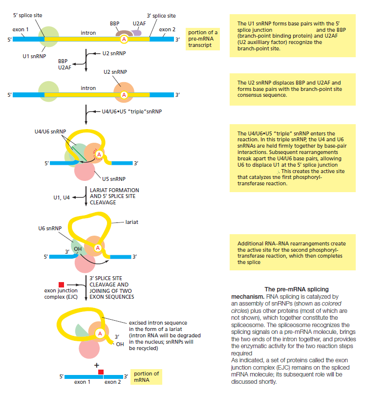
The snRNAs perform many of the spliceosome’s mRNA recognition events. Splice site consensus sequences are recognized by non-snRNP factors; the branch-point sequence is recognized by the branch-point-binding protein (BBP), and the polypyrimidine tract and 3′ splice site are bound by two specific protein components of a splicing complex referred to as U2AF (U2 auxiliary factor), U2AF65 and U2AF35, respectively.

This is one more great example of an amazingly complex molecular machine, that will operate and exercise its precise orchestrated function properly ONLY with ALL components fully developed and formed and able to interact in a highly complex, ordered precise manner. Both, the software, and the hardware must be in place fully developed, or the machine will not work. No intermediate stage will do the job. And neither would snRNPs (U1, U2, U4, U5, and U6) have any function if not fully developed. And even if they were there, without the branch-point-binding protein (BBP) in place, nothing done, either, since the correct splice site could not be recognized. Had the introns and exons not have to emerge simultaneously with the Spliceosome? No wonder, does the paper: " Origin and evolution of spliceosomal introns " admit:  Evolution of the exon-intron structure of eukaryotic genes has been a matter of long-standing, intensive debate. 1 and it concludes that: The elucidation of the general scenario of the evolution of eukaryote gene architecture by no account implies that the main problems in the study of intron evolution and function have been solved. Quite the contrary, fundamental questions remain wide open. If the first evolutionary step would have been the arise of self-splicing Group II introns, then the question would follow: Why would evolution not have stopped there, since that method works just fine? 


There is no credible road map, how introns and exons, and the splice function could have emerged gradually. What good would the spliceosome be good for, if the essential sequence elements to recognize where to slice would not be in place? What would happen, if the pre mRNA with exons and introns were in place, but no spliceosome ready in place to do the post-transcriptional modification, and neither the splicing code, which directs the way where to splice?  In the article: ‘JUNK’ DNA HIDES ASSEMBLY INSTRUCTIONS, the author,  Wang,  observes that splicing "is a tightly regulated process, and a great number of  diseases are caused by the 'misregulation' of splicing in which the gene was not cut and pasted correctly." Missplicing in the cell can have dire consequences as the desired product is not produced, and often the wrong products can be toxic for the cell. For this reason, it has been proposed that  ATPases are important for ‘proofreading’ mechanisms that promote fidelity in splice site selection.

In his textbook Essentials of Molecular Biology, George Malacinski points out why proper polypeptide production is critical:
"A cell cannot, of course, afford to miss any of the splice junctions by even a single nucleotide, because this could result in an interruption of the correct reading frame, leading to a truncated protein." 


The required precision is quite amazing, and even more astounding is the fact that these incredibly complex molecular machines are able and capable to do the job in the precise manner as needed. 

Following the binding of these initial components, the remainder of the splicing apparatus assembles around them, in some cases displacing some of the previously bound components.

Question: How could the information to assemble the splicing apparatus correctly have emerged gradually? In order to do so, had the assembly parts not have to be there, at the assembly site, fully developed, and ready for recruitment?  Had the availability of these parts not have to be synchronized so that at some point, either individually or in combination, they were all available at the same time? Had the assembly not have to be coordinated in the right way right from the start? Had the parts not have to be mutually compatible, that is, ‘well-matched’ and capable of properly ‘interacting’? even if subsystems or parts are put together in the right order, they also need to interface correctly.


Is it feasible that this complex machine was the result of progressive evolutionary development, in which simple molecules are the start of the biosynthesis chain and are then progressively developed in sequential steps if the end goal is not known by the process and mechanism promoting the development?  How could each intermediate in the pathway be an endpoint in the pathway, if that endpoint had no function? Did not each intermediate have to be usable in the past as an end product? And how could the be usable, if the amino acid sequence chain had only a fraction of the fully developed sequence? How could successive steps be added to improve the efficiency of a product where there was no use for it at this stage?  Despite the fact that proponents of naturalism embrace this kind of scenario, it seems obvious that is extremely unlikely to be possible that way.

Martin and Koonin admit in their paper  “Hypothesis: Introns and the origin of nucleus-cytosol compartmentalization,”:  The transition to spliceosome-dependent splicing will also impose an unforgiving demand for inventions in addition to the spliceosome. And furthermore: More recent are the insights that there is virtually no evolutionary grade detectable in the origin of the spliceosome, which apparently was present in its (almost) fully-fledged state in the common ancestor of eukaryotic lineages studied so far. That's a surprising admittance.

This means that the spliceosome appeared fully formed almost abruptly and that the intron invasion took place over a short time and has not changed for supposedly hundreds of millions of years.

In another interesting paper : Breaking the second genetic code, the authors write 2 :  The genetic instructions of complex organisms exhibit a counter-intuitive feature not shared by simpler genomes: nucleotide sequences coding for a protein (exons) are interrupted by other nucleotide regions that seem to hold no information (introns). This bizarre organization of genetic messages forces cells to remove introns from the precursor mRNA (pre-mRNA) and then splice together the exons to generate translatable instructions. An advantage of this mechanism is that it allows different cells to choose alternative means of pre-mRNA splicing and thus generates diverse messages from a single gene. The variant mRNAs can then encode different proteins
with distinct functions. One difficulty with understanding alternative pre-mRNA splicing is that the selection of particular exons in mature mRNAs is determined not only by intron sequences adjacent to the exon boundaries but also by a multitude of other sequence elements present in both exons and introns. These auxiliary sequences are recognized by regulatory factors that assist or prevent the function of the spliceosome — the molecular machinery in charge of intron removal.

Moreover, the coupling between RNA processing and gene transcription influences alternative splicing, and recent data implicate the packing of DNA with histone proteins and histone covalent modifications — the epigenetic code — in the regulation of splicing. The interplay between the histone and the splicing codes will, therefore, need to be accurately formulated in future approaches. 

Question: How could natural mechanisms have provided the tuning, synchronization, and coordination between the histone and the splicing codes? First, these two codes and the carrier proteins and molecules ( the hardware and software ) would have to emerge by themselves, and in a second step orchestrate their coordination. Why is it reasonable to believe, that unguided, random chemical reactions would be capable of emerging with the immensely complex organismal functions? 

Fazale Rana puts it nicely:  Astounding is the fact that other codes, such as the histone binding code, transcription factor binding code, the splicing code, and the RNA secondary structure code, overlap the genetic code. Each of these codes plays a special role in gene expression, but they also must work together in a coherent integrated fashion.

1) http://www.biologydirect.com/content/7/1/11
2) http://nar.oxfordjournals.org/content/early/2013/11/07/nar.gkt1053.full.pdf


The spliceosome, the splicing code, and pre - mRNA processing in eukaryotic cells

Transcription Elongation in Eukaryotes Is Tightly Coupled to RNA Processing

Bacterial mRNAs are synthesized by the RNA polymerase starting and stopping at specific spots on the genome. The situation in eukaryotes is substantially different. In particular, transcription is only the first of several steps needed to produce a mature mRNA molecule. Other critical steps are the covalent modification of the ends of the RNA and the removal of intron sequences that are discarded from the middle of the RNA transcript by the process of RNA splicing

The spliceosome , the splicing code, and pre - mRNA processing in eukaryotic cells From_g10

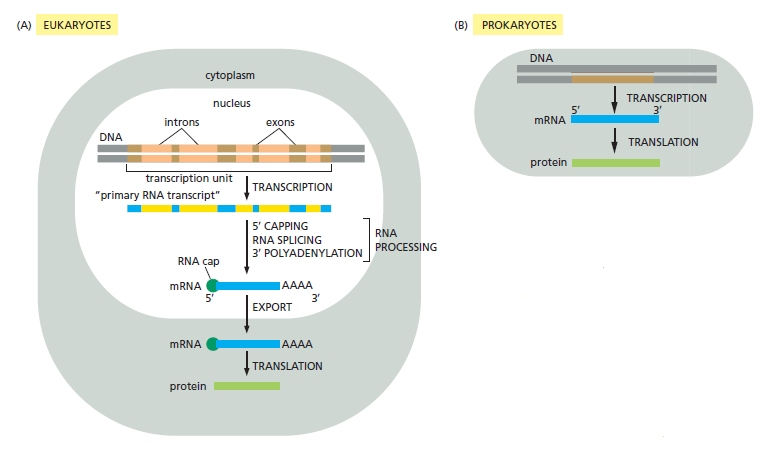
Comparison of the steps leading from gene to protein in eukaryotes and bacteria. The final level of a protein in the cell depends on the efficiency of each step and on the rates of degradation of the RNA and protein molecules.

(A) In eukaryotic cells, the mRNA molecule resulting from transcription contains both coding (exon) and noncoding (intron) sequences. Before it can be translated into protein, the two ends of the RNA are modified, the introns are removed by an enzymatically catalyzed RNA splicing reaction, and the resulting mRNA is transported from the nucleus to the cytoplasm. For convenience, the steps in this figure are depicted as occurring one at a time; in reality, many occur concurrently. For example, the RNA cap is added and splicing begins before transcription has been completed. Because of the coupling between transcription and RNA processing,
intact primary transcripts—the full-length RNAs that would, in theory, be produced if no processing had occurred—are found only rarely.
(B) In prokaryotes, the production of mRNA is much simpler. The 5ʹ end of an mRNA molecule is produced by the initiation of transcription, and the 3ʹ end is produced by the termination of transcription. Since prokaryotic cells lack a nucleus, transcription and translation take place in a common compartment, and the translation of a bacterial mRNA often begins before its synthesis has been completed.


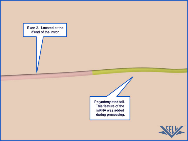
Both ends of eukaryotic mRNAs are modified: by capping on the 5ʹ end and by polyadenylation of the 3ʹ end 1 These special ends allow the cell to assess whether both ends of a mRNA molecule are present (and if the message is therefore intact) before it exports the RNA from the nucleus and translates it into protein.

Question: How could and would unguided, natural processes, where no intelligence is involved, emerge with that check out if both ends of mRNA are present? Quality control is usually an exercise attributed to intelligence.

RNA splicing joins together the different portions of a protein-coding sequence, and it provides eukaryotes with the ability to synthesize several different proteins from the same gene. A simple strategy has evolved

Why not designed? figuring out what strategy to apply to get the best results is a mental process

to couple all of the above RNA processing steps to transcription elongation. As discussed previously, a key step in transcription initiation by RNA polymerase II is the phosphorylation of the RNA polymerase II tail, also called the CTD (C-terminal domain). This phosphorylation, which proceeds gradually as the RNA polymerase initiates transcription and moves along the DNA, not only helps dissociate the RNA polymerase II from other proteins present at the start point of transcription, but also allows a new set of proteins to associate with the RNA polymerase tail that function in transcription elongation and RNA processing. As discussed next, some of these processing proteins are thought to “hop” from the polymerase tail onto the nascent RNA molecule to begin processing it as it emerges from the RNA polymerase. Thus, we can view RNA polymerase II in its elongation mode as an RNA factory that not only moves along the DNA synthesizing an RNA molecule but also processes the RNA that it produces

The spliceosome , the splicing code, and pre - mRNA processing in eukaryotic cells Rna_po10


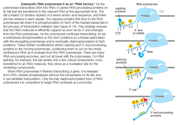
Fully extended, the CTD is nearly 10 times longer than the remainder of RNA polymerase. As a flexible protein domain, it serves as a scaffold or tether, holding a variety of proteins close by so that they can rapidly act when needed. This strategy, which greatly speeds up the overall rate of a series of consecutive reactions, is one that is commonly utilized in the cell.

RNA Capping Is the First Modification of Eukaryotic Pre-mRNAs

As soon as RNA polymerase II has produced about 25 nucleotides of RNA, the 5ʹ end of the new RNA molecule is modified by addition of a cap that consists of a modified guanine nucleotide. Three enzymes, acting in succession, perform the capping reaction: one (a phosphatase) removes a phosphate from the 5ʹ end of the nascent RNA, another (a guanyl transferase) adds a GMP in a reverse linkage (5ʹ to 5ʹ instead of 5ʹ to 3ʹ), and a third (a methyl transferase) adds a methyl group to the guanosine

The spliceosome , the splicing code, and pre - mRNA processing in eukaryotic cells Dsfsdf10

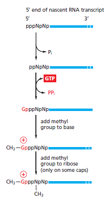
The reactions that cap the 5ʹ end of each RNA molecule synthesized by RNA polymerase II. The final cap contains a novel 5ʹ-to-5ʹ linkage between the positively charged 7-methyl G residue and the 5ʹ end of the RNA transcript . The letter N represents any one of the four ribonucleotides, although the nucleotide that starts an RNA chain is usually a purine (an A or a G).

Because all three enzymes bind to the RNA polymerase tail phosphorylated at the Ser5 position—the modification added by TFIIH during transcription initiation— they are poised to modify the 5ʹ end of the nascent transcript as soon as it emerges from the polymerase. The 5ʹ-methyl cap signifies the 5ʹ end of eukaryotic mRNAs, and this landmark helps the cell to distinguish mRNAs from the other types of RNA molecules present in the cell. For example, RNA polymerases I and III produce uncapped RNAs during transcription, in part because these polymerases lack a CTD. In the nucleus, the cap binds a protein complex called CBC (cap-binding complex), which, as we discuss in subsequent sections, helps a future mRNA be further processed and exported. The 5ʹ-methyl cap also has an important role in the translation of mRNAs in the cytosol, as we discuss later in the chapter.

RNA Splicing Removes Intron Sequences from Newly Transcribed Pre-mRNAs

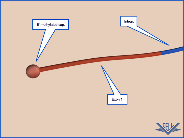
The protein-coding sequences of eukaryotic genes are typically interrupted by noncoding intervening sequences (introns). Discovered in 1977, this feature of eukaryotic genes came as a surprise to scientists, who had been, until that time, familiar only with bacterial genes, which typically consist of a continuous stretch of coding DNA that is directly transcribed into mRNA. In marked contrast, eukaryotic genes were found to be broken up into small pieces of coding sequence (expressed sequences or exons) interspersed with much longer intervening sequences or introns; thus, the coding portion of a eukaryotic gene is often only a small fraction of the length of the gene

The spliceosome , the splicing code, and pre - mRNA processing in eukaryotic cells Intron10

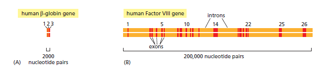
Structure of two human genes showing the arrangement of exons and introns.
(A) The relatively small β-globin gene, which encodes a subunit of the oxygen-carrying protein hemoglobin, contains 3 exons .
(B) The much larger Factor VIII gene contains 26 exons; it codes for a protein (Factor VIII) that functions in the bloodclotting pathway. The most prevalent form of hemophilia results from mutations in this gene.

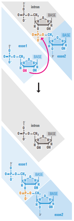
Both intron and exon sequences are transcribed into RNA. The intron sequences are removed from the newly synthesized RNA through the process of RNA splicing. The vast majority of RNA splicing that takes place in cells functions in the production of mRNA, and our discussion of splicing focuses on this so-called precursor-mRNA (or pre-mRNA) splicing. Only after 5ʹ- and 3ʹ-end processing and splicing have taken place is such RNA termed mRNA. Each splicing event removes one intron, proceeding through two sequential
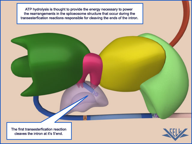
phosphoryl-transfer reactions known as transesterifications; these join two exons together while removing the intron between them as a “lariat”

The spliceosome , the splicing code, and pre - mRNA processing in eukaryotic cells Mrna_s10

This is a highly ordered, sequential, and precise mechanism. How did this function emerge naturally? How was the specific adenine nucleotide put into the right place to be spliced? trial and error? In order to propose a natural origin, these questions need to be explained in a compelling manner.

The machinery that catalyzes pre-mRNA splicing is complex, consisting of five additional RNA molecules and several hundred proteins, and it hydrolyzes many ATP molecules per splicing event. This complexity ensures that splicing is accurate, while at the same time being flexible enough to deal with the enormous variety of introns found in a typical eukaryotic cell. It may seem wasteful to remove large numbers of introns by RNA splicing. In attempting to explain why it occurs, scientists have pointed out that the exon-intron arrangement would seem to facilitate the emergence of new and useful proteins over evolutionary timescales.

First of all, proponents of evolution should be able to explain why such a piece of complex machinery would emerge AT ALL, and why there should be a need of emergence of such complexity if bacterial cells work just fine without it. What I see here, is typical simplistic assertions without giving a considerable thought about the viability of random mutations to produce such enormous complexity

Thus, the presence of numerous introns in DNA allows genetic recombination to readily combine the exons of different genes, enabling genes for new proteins to evolve emerge more easily by the combination of parts of preexisting genes. RNA splicing also has a present-day advantage. The transcripts of many eukaryotic genes (estimated at 95% of genes in humans) are spliced in more than one way, thereby allowing the same gene to produce a corresponding set of different proteins.

Why should nature give preference to such a completely new method of producing a variety of proteins, rather than just let mutations and natural selection doing the job? It seems to me, in order to keep the pre-established fact of evolution, their proponents simply shut up with their critical thinking, and accept the fact of evolution, no matter how unlikely it seems

The spliceosome , the splicing code, and pre - mRNA processing in eukaryotic cells Altern10

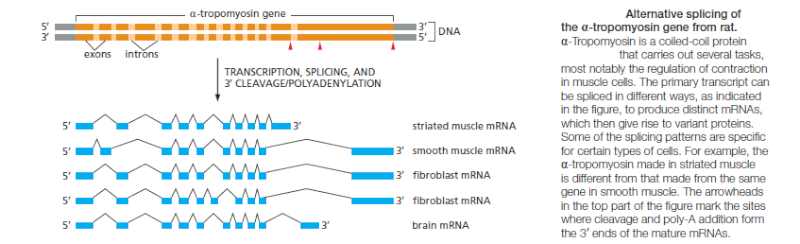
Rather than being the wasteful process it may have seemed at first sight, RNA splicing enables eukaryotes to increase the coding potential of their genomes.

In order to splice out the introns from exons, the cell required epigenetic information beside the complex machinery in place to do this complex task. Where did this information come from? trial and error?

Nucleotide Sequences Signal Where Splicing Occurs

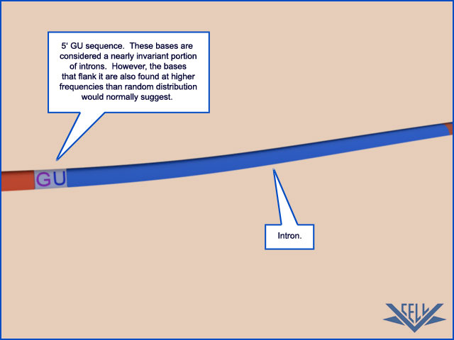
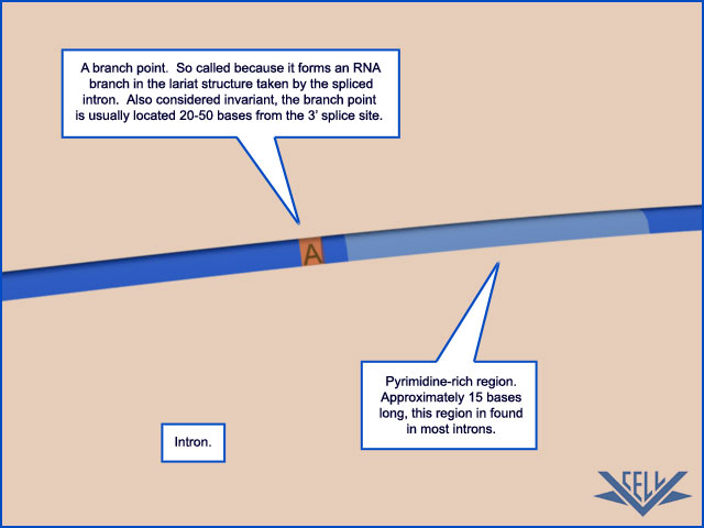
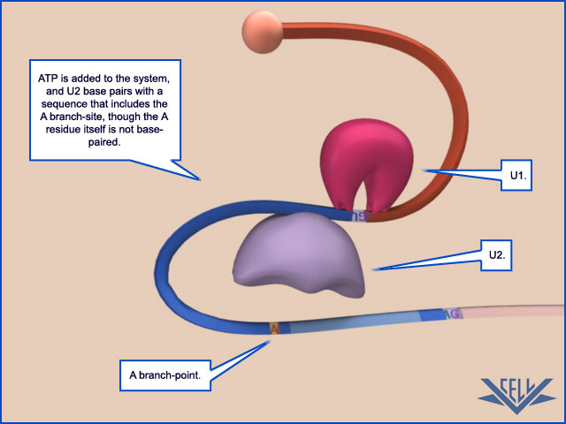
The mechanism of pre-mRNA splicing requires that the splicing machinery recognize three portions of the precursor RNA molecule: the 5ʹ splice site, the 3ʹ splice site, and the branch point in the intron sequence that forms the base of the excised lariat. Not surprisingly, each site has a consensus nucleotide sequence that is similar from intron to intron and provides the cell with cues for where splicing is to take place.

The spliceosome , the splicing code, and pre - mRNA processing in eukaryotic cells Intron11

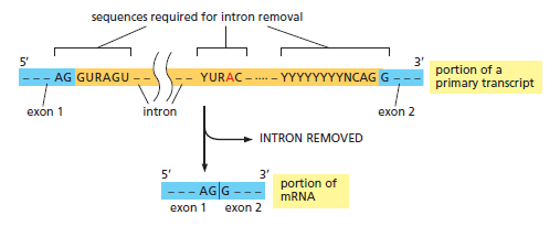
The consensus nucleotide sequences in an RNA molecule that signal the beginning and the end of most introns in humans. The three blocks of nucleotide sequences shown are required to remove an intron sequence. Here A, G, U, and C are the standard RNA nucleotides; R stands for purines (A or G); and Y stands for pyrimidines (C or U). The A highlighted in red forms the branch point of the lariat produced by splicing. Only the GU at the start of the intron and the AG at its end are invariant nucleotides in the splicing consensus sequences. Several different nucleotides can occupy the remaining positions, although the indicated nucleotides are preferred. The distances along the RNA between the three splicing consensus sequences are highly variable; however, the distance between the branch point and 3ʹ splice junction is typically much shorter than that between the 5ʹ splice junction and the branch point.

However, these consensus sequences are relatively short and can accommodate extensive sequence variability; the cell incorporates additional types of information to ultimately choose exactly where, on each RNA molecule, splicing is to take place.

Its evident that foresight is required a) to make the splicing machinery, and b) to program the information right from the start, where to slice; the whole mechanism has to arise all at once, fully functional. A stepwise arise is extremely unlikely, not to say, impossible. What good would the spliceosome be good for, if there were no information at the same time in order to make the machinery do its job? The hardware and the software are interdependent.


The high variability of the splicing consensus sequences presents a special challenge for scientists attempting to decipher genome sequences. Introns range in size from about 10 nucleotides to over 100,000 nucleotides, and choosing the precise borders of each intron is a difficult task even with the aid of powerful computers.

But cells " know " where to splice for a long time..... how did they " learn " that feat?

The possibility of alternative splicing compounds the problem of predicting protein sequences solely from a genome sequence. This difficulty is one of the main barriers to identifying all of the genes in a complete genome sequence, and it is one of the primary reasons why we know only the approximate number of different proteins produced by the human genome.

RNA Splicing Is Performed by the Spliceosome

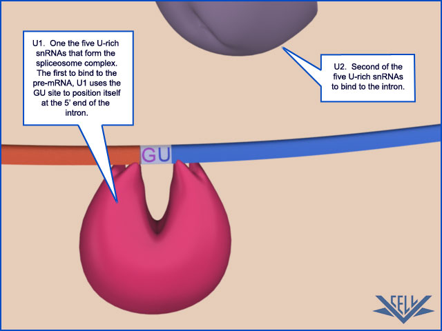
Unlike the other steps of mRNA production we have discussed, key steps in RNA splicing are performed by RNA molecules rather than proteins. Specialized RNA molecules recognize the nucleotide sequences that specify where splicing is to occur and also catalyze the chemistry of splicing. These RNA molecules are relatively short (less than 200 nucleotides each), and there are five of them, U1, U2, U4, U5, and U6. Known as snRNAs (small nuclear RNAs), each is complexed with at least seven protein subunits to form an snRNP (small nuclear ribonucleoprotein).

Question: what good would there be for these Specialized RNA molecules to arise, if there were no machinery to subsequently splice the mRNA's ? even more, the make of the subunits has to be explained as  well, since, by their own, there is no function for them

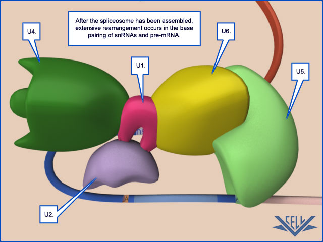
These snRNPs form the core of the spliceosome, the large assembly of RNA and protein molecules that performs pre-mRNA splicing in the cell. During the splicing reaction, recognition of the 5ʹ splice junction, the branch-point site, and the 3ʹ splice junction is performed largely through base-pairing between the snRNAs and the consensus RNA sequences in the pre-mRNA substrate. The spliceosome is a complex and dynamic machine. When studied in vitro, a few components of the spliceosome assemble on pre-mRNA and, as the splicing reaction proceeds, new components enter and those that have already performed their tasks are jettisoned


The spliceosome , the splicing code, and pre - mRNA processing in eukaryotic cells Splice10

The Spliceosome Uses ATP Hydrolysis to Produce a Complex Series of RNA–RNA Rearrangements

ATP hydrolysis is not required for the chemistry of RNA splicing per se since the two transesterification reactions preserve the high-energy phosphate bonds. However, extensive ATP hydrolysis is required for the assembly and rearrangements of the spliceosome. Some of the additional proteins that make up the spliceosome use the energy of ATP hydrolysis to break existing RNA–RNA interactions to allow the formation of new ones. Each successful splice requires approximately 200 proteins if we include those that form the snRNPs. What is the purpose of these rearrangements? First, they allow the splicing signals on the pre-RNA to be examined by snRNPs several times during the course of
splicing. For example, the U1 snRNP initially recognizes the 5ʹ splice site through conventional base-pairing; as splicing proceeds, these base pairs are broken (using the energy of ATP hydrolysis) and U1 is replaced by U6

The spliceosome , the splicing code, and pre - mRNA processing in eukaryotic cells Splice11

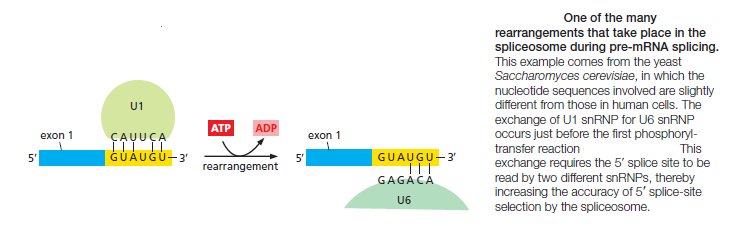
This type of RNA–RNA rearrangement (in which the formation of one RNA–RNA interaction requires the disruption of another) occurs several times during splicing and allows the spliceosomes to check and recheck the splicing signals, thereby increasing the overall accuracy of splicing.

How could this recheck or proofreading have emerged naturally, without a guiding force, without an intelligent designer set up the function with the specific goal to reach a certain, high level of accuracy of the job?


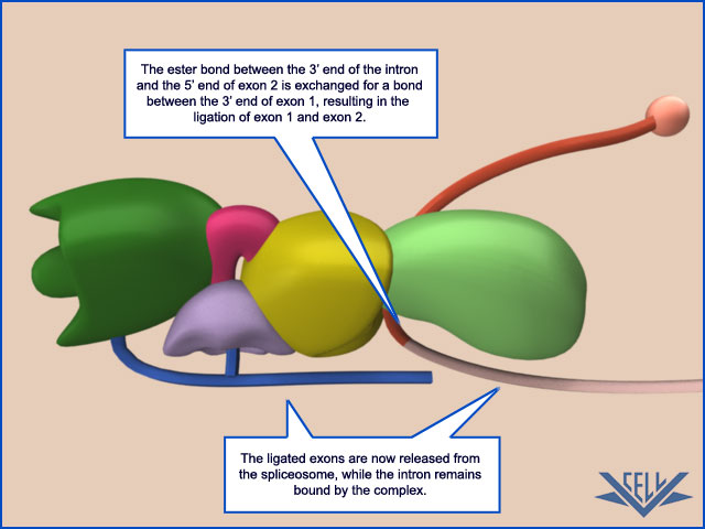
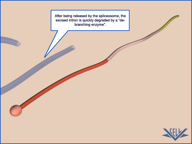
Second, the rearrangements that take place in the spliceosome create the active sites for the two transesterification reactions. These two active sites are created, one after the other, and only after the splicing signals on the pre-mRNA have been checked several times. This orderly progression ensures that splicing accidents occur only rarely. One of the most surprising features of the spliceosome is the nature of the catalytic sites: they are formed by both protein and RNA molecules, although the RNA molecules catalyze the actual chemistry of splicing. In the last section of this chapter, we discuss in general terms the structural and chemical properties of RNA molecules that allow them to act as catalysts. Once the splicing chemistry is completed, the snRNPs remain bound to the lariat. The disassembly of these snRNPs from the lariat (and from each other) requires another series of RNA–RNA rearrangements that require ATP hydrolysis,thereby returning the snRNAs to their original configuration so that they can be used again in a new reaction.

Question: Does the ability to rearrange to exercise new future reactions indicate the planning guide of an intelligent creator? The matter has no specific goals like this, and cannot create these specific goals of repetitive complex functions

At the completion of a splice, the spliceosome directs a set of proteins to bind to the mRNA near the position formerly occupied by the intron. Called the exon junction complex (EJC), these proteins mark the site of a successful splicing event and, as we shall see later in this chapter, influence the subsequent fate of the mRNA.

Other Properties of Pre-mRNA and Its Synthesis Help to Explain the Choice of Proper Splice Sites

Intron sequences vary enormously in size, with some being in excess of 100,000 nucleotides. If splice-site selection were determined solely by the snRNPs acting on a preformed, protein-free RNA molecule, we would expect frequent splicing mistakes—such as exon skipping and the use of “cryptic” splice sites

The spliceosome , the splicing code, and pre - mRNA processing in eukaryotic cells Erertt10

The fidelity mechanisms built into the spliceosome to suppress errors, however, are supplemented by two additional strategies that further increase the accuracy of splicing. The first is a simple consequence of splicing being coupled to transcription. As transcription proceeds, the phosphorylated tail of RNA polymerase carries several components of the spliceosome and these components are transferred directly from the polymerase to the RNA as the RNA emerges from the polymerase. This strategy helps the cell keep track of introns and exons: for example, the snRNPs that assemble at a 5ʹ splice site are initially presented only with the single 3ʹ splice site that emerges next from the polymerase; the potential sites further downstream have not yet been synthesized. The coordination of transcription with splicing is especially important in preventing inappropriate exon skipping.
A strategy called “exon definition” also helps cells choose the appropriate splice sites. Exon size tends to be much more uniform than intron size, averaging about 150 nucleotide pairs across a wide variety of eukaryotic organisms

The spliceosome , the splicing code, and pre - mRNA processing in eukaryotic cells Tretrt10

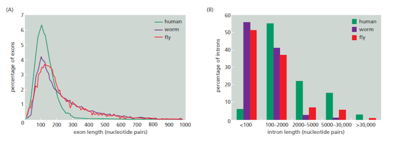
Variation in intron and exon lengths in the human, worm, and fly genomes.
(A) Size distribution of exons.
(B) Size distribution of introns. Note that exon length is much more uniform than intron length.

Through exon definition, the splicing machinery can seek out the relatively homogeneously sized exon sequences. As RNA synthesis proceeds, a group of additional components (most notably SR proteins, so-named because they contain a domain rich in serines and arginines) assemble on exon sequences and help to mark off each 3ʹ and 5ʹ splice site, starting at the 5ʹ end of the RNA

The spliceosome , the splicing code, and pre - mRNA processing in eukaryotic cells 76876810

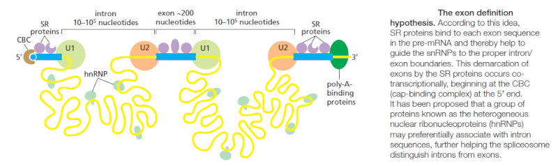
These proteins, in turn, recruit U1 snRNA, which marks the downstream exon boundary, and U2 snRNA, which specifies the upstream one. By specifically marking the exons in this way and thereby taking advantage of the relatively uniform size of exons, the cell increases the accuracy with which it deposits the initial splicing components on the nascent RNA and thereby avoids “near miss” splice sites. How the SR proteins discriminate exon sequences from intron sequences is not understood in detail; however, it is known that some of the SR proteins bind preferentially to specific RNA sequences in exons, termed splicing enhancers. In principle, since any one of several different codons can be used to code for a given amino acid, there is freedom to evolve the exon nucleotide sequence so as to form a binding site for an SR protein, without necessarily affecting the amino acid sequence that the exon specifies. Both the marking of exon and intron boundaries and the assembly of the spliceosome begin on an RNA molecule while it is still being elongated by RNA polymerase at its 3ʹ end. However, the actual chemistry of splicing can take place later. This delay means that intron sequences are not necessarily removed from a premRNA molecule in the order in which they occur along the RNA chain.

Chromatin Structure Affects RNA Splicing


Although it may seem at first counterintuitive, the way a gene is packaged into chromatin can affect how the RNA transcript of that gene is ultimately spliced. Nucleosomes tend to be positioned over exons (which are, on average, close to the length of DNA in a nucleosome), and it has been proposed that these act as “speed bumps,” allowing the proteins responsible for exon definition to assemble on the RNA as it emerges from the polymerase. In addition, changes in chromatin structure are used to alter splicing patterns. There are two ways this can happen. First, because splicing and transcription are coupled, the rate at which RNA polymerase moves along DNA can affect RNA splicing. For example, if polymerase is moving slowly, exon skipping  is minimized: assembly of the initial spliceosome may be complete before an alternative choice of splice site even emerges from the RNA polymerase. The nucleosomes in condensed chromatin can cause polymerase to pause; the pattern of pauses in turn affects the extent of RNA exposed at any given time to the splicing machinery. There is a second and more direct way that chromatin structure can affect RNA splicing. Although the details are not yet understood, specific histone modifications attract components of the spliceosome, and, because the chromatin being transcribed is in close association with the nascent RNA, these splicing components can easily be transferred to the emerging RNA. In this way, certain types of histone modifications can affect the final pattern of splicing.

RNA Splicing Shows Remarkable Plasticity

We have seen that the choice of splice sites depends on such features of the premRNA transcript as the strength of the three signals on the RNA (the 5ʹ and 3ʹ splice junctions and the branch point) for the splicing machinery, the co-transcriptional assembly of the spliceosome, chromatin structure, and the “bookkeeping” that underlies exon definition. We do not know exactly how accurate splicing normally is because there are several quality control systems that rapidly destroy mRNAs whose splicing goes awry. However, we do know that, compared with other steps in gene expression, splicing is unusually flexible. Thus, for example, a mutation in a nucleotide sequence critical for splicing of a particular intron does not necessarily prevent splicing of that intron altogether. Instead, the mutation typically creates a new pattern of splicing

The spliceosome , the splicing code, and pre - mRNA processing in eukaryotic cells Splici10

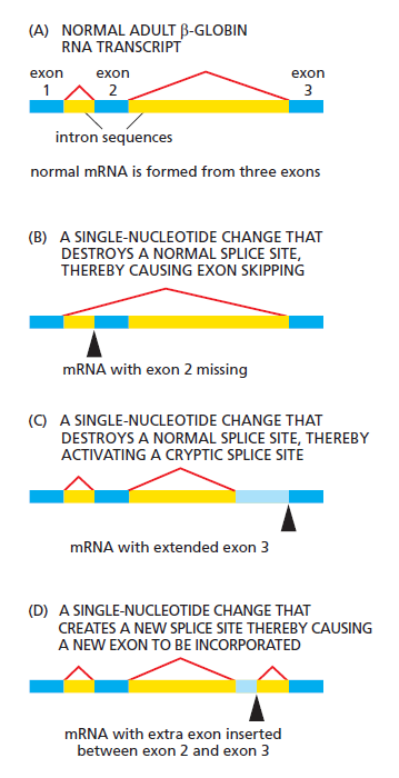
Abnormal processing of the β-globin primary RNA transcript in humans with the disease β thalassemia. In the examples shown, the disease (a severe anemia due to aberrant hemoglobin synthesis) is caused by splice-site mutations found in the genomes of affected patients. The dark blue boxes represent the three normal exon sequences; the red lines connect the 5ʹ and 3ʹ splice sites that are used. In (B), (C), and (D), the light blue boxes depict new nucleotide sequences included in the final mRNA molecule as a result of the mutation denoted by the black arrowhead. Note that when a mutation leaves a normal splice site without a partner, an exon is skipped (B) or one or more abnormal cryptic splice sites nearby is used as the partner site (C).

Most commonly, an exon is simply skipped (Figure B). In other cases, the mutation causes a cryptic splice junction to be efficiently used (Figure C). Apparently, the splicing machinery does pick out the best possible pattern of splice junctions, and if the optimal one is damaged by mutation, it will seek out the next best pattern, and so on. This inherent plasticity in the process of RNA splicing suggests that changes in splicing patterns caused by random mutations are important for genes and organisms. It also means that mutations that affect splicing can be severely detrimental to the organism: in addition to the β thalassemia, example presented in the Figure above, aberrant splicing plays important roles in the development of cystic fibrosis, frontotemporal dementia, Parkinson’s disease, retinitis pigmentosa, spinal muscular atrophy, myotonic dystrophy, premature aging, and cancer. It has been estimated that of the many point mutations that cause inherited human diseases, 10% produce aberrant splicing of the gene containing the mutation.
The plasticity of RNA splicing also means that the cell can easily regulate the pattern of RNA splicing. Alternative splicing can give rise to different proteins from the same gene and that this is a common strategy to enhance the coding potential of genomes. Some examples of alternative splicing are constitutive; that is, the alternatively spliced mRNAs are produced continuously by cells of an organism. However, in many cases, the cell regulates the splicing patterns so that different forms of the protein are produced at different
times and in different tissues .

Spliceosome-Catalyzed RNA Splicing Probably emerged from Self-splicing Mechanisms

When the spliceosome was first discovered, it puzzled molecular biologists. Why do RNA molecules instead of proteins perform important roles in splice-site recognition and in the chemistry of splicing? Why is a lariat intermediate used rather than the apparently simpler alternative of bringing the 5ʹ and 3ʹ splice sites together in a single step, followed by their direct cleavage and rejoining?  It is likely that early cells used RNA molecules rather than proteins as their major catalysts and that they stored their genetic information in RNA rather than in DNA sequences. RNA-catalyzed splicing reactions presumably had critical roles in these early cells. As evidence, some self-splicing RNA introns (that is, intron sequences in RNA whose splicing out can occur in the absence of proteins or any other RNA molecules) remain today—for example, in the nuclear rRNA genes of the ciliate Tetrahymena, in a few bacteriophage T4 genes, and in some mitochondrial and chloroplast genes. In these cases, the RNA molecule folds into a specific three-dimensional structure that brings the intron/exon junctions together and catalyzes the two transesterification reactions. A self-splicing intron sequence can be identified in a test tube by incubating a pure RNA molecule that contains the intron sequence and observing the splicing reaction. Because the basic chemistry of some self-splicing reactions is so similar to pre-mRNA splicing, it has been proposed that the much more involved process of pre-mRNA splicing evolved from a simpler, ancestral form of RNA self-splicing

RNA-Processing Enzymes Generate the 3ʹ End of Eukaryotic mRNAs

We have seen that the 5ʹ end of the pre-mRNA produced by RNA polymerase II is capped almost as soon as it emerges from the RNA polymerase. Then, as the polymerase continues its movement along a gene, the spliceosome assembles on the RNA and delineates the intron and exon boundaries. The long C-terminal tail of the RNA polymerase coordinates these processes by transferring capping and splicing components directly to the RNA as it emerges from the enzyme. In this section, we shall see that, as RNA polymerase II reaches the end of a gene, a similar mechanism ensures that the 3ʹ end of the pre-mRNA is appropriately processed. The position of the 3ʹ end of each mRNA molecule is specified by signals encoded in the genome


1) http://reasonandscience.heavenforum.org/t2022-3-end-cleavage-and-polyadenylation

More:
Protein recipe requires precise timing
Molecular architecture of the human U4/U6.U5 tri-snRNP


Last edited by Admin on Sat Jul 11, 2020 3:05 am; edited 12 times in total

https://reasonandscience.catsboard.com

2The spliceosome , the splicing code, and pre - mRNA processing in eukaryotic cells Empty RNA SPLICING Wed Sep 09, 2015 8:09 pm

Otangelo


Admin

RNA SPLICING 1

Introns must be removed from precursor RNAs

The mature transcript for many genes is encoded in a discontinuous manner in a series of discrete exons, which are separated from each other along the DNA strand by non-coding introns. mRNAs, rRNAs, and tRNAs can all contain introns that must be removed from precursor RNAs to produce functional molecules. There are a number of different classes of intron, with their relative distribution and abundance depending on the kingdom. Some are excised by proteins, some by RNPs, and some excise themselves, yet all use some variation of a series of transesterification reactions that we describe below. Most introns do not themselves contain genes and are simply removed from the precursor RNAs and degraded. Exceptions to this rule are the snoRNAs and certain miRNAs in invertebrates and mammals  as they can be present in the introns of mRNAs. While introns are widespread, they are far more common in eukaryotes than bacteria. Indeed, the existence of introns has allowed for a process referred to as exon shuffling wherein the DNA encoding exons can be exchanged and reordered through genetic recombination between intronic DNA. This process thus allows for the emergence of novel genes and is presumed to have played a major part in the evolution of eukaryotic genomes. The differential removal of introns from an RNA transcript can result in the production of different spliced RNAs from a single gene, thereby increasing the number of gene products encoded in an organism’s genome.

RNA splicing occurs by two transesterification reactions

There are several different pathways by which introns are removed from genes, but most of them involve the same underlying chemical reactions. The two exons that surround an intron are not directly joined to one another in a single step. Instead, a two-step reaction occurs in which the intron is first detached from exon 1, freeing this exon to react with exon 2. Both of these reaction steps are transesterifications in which a single phosphodiester bond between two nucleotides is broken and replaced with an energetically equivalent phosphodiester bond

The spliceosome , the splicing code, and pre - mRNA processing in eukaryotic cells Transe11

The transesterification reaction of RNA splicing. The 3′ OH of an incoming nucleotide (or RNA) (pink) attacks and breaks the 3′–5′ phosphodiester bond between two nucleotides in the target RNA (orange). As a result, a new 3′–5′ phosphodiester bond is formed between the incoming nucleotide (or RNA) and the target RNA. Alternatively, the 2′ OH of the incoming RNA can attack the target RNA, in which case the incoming and target RNA become joined though a 2′–5′ phosphodiester bond (not shown).

There are two implications for replacing one chemical bond with a similar chemical bond: first, the reaction can occur without the net input of energy from an energy source such as ATP; and, second, the reaction can be readily reversible. In fact, reverse splicing reactions by certain classes of intron, which allow them to insert into duplex DNA, are thought to have played a significant role in dispersing introns and thus in shaping the evolution of the genome. Most introns in eukaryotes are removed by a complex ribonucleoprotein machine called the spliceosome. However, a few introns in eukaryotes, bacteria, and organelles are able to catalyze their own removal from RNA and are therefore called self-splicing introns.


EUKARYOTIC mRNA SPLICING BY THE SPLICEOSOME

Splicing of eukaryotic mRNAs is carried out by a large RNP machine called the spliceosome

Genes in higher eukaryotes usually contain several introns. These introns are often of considerable length, sometimes extending to many thousands of bases. As such, they can account for 90% of the length of a typical precursor mRNA

The spliceosome , the splicing code, and pre - mRNA processing in eukaryotic cells Intron12

By comparison, lower eukaryotes such as yeast have fewer introns, and they are generally short (<300 nucleotides). The vast majority of eukaryotic introns are not self-splicing, and the formidable task of identifying and splicing together exons among all the intronic RNA is performed by a large ribonucleoprotein machine, the spliceosome. The spliceosome is composed of several individual small nuclear ribonucleoproteins (snRNP, pronounced ‘snurps’, which comprise both RNA and protein components) and many more additional proteins that come and go during the splicing reaction.The 2′ OH of an adenosine residue located within the intron itself attacks the exon 1–intron boundary, detaching the intron from the exon and producing a branched intron structure (the lariat). Next, the terminal 3′ OH of the newly released exon attacks the intron–exon 2 junction, splicing together the two exons and releasing the lariat intron. The high similarity in mechanism between the transesterification reactions catalyzed by the group II intron and the spliceosome has led to the idea that the simpler self-splicing intron is the evolutionary predecessor of the more complicated multicomponent eukaryotic machinery.

Splice site recognition and spliceosome assembly are dominated by RNA–RNA interactions

Before splicing can occur, the spliceosome must identify the splice sites between introns and exons – the sites at which exons are separated from their neighboring introns, and at which two exons are subsequently attached. In contrast to the group I and II introns, where the splice sites are defined by the three-dimensional structures of the introns themselves, the spliceosome identifies splice sites by recognizing short sequence motifs found in each pre-mRNA. Key sequences are the 5′ and 3′ splice sites, a branch-point nucleotide within the intron, and a polypyrimidine tract before the 3′ splice site

In addition to these essential sequence elements, there are other sequences within the exons and in nearby intronic regions that also help to define the exon–intron junctions. These additional sequence determinants allow cells to produce different mature RNAs from a precursor RNA depending on which introns and exons are recognized by spliceosomes. The spliceosome is composed of five snRNPs (U1, U2, U4, U5, and U6) each containing an RNA molecule called an snRNA that is usually 100–300 nucleotides long, plus additional protein factors that recognize specific sequences in the mRNA or promote conformational rearrangements in the spliceosome required for the splicing reaction to progress.  These snRNAs perform many of the spliceosome’s mRNA recognition events by forming Watson–Crick base pairs with the precursor mRNA and one another. Other splice site consensus sequences are recognized by non-snRNP factors; the branch-point sequence is recognized by the branch-point-binding protein (BBP), and the polypyrimidine tract and 3′ splice site are bound by two specific protein components of a splicing complex referred to as U2AF (U2 auxiliary factor), U2AF65 and U2AF35, respectively

This is one more great example of a molecular machine, that will operate and exercise its complex function properly ONLY with ALL components fully developed and formed and able to interact in a highly complex, ordered , orchestrated manner. Both, the software, and the hardware, must be in place fully developed, or the mechanism will not work. No intermediate stage will do the job. And neither would  snRNPs (U1, U2, U4, U5, and U6) have any function if not fully developed. And even if they were there, without the branch-point-binding protein (BBP) in place, nothing done, either. There is no credible road map, how the function could have emerged gradually. What good would the spliceosome be good for, if the essential sequence elements to recognise where to slice would not be in place ? What would happen, if the pre mRNA with exons and introns were in place, but no spliceosome ready in place to do the post transcriptional modification ?  

Following the binding of these initial components, the remainder of the splicing apparatus assembles around them, in some cases displacing some of the previously bound components.

Question: How could the information to assemble the splicing apparatus correctly have emerged gradually ? In order to do so, had the assembly parts not have to be there, at the assembly site, fully developed, and ready for recruitment?  Had the availability of these parts not have  to be synchronized so that at some point, either individually or in combination, they were all available at the same time ? Had the assembly not have to be coordinated in the right way right from the start ? Had the parts not have to be mutually compatible, that is, ‘well-matched’ and capable of properly ‘interacting’ ? even if sub systems or parts are put together in the right order, they also need to interface correctly.

Is it feasable that this complex machine were the result of a progressive evolutionary development, in which simple molecules are the start of the biosynthesis chain and are then progressively developed in sequencial steps, if the end goal is not known by the process and mechanism promoting the development ?  How could  each intermediate in the pathway be a end point in the pathway, if that end point had no function ? Did not  each intermediate have to be usable in the past as an end product ? How could successive steps be added to improve the efficiency of a product where there was no use for it at this stage ?  Despite the fact that proponents of naturalism embrace this kind of scenary, it seems obvious that is extremely unlikely to be possible that way.


The precursor mRNA then undergoes a structural rearrangement, the first transesterification step takes place and the lariat forms . Other rearrangements in the spliceosome next bring together the newly freed exon 1 and the intron–exon 2 junction – the two RNA pieces that will be joined to create the final mRNA product – and the second transesterification reaction completes the splicing . While the molecular details of the individual steps are not fully understood, it is clear that the spliceosome is a dynamic ribonucleoprotein machine, and that conformational changes in the RNA components themselves are central to the spliceosome’s function. Indeed, the known ATP-dependence of the splicing reactions is thought to reflect the requirements of the many helicases that promote and regulate these RNA-based conformational rearrangements. Based on similarities in mechanism to the group II intron, it is also generally believed that the snRNAs themselves (the U2 and U6 snRNAs being the primary candidates) must play a central functional role in catalysis.

Protein factors contribute in multiple ways to pre-mRNA splicing

In addition to the snRNPs there are hundreds of other proteins that are associated with the splicing machinery and play important roles in the process. For example, PRP8 is an essential, large protein found near the active site of the assembled spliceosome, and which has long been thought to be well positioned (and seemingly ubiquitously present in biochemical experiments) to play a critical role in catalysis. A number of ATPases probably facilitate structural rearrangements of the snRNAs during the sequential splicing steps as well as promoting mRNA and intron lariat release after the reaction is complete. It also has been proposed that these same ATPases are important for ‘proofreading’ mechanisms that promote fidelity in splice site selection. Missplicing in the cell can have dire consequences as the desired product is not produced, and often the wrong products can be toxic for the cell. Such an expenditure of energy for the purpose of promoting fidelity is a common feature of biological reactions. Other proteins appear to regulate the splicing reactions. For example, the SR proteins (named after the serine(S)/arginine(R) dipeptide repeats that they possess) bind to the pre-mRNA where they are thought to help recruit other components of the spliceosome to the 5′ and 3′ splice sites. Some SR proteins play an important constitutive role in splicing reactions, whereas others may be produced in a cell-type specific manner where they are involved in controlling alternative exon splicing reactions. SR proteins can be heavily modified by phosphorylation on their abundant serine residues and the extent of this phosphorylation may be altered throughout the splicing cycle to modulate splicing events. Extensive study of the human spliceosome has shown that some of its integral components are also involved in transcription, polyadenylation, nuclear mRNA export, and translation broadly highlighting the intimate coordination of biochemical events in mRNA biogenesis . In one well-understood example of such coordination, the process of splicing leaves behind a set of proteins, the exon junction complex (EJC), at splice junctions, thus ‘marking’ the transcript as processed and ready for export and translation. These EJCs are eventually recognized by the translational machinery in a process that evaluates whether the mRNA represents a full-length product (a form of quality control).


mRNA Splicing -- Movie Narrative (Advanced Look)

The following animation will describe the process of RNA splicing--an important step in creating the mRNA that is involved in protein synthesis, via the process of translation. Key factors in this process include: RNA, possessing introns and exons, and the spliceosome.

Here we see an RNA molecule with a single intron. Several signals exist within the intron that are used in the splicing process. From the 5' end of the intron, these are, GU, the A branch site, a pyrimidine-rich region, and the 3' AG. The AG and GU sequences define the beginning and end of the intron.

Splicing is mediated by the spliceosome, which consists of several protein-RNA complexes. The first step involves two complexes that bind near the GU sequence. The RNA in then looped, and three other protein-RNA complexes bind. This final complex then undergoes a conformation change.

Introns are non-coding RNA sequences that must be removed before translation. The process of removing the intron is called splicing. The intron is then cleaved at the 5' GU sequence and forms a lariat at the A branch site. The 3' end of the intron is next cleaved at the AG sequence, and the two exons are ligated together.

As the spliced mRNA is released from the spliceosome, the intron debranches, and is then degraded.


The spliceosome , the splicing code, and pre - mRNA processing in eukaryotic cells 009910

The primary transcript contains sequences known as exons and introns.

The spliceosome , the splicing code, and pre - mRNA processing in eukaryotic cells 020210

This primary transcript contains two exons and one intron. Here we see exon 1.

The spliceosome , the splicing code, and pre - mRNA processing in eukaryotic cells 034210

Here we see the intron. Introns contain sequences that guide the splicing process. These include a 5' GU (GT in DNA) sequence...

The spliceosome , the splicing code, and pre - mRNA processing in eukaryotic cells 044810

...an A branch site located near a pyrimidine-rich region...

The spliceosome , the splicing code, and pre - mRNA processing in eukaryotic cells 051610

...and a 3' AG sequence.

The spliceosome , the splicing code, and pre - mRNA processing in eukaryotic cells 062510

Exon 2 is found on the opposite end of the intron. The 3' end of the primary transcript also contains the poly-A tail added during mRNA processing.

The spliceosome , the splicing code, and pre - mRNA processing in eukaryotic cells 088010

The spliceosome is built in several stages. First, one of the proteins from the complex binds near the GU sequence.

The spliceosome , the splicing code, and pre - mRNA processing in eukaryotic cells 108010

The growing protein complex then brings the 5' GU end of the intron to the pyrimidine-rich region, folding the 5' end of the intron over itself.

The spliceosome , the splicing code, and pre - mRNA processing in eukaryotic cells 135010

Here we see the spliceosome fully assembled. Several more proteins have joined to make the mature complex.

The spliceosome , the splicing code, and pre - mRNA processing in eukaryotic cells 165810

Once fully assembled, the splicesome undergoes a conformational change.

The spliceosome , the splicing code, and pre - mRNA processing in eukaryotic cells 176810

Splicing can now begin. First, the 5' end of the intron is cleaved.

The spliceosome , the splicing code, and pre - mRNA processing in eukaryotic cells 184010

The 5' GU end of the intron is then moved toward the A branch site, creating a lariat structure.

The spliceosome , the splicing code, and pre - mRNA processing in eukaryotic cells 202110

Next, the 3' end of the intron is cleaved

The spliceosome , the splicing code, and pre - mRNA processing in eukaryotic cells 204510

Once the intron has been fully cleaved, the two exons are ligated together.

The spliceosome , the splicing code, and pre - mRNA processing in eukaryotic cells 246510

The spliceosome then dissociates.

The spliceosome , the splicing code, and pre - mRNA processing in eukaryotic cells 269510

Following splicing, the lariat intron quickly degrades.

The spliceosome , the splicing code, and pre - mRNA processing in eukaryotic cells 275010

The mature strand of mRNA is now ready for translation.


1) Molecular biology, principles of genome function


Last edited by Admin on Sun Nov 15, 2015 7:12 pm; edited 10 times in total

https://reasonandscience.catsboard.com

Otangelo


Admin

Introns Stump Evolutionary Theorists 1

Introns are spacers between genes.  For several decades now, it has been a puzzle why they are there, and why a complex machine called a spliceosome takes them out and joins the active genetic parts – the exons – together. Only eukaryotes have spliceosomes, though; mitochondria have “group II introns” and some mRNAs may have them.  Their presence and numbers in various groups presents a bewildering array of combinations.  Figuring out a phylogenetic tree for introns has eluded evolutionary geneticists, as has understanding their origin and functions .  Why do genes come in pieces that have to be reassembled?
William Martin and Eugene Koonin said in Nature 2 that “The discovery of introns had a broad effect on thoughts about early evolution.”  Some theories have been falsified, and others remain in the running.  Consider the scope of the problems:

A current consensus on introns would be that prokaryotes do indeed have group II introns but that they never had spliceosomes; hence, streamlining in the original sense (that is, loss of spliceosomal introns) never occurred in prokaryotes, although it did occur in some eukaryotes such as yeast or microsporidia.  An expansion of that consensus would be that spliceosomes and spliceosomal introns are universal among eukaryotes, that group II introns originating from the mitochondrion are indeed the most likely precursors of eukaryotic mRNA introns and spliceosomal snRNAs, and that many—conceivably most—eukaryotic introns are as old as eukaryotes themselves.  More recent are the insights that there is virtually no evolutionary grade detectable in the origin of the spliceosome, which apparently was present in its (almost) fully fledged state in the common ancestor of eukaryotic lineages studied so far, and that the suspected source of introns—mitochondria, including their anaerobic forms, hydrogenosomes and mitosomes—was also present in the common ancestor of contemporary eukaryotes (the only ones whose origin or attributes require explanation).
    This suggests that intron origin and spread occurred within a narrow window of evolutionary time: subsequent to the origin of the mitochondrion, but before the diversification of the major eukaryotic lineages. This, in turn, indicates the existence of a turbulent phase of genome evolution in the wake of mitochondrial origin, during which group II introns invaded the host’s chromosomes, spread as transposable elements intohundreds—perhaps thousands—of positions that have been conserved to the present, and fragmented into both mRNA introns and snRNA constituents of the spliceosome.


This means that a complex molecular machine, the spliceosome  appeared fully formed almost abruptly, and that the intron invasion took place over a short time and has not changed for hundreds of millions of years.  They submitted a new hypothesis:

Here we revisit the possible evolutionary significance of introns in light of mitochondrial ubiquity.  We propose that the spread of group II introns and their mutational decay into spliceosomal introns created a strongselective pressure to exclude ribosomes from the vicinity of the chromosomes—thus breaking the prokaryotic paradigm of co-transcriptional translation and forcing nucleus-cytosol compartmentalization, which allowed translation to occur on properly matured mRNAs only.


But this means that the nucleus, nucleolus and other complex structures also had to appear in a very brief period of time.  It means that the engulfed organism that somehow became mitochondria had to transfer its introns rapidly into a genome lacking a nucleus.  It means the nucleus had to evolve quickly to segregate the new mitochondrial genes from the nuclear genes.  A lot had to happen quickly.  “This bipartite cell would not be an immediate success story: it would have nothing but problems instead,” they admitted, but they believed that natural selection would favor the few that worked out a symbiotic relationship with their new invaders.
    This is not the end of the problems.  The group II introns would have had to embed themselves with reverse transcriptase and maturase without activating the host’s defenses, then evolve into spliceosome-dependent introns and remain unchanged forever after.  Then those embedded group II introns would undergo mutational decay, interfering with gene expression.  Will this work without some miracles?

problem of a much more severe nature arises, however, with the mutational decay of group II introns, resulting in inactivation of the maturase and/or RNA structural elements in at least some of the disseminated copies.  Modern examples from prokaryotes and organelles suggest that splicing with the help of maturase and RNA structural elements provided by intact group II introns in trans could have initially rescued gene expression at such loci, although maturase action in trans is much less effective than in cis.  Thus, the decay of the maturase gene in disseminated introns poses a requirement for invention of a new splicing machinery.  However, as discussed below, the transition to spliceosome-dependent splicing will also impose an unforgiving demand for inventions in addition to the spliceosome.

A spliceosome is not an easy thing to invent; it has five snRNAs and over 200 proteins, making it one of the most complex molecular machines in the cell.  Not only that, they appeared in primitive eukaryotes and have been largely conserved since.  Perhaps the miracles can be made more believable by dividing them into smaller steps:


It seems that the protospliceosome recruited the Sm-domain, possibly to replace the maturase, while retaining group II RNA domains (snRNAs) ancestrally germane to the splicing mechanism.  While the later evolution of the spliceosome entailed diversification with the recruitment of additional proteins—leading to greater efficiency—the simpler, ancestral protospliceosome could, in principle, rescue expression of genes containing degenerate group II introns in a maturase-independent manner, but at the dear cost of speed.


Will a lateral pass from maturase to incipient spliceosome during a long field run lead to a touchdown?  If a stumbling protospliceosome could survive, in spite of vastly decreased translation rate, it might have been able to run the distance with natural selection’s encouragement, they think.  Players would be falling left and right in this “extremely unhealthy situation,” they say, and “the prospects of any descendants emerging from this situation are bleak.”  How could the game go on, then?  “The only recognizable mechanism operating in favour of this clumsy chimaera is weakened purifying selection operating on its exceptionally small initial population.”  Purifying selection means weeding out losers, not adding new champions.  “Finding a solution to the new problem of slow spliceosomes in the presence of fast and abundant ribosomes required an evolutionary novelty.”
    They winnow down the possibilities.  Getting instant spliceosomes smacks too much of an improbable feat.  Getting rid of spliceosomal introns from DNA apparently did not occur.  Their solution?  The invention of the nucleus, where slow spliceosomes could operate without competition from fast ribosomes.
    This adds new miracles, however.  The nucleus has highly complex pores that permit only authenticated molecules into the inner sanctum.  They think, however, that it must have happened, somehow: “Progeny that failed to physically separate mRNA processing from translation would not survive, nor would those that failed to invent pore complexes to allow chromosome-cytosol interaction.”  So pick your miracles: since necessity is the mother of invention, “The invention of the nucleus was mandatory to allow the expression of intron-containing genes in a cell whose ribosomes were faster than its spliceosomes.”
    The near-miraculous arrival of the nucleus is underscored by other feats it performs: “In addition to splicing, eukaryotes possess elaborate mRNA surveillance mechanisms, in particular nonsense-mediated decay (NMD), toassure that only correctly processed mature mRNAs are translated, while aberrant mRNAs and those with premature termination codons are degraded.”  How could this originate?  Again, necessity must have driven the invention: “The initial intron invasion would have precipitated a requirement for mechanisms to identify exon junctions and to discriminate exons (with frame) from introns (without frame), as well as properly from improperly spliced transcripts.  Thus, NMD might be a direct evolutionary consequence of newly arisen genes-in-pieces.”  But then, if it is verified that some translation occurs in the nucleus, that would be “difficult to reconcile with our proposal.”
    They ended with comparing their hypothesis with others.  “Our suggestion for the origin of the nucleus differs from previous views on the topic,” they boasted, “which either posit that the nuclear membrane was beneficial to (not mandatory for) its inventor by protecting chromosomes from shearing at division, or offer no plausible selective mechanism at all.”  At least theirs is simpler and includes some requirements to select for the cells with the best inventors – or the ones with the luckiest miracles.

Was any of this storytelling useful?  The shenanigans they pulled, couched in biochemical jargon, can be summarized by two principles in their own imaginations: (1) since the cell needed these superbly-crafted machines, it had to invent them somehow, and (2) since evolution is a fact, it had to happen somehow.  Do you catch any hint of a mechanism for actually inventing a 200-protein supermachine that would actually work?  Did you find any hint that any cell any time had a “protospliceosome” that only worked half-way?  All this was pure fiction built on childlike faith in evolution.
    Presenting a hypothesis in science is fine, but how would they ever test something like this?  They offered a few tests that could discriminate between their just-so story and other just-so stories, but nothing that could explain how a spliceosome, or a nuclear membrane with its elaborate pore complexes, or nonsense-mediated decay could have been invented from scratch just because a cell needed these things.
    Would that evolutionists would get off this storytelling kick and do something useful with their lives.  Let’s find a cure for cancer.  Let’s find better sources of energy, and think of ways to reduce risks of disease and terrorism, and use science to improve our lives and our world.  Stringing together uncooperative data into a fictional account of prehistory will accomplish nothing and is wasting time and money in a world desperately in need of the productive possibilities of true science. 


1) http://creationsafaris.com/crev200603.htm#20060309a
2) Martin and Koonin, “Hypothesis: Introns and the origin of nucleus-cytosol compartmentalization,” Nature 440, 41-45 (2 March 2006) | doi:10.1038/nature04531.

further readings: http://creation.com/splicing-and-dicing-the-human-genome


Last edited by Admin on Sat Sep 12, 2015 7:50 pm; edited 1 time in total

https://reasonandscience.catsboard.com

Otangelo


Admin

The Spliceosome: The Most Complex Cellular Machine Yet 1

A molecular machine with 4 RNAs and 145 proteins: that’s the spliceosome, writes a team of Harvard biochemists in September 12 Nature. Its job?  “The precise excision of introns from pre-messenger RNA is performed by the spliceosome, a macromolecular machine containing five small nuclear RNAs and numerous proteins.”  Why higher organisms have so many introns (non-coding regions of DNA) and smaller exons (coding regions), and how the exons are joined, is on the cutting edge of DNA research.  Formerly considered “junk DNA,” the introns seem to play an essential role in gene expression.  They also may provide flexibility for coding regions to join in multiple ways, extending the information content of the DNA.  In any event, the splicing of exons together correctly has little tolerance for error, and the spliceosome helps ensure that an accurate messenger RNA gets built before being sent to the ribosome, where the protein product will be assembled. “...we identify 145 distinct spliceosomal proteins,” they announce, “making the spliceosome the most complex cellular machine so far characterized.” Furthermore, the authors find that this machinery is highly conserved (unevolved) between yeast and metazoans [multicellular organisms], including humans:

The potentially greater complexity of the human spliceosome is not unexpected in light of the vastly greater complexity of splicing in metazoans compared to yeast. Indeed, most metazoan pre-mRNAs contain multiple introns, the introns are typically thousands of nucleotides, and the splicing signals are weakly conserved. Superimposed on this complexity is the high frequency of alternative splicing, which is in turn further complicated owing to regulation. Thus, many of the metazoan-specific proteins may play roles in the accurate recognition and joining of exons.

Here we see another complex molecular machine, composed of nearly 150 coordinated parts, that operates with skill and precision. “Spliceosomes undergo multiple assembly stages and conformational changes during the splicing reaction,” say the authors, indicating that these machines have many moving parts. They conclude with an acknowledgement that the cell is a veritable factory of complex machinery:

The observation that the spliceosome is associated with numerous proteins that function in coupling splicing to other steps in gene expression provides compelling evidence for the emerging concept of an extensively coupled network of gene expression machines.

Here again we notice in this paper, that the more that biological complexity is described, the less speculation one can find about how all this complexity evolved. Nobody seems to want to touch that question with a ten-foot polypeptide.

1) http://creationsafaris.com/crev0902.htm

https://reasonandscience.catsboard.com

Otangelo


Admin

Secrets of the Spliceosome Revealed 1

A husband and wife team from Hebrew University has revealed the structure of the spliceosome, one of the most complex molecular machines in the cell , in more detail than ever before, says EurekAlert. 2 The spliceosome is responsible for cutting out the introns in messenger RNA after it has transcribed DNA, and also for “alternative splicing” that rearranges the exons to produce a variety of proteins from the same DNA template: “Alternative splicing, which underlies the huge diversity of proteins in the body by allowing segments of the genetic code to be strung together in different ways, takes place in the spliceosome as well.”
   The Sperlings found a tunnel between the two major subunits of the machine where they believe the cutting and splicing operations take place, and also a cavity that might provide a safe haven for the messenger RNA strand, like a waiting room, before its surgery.  Also, they found that four spliceosomes are bound together into a “supraspliceosome” which is able to do “simultaneous multiple interactions, rather than by a stepwise assembly” as inferred from other experiments in vitro.  Their investigation in vivo (within a functioning, living cell) revealed even more complexity in the composite machine than had been seen in the individual machines:

Such a large number of interactions that the cell has to deal with can be regulated within the supraspliceosome.  Having the native spliceosomes as the building blocks of this large macromolecular assembly, thislarge number of interactions can be compartmentalized into each intron that is being processed.  At the same time, the whole supraspliceosome enables the communication between the native spliceosomes, which is needed for regulated splicing.  The organization of the supraspliceosome, like other macromolecular assemblies that exist as preformed entities, avoids the necessity to recruit the multitude of splicing componentseach time the spliceosome turns over.  In that sense, the overall coordination of the cellular interactions is reduced from the hard work of repeatedly placing each piece in the correct position of the puzzle to therelatively simpler work of coordinating the preformed puzzle.

In short, “The supraspliceosome represents a stand-alone complete macromolecular machine capable of performing splicing of every pre-mRNA independent of its length or number of introns.”  They found that the individual spliceosomes are joined with a flexible joint like a hinge to provide flexible interactions and communication.

Can’t get enough of these molecular machines.  And can’t repeat often enough that the more detail a scientific paper reveals about the complex workings inside the cell, the less they have to say about evolution.  Quiz: how many times was evolution mentioned in this paper?  Answer: zilch, zero, nada.  They didn’t even say, “watch this space.” 

Wonders of the Spliceosome Coming to Light 3

The more we learn about a vital molecular machine in the nucleus, the spliceosome, the more complex and important it seems.
The spliceosome is a large “slicer and dicer” that takes DNA transcripts (messenger RNA) and prepares them for export out of the nucleus, where they will be translated into proteins.  Science Daily described what molecular biologists have learned about this amazing multi-function machine:

The process of splicing is carried out by a highly complex molecular machine termed the spliceosome. Human spliceosomes are built up from protein and RNA molecules. They contain some 170 different proteins and five RNA molecules termed “small nuclear RNAs” (snRNAs). It is currently believed that certain snRNAs represent the tools with which the spliceosome carries out the cutting and joining of RNA sections, turning the messenger RNA’s precursor (“pre‑mRNA”) into mature messenger RNA. The proteins of the spliceosome are needed to bring these tools to the right place at the right time, and to set them into operation. Splicing processes in higher organisms are very highly regulated. In fact, differing patterns of excision and joining of a given pre‑mRNA molecule can lead to any one of a selection of different mature mRNA molecules — all from the same gene. This ability to select the mRNA product according to need is termed “alternative splicing,” and it is thought to be the most important means by which human cells manage to produce a vast spectrum of different proteins from a relatively restricted number of protein-encoding genes. 

So far, we’ve seen precision tools that arrive at precision times to do precision jobs.  We’ve seen that this multi-part, complex machine, aided by multiple other proteins and small RNA molecules, is capable of turning a transcribed gene into a vast array of protein templates by means of alternative splicing.  Years ago, it seemed a mystery why genes contained many apparently useless regions of code, dubbed introns, that had to be cut out of the messenger RNA .  The spliceosome’s magic of alternative splicing is providing clues.
The article, based on a press release from the Free University of Berlin, used some pithy analogies to help readers understand the process.  One of the tools was likened to a knife in a sheath, that safely moves to the cutting site, waits for a “start signal,” then unsheathes itself and goes to work.  The start signal is given by another machine with a “remarkable molecular architecture” that enables the knife.  But that start-signal machine is held on a short leash by another machine, preventing it from giving the start signal.  That machine acts like a “plug in a stopper,” the researchers said, making sure the start signal is only given at the right time.
But then, the researchers found another machine that works in tandem with the “plug,” regulating the “start signal” independently.  “The existence of two or more different mechanisms to regulate the same cellular process underlines the importance of the exact timing of this process for the overall process of RNA splicing,” one of the researchers said.
This information is not just academic.  “In humans, errors in this control mechanism can lead to blindness.”  Could this machine have evolved by chance?  The article does not mention evolution.  It did say, though, that the spliceosome has some 170 different proteins.  Could chance build just one protein?


1) http://creationsafaris.com/crev200409.htm
2) http://www.cell.com/molecular-cell/abstract/S1097-2765(04)00445-9?_returnURL=http%3A%2F%2Flinkinghub.elsevier.com%2Fretrieve%2Fpii%2FS1097276504004459%3Fshowall%3Dtrue&cc=y=
3) http://crev.info/2013/06/wonders-of-the-spliceosome-coming-to-light/


Last edited by Admin on Mon Sep 14, 2015 8:15 am; edited 2 times in total

https://reasonandscience.catsboard.com

Otangelo


Admin

Darwins doubt, pg.241

Extant eukaryotic organisms depend on a sophisticated molecular machine called a spliceosome—a machine that excises introns and fuses together exons (the portions of the genome that code for proteins) before gene expression takes place. “This large complex,” observes cell biologist Melissa Jurica, is “composed of over 150 individual proteins” and several structural RNAs, and thus “may indeed deserve the moniker ‘the most complicated macromolecular machine in the cell.’ ” So where do spliceosomes, and the genes necessary to produce them, come from? Lynch doesn’t say, though he recognizes, of course, the importance of this molecular machinery to gene expression and to his scenario. As he explains, “The problem is that introns are inside genes and get transcribed to mRNA but then have to be spliced out perfectly. If you’re one nucleotide off, you get a dead transcript.” Nevertheless, Lynch’s theory presupposes, but does not explain, the origin of the genetic information necessary to produce the spliceosomes that perform this function. He certainly does not explain the origin of these massive multi-protein, multi-RNA complexes by reference to any neutral evolutionary process. Nor can he, since his theory of genomic accretion and expression presupposes the existence of precisely such intricate machines. Instead, as my colleague Paul Nelson has put it rather colorfully, “to get Lynch’s theory of genomic accretion up and running, a great deal of complicated molecular machinery must be rolled in from offstage.”

Of course, it could be argued that these machines and systems arose much earlier with the origin of the eukaryotic cell as the result of selection-driven evolution in the large populations of simpler unicellular organisms in which, according to Lynch’s theory, natural selection played a more significant role. Nevertheless, Lynch does not make that argument—and for good reason. Most evolutionary biologists today recognize the origin of the eukaryotic cell as a completely unsolved problem—unexplained by either neutral or adaptive theories of evolution. Of course, insofar as these molecular machines are present in even one-celled eukaryotic organisms, they would have arisen, presumably, well before the origin of animals. Thus, explaining their origin is not, strictly speaking, directly relevant to explaining the Cambrian explosion. Nevertheless, Lynch’s inability to account for their origin reflects directly on the credibility of his theory—at least insofar as it seeks to offer a comprehensive account of the mechanisms by which biological information and complexity arise during the history of life.

https://reasonandscience.catsboard.com

Otangelo


Admin

A Clever Spliceosome Mechanism Was Just Reported 3

In the seventeenth century clocks were a favorite comparison with the complex workings of nature. In the eighteenth century the analogy switched to watches. Now, with the latest crystal structure mapping of the incredible spliceosome machine, which edits newly transcribed gene transcripts, we’re back to clocks. But this time the complexity services evolution rather than design. First for an explanation of the results:

A grandfather clock is, on its surface, a simple yet elegant machine. Tall and stately, its job is to steadily tick away the time. But a look inside reveals a much more intricate dance of parts, from precisely-fitted gears to cable-embraced pulleys and bobbing levers.

Like exploring the inner workings of a clock, a team of University of Wisconsin-Madison researchers is digging into the inner workings of the tiny cellular machines called spliceosomes, which help make all of the proteins our bodies need to function.

The spliceosome is truly an amazing molecular machine. In fact one of the new findings was a clever, unique interlocking mechanism between a protein and RNA in the spliceosome. And what does such complexity suggest to evolutionists?

Could this be a challenge for the theory that cannot even explain how a single protein could have evolved, let alone a massive molecular machine such as the spliceosome?

By no means. In fact, the proponents of evolution simply concluded that evolution must be even smarter than we thought it was. For such a clever mechanism must mean that protein and RNA have (somehow) evolved together in a much more coordinated fashion than was previously thought:

What's so cool is the degree of co-evolution of RNA and protein. It's obvious RNA and protein had to be pretty close friends already to evolve like this.

Funny how a contradiction is cool. In fact what is cool is the mechanism itself that was discovered. Its hypothetical evolution is what philosophers call a multiplied entity. Evolutionists are constantly adding their unnecessary explanatory mechanisms which add nothing to the science except an unlikely origins narrative.

Mechanisms such as these remind us that biology, like clocks, is full of parts that fit together. That means that both parts are required for the mechanism to work.

That easily contradicts evolution’s blind action, which can’t even reroute a nerve. How could it luckily evolve two parts together? What is needed is a gradual pathway of functional intermediates. Needless to say proponents of evolution know of no such pathway. That doesn’t mean it doesn’t exist, but it does raise the question of how evolutionists can be so certain that it exists. Particularly when evolution cannot even explain how a single protein could have evolved.

Religion drives science, and it matter.

Humans' tiny cellular machines: Spliceosomes in detail 2

A grandfather clock is, on its surface, a simple yet elegant machine. Tall and stately, its job is to steadily tick away the time. But a look inside reveals a much more intricate dance of parts, from precisely-fitted gears to cable-embraced pulleys and bobbing levers.

Like exploring the inner workings of a clock, a team of University of Wisconsin-Madison researchers is digging into the inner workings of the tiny cellular machines called spliceosomes, which help make all of the proteins our bodies need to function. In a recent study published in the journal Nature Structural and Molecular Biology, UW-Madison's David Brow, Samuel Butcher and colleagues have captured images of this machine, revealing details never seen before.

In their study, they reveal parts of the spliceosome -- built from RNA and protein -- at a greater resolution than has ever been achieved, gaining valuable insight into how the complex works and also how old its parts may be.

By better understanding the normal processes that make our cells tick, this information could some day act as a blueprint for when things go wrong. Cells are the basic units of all the tissues in our bodies, from our hearts to our brains to our skin and lungs.

It may also help other scientists studying similar cellular machinery and, moreover, it provides a glimpse back in evolutionary time, showing a closer link between proteins and RNA, DNA's older cousin, than was once believed.

"It gives us a much better idea of how RNA and proteins interact than ever before," says Brow, a UW-Madison professor of biomolecular chemistry.

The spliceosome is composed of six complexes that work together to edit the raw messages that come from genes, cutting out (hence, splicing) unneeded parts of the message. Ultimately, these messages are translated into proteins, which do the work of cells. The team created crystals of a part of the spliceosome called U6, made of RNA and two proteins, including one called Prp24.

Crystals are packed forms of a structure that allow scientists to capture three-dimensional images of the atoms and molecules within it. The crystals were so complete, and the resolution of the images so high, the scientists were able to see crucial details that otherwise would have been missed.

The team found that in U6, the Prp24 protein and RNA -- like two partners holding hands -- are intimately linked together in a type of molecular symbiosis. The structure yields clues about the relationship and the relative ages of RNA and proteins, once thought to be much wider apart on an evolutionary time scale.

"What's so cool is the degree of co-evolution of RNA and protein," Brow says. "It's obvious RNA and protein had to be pretty close friends already to evolve like this." The images revealed that a part of Prp24 dives through a small loop in the U6 RNA, a finding that represents a major milestone on Brow and Butcher's quest to determine how U6's protein and RNA work together. It also confirms other findings Brow has made over the last two decades.

"No one has ever seen that before and the only way it can happen is for the RNA to open up, allow the protein to pass through, and then close again," says Butcher, a UW-Madison professor of biochemistry.

Ultimately, Butcher says they want to understand what the entire spliceosome looks like, how the machines get built in cells and how they work.

While this is the first protein-RNA link like this seen, Brow doesn't believe it is unique. Once more complete, high-resolution images are captured of other RNA-protein machines and their components, he thinks these connections will appear more commonly. He hopes the findings mark a transition in the journey to understand these cellular workhorses.

"It's exciting studying these machines," he says. "There are only three big RNA machines. Ours evolved 2 billion years ago. But once it's figured out, it's done."

The U6 crystal structure was imaged using the U.S. Department of Energy Office of Science's Advanced Photon Source at Argonne National Laboratory. The work was funded by a joint grant from the National Institutes of Health shared by Brow and Butcher.


Core structure of the U6 snRNP at 1.7 Å resolution 1

The spliceosome is a dynamic assembly of five small nuclear ribonucleoproteins (snRNPs) that removes introns from eukaryotic pre-mRNA. U6 is the most conserved of the spliceosomal snRNAs and participates directly in catalysis. Here, we report the crystal structure of the Saccharomyces cerevisiae U6 snRNP core, containing most of U6 snRNA and all four RRM domains of the Prp24 protein. It reveals a unique interlocked RNP architecture that sequesters the 5′ splice site-binding bases of U6 snRNA. RRMs 1, 2 and 4 of Prp24 form an electropositive groove that binds double-stranded RNA and may nucleate annealing of U4 and U6 snRNAs. Substitutions in Prp24 that suppress a mutation in U6 localize to direct RNA-protein contacts. Our results provide the most complete view to date of a multi-RRM protein bound to RNA, and reveal striking co-evolution of protein and RNA structure.

The spliceosome , the splicing code, and pre - mRNA processing in eukaryotic cells Nihms510

Structure of the yeast U6–Prp24 complex.
(a) Secondary structure of S. cerevisiae U6(30-101)-A62G,U100C,U101C mutant snRNA bound to Prp24, as observed in the crystal structure. Dashed gray lines indicate regions of the RNA that were deleted to facilitate crystallization. Nucleotides 71–76 and 101 are disordered in the crystal, and red nucleotides are mutated relative to the wild-type U6 sequence. Dashes represent Watson-Crick base-pairing, while open and closed circles denote non-Watson-Crick pairing.
(b) Domain architecture of the Prp24 protein from S. cerevisiae. The first 33 and last 44 amino acids of Prp24 (in white) were deleted from the construct used in crystallization trials.
(c,d) Two views of the crystal structure of the U6-A62G–Prp24 complex, rotated 90° relative to one another. U6 snRNA is colored salmon and the Prp24 domains are colored as in panel b. A cartoon schematic of the entwined protein/RNA topology is shown in (c).


1) http://www.ncbi.nlm.nih.gov/pmc/articles/PMC4141773/
2) http://www.sciencedaily.com/releases/2014/06/140602150707.htm
3) http://darwins-god.blogspot.com.br/2014/06/a-clever-spliceosome-mechanism-was-just.html

https://reasonandscience.catsboard.com

8The spliceosome , the splicing code, and pre - mRNA processing in eukaryotic cells Empty Splicing Together the Case for Design Sat Sep 12, 2015 8:06 am

Otangelo


Admin

Splicing Together the Case for Design 1

Nearly 95 percent of human genes undergo alternate splicing. This means that the 20,000 to 30,000 genes in the human genome actually specify more proteins (perhaps more than 100,000) than are directly encoded into the DNA. Given the exactness of the splicing process and its sensitivity to errors, it’s astounding that alternate splicing occurs at all. For the cell to successfully carry out alternate splicing, the nucleotide sequences and the placement of exons have to be carefully orchestrated.

A team of biochemists recently made progress in understanding the signals that constitute the splicing code. Their achievement is a significant one. This code, which plays a key role in the expression of genetic information, is extremely confusing and has been difficult for biochemists to grasp in the past. In eukaryotes, splicing occurs after a gene is copied into an mRNA molecule. The intron sequences are excised and the exons spliced together by a protein-RNA complex known as a spliceosome.

Biochemists learned that the spliceosome can splice the same mRNA in different ways to produce a range of functional proteins. This alternate splicing occurs because the spliceosome does not necessarily use all the splice sites. Alternate splicing allows the cell to produce several different proteins from the same mRNA and, ultimately, from the same gene.

The Gene Splicing Code

Researchers had some understanding of the factors that influence alternate splicing. Still, they remained unable to predict which product of the splicing process would form under varying physiological conditions. The problem is that a large number of factors—all stemming from the gene splicing code—operate in conjunction to determine the splicing product. These complex factors include the DNA sequence at the intron-exon boundaries, auxiliary sequences remote from the boundary region, and the location of the auxiliary sequences.

The researchers developed a computer program that uses a vast quantity of existing data on RNA splicing patterns for a wide array of RNA molecules. The data came from a large number of biological sources. The researchers used it to develop a prediction algorithm that, at least in part, uncovered the splicing code. With the splicing code in hand, the biochemists successfully predicted alternative splicing events and even predicted the future discovery of new splice products.

Much more work is needed to fully unravel the splicing code, but the progress made by these researchers sets the stage for biochemists to develop a more fundamental understanding of information harbored in DNA and functionally realized through gene expression.

Remarkably, the genetic code appears to be highly optimized, further indicating design. Equally astounding is the fact that other codes, such as the histone binding code, transcription factor binding code, the splicing code, and the RNA secondary structure code, overlap the genetic code. Each of these codes plays a special role in gene expression, but they also must work together in a coherent integrated fashion.

Regulation of Alternative Splicing by Histone Modifications 2

Our results demonstrate a role for histone modifications in AS control. We propose the existence of adaptor systems consisting of histone modifications, a chromatin-binding protein that reads the histone marks, and an interacting splicing regulator (Fig. 5D). Such complexes are a means for epigenetic information to be transmitted to the pre-mRNA processing machinery and probably act by favoring the recruitment of specific splicing regulators to the pre-mRNA, thus defining splicing outcome. Although our observations argue for a direct link between histone modifications and the splicing machinery, histone marks may also affect splice site choice indirectly.


The existence of multiple overlapping codes also implies the work of a Creator. It would take superior reasoning power to structure the system in such a way that it can simultaneously harbor codes working in conjunction instead of interfering with each other. As I have written elsewhere, the genetic code is in fact optimized to harbor overlapping codes, further evincing the work of a Mind.

Fazale Rana, Cell's design, pg.152:

In his textbook Essentials of Molecular Biology, George Malacinski points out why proper polypeptide production is critical:

"A cell cannot, of course, afford to miss any of the splice junctions by even a single nucleotide, because this could result in an interruption of the correct reading frame, leading to a truncated protein."

Remarkably, in light of this restriction, the spliceosome joins together the same mRNA in different ways to produce a range of functional proteins. This alternate splicing occurs because not all splice sites are necessarily used by the spliceosome. Alternate splicing allows the cell to produce several different proteins from the same mRNA and ultimately from the same gene.  Proteins involved in the splicing process help determine the splicing pat- tern of mRNA. For example, some proteins bind to splice sites preventing access by the spliceosome. By varying the binding pattern of these proteins, a variety of mRNAs can be produced

1) http://www.reasons.org/articles/articles/splicing-together-the-case-for-design-part-1-of-2
2) http://www.ncbi.nlm.nih.gov/pmc/articles/PMC2913848/


Last edited by Admin on Sat Sep 12, 2015 4:44 pm; edited 1 time in total

https://reasonandscience.catsboard.com

9The spliceosome , the splicing code, and pre - mRNA processing in eukaryotic cells Empty Deciphering the splicing code Sat Sep 12, 2015 9:08 am

Otangelo


Admin

Breaking the second genetic code 2

Diverse messenger RNAs, and thus proteins, can be generated from a single piece of DNA. A computational approach is helping to uncover complex combinatorial rules by which specific gene instructions are selected.

At face value, it all sounds simple: DNA makes RNA, which then makes protein. But the reality is much more complex. For instance, depending on what further processing thetranscribed messenger RNA sequence undergoes before being translated into a protein, it could code for different proteins. Classical experiments in the 1960s deciphered the code by which triplets of nucleotides — the units that make up DNA and RNA — are translated into amino acids, the building blocks of proteins. This code, initially deciphered in bacteria, is shared by all known forms of life. But the genetic instructions of complex organisms exhibit a counter-intuitive feature not shared by simpler genomes: nucleotide sequences coding for a protein (exons) are interrupted by other nucleotide regions that seem to hold no information (introns). This bizarre organization of genetic messages forces cells to remove introns from the precursor mRNA (pre-mRNA) and then splice together the exons to generate translatable instructions. An advantage of this mechanism is that it allows different cells to choose alternative means of pre-mRNA splicing and thus generates diverse messages from a single gene. The variant mRNAs can then encode different proteins
with distinct functions3 (Fig. 1a, overleaf).

The spliceosome , the splicing code, and pre - mRNA processing in eukaryotic cells Pre_mr11

Knowledge of how cells produce alternative mRNAs is essential to understanding the output of our genome and its regulation. However, predicting the alternatively spliced productsof a gene in different tissues or under varying physiological conditions has proved difficult. Barash et al. undertake an epic assault on this problem, providing considerable hope that the ‘splicing code’ is indeed breakable. One difficulty with understanding alternative pre-mRNA splicing is that the selection of particular exons in mature mRNAs is determined not only by intron sequences adjacent to the exon boundaries, but also by a multitude of other sequence elements present in both exons and introns. These auxiliary sequences are recognized by regulatory factors that assist or prevent the function of the spliceosome — the molecular machinery in charge of intron removal. A second difficulty is that the effects of a particular sequence or factor can vary depending on its location relative to the intron–exon boundaries or other regulatory motifs. The challenge, therefore, is to compute the algebra of a myriad of sequence motifs, and the mutual relationships between the regulatory factors that recognize them, to predict tissue-specific splicing. To achieve this, Barash et al.1 provided a computer with two types of information (Fig. 1b). The researchers gathered microarray data evaluating the ratio between inclusion and skipping of more than 3,000 alternatively spliced exons in four types of mouse tissue. They also took advantage of the collective knowledge generated by the splicing research community to compile thousands of RNA sequence features corresponding to known binding sites for regulatory factors, as well as sequence motifs enriched around alternatively spliced RNA regions, even if their cognate regulatory factors remain unknown. Moreover, they considered characteristics of the exon/ intron organization, their evolutionary conservation, the folded structure of the RNA chain and the relationships among all these elements. The computer was then asked to identify the combination of features that could best explain the experimentally determined tissue-specific selection of exons.

Considering the complexity of the system, the approach achieved notable successes. It correctly identified alternative exons, and predicted their differential regulation betweenpairs of tissue types with considerable accuracy. The code identified features whose distribution and frequent co-occurrence with other sequence elements is associated with tissuespecific regulation. This allows reinterpretation of the function of previously defined regulatory motifs and suggests previously unknown properties of known regulators as well as unexpected functional links between them. For instance, the code inferred that the inclusion of exons that lead to truncated proteins is a common mechanism of gene-expression control during the transition between embryonic and adult tissues. Despite these successes, however, Barash and colleagues’ work may be better seen as revealing the first piece of a much larger Rosetta Stone required to interpret the alternative messages of our genomes. The expected wave of massive data sets generated by high throughput technologies  should soon provide further inputs for improving the code. These include identification in vivo of binding sites for regulatory proteins by techniques such as cross-linking/ immunoprecipitation (CLIP), extensive description of mRNA variants by high-throughput sequencing, and functional characterization of regulators by RNA interference screens. The code is likely to work in a cell-autonomous manner and, consequently, may need to account for more than 200 cell types in mammals. It will also have to deal with the extensive diversity of alternative-splicing patterns beyond simple decisions of single exon inclusion or skipping. The limited evolutionary conservation of alternative-splicing regulation (estimated to be around 20% between humans and mice) opens up the question of species-specific codes. Moreover, coupling between RNA processing and gene transcription influences alternative splicing, and recent data implicate the packing of DNA with histone proteins and histone covalent modifications — the epigenetic code — in the regulation of splicing. The interplay between the histone and the splicing codes will therefore need to be accurately formulated in future approaches.

Question: How could natural mechanisms have provided  the tuning, synchronization and coordination  between the histone and the splicing codes ? First, these two codes and the carrier proteins and molecules ( the hardware and software ) would have to emerge by themself, and in a second step orchestrate  their coordination. Why is it reasonable to believe, that unguided, random chemical reactions would be able to the immensly complex task ?  

The same applies to the still poorly understood influence of complex RNA structures on alternative splicing. Deciphering the genetic code allowed the identification of protein-coding genes and thus provided a key conceptual framework for understanding genome organization. An important measurement of the value of Barash and co-workers’ paper will be its usefulness in interpreting the output of genes in genome-sequencing projects and in rationalizing changes in alternative splicing caused by natural sequence variation or underlyingpathological conditions. Another key assessor of this and other codes of post-transcriptional regulation  will be their amenability to converting large data sets of intriguing relationships between sequence motifs into testable hypotheses that will help to unravel the underlying mechanisms, the code’s molecular fabric.


Deciphering the splicing code 1

Alternative splicing has a crucial role in the generation of biological complexity, and its misregulation is often involved in human disease. Here we describe the assembly of a ‘splicing code’, which uses combinations of hundreds of RNA features to predict tissue-dependent changes in alternative splicing for thousands of exons. The code determines new classes of splicing patterns, identifies distinct regulatory programs in different tissues, and identifies mutation-verified regulatory sequences. Widespread regulatory strategies are revealed, including the use of unexpectedly large combinations of features, the establishment of low exon inclusion levels that are overcome by features in specific tissues, the appearance of features deeper into introns than previously appreciated, and the modulation of splice variant levels by transcript structure characteristics. The code detected a class of exons whose inclusion silences expression in adult tissues by activating nonsense-mediated messenger RNA decay, but whose exclusion promotes expression during embryogenesis. The code facilitates the discovery and detailed characterization of regulated alternative splicing events on a genome-wide scale.

Transcripts from approximately 95% of multi-exon human genes are spliced in more than one way, and in most cases the resulting transcripts are variably expressed between different cell and tissue types. This process of alternative splicing shapes how genetic information controls numerous critical cellular processes, and it is estimated that 15% to 50% of human disease mutations affect splice site selection. Tissue-dependent splicing is regulated by trans-acting factors, cis-acting RNA sequence motifs, and other RNA features, such as exon length and secondary structure. For nearly two decades, researchers have sought to define a regulatory splicing code in the form of a set of RNA features that can account for abundances of spliced isoforms. Through detailed investigation of a small number of examples of regulated splicing, it is clear that a splicing code must account for various features that act together to control splicing. Furthermore, a code should enable the reliable prediction of the regulatory properties of previously uncharacterized exons and the effects of mutations within regulatory elements. Here we describe a method for inferring a splicing regulatory code that addresses these challenges (Fig. 1a). 

The spliceosome , the splicing code, and pre - mRNA processing in eukaryotic cells Splici11

Expansion of the eukaryotic proteome by alternative splicing 3

Alternative splicing: staggering information and control: 4

Nilson and Graveley contributed a review article for the series about how alternative splicing expands the genome.  This refers to the fact that a compact library of genes can be read in different ways, to generate more information in less space.  It would be like a software library with modules that can be joined in various ways to produce a variety of outcomes.  Like the other papers, this one has plenty of “wow factor”


The collection of components required to carry out the intricate processes involved in generating and maintaining a living, breathing and, sometimes, thinking organism is staggeringly complex.  Where do all of the parts come from?  Early estimates stated that about 100,000 genes would be required to make up a mammal; however, the actual number is less than one-quarter of that, barely four times the number of genes in budding yeast.  It is now clear that the ‘missing’ information is in large part provided by alternative splicing, the process by which multiple different functional messenger RNAs, and therefore proteins, can be synthesized from a single gene


A realization has been growing that alternative splicing, once thought unusual, is common.  Here’s a “spectacular example,” they noted: a gene in a fruit fly can produce 38,016 distinct messenger RNAs, “a number far in excess of the total number of genes (~14,500) in the organism.”  This means that there is far more information encoded in the genome than earlier believed: “the number of functionally distinct proteins that could be encoded by the genome is staggering.”  They said it now appears that “alternative splicing is one of the main sources of proteomic diversity in multicellular eukaryotes.”     This raises obvious questions about oversight and control.  What tells the fruit fly which one of the 38,000 protein products is needed at a particular time from that particular gene?  “The biochemical mechanisms that control splice-site usage, and therefore alternative splicing, are complex and in large part remain poorly understood,” they said.  “It is clear that there cannot be specific and distinct factors dedicated to each of the more than 100,000 alternative splicing decisions that occur in human cells; several genomes worth of regulatory proteins would be required if this were the case.”  Apparently, a small number of proteins are involved in alternative splicing events.  But what regulates the regulators?  “How can this handful of splicing regulators be responsible for controlling the plethora of alternative splicing events that occur?”  Again, this is “far from understood.”  The complexity truly is staggering when just the known mechanisms are listed: 

The number of mechanisms that are known to be involved in splicing regulation approximates the number of specific splicing decisions that have been analysed in any detail. These mechanisms range from straightforward ones, such as steric blocking of splice sites or positive recruitment of the splicing machinery, to more complicated ones, such as formation of ‘dead-end’ complexes, blocking of splice-site communication or facilitation of splice-site communication.  Even these mechanisms are poorly understood at a detailed biochemical level (for example, what distinguishes dead-end complexes from productive complexes remains unclear).     The picture becomes even cloudier when splicing (and alternative splicing) is viewed not as a static process but as a highly dynamic process encompassing a large (yet to be defined) number of kinetic steps.  It is now clear that many factors can have marked effects on splicing patterns; these include transcription rate, core-splicing-machinery levels, intron size and competition between splice sites.

So the kinetic factors add another dimension to the effects of alternative splicing.  Add to this the effects of chromatin structure (the “histone code”) and staggering seems an understatement.  Had enough yet?  “Last, before leaving the mechanistic aspects of alternative splicing, it should be noted that we have understated the complexity of the mechanisms involved.”  At this point, when the reader is about to collapse from overload, they keep rubbing it in: “it is clear that context affects function, and this adds a layer of complexity to the already complex field of alternative and regulated splicing.”     Surely these authors would not think this all evolved, would they?  Actually, they did.  In a confusing section about “bioinformatics,” a word that connotes intelligent design, they suggested that alternative splicing provides “evolutionary plasticity” – a more fluid environment in which mutations could cause significant evolution over point mutations on a gene.  But at the current time, these are only suggestions, if you can “envisage” them:

These examples show the high level of evolutionary plasticity that alternative splicing provides.  Because small changes (that is, point mutations) in either exons or introns can create or destroy splicing control elements, it is easy to envisage that splicing patterns are constantly evolving: advantageous mutations would rapidly be selected for, and deleterious mutations would be selected against.  Indeed, we speculate that ‘non-conserved’ changes in splicing patterns might underlie the observed phenotypic variations between species and between individuals within species.  Recent studies have provided insight into the way in which human exons have evolved and the extent of alternative splicing differences between humans and chimpanzees.  Additional studies along these lines are likely to improve the understanding of how alternative splicing contributes to speciation and phenotypic diversity.

Thus, the real “understanding” is only a promissory note dependent on future research.  Will anyone remember to check back in a decade and see how the promissory note paid off?  Or will this be an example of a misuse of the power of suggestion?     The authors are not ignorant of the questions this research raises about final causes.  “Another crucial question is how many mRNA isoforms are functionally relevant?  Teleology suggests that if an isoform exists, it is important (similarly to the way in which ‘junk’ DNA is now considered to be treasure),” they noted, as if smarting from the realization that the “junk DNA” paradigm has imploded.  “But this idea [teleology] is hard to prove and is difficult for some to accept.”  First of all, we don’t know how many isoforms [products of alternative splicing] are functional, and “the question of how many alternative splicing events are functionally relevant is destined to remain unanswered for some time.”  A number of tantalizing possibilities appear on the horizon. 

Another outstanding question is whether there is a decipherable ‘splicing code’.  Will a computer be able to predict reliably the splicing patterns in a cell or organism?  Despite the numerous variables (known and unknown) involved in splice-site choice, rapid progress has been made in this area.  But it is not clear when or whether this Rosetta stone of splicing will emerge.…     Much remains to be learned about the mechanisms of alternative splicing and the regulatory networks of alternative splicing.  It is clear that researchers are only beginning to understand the diversity and details of the mechanisms that are used to regulate alternative splicing, as well as the factors involved.  Recent technological advances, particularly in genomic analysis, suggest that the next few years are likely to be filled with many exciting and unanticipated discoveries that could rapidly reveal the mysteries of the field. 

1) http://www.nature.com/nature/journal/v465/n7294/abs/nature09000.html
2) http://sites.utoronto.ca/intron/465045a.pdf
3) http://www.ncbi.nlm.nih.gov/pmc/articles/PMC3443858/
4) http://crev.info/2010/02/building_a_cell_staggering_complexity/

further readings:

http://www.genomebiology.com/2013/14/10/R114
http://splicingcodes.com/
http://www.ncbi.nlm.nih.gov/pmc/articles/PMC3443858/


Last edited by Admin on Sat Sep 12, 2015 5:38 pm; edited 6 times in total

https://reasonandscience.catsboard.com

Otangelo


Admin

The Spliceosome: A Dynamic Ribonucleoprotein Machine 4

The spliceosome has been described as one of "the most complex macromolecular machines known," "composed of as many as 300 distinct proteins and five RNAs" (Nilsen, 2003).


 The animation above reveals this astonishing machine at work on the precursor mRNA, cutting out the non-coding introns and splicing together the protein-coding exons.


The spliceosome , the splicing code, and pre - mRNA processing in eukaryotic cells Spliceosome


Introns (which, unlike exons, do not code for proteins) can be of considerable length in higher eukaryotes, even spanning many thousands of bases and sometimes comprising some 90% of the precursor mRNA. In contrast, lower eukaryotes such as yeast possess fewer and shorter introns, which are typically fewer than 300 bases in length. Since introns are the non-coding segments of genes, they are removed from the mRNA before it is translated into a protein. This is not to say, of course, that introns are without important function in the cell (as I discuss here).
Comprising the spliceosome, shown at right (excerpted from Frankenstein et al., 2012)[1] are several small nuclear ribonucleoproteins (snRNPs) -- called U1, U2, U4, U5 and U6 -- each of which contains an RNA known as an snRNA (typically 100-300 nucleotides in length) -- and many other proteins that each contribute to the process of splicing by recognizing sequences in the mRNA or promoting rearrangements in spliceosome conformation. The spliceosome catalyzes a reaction that results in intron removal and the "gluing" together of the protein-coding exons.
RNA SplicingThe spliceosome , the splicing code, and pre - mRNA processing in eukaryotic cells RNA%20splicing

The first stage in RNA splicing is recognition by the spliceosome of splice sites between introns and exons. Key to this process are short sequence motifs. These include the 5' and 3' splice sites (typically a GU and AG sequence respectively); the branch point sequence (which contains a conserved adenosine important to intron removal); and the polypyrimidine tract (which is thought to recruit factors to the branch point sequence and 3' splice site). These sequence motifs are represented in the illustration below:

The spliceosome , the splicing code, and pre - mRNA processing in eukaryotic cells Intron%20splice%20sites

The U1 snRNP recognizes and binds to the 5' splice site. The branch point sequence is identified and bound by the branch-point-binding protein (BBP). The 3' splice site and polypyrimidine tract are recognized and bound by two specific components of a protein complex called U2 auxiliary factor (U2AF): U2AF35 and U2AF65 respectively.
Once these initial components have bound to their respective targets, the rest of the spliceosome assembles around them. Some of the previously bound components are displaced at this stage: For instance, the BBP is displaced by the U2 snRNP, and the U2AF complex is displaced by a complex of U4-U5-U6 snRNPs. The U1 and U4 snRNPs are also released. The first transesterification reaction then takes place, and a cut is made at the 5' splice site and the 5' end of the intron is subsequently connected to the conserved adenine found in the branch point sequence, forming the so-called "lariat" structure. This is followed by the second transesterification reaction which results in the splicing together of the two flanking exons. See this page for a helpful animation of the splicing process.

Other Important Protein Factors
The spliceosome , the splicing code, and pre - mRNA processing in eukaryotic cells Spliceosome%20assembly
Many other proteins play crucial roles in the RNA splicing process. One essential component is PRP8, a large protein that is located near the catalytic core of the spliceosome and that is involved in a number of critical molecular rearrangements that take place at the active site (for a review, see Grainger and Beggs, 2005). What is interesting is that this protein, though absolutely crucial to the RNA splicing machinery, bears no obvious homology to other known proteins.

The SR proteins, characterized by their serine/arginine dipeptide repeats and which are also essential, bind to the pre-mRNA and recruit other spliceosome components to the splice sites (Lin and Fu, 2007). SR proteins can be modified depending on the level of phosphorylation at their serine residues, and modulation of this phosphorylation helps to regulate their activity, and thus coordinate the splicing process (Saitoh et al., 2012; Plocinik et al., 2011; Zhonget al., 2009; Misteli et al., 1998). The illustration above (from here) shows the binding of SR proteins to splicing enhancer sites, which promotes the binding of U1 snRNP to the 5' splice site, and U2AF protein to the polypyrimidine tract and 3' splice site.

There are also ATPases that promote the structural rearrangements of snRNAs and release by the spliceosome of mRNA and the intron lariat. It is even thought that ATP-dependent RNA helicases play a significant role in "proofreading" of the chosen splice site, thus preventing the potentially catastrophic consequences of incorrect splicing (Yang et al., 2013; Semlow and Staley, 2012; Egecioglu and Chanfreau, 2011).

The Exon Junction Complex

The exon junction complex (EJC) is a protein complex comprised of several protein components (RNPS1, Y14, SRm160, Aly/REF and Magoh) left behind near splice junctions by the splicing process (Hir and Andersen, 2008). Their function is to mark the transcript as processed, and thus ready for export from the nucleus to the cytoplasm, and translation at the ribosome. The EJC is typically found 20 to 24 nucleotides upstream of the splice junction.
The EJC also plays an important role in nonsense mediated decay, a surveillance system used in eukaryotes to destroy transcripts containing premature stop codons (Trinkle-Mulcahy et al., 2009; Chang et al., 2007;Gehring et al., 2005). Upon encountering an EJC during translation, the ribosome displaces the complex from the mRNA. The ribosome then continues until it reaches a stop codon. If, however, the mRNA contains a stop codon before the EJC, the nonsense mediated decay pathway is triggered. The EJC and its position thus contribute to transcript quality control.

The Evolution of the Spliceosome

A popular hypothesis regarding the origins of the spliceosome is that its predecessor was self-splicing RNA introns (e.g. Valadkhan, 2007). Such a hypothesis makes sense of several observations. For example, a simpler way to achieve splicing presumably would be to bring the splice sites together in one step to directly cleave and rejoin them. The proposed scenario, however, would explain the use of a lariat intermediate, since a lariat is generated by group II RNA intron sequences (Lambowitz1 and Zimmerly, 2011; Vogel and Borner, 2002).

The hypothesis also helps to clarify why RNA molecules play such an important part in the splicing process. Examples of self-splicing RNA introns still exist today (e.g., in the nuclear rRNA genes of the ciliate Tetrahymena) (Hagen and Cech, 1999; Price et al., 1995; Price and Cech, 1988; Kruger et al., 1982).
These observations may be taken as evidence as to the spliceosome's evolutionary predecessor, but they are hardly helpful in elucidating a plausible scenario for transitioning from one to the other. The spliceosome machinery is far more complex and sophisticated than autocatalytic ribozymes, involving not just five RNAs but hundreds of proteins.

Conclusion

The spliceosome is truly one of the most remarkable molecular machines in the cell. My purpose here was only to offer readers a small glimpse of this elegant work of nanotechnology, leaving out, of course, much important detail. As I venture deeper and deeper into the hidden world of the cell, the more I am filled with a tremendous sense of awe at the sheer genius and beauty of the design. If such engineering sophistication were encountered in any other realm of inquiry, it would immediately be attributed to intelligence. If biological systems give every appearance of having been designed, are we not justified -- in the absence of a viable alternative explanation -- in inferring that they most likely are the product of design?


How the Genome "Decides" Where to Splice 1

The spliceosome , the splicing code, and pre - mRNA processing in eukaryotic cells 800px-Two-step_Splicing_Reaction
Once upon a time, scientists believed that DNA that did not code for proteins was "junk" DNA. They believed that this junk DNA was a leftover from natural selection's trial-and-error process. This story is no longer the ruling myth it once was. Research from the 1990s to the present indicates that this "junk" is hardly junk at all.

Non-coding DNA is implicated in many important processes, including embryonic development, and regulating translation and transcription. Now we see that coding within the junk "non-coding" DNA helps the genome "decide" what parts to cut out and what parts to leave in before making the messenger RNA that code for proteins. Some genetic diseases may be due to mutations within this part of the genome.


Jonathan Wells : The myth of junk DNA, page 41:


The spliceosome , the splicing code, and pre - mRNA processing in eukaryotic cells Junk_d10

(For a good discussion and list of references indicating the activity and use of junk DNA, see Jonathan Wells's The Myth of Junk DNA. And see here for an ENV article on non-coding RNA.)
Non-coding RNA segments (derived from non-coding DNA segments) are referred to as introns; the coding RNA segments are called exons. Eukaryotes (organisms whose cells have a nucleus) have a rather elegant procedure for removing the introns before making the messenger RNA that builds proteins. This procedure is known as splicing because not only are introns removed, but the exons are then spliced together.


There are genetic markers that tell the enzymes were to make the first cut. This marker is the 5' marker. Then there is a marker for what is called a branch point. This branch point will undergo a chemical reaction with the newly cleaved 5' site and create a looped, lasso-like structure called a lariat (see this figure). Finally, the other end of the lasso is cleaved at the 3' marker, and viola! The intron has been removed and the exons are ready to move on to the next phase in protein construction.
The leftover intron is now a lariat, a looped string of nucleotides. This lariat is unstable so it eventually breaks apart, never to be heard from again. Scientists have trouble studying these lariats in the body because they are so unstable. However, studying lariats could help scientists understand many aspects of protein production and possible causes for mutation-based diseases.


This nice, neat trick of the genome has another interesting feature to it. Sometimes alternative splicing occurs. When this happens the intron is not just removed. Sometimes an exon is also removed or sometimes parts of introns are left in place to be transcribed into messenger RNA. This totally changes the code for the protein, meaning one segment of DNA may actually code for multiple proteins. This vastly increases the complexity of the genome, and brings up a question:


How does the genome decide when to perform alternative splicing and when to do "normal" splicing?


A paper in Nature Structural and Molecular Biology highlights some extensive studies done in cataloging lariats using a method based on reverse transcriptase produces and modeling. This cataloging allows scientists to identify branch points. Researchers have found that these branch points affect the genomic decision to splice the genome.
Remember, the branch point is where the "knot" in the lariat (lasso) is formed, but the rope can be cut off at various locations away from the knot. The authors were able to deduce some general rules for where the enzymes cut the rope based on the 3' splice site location relative to the branch point. The rules are bit complicated because they are based on nuances in the genome sequence, but see the research article for some ways that the authors tested these rules on known branch points and splice sites.


Furthermore, studies show that when a mutation occurs within an intron and causes a disease, these mutations are usually found at branch points. Apparently the branch points are important pieces of code where one mutation can wreak havoc on protein assembly.


What does this have to do with evolution or intelligent design? First, one of the predictions of intelligent design was that so-called "junk DNA" was not actually junk. Many scientists considered junk DNA part of the evolutionary narrative. Even after studies revealed that junk DNA had a functional purpose, many staunch Darwinists continued to support their view that the majority of the genome was useless "junk," an indication of their own evolutionary bias.


Secondly, the genome is an information-carrying system, so when we use terms like "makes decisions" we are using design language. The rhetoric in the article makes the program (the genome) the subject, performing the action of making decisions, but as with any computer program, these decisions are programmed into the code.
The authors of this article were likely not implying that the genome, in and of itself, is in any way sentient, but were looking for a mechanism for decision-making. However, this mechanism does not help us understand how the genome seems programmed to know when to code for what protein, given that more than one protein can be formed based on different splice site locations.
This sounds very much like computer programming, where options are programmed into the code, but the code is not itself making the decisions.


Yet Another Blow to "Junk DNA": Paper Shows How Introns Are Key to the Splicing Code 2


In the past, we've debated Darwinian advocates on the extent to which introns are functional, with those on the Darwin side steadfastly maintaining that they are largely useless genetic junk.
In that context, a news piece at Futurity, "'Junk' DNA hides assembly instructions," caught my attention. It explains that both exons (the parts of a gene that determine a protein's amino acid sequence) and introns (the intervening sections that are removed during protein assembly) affect the way genes are assembled. We've discussed this "splicing code" previously here on ENV, but basically it instructs cells how to mix and match pieces of RNA to create a myriad of different protein-coding mRNA transcripts from just a few genes. This helps explain how our cells can have so many diverse proteins but only, say, some 20,000 protein-coding gene regions in our DNA.


The lead author of the study, Yang Wang of the University of North Carolina, is quoted in the Futurity article stating that "the sequencing element in both exons and introns can regulate the splicing process" but "90 percent of the sequence is hidden in the gene's introns." Wang's research study, published inNature Structural & Molecular Biology, "A complex network of factors with overlapping affinities represses splicing through intronic elements," explains why introns are extremely important in regulating this splicing code:


The specificity of splicing is mainly defined by splice-site and branchpoint sequences located near the 5′ and 3′ ends of introns. Beyond these core signals, multiple cis-acting splicing-regulatory elements (SREs) have essential roles in controlling splicing specificity. These SREs are conventionally classified as exonic splicing enhancers (ESEs) or silencers (ESSs) and intronic splicing enhancers (ISEs) or silencers (ISSs). SREs generally function by recruiting trans-acting splicing factors that interact favorably or unfavorably with the core splicing machinery such as the small nuclear ribonucleoprotein (snRNP) complexes U1 or U2. In mammals, the splicing of each gene is controlled by multiple SREs and corresponding splicing factors, whose combinatorial actions determine the final splicing outcome.

(Yang Wang et al., "A complex network of factors with overlapping affinities represses splicing through intronic elements," Nature Structural & Molecular Biology, Vol. 20:36-45 (2013) (internal citations omitted).)

The study sought to identify "intronic splicing regulatory elements" which regulate splicing in different ways, either by enhancing splicing or inhibiting it. The innovative study is elaborated in the Futurity article:


Their discovery was accomplished by inserting an intron into a green fluorescent protein (GFP) "reporter" gene. These introns of the reporter gene carried random DNA sequences. When the reporter is screened and shows green it means that portion of the intron is spliced.

"The default is dark, so any splicing enhancer or silencer can turn it green," Wang explains. "In this unbiased way we can recover hundreds of sequences of inhibited or enhanced splicing."
The study collaborators put together a library of cells that contain the GFP reporter with the random sequence inserted. Thus, when researchers looking at the intron try to determine what a particular snippet of genetic information does and its effect on gene function, they can refer to the splicing regulatory library of enhancers or silencers.
"So it turns out that the sequencing element in both exons and introns can regulate the splicing process," Wang says. "We call it the splicing code, which is the information that tells the cell to splice one way or the other. And now we can look at these variant DNA sequences in the intron to see if they really affect splicing, or change the coding pattern of the exon and, as a result, protein function."

Wang further observes that splicing "is a tightly regulated process, and a great number of human diseases are caused by the 'misregulation' of splicing in which the gene was not cut and pasted correctly." This implies that important protein products are produced by splicing, meaning that the splicing code plays an important functional role in cells.

Studies like this one add to the weight of evidence showing introns are vital -- in this case, for splicing of RNA to create important protein-coding transcripts. The "junk" in the genome turns out, once again, to be very much otherwise.


You Won’t Believe This One: Gene Splicing Stuns and Bewilders proponents of evolution 3


Years after the universal DNA code was discovered, several other codes were also discovered which were not only astonishingly complex, but they were not universal. One such code is the so-called splicing code.

In higher organisms many of the genes are broken up into expressed regions, or exons, which are separated by intervening regions, or introns. After the gene is copied the transcript is edited, splicing out the introns and glueing together the exons. Not only is it a fantastically complex process, it also adds tremendous versatility to how genes are used. A given gene may be spliced into alternate sets of exons, resulting in different protein machines. There are three genes, for example, that generate over 3,000 different spliced products to help control the neuron designs of the brain.

And how does the splicing machinery know where to cut and paste? There is an elaborate code that the splicing machinery uses to decide how to do its splicing. This splicing code is extremely complicated, using not only sequence patterns in the DNA transcript, but also the shape of transcript, as well as other factors.

What is also complex about the new code is that it is context-dependent. In fact it even varies in different tissue types within a species. And studies of RNA binding proteins show even more complexity. These proteins are part of the molecular splicing machinery and they often regulate each other leading to an “unprecedented degree of complexity and compensatory relationships.” As one researcher explained:

We identified thousands of binding sites and altered splicing events for these hnRNP proteins and discovered that, surprisingly these proteins bind and regulate each other and a whole network of other RNA binding proteins.

Regulate each other and a whole network of other RNA binding proteins? Needless to say there is no scientific explanation for how this marvel could have evolved. And since this code is not universal but, quite the opposite, highly varying even between tissues, we can safely conclude the “universal code” prediction of evolution is falsified.


If evolution is true then we expect codes to be universal. Here we have an obvious example of a code that most definitely is not universal, so the prediction is false. And if a prediction is false, then either the theory is false, or it must be modified. But with so many falsifications, and so many modifications that make no sense on evolution, it is obvious that something is very wrong with the theory. In this case we would have to say that random mutations just happened to create many different splicing codes, over and over, of unimaginable complexity.

1. Mark Ridley, Evolution. (Boston: Blackwell Scientific Publications, 1993) 49.


1) http://www.evolutionnews.org/2012/06/how_the_genome061291.html
2) http://www.evolutionnews.org/2013/01/yet_another_blo068311.html
3) http://darwins-god.blogspot.com.br/2012/06/you-wont-believe-this-one-gene-splicing.html
4) http://www.evolutionnews.org/2013/09/the_spliceosome_1076371.html

https://reasonandscience.catsboard.com

Otangelo


Admin

http://splicejunction.blogspot.com.au/

Heinrich Ferreira Thanks for posting this link. The implications of the link between the Spliceosome and exons is is truly far reaching. To me this is so far the most profound demonstration of Intelligent and deliberate "programming" of life I have come across, in my opinion, second only to the regulated self assembling and operation of the ATP Synthase and the bacterial Flagellar motor.

https://reasonandscience.catsboard.com

Otangelo


Admin

Proofreading and spellchecking: A two-tier strategy for pre-mRNA splicing quality control 1

Multi-tier strategies exist in many biochemical processes to ensure a maximal fidelity of the reactions. In this review, we focus on the two-tier quality control strategy that ensures the quality of the products of the pre-mRNA splicing reactions catalyzed by the spliceosome. The first step in the quality control process relies on kinetic proofreading mechanisms that are internal to the spliceosome and that are performed by ATP-dependent RNA helicases. The second quality control step, spellchecking, involves recognition of unspliced pre-mRNAs or aberrantly spliced mRNAs that have escaped the first proofreading mechanisms, and subsequent degradation of these molecules by degradative enzymes in the nucleus or in the cytoplasm. This two-tier quality control strategy highlights a need for high fidelity and a requirement for degradative activities that eliminate defective molecules. The presence of multiple quality control activities during splicing underscores the importance of this process in the expression of genetic information.

1) http://www.ncbi.nlm.nih.gov/pmc/articles/PMC3039138/

https://reasonandscience.catsboard.com

Otangelo


Admin

Nature Reports Discovery of "Second Genetic Code" But Misses Intelligent Design Implications


Last month Rob Crowther wrote about a news article in Nature that opposed junk-DNA thinking. According to a new Nature News story, "The code within the code: Computational biologists grapple with RNA's complexity," scientists are just beginning to understand the complexity of the processes that create proteins in our cells. The article reports that the distinction we normally see in human technology between hardware and software breaks down in biology, where molecules like RNA can both carry messages and help process those messages -- a "second genetic code," or the "splicing code":

One of the most beautiful aspects of the genetic code is its simplicity: three letters of DNA combine in 64 different ways, easily spelled out in a handy table, to encode the 20 standard amino acids that combine to form a protein. But between DNA and proteins comes RNA, and an expanding realm of complexity. RNA is a shape-shifter, sometimes carrying genetic messages and sometimes regulating them, adopting a multitude of structures that can affect its function. In a paper published in this issue (see page 53), a team of researchers led by Benjamin Blencowe and Brendan Frey of the University of Toronto in Ontario, Canada, reports the first attempt to define a second genetic code: one that predicts how segments of messenger RNA transcribed from a given gene can be mixed and matched to yield multiple products in different tissues, a process called alternative splicing. This time there is no simple table -- in its place are algorithms that combine more than 200 different features of DNA with predictions of RNA structure.

The article further explains that many small RNAs may regulate gene expression:

Much of the enthusiasm for understanding RNA is motivated by the discovery of small RNAs that do not code for protein, yet can regulate gene expression. The hunt is on to catalogue these RNAs and their targets -- a quest aided by advances in algorithm design and the accumulation of genome sequences. This allows researchers to search the vast stretches of noncoding DNA between genes: the conservation of sections in many species could suggest that they have important functions.

Rebutting those who claim that much of our genome is useless, the article reports that "95% of the human genome is alternatively spliced, and that changes in this process accompany many diseases." The actual research paper reports that much of this activity helps determine cell and tissue types, and the complexity of this "splicing code" is mind-boggling:

Alternative splicing has a crucial role in the generation of biological complexity, and its misregulation is often involved in human disease. Here we describe the assembly of a 'splicing code', which uses combinations of hundreds of RNA features to predict tissue-dependent changes in alternative splicing for thousands of exons. The code determines new classes of splicing patterns, identifies distinct regulatory programs in different tissues, and identifies mutation-verified regulatory sequences. Widespread regulatory strategies are revealed, including the use of unexpectedly large combinations of features, the establishment of low exon inclusion levels that are overcome by features in specific tissues, the appearance of features deeper into introns than previously appreciated, and the modulation of splice variant levels by transcript structure characteristics. The code detected a class of exons whose inclusion silences expression in adult tissues by activating nonsense-mediated messenger RNA decay, but whose exclusion promotes expression during embryogenesis. The code facilitates the discovery and detailed characterization of regulated alternative splicing events on a genome-wide scale.

(Yoseph Barash, John A. Calarco, Weijun Gao, Qun Pan, Xinchen Wang, Ofer Shai, Benjamin J. Blencowe, & Brendan J. Frey, "Deciphering the splicing code," Nature, Vol. 465:53-59 (May 6, 2010).)

A summary of this article also titled "Breaking the Second Genetic Code" in the print edition of Nature summarized this research thusly: "At face value, it all sounds simple: DNA makes RNA, which then makes protein. But the reality is much more complex. ... The code is likely to work in a cell-autonomous manner and, consequently, may need to account for more than 200 cell types in mammals." So what we're finding in biology are:

[*]"beautiful" genetic codes that use a biochemical language;
Deeper layers of codes within codes showing an "expanding realm of complexity";
Information processing systems that are far more complex than previously thought (and we already knew they were complex), including "the appearance of features deeper into introns than previously appreciated"


[*]

While Nature's articles on the splicing code are reporting evidence of protein-generating functions in our cells that require complex computer algorithms just to understand, what's incredible is that a Nature piece published just two days earlier was titled "What a shoddy piece of work is man." You read that right. Amazingly, it used purported deficiencies in the same protein-generating processes to argue against intelligent design 

The ubiquity of introns -- sequences that must be expensively excised from transcribed genes before translation to proteins -- seems to be a potentially harmful encumbrance. And numerous regulatory mechanisms are needed to patch up problems in gene activity; for example, by silencing or destroying imperfectly transcribed mRNA -- the template for protein synthesis. Regulatory breakdowns may cause disease.

Apparently forgetting that Scientific American argued that "[t]he failure to recognize the importance of introns 'may well go down as one of the biggest mistakes in the history of molecular biology'" and that the paper by Baresh et al. furthered this trend by finding "features deeper into introns than previously appreciated," the "What a shoddy piece of work is man" editorial goes on to argue that "botches are ... precisely what we would expect from Darwinian evolution," and asks, "Why design a genome so poorly that it needs all this surveillance?"
As an attorney who studied product liability law in law school, I've always been amazed by the fallacies in such spectacularly shallow critiques of ID. Tort law recognizes various classes of product defects that can render manufacturers liable. One class is the design defect, where the design itself is fundamentally flawed. Another is a manufacturing defect -- where the standard design might work perfectly fine but occasionally a unit rolls off the production line with a mistake.
While manufacturers are typically held strictly liable in either situation, obviously the former case -- a fundamental, ubiquitous design flaw -- will wreak far more havoc on consumers than the latter case -- the occasional manufacturing mistake. Yet the "breakdowns" that "cause disease" cited byNature are akin to the latter, less severe type of manufacturer's error, where the design might be fundamentally optimal but sometimes things break.
To argue that such breakdowns or disease refute ID is no better than citing the age-old theological objection regarding the problem of evil. In fact, this is exactly what Nature admits it's doing, quoting a scientist contending that design flaws "extend the age-old theodicy challenge, traditionally motivated by obvious imperfections at the levels of human morphology and behavior, into the innermost molecular sanctum of our physical being." But this argument has zero impact upon scientific arguments for ID (nor does it refute traditional theistic views of God unless you turn a blind eye to millennia of theological solutions to this problem). Does the presence of manufacturing defects necessarily mean that there is no intelligently designer -- the manufacturer -- behind the process? Of course not.
And when manufacturers use error-checking mechanisms -- like what Naturedisparages as undesigned "surveillance" mechanisms in the cell -- aren't those quality-control procedures also intelligently designed? How does the existence of a quality-control mechanism refute design? It's hard to see how the existence of a quality control mechanism refutes design when all such mechanisms are, themselves, products of design.
The lesson here is that Nature is much more deliberately careful when expounding the complex details of biochemistry than it is when dealing with ID. One day Nature claims that error-checking mechanisms in the protein-creation process entail unnecessary "surveillance" that refutes ID. Two days later, it's marveling at the "beautiful aspects of the genetic code" and the "expanding realm of complexity" we're uncovering as we learn more about the inner workings of the processes that generate proteins.
The bottom line is that the more we learn about biology, the more we're finding evidence of mass amounts of hidden functions for DNA and RNA. We're also discovering a dramatic increase in the known complexity of the processes that create proteins, tissues, and cell-types. While Nature is eager to use fallacious arguments that attack ID (what they call with great bias, "the pseudo-scientific face of religious creationism") it's missing the huge evidence for ID that's starting it in the face.
Nonetheless, if admitting your problem is the first step towards getting over it, then there's a glimmer of hope. The article, "What a shoddy piece of work is man," states:

However -- although heaven forbid that this should seem to let ID off the hook -- it is worth pointing out that some of the genomic inefficiencies Avise lists are still imperfectly understood. We should be cautious about writing them off as 'flaws', lest we make the same mistake evident in the labelling as 'junk DNA' genomic material that seems increasingly to play a biological role. There seems little prospect that the genome will ever emerge as a paragon of good engineering, but we shouldn't too quickly derogate that which we do not yet understand.

By acknowledging that it was a mistake to "labe[l] as 'junk DNA' genomic material that seems increasingly to play a biological role," Nature is beginning to see that a Darwinian mindset which apparently denigrates the complexity of biology is known to lead science down the wrong path.
One thing is clear: Nature shouldn't expect its readers to take seriously editorial assertions like "[t]here seems little prospect that the genome will ever emerge as a paragon of good engineering" when two days later it publishes a second article reporting a "code within the code" of our DNA.
With such willfully blind and biased treatments of ID coming from the world's most prestigious scientific journal, no wonder journals like BIO-Complexity are necessary to give ID the chance for a fair hearing. Heaven forbid we should letNature off the hook for refusing to take ID the least bit seriously.

http://www.evolutionnews.org/2010/05/nature_reports_discovery_of_se034471.html


Last edited by Admin on Tue Nov 24, 2015 2:34 pm; edited 1 time in total

https://reasonandscience.catsboard.com

Otangelo


Admin

A few notes on the 'species-specific' alternative splicing code:

First off, there is an alternative splicing code:


Deciphering the splicing code - May 2010
Excerpt: Here we describe the assembly of a ‘splicing code’, which uses combinations of hundreds of RNA features to predict tissue-dependent changes in alternative splicing for thousands of exons. The code determines new classes of splicing patterns, identifies distinct regulatory programs in different tissues, and identifies mutation-verified regulatory sequences.,,,
http://www.nature.com/…/jo…/v465/n7294/full/nature09000.html

Breakthrough: Second Genetic Code Revealed - May 2010
Excerpt: The paper is a triumph of information science that sounds reminiscent of the days of the World War II codebreakers. Their methods included algebra, geometry, probability theory, vector calculus, information theory, code optimization, and other advanced methods. One thing they had no need of was evolutionary theory,,,
http://crev.info/…/breakthrough_second_genetic_code_revealed

Researchers Crack 'Splicing Code,' Solve a Mystery Underlying Biological Complexity - May 2010
Excerpt: "Understanding a complex biological system is like understanding a complex electronic circuit. Our team 'reverse-engineered' the splicing code using large-scale experimental data generated by the group,"
http://www.sciencedaily.com/releases/2010/…/100505133252.htm

Secondly, alternative splicing is astonishing:
Researchers Crack ‘Splicing Code,’ Solve a Mystery Underlying Biological Complexity 
Excerpt: “For example, three neurexin genes can generate over 3,000 genetic messages that help control the wiring of the brain,” says Frey. “Previously, researchers couldn’t predict how the genetic messages would be rearranged, or spliced, within a living cell,” Frey said. “The splicing code that we discovered has been successfully used to predict how thousands of genetic messages are rearranged differently in many different tissues. 
http://www.sciencedaily.com/releases/2010/…/100505133252.htm

Design In DNA – Alternative Splicing, Duons, and Dual coding genes – video (5:05 minute mark)
https://www.youtube.com/watch?v=Bm67oXKtH3s#t=305

The Extreme Complexity Of Genes – Dr. Raymond G. Bohlin - video
http://www.metacafe.com/watch/8593991/

Time to Redefine the Concept of a Gene? - Sept. 10, 2012 
Excerpt: As detailed in my second post on alternative splicing, there is one human gene that codes for 576 different proteins, and there is one fruit fly gene that codes for 38,016 different proteins!
While the fact that a single gene can code for so many proteins is truly astounding, we didn’t really know how prevalent alternative splicing is. Are there only a few genes that participate in it, or do most genes engage in it? The ENCODE data presented in reference 2 indicates that at least 75% of all genes participate in alternative splicing. They also indicate that the number of different proteins each gene makes varies significantly, with most genes producing somewhere between 2 and 25.
Based on these results, it seems clear that the RNA transcripts are the real carriers of genetic information. This is why some members of the ENCODE team are arguing that an RNA transcript, not a gene, should be considered the fundamental unit of inheritance.
http://networkedblogs.com/BYdo8

Landscape of transcription in human cells – Sept. 6, 2012
Excerpt: Here we report evidence that three-quarters of the human genome is capable of being transcribed, as well as observations about the range and levels of expression, localization, processing fates, regulatory regions and modifications of almost all currently annotated and thousands of previously unannotated RNAs. These observations, taken together, prompt a redefinition of the concept of a gene.,,,
Isoform expression by a gene does not follow a minimalistic expression strategy, resulting in a tendency for genes to express many isoforms simultaneously, with a plateau at about 10–12 expressed isoforms per gene per cell line.
http://www.nature.com/…/jo…/v489/n7414/full/nature11233.html

Thirdly, the alternative splicing code is 'species specific'
Canadian Team Develops Alternative Splicing Code from Mouse Tissue Data 
Excerpt: “Our method takes as an input a collection of exons and surrounding intron sequences and data profiling how those exons are spliced in different tissues,” Frey and his co-authors wrote. “The method assembles a code that can predict how a transcript will be spliced in different tissues.” 
http://www.genomeweb.com/…/canadian-team-develops-alternati…

And yet these supposed 'junk intron sequences', that Darwinists use to ignore, that were used to decipher the splicing code of different tissue types in an organism, are found to be exceptionally different between chimpanzees and Humans:
Modern origin of numerous alternatively spliced human introns from tandem arrays – 2006
Excerpt: A comparison with orthologous regions in mouse and chimpanzee suggests a young age for the human introns with the most-similar boundaries. Finally, we show that these human introns are alternatively spliced with exceptionally high frequency.
http://www.pnas.org/content/104/3/882.full

Characterization and potential functional significance of human-chimpanzee large INDEL variation - October 2011
Excerpt:,,, we categorized human-chimpanzee INDEL (Insertion, Deletion) variation mapping in or around genes and determined whether this variation is significantly correlated with previously determined differences in gene expression.
Results: Extensive, large INDEL (Insertion, Deletion) variation exists between the human and chimpanzee genomes. This variation is primarily attributable to retrotransposon insertions within the human lineage. There is a significant correlation between differences in gene expression and large human-chimpanzee INDEL variation mapping in genes or in proximity to them.
http://www.mobilednajournal.com/content/…/1759-8753-2-13.pdf

Jonathan Wells comments on the 'Darwinian Logic', within the preceding paper, that falsely attributed the major differences that were found in INDEL variatiation to unguided Darwinian processes:
Darwinian Logic: The Latest on Chimp and Human DNA – Jonathan Wells - October 2011
Excerpt: Protein-coding regions of DNA in chimps and humans are remarkably similar -- 98%, by many estimates -- and this similarity has been used as evidence that the two species are descended from a common ancestor. Yet chimps and humans are very different anatomically and behaviorally, and even thirty years ago some biologists were speculating that those differences might be due to non-protein-coding regions, which make up about 98% of chimp and human DNA. (In other words, the 98% similarity refers to only 2% of the genome.) Now a research team headed by John F. McDonald at Georgia Tech has published evidence that large segments of non-protein-coding DNA differ significantly between chimps and humans,,,, If the striking similarities in protein-coding DNA point to the common ancestry of chimps and humans, why don’t dissimilarities in the much more abundant non-protein-coding DNA point to their separate origins?
http://www.evolutionnews.org/…/the_latest_on_chimp_and_huma…

This following, more recent, paper also found that Alternative Splicing patterns to be 'species specific':
Evolution by Splicing - Comparing gene transcripts from different species reveals surprising splicing diversity. - Ruth Williams - December 20, 2012
Excerpt: A major question in vertebrate evolutionary biology is “how do physical and behavioral differences arise if we have a very similar set of genes to that of the mouse, chicken, or frog?”,,,
A commonly discussed mechanism was variable levels of gene expression, but both Blencowe and Chris Burge,,, found that gene expression is relatively conserved among species.
On the other hand, the papers show that most alternative splicing events differ widely between even closely related species. “The alternative splicing patterns are very different even between humans and chimpanzees,” said Blencowe.,,,
http://www.the-scientist.com/…

Gene Regulation Differences Between Humans, Chimpanzees Very Complex – Oct. 17, 2013
Excerpt: Although humans and chimpanzees share,, similar genomes, previous studies have shown that the species evolved major differences in mRNA (messenger RNA) expression levels.,,,
http://www.sciencedaily.com/releases/2013/…/131017144632.htm

,,,Alternative splicing,,, may contribute to species differences - December 21, 2012
Excerpt: After analyzing vast amounts of genetic data, the researchers found that the same genes are expressed in the same tissue types, such as liver or heart, across mammalian species. However, alternative splicing patterns—which determine the segments of those genes included or excluded—vary from species to species.,,,
The results from the alternative splicing pattern comparison were very different. Instead of clustering by tissue, the patterns clustered mostly by species. "Different tissues from the cow look more like the other cow tissues, in terms of splicing, than they do like the corresponding tissue in mouse or rat or rhesus," Burge says. Because splicing patterns are more specific to each species, it appears that splicing may contribute preferentially to differences between those species, Burge says,,,
Excerpt of Abstract: To assess tissue-specific transcriptome variation across mammals, we sequenced complementary DNA from nine tissues from four mammals and one bird in biological triplicate, at unprecedented depth. We find that while tissue-specific gene expression programs are largely conserved, alternative splicing is well conserved in only a subset of tissues and is frequently lineage-specific. Thousands of previously unknown, lineage-specific, and conserved alternative exons were identified;
http://phys.org/…/2012-12-evolution-alternative-splicing-rn…

Of related interest to species specific alternative splicing in tissues is the fact that there is also tissue-specific spatial organization of genomes :
Tissue-specific spatial organization of genomes - 2004
Excerpt: Using two-dimensional and three-dimensional fluorescence in situ hybridization we have carried out a systematic analysis of the spatial positioning of a subset of mouse chromosomes in several tissues. We show that chromosomes exhibit tissue-specific organization. Chromosomes are distributed tissue-specifically with respect to their position relative to the center of the nucleus and also relative to each other. Subsets of chromosomes form distinct types of spatial clusters in different tissues and the relative distance between chromosome pairs varies among tissues. Consistent with the notion that nonrandom spatial proximity is functionally relevant in determining the outcome of chromosome translocation events, we find a correlation between tissue-specific spatial proximity and tissue-specific translocation prevalence.
Conclusion: Our results demonstrate that the spatial organization of genomes is tissue-specific and point to a role for tissue-specific spatial genome organization in the formation of recurrent chromosome arrangements among tissues.
http://genomebiology.com/content/5/7/R44

And as Dr. Wells has pointed out, the 3-D spatial arrangement of parts in the cell is not reducible to DNA sequences:

Not in the Genes: Embryonic Electric Fields – Jonathan Wells – December 2011
Excerpt: although the molecular components of individual sodium-potassium channels may be encoded in DNA sequences, the three-dimensional arrangement of those channels — which determines the form of the endogenous electric field — constitutes an independent source of information in the developing embryo.
http://www.evolutionnews.org/…/12/not_in_the_gene054071.html


The 'species-specific' alternative splicing code:

First off, there is an alternative splicing code:


Deciphering the splicing code - May 2010

Excerpt: Here we describe the assembly of a ‘splicing code’, which uses combinations of hundreds of RNA features to predict tissue-dependent changes in alternative splicing for thousands of exons. The code determines new classes of splicing patterns, identifies distinct regulatory programs in different tissues, and identifies mutation-verified regulatory sequences.,,,

http://www.nature.com/nature/journal/v465/n7294/full/nature09000.html


Breakthrough: Second Genetic Code Revealed - May 2010

Excerpt: The paper is a triumph of information science that sounds reminiscent of the days of the World War II codebreakers. Their methods included algebra, geometry, probability theory, vector calculus, information theory, code optimization, and other advanced methods. One thing they had no need of was evolutionary theory,,,

http://crev.info/content/breakthrough_second_genetic_code_revealed


Researchers Crack 'Splicing Code,' Solve a Mystery Underlying Biological Complexity - May 2010

Excerpt: "Understanding a complex biological system is like understanding a complex electronic circuit. Our team 'reverse-engineered' the splicing code using large-scale experimental data generated by the group,"

http://www.sciencedaily.com/releases/2010/05/100505133252.htm


Secondly, alternative splicing is astonishing:


Researchers Crack ‘Splicing Code,’ Solve a Mystery Underlying Biological Complexity

Excerpt: “For example, three neurexin genes can generate over 3,000 genetic messages that help control the wiring of the brain,” says Frey. “Previously, researchers couldn’t predict how the genetic messages would be rearranged, or spliced, within a living cell,” Frey said. “The splicing code that we discovered has been successfully used to predict how thousands of genetic messages are rearranged differently in many different tissues.

http://www.sciencedaily.com/releases/2010/05/100505133252.htm


Design In DNA – Alternative Splicing, Duons, and Dual coding genes – video (5:05 minute mark)

https://www.youtube.com/watch?v=Bm67oXKtH3s#t=305   


The Extreme Complexity Of Genes – Dr. Raymond G. Bohlin - video

http://www.metacafe.com/watch/8593991/


Time to Redefine the Concept of a Gene? - Sept. 10, 2012

Excerpt: As detailed in my second post on alternative splicing, there is one human gene that codes for 576 different proteins, and there is one fruit fly gene that codes for 38,016 different proteins!

While the fact that a single gene can code for so many proteins is truly astounding, we didn’t really know how prevalent alternative splicing is. Are there only a few genes that participate in it, or do most genes engage in it? The ENCODE data presented in reference 2 indicates that at least 75% of all genes participate in alternative splicing. They also indicate that the number of different proteins each gene makes varies significantly, with most genes producing somewhere between 2 and 25.

Based on these results, it seems clear that the RNA transcripts are the real carriers of genetic information. This is why some members of the ENCODE team are arguing that an RNA transcript, not a gene, should be considered the fundamental unit of inheritance.

http://networkedblogs.com/BYdo8    


Landscape of transcription in human cells – Sept. 6, 2012

Excerpt: Here we report evidence that three-quarters of the human genome is capable of being transcribed, as well as observations about the range and levels of expression, localization, processing fates, regulatory regions and modifications of almost all currently annotated and thousands of previously unannotated RNAs. These observations, taken together, prompt a redefinition of the concept of a gene.,,,

Isoform expression by a gene does not follow a minimalistic expression strategy, resulting in a tendency for genes to express many isoforms simultaneously, with a plateau at about 10–12 expressed isoforms per gene per cell line.

http://www.nature.com/nature/journal/v489/n7414/full/nature11233.html


Thirdly, the alternative splicing code is 'species specific'


Canadian Team Develops Alternative Splicing Code from Mouse Tissue Data

Excerpt: “Our method takes as an input a collection of exons and surrounding intron sequences and data profiling how those exons are spliced in different tissues,” Frey and his co-authors wrote. “The method assembles a code that can predict how a transcript will be spliced in different tissues.”

http://www.genomeweb.com/informatics/canadian-team-develops-alternative-splicing-code-mouse-tissue-data


And yet these supposed 'junk intron sequences', that Darwinists use to ignore, that were used to decipher the splicing code of different tissue types in an organism, are found to be exceptionally different between chimpanzees and Humans:


Modern origin of numerous alternatively spliced human introns from tandem arrays – 2006

Excerpt: A comparison with orthologous regions in mouse and chimpanzee suggests a young age for the human introns with the most-similar boundaries. Finally, we show that these human introns are alternatively spliced with exceptionally high frequency.

http://www.pnas.org/content/104/3/882.full


Characterization and potential functional significance of human-chimpanzee large INDEL variation - October 2011

Excerpt:,,, we categorized human-chimpanzee INDEL (Insertion, Deletion) variation mapping in or around genes and determined whether this variation is significantly correlated with previously determined differences in gene expression.

Results: Extensive, large INDEL (Insertion, Deletion) variation exists between the human and chimpanzee genomes. This variation is primarily attributable to retrotransposon insertions within the human lineage. There is a significant correlation between differences in gene expression and large human-chimpanzee INDEL variation mapping in genes or in proximity to them.

http://www.mobilednajournal.com/content/pdf/1759-8753-2-13.pdf


Jonathan Wells comments on the 'Darwinian Logic', within the preceding paper, that falsely attributed the major differences that were found in INDEL variation to unguided Darwinian processes:


Darwinian Logic: The Latest on Chimp and Human DNA – Jonathan Wells - October 2011

Excerpt: Protein-coding regions of DNA in chimps and humans are remarkably similar -- 98%, by many estimates -- and this similarity has been used as evidence that the two species are descended from a common ancestor. Yet chimps and humans are very different anatomically and behaviorally, and even thirty years ago some biologists were speculating that those differences might be due to non-protein-coding regions, which make up about 98% of chimp and human DNA. (In other words, the 98% similarity refers to only 2% of the genome.) Now a research team headed by John F. McDonald at Georgia Tech has published evidence that large segments of non-protein-coding DNA differ significantly between chimps and humans,,,, If the striking similarities in protein-coding DNA point to the common ancestry of chimps and humans, why don’t dissimilarities in the much more abundant non-protein-coding DNA point to their separate origins?

http://www.evolutionnews.org/2011/10/the_latest_on_chimp_and_human052291.html


This following, more recent, paper also found that Alternative Splicing patterns to be 'species specific':


Evolution by Splicing - Comparing gene transcripts from different species reveals surprising splicing diversity. - Ruth Williams - December 20, 2012

Excerpt: A major question in vertebrate evolutionary biology is “how do physical and behavioral differences arise if we have a very similar set of genes to that of the mouse, chicken, or frog?”,,,

A commonly discussed mechanism was variable levels of gene expression, but both Blencowe and Chris Burge,,, found that gene expression is relatively conserved among species.

On the other hand, the papers show that most alternative splicing events differ widely between even closely related species. “The alternative splicing patterns are very different even between humans and chimpanzees,” said Blencowe.,,,

http://www.the-scientist.com/?articles.view%2FarticleNo%2F33782%2Ftitle%2FEvolution-by-Splicing%2F


Gene Regulation Differences Between Humans, Chimpanzees Very Complex – Oct. 17, 2013

Excerpt: Although humans and chimpanzees share,, similar genomes, previous studies have shown that the species evolved major differences in mRNA (messenger RNA) expression levels.,,,

http://www.sciencedaily.com/releases/2013/10/131017144632.htm


,,,Alternative splicing,,, may contribute to species differences - December 21, 2012

Excerpt: After analyzing vast amounts of genetic data, the researchers found that the same genes are expressed in the same tissue types, such as liver or heart, across mammalian species. However, alternative splicing patterns—which determine the segments of those genes included or excluded—vary from species to species.,,,

The results from the alternative splicing pattern comparison were very different. Instead of clustering by tissue, the patterns clustered mostly by species. "Different tissues from the cow look more like the other cow tissues, in terms of splicing, than they do like the corresponding tissue in mouse or rat or rhesus," Burge says. Because splicing patterns are more specific to each species, it appears that splicing may contribute preferentially to differences between those species, Burge says,,,

Excerpt of Abstract: To assess tissue-specific transcriptome variation across mammals, we sequenced complementary DNA from nine tissues from four mammals and one bird in biological triplicate, at unprecedented depth. We find that while tissue-specific gene expression programs are largely conserved, alternative splicing is well conserved in only a subset of tissues and is frequently lineage-specific. Thousands of previously unknown, lineage-specific, and conserved alternative exons were identified;

http://phys.org/news/2012-12-evolution-alternative-splicing-rna-rewires.html


Of related interest to species specific alternative splicing in tissues is the fact that there is also tissue-specific spatial organization of genomes :


Tissue-specific spatial organization of genomes - 2004

Excerpt: Using two-dimensional and three-dimensional fluorescence in situ hybridization we have carried out a systematic analysis of the spatial positioning of a subset of mouse chromosomes in several tissues. We show that chromosomes exhibit tissue-specific organization. Chromosomes are distributed tissue-specifically with respect to their position relative to the center of the nucleus and also relative to each other. Subsets of chromosomes form distinct types of spatial clusters in different tissues and the relative distance between chromosome pairs varies among tissues. Consistent with the notion that nonrandom spatial proximity is functionally relevant in determining the outcome of chromosome translocation events, we find a correlation between tissue-specific spatial proximity and tissue-specific translocation prevalence.

Conclusion: Our results demonstrate that the spatial organization of genomes is tissue-specific and point to a role for tissue-specific spatial genome organization in the formation of recurrent chromosome arrangements among tissues.

http://genomebiology.com/content/5/7/R44    


And as Dr. Wells has pointed out, the 3-D spatial arrangement of parts in the cell is not reducible to DNA sequences:


Not in the Genes: Embryonic Electric Fields – Jonathan Wells – December 2011

Excerpt: although the molecular components of individual sodium-potassium channels may be encoded in DNA sequences, the three-dimensional arrangement of those channels — which determines the form of the endogenous electric field — constitutes an independent source of information in the developing embryo.
http://www.evolutionnews.org/2011/12/not_in_the_gene054071.html


Last edited by Admin on Tue Nov 24, 2015 2:37 pm; edited 1 time in total

https://reasonandscience.catsboard.com

Otangelo


Admin

The Irreducibly Complex Genome: Designed from the Beginning

The concept of what comprises a gene and how it works has changed markedly since the beginning of the modern genomics era about 35 years ago when the first viral gene was sequenced.1 Since then, entire microbial, plant, and animal genomes have been sequenced.

When research into gene function began, it was widely assumed that a one-to-one relationship existed between genes and their RNA and protein products. However, genome sequencing projects soon revealed that the large number of RNAs and corresponding proteins being discovered were hundreds of times more numerous than the number of genes found in the DNA sequence. We now know that this is due to the many complex mechanisms associated with gene function. In plants and animals, a gene typically produces a messenger RNA (transcript) from multiple segments of DNA in a gene region. These coding segments are called exons, while the non-coding segments (introns) are spliced out in the processing of RNA. A single gene region can produce a variety of transcripts by adding, multiplying, or eliminating exons in a process called alternative splicing (see Figure 1). For example, three neurexin genes in humans can produce over 3,000 different transcripts.2


This author is currently summarizing key points from secular research in the area of gene function to produce a literature review for journal publication that demonstrates the irreducible complexity of gene function. This project will show that concepts of genome evolution are incredibly oversimplified, disregarding the immense levels of functional complexity unveiled by just a few decades of genomics research.
In brief, it is now known that gene function involves: 1) diverse regulatory DNA sequences functioning as control features located throughout gene regions, 2) complex interconnections between genes and gene networks, 3) dynamic regulation of three-dimensional chromosome architecture, 4) the interplay of DNA chemistries and conformational features, 5) cell tissue type and physiological state, and 6) the effects of DNA sequence variation within gene pools. Even these categories can be further broken down into sub-fields of study.

Scientists have attempted to deduce a predictive splicing code for many genes.3,4 This effort has been complicated by the alternative splicing between genes located on completely different chromosomes.4 For this to occur, genes in different regions of the genome are dynamically positioned within close physical proximity of each other and transcribed in highly complex gene factory zones.3 All six of the broad mechanism categories described above are involved at this level of gene function, providing a virtual symphony of unfathomable biological complexity.

Our ever-increasing knowledge of the intelligently designed genome is fully discrediting concepts of genome evolution via natural processes. The genome is an irreducibly complex system designed and implemented from the very beginning with specific uniqueness to each and every created kind, as indicated in the book of Genesis.

References

See Sherwin, F. 2011. So, What Is a Gene? Acts & Facts. 40 (10): 16.
U of T researchers crack “splicing code,” solve a mystery underlying biological complexity. University of Toronto news release, May 5, 2010.
Barash, Y. et al. 2010. Deciphering the splicing code. Nature. 465 (7294): 53-59.
Horiuchi, T. and T. Aigaki. 2006. Alternative trans-splicing: a novel mode of pre-mRNA processing. Biology of the Cell. 98 (2): 135-140.
* Dr. Tomkins is Research Associate at the Institute for Creation Research and received his Ph.D. in Genetics from Clemson University.

http://www.icr.org/article/irreducibly-complex-genome-designed/

https://reasonandscience.catsboard.com

Otangelo


Admin

The remarkable story of eukaryotic introns


The “genes in pieces” (exon-intron) architecture of the protein-coding (and some RNA-coding) genes in eukaryotes is a truly astonishing feature (we might not always see it that way, only because we are so used to the concept of splicing, given that the discovery is more than 30 years old at the time of writing). Why would genes be interrupted by multiple noncoding sequences, most of which have no demonstrable function and are excised from the transcript by an elaborate molecular machine (evolved solely for this purpose) only to be destroyed? This almost defies imagination. When the introns were discovered in 1977, Walter Gilbert quickly came up with the enticing “introns early” hypothesis that formed the basis of the so-called “exon theory of genes” (Gilbert, 1978). In essence, Gilbert proposed that introns accompanied life from the earliest stages of its evolution and played a
key role in the evolution of protein-coding genes by allowing joining of short sequences encoding primordial peptides via recombination of adjacent noncoding sequences. The formulation of this idea was followed by more than 20 years of attempts to validate the existence of primordial introns by analysis of various features of extant ones (de Souza, et al., 1998). We will not review this effort here. Suffice it to say that no convincing evidence has ever been found. Of course, it does not help the introns-early case that no prokaryotes possess a spliceosome or spliceosomal-type introns, although Gilbert and his colleagues argued that this is a result of evolutionary “streamlining.” The strongest argument against “introns early” probably is the demonstration of the ancestral relationship between bacterial self-splicing introns and the spliceosomal introns. This finding implies that, even if there were introns at the earliest stages of the evolution of life, these introns were completely different from modern ones, and the latter cannot carry any “memory” of the primordial evolution. The spliceosomal introns and the entire splicing system are thus a purely eukaryotic feature, one of those that define the “eukaryote state.” So why do so many introns interrupt eukaryotic genes? The only reasonable answer seems to be that they are there because their ancestors invaded eukaryotic genes during eukaryogenesis or soon afterward, and mechanisms to efficiently remove them from primary transcripts evolved and ensured survival of the organismal lineage with the strange genes in pieces. Nice just so story After that, the selective pressure to eliminate introns in many lineages of eukaryotes was insufficiently strong to get rid of most of them, although this is precisely what happened in other lineages that evolved under stronger purifying selection. This is certainly not to deny functional importance to introns altogether: Some of them are known to contribute to expression regulation (Le Hir, et al., 2003), whereas others even contain nested genes (Assis, et al., 2008). Moreover, introns provide for the possibility of alternative splicing, a key mechanism for the creation of structural and functional diversity of proteins  in multicellular eukaryotes . On the whole, however, the persistence of introns seems to depend largely on the strength of purifying selection against them. The population-genetic aspects of intron loss and gain are considered in Chapter 8; here I briefly discuss the results of comparativegenomic reconstructions of intron evolution and additional ideas on the nature of the genomes of the earliest eukaryotes related to the earlier scenario of eukaryogenesis.


from the book: The Logic of Chance: The Nature and Origin of Biological Evolution By Eugene V. Koonin page 142

https://reasonandscience.catsboard.com

Sponsored content


Back to top  Message [Page 1 of 1]

Permissions in this forum:
You cannot reply to topics in this forum