ElShamah - Reason & Science: Defending ID and the Christian Worldview
Would you like to react to this message? Create an account in a few clicks or log in to continue.
ElShamah - Reason & Science: Defending ID and the Christian Worldview

Welcome to my library—a curated collection of research and original arguments exploring why I believe Christianity, creationism, and Intelligent Design offer the most compelling explanations for our origins. Otangelo Grasso


You are not connected. Please login or register

Primary Cilium a Cell’s Antenna or Its Brain

Go down  Message [Page 1 of 1]

Otangelo


Admin

Primary Cilium a Cell’s Antenna or Its Brain

https://reasonandscience.catsboard.com/t2089-primary-cilium-a-cells-antenna-or-its-brain

The argument from wound healing cilium     
1. The cilium that looks like an antenna on most human cells, orients cells to move in the right direction at the speed needed to heal wounds, and so acts like a Global Positioning System (GPS) that helps ships navigate to their destinations.
2. “The really important discovery is that the primary cilium detects signals, which tell the cells to engage their compass reading and move in the right direction to close the wound.”
3. “Protruding through the cell membrane, primary cilia occur on almost every non-dividing cell in the body.”
4. “Once written off as a vestigial organelle discarded in the evolutionary dust, primary cilia in the last decade have risen to prominence as a vital cellular sensor at the root of a wide range of health disorders, from polycystic kidney disease to cancer to left-right anatomical abnormalities.”
5. The unavoidable importance of the primeval cilium for the survival of the cell and its wonderful design proves the existence of the primeval designer God.
6. God necessarily exists.


The flagellum is a prima facie example of intelligent design, which has become well known since Behe's book Darwins Blackbox. In his following book, The edge of evolution, Behe describes in detail the Cilia, the non-motile sister of the flagellum. It is less known.  What science has discovered in recent years, is a serious challenge for proponents of naturalistic origins.  Jon Lieff describes the amazing capabilities of the cilia  in his article: 1

Primary Cilium a Cell’s Antenna or Its Brain Bs-pri11
Given the sophistication of the cilium’s function, it is not surprising the cilium is one of the most structurally complex organelles in the cell. 2

Almost every human cell has a little-known structure called the primary cilium. It is similar to the well known motile cilia, but without special structures for movement. This solitary, unmoving structure, most often sticking out of cells, was considered a vestigial organ. While first noted in 1987, only recently has the primary cilium been proven to have very important sensory functions—an antenna receiving information about events external to the cell. More recently it has been found to be critical for cellular communication and signaling in fetal development of all cells, but particularly brain cells. Specialized versions of the primary cilium appear in key places for sensation, such as receptors for hearing, sight and smell. Recently, a close link was found to the all-important centriole spindle that directs all cell division. Equally unusual is the discovery of a very complex theinheritanunique transport system within the primary cilium, whose defects now account for many important diseases. Is the primary cilium a cell’s antenna or its brain?

Ninety-nine percent of mammal cells have a protrusion of the outer membrane, called the primary cilium. This includes all neurons and glial cells. Unlike other cilia, it is not an organ for movement. Abnormalities in the primary cilium cause retinitis pigmentosa, hydrocephalus, polycystic kidney disease, malformations of the skeleton, defects of the neural tube and some obesity.


Primary Cilium a Cell’s Antenna or Its Brain Neuron10


Cilia and Flagella
Cellular protrusions or tails have been called flagella and cilia. Flagella are long and cilia are short. Flagella and mobile cilia have the same structure designed for movement with cross-connections between 9 primary microtubules. The primary cilium does not have the Sperm connections for movement. When flagella are present in a cell, there is a small number—less than 5. There can be over a hundred cilia in rows.

Sperm cells are mostly a tail that moves; it is a very long flagellum. Some cells have rows of many cilia that often function in a synchronized movement. Cells that line the surface of the lungs and respiratory passages have 200 cilia all moving and beating using a complex swimming motion with a stroke for power and then one for recovery. These cilia move water and mucous along the tubes. In the ear hair cells have Axeneme detail many cilia that pick up pressure and sound. Cilia propel fluid in the CSF to circulate in the ventricles through pores to spinal cord, brain stem, cerebellum and cortex.

Bacterial cilia and flagella have many different structures compared with the one structure in all eukaryote cell. Each flagella bacteria species have many different structures, with widely varying proteins.

All bacterial flagella work differently than the tails of animal and human cells. Bacteria have rotating flagella like propellers—turning three to fifteen times per second. With this propeller machinery, they move very rapidly—up to 50 cell lengths per second.

Swimming algae synchronize their two large flagella making a movement like a breaststroke. In order to maintain synchronization, the algae rocks, which speeds up or slows down the movement. While it appears to be simple, this movement involves ten thousand complex molecular machines working together.


Primary Cilium a Cell’s Antenna or Its Brain Greena10


The Primary Cilium – PI
For a hundred years it was thought that the beating movement was the main function of cells’ tails. Recently they are noted to be antenna with sensory machinery. With no center microtubules, the primary cilium is a unique environment 1/10,000th the size of the cell. Because it is a circumscribed small area with a very specific diameter, unusual neuron cilium autismproteins can anchor there and perform unique functions. There are many different sensory receptors in the membrane responding to the environment, tracking mechanical and chemical forces and sending signals to the organism. Some properties of the membrane are unique in the primary cilium. Because it is separate from the rest of the cell, special proteins can accumulate 100 times more than other places and make the signaling much more efficient.

The single, non-beating primary cilia have many different receptors and very important functions. In the kidneys the primary cilium responds to the flow of the liquid through tubules. Bending from the pressure triggers calcium signaling and is part of critical kidney regulation.  smell receptor primary cilium are now known to sense different chemicals, concentrations of ions, temperature and gravity. In the nose the olfactory receptors are in primary cilium.  In the eye, the light-sensing receptor is an outpouching at the tip of the primary cilium. The PI senses light wavelengths in eye cells, pressure in cartilage, and blood flow in heart cells.In the ear, cilia sense vibrations. Although it was thought that mobile cilia were present in many cells, in fact, more cells have non-mobile primary cilium.

An important recent study shows that blocking receptors on the primary cilium in mice causes memory loss. A protein (PQBP1) was identified that if absent eliminates the PI in neurons causing problems with memory and learning. PQBP1 is usually found only in the cell’s nucleus, but in the neuron, it is in the base of the primary cilium. PQBP1 appears to bind to another protein that normally would suppress the cilium. It stops the suppression, allowing the creation of the PI in the neuron. PI has also been shown to be critical for fetal brain development. Moving cilia are critical to migration of neurons and glia in developing brain. But, the non-moving PI is involved in communication with other cells during these migrations. In the hippocampus, where new brain cells originate in adults (neurogenesis), PIs are necessary for stem cells making new neurons. When receptors in the PI respond to the external environment of the cell, receptors activate cascades that communicate to the cell nucleus using special transport motors. Without the PI in neurons during development, several brain diseases occur.

Transport System
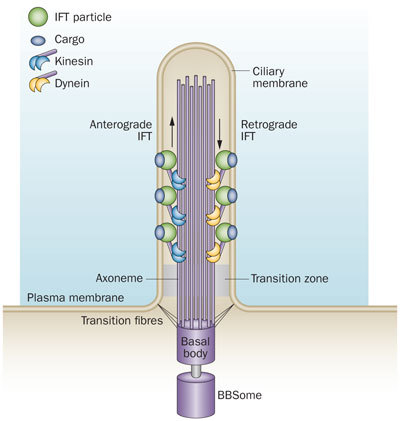
The primary cilium is a hair-like structure that protrudes from the cell surface. Microtubules form the core structure of the cilium, the axoneme. Protein cargo is transported up and down the cilium via anterograde and retrograde IFT mediated by kinesin and dynein motor proteins, respectively, which travel along the axoneme.
Primary Cilium a Cell’s Antenna or Its Brain Nrneur10


the intra-flagella transport system IFT in the PI was observed and connected to polycystic kidney disease. It was observed that the microtubule structure, called axoneme, grows from the base of the primary cilium, called the basal body. When the cilium is being built, vesicles transport protein pieces to the growing cilium from the base.

Recently, the complexity of the transport system has been described. A special protein attaches and drags proteins through the sea of phospholipids in the membrane, and pulls them into the primary cilium . In this way the primary cilium becomes the communication hub of the cell. Already a number of critical signaling cascades have been shown to live in the primary cilium. This includes the critical hedgehog and retinal signaling pathways.


Primary Cilium a Cell’s Antenna or Its Brain F1_lar10


The transport system uses motors that travel along microtubules to get the important material to the tip of the primary cilium from the base. Special motors are built at the base of the primary cilium and they pull many different types of material into the primary cilium—receptor proteins and building blocks for microtubules.Once at the tip of the PI, the motors deposit the cargo. At the tip Kinesian color the motor is altered and becomes a different machine to bring signaling material down the PI to the base. At the based messages are created and sent to the nucleus.

Once at the base of the primary cilium, the motor rearranges itself and becomes the train that drives cargo up into the primary cilium. The train that pulls this material to the tip of the primary cilium is made of at least four motors, one type active at a time. These motors are not just motors; they also interact with the membrane to regulate other functions including sensing extracellular situations and influencing decisions during fetal development. These motors are also able to connect through the membrane to objects outside of the cell to stimulate different types of cell movement. In this situation, the motor is anchored to a particular spot, but the entire cell moves when the motor is turned on.

This very complex motor system is critical for the elaborate function of the primary cilium by transporting all receptors and signaling materials that are used for the antenna function.

Defects in these motors in the eye can cause blindness. In the cells of the eye, the tip of the PI is very large bulb and houses the sensors that respond to light but still has a narrow area connecting with the large cell body. All of the light signals have to go through this narrow tube and a defect leads to blindness. Proteins used for sensing light are fragile and very active and many are imperfect. The cell is very dependent on the transport system of the PI to continually restock the proteins. Retinitis pigmentosa is one of the many diseases (ciliopathies) related to defects in this transport system.

T Cell Cilium
Until recently, it was not obvious that lymphocytes have PIs. Now, it has been shown that when T cells form critical synapses with other cells—other T cells for activation, dendritic cells to receive antigens, other immune cells for regulation, and targeted cells for destruction—it is actually a PI variant in the T cell.

The base of the cilium consists of a complex microtubule structure called the microtubule-organizing center, or MTOC (also called a centrosome). This is where microtubules are built. This MTOC becomes the focus of the synapses T cells form with other cells for these critical functions. The same PI transport system is used to transport vesicles during these close encounters with other cells. Vesicles are transported by the cilium transport system to the membrane to kill the targeted cell. The transport system brings special receptors that are recycled during T cell activation. T cells, like PIs, also, use the hedgehog signaling process during activation.

Autophagy and PI
Another critical function in all cells is the pathway of growth and the opposite, cellular self-destruction. The autophagy process is a critical quality control for communities of cells, such as organs. Neurodegenerative disease and cancer are highly connected to cascades that lead to runaway growth at one end and cell self-destruction on the other. 

Signaling from the primary cilium, such as using the Hedgehog pathway, appears to induce autophagy. Autophagy and PIs both utilize the same set of complex molecules. It has been found recently that the PI is necessary for stimulation of autophagy. The autophagy machinery is at the base of the PI. The PI regulates autophagy and the autophagy proteins direct the receptors in the PI. All critical sensory and communicating functions are connected with both the PI and autophagy mechanism.

Is the Primary Cilium a Cell’s Antenna or Its Brain?
the primary cilium is the brain of a cell, then it raises the question as to how the critical microtubule structures relate to this. The Penrose-Hammerof theory of the biological basis of mind is based on quantum properties of microtubules. In this theory, unique quantum computing processes occur in the exact unusual structure of microtubules. Currently, this theory is unable to be proven but does provide a speculation as to how the all-important mitotic spindle and the primary cilium might serve as a brain for the cell.

Previous posts have described intelligence in microbes. Microbes swim, communicate, find food, and perform group actions without synapses. All of these sensing and movement behaviors involve microtubules. In the human neuronal synapse, neuroplasticity (  refers to changes in neural pathways and synapses due to changes in behavior, environment, neural processes, thinking, and emotions - as well as to changes resulting from bodily injury )  depends upon the constantly changing microtubule structures. Just like the primary cilium and the mitotic spindle, purposeful complex behavior at many levels appears to involve the microtubules. The extremely complex mitotic spindle in cell division includes a small sac of the PI material. Is this sac passing on specific information encoded in the microtubule structure for the next generation?

With the primary cilium at the center of cellular sensation, communication, movement, cell division, autophagy and decision making, it is clearly a hub of purposeful behavior in all cells including the neuron. Is it possible that the primary cilium is the brain of the cell?

1) http://jonlieffmd.com/blog/is-the-primary-cilium-a-cells-antenna-or-its-brain#sthash.VkxokOmV.dpuf
2) http://jcs.biologists.org/content/123/4/499


Last edited by Admin on Sat May 09, 2020 5:12 pm; edited 13 times in total

https://reasonandscience.catsboard.com

2Primary Cilium a Cell’s Antenna or Its Brain Empty cilia Tue Jul 14, 2015 3:13 pm

Otangelo


Admin

cilia

Primary Cilium a Cell’s Antenna or Its Brain Centri10


In the primary cilium (a) of renal epithelial cells (b), 'cargo' proteins are trafficked along the microtubule tracks from the Golgi apparatus stack to the tip of the cilia using the motor protein kinesin II and back down using the cytoplasmic dynein 1b (not shown) (c). In an analogous fashion, approximately 109 molecules of the visual pigment rhodopsin are transferred up and down the connecting cilia in the human retina every day. IFT, intraflagellar transport.

Primary Cilium a Cell’s Antenna or Its Brain Centri11

Cilia are responsible for cell communication and play a key role in the receptor cells of sensory systems. For example, they are essential for odor detection in the nose and light reception in the eye. Because cilia are such a key element of cells, defects in genes that are involved in cilia development or function can cause complicated syndromes involving multiple organs and tissues.

Bardet-Biedl and Joubert syndromes are examples of ciliopathies with symptoms that include deafness, kidney disease, and degeneration of the retina. Meckel syndrome is a ciliopathy so dangerous babies with the genetic defect rarely make it to term.

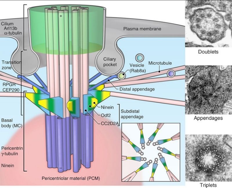
On individual cells, cilia grow from the basal body, a circular dent on the outer membrane acting as a platform. Supporting structures called distal and subdistal appendages, which are like the flying buttresses supporting Notre Dame Cathedral, anchor the platform in the basal body, priming it for the growth of cilia. Once anchored, the structures that form the cilium begin to extend from the site. Inside are a variety of proteins essential to maintain the cilium. Cc2d2a is believed to make a structural protein needed for cilia growth, but its precise functions have been unclear.

https://reasonandscience.catsboard.com

3Primary Cilium a Cell’s Antenna or Its Brain Empty Can You Feel The Heat? Your Cilia Can Tue Jul 14, 2015 6:55 pm

Otangelo


Admin

that several molecular machines within cells (such as the cilium and intraflagellar transport system, and the require the coordinated interaction of multiple protein parts in order to
maintain their function.

Can You Feel The Heat? Your Cilia Can 1

https://reasonandscience.catsboard.com/t2089-primary-cilium-a-cells-antenna-or-its-brain#3659

Cilia present in sensory nerve cells play an important role in our ability to sense touch and heat.

Johns Hopkins researchers and colleagues have found a previously unrecognized role for tiny hair-like cell structures known as cilia: They help form our sense of touch.

cilia are important for many biological processes, including three of our five senses: vision, hearing, and smell (ciliopathies are often characterized by loss or deficiency in these senses). “That leaves two unexplored possibilities,” says Katsanis. “Taste and touch; we tried touch.”

Johns Hopkins researchers say they have figured out how human and all animal cells tune in to a key signal, one that literally transmits the instructions that shape their final bodies.  It turns out the cells assemble their own little radio antenna on their surfaces to help them relay the proper signal to the developmental proteins “listening” on the inside of the cell.     The transmitters are primary cilia, relatively rigid, hairlike “tails” that respond to specialized signals from a host of proteins, including a key family of proteins known as Wnts.  The Wnts in turn trigger a cascade of shape-making decisions that guide cells to take specific shapes, like curved eyelid cells or vibrating hair cells in the ear, and even make sure that arms and legs emerge at the right spots.

The case for Intelligent Design gets stronger with  new findings like this. And the cilia is truly a mind blowing example.   Imagine: a radio antenna on each cell, signalling the inside world about the outside world.  Most signal-relay stations we know about were intelligently designed.     Signal without recognition is meaningless.  Communication implies a signalling convention (a “coming together” or agreement in advance) that a given signal means or represents something: e.g., that S-O-S means “Send Help!” or, in this case, that Wnt proteins mean “put this arm here.”  The transmitter and receiver can be made of non-sentient materials, but the functional purpose of the system always comes from a mind.  The mind uses the material substances to perform an algorithm that is not itself a product of the materials or the blind forces acting on them.  Thus the analogy in the press release: cilia are just like radio antennas.  Antennas may be composed of mindless matter, but they are marks of a mind behind the intelligent design. 3

Cilia are composed of multiple interacting parts, all of which must be present for the cilium to work. A single cilium is made up of some 600 protein pieces—more than many other cellular structures. 2  Removal or damage to a single part destroys the cilium and results in serious disease to the organism

1) http://www.hopkinsmedicine.org/news/media/releases/can_you_feel_the_heat_your_cilia_can
2) http://www.livescience.com/21211-cilia-biology-brooms-nigms.html
3) http://crev.info/2007/10/cilia_are_antennas_for_human_senses_and_development/


Last edited by Admin on Tue Jan 29, 2019 3:08 pm; edited 8 times in total

https://reasonandscience.catsboard.com

Otangelo


Admin

Taking on Behe's Challenge: Evolve Me a Cilium 1

In Darwin's Black Box (1996) and again in The Edge of Evolution (2007), Michael Behe challenged the scientific community to explain irreducibly complex molecular machines, like the cilium and bacterial flagellum, in Darwinian terms. The eukaryotic cilium, especially, represented "irreducible complexity squared," he said, with further research showing a complex transport system, Intraflagellar Transport (IFT), acting like a system of "freight cars" at a "construction site," moving cargo up and down the cilium with forward and reverse motors. His first literature search in 1996 showed "only a few attempts" to Darwinize the cilium. By 2007, nothing had changed:

An updated search of the science journals, where experts in the field publish their work, again shows no serious progress on a Darwinian explanation for the ultracomplex cilium. Despite the amazing advance of molecular biology as a whole, despite the sequencing of hundreds of entire genomes and other leaps in knowledge, despite the provocation of Darwin's Black Box itself, in the more than ten years since I pointed it out the situation concerning missing Darwinian explanations for the evolution of the cilium is utterly unchanged. (Behe, The Edge of Evolution, p. 95)

What you find in the PNAS paper is basically this. They selected 21 of the 33 protein parts from the three IFT modules, "based on homology relationships," and did a sequence search. Whoa, hold on there! That's assuming what needs to be proved. (More on homology in a bit.)

This classification was based on sequence similarity of IFT subunits to the COPI-α and-β′ subunits, further supported by secondary structure predictions. However, a full phylogenetic reconstruction and structural analysis of the IFT complex has not been performed.

They weren't about to perform one, either, in this paper at least. To make the problem more manageable (instead of having to explain a whole working cilium), they pared the data set down even more. When some of the domains "prevented unambiguous alignment" of sequences, they ignored all but two subunits for comparing. From those, they searched for a sequence common between COPI and IFT, finding one about 150 amino acid units long that "aligned consistently without the need to insert long gaps into the alignment." How's that for cherry picking? In other words, they only examined data that already fit their evolutionary hypothesis. What about the other 12 proteins that do not share sequence similarity with COPI? The flippancy with which they dismissed potential contrary evidence is breathtaking:
The remaining IFT subunits (Fig.1A, white) do not share any detectable sequence relationships with each other, or with any other proteins. Hence, as they do not contain any phylogenetic information on the origin of the IFT complex, they will not be further discussed.

Translation: since the other subunits do not fit our story line, we will ignore them. Arguably the most important question is, how did those other unique proteins, with no sequence relationships with any other proteins, "emerge" by a Darwinian process?

In Signature in the Cell, Stephen Meyer cited Doug Axe's work on the probability of getting one protein by chance. "The odds of getting even one functional protein of modest length (150 amino acids) by chance from a prebiotic soup," he wrote, "is no better than 1 chance in 10^164 " (Meyer, p. 212). That staggering improbability is so inconceivably low, it would never happen in the entire universe. It doesn't matter if you're trying to get a protein to "emerge" in a prebiotic soup or a Darwinian cell: unguided processes are unguided processes. Evolutionists cannot invoke purpose, goal, or natural law, as Meyer explained. The unique proteins defy evolution from the starting gate. No wonder van Dam and team say, "they will not be further discussed."

Tricks of the Trade

The paper's thesis boils down to the old homology argument. Jonathan Wells discussed this at length in Icons of Evolution (2000). It's a fallacy of circular reasoning: "a vicious circle: Common ancestry demonstrates homology which demonstrates common ancestry" (Wells, p. 63). Evolutionists pick similarities that fit their evolutionary story and call them "homologous," but reject similarities that don't fit the story, calling them "analogous." In their Supporting Information file, we find van Dam et al. using that old Darwinian trick, calling non-homologous parts "convergent":

The origin of BBS4 and BBS8 from an ancestral e-IFT subunit argues against an independent origin of the BBSome and IFT as coatomer-like complexes, and argues instead for convergent evolution of the structures of the BBS1, -2, -7, and -9 subunits to resemble the coatomer β-propeller structure.

In other words, those proteins are analogous, not homologous. As if that helps. Now, they have to account for four independent origins converging on the same structures. Sorry; they used up their probabilistic resources long ago, trying to gloss over the origin of 12 unique IFT proteins. There aren't any left for this tale.
Another trick is to invoke language like "proto-IFT complex" and "protocoatomer" that embed evolutionary assumptions in their terminology. Where did IFT come from? From proto-IFT, of course. With tricks like that, Darwinism can't lose.

Progress Through Loss

The most illuminating section of their paper concerns IFT loss, not gain. They discuss how seed plants and most fungi lack cilia entirely, but some fungi and protists get by with only part of the IFT set. It appears, for instance, that the BBSome, which packages proteins for the freight cars, is not essential. A fungus, a moss, a lycophyte and some parasitic protists have cilia without it. (Note: "flagellum" is often used as a synonym for "eukaryotic cilium" but is unrelated to the bacterial flagellum.)

This pattern of BBSome loss thus appears to precede the loss of the cilium, and may indicate a reduced role for cilia in BBSome-negative lineages before the cilium is lost entirely. The existence of multiple species with functional flagella, but lacking the BBSome, suggests that the BBSome is a nonessential component of IFT....
Disrupting the expression of BBSome subunits has profound effects on the other IFT complexes. BBSome dysfunction results in instability and incorrect assembly of the IFT complex, resulting in dissociated IFT-A and IFT-B complexes. This suggests that there is functional interaction between the BBSome and IFT-A and B. However, from our analysis, it appears that the removal of the BBSome can be tolerated in some species, indicating that this functional interaction must be nonessential. It will be interesting to understand how species compensate for loss of the BBSome, and what evolutionary steps are required to facilitate that loss.

They point out that Bardet-Biedl Syndrome, a severe genetic disease in humans that leads to many problems as diverse as obesity, night blindness, kidney failure, and dental crowding, is the result of BBSome malfunction. Sufferers of this "ciliopathy" can live and produce viable offspring. That doesn't mean they are making evolutionary progress.

For the other organisms that "tolerate" BBSome loss, the researchers ask a fair question: how do they compensate? Would they be better off with the BBSome? That might be a good project for ID research. Trypanosomes even get by without IFT-A, leading them to postulate that "IFT-B could be viewed as the most critical subcomplex, as it is the last to be retained, and hence its presence essentially dictates if a cilium is present." Such observations might provoke another ID project: what is the extent of a cilium's irreducible complexity? The Darwinian team errs, though, by assuming "evolutionary steps are required to facilitate that loss" of the BBSome or IFT-A. That's nonsense; requirements are written by intelligent designers.

Concession Stand

The authors make admissions that argue against their hypothesis. For instance, they concede that conservation (not evolution) is so pervasive, it's "remarkable":

The orthologous IFT and BBSome subunit sequences are well conserved, despite the large evolutionary distances between them, and despite the variable presence of the subunits per species.... The high similarity between the predicted secondary structure elements suggests that the orthologous proteins in the IFT complex are structurally conserved to a remarkable level.
They did find sequence variations. They did outline possible patterns of loss in various lineages. But those are not the big issue. Mutations or substitutions that do not affect the structural operation of IFT might be tolerated. But clearly, an intact cilium with all the components working is optimal. Some boaters can get by with oars or hand paddling when the outboard motor is broken or sputtering. How the motor got there is the big question. For that, van Dam et al. make a huge concession that undermines their whole Darwinian tale:
All the subunits reported for the human IFT complexes are conserved throughout the eukaryotic lineage. Therefore, IFT-A, IFT-B, and BBSome were likely present in the last eukaryotic common ancestor (LECA) and comprised all currently known IFT subunits from human and Chlamydomonas reinhardtii, in agreement with earlier observations.

Let's get this story straight. Once upon a time, a complete, working cilium with all the correct components, and with all the right genetic assembly instructions, just "emerged" in some mythical common ancestor. Maybe evolution "repurposed" some protein-coating genes after a mistake duplicated them. However it happened, all those parts were "conserved" the rest of the way, from simple one-celled Chlammy to complex trillion-celled Sammy. During evolution, some branches of the eukaryotic tree lost some parts, but the ones that didn't die are getting along OK.


This proves Darwinism can explain "such a complex and highly organized organelle as the cilium." Take that, Dr. Behe!

Final Score


We can predict Behe's yawning response to the new "Darwinian conjecture" in this paper: "in the more than [now 17] years since I pointed it out the situation concerning missing Darwinian explanations for the evolution of the cilium is utterly unchanged."
1) http://www.evolutionnews.org/2013/04/taking_on_behes071121.html

Nick Matzkes response :

Of cilia and silliness (more on Behe)

http://pandasthumb.org/archives/2007/06/of-cilia-and-si.html


Last edited by Admin on Sun Jul 26, 2015 4:36 pm; edited 2 times in total

https://reasonandscience.catsboard.com

Otangelo


Admin

The little hair-like projections on cells, called cilia, have more functions than previously believed.  A press release from Johns Hopkins University said that researchers found cilia are important for the sense of touch – particularly, for heat sensation.  In fact, cilia are implicated in at least three of the five traditional senses.     The article explained that some people thought to have psychological problems may actually be victims of “ciliopathy” or defects in cilia formation.  Dr. Nico Katsanis said, “People with ciliopathies are often thought to have mental retardation or autism because they appear ‘slow’.  Now it appears that many aspects of their mental capacity may be just fine, they are just slow because they can’t sense things as well as other individuals.”     Another press release from Johns Hopkins earlier in the month reported that Katsanis’ team found that cilia act like little radio antennas that control the development of the body: - See more at: http://crev.info/2007/10/cilia_are_antennas_for_human_senses_and_development/#sthash.sLkzlmXB.dpuf

https://reasonandscience.catsboard.com

Otangelo


Admin

Motile Cilia and Flagella Are Built from Microtubules and Dyneins 1

Just as myofibrils are highly specialized and efficient motility machines built from
actin and myosin filaments, cilia and flagella are highly specialized and efficient
motility structures built from microtubules and dynein. Both cilia and flagella are
hairlike cell appendages that have a bundle of microtubules at their core. Flagella
are found on sperm and many protozoa. By their undulating motion, they enable
the cells to which they are attached to swim through liquid media. Cilia are organized
in a similar fashion, but they beat with a whiplike motion that resembles the
breaststroke in swimming. Ciliary beating can either propel single cells through a
fluid (as in the swimming of the protozoan Paramecium) or can move fluid over
the surface of a group of cells in a tissue. In the human body, huge numbers of cilia
(109/cm2 or more) line our respiratory tract, sweeping layers of mucus, trapped
particles of dust, and bacteria up to the mouth where they are swallowed and ultimately
eliminated. Likewise, cilia along the oviduct help to sweep eggs toward the
uterus. The movement of a cilium or a flagellum is produced by the bending of its
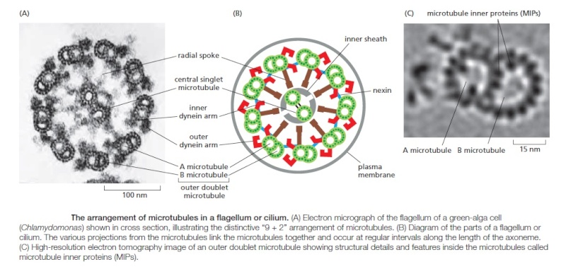
core, which is called the axoneme. The axoneme is composed of microtubules
and their associated proteins, arranged in a distinctive and regular pattern. Nine
special doublet microtubules (comprising one complete and one partial microtubule
fused together so that they share a common tubule wall) are arranged in
a ring around a pair of single microtubules (Figure 16–63). Almost all forms of
motile eukaryotic flagella and cilia (from protozoans to humans) have this characteristic
arrangement. The microtubules extend continuously for the length of
the axoneme, which can be 10–200 μm. At regular positions along the length of the
microtubules, accessory proteins cross-link the microtubules together.

Primary Cilium a Cell’s Antenna or Its Brain Sfsfsd15

Primary Cilium a Cell’s Antenna or Its Brain Ssfsds12

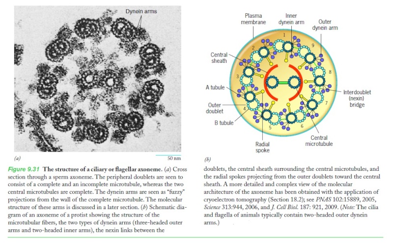
Molecules of axonemal dynein form bridges between the neighboring doublet
microtubules around the circumference of the axoneme

Ciliary dynein. Ciliary
(axonemal) dynein is a large protein
assembly (nearly 2 million daltons)
composed of 9–12 polypeptide chains, the
largest of which is the heavy chain of more
than 500,000 daltons. (A) The heavy chains
form the major portion of the globular
head and stem domains, and many of the
smaller chains are clustered around the
base of the stem. There are two heads
in the outer dynein in metazoans (shown
here), but three heads in protozoa, each
formed from their own heavy chain. The
tail of the molecule binds tightly to an
A microtubule, while the large globular
heads have an ATP-dependent binding
site for a B microtubule (see picture above).
When the heads hydrolyze their bound ATP,
they move toward the minus end of the
B microtubule, thereby producing a sliding
force between the adjacent microtubule
doublets in a cilium or flagellum (see
Figure 16–59). (B) Freeze-etch electron
micrograph of a cilium showing the dynein
arms projecting at regular intervals from
the doublet microtubules. (B, courtesy of
John Heuser.)


When the motor domain of this dynein is activated, the dynein molecules attached to
one microtubule doublet (see Figure 16–59) attempt to walk along the adjacent
microtubule doublet, tending to force the adjacent doublets to slide relative to
one another, much as actin thin filaments slide during muscle contraction. However,
the presence of other links between the microtubule doublets prevents this
sliding, and the dynein force is instead converted into a bending motion

Primary Cilium a Cell’s Antenna or Its Brain Ssfsds10


In humans, hereditary defects in axonemal dynein cause a condition called
primary ciliary dyskinesia or Kartagener’s syndrome. This syndrome is characterized
by inversion of the normal asymmetry of internal organs (sinus inversus) due
to disruption of fluid flow in the developing embryo, male sterility due to immotile
sperm, and a high susceptibility to lung infections due to paralyzed cilia being
unable to clear the respiratory tract of debris and bacteria.
Bacteria also swim using cell-surface structures called flagella, but these do
not contain microtubules or dynein and do not wave or beat. Instead, bacterial
flagella are long, rigid helical filaments, made up of repeating subunits of the protein
flagellin. The flagella rotate like propellers, driven by a special rotary motor
embedded in the bacterial cell wall. The use of the same name to denote these two
very different types of swimming apparatus is an unfortunate historical accident.

Primary Cilium a Cell’s Antenna or Its Brain Ssfs10

1) molecular biology of the cell, 6th edition, pg.941

https://reasonandscience.catsboard.com

Sponsored content


Back to top  Message [Page 1 of 1]

Permissions in this forum:
You cannot reply to topics in this forum