


Microglia the Most Intelligent Brain Cells 1
http://reasonandscience.heavenforum.org/t2088-microglia-the-most-intelligent-brain-cells
As both unique immune cells and unique brain cells that constantly change shape and have numerous different functions, are microglia the most intelligent brain cells? Microglia travel independently, not attached to any structure, constantly circling a territory with extended arms repeatedly tapping all axons, dendrites and synapses looking to detect any suboptimal functioning. Their constant surveillance of the brain protects against any microbe invaders, demyelination, trauma and cancerous or defective cells.
But, this is just the beginning of their amazing functions. They communicate with complex wireless signaling to neurons and astrocytes, determining how many brains cells are needed and when to eliminate a synapse—eating the defective or unused synapses. They determine MG green synapses blue and redthe rate of production of new cells from stem cells and the rate of total brain growth in the fetus—eating excessive stem cells. They morph into many different shapes and types of immune cells to fight a multitude of different invaders. How can we not say that these are intelligent cells?
Microglia have as many functions, if not more, than T cells. The independent intelligent T cells travel through the entire body, finding invaders, trauma and cancer. They are critical for cognitive brain function sending signals to brain cells.
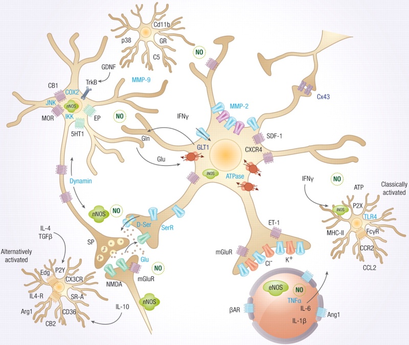
Microglia are the resident immune cells in the brain, but do so much more than just immune surveillance. They help establish neuronal networks in the fetus. In the adult, microglia are actively involved in pruning neurons that are established and nourished by astrocytes. They produce signals that nourish and stimulate neuronal growth and axon migration. The neuron is the master of information processing and transfer. The astrocyte provides nourishment and blood flow to the neurons and participates in the information transfer with its own system; astrocytes, also, establish a protective structure for the neuron and control blood flow to the region. But, it is the microglia who travel throughout the brain participating in surveillance, stimulation, cleanup, and maintenance tasks while communicating with all other cells.
Where Do Microglia Come From?
Because they are so unique even among immune cells, there has been a question about their lineage. Recently, it was discovered that they come from a different source in the fetus than the neuron, astrocyte, and oligodendrocyte. They are, in fact, a close cousin of the macrophage, a white blood cell whose major function is gobbling up invaders.
Microglia arise from mesodermal tissue in the yolk sac on day 9 in humans
and then travel through the blood to the brain where they stay. In the brain, microglia replenish their own stock—making fewer new cells when they are quietly circling and tapping synapses, and more when they are fighting invaders or debris. They are a stable independent community of cells patrolling their individual territory.
Ordinary macrophages originate from the bone marrow and travel through the blood to different regions of the body as needed. Strangely, there exist special subpopulations of microglia that are, also, from bone marrow. These macrophage cells live in the perivascular region. If a large number of microglia are eliminated in a battle with microbes, then macrophages from outside the brain take their place. These substitute cells gradually, over a period of 8 months, accommodate to the brain, but never learn all of the functions of microglia.
Microglia are much more highly regulated than ordinary macrophages, with specific territories and very exact immune responses like a T cell.
Microglia in Many Shapes and Roles
Microglia are as common as neurons in the brain. It took many years to figure out that a wide variety of different shaped cells in the brain were actually all the same. Microglia multiple cells CROPhave many different shapes for different functions and therefore have been difficult to study. No one realized that the large blob-like macrophages were the same as star-like cells tapping synapses. Microglia are found near any inflammation, trauma, autoimmune disease, and cancer. Under normal circumstances they are present evenly throughout the brain.
From a spider like resting state, microglia become a big round blob during attacks on microbes as a macrophage. Microglia are very sensitive, part of the immune system and Ramified the brain. With the slightest injury to a nerve or a microbe, microglia suddenly become active and often change shape.
Before modern cellular imaging, it appeared that microglia were very quiet with many long arms. Recently, they have been observed moving steadily like amoeba in their particular territory with many very long arms sticking through the astrocyte and neuronal matrix, wrapping around synapses, axons and dendrites and constantly while moving studying the terrain.
The arms appear to grow, shrink and then regrow. Microglia are, by far, the most active cells in the brain.
Microglia are the most individualistic cell in the brain and function alone, although in constant wireless communication with both immune and brain cells, like the T cell. They have specific territory and don’t tread on each other’s region of surveillance unless there is danger when they swing into action and can go anywhere.
Microglia Respond to Many Different Threats
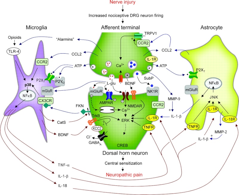
Microglia respond to many different stimuli. Surprisingly, they respond to distant threats in the nervous system as well as local. The response can occur within minutes and last for days or longer and can occur in many different ways. They are the only cells that are not connected in the brain and respond to a wide variety of wireless cytokine signals.
When damage occurs in the brain from trauma or infection, microglia are the first responders starting to clean up the debris. They were first discovered near injury, looking like amoebae crawling to the injury site and eating dead microbes and neurons along the way—making room for healing.
Microglia respond to internal and external threats to the entire body. These responses are sometimes related to specific brain regions, but sometimes not. Stress increases microglia in the brain’s stress regions . They are general protectors of the spinal cord and brain. Those that respond to pain are not just inflammatory, but, appear to be involved in higher level coordination of defense with many other types of cells.
Microglia don’t usually use the antigen process. But, when they respond as an immune cell they do use MHC antigens as do lymphocytes. Human microglia has one very specific gene that sends a signal, which suppresses inflammatory reactions in the brain.
Constant Wireless Communication – Signaling
Like T cells, microglia grow a wide range of receptors that can pick up a large number of cytokine signals from other immune and brain cells that are circulating in blood and tissue. Humans, likewise, build their own types of receptors—radios, TV, cellular phones, and laptops—that receive the many wireless signals floating in the air. Both microglia and humans also return signals—microglia use cytokines and neurotransmitters, while humans send radio waves, text and voice messages and emails.
Microglia, using receptors and signals, are in constant communication with neurons, astrocytes and immune cells. Microglia receive neuronal signals of neurotransmitters and send signals mediating neuronal activity. Neurons send signals that stimulate microglia inflammatory behavior and synapse and apoptosis pruning Signalling behavior. These microglia behaviors are gradually trained in response to their relationship with neurons.
Signals can promote or fight inflammation and can use MHC receptors when necessary. Microglia are the only cell in the brain that has a complement receptor.
Many very specific signals are secreted in specific disease states such as pain, autoimmune disease, trauma, prion disease, Alzheimer’s and other degenerative brain disease. Signals include cytokines, neurotransmitters, chemokines (specific cytokines that tell cells where to travel) and proteases that alter the extracellular matrix.
Microglia Care for Neurons
With special dyes, microglia have been observed constantly moving in a specific circular region of about 80 micrometers every several hours with their long arms tapping and examining every part of the region.
One of their roles is to find synapses marked by complement that need to be eliminated. But, their role in eating defective or poorly used synapses is, also, regulated by a back and forth signaling communication with neurons and astrocytes. Healthy neurons secrete signals to tell the microglia not to attack the synapse, while complement can be attracted by the signals from the neurons themselves. Astrocytes send signals to draw microglia to the pre and post synaptic cells of specific synapses. Somehow, microglia know how many new neurons are needed and stop stem cells from making too many. astrocytes are active, along with microglia, in pruning some of the unused synapses.
During normal function, the microglia are active in stimulating new neurons and new neuronal connections and providing guidance for long travelling cells and axons. When neurons are overgrown from lack of use and would create problems if not pruned, the microglia are the cells that eat back the unnecessary growth. When defective, neurons can trigger apoptosis and kill themselves; it is the microglia that clean up the debris.
Microglia are involved in memory production. They produce nutrients for neurons that stimulate new dendrite and axon budding. They are actively involved in signals and nutrients for the developing synapse along with astrocytes. One of the most active areas for microglia is the hippocampus the center of memory.
Microglia Necessary to Build the Fetal Brain
When the fetal brain rapidly grows at the fantastic rate of 250,000 new neurons each second during the last month of pregnancy, the connections that are not used by experience are eliminated by microglia. During childhood and adolescence many connections are made and, also, pruned by experience. It is microglia that mold the adult brain through this process.
Microglia control the number of stem cells that determine how large the cortex grows in the fetal brain. They hover near the neural stem cells in the developing brain and when activated there is less stem cell activity. They are the inhibitory cells of the growing brain. There are specific regions where neurons grow in the fetus as the brain is built. These proliferative zones are filled with microglia. As the brain is built, the number of microglia changes and they evenly space themselves in their individual territories throughout the entire brain.
Microglia Critical for Neuroplasticity
Microglia are critical for neuroplasticity and for the development of synapses. Many experiments show the dramatic rewiring of axon/dendrite connections during any changing circumstances. One experiment with mice in light and dark conditions showed that during several days of dark dendritic spines connecting to axons shrivel and then grow again when there is stimulation with light.
When the dendrites are shrinking, microglia are present to eliminate the unnecessary ones. When the dendrites are growing, the microglia move away and continue their surveillance. The microglia are removing the unused, weak and useless connections to allow the new connections to be vigorous.
Microglia, also, secrete many special nerve factors that stimulate more neurons, survival of neurons, and more connections. They also stimulate myelination as well as blood growth. Eating of synapses can actually stimulate regeneration of neurons.
Are Microglia the Most Intelligent Brain Cells
The remarkable microglia are involved in scavenging, phagocytosis, release of toxins and complex signals; they present antigens, prune synapses, control stem cells and brain circuitry, fight microbes and cancer, repair trauma, and respond to autoimmune diseases. Microglia have a vast number of receptors and signals that stay in constant communication with astrocytes, neurons, and all the immune cells.
Like the T cell, how can we not think that microglia are very intelligent cells?
One of the great stumbling blocks for Darwin is explaining how a multitude of factors simultaneously coalesce to form a unified, functioning system. 4 The human brain could not have evolved as a result of the addition of one factor at a time. Its unity and phantasmagorical complexity defies any explanation that relies on pure chance. It would be an underestimation of the first magnitude to say that today's neurophysiologists know more about the structure and workings of the brain than did Darwin and his associates.
Scientists in the field of brain research now inform us that a single human brain contains more molecular-scale switches than all the computers, routers and Internet connections on the entire planet! According to Stephen Smith, a professor of molecular and cellular physiology at the Stanford University School of Medicine, the brain's complexity is staggering, beyond anything his team of researchers had ever imagined, almost to the point of being beyond belief. In the cerebral cortex alone, each neuron has between 1,000 to 10,000 synapses that result, roughly, in a total of 125 trillion synapses, which is about how many stars fill 1,500 Milky Way galaxies!
A single synapse may contain 1,000 molecular-scale switches. A synapse, simply stated, is the place where a nerve impulse passes from one nerve cell to another.
Phantasmagorical as this level of unified complexity is, it places us merely at the doorway of the brain's even deeper mind-boggling organization. Glial cells in the brain assist in neuron speed. These cells outnumber neurons 10 times over, with 860 billion cells. All of this activity is monitored by microglia cells that not only clean up damaged cells but also prune dendrites, forming part of the learning process. The cortex alone contains 100,000 miles of myelin-covered, insulated nerve fibers.
The process of mapping the brain would indeed be time-consuming. It would entail identifying every synaptic neuron. If it took a mere second to identify each neuron, it would require four billion years to complete the project. What makes all of this even more astonishing is the fact that the brain is 60% fat. In addition, a person's brain, in all its unified complexity, evolved from a single, microscopic cell!
1) http://jonlieffmd.com/blog/are-microglia-the-most-intelligent-brain-cells
2) http://discovery.lifemapsc.com/in-vivo-development/yolk-sac
3) http://m.harunyahya.com/tr/Buku/989/The-Miracle-Of-The-Immune-System/chapter/3768/Cells-on-duty-in-the-system
4) http://www.evolutionnews.org/2015/02/national_cathol093551.html
http://reasonandscience.heavenforum.org/t2088-microglia-the-most-intelligent-brain-cells
As both unique immune cells and unique brain cells that constantly change shape and have numerous different functions, are microglia the most intelligent brain cells? Microglia travel independently, not attached to any structure, constantly circling a territory with extended arms repeatedly tapping all axons, dendrites and synapses looking to detect any suboptimal functioning. Their constant surveillance of the brain protects against any microbe invaders, demyelination, trauma and cancerous or defective cells.
But, this is just the beginning of their amazing functions. They communicate with complex wireless signaling to neurons and astrocytes, determining how many brains cells are needed and when to eliminate a synapse—eating the defective or unused synapses. They determine MG green synapses blue and redthe rate of production of new cells from stem cells and the rate of total brain growth in the fetus—eating excessive stem cells. They morph into many different shapes and types of immune cells to fight a multitude of different invaders. How can we not say that these are intelligent cells?
Microglia have as many functions, if not more, than T cells. The independent intelligent T cells travel through the entire body, finding invaders, trauma and cancer. They are critical for cognitive brain function sending signals to brain cells.
Microglia are the resident immune cells in the brain, but do so much more than just immune surveillance. They help establish neuronal networks in the fetus. In the adult, microglia are actively involved in pruning neurons that are established and nourished by astrocytes. They produce signals that nourish and stimulate neuronal growth and axon migration. The neuron is the master of information processing and transfer. The astrocyte provides nourishment and blood flow to the neurons and participates in the information transfer with its own system; astrocytes, also, establish a protective structure for the neuron and control blood flow to the region. But, it is the microglia who travel throughout the brain participating in surveillance, stimulation, cleanup, and maintenance tasks while communicating with all other cells.
Where Do Microglia Come From?
Because they are so unique even among immune cells, there has been a question about their lineage. Recently, it was discovered that they come from a different source in the fetus than the neuron, astrocyte, and oligodendrocyte. They are, in fact, a close cousin of the macrophage, a white blood cell whose major function is gobbling up invaders.
Microglia arise from mesodermal tissue in the yolk sac on day 9 in humans
Embryonic Development of the Yolk Sac: 2
The yolk sac initially appears as a transient primary yolk sac, thought to develop, in humans, from the hypoblast cells.
During the end of the second week of gestation (12-14 dpc), the lower half of the primary yolk sac is pinched off to form the definitive yolk sac, while a second wave of hypoblast endoderm cells (yolk sac endoderm) form the inner lining of the definitive yolk sac.Structure and Function of the Yolk Sac:
The yolk sac is situated on the ventral aspect of the embryo and is one of the three embryonic cavities (chorion,amnion and yolk sac) that appear as of day 8 of human development.
and then travel through the blood to the brain where they stay. In the brain, microglia replenish their own stock—making fewer new cells when they are quietly circling and tapping synapses, and more when they are fighting invaders or debris. They are a stable independent community of cells patrolling their individual territory.
Ordinary macrophages 3
Immobile police forces: These are immobile macrophages, which are situated in the gaps in various tissues. They perform phagocytosis on the micro-organisms from where they are, without moving. Phagocytosis in progress. The macrophage (yellow) while digesting the bacteria (blue).
If the invader antigens (foreign micro-organisms) are few enough for the present eater cells to deal with, they are destroyed with no extra alarm being given. But if the invader microbes are too great in number, the eater cells may fail to get them under control. Unable to digest all of them, they expand in size. When distended by the antigens, the cells burst, causing a liquid substance (pus) to overflow. This does not mean that the war is lost. So far, the eater cells have just met the microbes, which have still many tougher barriers to pass. The formation of pus activates the lymphocytes, which have been delivered from the bone marrow, the lymph nodes, and above all, the thymus. In a second wave of defence, the newly arriving defence cells attack everything they find around, including cell debris, available antigens, and even old white blood cells. These defence cells are the real eater cells, — the macrophages, a type of phagocyte.
The First Aid Forces: The Macrophages
When the war becomes intense, the macrophages swing into action. Macrophages operate in a specific manner exclusive to themselves. They do not become involved in a one-to-one combat like the antibodies. Unlike the antibodies, they do not work with a system similar to a bomb aimed at a single target. Just like a gun firing lead shot, or a bomb that can be aimed at many targets together, the macrophages can destroy a great number of enemies together, all at the same time.Like all other defence cells, the macrophages are also derived from the bone marrow. The macrophages, which have a very long life span, can live for months, and even years. Despite their small size (10-15 micrometers), they are highly crucial for human life. They possess the ability to absorb and digest big molecules in the cell through phagocytosis (ingestion). Their characteristic of ingestion makes them the scavengers of the defence system. They remove all materials that need to be cleaned up, such as micro-organisms, antigen-antibody complexes, and other substances similar in structure to an antigen. At the end of these processes, substances that would be qualified as antigens are digested, and thus pose no further threat to the organism.

Ordinary macrophages originate from the bone marrow and travel through the blood to different regions of the body as needed. Strangely, there exist special subpopulations of microglia that are, also, from bone marrow. These macrophage cells live in the perivascular region. If a large number of microglia are eliminated in a battle with microbes, then macrophages from outside the brain take their place. These substitute cells gradually, over a period of 8 months, accommodate to the brain, but never learn all of the functions of microglia.
Microglia are much more highly regulated than ordinary macrophages, with specific territories and very exact immune responses like a T cell.
Microglia in Many Shapes and Roles
Microglia are as common as neurons in the brain. It took many years to figure out that a wide variety of different shaped cells in the brain were actually all the same. Microglia multiple cells CROPhave many different shapes for different functions and therefore have been difficult to study. No one realized that the large blob-like macrophages were the same as star-like cells tapping synapses. Microglia are found near any inflammation, trauma, autoimmune disease, and cancer. Under normal circumstances they are present evenly throughout the brain.
From a spider like resting state, microglia become a big round blob during attacks on microbes as a macrophage. Microglia are very sensitive, part of the immune system and Ramified the brain. With the slightest injury to a nerve or a microbe, microglia suddenly become active and often change shape.
Before modern cellular imaging, it appeared that microglia were very quiet with many long arms. Recently, they have been observed moving steadily like amoeba in their particular territory with many very long arms sticking through the astrocyte and neuronal matrix, wrapping around synapses, axons and dendrites and constantly while moving studying the terrain.
The arms appear to grow, shrink and then regrow. Microglia are, by far, the most active cells in the brain.
Microglia are the most individualistic cell in the brain and function alone, although in constant wireless communication with both immune and brain cells, like the T cell. They have specific territory and don’t tread on each other’s region of surveillance unless there is danger when they swing into action and can go anywhere.
Microglia Respond to Many Different Threats
Microglia respond to many different stimuli. Surprisingly, they respond to distant threats in the nervous system as well as local. The response can occur within minutes and last for days or longer and can occur in many different ways. They are the only cells that are not connected in the brain and respond to a wide variety of wireless cytokine signals.
When damage occurs in the brain from trauma or infection, microglia are the first responders starting to clean up the debris. They were first discovered near injury, looking like amoebae crawling to the injury site and eating dead microbes and neurons along the way—making room for healing.
Microglia respond to internal and external threats to the entire body. These responses are sometimes related to specific brain regions, but sometimes not. Stress increases microglia in the brain’s stress regions . They are general protectors of the spinal cord and brain. Those that respond to pain are not just inflammatory, but, appear to be involved in higher level coordination of defense with many other types of cells.
Microglia don’t usually use the antigen process. But, when they respond as an immune cell they do use MHC antigens as do lymphocytes. Human microglia has one very specific gene that sends a signal, which suppresses inflammatory reactions in the brain.
Constant Wireless Communication – Signaling
Like T cells, microglia grow a wide range of receptors that can pick up a large number of cytokine signals from other immune and brain cells that are circulating in blood and tissue. Humans, likewise, build their own types of receptors—radios, TV, cellular phones, and laptops—that receive the many wireless signals floating in the air. Both microglia and humans also return signals—microglia use cytokines and neurotransmitters, while humans send radio waves, text and voice messages and emails.
Microglia, using receptors and signals, are in constant communication with neurons, astrocytes and immune cells. Microglia receive neuronal signals of neurotransmitters and send signals mediating neuronal activity. Neurons send signals that stimulate microglia inflammatory behavior and synapse and apoptosis pruning Signalling behavior. These microglia behaviors are gradually trained in response to their relationship with neurons.
Signals can promote or fight inflammation and can use MHC receptors when necessary. Microglia are the only cell in the brain that has a complement receptor.
Many very specific signals are secreted in specific disease states such as pain, autoimmune disease, trauma, prion disease, Alzheimer’s and other degenerative brain disease. Signals include cytokines, neurotransmitters, chemokines (specific cytokines that tell cells where to travel) and proteases that alter the extracellular matrix.
Microglia Care for Neurons
With special dyes, microglia have been observed constantly moving in a specific circular region of about 80 micrometers every several hours with their long arms tapping and examining every part of the region.
One of their roles is to find synapses marked by complement that need to be eliminated. But, their role in eating defective or poorly used synapses is, also, regulated by a back and forth signaling communication with neurons and astrocytes. Healthy neurons secrete signals to tell the microglia not to attack the synapse, while complement can be attracted by the signals from the neurons themselves. Astrocytes send signals to draw microglia to the pre and post synaptic cells of specific synapses. Somehow, microglia know how many new neurons are needed and stop stem cells from making too many. astrocytes are active, along with microglia, in pruning some of the unused synapses.
During normal function, the microglia are active in stimulating new neurons and new neuronal connections and providing guidance for long travelling cells and axons. When neurons are overgrown from lack of use and would create problems if not pruned, the microglia are the cells that eat back the unnecessary growth. When defective, neurons can trigger apoptosis and kill themselves; it is the microglia that clean up the debris.
Microglia are involved in memory production. They produce nutrients for neurons that stimulate new dendrite and axon budding. They are actively involved in signals and nutrients for the developing synapse along with astrocytes. One of the most active areas for microglia is the hippocampus the center of memory.
Microglia Necessary to Build the Fetal Brain
When the fetal brain rapidly grows at the fantastic rate of 250,000 new neurons each second during the last month of pregnancy, the connections that are not used by experience are eliminated by microglia. During childhood and adolescence many connections are made and, also, pruned by experience. It is microglia that mold the adult brain through this process.
Microglia control the number of stem cells that determine how large the cortex grows in the fetal brain. They hover near the neural stem cells in the developing brain and when activated there is less stem cell activity. They are the inhibitory cells of the growing brain. There are specific regions where neurons grow in the fetus as the brain is built. These proliferative zones are filled with microglia. As the brain is built, the number of microglia changes and they evenly space themselves in their individual territories throughout the entire brain.
Microglia Critical for Neuroplasticity
Microglia are critical for neuroplasticity and for the development of synapses. Many experiments show the dramatic rewiring of axon/dendrite connections during any changing circumstances. One experiment with mice in light and dark conditions showed that during several days of dark dendritic spines connecting to axons shrivel and then grow again when there is stimulation with light.
When the dendrites are shrinking, microglia are present to eliminate the unnecessary ones. When the dendrites are growing, the microglia move away and continue their surveillance. The microglia are removing the unused, weak and useless connections to allow the new connections to be vigorous.
Microglia, also, secrete many special nerve factors that stimulate more neurons, survival of neurons, and more connections. They also stimulate myelination as well as blood growth. Eating of synapses can actually stimulate regeneration of neurons.
Are Microglia the Most Intelligent Brain Cells
The remarkable microglia are involved in scavenging, phagocytosis, release of toxins and complex signals; they present antigens, prune synapses, control stem cells and brain circuitry, fight microbes and cancer, repair trauma, and respond to autoimmune diseases. Microglia have a vast number of receptors and signals that stay in constant communication with astrocytes, neurons, and all the immune cells.
Like the T cell, how can we not think that microglia are very intelligent cells?
One of the great stumbling blocks for Darwin is explaining how a multitude of factors simultaneously coalesce to form a unified, functioning system. 4 The human brain could not have evolved as a result of the addition of one factor at a time. Its unity and phantasmagorical complexity defies any explanation that relies on pure chance. It would be an underestimation of the first magnitude to say that today's neurophysiologists know more about the structure and workings of the brain than did Darwin and his associates.
Scientists in the field of brain research now inform us that a single human brain contains more molecular-scale switches than all the computers, routers and Internet connections on the entire planet! According to Stephen Smith, a professor of molecular and cellular physiology at the Stanford University School of Medicine, the brain's complexity is staggering, beyond anything his team of researchers had ever imagined, almost to the point of being beyond belief. In the cerebral cortex alone, each neuron has between 1,000 to 10,000 synapses that result, roughly, in a total of 125 trillion synapses, which is about how many stars fill 1,500 Milky Way galaxies!
A single synapse may contain 1,000 molecular-scale switches. A synapse, simply stated, is the place where a nerve impulse passes from one nerve cell to another.
Phantasmagorical as this level of unified complexity is, it places us merely at the doorway of the brain's even deeper mind-boggling organization. Glial cells in the brain assist in neuron speed. These cells outnumber neurons 10 times over, with 860 billion cells. All of this activity is monitored by microglia cells that not only clean up damaged cells but also prune dendrites, forming part of the learning process. The cortex alone contains 100,000 miles of myelin-covered, insulated nerve fibers.
The process of mapping the brain would indeed be time-consuming. It would entail identifying every synaptic neuron. If it took a mere second to identify each neuron, it would require four billion years to complete the project. What makes all of this even more astonishing is the fact that the brain is 60% fat. In addition, a person's brain, in all its unified complexity, evolved from a single, microscopic cell!
1) http://jonlieffmd.com/blog/are-microglia-the-most-intelligent-brain-cells
2) http://discovery.lifemapsc.com/in-vivo-development/yolk-sac
3) http://m.harunyahya.com/tr/Buku/989/The-Miracle-Of-The-Immune-System/chapter/3768/Cells-on-duty-in-the-system
4) http://www.evolutionnews.org/2015/02/national_cathol093551.html
Last edited by Admin on Fri Apr 28, 2017 6:23 am; edited 5 times in total