

NEURONS & SYNAPSES
HOW MANY NEURONS?
The vast majority of books and documentaries about the human brain cite the number of neurons to be 100 billion. While writing a previous book, I searched for the source of this claim and discovered that apparently none exists. It does not seem to be supported by evidence. The 100-billion-neuron claim seems to be nothing more than an agreed-upon guess that everyone keeps repeating.
Fortunately, a team of scientists decided to find out just how many neurons a typical human brain really does contain. They couldn’t just count them one by one, of course, because that would take centuries. So they liquefied brains in blenders—one at a time—made sure the neurons were equally distributed in the resulting fluid, and then counted sample portions. This allowed them to extrapolate a total neuron count for the entire brain. The total number of neurons for a human brain the researchers came up with was 86 billion.50 This is significantly lower than 100 billion but still an astonishing amount. The surface area of this many neurons—just one brain’s worth—has been estimated to be the equivalent of more than three football fields.
From ‘At Least Know This: Essential Science to Enhance Your Life’, by Guy P. Harrison (Prometheus Books)
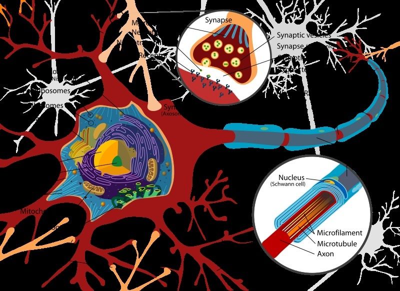
The core component of the nervous system in general, and the brain in particular, is the neuron or nerve cell, the “brain cells” of popular language. A neuron is an electrically excitable cell that processes and transmits information by electro-chemical signalling. Unlike other cells, neurons never divide, and neither do they die off to be replaced by new ones. By the same token, they usually cannot be replaced after being lost, although there are a few exceptions.
The average human brain has about 100 billion neurons (or nerve cells)
Each synapse functions like a microprocessor, and tens of thousands of them can connect a single neuron to other nerve cells. In the cerebral cortex alone, there are roughly 125 trillion synapses, which is about how many stars fill 1,500 Milky Way galaxies.
One synapse, by itself, is more like a microprocessor--with both memory-storage and information-processing elements--than a mere on/off switch. In fact, one synapse may contain on the order of 1,000 molecular-scale switches. A single human brain has more switches than all the computers and routers and Internet connections on Earth.
Each neuron may be connected to up to 10,000 other neurons, passing signals to each other via as many as 1,000 trillion synaptic connections, equivalent by some estimates to a computer with a 1 trillion bit per second processor. Estimates of the human brain’s memory capacity vary wildly from 1 to 1,000 terabytes (for comparison, the 19 million volumes in the US Library of Congress represents about 10 terabytes of data).
http://www.human-memory.net/brain_neurons.html
Researchers in Germany have created an exquisitely detailed three-dimensional model of a nerve terminal
https://www.youtube.com/watch?v=icey1NYP7Pw
The electrochemical jelly inside your head contains something like one quadrillion synapses, the junctions at which nerve cells talk to one other by converting electrical signals into chemical ones and then back again. They have two components (sometimes three): the nerve terminal of one cell, which stores and releases neurotransmitter molecules, and the 'post-synaptic' membrane of another cell, which contains binding sites for the neurotransmitters.
Synapses are miniscule – nerve terminals are about one thousandth of a millimetre in diameter, and the space between them and the membrane they contact a mere 20-40 millionths of a millimetre wide – and are densely packed in the grey matter of the brain tissue, making them notoriously difficult to study. But researchers in Germany have now created an exquisitely detailed model of a nerve terminal, which shows the distribution of 300,000 individual protein molecules in the nerve terminal in atomic detail, and hints at how neurotransmitter release is regulated.
Inside the nerve terminal, neurotransmitter molecules are stored in tiny spheres called synaptic vesicles, which are "docked" in an "active zone" just beneath the cell membrane. When a nervous impulse arrives at the terminal, it causes a few of the vesicles to fuse with the membrane and release their contents. Later on, the spent vesicles are recycled – they are pulled back out of the membrane, re-filled with neurotransmitter molecules, and eventually re-used.
At any given terminal, vesicle fusion can occur hundreds of times per second, as trains of impulses arrive one after the other. The whole process of vesicle docking, fusion and recycling is therefore tightly regulated, to ensure that there is a ready supply of vesicles that can fuse in quick succession and maintain the rapid bursts of neuronal activity.
http://www.sciencemag.org/content/344/6187/1023
HOW MANY NEURONS?
The vast majority of books and documentaries about the human brain cite the number of neurons to be 100 billion. While writing a previous book, I searched for the source of this claim and discovered that apparently none exists. It does not seem to be supported by evidence. The 100-billion-neuron claim seems to be nothing more than an agreed-upon guess that everyone keeps repeating.
Fortunately, a team of scientists decided to find out just how many neurons a typical human brain really does contain. They couldn’t just count them one by one, of course, because that would take centuries. So they liquefied brains in blenders—one at a time—made sure the neurons were equally distributed in the resulting fluid, and then counted sample portions. This allowed them to extrapolate a total neuron count for the entire brain. The total number of neurons for a human brain the researchers came up with was 86 billion.50 This is significantly lower than 100 billion but still an astonishing amount. The surface area of this many neurons—just one brain’s worth—has been estimated to be the equivalent of more than three football fields.
From ‘At Least Know This: Essential Science to Enhance Your Life’, by Guy P. Harrison (Prometheus Books)
The core component of the nervous system in general, and the brain in particular, is the neuron or nerve cell, the “brain cells” of popular language. A neuron is an electrically excitable cell that processes and transmits information by electro-chemical signalling. Unlike other cells, neurons never divide, and neither do they die off to be replaced by new ones. By the same token, they usually cannot be replaced after being lost, although there are a few exceptions.
The average human brain has about 100 billion neurons (or nerve cells)
Each synapse functions like a microprocessor, and tens of thousands of them can connect a single neuron to other nerve cells. In the cerebral cortex alone, there are roughly 125 trillion synapses, which is about how many stars fill 1,500 Milky Way galaxies.
One synapse, by itself, is more like a microprocessor--with both memory-storage and information-processing elements--than a mere on/off switch. In fact, one synapse may contain on the order of 1,000 molecular-scale switches. A single human brain has more switches than all the computers and routers and Internet connections on Earth.
Each neuron may be connected to up to 10,000 other neurons, passing signals to each other via as many as 1,000 trillion synaptic connections, equivalent by some estimates to a computer with a 1 trillion bit per second processor. Estimates of the human brain’s memory capacity vary wildly from 1 to 1,000 terabytes (for comparison, the 19 million volumes in the US Library of Congress represents about 10 terabytes of data).
http://www.human-memory.net/brain_neurons.html
Researchers in Germany have created an exquisitely detailed three-dimensional model of a nerve terminal
https://www.youtube.com/watch?v=icey1NYP7Pw
The electrochemical jelly inside your head contains something like one quadrillion synapses, the junctions at which nerve cells talk to one other by converting electrical signals into chemical ones and then back again. They have two components (sometimes three): the nerve terminal of one cell, which stores and releases neurotransmitter molecules, and the 'post-synaptic' membrane of another cell, which contains binding sites for the neurotransmitters.
Synapses are miniscule – nerve terminals are about one thousandth of a millimetre in diameter, and the space between them and the membrane they contact a mere 20-40 millionths of a millimetre wide – and are densely packed in the grey matter of the brain tissue, making them notoriously difficult to study. But researchers in Germany have now created an exquisitely detailed model of a nerve terminal, which shows the distribution of 300,000 individual protein molecules in the nerve terminal in atomic detail, and hints at how neurotransmitter release is regulated.
Inside the nerve terminal, neurotransmitter molecules are stored in tiny spheres called synaptic vesicles, which are "docked" in an "active zone" just beneath the cell membrane. When a nervous impulse arrives at the terminal, it causes a few of the vesicles to fuse with the membrane and release their contents. Later on, the spent vesicles are recycled – they are pulled back out of the membrane, re-filled with neurotransmitter molecules, and eventually re-used.
At any given terminal, vesicle fusion can occur hundreds of times per second, as trains of impulses arrive one after the other. The whole process of vesicle docking, fusion and recycling is therefore tightly regulated, to ensure that there is a ready supply of vesicles that can fuse in quick succession and maintain the rapid bursts of neuronal activity.
http://medicalxpress.com/news/2014-05-highly-3d-individual-neural-synapse.html
(Medical Xpress)—A team of researchers in Germany has created a very highly detailed 3D computer model of an individual rat synapse showing the distribution of approximately 30,000 proteins involved in the process of sending a message from one neuron to another. In their paper published in the journal Science, the team describes how they combined several imaging techniques to create the model, and what it is able to display.
In simple terms, neurons transmit messages between one another via synapses—parts of neurons dedicated to converting electrical signals to chemical signals and vice versa. Synapses generally come in two varieties, the kind that send and the kind that receive signals. Neurons have both kinds of course, which allows them to send and receive messages. To send a message, electrical signals from the neuron travel to the sending synapse where they encounter vesicles. Prodded by the electrical signal, the vesicle releases chemicals called neurotransmitters into the space between (the cleft) the sending synapse on one neuron and the receiving synapse on another. The receiving synapse then processes the message and responds based on the signal it receives.
In the sending synapse, the vesicles are made ready for action by being loaded with neurotransmitters and locked into position. Once they release their cargo, they are moved back out of the way so other vesicles can work while they are being refilled—once ready, they are again placed into position and put to use. This constant recycling goes on over and over, allowing neurons to process a steady stream of messages. In this new effort, the team in Germany has used a variety of microscopic methods to capture the structure of the sending synapse and what happens to it as recycling occurs and messages are sent.


http://www.sciencemag.org/content/344/6187/1023
Last edited by Admin on Fri Apr 05, 2019 12:34 am; edited 2 times in total
