Bacterial - Mycoplasmas (Mollicutes)
Mollicutes, informally referred to as the mycoplasmas, are extraordinary organisms! They are
the smallest known cells, typically the size of large viruses at only 200 to 300 nm in diameter (1
nanometer, 1 nm = 1 millionth of a millimetre) and they have the smallest genomes of any known
bacteria. The genome of Mycoplasma genitalium, for example, is only 580,073 bases long and
contains a mere 517 genes. Though still complex in real terms, this is a very minimilistic genome
and mycoplasmas are of especial interest to scientists working to build simple cells in the lab.
Mycoplasmas are often described as the simplest free-living cells, although most are parasitic or
commensals dependent on their host for certain functions, however, they are independent in the
sense that they have their own power sources, fermenting fuels to make ATP (unlike the tiny
chlamydial elementary bodies which are energy parasites dependent on ATP provided by the
host cells they live within). Most 'respire' anaerobically by fermentation without the use of
electron transport chains (ETCs) although rudimentary ETCs may be present in the cytoplasm
(or attached to the cell membrane in Acholeplasma). Their simplicity is in large part due to
evolutionary degeneration - they are dependant on their hosts for many metabolic functions that
they would otherwise have to carry out on their own and so they have lost these functions,
including the ETC.
As prokaryotes they lack a nuclear envelope, possessing a DNA/protein nucleoid rather than a
nucleus. As bacteria we tend to think of them as single-celled organisms, however, like most
bacteria they can form biofilms - loose associations in which the cells remain distinctly separate
but are embedded in a common slime matrix. Despite this, they do not seem to use
quorum-sensing to sense the presence of other bacteria and communicate in order to form
biofilms. Most bacteria use quorum-sensing in biofilm formation, but it is thought that
mycoplasmas form these structures without communication between the component cells.
Biofilms are not true multicellular organisms, since the cells do not communicate via cell-to-cell
junctions (pores and channels that join the cytoplasm of neighbouring cells together, as do gap
junctions in animal cells, plasmodesmata in plants and microdesmata in some cyanobacteria).
Indeed, in a typical biofilm the cells are not generally in direct physical contact with other cells.
Mycoplasmas also lack protective cell walls - they have no peptidoglycan like most bacteria and
no rigid layer at all in their cell envelopes, which instead consists of single cell membranes
coated in carbohydrates (forming the glycocalyx or slime coat) rather like animal cells and
protozoa. Like mammalian cells they are osmotically sensitive. Having no wall to maintain their
shape they will swell and burst in distilled water. Again, this may be a feature that their ancestors
had which was lost as their host maintains an osmotically stable environment which is also
generally free of mechanical trauma. Many do incorporate sterols, manufactured by their hosts,
however, which strengthen their membranes by making them more rigid. Acholeplasma is able to
grow without sterols, but will incorporate them if they are available and may manufacture
carotenoids to strengthen its membrane.
Life as parasites
Most mycoplasmas are parasites or commensals, living inside other organisms. Thermoplasma
acidophilum is unusual in being found in acidic coal refuse piles where internal temperatures
reach 55 degrees C. Spiroplasma infects plants and the arthropods which carry it from plant to
plant, infecting the haemolymph, gut and salivary glands of insects. Some may cause repiratory
infections in humans, for example Mycoplasma pneumoniae can cause a type of pneumonia
(though it is by no means the only cause of this disease). Mycoplasma may also infect the
synovial membranes of the joints of vertebrates, causing a form of arthritis.
Mycoplasmas require complex nutritional requirments if grown in the lab, since they depend on
their hosts for complex growth requirements, such as fatty acids, vitamins, purines, pyramidines,
and also the sterols for their membranes. When grown on agar they form characteristic small
circular colonies with a nipple-like or fried egg-like appearance.
Ureaplasma lives in the mouth, respiratory and genital tracts of mammals and humans and has a
novel way of obtaining energy. Whereas most mollicutes generate ATP by anaerobic
fermentation, Ureaplasma exhibits an unusual form of respiration in which urea (a waste product
of mammalian metabolism) is hydrolysed by an enzyme called a urease to form ammonium
(NH4+) which acts as a source of protons (H+) to power the ATPase by generating a proton
gradient across the cell membrane (essentially generating positive electric charge which flows
through the ATPase which acts as an electric motor whose rotation energy is sued to make ATP).
Many mycoplasmas have been implicated in causing plant disease, though since these are often
poorly characterised they are often referred to tentatively as mycoplasma-like organisms (MLOs).
Cell shape and cell motility - novel mechanisms of locomotion
Mycoplasmas are often described as pleomorphic: having variable shape, especially types like
Mycoplasma, although this is more true when growth conditions are sub-optimal (it is dificult to
grow parasites out of their hosts!) and in optimal conditions their form is more consistent.
The simplest cells?
Mycoplasma
Above: Mycoplasma grown under optimal conditions tends to have a more regular form. These organisms
glide by means of the apical protrusion protruding from their front end.
For a long time it was though that bacteria lacked an internal cytoplasmic skeleton (cytoskeleton)
as found in eukaryotic cells. This is because bacterial cells are much smaller and have a cell wall
to support them (for a while it was thought that plant cells might not have a cytoskeleton because
of their cell walls, but they do). However, it is now known that bacteria do have a cytoskeleton,
although one that is much less developed than in plant and animal cells. Indeed, in normal
electron microscopy no cytoskeleton may be evident at all in bacteria, except perhaps the
occasional tubular structure. It is now realised, however, that cytoskeletal structures form during
cell division and also during cell growth, at least in some forms, where cytoskeletal filaments
direct the deposition of new peptiodoglycan fibres in the cell wall (see growth in bacteria for more
details). In some cases cytoplasmic filaments are also seen to anchor flagella motors.
Mycoplasmas, although tiny, lack supporting cell walls and so their cytoskeletons have more work
to do and are better developed. (Animal cells and protozoa, being large and wall-less have
especially well developed cytoskeletons).
The cytoskeletons of mollicutes are also involved in locomotion. These bacteria lack flagella,
possibly because they have no rigid cell walls in which to anchor the rotary motors. Spiroplasma,
being helical in shape moves by flexing, creeping and swimming by rotating in cork-screw
fashion, rather like spirochaetes, except that spirochaetes use endoflagella to produce these
movements whereas Spiroplasma has no endoflagella. Instead Sprioplasma has a unique helical
protein fibres (3.6 nm diameter) in the cytoplasm, grouped into bundles or ribbons and a second
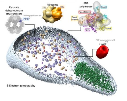
helical structure made of an actin-like protein MreB. Cryo-electron tomography, a new technique
for visualising cell structure at a molecular scale (less than 5 nm resolution) whilst preserving the
structure of living cells almost intact has revealed two ribbons of thicker filaments with a band of
thinner filaments in-between. At least one of these structures is thought to be contractile.
These fibrils are positioned just beneath the cell membrane and are thought to be involved in
maintaining helical shape as well as in motility and daughter-chromosome separation during cell
division. Indeed, these fibrils bear some resemblance to those seen in walled bacteria, for
example an MreB-like protein, Mbl (an MRb homologue) in the rod-shaped Bacillus subtilis form
helical filaments that direct the deposition of helical fibres of peptidoglycan in the cell wall during
cell growth (cell elongation) whilst MreB controls cell width. However, the other component of
Spiroplasma's helical fibres is apparently unique to Spiroplasma.
Spiroplasma rotates as it swims, corkscrewing its way along, which is an advantage when
swimming in highly viscous (sticky) fluids and, perhaps not surprisingly, Spiroplasma is
viscotactic, moving towards regions of high fluid viscosity and also exhibits chemotaxis. They
change helicity, from anticlockwise to clockwise at intervals, and often transitional cells are seen
with mixed helicity, caught in the act of changing. They can also change the pitch (steepness of
turns) of the helix with waves of change in helicity traveling down the length of the cell as pairs of
tiny kinks travelling from anterior to posterior. The kinks in each pair are on average 0.26
seconds apart, with the second kink appearing as the first nears the posterior end of the cell.
The kinks move at about 10.5 micrometres per second and the cell swims in the opposite
direction at about 3 micrometres per second, faster (up to about 5 micrometres per second) if
the medium is thickened (made more viscous) by the addition of 0.5% methylcellulose - like
spirochaetes they swim faster in high viscosity fluids. These observations suggest that the cells
rotate and swim due to flexing of the helical filaments.
Gliding motility in Mycoplasma
Many mollicutes move by gliding across a solid surface. Of these, the fastest is Mycoplasma
mobile which can glide at 2.0 to 4.5 micrometres per second on a glass surface. This is
astonishingly fast for a gliding organism. Like many mollicutes, Mycoplasma mobile moves by
means of an anterior nose-like anterior projection which generates traction against the substrate
apparently by means of about 400 minute (less than about 50 nm long) 'leg-like' proteins
(possibly the protein Gli349) in the cell membrane which are thought to alternately adhere to,
detach, advance and reattach to the surface, in a stepping manner, pulling the cell along.
Alternatively, other electron microscope evidence shows a much smaller number of relatively
long spikes (less than about 50 nm long) which may be the leg-like structures or separate
adhesion organelles. The cells can only glide forwards and can not reverse. ATP hydrolysis
provides the energy for this movement. Inside the apical protrusion are prominent cytoskeletal
structures - a solid nose-cap or hexagonal-lattice of a specific protein in the very tip of the
protrusion, forming an oval or hemispherical cap 235 nm wide and 155 nm long attached to
dozens of flexible protein 'tentacles' (inside the cytoplasm). The tentacles have 20 nm particles
(proteins?) attached to them at 30 nm intervals and it is thought that the leg-like proteins in the
cell membrane attach to these tentacles, transmitting the force of traction they generate to the
rest of the cell.
Suggested Bibliography
Below are some of the key references used in writing this article:
Spiroplasma fibrils - http://www.sciencedirect.com/science/article/pii/S0014482704004525
Makoto Miyata, and Jennifer D. Petersen, 2004. Spike Structure at the Interface between
Gliding Mycoplasma mobile Cells and Glass Surfaces Visualized by Rapid-Freeze-and-
Fracture Electron Microscopy. J. Bacteriology 186: 4382–4386.
Mayer, F. 2003. Cytoskeletons in prokaryotes. Cell Biology International 27: 429–438.
Makoto Miyata and Hiroshi Ogaki, 2006. Cytoskeleton of Mollicutes. J Mol Microbiol
Biotechnol 11: 256–264.
Daisuke Nakane and Makoto Miyata, 2007. Cytoskeletal ‘‘jellyfish’’ structure of
Mycoplasma mobile. PNAS 104: 19518–19523.
Models of Mycoplasma pneumoniae show a different arrangement of cytoskeletal structures in
the apical protrusion. There is still a cap-like structure right inside the tip and attached to this and
extending down the central axis of the protrusion is a rod-like structure, attached to the
surrounding cell membrane by radial spokes along its length and anchored at the base of the
protrusion in a disc-like structure, also connected to the cell membrane by radial filaments. Other
cytoskeletal structures have also been observed in this species, including a helical mesh just
beneath the cell membrane and fibres extending into other regions of the cell. A different mode
of locomotion has been suggested for Mycoplasma pneumoniae in which the apical protrusion
extends, then membrane proteins in the apical protrusion attach to the substrate followed by
contraction pulling the rest of the cell forwards before the cycle repeats.
Mycoplasma mobile
Above: a model of Mycoplasma mobile crawling across a surface, looking down from above,
showing the cap structure (orange) of the apical protrusion and the trailing 'tentacles' (purple)
and the membranous protein 'legs' (cyan). The 'legs' or 'feet' move forwards and bind to the
surface and then bend or rotate backwards in some manner, pulling the cell forwards (to the right
in this diagram) and then detach and swing forwards to repeat the cycle.
Mycoplasma pneumoniae model
Above: a model of the cytoskeleton of Mycoplasma pneumoniae, showing some of the key
protein structures involved in gliding motility. There is an apical cap or plug (cyan) with a central
core of tubules connected to it which span the length of the apical protuberance (yellow). Radial
spokes (orange) connect this tubule core to the cell membrane. At the base of the apical
protuberance, the core connects to a 'wheel-like' or 'bowl-like' structure (green and orange)
connected to the cell membrane by radiating filaments (green rods) and to cytoskeletal filaments
which cross the cell body (magenta). Some of the adhesion proteins which coat the outside of the
membrane of the apical protuberance are shown (red balls and rods). One model has the apical
protuberance extending and then adhering to the surface via the adhesion proteins whilst the
core of tubules contracts, transmitting force to the rest of the cytoskeleton via the wheel-like
structure, pulling the cell body forward.
Minimalist cells?
As the simplest living cells so far discovered on earth, the mollicutes have attracted much recent
research, not just because they are agents of disease, but because these fascinating organisms
may give some insight into how cells first evolved. (Viruses are simpler, but these are acellular).
However, with their 600 genes or so they manage a considerable degree of complexity as can be
seen by the (highly simplified) account we have given of their cytoskeleton and motility
mechanisms. Mollicutes are hard to study, because they are so small, and only recently has real
progress been made on understanding their cellular machinery, which is complex! Much remains
uncertain and controversial, which is one reason why we avoided details - it should be most
interesting to see the progress in these fascinating areas of research over the coming years!
It would appear that, despite their relative simplicity, the mollicutes are highly evolved: as
parasites and commensals with some degree of degeneration of their ancestral systems, in
addition to evolution of novel systems. Nevertheless they remain the smallest and simplest cells
that are known to be able to survive as free-living organisms (albeit under carefully controlled
laboratory conditions or in specialist habitats such as coal piles) and understanding how they
function is of major importance. This still leaves the mystery as to how the first cells evolved.
There would appear to be no surviving simpler cells, and also none appear to be currently
evolving. What special conditions enabled cells to evolve but prevent them from doing so today?
In the current absence of simpler cells this question is difficult to answer. However, what mollictes
show us is how much can be done with relatively few genes and proteins.
Simpler cells?
There is one line of thought that hypothesises that simpler cells do currently exist on earth, but
have so far evaded detection. Indeed, the smallest cellular life-forms, such as mollicutes and
Chlamydia are still poorly understood and many more similar organisms no doubt await detection.
(Such as the MLOs, also called phytoplasms, which cause over 600 known plant diseases). In
recent years controversial claims that minute bacteria, called nanobacteria, of the order of 100
nm across or less, have been discovered remain to be verified. Often found inside rocks and
other 'primordial' habitats, these 'organisms' may simply be some form of mineral growth. Indeed,
as the controversy over the Martian meteorite, ALH84001, and its possible nanofossils has
shown - proving the existence of the smallest cells, past or present, will likely be extremely difficult.
Almost certainly, the vast majority of bacteria remain undiscovered. Most do not grow on
standard agars and only recently has the vast diversity of bacteria and archaebacteria in the
Earth's oceans been realised. Bacteria are also being discovered in unlikely places, from
Antarctic deserts to rocks deep beneath the Earth's surface. No doubt there are many surprises
yet to be revealed by the ancient and complex world of prokaryotes!


