ElShamah - Reason & Science: Defending ID and the Christian Worldview
Would you like to react to this message? Create an account in a few clicks or log in to continue.
ElShamah - Reason & Science: Defending ID and the Christian Worldview

Otangelo Grasso: This is my library, where I collect information and present arguments developed by myself that lead, in my view, to the Christian faith, creationism, and Intelligent Design as the best explanation for the origin of the physical world.


You are not connected. Please login or register

The recent groundbreaking scientific research which explains the real mechanisms of biodiversity

Go down  Message [Page 1 of 1]

Otangelo


Admin

The recent groundbreaking scientific research which explains the real mechanisms of biodiversity

https://reasonandscience.catsboard.com/t2293-the-recent-groundbreaking-scientific-research-which-explains-the-real-mechanisms-of-biodiversity

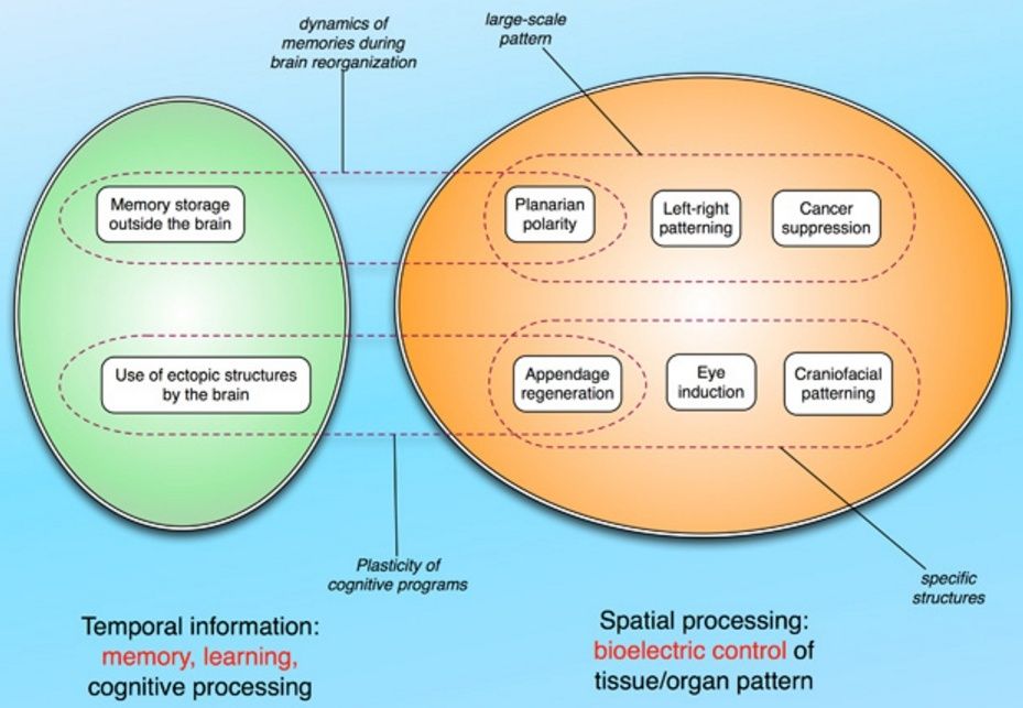
The assertion that evolution is a fact, is repeated like a mantra by proponents of evolution and naturalism, which try in that way to justify their unbelief in an intelligent creator. One of the most frequent claims is that microevolution and macroevolution are the same on a different timescale. And that there is no mechanism that prevents micro to become macro. The ones that are better informed, IMHO, know that the mechanism that provokes change and evolutionary novelties above the species level, that is, the change from bacteria do man, is UNKNOWN. In order to explain the origin of biodiversity, body shape, body plans, organ development, and ultimately, if the claim of macro-evolution from Luca to homo sapiens is true, we need first to understand how organs, limbs, and creatures arise, what mechanism determines what size and shape they should be when they are growing. What mechanism programs the cell to "know" how and where to form an organ? How does the creature know how to rebuild a severed limb to the correct size, shape, and orientation? Once this is elucidated, we can ask if the same mechanisms explain biodiversity.  It seems that groundbreaking scientific research is starting to unravel this longstanding mystery, and it's far from being explained through neodarwiniam predictions and claims, but epigenetic mechanisms, which will be elucidated below. 

A great deal is now known about morphogen gradients in the developing embryo—how cells know where to go and what types of cells to become. Recent research shows that electric potentials in nonbrain cells are a signal for creating patterns during the development and during the rebuilding of organs. This information of different field potentials surrounding individual cells can give information for the developing organ. Now, research from the laboratory of Dr. Michael Levin is demonstrating new ways that cells signal with electricity and the great importance of electrical properties for individual cells and tissues. He describes how electrical gradients and fields are critical in the 3D function and shape of cells and organs. 
 A prominent aspect of multi-cellular creatures is that they have organs of a particular size and shape. When regeneration occurs in reptiles the same exact shape is grown. Information for the cellular activity appears to exist in the space that will make up the specifically shaped organ. Levin notes that “cancer can be seen as an error of geometry, because tumor cells grow, migrate, and function without regard for the orderly structure within which they occur.”
 Each cell has specific electrical gradients and properties that together form a large electric field of information. This field of information can show individual cells in the embryo how to behave. This is analogous to the fact that electrical flow between cells in the early embryo  forms the basic network that is then built into a formal structure with elaborate chemical synapses. This, also, occurs during rebuilding of tissue. Somehow, the information of the electrical flow through the electric gap junction synapses determines the future structure. Electrical signals now appear to be critical in forming the shape of organs, the very function and identity of organs, and the creation of new limbs on animals that regenerate. In these animals, stem cell behavior is directed by currents created with potassium, sodium, chloride, and protons that affect the genetic networks of cells at a distance. Recent research shows that limbs can be influenced by proton and sodium alterations. New types of ion channels, pumps and electrical connections have now been found in a variety of different organs.


This is remarkable. If electric gap junction synapses determine cell shape, then macro change attributed commonly to macroevolution and structural biological novelties should also depend on these.

Electrical signaling is key for cells to properly interpret their environment, and when this process goes awry, the cells default to a cancer program. 4
While ion flows control cell-level behaviors such as migration, differentiation, and proliferation, bioelectric signals also function as master regulators of large-scale shape in many contexts: a simple signal can induce complex, highly orchestrated, self-limiting downstream morphogenetic cascades. For example, an unmodulated flux of protons can cause the formation of a complete tail of the right rise and tissue composition. 


Our data suggest that the mechanism by which blastema cells polls the rest of the host (to determine where the wound is located and what other tissues already exist in the fragment and thus don't need to be recreated) is mediated by physiological signals passing through nerves and long-range gap junctional paths.

A significant component of morphogenetic cues are ionic in nature. Remarkably, however, this effect is non-local in nature - it is the transmembrane potential of other, quite distant cells that determines the metastasis-like effect.
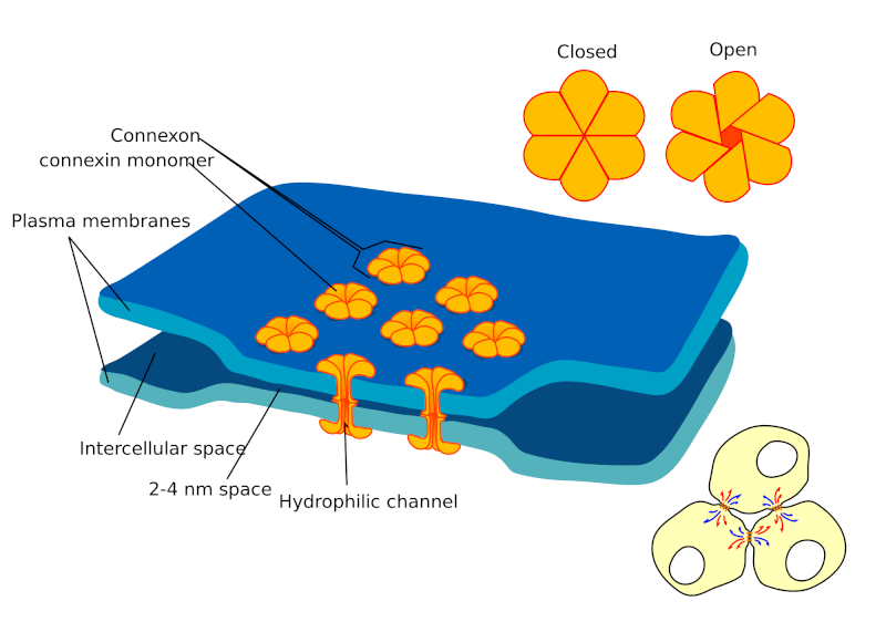
Many critical questions remain about how cellular polarity is synchronized and amplified across embryonic fields to allow cells to ascertain their position with respect to the midline. We identified a dependence of asymmetric gene expression on early communication between left and right sides in the chick and frog. For example, expression of left-sided markers depends on events occurring on the right side, during very early stages, suggesting that the two sides need to coordinate their decision with respect to the L-R identity of each. One mechanism for communicating between cells and tissues involves gap junctions: multimers of connexin proteins form channels between cells and pass small molecules, subject to complex regulation by various signals.


So it seems within gap junctions that action happens, but the action per se is REGULATION AND COMMUNICATION THROUGH VARIOUS SIGNALS. THATS THE KEY.

We showed that gap junction are crucially involved in L-R patterning in early embryos of Xenopus and chick. gap junctions are a bioelectric patterning element that sets up domains of isopotential cell fields during morphogenesis.
Serotonin signaling is used in information exchange between cells in processes such as L-R patterning and control of timing and cell movement during gastrulation. We have shown that serotonin is utilized by both chick and frog embryos, at very early stages, as a small molecule signal which is transported in a left-right gradient and regulates the development of laterality. Indeed, we now know that the early frog embryo is literally an electrophoresis chamber, which uses voltage potentials to generate consistently biased left-right gradients in serotonin in an epigenetic process not dependent on zygotic gene expression. We have modeled this process quantitatively, and characterized novel intracellular serotonin-binding proteins which directly activate asymmetric gene expression after their rightward movement, linking an early biophysical process to transcriptional regulation via chromatin modification pathways. Serotonin is also a key mediator for bioelectric control of neuronal outgrowth from transplants. 

Electrical Fields Guiding 3D Shape of Cells and Organs

How does the cell know what size and shape it should be? Many cells alter their shape to provide different functions, like microglia. Even more complex is the question as to how organs, limbs, and creatures know what size and shape they should be when they are growing. How do the cells know how and where to form an organ? How does the creature know how to rebuild a severed limb to the correct size, shape, and orientation?

There are thousands of these same questions, including how astrocytes and neurons know the exact networks they should form. How immune cells know how to travel through very complex differing 3D environments? An equally difficult question has been addressed in previous posts as to how the

cell knows exactly what shape a coded sequence in a protein will take. Both cells and microbes alter the codes of their protein toxins that require extremely detailed and accurate shapes. In fact, modern science cannot calculate what shape a 400 amino acid coded sequence will be when it folds into a protein. It would take all the supercomputers together two thousand years to calculate the folding of one average protein. Yet, proteins assemble into the exact shape in a millisecond, helped by very complex chaperone molecules. Cells routinely edit their messenger RNA with alternative splicing to make a whole variety of shapes. How do they know how to do this?

Research shows that: If a cell of a two cell embryo is removed it still becomes the full creature.

Starved flatworms shrink keeping perfect proportions among the organs
Planaria can reproduce the entire body from a small piece
Amphibians can grow a perfectly proportioned limb
Many structures need nerve-mediated information in order to maintain shape and function


For all of these questions, it is hard to imagine the information of the 3D shape being inside one cell, or even a group of cells. It seems more likely that a field of information somehow guides these processes. Is this evidence for an electrical field of information forming 3D shapes of cells, organs and creatures, also, related to the influence of mind?

Electrical Gradients and Fields In and Around Cells


The recent groundbreaking scientific research which explains the real mechanisms of  biodiversity Gap_ce10


Previous posts have demonstrated the critical part that electrical synapses play in forming the structure of the neuronal network that uses chemical synapses. Also, a previous post showed that field potentials in and around brain cells, while poorly understood, are important for specific functions. Multiple posts have documented the elaborate communication that occurs between cells through signaling including neurotransmitters, brain factors, cytokines and hormones.

A significant amount of research shows the importance of various gradients in the space between cells for communication. This was noted in the posts on the movement of platelets that attract and signal to other immune cells. Another post showed the elaborate travels of leukocytes. A great deal is now known about morphogen gradients in the developing embryo—how cells know where to go and what types of cells to become.


Recent research shows that electric potentials in non brain cells are a signal for creating patterns during development and during re building of organs. This information of different field potentials surrounding individual cells can give information for the developing organ. Now, research from the laboratory of Dr. Michael Levin is demonstrating new ways that cells signal with electricity and the great importance of electrical properties for individual cells and tissues. He describes how electrical gradients and fields are critical in the 3D function and shape of cells and organs. Professor Levin notes that cancer cells use electric gradients for many purposes. In fact, aberrations in electrical processes can lead to cancer. Differences in electrical potential in cells are important in determining whether a cancer metastasizes and electrical states in one cell can have effects on distant cells, triggering metastasis. These electrical potentials in one cell can signal other cells that trigger genetic networks and epigenetic effects.

Electrical State of Cells


The recent groundbreaking scientific research which explains the real mechanisms of  biodiversity Magnus-Manske-WIK-Protein_gates-300x225
From Magnus Manske
The electric state of a cell is determined by many complex factors. One is the opening and closing of ion channel proteins, which control the movement of charged particles (ions) across the cell membrane. Changes of the cells’ electrical potential via the activity of ion channels are shown to be able to suppress or trigger cancer. Another factor is electrical synapses that transfer electricity from one cell to another.
Different electrical properties of cells affect what type of cells are made from stem cells, how much reproduction goes on, where cells travel to and their shape and orientation. Electrical gradients and fields certainly affect travelling immune cells, stem cells, and all brain cells. They also exert profound effects on other somatic cell types during embryonic development and regenerative repair. Electrical properties are simultaneous with chemical metabolism using genetic network processing. They are critical for healing wounds and responding to infections. They determine the symmetry and shape of organs.
Differential ion channel activity in the body sets up specific patterns of voltage potential throughout tissues. During development, these electrical properties of cells determine what types of cells they become. For example, two specific groups of cells in the embryo have increased electrical potential and these become the two eyes. Research shows that by changing the electrical potential of cells at a critical time when eyes are being made, eyes can be induced to form elsewhere, effectively reprogramming other cell types into a complete visual organ.

While electrical activity can drive downstream genetic changes, just changing the electrical gradient can dramatically change the cell’s behavior. After an amputation, controlling the voltage potentials of wound cells causes the regrowth of a limb. The specification of the details appears to exist in the electrical field. At specific regions of the animal’s body, either a tail or limb grow due to the electrical signaling, which kickstarts a “build whatever goes here” program.
Electrical fields that are being discovered with multiple influences could contain this 3D information. Altering the electric field alters the information and the biological results.

Signaling with Chemicals and Electricity


Quite a number of previous posts reveal the elaborate signaling of microbes and immune cells such as T cells and platelets. The critical intestinal epithelial cells and skin cells are involved in very elaborate signaling between large numbers of microbes on one side and many immune cells on the others. Of course, brains are known to use elaborate signaling.

A previous post noted that cancer is like a microbe colony in that they work together for their ends. This view of cancer is that many cells in an environment trigger the actions of the community of cancer cells. Another view of cancer is as a defection of individual cells from the goals of the organism; this can occur by cells’ being isolated from, or mis-interpreting, the patterning cues that normally orchestrate cell behavior into complex anatomy. The new research by Levin adds a very large new dimension in the way electrical signals between cells might be critical in this cancer development.

A prominent aspect of multi cellular creatures is that they have organs of a particular size and shape. When regeneration occurs in reptiles the same exact shape is grown. Information for the cellular activity appears to exist in the space that will make up the specifically shaped organ. Levin notes that “cancer can be seen as an error of geometry, because tumor cells grow, migrate, and function without regard for the orderly structure within which they occur.”

This can occur through each cell having specific electrical gradients and properties that together form a large electric field of information. This field of information can show individual cells in the embryo how to behave. This is analogous to the fact that electrical flow between cells in the early embryo (see post on electrical synapses) forms the basic network that is then built into a formal structure with elaborate chemical synapses. This, also, occurs during rebuilding of tissue. Somehow, the information of the electrical flow through the electric gap junction synapses determines the future structure.
Electrical signals now appear to be critical in forming the shape of organs, the very function and identity of organs, and the creation of new limbs on animals that regenerate. In these animals, stem cell behavior is directed by currents created with potassium, sodium, chloride, and protons that affect the genetic networks of cells at a distance. Recent research shows that limbs can be influenced by proton and sodium alterations. New types of ion channels, pumps and electrical connections have now been found in a variety of different organs.

This is remarkable. If electric gap junction synapses determine cell shape, then  macro change attributed commonly to macroevolution  and structural biological novelties should also depend on these. 

Voltage Gradients in Non Brain Cells


Cells not in the brain, also, use ion channels that create an electrical potential between the inside and outside of the cell. These do not send a signal down a long axon wire as neurons do, but the electrical potentials are critical for the cell’s behavior. In a previous post, calcium cyclic signaling was noted to be the crucial signal between plants and microbes when forming nitrogen factories.

Whole tissues and sheets of cells, also, have flows of electricity through electrical synapses called gap junctions (just like neurons). When the skin is broken the electrical gradients and fields are disrupted and these are a signal for immune cells to repair the tissue. An even larger field exists over an entire organ, and this field has crucial information regarding the shape and function of the organ.
It is very difficult to study, but even inside the cell, many membranes exist for the nucleus, the mitochondria and other organelles such as the critical information center of the endoplasmic reticulum. Electrical potentials and information fields exist in these, also.

Cancer and Electrical Gradients


Cancer cells’ behavior is very tied to electrical signals. They use them as clues to avenues of travel. Electrical fields determine shape changes in the cells. The electrical potential, also, affects what type of cell is produced from stem cells. 

Cancer cells have altered electrical activity and use different ion channels and pumps. They have different transporter molecules that affect how the ions travel and, therefore, the electrical gradients. It now appears that these alterations in ion channels are signatures of different cancers and are critical to their formation. Many differences in the effects of cytokines and neurotransmitter signals, also, appear to be tied to these electrical differences. These ion channels influence all aspects of metastasis. In fact, oncogenes (special genes that cause a cell to become cancerous) are related to various different ion channels.


Research in cancer has been increasingly difficult because of the many different mutations that are found in types of cancers, and, more recently, in individuals. The studies have shown that there is a wide range of different mutations that don’t fall into any noticeable patterns.


Dr. Levin’s work points in a different direction entirely. He notes that the gradients and fields of electrical information may be a more important way to understand this process. It is the very space that the cells are living in that appears to be aberrant and causing the cancers. Electrical signaling is key for cells to properly interpret their environment, and when this process goes awry, the cells default to a cancer program.


Studies show that cancer cells can be differentiated from other cells by their electrical properties. The overall electrical status of the cancer cell is determined by the sum of all the ions and pumps. There are a vast amount of different possible genetic scenarios in this basic measurement of electrical status.

But, in fact, the simple electrical status appears to identify cancer cells. Dr. Levin likens this to “pressure” in physics – a group property that can be implemented with a wide variety of underlying molecular details (e.g., different ion channel genes). He notes that the statistical electrical study of cancer electric gradients might be the most information. He notes that the extremely complex analysis of all of the ion channels might not even reveal the critical information (this type of research is currently not even feasible).

Just analyzing messenger RNA and specific proteins made will not determine all the factors altering the global electrical fields because electrical state is a function of the 3D open/closed states of the channels, not merely their presence at the mRNA and protein levels.


The large electrical fields themselves appear to create the regulation that occurs. A study showed that by increasing the electric potential in cancer cells, metastatic activity can be suppressed, despite specific proteins being present that are known to be involved in metastatic activity.

Information Fields and Geometry


All of the signals that affect a cell, together, are called the morphogenic field. It includes all of the instructions that come from all secreted cellular signals, including cytokines and the effects of all the molecules in the extra cellular space (see post on Extracellular Space and Neuroplasticity). This includes many gradients that are used by immune cells to gather information and attract other cells. An unusual feature recently discovered as a cause of cancer is special proteins that produce cells in particular orientations—called planar cell polarity. When this polarity is altered it can cause cancer.


Electrical gradients are now shown to be critical as well. When the usual gradient or field information is somehow altered it can cause cancer. Each embryo has its own innate field. It has been observed that cells in the midst of a live organ are not as susceptible to the same chemical influences to become cancerous. They are much more able to become cancer when isolated from the influences of other cells. When the region’s electrical properties are different, they are less likely to become cancerous.


Signals from neurons are, also, a factor in causing cancer. When nerves are cut, then cancers occur more frequently. These neuron signals appear to be contributing to the field information in local regions.


A striking example of the field is when shunts are needed between the abdomen and the blood. These shunts send large amounts of cancer cells to many regions, but they do not become metastatic in some areas, but do in others. Another is when the shape of a region is altered by surgery it is more likely to become cancerous, possibly by altering the field.
Cancer cells turn away from the community aspect of the entire organism and become involved in individual behavior and their own new community (see post on Cancer – The Emperor of Cells.) The cancer cells either aren’t given appropriate information in the field about the creature, can’t extract the information or don’t care anymore. Cancers form their own organs and fields. The cancer cells, also, form their own information fields and communicate and cooperate using them. They reproduce and evolve to be stronger as a community. They signal each other to fight microbes and immune attacks. Cancer cells eliminate communication to the body and tissues of the body. But, they maintain the same junctions and communications among this smaller community.

Where is the Information


The study of electric fields brings up the problem of orders of magnitude of effect and the question of emergent properties. While at the level of genetic network activity, each cell in the field has definite behaviors—they are all influenced by the overall information fields. All levels are at play—the genetic, epigenetic, individual cells and networks of cells.

Having information in morphogenic fields including electrical gradients is similar to the notion that mental events occur in neuronal electric networks. Dr. Levin speculates that the same way that information is contained in neural networks, information could be maintained in electrical fields of cells, organs and creatures.
All of this, raises the question, of course, about its relation to cellular intelligence in particular and mind in nature in general.


Which Comes First – Electric Fields or Biochemistry


The genetic networks are very critical in electric signaling because cells use protein ion channels imbedded in the membrane. These proteins affect distant actions and are very involved in creating the overall gradients.

In one of many circular genetic processes, the actions of the genetic machinery are controlled by electrical gradients and the elaborate signaling of the genetic networks create the electrical gradient

Nices Catch22 situation. 

Patterning occurs by a continuous interplay of genetics and physics.
This conundrum indicates that there is a more global regulation of the entire process. In fact, research shows that the electrical influences cannot be reduced to simple chemistry inside of a cell. Electrical gradients of individual cells and many cells contribute to the creation of the 3D shapes and functions of organs and creatures. Examples of this include the creation of the embryonic eye, the head, and the regeneration of limbs exactly to size. For movement of cells, when electrical and chemical gradients are in competition, the electrical gradient wins.
The example of the eye is instructive. Dr. Levin notes that it shows the electrical information is more crucial. It is the electrical properties of the regions forming the two eyes that determine where the eyes will go. When this electrical property is altered, the eye forms in different places. This effect cannot be replicated outside of the head by any of the known “master” eye genes.

What Signals Could Describe 3 Dimensions?


How does a cell first create a shape of an organ in the fetus and then maintain it throughout life, with many new cells being generated? Signals can let a cell know about position, size and polarity and orientation. It is either cell-to-cell communication, which is basically the current research goal, or a pre formed field or organizing template. The 3D process in the fetus is called morphogenesis. Each cell in the embryo has to know its position relative to others and differentiates into the appropriate type of cell for that position.

Field structures in physics are similar to the concept of morphogenetic field. A field includes information at each location. It includes regulation at a distance. The gradient is a concept already used in studies of development. Current research shows that electrical potentials and gradients in the space between cells are an important source of information for cells, and control the movement of chemical signals such as serotonin and butyrate.

Electrical Fields Guiding 3D Shape of Cells and Organs


While both chemical and electrical factors are clearly relevant to determining the three dimensional function and shapes of cells, organs and creatures, it appears that an information field of some kind would be necessary for the level of information needed. Recent research shows the importance of the electrical potentials, gradients and fields.
Among theories of what mind could be (see post), information fields have been proposed. Perhaps this current research furthers such a view.

1) http://reasonandscience.heavenforum.org/t1826-how-does-evolution-supposedly-work
2) http://reasonandscience.heavenforum.org/t2279-what-prevents-the-transition-from-micro-to-macro-evolution
3) http://jonlieffmd.com/blog/electrical-fields-guiding-3d-shape-of-cells-and-organs
4) http://www.drmichaellevin.org/research.html
http://www.ncbi.nlm.nih.gov/pmc/articles/PMC3413735/


Last edited by Otangelo on Mon Aug 01, 2022 4:51 pm; edited 15 times in total

https://reasonandscience.catsboard.com

Otangelo


Admin

Morphogenetic Systems as cognitive agents

Orchestration of the activity of billions of cells into the formation of tissues, organs, and whole bodies does not stop at embryogenesis. In adulthood, even though all cells eventually get replaced, the whole structure keeps a coherent shape for up to 2 centuries (e.g., tortoises). Moreover, some creatures are able to regenerate large parts of their body; for example, salamanders can re-grow entire lost limbs. Thus, living systems constantly monitor their shape for deviations and often can initiate processes to correct the damage and thus restore their "target morphology". These properties are not only of central importance to the fundamental understanding of embryogenesis, regeneration, cancer, and evolution, but are also crucial outside of biology: cybernetics, complexity theory, control theory, and engineering would benefit greatly from an understanding of how such complex, robust, and self-regulating machines can be designed and built. Robots that sensed (and repaired) damage would have immense scientific impact in space exploration, nanotechnology, and other areas where highly adaptive, massively parallel control algorithms are needed. Interestingly, although we are learning ever more about molecular pathways, we still know very little about how living systems regulate and remodel large-scale shape.

Current efforts are largely dominated by the molecular genetic approach. We are rapidly acquiring an immense amount of detail about which gene products interact with which other gene products. We also have functional experiments (inactivate gene A, or introduce gene product B in some region, and see a change in patterning of some organ); from these biologists derive models of control signals propagated among cells that direct their behavior and thus control patterning. While data grow exponentially, true insight into shape generation and repair is significantly impaired because bioinformatics is focused on gene sequences but not applicable to analyses of shape. Thus, several fields are stymied by a lack of conceptual and computerized tools to link mechanistic understanding of molecular signals with behavior of the patterning systems they encode. The field is missing (1) convenient symbolic mathematical tools with which to formalize shape and changes in shape, such that the outcomes of patterning experiments can be stored in a searchable database (like Entrez at NCBI, but for morphogenesis instead of gene expression), (2) generally-accessible agent-based virtual environments within which mechanistic models of patterning can be simulated in silico and integrated with existing data for testing and derivation of key regulatory properties, and (3) accessible artificial intelligence tools to help discover models consistent with experimental results in fields where the data are so abundant and complex that scientists cannot invent models consistent with empirical data.

We are using the data on genetic and bioelectrical mechanisms of regeneration in planarian flatworms (a very popular model system for molecular genetics work) as a proof-of-principle to 1) create a prototype for a symbolic mathematical formalism for encoding knowledge about shape, 2) implement a computing platform (expert system on planarian regeneration) so that anyone can query the existing literature for information about functional experiments that modify morphology, 3) produce a flexible and easy-to-use system for modeling the patterning consequences of control networks including both biochemical and physiological mechanisms, 4) create an Artificial Intelligence tool to assist users to discover mechanistic, constructivist models of signaling among components that match sets of functional data on patterning pathways, 5) use this system to identify a model explaining some of the remarkable regenerative abilities of planarian worms, which can regenerate any part of their body regardless of how they are cut, and 6) experimentally test new predictions of the models we identify in this way. Our work is yielding conceptual modeling and automated mining tools to revolutionize the building of algorithmic, understandable models directly from functional data that are too difficult to discover manually, thus impacting many fields of biology and engineering.
Using techniques from artificial intelligence, computational neuroscience, and cognitive science to make models of morphogenesis - treating patterning systems as primitive cognitive agents

Modeling pattern formation and cell regulation as neural-like circuits with plasticity, memory, and goal satisfaction circuits; using modulation of global neurotransmitter and electrical synapse properties to write pattern memories and behavioral repertoires into living tissue
Constructing quantitative models of patterning using extremal (least-action) principles

http://ase.tufts.edu/biology/labs/levin/research/newdirections.htm


Last edited by Otangelo on Mon Aug 01, 2022 4:58 pm; edited 1 time in total

https://reasonandscience.catsboard.com

Otangelo


Admin

Research on the Dynamics of Information Processing in Biological Structures

Biological information comes in (at least) 2 flavors: 

spatial information (3-dimensional structure or topology of tissues, organs, and whole organisms),

and temporal information (perceived patterns within environmental stimuli that occur in time).

Both of these kinds of information need to be detected, remembered, and processed by cells and tissues to guide their function. Our lab uses a convergence of molecular biophysics and computational modeling to understand how this occurs at multiple levels of organization. The results of this effort will not only shed light on the fundamental nature of real living creatures but also will provide important clues to the capabilities of "life-as-it-could-be" in synthetic biology or hybrid cybernetic systems. The former ties our work to the biomedicine of birth defects, traumatic injury, and cancer. The latter has implications for the design of artificial life and the engineering of robust, adaptive devices and novel computational media. Altogether, we view this as a branch of information or computer science as much as it is biology.

Our lab current mind-map can be schematized this way:

The recent groundbreaking scientific research which explains the real mechanisms of  biodiversity Neuron11

Temporal Information

Biological structures have remarkable abilities to perceive patterns in the signals which impinge upon them, which manifest as learning, plasticity, and adaptive behavior. Although traditionally this is studied by neurobiology and behavior science, it is not exclusively a property of neural networks. We are interested in signal processing and pattern inference by somatic tissues that detect and organize information during pattern formation and homeostatic physiology. This includes questions of memory storage outside the CNS, adaptive plasticity in the brain, neural control of growth and form, and the mapping of cognitive programs on radically altered body structures. 

Spatial Information

Most of the interesting questions in biology boil down to the control of shape. We all start life as a single cell – the egg, which somehow self-assembles into an incredibly complex organism (whether it be an oak tree, rabbit, or snail). The question of how it is able to achieve its intended pattern (or "morphology") is the main issue of developmental biology. However, this problem is relevant throughout the life-span: as the body's cells age and die, they are replaced so that the organism remains intact. Moreover, some organisms are good at repairing damage – salamanders re-grow limbs, hearts, eyes, and jaws if they are amputated. Thus, the body has to know when it is damaged, and decide precisely which growth programs to activate to get back to the original shape (and know when to stop growing). Even cancer is part of this puzzle, because tumors are, in an important sense, a disease of geometry – cancer results when cells stop attending to the normally tight patterning controls of the organism, and can sometimes be tamed by the strong patterning influence of regenerative or developmental processes. Thus, developmental, regeneration, and cancer biology all share a fundamental set of questions: how do cellular systems know what shape to build, and through what molecular mechanisms do they build that shape? We are interested in the information processing, communication, and computations that go on as cell groups perceive current patterning states of the host and change their behavior towards specific morphogenetic goals.

http://ase.tufts.edu/biology/labs/levin/research/index.htm

https://reasonandscience.catsboard.com

4The recent groundbreaking scientific research which explains the real mechanisms of  biodiversity Empty Temporal Information Sun Feb 07, 2016 3:38 pm

Otangelo


Admin

Temporal Information


The Properties of Memory Storage and Transmission in Tissue:

Flatworms can learn in a variety of behavioral paradigms and are a unique model system in which regeneration and memory can be studied in the same animal. We are asking how and where information is encoded and how it can be imprinted upon the regenerating brain by other tissues. 

The ability to manipulate large-scale anatomy using our unique bioelectric reagents offers an excellent opportunity to study the plasticity and dynamic properties of the brain-body interface. In organisms with ectopic sense organs or limbs, how does the brain recognize the presence of these extraneous (and evolutionarily unexpected) structures, and how does it incorporate their data and abilities into functional behavioral programs? We are currently pursuing this question using behavioral and neurophysiological analysis of tadpoles with extra eyes in the flank and other regions. Understanding the means by which the CNS recognizes tissues in aberrant locations will enrich fields such as sensory augmentation, regenerative repair of injury, brain plasticity, morphogenetic surveillance during pattern formation, and synthetic biology/bioengineering.

Spatial Information


The capacity to generate a complex organism from the single cell of a fertilized egg is one of the most amazing qualities of multicellular creatures. The processes involved in laying out a basic body plan and defining the structures that will ultimately be formed depend upon a constant flow of information between cells and tissues. The Levin laboratory studies the molecular mechanisms that cells use to communicate with one another in the 4-dimensional dynamical system known as the developing embryo. We also study the flow of information necessary for an injured system to recognize what structures must be rebuilt, and the algorithms that coordinate individual cell activity towards specific patterning outcomes during remodeling and cancer suppression. Through experimental approaches and mathematical modeling, we examine the processes governing large-scale pattern formation and biological information storage during animal embryogenesis and regeneration. Our investigations are directed toward understanding the mechanisms of signaling between cells and tissues that allows a biological system to reliably generate, maintain, and repair a complex morphology. We study these processes in the context of embryonic development, regeneration and cancer, with a particular emphasis on the biophysics of cell behavior. In complement to other groups focusing on gene expression networks and biochemical signaling factors, we are pursuing, at a molecular level, the roles of endogenous voltages, and bioelectric gradients as epigenetic carriers of morphological information. Using gain- and loss-of-function techniques to specifically modulate cells' ion flow we have the ability to regulate large-scale morphogenetic events relevant to limb formation, eye induction, craniofacial and neural patterning, limb regeneration, head remodeling, etc. While our focus is on the fundamental mechanisms of pattern regulation, this information will also result in important clinical advances through harnessing bioelectrical controls of cell behavior for regenerative medicine.


Bioelectrical controls of vertebrate appendage regeneration


The recent groundbreaking scientific research which explains the real mechanisms of  biodiversity ResearchFrogLegs
The recent groundbreaking scientific research which explains the real mechanisms of  biodiversity ResearchTailBlastemasVoltage
The recent groundbreaking scientific research which explains the real mechanisms of  biodiversity ResearchExtraArmsFrog
Regeneration is a fascinating example of pattern regulation, and has important biomedical implications. A regenerating system must not only recognize damage, but also pursue a goal-directed process of restoring the missing structures (and crucially, know to stop when this process is complete, thus avoiding cancerous overgrowth). Interestingly, systems with high regenerative ability have low susceptibility to neoplasm, contrary to the simple view in which cellular plasticity and propensity for proliferation should go together in cancer and regeneration. Instead, data suggest that the morphogenetic controls imposed during regeneration can prevent cells from ignoring the patterning cues of the host (as occurs in many cancers). What is the mechanistic nature of these controls? Our lab studies the role of voltage gradients, and how these biophysical controls couple to genetic and epigenetic pathways in the induction of regeneration and the imposition of correct morphology on the restored tissue. We mainly use two model systems to understand these processes: Xenopus laevistadpoles and planarian flatworms. 


While vertebrate regeneration is considered to be limited, the Xenopus tadpole is able to regenerate its tail - a complex appendage containing spinal neurons, muscle, skin, and vasculature. We identified three electrogenic proteins whose activity is required for the production of a depolarization zone that underlies regeneration in the blastema and demonstrated that a proton flux from the wound epithelium is necessary and sufficient to drive the downstream events of regeneration, including cell proliferation, innervation, and expression of regeneration-specific markers. We are currently working on inducing regeneration of limbs, eyes, tails, and craniofacial structures in normally non-regenerating species by providing the appropriate bioelectric signals to the cells at the wound site. While ion flows control cell-level behaviors such as migration, differentiation, and proliferation, bioelectric signals also function as master regulators of large-scale shape in many contexts: a simple signal can induce complex, highly orchestrated, self-limiting downstream morphogenetic cascades. For example, an unmodulated flux of protons can cause the formation of a complete tail of the right rise and tissue composition. Inducing the host to form structures it already knows how to make is a very desirable property for regenerative medicine approaches since avoids the inevitable complexity explosion of having to micromanage (directly bioengineer) the creation of complex organs and appendages. We are also pursuing novel long-range sources of patterning information for regeneration, such as the body-wide bioelectric cues that regulate formation of the nascent brain and suppression of tumorigenesis.


Bioelectrical, non-local controls of regenerative polarity in planaria


The recent groundbreaking scientific research which explains the real mechanisms of  biodiversity ResearchWorm1
The recent groundbreaking scientific research which explains the real mechanisms of  biodiversity ResearchWorm2
Planarian flatworms have an impressive capacity for regeneration. They are able to regenerate large parts of the body, and are continuously maintained by a well-characterized resident population of adult stem cells. Upon cutting, these organisms are able to regenerate the head and tail at their appropriate locations. What mechanisms determine the polarity and allow tissue re-patterning to take place? Consider: after bisection, cells on one side of the cut (in the head fragment's posterior end) will form a tail, while cells which were their immediate neighbors before the cut will make a head (the tail fragment's anterior half). Our data suggest that the mechanism by which blastema cells polls the rest of the host (to determine where the wound is located and what other tissues already exist in the fragment and thus don't need to be recreated) is mediated by physiological signals passing through nerves and long-range gap junctional paths. We have identified endogenous ion fluxes and voltage gradients maintained by specific ion pumps which are crucial for the determination of anterior-posterior polarity during regeneration; manipulating these signals allows us to specify tissue identity and thus control the anatomical structure of regenerating worms. Through studying the roles of electrical polarity (maintained by ion channel and gap junction systems) in planarian regeneration we are gaining insight into the control of regeneration and morphogenesis by endogenous ion fluxes and into the mechanisms by which stem cell differentiation is integrated into functional organ/tissue systems within the organism. Most importantly, we've recently shown that a rapid, transient alteration of the physiological signals underlying morphostasis and regeneration is maintained in perpetuity! That is, worms forming 2 heads (one at each end) because of a 2-day disruption of gap junctional signals will continue to form 2 heads through subsequent months of amputation or fission in normal conditions. These data illustrate how information embedded in physiological networks can be solidified into permanent alteration of the large-scale structure bodyplan. More broadly, this work identifies a molecular glimpse of how the "target morphology" of an animal (the form towards which regeneration regulates) can be permanently reset, and reveals that a drastic change in body structure and behavior can be maintained across a complex metazoan's organism's normal mode of reproduction without any change in DNA sequence.


Cancer as a problem of morphogenetic disorganization


The recent groundbreaking scientific research which explains the real mechanisms of  biodiversity ResearchHyperpigmentation


One view of cancer (distinct from the current paradigm of intrinsically "cancerous" cells resulting from specific DNA modifications) is as a problem of organization within a "society of cells". Cancer is, in some sense, a disease of geometry - a failure of cells to attend to the signals that normally organize their behavior towards the patterning needs of the host. This view is supported by classical and recent data showing that aggressive cancer cells can be normalized by regenerative and embryonic environments - context is crucial, and environments in which tight morphogenetic cues are being imposed have the ability to reverse or reboot cancer phenotypes. The converse is also true: we have found a bioelectrical property that imposes a neoplastic-like phenotype upon pigment cells in vivo. This is not surprising, given that a significant component of morphogenetic cues are ionic in nature. Remarkably however, this effect is non-local in nature - it is the transmembrane potential of other, quite distant cells that determines the metastasis-like effect. We are pursuing several lines of inquiry including 1) developing methods for tracking bioelectrical signatures of pre-tumor cells as a non-invasive diagnostic modality, 2) understanding how voltage properties of distant cells can be a cancer-triggering event in the body, and 3) learning to control transmembrane potential of key cell types to normalize existing tumors.

Left-Right Asymmetry

The recent groundbreaking scientific research which explains the real mechanisms of  biodiversity ResearchLRA1The recent groundbreaking scientific research which explains the real mechanisms of  biodiversity ResearchLRA2
The vertebrate body plan is basically bilaterally-symmetrical; however, consistent and well-conserved asymmetries of the brain and visceral organs are superimposed upon the fundamental structure. Asymmetries in the left-right axis present a number of deep puzzles which link evolutionary biology, clinical medicine, biochemistry, embryology, cognitive science, and perhaps even quantum parity violations. Strikingly, it is now known that even single mammalian cells in culture maintain a consistent left-right axis. We are working to understand the mechanisms by which the embryo aligns the left-right axis with respect to the other two axes, and imposes this spatial information on macroscopic cell fields prior to the morphogenesis of the asymmetric organs. In contrast to popular models of asymmetry initiation by extracellular fluid flow during gastrulation, our lab studies much earlier, intracellular events that break symmetry and establish consistent asymmetry as a form of planar cell polarity, using physiological mechanisms to amplify cytoskeletal chirality across cell fields.

Gap Junctions in Pattern Formation:
While asymmetrically expressed genes have been identified in several vertebrate systems, many critical questions remain about how cellular polarity is synchronized and amplified across embryonic fields to allow cells to ascertain their position with respect to the midline. We identified a dependence of asymmetric gene expression on early communication between left and right sides in the chick and frog. For example, expression of left-sided markers depends on events occurring on the right side, during very early stages, suggesting that the two sides need to coordinate their decision with respect to the L-R identity of each. One mechanism for communicating between cells and tissues involves gap junctions: multimers of connexin proteins form channels between cells and pass small molecules, subject to complex regulation by various signals.

So it seems within gap junctions that action happens, but the action per se is REGULATION AND COMMUNICATION THROUGH VARIOUS SIGNALS. THATS THE KEY. 

Using misexpression of Connexin proteins and their mutants to disrupt and induce long-range gap junctional paths in early chick and frog embryos, we showed that gap junctions are crucially involved in L-R patterning in early embryos of Xenopus and chick. The data suggest the presence of a unidirectional circumferential flow of small molecules through gap junctions across the whole embryonic field during blastula/early gastrula stages. Our research focuses on understanding the mechanisms upstream and downstream of specific gap junction communications (GJC) in embryos, as they relate to pattern formation and growth control, and on identifying the small molecule morphogens that traverse junctions. More broadly, we focus on gap junctions as a bioelectric patterning element that sets up domains of isopotential cell fields during morphogenesis. More recently, we have identified roles for the electrical synapses known as gap junctions in mediating bioelectric regulation of tumorigenesis, brain patterning, and neural pathfinding.


Bioelectric Aspects of Very Early Left-Right Patterning

L-R asymmetry can only be derived from gap-junctional movement of determinants that is directionally biased. In frog and chick embryos, we have identified a set of four ion transporters which reliably establish a voltage gradient along the midline. This gradient is required for normal asymmetry, and our current quantitative models suggest that it is sufficient to redistribute small molecule morphogens from left to right through the gap junctional paths. These transporters establish a battery across the midline, and in Xenopus, this occurs by the second cell cleavage. Molecular localization of ion channel/pump proteins, and direct detection of asymmetric ion flows (H+ and K+ ions) reveal that these embryos know their left from their right within about 2 hours of fertilization. What establishes the right-sided ion flow? Our latest work has focused on the cytoskeleton, and we showed that the early protein localization machinery is consistently right-biased, allowing intracellular kinesin and dynein motors to deliver maternal ion transporter cargo to the right ventral blastomere (thus establishing the battery and electromotive force for the trans-junctional morphogen(s)). We are currently characterizing the intracellular microtubule organizing center whose chirality is likely to be the ultimate origin of asymmetry, as its orientation with the other two axes is established during fertilization. Remarkably, the role of cytoskeletal proteins in directing asymmetry is conserved even across independent origins of multicellularity, as we were able to show that the same tubulin mutations randomize asymmetry in frog, human cells, and C. elegans as do in plants.We have also pursued roles for planar cell polarity in spreading LR information across a blastoderm.


The Role of Serotonin in Embryogenesis:


The importance of serotonin in neuronal function is well established. Interestingly, it also has roles in early embryogenesis, long before nerve systems appear. This is probably indicative of evolutionarily early systems of cell signaling which became co-opted by neurons when they arose. Taking advantage of the well-characterized pharmacology and genetics of many steps in the serotonin signaling pathway, we are studying how serotonin signaling is used in information exchange between cells in processes such as L-R patterning and control of timing and cell movement during gastrulation. We have shown that serotonin is utilized by both chick and frog embryos, at very early stages, as a small molecule signal which is transported in a left-right gradient and regulates the development of laterality. Indeed, we now know that the early frog embryo is literally an electrophoresis chamber, which uses voltage potentials to generate consistently biased left-right gradients in serotonin in an epigenetic process not dependent on zygotic gene expression. We have modeled this process quantitatively, and characterized novel intracellular serotonin-binding proteins which directly activate asymmetric gene expression after their rightward movement, linking an early biophysical process to transcriptional regulation via chromatin modification pathways. Serotonin is also a key mediator for bioelectric control of neuronal outgrowth from transplants


Mathematical Modeling and Physiomics:


The recent groundbreaking scientific research which explains the real mechanisms of  biodiversity ResearchBioelectricalCellPhaseSpace
The recent groundbreaking scientific research which explains the real mechanisms of  biodiversity ResearchGradient
Molecular biology and genomics are revealing a constantly expanding amount of information about genes, their products, and the way they interact. It is notoriously difficult to control or make predictions about systems involving mutual interactions of even a few components because of feedback loops and the basic results of dynamical systems theory. Indeed, looking at a high-resolution mechanistic pathway, such as painstakingly elucidated in many recent studies, is insufficient for knowing what biological pattern this transcriptional network results in. It is essential to develop constructive, synthetic models of morphogenesis which integrate 3-dimensional shape from the function of molecular components and pathways. To fully understand the implications of information coming from genome projects and biochemical analyses of gene activities for morphogenesis, a synthesis is needed. We are attempting to use the mathematical and computer modeling tools of chaos, information, and complexity theories to understand large-scale patterning and control properties of bioelectrical mechanisms and small molecule transport among cell groups. Our main efforts along these lines are directed towards 

(1) development of a formalization for morphogenetic processes (bioinformatics of shape, beyond gene/protein sequence, and automated model discovery), 
(2) testing the hypothesis that cell behavior can be understood as the segregation and movement of cell states through a multi-dimensional state space with axes defined by bioelectrical parameters such as membrane voltage, K+ content, pH, nuclear membrane potential, etc., and 
(3) developing quantitative models integrating physiology and genetics of ion transporter function during early left-right asymmetry.


Computational Approaches to Pattern Formation:


The recent groundbreaking scientific research which explains the real mechanisms of  biodiversity ResearchComputational1
The recent groundbreaking scientific research which explains the real mechanisms of  biodiversity ResearchComputational2
Bioinformatics has revolutionized molecular biology, but the field still largely lacks necessary computational tools to crack the problem of pattern regulation and control. With each new functional or high-resolution genomic dataset, it becomes ever more difficult to come up with constructive models that explain how complex pattern arises, and what steps could be taken to induce the kinds of patterning changes required for regenerative medicine and the repair of birth defects. Thus, we are working towards a set of software tools that utilize the principles of machine learning and artificial intelligence to establish a bioinformatics of shape. Our goal is to understand the fundamental rules that allow complex patterns to be formed and to repair themselves after damage, and to discover models of these processes that are not merely lists of necessary gene products but algorithms that clearly reveal the regulation of shape, size, topology, and anatomical arrangement. To this end, we have produced novel formalisms for describing functional experiments and resulting anatomical outcomes, and a genetic algorithm-based platform for discovery of mechanistic models that explain complex patterning results in the published literature. This resulted in the first quantitative model of planarian regeneration explaining numerous experimental datasets, and the first regenerative model discovered by a non-human intelligence (a machine learning platform). Importantly, our goal is not only to understand specifics of real model organisms (e.g., planarian regeneration) but to uncover broad general principles useful for synthetic morphology applications - an Artificial Life perspective. Another aspect of this work is the study of computation in biological tissues (which process information in order to regulate and remodel their shape). Thus for example, we are not simply trying to make computational models to explain planarian regeneration, but rather, we see the planarian itself as a model of the kind of computation we want to understand. Additional projects include computational models of emergent signaling dynamics that explain stochastic behavior and pattern dysregulation in cancer.

http://ase.tufts.edu/biology/labs/levin/research/temporal.htm


Last edited by Otangelo on Mon Aug 01, 2022 4:53 pm; edited 2 times in total

https://reasonandscience.catsboard.com

Otangelo


Admin

Left-right patterning in Xenopus conjoined twin embryos requires serotonin signaling and gap junctions 1

Xenopus is a clawed frog is a genus of highly aquatic frogs native to sub-Saharan Africa.

We show that serotonergic and gap-junctional signaling, but not proton or potassium flows, are required for the secondary organizer to appropriately pattern its LR axis in a multicellular context. After fertilization, one of the key milestones of embryogenesis is the establishment of the primary body axes. Developmental biologists have long been intrigued by the mechanisms responsible for orienting the left-right (LR) axis, which is defined with respect to the anterior-posterior and dorsal-ventral axes. Although the vast majority of vertebrate species display LR-symmetrical external body plans, these animals have consistent asymmetries in the position and shape of the internal organs including the heart, stomach, gall bladder, spleen, and brain. Birth defects that disrupt LR patterning affect about 1 in 6000 live births, but are often accompanied by severe medical consequences, particularly when LR placement of individual organs in the body is randomized, a condition referred to as heterotaxia. The establishment and orientation of the LR axis requires 3 distinct steps that take place at progressively later stages of development: symmetry breaking, when the two sides of the embryo first become different and the nascent LR axis is consistently aligned with respect to the other 2 body axes; the conversion of these asymmetries to LR-biased expression of genes such as the Nodal-Lefty-Pitx cassette; and the translation of this asymmetric gene expression into asymmetric morphology and position of organs. Although the last two phases are fairly well understood and are generally considered not controversial, there remain many questions about the earliest steps in symmetry breaking. 

Gap junctions: versatile mediators of long-range developmental signals 2

My lab works on developmental bioelectricity, studying how cells communicate via endogenous gradients of plasma membrane resting potential (Vmem) in order to coordinate their activity during pattern regulation . It is well-known that resting potential is an important regulatory parameter for individual cells’ proliferation, differentiation, and oncogenic potential . Voltage itself is an important “master control knob” because the same morphogenetic phenotype (e.g., inducing eye formation or metastatic conversion) can be induced by using sodium, potassium, chloride, or even proton flows to achieve a particular Vmem level. The chemical nature of the ion (and the genetic identity of the channel) often does not matter, as long as the voltage gradient is established correctly for a particular downstream outcome. In this Node post, I wanted to briefly mention a few of our recent studies which highlight an exciting new aspect of this field: long-range signaling via gap junctions.
Gap junctions (GJs) are electrical synapses – direct conduits for small molecules between cells, which can be used to form isoelectric compartments in vivo; they have numerous roles in normal development and disease. Most importantly, they are extremely versatile signaling elements , because they both regulate cellular resting potential and are themselves voltage-gated.  GJs are able to function as a kind of transistor, allowing voltage to control current flow. Because they are ideally-suited to process information in physiological cell networks, is no surprise that gap junctional communication is a key regulator of brain activity, developmental patterning, and carcinogenesis. One of our recent studies investigated the role of endogenous bioelectric gradients in brain formation in the Xenopus lae


The recent groundbreaking scientific research which explains the real mechanisms of  biodiversity Pai-fig-500x680

vis embryo. Early frog embryos exhibit a characteristic hyperpolarization of cells lining the neural tube; disruption of this spatial gradient of the transmembrane potential (Vmem ), using misexpression of depolarizing channels, diminishes or eliminates the expression of early brain markers, and causes anatomical mispatterning of the brain. Conversely, forced establishment of the brain-specific voltage pattern (using expression of select ion channels) was able to rescue brain defects induced by mutant Notch protein (a potent regulator of neurogenesis), and even induce ectopic brain tissue in posterior regions of the tadpole.


In addition to cell-autonomous effects, we showed that hyperpolarization of transmembrane potential (Vmem ) in ventral cells, well-outside the brain, induced upregulation of neural cell proliferation. These long-range effects were mediated by gap junctional communication, and another recent paper extended such long-range regulation of cell division to similar non-local control of apoptosis (Pai, 2015). We suggested a model in which brain cells coordinate growth and sculpting decisions with the remaining tissues (to determine appropriate location, size, and boundaries of the nascent brain) via electrical signals mediated by GJ paths.

The recent groundbreaking scientific research which explains the real mechanisms of  biodiversity Brook-fig-300x217

Interestingly, a similar story was found for tumorigenesis in Xenopus (Chernet et al., 2014). mRNA encoding mutant KRAS induces tumors in a zebrafish cancer model (Le et al., 2007). We showed that the same thing happens in Xenopus (complete with induced angiogenesis, overproliferation, expression of tumor markers, and immune response); remarkably, a specific bioelectric state of cells at a considerable distance (on the other side of the body) can suppress tumor formation, despite strong expression of the oncogene. The effect is mediated by butyrate signaling (Chernet and Levin, 2014), which links voltage regulation to chromatin modification, and – GJs. These data are part of a growing body of evidence (Bizzarri and Cucina, 2014; Chernet and Levin, 2013; Soto and Sonnenschein, 2011; Tarin, 2011) highlighting aspects of cancer as a “disease of geometry” – a disorder of patterning cues and cell:cell communication that normally harnesses cell activity towards specific morphogenetic goals and away from tumorigenesis.


It appears that in diverse contexts, such as embryonic establishment of pattern and tumor suppression, GJs link bioelectric and biochemical pathways to regulate events at considerable distance. Thus, future work must focus not only on ever-more detailed dissection of biophysical signaling events within single cells, but also address group dynamics and large-scale emergent properties of physiological networks linked by electrical synapses (Donnell et al., 2009; Levin, 2014a; Saraga et al., 2006; Schiffmann, 2008; Steyn-Ross et al., 2007). Multicellular models of GJ signaling will surely contribute to the understanding of patterning and deviations from normal growth and form.


1) file:///E:/Downloads/ft799.pdf
2) http://thenode.biologists.com/gap-junctions-versatile-mediators-of-long-range-developmental-signals/research/


Last edited by Otangelo on Mon Aug 01, 2022 4:57 pm; edited 1 time in total

https://reasonandscience.catsboard.com

Otangelo


Admin

Darwin's doubt, page 211:

Ion Channels and Electromagnetic Fields


Membrane patterns can also provide epigenetic information by the precise arrangement of ion channels—openings in the cell wall through which charged electrical particles pass in both directions. For example, one type of channel uses a pump powered by the energy-rich molecule ATP to transport three sodium ions out of the cell for every two potassium ions that enter the cell. Since both ions have a charge of plus one (Na+, K+), the net difference sets up an electromagnetic field across the cell membrane. 1 Experiments have shown that electromagnetic fields have “morphogenetic” effects—in other words, effects that influence the form of a developing organism. In particular, some experiments have shown that the targeted disturbance of these electric fields disrupts normal development in ways that suggest the fields are controlling morphogenesis.2 Artificially applied electric fields can induce and guide cell migration. There is also evidence that direct current can affect gene expression, meaning internally generated electric fields can provide spatial coordinates that guide embryogenesis.3 Although the ion channels that generate the fields consist of proteins that may be encoded by DNA (just as microtubules consist of subunits encoded by DNA), their pattern in the membrane is not. Thus, in addition to the information in DNA that encodes morphogenetic proteins, the spatial arrangement and distribution of these ion channels influences the development of the animal.

1) http://web.as.uky.edu/Biology/faculty/cooper/Bio450-AS300/K%20and%20Na%20lab/Nobel%20prize%20Na-K%20pump.pdf
2)  Bioelectromagnetics in Morphogenesis Michael Levin*
https://www.researchgate.net/profile/Michael_Levin2/publication/10696851_Levin_M_Bioelectromagnetics_in_morphogenesis_Bioelectromagnetics_24_295-315/links/0c96052e6a8c9ae0c6000000.pdf
3) Three-Dimensional Gradients of Voltage During Development of the Nervous System as Invisible Coordinates for the Establishment of Embryonic Pattern
http://onlinelibrary.wiley.com/doi/10.1002/aja.1002020202/pdf

https://reasonandscience.catsboard.com

Otangelo


Admin

Endogenous electric fields as guiding cue for cell migration

This review covers two topics: (1) “membrane potential of low magnitude and related electric fields (bioelectricity)” and (2) “cell migration under the guiding cue of electric fields (EF).”Membrane potentials for this “bioelectricity” arise from the segregation of charges by special molecular machines (pumps, transporters, ion channels) situated within the plasma membrane of each cell type (including eukaryotic non-neural animal cells). The arising patterns of ion gradients direct many cell- and molecular biological processes such as embryogenesis, wound healing, regeneration. Furthermore, EF are important as guiding cues for cell migration and are often overriding chemical or topographic cues. In osteoblasts, for instance, the directional information of EF is captured by charged transporters on the cell membrane and transferred into signaling mechanisms that modulate the cytoskeleton and motor proteins. This results in a persistent directional migration along an EF guiding cue. As an outlook, we discuss questions concerning the fluctuation of EF and the frequencies and mapping of the “electric” interior of the cell. Another exciting topic for further research is the modeling of field concepts for such distant, non-chemical cellular interactions.

Each cell (not only neural cells!) produces a membrane potential that is specific for its type and tissue and which is also specific for its degree of differentiation. It normally varies from −40 mV to −90 mV in differentiated cells and from −8.5 mV in the fertilized egg, −23 mV in the four-cell and −25 mV in the 16-cell frog embryo. The electric nature of these membrane potentials producing endogenous electric fields (EF, direct currents, DC and ultra-low frequency electromagnetic fields, UL-EMF ), comes from the segregation of charges by molecular machines like pumps, transporters and ion channels that are mostly situated in the plasma membrane. Ions and charged molecules can also pass from cell to cell by gap junctions and by this a gradient is produced and again an EF. It must be emphasized that this endogenous EF are steady or slowly changing gradients. They have lower membrane potential values than the typical action potentials in nerve or muscle cells (10–20 V/cm are needed for stimulation with surface electrodes), and do not show spikes of electric activities but smooth potentials that can change over a longer period (days till weeks—e.g., during wound healing)and even form UL-EMF. It is an intrinsic property of any cell to generate these potentials of low magnitude. Not only small ions like protons, sodium or potassium can be involved in EF patterning, but also larger biomolecules like tissue factors, growth hormones, transmitters and signaling molecules like serotonin and others—nearly all possess electrical charges besides their chemical and receptor-mediated action.   All these factors carry information for a single cell but also for the neighboring cells. Thus, an extreme complex picture emerges, “drawn” by cell- and molecular biological methods.

Taken together, the membrane potential—generated EF is possibly the first and most subtle hitherto detectable general biological information system. This obvious concept of EF fields for coding information has been ignored for  a long time in biology, and is still not integrated into modern molecular and cell biology

Such EFs are in general the first information cues that determine domains like anterior/posterior or left/right in the very early embryo, e.g., in the flatworm (planaria) Even within the circumference of a plasma membrane in unicellular organisms (protozoa), regular and sharply confined patterns could be found that define, e.g., the position of cilia and other features. These EF, which per se are also able to de- or hyperpolarize the membrane potential, subsequently induce the expression of signaling factors, so that finally morphological patterns arise that can be observed as distinctive folding, proliferation and migration of different cell groups. A disruption of this information cascade during development, e.g., by blocking of cell membrane-bound proton transporters, will ultimately lead to malformation, dislocation of organs and other severe defects .

EF and ion flows are tightly involved in developmental differentiation control. Areas of similar EF confine related cell groups of iso—electric and iso—pH value. It may also be possible that the same frequency of UL EMF patterns (see “Outlook”) represents this common denominator. It is well documented that vertebrate embryos possess steady voltage gradients, particularly in areas where major developmental events occur in relation to cell movement and cell division.

These electric field patterns precede major morphological events in development, e.g., electric currents precede and predict the point of emergence of the limb bud by several days in the amphibian embryo pre-limb bud region. A good example of major patterning events is left—right patterning, because it directs the position of organs like heart, liver and organs of the digestive tract asymmetrically to the left—right axis. This example shows, how the subtle field information of the EF is transferred to more fixed biochemical signaling pathways and transferred down to the genome level, finally ending in morphological patterning.

Distribution of cell membrane components begins already in the fertilized egg and ends in an asymmetric distribution of ion channels and pumps. This can lead to asymmetric ion gradients

https://reasonandscience.catsboard.com

Otangelo


Admin

The bioelectric code: An ancient computational medium for dynamic control of growth and form

The recent groundbreaking scientific research which explains the real mechanisms of  biodiversity Patter10

The recent groundbreaking scientific research which explains the real mechanisms of  biodiversity Patter11
Pattern regulation as a closed-loop homeostatic process. 
(A) An egg will give rise to a species-specific anatomical construct. However, DNA does not directly encode the geometrical layout of tissues and organs, requiring a process of decoding the genomic information into the spatial configuration. 
(B) This process is usually described as a feed-forward system where the activity of gene-regulatory networks within cells results in the expression of effector proteins that, via structural properties and physical forces, result in the emergence of complex shape. In this view, there is no master plan for pattern − only a bottom-up emergent process driven by self-organization and parallel activity of large numbers of agents (cells); this class of models is difficult to apply to a number of biological phenomena. Some species, including many mammals, utilize regulative development which can adjust to radical deformations to the normal developmental sequence. 
(C) Their embryos can be divided in half, giving rise to perfectly normal monozygotic twins each of which has regenerated the missing cell mass. 
(D) Their embryos can also be combined, giving rise to a normal (but slightly larger) embryo in which no parts are duplicated. 
(E) The ability to achieve a specific target morphology despite different starting configurations (flexible morphogenesis) is clearly revealed in the Xenopus tadpole. The normal deformations of the face from that of a tadpole to that of a frog do not break down when tadpole faces are produced with organs (eyes, nostrils, etc.) in aberrant locations: rather than performing a hardwired set of movements (which would create an abnormal frog face if the components start out from incorrect locations), it orchestrates a set of appropriately altered deformations that cease when the correct frog face is produced. This kind of the pattern-homeostatic process must store a set-point that serves as a stop condition; however, as with most types of memory, it can we specifically modified by experience. In the phenomenon of trophic memory 
(F), damage created at a specific point on the branched structure of deer antlers is recalled as ectopic branch points in subsequent years’ antler regeneration. This reveals the ability of cells at the scalp to remember the spatial location of specific damage events and alter cell behavior to adjust morphogenesis appropriately − a pattern memory that stretches across months of time and considerable spatial distance. 
(G) These kinds of capabilities suggest that patterning is fundamentally a homeostatic process − a closed-loop control system which employs feedback to minimize the error (distance) between a current shape and the stored target morphology. Although these kinds of decisionmaking models are commonplace in engineering, they are only recently beginning to be employed in biology (Barkai and Ben-Zvi, 2009; Pezzulo and Levin, 2016). Panels A,C were created by Justin Guay of Peregrine Creative. Panel C contains a photo by Oudeschool via Wikimedia Commons. Panels E,F are reprinted with permission from (Vandenberg et al., 2012) and (Bubenik and Pavlansky, 1965) respectively.

It has been recognized since ancient times that the egg of a given species gives rise to an individual with the appropriate anatomy of that species (Figure above).How does this occur? What is responsible for the remarkable multi-scale complexity of metazoan organisms, from the distribution of cell types among tissues to the topological shape and arrangement of the body organs, and the geometric layout of the entire body plan? It is widely believed that the answer lies within the genome, but it is not that simple; DNA simply encodes specific proteins − there is no direct encoding of the anatomical structure. Thus, it is clear from first principles that pattern control involves a code: the encoding of anatomical positions and structures within the egg or other cell type, and the progressive decoding of this information as cells implement invariant morphogenesis (Fig. B). It should be noted that the current understanding of these codes is in its infancy and many fundamental questions remain to be addressed. Despite the progress of genetics and molecular genomics, we are not yet able to predict the anatomical structure of an organism from its genomic sequence (other than by comparing it to genomes whose anatomy we already know), nor in general, do we know how to encode instructions to cells to induce them to develop anatomical structures to a desired functional specification. Indeed, the mystery is revealed to be even deeper than that of embryogenesis, in which the same initial starting condition (the egg) develops into the appropriate target morphology of a given species. Many types of animals exhibit an extensive capacity for regeneration or remodeling; these organisms can restore complex body organs or appendages after dramatic morphological changes such as amputation. For example, planarian flatworms can rebuild any missing part of their body (including the head), while axolotls can regenerate eyes, limbs, tails, jaws, ovaries, and portions of the brain. Such examples reveal that living systems exhibit highly adaptive and context-sensitive pattern homeostasis. Individual cell behaviors are directed towards the maintenance and repair of a specific anatomical configuration. When the correct target morphology is achieved, large-scale remodeling and growth ceases.

Why do we need a code representing homeostatic goal states in tissue properties?
The current paradigm recognizes that different types of codes participate in pattern control. Examples include gradients of gene products that dictate positional information via chemical signals, such as HOX codes, and epigenetic codes that regulate transcriptional cascades via chromatin modification. However, the processes underlying embryogenesis are largely thought of as a ‘feed-forward’ system: the progressive unrolling of the genome in each cell results in specific cellular events which, integrated over large numbers of cellular agents over space and time, results in the emergence of a complex and highly organized forms. The mainstream consensus is that there is no overall encoding of the target morphology: the process is controlled by local events, and the resulting complex pattern is the result of emergence and self-organization. And yet, many of the examples of complex pattern regulation are challenging to explain as an open-loop, purely-emergent process (Fig. 1C,D). For example, embryos of many species can be cut in half or deformed at early stages and yet, can still achieve the morphology of a normal organism (e.g., monozygotic twins from embryo splitting). The ability to achieve the exact same end result from different starting configurations (e.g., a planarian or salamander limb cut at different positions) is highlighted especially starkly by the process of metamorphosis. Becoming a frog requires the tadpole to rearrange its face − the various craniofacial organs move to new positions during metamorphosis. This is normally a stereotypical process, but it was recently discovered that if “Picasso” tadpoles are created (where the eyes, nostrils, and other structures are in aberrant positions), the animals will still turn into largely normal frogs: the organs move in new ways, but still achieve normal frog face target morphology (Fig. 1E). This means that genetics does not specify hardwired movements of the organs, but rather contribute to the function of a plastic system that enables diverse responses to abnormal starting states so that an invariant (and thus encoded) outcomes result.

While this kind of pattern memory is clearly stable, itis not read-only − it can be rewritten. Modifications made to the shape and size of limbs in crustacea and amphibians respectively are “learned” by the system, resulting in permanent changes to the target morphology (the pattern towards which regeneration builds) upon future rounds of regeneration. A most impressive example (Fig. 1F) is that of trophic memory in deer, in which some species shed and re-grow a consistent branching pattern of antlers (bone and innervation) each year. It was observed that damage made to one point in the branched structure resulted in ectopic branches being produced at the same point in subsequent years of growth. This means that the growth plate in the scalp somehow ‘remembers the location of damage for months, as the whole antler rack falls off and is regenerated, and then triggers the cell behaviors needed to form an ectopic branch in just the right place. This type of spatial memory in remaining scalp cells (recalling events that occurred at a significant distance in space and time) is especially difficult to reconcile with typical “molecular pathway” arrow models (or gene-regulatory networks) and strongly suggests a spatial encoding system.


Taken together, these examples strongly suggest a ‘closed-loop’ (feed-back based) pattern homeostatic process (Fig. 1G). Systems guided by pure emergence are notoriously difficult to control and study − knowing which low-level rule to perturb experimentally and how to alter it, in order to reach a desired large-scale outcome in a recurrent process is an extremely difficult inverse problem (imagine trying to determine how to modify a function such as z = z2 + c if one wants to add an extra geometric feature to its resulting fractal image). If modular, representational information (i.e., the encoding of large-scale structure) exists, then re-writing the code to allow the cells to “build to spec” might enable much more efficient control of growth and form compared to approaches that by micromanage individual cell behaviors.  In its flight from vitalism and teleology, modern biology has preferred models of emergence and de-centralized control. However, explicitly represented goal states no longer need to be anathema to biology. Over the last 50+ years, cybernetics, control systems theory, and computer science have revealed frameworks for rigorous means of implementing mechanisms that store complex states and pursue them as homeostatic setpoints.

Here, we argue that it is time to consider the possibility that the known emergent features of cell behavior are augmented by a complementary set of top-down controls in which at least some aspects of target anatomical states are encoded within tissue properties.

My comment: Top-down encoding, of course, raises the question: Who or what was on top, doing the encoding ? is that no evidence of intelligent design?

If tissues must somehow remember at least some aspect of a target state in their physicochemical properties, then encoding and decoding is necessary, since living tissues are storing information about a future (counterfactual) state in their current structure − this is the quintessential context of code. What mechanisms could underlie such a morphogenetic code? Based on the observed examples of stable yet re-writeable anatomical structure, it would have to be a system that supported long-term but labile memory, with capability to sense/measure large-scale spatio-temporal signals. It would also have to have holographic properties (storing information about the whole in individual pieces) and be able to harness individual cell activities toward group-level goals. While many architectures could in principle support this kind of control system, two well-studied examples do all of the above: cognition in the living brain, and engineered artificial information-processing devices (computers). What these systems have in common is their reliance on electrical signaling. We next consider the role of bioelectrical events in encoding and decoding anatomical structure, keeping in mind that bioelectrics is perhaps but a single component of the rich morphogenetic code that regulates the shape of life at all scales.

Developmental bioelectricity: an encoding medium for pattern control
Neural and non-neural Brains use electrical networks to implement decision-making and memory and to harness the triggering of specific cell behaviors (muscle contractions, gland secretions, and changes in cell proliferation and gene expression)to large-scale goals that are represented in cognitive constructs. It is becoming increasingly appreciated that communication via electrical processes is not unique to nervous systems. The major ion channel families are present near the origin of multicellularity and show significant expansion with the complexification of the metazoan body plan. The same is true of neurotransmitter signaling, revealing that modern neural networks are an optimized form of a much more primitive cell type that utilized these same pathways to handle its physiological, behavioral, and structural decision-making.

While the somatic bioelectric dynamics in these early forms are still unknown, a large literature on electrophysiology in aneural systems from plants, to fungi, to unicellular algae, to bacteria reveals that computational value of physiological networks exists from very early on. Recognizing the need for a medium that supports complex decision-making, classical biologists investigated developmental bioelectrics at the very dawn of mechanistic investigations into the control of body patterning. More recently, with studies of ion channel expression and function, as well as work on pre-neural neurotransmitter roles (Buznikov et al., 1996; Levin et al., 2006; Sullivan and Levin, 2016), the parallels between brain and body in terms of electrical dynamics are becoming ever clearer (Fig. A below).

The recent groundbreaking scientific research which explains the real mechanisms of  biodiversity Mechan10

Mechanisms and functionality conserved between brain function and pattern regulation. 
(A) The hardware of the brain consists of ion channels that regulate electrical activity and highly tunable synapses that propagate electrical and neurotransmitter-mediated signals across a network. This hardware supports a wide range of electrical dynamics that implement memory and goal-seeking behavior (and are not directly encoded by the genome, and can be modified by experience, although they have default modes of inborn activity corresponding to instincts). Other (non-neural) cell types have exactly the same ion channel, electrical synapse (gap junction), and neurotransmitter machinery. They likewise support a kind of control software, implemented in time-varying electrochemical dynamics across tissues, which underlies patterning decisions. In both cases, techniques from the field of “neural decoding” can be used to extract embedded semantics (cognitive content in the case of the brain, anatomical pre patterns in the case of tissues) from bioelectrical state readings. The computational analogy is not meant to suggest that tissues (or the brain) operate specifically via the Von Neumann architecture used by today’s computers. Interestingly, the central nervous systems (CNS) provides important input into pattern regulation. When a nerve cord is cut and deviated to the side wall of worms 
(B), the direction of the nerve end specifies whether a tail or head anatomical structure is produced. In tadpole tail regeneration 
(C), laser-induced damage created within the spinal cord produces distinct changes to the shape of the regenerating tail depending on the number and location of the pinpoint holes. 

Indeed, the contribution of neural signals to patterning, seen in older work on neural determination of head-tail polarity in regeneration (Fig. 2B), as well as in more recent data on the spinal cord’s role in guiding tail regeneration (Fig. 2C) and on the brain’s role in muscle and peripheral nerve patterning, helps blur the boundary between electrical activity in the brain and patterning processes in the body.

A basic introduction to developmental bioelectricity
Bioelectric circuits consist of ion channel proteins, which passively segregate charges across the membrane, and ion pumps, which use energy to transport ions against concentration gradients. Ion translocators in the plasma membrane set the resting potential of each cell (measured in millivolts mV). In addition to local voltage potentials, bioelectric events can be propagated across long distances by: 

ephaptic field effects, 
transepithelial electric fields, 
tunneling nanotubes, 
transfer of ion channels via exosomes, 
gap junctional connectivity implemented by Connexin and Innexin proteins. 

Whereas ion channels and pumps exchange ions with the outside milieu, gap junctions (GJs) are channels that enable direct cell-to-cell transfer of electrical signals (and other small molecules), from the cytoplasm of one cell to its neighbor. Cells connect to each other via such electrical synapses, which facilitate the formation of bioelectrical networks that help shape the distribution of resting potential levels (Vmem) within large groups of cells and form isopotential cell fields in vivo demarcated by GJ isolation zones. “Vmem gradients” are defined as patterned spatial differences among the Vmem values of cells across anatomical distances (Fig. A below). 

The recent groundbreaking scientific research which explains the real mechanisms of  biodiversity Develo10


Developmental bioelectricity.
(A) Individual cells express ion channels and pumps in their membrane, which establish cell resting potentials. Voltage-sensitive fluorescent dyes can be used to view the Spatio-temporal patterns of these potentials in vivo, such as the flank of a tadpole seen here. 
(B) Changes in voltage are transduced into second messenger cascades and downstream transcriptional responses by a variety of mechanisms including voltage-gated calcium channels, voltage-powered transporters of serotonin and butyrate, voltage-sensitive phosphatases, and electrophoresis through gap junctions. 
(C) Thus, the activity of ion channels and pumps are transduced into changes of gene expres​sion(which may include other ion channels, thus forming a feedback cycle). Spatial patterns of voltage and their signaling consequences serve as pre patterns for normal morphogenesis, such as the pre patterns of the tadpole face, or disease states such as tumors induced in tadpoles by expression of human oncogenes, detected by their bioelectric disruption 
(E) before they become morphologically obvious (F, close-up in G). Panel E-G

“Bioelectrical signals” are defined as a temporal change in such a pattern, which can trigger downstream patterning cascades. Cells respond to their own electrical states as well as those of their neighbors, via a range of transduction mechanisms (Fig. 3B,C;) that convert bioelectrical properties into transcriptional responses via second-messenger cascades. These include the familiar calcium pathway, and the movement of neurotransmitter molecules under voltage-gated or electrophoretic forces, precisely the same strategy used in the central nervous system. One example is the electrophoretically-controlled movement of serotonin across the early left-right axis of the developing frog embryo − a second-messenger signal that triggers asymmetric gene expression by differentially binding an intracellular receptor and chromatin modification machinery. Another example is the bioelectrically-powered movement of butyrate in and out of cells, which targets the same chromatin modifier (histone deacetylase) to regulate tumorigenesis.

What bioelectric signals do: instructive influence over morphogenesis
Bioelectric properties control cell behaviors, often acting as a switch between the organized collective behavior of a patterned tissue and tumorigenesis. For example, transmembrane voltage levels control the proliferation of a wide range of cell types, and Vmem regulates differentiation in a range of stem/progenitor and IPS cells. Proliferation and cell shape are known to be regulated by voltage properties, which encode important parameters at the level of individual cells. However, rich encoding properties for bioelectricity are revealed when network (multicellular)-level roles are examined.

Classic investigations of bioelectricity utilized applied electric fields via electrodes,which showed that numerous cell types read electric field lines to decode positional information and direction information for morphogenesis and migration respectively. New techniques include voltage-sensitive fluorescent dyes to read out Vmem information in vivo, and the misexpression of ligand- or light-gated ion channels and pumps to write desired voltage changes into tissues in a spatially- and temporally-controlled manner. Computational tools that provide physiology-level modeling platforms for understanding bioelectric dynamics have also been important and quantitative theory for inferring information-processing aspects of bioelectric state change. All of these developments have proceeded in parallel with similar efforts in neural decoding and inception of false memories via bioelectric editing in the field of neuroscience. The information content of bioelectrical cell states is thus an active area of investigation. One property likely mediated by these gradients is that of positional information, as long-range bioelectrical gradients are an ideal modality for coordinating spatial configuration with functional decision-making at the cell level. Future work will determine how electrically-mediated positional signaling integrates with the molecular-genetic systems of positional memory, such as the HOX code in the skin and muscle. Importantly, however, recent work has begun mechanistically linking bioelectric activity in cells with canonical pathways such as BMP.

Using a range of new strategies for functionally investigating the importance of bioelectric states, recent work has revealed that voltage gradients instruct much more than cell-level behaviors. Specific bioelectric pre patterns (Fig.D) have been found inthe face and brain, which appear to encode locations of specific anatomical features. When these pre patterns are artificially altered(Fig. E-G), predictable changes in downstream gene expression and anatomy result. Moreover, alterations of bioelectrical pattern(by introducing new ion channels, or using drugs to target endogenous channels) can induce regeneration of entire appendages in tadpoles, change the size of body structures in planaria and zebrafish,and induce reversal or randomization of the whole body axes, regulating left-right asymmetry in chick and frog and anterior-posterior anatomical polarity in planarian regeneration.All of this work clearly demonstrates that bioelectricity is an important input into pattern regulation, operating in embryogenesis, regeneration, and cancer suppression. But does it implement a code? Or is it just another set of mechanisms, like the biochemical gradients and mechanical forces that also participate in pattern regulation? Since these codes must be physically embodied, we must next decide what features constitute a code, and demarcate a set of events as best described as a coding/decoding system as opposed to apurely mechanical mechanism. A thorough philosophical discussion of principled criteria for treating systems from mechanical vs. information-processing perspectives has been presented previously. 

Cracking the bioelectric code
Why is bioelectric signaling an example of a code? We use several key features to define the existence of a “code”, in distinction to a purely mechanistic set of consecutive physical states: when does some property “encode” information, rather than merely causing downstream events? This is a complex philosophical issue related to an extensive literature on causation and semiotics,and it is likely that the distinction is a continuum without a sharp definitive distinction. Here we attempt to only give a few practical guidelines, to illustrate what is meant by a code in the context of biological patterning. The first feature focuses on the fact that a signal’s encoded meaning is not inherently tied to the physical property of said signal, but rather it is arbitrary and defined by (evolutionary) convention −amply illustrated by the extensive use of symbolic codes in human technology. 

My comment: The author claims that a signal’s encoded meaning is not inherently tied to the physical property of said signal, but rather it is arbitrary and defined by (evolutionary) convention −amply illustrated by the extensive use of symbolic codes in human technology. This is blatantly false. Conventions are set by minds, by intelligence, something that evolution inherently lacks. This sets a significant paradigm shift - from evolution to intelligent setup. 

A signal encodes some state if its presence triggers that state to occur because of how the system interprets that signal, not because it’s a physical event that forces the outcome. 

My comment: Interpretation depends on the pre-establishment of the meaning of the signal, and the assignment of meaning is always something done by intelligence. 

An electric field may force the movement of a charged particle − this is not an example of a code; a pattern of electric events may be interpreted by cells and cue them to differentiate − this is a code in the sense that those electrical events do not in themselves require any link to differentiation, and evolution intelligence could have wired the system so that those same events instead signaled the need for programmed cell death. As another example, the HOX code is a code because a combination of HOX transcripts causes specific structures (e.g., wings vs. legs) to form in an organism, but none of those proteins actually have any wing- or leg-like properties or activities − it is the interpretation of this arbitrary signal by cell groups that give it meaning and a functional context. Thus, the decoding process is key. For example, a heat pulse that denatures proteins and induces cell death is not an encoded signal because its effects are physical and direct, not requiring an evolved system to interpret it. The bio-electric code is a code in this sense because the voltage properties do not themselves imply any particular kind of patterned structure− it is the act of decoding of voltage-mediated signals by cell groups that interprets the code in constructing and remodeling anatomy.

A closely-related property of codes is that the physical implementation is not as important as the information content. This is also true of the bioelectric code because it has been repeatedly found that cells can respond not to individual channel proteins or individual ion species, but to resting potential − voltage, which is an aggregate property and can be encoded by a range of chloride, sodium, and potassium concentrations.

My comment: So it is the concentration gradient that provides the relevant signals, that trigger a certain outcome. 

There are exceptions to this(e.g., ion channels that operate as binding proteins for other partners, and calcium, which functions in extremely small quantities a unique chemical signal rather than setting Vmem, but over-all the same outcome can be achieved by triggering appropriate Vmem states regardless of which ion translocator or ion is used. For example, tail regeneration in tadpoles can be rescued after the shut-down of the native V-ATPase proton pump by misexpression of a completely different pump from yeast that has no sequence or structural homology. Likewise, eye patterning can be induced by a range of channels that set Vmem to a specific level, while metastatic behavior in normal melanocytes can be induced or rescued using chloride, sodium, or potassium as long as the appropriate Vmem signal is provided. The bioelectric code signals by virtue of its physiological state, with no1:1 correspondence to any specific gene product.

Aside from philosophical debates on the precise definition of a“code”, the value of a metaphor lies in its explanatory power − its ability to drive new research, uncover new phenomena, and enable capabilities that competing paradigms did not facilitate. One example of a conceptual gap left by today’s paradigm concerns the most important aspect of regeneration: the stop condition. How do regenerative processes know when to stop − when the correct target morphology has been achieved? This question can be put into sharp focus with the following thought experiment (Fig. below). 

The recent groundbreaking scientific research which explains the real mechanisms of  biodiversity How_to10
How to determine what pattern cells will build to?
This schematic describes a thought experiment to focus attention on the stop condition for regeneration: how do cells decide what pattern constitutes ‘correct, finished’ repair so that they can stop growth and remodeling? 
(A) Different species of planaria have (and regenerate) different shapes of heads, for example, round or flat. 
(B) If half of the stem cells (neoblasts) of one species are destroyed (by irradiation), and some neoblasts from another species are transplanted 
(C), we can amputate the resulting worm
(D) and ask: what head shape will it regenerate? Perhaps it will be an in-between (averaged) shape, or perhaps one of the sets of neoblasts is dominant, or perhaps the head will undergo continuous and unceasing deformation as neither set of neoblasts is ever satisfied with the current shape of the head. It is important to note that none of the excellent molecular-genetic work in this field has given rise to a model that can make a prediction (or constrain) the outcome of this kind of question. This thought experiment illustrates the fact that questions of control theory, representation (encoding), and algorithmic control over regeneration have been so far largely left out of mechanistic work in pattern control.

Suppose neoblasts (adult stem cells) from two planaria with distinct head shapes are combined into one body, and that body is decapitated. What head shape will result? Would it be an average between the two, a dominant shape, or a continuously metamorphosing head (as neither population of neoblasts is ever entirely happy with the current head shape)? The striking fact is that, despite numerous high-resolution analyses of stem cell function and gene-regulatory circuits in planaria, there are no models in this field that can make a prediction on this very basic question. The understanding of regeneration and the link of known facts to the key question of how it knows what to build and when to stop exhibits a profound conceptual gap not filled by existing models, coding, or otherwise. The hypothesis that bioelectric signaling is truly implementing a code makes a number of unique predictions. What has been the benefit of treating bioelectrics as a computational layer? Over the last 20 years, we have used concepts from a computer and cognitive science to model bioelectrics as an endogenous computational medium that processes information that must be encoded and decoded by cell groups. By suggesting the first applications of a neurotransmitter, ion channel, and electrical synapse-modifying methods to pattern regulation, this perspective has given rise to a variety of unique new findings.

Testing the predictions of a code-based view of pattern regulation
The first prediction was that bioelectric properties would encode patterns at levels above the cellular level − large-scale features of anatomy that cannot be defined at the single-cell state(Fig. below).

The recent groundbreaking scientific research which explains the real mechanisms of  biodiversity Bioele10
Bioelectric modification of large-scale pattern.
Counter to early predictions that voltage outside the nervous system was a housekeeping parameter, it has been observed that specific alteration of resting potential patterns in vivo by misexpression of new ion channels can give rise to coherent, modular changes in the anatomical arrangement. In Xenopus laevis, when specific ion channel mRNAis injected into embryonic blastomeres, regions of the animal − even those outside the anterior neural field − can be induced to form a complete eye. Eye structures can be induced inside the gut (A), tail, or spinal cord (B); in some cases, these eyes possess all the correct tissue layers of a normal eye (C), revealing master-level regulator control of organ formation (triggered by a simple manipulation, not micromanagement of individual cell fates and positions). These data also reveal the ability of bioelectric signals to overcome traditional limits on tissue competence (as, for example, the master eye gene Pax6 cannot produce eyes outside the head, and it was thought that gut endoderm was not competent to form eyes). (D) Drug cocktails using blockers and activators of endogenous channels can be used to trigger a regenerative response; shown here is a monensin ionophore cocktail inducing the expression of the MSX1 gene and subsequent regeneration of the leg (including distal elements such as toes and toenails,E, close-up in E’) in a post-metamorphic frog that normally does not regenerate legs. 

Experiments designed to target regions of living frog embryos with specific ion channels and thereby alter local voltage maps in a whole area during development revealed that this strategy can indeed be used to induce whole eye formation in the gut or other regions outside the head. Thus, it is possible to rewrite the morphogenetic fate of not just single cells, but tissues in a modular fashion − specifying the message “build an eye here” without needing to directly micromanage each cell’s differentiation and placement into the incredibly complex structure of a whole eye. A similar phenomenon was observed during the reprogram-ming of a planarian blastema from tail to a complete head, and in the induction of complete tail regeneration. In each of these cases, the information content provided by the exogenous channel was very small − certainly not enough to directly specify the structure of the newly-formed organ. This is one sure sign of code, in which a simple message can trigger far more complex responses after it is decoded by the recipient. This is a clear example in which such questions bear on practical issues of biomedicine: this type of “subroutine call” effect, where a simple master trigger causes a complex, self-limiting morphogenetic response, may allow regenerative medicine to provide repair of organs long before we understand everything needed to directly build a complex structure from scratch. It should be noted also that in the case of the eye, molecular-genetic master regulators, like Pax6, cannot initiate eye formation outside the anterior neural field. Thus, the bioelectric data not only reveal extensions to overly-limiting views of tissue competence that emerged from biochemical studies but enable a novel capability not previously possible to achieve.A second prediction was that the bioelectric code should allow a rich layer of control that can override genome-default outcomes. One example of this is recent work showing that contrary to the view of cancer as exclusively an irrevocable phenotype caused by clonal expansion of genetically mutated founder cells, metastatic disease can be triggered by disruption of bioelectric signaling while oncogene-induced tumors can be suppressed by enforcing appropriate bioelectric despite the fact that the oncogene is still strongly expressed.

Similarly, it was shown that brain defects induced by a mutated Notch gene can be rescued, resulting in normal brain structure, gene expression, and behavior (IQ), by artificially inducing brain-specific bioelectric pre-patterning during Xenopus development. All of these examples illustrate the dissociation of genetic state from the outcome (predictions based on transcriptomic, proteomic, or genomic analyses of each of those cases would have been incorrect), and highlight the essential nature of bioelectrically-encoded information in health and disease. A third prediction is that it should be possible to coax significantly different anatomies from the same wild-type genome,using simple perturbations that result in coherent changes in large-scale structure without requiring extensive tweaking of underlying mechanisms (Fig. A,B below).

The recent groundbreaking scientific research which explains the real mechanisms of  biodiversity Re-wri10

Re-writing form by targeting bioelectric circuits.
(A) Altering the electrical properties of tissue (by targeting ion channels, pumps, and gap junctional connectivity) in planarian fragments can be used to alter the head-tail polarity (producing double- or no-headed worms) or change the size of head structures. Moreover, such manipulations 
(B) can give rise to drastically altered forms that even depart from the normal flat anatomy of planaria, all with a wild-type genomic sequence. Most importantly 
(C), such changes can be permanent:double-headed planaria produced in this way continue to regenerate as double-headed when the ectopic heads are amputated, in plain water, revealing that brief (48 h)targeting of the bioelectric network to induce a different circuit state (with depolarized regions at both ends) permanently re-species the target morphology to which each piece of this worm would regenerate upon damage. The worms can be set back to normal by treatment with pump-blocking reagents that restore the normal bioelectric encoded pattern. Panels in (A) are Reproduced with permission from and. Panels in B are reproduced with permission from.

A recent example of this is the observation that temporary reduction of the bio-electric connectivity in planarian tissues during regeneration could induce a piece of Girardia dorotocephala planarian to regenerate heads appropriate to two other species of planaria. Fragments of Girardia flatworms, treated with a gap junction uncoupler, regenerated head shapes, brain morphology, and stem cell distributions appropriate to two other extant species of planaria and completely different from their genome-default morphologies. Despite a wild-type genomic sequence, bodies can produce anatomical structures quantitatively similar to those of species 150 million years distant. The evolutionary prevalence of morphological change via changes in the bioelectric layer remains to be studied. However, the fact that other species’-specific anatomies can emerge from the same genome is likely to be an important part of understanding the relationship between genotype and anatomical phenotype during evolution for the following reason. Consider planaria; their most frequent mode of reproduction is fission followed by regeneration; thus, they escape Weissmann’s Barrier −without an obligate sperm and egg stage, somatic mutations propagate into the next generation (as long as they are not lethal to the cell). Planaria have been subject to somatic mutations and yet their morphology is robust − they regenerate correct planarian anatomy every single time, without developing cancer or aging. Moreover, planaria are the only model system in which no genetic patterning mutant lines are available. Every other model (C. elegans, zebrafish, chick, mouse, frog, etc.) offers genetic mutants with altered morphology. With only one exception(produced by a bioelectric perturbation described below), flatworm lines are always normal, perfect planaria.

How is it possible that with a highly variable genome, as befits a system in which somatic mutations are heritable, planarian regenerative and developmental processes exhibit the highest-fidelity of any other model species? The inability of any existing data or models to answer this question (or indeed, to address the more general question of how much mutagenesis we would expect a genome to experience before anatomical change results), highlights how much remains to be learned about morphogenetic codes beyond the genetic code. All of the above examples reveal the importance of information encoded in physiological networks as a mediator of the morphogenetic code between the genome and the anatomy. Bio-electric signaling is a new type of epigenetic process, with dynamics quite distinct from that of chromatin-modifying effects as it is fundamentally distributed (multi-cellular scale) and encodes patterning, not only metabolic and differentiation state information. Finally, the most fundamental aspect of the bioelectric code model is the implication that it should be possible to edit the encoded target state to which cells build, as an alternative to revising the local rules followed by each cell. If target morphology is indeed encoded as a kind of memory for anatomical patterning, then it should be possible to re-write it. A key property of memory is its lability. Can patterns be permanently altered without genomic editing? A set of studies over the last few years has established a model of trophic memory more tractable than deer antlers and crab claws: the planarian (Fig. C above). Altering the bio-electric network of regenerating planaria, using a pharmacological reagent that leaves tissues within 24 h, produces animals that are indistinguishable from normal by anatomy, histology, key markergene expression, or stem cell distribution; however, if amputatedin plain water, a proportion of these animals give rise to double-headed worms. This stochastic state, in which the pattern to which they will regenerate has been de-coupled from their current pattern (a unique outcome in regeneration), persists indefinitely − itis stable, with no more perturbation. The only thing different about such “cryptic” animals is that their endogenous bioelectric states differ from true wild-type worms − a fact that can be decoded(readout) by human observers using voltage-reporting dyes, and by cells themselves during regeneration. Moreover, double-head worms produced by this process remain double-headed in per-petuity, despite a normal genomic sequence, revealing that target morphology can be re-written by bioelectric circuit change. Every piece of that worm acquires a different (i.e.,double-headed) encoded goal state to which it will regenerate, and this process is stable to the worm’s normal mode of reproduction(fission), revealing a novel epigenetic pathway that may be important for evolutionary plasticity. Most crucially, these manipulations reveal the existence of an encoded pattern memory, because they're-write it, altering what the tissue will do in the future (latency −a key aspect of the definition of memory).

Major knowledge gaps
The idea of electrical properties of the tissue being important, even instructive, aspects of patterning has been around for a long time; prescient classical workers like H. S. Burr long ago suspected that bioelectric pre patterns encoded anatomical states. However, modern formulations of the bioelectric code concept that cohere with the advances in information sciences, computational neuroscience, and molecular evolutionary genetics comprise an emerging field. Emerging tools are coming on-line to begin to interrogate the encoding process and structure, and the mechanisms by which this code is read and could be re-written. One major knowledge gap is not knowing precisely what is being encoded. Models that appear appropriate to specific cases include: individual cell states (cancer, mitotic control), paint-by-number direct pre patterns for gene expression domains (craniofacial patterning), positional or size information (neural tube, tail size), or selection of discrete patterning subroutines at the organ (limb, tail)and axial polarity (left-right and head-tail decisions). It is essential to extend the existing tools to enable the writing of arbitrary bio-electric state to determine which aspects of pattern control can be written and whether the encoding of these messages are via spatial patterns of voltage, temporal fluctuations, or both. The ability to read a bioelectric pattern and predict the resulting anatomy, and conversely, to induce a bioelectric pattern that will drive morphogenesis of the desired form, is the long-term goal of the research program focused on cracking the bioelectric code. Another key area of current investigation is the ontogenic origin of the code: where do bioelectric pre patterns come from? In regeneration, and in clonal reproduction (e.g., in planarian reproductive fission), the patterns can be driven by remaining tissue − they serve as pattern memories that instruct new growth, as recently shown in flatworms. What about organisms that develop from a single fertilized egg cell? 

In this case, there are two (not mutually exclusive) ways for bioelectric patterns to arise. One is via self-organization and symmetry breaking driven by amplification and feedback loops in the electric circuit. This has been well-studied for biochemical signals, and likewise occurs in electric networks (neural and non-neural). The implication is that while every multi-cellular electric circuit has a default bioelectric dynamic, this dynamic can be changed by experience (environmental or experimental perturbation) and thus diverge from the genome-default. This means that bioelectric circuits are analogous to Turing-type self-organization in the sense that they emerge from symmetry-breaking generic processes, but have additional capabilities because tunable synapses (voltage-gated channels and gap junctions) enable the stable patterns to change based on its physiological history. A second possible origin of macroscopic bioelectric patterns in development should be mentioned, although it is at present purely hypothetical. It is possible that at least some aspects of large-scale organismic bioelectric pre patterns are projected upon(represented in) subcellular domains on the surface of the egg cell. As the cells proliferate during embryonic cleavage stages, these domains would be partitioned into differential bioelectric proper-ties among distinct cells (and of course become modified beyond simple expansion by the complex feedback loops in electrically-interacting cells). It is known that cells bear many different domains on their surface, as small as a few microns in size, each of which can support distinct Vmem even though adjacent. While the functional relevance of these domains is completely unknown, it is conceivable that even the bioelectric distributions on the surface of a single cell encode important information. If true, such a phenomenon on the surface of an egg cell provides a potential mechanism for passing complex patterns across generations without the need for the pattern encoding to start de novo in each generation . Evidence for such conjecture can be sought for in future work using optogenetics and lipid raft targeting mechanisms in large eggs such as Xenopus, although it has already begun to be addressed as a computational medium in neuroscience.

Unification: neural vs. non-neural bioelectric codes
How related are developmental bioelectric codes to the more familiar electrical signaling that encodes information in the nervous system? The mechanisms that implement the bioelectric code are not new, exotic inventions. These are ancient, primitive, highly-conserved properties that had touse ion flux to drive adaptive behavior, patterning, and metabolism all in one cell. It is, therefore, no coincidence that there functions are speed-optimized in organisms with brains, using nerves to drive the behavior of muscles (and thus rapidly move the organism through 3D space) while other cell types continue to talk to each other using slower bioelectric signals (moving the body configuration through morphospace. We recently tested another prediction of this view at the level of molecular biology, by asking how much overlap there was between genes involved in patterning and those involved in memory and cognition. We extracted all the genes from the REGene database that are implicated in regeneration and subjected these to a subnetwork enrichment analysis in Pathway Studio (Elsevier) to determine what biological processes were related to this unique list. Of the more than ∼2000 transcripts collected in “model species” (e.g. chimpanzee, frog, chicken, zebrafish, fruitfly, and others) from REGene, 1309 unique transcripts were mapped successfully into Pathway Studio using gene name + Alias. The term “Regeneration” was ranked as the top bio-logical process related to the transcripts extracted from the REGenedatabase. There were a number of additional processes related to the gene set used for “regeneration”, notably learning/memory (enrichment P < 0.0001). Of the 700 transcripts related to learning/memory in the Resnet database, 177 of these overlapped with the original list from REGene, thus ∼25% of transcripts implicated in regeneration are also involved in the process of learning/memory based upon the two databases. Of the genes that mapped successfully into Pathway Studio, there was 30% overlap between the two categories. 

The analysis also identified significant overlap between genes involved in learning/memory and those involved in development and cancer. Given the ancient origin of bioelectricity and the molecular conservation of mechanisms and algorithms by which the brain and body compute, how much overlap might there be between the developmental bioelectric code and the neural bio-electric code that underlies cognition? We suggest that the bioelectric code is a linking nexus between cognitive neuroscience, developmental biology, and the field of primitive cognition. It is likely that many tissues in vivo are a kind of excitable medium, which supports morphological computation via a range of physiological signals, including bioelectric ones. If so, they are a remarkably useful proof-of-principle model for the field of unconventional computation − living tissues erase the boundary between the computational unit and the body it drives; they process information about the shape changes to make to their own structure. A computational architecture that can robustly reason about its own shape and change that shape ‘on the fly’ is the dream of today’s robotics and morphological computation communities. Indeed, recent studies have shown that brains retain information despite drastic remodeling − this is seen in the work on persistence of memory during the transition from caterpillar to butterfly (in which the brain is largely destroyed and recreated), and in recent work confirming McConnell’s early studies showing that when trained planaria are decapitated, their tails grow new heads and remember their original learning. The inter-face of information encoding in brain and body, especially during contests such as regeneration of the CNS in which both episodic and somatic memories must play a role, reveals the lack of a sharp dividing line between neuroscience and pattern formation.

Information encoding in living tissue is the fundamental umbrella under which regenerative biology, neuroscience, robotics, etc. all operate. It is likely that conceptual tools of computational neuroscience will help crack the patterning bioelectric code, for example by determining how spatial patterns can be represented as memories within a (non)neural network (Fig. below).

The recent groundbreaking scientific research which explains the real mechanisms of  biodiversity A_neur10

A neural network view of bioelectric circuits.
(A) One way to think about the regenerative (pattern-homeostatic) process is as a dynamical system in which the correct shape represents an attractor. Damage raises the energy of the system (here represented by the ball leaving the lowest-energy state), but it settles back to the correct state by a least-action mechanism minimizing error. The field of artificial neural networks (ANNs) and computational neuroscience, which have begun to explain the holographic storage of patterning information in networks, provides a number of conceptual frameworks in which to understand the function of non-neural bioelectric networks as implementing a distributed,self-correcting pattern mechanism. 
(B) Specifically, in certain kinds of networks, attractors in their state space correspond to specific memories. We propose a model in which attractor states of bioelectric circuits correspond to specific anatomical layouts by controlling cell behaviors such as proliferation and differentiation. This provides a quantitative, mechanistic approach to understand how electrical signaling encodes pattern memories. Deforming the landscape, or altering cells’ interpretation of the network’s instructions, are both ways to manipulate the outcome. 
(C) Another important insight provided by the field of artificial neural networks is that of encoding“high level” items: middle layers of ANNs encode emergent features of the inputs; this provides a way to think about how cell signaling networks (in particular, electrical networks) could encode and decode information about parameters above the single-cell level (organ size, topological arrangement, etc.).

It is just as likely that progress on decoding the simpler (?) encoding of patterns in non-neural contexts will help efforts to develop neural decoding (i.e., to extract first-person cognitive content from brain scans). Integrated cases (such as storage of learned memories in the bodies of planaria and their imprinting on the nascent brain)are expected not only to enrich our understanding of the material embodiments of mind but to also drive transformative applications in-memory technology. Research in regenerative biology is already driving extensions of computer science, such as the investigations of the stability properties of artificial neural networks under topology change; this has only begun to be explored and is not only useful as a model for traumatic brain injury repair and computational psychiatry but also as an extension of the artificial neural network (ANN) paradigm and connectionist theory in general to non-neural substrates

Likewise, we cannot understand the development process until we really understand the relationship between genomes and anatomy. It is consequently of the utmost importance to understand how shape is encoded in cellular properties and learn to control that shape. Ever-finer reductive drill down into single-cell molecular events is not going to be sufficient; a complementary synthetic effort to understand the algorithms and encoded meaning (not just the mechanisms) of pattern regulation is essential. Alongside the genetic and (chromatin) epigenetic codes operate a crucial set of controls called the bioelectric code. While the encoding of pattern outcomes to bioelectric states is only known fora small handful of examples, it is clear that profound lessons about the source and nature of the information that determines anatomical pattern remain to be learned. Manipulation of long-term, the large-scale anatomical structure can now be achieved by transiently-writing the bioelectric states of living tissue. Work in tractable animal models is now beginning to be done in human cells, and this field has many applications in human therapeutics aimed at cellular control and the creation of novel bioengineered constructs. Bioelectricity is not just another pathway. It is uniquely suited for the control of complex outcomes (encoding) because voltage-sensitive ion channels and electrical synapses implement signaling elements that are sensitive to history (voltage-gated ion currents −in effect, transistors). Those elements are easily turned into memory circuits and logic gates, which in turn enable flexible, robust computational properties. Still, it is not just about bioelectricity; ionic circuits are only one layer of the morphogenetic code (Fig. below).

The recent groundbreaking scientific research which explains the real mechanisms of  biodiversity The_re10
The relationship of the bioelectric and genetic code.
The genetic code specifies proteins via gene-regulatory networks, the activity of which results in emergent patterning events. Coupled to this (but having its own distinct dynamics and capabilities) is the bioelectric code, in which cell networks use direct, indirect, or neural-like representations of specific patterns at different scales of the organization to specify instructions for modifying anatomy. Together, these codes serve as pattern memory.

The main point is that biological pattern regulation is a combination of emergent features that fill in local features and top-down controls that make decisions about large-scale patterning. One direction for future work is to attempt to re-write the encoded goal state, instead of manipulating individual cells; to the extent that cells may be “universal constructors”, deforming their perception space landscape and letting them do what they are good at (finding stable attractors within that space), might be a most efficient path to pattern control.

Major opportunities in this field include the development of quantitative theory − conceptual models based on dynamical systems theory, least-action field-like principles, and Bayesian inference. Integrating the fundamentals of bioelectrical dynamics with the increasing progress in the understanding of gene-regulatory networks and physical forces in patterning will be an essential next step. One important aspect however is scale-invariance: numerous aspects of pattern control, such as intercalation of positional information values, appear to work similarly in single cells as they do in multicellular organisms. Thus, it may be possible to derive fundamental laws in which patterning out-comes can utilize diverse underlying mechanisms to implement them. If indeed information processing is ancient, one of such fundamental laws may be the drive to reduce high-level measurable such as surprise. Cells may have an innate capacity to learn to anticipate their environment and act in a way that maximizes reward; this has already been shown for cultured neurons in vitro, which can learn to operate a virtual flight simulator “body”. Thus, direct training of non-neural tissues by providing positive and negative reinforcement for patterning behavior could be a way to offload the computational complexity of patterning tasks from the bioengineer onto the living system, rewarding for desired behavior and letting the system self-organize internal processes to achieve the necessary goal. This kind of generalized plasticity in service to specific outcomes closely related to a key insight that drove the development of the computer science revolution − the independence of hardware and software, and the ability to run the same software on different hardware, or obtain different behavior from the same hardware by changing the software. If bioelectric dynamics running on genome-specified ion channel complements in cells can be treated as a kind of software, the next revolution in biology could be likewise driven in part by the realization that we do not have to manipulate living systems at the level of their “machine code” (affecting specific molecules), but at the level of information − re-writing the encoded goal states and thus gaining a more top-down control overgrowth and form with myriad applications to biomedicine and robust technology 


https://sci-hub.ren/https://www.sciencedirect.com/science/article/abs/pii/S0303264717302848


Last edited by Otangelo on Mon Aug 01, 2022 4:55 pm; edited 2 times in total

https://reasonandscience.catsboard.com

Otangelo


Admin

The bioelectric code: An ancient computational medium for dynamic control of growth and form: by design, or unguided mechanisms?

https://reasonandscience.catsboard.com/t2293-the-recent-groundbreaking-scientific-research-which-explains-the-real-mechanisms-of-biodiversity#7704

From: The bioelectric code: An ancient computational medium for dynamic control of growth and form 2 September 2017
https://sci-hub.tw/https://www.sciencedirect.com/science/article/abs/pii/S0303264717302848

It has been recognized since ancient times that the egg of a given species gives rise to an individual with the appropriate anatomy of that species. How does this occur? What is responsible for the remarkable multi-scale complexity of organisms, from the distribution of cell types among tissues to the topological shape and arrangement of the body organs, and the geometric layout of the entire body plan? It is widely believed that the answer lies within the genome, but it is not that simple; DNA simply encodes specific proteins − there is no direct encoding of the anatomical structure. Thus, it is clear that pattern control involves a code: the encoding of anatomical positions and structures within the egg or other cell type, and the progressive decoding of this information as cells implement morphogenesis. The current paradigm recognizes that different types of codes participate in pattern control. 

My comment: This confirms what I wrote in the article: 


Where Do Complex Organisms Come From?
https://reasonandscience.catsboard.com/t2316-evolution-where-do-complex-organisms-come-from

the mechanisms that explain organismal form and complex multicellular architecture are preprogrammed instructional complex INFORMATION encoded in various genetic and epigenetic languages and communication by various signaling codes through various signaling networks. At least seventeen different mechanisms ( there are probably more) and twenty-four epigenetic codes, amongst them, the bioelectric code are involved.  That recognition is a milestone that directly opposes and challenges a long-held, and still today widely proclaimed gene-centric view based on unguided evolutionary mechanisms. Codified instructional complex information does never arise buy unguided natural events, but always involves an intelligent mind. 

Organisms have a type of spatial memory in remaining scalp cells (recalling events that occurred at a significant distance in space and time) which is especially difficult to reconcile just with gene-regulatory networks and strongly suggests a spatial encoding system. Modular, representational information (i.e., the encoding of large-scale structure) exists, this code allows the cells to “build to spec” enabling much more efficient control of growth and form compared to approaches that by micromanage individual cell behaviors. Over the last 50+ years, cybernetics, control systems theory, and computer science have revealed frameworks for rigorous means of implementing mechanisms that store complex states and pursue them as homeostatic setpoints.

It is time to consider the possibility that the known emergent features of cell behavior are augmented by a complementary set of top-down controls in which at least some aspects of target anatomical states are encoded within tissue properties.

My comment: Top-down encoding, of course, raises the question: Who or what was on top, doing the encoding ? is that no evidence of intelligent design?

If tissues must somehow remember at least some aspect of a target state in their physicochemical properties, then encoding and decoding is necessary since living tissues are storing information about a future (counterfactual) state in their current structure − this is the quintessential context of code. What mechanisms could underlie such a morphogenetic code? Based on the observed anatomical structures, it has to be a system that supports long-term memory, with the capability to sense/measure large-scale Spatio-temporal signals. It would also have to have holographic knowledge (storing information about the whole in individual pieces) and be able to specify individual cell activities based on group-level goals. While many architectures could in principle support this kind of control system, two well-studied examples do all of the above: cognition in the living brain, and engineered artificial information-processing devices (computers). What these systems have in common is their reliance on electrical signaling. There is a relevant role of bioelectrical events in encoding and decoding anatomical structure, keeping in mind that bioelectrics is perhaps but a single component of several epigenetic morphogenetic codes that regulate the shape of life at all scales.

Neurotransmitter signaling, reveals that modern neural networks are a far more advanced and optimized system than cells that utilize these same pathways to handle its physiological, behavioral, and structural decision-making.

The hardware of the brain consists of ion channels that regulate electrical activity and highly tunable synapses that propagate electrical and neurotransmitter-mediated signals across a network. This hardware supports a wide range of electrical dynamics that implement memory and goal-seeking behavior (and are not directly encoded by the genome).  Interestingly, the central nervous systems (CNS) provides important input into pattern regulation.  Bioelectric circuits consist of ion channel proteins, which passively segregate charges across the membrane, and ion pumps, which use energy to transport ions against concentration gradients. Ion translocators in the plasma membrane set the resting potential of each cell (measured in millivolts mV). In addition to local voltage potentials, bioelectric events can be propagated across long distances by: 

ephaptic field effects, 
transepithelial electric fields, 
tunneling nanotubes, 
transfer of ion channels via exosomes, 
gap junctional connectivity implemented by Connexin and Innexin proteins. 

Why is bioelectric signaling an example of a code? We use several key features to define the existence of a “code”, in distinction to a purely mechanistic set of consecutive physical states: when does some property “encode” information, rather than merely causing downstream events? This is a complex philosophical issue related to an extensive literature on causation and semiotics. The first feature focuses on the fact that a signal’s encoded meaning is not inherently tied to the physical property of said signal, but rather it is arbitrary and defined by (evolutionary) convention −amply illustrated by the extensive use of symbolic codes in human technology. 

My comment: The author claims that (evolutionary) convention − explains the origin of codified information. This is blatantly false. Conventions are set by minds, by intelligence, something that evolution inherently lacks. This sets a significant paradigm shift - from evolution to intelligent setup. 

A signal encodes some state if its presence triggers that state to occur because of how the system interprets that signal, not because it’s a physical event that forces the outcome. 

My comment: Interpretation depends on the pre-establishment of the meaning of the signal, and the assignment of meaning is always something done by intelligence. 

The bio-electric code is a code because the voltage properties do not themselves imply any particular kind of patterned structure− it is the act of decoding of voltage-mediated signals by cell groups that interprets the code in constructing and remodeling anatomy. A closely-related property of codes is that the physical implementation is not as important as the information content. This is also true of the bioelectric code because it has been repeatedly found that cells can respond not to individual channel proteins or individual ion species, but to resting potential − voltage, which is an aggregate property and can be encoded by a range of chloride, sodium, and potassium concentrations.

My comment: So it is the concentration gradient that provides the relevant signals, that trigger a certain outcome. 

Over the last 20 years, we have used concepts from a computer and cognitive science to model bioelectrics as a computational medium that processes information that must be encoded and decoded by cell groups. Neurotransmitters, ion channels and electrical synap
se-modifying methods do pattern regulation. One major knowledge gap is not knowing precisely how information is being encoded.  We suggest that the bioelectric code is a linking nexus between cognitive neuroscience, developmental biology, and the field of primitive cognition. It is likely that many tissues in vivo are a kind of excitable medium, which supports morphological computation via a range of physiological signals, including bioelectric ones. The inter-face of information encoding in brain and body, especially during contests such as regeneration of the CNS in which both episodic and somatic memories must play a role, reveals the lack of a sharp dividing line between neuroscience and pattern formation.

Information encoding in living tissue is the fundamental umbrella under which regenerative biology, neuroscience, robotics, etc. all operate. 
Bioelectric circuits define specific anatomical layouts by controlling cell behaviors such as proliferation and differentiation. Alongside the genetic and (chromatin) epigenetic codes operate a crucial set of controls called the bioelectric code.  Bioelectricity is not just another pathway. It is uniquely suited for the control of complex outcomes (encoding) because voltage-sensitive ion channels and electrical synapses implement signaling elements (voltage-gated ion currents −in effect, transistors). 

The genetic code specifies proteins via gene-regulatory networks. Coupled to this (but having its own distinct dynamics and capabilities) is the bioelectric code, in which cell networks use direct, indirect, or neural-like representations of specific patterns at different scales of the organization to specify instructions for modifying anatomy. Together, these codes serve as pattern memory. Integrating the fundamentals of bioelectrical dynamics with the increasing progress in the understanding of gene-regulatory networks and physical forces in patterning will be an essential next step.


The recent groundbreaking scientific research which explains the real mechanisms of  biodiversity The_re10[/size]


Last edited by Otangelo on Mon Aug 01, 2022 4:56 pm; edited 1 time in total

https://reasonandscience.catsboard.com

Otangelo


Admin

In biology, developmental bioelectricity refers to the regulation of cell, tissue, and organ-level patterning and behavior as the result of endogenous electrically-mediated signaling. Cells and tissues of all types use ion fluxes to communicate electrically. The charge carrier in bioelectricity is the ion (charged atom), and an electric current and field is generated whenever a net ion flux occur. Endogenous electric currents and fields, ion fluxes, and differences in resting potential across tissues comprise an ancient and highly conserved communicating and signaling system. It functions alongside (in series and in parallel to) biochemical factors, transcriptional networks, and other physical forces to regulate the cell behavior and large-scale patterning during embryogenesis, regeneration, cancer, and many other processes.

This electrical activity is often used during embryogenesis, regeneration, and cancer - it is one layer of the complex field of signals that impinge upon all cells in vivo and regulate their interactions during pattern formation and maintenance. This is distinct from neural bioelectricity (classically termed electrophysiology), which refers to the rapid and transient spiking in well-recognized excitable cells like neurons and myocytes; and from bioelectromagnetics, which refers to the effects of applied electromagnetic radiation, and endogenous electromagnetics such as biophoton emission and magnetite.

https://en.wikipedia.org/wiki/Bioelectricity

https://reasonandscience.catsboard.com

Otangelo


Admin

Electromagnetism & Morphogenesis, by evolution, or design?

1. One of the widely debated key questions in evolutionary biology is if traditional claims based on genetic mutations, natural selection, drift, and gene flow explain sufficiently the creation and maintenance of organismal form, complex biological systems,  from cellular to body, guiding morphogenesis.

2. Recent groundbreaking scientific discoveries are demonstrating that one key issue, cell migration (galvanotropism, galvanotaxis or electrotaxis) is not due to change in allele frequencies in genes, but endogenous electric currents and fields, generated by molecules, program the formation of extracellular molecular gradients which play ai instructive role, guiding and generating cues of the migratory trajectory of cells to their end destination in the body during development. Complex pattern formation requires mechanisms to coordinate individual cell behavior towards the anatomical needs of the organism. Alongside the well-studied biochemical and genetic signals functions an important and powerful system of bioelectrical communication. All cells, not just excitable nerve, and muscle utilize ion channels and pumps to drive standing gradients of ion content and transmembrane resting potential. Bioelectrical properties are key determinants of cell migration, differentiation, and proliferation. Spatio-temporal gradients of transmembrane voltage potentials are instructive cues that encode positional information and organ identity, and thus regulates the creation and maintenance of large-scale shape. In a variety of model systems, it is now clear that bioelectric pre patterns function during embryonic development, organ regeneration, and cancer suppression."

3. Furthermore, the collective oscillations of calcium Ca2+ ions on the surface of cell membranes also contribute to generating endogenous electromagnetic fields and there is information encoded both in the amplitude modulation and in the frequency modulation of Ca2+ oscillations. Calcium (Ca2+) oscillations are ubiquitous signals present in all cells providing efficient means to transmit intracellular biological information. They regulate a wide spectrum of cellular processes, including fertilization, proliferation, differentiation, muscle contraction, learning, and cell death. Information encoded in Calcium (Ca2+) oscillations generate a huge spatial and temporal diversity of signals since a Ca2+ response can exhibit infinite patterns.  Through an intricate concert of action between several Ca2+ transporters in the cell, the cytosolic Ca2+ concentration can start to oscillate, much like a radio signal. Specific information can thereby be efficiently encoded in the signal and transmitted through the cell without harming the cell itself. These endogenous electric fields generate three-dimensional coordination systems for embryo development. The genome is tightly linked to bioelectric signaling, via ion channel proteins that shape the gradients, downstream genes whose transcription is regulated by voltage, and transduction machinery that converts changes in bioelectric state to second-messenger cascades. The data clearly indicates that bioelectric signaling is an autonomous layer of control not reducible to a biochemical or genetic account of cell state.

4. Programming, the generation of instructions for complex pattern formation, coordination, communication, the encoding of information, orchestration of actions, the generation of informational radio signals, it's encoding, transmission, and decoding are all things exclusively done by intelligent minds with preset goals.

5. All those things are observed during morphogenesis and the development of complex organismal architecture. Therefore, the source of the biological development of complex multicellular organisms is best explained by intelligent design.

https://reasonandscience.catsboard.com/t2982-electromagnetism-morphogenesis

Frequency decoding of calcium oscillations
https://sci-hub.tw/https://www.sciencedirect.com/science/article/pii/S0304416513005163


Last edited by Admin on Sat Aug 08, 2020 8:39 am; edited 1 time in total

https://reasonandscience.catsboard.com

Otangelo


Admin

The Computational Boundary of a “Self”: Developmental Bioelectricity Drives Multicellularity and Scale-Free Cognition

https://www.frontiersin.org/articles/10.3389/fpsyg.2019.02688/full


The brain is required for normal muscle and nerve patterning during early Xenopus development
Possible roles of brain-derived signals in the regulation of embryogenesis are unknown. Here we use an amputation assay in Xenopus laevis to show that absence of brain alters subsequent muscle and peripheral nerve patterning during early development. The observed defects occur at considerable distances from the head, suggesting that the brain provides long-range cues for other tissue systems during development. Overexpression of a hyperpolarization-activated cyclic nucleotide-gated ion channel rescues the muscle phenotype and the neural mispatterning that occur in brainless embryos, even when expressed far from the muscle or neural cells that mispattern. We identify a previously undescribed developmental role for the brain and reveal a non-local input into the control of early morphogenesis that is mediated by neurotransmitters and ion channel activity.1

The brain and central nervous system (CNS) generate information that controls muscle activity to implement behavior in adult organisms. However, the CNS may also provide an instructive influence over the behavior of multiple cell types during the establishment, repair and maintenance of complex anatomical patterns in vivo. The role of CNS in regeneration is instructive, not merely permissive. One of the most interesting contexts for neural pattern control is embryogenesis, when anatomical structures are first established.

Bioelectrical understanding and engineering of cell biology
To go beyond the status quo, the understanding of cell behaviors emerging from molecular genetics must be complemented with physical and physiological ones, focusing on the intracellular and extracellular conditions within and around cells. Here, we argue that such a combination of genetics, physics, and physiology can be grounded on a bioelectrical conceptualization of cells. 2 Multicellular organization, and development more broadly, can, and should, be studied as a bioelectrical paradigm By cell behaviors, we refer to high-level processes such as proliferation, dormancy and differentiation that are underpinned by dynamic changes in gene expression programs, metabolic flux switching, and mechanical cell properties. The bioelectrical conceptualization of cell behavior can be illustrated with an analogy between a biological cell and a battery, both of which use redox reactions and ion movements. The partitioning of charged molecules and ions gives rise to electrical and chemical potential differences across cellular membranes, and electrochemical gradients (ion motive forces, IMFs). A membrane potential (MP) arises from the combined electrical potential differences across a given membrane due to all charged molecules.

The recent groundbreaking scientific research which explains the real mechanisms of  biodiversity Bioele11
The basis for a bioelectrical view of cells can be motivated by drawing an analogy between a battery (a) and a biological cell (b). Both systems rely on ion flows and redox reactions across interfaces. The maintenance of the MP and the coupling of IMFs to membrane-bound chemical reactions are key components of cellular bioenergetics and physiology, as recognized in the chemiosmotic theory of respiration. While the specific mechanisms of MP and IMF can be different across different membranes (e.g. mitochondrial versus plasma membrane in mammalian cells or the inner versus outer membranes in a bacterial cell), their generation always involves electro-static and electro-dynamic processes.

The electrochemical nature of the cells and their microenvironments gives rise to a coupling between cell physiology and bioelectricity (i.e. MP and IMF)

The recent groundbreaking scientific research which explains the real mechanisms of  biodiversity Cartoo10

Cartoon illustration of the coupling between the bioelectrical nature of the cell, in particular MP and IMF, and higher-level cellular behaviors. This bioelectrical conceptualization of the cell provides not only plausible explanations for many cell behaviours, but also a new framework to re-formulate much of the existing knowledge in cell physiology.


1. https://www.nature.com/articles/s41467-017-00597-2
2. https://royalsocietypublishing.org/doi/10.1098/rsif.2020.0013

https://reasonandscience.catsboard.com

Otangelo


Admin

Bioelectric gene and reaction networks: computational modelling of genetic, biochemical and bioelectrical dynamics in pattern regulation
2017 Sep 27
Gene regulatory networks (GRNs) describe interactions between gene products and transcription factors that control gene expression. GRNs have enhanced comprehension of biological pattern formation. However, although it is well known that biological systems exploit an interplay of genetic and physical mechanisms, instructive factors such as transmembrane potential (Vmem) have not been integrated into full GRN models. Here we extend regulatory networks to include bioelectric signalling, developing a novel synthesis: the bioelectricity-integrated gene and reaction (BIGR) network. Using in silico simulations, we highlight the capacity for Vmem to alter steady-state concentrations of key signalling molecules inside and out of cells. We characterize fundamental feedbacks where Vmem both controls, and is in turn regulated by, biochemical signals and thereby demonstrate Vmem homeostatic control, Vmem memory and Vmem controlled state switching. BIGR networks demonstrating hysteresis are identified as a mechanisms through which more complex patterns of stable Vmem spots and stripes, along with correlated concentration patterns, can spontaneously emerge. As further proof of principle, we present and analyse a BIGR network model that mechanistically explains key aspects of the remarkable regenerative powers of creatures such as planarian flatworms. The functional properties of BIGR networks generate the first testable, quantitative hypotheses for biophysical mechanisms underlying the stability and adaptive regulation of anatomical bioelectric pattern.

Large-scale biological patterning in development, regeneration and disease remains among the most fundamental and important questions facing modern biology. Metazoan organisms reliably self-assemble a complex body plan from a single fertilized egg cell; furthermore, many animals, such as salamanders and planaria, are able to repair or remodel their bodies back to the correct shape despite injury and other types of drastic perturbation such as limb amputation. Understanding the mechanisms that control the formation and regulation of organism-scale biological patterns may allow us to mitigate birth defects, implement organ regeneration strategies and to prevent, heal or even reprogramming the cancer state. It is crucial to begin to understand and exploit the multicellular algorithms and dynamics that control anatomy and its remodelling, in addition to the details of subcellular signalling pathways.

Biological pattern formation is highly complex, involving numerous biomolecular mechanisms that lead to formation of instructive chemical patterns in a tissue collective, as well as mechanical considerations concerning shape changes and movements of individual cells and the tissue substratum as a whole. Individual cells produce a variety of substances which may: 

(i) have the capacity to influence the production of other substances via genetic expression or chemical reactions, 
(ii) travel through the cellular collective via diffusive transport, thereby generating patterns of spatial concentration, and 
(iii) ultimately lead to changes in the gene expression profile of a cell, thereby serving to establish cell identity and other subsequent cell behaviours. 

A core concept in chemical patterning is that of positional information, whereby the dynamics of the complete tissue system establish positional information as spatial patterns of an instructive signal, such as a concentration gradient of a morphogenic substance, which cells ‘interpret’ in order to determine their position in space and ‘decide’ on an outcome appropriate for the generation of a specific final body plan.

Gene regulatory networks (GRNs) help supply mechanistic details to more precisely define how cells can interpret chemical signals, and how this in turn establishes cell fate outcomes. GRN models describe a collection of biomolecular agents that interact with one another in activation or inhibition relationships to ultimately control the expression profile of genes in a cell. As a cell's profile of genetic expression is a primary defining feature of the cell's phenotype, GRNs maintain an effective description of cell status, with the ability to predict and determine cell fate under various circumstances. By assembling and synthesizing well-supported equations that describe physical processes such as diffusion, continuous mathematical models of GRN dynamics predict how gene regulatory substances diffuse through the cell network and represent one mechanism involved in the generation of biomolecular positional information as a function of time and space. Importantly, these models provide a clear and reasonable connection between the patterned concentration of a substance and the pattern of cell fate outcomes. Indeed, GRNs have been used to successfully describe aspects of pattern formation during morphogenesis and regeneration. However, published GRNs are commonly composed of 

(i) nodes that are exclusively gene products, RNAs, proteins or other regulators and 
(ii) edge relationships (connecting lines or arrows) describing activation or inhibition interactions between nodes. 

In vivo, however, other types of chemical reactions, particularly those of metabolism, are well known to affect gene expression and other factors influencing physiological status. Therefore, a heterogeneous network structure involving gene and reaction products, as well as chemical reactions between nodes, can be defined; this suggests an integration that bridges the gap between GRNs and reaction–diffusion (RD) schemes and enables thinking in terms of general regulatory networks. Understanding the spatial dynamics of such networks in the context of establishing or remodelling complex anatomical structures, as well as integrating with the physics known to be involved in morphogenesis, is a key challenge for biomedicine as well as basic evolutionary developmental biology.

While chemical and mechanical perspectives greatly assist our understanding of biological patterning, a wide array of experimental studies performed over the last century have uncovered bioelectric signals (ion-flux dependent phenomena, such as trans-membrane potential (Vmem)) as another crucial instructive factor. For example, it has long been known that the application of external electric fields can switch the polarity of head/tail regeneration in planarian fragments and hydra. While classical data in this field used applied fields delivered via electrodes, the recent decade has seen the development of novel molecular tools for manipulating resting potential of cells in vivo. Endogenous bioelectric signalling ultimately arises from Vmem, an electrical property exhibited by all living cells, which has physiological outcomes from the scale of genetic expression to the development of whole organs. In single cells, Vmem influences key behaviours such as the proliferation, migration and differentiation of somatic, stem and cancer cell state. On the larger scale, networks of cells (of all types, not just neurons) communicate via endogenous dynamics of spatio-temporally distributed Vmem to form circuits that regulate organ-level patterning. Endogenous bioelectric signalling has been shown to be involved in the regulation of brain size, appendage length, head shape and axial patterning of the entire body. Moreover, experimental manipulation of endogenous Vmem gradients in Xenopus has induced the formation of complete eyes in posterior locations even including the gut, triggered regeneration of entire appendages including limbs and spinal cord, and normalized tumours. Recent studies have identified the endogenous ion channels and pumps responsible for specific patterning functions of Vmem, and uncovered several mechanisms by which Vmem changes are transduced into second messenger cascades that initiate highly conserved transcriptional and chromatin changes downstream of bioelectric signalling.

Two main knowledge gaps face the field. First, there is little understanding of the large-scale Spatio-temporal dynamics of somatic bioelectrical networks, and it remains largely unclear how developmental bioelectricity is involved in the storage and process of spatial information that guides the pattern homeostasis observed in regulative development and regeneration. Specifically, multicellular bioelectric circuits have the potential for complex and non-obvious behaviour making quantitative modelling essential in order to predict behaviour and rationally design intervention strategies that manipulate Vmem towards states that induce desired downstream changes in growth and form. Second, while it is widely recognized that a true picture of patterning must include both genetic and biophysical control systems, there has been no integration of GRN models with this important aspects of biophysics. For example, recent models of planarian regeneration have been proposed in terms of GRNs or RD models and evidence has been collected for an instructive patterning role of bioelectricity; however, no models exist which integrate the known genetic and bioelectric data into a comprehensive picture.

We describe here a substantial extension of our recent work in which we presented the first simulation environment for studying spatialized developmental bioelectricity. Specifically, we examined the different ways that Vmem can interact with, and direct outcomes in, heterogeneous networks that blend conventional GRNs with bioelectric signals. We focus on mechanisms besides the obvious impact that general gene expression can have on the presence of proteinaceous ion pumps and channels in a cell—an interesting topic that has been recently modelled in a single gene–Vmem interaction loop by Cervera et al. We began by examining the ways that Vmem can influence steady-state levels of important signalling molecules in cells. We then identified the specific functional network moieties involved by introducing Vmem signals to regulatory networks and examining their specific functions as meta-structures of regulatory networks, analogous to components of an electrical circuit board. Finally, we examined the potential influence of Vmem signals in a cell network by modelling sheets of cells coupled by the electrical synapses known as gap junctions (GJs), demonstrating how positional information profiles are fundamentally different when Vmem is considered. To effectively explore the role of Vmem in biological pattern control, we focus on two different kinds of network-based dynamic systems: 

(i) mixed regulatory signal networks, referred to as Bioelectricity-Integrated Gene and Reaction (BIGR) networks, which arise in single cells and wherein the network nodes may be a diverse array of entities such as the concentration of expressed gene products or enzymes (as in conventional GRNs), transmembrane voltage Vmem, or ion channel states and 
(ii) spatial networks comprised of coupled biological cells, wherein each cell internally supports a BIGR network. 

The nodes of the spatial network are cells and the intercellular coupling occurs via electrodiffusive transport of charged substances via gap junctions GJs. As proof of principle, we use a simple model of an axially regenerating animal (e.g. planaria) as a focus point to illustrate combined features of BIGR and cell networks, and to highlight unique roles of bioelectrical signalling in pattern formation and regulation. Importantly, it is not currently known what the functional capabilities of bioelectric signals are, especially when coupled with GRNs. This analysis of BIGR dynamics reveals for the first time key basic properties of such hybrid networks, facilitating their modelling via our new platform. Our goal is to lower the barrier for researchers working on pattern regulation to formulate testable, quantitative, generative models to explain developmental/regenerative data, and to help design strategies for biomedical and bioengineering contexts.

https://reasonandscience.catsboard.com

Otangelo


Admin

Katy Ong: Multiple feedback mechanisms fine-tune Rho signaling to regulate morphogenetic outcomes 17 APRIL 2019
https://journals.biologists.com/jcs/article/132/8/jcs224378/103/Multiple-feedback-mechanisms-fine-tune-Rho

https://reasonandscience.catsboard.com

Sponsored content


Back to top  Message [Page 1 of 1]

Permissions in this forum:
You cannot reply to topics in this forum