ElShamah - Reason & Science: Defending ID and the Christian Worldview
Would you like to react to this message? Create an account in a few clicks or log in to continue.
ElShamah - Reason & Science: Defending ID and the Christian Worldview

Welcome to my library—a curated collection of research and original arguments exploring why I believe Christianity, creationism, and Intelligent Design offer the most compelling explanations for our origins. Otangelo Grasso


You are not connected. Please login or register

DNA replication of prokaryotes

Go to page : 1, 2, 3  Next

Go down  Message [Page 1 of 3]

1DNA replication of prokaryotes Empty DNA replication of prokaryotes Sat Aug 02, 2014 12:23 pm

Otangelo


Admin

DNA replication, and its mind boggling nano technology  that defies naturalistic explanations

https://reasonandscience.catsboard.com/t1849-dna-replication-of-prokaryotes

The Argument of the Original Replicator
In prokaryotic cells, DNA replication involves more than thirty specialized proteins to perform tasks necessary for building and accurately copying the genetic molecule.
Each of these proteins is essential and required for the proper replicating process. Not a single one of these proteins can be missing, otherwise, the whole process breaks down, and is unable to perform its task correctly. DNA repair mechanisms must also be in place,  fully functional, and working properly, otherwise, the mutation rate will be too high, and the cell dies. 18
The individual parts and proteins require by themselves complex assembly proteins to be built.
The individual parts, assembly proteins, and proteins individually would have no function on their own. They have only functions interconnected in the working whole.
The individual parts must be readily available on the construction site of the DNA replication complex, being correctly interlocked, interlinked, and have the right interface compatibility to be able to interact correctly together. All this requires information and meta-information ( information that directs the expression of the genomic information for construction of the individual proteins, and correct timing of expression, and as well the information of the correct assembly sequence. )
Evolution is not a capable driving force to make the DNA replicating complex, because evolution depends on cell replication through the very own mechanism we try to explain. It takes proteins to make DNA replication happen. But it takes the DNA replication process to make proteins. That’s a catch 22 situation.
DNA replication requires coded, complex, specified information and meta-information, and the DNA replication process is irreducibly complex.
Therefore, DNA replication is best explained through design.  

Tan, Change; Stadler, Rob. The Stairway To Life:
Combining the nucleotides
The natural replication process of DNA  for E. coli has an error rate of approximately 1 in 1,000,000,000. It is not only incredibly accurate, but it is also incredibly fast (about one thousand nucleotides per second in E. coli). If DNA was scaled up to be one meter in diameter, the protein-based molecular machinery that replicates DNA “would move at approximately 600 km/hr (375 mph), and the replication machinery would be about the size of a FedEx delivery truck. Replicating the E. coli genome would be a 40 min, 400 km (250 mile) trip for two such machines, which would, on average make an error only once every 170 km (106 miles)”

Origin and Evolution of DNA and DNA Replication Machineries
The transition from the RNA to the DNA world was a major event in the history of life. The invention of DNA required the appearance of enzymatic activities for both synthesis of DNA precursors, retro-transcription of RNA templates and replication of single and double-stranded DNA molecules. Several of these enzymatic activities have been invented independently more than once, indicating that the transition from RNA to DNA genomes was more complex than previously thought. The distribution of the different protein families corresponding to these activities in the three domains of life (Archaea, Eukarya, and Bacteria) is puzzling. In many cases, Archaea and Eukarya contain the same version of these proteins, whereas Bacteria contain another version. However, in other cases, such as thymidylate synthases or type II DNA topoisomerases, the phylogenetic distributions of these proteins do not follow this simple pattern. Several hypotheses have been proposed to explain these observations, including independent invention of DNA and DNA replication proteins, ancient gene transfer and gene loss, and/or nonorthologous replacement. 
https://www.ncbi.nlm.nih.gov/books/NBK6360/?fbclid=IwAR34sXRkv5xbOPBZEmSZhbB04SCYaY7cYsTOibuCSfH9aYC3NGohfWSEHCE

1. DNA Polymerase:
a. “DNA polymerases are spectacular molecular machines that can accurately copy genetic material with error rates on the order of 1 in 10^5 bases incorporated, not including the contributions of proofreading exonucleases.”
b. Part of the machine rotates 50° as the machine translocates along the DNA.  These machines copy millions of base pairs of DNA every cell division so that each daughter cell gets an accurate copy.
c. “Although the polymerases are divided into several different families, they all share a common two metal-ion catalytic mechanism, and most of them are described as having fingers, palm, and thumb domains: the palm contains metal-binding catalytic residues, the thumb contacts DNA duplex, and the fingers form one side of the pocket surrounding the nascent base pair.”  Three phases occur during each step along the DNA chain: the fingers open, the machine moves one base pair as it rotates, then the base in the “palm” is placed into the “pre-insertion site,” while another moving part prevents further movement till the operation is completed.  Then the process repeats – millions of times per operation.
d. In no one of the articles describing DNA polymerase the word evolution was mentioned; no one can give this as an explanation.

The Argument of the Original Replicator
1. Evolution is the process by which an organism evolves from simpler ancestors.
2. Evolution by itself cannot explain how the original ancestor—the first living thing—came into existence (from 1).
3. The theory of natural selection can deal with this problem only by saying that the first living thing evolved out of non-living matter (from 2).
4. That original non-living matter (call it the Original Replicator) must be capable of:
(a) self-replication,
(b) generating a functioning mechanism out of surrounding matter to protect itself against falling apart, and
(c) surviving slight mutations to itself that will then result in slightly different replicators.
5. The Original Replicator is complex (from 4).
6. The Original Replicator is too complex to have arisen from purely physical processes (from 5 and The Classical Teleological Argument). For example, DNA, which currently carries the replicated design of organisms, cannot be the Original Replicator, because DNA molecules require a complex system of proteins to remain stable and to replicate, and could not have arisen from natural processes before complex life existed.
7. Natural selection cannot explain the origin and complexity of the Original Replicator (from 3 and 6).
8. The Original Replicator must have been created rather than have evolved (from 7 and The Classical Teleological Argument). Biologists and chemists have a theory with many ‘maybe’ which not everyone accepts as conclusive saying that it is theoretically possible for a simple physical system to make exact copies of itself from surrounding materials. Since then they have identified a number of naturally occurring molecules and crystals that can replicate in ways that could lead to natural selection (in particular, that allow random variations to be preserved in the copies). Once a molecule replicates, the process of natural selection can start creating, and the replicator can accumulate matter and become more complex, eventually leading to precursors of the replication system used by living organisms today. 
9,"Unless the molecule can literally copy itself," Joyce and Orgel note, "that is, act simultaneously as both template and catalyst, it must encounter another copy of itself that it can use as a template." Copying any given RNA in its vicinity will lead to an error catastrophe, as the population of RNAs will decay into a collection of random sequences. But to find another copy of itself, the self-replicating RNA would need (Joyce and Orgel calculate) a library of RNA that "far exceeds the mass of the earth."In the face of these difficulties, they advise, one must reject the myth of a self-replicating RNA molecule that arose de novo from a soup of random polynucleotides. Not only is such a notion unrealistic in light of our current understanding of prebiotic chemistry, but it should strain the credulity of even an optimist's view of RNA's catalytic potential. If you doubt this, ask yourself whether you believe that a replicase ribozyme would arise in a solution containing nucleoside 5'-diphosphates and polynucleotide phosphorylase!
10. Where you get the idea that from matter life is possible. Evolutionist: "In the future we will do it." But in the original condition you show something. Just like formerly they were flying balloons. So because they were flying, they could say that "in the future we shall fly a big city– a Boeing 747." And in the history we can see that that is not impossible, because in the beginning condition or initiative condition we see that big things can be flown. But here and now you cannot even prepare an ant. You have not been able to prepare even a small ant, germ. Show me. So why do you say, "In future I shall do it"?
11.  Anything that was created requires a Creator.
12.  God exists.


 For a nonliving system, questions about irreducible complexity are even more challenging for a totally natural non-design scenario, because natural selection — which is the main mechanism of Darwinian evolution — cannot exist until a system can reproduce. 17 For an origin of life we can think about the minimal complexity that would be required for reproduction and other basic life-functions.  Most scientists think this would require hundreds of biomolecular parts, not just the five parts in a simple mousetrap or in my imaginary LMNOP system.  And current science has no plausible theories to explain how the minimal complexity required for life (and the beginning of biological natural selection) could have been produced by natural process before the beginning of biological natural selection.

DNA replication is the most crucial step in cellular divisiona process necessary for life, and errors can cause cancer and many other diseases. Genome duplication presents a formidable enzymatic challenge, requiring the high fidelity replication of millions of bases of DNA. It is a incredible system involving a city of proteins, enzymes, and other components that are breathtaking in their complexity and efficiency. 

How do you get a living cell capable of self-reproduction from a “protein compound … ready to undergo still more complex changes”? Dawkins has to admit:

“Darwin, in his ‘warm little pond’ paragraph, speculated that the key event in the origin of life might have been the spontaneous arising of a protein, but this turns out to be less promising than most of Darwin’s ideas. … But there is something that proteins are outstandingly bad at, and this Darwin overlooked. They are completely hopeless at replication. They can’t make copies of themselves. This means that the key step in the origin of life cannot have been the spontaneous arising of a protein.” (pp. 419–20)

The process of DNA replication depends on many separate protein catalysts to unwind, stabilize, copy, edit, and rewind the original DNA message. In prokaryotic cells, DNA replication involves more than thirty specialized proteins to perform tasks necessary for building and accurately copying the genetic molecule. These specialized proteins include DNA polymerases, primases, helicases, topoisomerases, DNA-binding proteins, DNA ligases, and editing enzymes. DNA needs these proteins to copy the genetic information contained in DNA. But the proteins that copy the genetic information in DNA are themselves built from that information. This again poses what is, at the very least, a curiosity: the production of proteins requires DNA, but the production of DNA requires proteins. 

Proponents of Darwinism are at a loss to tell us how this marvelous system began.  Charles Darwin's main contribution, natural selection, does not apply until a system can reproduce all its parts.  Getting a reproducible cell in a primordial soup is a giant leap, for which today's evolutionary biologists have no answer, no evidence, and no hope.  It amounts to blind faith to believe that undirected, purposeless accidents somehow built the smallest, most complex, most efficient system known to man.

Several decades of experimental work have convinced us that DNA synthesis and replication actually require a plethora of proteins. 

Replication of the genetic material is the single central property of living systems. Dawkins provocatively claimed that organisms are but vehicles for replicating and evolving genes, and I believe that this simple concept captures a key aspect of biological evolution. All phenotypic features of organisms—indeed, cells and organisms themselves as complex physical entities—emerge and evolve only inasmuch as they are conducive to genome replication. That is, they enhance the rate of this process, or, at least, do not impede it. 

DNA replication is an enormously complex process with many different components that interact to ensure the faithful passing down of genetic components that interact to ensure the faithful passing down of genetic information to the next generation. A large number of parts have to work together to that end. In the absence of one or more of a number of the components, DNA replication is either halted completely or significantly compromised, and the cell either dies or becomes quite sick. Many of the components of the replication machinery form conceptually discrete sub-assemblies with conceptually discrete functions.

Wiki mentions that a key feature of the DNA replication mechanism  is that it is designed to replicate relatively large genomes rapidly and with high fidelity. Part of the cellular machinery devoted to  DNA replication and DNA-repair. The regulation of DNA replication is a vital cellular process. It is controlled by a series of mechanisms. One point of control is by modulating the accessibility of replication machinery components ( called the replisome )  to the single origin (oriC) region on the DNA. DNA replication should take place only when a cell is about to divide. If DNA replication occurs too frequently, too many copies of the bacterial chromosome will be found in each cell. Alternatively, if DNA replication does not occur frequently enough, a daughter cell will be left without a chromosome. Therefore, cell division in bacterial cells must be coordinated with DNA replication.

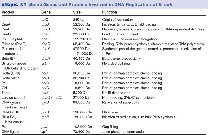
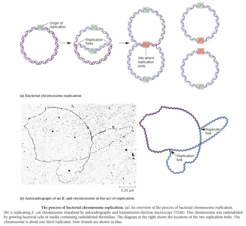
In prokaryotes, the DNA is circular.  Replication starts at a single origin (ori C) and is bi-directional. The region of replicating DNA associated with the single origin is called a replication bubble  and consists of two replication forks moving in opposite direction around the DNA circle. During DNA replication, the two parental strands separate and each acts as a template to direct the enzyme catalysed synthesis of a new complementary daughter strand following the normal base pairing rule. At least 10 different enzymes or proteins  participate in the initiation phase of replication. Three basic steps involved in DNA replication are Initiation, elongation and termination, subdivided in eight discrete steps.

http://reasonandscience.heavenforum.org/t1849-dna-replication-of-prokaryotes#4365

Initiation phase: 

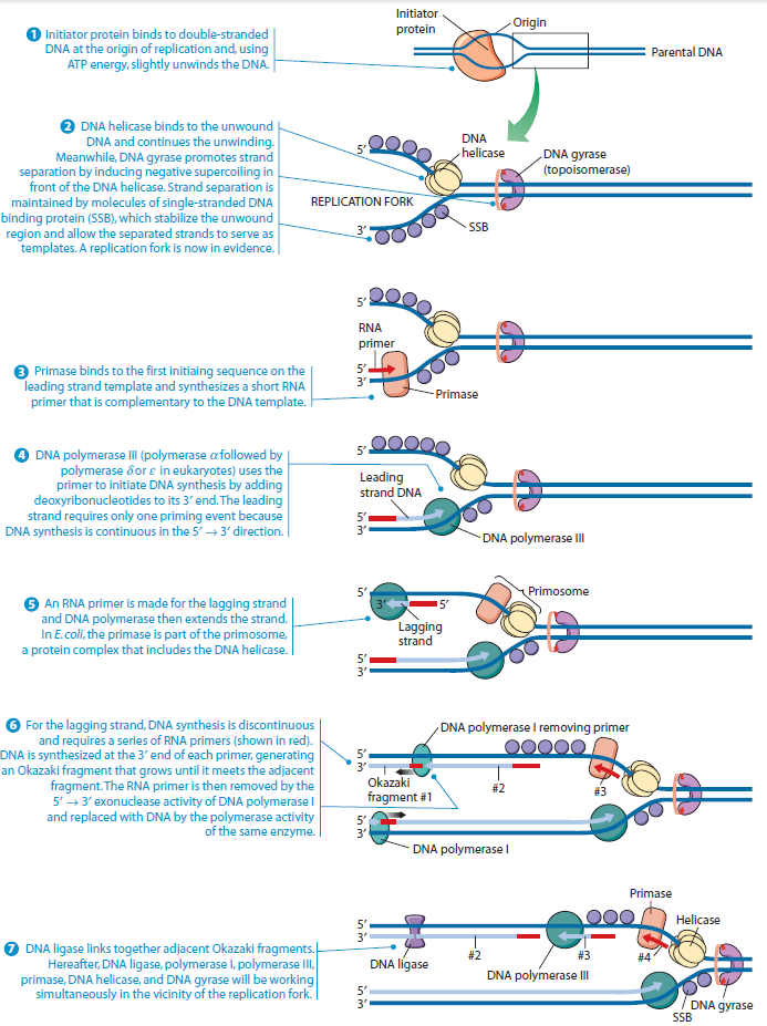
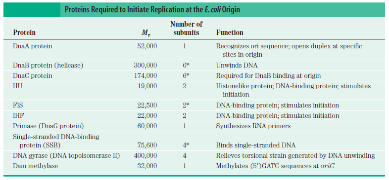
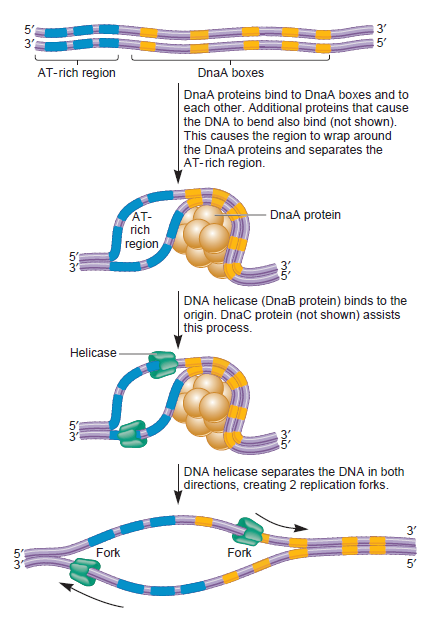
Step 1: Initiation begins, when DNA binds around an initiator protein complex DnaA with the goal to pull the two DNA strands apart. That  creates a number of problems. First of all, the two strands like to be together - they stick to each other just as if they had tiny magnets up and down their length. In order to pull apart the DNA you have to put energy into the system. In modern cells, a protein called DnaA  binds to a specific spot along the DNA, called single origin ( oriC ) and the protein proceeds to open up the double strand. The protein is a monomer, has motifs to bind to unique monomer sites, also they have motifs for protein-protein interaction, thus they can form clusters.   They have hydrophobic regions for helical coiling and protein–protein interactions.  Binding of the monomers to DnaA-A boxes, in ATP dependent manner (proteins have ATPase activity), leads to cooperative binding of more proteins.  This clustering of proteins on DNA makes the DNA to wrap around the proteins, which induces torsional twist and it is this left handed twist that makes DNA to melt at 13-mer region and AT rich region; perhaps the negative super helical topology in this region may further facilitate the melting of the DNA. Opening or unwinding of dsDNA ( double strand DNA )  into single stranded region is an important event in initiation.  

Single-strand binding protein (SSB)
https://reasonandscience.catsboard.com/t1849p15-dna-replication-of-prokaryotes#4377

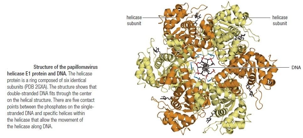
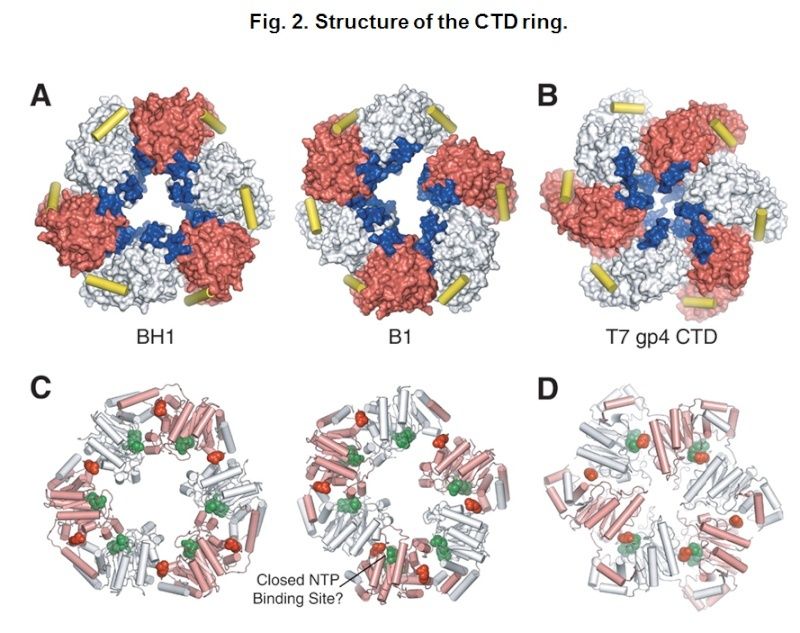
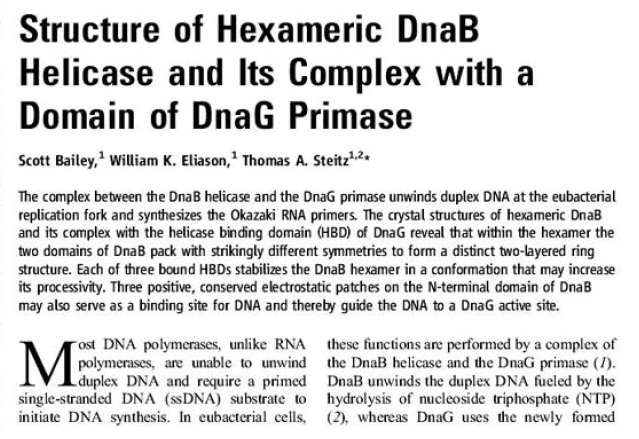
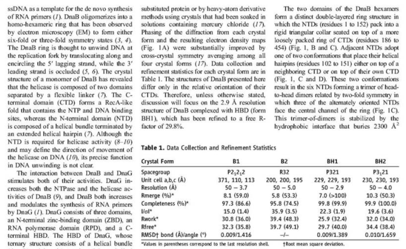
The Hexameric DnaB Helicase
https://reasonandscience.catsboard.com/t1849p15-dna-replication-of-prokaryotes#4367

DnaC, and strategies for helicase recruitment and loading in bacteria
https://reasonandscience.catsboard.com/t1849p15-dna-replication-of-prokaryotes#4371

Unwinding the DNA Double Helix Requires DNA Helicases,Topoisomerases, and Single- Stranded DNA Binding Proteins
https://reasonandscience.catsboard.com/t1849p15-dna-replication-of-prokaryotes#4374

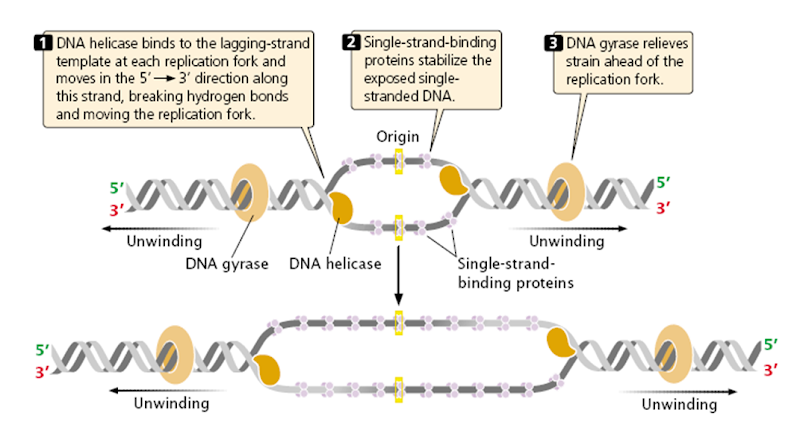
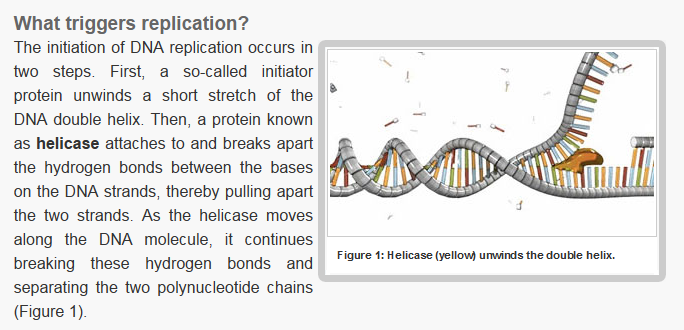
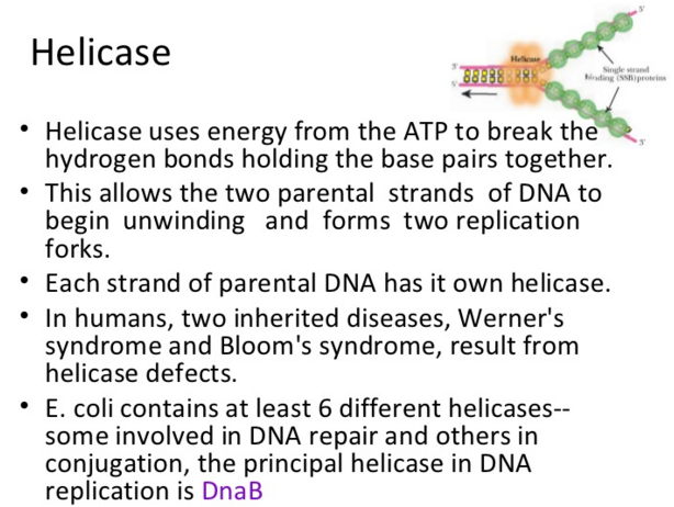
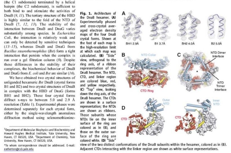
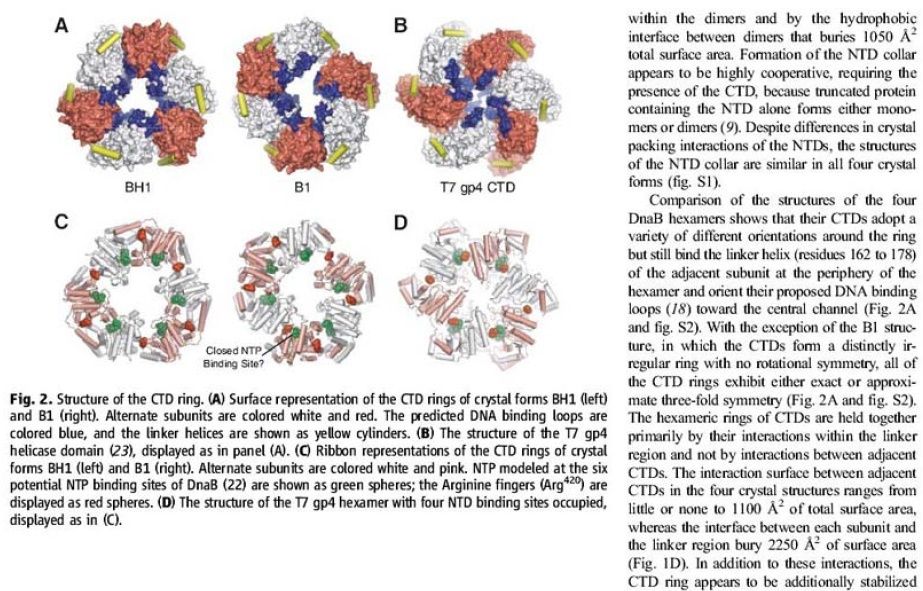
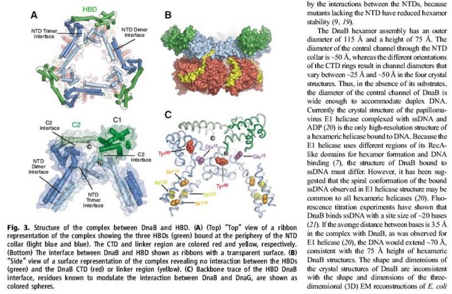
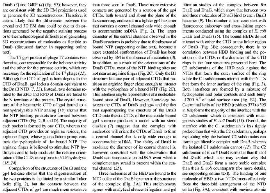
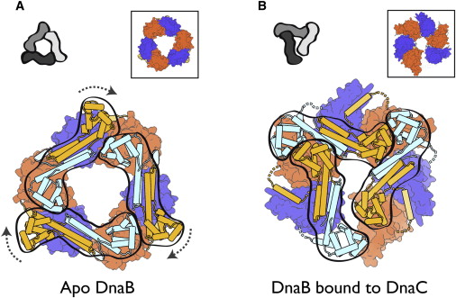
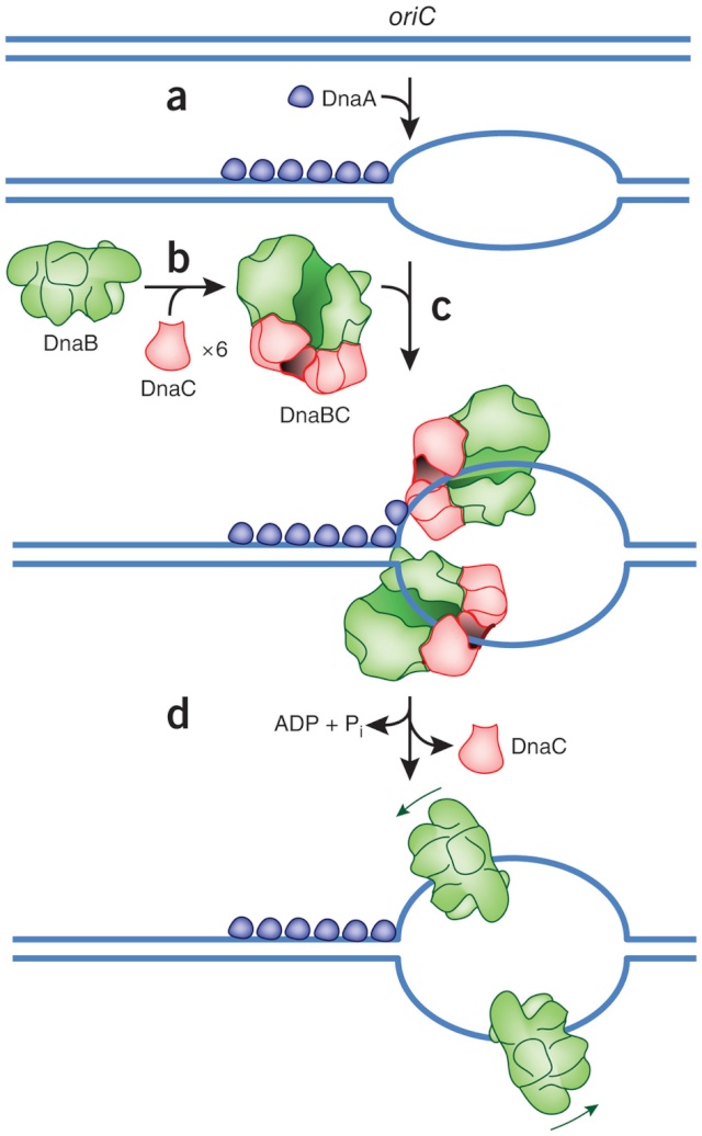
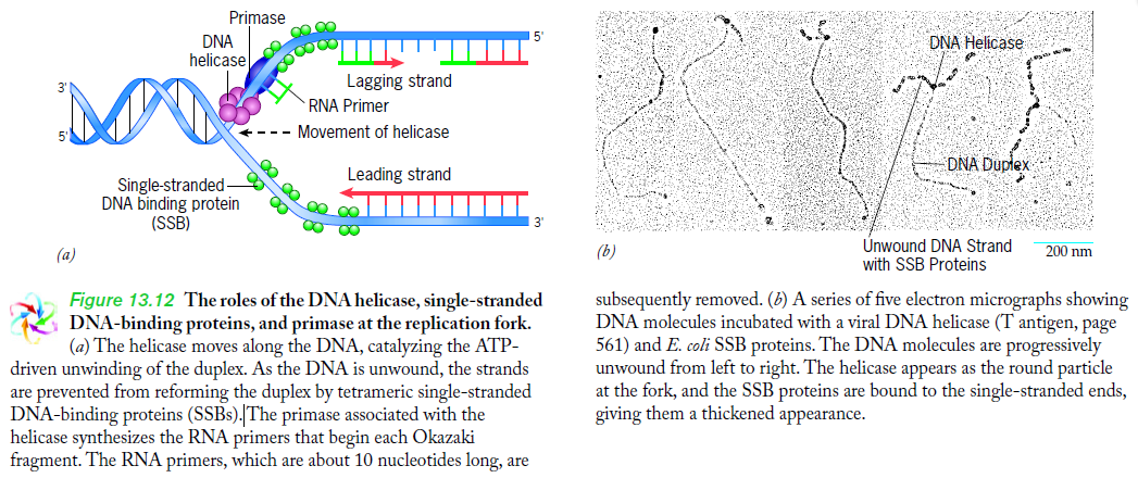
Step 2:  During DNA replication, the two strands of the double helix must unwind at each replication fork to expose the single strands to the enzymes responsible for copying them. Three classes of proteins with distinct functions facilitate this unwinding process: DNA helicases, topoisomerases, and single-stranded DNA binding proteins ( SSB's). Helicase ( DnaB ) now comes along. The helicase exposes a region of single-stranded DNA that must be kept open for copying to proceed. Helicase is like a snowplow; it is a molecular machine that plows down the middle of the double helix, pushing apart the two strands. this allows the polymerase and associated proteins to travel along behind it in ease and comfort.  DnaB helicase alone has no affinity for ssDNA ( single stranded DNA ) bound by SSB (single- stranded binding protein). Thus, entry of the DnaB helicase complex into the unwound oriC depends on DnaC, a  additional protein factor. DnaC helps or facilitates the helicase to be loaded onto ssDNA  at the replication fork in ATP dependent manner. The DnaB-DnaC complex forms a topologically open, three-tiered toroid.  DnaC remodels DnaB to produce a cleft in the helicase ring suitable for DNA passage. DnaC’s  fold is dispensable for DnaB loading and activation. DnaB possesses autoregulatory elements that control helicase loading and unwinding. Using energy derived from ATP hydrolysis, these proteins unwind the DNA double helix in advance of the replication fork, breaking the hydrogen bonds as they go. Helicase recruitment and loading in bacteria is a remarkable process. Following video shows how that works: 

https://www.youtube.com/watch?v=YzNuLsqMqyE


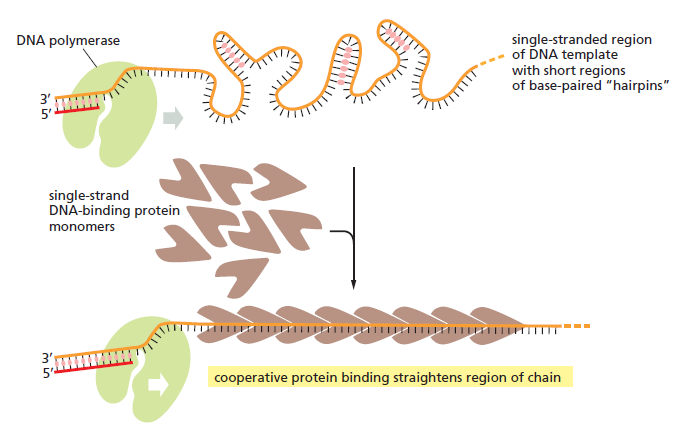
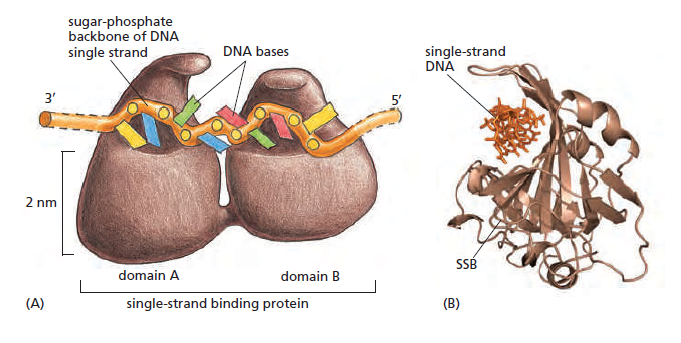
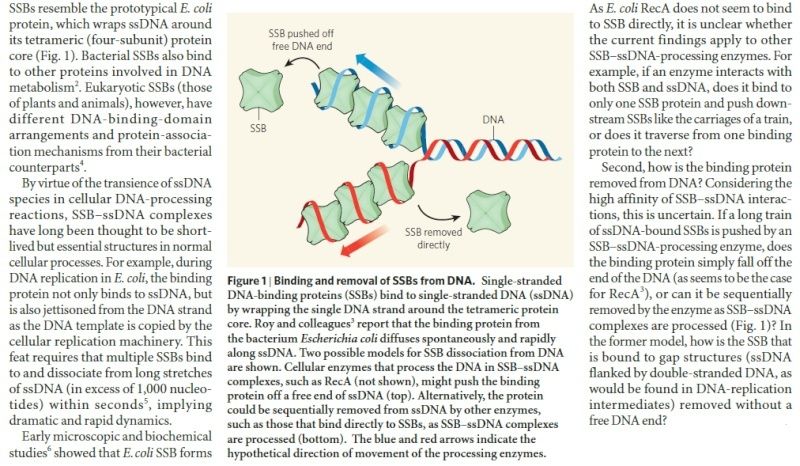
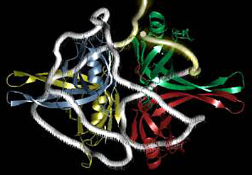
There is a problem, though, with this setup. If you push apart two DNA strands they generally do not float around separately. If they are close to one another they will rapidly snap back and form a double strand again almost as soon as the helicase passes. Even if the strands are not near each other, a single strand will usually fold up and form hydrogen bonds with itself - in other words, a tangled mess. So it is not enough to push apart the two strands of DNA; there must be a way to keep the strands apart once they have been separated. In modern cells this job is done by single-strand binding proteins, or SBB's. As the helicase separates the strands of DNA, SSB's bind to the single-stranded DNA and coats them.   . SSB's prevent DNA from reannealing. SSB's associate to form tetramers around which the DNA is wrapped in a manner that significantly compacts the single-stranded DNA. There is another difficulty in being a double helix. The unwinding associated with DNA replication would create an intolerable amount of supercoiling and possibly tangling in the rest of the DNA. It can be illustrated with a simple example. Take two intertwined shoelaces and ask a friend to hold them together at each end. Now take a pencil, insert it between the strands near one end, and start pushing it down toward the other end. As you can see, shoestrings behind the pencil become melted, in the jargon of biochemistry. The shoestrings ahead of the pencil become more and more tangled. It becomes harder and harder to push the pencil forward.  Helicase and polymerase encounter the same problem with DNA. It does not matter whether you are talking about interwined strings or interwined DNA strands. The problem of tangling is the result of the topological interconnectness of the two strands. If this problem persisted for very long in a  cell, DNA replication would grind to a halt. However, the cell contains several enzymes, called topoisomerases, to take care of the difficulty. The way in which they do so can be illustrated with a enzyme called gyrase. Gyrase binds to DNA, pulls them apart and allows a separate portion of the DNA to pass through the cut. It then reseals the cut and lets go of the DNA. This action decreases the number of twists in DNA. The parental DNA is unwound by DNA helicases and SSB (travels in 5’-3’ direction), the resulting positive super-coiling (torsional stress) is relieved by topoisomerse I and II (DNA gyrase) by inducing transient single stranded breaks.Topoisomerases are amazing enzymes. In this topic, a video shows how they function : 

Topoisomerase II enzymes, amazing evidence of design
https://reasonandscience.catsboard.com/t2111-topoisomerase-ii-enzymes-amazing-evidence-of-design?highlight=topoisomerase

In modern organisms, helicase, SSB, and gyrase all are required at the replication fork. Mutants in which any of them are missing are not viable - they die.

Question: Had not all three parts, the SSB binding proteins, the topoisomerase, and the helicase and the DnaC loading proteins not have to be there all at once, otherwise, nothing goes? They might exercise their function on their own, but then they would not replicate DNA or have function in a bigger picture. It's evident that they had to come together to provide a functional whole.  What we see here are highly coordinated, goal-oriented tasks with specific movements designed to provide a specific outcome. Auto-regulation and control that seems required beside constant energy supply through ATP enhance the difficulty to make the whole mechanism work in the right manner. All this is awe-inspiring and evidences the wise guidance and intelligence required to make all this happening in the right way.

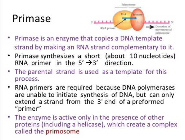
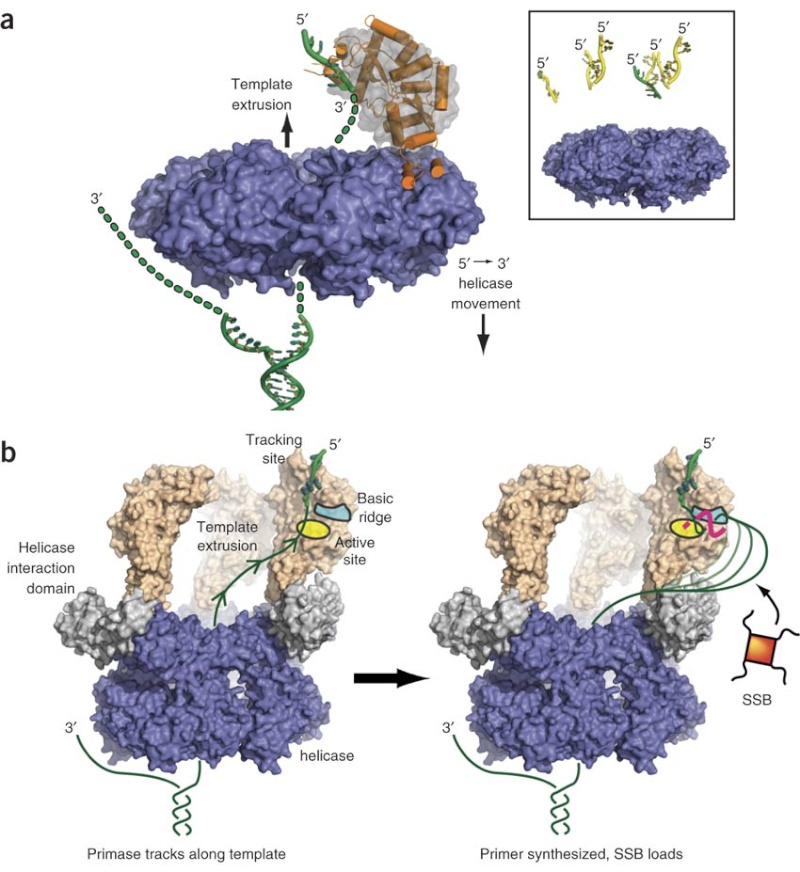
Step 3:  The enzyme DNA primase (primase, an RNA polymerase)  attaches to the DNA and synthesizes a short RNA primer to initiate synthesis of the leading strand of the first replication fork.

Elongation phase : 

Step 4: In the elongation fase, DNA polymerase III extends the RNA primer made by primase.

DNA Polymerase
https://reasonandscience.catsboard.com/t1849p15-dna-replication-of-prokaryotes#4375

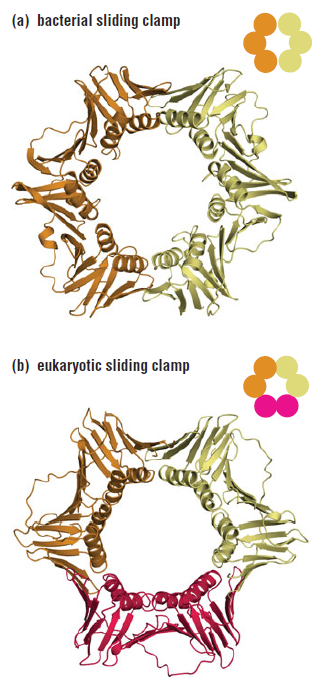
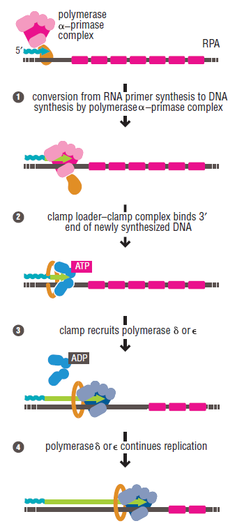
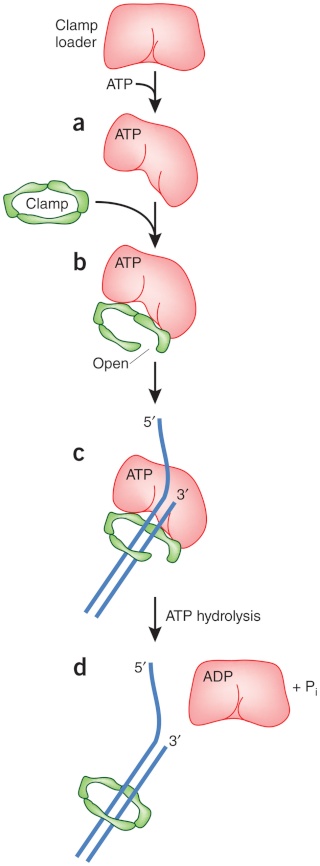
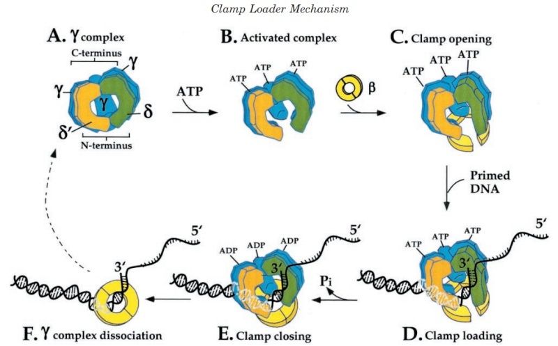
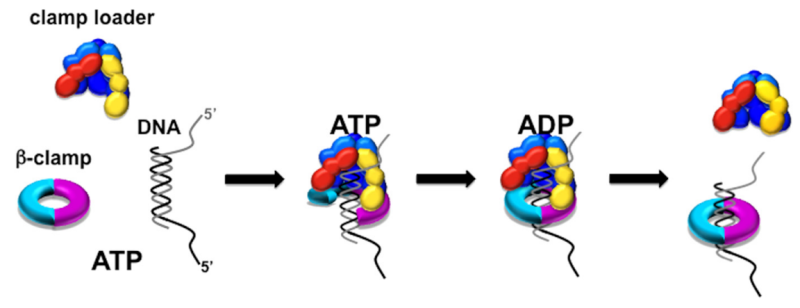
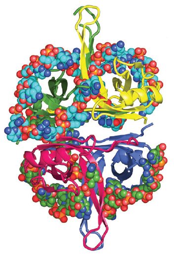
DNA polymerase possesses separate catalytic sites for polymerization and degradation of nucleic acid strands. All DNA polymerases make DNA in 5’-3’ direction . A ring-shaped sliding clamp protein encircles the DNA double helix and binds to DNA polymerase, thereby allowing the DNA polymerase to slide along the DNA while remaining firmly attached to it. Most enzymes work by colliding with their substrate, catalyzing a reaction and dissociating from the product. If that were the case with DNA polymerase, then it would bind to DNA, add a nucleotide to the new chain that was being made, and then fall off of the chain. Then ,put the next nucleotide onto the growing end,  bind it and catalyze the addition. This same cycle would have to repeat itself a very large number of times to complete a new DNA chain. Polymerases however catalyze the addition of a nucleotide but do not fall off the DNA. Rather, they stay bound to it, until the next nucleotide comes in, and then they catalyze its addition to the chain. and they again stay bound. If it were not so, the replication process would be very slow. In the cell, polymerases stay on the DNA until their job is completed, which might be only after millions of nucleotides have been joined. This velocity is only possible because of clamp proteins. These have a ring shape. The ring can be opened up.  These clamp proteins are joined to the DNA polymerase in a intricate way, through a clamp loader protein, which has a remarkable shape similar to a human hand. It takes the clamp, like a hand with five fingers would grab it, opens it up becoming like a doughnut shape,where the whole hole in the middle is big enough to accommodate the DNA,  and then, when it is on the DNA, it positions it in a precise manner on the DNA polymerase, where it stays bound until it reaches the end of its polymerizing job. Through this ingenious process, the clamp stabilizes the DNA, making it possible to increase the speed of polymerization dramatically.  They can be seen here:

The sliding clamp and clamp loader
https://reasonandscience.catsboard.com/t1849p15-dna-replication-of-prokaryotes#4376

Question: How would and could natural, unguided processes have figured out 1. the requirement of high-speed of polymerization? How could they have figured out the right configuration and process to do so? how could natural processes have emerged with the right proteins incrementally, with the hand-shaped clamp loader, and the precisely fitting clamp, enabling the fast process ?? Even the most intelligent scientists are still not able to imagine how this process is engineered?  Furthermore, the process requires molecular energy in the form of ATP, and everything must fit together, and be functional. Without the clamp loader protein, the clamp could not be positioned to the polymerase enzyme, and processivity would not rise to the required speed. The whole process must also be regulated and controlled. How could that regulation have been programmed? Trial and error? 

Several Proteins Are Required for DNA Replication at the Replication Fork
https://reasonandscience.catsboard.com/t1849p15-dna-replication-of-prokaryotes#4398

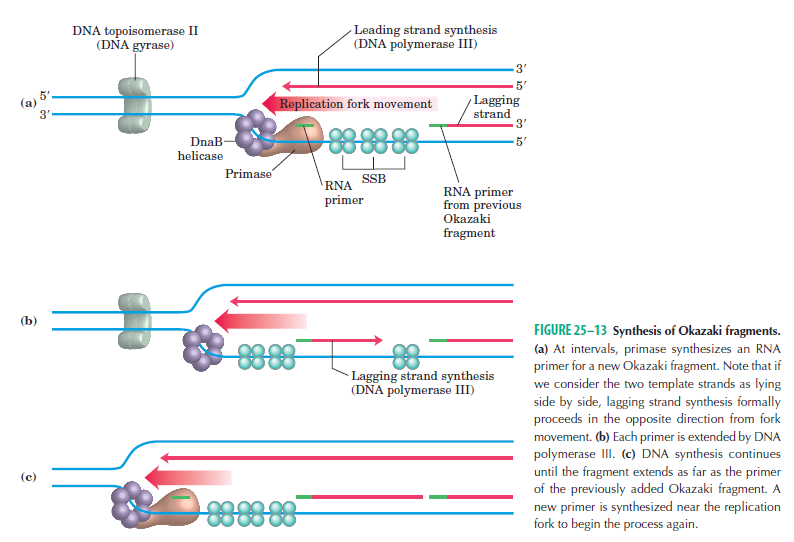
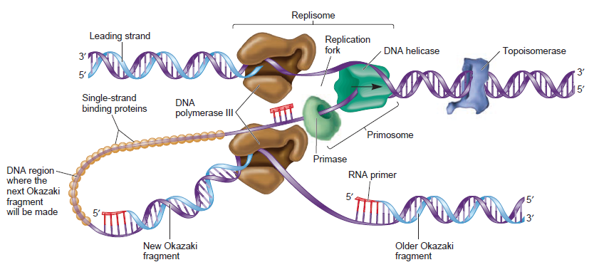
The various proteins involved in DNA replication are all closely associated in one large complex, called a replisome.  
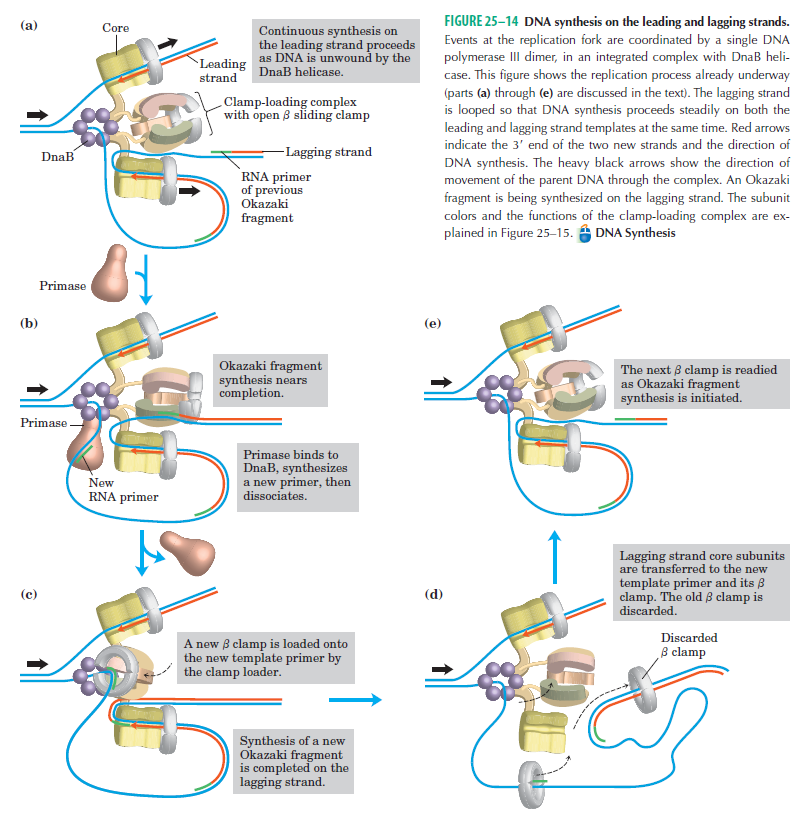
Leading strand synthesis:  On the template strand with 3’-5’ orientation, new DNA is made continuously in 5’-3’ direction towards the replication fork. The new strand that is continuously synthesized in 5’-3’ direction is the leading strand. 
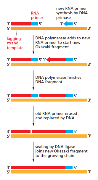
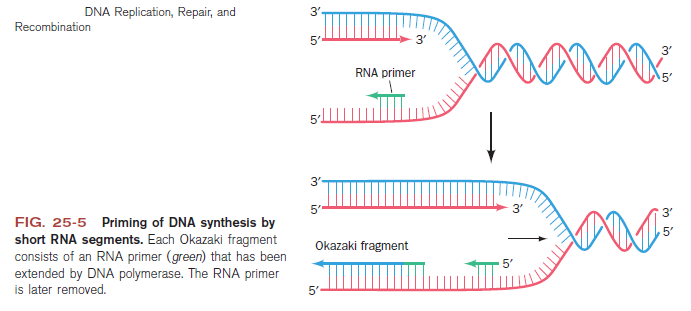
Lagging strand synthesis: In the lagging strand, the synthesis of DNA also elongates in a 5ʹ to 3ʹ manner, but it does so in the direction away from the replication fork. In the lagging strand, RNA primers must repeatedly initiate the synthesis of short segments of DNA; thus, the synthesis has to be discontinuous.

The Primase (DnaG) enzyme, and the primosome complex
https://reasonandscience.catsboard.com/t1849p15-dna-replication-of-prokaryotes#4379

 The length of these fragments in bacteria is typically 1000 to 2000 nucleotides. In eukaryotes, the fragments are shorter—100 to 200 nucleotides. Each fragment contains a short RNA primer at the 5ʹ end, which is made by primase. The remainder of the fragment is a strand of DNA made by DNA polymerase III. The DNA fragments made in this manner are known as Okazaki fragments. To complete the synthesis of Okazaki fragments within the lagging strand, three additional events must occur: removal of the RNA primers, synthesis of DNA in the area where the primers have been removed, and the covalent attachment of adjacent fragments of DNA. In E. coli, the RNA primers are removed by the action of DNA polymerase I. This enzyme has a 5ʹ to 3ʹ exonuclease activity, which means that DNA polymerase I digests away the RNA primers in a 5ʹ to 3ʹ direction, leaving a vacant area. DNA polymerase I then synthesizes DNA to fill in this region. It uses the 3ʹ end of an adjacent Okazaki fragment as a primer. , DNA polymerase I would remove the RNA primer from the first Okazaki fragment and then synthesize DNA in the vacant region by attaching nucleotides to the 3ʹ end of the second Okazaki fragment. After the gap has been completely filled in, a covalent bond is still missing between the last nucleotide added by DNA polymerase I and the adjacent DNA strand that had been previously made by DNA polymerase III. To the left of the origin, the top strand is made continuously, whereas to the right of the origin it is made in Okazaki fragments. By comparison, the synthesis of the bottom strand is just the opposite. To the left of the origin it is made in Okazaki fragments and to the right of the origin the synthesis is continuous. Finally the two ends of the fragment have to be joined together; this is the job of an enzyme called DNA ligase.  After the completion of one Okazaki fragment , the equipment has to be released, the clamp has to let go, and a new clamp has to be loaded at the beginning of the next fragment. Clearly the formation and control of the replication fork is an enormously complex process. 

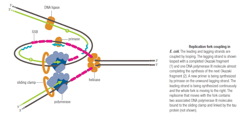
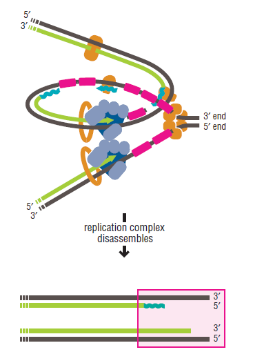
Step 5:   After DNA synthesis by DNA pol III, DNA polymerase I uses its 5’-3’ exonuclease activity to remove the RNA primer and fills the gaps with new DNA. In the next step, finally DNA ligase joins the ends of the DNA fragments together.  As the replisome moves along the DNA in the direction of the replication fork, it must accommodate the fact that DNA is being synthesized in opposite directions along the template on the two stands. Picture above  provides a schematic model illustrating how this might be accomplished by folding the lagging strand template into a loop.Creating such a loop allows the DNA polymerase molecules on both the leading and lagging strands to move in the same physical direction, even though the two template strands are oriented with opposite polarity. The replisome faces special challenges as it makes new DNA at rates that can approach 1,000 nucleotides per second. Unlike the machines that make proteins and RNA, which work relatively sluggishly and in a linear fashion, the replisome must simultaneously copy two strands of DNA that are aligned in opposite directions (5ʹ to 3ʹ and 3ʹ to 5ʹ)Replisome chemistry obeys two rules


Questions: How did they arise with that capability to " obey two rules " ?  Suppose a primitive polymerase were duplicated and somehow started to replicate the second strand in the opposite direction while remaining attached to the first strand -  how could that change have been directed, and why should that feat have happened randomly? 


DNA replication of prokaryotes Dna_po19

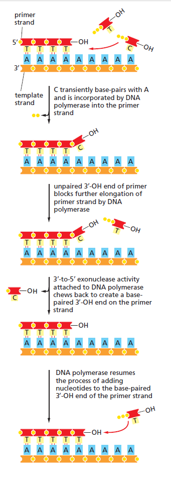
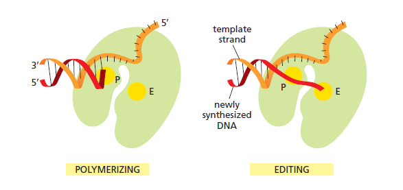
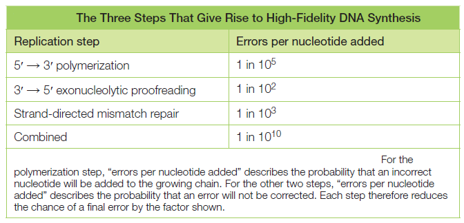
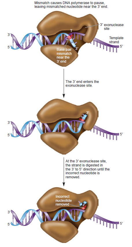
The DNA polymerase holoenzyme alone would not be able to duplicate the long DNA faithfully. Tests have shown that Polymerase III alone gets stuck. Furthermore, Polymerase III is not a simple enzyme. Its rather three enzymes in one. Beside replicating DNA, it can also degrade DNA in two different ways. It does so by three different, discrete regions of the molecule. The exonuclease activity plays a critical role in replication. It allows the enzyme to proofread the new DNA and cut out any mistakes it has made. Although the polymerase reads the sequence of the old DNA to produce a new DNA, it turns out that simple base bairing allows about one mistake per thousand base pairs copied. Proofreading reduces errors to about one mistake in a million base pairs. The question is if wheter  a proofreading exonuclease and other DNA repair mechanisms had to be present in the very first cell. 

Eigen’s theory revealed the existence of the fundamental limit on the fidelity of replication (the Eigen threshold): If the product of the error (mutation) rate and the information capacity (genome size) is below the Eigen threshold, there will be stable inheritance and hence evolution; however, if it is above the threshold, the mutational meltdown and extinction become inevitable (Eigen, 1971). The Eigen threshold lies somewhere between 1 and 10 mutations per round of replication (Tejero, et al., 2011) regardless of the exact value, staying above the threshold fidelity is required for sustainable replication and so is a prerequisite for the start of biological evolution. Indeed, the very origin of the first organisms presents at least an appearance of a paradox because a certain minimum level of complexity is required to make self-replication possible at all; high-fidelity replication requires additional functionalities that need even more information to be encoded (Penny, 2005). The crucial question in the study of the origin of life is how the Darwin-Eigen cycle started—how was the minimum complexity that is required to achieve the minimally acceptable replication fidelity attained? In even the simplest modern systems, such as RNA viruses with the replication fidelity of only about 10^3 and viroids that replicate with the lowest fidelity among the known replicons (about 10^2; Gago, et al., 2009), replication is catalyzed by complex protein polymerases. The replicase itself is produced by translation of the respective mRNA(s), which is mediated by the immensely complex ribosomal apparatus. Hence, the dramatic paradox of the origin of life is that, to attain the minimum complexity required for a biological system to start on the Darwin-Eigen spiral, a system of a far greater complexity appears to be required. How such a system could evolve is a  puzzle that defeats conventional evolutionary thinking, all of which is about biological systems moving along the spiral; the solution is bound to be unusual. 

DNA damage and repair
https://reasonandscience.catsboard.com/t2043-dna-repair?highlight=dna+repair
https://reasonandscience.catsboard.com/t1849p30-dna-replication-of-prokaryotes#4401

Replication forks may stall frequently and require some form of repair to allow completion of chromosomal duplicationFailure to solve these replicative problems comes at a high price, with the consequences being genome instability, cell death and, in higher organisms, cancer. Replication fork repair and hence reloading of DnaB may be needed away from oriC at any point within the chromosome and at any stage during chromosomal duplication. The potentially catastrophic effects of uncontrolled initiation of chromosomal duplication on genome stability suggests that replication restart must be regulated as tightly as DnaA-directed replication initiation at oriC. This implies reloading of DnaB must occur only on ssDNA at repaired forks or D-loops rather than onto other regions of ssDNA, such as those created by blocks to lagging strand synthesis.Thus an alternative replication initiator protein, PriA helicase, is utilized during replication restart to reload DnaB back onto the chromosome 

Question: Could the first cell, with its required complement of genes coded for by DNA, have successfully reproduced for a significant number of generations without a proofreading function? A further question is how the function of synthesis of the lagging strand could have arisen, and the machinery to do so. That is, the Primosome, and the function of Polymerase I to remove the short peaces of RNA that the cell uses to prime replication, allowing the polymerase III function to fill the gap. These functions all require precise regulation, and coordinated functional machine-like steps. These are all complex, advanced functions and had to be present right from the beginning. How could this complex machinery have emerged in a gradual manner? the Primosome had to be fully functional, otherwise, polymerisation could not have started, since a prime sequence is required.

Step 6: Finally DNA ligase joins the ends of the DNA fragments together.

Termination phase: 

Termination of DNA replication
https://reasonandscience.catsboard.com/t1849p15-dna-replication-of-prokaryotes#4399

Step 7: The two replication forks meet ~ 180  degree opposite to ori C, as DNA is circular in prokaryotes. Around this region there are several terminator sites which arrest the movement of forks by binding to the tus gene product, an inhibitor of helicase (Dna B).
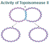
Step 8: Once replication is complete, the two double stranded circular DNA molecules (daughter strands) remain interlinked. Topoisomerase II makes double stranded cuts to unlink these molecules.

The Astonishing Precision of DNA Replication

The astonishing accuracy and speed of DNA replication in organisms like E. coli underscore the remarkable efficiency of the molecular machinery involved in this essential biological process. Let's examine the implications of these statistics and the incredible capabilities of the replication machinery at a larger scale. With an error rate of approximately 1 in 1,000,000,000, DNA replication in E. coli achieves a level of fidelity that is unparalleled in human-made processes. This precision is a testament to the extremely accurate operating mechanisms and quality control systems in place during DNA synthesis. Such low error rates are crucial for maintaining the genetic integrity of an organism over countless generations. Moreover, the speed at which DNA replication occurs is equally remarkable. E. coli, a model organism for studying this process, can replicate at a rate of about one thousand nucleotides per second. When scaled up to human proportions, this would mean that the replication machinery, comprised of various enzymes and proteins, would be moving at an astounding speed of approximately 600 kilometers per hour (375 miles per hour). To put this into perspective, if we were to imagine DNA as a massive, one-meter-diameter circle, the replication machinery would be akin to a swift vehicle hurtling along at highway speeds. Visualizing this, it becomes clear that the replication machinery is not only swift but also highly efficient, as it manages to process and replicate DNA accurately at such high velocities. Now, consider the scenario where DNA is scaled up to such proportions that it is one meter in diameter. In this hypothetical scenario, the protein-based machinery responsible for DNA replication would be colossal, comparable in size to a FedEx delivery truck. This analogy underscores the complex and intricate nature of the molecular components involved in the replication process. Furthermore, let's contemplate the practical implications of this speed and accuracy. If we were to embark on a journey to replicate the entire E. coli genome, which consists of approximately 4.6 million base pairs, using this machinery, it would be a remarkably swift endeavor. The replication process would take a mere 40 minutes to complete a 400-kilometer (250-mile) journey. To put it in perspective, during this brief journey, these molecular machines, while moving at a breakneck pace, would only make an error in the genetic code once every 170 kilometers (106 miles). This astonishing level of precision allows organisms like E. coli to maintain their genetic information with incredible fidelity as they reproduce and pass their DNA on to future generations. The combination of extreme accuracy and rapidity in DNA replication in organisms like E. coli is a testament to the efficiency and sophistication of the molecular machinery involved. These attributes ensure the faithful transmission of genetic information, a fundamental requirement for the perpetuation of life on Earth.

According to mainstream scientific papers, the following twenty protein and protein complexes are essential for prokaryotic DNA replication. Each one mentioned below. They cannot be reduced. If one is missing, DNA replication cannot occur: 

Pre-replication complex  Formation of the pre-RC is required for DNA replication to occur
DnaA The crucial component in the initiation process is the DnaA protein
DiaA this novel protein plays an important role in regulating the initiation of chromosomal replication via direct interactions with the DnaA initiator.
DAM methylase  It’s gene expression requires full methylation of GATC at its promoter region. 
DnaB helicase Helicases are essential enzymes for DNA replication, a fundamental process in all living organisms.
DnaC Loading of the DnaB helicase is the key step in replication initiation.  DnaC is essential for replication in vitro and in vivo
HU-proteins  HU protein is required for proper synchrony of replication initiation
SSB Single-stranded binding proteins  Single-stranded DNA binding proteins are essential for the sequestration and processing of single-stranded DNA. 6
SSBs from the OB domain family play an essential role in the maintenance of genome stability, functioning in DNA replication, the repair of damaged DNA, the activation of cell cycle checkpoints, and in telomere maintenance. SSB proteins play an essential role in DNA metabolism by protecting single-stranded DNA and by mediating several important protein–protein interactions. 7
Hexameric DNA helicases DNA helicases are essential during DNA replication because they separate double-stranded DNA into single strands allowing each strand to be copied.

DNA polymerase I and III DNA polymerase 3 is essential for the replication of the leading and the lagging strands whereas DNA polymerase 1 is essential for removing of the RNA primers from the fragments and replacing it with the required nucleotides. 
DnaG Primases  They are essential for the initiation of such phenomena because DNA polymerases are incapable of de novo synthesis and can only elongate existing strands 
Topoisomerases  are essential in the separation of entangled daughter strands during replication. This function is believed to be performed by topoisomerase II in eukaryotes and by topoisomerase IV in prokaryotes. Failure to separate these strands leads to cell death. 

Sliding clamp and clamp loader the clamp loader is a crucial aspect of the DNA replication machinery.  Sliding clamps are DNA-tracking platforms that are essential for processive DNA replication in all living organisms 
Primase (DnaG) Primases are essential RNA polymerases required for the initiation of DNA replication, lagging strand synthesis and replication restart.  They are essential for the initiation of such phenomena because DNA polymerases are incapable of de novo synthesis and can only elongate existing strands. 
RTP-Ter complex Ter sequences would not seem to be essential, but they may prevent overreplication by one fork in the event that the other is delayed or halted by an encounter with DNA damage or some other obstacle
Ribonuclease H  RNase H1 plays essential roles in generating and clearing RNAs that act as primers of DNA replication. 
Replication restart primosome Replication restart primosome is a complex dynamic system that is essential for bacterial survival. 
DNA repair: 
RecQ helicase  In prokaryotes RecQ is necessary for plasmid recombination and DNA repair from UV-light, free radicals, and alkylating agents. 
RecJ nuclease the repair machinery must be designed to act on a variety of heterogeneous DNA break sites.

Commentary: I am not aware of any scientific literature that provides a comprehensive, detailed explanation for the naturalistic emergence of DNA replication or any of its components de novo, without attributing a role to intelligent intervention. The intricate systems underpinning DNA replication surpass the capacity of unguided natural processes to explain them adequately. The high level of complexity and precision inherent in these mechanisms strongly suggests, in my perspective, the influence of an intelligent designer. The substantial coordination and specificity observed in these systems do not readily lend themselves to explanations rooted solely in chance or spontaneous generation, bolstering the argument for intelligent design as a credible and compelling explanation for the origin and mechanisms of DNA replication. Precisely because each of the detailed parts is essential, the entire system had to emerge simultaneously. Intelligence is known to have the capability to envision, design, and construct such motor-like systems based on extensive information. This knowledge further affirms that the emergence of such an intricate system through evolutionary processes is implausible. This is especially pertinent considering that the process of evolution itself inherently relies on the already established mechanism of DNA replication. Given these circumstances, it's rational to posit design as the most compelling explanation. The prospect of such a complex, interdependent system originating by chance is an untenable proposition. The individual components of the DNA replication system lack utility in isolation, and there's no plausible explanation for the spontaneous, random assembly of such an ordered, purposeful structure from disorder. It defies reason to assume that matter would arbitrarily organize itself into a highly specialized, complex, machine-like system, further reinforcing the argument for intelligent design as the most persuasive explanation for the origin of DNA replication.

Claim: DNA replication is due to millions of years of evolution, no god required!
Reply: DNA replication is a fundamentally complex process, essential for the propagation of life as it ensures the transmission of genetic information from one generation to the next.  DNA replication had to be fully established at the onset of life and is grounded in the understanding that it is indispensable for cellular self-replication and subsequent evolutionary processes. Without a reliable mechanism to ensure accurate replication of genetic information, the continuity and diversification of life would be unattainable. The process of DNA replication in itself is a marvel of molecular coordination and precision. It involves a host of specialized proteins and enzymes that work in synchrony to unwind the DNA double helix, synthesize complementary strands, and correct errors to ensure the fidelity of the replicated DNA. For instance, the enzyme DNA polymerase plays a crucial role in synthesizing the new DNA strands, while helicase is responsible for unwinding the double helix, and ligase seals gaps to produce a continuous DNA strand.

The origin of this intricate system poses a substantial puzzle. How could such a complex, highly coordinated process spontaneously emerge in the primordial environment? The system's intricacy and the interdependence of its components render it a challenging scenario for gradual evolutionary assembly. Each component within the DNA replication machinery is integral, and the absence or malfunction of even a single element could lead to the collapse of the entire process. Moreover, DNA carries within its structure the coded instructions necessary for building and operating the organism. This information is fundamental for the synthesis of proteins, the workhorses of the cell responsible for virtually every task necessary for the cell’s structure, function, and regulation. The paradox here lies in the fact that the machinery required for reading and interpreting this genetic information (such as ribosomes and various RNA molecules) is itself encoded within the DNA. This creates a scenario akin to a “chicken and egg” problem: the information in DNA is necessary to produce the machinery that reads the information, yet this machinery is required to read and implement the information contained within the DNA.

The multifaceted and complex nature of DNA replication serves as a testimony to a purposeful creation by an intelligent source. The intricacies of the DNA replication process defy the expectations of a random emergence and autonomous assembly of life's fundamental machinery. The coordinated action of various proteins and enzymes such as DNA polymerase, helicase, and ligase, each performing a unique and indispensable function, reflects a system that is designed with precision and foresight. The level of coordination and complexity involved in DNA replication cannot be understated. The seamless interplay among various components to ensure the accurate duplication of genetic information exemplifies an orchestrated and purposeful operation, aligning more with deliberate design than with accidental emergence. The presence of multiple, interdependent components working concurrently to achieve a specific outcome – the accurate replication of DNA – resonates with the characteristics of designed systems observed in human-engineered technologies. Additionally, the information coding within the DNA molecule, essential for the construction and operation of living organisms, echoes the principles of intelligent communication systems. The precise, coded information embedded within the DNA sequence, and its role in dictating the synthesis of proteins, underscores the presence of a sophisticated information storage and retrieval system akin to advanced computational algorithms. This analogy bolsters the argument for intelligent design as the source of such a high-level information management system. The DNA replication system embodies irreducible complexity as it is composed of multiple components, each essential for the system's function. The absence of any single element would render the system nonfunctional, suggesting that the system must have been fully formed from the onset, contrary to the gradual assembly postulated by evolutionary mechanisms. The order and precision inherent in the DNA replication process further reinforce the argument for intelligent design. The specificity in the function of each component, the timing of each step, and the error-checking and correction mechanisms underscore a level of order and precision consistent with purposeful design rather than random occurrence. In view of these facts, the DNA replication process, with its complexity, coordination, information coding, irreducible complexity, and order and precision, stands as a compelling illustration of purposeful and intelligent design in the biological realm. They argue that such a sophisticated and intricate system, essential for life, more plausibly originates from an intelligent source than from unguided naturalistic processes.

https://www.youtube.com/watch?v=0Ha9nppnwOc


more: http://www.powershow.com/view1/214239-ZDc1Z/Biology_is_based_on_the_most_complex_chemical_systems_known_to_man_powerpoint_ppt_presentation


1) http://www.weizmann.ac.il/plants/Milo/images/MolMachinesSize120116Clean.pdf
2) http://cshperspectives.cshlp.org/content/5/10/a010116.full
3) https://en.wikipedia.org/wiki/Control_of_chromosome_duplication
4) http://www.biochem.umd.edu/biochem/kahn/molmachines/replication/Dna%20C%20protein.htm
5) http://www.nature.com/nsmb/journal/v15/n1/full/nsmb1356.html
6) http://www.biomedcentral.com/1471-2199/14/9
7) http://www.ncbi.nlm.nih.gov/pmc/articles/PMC3632105/
8 ) Meyer, signature of the cell, page 111
9 ) http://journal.frontiersin.org/article/10.3389/fmicb.2014.00735/full#F1
10 ) http://creationsafaris.com/epoi_c08.htm
11) http://creationsafaris.com/ar_srds.htm
12) http://www.ncbi.nlm.nih.gov/books/NBK6360/
13) from the book: The Logic of Chance: The Nature and Origin of Biological Evolution By Eugene V. Koonin, page 266
14) https://www-als.lbl.gov/index.php/science-highlights/science-highlights/669-structures-of-clamp-loader-complexes-are-key-to-dna-replication.html
15) http://www.nature.com/nature/journal/v429/n6993/full/nature02585.html
16) http://ethos.bl.uk/OrderDetails.do?uin=uk.bl.ethos.604184
17) http://www.asa3.org/ASA/education/origins/ic-cr.htm
18)  Inaccurate replication would likely have limited the size of the progenote genome due to the risk of “error catastrophe,” the accumulation of so many genetic mistakes that the organism is no longer viable. To illustrate this point, consider the problem of replicating a genome of one million bases, which is sufficient to encode a few hundred RNAs and proteins. (The smallest known genome for an extant free-living bacterium is that of Pelagibacter ubique, which consists of 1.3 million bases.) If replication were even modestly faithful, with an error frequency of 0.1%, every replication of a genome consisting of 1 million bases would result in 1000 errors, approximately one or two in every gene. Some of those errors would have been harmless, and a few might have been beneficial, but many would have been detrimental, leading to macromolecules with impaired functions. 

further readings:

Resources for CHAPTER 3: REPLICATION :  http://users.path.ox.ac.uk/~pcook/w1/3Replication.html#The_mechanics_of_synthesis_at_the_fork


Last edited by Otangelo on Fri Sep 29, 2023 6:26 am; edited 125 times in total

https://reasonandscience.catsboard.com

2DNA replication of prokaryotes Empty Replication: Mechanism of Replication Fri Nov 06, 2015 4:10 am

Otangelo


Admin

DNA replication of prokaryotes

DNA replication is the most crucial step in cellular division, a process necessary for life, and errors can cause cancer and many other diseases. Genome duplication presents a formidable enzymatic challenge, requiring the high fidelity replication of millions of bases of DNA.

How do you get a living cell capable of self-reproduction from a “protein compound … ready to undergo still more complex changes”? Dawkins has to admit:

“Darwin, in his ‘warm little pond’ paragraph, speculated that the key event in the origin of life might have been the spontaneous arising of a protein, but this turns out to be less promising than most of Darwin’s ideas. … But there is something that proteins are outstandingly bad at, and this Darwin overlooked. They are completely hopeless at replication. They can’t make copies of themselves. This means that the key step in the origin of life cannot have been the spontaneous arising of a protein.” (pp. 419–20)

The process of DNA replication depends on many separate protein catalysts to unwind, stabilize, copy, edit, and rewind the original DNA message. In prokaryotic cells, DNA replication involves more than thirty specialized proteins to perform tasks necessary for building and accurately copying the genetic molecule. These specialized proteins include DNA polymerases, primases, helicases, topoisomerases, DNA-binding proteins, DNA ligases, and editing enzymes. DNA needs these proteins to copy the genetic information contained in DNA. But the proteins that copy the genetic information in DNA are themselves built from that information. This again poses what is, at the very least, a curiosity: the production of proteins requires DNA, but the production of DNA requires proteins. 8

DNA replication and protein synthesis is a mind-boggling system involving a city of proteins, enzymes, and other components that are breathtaking in their complexity and efficiency. 11  Proponents of Darwinism are at a loss to tell us how this marvelous system began.  Charles Darwin's main contribution, natural selection, does not apply until a system can reproduce all its parts.  Getting a reproducible cell in a primordial soup is a giant leap, for which today's evolutionary biologists have no answer, no evidence, and no hope.  It amounts to blind faith to believe that undirected, purposeless accidents somehow built the smallest, most complex, most efficient system known to man.

Several decades of experimental work have convinced us that DNA synthesis and replication actually require a plethora of proteins. 12

Replication of the genetic material is the single central property of living systems. Dawkins provocatively claimed that organisms are but vehicles for replicating and evolving genes, and I believe that this simple concept captures a key aspect of biological evolution. All phenotypic features of organisms—indeed, cells and organisms themselves as complex physical entities—emerge and evolve only inasmuch as they are conducive to genome replication. That is, they enhance the rate of this process, or, at least, do not impede it. 13

How DNA Duplicates Itself  10

DNA replication is the vital process on which all heredity depends.  It is reproduction at the molecular level.  Without this DNA copy-making process, life could not be passed along with continuity, if at all.

Francis Crick  described a preliminary problem:
There are still a number of things about the process we do not understand, not the least of which is the fact that the two chains are not lying side by side, but are wound round one another, and that in order for the replication to take place they must be at some stage unwound In addition, the process appears to be one of great precision.11

   
Those who have examined electron micrographs of DNA have noticed the coiled, doubled, knotted twisting and turning that, from our point of observation, appear common in DNA molecules.  Anyone who has ever tried to untangle a microphone cord or lamp cord will wonder how on earth the DNA thread ever can manage this intricate feat.  It must progressively divide, making two double-helix chains in place of one along the entire length of the molecule.  The DNA, remember, is much longer than the diameter of the cell, sometimes about a thousand times as long!12  Ideas on how the unwinding may occur

by rotation as the replication proceeds are feverishly under study.  In fact, it is reported that the rotation during this unwinding occurs at the rate of more than seventy-five turns per second per growing point in bacteria. ( we know today that topoisomerase enzymes do the job in a amazing manner which evidences intelligent design all along )
Pictures have been taken of the DNA molecule of the smallest living thing, to which we have referred before, the Mycoplasma hominis H39.  This DNA is in the form of a long threadlike molecule, joined together in a circle.  Actual replication in process was photographed by H. R.  Bode and Harold Morowitz at Yale.13
The molecule begins dividing into two threads at a certain point, and the division apparently continues until there are two circular loops of DNA instead of one.  These then separate and become the DNA for two daughter cells.  This is an unbelievable “miracle” when you look at the seeming tangles of the long thread during the duplicating process.  Somehow it happens successfully, with a built-in wisdom which at this stage we cannot fathom.  The mechanism involves a growing point complex, containing special proteins and possibly RNA.  This complex may be attached to the cell membrane.
Complementary Pairing Is the Secret of Replication
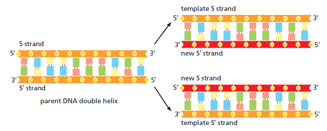
The replication process seems to work in this way.  When the two strands begin to split apart, this leaves each half of the ladder separate.  (See Figure 11.)  Each “base” is thus left with no partner. Floating around in the “juice,” there are various cell parts which have already been made on instructions from the DNA.  These free-floating parts include nucleotides, ready to be fitted together to form a strand of the DNA spiral.  The nucleotides are in an activated condition, with extra phosphates added to give them energy for uniting. From the multitude of free-floating nucleotides, the correct matching ones come alongside the divided strands of the DNA which is duplicating.  As we recall, each base will match none but its one-and-only opposite type.  These then link up to the existing strand and to each other, with the aid of enzymes.  When the process is completed, theDNA is again a ladderlike, double helix.  Each half of the original is replicating at the same

DNA replication or duplication takes place when the ladder-like double helix divides down the center and a new strand forms alongside each of the divided halves, thus making two complete ladders.  By the rules of base pairing, the new strand in each case will be identical to the side it replaces.  The nucleotide units from which it is assembled are preformed and available in activated form in the cell.  The linking is done by enzymes at a very rapid rate, in opposite directions on the two strands-continuously on one and perhaps in short sections on the other near the fork.

Each finally becomes a complete double spiral.  Complementary pairing insures that it will be identical to the original.
This is the secret of heredity.  This is the ingenious method by which like begets like, and life is passed on to the next generation with continuity and exactness.  We still scarcely realize how greatly everything depends on DNA, the only means of duplicating life.
While joined, the two sides of the helix protect the bases between them.  When replicating, each half can make a complementary copy.  That is why there are two sides instead of one.14  When Watson discovered this, he said it was “too pretty not to be true.”15  It has been described as “this exquisite capability of nucleic acids to direct their own replication.”16  This duplication is so accurate that it would correspond to a rate of error of less than one letter in an entire set of the Encyclopaedia Britannica.17

From “The Synthesis of DNA” by Arthur Kornberg.  Copyright © October, 1968, by Scientific American, Inc.  All rights reserved.


Prokaryotic DNA replication The initiation of DNA replication is mediated by DnaA, a protein that binds to a region of the origin known as the DnaA box. In E. coli, there are 4 DnaA boxes, each of which contains a highly conserved 9 bp consensus sequence 5' - TTATCCACA - 3'. Binding of DnaA to this region causes it to become negatively supercoiled. Following this, a region of OriC upstream of the DnaA boxes (known as DnaB boxes) become melted. There are three of these regions, and each is 13 bp long, and AT-rich (which facilitates meltingbecause less energy is required to break the two hydrogen bonds that form between A and T nucleotides). This region has the consensus sequence 5' - GATCTNTTNTTTT - 3'. Melting of the DnaB boxes requires ATP (which is hydrolyzed by DnaA). Following melting, DnaA recruits a hexameric helicase (six DnaB proteins) to opposite ends of the melted DNA. This is where the replication fork will form. Recruitment of helicase requires six DnaC proteins, each of which is attached to one subunit of helicase. Once this complex is formed, an additional four DnaA proteins bind to the original four DnaA proteins to form four DnaA dimers. DnaC is then released, and the prepriming complex is complete. In order for DNA replication to continue, SSB protein is needed to prevent the single strands of DNA from forming any secondary structures and to prevent them from reannealing, and DNA gyrase is needed to relieve the stress (by creating negative supercoils) created by the action of DnaB helicase. The unwinding of DNA by DnaB helicase allows for primase (DnaG) and RNA polymerase to prime each DNA template so that DNA synthesis can initiate.

Control of chromosome duplication In cell biology, eukaryotes possess a regulatory system that ensures that DNA replication occurs only once per cell cycle.  A key feature of the DNA replication mechanism in eukaryotes is that it is designed to replicate relatively large genomes rapidly and with high fidelity. Replication is initiated at multiple origins of replication on multiple chromosomes simultaneously so that the duration of S phase is not limited by the total amount of DNA.[1] This flexibility in genome size comes at a cost: there has to be a high-fidelity control system that coordinates multiple replication origins so that they are activated only once during each S phase. If this were not the case, daughter cells might inherit an excessive amount of any DNA sequence, which could lead to many harmful effects.[2]

Its remarkable that Wikipedia describes DNA replication as designed......and that the process requires " a high-fidelity control system that coordination of  multiple replication origins so that they are activated only once during each S phase. ". Coordination and high-fidelity control is something that we understand intelligence is able to create, and requires goal oriented thinking, planning, and forsight. Random processes are not capable of such complex tasks of programming.

DNA replication in prokaryotes, overview :

E. coli DNA polymerase I (Pol I), which has 3" to 5" and 5" to 3" exonuclease activities in addition to its 5" to3" polymerase activity, excises RNA primers and replaces them with DNA. Pol III is the primary polymerase in E. coli.  To initiate replication, parental strands are first melted apart at a specific site named oriC and further unwound by a helicase. Single-strand binding protein (SSB) prevents the resulting single strands from reannealing. A primase-containing primosome synthesizes an RNA primer. Because DNA polymerases operate only in the 5" to 3"  direction, the lagging strand template must loop back to the replisome, which contains two polymerases. A sliding clamp increases Pol III’s processivity. DNA ligase seals the nicks between Okazaki fragments. Replication proceeds until the two replication forks meet between oppositely facing Ter sequences. The high fidelity of DNA replication is achieved by the regulation of dNTP levels, by the low error rate of the polymerase reaction, by the requirement for RNA primers, by 3" to 5" proofreading, and by DNA repair mechanisms.

Parts required in prokaryotes

DNA replication initiation, overview

During the initiation stage of DNA replication, double-stranded DNA within the origin of replication (oriC) is melted through the action of the initiator protein, DnaA, generating single-stranded DNA substrates for replication. The primosome (DnaB6–DnaG3) and DNA polymerase III holoenzyme (Pol III HE) complexes are assembled at the melted origin and together proceed bidirectionally around the circular chromosome. The exact pathways for assembly of the E. coli primosome and Pol III HE complexes are not yet known, but single-molecule Förster (fluorescence) resonance energy transfer studies have shown that in the bacteriophage T4 replication system, the primosome is assembled through hierarchical association of each component, whereas Pol HE can be assembled through multiple pathways

oriC-encoded instructions for the initiation of bacterial chromosome replication 9

The bacterial origins differ across organisms in the organization of their DNA modules, but all origins encode comprehensive instructions for the assembly and disassembly of the orisome-forming proteins, enabling the timely regulation of this first and crucial step in chromosomal replication. The instructions direct the sequential binding of DnaA molecules to the available array of high- and low-affinity DnaA boxes to form a nucleoprotein complex that triggers the unwinding of DNA within the AT-rich region of the oriC. The oriC-encoded instructions also guide a number of other oriC-binding proteins that directly or indirectly respond to environmental signals and induce or repress formation of the DnaA-oriC complex, thereby modulating replication initiation. Tight regulation of the initiation process is achieved in all bacteria, albeit via different strategies involving various oriC binding proteins, many of which play additional roles in cell-cycle regulation. In pathogens, the functions of some initiation regulators may also depend on interactions with the host cell cycle; however, such interactions have not yet been thoroughly elucidated. It is important to remember that origins do not contain universal instructions. Only origins from very closely related organisms exhibit similar organizations, and the repertoire of regulatory proteins is unique for each species or group of related organisms. That enables a bacterium to perfectly adjust its replication to the cell cycle and coordinate its growth with external stimuli. As reviewed herein, we know a great deal about origins and their structures. To continue progressing in this field, we need detailed analyses of orisome formation, as has already been done for E. coli and (to a lesser extent) a limited number of other organisms (e.g., B. subtilis or M. tuberculosis). Future studies should examine how differences in origin structure are translated to the species-specific characteristics of DnaA oligomerization and its control by regulatory proteins.

Pre-replication complex ( composes the parts described individually below )
A pre-replication complex (pre-RC) is a protein complex that forms at the origin of replication during the initiation step of DNA replication. Formation of the pre-RC is required for DNA replication to occur. Complete and faithful replication of the genome ensures that each daughter cell will carry the same genetic information as the parent cell. Accordingly, formation of the pre-RC is a very important part of the cell cycle.

Origin of replication The origin of replication (also called the replication origin) is a particular sequence in a genome at which replication is initiated.[1] This can either involve the replication of DNA in living organisms such as prokaryotes and eukaryotes, or that of DNA or RNA in viruses, such as double-stranded RNA viruses.
Most bacteria have a single circular molecule of DNA, and typically only a single origin of replication per circular chromosome.
The onset of genomic DNA synthesis requires precise interactions of specialized initiator proteins with DNA at sites where the replication machinery can be loaded. These sites, defined as replication origins, are found at a few unique locations in all of the prokaryotic chromosomes examined so far. 1 Close examination of bacterial and archaeal replication origins reveals an array of DNA sequence motifs that position individual initiator protein molecules and promote initiator oligomerization on origin DNA.

Question: How did these DNA sequence motifs arise, and how did the proteins " learn " that these specific sequences mean " place to open the DNA strand and start replication ? " trial and error ?

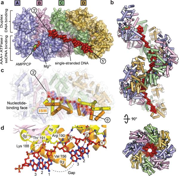
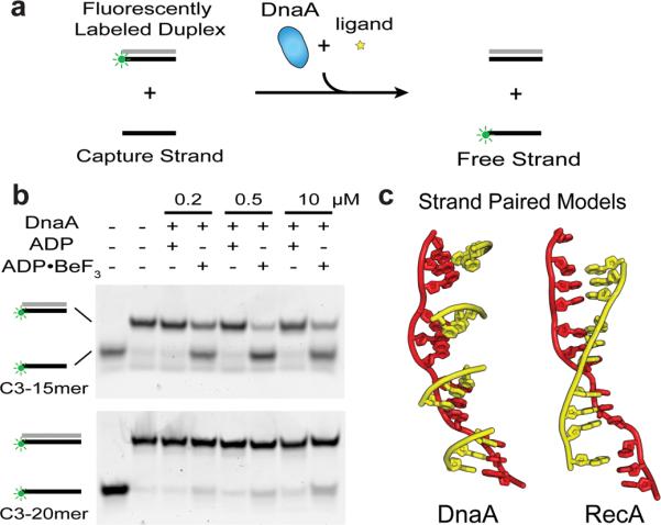
DnaA  At least 10 different enzymes or proteins participate in the initiation phase of replication. They open the DNA helix at the origin and establish a prepriming complex for subsequent reactions. The crucial component in the initiation process is the DnaA protein, a member of the AAA+ ATPase protein family (ATPases associated with diverse cellular activities). Many AAA+ ATPases, including DnaA, form oligomers and hydrolyze ATP relatively slowly.
DnaA is a protein that activates initiation of DNA replication in bacteria.[1] It is a replication initiation factor which promotes the unwinding of DNA at oriC.[1] The onset of the initiation phase of DNA replication is determined by the concentration of DnaA.[1] DnaA accumulates during growth and then triggers the initiation of replication.[1] Replication begins with active DnaA binding to 9-mer (9-bp) repeats upstream of oriC.[1] Binding of DnaA leads to strand separation at the 13-mer repeats.[1] This binding causes the DNA to loop in preparation for melting open by the helicase DnaB.

So DnaA will wrap around DNA , creating a positive tension. In order to release that DNA tension, further upstream on the DNA strand , the sequences will get melted, and the two strands will open up, the strands will be separated. That is the actual goal. So there is a goal oriented process, which is achieved through DnaA proteins, which do have no goal by their own.

DiaA DiaA, that is required for the timely initiation of replication during the cell cycle.  DiaA binds directly and specifically to the DnaA initiator protein.DiaA forms a stable complex with DnaA, even when the two proteins coexist in crude extracts (Fig. 7). Therefore, we propose that this novel protein plays an important role in regulating the initiation of chromosomal replication via direct interactions with the DnaA initiator.

DAM methylase Initiation of replication requires the expression of dnaA gene, which is transcribed earlier to replication and translated to produce DNA-A protein.  It’s gene expression requires full methylation of GATC at its promoter region. DNA adenine methyltransferase (Dam) is an enzyme of ~32 kDa that does not belong to a restriction/modification system. The target recognition sequence for E. coli Dam is GATC, as the methylation occurs at the N6 position of the adenine in this sequence (G meATC). Role in regulation of replication   Dam methylase, an abbreviation for Deoxyadenosine methylase, is an enzyme that adds a methyl group to the adenine of the sequence 5'-GATC-3' in newly synthesized DNA. Immediately after DNA synthesis, the daughter strand remains unmethylated for a short time

DnaB helicase is an enzyme in bacteria which opens the replication fork during DNA replication. Although the mechanism by which DnaB both couples ATP hydrolysis to translocation along DNA and denatures the duplex is unknown, a change in the quaternary structure of the protein involving dimerisation of the N-terminal domain has been observed and may occur during the enzymatic cycle.[1] Initially when DnaB binds to dnaA, it is associated with dnaC, a negative regulator. After DnaC dissociates, DnaB binds dnaG. The N-terminal has a multi-helical structure that forms an orthogonal bundle.[1] The C-terminal domain contains an ATP-binding site and is therefore probably the site of ATP hydrolysis. The complex formed at the replication origin also includes several DNA-binding proteins- Hu, IHF and FIS that facilitate DNA bending. Helicases are essential enzymes for DNA replication, a fundamental process in all living organisms. The DnaB family are hexameric replicative helicases that unwind duplex DNA and coordinate with RNA primase and other proteins at the replication fork in prokaryotes. 5

DnaC  The DnaC protein, another AAA+ ATPase, then loads the DnaB protein onto the separated DNA strands in the denatured region. A hexamer of DnaC, each subunit bound to ATP, forms a tight complex with the hexameric, ring-shaped DnaB helicase. This DnaC-DnaB interaction opens the DnaB ring, the process being aided by a further interaction between DnaB and DnaA. Two of the ring-shaped DnaB hexamers are loaded in the DUE, one onto each DNA strand. The ATP bound to DnaC is hydrolyzed, releasing the DnaC and leaving the DnaB bound to the DNA. Loading of the DnaB helicase is the key step in replication initiation. As a replicative helicase, DnaB migrates along the single-stranded DNA in the 5'→3' direction, unwinding the DNA as it travels. The DnaB helicases loaded onto the two DNA strands thus travel in opposite directions, creating two potential replication forks. All other proteins at the replication fork are linked directly or indirectly to DnaB. DnaC  Cdc6  Recruitment of helicase requires six DnaC proteins, each of which is attached to one subunit of helicase. Once this complex is formed, an additional four DnaA proteins bind to the original four DnaA proteins to form four DnaA dimers. DnaC is then released, and the prepriming complex is complete.  DnaC was mentioned in Interaction of Escherichia coli dnaB and dnaC(D) gene products in vitro by Wickner, S. & Hruwitz, J. in 19751. DnaC is essential for replication in vitro and in vivo. The role of this protein centers around formation of the dnaB-dnaC complex, from which it delivers the helicase to its site of action on the DNA template.

HU-proteins Bacterial histone-like HU proteins are critical to maintenance of the nucleoid structure. Many different characteristics of HU protein help to initiates DNA replication. HU protein is required for proper synchrony of replication initiation

SSB Single-stranded binding proteins (not to be confused with the E. coli protein, Single-strand DNA-binding protein, SSB) are a class of proteins that have been identified in both viruses and organisms from bacteria to humans. Single-stranded DNA binding proteins are essential for the sequestration and processing of single-stranded DNA. 6
SSBs from the OB domain family play an essential role in the maintenance of genome stability, functioning in DNA replication, the repair of damaged DNA, the activation of cell cycle checkpoints, and in telomere maintenance. The importance of SSBs in these processes is highlighted by their ubiquitous nature in all kingdoms of life.SSB proteins play an essential role in DNA metabolism by protecting single-stranded DNA and by mediating several important protein–protein interactions. 7

Hexameric DNA helicases DNA helicases are essential during DNA replication because they separate double-stranded DNA into single strands allowing each strand to be copied.

DNA polymerase I and III DNA polymerase 3 is essential for the replication of the leading and the lagging strands whereas DNA polymerase 1 is essential for removing of the RNA primers from the fragments and replacing it with the required nucleotides.


DnaG Primases synthesize RNA oligonucleotides (primers) on single stranded DNA (ssDNA) in both prokaryotic and eukaryotic organisms at the start of DNA replication.  Primases are also involved in lagging strand synthesis and replication restart.  They are essential for the initiation of such phenomena because DNA polymerases are incapable of de novo synthesis and can only elongate existing strands (4); as such, primases are foundationally important for cell proliferation. 2

Topoisomerases Topo II, which separates the two DNA duplexes following replication, is essential for viability. Topoisomerase II enzymes 8  are essential in the separation of entangled daughter strands during replication. This function is believed to be performed by topoisomerase II in eukaryotes and by topoisomerase IV in prokaryotes. Failure to separate these strands leads to cell death.

Sliding clamp and clamp loader the clamp loader is a crucial aspect of the DNA replication machinery. 14  Sliding clamps are DNA-tracking platforms that are essential for processive DNA replication in all living organisms 15  The protein that copies DNA (DNA polymerase) requires a ring-shaped protein complex, called the sliding clamp, to hold it onto the DNA, so that the polymerase can move at high speed. As it sequentially copies the nucleotides that make up the DNA strand, synthesis can occur as fast as 1000 nucleotides per second. However, the sliding clamp cannot get onto DNA by itself and requires a separate complex of proteins, called the clamp loader, to wrap the sliding clamp ring around DNA

Primase (DnaG) Primases are essential RNA polymerases required for the initiation of DNA replication, lagging strand synthesis and replication restart.   All DNA synthesis, both of leading and lagging strands, requires the prior synthesis of an RNA primer. Primer synthesis in E. coli is mediated by an 600-kD protein assembly known as a primosome, which includes the DnaB helicase and an RNA synthesizing primase called DnaG, as well as five other types of subunits. Primases are also involved in lagging strand synthesis and replication restart.  They are essential for the initiation of such phenomena because DNA polymerases are incapable of de novo synthesis and can only elongate existing strands.

RTP-Ter complex The Ter sequences are arranged on the chromosome to create a trap that a replication fork can enter but cannot leave. The Ter sequences function as binding sites for the protein Tus (terminus utilization substance). The Tus-Ter complex can arrest a replication fork from only one direction. Only one Tus-Ter complex functions per replication cycle—the complex first encountered by either replication fork. Given that opposing replication forks generally halt when they collide, Ter sequences would not seem to be essential, but they may prevent overreplication by one fork in the event that the other is delayed or halted by an encounter with DNA damage or some other obstacle

Ribonuclease H RNase H is responsible for removing the RNA primer, allowing completion of the newly synthesized DNA. RNase H1 plays essential roles in generating and clearing RNAs that act as primers of DNA replication. RNase H1 primer processing is needed not only for initiation of replication, but also late in the replication cycle. 16

Replication restart primosome Replication restart primosome is a complex dynamic system that is essential for bacterial survival. This system uses various proteins to reinitiate chromosomal DNA replication to maintain genetic integrity after DNA damage. The replication restart primosome in Escherichia coli is composed of

DNA repair:
RecQ helicase  In prokaryotes RecQ is necessary for plasmid recombination and DNA repair from UV-light, free radicals, and alkylating agents. This protein can also reverse damage from replication errors. 8 The RecQ family of helicases are enzymes that unwind DNA so that replication, transcription, and DNA repair can occur. 9

RecJ nuclease Breaks in DNA are repaired by homologous recombination. Because the structure of DNA ends at a break site can be variable, the repair machinery must be designed to act on a variety of heterogeneous DNA break sites. Bacterial RecQ helicase and RecJ nuclease initiate the repair of double-stranded DNA breaks; however, neither protein alone can deal with the broad range of physiological ends.
The assembly of the protein complexes within the forked DNA responsible for reloading the replicative DnaB helicase anywhere on the chromosome for genome duplication requires the coordination of transient biomolecular interactions.

How big are the molecular machines of the central dogma? 1

there is a closed loop from DNA to DNA which signifies DNA replication. This process of replication is carried out by a macromolecular complex known as the replisome. The E. coli replisome is a collection of distinct protein machines that include helicase (52 kDa (each of 6 subunits) BNID 104932), primase (65 kDa BNID 104932) and the DNA polymerase enzyme complex (791 kDa in several units of the complex, BNID 104931). To put the remarkable action of this machine in focus, an analogy has been suggested (Baker & Bell, Cell 1998) in which one thinks of the DNA molecule in human terms by imagining it to have a diameter of 1 m (to get a sense of the actual size of the replication complex relative to its DNA substrate, see Fig. 2). At this scale, the replisome has the size of a FedEx truck, and it travels along the DNA at roughly 600 km/hr. Genome replication is a 400 km journey in which a delivery error occurs only once every several hundred kilometers, this despite the fact that a delivery is being made roughly six times for every meter traveled. During the real replication process, the error rate is even lower as a result of accessory quality control steps (proofreading and mismatch correction) ensure that a wrong delivery happens only once in about 100 trips. )

Overview of the whole replication process :

In prokaryotes, the DNA is circular.  Replication starts at a single origin (ori C) and is bi-directional and semi-conservative.
The region of replicating DNA associated with the single origin is called a replication bubble or replication eye and consists of two replication forks moving in opposite direction around the DNA circle.
During DNA replication, the two parental strands separate and each acts as a template to direct the enzyme catalysed synthesis of a new complementary daughter strand following the normal base pairing rule.
Three basic steps involved in DNA replication are Initiation, elongation and termination.


I. Initiation
Step 1: Binding of DNA around an initiator protein complex DNA-A ATP ~30-40.
The DNA B or helicase unwinds ori C (origin of replication) and extends the single stranded region for copying.
Step 2: Single strand binding protein (SSB) binds to this single stranded region to protect it from breakage and to prevent it from renaturing.
As the parental DNA is unwound by DNA helicases and SSB (travels in 5’-3’ direction), the resulting positive supercoiling (torsional stress) is relieved by topoisomerse I and II (DNA gyrase) by inducing transient single stranded breaks.


DNA replication of prokaryotes Image_thumb%25255B13%25255D


Step 3: The enzyme DNA primase (primase, an RNA polymerase) then attaches to the DNA and synthesises a short RNA primer to initiate synthesis of the leading strand of the first replication fork.


II. Elongation
Step 4: DNA polymerase III extends the RNA primer made by primase.
DNA polymerase possesses separate catalytic sites for polymerisation and degradation of nucleic acid strands.
All DNA polymerases make DNA in 5’-3’ direction
Leading strand synthesis
On the template strand with 3’-5’ orientation, new DNA is made continuously in 5’-3’ direction towards the replication fork. The new strand that is continuously synthesized in 5’-3’ direction is the leading strand.
Lagging strand synthesis
On the template strand with 5’-3’ orientation, multiple primers are synthesized at specific sites by primase (primosome complex) and DNA pol III synthesizes short pieces of new DNA (about 1000 nucleotides long) new DNA is in 5’-3’ direction.
These small DNA fragments that are discontinuously synthesises are calledOkazaki fragments (named after the discoverer Reigi Okazaki). The new strand which is discontinuously synthesised in small fragments is called the lagging strand.


DNA replication of prokaryotes Image%25255B35%25255D


DNA polymerase III synthesizes DNA for both leading and lagging strands.
Step 5: After DNA synthesis by DNA pol III, DNA polymerase I uses its 5’-3’ exonuclease activity to remove the RNA primer and fills the gaps with new DNA.
Step 6: Finally DNA ligase joins the ends of the DNA fragments together.


III. Termination
Step 7: The two replication forks meet ~ 180  degree opposite to ori C, as DNA is circular in prokaryotes. Around this region there are several terminator sites which arrest the movement of forks by binding to the tus gene product, an inhibitor of helicase (Dna B).
Step 8: Once replication is complete, the two double stranded circular DNA molecules (daughter strands) remain interlinked. Topoisomerase II makes double stranded cuts to unlink these molecules.
DNA replication of prokaryotes Image_thumb%25255B38%25255D
The mechanism of DNA replication in eukaryotes is same as that of prokaryotes. These are the major differences between DNA replication in prokaryotes and Prokaryotes


DNA replication of prokaryotes Prokar10

A Summary of DNA Replication in Bacteria. Starting with the initiation event at the replication origin, this figure depicts DNA replication in E. coli in seven steps. Two replication forks move in opposite directions from the origin, but only one fork is illustrated for steps 2 - 7  . The various proteins shown here as separate entities are actually closely associated (along with others) in a single large complex called a replisome. The primase and DNA helicase are particularly closely bound and, together with other proteins, form a primosome. Parental DNA is shown in dark blue, newly synthesized DNA in light blue, and RNA in red. This series of diagrams does not show the topological arrangement of the DNA strands.

Putting It All Together:DNA Replication in Summary


Figure above reviews the highlights of what we currently understand about the mechanics of DNA replication in E. coli. Starting at the origin of replication, the machinery at the replication fork sequentially adds the different proteins required for synthesizing DNA—that is, DNA helicase, DNA gyrase, SSB, primase, DNA polymerase, and DNA ligase. Several other proteins (not illustrated) are also involved in improving the overall efficiency of DNA replication. For example, a ring-shaped sliding clamp protein encircles the DNA double helix and binds to DNA polymerase, thereby allowing the DNA polymerase to slide along the DNA while remaining firmly attached to it. The various proteins involved in DNA replication are all closely associated in one large complex, called a replisome, that is about the size of a ribosome. The activity and movement of the replisome is powered by the hydrolysis of nucleoside triphosphates. These include both the nucleoside triphosphates (used by DNA polymerases and primase as building blocks for DNA and RNA synthesis) and the ATP hydrolyzed by several other DNA replication proteins (including DNA helicase, DNA gyrase, and DNA ligase). As the replisome moves along the DNA in the direction of the replication fork, it must accommodate the fact that DNA is being synthesized in opposite directions along the template on the two stands. Picture above  provides a schematic model illustrating how this might be accomplished by folding the lagging strand template into a loop. Creating such a loop allows the DNA polymerase molecules on both the leading and lagging strands to move in the same physical direction, even though the two template strands are oriented with opposite polarity. 


How did natural mechanisms find out about the need of opposite polarity ?
 
DNA replication of prokaryotes Sem_ty18
Arrangement of Proteins at the Replication Fork. This model shows how some of the key replication proteins illustrated in Figure 19-13 are organized at the replication fork. The main distinguishing feature is that the lagging strand DNA is folded into a loop, thereby allowing the DNA polymerase molecules on the leading and lagging strands to come together and move in the same physical direction (black arrows) even though the two template strands are oriented with opposite polarity in the parental DNA molecule.


Parts required for DNA replication :

http://www.genome.jp/kegg-bin/show_pathway?map03030
DNA replication of prokaryotes Kegg_p10


https://en.wikipedia.org/wiki/Replication_factor_C
DNA replication of prokaryotes Dna_re12

DNA replication of prokaryotes Protei10
DNA replication of prokaryotes Protei11
DNA replication of prokaryotes Sfmb2e10


DNA replication of prokaryotes Dna_a_11


DNA replication of prokaryotes Dsffsd10

DNA replication of prokaryotes Nrm24210

DNA replication of prokaryotes Sem_ty16


Animations:

http://www.wiley.com/college/pratt/0471393878/instructor/animations/dna_replication/index.html
http://www.johnkyrk.com/DNAreplication.html

thats a great step by step tutorial :

http://telstar.ote.cmu.edu/biology/animation/DnaReplication/replication.html


Last edited by Otangelo on Sat Mar 13, 2021 8:07 pm; edited 31 times in total

https://reasonandscience.catsboard.com

3DNA replication of prokaryotes Empty STRUCTURAL OVERVIEW OF DNA REPLICATION Sat Nov 07, 2015 1:34 am

Otangelo


Admin

STRUCTURAL OVERVIEW OF DNA REPLICATION

Because they bear directly on the replication process, let’s begin by recalling a few important structural features of the double helix. The double helix is composed of two DNA
strands, and the individual building blocks of each strand are nucleotides. The nucleotides contain one of four bases: adenine, thymine, guanine, or cytosine. The double-stranded structure is held together by base stacking and by hydrogen bonding between the bases in opposite strands. A critical feature of the doublehelix structure is that adenine hydrogen bonds with thymine, and guanine hydrogen bonds with cytosine. This rule, known as the AT/GC rule, is the basis for the complementarity of the base sequences in double-stranded DNA. Another feature worth noting is that the strands within a double helix have an antiparallel alignment. This directionality is determined by the orientation of sugar molecules within the sugar-phosphate backbone. If one strand is running in the 5ʹ to 3ʹ direction, the complementary strand is running in the 3ʹ to 5ʹ direction. The issue of directionality will be important when we consider the function of the enzymes that synthesize new DNA strands. In this section, we will consider how the structure of the DNA double helix provides the basis for DNA replication.

Existing DNA Strands Act as Templates for the Synthesis of New Strands

As shown in Figure 11.1a , DNA replication relies on the complementarity of DNA strands according to the AT/GC rule. During the replication process, the two complementary strands of DNA come apart and serve as template strands, or parental strands, for the synthesis of two new strands of DNA. After the double helix has separated, individual nucleotides have access to the template strands. Hydrogen bonding between individual nucleotides and the template strands must obey the AT/GC rule. To complete the replication process, a covalent bond is formed between the phosphate of one nucleotide and the sugar of the previous nucleotide. The two newly made strands are referred to as the daughter strands. Note that the base sequences are identical in both double-stranded molecules after replication (Figure 11.1b). Therefore, DNA is replicated so that both copies retain
the same information—the same base sequence—as the original molecule.

DNA replication of prokaryotes Dna_re16


Last edited by Admin on Wed Nov 25, 2015 3:33 pm; edited 8 times in total

https://reasonandscience.catsboard.com

4DNA replication of prokaryotes Empty DNA Replication: An Engineering Marvel Sat Nov 07, 2015 2:58 am

Otangelo


Admin

DNA Replication: An Engineering Marvel 1

Recently I have been reviewing some literature on the elegant molecular mechanisms by which DNA is replicated. As an undergraduate biology student, I recall being struck by their sheer complexity, sophistication, and intrinsic beauty. As I read about such a carefully orchestrated process, involving so many specific enzymes and protein complexes, and its extraordinary accuracy, it was almost as though the word "design" jumped off the pages of my textbook and slapped me in the face. The rate of DNA replication has been measured as a whopping 749 nucleotides per second (McCarthy et al., 1976) and the error rate for accurate polymerases is believed to be in the range of 10^-7 and 10^-8, based on studies of E. coli and bacteriophage DNA replication (Schaaper, 1993).
I want to provide here a brief overview of the central processes involved in DNA replication. In subsequent articles, I will examine the individual components in more detail.
DNA Replication is Semi-Conservative
DNA replication is said to be semi-conservative: that is to say, each of the two strands serves as a template for the biosynthesis of a new daughter strand -- complementary to the template (Meselson and Stahl, 1958; Parkhomchuk et al., 2009). Thus, two new double helices are produced -- each of which possesses one old strand and one new strand.
The Replication Fork


DNA replication of prokaryotes DNA%20replication%20fork


DNA replication commences at particular sites known as origins of replication (ori). Since the replication machinery progresses away from the origins of replication in two directions, a "replication bubble" is formed. At the origins of replication, the DNA double helix is opened and unwound on both sides. This forms what is known as "replication forks." These replication forks subsequently progress along the DNA in opposite directions, as DNA biosynthesis occurs.
The Initiation Phase
The first step of DNA replication is the initiation phase, a process that is under tight regulation to ensure that it happens no more than one time during cell-division (Mott and Berger, 2007; Nasheuer et al., 2002). This is the stage at which the DNA double helix is opened and unwound to expose the single strands to the enzymes and protein-complexes involved in the replication process. The double-stranded DNA is opened up by an initiator protein. The initiator protein also recruits a specialized class of proteins known as helicases. Helicases are responsible for unwinding the DNA. Once the double-helix has been unwound and opened to form single-strands of DNA, DNA binding proteins associate with the DNA to prevent the single-stranded DNA from winding back on itself.
The Clamp Loader and Sliding Clamp
Two protein complexes, the clamp loader and sliding clamp, are involved in recruiting the DNA polymerase and ensuring that it remains associated with the DNA (Idiani and O'Donnell, 2006; Miyata et al., 2004; Trakselis et al., 2001). The clamp loader is responsible for loading the sliding clamp onto the DNA. After recruiting the DNA polymerase, the sliding clamp literally slides with the DNA polymerase as it moves along the DNA template, keeping them firmly clamped together.
The Elongation Phase
The next stage of the process is known as the elongation phase. This is the stage at which the DNA strands are copied into daughter strands by the replication machinery. The synthesis of the new daughter strand from each of the parent strands is catalyzed by an enzyme complex known as DNA polymerase. DNA polymerase progresses along the template strand, reads the nucleotide bases and adds the complementary nucleotide. The chemistry of this reaction and the polarity of the DNA molecule, however, only permit nucleotides to be added at the 3' end of the elongating strand. This means that DNA replication can only happen in the 5' to 3' direction. DNA polymerase has a remarkably high fidelity, thanks to its built-in proofreading and error-correcting facility.
Synthesis of Primers
The DNA polymerase enzyme is incapable of synthesizing a novel strand from scratch -- it is able only to add nucleotides to the 3' end of already-present nucleotides. Short strands of RNA called primers are therefore synthesized by an enzyme called primase (the RNA primers will ultimately be degraded and replaced with DNA). This raises an interesting design question: Why would a designer engineer DNA polymerase such that it is unable to begin a new strand on its own? RNA polymerase, after all, is perfectly able to perform this operation. What is the design logic in unnecessarily, so it seems, involving the extra steps in synthesizing RNA primers and later removing them and replacing them with DNA? One possible explanation is that the extra stage affords an additional proofreading step -- in which case, this apparent extra complexity could be part of a design strategy.
The Leading and Lagging Strands
The two strands of a DNA helix are anti-parallel: that is to say, they have opposite orientations. This raises a difficulty since, as mentioned previously, DNA can only be synthesized in a 5' to 3' direction. For one strand known as the leading strand, DNA synthesis is continuous. For the other strand, known as the lagging strand, DNA replication proceeds discontinuously. For replication of the lagging strand to occur, short DNA fragments (known as Okazaki fragments) need to be created constantly from 5' to 3', each of them separated by ~10-nucleotide RNA primers. These are then joined by DNA ligase to form a continuous strand.
Relieving DNA Supercoiling
DNA supercoiling is induced by the torsional stress generated by the polymerases and helicases. Strand separation can thus be accompanied by DNA over-winding. A class of enzymes called Topoisomerases relieve this torsional stress by breaking the DNA backbone and relaxing the supercoils. The DNA backbone is then ligated back together. In so doing, this class of enzymes removes the supercoils.
Termination of DNA Replication
DNA replication terminates when the two replication forks, which move in opposite directions, meet. In bacteria such as E. coli, this takes place at particular terminator sequences (ter), which associate with a specific DNA-binding protein called Tus. Terrence A. Brown explains how termination in bacteria occurs:


The mode of action of Tus is quite unusual. When bound to a terminator sequence, a Tus protein allows a replication fork to pass if the fork is moving in one direction, but blocks progress if the fork is moving in the opposite direction around the genome. The directionality is set by the orientation of the Tus protein on the double helix. When approached from one direction, Tus blocks the passage of the DnaB helicase, which is responsible for progression of the replication fork, because the helicase is faced with a 'wall' of β-strands which it is unable to penetrate. But when approaching from the other direction, DnaB is able to disrupt the structure of the Tus protein, probably because of the effect that unwinding of the double helix has on Tus, and so is able to pass by (Figure 13.22B; Kamada et al., 1996).
The orientation of the termination sequences, and hence of the bound Tus proteins, in the E. coli genome is such that both replication forks become trapped within a relatively short region on the opposite side of the genome to the origin (see Figure 13.22A). This ensures that termination always occurs at or near the same position. Exactly what happens when the two replication forks meet is unknown, but the event is followed by disassembly of the replisomes, either spontaneously or in a controlled fashion. The result is two interlinked daughter molecules, which are separated by topoisomerase IV.


Far less is known about termination in eukaryotes, in which there are no specific termination sites. For a discussion of one model of replication-fork termination in eukaryotes, see Fachinetti et al. (2010).
Maintaining Telomeres
Since eukaryotes have linear chromosomes, the DNA polymerase is unable to continue all the way to the end of the chromosomes. This leads to a progressive telomere shortening with each round of cell division. In the germ cells (which pass on their DNA to the next generation), the enzyme telomerase is responsible for maintaining telomere length.
Conclusion
The systems responsible for DNA replication are well beyond the explanatory efficacy of unguided processes involving chance and necessity. Indeed, machinery of the complexity and sophistication of that described above is, in all of our experience, habitually associated with intelligent agency. This brief description barely amounts the tip of the iceberg. Later, I will examine these intricate processes in more detail.


Replicating DNA with Extraordinary Fidelity: Meet DNA Polymerase


In a previous article, I gave a brief overview of the complex molecular mechanisms governing DNA replication. Now, I will focus specifically on the replication enzyme DNA polymerase.
DNA polymerase is the enzyme responsible for synthesizing new strands of DNA, complementary to the sequence of the template strand. The unidirectional DNA polymerase progresses along the template strand in a 3'-5' direction, since it requires a pre-existing 3'-OH group for the adding of nucleotides. The daughter strand is, consequently, synthesized in a 5'-3' direction (opposite to the direction of movement of the polymerase since the two strands have an anti-parallel orientation).
There are six different families of DNA polymerases -- A, B, C, D, X and Y (Rothwell and Waksman, 2005). These families differ from one another in their design, being specialized for a variety of purposes. For example, DNA polymerase I, found in E. coli, belongs to the A family of polymerases. Beyond its role in finishing DNA replication and removing the RNA primers, DNA polymerase I contains a 5' to 3' exonuclease domain, in addition to the 3' to 5' exonuclease domain, that allows it to remove nucleotides both in front of and behind it (more on proofreading by exonuclease domains shortly) (Ishino et al., 1995). Polymerases in the B, C and D families are known for their high fidelity (owing to their intrinsic 3' to 5' proofreading exonuclease), and are found in eukaryotes, bacteria and archaea respectively. The X family (e.g. eukaryotic polymerases pol β, Pol σ, Pol λ, Pol μ) plays a role in DNA repair, filling in the gaps created during the process (Yamtich and Sweasy, 2010). Whereas most polymerases cannot replicate past bulky lesions in damaged DNA, the Y family are able to replicate past them (Washington et al., 2010).
DNA polymerases generally share a common structural framework, with fingers, thumb and palm subdomains that make up the polymerase domain. The diagram below, excerpted from Beard and Wilson (2003), shows the structure of T7 DNA polymerase, revealing its two domains, a polymerase domain (colored) and a proofreading exonuclease domain (gray). The polymerase domain is comprised of three subdomains: fingers, thumb and palm. The finger domain positions incoming nucleoside triphosphates in relation to the template strand. The thumb domain is thought to function in the processivity, positioning and translocation of the DNA, holding in place the elongating DNA duplex. The β-sheet that comprises the palm domain is where the enzyme's active site lies, which catalyzes the transfer of phosphoryl groups in the phosphoryl transfer reaction.


DNA replication of prokaryotes DNA%20polymerase%20structure


How does DNA polymerase add new nucleotides to the elongating strand? The polymerase's active site, found in the β-sheet that makes up the palm subdomain catalyes a phosphoryl transfer reaction. It forms a phosphodiester bond by linking the 3' hydroxyl group at the end of the template strand to the nucleotide's 5' phosphoryl group. The first step in the process is a nucleophilic attack on the α-phosphate of the incoming nucleoside triphosphate by the 3' OH of the growing chain. This reaction releases pyrophosphate (PPi). Within the active site, there are two conserved aspartate residues. The magnesium ions on the carboxylate groups of those aspartates is critical to the reaction. These carboxylate groups co-ordinate the magnesium ions and facilitate their participation in the catalysis by holding them in the right orientation. One of the two magnesium ions activates the 3' OH group of the terminal nucleotide. The other is responsible for stabilizing a developing negative charge on the leaving oxygen on the incoming nucleoside triphosphate. Side chains on an alpha helix in the finger domain interact with the incoming triphosphate to also stabilize it. Hydrolysis of the pyrophosphate released in this process generates the energy required for driving the reaction forward. For a more detailed review of the mechanisms involved, I refer readers to Rothwell and Waksman (2005).
The process by which DNA polymerase selects the correct nucleotide is less well understood. For a discussion, I refer readers to Markiewicz et al. (2012).
As I stated in my previous post, the rate at which DNA polymerase replicates DNA is thought to be a whopping 749 nucleotides per second (McCarthy et al., 1976) and the error rate for accurate polymerases is believed to be in the range of 10^-7 and 10^-8, based on studies of E. coli and bacteriophage DNA replication (Schaaper, 1993). This extraordinarily high fidelity is accomplished by a remarkable proofreading and error-correcting facility built into the enzyme, which checks the identity of nucleotides both during and after polymerization.
The first level of monitoring occurs by virtue of the fact that, when base-paired with the complementary strand (A with T, or C with G), correct nucleotides precisely fit into the active site, whereas nucleotides that are incorrectly matched will possess a different geometry and will not will not fit so precisely into the active site (Johnson and Beese, 2004).
Sometimes, however, this first level of monitoring will fail to prevent the entry of an incorrect nucleotide. But, thankfully, there is also a second level of proofreading. In addition to the polymerase active site, DNA polymerase possesses a 3' to 5' exonuclease active site, which can cleave an incorrect nucleotide from the 3' end of the growing DNA strand before synthesis of the subsequent nucleotide. When an incorrect nucleotide is mistakenly incorporated, the polymerase's rate of activity is significantly delayed. Studies have shown that the presence of mismatch can reduce the polymerase's efficiency of subsequent elongation by as much as a hundred to a million fold (Kunkel and Bebenek, 2000; Goodman et al., 1993; Echols and Goodman, 1991). This gives enough time for spontaneous denaturing of the DNA at the 3' end, thereby facilitating the transfer of the 3' end with the mismatched nucleotide to the polymerase's 3' exonuclease site, which catalyzes the removal of multiple nucleotides from the 3' end of the DNA strand. The 3' end is subsequently positioned back into the polymerase active site, and the polymerase can then continue its DNA synthetic activity in the 5' to 3' direction.


It should be noted, however, that not all DNA polymerases possess an intrinsic proofreading exonuclease. Polymerases belonging to the Y family, for example, tend to be significantly less accurate (Friedberg et al., 2001). Members of this family "lack an intrinsic proofreading exonuclease, exhibit low processivity, replicate DNA with low fidelity, and are believed to assist replication complexes stalled at DNA lesions," (Beard et al., 2002).
DNA polymerase is just one of numerous protein complexes that play an important role in DNA replication. A general overview of the machinery involved in the process of replication is more than adequate grounds to justify a design inference. As we drill down and examine the individual subcomponents that make up the cell's DNA replication machinery, the case for design becomes ever harder to ignore. In subsequent articles I will continue this exploration of the intricate molecular processes underlying DNA biosynthesis.


Some Perspective on the "Mechanical Prowess" of DNA Replication 

3

I recently published two articles (here and here) on the mechanisms by which a cell's DNA is replicated. Now I've just encountered a paper, published in the journal Cell in 1998, that does an excellent job of describing the engineering prowess of these molecular machines. The paper is titled "Polymerases and the Replisome: Machines within Machines," and it makes the following statements about the processes of DNA replication:

Synthesis of all genomic DNA involves the highly coordinated action of multiple polypeptides. These proteins assemble two new DNA chains at a remarkable pace, approaching 1000 nucleotides (nt) per second in E. coli. If the DNA duplex were 1 m in diameter, then the following statements would roughly describe E. coli replication. The fork would move at approximately 600km/hr (375 mph), and the replication machinery would be about the size of a FedEx delivery truck. Replicating the E. coli genome would be a 40 min, 400 km (250 mile) trip for two such machines, which would, on average make an error only once every 170 km (106 miles). The mechanical prowess of this complex is even more impressive given that it synthesizes two chains simultaneously as it moves. Although one strand is synthesized in the same direction as the fork is moving, the other chain (the lagging strand) is synthesized in a piecemeal fashion (as Okazaki fragments) and in the opposite direction of overall fork movement. As a result, about once a second one delivery person (i.e. polymerase active site) associated with the truck must take a detour, coming off and then rejoining its template DNA strand, to synthesize the 0.2km (0.13 mile) fragments.
It can hardly be disputed that DNA replication is extraordinary. Stay tuned for a forthcoming article on DNA helicase.

1) http://www.evolutionnews.org/2013/01/dna_replication068061.html
2) http://www.evolutionnews.org/2013/01/replicating_dna068131.html
3) http://www.evolutionnews.org/2013/02/some_perspectiv068951.html


Last edited by Admin on Wed Nov 25, 2015 3:34 pm; edited 1 time in total

https://reasonandscience.catsboard.com

5DNA replication of prokaryotes Empty Re: DNA replication of prokaryotes Sun Nov 08, 2015 2:52 pm

Otangelo


Admin

http://en.wikibooks.org/wiki/Structural_Biochemistry/Nucleic_Acid/DNA/Replication_Process

What was the chemical pathway for an "RNA world" to transform into a "DNA/protein world." ?

DNA replication must not have been present in LUCA because the DNA replication machinery in today’s species reveals too many differences.

Furthermore the aminoacyl tRNA synthetases fail to form an evolutionary tree. So evolutionists must believe HGT caused the confusion. There is no independent evidence that HGT changed around the aminoacyl tRNA synthetases. The evidence simply is the failure to find an adequate evolutionary tree to explain these enzymes.

DNA polymerase is a polypeptide enzyme

http://www.nature.com/scitable/topicpage/cells-can-replicate-their-dna-precisely-6524830

DNA replication of prokaryotes Cells_10

Hexameric helicases


http://reasonandscience.heavenforum.org/t1438-hexameric-helicases

http://www.evolutionnews.org/2013/02/unwinding_the_d_1069371.html

this is a highly organized, sofisticated and orchestrated movement, like a roboter,executing a specific task:

https://www.youtube.com/watch?v=Oy1RGlT0vv0

In order to be able to unwind the dna strand, the initial opening of the double helix (at the origin of replication) is performed by an initiator protein.

How did chance, physical necessity, or natural selection " know " a initiator protein would be needed, and how it would have to be , and where employd ?

The helicase rotational speed of up to 10,000 rotations per minute !!!!! How astonishing and marvellous .


https://www.youtube.com/watch?v=TC2mYWR8754

http://www.rcsb.org/pdb/101/motm.do?momID=3

The Secret of Life

DNA polymerase plays the central role in the processes of life. It carries the weighty responsibility of duplicating our genetic information. Each time a cell divides, DNA polymerase duplicates all of its DNA, and the cell passes one copy to each daughter cell. In this way, genetic information is passed from generation to generation. Our inheritance of DNA creates a living link from each of our own cells back through trillions of generations to the first primordial cells on Earth. The information contained in our DNA, modified and improved over millennia, is our most precious possession, given to us by our parents at birth and passed to our children.

Amazing Accuracy

DNA polymerase is the most accurate enzyme. It creates an exact copy of your DNA each time, making less than one mistake in a billion bases. This is far better than information in our own world: imagine reading a thousand novels, and finding only one mistake. The excellent match of cytosine to guanine and adenine to thymine, the language of DNA, provides much of the specificity needed for this high accuracy. But DNA polymerase adds an extra step. After it copies each base, it proofreads it and cuts it out if the base is wrong.


Exploring the Structure

These simple DNA polymerases are shaped roughly like a hand. A cleaved version of the E. coli enzyme was studied: the missing part, which you will not find in the PDB file, is shown with a green outline. The space between the "fingers" and the "thumb" is just the right size for a DNA helix. But surprisingly, DNA actually fits into the palm when the enzyme is at work. In these pictures, the template strand is colored purple and the new strand is colored green. The enzyme contains three separate active sites. The polymerase site, near the top in these pictures, synthesizes the new strand by adding nucleotides. The 3'-5' exonuclease site, near the center in the E. coli polymerase, proofreads the new additions.


DNA Polymerase

All living organisms have DNA polymerases. Some, like the ones pictured here, are quite simple: one enzyme does it all. The ones in our own cells are more complex, composed of separate proteins that unwind the helix, build an RNA primer, and build the new strand. Some even have a ring-shaped protein that clamps the polymerase to the DNA strand. A single cell often has several different polymerases: complex ones that do the major DNA replication when the cell divides, and simpler ones that help in day-to-day repair and maintenance of the DNA.

http://mindfuller.tumblr.com/post/51179989275/step-5-why-evolution-isnt-random-or-irreducibly

In general, DNA polymerases are extremely accurate, making less than one mistake for every 100,000,000 nucleotides added. Some DNA polymerases even have the ability to proofread and can remove nucleotides to correct mismatched bases. However, other DNA polymerases are able to “read through” certain kinds of mutations, at the expense of accuracy in error-free regions. The overall result is that DNA replication and even maintenance processes can introduce mutations.


DNA Polymerase I:  Scientific papers tend to be reserved in their language, but the authors of a paper in Structure1 couldn’t help themselves: “DNA polymerases are spectacular molecular machines that can accurately copy genetic material with error rates on the order of 1 in 105 bases incorporated, not including the contributions of proofreading exonucleases.”  Their paper went into detail on how the “fingers” and “thumb” of the machine open and close in precise sequence as the machine moves along the DNA strand base by base.  Part of the machine rotates 50° as the machine translocates along the DNA.  These machines copy millions of base pairs of DNA every cell division so that each daughter cell gets an accurate copy.  The research was done on a bacterium that lives in hot springs.     Pata and Jaeger, who reviewed the paper by Golosov et al in Structure,2 included a diagram showing the “conformational changes” that DNA polymerase I undergoes in its action along the DNA strand.  “After more than fifty years of research, the DNA polymerases responsible for copying the genetic material are some of the most well characterized enzymes in all of biology,” they said.  “Although the polymerases are divided into several different families, they all share a common two metal-ion catalytic mechanism, and most of them are described as having fingers, palm, and thumb domains: the palm contains metal-binding catalytic residues, the thumb contacts DNA duplex, and the fingers form one side of the pocket surrounding the nascent base pair.”  Three phases occur during each step along the DNA chain: the fingers open, the machine moves one base pair as it rotates, then the base in the “palm” is placed into the “pre-insertion site,” while another moving part prevents further movement till the operation is completed.  Then the process repeats – millions of times per operation.     A paper in PNAS3 on DNA Polymerase I noted that “The remarkable fidelity of most DNA polymerases depends on a series of early steps in the reaction pathway which allow the selection of the correct nucleotide substrate, while excluding all incorrect ones, before the enzyme is committed to the chemical step of nucleotide incorporation.”  Their paper also discussed numerous conformational changes in the operation – some that precede the emplacement of the nucleotide at each step.  They described how the fingers-closing step forms “a snug binding pocket around the nascent base pair.”  They discussed at length how the machine prevents mismatched bases at several stages of the operation.  None of the authors of these three papers used the word evolution. 

1) http://crev.info/2010/01/molecular_machines_use_moving_parts/#sthash.BcW4RC4k.dpuf


Last edited by Admin on Wed Nov 11, 2015 10:56 am; edited 1 time in total

https://reasonandscience.catsboard.com

6DNA replication of prokaryotes Empty Mechanisms of DNA Replication Mon Nov 09, 2015 2:44 pm

Otangelo


Admin

Mechanisms of DNA Replication 1

Duplication of DNA, the information repository for life, is a vital process for all cellular organisms. Therefore the replication process must be faithfully performed at the passing of each generation to maintain the species. Duplication of DNA requires that the two strands of the double helix be separated; each strand is then used as a template to produce two new daughter strands from the original parental duplex DNA. Presently, these daughter chromosomes will segregate into two newly formed cells.

Numerous proteins cooperate to perform this delicate and vital task. Initially, several proteins conspire to pry apart the two strands at a position in the chromosome called an origin. A helicase then enters the opening and unzips, or unwinds, the DNA strands, acting with topoisomerases that remove the spiral turns. A multiprotein replicase machinery then coordinates the synthesis of two new strands at the same time.

Architecture and dynamic action of the replisome at a replication fork. The clamp loader (green) is at the center of the E. coli replicating machinery. Protein arms extend out from the clamp loader to bind the two DNA polymerases (yellow). These arms also bind to the ring-shaped DnaB helicase. The leading-strand polymerase moves with its clamp (red) in the direction of helicase-driven DNA unwinding (light purple). Because of the antiparallel structure of duplex DNA, the lagging-strand polymerase must extend DNA opposite the direction of unwinding, resulting in a DNA loop. This strand is therefore made as a series of DNA fragments; each fragment generates a loop that is dismantled when the polymerase bumps into the fragment it made last. Loops are started every 1–2 seconds by primase (dark purple), which synthesizes a short RNA primer to get the polymerase started. The clamp loader repeatedly loads b clamps onto the RNA start sites for use by the lagging-strand polymerase.

Michael O'Donnell lab.

One of our goals is to understand the function of each of the numerous proteins involved in the replication process. Our earlier work focused on a representative of the bacterial branch of life, Escherichia coli. These studies revealed fundamental aspects of the replication process that generalize to all cell types. This work is now textbook material and we have recently centered our attention on Saccharomyces cerevisiae, Bakers yeast, a representative cell of the eukaryotic branch of life. Eukaryotic cells include those of which humans are composed. Our studies of bacteria revealed fundamental processes of replication that also occur in eukaryotes. Study of eukaryotic cells may reveal specific processes that regulate replication and prevent uncontrolled cell growth that occurs in cancers.

The E. coli replicase, DNA polymerase III (Pol III) holoenzyme, consists of 10 different protein "gears"; some are present in multiple copies, for a total of 17 proteins. Within this machine are multiple copies of the DNA polymerase that synthesizes DNA enabling them to copy both strands of the parental duplex at the same time. Another subunit, called ε, is a nuclease that can degrade DNA. Therefore, when a polymerase makes a mistake, the exonuclease removes the mistake, allowing the polymerase to try again.

Pol III holoenzyme also contains a subunit called β that is shaped like a doughnut and completely encircles DNA, acting as a mobile sliding tether to hold the polymerase to DNA. The strategy of using a ring-shaped clamp is conserved in eukaryotes, where the sliding clamp is called proliferating cell nuclear antigen (PCNA). The ring shaped structure of the β and PCNA clamps are nearly superimposable, as determined by crystal structure studies in collaboration with John Kuriyan's laboratory (HHMI, University of California, Berkeley).

Sliding clamps do not get onto DNA by themselves. For this, a group of five subunits harness the energy of ATP to open the clamp, position it onto DNA, and then close it. In our collaboration with the Kuriyan lab, the crystal structures of clamp loaders from both bacteria and eukaryotic cells have been solved, and their mechanisms of action have been determined.

The overall architecture of proteins at the bacterial replication fork, called a replisome, has emerged from these studies (see the movie). At the center is the clamp loader; it has protein arms that bind the Pol III cores for simultaneous synthesis of both strands of duplex DNA. The clamp loader arms also bind the DnaB helicase, a hexamer that encircles one DNA strand. The leading-strand Pol III continuously travels with the helicase during DNA unwinding, but due to the antiparallel structure of DNA, the lagging strand is synthesized in the opposite direction of DNA unwinding. Thus the lagging strand is made as a series of small fragments, each started by an RNA primer (e.g., by primase). RNA primers are extended by Pol III, resulting in a DNA loop. When the polymerase finishes, it bumps into a fragment made previously and disengages from its β ring, allowing it to recycle to the next upstream RNA primer onto which the clamp loader has loaded a new clamp.

Chromosomes are very long DNA molecules, and the replisome constantly encounters proteins and DNA lesions in its path. We have studied replisome collisions with proteins. When the replisome collides with RNA polymerase traveling in the same direction, the replisome remains on DNA and takes over the RNA without pause. The replisome pauses upon collision with RNA polymerase in the opposite direction, but recruits a specialized protein that displaces the RNA polymerase. We have studied how the replisome deals with DNA lesions incurred by sunlight or oxidative damage. In our collaboration with Myron Goodman (University of Southern California), we discovered a new class of DNA polymerase (Pol V) that functions with the clamp to extend DNA across a lesion, resulting in a mutation. This class of polymerase, referred to as translesion synthesis (TLS) polymerases, is now found in all cell types.

Eukaryotic cells have more complicated replisomes with many more proteins than the bacterial replisome. Currently, the extra proteins have unknown functions. To name a few of the extra proteins, the eukaryotic Cdc45/Mcm2-7/GINS complex (the CMG helicase) contains 11 subunits within which is a ring shaped hexamer like the bacterial hexameric helicase. The actions of these extra proteins are largely unknown. Eukaryotes also contain two different polymerases for the leading and lagging strands, but why this should be the case is not understood. Many eukaryotic replisome proteins appear to be modified by cell cycle control or other control proteins.

It has long been a goal of the laboratory to reconstitute eukaryotic replisomes and to determine how it works. We have now purified ample stocks of all the necessary eukaryotic proteins. The most difficult to obtain, and the most recent addition to the lab, is the 11 subunit CMG helicase. In our initial studies to assemble the eukaryotic replisome we are already finding answers to perplexing questions. We now understand how the two polymerases are specified to their respective strand. Specifically, we find that the CMG helicase ejects Pol δ from PCNA on the leading strand, while the CMG helicase directly binds to and stabilizes Pol ε for activity on the leading strand. We also have a tentative answer to the question: "Why do eukaryotes use two different polymerases for the leading and lagging strands?" We find Pol ε cannot strand displace, or "push ahead." The CMG helicase may be functioning exclusively with Pol ε in order to harness this property and thereby protect itself from displacement by other proteins. Going forward, we have set a foundation to address numerous questions in the future about how the replisome works and how it interfaces with cellular pathways of repair and control of cell growth.


The Eureka Enzyme! 2

Imagine this: it is 12 AM, and you are just starting the ten page research paper you’ve known about for weeks merely eight hours before it is due. Sweat drops on the keyboard as your fingers glide across the keyboard, there is no time to waste, but can you guarantee you will not make any grammatical errors along the way? Is there such thing as an organism/molecule that can work at a speedy pace AND make minimal, if any, errors? DNA polymerase I can! The enzyme works at adding 16-20 nucleotides/s to a new DNA strand in E.coli, which is slow for a polymerase. Its fellow isozyme, DNA polymerase III, can add 250-1,000 nucleotides/s in E. coli The accuracy of DNA polymerase enzyme is remarkable, research has shown it inserts an incorrect base pair every 10 bases in eukaryotes. Not impressed? DNA polymerase is also its own editor! It has proofreading capabilities and inhibits the addition of a nucleotide following a mismatch, increasing the accuracy of DNA polymerase 10 fold. Spell check cannot compare. If one factors in the enzyme’s proofreading capabilities, the enzyme makes an error every 10 base pairs! Still not convinced DNA polymerase is the shizz? Think you could live without it? Think again. If a nucleotide mismatch goes unrepaired, known as a mutation, it can go unnoticed in the organism, dubbed a silent mutation, or it can be deleterious to the organism. This single nucleotide change alters only one amino acid in the protein chain, but the results are devastating. One of the most well known example of a point mutation is sickle-cell anemia, and without daily treatment the average life expectancy of the patient is 20-40 years. There has also been strong evidence linking cancer and the amount of mutations in mammals. You know how you are always told to wear sunscreen while basking in the sunshine? This is why; UV light induces the formation of dimers within DNA and contributes to the 10 percent of all DNA damages caused by environmental factors. Dimers cause kinks and bends in DNA, which results in permanent damage. This can ultimately lead to skin cancer, the number one form of cancer in the United States. DNA polymerase is also an important target for anti-viral drugs, as DNA viruses program their own DNA polymerases. This has been used to combat viruses such as herpes, which infects 16.2 percent of the U.S. population, or about one out of six, people 14-49 years of age. DNA polymerase was destined for great things since its discovery in 1956. It was coined the “eureka” enzyme before all of its fundamental roles in life were revealed. Without this enzyme, life as we know it would cease to exist. As the late John Calvin once said, “DNA is life, the rest is just details.”


1) https://www.hhmi.org/research/mechanisms-dna-replication
2) http://lnicole3.blogspot.com.br/


Last edited by Admin on Thu Nov 19, 2015 9:49 am; edited 2 times in total

https://reasonandscience.catsboard.com

7DNA replication of prokaryotes Empty Re: DNA replication of prokaryotes Mon Nov 09, 2015 2:45 pm

Otangelo


Admin

DNA REPLICATION INTRODUCTION

Before cells divide, they must make a complete and faithful copy of the DNA in their chromosomes. This process of copying is called DNA replication. During replication, each strand of the DNA double helix is copied to make a new strand, thus producing two new daughter DNA double helices. Replication is a carefully orchestrated process, requiring the activity of several important enzymes. It is also a process for which accuracy is paramount: the viability of successive generations of cells is dependent on the process of DNA replication ensuring that the daughter cells produced from each round of cell division receive an exact copy of their parent cell’s DNA. Failure to maintain the DNA sequence of a cell line from generation to generation can have serious consequences both for the cell itself and for the organism of which it forms a part. In this chapter we explore DNA replication by first considering the various stages that make up the overall process of replication, building a broad picture of how the cell moves from having one copy of a DNA double-stranded helix to having two. We then consider the key components of the molecular machinery that makes replication happen. Finally, we revisit each stage of replication in more detail to discover how the different molecular components come together to ensure that DNA replication happens as it should, and how regulatory mechanisms ensure that DNA is replicated only once in each cell-division cycle.

Each strand of the DNA double helix serves as a template for the synthesis of a complementary new strand

DNA replication is a fundamental process carried out by all living organisms. It is therefore not surprising that the basic mechanism of replication had an early  origin and is the same in bacteria, archaea, and eukaryotes. According to this basic mechanism of replication, both strands of the parental DNA molecule are copied during replication, each strand serving as a template for the synthesis of a new daughter strand (Figure below). We call this process semi-conservative: when replication is complete, two daughter helices have been produced, each containing one old strand (which has been conserved from the parent) and one new strand.

DNA replication of prokaryotes Semi_c10

We call this process semi-conservative: when replication is complete, two daughter helices have been produced, each containing one old strand (which has been conserved from the parent) and one new strand.  The process of copying the template strand involves the recognition of a base in the template strand and the addition of the complementary base to the new daughter strand

DNA replication of prokaryotes Templa10

This ensures that not only are the daughter molecules identical to the parent DNA, they are also identical to each other. Consequently, when the cell divides and one copy is passed to each daughter cell, both cells receive exactly the same genetic material.

DNA synthesis occurs at replication forks that move outwards from origins of replication


In chromosomes, DNA replication starts at specialized sites called origins of replication and moves away from an origin in both directions, creating a structure known as a replication bubble .

DNA replication of prokaryotes Bidire10

Bidirectional DNA replication.
(a) Replication initiates at an origin (red) and proceeds by the copying of both strands of DNA in both directions away from the origin. This creates what is known as a replication bubble.
(b) At either end of the bubble are moving replication forks at which the DNA helix is being continuously unwound and DNA is being synthesized. Newly synthesized DNA is shown in green. The yellow box shows a close-up of one replication fork. The strand being synthesized continuously in the 5′ to 3′ direction is the leading strand (solid green arrow on the bottom strand) and the strand being synthesized discontinuously is the lagging strand (short green arrows on the top strand of the fork). These DNA fragments will be eventually joined to form a continuous DNA strand.


The DNA double helix is opened at the origins of replication and then unwound on both sides of the origin to form structures called replication forks. The replication forks are the sites at which single-stranded DNA is exposed and at which DNA synthesis occurs. As replication enters elongation phase, the two replication forks that have originated from a single origin of replication move away from each other, proceeding along the DNA template strand in opposite directions.

DNA replication comprises three distinct phases

The process of DNA replication can be divided into three consecutive phases: initiation, elongation, and termination. The overall process of DNA replication is summarized in Figure below:

DNA replication of prokaryotes Sdfsd10

Steps in DNA replication. DNA replication can be described as a series of discrete steps.
Initiation:
(1) Helicase loading: helicases (light orange rings) are recruited to the origin (red) by initiator proteins (not shown), the DNA double helix is opened to allow both strands to be copied. This small
replication bubble is now ready for DNA replication to start.
(2) Priming: primases (light orange) are recruited by the helicases and initiate the synthesis of RNA primers (turquoise) on both strands. The primers allow DNA polymerases to synthesize the DNA strands. Bacterial primase is shown here for simplicity. In eukaryotes, pol α-primase synthesizes an RNA primer followed by a short stretch of DNA. Elongation:
(3) Loading of the sliding clamp: the clamp loader (not shown) binds to the 3′ primer–template junction and places the sliding clamp (light orange) around the DNA.
(4) Recruitment of the replicative polymerase (purple): interaction with the sliding clamp places the polymerase in the correct position to elongate the 3′ end of the primer.
(5) Bidirectional fork movement and strand elongation: the two forks move away from the origin in opposite directions and new DNA synthesis occurs on both strands. DNA synthesis on the lagging strand is discontinuous, producing short segments of DNA. Successive segments are each started at the replication fork and are elongated in the 5′ to 3′ direction.
(6) Termination: once replication is complete, DNA ligase (not shown) seals any remaining gaps in the DNA backbone between DNA segments, resulting in two complete daughter molecules.


We will first develop an overall picture of the process of DNA replication, identifying the key components in the molecular machinery that we will examine in more detail later. Towards the end of the chapter, we will further discuss each step in the process.

Initiation of replication occurs at origins

During initiation, the first phase of DNA replication, the helix of the doublestranded parent DNA is opened up in order to give replication enzymes and other proteins access to the single strands that will form the templates for the daughter strands that are to be synthesized (Figure above, step 1).  initiation occurs at specific origins of replication termed ori (see Figure below).

DNA replication of prokaryotes Replic11

Replication origins initiate bidirectional replication.
(a) A circular bacterial chromosome has a single origin (red), which initiates replication that proceeds in both directions around the chromosome and terminates at the ter region (yellow).
(b) The multiple origins on a linear eukaryotic chromosome initiate bidirectional replication independently at many sites along the chromosome. Chromosomes also contain a centrosome (in orange) needed for chromosome segregation, and telomeres at chromosomes end (turquoise circles) that protect the ends from attrition.


The origin is recognized by a specific initiator protein that opens the double-stranded DNA and recruits a class of enzymes called helicases (Figure above step 1). DNA helicases act to unwind the double-stranded DNA to form the single-stranded templates required for DNA replication. As soon as single-stranded DNA is formed, it is coated with single-stranded binding proteins, which are removed as DNA synthesis proceeds. Initiation is under strict control to ensure that DNA replication is initiated only once per cell division.

During elongation each base in the parent DNA strand is read by DNA polymerase to direct synthesis of a daughter strand in a 5! to 3! direction

After the replication machinery is in place, and the double-stranded DNA has been opened up during initiation, we enter the elongation phase of DNA replication. During this phase, the replication machinery moves along the parent DNA strands, copying the strands into daughter strands as it proceeds. In other words, as the replication machinery moves along the DNA template strands, the daughter strands are gradually elongated. The synthesis of a new DNA strand is catalyzed by an enzyme called DNA polymerase. This enzyme moves along the template, reading the template nucleotides one at a time and adding the complementary nucleotide to the end of the new strand. The DNA polymer has a distinct polarity, and the chemistry of the nucleotide-addition reaction only allows DNA polymerase to add new nucleotides to the 3′ end of the growing DNA strand. This has the very important consequence that DNA replication can only proceed in a 5′ to 3′ direction.

DNA replication starts with the synthesis of a short stretch of RNA

One unusual feature of DNA polymerase is that it cannot build a new DNA strand from the very start of the parent strand; it can only add nucleotides to the 3′ end of a nucleotide fragment that already exists. The nucleotide fragment on which the DNA polymerase builds its daughter strand is a short strand of RNA termed a primer, which provides the 3′ end the DNA polymerase requires. So we see that, before elongation can proceed fully, an RNA primer must be generated (Figure above, step 2). The RNA primer is synthesized by a specialized polymerase called primase, which – unlike DNA polymerase – can initiate a new RNA strand without the need for a pre-existing 3′ end. To complete DNA replication, the RNA primer is degraded and replaced with DNA. It is not known why DNA polymerase cannot start a new strand on its own while RNA polymerase can start new strands at will. It may be that the priming of replication by primase and later removal of the RNA allows an extra proofreading step to increase the accuracy of the replication process.

DNA polymerase is recruited to the DNA at primer–template junctions

After the primer is synthesized, the stage is now set for the recruitment of DNA polymerase. This recruitment – and the maintained association of DNA polymerase with the template strand – is mediated by two specific protein complexes, the clamp loader, and the sliding clamp. As the name suggests, the clamp loader acts to load the sliding clamp on to the DNA template. The sliding clamp then recruits DNA polymerase to the template, ready for DNA synthesis to commence. Again, as indicated by its name, the sliding clamp remains clamped to the DNA template, sliding along it as elongation occurs, and keeping the DNA polymerase tethered to the template in so doing. The clamp loader and sliding clamp play an important role in recruiting the DNA polymerase to the appropriate location on the DNA template: they localize specifically at the 3′ primer–template junction, the region where DNA synthesis needs to commence (Figure above, step 3). In eukaryotes the first few nucleotides of the daughter strand are synthesized by DNA polymerase α, which acts in tight association with primase (forming a complex called DNA polymerase α–primase). The replicative polymerase is then recruited to begin processive elongation (Figure above, step 4). In eukaryotes, this involves a switch from DNA polymerase α–primase to the replicative polymerase δ or ε.

DNA synthesis is continuous on one strand and discontinuous on the other strand

The fact that DNA can only be synthesized in the 5′ to 3′ direction poses a problem at the replication fork: both strands of the DNA molecule must be copied at the same time but yet the two strands have opposite orientations – one strand runs in the 5′ to 3′ direction, while its complementary strand runs in the 3′ to 5′ direction. To solve this problem, DNA synthesis on the two strands is different (Figure above about bidirectional replication). On one strand, synthesis is continuous: the daughter DNA can be synthesized as a single, uninterrupted strand from 5′ to 3′. This strand is known as the leading strand. By contrast, on the other strand, known as the lagging strand, synthesis occurs discontinuously: a series of short DNA fragments are synthesized from 5′ to 3′, which are then subsequently joined into a continuous strand. Primer synthesis and polymerase loading occur once on the leading strand, but must occur repeatedly on the lagging strand.

Polymerases, helicases, and primases travel with the replication fork

The helicases that unwind the DNA at the origin travel ahead of the polymerases to unwind the DNA at the fork. Directly behind the helicase, the leading-strand polymerase synthesizes DNA in the 5′ to 3′ direction, while a substantial region of single-stranded DNA is generated on the lagging strand and is bound by single- stranded binding proteins, which bind and protect the single-stranded DNA. Primase at the fork binds this single-stranded region of DNA and synthesizes a new primer. This enables the initiation of further DNA synthesis on the lagging strand to yield a relatively short segment of new DNA (Figure above, step 5). DNA synthesis on the leading and lagging strands is carried out by separate DNA polymerases, both of which form part of the replication machinery. As we will see in the sections that follow, the two polymerases move together along the DNA. This entails the looping of the lagging strand to allow simultaneous 5′ to 3′ synthesis on both strands. Thus, the illustration above is somewhat artificial as it does not show the coupling of the two polymerases. However, it illustrates in
a simplified manner the principles and the order of the steps that occur.

Termination of replication occurs either when two forks meet or at the ends of linear chromosomes

Termination of DNA replication occurs when the two forks moving in opposite directions meet and the replication complexes are disassembled. In bacteria this occurs at a specific site, ter , while in eukaryotes no specific termination sequence is required. After elongation by the replicative polymerases, the RNA primers are removed and the two adjacent newly synthesized DNA strands are connected by DNA ligase (Figure above, step 6). There is presumably a regulated mechanism to disassemble the two large fork complexes that meet between adjacent origins. However, little is known about the details of this final step. Finally, at the very end of a linear
chromosome, termination occurs when the fork reaches the end and, presumably, falls off.

DNA REPLICATION: CORE COMPONENTS

Having reviewed the key aspects of DNA replication, we will now examine the proteins that are key players in the replication machinery. After we have learned about the proteins themselves, we will bring both process and machinery together to see in more detail how the cell achieves the remarkable feat of replicating DNA with as few errors as possible.

DNA POLYMERASES: STRUCTURE AND FUNCTION

DNA polymerases make a complementary copy of one DNA strand

The central enzymes in DNA replication are DNA polymerases, which move along the template strand of DNA, reading the bases and adding the correct complementary nucleotide to the new strand. At each position, DNA polymerase selects the correct nucleotide to be added, catalyzes its linkage to the 3′ OH end of the chain, and then moves to the next template residue, maintaining contact with the template during the process. The accuracy or fidelity of DNA replication is essential for correct transmission of the genetic material from one generation to the next. If errors are allowed to creep in, mutations occur that could affect the gene product or regulatory regions, neither of which is desirable. Much of this accuracy is due to the activity of DNA polymerases: in addition to selecting the correct nucleotide for addition, many polymerases also proofread for errors. If the wrong nucleotide is added, it is recognized and removed by an exonuclease activity in the polymerase, and the correct nucleotide is inserted.

DNA polymerases carry out a number of different functions in the cell

All cells have a number of different DNA polymerases that perform distinct functions. Some polymerases are responsible for replicating the genome, some for the repair of DNA, and some for continuing replication across damaged DNA. Others have even more specialized functions, such as the replication of telomeres. Some polymerases comprise just a single protein, which exhibits a range of functions, including nucleotide addition, proofreading and substrate specificity. By contrast, other polymerases comprise multiple subunits, with each subunit having a distinct function, such that full polymerase activity is only achieved when all subunits come together to form a complex. In this section we will discuss
the common features of polymerase structure and function focusing on the major replicative polymerases. The major replicative polymerases that are involved with genome duplication are DNA polymerase III in bacteria and DNA polymerases δ and ε in eukaryotes. These enzymes synthesize DNA rapidly and with high fidelity. The high degree of conservation of the replicative polymerases in bacteria, archaea, and eukaryotes reflects the early evolutionary origin of their specialized function. While we note above that some polymerases comprise just a single protein, the major replicative polymerases are composed of multiple protein subunits that help them achieve speed while also enabling them to maintain the fidelity of replication. Bacterial DNA replication proceeds at a rate of around 1000 nucleotides per second, while eukaryotic chromosomes are replicated more slowly, at the rate of around 50 nucleotides per second. To achieve this speed, polymerases have specialized subunits that carry out specific functions. Some polymerase subunits are
involved in proofreading – monitoring for errors in the way one would do when proofreading text – while others help attach the polymerase to the DNA template so that it can replicate long stretches of DNA before it dissociates. This ability to remain associated with the template is referred to as the processivity of a polymerase.

The structure of a DNA polymerase resembles a right hand

The most highly conserved component of the polymerase complex is the subunit that contains the catalytic site for nucleotide addition. The structure of this core polymerase is exemplified by that of DNA polymerase I (pol I) from E. coli. Pol I is a specialized polymerase that is essential for finishing DNA replication and removing the RNA primers that initiated replication. It is composed of a single protein chain that folds into three structural domains that, together, look something like your right hand. Because of this resemblance, the three domains are called the palm, the fingers, and the thumb

DNA replication of prokaryotes Dna_po10

DNA polymerase resembles a right hand. 
(a) Crystal structure of the Klenow fragment of E. coli DNA polymerase I, which comprises the polymerase domains and the 3′ to 5′ exonuclease activity, but lacks the 5′ to 3′ exonuclease domain of pol I. The polymerase domain resembles a right hand with the fingers on the side, the palm domain forming a cleft where the DNA will lie and the thumb on the top. The exonuclease domain for this class of polymerases is to one side of the three core polymerase domains (PDB 1KLN). 
(b) Schematic representation of the polymerase structure in (a). The palm domain is in the interior of the structure. The single-stranded template DNA is fed past the fingers and into the active site in the palm where nucleotide addition is catalyzed. The incoming nucleotide is joined to the free 3′ OH in the polymerase active site in the palm. The newly synthesized double helix exits from the back of the polymerase domains.


The spatial relationship of the three domains to each other and the structure of the finger and thumb regions vary among different polymerases, but the overall structure and catalytic mechanism is conserved in polymerases from all organisms. The most highly conserved domain is the palm, which forms a cleft into which the growing double-stranded DNA fits. Meanwhile, the single-stranded template strand wraps through the fingers. During the elongation phase of DNA replication, ‘free’ nucleoside triphosphates are added to the growing daughter strand in a reaction catalyzed by the polymerase. In most polymerases, the finger domain positions the incoming nucleoside triphosphate in relation to the template DNA. The thumb domain helps to hold the elongating duplex DNA and also maintains the contact with the template that permits processive synthesis. In DNA pol I, the 3′ to 5′ exonuclease function that removes incorrect bases is present as an additional domain of the same protein (see Figure above).
The active site of the enzyme, where the reaction catalyzed by the enzyme occurs, lies deep in the cleft of the beta sheet comprising the palm domain. At the active site, carboxylate groups of two aspartate residues coordinate two magnesium ions that participate in catalysis.

The active site of the enzyme, where the reaction catalyzed by the enzyme occurs, lies deep in the cleft of the beta sheet comprising the palm domain. At the active site, carboxylate groups of two aspartate residues coordinate two magnesium ions that participate in catalysis, as described in detail below.

The polymerase active site catalyzes the addition of nucleotides onto the growing DNA chain

The active site of the polymerase catalyzes a phosphoryl transfer reaction that adds a nucleotide to the nascent DNA strand by linking the 5′ phosphoryl group on the nucleotide to the 3′ hydroxyl on the end of the chain to form a phosphodiester bond. The continuous series of phosphate bonds linking adjacent nucleotides in the DNA molecule comprises the so-called DNA backbone, which lies on the outside of the double helix. Let us now explore the mechanism of this reaction in a little more detail.
Incoming nucleotides are in the form of nucleoside triphosphates. The reaction proceeds through a nucleophilic attack by the nascent chain 3′ OH on the α-phosphate of the incoming nucleoside triphosphate, releasing pyrophosphate (PPi)

DNA replication of prokaryotes Cataly10

Two-metal-ion mechanism of catalysis. The carboxylate groups of two conserved aspartate residues in the polymerase active site coordinate two metal ions (typically magnesium ions [Mg2+] in DNA polymerases)
and hold them in the correct orientation to participate in catalysis. One Mg2+ interacts with the 3′ OH group of the growing strand, and the other interacts with the incoming nucleoside triphosphate (green and orange). The Mg2+ bound to the growing strand facilitates the attack of the 3′ OH on the α-phosphoryl group of the incoming nucleoside triphosphate and a new O–P bond is formed. The two phosphate groups highlighted in orange are released as pyrophosphate and are subsequently hydrolyzed. Both metal ions stabilize the structure of the transition state of the reaction. This twometal-ion catalyzed phosphoryl transfer is similar to that used in all polymerases, including RNApolymerases.

Critical to this reaction are two Mg2+ ions present in the active site that are positioned by carboxylate moieties on two conserved aspartate residues. One of these Mg2+ ions functions primarily to activate the 3′ OH of the terminal nucleotide, whereas the second Mg2+ interacts with the incoming nucleoside triphosphate and stabilizes a developing negative charge on the leaving oxygen during the reaction. The incoming triphosphate is also stabilized by interactions with side chains on an alpha helix in the finger domain. Subsequent hydrolysis of the released pyrophosphate provides the free energy that drives the reaction forward. The general chemical features of this polymerization reaction – including the nucleophilic attack, the use of two metal ions in the active site to promote the reaction, and the use of pyrophosphate hydrolysis to drive the reaction forward – are common
features in biological systems.


DNA replication of prokaryotes 16_1110

DNA POLYMERASES: FIDELITY AND PROCESSIVITY

The fidelity of DNA replication is assured

The accuracy, or fidelity, of DNA replication is of utmost importance to the cell. Errors in DNA synthesis cause changes in the genetic code that will be passed to by proofreading mechanisms all subsequent cells and can alter essential cellular functions. A replicative polymerase typically only makes one uncorrected error for every 100 000 nucleotides synthesized. This remarkable fidelity is achieved by activities that monitor the identity of nucleotides at two distinct stages: during the polymerization reaction itself, and after polymerization has occurred. Question : How was this remarkable fidelity and the mechanism of monitoring achieved ? Trial and error ? At the first level, the polymerase recognizes correct nucleotides because they fit precisely into the active site when base-paired with the template strand. The geometry of an adenine–thymine (A–T) pair and a cytosine–guanine (C–G) pair are similar to each other and will fit neatly in the active site, whereas mismatched nucleotides have a different geometry that does not fit as well (Figure below).

DNA replication of prokaryotes Pirimi10

Structure of mismatched base pairs. Incorrectly base-paired nucleotides have a distinctly different molecular shape from that of a correct base pair. The top row shows the shape of a correct G–C base pair outlined in
gray. Superimposed in pink is the shape of an incorrect G–T pair. The difference in the two shapes is obvious. The two lower rows illustrate the same point for G–A and G–G mispairings. The correct base pair is outlined in gray in each case; note how similar the shapes of G–C and T–A base pairs are to each other. Correctly base-paired nucleotides fit into the polymerase active site whereas incorrectly base-paired nucleotides do not.


In turn, this correct geometry favors the catalytic reaction and the addition of the incoming nucleotide onto the new DNA strand. Despite these spatial constraints, an incorrect nucleotide is occasionally added to the new DNA strand. This error can be corrected by proofreading, which serves as a second level of monitoring. Most polymerases have an exonuclease activity that cleaves the final nucleotide from a DNA strand if it is incorrect before the next one is added. This activity is referred to as 3′ to 5′ exonuclease activity as the nucleotide is removed from the 3′ end of the newly synthesized DNA strand. The polymerase active site and the exonuclease active site are spatially separated on the polymerase such that, when a correct base is added, the polymerase simply moves on to the next template position and proceeds with polymerization. However, when a mispaired nucleotide is incorporated, the rate of incorporation of the next nucleotide by the polymerase is substantially slowed, thus giving the exonuclease time to function. For the excision reaction associated with proofreading, the 3′ end of the newly generated DNA is moved from the polymerase active site into the exonuclease active site, where the terminal nucleotide is removed This is a remarkable mechanism which is life essential, and has all apparent features of being accurately projected, engineered and designed.

DNA replication of prokaryotes 21l4kck

Steps in proofreading. When an incorrect base pair is incorporated despite the discrimination step described in Figure 6.7, it can be removed from the end by editing. The mispaired base at the 3′ end of the growing strand (shown in pink) is detected and the 3′ end is moved to the 3′ to 5′ exonuclease site on the polymerase. This editing mode involves breaking several base pairs between the nascent strand and the template to relocate the 3′ end. In the editing site the 3′-most nucleotide is removed from the DNA chain and the 3′ end is then allowed to return to the polymerase active site in the palm. Now a new round of nucleotide addition can occur in the polymerase active site.

The 3′ end is then repositioned in the polymerase active site and elongation can proceed. These events are all accomplished while the polymerase enzyme remains associated with the template DNA. The recognition of correct nucleotides based on their geometry derives from the inherent energetic differences between correct and incorrect nucleotides. As such, proofreading at this level does not need an input of energy for recognition to occur because the enzyme does not actually get as far as adding the wrong nucleotide to the growing chain. By contrast, if an incorrect nucleotide is incorporated, the cell does need to expend energy – in this case, to excise the incorrect nucleotide. Such processes that allow an enzyme to achieve greater discrimination in a second step than permitted by the inherent energetic differences in the interactions of the correct and incorrect nucleotide are referred to as kinetic proofreading, and typically are associated with energetic expenditure (the removal of incorrect nucleotide(s) in this case). The energetic costs associated with kinetic proofreading can be considered a cost to the cell for increasing the fidelity of DNA replication as needed for propagation of the genome, beyond that which would be possible if only the first level of proofreading – the non-energy requiring step – occurred. Two-step mechanisms for ensuring accuracy are a common feature of fundamental molecular processes.

SPECIALIZED POLYMERASES


Distinct DNA polymerase families have specialized roles in DNA replication

The replicative DNA polymerases – DNA polymerase III in bacteria and DNA polymerases δ and ε in eukaryotes – copy the bulk of the DNA during the replication stage of the cell cycle. In addition to these replicative DNA polymerases, however, there are a number of other important DNA polymerase enzymes in both bacterial and eukaryotic cells. The overall structure and mechanism of these different DNA polymerases is remarkably conserved, but they differ from each other in subtle ways that allow them to carry out distinct functions in the cell. The active site and the catalytic mechanism for nucleotide polymerization are conserved among polymerases. Beyond the active site, however, other regions of the polymerase can vary considerably. DNA polymerases are grouped into six different families – A, B, C, D, X, and Y – on the basis of structural similarity and conserved sequence domains

DNA replication of prokaryotes Dna_po10

Strikingly, in any given organism the similarities within a family are greater than the similarities between the different polymerases. The A family of polymerases includes the DNA polymerase I from E. coli, which is involved in finishing DNA replication and removing the RNA primers. The complete native DNA polymerase I contains a 5′ to 3′ exonuclease domain that allows it to remove DNA or RNA ahead of it; it also features the 3′ to 5′ proofreading exonuclease activity already noted. The B, C, and D families contain the replicative polymerases of eukaryotes, bacteria, and archaea, respectively. These polymerases have high fidelity and possess a 3′ to 5′ proofreading exonuclease. The X family of polymerases is specialized for DNA repair, filling in the gaps generated in DNA during repair. Y family polymerases are specialized to replicate past bulky adducts in damaged DNA; other polymerases will stop polymerization when they encounter such lesions.

Reverse transcriptases are DNA polymerases that use RNA templates to make DNA

In addition to the DNA polymerases that are involved in DNA replication and repair, eukaryotic cells and some viruses have DNA polymerases that copy RNA into DNA. These are called reverse transcriptases. Unlike all the other DNA polymerases described so far, reverse transcriptases use single-stranded RNA as a template to synthesize a complementary DNA strand. The structure of reverse transcriptase is very much like that of other polymerases: it has thumb and finger domains and a palm domain. The active site of reverse transcriptase is similar to that of other DNA polymerases and uses the same catalytic mechanism for linking nucleotides together.
Reverse transcriptases are encoded by viruses as well as DNA elements called retrotransposons, which are found in eukaryotic genomes. These are a class of the transposable genetic elements.


DNA HELICASES: UNWINDING OF THE DOUBLE HELIX

DNA helicase unwinds double-stranded DNA for copying


Having now considered the key enzymes involved in the actual synthesis of DNA during replication, let us now turn to an enzyme that has an important function before synthesis can actually occur – the unwinding of the double-stranded helix of the parent DNA. When base-paired in the double-stranded helical DNA molecule, nucleotides are inaccessible to polymerases, making copying impossible. Both primase and polymerases need access to single-stranded DNA. To achieve this, the internal base-pairing must be broken and the helix unwound. The first step in this unwinding process is the initial opening of the helix, a step performed by the initiator protein at the origin of replication. Once opened, the unwinding of the double helix to expose single-stranded DNA for copying can begin. This unwinding process is catalyzed by an enzyme called DNA helicase.  The cell also needs to
open the helix for DNA repair and recombination, and there are a unique set of DNA helicases for this particular case of DNA unwinding. Helicases open the double-stranded DNA and then travel with the replication fork, continuously unwinding the DNA to provide a template for the polymerase to copy. The helicases involved in replication in both bacteria and eukaryotes are composed of six subunits that form a ring structure that surrounds one strand of the DNA. The structure of the replicative DNA helicase from papillomavirus provides insights into the mechanism of helicase action. This viral helicase is a hexamer of one protein, E1. In the co-crystal of protein and DNA, a single strand of DNA fits into the center of the channel formed by the hexamer ring, as depicted in Figure below.

DNA replication of prokaryotes Helica10

The other strand of the DNA is displaced by the helicase and bound by single-stranded binding proteins. Each monomer possesses a series of DNAbinding loops such that, when the six monomers come together to form the hexamer, the DNA binding loops form a spiral staircase on the inside of the channel which binds the DNA, and move along it one nucleotide at a time. Each movement of the DNA binding loop within each monomer requires adenosine triphosphate (ATP) hydrolysis; thus to move six nucleotides requires six ATP molecules, making DNA unwinding an energy costly process. 

DNA replication of prokaryotes Helica10

In E. coli, the helicase associated with DNA replication is known as DnaB and is a hexamer of six identical subunits. In eukaryotes and archaea, the replicative helicase is called MCM and is a complex of six different proteins, Mcm2–7, which assemble to form a ring. The eukaryotic replicative helicase is conserved throughout evolution and requires accessory factors for helicase activity.

Single-stranded DNA-binding proteins keep DNA strands separated

The helicase exposes a region of single-stranded DNA that must be kept open for copying to proceed. This is achieved by coating the strand with single-stranded binding proteins. In bacteria, a monomeric protein called Single-Stranded Binding protein (SSB protein) associates to form tetramers around which the DNA is wrapped in a manner that significantly compacts the single-stranded DNA. In eukaryotes, the single-stranded binding protein is a complex of three different subunits called replication protein A (RPA). The SSB and RPA proteins both stabilize the single-stranded DNA and interact specifically with other proteins needed for replication. Coating of single strands is particularly important on the lagging strand, because long stretches of singlestranded DNA are generated as a result of the discontinuous nature of replication on this strand. (Remember that replication of the lagging strand requires the synthesis of a series of short fragments of DNA, which are later joined to form a continuous strand. The regions of single-stranded DNA are protected by a coat of single-stranded binding proteins before they are copied.)

Topoisomerases assist helicases by removing supercoils from DNA

As helicases and polymerases move along DNA, unwinding and copying it as they go, they generate torsional stress in the double-stranded helical DNA in front of them. Since the ends of the chromosome are not free to rotate, strand separation during DNA replication is accompanied by an overwinding of the DNA ahead of the replication fork

DNA replication of prokaryotes Superc10

To envision how this happens, imagine a long piece of wool (which consists of many thin wool fibers coiled about one another in a helical manner) with one end held immobile. If you tease apart the free end into two strands and start to pull them apart, the wool ahead of the separating strands will become increasingly twisted as more and more of the wool is separated. As more of the wool ‘helix’ is unwound, the unwinding will be met with increasing resistance, making the process of unwinding more difficult. Changes in the winding of the two DNA strands –defined as twist – lead to changes in DNA supercoiling if the ends of the DNA are prevented from rotating or if the template is a covalently closed circle. The supercoils that build up ahead of the replication fork generate superhelical tension that can make strand separation increasingly more difficult. This superhelical tension is the equivalent of the resistance we describe in our example of the teasing apart of strands of wool, above. One full turn of the double helix is unwound for every ten base pairs of DNA that the replication machinery travels along. With unwinding happening at such a pace, the tension generated ahead of the replication machinery as unwinding occurs can quickly become considerable. The supercoils introduced during replication make it progressively more difficult for the fork to move. So how is this overwinding overcome to enable the cell to generate single-stranded DNA during replication? The answer lies in a family of enzymes called topoisomerases. Topoisomerases remove supercoils and thereby relieve the torsional stress that is present in the overwound DNA. They achieve this by transiently breaking the DNA backbone, thereby changing the DNA linking number and allowing the supercoils to relax. The name of this family of enzymes denotes the fact that the substrate and product of a topoisomerase reaction are chemically identical, yet differ in topology. There are several different ways that topoisomerases couple strand cleavage with a change in template supercoiling, and these enzymes are therefore grouped according to the particular mechanism they use. Type IA and IB topoisomerases break one of the two strands and do not require ATP; type II topoisomerases cleave both strands, in a reaction that is powered by ATP hydrolysis. Both types of topoisomerases are present at replication forks in both bacteria and eukaryotes. Because they break the DNA backbone, topoisomerases can also untangle DNA that is inappropriately linked or knotted.

THE SLIDING CLAMP AND CLAMP LOADER

A sliding clamp and associated clamp loader increase the processivity of DNA polymerase

Let us now move on to consider some other proteins that play important roles during DNA replication. We noted earlier that the replicative polymerases are highly processive – that is, they remain associated with the DNA template strand for a considerable length of time before dissociating. The high processivity of the replicative polymerases is largely due to an important multisubunit protein called the sliding clamp that keeps the polymerase tethered to the DNA. This clamp is assembled by another multisubunit protein known as the clamp loader. The structures of these proteins are highly conserved from bacteria to archaea and eukaryotes, and are of fundamental importance to efficient replication. The clamp loader, as well as other proteins associated with replication, are members of the AAA+ family of ATPases. (AAA+ stands for ATPases associated with a variety of cellular activities.) These proteins all share a conserved region of 220 amino acids that includes an ATPase domain. We shall see that many of the actions of AAA+ ATPases associated with replication undergo conformational changes; these changes, which are required for their functions, are driven by ATP hydrolysis.

DNA replication of prokaryotes Slidin10

Structures of the bacterial and eukaryotic sliding clamps. (a) The β protein sliding clamp from E. coli is composed of two identical β proteins, each of which has three similar domains. Dimerization forms a ring that has six similar domains (PDB 2POL). (b) The eukaryotic sliding clamp is made of three molecules of the PCNA protein. Each PCNA molecule has two similar domains and the trimer of PCNAs forms a ring with six domains (PDB 1AXC). The structures of the two sliding clamps are remarkably similar despite differences in protein sequence and the number of protein subunits. The DNA is threaded through the hole in the center of the ring.

The ring-shaped sliding clamp keeps DNA polymerase attached to the DNA

The sliding clamp is a donut-shaped protein complex (Figure 6.16) that completelynencircles the double-stranded DNA. The clamp is a very stable structure and, once clamped onto the DNA, will remain associated with it (and will ensure that the DNA polymerase is tethered to the DNA template) while thousands of nucleotides are copied. Sliding clamp proteins are present in bacteria, archaea and eukaryotes. In bacteria they are known as the a protein and in eukaryotes as PCNA. The name PCNA, which stands for Proliferating Cell Nuclear Antigen, reflects the history of discovery of this protein, which was first identified by specific antibodies that recognized a protein present in the nuclei of dividing cells but not resting cells. Because antibody recognition of PCNA changed in the cell cycle, it was thought that the protein levels changed during the cell cycle. However, this protein is actually present at a constant level; it is only antibody accessibility that varies throughout the cell cycle. Thus the protein itself does not undergo periodic destruction . Overall, the sliding clamp has a very similar structure in all organisms, reflecting conservation of the mechanism of DNA replication. In E. coli the ring is a homodimer of β proteins. Each β protein has three similar domains, so dimerization generates a structure with six symmetrically arranged domains. In eukaryotes, an analogous structure is made by the interaction of three copies of the PCNA protein, each of which is composed of two domains with structural similarity to those of bacterial β protein. Thus, the E. coli and eukaryotic sliding clamps have a very similar ring structure consisting of six domains surrounding a central cavity of 35 Å that can accommodate duplex DNA. During replication, the clamp binds DNA polymerase and slides along the DNA with the polymerase, thus keeping the polymerase tethered to the DNA. PCNA interacts with DNA polymerase δ through a motif of eight amino acids present in each protein subunit, a motif that is conserved in eukaryotic and archaeal sliding clamps. The β protein has a similar short peptide motif that interacts with DNA polymerase III, further indicating the conservation of these proteins. We will see that a similar ring-shaped structure is formed by a complex of proteins that are involved in detecting sites of DNA damage, and is similarly thought to track along the DNA.


Last edited by Admin on Thu Nov 19, 2015 10:58 am; edited 18 times in total

https://reasonandscience.catsboard.com

8DNA replication of prokaryotes Empty Origin of replication Tue Nov 10, 2015 6:28 pm

Otangelo


Admin

The clamp loader assembles the sliding clamp onto DNA

The sliding clamp, which exists as a ring, must somehow be loaded onto the DNA when needed for replication. This requires that the sliding clamp be opened, DNA be inserted into the center, and the clamp be allowed to close again. This is accomplished by the clamp loader, which also has a multiprotein ring structure. The core of the clamp loader in both bacteria and eukaryotes consists of five protein subunits as illustrated in Figure below.

DNA replication of prokaryotes Eukary10

In E. coli it is composed of three copies of a protein subunit called γ, one δ subunit and one δ′ subunit. In eukaryotes, the clamp loader is known as Replication Factor C (RFC), and consists of five different protein subunits, called Rfc1–5. Some subunits of both the bacterial and eukaryotic clamp loader are AAA+ ATPases, and the cycle of sliding clamp loading to DNA is driven by conformational changes brought about by the binding of ATP to these ATPases, and subsequent ATP hydrolysis. In the absence of nucleotides, the clamp loader has low affinity for the sliding clamp. Upon binding ATP, however, the clamp loader undergoes a
conformational change, which allows it to bind and force open the sliding clamp (Figure below).

DNA replication of prokaryotes Slidin11

Assembly of sliding clamp by the clamp loader onto primer–template DNA.
(1) The binding of ATP to the clamp loader (blue) leads to its association with the sliding clamp (orange) and opening of the sliding clamp.
(2) The opened clamp loader–sliding clamp has high affinity for the 3′ end of a primer–template junction in the DNA. 
(3) DNA binding triggers ATP hydrolysis leading to clamp closure and the release and recycling of the clamp loader.
(4) The sliding clamp is now loaded on the primer–template junction and can recruit DNA polymerase (purple) to the site to begin elongation.


In bacteria, the δ subunit interacts with and opens the sliding clamp, while, in eukaryotes, the Rfc1 subunit is involved in ring opening. The clamp loader–sliding clamp complex, which assumes a spiral shape rather than a closed circle, has high affinity for the primer template junction – the region In bacteria, the δ subunit interacts with and opens the sliding clamp, while, in eukaryotes, the Rfc1 subunit is involved in ring opening. The clamp loader–sliding clamp complex, which assumes a spiral shape rather than a closed circle, has high affinity for the primer template junction – the region encompassing the 3′ end of the newly synthesized primer as it associates with the template DNA. In eukaryotes, the complex engages with the short sequence of DNA made by polymerase α–primase, which lies at the end of the primer strand. Binding of the clamp loader–sliding clamp complex to this DNA structure stimulates ATPase activity, which leads to another conformational change that closes the sliding clamp and releases the clamp loader from the DNA. The sliding clamp is thus ready to bind DNA polymerase at exactly the right position for the polymerase to begin elongation (Figure above). On dissociation of the adenosine diphosphate (ADP; the product of ATP hydrolysis), the clamp loader can be recharged with ATP and the cycle repeated. We shall see later that although the clamp loader is released from the sliding clamp and the DNA, it remains associated with the polymerase.

DNA REPLICATION: MECHANISM


The proteins required for DNA replication must be assembled into a replication complex and be loaded onto the DNA in a highly coordinated fashion. After the DNA duplex is opened by initiator proteins, helicases are recruited to the singlestranded DNA. The helicases help recruit primase, then the sliding clamp and DNA polymerases are loaded onto the DNA as soon as a primer is synthesized. Both strands of the DNA are then copied, the leading strand continuously and the lagging strand discontinuously. As replication proceeds, newly synthesized DNA segments are joined together to finally generate two identical daughter DNA molecules. Each step in DNA replication must occur at the proper time and only when the previous steps are completed. This is accomplished by the successive recruitment of proteins to DNA and close regulation of their activity, which also helps guarantee the accuracy of replication.

ORIGINS AND INITIATION OF DNA REPLICATION

Replication begins at discrete sites on the chromosome


Replication origins are the sites at which the DNA helix is initially opened. Proteins called initiator proteins bind to origins to allow loading of the DNA helicases, which drive the unwinding of the helix.

DNA replication of prokaryotes Initia10

Initiation of replication occurs at specific sites in DNA.
Origins of replication typically have a specific DNA sequence (orange dashes) that binds the initiator protein (green) with high affinity, adjacent to an AT-rich DNA sequence (pink). Binding of the initiator protein to its high-affinity binding site facilitates unwinding of the AT-rich region. Replication proteins, such as DNA helicase (orange), can then bind to the
unwound DNA and begin to recruit other proteins for DNA replication.


In addition, the initiator proteins also recruit many of the other proteins required for replication. Although origins in some organisms contain specific DNA sequence elements, it is the ability to bind initiator proteins that marks a site as a DNA replication origin. The initiator proteins in both bacteria and eukaryotes are members of the AAA+ family of ATP-binding proteins. In E. coli, the initiator protein is a single-subunit protein called DnaA, which self-associates into a multisubunit complex when bound to ATP. In eukaryotes, the initiator is a protein complex called the Origin Recognition Complex (ORC). As described in Experimental approach, this complex was first identified in the budding yeast Saccharomyces cerevisiae, where it has six essential subunits, Orc1–6. Five of these (Orc1–5) are members of the AAA+ family. The binding and hydrolysis of ATP is used to regulate the initiation process. ATP binding by Orc1, for example, is required for the ORC to bind to DNA, while for DnaA it regulates the ability to form oligomers. There are homologs of ORC proteins in all eukaryotes, and despite the differences in origin structure, it appears that all eukaryotes require the ORC for origin activation.

The initiation of replication requires DNA unwinding and recruitment of helicases

The binding of initiator proteins to DNA specifies where replication begins. In many cases these initiator proteins bind to a well-defined DNA sequence. Typically, an origin of replication has recognizable binding sites for the initiator proteins and an adjacent AT-rich region that facilitates unwinding. A–T base-paired DNA requires less energy to separate than C–G base pairs due to there being fewer hydrogen bonds between the bases. These AT-rich regions are sometimes referred to as DNA unwinding elements. Organisms with well-defined, specific origin sequences include E. coli, bacteriophage lambda, S. cerevisiae, and mammalian viruses. The study of E. coli has revealed much about the steps by which initiation occurs. The E. coli origin, termed oriC for ‘origin of chromosomal replication’, consists of a 245-base sequence. This contains seven 9 bp elements called DnaA boxes, which bind seven or more molecules of the initiator protein DnaA with high affinity. When fully loaded with ATP, the DnaA protein AAA+ domains multimerize into a spiral, or filament.

DNA replication of prokaryotes Initia11

This filament interacts with the DnaA boxes along the DNA strand. This interaction distorts the strand, causing a bend in the strand with an angle of up to 40°. The DNA is then further bent by the binding of additional bacterial histone-like proteins. This binding of DnaA, and perhaps also the bending of the DNA, facilitates the local unwinding of DNA in the AT-rich region to yield single-stranded DNA. The DnaA protein may then interact directly
with the single-stranded DNA to facilitate strand opening. As well as generating single-stranded DNA at the origin, DnaA also recruits the bacterial DNA helicase, DnaB, to the origin. Since the DNA helicase forms a ring structure around a single strand of DNA, similar to the loading of the sliding clamp discussed above, DnaB needs to be loaded onto the DNA by another protein called DnaC. To accomplish this, a complex of two hexameric DnaB molecules and the helicase loader protein DnaC interacts with DnaA bound at the origin

DNA replication of prokaryotes Initia12

Initiation of DNA replication at E. coli oriC
(1) The initiator protein DnaA binds to three high-affinity DNA elements called DnaA boxes at the E. coli origin. The binding of the first DnaA proteins allows the binding of additional DnaA proteins. When complexed with ATP, these proteins oligomerize into a right-handed helical oligomer. 
(2) The wrapping of the DNA around the protein complex and the DnaA itself facilitate local unwinding of the adjacent AT-rich region. 
(3) The helicase loader DnaC binds the helicase  DnaB and loads it onto the single-stranded DNA. 
(4) DnaC then dissociates, and the origin is ready for recruitment of primase and other replication proteins.

These interactions with DnaA and DnaC are essential for bringing the DnaB helicase to the origin. Once the helicase is loaded, primase and polymerase can be recruited. DNA replication origins in the yeast S. cerevisiae have a similar general structure to that of oriC. A typical origin in this yeast is about 100 bp long and contains two sites, A1 and B1, at which the ORC binds. Adjacent to these sites are AT-rich regions B2 and B3, which also interact with the ORC and may facilitate unwinding

DNA replication of prokaryotes Origin11

As in E. coli, the origin DNA is wrapped around the ORC complex. ORC recruits two helicase-loading proteins Cdc6 and Cdt1, which in turn recruit the helicase Mcm2–7. The complex of all these proteins is called the prereplicative complex. Unlike assembly of the initiation complex at the bacterial origin, assembly of this complex is not sufficient to initiate replication. Instead, it is loaded onto DNA well before DNA replication is actually due to begin, and the complex must be specifically activated to initiate replication. This regulation ensures that replication occurs at the correct time in the cell cycle

Each of these steps must be explained. Lets not forget that evolution is not a viable option, since it relies on the very own mechanism we describe here. All we have left, are random natural unguided forces, if a intelligent creator is excluded. 

Chromatin structure determines replication origins in some organisms

Whereas origins are determined by unique DNA sequences in many unicellular organisms and viruses, chromatin structure appears to play an important role in multicellular organisms. In humans and other animals, replication is reproducibly initiated in specific chromosomal regions, and yet these regions do not contain discrete, highly conserved DNA sequences as found in yeast and bacterial origins, and mammalian viruses. Despite the lack of a specific origin sequence, however, the ORC is still required for the initiation of replication. How does chromatin structure have its effect? In Drosophila, the ORC is recruited to origins where histone tails are hyperacetylated. When the overall level of histone acetylation is artificially increased, there is an increase in origin firing (the initiation of replication from an origin), indicating that chromatin structure can determine origin activity. The regulation of transcription initiation also involves acetylation of histone tails. Thus, proteins such as histone acetyltransferases, which are known to regulate transcription through their effect on chromatin structure, may also play a role in regulating the usage of replication origins. Other organisms that do not have specific, high-affinity binding sites for ORC may also make use of aspects of chromatin structure, such as histone modification, to specify origin location and usage.

LEADING AND LAGGING STRAND SYNTHESIS

DNA primase synthesizes the RNA primers that start all new DNA strands

After an origin of replication has been opened and helicases and primases have been recruited, DNA replication can begin. The first step in DNA replication is the synthesis of primers. Primers are required on both the leading and lagging strands. Because of the discontinuous nature of DNA synthesis, however, priming occurs much more frequently on the lagging strand . A primer is a short stretch of RNA or RNA + DNA, which is synthesized by a specialized polymerase known as primase .

DNA replication of prokaryotes Primas10

Primase binds single-stranded DNA that is coated with single-stranded binding proteins. Unlike DNA polymerases, which require a 3′ OH group onto which the DNA polymerase can add nucleotides to effect growth of the new strand, RNA polymerases such as primase can start RNA synthesis de novo. On the leading strand, initiation of DNA replication occurs only at the origin. By contrast, during discontinuous DNA replication on the lagging strand, primase must synthesize a primer for each new DNA fragment. In bacteria, a short RNA primer of around 10–30 bases is synthesized by a primase that then hands off replication directly to DNA polymerase III, the bacterial replicative polymerase. In eukaryotes, however, the primase complex contains four subunits: two subunits that function as a primase, bound in a complex with the DNA polymerase α catalytic subunit and an accessory subunit. The polymerase α–primase complex synthesizes a stretch of 10–30 nucleotides of RNA, which the polymerase α subunit of the complex elongates with a short stretch of DNA. After this primer synthesis, the replicative polymerases δ or ε take over and carry out the rest of the elongation phase of DNA replication. This handover from polymerase α–primase to polymerase δ or ε is known as polymerase switching (Figure below).

DNA replication of prokaryotes Polyme10

The polymerase switch. In eukaryotes, the RNA primer is synthesized by the polymerase α–primase complex (pink), which binds to single-stranded DNA coated with the single-stranded binding protein RPA. It synthesizes an initial RNA primer and then changes to DNA synthesis. Polymerase α is soon replaced by one of the more processive polymerases, δ or ε. The polymerase switch occurs as a result of the binding of the clamp loader (blue) onto the junction between the 3′ end of the primer and the single-stranded DNA template. The clamp loader loads the sliding clamp onto DNA (orange), which in turn recruits polymerase δ or ε (purple). Polymerase switch occurs each time a new DNA strand is started on both the leading and the lagging strands.

Polymerase switch occurs each time a new DNA strand is started on both the leading and the lagging strands. The replicative polymerase is recruited by the sliding clamp, which is loaded onto DNA at the primer–template junction by the clamp loader. The sliding clamp is placed around the double-stranded DNA by the clamp loader at the free 3′ end that is to be elongated. The clamp loader is then released and polymerase δ or ε binds to the same face of the clamp where the loader had bound. This sequence of events ensures that the DNA polymerase is loaded onto the DNA only after a primer has been made, and is also in the right place to begin DNA elongation.

The lagging strand is synthesized in short stretches of DNA called Okazaki fragments

As the replication fork progresses along the chromosome, DNA is synthesized on both template strands, a process that requires two polymerases. As the two strands have opposite chemical polarities this creates a problem: for the fork to move in one direction, the polymerases would apparently have to synthesize one strand in the 3′ to 5′ direction and the other in the 5′ to 3′ direction. However, DNA polymerases cannot synthesize DNA in the 3′ to 5′ direction. Instead, the lagging strand is synthesized in the 5′ to 3′ direction, but in short discontinuous segments. This discontinuous synthesis is coupled to the movement of the replication fork. The initiation of new DNA synthesis on the lagging strand is carried out in an identical manner to that described above for the leading strand, but occurs much more frequently and rapidly. The short stretches of DNA synthesized on the lagging strand are called Okazaki fragments (after their discoverer); once each fragment is completed, it is joined to the previous fragment in a process called Okazaki fragment maturation to make a continuous length of DNA.

Okazaki fragment maturation generates a continuous DNA strand

Okazaki fragment maturation takes place in several steps, as illustrated in Figures below:

DNA replication of prokaryotes Okazak10

First, the RNA primer is removed and replaced by DNA, and then the two adjacent DNA fragments are ligated – that is, joined together. The mechanism of Okazaki fragment maturation differs slightly between bacteria and eukaryotes. In bacteria, DNA polymerase III synthesizes DNA up to the beginning of the next RNA primer, then stops and dissociates from the sliding clamp and the DNA. The sliding clamp remains on the DNA and is involved in recruiting the proteins for Okazaki fragment maturation. Primer removal is the task of another polymerase, DNA polymerase I , which possesses both 3′ to 5′ (proofreading) and 5′ to 3′ exonuclease activity, and uses the latter to remove the RNA primer of the previous Okazaki fragment. Just after RNA removal, DNA polymerase I uses its polymerase activity to fill in the gap left by  the RNA with new DNA. When it reaches the beginning of the next DNA segment fragment, the DNA polymerase cannot seal the final nick in the DNA backbone that remains; this is done by an enzyme called DNA ligase, which can catalyze the formation of a covalent bond between adjacent 5′ and 3′ ends. This ligation reaction links the two fragments together into one continuous DNA strand. Okazaki fragment maturation in eukaryotes is similar in principle, but removal of the RNA primer and the filling-in with new DNA are carried out in a slightly different way. When it reaches a primer, polymerase δ or ε continues synthesizing DNA rather than stopping. This results in the end of the strand that was ahead of the polymerase being lifted like a flap and displaced as the polymerase runs into it. This strand displacement occurs with the assistance of several polymerase accessory proteins. A protein called Fen1 then cleaves the loosened and partly displaced single-stranded DNA, or flap . Fen1 is an endonuclease that cleaves single-stranded nucleic acids internally. Under some circumstances Fen1 cleavage does not occur right away and a long, displaced single strand is created. Dna2, which has both helicase and endonuclease activity, can then cleave the long flap, which is followed by further processing by Fen1. After cleavage and consequent removal of the RNA primer, DNA ligase joins the 3′ end of the newly made DNA to the 5′ end of the adjacent DNA. In archaea, a homolog of Fen1 is required for Okazaki fragment maturation, suggesting that this mechanism is conserved between archaea and eukaryotes. DNA elongation and Okazaki fragment maturation all take place in the context of the replication complex at the replication fork, which is moving along the DNA and synthesizing both the leading strand and lagging strand simultaneously. In the next several sections we will examine how this coordinated synthesis is achieved.

THE REPLICATION FORK


Leading and lagging strand synthesis is coupled at the replication fork

While leading strand synthesis proceeds continuously from 5′ to 3′, with the polymerase moving in the same direction as the moving fork, lagging strand synthesis is discontinuous, with the polymerase moving in the direction opposite to that of the fork (Figure a).

DNA replication of prokaryotes The_re10

The movement of the polymerases is nonetheless coordinated, with the lagging strand looping around the fork to allow both polymerases to travel together in the direction of fork movement (Figure b). The coordination
of leading and lagging strand polymerases allows the replication of both strands to be regulated together. If leading strand replication stalls, lagging strand replication will also be halted. For example, if damaged DNA is encountered on one strand, the entire fork complex will halt and wait for repair. The coupling of DNA replication on the leading and lagging strands in bacteria is achieved by physically associating the proteins replicating each strand into one large protein complex called the replication complex or replisome, which associates with the DNA as a whole as the fork moves. Proteins within the complex can engage and disengage from the DNA itself, but remain associated with the fork, ready to act when required.

Leading and lagging strand synthesis in E. coli is coordinated by the replisome


The coupling of leading and lagging strand polymerases is best understood for bacterial replication complexes.

DNA replication of prokaryotes Replic10

In E. coli the multisubunit assembly of DNA polymerases, sliding clamps, and clamp loader is called the DNA polymerase III holoenzyme or the replisome. It contains two copies of the multisubunit DNA polymerase III together with sliding clamps, and a clamp loader that continually reloads sliding clamps on the lagging strand. One polymerase replicates the leading strand and one replicates the lagging strand as the fork progresses along the double helix. The polymerases are linked together via two subunits of a protein called tau (τ), which is associated with the clamp loader, and which also links the complex to the helicase. (The helicase unwinds the DNA double strand ahead of the moving replisome.) One of the functions of the replication complex is to keep a laggingstrand polymerase associated with the fork even though it is being released from the DNA at the end of each Okazaki fragment. This association of polymerase, clamp loader, and sliding clamp means that the loading of a new clamp and a polymerase onto DNA at the beginning of each Okazaki fragment can be efficiently coordinated with the progress of the polymerase on the leading strand. A bacterial replication fork moves at a rate of around 750–1000 bp per second and there is a new Okazaki fragment every 1–2 kb, so every second or two the clamp loader is putting a new clamp onto a newly primed 3′ end and a polymerase is reengaging
with a clamp and DNA.

The eukaryotic replication complex shares many features of the E. coli complex

The basic mechanism of replication fork progression in eukaryotes is very similar to that described above for E. coli. It is thought that leading and lagging strand synthesis may be coupled in eukaryotes, like they are in E. coli. However, unlike the replisome of E. coli, the eukaryotic replication proteins do not seem to exist in a single, large complex as evidenced by the way they fail to co-purify as such. Therefore, when we refer to the eukaryotic replication complex, we are really considering a collection of proteins working in concert, but not necessarily as a tightly bound complex. Both polymerase δ and polymerase ε can be found at the replication fork, and there is good evidence that polymerase ε is primarily responsible for replicating the leading strand and polymerase δ for the lagging strand. Unlike the bacterial replisome, and as we note above, the polymerases in the eukaryotic replication complex do not appear to be physically associated with one another and no protein that links the two polymerases, equivalent to bacterial tau, has been identified. However, as in bacteria, a clamp loader and sliding clamp appear to be part of the replication complex, recycling the lagging strand polymerase after synthesis of each Okazaki fragment. In eukaryotes, there is also a polymerase switch at the beginning of each Okazaki fragment, and the polymerase α–primase is part of the assembled eukaryotic replication machine.

TERMINATION OF DNA REPLICATION


We have explored the process by which DNA replication is initiated, and how two daughter strands are synthesized using the original double-stranded parent as a template. We now consider what happens when replication is complete – the process of termination. The meeting of two replication forks terminates replication For the circular chromosomes of bacteria, termination of DNA replication occurs when the two replication forks moving away from the single origin meet on the opposite side of the circle.

DNA replication of prokaryotes Replic11

As the forks approach each other, the replication complex must disassemble and the two growing strands must be joined. This results in the interlinking of the two new daughter DNAs. Therefore, the two circular DNA molecules must be resolved to allow the duplicated chromosomes to separate. This resolution can be carried out by topoisomerases. The linear chromosomes of eukaryotes have multiple origins of replication, each of which initiates a replication bubble. Consequently, replication terminates at multiple points. As replication proceeds, two replication forks from different bubbles will meet in the same way as the two bacterial replication forks. At that point, replication must be terminated and the two growing strands joined.

DNA replication of prokaryotes Replic12


The chromosomes, which are intertwined as a result of replication, must also be unlinked by topoisomerase as described above for bacterial chromosomes.

Termination of replication occurs at a specific site in bacterial chromosomes


The termination of DNA replication occurs when two forks approach each other. In E. coli, termination occurs at a DNA region called ter on the opposite side of the circular chromosome from the replication origin, oriC. As the two replication forks that initiate at oriC progress at the same pace around the chromosome, they will meet near ter, which contains specific protein binding sites. The ter element is bound by a replication terminator protein known as Tus in E. coli. When the forks approach each other, one fork arrests when it is stopped by the bound Tus protein. When the other fork arrives it too arrests. At this point, disassembly of the forks presumably occurs, leaving a short length of single-stranded DNA uncopied. This gap is assumed to be filled in by a specialized polymerase, DNA polymerase I, which is also used in DNA repair. The molecular details of how the fork is disassembled to allow repair synthesis are not yet clear. In circular bacterial chromosomes or circular viral genomes, termination of replication
results in two daughter molecules that are linked together, or catenated. These daughters are separated in a process called decatenation, which involves strand breakage by either type IA or type II topoisomerases. Breakage and strand passage of the single-stranded DNA by type IA topoisomerase before replication is complete will unlink the almost-complete molecules. By contrast, double strand DNA breakage and passage by type II topoisomerases can unlink the fully replicated daughter molecules.

Replication does not usually terminate at specific sites in eukaryotic chromosomes


The existence of multiple replication origins in eukaryotes means that there is no one specific site where termination occurs, in contrast to the situation in bacteria. Instead, termination can occur anywhere on the chromosome. We do not yet know how the two converging replication forks interact to allow their disassembly and assure the completion of DNA synthesis between them. There are, however, some regions in eukaryotic chromosomes where replication is blocked in one direction by a specialized structure. In budding yeast, for example, there is a specific termination site termed the replication fork barrier in the cluster of repeated genes encoding ribosomal DNA (rDNA). A protein called FOB1 binds to a specific DNA sequence at the fork barrier and blocks progression of replication forks coming from one direction. This ensures that the repeated rRNA genes are always replicated in the same direction; if a fork approaches from the other direction, it is stopped. This mechanism may be specific to sites of repeated genes and may help to prevent errors in copying the repeated DNA sequences. Due to the double-helical nature of DNA, replication of linear chromosomes is translated into helical intertwining of the two finished daughter molecules. As in the case of concatenated circular chromosomes, the intertwining of very long linear eukaryotic chromosomes also requires topoisomerases for resolution. The long bulky nature of the chromosome does not allow for passive untangling from the ends, and topoisomerases are required for the daughter chromosomes to be separated. In contrast to internal origins, replication termination at the end of a linear chromosome can occur by the polymerase simply copying to the end of the molecule. When this happens, however, it poses a problem: how can the polymerase replicate all of the nucleotides at the very end of the DNA sequence? We explore the answer in the next section.

THE END-REPLICATION PROBLEM AND TELOMERASE


The end of a linear DNA molecule cannot be completely replicated by the replication complex

The mechanism of DNA replication described so far allows faithful copying of the vast majority of chromosomal DNA. For organisms with linear chromosomes, however, it results in the non-replication and eventual loss of some sequence from the ends of the DNA molecules. This loss occurs because lagging strand synthesis cannot complete the copying of the very end of a linear DNA molecule. This is known as the end-replication problem and, after repeated rounds of replication, leads to considerable loss of sequence from chromosome ends.Incomplete replication is often described as arising from RNA primer removal. However, in principle, it can occur in several ways. The removal of the last Okazaki fragment synthesized before the replication complex runs off the end of a DNA template strand will leave a single-stranded region of DNA.

DNA replication of prokaryotes End_re10

Inability to fill the gap left after the removal of the last RNA primer is one cause of the end-replication problem. After initiation of replication, leading and lagging strand synthesis proceeds to the end of the chromosomal DNA. During replication, the RNA primers between adjacent Okazaki fragments are removed and replaced with DNA, and the fragments are ligated to eventually produce one long continuous strand. The terminal RNA primer on the lagging strand has, however, no adjacent fragment to ligate to and so once it is removed, the lagging strand is shorter. When this shortened strand is replicated at the next round of DNA synthesis that sequence will be permanently lost.

Alternatively, the dissolution of the replication fork when it reaches the end of the leading strand may result in premature termination of DNA synthesis on the looped-out lagging strand and may also lead to incomplete copying of the leading strand

DNA replication of prokaryotes Loss_o10

Dissolution of the replication complex may lead to the loss of the terminal Okazaki fragment. A replication fork is shown with the single-stranded DNA bound by SSB. The replication fork machinery disassembles when it
reaches the end of a DNA molecule. Because the lagging strand is looped out to allow coordinated leading and lagging strand synthesis, the last Okazaki fragment begun may not be completed before the fork disassembles. This leads to loss of sequence on the lagging strand. A small region on the leading strand may also not be copied after fork collapse.

The end-replication problem can be solved by several different mechanisms


Cells and viruses have several different mechanisms for overcoming the endreplication problem. 

DNA replication of prokaryotes End_re11

In circular chromosomes, such as those of most bacteria, the problem is avoided altogether. Viruses with linear genomes have  several different mechanisms to replicate the ends of their DNA. For example, bacteriophage T4 strings several copies of its genome together before replication to reduce the number of ends. Another mechanism is found in the vaccinia virus. Its double-stranded DNA genome has the two strands covalently linked to each other at each end of the DNA, creating closed hairpins. At each end, replication proceeds around the hairpin, and the daughter molecules are subsequently processed to generate two complete, new genomes. In bacteriophage φ29 and mammalian adenovirus, replication is primed by a terminal protein and not by an RNA primer; by initiating from a protein there is no terminal RNA primer to remove and the full DNA sequence can be maintained. Eukaryotes, in contrast, overcome the end-replication problem in an entirely different way. Eukaryotic chromosomes have telomeres at their ends, which consist of simple sequence repeats. The length of the whole tract of telomere repeats varies from less than 100 bp to more than 20 000 bp, depending on the species. The bulk of the telomere sequence is replicated by the replicative polymerases as they move along the double-stranded telomere DNA, and, for the reasons described above, the ends of the telomeres grow shorter at each round of replication. Telomeres can, however, be elongated by the enzyme telomerase independently of the standard DNA replication machinery. This enzyme adds specific telomere DNA sequences de novo onto the chromosome ends: when the telomeres shorten, telomerase is recruited to add telomere repeats. This results in net telomere elongation and allows telomeres to be maintained around an equilibrium length. In Drosophila, telomere length is maintained by yet another mechanism: transposable elements called Het and TART transpose onto the ends of the telomeres to maintain their length.

Now, thats astonishing. This is clear evidence of a planning creator, which had forsight of the engineering problem , and created not one, but several different mechanisms to overcome the endreplication problem. The most stunning is that there are telomerase enzymes recruited to add telomere repeats. Think about this. How could natural processes have had forsight of the problem and solved it, inventing telomerase proteins ? Natural unguided mechanisms without the ability of planning and problem solving are simply uncapable to solve the problem.

Uncommon descent writes about it following : 1

Were one to design the encoded DNA “blueprint” of life, would not one incorporate ways to preserve that “blueprint”? The Nobel prize in medicine has just been awarded for discovery of features that look amazingly like design to preserve chromosomes. These telomeres can probably be shown to be essential to survival, and are likely to be irreducibly complex. If so, how can macro evolution explain the origin of this marvelous preservation feature that appears to be an Intelligent Design?

but of course, there are always the skeptics promptly and readily spouting their criticism and unsbelief.... like Laurence Moran with his article [url=IDiots and Telomeres]http://sandwalk.blogspot.com.br/2009/10/idiots-and-telomeres.html[/url] , but as always, unable to substantiate his criticism, but rather name call his oponents.

[list="color: rgb(0, 0, 0); font-family: arial, helvetica, sans, sans-serif, univers;"]
[*]Telomere linemen: 2  Researchers at the Wistar Institute identified another key player in the cell’s task of keeping its chromosome ends (telomeres) intact.  A two-part protein named Cdc13, which has a “crucial support role in maintaining and lengthening telomeres,” is able to simultaneously grip the tail end of the telomere while recruiting the telomeraseenzyme to add more cap units.  Emmanuel Skordalakes of Wistar had an interesting analogy to describe the action: “You can think of Cdc13 as if it were you hanging on to the edge of a cliff, with one grip stronger than the other,” he said.  “You’re going to keep that strong hand on the cliff’s edge while your weaker hand reaches into your pocket for your phone.”
[/list]


1) http://www.uncommondescent.com/biology/dna-preservation-discovery-wins-nobel-prize/
2) http://creationsafaris.com/crev201009.htm


Last edited by Admin on Mon Nov 16, 2015 4:52 pm; edited 19 times in total

https://reasonandscience.catsboard.com

9DNA replication of prokaryotes Empty Re: DNA replication of prokaryotes Sat Nov 14, 2015 8:20 am

Otangelo


Admin

Origin of replication

The origin of replication (also called the replication origin) is a particular sequence in a genome at which replication is initiated.1 This can either involve the replication of DNA in living organisms such as prokaryotes and eukaryotes, or that of DNA or RNA in viruses, such as double-stranded RNA viruses.
DNA replication may proceed from this point bidirectionally or unidirectionally.
The specific structure of the origin of replication varies somewhat from species to species, but all share some common characteristics such as high AT content(repeats of adenine and thymine are easier to separate because their base stacking interactions are not as strong as those of guanine and cytosine2). The origin of replication binds the pre-replication complex, a protein complex that recognizes, unwinds, and begins to copy DNA.

Types

The two types of replication origin are:

  • Narrow or broad host range


  • High- or low-copy number


There are also significant differences between prokaryotic and eukaryotic origins of replication:

  • Most bacteria have a single circular molecule of DNA, and typically only asingle origin of replication per circular chromosome.3


  • Most archaea have a single circular molecule of DNA, and several origins of replication along this circular chromosome.4


  • Eukaryotes often have multiple origins of replication on each linear chromosome that initiate at different times (replication timing), with up to 100,000 present in a single human cell.5 Having many origins of replication helps to speed the duplication of their (usually) much larger store of genetic material. The segment of DNA that is copied starting from each unique replication origin is called a replicon.


Origins of replication are typically assigned names containing "ori".

Prokaryotic

Bacterial Genome Origins

The genome of E. coli consists of a single circular DNA molecule of approximately 4.6 x 10⁶ nucleotide pairs. DNA replication typically begins at a single origin of replication. In E. coli, the origin of replication — oriC — consists of three A–T rich 13-mer repeats and four 9-mer repeats. Ten to 20 monomers of the replication initiator protein DnaA bind to the 9 mer repeats, and the DNA coils around this protein complex forming a protein core. This coiling stimulates the AT rich region in the 13 mer sequence to unwind, allowing the helicase loader DnaC to load the replicative helicase DnaB to each of the two unwound DNA strands. The helicase DnaB forms the basis of the replisome, a complex of enzymes that performs DNA replication.6

Bacterial Plasmid Origins

Many bacteria, including E. coli, contain plasmids that each contain an origin of replication. These are separate from the origins of replication that are used by the bacteria to copy their genome and often function very differently. For example, the E. coli plasmid pBR322 uses a protein called Rop/Rom to regulate the number of plasmids that are within each bacterial cell[citation needed]. The most common origin of replication that is used in plasmids for genetic engineering is called pUC. This origin is derived from pBR322 but it contains two mutations. One single point mutation in the origin itself and another that deletes the Rop/Rom gene. This removes all the regulatory constraints on the plasmids replication and the bacteria then go from producing 30-40 plasmids per cell with pBR322 up to producing over 500 with pUC. This allows genetic engineers to produce large quantities of DNA for research purposes. Other origins of replication include pSC101 (derived from Salmonella, around 5 copies per cell), 15A origin (derived from p15A, 10-20 copies per cell) and Bacterial artificial chromosomes (1 copy per cell).7

Eukaryotic

In eukaryotes, the budding yeast Saccharomyces cerevisiae has the best characterised replication origins. These origins were first identified by their ability to support the replication of mini-chromosomes or plasmids, giving rise to the name Autonomously replicating sequences or ARS elements. Eachbudding yeast origin consists of a short (~11 bp) essential DNA sequence (called the ARS consensus sequence or ACS) that recruits replication proteins.
In other eukaryotes, including humans, the DNA sequences at the replication origins vary. Despite this sequence variation, all the origins form a base for assembly of a group of proteins known collectively as the pre-replication complex (pre-RC):

  • First, the origin DNA is bound by the origin recognition complex (ORC) which, with help from two further protein factors (Cdc6 and Cdt1), load themini chromosome maintenance (or MCM) protein complex.


  • Once assembled, this complex of proteins indicates that the replication origin is ready for activation. Once the replication origin is activated, the cell's DNA will be replicated.


In metazoans, pre-RC formation is inhibited by the protein geminin, which binds to and inactivates Cdt1. Regulation of replication prevents the DNA from being replicated more than once each cell cycle.
In humans an origin of replication has been originally identified near the Lamin B2 gene on chromosome 19 and the ORC binding to it has extensively been studied.8

Viral

Viruses often possess a single origin of replication.
A variety of proteins have been described as being involved in viral replication. For instance, Polyoma viruses utilize host cell DNA polymerases, which attach to a viral origin of replication if the T antigen is present.


Ori is the DNA sequence that signals for the origin of replication, sometimes referred to simply as origin. In E. coliori is some 250 nucleotides in length for the chromosomal origin (oriC). The plasmid ori sequences are similar to oriC.
During conjugation, the rolling circle mode of replication starts at the oriT ('T' for transfer) sequence of the FAT plasmid.
Bacteria have a single origin for replication. Eukaryotes have multiple replicons, each with an ori. The replicons range from 40 kb length, in yeast and Drosophila, to 300 kb in plants.
Mitochondrial DNA in many organisms has two ori sequences. In humans, they are called oriH and oriL for the heavy and light strand of the DNA, each is the origin of replication for single-stranded replication.

https://reasonandscience.catsboard.com

Otangelo


Admin

All organisms must duplicate their DNA with extraordinary accuracy before each cell division. We will explore how an elaborate “replication machine” achieves this accuracy, while duplicating DNA at rates as high as 1000 nucleotides per second.

Base-Pairing Underlies DNA Replication and DNA Repair


DNA templating is the process in which the nucleotide sequence of a DNA strand (or selected portions of a DNA strand) is copied by complementary base-pairing (A with T, and G with C) into a complementary DNA sequence (Figure below). This process entails the recognition of each nucleotide in the DNA template strand by a free (unpolymerized) complementary nucleotide, and it requires that the two strands of the DNA helix be separated. This separation allows the hydrogen-bond donor and acceptor groups on each DNA base to become exposed for base-pairing with the appropriate incoming free nucleotide, aligning it for its enzyme-catalyzed polymerization into a new DNA chain.

DNA replication of prokaryotes The_dn10

The DNA double helix acts as a template for its own duplication

Because the nucleotide A will successfully pair only with T, and G only with C, each strand of DNA can serve as a template to specify the sequence of nucleotides in its complementary strand by DNA base-pairing. In this way, a double-helical DNA molecule can be copied precisely.

The first nucleotide polymerizing enzyme, DNA polymerase, was discovered in 1957. The free nucleotides that serve as substrates for this enzyme were found to be deoxyribonucleoside triphosphates, and their polymerization into DNA required a single-stranded DNA template. The stepwise mechanism of this reaction is illustrated in Figures below 

DNA replication of prokaryotes The_ch11

The chemistry of DNA synthesis

The addition of a deoxyribonucleotide to the 3′ end of a polynucleotide chain (the primer strand) is the fundamental reaction by whichDNA is synthesized. As shown, base-pairing between an incoming deoxyribonucleoside triphosphate and an existing strand of DNA(the template strand) guides the formation of the new strand of DNA and causes it to have a complementary nucleotide sequence.


DNA replication of prokaryotes Dna_sy11


DNA synthesis catalyzed by DNA polymerase

(A) As indicated, DNA polymerase catalyzes the stepwise addition of a deoxyribonucleotide to the 3′-OH end of a polynucleotide chain, the primer strand, that is paired to a second template strand. The newly synthesized DNA strand therefore polymerizes in the 5′-to-3′ direction as shown in the previous figure. Because each incoming deoxyribonucleoside triphosphate must pair with the template strand to be recognized by the DNA polymerase, this strand determines which of the four possible deoxyribonucleotides (A, C, G, or T) will be added. The reaction is driven by a large, favorable free-energy change, caused by the release of pyrophosphate and its subsequent hydrolysis to two molecules of inorganic phosphate.
(B) The structure of an E. coli DNA polymerase molecule, asdetermined by x-ray crystallography. Roughly speaking, it resembles a right hand in which the palm, fingers, and thumb grasp theDNA. This drawing illustrates a DNA polymerase that functions during DNA repair, but the enzymes that replicate DNA have similar features.

The DNA Replication Fork Is Asymmetrical


During DNA replication inside a cell, each of the two old DNA strands serves as a template for the formation of an entire new strand. Because each of the two daughters of a dividing cell inherits a new DNA double helix containing one old and one new strand (Figure below), the DNA double helix is said to be replicated “semiconservatively” by DNA polymerase. How is this feat accomplished?


DNA replication of prokaryotes The_se10

The semiconservative nature of DNA replication. In a round of replication, each of the two strands of DNA is used as a template for the formation of a complementary DNA strand. The original strands therefore remain intact through many cell generations.


Analyses carried out in the early 1960s on whole replicating chromosomes revealed a localized region of replication that moves progressively along the parental DNA double helix. Because of its Y-shaped structure, this active region is called a replication fork (Figure below). At a replication fork, the DNA of both new daughter strands is synthesized by a multienzyme complex that contains the DNA polymerase.

DNA replication of prokaryotes Ch5f610

An active zone of DNA replication moves progressively along a replicating DNA molecule, creating a Y-shaped DNA structure known as a replication fork: the two arms of each Y are the two daughter DNA molecules, and the stem of the Y is the parental DNA helix. In this diagram, parental strands are orange; newly synthesized strands are red.


Initially, the simplest mechanism of DNA replication seemed to be the continuous growth of both new strands,nucleotide by nucleotide, at the replication fork as it moves from one end of a DNA molecule to the other. But because of the antiparallel orientation of the two DNA strands in the DNA double helix (see Figure 5-2), this mechanism would require one daughter strand to polymerize in the 5′-to-3′ direction and the other in the 3′-to-5′ direction. Such a replication fork would require two different DNA polymerase enzymes. One would polymerize in the 5′-to-3′ direction, where each incoming deoxyribonucleoside triphosphate carried the triphosphate activation needed for its own addition. The other would move in the 3′-to-5′ direction and work by so-called “head growth,” in which the end of the growing DNA chain carried the triphosphate activation required for the addition of each subsequent nucleotide (Figure below). Although head-growth polymerization occurs elsewhere in biochemistry, it does not occur in DNA synthesis; no 3′-to-5′ DNA polymerase has ever been found.


DNA replication of prokaryotes The_st10


An incorrect model for DNA replication. Although it might seem to be the simplest possible model for DNA replication, the mechanism illustrated here is not the one that cells use. In this scheme, both daughter DNA strands would grow continuously, using the energy of hydrolysis of the two terminal phosphates(yellow circles highlighted by red rays) to add the next nucleotide on each strand. This would require chain growth in both the 5′-to-3′ direction (top) and the 3′-to-5′ direction (bottom). No enzyme that catalyzes 3′-to-5′ nucleotide polymerization has ever been found.


The High Fidelity of DNA Replication Requires Several Proofreading Mechanisms


The fidelity of copying DNA during replication is such that only about one mistake occurs for every 10^10 nucleotides copied. This fidelity is much higher than one would expect from the accuracy of complementary base-pairing. The standard complementary base pairs  are not the only ones possible. For example, with small changes in helix geometry, two hydrogen bonds can form between G and T in DNA. In addition, rare tautomeric forms of the four DNA bases occur transiently in ratios of 1 part to 104 or 105. These forms mispair without a change in helix geometry: the rare tautomeric form of C pairs with A instead of G, for example. If the DNA polymerase did nothing special when a mispairing occurred between an incoming deoxyribonucleoside triphosphate and the DNA template, the wrong nucleotide would often be incorporated into the new DNA chain, producing frequent mutations. The high fidelity of DNA replication, however, depends not only on the initial base-pairing but also on several “proofreading” mechanisms that act sequentially to correct any initial mispairings that might have occurred. DNA polymerase performs the first proofreading step just before a new nucleotide is covalently added to the growing chain. Our knowledge of this mechanism comes from studies of several different DNA polymerases, including one produced by a bacterial virus, T7, that replicates inside E. coli. The correct nucleotide has a higher affinity for the moving polymerase than does the incorrect nucleotide, because the correct pairing is more energetically favorable. Moreover, after nucleotide binding, but before the nucleotide is covalently added to the growing chain, the enzyme must undergo a conformational change in which its “grip” tightens around the active site. Because this change occurs more readily with correct than incorrect base-pairing, it allows the polymerase to “double- check” the exact base-pair geometry before it catalyzes the addition of the nucleotide. Incorrectly paired nucleotides are harder to add and therefore more likely to diffuse away before the polymerase can mistakenly add them. The next error-correcting reaction, known as exonucleolytic proofreading, takes place immediately after those rare instances in which an incorrect nucleotide is covalently added to the growing chain. DNA polymerase enzymes are highly discriminating in the types of DNA chains they will elongate: they require a previously formed, base-paired 3ʹ-OH end of a primer strand. Those DNA molecules with a mismatched (improperly base-paired) nucleotide at the 3ʹ-OH end of the primer strand are not effective as templates because the polymerase has difficulty extending such a strand. DNA polymerase molecules correct such a mismatched primer strand by means of a separate catalytic site (either in a separate subunit or in a separate domain of the polymerase molecule, depending on the polymerase). This 3ʹ-to-5ʹ proofreading exonuclease clips off any unpaired or mispaired residues at the primer terminus, continuing until enough nucleotides have been removed to regenerate a correctly base-paired 3ʹ-OH terminus that can prime DNA synthesis. In this way, DNA polymerase functions as a “self-correcting” enzyme that removes its own polymerization errors as it moves along the DNA

DNA replication of prokaryotes Exonuc10

Exonucleolytic proofreading by DNA polymerase during DNA replication. In this example, a C is accidentally incorporated at the growing 3ʹ-OH end of a DNA chain. The part of DNA polymerase that removes the misincorporated nucleotide is a specialized member of a large class of enzymes, known as exonucleases, that cleave nucleotides one at a time from the ends of polynucleotides.

DNA replication of prokaryotes Editin10

Editing by DNA polymerase. DNA polymerase complexed with the DNA template in the polymerizing mode (left) and the editing mode (right). The catalytic sites for the exonucleolytic (E) and the polymerization (P) reactions are indicated. In the editing mode, the newly synthesized DNA transiently unpairs from the template and enters the editing site where the most recently added nucleotide is catalytically removed.

The requirement for a perfectly base-paired primer terminus is essential to the self-correcting properties of the DNApolymerase. It is apparently not possible for such an enzyme to start synthesis in the complete absence of a primer without losing any of its discrimination between base-paired and unpaired growing 3′-OH termini. By contrast, the RNA polymerase enzymes involved in gene transcription do not need efficient exonucleolytic proofreading: errors in making RNA are not passed on to the next generation, and the occasional defective RNA molecule that is produced has no long-term significance. RNA polymerases are thus able to start new polynucleotide chains without a primer.
An error frequency of about 1 in 104 is found both in RNA synthesis and in the separate process of translating mRNAsequences into protein sequences. This level of mistakes is 100,000 times greater than that in DNA replication, where a series of proofreading processes makes the process remarkably accurate.


DNA replication of prokaryotes The_th10


Only DNA Replication in the 5′-to-3′ Direction Allows Efficient Error Correction


The need for accuracy probably explains why DNA replication occurs only in the 5′-to-3′ direction. If there were a DNA polymerase that added deoxyribonucleoside triphosphates in the 3′-to-5′ direction, the growing 5′-chain end, rather than the incoming mononucleotide, would carry the activating triphosphate. In this case, the mistakes in polymerization could not be simply hydrolyzed away, because the bare 5′-chain end thus created would immediately terminate DNA synthesis. It is therefore much easier to correct a mismatched base that has just been added to the 3′ end than one that has just been added to the 5′ end of a DNA chain. Although the mechanism for DNAreplication  seems at first sight much more complex than the incorrect mechanism, it is much more accurate because all DNA synthesis occurs in the 5′-to-3′ direction. Despite these safeguards against DNA replication errors, DNA polymerases occasionally make mistakes. However, cells have yet another chance to correct these errors by a process called strand-directed mismatch repair. Lets describe the other types of proteins that function at the replication fork .


A Special Nucleotide-Polymerizing Enzyme Synthesizes Short RNA Primer Molecules on the Lagging Strand


For the leading strand, a special primer is needed only at the start of replication: once a replication fork is established, the DNA polymerase is continuously presented with a base-paired chain end on which to add new nucleotides. On the lagging side of the fork, however, every time the DNA polymerase completes a short DNA Okazaki fragment (which takes a few seconds), it must start synthesizing a completely new fragment at a site further along the template strand. A special mechanism is used to produce the base-paired primer strand required by this DNA polymerase molecule. The mechanism involves an enzyme called DNA primase, which uses ribonucleoside triphosphates to synthesize short RNA primers on the lagging strand (Figure below). In eucaryotes, these primers are about 10 nucleotides long and are made at intervals of 100–200 nucleotides on the lagging strand.


DNA replication of prokaryotes Rna_pr10


RNA primer synthesis

A schematic view of the reaction catalyzed by DNA primase, the enzyme that synthesizes the short RNA primers made on the lagging strand using DNA as a template. Unlike DNA polymerase, this enzyme can start a new polynucleotide chain by joining two nucleosidetriphosphates together. The primase synthesizes a short polynucleotide in the 5′-to-3′ direction and then stops, making the 3′ end of this primer available for the DNA polymerase.


 RNA is very similar in structure to DNA. A strand of RNA can form base pairs with a strand of DNA, generating a DNA/RNA hybrid double helix if the two nucleotide sequences are complementary. The synthesis of RNA primers is thus guided by the same templating principle used for DNA synthesis .
Because an RNA primer contains a properly base-paired nucleotide with a 3′-OH group at one end, it can be elongated by the DNA polymerase at this end to begin an Okazaki fragment. The synthesis of each Okazaki fragment ends when this DNA polymerase runs into the RNA primer attached to the 5′ end of the previous fragment. To produce a continuous DNA chain from the many DNA fragments made on the lagging strand, a special DNA repair system acts quickly to erase the old RNA primer and replace it with DNA. An enzyme called DNA ligase then joins the 3′ end of the new DNA fragment to the 5′ end of the previous one to complete the process (Figures below).


DNA replication of prokaryotes The_sy10


The synthesis of one of the many DNA fragments on the lagging strand

In eucaryotes, RNA primers are made at intervals spaced by about 200 nucleotides on the lagging strand, and each RNA primer is approximately 10 nucleotides long. This primer is erased by a special DNA repair enzyme (an RNAse H) that recognizes an RNA strand in an RNA/DNA helix and fragments it; this leaves gaps that are filled in by DNA polymerase and DNA ligase.


DNA replication of prokaryotes Dna_li10


The reaction catalyzed by DNA ligase

This enzyme seals a broken phosphodiester bond. As shown, DNA ligase uses a molecule of ATP to activate the 5′ end at the nick (step 1) before forming the new bond (step 2). In this way, the energetically unfavorable nick-sealing reaction is driven by being coupled to the energetically favorable process of ATP hydrolysis.


Why might an erasable RNA primer be preferred to a DNA primer that would not need to be erased? The argument that a self-correcting polymerase cannot start chains de novo also implies its converse: an enzyme that starts chains anew cannot be efficient at self-correction. Thus, any enzyme that primes the synthesis of Okazaki fragments will of necessity make a relatively inaccurate copy (at least 1 error in 105). Even if the copies retained in the final product constituted as little as 5% of the total genome (for example, 10 nucleotides per 200-nucleotide DNA fragment), the resulting increase in the overall mutation rate would be enormous. It therefore seems likely that the evolution of RNA rather than DNA for priming brought a powerful advantage to the cell: the ribonucleotides in the primer automatically mark these sequences as “suspect copy” to be efficiently removed and replaced.


Special Proteins Help to Open Up the DNA Double Helix in Front of the Replication Fork


For DNA synthesis to proceed, the DNA double helix must be opened up ahead of the replication fork so that the incoming deoxyribonucleoside triphosphates can form base pairs with the template strand. However, the DNA double helix is very stable under normal conditions; the base pairs are locked in place so strongly that temperatures approaching that of boiling water are required to separate the two strands in a test tube. For this reason, DNA polymerases and DNA primases can copy a DNA double helix only when the template strand has already been exposed by separating it from its complementary strand. Additional replication proteins are needed to help in opening the double helix and thus provide the appropriate single-stranded DNA template for the DNA polymerase to copy. So there is interdependence here ! Two types of protein contribute to this process


DNA helicases and 
single-strand DNA-binding proteins.


DNA helicases were first isolated as proteins that hydrolyze ATP when they are bound to single strands of DNA. The hydrolysis of ATP can change the shape of a protein molecule in a cyclical manner that allows the protein to perform mechanical work. DNA helicases use this principle to propel themselves rapidly along a  DNA single strand. When they encounter a region of double helix, they continue to move along their strand, thereby prying apart the helix at rates of up to 1000 nucleotide pairs per second (Figures below).


DNA replication of prokaryotes An_ass10


An assay used to test for DNA helicase enzymes

A short DNA fragment is annealed to a long DNA single strand to form a region of DNA double helix. The double helix is melted as the helicase runs along the DNA single strand, releasing the short DNA fragment in a reaction that requires the presence of both the helicase protein and ATP. The rapid step-wise movement of the helicase is powered by its ATP hydrolysis


DNA replication of prokaryotes The_st11


The structure of a DNA helicase

(A) A schematic diagram of the protein as a hexameric ring. (B) Schematic diagram showing a DNA replication fork and helicase to scale. (C) Detailed structure of the bacteriophage T7 replicative helicase, as determined by x-ray diffraction. Six identical subunits bind and hydrolyze ATP in an ordered fashion to propel this molecule along a DNA single strand that passes through the central hole. Redindicates bound ATP molecules in the structure. 


The unwinding of the template DNA helix at a replication fork could in principle be catalyzed by two DNA helicases acting in concert—one running along the leading strand template and one along the lagging strand template. Since the two strands have opposite polarities, these helicases would need to move in opposite directions along a DNA single strand and therefore would be different enzymes. Both types of DNA helicase exist. In the best understood replication systems, a helicase on the lagging-strand template appears to have the predominant role.
Single-strand DNA-binding (SSB) proteins, also called helix-destabilizing proteins, bind tightly and cooperatively to exposed single-stranded DNA strands without covering the bases, which therefore remain available for templating. These proteins are unable to open a long DNA helix directly, but they aid helicases by stabilizing the unwound, single-stranded conformation. In addition, their cooperative binding coats and straightens out the regions of single-stranded DNA on the lagging-strand template, thereby preventing the formation of the short hairpin helices that readily form in single-strand DNA (Figures below). These hairpin helices can impede the DNA synthesis catalyzed by DNA polymerase.


DNA replication of prokaryotes The_ef12


The effect of single-strand DNA-binding proteins (SSB proteins) on the structure of single-stranded DNA

Because each protein molecule prefers to bind next to a previously bound molecule, long rows of this protein form on a DNA single strand. This cooperative binding straightens out the DNA template and facilitates the DNA polymerization process. The “hairpin helices” shown in the bare, single-stranded DNA result from a chance matching of short regions of complementary nucleotide sequence; they are similar to the short helices that typically form in RNA molecules


DNA replication of prokaryotes Human_10


The structure of the single-strand binding protein from humans bound to DNA

(A) A front view of the two DNA binding domains of RPA protein, which cover a total of eight nucleotides. Note that the DNA bases remain exposed in this protein–DNA complex. (B) A diagram showing the three-dimensional structure, with the DNA strand (red) viewed end-on.


A Moving DNA Polymerase Molecule Stays Connected to the DNA by a Sliding Ring


On their own, most DNA polymerase molecules will synthesize only a short string of nucleotides before falling off the DNA template. The tendency to dissociate quickly from a DNA molecule allows a DNA polymerase molecule that has just finished synthesizing one Okazaki fragment on the lagging strand to be recycled quickly, so as to begin the synthesis of the next Okazaki fragment on the same strand. This rapid dissociation, however, would make it difficult for the polymerase to synthesize the long DNA strands produced at a replication fork were it not for an accessory protein that functions as a regulated clamp. This clamp keeps the polymerase firmly on the DNA when it is moving, but releases it as soon as the polymerase runs into a double-stranded region of DNA ahead.
How can a clamp prevent the polymerase from dissociating without at the same time impeding the polymerase's rapid movement along the DNA molecule? The three-dimensional structure of the clamp protein, determined by x-ray diffraction, reveals that it forms a large ring around the DNA helix. One side of the ring binds to the back of the DNApolymerase, and the whole ring slides freely along the DNA as the polymerase moves. The assembly of the clamp around DNA requires ATP hydrolysis by a special protein complex, the clamp loader, which hydrolyzes ATP as it loads the clamp on to a primer-template junction (Figure below).


Last edited by Admin on Thu Nov 26, 2015 1:20 pm; edited 5 times in total

https://reasonandscience.catsboard.com

11DNA replication of prokaryotes Empty d Sun Nov 15, 2015 12:35 am

Otangelo


Admin

c


Last edited by Admin on Thu Nov 26, 2015 12:03 pm; edited 1 time in total

https://reasonandscience.catsboard.com

Otangelo


Admin

Elongation The elongation phase of replication includes two distinct but related operations: leading strand synthesis and lagging strand synthesis. Several enzymes at the replication fork are important to the synthesis of both strands. Parent DNA is first unwound by DNA helicases, and the resulting topological stress is relieved by topoisomerases. Each separated strand is then stabilized by SSB. From this point, synthesis of leading and lagging strands is sharply different. Leading strand synthesis, the more straightforward of the two, begins with the synthesis by primase (DnaG protein) of a short (10 to 60 nucleotide) RNA primer at the replication origin. DnaG interacts with DnaB helicase to carry out this reaction, and the primer is synthesized in the direction opposite to that in which the DnaB helicase is moving. In effect, the DnaB helicase moves along the strand that becomes the lagging strand in DNA synthesis; however, the first primer laid down in the first DnaGDnaB interaction serves to prime leading strand DNA synthesis in the opposite direction. Deoxyribonucleotides are added to this primer by a DNA polymerase III complex linked to the DnaB helicase tethered to the opposite DNA strand. Leading strand synthesis then proceeds continuously, keeping pace with the unwinding of DNA at the replication fork. Lagging strand synthesis, as we have noted, is accomplished in short Okazaki fragments (Fig. 25–13a).

DNA replication of prokaryotes Dna_sy11

 First, an RNA primer is synthesized by primase and, as in leading strand synthesis, DNA polymerase III binds to the RNA primer and adds deoxyribonucleotides (Fig. 25–13b). On this level, the synthesis of each Okazaki fragment seems straightforward, but the reality is quite complex. The complexity lies in the coordination of leading and lagging strand synthesis. Both strands are produced by a single asymmetric DNA polymerase III dimer; this is accomplished by looping the DNA of the lagging strand as shown in Figure 25–14, bringing together the two points of polymerization. The synthesis of Okazaki fragments on the lagging strand entails some elegant enzymatic choreography. DnaB helicase and DnaG primase constitute a functional unit within the replication complex, the primosome. DNA polymerase III uses one set of its core subunits (the core polymerase) to synthesize the leading strand continuously, while the other set of core subunits cycles from one Okazaki fragment to the next on the looped lagging strand. DnaB helicase, bound in front of DNA polymerase III, unwinds the DNA at the replication fork (Fig. 25–14a) as it travels along the lagging strand template in the 5n3 direction. 

DNA replication of prokaryotes Dna_sy10
DnaG primase occasionally associates with DnaB helicase and synthesizes a short RNA primer (Fig. 25–14b). A new  sliding clamp is then positioned at the primer by the clamp-loading complex of DNA polymerase III (Fig. 25–14c). When synthesis of an Okazaki fragment has been completed, replication halts, and the core subunits of DNA polymerase III dissociate from their  sliding clamp (and from the completed Okazaki fragment) and associate with the new clamp (Fig. 25–14d, e). This initiates synthesis of a new Okazaki fragment. As noted earlier, the entire complex responsible for coordinated DNA synthesis at a replication fork is known as the replisome. The proteins acting at the replication fork are summarized in Table 25–4.  This complex binds to ATP and to the new  sliding clamp. The binding imparts strain on the dimeric clamp, opening up the ring at one subunit interface (Fig. 25–15). The newly primed lagging strand is slipped into the ring through the resulting break. The clamp loader then hydrolyzes ATP, releasing the  sliding clamp and allowing it to close around the DNA.

DNA replication of prokaryotes Dna_sy12
The replisome promotes rapid DNA synthesis, adding 1,000 nucleotides/s to each strand (leading and lagging). Once an Okazaki fragment has been completed, its RNA primer is removed and replaced with DNA by DNA polymerase I, and the remaining nick is sealed by DNA ligase

DNA Replication, Recombination, and Repair Outline


DNA Structures

  • General (HERE)

  • Base pairs (HERE)

  • Double helix (HERE)

  • B-DNA helix vs. A-DNA helix (HERE)

  • Z-DNA helix (HERE)

  • ABZ Summary (HERE)


Basic replication strategy (HERE and HERE)
DNA Replication Mechanisms

  • DNA Polymerase structure (Klenow fragment) (HERE)

    • Metal ions in catalysis (HERE)


  • Primase priming (HERE)

  • DNA ligase catalysis (HERE)

  • Helicase (Video HERE), SSB, Topoisomerase (HERE)

  • DNA Polymerase III holoenzyme (HERE)

  • Sliding clamp (HERE)

  • Replication fork (HERE)

  • Okazaki fragments (HERE) / Leading/lagging considerations (Kevin's figure HERE) - Video HERE

  • Leading/lagging strand "trombone" coordination (HERE)

  • Video HERE


Topological Considerations

  • Topoisomers (relaxed versus supercoiled) (HERE)

  • Linking, Twisting, Writhing (HERE / HERE / HERE / HERE / HERE)

  • Topoisomerases - enzymes affecting DNA topology

    • Topoisomerase I (HERE)
    • Topoisomerase II (HERE) /Inhibitors (HERE and HERE)


Replication Initiation

  • E. coli replication origin (HERE)

  • Binding of dnaA (HERE) /

  • Pre-priming complext - dnaA, dnaB, dnaC, dnaG, beta Clamp (HERE)

  • DNA polymerases (HERE)

  • Eukaryotic cell cycle (HERE)

  • Telomere formation (HERE / Shortening (Kevin's figure HERE)


DNA Damage

  • 8-Oxoguanine-adenine base pair (HERE)

  • Adenine deamination (HERE)

  • Aflatoxin activation (HERE)

  • Thymine dimers (HERE)

  • Cross linking agent (HERE)


DNA Repair

  • Proofreading (HERE)

  • Mismatch repair (HERE)

  • Nucleotide excision repair (HERE)

  • Uracil repair (HERE)

  • Huntington's disease

  • Cancer from DNA repair defects

    • HNPCC (Lynch syndrome)
    • p53 damage
    • Agents for treating damage DNA (HERE)


  • Ames test (Figure 28.46)


Recombination

  • Scheme (HERE)

  • Strand Invasion (HERE)

  • Holliday junction (HERE)


Last edited by Admin on Wed Nov 25, 2015 9:20 am; edited 20 times in total

https://reasonandscience.catsboard.com

13DNA replication of prokaryotes Empty DNA Replication—A Signature Style Sun Nov 15, 2015 10:29 am

Otangelo


Admin

Cell's design, Fazale Rana, pg.220

DNA Replication—A Signature Style

DNA consists of two polynucleotide chains aligned in antiparallel fashion. (The two strands are arranged parallel to one another with the starting point of one strand in the polynucleotide duplex located next to the ending point of the other strand and vice versa. The paired polynucleotide chains twist around each other forming the well-known DNA double helix. The polynucleotide chains are generated using four different nucleotides: adenosine (A), guanosine (G), cytidine (C), and thymidine (T).  A special relationship exists between the nucleotide sequences of the two DNA strands. These sequences are considered complementary. When the DNA strands align, the A side chains of one strand always pair with T side chains from the other strand. Likewise, the G side chains from one DNA strand always pair with С side chains from the other strand. Biochemists refer to these relationships as base-pairing rules. As a result of this base pairing, if biochemists know the sequence of one DNA strand, they can readily determine the sequence of the other strand. Base pairing plays a critical role in DNA replication.

Following a Pattern

The nucleotide sequences of the parent DNA molecule function as a template directing the assembly of the DNA strands of the two daughter molecules. It is a semiconservative process because after replication, each daughter DNA molecule contains one newly formed DNA strand and one strand from the parent molecule. Conceptually, template-directed, semiconservative DNA replication entails the separation of the parent DNA double helix into two single strands. According to the base-pairing rules, each strand serves as a template for the cell’s machinery to follow as it forms a new DNA strand with a nucleotide sequence complementary to the parent strand. Because each strand of the parent DNA molecule directs the production of a new DNA strand, two daughter molecules result. Each possesses an original strand from the parent molecule and a newly formed DNA strand produced by a template-directed synthetic process.

The Start of It All

DNA replication begins at specific sites along the DNA double helix. For template-directed, semiconservative DNA replication each strand serves as a template for the cell’s machinery to assemble a new DNA strand. Each of the two “daughter” molecules possesses an original strand from the “parent” molecule and a newly formed DNA strand. Typically, prokaryotic cells have only a single origin of replication. More complex eukaryotic cells have multiple origins. The DNA double helix unwinds locally at the origin of replication to produce a replication bubble (see figure 11.3). The bubble expands in both directions from the origin during the course of replication. Once the individual strands of the DNA double helix unwind and are exposed within the replication bubble, they are available to direct the production of the daughter strand. The site where the double helix continuously unwinds is the replication fork. Because DNA replication proceeds in both directions away from the origin, each bubble contains two replication forks.

Moving On

DNA replication can proceed only in a single direction, from the top or the DNA strand to the bottom. Because the strands that form the DNA double helix align in an antiparallel fashion with the top of one strand juxtaposed to the bottom of the other strand, only one strand at each replication fork has the proper orientation (bottom-to-top) to direct the assembly of a new strand in the top-to-bottom direction. For this leading strand, DNA replication proceeds rapidly and continuously in the direction of the advancing replication fork. DNA replication can’t proceed along the strand with the top-to-bottom orientation until the replication bubble expands enough to expose a sizeable stretch of DNA. When this happens, DNA replication moves away from the advancing replication fork. It can proceed only a short distance along the top-to-bottom oriented strand before the replication process has to stop and wait for more of the parent DNA strand to be unwound. After a sufficient length of the parent DNA template is exposed the second time, DNA replication can proceed again, but only briefly before it has to stop and wait for more DNA to become available.  The process of discontinuous DNA replication takes place repeatedly until the entire strand is replicated. Each time DNA replication starts and stops, a small fragment of DNA is produced. These pieces of DNA (that eventually comprise the daughter strand) are called Okazaki fragments after the biochemist who discovered them. The discontinuously produced strand is the lagging strand, because DNA replication for this strand lags behind the more rapidly, continuously produced leading strand One additional point: the leading strand at one replication fork is the lagging strand at the other replication fork because the replication forks at the two ends of the replication bubble advance in opposite directions. Considering the complexity of DNA replication described up to this point, it’s hard to imagine this process evolving once, let alone independently on two separate occasions. But there is even more that makes it difficult to fathom how DNA replication could have occurred by naturalistic processes.

The Protein Palette

An ensemble of proteins is needed to carry out DNA replication Once the origin recognition complex (which consists of several different proteins) identifies the replication origin, a protein called helicase unwinds the DNA double helix to form the replication fork. The process of helix unwinding introduces torsional stress in the DNA helix downstream from the replication fork. Another protein, gyrase, relieves the stress preventing the DNA molecule from supercoiling, like the cord attached to the telephone receiver after the phone is hung up. Single-strand binding proteins bind to the DNA strands exposed by the unwinding process. This association keeps the fragile DNA strands from breaking apart. Once the replication fork is established and stabilized, DNA replication can begin. Before the newly formed daughter strands can be produced, a small RNA primer must be made. The protein that synthesizes new DNA by reading the parent DNA template strand—DNA polymerase—can’t start from scratch. It must be primed. A massive protein complex, the primosome, which consists of over fifteen different proteins, produces the RNA primer needed by DNA polymerase.

Primed and Ready to Go

Once primed, DNA polymerase will continuously produce DNA along the leading strand. However, for the lagging strand, DNA polymerase can only generate DNA in spurts to produce Okazaki fragments. Each time DNA polymerase generates an Okazaki fragment, the primosome complex must produce a new RNA primer. After DNA replication is completed, the RNA primers are removed from the continuous DNA of the leading strand and the Okazaki fragments that make up the lagging strand. A protein called a 3’-5’ exonuclease removes the RNA primers. A different DNA polymerase fills in the gaps created by the removal of the RNA primers. Finally, a ligase protein connects all the Okazaki fragments together to form a continuous piece of DNA out of the lagging strand. This cursory description of DNA replication clearly illustrates its complexity and intricacies. (Many details were left out.) It’s phenomenal to think this biochemical system evolved a single time, let alone twice. There is no obvious reason for DNA replication to take place by a semiconservative, RNA primer-dependent, bidirectional process that depends on leading and lagging strands to produce DNA daughter molecules. Even if DNA replication could have evolved independently on two separate occasions, it’s reasonable to expect that functionally distinct processes would emerge for bacteria and archaea/eukaryotes given their idiosyncrasies. But, they did not.

No Other Style Like the Creator’s

Considering the complexity of life’s chemical systems, pervasive molecular convergence fits uncomfortably within an evolutionary framework. Paleontologist J. William Schopf, one of the world’s leading authorities on Earth’s early life says,Because biochemical systems comprise many intricately interlinked pieces, any particular full-blown system can only arise once.... Since any complete biochemical system is far too elaborate to have evolved more than once in the history of life, it is safe to assume that microbes of the primal LCA [last common ancestor] cell line had the same traits that characterize all its present-day descendents.
The pattern expected by Schopf and other evolutionary biologists is simply not observed at the biochemical level. An inordinate number of examples of molecular convergence have already been discovered. In all likelihood, many more will be identified in the future. Each new instance of molecular convergence makes an evolutionary explanation for life less likely. Throughout this book, the case for biochemical design has been made by comparing the most salient features of life’s chemistry with the hallmark characteristics of human designs. The close analogy between biochemical systems and human designs logically compels one conclusion: life’s most fundamental processes and structures reflect the artistry of a Creator. The pervasiveness of molecular convergence—the recurrence of designs—throughout the biological realm adds many more works of art to his portfolio. The Divine Artist creates with a style all his own.


Last edited by Admin on Sun Nov 15, 2015 11:20 am; edited 2 times in total

https://reasonandscience.catsboard.com

14DNA replication of prokaryotes Empty DnaA Sun Nov 15, 2015 11:10 am

Otangelo


Admin

Pre-replication complex 1

A pre-replication complex (pre-RC) is a protein complex that forms at the origin of replication during the initiation step of DNA replication. Formation of the pre-RC is required for DNA replication to occur. Complete and faithful replication of the genome ensures that each daughter cell will carry the same genetic information as the parent cell. Accordingly, formation of the pre-RC is a very important part of the cell cycle.

In bacteria, the main component of the pre-RC is DnaA. The pre-RC is complete when DnaA occupies all of its binding sites within the bacterial origin of replication (oriC).

DNA replication is central to all extant cellular organisms. There are substantial functional similarities between the bacterial and the archaeal/eukaryotic replication machineries, including but not limited to defined origins, replication bidirectionality, RNA primers and leading and lagging strand synthesis. However, several core components of the bacterial replication machinery are unrelated or only distantly related to the functionally equivalent components of the archaeal/eukaryotic replication apparatus. This is in sharp contrast to the principal proteins involved in transcription and translation, which are highly conserved in all divisions of life. 2

If its very different, then this is one more reason to be skeptic about the endosymbiosis theory. 

The eukaryotic pre-RC is the most complex and highly regulated pre-RC. In most eukaryotes it is composed of six ORC proteins (ORC1-6), Cdc6, Cdt1, and a heterohexamer of the six MCM proteins (MCM2-7). The MCM heterohexamer arguably arose via MCM gene duplication events and subsequent divergent evolution. The pre-RC of Schizosaccharomyces pombe (S. pombe) is notably different from that of other eukaryotes; Cdc6 is replaced by the homologous Cdc18 protein. Sap1 is also included in the S. pombe pre-RC because it is required for Cdc18 binding. The pre-RC of Xenopus laevis (X. laevis) also has an additional protein, MCM9, which helps load the MCM heterohexamer onto the origin of replication.[1]

Recognition of the origin of replication

Recognition of the origin of replication is a critical first step in the formation of the pre-RC. In different domains of life this process is accomplished differently.
In prokaryotes, origin recognition is accomplished by DnaA. DnaA binds tightly to a 9-base pair consensus sequence in oriC; 5' – TTATCCACA – 3'. There are 5 such 9-bp sequences (R1-R5) and 4 non-consensus sequences (I1-I4) within oriC that DnaA binds with differential affinity. DnaA binds R4, R1, and R2 with high affinity and R5, I1, I2, I3, and R3 with lesser affinity. The pre-RC is complete when DnaA occupies all of the high and low affinity 9-bp binding sites.[2]

Loading of the pre-replication complex

Assembly of the pre-replication complex only occurs during late M phase and early G1 phase of the cell cycle when cyclin-dependent kinase (CDK) activity is low. This timing and other regulatory mechanisms ensure that DNA replication will only occur once per cell cycle. Assembly of the pre-RC relies on prior origin recognition, either by DnaA in prokaryotes.
The pre-RC of prokaryotes is complete when DnaA occupies all possible binding sites within the oriC.

Initiation of replication

After the pre-RC is formed it must be activated and the replisome assembled in order for DNA replication to occur.
In prokaryotes, DnaA hydrolyzes ATP in order to unwind DNA at the oriC. This denatured region is accessible to the DnaB helicase and DnaC helicase loader. Single-strand binding proteins stabilize the newly formed replication bubble and interact with the DnaGprimase. DnaG recruits the replicative DNA polymerase III, and replication begins.

1) https://en.wikipedia.org/wiki/Pre-replication_complex
2) http://nar.oxfordjournals.org/content/27/17/3389.full


Last edited by Admin on Sat Nov 28, 2015 3:23 am; edited 7 times in total

https://reasonandscience.catsboard.com

15DNA replication of prokaryotes Empty DnaA Sun Nov 15, 2015 11:33 am

Otangelo


Admin

DnaA  1

Initiation of replication requires the expression of dna-A gene, which is transcribed earlier to replication and translated to produce DNA-A protein.  It’s gene expression requires full methylation of GATC at its promoter region.

DNA methylation in bacteria E. coli  DNA adenine methyltransferase (Dam) is an enzyme of ~32 kDa that does not belong to a restriction/modification system. The target recognition sequence for E. coli Dam is GATC, as the methylation occurs at the N6 position of the adenine in this sequence (G meATC).

Role in regulation of replication   Dam methylase, an abbreviation for Deoxyadenosine methylase, is an enzyme that adds a methyl group to the adenine of the sequence 5'-GATC-3' in newly synthesized DNA. Immediately after DNA synthesis, the daughter strand remains unmethylated for a short time

The firing of the origin of replication (oriC) in bacteria cells is highly controlled to ensure DNA replication occurs only once during each cell division. Part of this can be explained by the slow hydrolysis of ATP by DnaA, a protein that binds to repeats in the oriC to initiate replication. Dam methylase also plays a role because the oriC has 11 5'-GATC-3' sequences (in E. coli). Immediately after DNA replication, the oriC is hemimethylated and sequestered for a period of time. Only after this, the oriC is released and must be fully methylated by Dam methylase before DnaA binding occurs.


 The protein is a monomer, has motifs to bind to unique monomer sites, also they have motifs for protein-protein interaction, thus they can form clusters.   Proteins don’t have helix turn helix motif nor they have zinc finger structure, but they have hydrophobic regions for helical coiling and protein–protein interactions, yet the characteristic features of protein binding to DNA is still to be understood.  Binding of the monomers to DnaA-A boxes, in ATP dependent manner (proteins have ATPase activity), leads to cooperative binding of more proteins.  This clustering of proteins on DNA makes the DNA to wrap around the proteins, which induces torsional twist and it is this left handed twist that makes DNA to melt at 13-mer region and AT rich region; perhaps the negative super helical topology in this region may further facilitate the melting of the DNA. Opening or unwinding of dsDNA into single stranded region is an important event in initiation.

DNA replication of prokaryotes Dnaa_b10

DNA replication of prokaryotes Dsffsd10
The events that occur at oriC to initiate the DNA replication process. To initiate DNA replication, DnaA proteins bind to the five DnaA boxes, which causes the DNA strands to separate at the AT-rich region. DnaA and DnaC proteins then recruit DNA helicase (DnaB) into this region. Each DNA helicase is composed of six subunits, which form a ring around one DNA strand and migrate in the 5ʹ to 3ʹ direction. As shown here, the movement of two DNA helicase proteins serves to separate the DNA strands beyond the oriC region.

DNA replication is initiated by the binding of DnaA proteins to sequences within the origin known as DnaA box sequences. The DnaA box sequences serve as recognition sites for the binding of the DnaA proteins. When DnaA proteins are in their ATP-bound form, they bind to the five DnaA boxes in oriC to initiate DNA replication. DnaA proteins also bind to each other to form a complex . With the aid of other DNA-binding proteins, such as HU and IHF, this causes the DNA to bend around the complex of DnaA proteins and results in the separation of the AT-rich region. Because only two hydrogen bonds form between AT base pairs, whereas three hydrogen bonds occur between G and C, the DNA strands are more easily separated at an AT-rich region. Following separation of the AT-rich region, the DnaA proteins, with the help of the DnaC protein, recruit DNA helicase proteins to this site. DNA helicase is also known as DnaB protein. When a DNA helicase encounters a double-stranded region, it breaks the hydrogen bonds between the two strands, thereby generating two single strands. Two DNA helicases begin strand separation within the oriC region and continue to separate the DNA strands beyond the origin. These proteins use the energy from ATP hydrolysis to catalyze the separation of the doublestranded parental DNA. In E. coli, DNA helicases bind to singlestranded DNA and travel along the DNA in a 5ʹ to 3ʹ direction to keep the replication fork moving. The action of DNA helicases promotes the movement of two replication forks outward from oriC in opposite directions. This initiates the replication of the bacterial chromosome in both directions, an event termed bidirectional replication.

Initiation The E. coli replication origin, oriC, consists of 245 bp and contains DNA sequence elements that are highly conserved among bacterial replication origins. The general arrangement of the conserved sequences is illustrated in Figure 25–11.

DNA replication of prokaryotes Ewrwer10

Two types of sequences are of special interest: five repeats of a 9 bp sequence (R sites) that serve as binding sites for the key initiator protein DnaA, and a region rich in AUT base pairs called the DNA unwinding element (DUE). There are three additional DnaA-binding sites (I sites), and binding sites for the proteins IHF (integration host factor) and FIS (factor for inversion stimulation). These two proteins were discovered as required components of certain recombination reactions described later in this chapter, and their names reflect those roles. Another DNA-binding protein, HU (a histonelike bacterial protein originally dubbed factor U), also participates but does not have a specific binding site. At least 10 different enzymes or proteins  participate in the initiation phase of replication. They open the DNA helix at the origin and establish a prepriming complex for subsequent reactions. The crucial component in the initiation process is the DnaA protein, a member of the AAA ATPase  protein family (ATPases associated with diverse cellular activities). Many AAA ATPases, including DnaA, form oligomers and hydrolyze ATP relatively slowly. This ATP hydrolysis acts as a switch mediating interconversion of the protein between two states. In the case of DnaA, the ATP-bound form is active and the ADP-bound form is inactive. Eight DnaA protein molecules, all in the ATP-bound state, assemble to form a helical complex encompassing the R and I sites in oriC (Fig. 25–12)

DnaA has a higher affinity for the R sites than I sites, and binds R sites equally well in its ATP- or ADP-bound form. The I sites, which bind only the ATP-bound DnaA, allow discrimination between the active and inactive forms of DnaA. The tight right-handed wrapping of the DNA around this complex introduces an effective positive supercoil. The associated strain in the nearby DNA leads to denaturation in the AUT-rich DUE region. The complex formed at the replication origin also includes several DNA-binding proteins—HU, IHF, and FIS—that facilitate DNA bending. The DnaC protein, another AAA ATPase, then loads the DnaB protein onto the separated DNA strands in the denatured region. A hexamer of DnaC, each subunit bound to ATP, forms a tight complex with the hexameric, ring-shaped DnaB helicase. This DnaC-DnaB interaction opens the DnaB ring, the process being aided by a further interaction between DnaB and DnaA. Two of the ring-shaped DnaB hexamers are loaded in the DUE, one onto each DNA strand. The ATP bound to DnaC is hydrolyzed, releasing the DnaC and leaving the DnaB bound to the DNA. Loading of the DnaB helicase is the key step in replication initiation. As a replicative helicase, DnaB migrates along the single-stranded DNA in the 5n3 direction, unwinding the DNA as it travels. The DnaB helicases loaded onto the two DNA strands thus travel in opposite directions, creating two potential replication forks. All other proteins at the replication fork are linked directly or indirectly to DnaB. The DNA polymerase III holoenzyme is linked through the  subunits; . As replication begins and the DNA strands are separated at the fork, many molecules of single-stranded DNA–binding protein (SSB) bind to and stabilize the separated strands, and DNA gyrase (DNA topoisomerase II) relieves the topological stress induced ahead of the fork by the unwinding reaction. Initiation is the only phase of DNA replication that is known to be regulated, and it is regulated such that replication occurs only once in each cell cycle. The mechanism of regulation is not yet entirely understood, but genetic and biochemical studies have provided insights into several separate regulatory mechanisms. Once DNA polymerase III has been loaded onto the DNA, along with the  subunits (signaling completion of the initiation phase), the protein Hda binds to the  subunits and interacts with DnaA to stimulate hydrolysis of its bound ATP. Hda is yet another AAA ATPase closely related to DnaA (its name is derived from homologous to DnaA). This ATP hydrolysis leads to disassembly of the DnaA complex at the origin. Slow release of ADP by DnaA and rebinding of ATP cycles the protein between its inactive (with bound ADP) and active (with bound ATP) forms on a time scale of 20 to 40 minutes. The timing of replication initiation is affected by DNA methylation and interactions with the bacterial plasma membrane. The oriC DNA is methylated by the Dam methylase (Table 25–3), which methylates the N6 position of adenine within the palindromic sequence (5) GATC. (Dam is not a biochemical expletive; it stands for DNA adenine methylation.) The oriC region of E. coli is highly enriched in GATC sequences—it has 11 of them in its 245 bp, whereas the average frequency of GATC in the E. coli chromosome as a whole is 1 in 256 bp. Immediately after replication, the DNA is hemimethylated: the parent strands have methylated oriC sequences but the newly synthesized strands do not. The hemimethylated oriC sequences are now sequestered by interaction with the plasma membrane (the mechanism is unknown) and by the binding of the protein SeqA. After a time, oriC is released from the plasma membrane, SeqA dissociates, and the DNA must be fully methylated by Dam methylase before it can again bind DnaA and initiate a new round of replication.

DNA stretching by bacterial initiators promotes replication origin opening 2

Many replication initiators form higher-order oligomers that process host replication origins to promote replisome formation. In addition to dedicated duplex-DNA-binding domains, cellular initiators possess AAA+ (ATPases associated with various cellular activities) elements that drive functions ranging from protein assembly to origin recognition. In bacteria, the AAA+ domain of the initiator DnaA has been proposed to assist in single-stranded DNA formation during origin melting. Here we show crystallographically and in solution that the ATP-dependent assembly of Aquifex aeolicus DnaA into a spiral oligomer creates a continuous surface that allows successive AAA+ domains to bind and extend single-stranded DNA segments. The mechanism of binding is unexpectedly similar to that of RecA, a homologous recombination factor, but it differs in that DnaA promotes a nucleic acid conformation that prevents pairing of a complementary strand. These findings, combined with strand-displacement assays, indicate that DnaA opens replication origins by a direct ATP-dependent stretching mechanism. Comparative studies reveal notable commonalities between the approach used by DnaA to engage DNA substrates and other, nucleic-acid-dependent, AAA+ systems.

Introduction

All organisms depend on ring- and spiral-shaped ATPase assemblies to carry out essential processes ranging from proteolysis and membrane trafficking, to signaling events and nucleic acid transactions. DNA replication onset in cells reflects one such process, employing ATP-dependent initiation factors to coordinate replisome assembly1, 2. Replication initiators of eukaryotes and prokaryotes contain AAA+-family ATPase domains

In bacteria, replication initiation relies on the DnaA protein 6–8. In Escherichia coli, multiple DnaA molecules bind to the replication origin, oriC, through several duplex DNA-binding sites, forming a large nucleoprotein complex in the presence of ATP 9–11. With the aid of of appropriate architectural proteins (such as Integration Host Factor) and negatively-supercoiled DNA, this complex subsequently melts an AT-rich, DNA-unwinding element (DUE) located adjacent to the duplex DnaA binding sites 12, 13. ATP also activates a secondary DNA-binding site within DnaA, postulated to reside within the AAA+ domain, which engages single-stranded regions of the DUE to form a stable open complex 12, 14–16. DnaA then collaborates with the bacterial helicase loader (DnaC in E. coli), to recruit two hexamers of the DnaB helicase to the origin and promote replisome assembly17–19.

Although most AAA+ enzymes form closed-ring assemblies 20, 21, structural studies have indicated that initiators and polymerase clamp-loaders form openring structures 14, 22–24. Among initiator/loader systems, DnaA is particularly unusual in that it has been seen to oligomerize into a right-handed, spiral filament 14. Two models have been proposed to explain how this structure might aid origin melting (Fig S1). In one, the wrapping of duplex DNA about a DnaA super-helix would constrain a positive supercoil, generating compensatory negative writhe that could aid opening of the neighboring DUE. In the other, the wrapped DnaA/DNA complex would serve as a nucleation center, allowing DnaA protomers to directly engage and melt the DUE, possibly through the initiator's ATPase elements. Thus far, experimental evidence has supported both models9, 14–16, 25, leaving open the question as to how DnaA catalyzes origin melting. The relationship of this mechanism to other initiation systems, or to AAA+/ASCE proteins overall, is also unclear.

A DnaA-ssDNA crystal structure

To examine these issues, we set out to determine the structure of DnaA bound to single-stranded DNA. Employing a truncation of Aquifex aeolicus DnaA consisting of the AAA+ and duplex-DNA-binding domains (which, like its E. coli counterpart16, 17, is active for both ATP-stimulated assembly and single-stranded-DNA (ssDNA) binding25), we first grew DNA-free crystals in the presence of Mg2+ and the non-hydrolyzable ATP mimic AMPPCP14. DNA substrates were then soaked into these crystals under low-salt conditions (Methods). Data collection and phasing by molecular replacement revealed four DnaA protomers per asymmetric unit, arranged in a spiral configuration that propagates into a continuous protein helix by the action of crystal-symmetry elements (Fig 1a, 1b), along with bound single-stranded DNA. Of the multiple substrates screened (Methods), dA12 yielded the highest-quality density (Fig S2a, S2b), and served as the best target for model building and refinement. The final structure, containing a DnaA:AMPPCP:Mg2+:dA12 stoichiometry of 4:4:4:1, was refined to an Rwork/Rfree of 24.9/26.8% at 3.35 Å resolution (Table S1).


DNA replication of prokaryotes Nihms-10

The ATPase pore of assembled DnaA binds ssDNA
a, Side view of the asymmetric unit, with DnaA subunits differentially colored. Single-stranded DNA is displayed as red sticks. AMPPCP and Mg2+, bound to chain A, are shown as spheres colored by element and in magenta, respectively; AMPPCP•Mg2+ bound to chains B-D are occluded in this view. 
b, Side and top views of oligomerized DnaA, reconstructed through crystal packing, showing twelve DnaA subunits and three strands of ssDNA. Coloring as per panel A. 
c, Side view of the DnaA tetramer with helices α3/α4 and α5/α6 highlighted in orange and yellow, respectively (“ISM” – initiator specific motif). Single-stranded DNA is shown as a transparent stick-and-surface representation colored by element; phosphates are further highlighted as red spheres. 
d, Protein-DNA contacts. Protein chains B (left) and C (right) are displayed with the same coloring as in c. Single-stranded DNA is colored by element.

The overall arrangement of DnaA subunits in the helical assembly is highly similar to a DNA-free form reported previously. AMPPCP•Mg2+ binds at the interface between neighboring subunits, with the γ-phosphate of AMPPCP coordinated by catalytic amino acids from pairs of adjoining AAA+ domains. Single-stranded DNA associates exclusively with the AAA+ elements of the initiator, with each protomer binding three nucleotides of the dA12 strand (Fig 1a). Almost all contacts are made through the phosphodiester backbone, exposing the DNA bases to solvent. Each trinucleotide segment adopts a B-form DNA conformation (Fig S3) with the bases between consecutive segments separated by large (~10 Å) gaps, extending the substrate by ~50% .

DnaA binds single-stranded DNA using just two pairs of helices, α3/α4 and α5/α6, both of which line the central channel of the protein assembly (Fig 1c). The geometry of these two elements creates a single conduit along the length of the DnaA superhelix that allows substrate to traverse consecutive DnaA protomers. Interestingly, helices α3/α4 also comprise the Initiator Specific Motif (ISM), which both promotes filament formation14, 19 and distinguishes DnaA as a member of initiator clade of the AAA+ superfamily26, 27.

DnaA uses a simple network of interactions to coordinate ssDNA. The ISM forms a shelf for each trinucleotide, in which a conserved hydrophobic residue, Val156, forms van der Waal contacts with the sugar and base of the first nucleotide in the triplet (Fig 1d). The central phosphate of each trinucleotide is bound by the electropositive, N-terminal helix dipole of α6 and hydrogen bonded by Thr191 (Fig 1c, 1d). These contacts are flanked by two positively charged residues, Arg190 and Lys188, which make salt-bridge interactions with the phosphates of nucleotides 1 and 3, respectively. Importantly, mutant initiators containing substitutions in these observed DNA-binding residues show reduced affinity for ssDNA in solution (Fig S4) confirming that the crystals captured a physiologically-meaningful initiator state. Moreover, mutations of the same positions in E. coli DnaA (amino acids Arg245, Lys243 and Val211) also disrupt ssDNA binding and origin melting15. Thus, the single-stranded DNA engagement strategy seen here appears conserved across bacterial species.
Structural similarities between DnaA and RecA

In considering the assembly patterns of oligomeric ATPases, we were struck by the similarity of DnaA to one system in particular: the homologous recombination protein, RecA. Although the cellular functions of these two proteins are fundamentally different (catalysis of DNA strand-exchange reactions versus replication origin melting and coordination of replisome assembly), both RecA and DnaA are predicated upon an ASCE ATPase fold27, 28. Like DnaA, RecA (and its Rad51/RadA orthologs) forms a helical assembly that engages DNA with its pore regions28–32. These shared physical properties led us to undertake a more detailed comparison of RecA and DnaA. Of the multiple models available, the structure of a RecA oligomer bound to single-stranded DNA33, representing the presynaptic complex formed during the initial stages of homologous recombination, is globally most similar to the DnaA state we observe (Fig 2a, 2b). As with DnaA, RecA contacts DNA almost exclusively through the phosphodiester backbone, which sits in the interior of a positively-charged filament pore. Each RecA protomer binds three nucleotides in a B-DNA conformation, with the base stacking between each triplet interrupted such that single-stranded DNA is extended ~1.5 fold compared to a B-form duplex (Fig 2c).

DNA replication of prokaryotes Nihms-11
DnaA engages ssDNA in a manner similar to RecA
a, View of a DnaA-AMPPCP-ssDNA pentamer (consisting of one full tetramer, as well as chain A (DnaAA') and its associated triplet from the adjacent asymmetric unit). AMPPCP•Mg2+ is shown as spheres colored by atom, ssDNA as red sticks. 
b, View of a RecA-ADP-AlF4-ssDNA pentamer (PDB ID 3CMW)33. ADP•AlF4•Mg2+ is shown as spheres colored by atom, ssDNA as red sticks. 
c, Comparison of ssDNA bound to DnaA (orange), RecA (green) and a strand of B-DNA (yellow).
d, Close-up view of triplet bound to DnaA (chain C) with magenta dashed lines indicating key contacts. 
e, Close-up view of triplet bound to RecA (protomer 2) with magenta dashed lines indicating key contacts. 
f, Side (left) and top (right) views of the triplets displayed in d and e aligned with each other.

The ability of RecA to stretch DNA to the extent observed crystallographically has been amply substantiated by a wealth of methodologies34–36. Using these efforts as a guide, we set out to determine whether the DNA conformation we observe bound to DnaA accurately represents the state of the substrate in solution. To accomplish this, we employed a bulk-phase Fluorescence Resonance Energy Transfer (FRET)-based ssDNA extension assay analogous to single-molecule approaches applied to RecA37. Using a poly-thymine DNA labeled with Cy3 and Cy5 (FR-dT21) (Table S3), we monitored changes in the length of single-stranded DNA resulting from DnaA binding (Fig 3a). Analogous studies were performed with RecA as a control. As both RecA and DnaA require ATP for formation of the oligomers observed in the structural models, we expected ATP-dependent extension to lead to a loss of FRET signal. We tested for extension both in the presence of ADP•BeF3, to avoid complications that might arise from nucleotide hydrolysis, and in the presence of ADP, which is known to promote DnaA disassembly. Pronounced extension was observed only in the presence of the ATP analog (Fig 3b and 3c), and not in the presence of ADP. The lengths of single-stranded DNA in the ATP-assembled states of both proteins, as calculated from the FRET data, were in close agreement with those observed in the crystal structures (Table S6). Likewise, mutations in ssDNA-binding amino acids and residues required for DnaA assembly all significantly reduced ssDNA extension (Fig S6), demonstrating that this activity depends on substrate binding to the pore of an initiator oligomer that forms only when activated by ATP.

How replication origins are opened for replisome assembly is an important, unanswered question. Given the similarities between the ssDNA binding and extension activities of DnaA and RecA, we reasoned that the initiator might directly destabilize and disrupt DNA duplexes. This activity is a known property of RecA38, albeit one that permits the recombinase to actively exchange DNA strands between target substrates30, 33.

To test this idea, we developed a DNA strand-displacement assay for DnaA. First, the initiator was incubated with a short duplex containing one fluorescently-labeled strand. Unlabeled competitor strand was then added to capture any unwound species (Fig 4a). Both ADP and ADP•BeF3 were tested to determine whether initiator assembly affected the outcome of the experiment, as were DNAs of different lengths and stabilities. Analysis of the resultant products by gel electrophoresis shows that DnaA readily unwinds a 15mer duplex DNA of moderate stability (Tm=43°C) in the presence of the ATP mimic (Fig 4b). By contrast, increasing the stability of the DNA substrate by ~30% (using a 20mer, Tm=55°C) weakens the unwinding activity of DnaA (Fig 4b), whereas increasing DNA stability even further (30mer, Tm=62°C) abrogates melting completely (Fig S7a). Importantly, ADP did not support strand displacement, nor did ssDNA binding and DnaA assembly mutants (Fig S7b, S7c). These controls indicate that dsDNA melting is dependent not only upon formation of an assembled DnaA oligomer, but that the initiator is fine-tuned to specifically disrupt DNAs of modest stability.

DNA replication of prokaryotes Nihms-12
DnaA directly melts duplex DNA
a, Schematic of strand displacement assay. The green circle represents the Cy3 fluorescent end-label used to follow the status of one DNA strand. Complementary strands of duplex substrates are colored grey and black. 
b, Strand displacement assay conducted with 15 and 20mer duplex substrates (C3–15mer and C3–20mer) in the presence and absence of different nucleotides. DnaA concentrations used are indicated above each lane. 
c, (left) Cartoon model showing how complementary base triplets (yellow) would pair (in a B-DNA manner) with ssDNA bound to DnaA (red). The orientation of successive DnaA-bound triplets is such that it prevents the formation of a continuous base-paired strand favoring duplex separation. (right) Same DNA view, but as seen in RecA, where triplets are oriented to allow pairing of an extended complementary strand to promote duplex formation and strand exchange (PDB ID 3CMX)33.


One significant functional difference between RecA and DnaA is that the recombination protein can drive a true strand-exchange reaction; that is, in addition to displacing one strand of a duplex, RecA can also pair homologous single-stranded DNA segments into a double-stranded molecule. By contrast, DnaA's function is to separate double-stranded origin regions. Inspection of the RecA and DnaA complexes reveals a physical basis for these differing properties: in DnaA, successive trinucleotide elements are arranged in a state incompatible with the formation of a continuous duplex, whereas ssDNA bound to RecA adopts a smoothly spiraled arrangement permitting the contiguous pairing of a complementary strand (Fig 4c). This distinction arises primarily from the 50° rotation between the nucleotides at the third position of each triplet seen in the RecA and DnaA models (Fig 2f). In DnaA, the orientation of this nucleotide appears to be stabilized by base stacking, whereas in RecA the β-hairpin insertion helps sculpt the configuration of the DNA to create a contiguous base-pairing surface.
Implications for origin melting

Together, our findings present the strongest evidence yet that DnaA melts replication origins by directly assisting with the separation and sequestration of duplex DNA strands (Fig S1c). Notably, this activity does not contradict the demonstrated need for other factors capable of reshaping and/or destabilizing DNA (e.g., IHF and negative supercoiling) during initiation12, 13. Rather, these elements likely help promote DnaA assembly and prime the origin for melting by what otherwise would be an inefficient unwindase. In this view, the AAA+ domains of DnaA may first engage only one of the two strands of duplex DNA with their ssDNA binding elements (possibly at reported ssDNA or ATP-DnaA binding sites15, 16). In the presence of ATP, which triggers initiator assembly, subunit/subunit interactions would help restructure the DNA backbone, stretching the contacted strand to facilitate melting. Reannealing would be disfavored by the non-contiguous arrangement of base triplets in the extended state (Fig 2d). Future studies will be needed to define the specific order and effect of these events further.

We envision that the propensity of DnaA to open DNA could be adjusted in other bacterial species by strengthening or weakening the association of its ATPase domains with DNA and/or each other. An attractive feature of such a mechanism is that it is amenable to additional layers of control by changes to DUE sequence, superhelical density, and co-resident architectural factors to ensure that a replication origin fires only when DnaA is both present and assembled properly. Such flexibility may have played a role in allowing DnaA to persist as the primary initiator in bacteria that have adapted to markedly different environmental niches.
Thematic patterns of substrate recognition in ASCE ATPases

The mechanism by which DnaA coordinates ssDNA also comports well with findings in other replication initiation systems and with ASCE ATPases in general. For instance, many oligomeric RecA and AAA+ enzymes bind substrate in the interior pore of a closed- or cracked-ring particle33, 39–41. DnaA follows this pattern. A comparison of DnaA to other, disparate nucleic acid-dependent AAA+ systems – e.g., polymerase clamp loaders and processive helicases – further shows that these factors also associate with client substrates in a remarkably analogous manner, using the same face of the core αβα ATP-binding fold to engage a short backbone stretch of their target DNAs (). For AAA+ proteins involved in initiation, these similar contact mechanisms have been differentially co-opted to assist with specific protein functions, ranging from the control of origin recognition (as seen in archaeal Orc1 proteins26, 42), to mediating processive DNA unwinding (viral superfamily 3 helicases40, 43). DnaA, with its ability to melt (but not translocate along) DNA, appears to employ an intriguing mix of some of the activities exhibited by related initiation systems. Future efforts will be needed to determine how subtle differences in the position and nature of substrate-binding surfaces, combined with specific alterations in the assembly patterns of central AAA+ domains, endow such molecular motors and switches with their distinct biochemical properties.

DNA replication of prokaryotes Adada10
Common DNA recognition strategies of AAA+ proteins
Structures of DNA-bound assemblies (top) and individual domains (bottom) for AAA+ proteins involved in replication. All recognize DNA using the same face of the AAA+ fold (violet) (bottom). 
a, Bacterial clamp-loader (γδδ′) complex (AAA+ domains – differentially colored) bound to primer-template DNA (PDB ID 3GLF)41. 
b, Archaeal initiators Orc1–1 (gray) and Orc1–3 (AAA+ domain - green) bound to origin DNA (PDB ID 2QBY)26. 
c, Bacterial initiator DnaA (AAA+ domains – gray/blue) bound to ssDNA. 
d, Viral initiator/helicase E1 (AAA+ domains – orange/gray) bound to ssDNA (PDB ID 2GXA)40. For all panels, DNA is shown as either red spheres (top), or as a red/grey cartoon (bottom).
Nucleotide co-factors bound to AAA+ domains (bottom) are represented as spheres colored by atom.


The replisome is a large protein complex that carries out DNA replication, starting at the replication origin. It contains several enzymatic activities, such as helicase, primase and DNA polymerase and creates a replication fork to duplicate both the leading and lagging strand.

http://www.nature.com/subjects/replisome

Reconstitution of a eukaryotic replisome reveals suppression mechanisms that define leading/lagging strand operation

http://elifesciences.org/content/4/e04988

1) http://mol-biol4masters.masters.grkraj.org/html/Prokaryotic_DNA_Replication3-E_coli_DNA_Replication.htm
2) http://www.nature.com/nature/journal/v478/n7368/full/nature10455.html
2) https://en.wikipedia.org/wiki/Dam_methylase


Last edited by Admin on Sun Nov 29, 2015 1:09 pm; edited 25 times in total
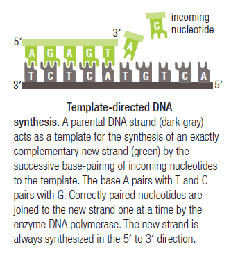
https://reasonandscience.catsboard.com

16DNA replication of prokaryotes Empty The Hexameric DnaB Helicase Sun Nov 15, 2015 5:27 pm

Otangelo


Admin

The Hexameric DnaB Helicase


DNA HELICASES: UNWINDING OF THE DOUBLE HELIX

DNA helicase unwinds double-stranded DNA for copying


Having now considered the key enzymes involved in the actual synthesis of DNA during replication, let us now turn to an enzyme that has an important function before synthesis can actually occur – the unwinding of the double-stranded helix of the parent DNA. When base-paired in the double-stranded helical DNA molecule, nucleotides are inaccessible to polymerases, making copying impossible. Both primase and polymerases need access to single-stranded DNA. To achieve this, the internal base-pairing must be broken and the helix unwound. The first step in this unwinding process is the initial opening of the helix, a step performed by the initiator protein at the origin of replication. Once opened, the unwinding of the double helix to expose single-stranded DNA for copying can begin. This unwinding process is catalyzed by an enzyme called DNA helicase.  The cell also needs to open the helix for DNA repair and recombination, and there are a unique set of DNA helicases for this particular case of DNA unwinding. Helicases open the double-stranded DNA and then travel with the replication fork, continuously unwinding the DNA to provide a template for the polymerase to copy. The helicases involved in replication in both bacteria and eukaryotes are composed of six subunits that form a ring structure that surrounds one strand of the DNA. The structure of the replicative DNA helicase from papillomavirusprovides insights into the mechanism of helicase action. This viral helicase is a hexamer of one protein, E1. In the co-crystal of protein and DNA, a single strand of DNA fits into the center of the channel formed by the hexamer ring, as depicted in Figure below.

DNA replication of prokaryotes Helica10

The other strand of the DNA is displaced by the helicase and bound by single-stranded binding proteins. Each monomer possesses a series of DNAbinding loops such that, when the six monomers come together to form the hexamer, the DNA binding loops form a spiral staircase on the inside of the channel which binds the DNA, and move along it one nucleotide at a time. Each movement of the DNA binding loop within each monomer requires adenosine triphosphate (ATP) hydrolysis; thus to move six nucleotides requires six ATP molecules, making DNA unwinding an energy costly process. 

DNA replication of prokaryotes Helica10

In E. coli, the helicase associated with DNA replication is known as DnaB and is a hexamer of six identical subunits. In eukaryotes and archaea, the replicative helicase is called MCM and is a complex of six different proteins, Mcm2–7, which assemble to form a ring. The eukaryotic replicative helicase is conserved throughout evolution and requires accessory factors for helicase activity.


DNA replication of prokaryotes Helica11

Functions of DnaB 2

This is a fascinating video which shows how DnaB helicase functions: 


DNA replication begins at the oriC. This is the site where proper replication occurs.  The site is 245 base pairs long.  This origin contains 4 nine-mers which are the binding sites for a protein called Dna A.  The Dna A protein controls the binding of Dna B to the origin.  This binding occurs when Dna A initiates the melting of 3 13-mers on the other end of the origin site.  This melting of the base pairs causes the Dna B to bind to the origin. Although there is still a problem with Dna B finding the site.  So Dna C binds to Dna B to help deliver the complex to the open complex or DNA melting region of origin.  After Dna B binds to the 13-mer melted region, this stimulates the binding of Dna G (primase).  Thus completing the primosome.  The Tao subunit of the DNA polymerase stabilizes and stimulates the Dna B helicase.  As the Dna B is stabilized, the protein begins to unwind the dsDNA as it hydrolyzes ATP.  The hydrolyzed ATP causes a slight structure change which helps in the ability to connect with the correct sites and move the ssDNA in one direction.  Once replication has started there are two main functions the Dna B helicase serves

1.   Consistently needs to bind for priming on the lagging strand because the lagging strand produces Okazaki fragments

2.   Dna B unwinds the parental DNA to give the pol III templates for the leading and lagging strand, this occurs in the direction of 5’ to 3’.  This is the same direction as the replication fork is moving


Structure of Hexameric DnaB Helicase and Its Complex with a Domain of DnaG Primase 1

The complex between the DnaB helicase and the DnaG primase unwinds duplex DNA at the eubacterial replication fork and synthesizes the Okazaki RNA primers. The crystal structures of hexameric DnaB and its complex with the helicase binding domain (HBD) of DnaG reveal that within the hexamer the two domains of DnaB pack with strikingly different symmetries to form a distinct two-layered ring structure. Each of three bound HBDs stabilizes the DnaB hexamer in a conformation that may increase its processivity. Three positive, conserved electrostatic patches on the N-terminal domain of DnaB may also serve as a binding site for DNA and thereby guide the DNA to a DnaG active site.

DNA replication of prokaryotes Hexame10

Architecture of the DnaB hexamer. 
(A) Experimentally phased and cross-crystal averaged electron density maps of the four DnaB crystal forms. Shown at the foot of each map is the high-resolution limit at which each map was calculated. 
(B) “Side” view, orthogonal to the ring axis, of a ribbon representation of the DnaB hexamer. The NTD, CTD, and linker region are colored blue, red, and yellow respectively. 
(C) “Top” view, looking down the ring axis, of the DnaB hexamer. The CTDs are shown in a surface representation; the NTDs are shown as ribbons. Those subunits whose NTDs lie on the inner surface of the ring are colored as in (B), and those on the outer surface of the ring are colored white. 
(D) “Side” view of the two distinct conformations of the DnaB subunits within the hexamer, colored as in (B). Adjacent CTDs interacting with the linker region are shown as white surface representations.

DNA replication of prokaryotes Hexame11

Structure of the CTD ring. 
(A) Surface representation of the CTD rings of crystal forms BH1 (left) and B1 (right). Alternate subunits are colored white and red. The predicted DNA binding loops are colored blue, and the linker helices are shown as yellow cylinders. 
(B) The structure of the T7 gp4 helicase domain (23), displayed as in panel (A). 
(C) Ribbon representations of the CTD rings of crystal forms BH1 (left) and B1 (right). Alternate subunits are colored white and pink. NTP modeled at the six potential NTP binding sites of DnaB (22) are shown as green spheres; the Arginine fingers (Arg420) are displayed as red spheres. 
(D) The structure of the T7 gp4 hexamer with four NTD binding sites occupied, displayed as in (C).


DNA replication of prokaryotes Hexame12

Structure of the complex between DnaB and HBD. 
(A) (Top) “Top” view of a ribbon representation of the complex showing the three HBDs (green) bound at the periphery of the NTD collar (light blue and blue). The CTD and linker region are colored red and yellow, respectively. (Bottom) The interface between DnaB and HBD shown as ribbons with a transparent surface. 
(B) “Side” view of a surface representation of the complex revealing no interaction between the HBDs (green) and the DnaB CTD (red) or linker region (yellow). [/ltr]
(C) Backbone trace of the HBD DnaB interface, residues known to modulate the interaction between DnaB and DnaG, are shown as colored spheres.


DNA replication of prokaryotes Hexame13


DNA interactions. 
(A) (Left) “Top” view of a surface representation of the NTD collar colored blue for positive and red for negative electrostatic potentials. An asterisk highlights the proposed ssDNA binding sites. (Right) A detailed “side” view of the proposed ssDNA binding site boxed in (A). 
(B) Speculative model of DnaB complexed with DnaG and replication fork DNA. The proteins are shown in a surface representation (DnaB NTD, light blue; DnaB CTD, red; DnaG HBD, green; DnaG RPD, pink; and DnaG ZBD, orange). The modeled DNA is shown as white- and wheat-colored spheres; the RNA primer is shown in dark blue.


DNA replication of prokaryotes Swerwe10


DNA replication of prokaryotes Ertert10


DNA replication of prokaryotes Ertert10


DNA replication of prokaryotes Ertert11

DNA replication of prokaryotes Ertert12

DNA replication of prokaryotes Ertert13

DNA replication of prokaryotes Ertert14

DNA replication of prokaryotes Ertert15

DNA replication of prokaryotes Aasdas10

1) http://www.sciencemag.org/content/318/5849/459.abstract
2) http://www.biochem.umd.edu/biochem/kahn/molmachines/replication/dna_b_protein.htm


Last edited by Admin on Wed Nov 25, 2015 8:51 am; edited 8 times in total

https://reasonandscience.catsboard.com

17DNA replication of prokaryotes Empty Re: DNA replication of prokaryotes Mon Nov 16, 2015 12:23 pm

Otangelo


Admin

Dna-B:


The protein, to begin with it is a monomer, but with the binding of Dna-C in one to one manner, subunits start assembling into hexamers to form of a ring. Then with the assistance of Dna-C, helicase ring opens and loads on to the strand, at the joint region of the fork. This assembles only on lagging strand, which is identified by the strand orientation from 5’ towards 3’ direction.  Two such helicases load, one at each fork joints.


DNA replication of prokaryotes Image007


https://www.biochem.wisc.edu
DNA replication of prokaryotes Image008
SSBs bound to single replicating DNA strands; http://www.mun.ca/

DNA replication of prokaryotes Image009


E.coli DNA-B subunits in the form a Hexameric ring

Once the Dna-B ring is formed Dna-C dissociates.  The Dna-B complex is a motor protein and acts as DNA dependent ATPase and using the energy it drives into the fork and unwinds the DNA progressively like unzipping the helical DNA.  Its direction of movement is from 5’ to 3’ on the lagging strand.  There is another protein called Rep A, which as monomer also binds to fork region but to the leading strand and moves in 3’ to 5’ direction.  However involvement of this protein in E.coli DNA replication is not substantiated.

DNA replication of prokaryotes Image010


Bacterial 3’ to 5’ DNA helicase; http://ww2.d155.org/
Prokaryotic Helicases:


Helicase II (Uvr-D)


DNA repair
Uvr AB complex


DNA repair
Helicase-IV


DNA repair
Rec-BCD


Recombination
T4 Dda?


Displaces SsBs
Helicase-I (encoded by F-plasmids)


Involved in DNA transfer during conjugation
Helicase-III


Unknown function


On RNA transcript, performs transcriptional termination
Helicase DnaB
hexamer
5’>-3’
Fork opening

Monomer
3’-5>’
Fork opening
DNA replication of prokaryotes Image011


Helicase in motion unwinding ds DNA; http://iespoetaclaudio.centros.educa.jcyl.es/


DNA replication of prokaryotes Image012


The unwinding of double stranded DNA using Dna B helicase. Dna G is the primase. This picture was gathered from“ http://chem-mgriep2.unl.edu/replic/Helicase.html


Last edited by Admin on Sun Nov 22, 2015 9:06 pm; edited 15 times in total

https://reasonandscience.catsboard.com

18DNA replication of prokaryotes Empty Scientists Watch Motors Unwind DNA Mon Nov 16, 2015 12:24 pm

Otangelo


Admin

Scientists Watch Motors Unwind DNA   1

Andrew Taylor and Gerald Smith from Fred Hutchinson Cancer Research Center (Seattle, WA) announced in Nature June 19 that “RecBCD enzyme is a DNA helicase with fast and slow motors of opposite polarity.”  In the same issue, Mark S. Dillingham, Maria Spies and Stephen C. Kowalczykowski of U.C. Davis came to a similar conclusion.  Working independently, these teams watched an important molecular motor in action and determined that it is two motors in one, with a slow motor and fast motor working side by side on the same track.  How can that be, and why?

    RecBCD helicase is the molecular machine that travels along a DNA double helix, unwinds it, and separates the strands so that the translation machinery can get to it.  This combination enzyme (RecB + RecC + RecD) is a member of a superfamily of helicases, or enzymes able to unwind and separate DNA.  Simpler helicases separate the two DNA strands into a Y-like tail, but RecBCD has the unusual property of creating a loose tail on the RecD side and a loop and a short tail on the RecB side (RecC, not a motor, appears to help RecB in its action).  Combined, RecBCD is among the fastest of helicases: it can cover 370 base pairs per second, according to Taylor and Smith, or up to 1000 base pairs per second, according to Kowalczykowski et al.
  Both the RecB and RecD motors can travel along DNA separately, but are polar opposites: one moves along one strand, one along the other.  Of the two, RecD is the speed demon; RecBC only moves 20% as fast.  The motors are not nearly as fast or stable acting alone.  Separately, they fall off the track after 50 base pairs, but together, can cover 400-600 times as much ground: 20,000 (Taylor and Smith) or 30,000 (Kowalczykowski) at full speed.
    So why two engines in this race car?  Taylor and Smith suggest that it adds stability; a motor is less likely to fall off the DNA track when combined with another, but why the speed difference?  This will take more study.  All they can conclude is, “This asymmetric feature might impart RecBCD enzyme’s asymmetry in other aspects of its promotion of genetic recombination.”


We’re going to stick our neck out and offer a hypothesis.  First of all, it is apparent from the speed and processivity (ability to process lots of letters without failure) that RecBCD is very well designed.  It doesn’t seem to slow RecD down to have the slower RecBC motor on the other track, but why don’t they both run at the same speed?  There must be a reason, and maybe the loop that RecBC forms is the clue.  In a fast winding device, like a tape drive, engineers often design a slack-uptake mechanism to prevent breakage if there is a sudden stop.  In older computer tape drives, for instance, a vacuum column maintained a loop of tape that could act as a buffer when the motors stopped or reversed direction.  Because RecBCD is so fast, maybe it was designed with a similar slack-adjusting loop on one side.
    We’ll have to wait and see whether this hunch has any merit.  Suffice it to say that we have again watched scientists uncover a superbly-efficient, highly-accurate biological machine, made up of multi-component parts, that does just what the cell needs doing.  For security reasons, DNA is tightly wrapped and hard to get to.  Once the helicase machinery is authenticated and allowed in, it needs to do its job fast, and that it does, exceptionally well.  1,000 base pairs a second: imagine!  It has to “melt” the chemical bonds between DNA letters at that high rate without causing collateral damage for its 20 to 30 second roller-coaster ride down the DNA tracks.  A good typist works about 70 words per minute; with an average word length of 5, that’s 350 letters per minute, or just under 6 letters per second.  A speed reader can go faster, but can anyone claim to read 200 words per second?  Behold RecBCD, the champ.  It’s busy at work inside your every cell, right now.  And oh, by the way, these scientists did their studies on those simple, primitive, lower forms of life: bacteria.  As you might expect, neither paper dares mention how these little machines could have evolved.

DNA replication of prokaryotes Helica13
DNA replication of prokaryotes Helica15
DNA replication of prokaryotes Helica13
DNA replication of prokaryotes Sem_ty12
DNA replication of prokaryotes Sem_ty11
DNA replication of prokaryotes Sem_ty13


1) http://creationsafaris.com/crev0603.htm


Last edited by Admin on Mon Nov 23, 2015 7:53 am; edited 12 times in total
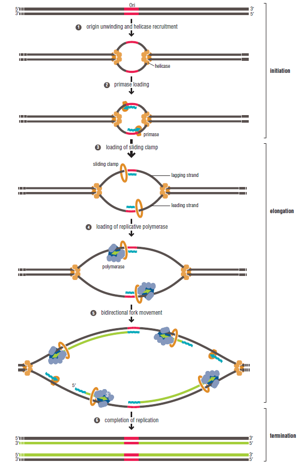
https://reasonandscience.catsboard.com

Otangelo


Admin

DnaC, and strategies for helicase recruitment and loading in bacteria 3

Introduction


DnaC is a monomer, binds to Dna-B in one to one manner (1:1). 5  It helps or facilitates the helicase to be loaded onto ssDNA at replication fork in ATP dependent manner. The DnaC-ATP binds to helicase hexamer and induces the opening of the helicase hexamer ring so that it can load on to single strand and encircle the strand at fork joint.  Once helicase loads on, Dna-C dissociates from the helicase subunits and helicase hexamer in association with ATP acts like a motor protein that moves into the fork and unwinds the DNA ahead of the fork.

The replication of eukaryotic and prokaryotic chromosomes, viruses and bacterial plasmids involves several analogous events and similarities in replisome architecture. For many systems, it has been demonstrated that specific initiation proteins, including bacterial DnaA protein, phage λ O protein, SV40 T antigen, plasmid replication initiation proteins (generally termed Rep) and the eukaryotic origin recognition complex (ORC), form complexes at the origin that serve as platforms for subsequent DNA replication initiation events. Despite certain similarities, the specific mechanism for replication initiation of a given replicon is dependent on both the structure of the replication origin and the nature of the replication initiation protein. The replication of extra-chromosomal replicons, such as plasmids, phages and viruses, is generally limited to a single host or a few closely related hosts (narrow host range). However, promiscuous plasmids of bacteria are able to replicate and maintain themselves in many distantly related bacterial species (broad host range). Consequently, versatile interactions of plasmid-encoded proteins and replication origins with host-specific replication factors might determine the mode of broad-host-range replicon initiation.

Origin structure and opening

The origins of prokaryotic and some eukaryotic replicons such as DNA viruses and Saccharomyces cerevisiae possess characteristic functional elements, including specific binding sites for the appropriate initiation protein and an AT-rich region where DNA duplex destabilization occurs. Plasmid origins usually contain multiple binding sites (iterons) for the plasmid-specific replication initiation protein as well as one or more binding sites for the host replication initiation protein, DnaA (DnaA boxes; Fig. 1).
Figure 1
Structural organization of some prokaryotic origins. The multiple repeat sequences (iterons), AT-rich and GC-rich regions, and DnaA box sequences are indicated. (Maps are not drawn to scale.)
Several lines of evidence suggest that these structural elements of the origin are employed for broad-host-range plasmid replication and maintenance in different host bacteria species. For example, the minimal origin of the broad-host-range plasmid RK2 (oriV; Fig. 1) possesses five iterons and is functional inEscherichia coli. However, the presence of three additional iterons stabilizes RK2 plasmid maintenance in Pseudomonas putida (Schmidhauser et al., 1983). In addition, the region with four DnaA boxes is essential for RK2 replication in E. coli, but is dispensable for replication of the plasmid in Pseudomonas aeruginosa(Shah et al., 1995; Doran et al., 1999). In the E. coli chromosome, the replication origin (oriC) contains five DnaA box sequences (Fig. 1).


DNA replication of prokaryotes 4-embo10

Structural organization of some prokaryotic origins. The multiple repeat sequences (iterons), AT-rich and GC-rich regions, and DnaA box sequences are indicated.

The binding of multiple DnaA molecules in the presence of the histone-like HU protein and the site-specific DNA-binding protein IHF (integration host factor) results in destabilization of the duplex DNA within the nearby AT-rich sequences of the oriC of E. coli (Messer et al., 2001). Origin opening of the narrow-host-range plasmids P1, F, R6K and pSC101 requires, in addition to E. coli DnaA, HU and/or IHF proteins, the binding of plasmid-encoded replication initiation proteins (Mukhopadhyay et al., 1993; Kawasaki et al., 1996; Lu et al., 1998; Park et al., 1998; Kruger et al., 2001; Sharma et al., 2001). Similarly, the formation of an open complex at the replication origin of the broad-host-range plasmid RK2 by the plasmid-encoded TrfA initiation protein requires E. coli HU, and is stabilized by E. coli DnaA (Konieczny et al., 1997).
In contrast to the chromosomal oriC, but similar to bacteriophage λ, plasmid origins do not require ATP for open complex formation (Schnos et al., 1988;Mukhopadhyay et al., 1993; Kawasaki et al., 1996; Lu et al., 1998; Park et al., 1998). A basis for this lack of dependence on ATP might be the intrinsic DNA curvature of these origins as well as origin bending, induced in an ATP-independent mode, by the complex of the plasmid-encoded Rep protein and the host HU or IHF (Stenzel et al., 1991; Doran et al., 1998; Lu et al., 1998; Komori et al., 1999; Sharma et al., 2001).


DnaB and other replicative helicases

The DnaB protein, the major replicative DNA helicase in E. coli (LeBowitz & McMacken, 1986), is a member of the hexameric DNA helicase family, which includes the T4 and T7 DNA helicases and plasmid RSF1010-encoded RepA, as well as the SV40 T antigen and the human MCM (minichromosome maintenance) protein (Patel & Picha, 2000). Although no sequence identity has yet been defined, these helicases form a ring structure with a central opening and are associated with DNA replication complexes. Mammalian MCM helicase is a complex of several different but related peptides. Interestingly, it was recently shown that the Methanobacterium thermoautotrophicum MCM protein can form heptameric rings (Yu et al., 2002). Several lines of evidence suggest that DNA passes through the central opening of the helicase ring, although an alternative model of DNA wrapping around the outside of the helicase ring has also been proposed (Patel & Picha, 2000).
E. coli DnaB is a multifunctional enzyme with a number of distinct activities including DNA binding, ATP hydrolysis, DNA unwinding and the stimulation of the DnaG primase for primer synthesis, which is required to start the polymerization reaction by the DNA polymerase holoenzyme. DnaB interacts with a number of proteins, including E. coli DnaA (Marszalek & Kaguni, 1994), DnaC (Wickner & Hurwitz, 1975), DnaG primase (Lu et al., 1996; Tougu & Marians, 1996) and the τ subunit of DNA polymerase (Kim et al., 1996), as well as the plasmid-encoded replication initiation proteins RepA of pSC101 (Datta et al., 1999), π of R6K (Ratnakar et al., 1996) and TrfA of RK2 (Pacek et al., 2001). The E. coli DnaB hexamer is present in vivo in a protein complex with six monomers of the DnaC protein and six ATP molecules (Wickner & Hurwitz, 1975; Lanka & Schuster, 1983; Fig. 2).
Figure 2


DNA replication of prokaryotes 4-embo11

Models for helicase recruitment and loading at plasmid, phage and bacterial chromosomal origins. Protein requirements and interactions required for helicase recruitment and loading are depicted. Thick arrows indicate the crucial interactions; dotted arrows indicate the direction of replication. 
(A) A physical interaction between E. coli DnaA and DnaB helicase as well as the activity of an accessory DnaC ATPase are essential for delivering the helicase to E. coli oriC. 
(B) During bacteriophage λ replication, the role of DnaC is performed by the λP protein, which binds the E. coli DnaB helicase and delivers it to oriλ by means of an interaction with the λO protein. 
(C) In addition to E. coli DnaA and DnaC proteins, helicase recruitment at narrow-host-range plasmid origins requires plasmid-specific replication initiation proteins (Rep). 
(D) Alternative mechanisms for helicase recruitment and loading at the origin of the broad-host-range plasmid RK2. The host-specific DnaA–DnaB interaction used for helicase recruitment at the DnaA boxes of the RK2 plasmid origin is applicable to plasmid replication inE. coli. In P. aeruginosa, helicase is recruited and loaded onto the RK2 origin in a DnaA- and DnaC-independent mode, through a specific interaction with the plasmid TrfA-44 replication initiation protein.

Models for helicase recruitment and loading at plasmid, phage and bacterial chromosomal origins. Protein requirements and interactions required for helicase recruitment and loading are depicted. Thick arrows indicate the crucial interactions; dotted arrows ...

E. coli DnaB helicase recruitment and loading

The E. coli DnaB helicase binds to single-stranded DNA (ssDNA) in an ATP-dependent manner. However, this activity alone is not sufficient for helicase loading at a replication origin because the DnaB hexamer by itself has no affinity for ssDNA bound by SSB (single- stranded binding protein). Thus, entry of the DnaB helicase complex into the unwound oriC depends on additional protein factors, and the mechanism behind this event is not fully understood. Chemical crosslinking, enzyme-linked immunosorbent assays and monoclonal antibody interference studies have shown that a physical interaction between E. coli DnaA and DnaB is essential for delivering the helicase to oriC (Marszalek & Kaguni, 1994). The loading of DnaB probably depends not only on DnaA binding to the DnaA boxes present in the E. coli oriC sequence (Fig. 2A) but also on DnaA binding to the open region of the origin, which is then stabilized for subsequent helicase loading (Speck & Messer, 2001). A specific physical interaction between DnaA and DnaB was also shown to be crucial during plasmid RK2 replication initiation in E. coli (Konieczny & Helinski, 1997; Fig. 2D). This DnaA–DnaB complex was found at the DnaA box region of oriV, which is separated by more than 200 base pairs (bp) from the RK2 origin opening and has a strict DnaA box sequence requirement for stable formation (Pacek et al., 2001).
In addition to the interaction between DnaB and DnaA, the helicase accessory ATPase protein, DnaC, is also required for helicase complex formation and helicase loading at E. coli oriC, as well as at several plasmid origins including RK2 (Konieczny & Helinski, 1997), R6K (Lu et al., 1998) and pSC101 (Datta et al., 1999). The T4 gp59 and B. subtilis DnaI proteins have a role similar to that of DnaC, serving as helicase loading factors during bacteriophage T4 and B. subtilisDNA replication initiation, respectively (Kreuzer & Morrical, 1994; Imai et al., 2000). Two eukaryotic proteins, Cdc6 and Cdt1, the latter recently identified as a novel component of the pre-replication complex, have been proposed to recruit the MCM complex in Xenopus and Saccharomyces (Baker & Bell, 1998; Bell & Dutta, 2002). During the replication of bacteriophage λ, the role of DnaC is performed by the λP protein, which is also an ATPase but shares no sequence similarity with DnaC. λP protein binds the E. coli DnaB helicase and delivers it to the λ origin (oriλ) by means of an interaction with the λO protein (Dodson et al., 1985; Fig. 2B). After the helicase complex has been formed at oriλ, it must be remodelled by the concerted actions of the E. coli chaperones DnaK, DnaJ and GrpE (Konieczny & Zylicz, 1999). At this step, DnaB is released from a tight interaction with λP. The requirement for molecular chaperones is decreased by a mutation in the λP gene, which weakens the interaction between the λP protein and the DnaB helicase (Konieczny & Marszalek, 1995).
The detailed molecular mechanism of helicase loading onto ssDNA is not fully understood. It has been shown that ssDNA-binding activity of λP and DnaC is involved in DnaB loading (Learn et al., 1997), and also that DnaC is released with concomitant hydrolysis of ATP during helicase loading onto oriC (Funnell et al., 1987; Allen & Kornberg, 1991). Recently, it was proposed that DnaC is a dual ATP/ADP switch protein, with DnaB and ssDNA triggering ATP hydrolysis by DnaC (Davey et al., 2002). Surprisingly, ATP is not required for the loading of DnaB by DnaC onto ssDNA, and the model proposes that DnaC–ATP loads the helicase onto oriC, but that conversion to DnaC–ADP is required before the helicase is active (Davey et al., 2002).
During replication initiation of plasmid RK2 in E. coli, the DnaA protein directs DnaB, in complex with DnaC, to the DnaA boxes in oriV of RK2. However, the helicase complex fails to unwind the template unless the plasmid initiation protein TrfA is also present (Konieczny & Helinski, 1997). It has been proposed that the helicase is repositioned from the DnaA boxes onto the AT-rich region via a direct contact with TrfA (Pacek et al., 2001). The loading of DnaB onto the ssDNA depends on the precise positioning of the DnaA boxes at oriV, as was shown by the disruption of helicase loading by the insertion of 6 bp between the DnaA boxes and the iterons at oriV, although opening was normal (Doran et al., 1998). Interactions between E. coli DnaB and plasmid Rep proteins have also been reported for the plasmids R6K (Ratnakar et al., 1996) and pSC101 (Datta et al., 1999), and these have been shown to be crucial for initial helicase complex formation at these plasmid origins. A mutant form of the DnaB protein that does not interact with pSC101 RepA fails to activate replication initiation at this origin. However, the mutant maintains its ability to support replication initiation at oriC (Datta et al., 1999). The plasmid R6K π protein and pSC101 RepA have also been shown to interact with the E. coli DnaA initiator (Lu et al., 1998; Sharma et al., 2001), which suggests a complex interaction involving the plasmid Rep protein in the formation of the prepriming complex, helicase loading, and activation.


Species-specific helicase recruitment and loading?

An intriguing question pertaining to DNA replication is whether or not the mechanism for helicase recruitment and loading described for E. coli oriC is also responsible for replication initiation of chromosomal and plasmid origins in other bacterial species. E. coli has traditionally been used as a model organism, but it is not clear whether these studies really do provide universal rules for all prokaryotes. This limitation can be overcome by studying promiscuous plasmids, which provide unique systems for exploring species-dependent replication mechanisms. Genetic and biochemical studies have suggested that these replicons have developed two major strategies to facilitate DNA replication in different genetic backgrounds: (1) initiation independent of host-DNA replication initiation factors, and (2) initiation dependent on versatile communication between plasmid and host-DNA replication initiation factors. Broad-host-range plasmids belonging to the IncQ incompatibility group (for example RSF1010) employ the first strategy by encoding three replication proteins that obviate the need for certain host proteins. The product of the repAgene of RSF1010 was found to have ssDNA-dependent ATPase and DNA helicase activity (Scherzinger et al., 1997), the repC product binds to the iterons and opens the origin region, creating the entry site for the RepA helicase (Scherzinger et al., 1991), and the repB product encodes a primase.
Broad-host-range plasmids belonging to the IncP group (e.g. RK2) rely on replication proteins from the host cell, and might therefore use a helicase loading mechanism adapted to the genetic background of the specific host bacterium. Recent results indicate that this is so (Caspi et al., 2001). In vitroexperiments with purified helicase from E. coliP. putida and P. aeruginosarevealed that, unlike the E. coli DnaB helicase, both Pseudomonas helicases could be delivered and activated at the RK2 oriV in the absence of a DnaC-like ATPase accessory protein (Fig. 2D). Further versatility is provided by two forms of the RK2 initiation protein (TrfA; 44 and 33 kDa), which are generated by alternative in-frame translational start sites (Kornacki et al., 1984; Shingler & Thomas, 1984). The requirement for each of these forms is hostspecific. Either form of the TrfA protein binds to the iterons located at oriV (Perri et al., 1991) and opens the origin at the AT-rich region (Caspi et al., 2001). Both are also functional in E. coliand P. putida, but only the 44-kDa protein is active in P. aeruginosa (Durland & Helinski, 1987; Fang & Helinski, 1991). Consistent with these observations are in vitro experiments showing that E. coli or P. putida DnaB is active with either TrfA-33 or TrfA-44, whereas P. aeruginosa DnaB specifically requires TrfA-44 for helicase complex formation and template unwinding (Caspi et al., 2001). The molecular basis for this difference has recently been elucidated (Y. Jiang, M. Pacek, D.R. Helinski, I.K. and A. Toukdarian, unpublished observations). Size-exclusion chromatography and helicase-activity assays with altered oriVtemplates showed that neither DnaA nor the DnaA box sequences were required for the formation and activity of the Pseudomonas helicase complex at RK2 oriV. Furthermore, biospecific interaction analysis with BIAcore revealed thatPseudomonas helicases form complexes with TrfA-44 but not with TrfA-33 bound to the oriV iterons. The deletion of a putative helical region at the amino terminus of TrfA-44 completely abolished Pseudomonas helicase complex formation at the iterons (Z. Zhong, D. Helinski and A. Toukdarian, unpublished observations).
These results suggest that, depending on the bacterial host, RK2 uses either a DnaA-dependent or a DnaA-independent pathway for helicase recruitment and activation (Fig. 2D). The DnaA-dependent pathway is specific for RK2 replication initiation in E. coli. The second pathway, employed in P. putida and P. aeruginosa, involves helicase recruitment through its interaction with TrfA-44 bound to iterons. Moreover, for the Pseudomonas sp. helicases, the host DnaA protein is not essential for helicase complex formation and activity at oriV.


Structure of a helicase–helicase loader complex reveals insights into the mechanism of bacterial primosome assembly 1


During the assembly of the bacterial loader-dependent primosome, helicase loader proteins bind to the hexameric helicase ring, deliver it onto the oriC DNA and then dissociate from the complex. Here, to provide a better understanding of this key process, we report the crystal structure of the ~570-kDa prepriming complex between the Bacillus subtilis loader protein and the Bacillus stearothermophilus helicase, as well as the helicase-binding domain of primase with a molar ratio of 6:6:3 at 7.5 Å resolution. The overall architecture of the complex exhibits a three-layered ring conformation. Moreover, the structure combined with the proposed model suggests that the shift from the ‘open-ring’ to the ‘open-spiral’ and then the ‘closed-spiral’ state of the helicase ring due to the binding of single-stranded DNA may be the cause of the loader release.

The replication of the bacterial chromosome is initiated at oriC where the initiator protein DnaA binds to start the assembly of the enzymatic replisome machine1. The early stages of this process involve the assembly of the primosome and the formation of a functional primosome2, 3. Subsequent to the remodelling of the replication origin induced by DnaA, the assembly of the bacterial loader-dependent primosome occurs in discrete steps and involves at least four different proteins (DnaA, helicase, helicase loader and primase) that act in a coordinated and sequential manner.
In the Escherichia coli system, the helicase loader protein, DnaC, complexed with ATP, binds to hexameric helicase DnaB and forms a DnaB6–DnaC6 complex, which has been confirmed by cryo-electron microscope (cryo-EM) studies4, 5. The loader protein delivers the helicase onto the melted DNA single strands of the DnaA–oriC nucleoprotein complex at the origin of replication2,6

The Bacterial DnaC Helicase Loader Is a DnaB Ring Breaker  4

DNA replication of prokaryotes 011


 The DnaB-DnaC complex forms a topologically open, three-tiered toroid. ► DnaC remodels DnaB to produce a cleft in the helicase ring suitable for DNA passage. ► DnaC’s AAA+ fold is dispensable for DnaB loading and activation. ► DnaB possesses autoregulatory elements that control helicase loading and unwinding


What we see here are highly coordinated , goal oriented tasks with specific  movements designed to provide a specific outcome. Auto-regulation and control   that seems required beside constant energy supply through ATP enhances the difficulty to make the whole mechanism work in the right manner. All this is awe inspiring and evidences the wise guidance and intelligence required to make all this happening in the right way. 


DNA replication of prokaryotes 1-s2.0-S0092867413002924-gr5
Figure 5. 
The DnaB N-Terminal Domain Collar Is Remodeled by DnaC
(A) The N-terminal homodimers of DnaB in the absence of nucleotide form a wide, closed-triangular collar (PDB 2R6A; Bailey et al., 2007b). DnaB RecA domains are displayed as surfaces, whereas the N-terminal domains and linker helices are shown as light-blue/orange cylinders.
(B) The N-terminal domains of DnaB within the DnaBC complex undergo a marked positional shift from the closed-ring state, forming new packing arrangements between dimers. The DnaB RecA domains form a cracked spiral (boxed inset); DnaC is omitted for clarity. A schematic of the arrangement for the N-terminal domain dimers is shown in the upper-left corner of each panel.
Arrows in (A) indicate the movement of the DnaB NTDs to the state shown in (B). See Figures S5, S6, andMovies S2 and S3.

DNA replication of prokaryotes 1-s2.0-S0092867413002924-figs6
Figure S6. 
Comparison of the Conformational States of DnaB Bound to Single-Stranded DNA and within the DnaBC Complex, Related to Figure 5
(A) The pitch of the DnaB RecA ATPase domains differs significantly between DnaBC (upper) and when bound to DNA and nucleotide (lower) (PDB ID 4ESV (Itsathitphaisarn et al., 2012)). Each subunit is shown as a surface representation and colored differently. DNA is not shown for clarity.
(B) Conformational differences between DnaB hexamers bound to either DnaC (top) or single-stranded DNA and nucleotide (bottom, (Itsathitphaisarn et al., 2012)). Top down (left) and side views (right) are shown. Each subunit is shown as a surface representation and colored differently, with the N-terminal domains (cylinders) depicted as brighter shades than their associated RecA domains (surfaces). The positions of the linker helix that connects each N-terminal and C-terminal domain within a subunit are also shown and labeled. The linker helix is visible in all DnaB crystal structures and serves to anchor the RecA domain of each subunit to the RecA domain of an adjoining protomer (Bailey et al., 2007b; Itsathitphaisarn et al., 2012; Lo et al., 2009; Wang et al., 2008); in the EM model, its position is inferred based on known crystallographic arrangements. In the DnaB⋅ssDNA complex, the N-terminal collar of helicase is cracked open at one subunit interface, but the packing between homodimers at other points uses the same surfaces as observed in closed-ring states (left, see also Figure 5), and hence differs from that seen in DnaBC. The RecA domains in the DnaB⋅ssDNA complex also split apart at one point, but the split occurs between a different subunit pair (colored orange and red) than in DnaBC (colored yellow and orange). This difference is due to the topological linkages between N-terminal homodimers (which obey dyadic symmetry) and their respective C-terminal domains (which follow cyclic symmetry), and by the ability of the N-terminal domains to shift between the two states – sitting on either their own RecA folds (as in DnaBC), or on a neighboring subunit’s RecA domain (DnaB⋅ssDNA). Because of the positional shift in the DnaB⋅ssDNA complex, and the relatively shallow pitch of this particle’s open-ring state, the linker helix between each N-terminal and C-terminal domain can pair with a partner subunit, bridging the one gap in the ring to topologically seal off the system (orange/red subunit pair). By contrast, the N-terminal domain configuration seen in DnaBC, coupled with the large subunit-to-subunit rise evident in the complex, creates a gap that is too large be spanned by the linker helix of one subunit (shown in yellow). As a consequence, the helicase ring in the DnaBC complex is topologically breached.

DNA replication of prokaryotes 1-s2.0-S0092867413002924-gr7
Figure 7. 
Mechanism of DnaC Action
(A) DnaC opens and remodels DnaB to facilitate DNA loading and unwinding. Closed-ring DnaB cannot engage a topologically closed DNA substrate. DnaC associates with the helicase, remodeling the N-terminal collar, and triggering helicase opening. In the presence of ATP, DnaC AAA+ domains further assemble into a helical conformation that stabilizes the open-ring complex and assists with DNA binding. ATP hydrolysis by DnaC and/or DnaG helps disengage the loader (Davey et al., 2002; Makowska-Grzyska and Kaguni, 2010), leaving an active helicase encircled around DNA.
(B) Hypothetical model showing how DnaC is free to associate with a DnaA filament even when bound to DnaB (as proposed in Mott et al., 2008). The model was generated by aligning a DnaA filament bound to ssDNA (PDB 3R8F; Duderstadt et al., 2011)—which bears an exposed arginine finger at the 5′ end of the complex—with the solvent-accessible nucleotide-binding face of the terminal DnaC protomer in DnaBC in a manner consistent with typical AAA+/AAA+ interactions. The superposition coaligns the pores of all three proteins, positioning DNA bound by the central channel of DnaA to enter into the helicase/loader complex.


In vivo, this delivery is associated with the initiator protein DnaA2, 7, whose amino-terminal domain (NTD) is thought to have a role in loading the helicase and helicase loader complex onto the oriC by interacting with helicase DnaB8. After the loader protein dissociates from the helicase ring, the NTD of the helicase interacts with the carboxy-terminal domain (CTD) of the primase and forms a functional primosome that synthesizes RNA primers9. Primosome assembly in Gram-positive bacteria is different in the details, including that the corresponding helicase is named DnaC in some bacteria such as Bacillus subtilis, and the loader protein is DnaI; the assembly of the helicase and loader protein complex onto the replication origin is assisted by a pair of co-loader proteins DnaB and DnaD in B. subtilis10, 11, 12, 13.


The mechanism of helicase loading in bacteria 2

DNA replication of prokaryotes Nsmb_213

(a) The initiator DnaA binds to oriC, thus leading to DNA melting. 
(b) DnaB assembles with DnaC, thus leading to opening of the DnaB ring. 
(c) DnaA recruits the DnaB–DnaC complex to origins, where it assembles around ssDNA. 
(d) DNA-induced ATP hydrolysis promotes disassembly of DnaC, thus leaving DnaB encircling DNA. Pi, inorganic phosphate.


DNA replication of prokaryotes Nsmb_219

Upon ATP binding, the clamp loader transitions from a planar conformation into a right-handed helical conformation.
(a) The clamp loader binds to ATP and changes conformation. 
(b) The ATP-bound form of the clamp loader interacts with and forces open the clamp. 
(c) The complex of the clamp and clamp loader binds to a primer template. 
(d) Once bound to DNA, the clamp loader undergoes ATP hydrolysis, thus leading to dissociation from the clamp and leaving the clamp encircling the DNA.


DNA replication of prokaryotes Nsmb_215

(a,b) ORC binds to DNA (a) and recruits Cdc6 
(b). (ch) Two mechanisms for the recruitment of MCM2–7. Right, if MCM2–7–Cdt1 is in a closed conformation, it must form a partial contact with ORC–Cdc6 to open the MCM2–7 ring 
(c) so that DNA can enter the MCM2–7 ring, thus allowing MCM2–7 to stack coaxially with the ORC–Cdc6 complex for OCCM formation 
(d). Left, if MCM2–7–Cdt1 is normally open, it could accommodate DNA in its central channel 
(e) and then slide toward ORC–Cdc6 for OCCM formation 
(f). 
(g) ATP hydrolysis promotes dissociation of Cdt1 and conversion of OCCM to OCM. 
(h) A second MCM2–7 complex associates with OCM for MCM2–7 double-hexamer formation, the details of which are described in Figure 4.


DNA replication of prokaryotes Nsmb_217

(ae) Five possible models for double-hexamer formation. The first model
(a), in which two MCM2–7 complexes are loaded simultaneously, does not involve the OCM intermediate. The other four models 
(be) involve conversion of the OCM to a double hexamer. Complexes are depicted as in Figure 3. See main text for details.


1) http://www.nature.com/ncomms/2013/130919/ncomms3495/full/ncomms3495.html
2) http://www.nature.com/nsmb/journal/v21/n1/fig_tab/nsmb.2738_F1.html
3) http://www.ncbi.nlm.nih.gov/pmc/articles/PMC1315803/
4) http://www.sciencedirect.com/science/article/pii/S0092867413002924
5) http://mol-biol4masters.masters.grkraj.org/html/Prokaryotic_DNA_Replication3-E_coli_DNA_Replication.htm


Last edited by Admin on Mon Nov 23, 2015 7:30 am; edited 3 times in total
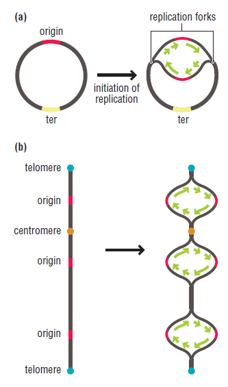
https://reasonandscience.catsboard.com

Otangelo


Admin

Unwinding the DNA Double Helix Requires DNA Helicases,Topoisomerases, and Single- Stranded DNA Binding Proteins

During DNA replication, the two strands of the double helix must unwind at each replication fork to expose the single strands to the enzymes responsible for copying them. Three classes of proteins with distinct functions facilitate this unwinding process: DNA helicases, topoisomerases, and single-stranded DNA binding proteins

DNA replication of prokaryotes Primas13

FIGURE 19-12 Proteins Involved in Unwinding DNA at the Replication Fork. Three types of proteins are involved in DNA unwinding. The actual unwinding proteins are the DNA helicases; the principal one in E. coli, which is part of the primosome, operates along the template for the lagging strand, as shown here. Single-stranded DNA binding proteins (SSB) stabilize the unwound DNA in an extended position. A topoisomerase forms a swivel ahead of the replication fork; in E. coli, this topoisomerase is DNA gyrase.

The proteins responsible for unwinding DNA are the DNA helicases. Using energy derived from ATP hydrolysis, these proteins unwind the DNA double helix in advance of the replication fork, breaking the hydrogen bonds as they go. In E. coli, at least two different DNA helicases are involved in DNA replication; one attaches to the lagging strand template and moves in a direction; the other attaches to the leading strand template. Both are part of the primosome, but the helicase is more important for unwinding DNA at the replication fork. The unwinding associated with DNA replication would create an intolerable amount of supercoiling and possibly tangling in the rest of the DNA were it not for the actions of topoisomerases. These enzymes create swivel points in the DNA molecule by making and then quickly resealing single- or double-stranded breaks in the double helix. Of the ten or so topoisomerases found in E. coli, the key enzyme for DNA replication is DNA gyrase, a type II topoisomerase (an enzyme that cuts both DNA strands). Using energy derived from ATP, DNA gyrase introduces negative supercoils and thereby relaxes positive ones. DNA gyrase serves as the main swivel that prevents overwinding (positive supercoiling) of the DNA ahead of the replication fork. In addition, this enzyme has a role in both initiating and completing DNA replication in E. coli—in opening up the double helix at the origin of replication and in separating the linked circles of daughter DNA at the end. The situation in eukaryotic cells is not as well understood, although topoisomerases of both types have been isolated. Once strand separation has begun, molecules of single-stranded DNA binding protein (SSB) quickly attach to the exposed single strands to keep the DNA unwound and therefore accessible to the DNA replication machinery. After a particular segment of DNA has been replicated, the SSB molecules fall off and are recycled, attaching to the next single-stranded segment.


Separating the duplex into the leading and lagging template strands (helicases)

DNA helicases are essential during DNA replication because they separate double-stranded DNA into single strands allowing each strand to be copied.

1) http://www.nature.com/scitable/definition/helicase-307

http://mol-biol4masters.masters.grkraj.org/html/Prokaryotic_DNA_Replication5-Mechanism_of_Replication.htm


Last edited by Admin on Fri Nov 27, 2015 4:56 pm; edited 14 times in total

https://reasonandscience.catsboard.com

21DNA replication of prokaryotes Empty DNA Polymerase Wed Nov 18, 2015 11:52 am

Otangelo


Admin

DNA Polymerase

DNA polymerase was first identified in lysates of Ecoli by Arthur Kornberg in 1956. 1 The ability of this enzyme to accurately copy a DNA template provided a biochemical basis for the mode of DNA replication that was initially proposed by Watson and Crick, so its isolation represented a landmark discovery in molecular biology. Ironically, however, this first DNA polymerase to be identified (now called DNA polymerase I) is not the major enzyme responsible for Ecoli DNA replication. Instead, it is now clear that both prokaryotic and eukaryotic cells contain several different DNA polymerases that play distinct roles in the replication and repair of DNA.
The multiplicity of DNA polymerases was first revealed by the isolation of a mutant strain of Ecoli that was deficient in polymerase I (Figure 5.1). Cultures of Ecoli were treated with a chemical (a mutagen) that induces a high frequency of mutations, and individual bacterial colonies were isolated and screened to identify a mutant strain lacking polymerase I. Analysis of a few thousand colonies led to the isolation of the desired mutant, which was almost totally defective in polymerase I activity. Surprisingly, the mutant bacteria grew normally, leading to the conclusion that polymerase I is not required for DNA replication. On the other hand, the mutant bacteria were extremely sensitive to agents that damage DNA (e.g., ultraviolet light), suggesting that polymerase I is involved primarily in the repair of DNA damage rather than in DNA replication per se.

The conclusion that polymerase I is not required for replication implied that Ecoli must contain other DNApolymerases, and subsequent experiments led to the identification of two such enzymes, now called DNA polymerases II and III. The potential roles of these enzymes were investigated by the isolation of appropriate mutants. Strains of Ecoli with mutations in polymerase II were found to grow and otherwise behave normally, so the role of this enzyme in the cell is unknown. Temperature-sensitive polymerase III mutants, however, were unable to replicate their DNA at high temperature, and subsequent studies have confirmed that polymerase III is the major replicative enzyme in Ecoli.
It is now known that, in addition to polymerase III, polymerase I is also required for replication of Ecoli DNA. The original polymerase I mutant was not completely defective in that enzyme, and later experiments showed that the residual polymerase I activity in this strain plays a key role in the replication process. The replication of Ecoli DNA thus involves two distinct DNA polymerases, the specific roles of which are discussed below.

All known DNA polymerases share two fundamental properties that carry critical implications for DNA replication (Figure 5.2). First, all polymerases synthesize DNA only in the 5′ to 3′ direction, adding a dNTP to the 3′ hydroxyl group of a growing chain. Second, DNA polymerases can add a new deoxyribonucleotide only to a preformed primer strand that is hydrogen-bonded to the template; they are not able to initiate DNA synthesis de novo by catalyzing the polymerization of free dNTPs. In this respect, DNA polymerases differ from RNA polymerases, which can initiate the synthesis of a new strand of RNA in the absence of a primer. As discussed later in this chapter, these properties of DNA polymerases appear critical for maintaining the high fidelity of DNA replication that is required for cell reproduction.

The synthesis of new DNA strands complementary to both strands of the parental molecule posed an important problem to understanding the biochemistry of DNA replication. Since the two strands of double-helical DNA run in opposite (antiparallel) directions, continuous synthesis of two new strands at the replication fork would require that one strand be synthesized in the 5′ to 3′ direction while the other is synthesized in the opposite (3′ to 5′) direction. ButDNA polymerase catalyzes the polymerization of dNTPs only in the 5′ to 3′ direction. How, then, can the other progeny strand of DNA be synthesized?
This enigma was resolved by experiments showing that only one strand of DNA is synthesized in a continuous manner in the direction of overall DNA replication; the other is formed from small, discontinuous pieces of DNA that are synthesized backward with respect to the direction of movement of the replication fork (Figure 5.4). These small pieces of newly synthesized DNA (called Okazaki fragments after their discoverer) are joined by the action of DNAligase, forming an intact new DNA strand. The continuously synthesized strand is called the leading strand, since its elongation in the direction of replication fork movement exposes the template used for the synthesis of Okazaki fragments (the lagging strand).

Although the discovery of discontinuous synthesis of the lagging strand provided a mechanism for the elongation of both strands of DNA at the replication fork, it raised another question: Since DNA polymerase requires a primer and cannot initiate synthesis de novo, how is the synthesis of Okazaki fragments initiated? The answer is that short fragments of RNA serve as primers for DNA replication (Figure 5.5). In contrast to DNA synthesis, the synthesis of RNA can initiate de novo, and an enzyme called primase synthesizes short fragments of RNA (e.g., three to ten nucleotides long) complementary to the lagging strand template at the replication fork. Okazaki fragments are then synthesized via extension of these RNA primers by DNA polymerase. An important consequence of such RNA priming is that newly synthesized Okazaki fragments contain an RNA-DNA joint, the discovery of which provided critical evidence for the role of RNA primers in DNA replication.

To form a continuous lagging strand of DNA, the RNA primers must eventually be removed from the Okazaki fragments and replaced with DNA. In Ecoli, RNA primers are removed by the combined action of RNase H, an enzyme that degrades the RNA strand of RNA-DNA hybrids, and polymerase I. This is the aspect of Ecoli DNA replication in which polymerase I plays a critical role. In addition to its DNA polymerase activity, polymerase I acts as an exonuclease that can hydrolyze DNA (or RNA) in either the 3′ to 5′ or 5′ to 3′ direction. The action of polymerase I as a 5′ to 3′ exonuclease removes ribonucleotides from the 5′ ends of Okazaki fragments, allowing them to be replaced with deoxyribonucleotides to yield fragments consisting entirely of DNA (Figure 5.6). In eukaryotic cells, other exonucleases take the place of Ecoli polymerase I in removing primers, and the gaps between Okazaki fragments are filled by the action of polymerase δ. As in prokaryotes, these DNA fragments can then be joined byDNA ligase.


DNA Polymerase III Is a Processive Enzyme that uses deoxyribonucleoside triphosphates


DNA replication of prokaryotes Dna_po18


Let’s now turn our attention to other enzymatic features of DNA polymerase. As shown in Figure 11.12 , DNA polymerases catalyze the covalent attachment between the phosphate in one nucleotide and the sugar in the previous nucleotide.

DNA replication of prokaryotes Dna_po15

Prior to bond formation, the nucleotide about to be attached to the growing strand is a dNTP. It contains three phosphate groups attached at the 5ʹ–carbon atom of deoxyribose. The dNTP first enters the catalytic site of DNA polymerase and binds to the template strand according to the AT/GC rule. Next, the 3ʹ–OH group on the previous nucleotide reacts with the phosphate group adjacent to the sugar on the incoming nucleotide. The breakage of a covalent bond between two phosphates in a dNTP is a highly exergonic reaction that provides the energy to form a covalent (ester) bond between the sugar at the 3ʹ end of the DNA strand and the phosphate of the incoming nucleotide. The formation of this covalent bond causes the newly made strand to grow in the 5ʹ to 3ʹ direction. As shown in Figure 11.12, pyrophosphate (PPi) is released. The term phosphodiester linkage (also called a phosphodiester bond) is used to describe the linkage between a phosphate and two sugar molecules. As its name implies, a phosphodiester linkage involves two ester bonds. In comparison, as a DNA strand grows, a single covalent (ester) bond is formed between adjacent nucleotides (see Figure 11.12). The other ester bond in the phosphodiester linkage—the bond between the 5ʹ-oxygen and phosphorus—is already present in the incoming nucleotide. DNA polymerase catalyzes the covalent attachment of nucleotides with great speed. In E. coli, DNA polymerase III attaches approximately 750 nucleotides per second! DNA polymerase III can catalyze the synthesis of the daughter strands so quickly because it is a processive enzyme. This means it does not dissociate from the growing strand after it has catalyzed the covalent joining of two nucleotides. Rather, as depicted in Figure 11.8a, it remains clamped to the DNA template strand and slides along the template as it catalyzes the synthesis of the daughter strand. The β subunit of the holoenzyme, also known as the clamp protein, promotes the association of the holoenzyme with the DNA as it glides along the template strand (refer back to Table 11.2). The β subunit forms a dimer in the shape of a ring; the hole of the ring is large enough to accommodate a double-stranded DNA molecule, and its width is about one turn of DNA. A complex of several subunits functions as a clamp loader that allows the DNA polymerase holoenzyme to initially clamp onto the DNA. The effects of processivity are really quite remarkable. In the absence of the β subunit, DNA polymerase can synthesize  DNA at a rate of approximately only 20 nucleotides per second. On average, it falls off the DNA template after about 10 nucleotides have been linked together. By comparison, when the β subunit is present, as in the holoenzyme, the synthesis rate is approximately 750 nucleotides per second. In the leading strand, DNA polymerase III has been estimated to synthesize a segment of DNA that is over 500,000 nucleotides in length before it inadvertently falls off.

Certain Enzymes of DNA Replication Bind to Each Other to Form a Complex


DNA replication of prokaryotes Dna_po16
FIGURE 11.15 A three-dimensional view of DNA replication. DNA helicase and primase associate together to form a primosome. The
primosome associates with two DNA polymerase enzymes to form a replisome.


Figure 11.15 provides a more three-dimensional view of the DNA replication process. DNA helicase and primase are physically bound to each other to form a complex known as a primosome. This complex leads the way at the replication fork. The primosome tracks along the DNA, separating the parental strands and synthesizing RNA primers at regular intervals along the lagging strand. By acting within a complex, the actions of DNA helicase and primase can be better coordinated. The primosome is physically associated with two DNA polymerase holoenzymes to form a replisome. As shown in Figure 11.15, two DNA polymerase III proteins act in concert to replicate the leading and lagging strands. The term dimeric DNA polymerase is used to describe two DNA polymerase holoenzymes that move as a unit toward the replication fork. For this to occur, the lagging strand is looped out with respect to the DNA polymerase that synthesizes the lagging strand. This loop allows the lagging-strand polymerase to make DNA in a 5ʹ to 3ʹ direction yet move toward the opening of the replication fork. Interestingly, when this DNA polymerase reaches the end of an Okazaki fragment, it must be released from the template DNA and “hop” to the RNA primer that is closest to the fork. The clamp loader complex (see Table 11.2), which is part of DNA polymerase holoenzyme, then reloads the enzyme at the site where the next RNA primer has been made. Similarly, after primase synthesizes an RNA primer in the 5ʹ to 3ʹ direction, it must hop over the primer and synthesize the next primer closer to the replication fork.

The Fidelity of DNA Replication Is Ensured by Proofreading Mechanisms

With replication occurring so rapidly, one might imagine that mistakes can happen in which the wrong nucleotide is incorporated into the growing daughter strand. Although mistakes can happen during DNA replication, they are extraordinarily rare. In the case of DNA synthesis via DNA polymerase III, only one mistake per 100 million nucleotides is made. Therefore, DNA synthesis occurs with a high degree of accuracy or fidelity. Why is the fidelity so high? First, the hydrogen bonding between G and C or A and T is much more stable than between mismatched pairs. However, this stability accounts for only part of the fidelity, because mismatching due to stability considerations accounts for 1 mistake per 1000 nucleotides. Two characteristics of DNA polymerase also contribute to the fidelity of DNA replication. First, the active site of DNA polymerase preferentially catalyzes the attachment of nucleotides when the correct bases are located in opposite strands. Helix distortions caused by mispairing usually prevent an incorrect nucleotide from properly occupying the active site of DNA polymerase. By comparison, the correct nucleotide occupies the active site with precision and undergoes induced fit, which is necessary for catalysis. The inability of incorrect nucleotides to undergo induced fit decreases the error rate to a range of 1 in 100,000 to 1 million. A second way that DNA polymerase decreases the error rate is by the enzymatic removal of mismatched nucleotides. As shown in Figure 11.16 , DNA polymerase can identify a mismatched
nucleotide and remove it from the daughter strand. A second way that DNA polymerase decreases the error rate is by the enzymatic removal of mismatched nucleotides. As shown in Figure 11.16 , DNA polymerase can identify a mismatched nucleotide and remove it from the daughter strand.

DNA replication of prokaryotes Dna_po17
FIGURE 11.16 The proofreading function of DNA polymerase. When a base pair mismatch is found, the end of the newly made strand is shifted into the 3ʹ exonuclease site. The DNA is digested in the 3ʹ to 5ʹ direction to release the incorrect nucleotide.

This occurs by exonuclease cleavage of the bonds between adjacent nucleotides at the 3ʹ end of the newly made strand. The ability to remove mismatched bases by this mechanism is called the proofreading function of DNA polymerase. Proofreading occurs by the removal of nucleotides in the 3ʹ to 5ʹ direction at the 3ʹ exonuclease site. After the mismatched nucleotide is removed, DNA polymerase resumes DNA synthesis in the 5ʹ to 3ʹ direction.

DNA Polymerase I :

DNA Polymerase I (or Pol I) is an enzyme that participates in the process of DNA replication. Discovered by Arthur Kornberg in 1956,[1] it was the first known DNA polymerase (and, indeed, the first known of any kind ofpolymerase). It was initially characterized in E. coli and is ubiquitous in prokaryotes. In E. coli and many other bacteria, the gene that encodes Pol I is known as polA. The E. coli form of the enzyme is composed of 928 amino acids, and is an example of a processive enzyme—it can sequentially catalyze multiple polymerisations.
Pol I possesses four enzymatic activities:

A 5'→3' (forward) DNA-Dependent DNA polymerase activity, requiring a 3' primer site and a template strand


A 3'→5' (reverse) exonuclease activity that mediates proofreading


A 5'→3' (forward) exonuclease activity mediating nick translation during DNA repair.


A 5'→3' (forward) RNA-Dependent DNA polymerase activity. Pol I operates on RNA templates with considerably lower efficiency (0.1–0.4%) than it does DNA templates, and this activity is probably of only limited biological significance.[2]


In the replication process, RNase H removes the RNA primer (created by Primase) from the lagging strand and then Polymerase I fills in the necessary nucleotides between the Okazaki fragments (see DNA replication) in a 5'→3' direction, proofreading for mistakes as it goes. It is a template-dependent enzyme—it only adds nucleotides that correctly base pair with an existing DNA strand acting as a template. DNA Ligase then joins the various fragments together into a continuous strand of DNA.
Despite its early characterisation, it quickly became apparent that Polymerase I was not the enzyme responsible for most DNA synthesis—DNA replication in E. coli proceeds at approximately 1,000 nucleotides/second, while the rate of base pair synthesis by Polymerase I averages only between 10 and 20 nucleotides/second. Moreover, its cellular abundance of approximately 400 molecules per cell did not correlate with the fact that there are typically only two replication forks in E. coli. Additionally, it is insufficiently processive to copy an entire genome, as it falls off after incorporating only 25-50 nucleotides. Its role in replication was proven when, in 1969, John Cairns isolated a viable Polymerase I mutant that lacked the polymerase activity.[3] Cairns' lab assistant, Paula De Lucia, created thousands of cell free extracts from E.coli colonies and assayed them for DNA-polymerase activity. The 3,478th clone contained the polA mutant, which was named by Cairns to credit "Paula" [De Lucia].[4] It was not until the discovery of DNA polymerase III that the main replicative DNA polymerase was finally identified.

http://biocyc.org/ECOLI/NEW-IMAGE?type=POLYPEPTIDE&object=CPLX0-3803
1) http://www.ncbi.nlm.nih.gov/books/NBK9940/


Last edited by Admin on Sun Nov 29, 2015 7:48 am; edited 12 times in total

https://reasonandscience.catsboard.com

22DNA replication of prokaryotes Empty The DNA sliding clamp and clamp loader Wed Nov 18, 2015 3:28 pm

Otangelo


Admin

The sliding clamp and clamp loader

DNA clamp, also known as a sliding clamp, is a protein fold that serves as a processivity-promoting factor in DNA replication. As a critical component of the DNA polymerase III holoenzyme, the clamp protein binds DNA polymerase and prevents this enzyme from dissociating from the template DNA strand. The clamp-polymerase protein–protein interactions are stronger and more specific than the direct interactions between the polymerase and the template DNA strand; because one of the rate-limiting steps in the DNA synthesis reaction is the association of the polymerase with the DNA template, the presence of the sliding clamp dramatically increases the number of nucleotides that the polymerase can add to the growing strand per association event. The presence of the DNA clamp can increase the rate of DNA synthesis up to 1,000-fold compared with a nonprocessive polymerase.[2]

Sliding clamps are DNA-tracking platforms that are essential for processive DNA replication in all living organisms 5

Following 3 illustrations: 
DNA replication of prokaryotes F4_lar11
Figure 4.
The clamp-loading mechanism. The catalytic reaction cycle for the loading of sliding clamps onto DNA by clamp loaders is shown as a schematic diagram. 
(A) Opening of the sliding clamp ring. In the absence of ATP, clamp loaders bind their respective clamps very weakly. On binding ATP, clamp loaders undergo a conformational change, which permits optimal interaction with the carboxy-terminal face of their respective clamp and subsequent opening of the clamp ring. 
(B) PT junction binding. The open clamp–clamp loader complex together specifically recognizes and binds a PT junction, adopting a “notched screw cap arrangement,” which matches the helical geometry of the DNA duplex and properly aligns the interfacial ATPase sites for hydrolysis. 
(C) Closure of the clamp ring. On hydrolysis of ATP, clamp loaders revert back to a low-affinity DNA-binding state and eject, leaving the PT DNA positioned within an opened sliding clamp ring. Concurrent or subsequent to ejection, electrostatic interactions between the positively charged inner surface of the sliding clamp ring and the negatively charged DNA drives closure of the open sliding clamp around DNA.


the whole movement of the clamp loading can be seen starting at 6,12min on this video :

https://www.youtube.com/watch?v=6mKNOlhw50w#t=371


DNA replication of prokaryotes Slidin13

Clamp loaders place sliding clamps at primer-template junctions for processive DNA replication. 2 When bound to ATP, clamp loaders are competent to bind and open the sliding clamp protein. This ternary complex can now bind to a primer-template junction, which activates the ATPase activity of the clamp loader. ATP hydrolysis causes the clamp loader to dissociate from the clamp and DNA, resulting in a loaded clamp that is competent for acting as a processivity factor for DNA polymerase. 


DNA replication of prokaryotes Clamp_10

Clamp loading mechanism. 3 The 3 subunits are arranged as a circular pentamer as indicated in diagram A. This “closed form” of complex does not bind  tightly because of steric hindrance of . In diagram B, ATP binding to the subunits activates the clamp loader by inducing conformational changes that open up the N-terminal region of the pentamer, allowing N-terminal domains to bind . In diagram C, the activated complex binds to  via contacts to N-terminal domains of , , and possibly . The subunit cracks the interface of the  ring. Diagram D shows positioning of DNA into the open ring, which triggers ATP hydrolysis and ring closing indicated in diagram E. After hydrolysis, complex dissociates leaving the  ring on the DNA (diagram F). 

DNA replication of prokaryotes Clamp_10

FIGURE 6. Model for the temporal order of events in loading the clamp on DNA. On the left,  complex, with ATP, forms a ternary complex composed of in an open conformation and DNA. DNA binding triggers hydrolysis of ATP followed by closing of around DNA. On the right, once is closed,  complex releases the DNA complex, resulting in a loaded clamp and freeing  complex to load another clamp.


The clamp loading reaction cycle is complex and composed of multiple steps driven by ATP binding and hydrolysis at multiple sites and interactions with two other ligands, the clamp and DNA. 6 These interactions with the clamp, DNA, and ATP likely promote conformational changes in the clamp loader that facilitate the next step in the reaction cycle to generate an ordered clamp loading mechanism that ensures that is loaded quickly, in the correct position, and with as little wasted effort as possible. This type of mechanism could potentially give the clamp loading reaction the efficiency required to keep pace with the moving replication fork. This work, through the use of unique fluorescent assays, helps to fill in the gaps of the known  complex clamp loading mechanism and give a better understanding of how this remarkable enzyme, as well as the highly conserved clamp loaders from other organisms, functions in the cell. 


DNA replication of prokaryotes Crucia11
4

Clamp loaders are pentameric ATPases of the AAA+ family that operate to ensure processive DNA replication.They do so by loading onto DNA the ring-shaped sliding clamps that tether the polymerase to the DNA.
Initially thought to be a motor , the clamp loader is now better thought of as a timing device or molecular switch , related conceptually and in molecular mechanism to small GTPases such as Ras . The clamp loader must be bound to ATP in order to bind and open the clamp  and to bind primer-template DNA . ATP hydrolysis is, however, not necessary for clamp opening, which is thought to depend simply on the affinity of the ATP-bound clamp loader for the open conformation of the clamp: in the ADP or empty state, the clamp loader has low affinity for the clamp. The ATPase activity of the clamp loader is stimulated by binding both to the clamp and to DNA , and upon ATP hydrolysis the affinity of the clamp loader for both clamp and DNA is greatly diminished, leading to ejection of the clamp from the clamp loader.

STRUCTURES OF CLAMP-LOADER COMPLEXES ARE KEY TO DNA REPLICATION

Every time a cell divides, whether in humans or in other organisms, its chromosomes must be copied quickly but without mistakes. When copying errors do occur, the resulting mutations can lead to cancer or other life-threatening diseases, so understanding the copying process is important for improving human health. The protein that copies DNA (DNA polymerase) requires a ring-shaped protein complex, called the sliding clamp, to hold it onto the DNA, so that the polymerase can move at high speed. As it sequentially copies the nucleotides that make up the DNA strand, synthesis can occur as fast as 1000 nucleotides per second. However, the sliding clamp cannot get onto DNA by itself and requires a separate complex of proteins, called the clamp loader, to wrap the sliding clamp ring around DNA.


Recent research by Kelch et al. provides snapshots of clamp loaders in action. Their work clarifies the mechanism of DNA replication on a very detailed level, showing the many proteins involved in the process and how they interact synergetically to physically move along the DNA strand. For example, it shows how clamp loaders first break open the ring, so that DNA can slip into the central pore of the sliding clamp ring. Once DNA is bound, a switch flips in the clamp loader to close and release the sliding clamp around DNA, so that a polymerase can bind the clamp and start copying DNA.

DNA replication occurs when the enzyme DNA polymerase moves along DNA strands at high speed, copying nucleotides as it goes. A separate ring-shaped protein complex, called the sliding clamp, attaches the polymerase to the DNA with the help of a molecular machine, the clamp loader, whose action depends on ATP. How the clamp loader accomplishes this task was unknown until researchers from University of California, Berkeley, and Rockefeller University solved structures of the clamp loader bound to the sliding clamp, DNA, and an ATP analog. The structures, obtained at ALS Beamlines 8.2.1 and 8.2.2, reveal key insights into the mechanism by which the sliding clamp that facilitates replication of chromosomes is loaded onto DNA.
DNA replication is the most crucial step in cellular division, a process necessary for life, and errors can cause cancer and many other diseases. High-speed replication of chromosomal DNA (up to 1000 nucleotides per second) requires the DNA polymerase to be attached to a sliding clamp that prevents the polymerase from diffusing away from the DNA when it releases the DNA substrate during synthesis. In forms of all cellular life, sliding clamps are protein complexes that form rings around DNA, thereby providing a topological link for DNA polymerases to the DNA. Sliding clamps are also crucial components of various other cellular pathways, such as DNA repair, cell cycle control, and chromatin structure.
Sliding DNA clamps are loaded onto DNA by pentameric clamp-loader complexes. Among the two strands of the separated DNA (leading and lagging strands), the lagging-strand replication is semi-discontinuous; that is, it breaks into a chain of fragments (the Okazaki fragments). Clamp loaders must place a clamp at the start of each of these fragments in order to accomplish lagging-strand synthesis. 


This requires pre-programming  of the required steps at the right time, at the right place, in the right sequential order. No intermediately evolved clamp could do the job. It had to be functional right from the start. Energy must also be available in the form of ATP to do the mechanical movements. But the clamp-loader has no function by its own, since it helps the clamp to find its right place. Unless there is a end-goal, there would be no reason for this protein to emerge.  


Thus, the clamp loader is a crucial aspect of the DNA replication machinery. But the ring shape of the sliding clamp presents a topological problem: How is a closed circle loaded onto a chromosome?
Here is where the ATP comes in. Clamp loaders belong to the AAA+ family of ATPases, an important family of molecular enzymes that convert the chemical energy of ATP to mechanical work. When clamp loaders are in the ATP-bound state, they can bind with high affinity to the sliding clamp and, importantly, break the ring open and hold it in an open state. The open clamp/clamp-loader complex can then bind to so-called primer-template DNA to start the replication. Binding of DNA triggers the activation of the ATPase active sites. The subsequent hydrolysis of ATP causes the clamp loader to release from the clamp, which closes around the DNA. However, how clamp loaders accomplish these tasks was not known at the structural level.
To address the mechanism of clamp loading, the researchers solved the structure of the ternary complex of the clamp loader from bacteriophage T4 bound to a sliding clamp and DNA. The structure revealed that clamp-loader AAA+ modules form an ATP-dependent, right-handed spiral that matches the helical symmetry of DNA. The AAA+ spiral, in turn, holds the clamp in a right-handed, open, lock-washer shape, causing the clamp to be broken at one of the subunit interfaces.


DNA replication of prokaryotes 249kelch1


Top : The structure of the clamp loader bound to an open sliding clamp and primer template DNA. The clamp loader comprises five subunits (A through E), each consisting of an AAA+ module and a collar region. The collar domains assemble into a circular cap. The AAA+ modules form a symmetric, right-handed spiral that wraps around the DNA and holds the sliding clamp into an open lock-washer shape. 


Bottom: The conformation of the open sliding clamp. The clamp is opened by ~ 9 Å. It consists of six domains, which are distorted from the closed, planar conformation by the clamp loader. The relative domain rotations are mapped onto the clamp (right).
Further, the researchers identified a mechanism occuring away from the ATPase active site (allosteric mechanism) for activation of the ATPases in response to DNA binding and also defined a conformational change that occurs in response to ATP hydrolysis. Hydrolysis of ATP begins from an end of the AAA+ spiral, which causes the symmetric AAA+ spiral to break down. This allows the clamp to close around DNA and causes the clamp loader to lose its symmetric recognition of the clamp and DNA, resulting in an elegant mechanism for ejection of the clamp loader. Thus the ATP-dependent spiral of AAA+ modules is the key for controlling the clamp loader's function.


DNA replication of prokaryotes 249kelch2


Left: Structure of the clamp loader fully loaded with ATP and bound to an open clamp. The schematic at the bottom illustrates the clamp and the AAA+ modules of the clamp loader from the side such that all subunits can be seen simultaneously. In the ATP-loaded state, all AAA+ modules are positioned perfectly to match the clamp binding sites. 
Right: Structure of the clamp loader mimicking a post ATP-hydrolysis state in the B subunit and bound to a closed clamp. ATP hydrolysis causes the B subunit to disengage from the symmetric AAA+ spiral and from its binding site in the clamp, allowing the closure of the ring.


DNA replication of prokaryotes 249kelch3


A detailed mechanism for the clamp loading reaction. 
(1) In the absence of ATP, the clamp loader AAA+ modules cannot organize into a spiral shape. 
(2) Upon ATP binding, the AAA+ modules form a spiral that can bind and open the clamp. 
(3) Primer-template DNA must thread through the gaps between the clamp subunits I & III and the clamp loader A and A’ domains. 
(4) Upon DNA binding in the interior chamber of the clamp loader, ATP hydrolysis is activated. 
(5) ATP hydrolysis at the B subunit breaks the interface at the AAA+ modules of the B and C subunits and allows the clamp to close around primer-template DNA. Further ATP hydrolyses at the C and D subunits dissolve the symmetric spiral of AAA+ modules, thus ejecting the clamp loader because the recognition of DNA and the clamp is broken. The clamp is now loaded onto primer-template DNA, and the clamp loader is free to recycle for another round of clamp loading.

Research conducted by: B.A. Kelch, D.L. Makino, and J. Kuriyan (University of California, Berkeley and the Howard Hughes Medical Institute) and M. O'Donnell (Rockefeller University and the Howard Hughes Medical Institute).
Research funding: U.S. National Institutes of Health. Operation of the ALS is supported by the U.S. Department of Energy, Office of Basic Energy Sciences.
Publication about this research: B.A. Kelch, D.L. Makino, M. O'Donnell, and J. Kuriyan, "How a DNA polymerase clamp loader opens a sliding clamp," Science 334, 1675 (2011).

https://www-als.lbl.gov/index.php/contact/669-structures-of-clamp-loader-complexes-are-key-to-dna-replication.html

further readings : 

http://elifesciences.org/content/3/e03273


DNA replication of prokaryotes Slidin12

http://cshperspectives.cshlp.org/content/5/4/a010165.full
2) https://openi.nlm.nih.gov/detailedresult.php?img=3331839_1741-7007-10-34-2&req=4
3) http://www.jbc.org/content/276/50/47185.full.pdf
4) http://phys.org/news/2013-04-crucial-human-dna-replication.html
5) http://www.nature.com/nature/journal/v429/n6993/full/nature02585.html
6) http://www.jbc.org/content/288/2/1162.full.pdf


Last edited by Admin on Thu Nov 26, 2015 9:02 pm; edited 20 times in total
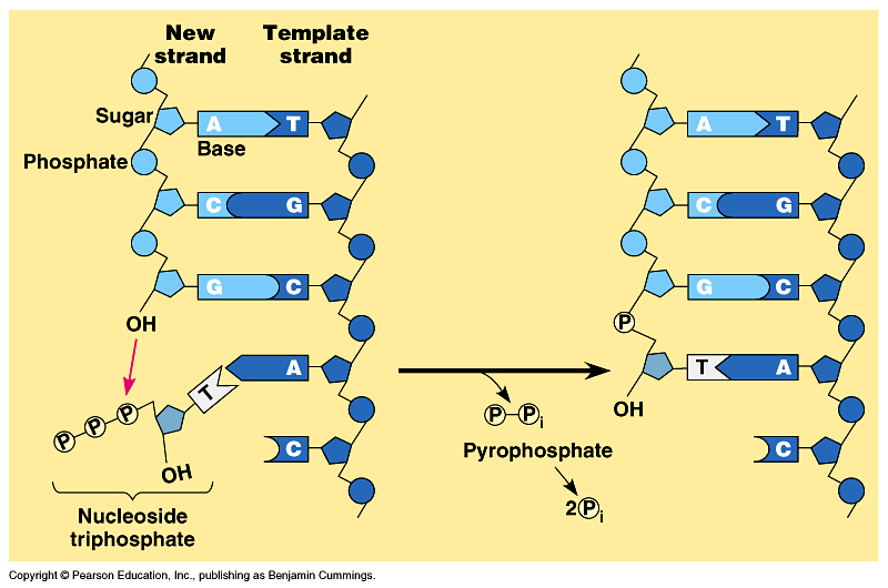
https://reasonandscience.catsboard.com

23DNA replication of prokaryotes Empty Single-strand binding protein (SSB) Wed Nov 18, 2015 5:55 pm

Otangelo


Admin

Single-strand binding protein (SSB)

Single-stranded binding proteins (not to be confused with the E. coli protein, Single-strand DNA-binding protein, SSB) are a class of proteins that have been identified in both viruses and organisms from bacteria to humans. The only organisms known to lack them are Thermoproteales, a group of extremophile archaea, where they have been displaced by the protein ThermoDBP (Thermoproteales-specific DNA Binding Protein). While many phage and viral single stranded binding proteins function as monomers and eukaryotes encode heterotrimeric RPA (Replication Protein A), the best characterized SSB is that from the bacteria E. coli which, like most bacterial single stranded binding proteins exists as a tetramer.

These proteins, though small (19.5-20kd), as they are translated they bind to negative charged phosphate (P^2-) groups of P-S-P backbone of ssDNA as tetramers, hence they are called single strand DNA binding proteins. The ssDNA wraps around the tetramer SSBs. Binding of the said proteins stabilizes the DNA and makes it as a rigid template and also prevents reannealing of the strands.  When they are bound they cover approximately 40-65 ntds. The DNA polymerase, while it is generating complementary strands SSBs are displaced. They bind DNA only when the single strand is free from polymerases. In addition they provide the DNA strand as a straight and unbent template, and stable structures for the enzyme to perform complementary strand synthesis.  They are also required for fork movement.

DNA replication of prokaryotes Ssb11

Single-stranded DNA-binding proteins keep DNA strands separated

The helicase exposes a region of single-stranded DNA that must be kept open for copying to proceed. This is achieved by coating the strand with single-stranded binding proteins. In bacteria, a monomeric protein called Single-Stranded Binding protein (SSB protein) associates to form tetramers around which the DNA is wrapped in a manner that significantly compacts the single-stranded DNA. In eukaryotes, the single-stranded binding protein is a complex of three different subunits called replication protein A (RPA). The SSB and RPA proteins both stabilize the single-stranded DNA and interact specifically with other proteins needed for replication. Coating of single strands is particularly important on the lagging strand, because long stretches of singlestranded DNA are generated as a result of the discontinuous nature of replication on this strand. (Remember that replication of the lagging strand requires the synthesis of a series of short fragments of DNA, which are later joined to form a continuous strand. The regions of single-stranded DNA are protected by a coat of single-stranded binding proteins before they are copied.)


Single-Strand Binding Protein Prevents DNA from Reannealing. The separated DNA strands behind an advancing helicase do not reanneal to form dsDNA because they become coated with single-strand binding protein (SSB). The SSB coat also prevents ssDNA from forming secondary structures (such as stem-loops) and protects it from nucleases. Evidently, DNA polymerase displaces SSB from the template strand as replication proceeds. E. coli SSB is a homotetramer of 177-residue subunits that can bind to DNA in several different ways. In the major binding mode 

DNA replication of prokaryotes Dnab_h10
FIG. 25-14 X-Ray structure of SSB in complex with dC(pC)34. The homotetramer, which has D2 symmetry, is viewed along one of its twofold axes with its other twofold axes horizontal and vertical. Each of its subunits
(which include the N-terminal 134 residues of the 177-residue polypeptide) are differently colored. Its two bound ssDNA molecules are drawn in space-filling form colored according to atom type with the upper strand C cyan, lower strand C green, N blue, O red, and P orange. (The lower strand is partially disordered and hence appears to consist of two fragments).

each U-shaped strand of ssDNA is draped across two of SSB’s four subunits. This would permit an unlimited series of SSB tetramers to interact end-to-end along the length of a ssDNA. The DNA-binding cleft of SSB, which is contained in its N-terminal 115 residues, is positively charged so that the protein can interact electrostatically with DNA phosphate groups. The cleft is too narrow to accommodate dsDNA.

SSBs from the OB domain family play an essential role in the maintenance of genome stability, functioning in DNA replication, the repair of damaged DNA, the activation of cell cycle checkpoints, and in telomere maintenance. The importance of SSBs in these processes is highlighted by their ubiquitous nature in all kingdoms of life 1

DNA replication of prokaryotes Qeqwee10
http://biochem.wustl.edu/lohmant/pubs/NV-Keck-SSB-2009.pdf
DNA replication of prokaryotes Slip_s12


DNA replication of prokaryotes Slip_s13
New Imaging Tools Show Protein Slip-Sliding Along DNA 

DNA replication of prokaryotes 15567
Single stranded DNA (gray tube) around a single stranded DNA binding protein (SSB, colored ribbons) mimicking a seam on the baseball surface. Scientists found that SSB protein does not sit on the DNA. Instead, SSB rapidly migrates on the DNA, likely utilizing the thermal fraying of ends of the DNA.

The job of single-strand DNA binding protein has always been to protect and preserve. The sticky surface of the protein attaches to single-stranded DNA and stabilizes the molecule during replication and repair. New research shows that the protein, once thought to be stiff and immobile, actually does a lot of slipping and sliding as it stabilizes DNA by wrestling it into place.

The new observations, made possible by new ways of looking at DNA, fit into a growing body of experimental evidence that is providing researchers with a greater appreciation of the dynamic interplay between DNA and the entourage of protein courtiers that shadow and groom the molecule.
In a paper published October 11, 2009, in the journal Nature, Howard Hughes Medical Institute investigator Taekjip Ha at the University of Illinois, Urbana-Champaign and his former graduate student, Rahul Roy, presently at Harvard University, collaborated on the studies with Alexander Kozlov and Timothy Lohman at Washington University School of Medicine in St. Louis. The researchers have shown for the first time that single-strand binding protein (SSB) does not stand in one place like a Buckingham Palace guard, but rather scoots along single-stranded DNA (ssDNA). Visualizing this movement is important, Ha says, because it may lead to greater understanding of the machines that repair and replicate DNA, which are intimately linked to cancer and aging.


http://bcove.me/br71p2xh

SSB diffusion movie in three segments. In the first, SSB diffusion via the rolling mechanism is illustrated. In the second, RecA filament growth via monomer addition biases SSB diffusion in a directional manner. In the third, SSB can melt secondary structures transiently via diffusion and promotes RecA filament formation.
SSB protein diffusion on single-stranded DNA stimulates RecA filament formation Rahul Roy, Alexander G. Kozlov, Timothy M. Lohman & Taekjip Ha Nature advance online publication 11 October 2009


“There is increasing evidence that the SSB protein-ssDNA complex, rather than being inert, is a dynamic, perhaps functionally important, unit,” Ha says of the findings.
“This work gives us an intimate understanding of how fluid and dynamic these SSB protein-ssDNA interactions actually are,” says Eric Greene, an HHMI Early Career Scientist and assistant professor in the Department of Biochemistry and Molecular Biophysics at Columbia University. “It’s really rather amazing that this protein slips and slides along DNA, and makes one wonder whether this might be a much more common behavior among DNA binding proteins than previously believed."
The iconic representation of DNA depicts the molecule as a double helical structure in which two strands of genetic material are intertwined. The double helix is the more common and stable form of DNA. But the two strands must occasionally separate—creating two single strands of DNA—so that enzymes can have access to copy or repair DNA. At these times, SSB steps in to keep the individual strands of DNA apart so these processes can proceed.
In the bacteria Escherichia coli, single-stranded DNA attaches to the SSB protein by winding around it like a seam on a baseball. Ha and his colleagues were curious about the relationship between SSB protein and single-stranded DNA. Their studies began by asking the simple question: How much force would it take to pull the single-stranded DNA from the SSB protein?
To find out, they planned to use a new tool that Ha’s group developed to measure both tension and fluorescence from individual biological molecules. To begin their experiments, the scientists synthesized a single strand of DNA that was just long enough to wind around the SSB protein and leave the ends protruding. This extra DNA ensured that the scientists would have something to grab and pull on. They placed a red fluorescent tag on one end of the single-stranded DNA, where it first joined the protein, and green on the other, where it left the protein.
The researchers used the fluorescent tags to measure the proximity of the DNA ends to one another. Ha explained that when the two tags are very close, they can transfer energy to one another and change color. The researchers were surprised to see significant FRET (fluorescence resonance energy transfer) fluctuations between the two tagged ends, suggesting that the single-stranded DNA ends moved in relation both to each other and to the SSB protein. That was the first clue that the SSB protein was not stuck on one spot of single-stranded DNA as had been assumed, prompting Ha’s team to shelve the original plan to measure forces and instead began to look into the possibility of the proteins’ migration on DNA.
“We were studying how the protein interacts with DNA but had no expectation that the protein would diffuse along the single-stranded DNA,” says Ha. Diffusion describes the sliding movement of the protein along DNA. The findings were surprising, but made sense considering the job the protein has to do, he adds. If the SSB protein attaches randomly to the single-stranded DNA, with no ability to adjust its position later—as scientists once thought—then there would be many unprotected gaps along the single-stranded DNA. But instead if the protein can diffuse on the DNA, such gaps would be quickly filled.
In order to confirm that the single-stranded DNA was, indeed, moving in relation to the SSB protein, Ha’s group next did a technically difficult experiment using three-color FRET. They used green dye to tag the SSB protein; red dye to tag one end of the single-stranded DNA; and purple dye for the other end. When the purple end approaches the green tag and the green tag is excited, the purple tag fluoresces. On the other hand, when the red tag is close to the green tag, which is then excited, the red end fluoresces. The FRET fluctuations on each single-stranded DNA end, tagged with red and purple, are inversely correlated indicating that the SSB protein indeed traveled from one end to the other.
“That’s another way of showing that motion is actually going on,” explains Ha.
Next the researchers wondered if the SSB protein might do more than just stabilize single strands of DNA. They made a strand of DNA in which part of the molecule was fused together in a hairpin-like structure. Hairpins occur naturally in single-stranded DNA, creating a kink that presents problems during replication and repair. Again using FRET, the researchers showed that SSB protein moves along the strand and melts hairpin structures. In effect, the protein irons out the strand – smoothing the way so that other key proteins involved in the replication or repair process can continue their work.
Ha said that one such protein, RecA, normally gets stuck if it hits a hairpin in the DNA. “RecA does not know how to extend over this hairpin structure, but SSB protein can dissolve it,” says Ha.


Cool. How did it eventually " learn " that feat ?? 


Ha hopes in the future to determine whether this same SSB protein diffusion mechanism occurs in human proteins. Two genes that are frequently mutated in breast and ovarian cancer—BRCA1 and BRCA2—produce proteins that help with DNA repair requiring single-stranded DNA intermediaries. Ha says that understanding the SSB protein-single-stranded DNA interaction in human proteins might help advance cancer research.
In the longer term, Ha dreams of using multi-color fluorescence and optical tweezers to look at as many as 10 different proteins as they interact with one another in real time.
“That’s a dream experiment,” he says. “Of course, we start simple.”


1) http://www.biomedcentral.com/1471-2199/14/9
3) http://www.hhmi.org/news/new-imaging-tools-show-protein-slip-sliding-along-dna

http://www.nature.com/nrmicro/journal/v11/n5/fig_tab/nrmicro2994_ft.html


Last edited by Admin on Sun Nov 29, 2015 10:16 am; edited 7 times in total
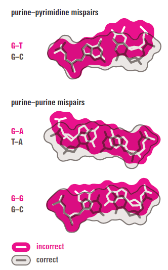
https://reasonandscience.catsboard.com

Otangelo


Admin

The Primase (DnaG) enzyme, and the primosome complex 

The primosome complex:


A group of proteins, that bind to origin site and synthesize primers to initiate replication, is called Primosomes.5
Constituents of Primosomes differ from one system to the other.
The said components assemble at a particular site called “pas”; means primosome assembling site.
In molecular biology, a primosome is a protein complex responsible for creating RNA primers on single stranded DNA during DNA replication.

The primosome consists of seven proteins: 

DnaG 
primase, 
DnaB helicase, 
DnaC helicase assistant, 
DnaT, 
PriA, 
Pri B, 
PriC. 


At each replication fork, the primosome is utilized once on the leading strand of DNA and repeatedly, initiating each Okazaki fragment, on the lagging DNA strand. Initially the complex formed by PriA, PriB, and PriC binds to DNA. Then the DnaB-DnaC helicase complex attaches along with DnaT. This structure is referred to as the pre-primosome. Finally, DnaG will bind to the pre-primosome forming a complete primosome. The primosome attaches 1-10 RNA nucleotides to the single stranded DNA creating a DNA-RNA hybrid. This sequence of RNA is used as a primer to initiate DNA polymerase III. The RNA bases are ultimately replaced with DNA bases byRNase H nuclease (eukaryotes) or DNA polymerase I nuclease (prokaryotes). DNA Ligase then acts to join the two ends together.


Primosomal protein DnaT DnaT is required for chromosomal DNA replication and for induction of replication in the absence of protein synthesis during the SOS response [Lark78, Masai86]. Though its precise role in cellular DNA replication is unknown, DnaT is a required component of the primosome, a complex of proteins capable of priming phiX174 DNA replication in vitro, and suspected of being involved in the restart of stalled replication forks in vivo [Arai81, Allen93]. DnaT complexes with PriA during primosome formation [Liu96]. DnaT appears to be specifically required for primosome function when replication stalls with a leading nascent strand rather than a gapped fork [Heller05].

DnaT is required for replication of plasmid pBR322 in vivo and in vitro, being specifically necessary for synthesis on the lagging strand [Masai89, Masai88]. It is also necessary for plasmid RSF1010 replication, and for rolling-circle replication of plasmid DNA generally [Scherzinger91, Allen93a]. It is not, however, needed for plasmid R1 replication either in vivo or in vitro [Masai89].


Synthesizes RNA Primers

All DNA synthesis, both of leading and lagging strands, requires the prior synthesis of an RNA primer. Primer synthesis in E. coli is mediated by an 600-kD protein assembly known as a primosome, which includes the DnaB helicase and an RNA synthesizing primase called DnaG, as well as five other types of subunits. DNA primase is the polymerase that synthesises small RNA primers for the Okazaki fragments made during discontinuous DNA replication. DNA primase is a heterodimer of two subunits, the small subunit Pri1 (48 kDa in yeast), and the large subunit Pri2. Both subunits participate in the formation of the active site, but the ATP binding site is located on the small subunit 

DnaG is a bacterial DNA primase and is encoded by the dnaG gene. The enzyme DnaG, and any other DNA primase, synthesizes short strands of RNA known as oligonucleotides during DNA replication. These oligonucleotides are known as primers because they act as a starting point for DNA synthesis. DnaG catalyzes the synthesis of oligonucleotides that are 10 to 60 nucleotides (the fundamental unit of DNA and RNA) long, however most of the oligonucleotides synthesized are 11 nucleotides.[1] These RNA oligonucleotides serve as primers, or starting points, for DNA synthesis by bacterial DNA polymerase III (Pol III). DnaG is important in bacterial DNA replication because DNA polymerase cannot initiate the synthesis of a DNA strand, but can only add nucleotides to a preexisting strand.[2] DnaG synthesizes a single RNA primer at the origin of replication. This primer serves to prime leading strand DNA synthesis. For the other parental strand, the lagging strand, DnaG synthesizes an RNA primer every few kilobases (kb). These primers serve as substrates for the synthesis of Okazaki fragments.[3]

DNA primase, a ubiquitous bacterial protein that synthesises the RNA primers for the Okazaki fragments in lagging strand DNA synthesis. Escherichia coli family member DnaG has been shown to interact with the replicative DnaB helicase, single-stranded DNA binding protein (SSB), and DNA polymerase III holoenzyme 3

DnaG Primases synthesize RNA oligonucleotides (primers) on single stranded DNA (ssDNA) in both prokaryotic and eukaryotic organisms at the start of DNA replication.  Primases are also involved in lagging strand synthesis and replication restart.  They are essential for the initiation of such phenomena because DNA polymerases are incapable of de novo synthesis and can only elongate existing strands (4); as such, primases are foundationally important for cell proliferation. 2 

RNA Primers Initiate DNA Replication

Since DNA polymerase can only add nucleotides to an existing nucleotide chain, how is replication of a DNA double helix initiated? Shortly after Okazaki fragments were first discovered, researchers implicated RNA in the initiation process through the following observations: (1) Okazaki fragments often have short stretches of RNA, usually 3–10 nucleotides in length, at their ends; (2) DNA polymerase can catalyze the addition of nucleotides to the end of RNA chains as well as to DNA chains; (3) cells contain an enzyme called primase that synthesizes RNA fragments about ten bases long using DNA as a template; and (4) unlike DNA polymerase, which adds nucleotides only to the ends of existing chains, primase can initiate RNA synthesis from scratch by joining two nucleotides together. These observations led to the conclusion that DNA synthesis is initiated by the formation of short RNA primers. RNA primers are synthesized by primase, which uses a single DNA strand as a template to guide the synthesis of a complementary stretch of RNA (Figure 19-11) 

DNA replication of prokaryotes Primas12

FIGURE 19-11 The Role of RNA Primers in DNA Replication. DNA synthesis is initiated with a short RNA primer in both bacteria and eukaryotes. This figure shows the process as it occurs for the lagging strand in E. coli.

Primase is a specific kind of RNA polymerase used only in DNA replication. Like other RNA polymerases, but unlike DNA polymerases, primases can initiate the synthesis of a new polynucleotide strand complementary to a template strand; they do not themselves require a primer. In E. coli, primase is relatively inactive unless it is accompanied by six other proteins, forming a complex called a primosome. The other primosome proteins
function in unwinding the parental DNA and recognizing target DNA sequences where replication is to be initiated. The situation in eukaryotic cells is slightly different, so the term primosome is not used. The eukaryotic primase is not as closely associated with unwinding proteins, but it is very tightly bound to DNA polymerase a, the main DNA polymerase involved in initiating DNA replication. Once an RNA primer has been created, DNA synthesis can proceed, with DNA polymerase III (or DNA polymerase a followed by polymerase d or ε in eukaryotes) adding successive deoxynucleotides to the end of the primer (Figure 19-11, 2). For the leading strand, initiation using an RNA primer needs to occur only once, when a replication fork first forms; DNA polymerase can then add nucleotides to the chain continuously in the direction. In contrast, the lagging strand is synthesized as a series of discontinuous Okazaki fragments, and each of them must be initiated with a separate RNA primer. For each primer, DNA nucleotides are added by DNA polymerase III until the growing fragment reaches the adjacent Okazaki fragment. No longer needed at that point, the RNA segment is removed and DNA nucleotides are polymerized to fill its place.

These are  extremely precise, pre-programmed sequences of steps exercised in a machine-like manner . If someone thinks about it unbiased, its self-evident that such a complex proceeding which requires several different, essential proteins and protein complexes and holoenzymes and molecular energy supply , helicases that unwind the dna strands like a turbine, could not have emerged naturally, in a step-wise manner. A planning mind had to invent, program, regulate, and bring the parts together in a meaningfull and functional way, the parts interacting and fitting correctly,  into a interlocked, interdependent, irreducible complex machinery, where if one peace missing, the whole process of replication would cease to function. Each of these proteins do not have any function by their own, only if correctly integrated into the replication system. 

 In E. coli, the RNA primers are removed by a exonuclease activity inherent to the DNA polymerase I molecule (distinct from the exonuclease activity involved in proofreading). At the same time, the DNA polymerase I molecule synthesizes DNA in the normal direction to fill in the resulting gaps (Figure 19-11,3 ). Adjacent fragments are subsequently joined together by DNA ligase. Why do cells employ RNA primers that must later be removed rather than simply using a DNA primer in the first place? Again, the answer may be related to the need for error correction. We have already seen that DNA polymerase possesses a exonuclease activity that allows it to remove incorrect nucleotides from the end of a DNA chain. In fact, DNA polymerase will elongate an existing DNA chain only if the nucleotide present at the end is properly base-paired. But an enzyme that initiates the synthesis of a new chain cannot perform such a proofreading function because it is not adding a nucleotide to an existing base-paired end. As a result, enzymes that initiate nucleic acid synthesis are not very good at correcting errors. By using RNA rather than DNA to initiate DNA synthesis, cells ensure that any incorrect bases inserted during initiation are restricted to RNA sequences destined to be removed by DNA polymerase I.


So how would nature have find out that  enzymes that initiate nucleic acid synthesis are not very good at correcting errors ? Did nature make some tests ? trial and error ?  

DNA replication of prokaryotes Primas11

 E. coli DnaG is a monomeric protein whose catalytic domain does not resemble any of the other DNA and RNA polymerases of known structure. Nevertheless, it catalyzes the same polymerization reaction to produce an RNA segment of 11 nucleotides. The primosome is propelled in the 5¿ S 3¿ direction along the DNA template for the lagging strand (i.e., toward the replication fork) in part by DnaB catalyzed ATP hydrolysis. This motion, which displaces the SSB in its path, is opposite in direction to that of template reading during DNA chain synthesis. Consequently, the primosome reverses its migration momentarily to allow primase to synthesize an RNA primer in the 5¿ S 3¿ direction (Fig. 25-5).

DNA replication of prokaryotes Qeqwee11

The primosome is required to initiate each Okazaki fragment. The single RNA segment that primes the synthesis of the leading strand can be synthesized, at least in vitro, by either primase or RNA polymerase (the enzyme that synthesizes RNA transcripts from a DNA template; Section 26-1), but its rate of synthesis is greatly enhanced when both enzymes are present. The pol /primase complex synthesizes a 7- to 10-nt RNA primer and extends it by an additional 15 or so deoxynucleotides. Its lack of proofreading activity is not problematic, since the first few residues of newly synthesized DNA are typically removed and replaced along with the RNA primer.

Elongation The elongation phase of replication includes two distinct but related operations: leading strand synthesis and lagging strand synthesis. Several enzymes at the replication fork are important to the synthesis of both strands. Parent DNA is first unwound by DNA helicases, and the resulting topological stress is relieved by topoisomerases. Each separated strand is then stabilized by SSB. From this point, synthesis of leading and lagging strands is sharply different. Leading strand synthesis, the more straightforward of the two, begins with the synthesis by primase (DnaG protein) of a short (10 to 60 nucleotide) RNA primer at the replication origin. DnaG interacts with DnaB helicase to carry out this reaction, and the primer is synthesized in the direction opposite to that in which the DnaB helicase is moving. In effect, the DnaB helicase moves along the strand that becomes the lagging strand in DNA synthesis; however, the first primer laid down in the first DnaG-DnaB interaction serves to prime leading strand DNA synthesis in the opposite direction. Deoxyribonucleotides are added to this primer by a DNA polymerase III complex linked to the DnaB helicase tethered to the opposite DNA strand. Leading strand synthesis then proceeds continuously, keeping pace with the unwinding of DNA at the replication fork. Lagging strand synthesis, as we have noted, is accomplished in short Okazaki fragments (Fig. 25–13a).

DNA replication of prokaryotes Dna_sy11

 First, an RNA primer is synthesized by primase and, as in leading strand synthesis, DNA polymerase III binds to the RNA primer and adds deoxyribonucleotides (Fig. 25–13b). On this level, the synthesis of each Okazaki fragment seems straightforward, but the reality is quite complex. The complexity lies in the coordination of leading and lagging strand synthesis. Both strands are produced by a single asymmetric DNA polymerase III dimer; this is accomplished by looping the DNA of the lagging strand as shown in Figure 25–14, bringing together the two points of polymerization. The synthesis of Okazaki fragments on the lagging strand entails some elegant enzymatic choreography. DnaB helicase and DnaG primase constitute a functional unit within the replication complex, the primosome. DNA polymerase III uses one set of its core subunits (the core polymerase) to synthesize the leading strand continuously, while the other set of core subunits cycles from one Okazaki fragment to the next on the looped lagging strand. DnaB helicase, bound in front of DNA polymerase III, unwinds the DNA at the replication fork (Fig. 25–14a) as it travels along the lagging strand template in the 5n3 direction. 

DNA replication of prokaryotes Dna_sy10
DnaG primase occasionally associates with DnaB helicase and synthesizes a short RNA primer (Fig. 25–14b). A new  sliding clamp is then positioned at the primer by the clamp-loading complex of DNA polymerase III (Fig. 25–14c). When synthesis of an Okazaki fragment has been completed, replication halts, and the core subunits of DNA polymerase III dissociate from their  sliding clamp (and from the completed Okazaki fragment) and associate with the new clamp (Fig. 25–14d, e). This initiates synthesis of a new Okazaki fragment. As noted earlier, the entire complex responsible for coordinated DNA synthesis at a replication fork is known as the replisome. The proteins acting at the replication fork are summarized in Table 25–4.  This complex binds to ATP and to the new  sliding clamp. The binding imparts strain on the dimeric clamp, opening up the ring at one subunit interface (Fig. 25–15). The newly primed lagging strand is slipped into the ring through the resulting break. The clamp loader then hydrolyzes ATP, releasing the  sliding clamp and allowing it to close around the DNA.

DNA replication of prokaryotes Dna_sy12
The replisome promotes rapid DNA synthesis, adding 1,000 nucleotides/s to each strand (leading and lagging). Once an Okazaki fragment has been completed, its RNA primer is removed and replaced with DNA by DNA polymerase I, and the remaining nick is sealed by DNA ligase

The helicase-binding domain of Escherichia coli DnaG primase interacts with the highly conserved C-terminal region of single-stranded DNA-binding protein

Replication Initiation Requires Helicase and Primase

The E. coli chromosome is a supercoiled DNA molecule of 4.6  106 bp. Since DNA polymerase requires a single-stranded template, other proteins participate in DNA replication by locating the replication initiation site, unwinding the DNA, and preventing the single strands from reannealing. Replication in E. coli begins at a 245-bp region known as oriC. Elements of this sequence are highly conserved among gram-negative bacteria. Multiple copies of a Section 2 Prokaryotic DNA Replication 52-kD protein known as DnaA bind to oriC and cause 45 bp of an AT-rich segment of the DNA to separate into single strands. This melting requires the free energy of ATP hydrolysis and is probably also facilitated by both the AT-rich nature of the DNA segment and the negative supercoiling (underwinding) of the circular DNA chromosome [the latter being generated by DNA gyrase, a type II topoisomerase  whose activity is required for prokaryotic DNA replication]. Helicases Unwind DNA. DnaA bound to oriC recruits two hexameric complexes of DnaB, one to each end of the melted region. DnaB is a helicase that further separates the DNA strands. Helicases are a diverse group of enzymes that unwind DNA during replication, transcription, and a variety of other processes. DnaB is one of 12 helicases expressed by E. coli. Helicases function by translocating along one strand of a double-helical nucleic acid so as to mechanically unwind the helix in their path, a process that is driven by the free energy of NTP hydrolysis. E. coli DnaB, a hexamer of identical 471-residue subunits, separates the two strands of the parental DNA by translocating along the lagging strand template in the 5¿ S 3¿ direction, while hydrolyzing ATP (it can also use GTP and CTP but not UTP). Some helicases move in the 3¿ S 5¿ direction, and some are dimers rather than hexamers. The E1 protein of bovine papillomavirus, a 605-residue hexameric helicase, translocates along ssDNA in the 3¿ S 5¿ direction (the opposite direction of DnaB). The X-ray structure of its C-terminal 274 residues in complex with a 13-nt poly(dT) and ADP  was determined by Leemor Joshua-Tor. Each protein subunit consists of two domains: a 74-residue Nterminal oligomerization domain and a 200-residue C-terminal AAA domain (AAA for ATPases associated with cellular activities; a functionally diverse protein family). The protein forms a two-layered hexagonal ring in which the oligomerization domains form a rigid collar with nearly perfect sixfold symmetry.

DNA replication of prokaryotes Dnab_h10

In contrast, the AAA domains deviate significantly from this symmetry (Fig. 25-13a). An ADP is bound at a radially peripheral site between each neighboring pair of AAA domains. The poly(dT) forms a right-handed helix that binds in the minimally 13-Å-diameter central channel of the AAA domain hexamer (which is too narrow to admit dsDNA) with its 5¿ end toward the top of the hexamer in Fig. 25-13. The DNA’s phosphate groups each interact with a positively charged loop (residues 505–508) that extends radially inward from each AAA domain such that these loops form an arrangement that resembles a right-handed spiral staircase that tracks the ssDNA’s sugar–phosphate backbone. Apparently, the protein steps through a series of ATP-driven conformational changes that, via interactions with the loops, pushes the ssDNA through the channel from bottom to top in Fig. 25-13b. During this process, each loop maintains its grip on the same phosphate group. ATP hydrolysis occurs toward the bottom of the spiral staircase and ADP release occurs between subunits located toward its top. A new ATP then binds to this site, which causes the topmost loop to drop to the bottom of the staircase, where it binds the next available phosphate group and repeats the catalytic cycle. Thus the E1 helicase mechanically separates the strands of dsDNA by pulling itself along the groove of one strand in its 3¿ S 5¿ direction but without turning relative to the DNA.

Identification of a DNA primase template tracking site redefines the geometry of primer synthesis


Primases are essential RNA polymerases required for the initiation of DNA replication, lagging strand synthesis and replication restart. Many aspects of primase function remain unclear, including how the enzyme associates with a moving nucleic acid strand emanating from a helicase and orients primers for handoff to replisomal components. Using a new screening method to trap transient macromolecular interactions, we determined the structure of the Escherichia coli DnaG primase catalytic domain bound to single-stranded DNA. The structure reveals an unanticipated binding site that engages nucleic acid in two distinct configurations, indicating that it serves as a nonspecific capture and tracking locus for template DNA. Bioinformatic and biochemical analyses show that this evolutionarily constrained region enforces template polarity near the active site and is required for primase function. Together, our findings reverse previous proposals for primer–template orientation and reconcile disparate studies to re-evaluate replication fork organization.


DNA replication of prokaryotes Nsmb_110


(a) Nucleic acid (sticks) occupies a binding groove formed by two DNA replication of prokaryotes Glyph-hairpins (green). Conserved residues previously known to be involved in primer synthesis are shown as gray sticks. Inset left, simulated annealing omit mFo – dFc difference map contoured at 3DNA replication of prokaryotes Glyph, with the final modeled conformation of the ssDNA shown in yellow. Inset right, side view showing how the DNA replication of prokaryotes Glyph-hairpins buttress ssDNA. 
(b) The template-binding groove is the most strongly basic (blue) feature of primase's surface. The active site, which binds divalent metals, is highly acidic (red) and located opposite the basic ridge.


DNA replication of prokaryotes Nsmb_111


(a) Contact map of primase's interaction with DNA as seen for the two molecules in the asymmetric unit. Hydrogen bonds or electrostatic interactions are indicated as dotted lines with atomic distances noted; solid vertical bars indicate van der Waals contacts. Conserved residues are shown in bold and evolutionarily coupled residues (Fig. 3) are shown in italics. Interactions within hydrogen-bonding distance are colored black; those that are too distant to allow hydrogen bonds are gray. 
(b) Close-up of nucleic acid-protein contacts within the template binding groove (molecule A above, molecule B below). 

Supplementary Movie

Interpolated ssDNA tracking via the basic groove.
Supplementary Movie -Download Movie (2MB)


DNA replication of prokaryotes Nsmb_112


(a) HSQC spectra of the wild-type Escherichia coli DnaG catalytic domain titrated with increasing concentrations of ssDNA. 
(b) Conservation of the ssDNA binding groove. DNA replication of prokaryotes GlyphEstat, a quantitative measure of amino acid conservation20, is relatively large for several of the ssDNA binding residues (orange). Surrounding surface-exposed residues that do not contact the DNA (white) tend to have relatively low DNA replication of prokaryotes GlyphEstat values. The positional frequencies of the residues are shown below the histogram as percentages. 
(c) Heat map showing the coupling between amino acids in the binding groove. DNA replication of prokaryotes GlyphDNA replication of prokaryotes GlyphEDNA replication of prokaryotes Glyph jstat measurements of coupling efficiency indicate that ssDNA binding residues within the ssDNA binding site covary in a manner that correlates with the structurally observed handoff of substrate within the protein. Gly194, which is relatively conserved but does not contact nucleic acid, is shown for reference. The perturbed amino acid position i is shown in each row, and the corresponding DNA replication of prokaryotes GlyphDNA replication of prokaryotes GlyphEDNA replication of prokaryotes Glyph jstat at position j within the basic groove is shown in columns. Self-coupling is shown in white.

DNA replication of prokaryotes Nsmb_113


(a) Table of single-stranded DNA (ssDNA) binding constants of full-length primase mutants. 
(b) Activity assays. De novo primer synthesis by full-length primase is severely impaired by single mutations within the template binding groove, either in the absence or presence of the DnaB helicase. 
(c) Polarity of DNA binding. ssDNA with a cross-linkable base near the 3' end forms a stable complex with a cysteine residue outside the entryway into the template binding groove in a time-dependent manner. The same template with a reactive base near the 5' end does not appreciably cross-link to primase, demonstrating that the groove binds DNA with a unique orientation.


DNA replication of prokaryotes Nsmb_114


(a) Model for heteroduplex orientation as defined by the observed structure of primase bound to single-stranded DNA (ssDNA). The observed template strand is shown in yellow. An A-form RNA-DNA hybrid (template strand, orange; product strand, blue) is modeled into the active site on the basis of the binding sites for catalytic metal ions within the active site (red spheres)6. Basic residues that surround the modeled nucleic acid and are known to be important for primase processivity are labeled. 
(b) Previous model for primer synthesis based on nucleic acid–free structures of the primase RNA polymerase domain9, 10. This configuration is incompatible with the ssDNA binding site observed here. 
(c) Solution structure of the E. coli primase zinc binding domain (ZBD) and RNA polymerase domains, obtained from small angle X-ray scattering reconstructions19, accommodates ssDNA in the observed binding site. Note that the ZBD also occludes the proposed exit channel for heteroduplex based on apo-DnaG models (Fig. 5b). The SAXS-modeled ZBD is shown in blue with the associated zinc ion as a sphere; the RPD is shown in gray with bound nucleic acid in CPK colors.


This video  illustrates beautyfully the movements of primase. The action is beautyful, almost like a ballet. Wonderful 


DNA replication of prokaryotes Nsmb_116


(a) Superposition of the single-stranded DNA (ssDNA)-bound E. coli primase RNA polymerase domain (RPD) (orange ribbons) on the T7 primase–helicase (blue and gray surface, PDB ID 1Q57). The orientation of the template binding groove would allow it to associate with and track along ssDNA as it is released from the helicase (blue surface) in the appropriate polarity. For clarity, only one primase molecule is shown. Inset, alignment of the RPD on each of the seven T7 gp4 primase modules consistently positions the tracking groove above and parallel to the central helicase axis, regardless of which gp4 protomer is chosen for the superposition. The nucleic acid strand shown in the main panel is highlighted in green. 
(b) Docking of the primase–ssDNA complex into the DnaB–DnaG helicase binding domain assembly28 orients the template tracking groove above the helicase in a manner similar to that observed from superposition with T7 gp4, shown in a. Following template capture and tracking (left), looping of ssDNA into the active site would result in primer synthesis and extrusion towards the outside of the primase–helicase complex (right), setting the stage for subsequent events in primer processing, such as handoff of the heteroduplex to the clamp loader via an interaction between primase and ssDNA binding protein (SSB)14. The zinc binding domain of primase has been omitted for clarity. The locations of the basic ridge and active site are highlighted.


http://www.ncbi.nlm.nih.gov/pmc/articles/PMC24018/
2) https://collab.itc.virginia.edu/access/content/group/f85bed6c-45d2-4b18-b868-6a2353586804/P/Ch28_Lim_S_DnaG/Ch28_DnaG_Lim_S_index_DnaG.html
3) https://www.ebi.ac.uk/interpro/entry/IPR006295
4) http://www.nature.com/nsmb/journal/v15/n2/full/nsmb.1373.html
5) http://mol-biol4masters.masters.grkraj.org/html/Prokaryotic_DNA_Replication8-Primosomes.htm


Last edited by Admin on Sat Nov 28, 2015 4:44 pm; edited 17 times in total
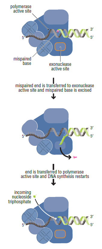
https://reasonandscience.catsboard.com

25DNA replication of prokaryotes Empty Histone-Like HU Proteins Sun Nov 22, 2015 6:30 am

Otangelo


Admin

Histone-Like HU Proteins

DNA replication of prokaryotes Image010

Bacterial histone-like HU proteins are critical to maintenance of the nucleoid structure. 1
HU is a small (10 kDa[11]) bacterial histone-like protein that resembles the eukaryotic Histone H2B. 2 HU acts similarly to a histone by inducing negative supercoiling into circular DNA with the assistance of topoisomerase. The protein has been implicated in DNA replication, recombination, and repair. With an α-helical hydrophobic core and two positively charged β-ribbon arms, HU binds non-specifically to dsDNA with low affinity but binds to altered DNA—such as junctions, nicks, gaps, forks, and overhangs—with high affinity. The arms bind to the minor groove of DNA in low affinity states; in high affinity states, a component of the α-helical core interacts with the DNA as well. 
HU was shown to participate in vitro in the initiation of DNA replication as an accessory factor to assist the action of DnaA protein in the unwinding of oriC DNA. 3
The DNA-binding protein HU plays an important role in the replication, recombination, and transcriptional regulation (Kamashev and Rouviere-Yaniv 2000)

HU protein in E. coli is a small, basic heat stable protein which is Nucleoid-associated.  This heterotypic dimmer protein is composed of two subunits; HUa and HUb, which weigh 9kDa each (4,8,10).  The main function of HU protein is to inhibit DNA supercoiling and to regulate DNA replication process (1,10).  Many different characteristics of HU protein help to initiates DNA replication.  These characteristics are nuceloid-assoicated (formed at origin of replication site), histone-like proteins (accelerating open complex formation, serves as a signal in the cell cycle), and architectural protein (interacting with supercoiled ds DNA) (4,10).  The HU protein in E.coli is a DNA binding protein that is bound to double stranded DNA (dsDNA) in the pre-initiation stage, before the primosome binds to the separated single strands (ssDNA). It is believed that the HU protein induces a conformational change in the dsDNA which destablizes it at the origin of replication, OriC. HU protein has also been shown to have a similar effect as histones on dsDNA, when complexed with topoisomerase, causing a localized supercoil. These findings were also described in a study two years earlier by Nicholas Dixon and Arthur Kornberg.
 
Some research scientists hypothesized that in vivo, HU protein is required for proper synchrony of replication initiation (7).  Also, it was suggested that HU protein acts as a negative modulator of seqA expression, which adjusts the replication initiation by avoiding fast re-initiation (9).  However, there is another suggestion that in the mutant cell, other histone-like proteins can replace HU protein in the initiation of replication 

In the course of the characterization of hupA hupB mutants, we observed that the simultaneous absence of the HU2 subunit and the MukB protein, implicated in chromosome partitioning, is lethal for the bacteria; 4 


1) http://www.horizonpress.com/cimb/v/v13/1.pdf
2) https://en.wikipedia.org/wiki/Bacterial_DNA_binding_protein
3) http://www.ncbi.nlm.nih.gov/pubmed/11278072
4) http://www.ncbi.nlm.nih.gov/pmc/articles/PMC179140/

https://reasonandscience.catsboard.com

Sponsored content


Back to top  Message [Page 1 of 3]

Go to page : 1, 2, 3  Next

Permissions in this forum:
You cannot reply to topics in this forum