The skin - marvel of intelligent design
http://reasonandscience.heavenforum.org/t2592-the-skin-prime-example-for-biomimetics-and-intelligent-design
In what single place can you find the following things: 19 million cells, 625 sweat glands, 90 oil glands, 65 hairs, 19 feet of blood vessels, and 19,000 sensory cells? The answer: in one square inch of human skin! The human skin is considered the largest organ in the body (about 16% of your body weight), and covers an area of 20 square feet. The skin has many different protective and metabolic functions that help keep your body stabilized. 7

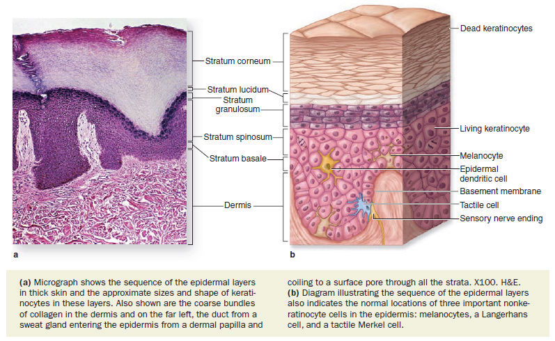
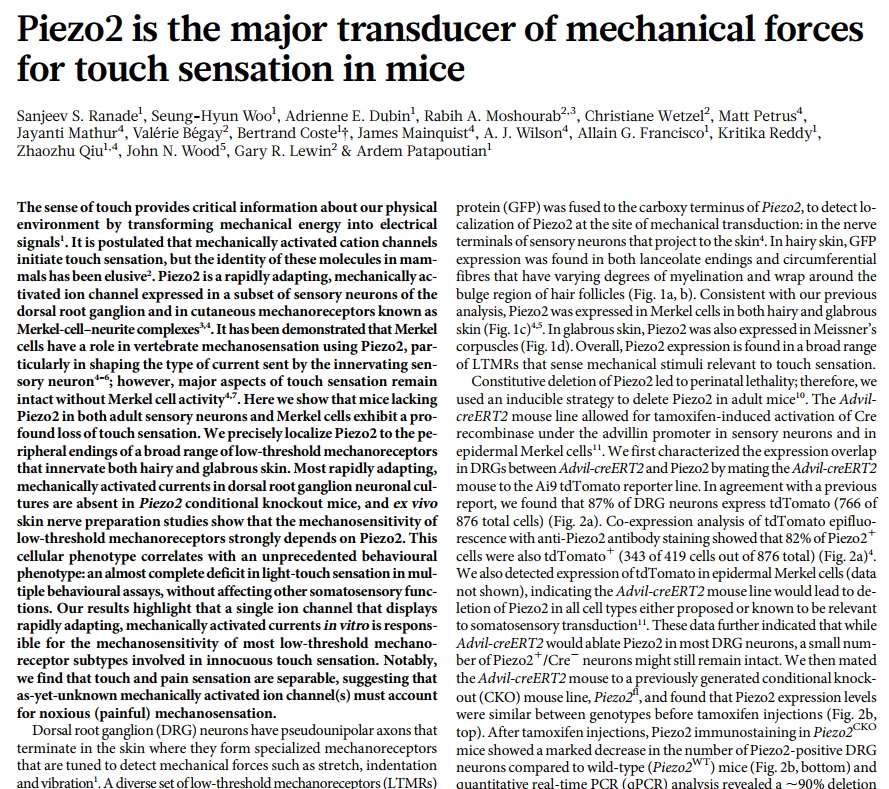
The epidermis consists mainly of a stratified squamous keratinized epithelium composed of cells called keratinocytes. There are also three much less abundant epidermal cell types: pigment-producing melanocytes, antigen-presenting Langerhans cells, and tactile epithelial cells called Merkel cells.

Sense of touch:
One of the most important functions of the skin is to provide us with a sense of touch. Werner Gitt explained it best:
The most important property of the skin is that it contains our sense of touch… The sense of touch is difficult to investigate. All other senses have a definite key organ which can be studied, but the skin is spread over the entire body and cannot easily be delimited or “switched off.” In the case of vision, scientists can observe blind persons to learn more about seeing, and they can study deaf people to learn more about hearing. But this is impossible for the sense of touch (1999, p. 41).
Receptors (from the Latin word receptor, meaning “recorder”) located at the ends of nerve fibers are used to detect stimuli and convert them into neural impulses to be sent to the brain through the peripheral and central nervous systems. Receptors also are located in the internal organs, muscles, and skeletal joints, and can detect information such as the temperature of a cup of coffee or the roughness of sand paper. Although we “touch” with our epidermis, the sense of touch actually is recorded in the dermis and passed on to the central nervous system.
Somatosensory System: The Ability To Sense Touch
Our sense of touch is controlled by a huge network of nerve endings and touch receptors in the skin known as the somatosensory system. This system is responsible for all the sensations we feel - cold, hot, smooth, rough, pressure, tickle, itch, pain, vibrations, and more. Within the somatosensory system, there are four main types of receptors: mechanoreceptors, thermoreceptors, pain receptors, and proprioceptors.
Question: In order to have an adequate sensation of touch and surrounding reality, had the proteins, enzymes, and various mechanisms providing the various sensations not have to emerge together? If let's suppose, heat or pain detectors were not present in the first skin, would our ur-ancestor not burn or hurt with ease, and die by skin inflammation, burn, and and infections?
It is important to understand how they adapt to a change in stimulus (anything that touches the skin and causes sensations such as hot, cold, pressure, tickle, etc). A touch receptor is considered rapidly adapting if it responds to a change in stimulus very quickly. Basically this means that it can sense right away when the skin is touching an object and when it stops touching that object. However, rapidly adapting receptors can't sense the continuation and duration of a stimulus touching the skin (how long the skin is touching an object). These receptors best sense vibrations occurring on or within the skin. A touch receptor is considered slowly adapting if it does not respond to a change in stimulus very quickly. These receptors are very good at sensing the continuous pressure of an object touching or indenting the skin but are not very good at sensing when the stimulus started or ended.
Mechanoreceptors:
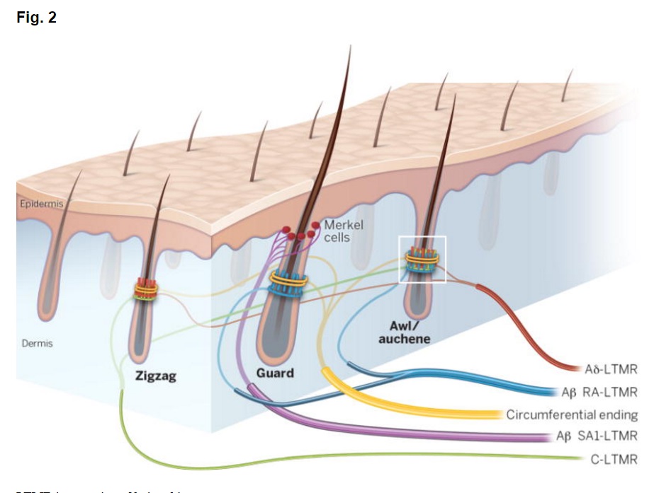
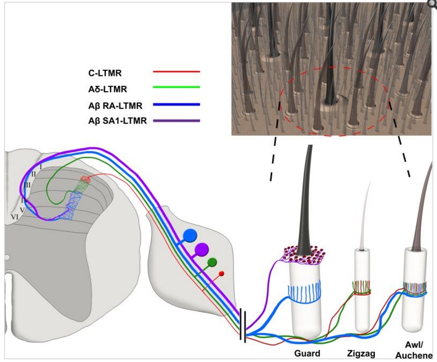
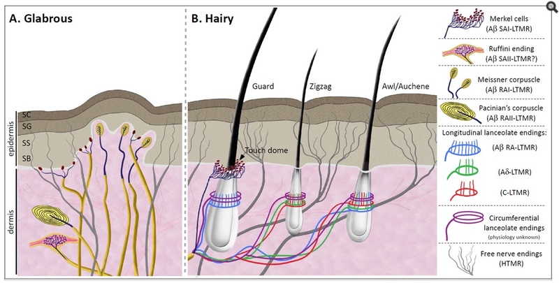
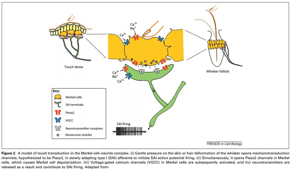
These receptors perceive sensations such as pressure, vibrations, and texture. There are four known types of mechanoreceptors whose only function is to perceive indentions and vibrations of the skin: Merkel's disks, Meissner's corpuscles, Ruffini's corpuscles, and Pacinian corpuscles.
The most sensitive mechanoreceptors, Merkel's disks and Meissner's corpuscles, are found in the very top layers of the dermis and epidermis and are generally found in non-hairy skin such as the palms, lips, tongue, soles of feet, fingertips, eyelids, and the face. Merkel's disks are slowly adapting receptors and Meissner's corpuscles are rapidly adapting receptors so your skin can perceive both when you are touching something and how long the object is touching the skin. Your brain gets an enormous amount of information about the texture of objects through your fingertips because the ridges that make up your fingerprints are full of these sensitive mechanoreceptors.
Located deeper in the dermis and along joints, tendons, and muscles are Ruffini's corpuscles and Pacinian corpuscles. These mechanoreceptors can feel sensations such as vibrations traveling down bones and tendons, rotational movement of limbs, and the stretching of skin. This greatly aids your ability to do physical activities such as walking and playing ball.
Thermoreceptors:
As their name suggests, these receptors perceive sensations related to the temperature of objects the skin feels. They are found in the dermis layer of the skin. There are two basic categories of thermoreceptors: hot and cold receptors.
Cold receptors start to perceive cold sensations when the surface of the skin drops below 95 ° F. They are most stimulated when the surface of the skin is at 77 ° F and are no longer stimulated when the surface of the skin drops below 41 ° F. This is why your feet or hands start to go numb when they are submerged in icy water for a long period of time.
Hot receptors start to perceive hot sensations when the surface of the skin rises above 86 ° F and are most stimulated at 113 ° F. But beyond 113 ° F, pain receptors take over to avoid damage being done to the skin and underlying tissues.
Thermoreceptors are found all over the body, but cold receptors are found in greater density than heat receptors. The highest concentration of thermoreceptors can be found in the face and ears (hence why your nose and ears always get colder faster than the rest of your body on a chilly winter day).
Pain receptors:
The scientific term is nocireceptor. "Noci-" in Latin means "injurious" or "hurt" which is a good clue that these receptors detect pain or stimuli that can or does cause damage to the skin and other tissues of the body. There are over three million pain receptors throughout the body, found in skin, muscles, bones, blood vessels, and some organs. They can detect pain that is caused by mechanical stimuli (cut or scrape), thermal stimuli (burn), or chemical stimuli (poison from an insect sting).
These receptors cause a feeling of sharp pain to encourage you to quickly move away from a harmful stimulus such as a broken piece of glass or a hot stove stop. They also have receptors that cause a dull pain in an area that has been injured to encourage you not to use or touch that limb or body part until the damaged area has healed. While it is never fun to activate these receptors that cause pain, they play an important part in keeping the body safe from serious injury or damage by sending these early warning signals to the brain.
Proprioceptors:
In Latin, the word "proprius" means "one's own" and is used in the name of these receptors because they sense the position of the different parts of the body in relation to each other and the surrounding environment. Proprioceptors are found in tendons, muscles, and joint capsules. This location in the body allows these special cells to detect changes in muscle length and muscle tension. Without proprioceptors, we would not be able to do fundamental things such as feeding or clothing ourselves.
While many receptors have specific functions to help us perceive different touch sensations, almost never are just one type active at any one time. When drinking from a freshly opened can of soda, your hand can perceive many different sensations just by holding it. Thermoreceptors are sensing that the can is much colder than the surrounding air, while the mechanoreceptors in your fingers are feeling the smoothness of the can and the small fluttering sensations inside the can caused by the carbon dioxide bubbles rising to the surface of the soda. Mechanoreceptors located deeper in your hand can sense that your hand is stretching around the can, that pressure is being exerted to hold the can, and that your hand is grasping the can. Proprioceptors are also sensing the hand stretching as well as how the hand and fingers are holding the can in relation to each other and the rest of the body. Even with all this going on, your somatosensory system is probably sending even more information to the brain than what was just described.
Nerve Signals: Making Sense of It All
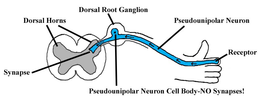
Of course, none of the sensations felt by the somatosensory system would make any difference if these sensations could not reach the brain. The nervous system of the body takes up this important task. Neurons (which are specialized nerve cells that are the smallest unit of the nervous system) receive and transmit messages with other neurons so that messages can be sent to and from the brain. This allows the brain to communicate with the body. When your hand touches an object, the mechanoreceptors in the skin are activated, and they start a chain of events by signaling to the nearest neuron that they touched something. This neuron then transmits this message to the next neuron which gets passed on to the next neuron and on it goes until the message is sent to the brain. Now the brain can process what your hand touched and send messages back to your hand via this same pathway to let the hand know if the brain wants more information about the object it is touching or if the hand should stop touching it.
Question: Has the brain and the somatosensory system and the nerves that interconnect them not have to emerge together with the skin, holding the sensory system in place?
The book Intelligent skins, by Michael Wigginton, is a prime example of the applicability of Intelligent design encountered in nature and applied and copied to human artifacts. In the case of the book, it takes as an example the human skin and draws parallels and how to apply the ingenious design methods used in the skin, to architecture, and engineering. Following an excerpt :
The Intelligent Skin Study, which is part of a broader Intelligent Building Programme, is related to its responsive performance, sometimes but not always in relation to the environmental performance of the whole building, and bears a much closer comparison with the biological idea of intelligence and response, such as is seen in the ‘natural intelligence’ of the human skin, and the science of artificial intelligence.
The skin operates as part of a holistic building metabolism and morphology, and will often be connected to other parts of the building, including sensors, actuators and command wires from the building management system. It has long been understood that the building skin may be made up of many layers, with multiple functions and integrated control. The human skin, is a protective organ that guards against the action of physical, chemical, and
bacterial threats to the internal organs. Consideration of this contains clues to The human skin consists of two distinct layers, the ‘epidermis’, which forms an outer layer, and the inner layer known as the ‘dermis’. The thickness of the skin varies from 0.5mm on the eyelids, up to 4mm on the palms of the hand and soles of the feet.4 The epidermis contains pigments, pores, and ducts, and is only a few cells thick. It is mainly composed of dead cells, whereas the inner layer (dermis) consists of a network of protein collagen, blood vessels, nerve endings (sense receptors), fat tissues, and the bases of hair follicles and sweat glands. The cells of the dermis are continually multiplying to replace those cells that are shed from the outer surface.

As part of the human skin’s role of regulating the body temperature at 36.8ºC, the sweat glands can cool the body by secreting moisture, which evaporates, and cools the body surface. The blood vessels in the dermis can supplement temperature regulation by contracting to reduce blood flow, and thus reduce radiant heat loss through the skin. The opposite effect is used to dissipate body heat. A fall in body temperature results in a reduced rate of metabolism, and if the temperature rises too high the functions of the cells may become impaired. These ‘autonomic’ processes, and many other similar ones are controlled involuntarily by the brain, the various distinctive operations of which deserve a separate mention.
Homeostasis - regulation of body temperature
Another important function of the skin is that it helps the body keep a constant temperature. Gillen, et al., wrote: “The word homeostasis comes from two Greek terms, homeo (alike or the same) and stasis (standing or remaining). Thus the word means remaining the same” (1999, italics, parenthetical items, and emp. in orig.). A person’s average body temperature is 98.6 degrees Fahrenheit, but if it increases by 7 or 8 degrees, and remains there for any of length of time, a person will almost certainly die. So how does the body keep a generally constant temperature? It does so via a method of cooling known as perspiration. The main sources of body heat are the internal organs that work all the time, such as the heart and kidneys. The heat created by these organs is carried off by the blood and distributed evenly throughout the body. This is an efficient way to diffuse the heat at a slow pace, but what happens when the body must get rid of heat quickly? Asimov explained:
We are equipped with tiny glands distributed all over our skin, about two million of them all together, the purpose of which is to bring water to the surface of the skin. On the surface this water is vaporized and heat is in this manner withdrawn from the body. The glands are sweat glands and the liquid produced is sweat or perspiration. A sweat gland consists of a tiny coiled tube, the main body of which situated deep in the dermis. The tube straightens out finally and extends up through the epidermis. The tiny opening on the surface is a pore and is just barely visible to the naked eye. When you are working or playing hard, and heat production is increased, the sweat glands accelerate their production of perspiration. This is also true when the temperature is unusually high. The rate of production may then outstrip the rate of evaporation, particularly if humidity is high, since the rate of evaporation declines with the rise in humidity. Perspiration will then collect on the body in visible drops and we are conscious of sweating (p. 265, italics in orig.).
The temperature determines how many sweat glands a person has, in the same way that the amount of sunlight determines how much melanin is in the skin. People who live in hot, humid climates tend to have more sweat glands, and produce perspiration with a smaller concentration of salt, than people living in colder, drier climates.
The skin also acts like a chemical-processing plant for the entire body. When you are outside, the skin absorbs ultraviolet rays from the Sun, and then uses them to convert chemicals into vitamin D. This vitamin is very important to our body because it helps stimulate the absorption of calcium. Without calcium, our bones grow thin and brittle, eventually leading to diseases such as rickets and osteomalacia (skeletal diseases that weaken bones). In addition, the epidermis contains a special pigment called melanin, which is responsible for the variety of color in our skin. It also acts as a protection against ultraviolet light. The melanin absorbs ultraviolet light without harming itself, and acts as a protective covering over the area beneath it. Like vitamin D, melanin is formed by the exposure to sunlight, so people in tropical regions have more melanin to protect them from the harmful ultraviolet rays, while people in northern regions have little traces of melanin because the Sun is rarely out for long periods of time. But not all people are able to produce melanin in their bodies. Occasionally, individuals are born who are incapable of forming any melanin at all. Their skin and hair are pinkish-white and their eyes are pinkish-red, because the tiny blood vessels are visible in the iris of their eyes (where there are typically colors such as blue, green, hazel, and brown). A person with this condition is referred to as an albino, indicating that they lack pigmentation in their skin. Albinism is not limited just to humans, but also is found in other species of animals as well (e.g., the white rat, the white elephant, the white tiger, etc.).
Furthermore, the skin also helps protect the inside of the body. If you have ever been to an amusement park, you probably have seen the bumper cars that you can drive to bump into other cars. Collisions in those cars are perfectly safe because of the rubber rings that surround the cars. The skin is like those rubber rings in that it acts like a shock absorber when you fall, protecting all of your internal organs. If we didn’t have this “shock absorber,” it would be practically impossible to do physical activities without damaging internal organs or bruising easily.
It is impossible that evolution could have produced such an important and complex organ as the human skin. The many intricacies of its functions are evidence of a Creator. One writer remarked: “The skin is a miracle of evolutionary engineering: it waterproofs the body, blocks out and destroys harmful bacteria, regulates temperature, and continuously communicates with the brain” (McCutcheon, 1989, p. 113). Yes, the skin is a “miracle” all right—but not a miracle of evolution. And yes, the skin was “engineered”—but the engineer was God
The ‘hypothalamus’. This organ, located at the centre of the base of the brain, serves as a link between the autonomic nervous system and the body’s endocrine system (hormone-releasing glands). The function of the hypothalamus is to integrate and ensure appropriate response to internal and external stimuli. It plays an important role in the regulation of most of the involuntary mechanisms of the body, including body temperature, sexual drive, and the menstrual cycle.
Skin is the primary sensing organ for external stressors, including heat, cold, pain, and mechanical tension. Three classes of receptors (thermoreceptors for heat and cold, nociceptor for pain and mechanoreceptors for mechanical changes) are responsible for transmitting the outside signals to the spinal cord, and then to the brain. The cutaneous sensory fibers also convey changes in temperature, pH, and inflammatory mediators to the central nervous system (CNS). The nerve terminals are often associated with receptors indicating close interaction. The brain responds to these signals, which in turn influence the stress responses in the skin. 2
Question: Has the brain and the sweat glands and the nerves that interconnect them not have to emerge together with the skin, holding the glands in place?
The p53 tumor protein, preventing the skin from cancer, and guardian of the genome
A protein known as the "master watchman of the genome" for its ability to guard against cancer-causing DNA damage has been found to provide an entirely different level of cancer protection: By prompting the skin to tan in response to ultraviolet light from the sun, it deters the development of melanoma skin cancer, the fastest-increasing form of cancer in the world. 3
The p53 protein is located in the nucleus of cells throughout the body, where it attaches (binds) directly to DNA. When the DNA in a cell becomes damaged by agents such as toxic chemicals, radiation, or ultraviolet (UV) rays from sunlight, this protein plays a critical role in determining whether the DNA will be repaired or the damaged cell will self-destruct (undergo apoptosis). If the DNA can be repaired, p53 activates other genes to fix the damage. If the DNA cannot be repaired, this protein prevents the cell from dividing and signals it to undergo apoptosis. By stopping cells with mutated or damaged DNA from dividing, p53 helps prevent the development of tumors. 4
Because p53 is essential for regulating cell division and preventing tumor formation, it has been nicknamed the "guardian of the genome."
Question: Had p53 not to emerge right from the start, otherwise skin cancer would have not been an exception, but the norm, and life without this protection protein would not survive UV radiation ?
The skin's acid coating
Your skin is coated with acid. While that might sound disturbing, the mild acidity of the skin's surface actually helps to maintain the strength and cohesiveness of the skin. Now researchers have discovered where this acidity comes from, and they suggest how it may help to hold the skin together. The acid is produced when enzymes break down fat-like molecules in skin cells, called phospholipids, into smaller acid-tipped fat molecules called fatty acids.
Stratum corneum acidification plays an important regulatory role for barrier function and also for integrity and cohesion. Human and mammalian newborn stratum corneum displays a near-neutral surface pH, which declines rapidly during the early first postnatal period. The stratum corneum pH gradient ranges from 4.5 to 5.0 in the outer stratum corneum and approaches neutrality in the lower stratum corneum. The functions of the acid mantle include antibacterial properties, showing that an acidic surface pH inhibits colonization with pathogenic bacteria. 5 The skin is generating the acid as it converts phospholipids into fatty acids, one of the natural steps in the formation of the skin barrier. Blocking this conversion has a marked effect on the acidity as well as the skin's integrity and cohesiveness.
Question: Had this acidicity not have to be present from day one to garantee integrity and cohesion , starting from day 1 ?
Human skin is a fantastic invention. It regulates heat, keeps body fluids in but allows selective absorption, protects against infection, is self-healing and self-regenerating, and is lined with thousands of advanced sensors for touch, heat, pain, and cold. It is our largest single organ, providing us a wetsuit with feeling. It is a key component of the God-given pleasure of sex. Skin is a living, breathing, dynamic, regenerating, and beautiful design, and points out one of the most intriguing differences between humans and apes. We are not really hairless; even the slickest parts of our skin, including the palms of our hands, are covered with tiny transparent hairs that are essential to the sense of touch. The way hair shafts grow to a certain prescribed length from the base while safely anchored inside their follicles is another amazing facet of the wonder of human skin.
1. Intelligent skins, Michael Wigginton, page 29
2. https://www.ncbi.nlm.nih.gov/pmc/articles/PMC4082169/
3. https://www.eurekalert.org/pub_releases/2007-03/dci-ot030507.php
4. https://ghr.nlm.nih.gov/gene/TP53
5. http://dermatologytimes.modernmedicine.com/dermatology-times/news/clinical/dermatology/acid-mantle-study-could-aid-drug-development
6. http://creationsafaris.com/crev0603.htm
7. http://www.apologeticspress.org/APContent.aspx?category=12&article=1411&topic=249
8. https://www.homesciencetools.com/a/skin-touch
http://reasonandscience.heavenforum.org/t2592-the-skin-prime-example-for-biomimetics-and-intelligent-design
In what single place can you find the following things: 19 million cells, 625 sweat glands, 90 oil glands, 65 hairs, 19 feet of blood vessels, and 19,000 sensory cells? The answer: in one square inch of human skin! The human skin is considered the largest organ in the body (about 16% of your body weight), and covers an area of 20 square feet. The skin has many different protective and metabolic functions that help keep your body stabilized. 7
The epidermis consists mainly of a stratified squamous keratinized epithelium composed of cells called keratinocytes. There are also three much less abundant epidermal cell types: pigment-producing melanocytes, antigen-presenting Langerhans cells, and tactile epithelial cells called Merkel cells.
Sense of touch:
One of the most important functions of the skin is to provide us with a sense of touch. Werner Gitt explained it best:
The most important property of the skin is that it contains our sense of touch… The sense of touch is difficult to investigate. All other senses have a definite key organ which can be studied, but the skin is spread over the entire body and cannot easily be delimited or “switched off.” In the case of vision, scientists can observe blind persons to learn more about seeing, and they can study deaf people to learn more about hearing. But this is impossible for the sense of touch (1999, p. 41).
Receptors (from the Latin word receptor, meaning “recorder”) located at the ends of nerve fibers are used to detect stimuli and convert them into neural impulses to be sent to the brain through the peripheral and central nervous systems. Receptors also are located in the internal organs, muscles, and skeletal joints, and can detect information such as the temperature of a cup of coffee or the roughness of sand paper. Although we “touch” with our epidermis, the sense of touch actually is recorded in the dermis and passed on to the central nervous system.
Somatosensory System: The Ability To Sense Touch
Our sense of touch is controlled by a huge network of nerve endings and touch receptors in the skin known as the somatosensory system. This system is responsible for all the sensations we feel - cold, hot, smooth, rough, pressure, tickle, itch, pain, vibrations, and more. Within the somatosensory system, there are four main types of receptors: mechanoreceptors, thermoreceptors, pain receptors, and proprioceptors.
Question: In order to have an adequate sensation of touch and surrounding reality, had the proteins, enzymes, and various mechanisms providing the various sensations not have to emerge together? If let's suppose, heat or pain detectors were not present in the first skin, would our ur-ancestor not burn or hurt with ease, and die by skin inflammation, burn, and and infections?
It is important to understand how they adapt to a change in stimulus (anything that touches the skin and causes sensations such as hot, cold, pressure, tickle, etc). A touch receptor is considered rapidly adapting if it responds to a change in stimulus very quickly. Basically this means that it can sense right away when the skin is touching an object and when it stops touching that object. However, rapidly adapting receptors can't sense the continuation and duration of a stimulus touching the skin (how long the skin is touching an object). These receptors best sense vibrations occurring on or within the skin. A touch receptor is considered slowly adapting if it does not respond to a change in stimulus very quickly. These receptors are very good at sensing the continuous pressure of an object touching or indenting the skin but are not very good at sensing when the stimulus started or ended.
Mechanoreceptors:
These receptors perceive sensations such as pressure, vibrations, and texture. There are four known types of mechanoreceptors whose only function is to perceive indentions and vibrations of the skin: Merkel's disks, Meissner's corpuscles, Ruffini's corpuscles, and Pacinian corpuscles.
The most sensitive mechanoreceptors, Merkel's disks and Meissner's corpuscles, are found in the very top layers of the dermis and epidermis and are generally found in non-hairy skin such as the palms, lips, tongue, soles of feet, fingertips, eyelids, and the face. Merkel's disks are slowly adapting receptors and Meissner's corpuscles are rapidly adapting receptors so your skin can perceive both when you are touching something and how long the object is touching the skin. Your brain gets an enormous amount of information about the texture of objects through your fingertips because the ridges that make up your fingerprints are full of these sensitive mechanoreceptors.
Located deeper in the dermis and along joints, tendons, and muscles are Ruffini's corpuscles and Pacinian corpuscles. These mechanoreceptors can feel sensations such as vibrations traveling down bones and tendons, rotational movement of limbs, and the stretching of skin. This greatly aids your ability to do physical activities such as walking and playing ball.
Thermoreceptors:
As their name suggests, these receptors perceive sensations related to the temperature of objects the skin feels. They are found in the dermis layer of the skin. There are two basic categories of thermoreceptors: hot and cold receptors.
Cold receptors start to perceive cold sensations when the surface of the skin drops below 95 ° F. They are most stimulated when the surface of the skin is at 77 ° F and are no longer stimulated when the surface of the skin drops below 41 ° F. This is why your feet or hands start to go numb when they are submerged in icy water for a long period of time.
Hot receptors start to perceive hot sensations when the surface of the skin rises above 86 ° F and are most stimulated at 113 ° F. But beyond 113 ° F, pain receptors take over to avoid damage being done to the skin and underlying tissues.
Thermoreceptors are found all over the body, but cold receptors are found in greater density than heat receptors. The highest concentration of thermoreceptors can be found in the face and ears (hence why your nose and ears always get colder faster than the rest of your body on a chilly winter day).
Pain receptors:
The scientific term is nocireceptor. "Noci-" in Latin means "injurious" or "hurt" which is a good clue that these receptors detect pain or stimuli that can or does cause damage to the skin and other tissues of the body. There are over three million pain receptors throughout the body, found in skin, muscles, bones, blood vessels, and some organs. They can detect pain that is caused by mechanical stimuli (cut or scrape), thermal stimuli (burn), or chemical stimuli (poison from an insect sting).
These receptors cause a feeling of sharp pain to encourage you to quickly move away from a harmful stimulus such as a broken piece of glass or a hot stove stop. They also have receptors that cause a dull pain in an area that has been injured to encourage you not to use or touch that limb or body part until the damaged area has healed. While it is never fun to activate these receptors that cause pain, they play an important part in keeping the body safe from serious injury or damage by sending these early warning signals to the brain.
Proprioceptors:
In Latin, the word "proprius" means "one's own" and is used in the name of these receptors because they sense the position of the different parts of the body in relation to each other and the surrounding environment. Proprioceptors are found in tendons, muscles, and joint capsules. This location in the body allows these special cells to detect changes in muscle length and muscle tension. Without proprioceptors, we would not be able to do fundamental things such as feeding or clothing ourselves.
While many receptors have specific functions to help us perceive different touch sensations, almost never are just one type active at any one time. When drinking from a freshly opened can of soda, your hand can perceive many different sensations just by holding it. Thermoreceptors are sensing that the can is much colder than the surrounding air, while the mechanoreceptors in your fingers are feeling the smoothness of the can and the small fluttering sensations inside the can caused by the carbon dioxide bubbles rising to the surface of the soda. Mechanoreceptors located deeper in your hand can sense that your hand is stretching around the can, that pressure is being exerted to hold the can, and that your hand is grasping the can. Proprioceptors are also sensing the hand stretching as well as how the hand and fingers are holding the can in relation to each other and the rest of the body. Even with all this going on, your somatosensory system is probably sending even more information to the brain than what was just described.
Nerve Signals: Making Sense of It All
Of course, none of the sensations felt by the somatosensory system would make any difference if these sensations could not reach the brain. The nervous system of the body takes up this important task. Neurons (which are specialized nerve cells that are the smallest unit of the nervous system) receive and transmit messages with other neurons so that messages can be sent to and from the brain. This allows the brain to communicate with the body. When your hand touches an object, the mechanoreceptors in the skin are activated, and they start a chain of events by signaling to the nearest neuron that they touched something. This neuron then transmits this message to the next neuron which gets passed on to the next neuron and on it goes until the message is sent to the brain. Now the brain can process what your hand touched and send messages back to your hand via this same pathway to let the hand know if the brain wants more information about the object it is touching or if the hand should stop touching it.
Question: Has the brain and the somatosensory system and the nerves that interconnect them not have to emerge together with the skin, holding the sensory system in place?
The book Intelligent skins, by Michael Wigginton, is a prime example of the applicability of Intelligent design encountered in nature and applied and copied to human artifacts. In the case of the book, it takes as an example the human skin and draws parallels and how to apply the ingenious design methods used in the skin, to architecture, and engineering. Following an excerpt :
The Intelligent Skin Study, which is part of a broader Intelligent Building Programme, is related to its responsive performance, sometimes but not always in relation to the environmental performance of the whole building, and bears a much closer comparison with the biological idea of intelligence and response, such as is seen in the ‘natural intelligence’ of the human skin, and the science of artificial intelligence.
The skin operates as part of a holistic building metabolism and morphology, and will often be connected to other parts of the building, including sensors, actuators and command wires from the building management system. It has long been understood that the building skin may be made up of many layers, with multiple functions and integrated control. The human skin, is a protective organ that guards against the action of physical, chemical, and
bacterial threats to the internal organs. Consideration of this contains clues to The human skin consists of two distinct layers, the ‘epidermis’, which forms an outer layer, and the inner layer known as the ‘dermis’. The thickness of the skin varies from 0.5mm on the eyelids, up to 4mm on the palms of the hand and soles of the feet.4 The epidermis contains pigments, pores, and ducts, and is only a few cells thick. It is mainly composed of dead cells, whereas the inner layer (dermis) consists of a network of protein collagen, blood vessels, nerve endings (sense receptors), fat tissues, and the bases of hair follicles and sweat glands. The cells of the dermis are continually multiplying to replace those cells that are shed from the outer surface.
As part of the human skin’s role of regulating the body temperature at 36.8ºC, the sweat glands can cool the body by secreting moisture, which evaporates, and cools the body surface. The blood vessels in the dermis can supplement temperature regulation by contracting to reduce blood flow, and thus reduce radiant heat loss through the skin. The opposite effect is used to dissipate body heat. A fall in body temperature results in a reduced rate of metabolism, and if the temperature rises too high the functions of the cells may become impaired. These ‘autonomic’ processes, and many other similar ones are controlled involuntarily by the brain, the various distinctive operations of which deserve a separate mention.
Homeostasis - regulation of body temperature
Another important function of the skin is that it helps the body keep a constant temperature. Gillen, et al., wrote: “The word homeostasis comes from two Greek terms, homeo (alike or the same) and stasis (standing or remaining). Thus the word means remaining the same” (1999, italics, parenthetical items, and emp. in orig.). A person’s average body temperature is 98.6 degrees Fahrenheit, but if it increases by 7 or 8 degrees, and remains there for any of length of time, a person will almost certainly die. So how does the body keep a generally constant temperature? It does so via a method of cooling known as perspiration. The main sources of body heat are the internal organs that work all the time, such as the heart and kidneys. The heat created by these organs is carried off by the blood and distributed evenly throughout the body. This is an efficient way to diffuse the heat at a slow pace, but what happens when the body must get rid of heat quickly? Asimov explained:
We are equipped with tiny glands distributed all over our skin, about two million of them all together, the purpose of which is to bring water to the surface of the skin. On the surface this water is vaporized and heat is in this manner withdrawn from the body. The glands are sweat glands and the liquid produced is sweat or perspiration. A sweat gland consists of a tiny coiled tube, the main body of which situated deep in the dermis. The tube straightens out finally and extends up through the epidermis. The tiny opening on the surface is a pore and is just barely visible to the naked eye. When you are working or playing hard, and heat production is increased, the sweat glands accelerate their production of perspiration. This is also true when the temperature is unusually high. The rate of production may then outstrip the rate of evaporation, particularly if humidity is high, since the rate of evaporation declines with the rise in humidity. Perspiration will then collect on the body in visible drops and we are conscious of sweating (p. 265, italics in orig.).
The temperature determines how many sweat glands a person has, in the same way that the amount of sunlight determines how much melanin is in the skin. People who live in hot, humid climates tend to have more sweat glands, and produce perspiration with a smaller concentration of salt, than people living in colder, drier climates.
The skin also acts like a chemical-processing plant for the entire body. When you are outside, the skin absorbs ultraviolet rays from the Sun, and then uses them to convert chemicals into vitamin D. This vitamin is very important to our body because it helps stimulate the absorption of calcium. Without calcium, our bones grow thin and brittle, eventually leading to diseases such as rickets and osteomalacia (skeletal diseases that weaken bones). In addition, the epidermis contains a special pigment called melanin, which is responsible for the variety of color in our skin. It also acts as a protection against ultraviolet light. The melanin absorbs ultraviolet light without harming itself, and acts as a protective covering over the area beneath it. Like vitamin D, melanin is formed by the exposure to sunlight, so people in tropical regions have more melanin to protect them from the harmful ultraviolet rays, while people in northern regions have little traces of melanin because the Sun is rarely out for long periods of time. But not all people are able to produce melanin in their bodies. Occasionally, individuals are born who are incapable of forming any melanin at all. Their skin and hair are pinkish-white and their eyes are pinkish-red, because the tiny blood vessels are visible in the iris of their eyes (where there are typically colors such as blue, green, hazel, and brown). A person with this condition is referred to as an albino, indicating that they lack pigmentation in their skin. Albinism is not limited just to humans, but also is found in other species of animals as well (e.g., the white rat, the white elephant, the white tiger, etc.).
Furthermore, the skin also helps protect the inside of the body. If you have ever been to an amusement park, you probably have seen the bumper cars that you can drive to bump into other cars. Collisions in those cars are perfectly safe because of the rubber rings that surround the cars. The skin is like those rubber rings in that it acts like a shock absorber when you fall, protecting all of your internal organs. If we didn’t have this “shock absorber,” it would be practically impossible to do physical activities without damaging internal organs or bruising easily.
It is impossible that evolution could have produced such an important and complex organ as the human skin. The many intricacies of its functions are evidence of a Creator. One writer remarked: “The skin is a miracle of evolutionary engineering: it waterproofs the body, blocks out and destroys harmful bacteria, regulates temperature, and continuously communicates with the brain” (McCutcheon, 1989, p. 113). Yes, the skin is a “miracle” all right—but not a miracle of evolution. And yes, the skin was “engineered”—but the engineer was God
The ‘hypothalamus’. This organ, located at the centre of the base of the brain, serves as a link between the autonomic nervous system and the body’s endocrine system (hormone-releasing glands). The function of the hypothalamus is to integrate and ensure appropriate response to internal and external stimuli. It plays an important role in the regulation of most of the involuntary mechanisms of the body, including body temperature, sexual drive, and the menstrual cycle.
Skin is the primary sensing organ for external stressors, including heat, cold, pain, and mechanical tension. Three classes of receptors (thermoreceptors for heat and cold, nociceptor for pain and mechanoreceptors for mechanical changes) are responsible for transmitting the outside signals to the spinal cord, and then to the brain. The cutaneous sensory fibers also convey changes in temperature, pH, and inflammatory mediators to the central nervous system (CNS). The nerve terminals are often associated with receptors indicating close interaction. The brain responds to these signals, which in turn influence the stress responses in the skin. 2
Question: Has the brain and the sweat glands and the nerves that interconnect them not have to emerge together with the skin, holding the glands in place?
The p53 tumor protein, preventing the skin from cancer, and guardian of the genome
A protein known as the "master watchman of the genome" for its ability to guard against cancer-causing DNA damage has been found to provide an entirely different level of cancer protection: By prompting the skin to tan in response to ultraviolet light from the sun, it deters the development of melanoma skin cancer, the fastest-increasing form of cancer in the world. 3
The p53 protein is located in the nucleus of cells throughout the body, where it attaches (binds) directly to DNA. When the DNA in a cell becomes damaged by agents such as toxic chemicals, radiation, or ultraviolet (UV) rays from sunlight, this protein plays a critical role in determining whether the DNA will be repaired or the damaged cell will self-destruct (undergo apoptosis). If the DNA can be repaired, p53 activates other genes to fix the damage. If the DNA cannot be repaired, this protein prevents the cell from dividing and signals it to undergo apoptosis. By stopping cells with mutated or damaged DNA from dividing, p53 helps prevent the development of tumors. 4
Because p53 is essential for regulating cell division and preventing tumor formation, it has been nicknamed the "guardian of the genome."
Question: Had p53 not to emerge right from the start, otherwise skin cancer would have not been an exception, but the norm, and life without this protection protein would not survive UV radiation ?
The skin's acid coating
Your skin is coated with acid. While that might sound disturbing, the mild acidity of the skin's surface actually helps to maintain the strength and cohesiveness of the skin. Now researchers have discovered where this acidity comes from, and they suggest how it may help to hold the skin together. The acid is produced when enzymes break down fat-like molecules in skin cells, called phospholipids, into smaller acid-tipped fat molecules called fatty acids.
Stratum corneum acidification plays an important regulatory role for barrier function and also for integrity and cohesion. Human and mammalian newborn stratum corneum displays a near-neutral surface pH, which declines rapidly during the early first postnatal period. The stratum corneum pH gradient ranges from 4.5 to 5.0 in the outer stratum corneum and approaches neutrality in the lower stratum corneum. The functions of the acid mantle include antibacterial properties, showing that an acidic surface pH inhibits colonization with pathogenic bacteria. 5 The skin is generating the acid as it converts phospholipids into fatty acids, one of the natural steps in the formation of the skin barrier. Blocking this conversion has a marked effect on the acidity as well as the skin's integrity and cohesiveness.
Question: Had this acidicity not have to be present from day one to garantee integrity and cohesion , starting from day 1 ?
Human skin is a fantastic invention. It regulates heat, keeps body fluids in but allows selective absorption, protects against infection, is self-healing and self-regenerating, and is lined with thousands of advanced sensors for touch, heat, pain, and cold. It is our largest single organ, providing us a wetsuit with feeling. It is a key component of the God-given pleasure of sex. Skin is a living, breathing, dynamic, regenerating, and beautiful design, and points out one of the most intriguing differences between humans and apes. We are not really hairless; even the slickest parts of our skin, including the palms of our hands, are covered with tiny transparent hairs that are essential to the sense of touch. The way hair shafts grow to a certain prescribed length from the base while safely anchored inside their follicles is another amazing facet of the wonder of human skin.
1. Intelligent skins, Michael Wigginton, page 29
2. https://www.ncbi.nlm.nih.gov/pmc/articles/PMC4082169/
3. https://www.eurekalert.org/pub_releases/2007-03/dci-ot030507.php
4. https://ghr.nlm.nih.gov/gene/TP53
5. http://dermatologytimes.modernmedicine.com/dermatology-times/news/clinical/dermatology/acid-mantle-study-could-aid-drug-development
6. http://creationsafaris.com/crev0603.htm
7. http://www.apologeticspress.org/APContent.aspx?category=12&article=1411&topic=249
8. https://www.homesciencetools.com/a/skin-touch
Last edited by Admin on Sat Aug 12, 2017 6:17 pm; edited 3 times in total