ElShamah - Reason & Science: Defending ID and the Christian Worldview
Would you like to react to this message? Create an account in a few clicks or log in to continue.
ElShamah - Reason & Science: Defending ID and the Christian Worldview

Welcome to my library—a curated collection of research and original arguments exploring why I believe Christianity, creationism, and Intelligent Design offer the most compelling explanations for our origins. Otangelo Grasso


You are not connected. Please login or register

Ribonucleotide reductase, one of the most essential enzymes of life, and how it buries the RNA world

Go down  Message [Page 1 of 1]

Otangelo


Admin

Ribonucleotide reductase, one of the most essential enzymes of life

http://reasonandscience.heavenforum.org/t2029-ribonucleotide-reductase-one-of-the-most-essential-enzymes-of-life-and-how-it-buries-the-rna-world#3428

This is one of the most essential enzymes of life 5

Ribonucleotide reduction is the only pathway for de novo synthesis of deoxyribonucleotides in extant organisms. This chemically demanding reaction, which proceeds via a carbon-centered free radical, is catalyzed by ribonucleotide reductase (RNR). The mechanism has been deemed unlikely to be catalyzed by a ribozyme, creating an enigma regarding how the building blocks for DNA were synthesized at the transition from RNA to DNA-encoded genomes.

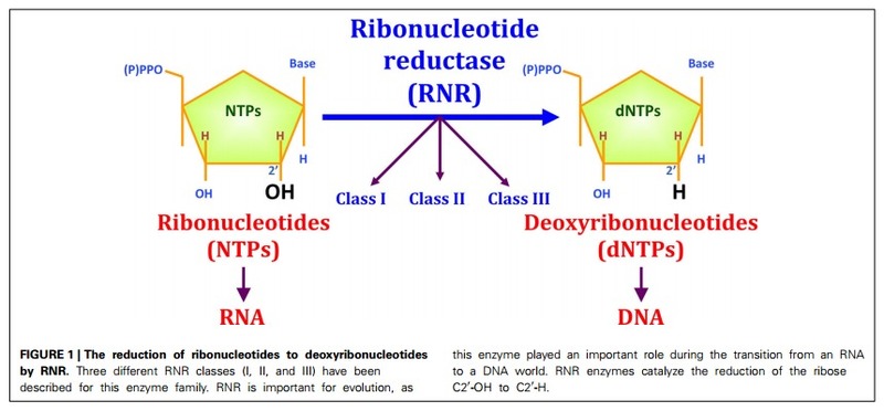
DNA is the genetic material in all cellular organisms plus many viruses. DNA’s building blocks, deoxyribonucleotides (dNTPs), are always synthesized by reduction of ribonucleotides (either NTPs or NDPs), the building blocks of RNA. 6


Origin of Ribonucleotide Reduction

How and when ribonucleotide reduction evolved is a question that is intimately associated with the transition from the RNA world to the modern RNA + protein + DNA world, since it is the only known de novo mechanism for dNTP synthesis.


The maintenance of life on Earth depends on the ability to reproduce. Reproduction requires an accurate and stable storage system for the genetic information in all organisms, including viruses. It has been recently suggested that the RNA molecule, with autoreplicative capacity, is the primary primitive molecule for the genetic information storage. Despite the wide acceptance of this idea, there are arguments against the concept of an RNA world that cannot be underestimated. 7

Today, three different RNR classes have been described, with little apparent similarity between them in terms of primary protein sequence (approximately 10–20% similarity). Thus, it could be assumed that each RNR class appeared independently from each other over time.

There we have a problem of convergent evolution. “ As Stephen J.Gould stated :

…No finale can be specified at the start, none would ever occur a second time in the same way, because any pathway proceeds through thousands of improbable stages. Alter any early event, ever so slightly, and without apparent importance at the time, and evolution cascades into a radically different channel. 11

That means, hardly we should find a enzyme evolving the same function. But thats exactly what supposedly happened. Not only did the RNR would have had to arise 3 times independently with different gene sets, but provided the same function. Should we not expect it to evolve just once, if the function is the same ?
Ribonucleotide reductase, one of the most essential enzymes of life, and how it buries the RNA world Fcimb_04_00052_pdf_Page_1
photos upload

Three main classes of ribonucleotide reductases (RNR) have been discovered that depend on different metal cofactors for the catalytic activity:

class I enzymes contain a diiron-oxygen cluster,
class II a cobalt containing cobalamin cofactor (vitamin B12), and
class III an 4Fe-4S iron-sulfur cluster coupled to S-adenosylmethionine (SAM) [PMID: 15158709].

But, surprisingly, there is a great similarity the reaction mechanism, allosteric regulation and three-dimensional structure (tertiary structure) of these enzymes, suggesting a potential common origin.

The enzymatic activation of class III RNR requires Sadenosylmethionine (SAM), one of the most ancestral molecules, with few steps required for its biosynthesis


Biosynthesis DNA is made from RNA. The deoxynucleotides are made from nucleotides with ribonucleotide reductases (RNR's), producing uracil-DNA or u-DNA. The uracil is then converted to thymine by adding a methyl group, making thymine-DNA or t-DNA, the kind that is actually used. 4)

The reaction catalyzed by RNR is strictly conserved in all living organisms.  Furthermore RNR plays a critical role in regulating the total rate of DNA synthesis so that DNA to cell mass is maintained at a constant ratio during cell division and DNA repair. A somewhat unusual feature of the RNR enzyme is that it catalyzes a reaction that proceeds via a free radical mechanism of action.The substrates for RNR are ADP, GDP, CDP and UDP. dTDP (deoxythymidine diphosphate) is synthesized by another enzyme (thymidylate kinase) from dTMP (deoxythymidine monophosphate). 1

The iron-dependent enzyme, ribonucleotide reductase (RNR), is essential for DNA synthesis.

The structures of a class III ribonucleotide reductase (RNR) and pyruvate formate lyase exhibit striking homology within their active site domains with respect to each other and to the previously published structure of a class I RNR. The common structures and the common complex-radical-based chemistry of these systems, as well as of the class II RNRs, suggest that RNRs evolved by divergent evolution and provide an essential link between the RNA and DNA world. 2

Genetic information can be stored stably only because a battery of DNA repair enzymes scan the DNA and replace the damaged nucleotides. Without these enzymes it would be inconceivable how primitive cells kept abreast of the constant high-level damage by the environment and by endogenous reactions. If unrepaired, cell death would result. 10 

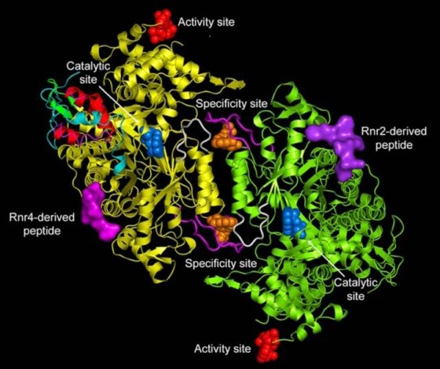
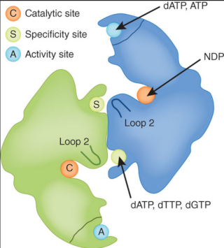
RNR is a complex of two dimeric proteins termed R1 and R2. 8

That brings us to the classic chicken and egg, catch22 situation.  RNR enzymes are required to make DNA. DNA is however required to make RNR enzymes. What came first ??
We can conclude with high certainty that this enzyme buries any RNA world fantasies, and any possibility of transition from  RNA to DNA world scenarios.


Ribonucleotide reductase, one of the most essential enzymes of life, and how it buries the RNA world Pharmaceuticals_04_01328f1_png_1000_750
windows screen capture


Ribonucleotide reductase, one of the most essential enzymes of life, and how it buries the RNA world Life_Free_Full_Text_The_Origin_and_Evolution
how to do a screenshot on a pc  9


Ribonucleotide reductase, one of the most essential enzymes of life, and how it buries the RNA world Ribonu11


1) http://en.wikipedia.org/wiki/Ribonucleotide_reductase
2) http://journal.frontiersin.org/article/10.3389/fcimb.2014.00052/abstract
3) http://www.ncbi.nlm.nih.gov/pubmed/11114511
4) http://evolutionwiki.org/wiki/RNA_world
5) http://www.nature.com/nsmb/journal/v18/n3/full/nsmb0311-251.html
6) file:///E:/Downloads/life-05-00604-v2.pdf
7) file:///E:/Downloads/fcimb-04-00052.pdf
8 ) http://ac.els-cdn.com/S0969212696001128/1-s2.0-S0969212696001128-main.pdf?_tid=c961a940-0289-11e5-82eb-00000aacb35f&acdnat=1432522831_8571d89fe458862e512f4f821993f918
9 ) http://www.mdpi.com/2075-1729/5/1/604/htm
10 ) http://creation.com/origin-of-life-critique#endRef49
11) Stephen J. Gould, Wonderful Life: The Burgess Shale and the Nature of History (New York, NY: W.W. Norton & Company, 1989)

further readings:

http://www.ncbi.nlm.nih.gov/pmc/articles/PMC19537/


Last edited by Admin on Fri 29 Jan 2016 - 10:45; edited 2 times in total

https://reasonandscience.catsboard.com

2Ribonucleotide reductase, one of the most essential enzymes of life, and how it buries the RNA world Empty The Synthesis of β-d-Ribose Thu 27 Aug 2015 - 16:57

Otangelo


Admin

Ribonucleotide Reductase and Deoxyribonucleotide Biosynthesis

http://oregonstate.edu/dept/biochem/hhmi/hhmiclasses/bb450/winter2002/ch22/c22rrdb.htm

Ribonucleotide reductase, one of the most essential enzymes of life, and how it buries the RNA world Rnr_en10

The Synthesis of β-d-Ribose


The abiotic origin of DNA is beset with problems similar to those seen with RNA.48 The synthesis of deoxyribose forms the nub. We have already mentioned the difficult synthesis of even small amounts of β-d-ribose for the in vitro production of RNA. Furthermore, we might have expected deoxyribonucleotides to be biosynthesised de novo from deoxyribose precursors. In real life, however, DNA components (the deoxyribonucleotides dADP, dCDP, dGDP and dUDP) are synthesised from their corresponding ribonucleotides by the reduction of the C2'position. The enzymes that do this are named ribonucleotide reductases. There are three main classes of reductases. All replace the 2'-OH group of ribose via some elegant free radical mechanisms.49,50 The class III anaerobic Escherichia coli reductase is thought to be the most closely related to the common reductase ancestor from which the three main classes are presumed to have evolved. It has been proposed that the pristine reductase enzyme, similar to present-day class III enzymes, arose before the advent of photosynthesis and therefore before the appearance of oxygen.
Now the E. coli class III enzyme mentioned above can be induced by culturing the bacteria under anaerobic conditions. This enzyme is an Fe-S protein that in its active form contains an oxygen-sensitive glycyl free radical.51 This poses a conundrum: the survival and continual evolution of an oxygen-sensitive enzyme when oxygen appeared. On the other hand, the class I reductases require oxygen for free radical generation. Surely they could not have evolved and operated in the anaerobic first cell in an oxygen-free environment.52 Moreover, one of the most remarkable aspects of this E. coli ribonucleotide class I reductase is its ability to maintain its highly reactive free radical state for a long period. Interestingly, this is achieved in vivo by internally generated oxygen. Four proteins have to be in place:

  • Flavin oxidoreductase, which releases superoxide ion (O2–),
  • Superoxide dismutase, to rapidly convert this destructive radical to H2O2 and O2,
  • A catalase, to disproportionate H2O2 to H2O and O2, and
  • A fourth protein, thioredoxin, that functions as a reductant.

The oxygen oxidises Fe II and a deeply buried tyrosyl residue (Tyr122). Herein lies a difficulty. The reductases are complex protein reaction centres acting in tandem on each other and on the 2'-OH group of ribose. These must all have co-evolved before DNA and along with RNA. Could this be seriously contemplated for a metabolically naive RNA “progenote”?
The origins of deoxyribose and of DNA therefore remain unsolved mysteries.
Even if the DNA molecule were assembled abiotically, there is the instability and decay of the polymer by hydrolysis of the glycosyl bonds and the hydrolytic deamination of the bases.53 Each human cell turns over 2,000–10,000 DNA purine bases every day owing to hydrolytic depurination and subsequent repair. Genetic information can be stored stably only because a battery of DNA repair enzymes scan the DNA and replace the damaged nucleotides. Without these enzymes it would be inconceivable how primitive cells kept abreast of the constant high-level damage by the environment and by endogenous reactions. If unrepaired, cell death would result. Indeed, the spontaneous errors resulting from intrinsic DNA instability are usually many times more dangerous than chance injuries from environmental causes.54 The enzymes of the DNA repair system are a marvel in themselves and have been rightfully recognised as such.55
Reports of the culture of Bacillus sphaericus from spores preserved in amber for over “25 million years” does not tally with what is known of the physico-chemical properties of DNA.56


http://creation.com/origin-of-life-critique


Last edited by Admin on Wed 19 Apr 2017 - 16:13; edited 1 time in total

https://reasonandscience.catsboard.com

Otangelo


Admin

Ribonucleotide reductase

This is one of the most essential enzymes of life 5

Ribonucleotide reduction is the only pathway for de novo synthesis of deoxyribonucleotides in extant organisms. This chemically demanding reaction, which proceeds via a carbon-centered free radical, is catalyzed by ribonucleotide reductase (RNR). The mechanism has been deemed unlikely to be catalyzed by a ribozyme, creating an enigma regarding how the building blocks for DNA were synthesized at the transition from RNA to DNA-encoded genomes. 10


1. An electron is transferred from a cysteine residue on R1 to a tyrosine radical on R2, generating a highly reactive cysteine thiyl radical. 2. This radical abstracts a hydrogen atom from C-3′ of the ribose unit. 3. The radical at C-3′ causes the removal of the hydroxide ion from the C-2′ carbon atom. Combined with a hydrogen atom from a second cysteine residue, the hydroxide ion is eliminated as water. 4. A hydroxide ion is transferred from a third cysteine residue. 5. The C-3′ radical recaptures the originally abstracted hydrogen atom. 6. An electron is transferred from R2 to reduce the thiyl radical. The deoxyribonucleotide is free to leave R1. The disulfide formed in the active site must be reduced to begin another reaction cycle.

Ribonucleotide reductase, one of the most essential enzymes of life, and how it buries the RNA world Sem_ty21

DNA is the genetic material in all cellular organisms plus many viruses. DNA’s building blocks, deoxyribonucleotides (dNTPs), are always synthesized by reduction of ribonucleotides (either NTPs or NDPs), the building blocks of RNA. 6


Origin of Ribonucleotide Reduction

How and when ribonucleotide reduction evolved is a question that is intimately associated with the transition from the RNA world to the modern RNA + protein + DNA world, since it is the only known de novo mechanism for dNTP synthesis.


The maintenance of life on Earth depends on the ability to reproduce. Reproduction requires an accurate and stable storage system for the genetic information in all organisms, including viruses. It has been recently suggested that the RNA molecule, with autoreplicative capacity, is the primary primitive molecule for the genetic information storage. Despite the wide acceptance of this idea, there are arguments against the concept of an RNA world that cannot be underestimated. 7

Today, three different RNR classes have been described, with little apparent similarity between them in terms of primary protein sequence (approximately 10–20% similarity). Thus, it could be assumed that each RNR class appeared independently from each other over time.

Three main classes of ribonucleotide reductases (RNR) have been discovered that depend on different metal cofactors for the catalytic activity:

class I enzymes contain a diiron-oxygen cluster,
class II a cobalt containing cobalamin cofactor (vitamin B12), and
class III an 4Fe-4S iron-sulfur cluster coupled to S-adenosylmethionine (SAM) [PMID: 15158709].

But, surprisingly, there is a great similarity the reaction mechanism, allosteric regulation and three-dimensional structure (tertiary structure) of these enzymes, suggesting a potential common origin.

The enzymatic activation of class III RNR requires Sadenosylmethionine (SAM), one of the most ancestral molecules, with few steps required for its biosynthesis


Biosynthesis DNA is made from RNA. The deoxynucleotides are made from nucleotides with ribonucleotide reductases (RNR's), producing uracil-DNA or u-DNA. The uracil is then converted to thymine by adding a methyl group, making thymine-DNA or t-DNA, the kind that is actually used. 4)

The reaction catalyzed by RNR is strictly conserved in all living organisms.  Furthermore RNR plays a critical role in regulating the total rate of DNA synthesis so that DNA to cell mass is maintained at a constant ratio during cell division and DNA repair. A somewhat unusual feature of the RNR enzyme is that it catalyzes a reaction that proceeds via a free radical mechanism of action.The substrates for RNR are ADP, GDP, CDP and UDP. dTDP (deoxythymidine diphosphate) is synthesized by another enzyme (thymidylate kinase) from dTMP (deoxythymidine monophosphate). 1

The iron-dependent enzyme, ribonucleotide reductase (RNR), is essential for DNA synthesis.

The structures of a class III ribonucleotide reductase (RNR) and pyruvate formate lyase exhibit striking homology within their active site domains with respect to each other and to the previously published structure of a class I RNR. The common structures and the common complex-radical-based chemistry of these systems, as well as of the class II RNRs, suggest that RNRs evolved by divergent evolution and provide an essential link between the RNA and DNA world. 2

RNR is a complex of two dimeric proteins termed R1 and R2. 8

That brings us to the classic chicken and egg, catch22 situation.  RNR enzymes are required to make DNA. DNA is however required to make RNR enzymes. What came first ??
We can conclude with high certainty that this enzyme buries any RNA world fantasies, and any possibility of transition from  RNA to DNA world scenarios.


Ribonucleotide reductase, one of the most essential enzymes of life, and how it buries the RNA world Pharma10

Ribonucleotide reductase, one of the most essential enzymes of life, and how it buries the RNA world Life_f11  9

Ribonucleotide reductase, one of the most essential enzymes of life, and how it buries the RNA world Ribonu10

Biosynthesis or RNR enzymes

In this study, we report our findings for two temperature-conditional Chl-deficient rice mutants, v3 and st1, which harbor mutations in the open reading frames (ORFs) of the V3 and St1 genes that encode the large and small subunits of ribonucleotide reductase (RNR), respectively. RNR is an essential enzyme for DNA replication and damage repair in all living organisms, because it provides the DNA precursors by catalyzing the de novo synthesis of deoxyribonucleotide diphosphates from their corresponding ribonucleotide diphosphates 10

Ribonucleotide reductases and thymidylate synthases are encoded in all cellular genomes and in the genomes of many DNA viruses. 11

The first ribonucleotide reductases and thymidylate synthases were thus made by ancestral ribosomes containing both RNA and proteins and that were capable to perform already accurate translation. The RNA to DNA transition thus should have occurred in a complex cellular environment suitable for the production of these enzymes. This environment had to be elaborated enough to support the entire metabolism for the production of RNA precursors (rNTPs), including mechanisms for energy production. Hence, the cellular environment in which DNA finally emerged was not as “simple” as sometimes imagined, but was certainly populated by elaborated cells and viruses with an already complex metabolic network and well-organized membrane systems.

That means: It takes a complex DNA world to make DNA.....


Ribonucleotide reductase, one of the most essential enzymes of life, and how it buries the RNA world Dna_su10


Cell survival depends on having a plentiful and balanced pool of the four chemical building blocks that make up DNA. However, if too many of these components pile up, or if their usual ratio is disrupted, that can be deadly for the cell. Chemists have discovered how a single enzyme maintains a cell's pool of DNA building blocks. 12


1) http://en.wikipedia.org/wiki/Ribonucleotide_reductase
2) http://journal.frontiersin.org/article/10.3389/fcimb.2014.00052/abstract
3) http://www.ncbi.nlm.nih.gov/pubmed/11114511
4) http://evolutionwiki.org/wiki/RNA_world
5) http://www.nature.com/nsmb/journal/v18/n3/full/nsmb0311-251.html
6) file:///E:/Downloads/life-05-00604-v2.pdf
7) file:///E:/Downloads/fcimb-04-00052.pdf
8 ) http://ac.els-cdn.com/S0969212696001128/1-s2.0-S0969212696001128-main.pdf?_tid=c961a940-0289-11e5-82eb-00000aacb35f&acdnat=1432522831_8571d89fe458862e512f4f821993f918
9 ) http://www.mdpi.com/2075-1729/5/1/604/htm
10) http://www.plantphysiol.org/content/150/1/388.full
11) http://www.ncbi.nlm.nih.gov/books/NBK6338/
12) http://www.sciencedaily.com/releases/2016/01/160112125415.htm

https://reasonandscience.catsboard.com

Otangelo


Admin

Ribonucleotide reductase

From Wikipedia, the free encyclopedia

Ribonucleotide reductase (RNR), also known as ribonucleoside diphosphate reductase, is an enzyme that catalyzes the formation of deoxyribonucleotides from ribonucleotides.1 Deoxyribonucleotides in turn are used in the synthesis of DNA. The reaction catalyzed by RNR is strictly conserved in all living organisms.2 Furthermore RNR plays a critical role in regulating the total rate of DNA synthesis so that DNA to cell mass is maintained at a constant ratio during cell division and DNA repair.3 A somewhat unusual feature of the RNR enzyme is that it catalyzes a reaction that proceeds via a free radical mechanism of action.45 The substrates for RNR are ADP, GDP, CDP and UDP. dTDP (deoxythymidine diphosphate) is synthesized by another enzyme (thymidylate kinase) from dTMP (deoxythymidine monophosphate).

Structural Biochemistry/Nucleic Acid/Nitrogenous Bases/Ribonucleotide Reductase

Classes of RNR


Class I: Class I RNRs consist two subgroups (Ia, Ib, and Ic) which differ only slightly in primary structure; however, both subgroups are common in that they contain two different dimeric subunits (R1 and R2) and require oxygen in order to form a stable radical. Class Ic RNRs are the most recently discovered, first found in Chlamydia trachomatis. Evidence also suggests its existence in archaea and eubacteria. The sequence of class Ic RNRs shows that residues in the PCET pathway and active site for nucleotide reductase are similar between the three subgroups.[3]

Class II: Class II RNRs form thiyl radicals with the help of adenosylcobalamin – which fulfills the role of the R2 subunit as a radical generator – and utilize thioredoxin or glutaredoxin as electron donors. Therefore, class II RNRs are made up of only one subunit and present as monomers or dimmers and neither require nor are inhibited by the presence of oxygen.

Class III: Class III RNRs, like Class I RNRs, are made up of two dimeric protein subunits (NrdG and NrdD); however, unlike in Class I RNRs which require R2 continuously to generate radicals, the small NrdG is only required during the activation of NrdD. The mechanism of Class III RNRs uses formate as an electron donor and generates an oxygen-sensitive glycyl radical, thus rendering the enzymes inactive in the presence of oxygen.


Radical Mechanism of RNR

Despite the differences in structure and electron donor, all three classes of RNR proceed via a free radical mechanism.[4] Ultimately RNR catalyzes a reaction which results in the replacement of the 2'-hydroxyl group of the ribose with a hydrogen atom resulting in a deoxyribose moiety.

Metallocofactor Assembly in Class I RNR[5]

Although the Class I RNR’s (Ia, Ib, and Ic) have comparable structures and pathways, the metallocofactors necessarily involved in the activity of RNRs to catalyze the conversion of nucleotides to deoxynucleotides differ remarkably. The mechanisms which generate these cofactors, both in vitro and in vivo, and examining how damaged cofactors are repaired show the significance of each subgroup’s dependence on different cofactors. Studies of the pathways and activation of these metallocofactors have helped our understanding of how biology prevents mismetallation from occurring and configures cluster formation in high yields. All three class I RNR share a common catalytic mechanism in which the metal cofactor is involved directly or indirectly in the oxidation of the conserved cysteine in the active site of alpha to thiol radical S•). Class I RNR oxidation occurs by the Y• in Ia and Ib.

Class IA: Class IA RNR requires a FeIIIFeIII-Y• cofactor. It is localized in β2 at the end of a hydrophobic channel, the supposed access route for O2 cluster assembly. In studies of E. coli, the in vivo process showed that incubation of apo-β2 of E. coli with FeII, O2, and reductant, resulted in self-assembly of the FeIIIFeIII-Y• cofactor. This process likely requires at minimum a single small protein or molecule to deliver FeII to apo-β2 and to deliver the extra reducing equivalent required to reduce O2 to H2O. This is also plausible because Ia RNRN binds MnII more tightly than FeII, thus requiring some type of chaperone protein to ensure proper metallation.

[/size]Class IB: Class IB RNR is active with both FeIIIFeIII-Y• and MnIIIMnIII-Y• cofactors. The enzymes can form active FeIIIFeIII-Y• cofactors in vitro, but only the MnIIIMnIII-Y• cofactor was found to be relevant in vivo. The mechanism of this formation has been proposed to occur via oxidation of a MnIIMnII center by a flavoprotein known as NrdI, an oxidant created by reduction of O2. In E.Coli, studies have found that the manganese cofactor is induced when iron is at premature levels in the cell, pointing to the significance of manganese in this and other organisms. There is also an extent of organism-dependent variation in metal homeo-stasis to be considered which may help explain why some organisms rely on either cofactor more frequently.

Class IC: Class IC RNR is unique from Class Ia and Ib RNRs due to its proposed bimetallocofactor, MnIVFeIII. The class Ic RNRs store a one-electron oxidizing equivalent in its metal cluster. In vitro self-assembly of Ic is similar to Ia and Ib in that it reacts with O2 and a reductant to form its respective MnIVFeIII cofactor; however, it differs in that it can also react with 2 equivalents of H2 O2 to form the active cofactor. The class Ic RNR has been isolated from its native organism in vivo, complicating its assembly as the two different metals have similar affinities for the protein. In vitro studies in C. trachomatis have shown the necessity of regulating levels of the metals, along with the order of addition.

There exists problems with proper metal loading within the three subunits of Class I RNR. In the class Ia RNR, it requires a FeIIIFeIII-Y• cofactor, but the protein tends to bind MnII more tightly than FeII. In e.coli, correct metallation of NrdB relies on the necessity of free MnII and FeII present, while iron chaperones are also present to overcome the preference to bind MnII. The issue in class Ib RNR is that it may bind to either FeIIIFeIII-Y• and MnIIIMnIII-Y• cofactors, but only the manganese cofactor was found to be relevant in vivo. Ib binding is dependent on the preference of individual organisms and the concentrations of each metal that they possess inherently. The class Ic RNR complicates metallocofactor assembly since it requires two different metals with similar affinities for the same protein. Regulation of both levels of the metal is important in order to prevent mismetallation and its success depends on the presence of both types of metals. In C. trachomatis, the absence of MnII or at a lower than required rate may lead to diiron cluster formation instead. Thus if these levels are not regulation, low activity and improper metallation occurs. In general, if there is trouble regulating the levels of any of the required metals in each class I RNR, this leads to low activity and improper metallation and ultimately DNA synthesis is affected.


Biosynthesis and Repair of Metal Cofactors in Class I RNR

Certain general principles and challenges exist when studying the metllocofactor formation with different metals and levels of complexity, as summarized below. Physiological expression conditions are taken into account in studies of metalloenzymes to confirm if the form of protein studied in vitro is the same as its active form in vivo. Class I RNRs can control the concentration of the active metal cofactors through biosynthetic and repeair pathways.

Cofactors of metal proteins are generated by specific biosynthetic pathways.The proteins involved in the biosynthetic pathway are often associated with the operon of the metalloprotein of interest, and certain factors can be analyzed by comparing genomic sequences.To facilitate the exchange of ligands and protein factors, metals are transferred in their reduced state.There exists a variety of protein factors which include: metal insertase or chaperone to deliver the metal to the active site, specific redox proteins which control the oxidation state of the metal, and GTPases or ATPases which aid in the folding and unfolding processes to allow the metal to be inserted in the active site.Due to biological redundancy that affect pathway factors, multiple deletions of genes are required in order to identify phenotypes within a gene deletion experiment.A hierarchy of metal delivery to proteins and its regulation is inferred but not completely understood.Compartmentalization (e.g. periplasm vs cytosol in prokaryotes) and affinities of proteins to bind certain metals preferentially are two likely factors that contribute to prevent mismetatallion at the cellular level.Several proteins have not been isolated from their native source and form heterologous expression systems and leading to mismetallation. Since the optimum level of activity is not fully known, incorrect clusters corresponding to low activity may not be recognized.Certain oxidants can cause damage to the metal clusters (e.g. NO and O2) and specific pathways are used in their repair.During changes of oxidaion states, protons are typically required for this metal oxidation. Ligands to metal binding can reorganize easily and rearrangement of the carboxylate ligands are critical to the cluster assembly process.

One of the biggest complications is that the metal required for activity is often not the metal that has the highest affinity for binding to a specific protein. The Irving-Williams series (MnII < FeII < CoII < NiII < CuII > ZnII) best describes the relative affinities of proteins for divalent metals, in addition to the dependence on the particular protein coordination environment where the binding takes place. For the latter metals in the series, chaperone proteins exist to aid their movement to the active sites, while intracellularly they are likely to exist as "free" metals at a low concentration. These chaperone proteins also have another function beside delivery, which is to help maintain low levels of free concentration of these metals to prevent mismetallation and binding between other proteins that require MnII and FeII. Compartmentalization can overcome a protein's binding preference, as certain activities occur in different parts of the cell which have and require varying amounts of a metal. In cyanobacteria, it was found that MnII dependent perisplasmic protein must fold in the cytosol where MnII exists freely in a higher amount than ZuII, CuI, and CuII.

https://en.wikipedia.org/wiki/Ribonucleotide_reductase[/size]


Last edited by Admin on Sat 30 Jan 2016 - 16:18; edited 1 time in total

https://reasonandscience.catsboard.com

Otangelo


Admin

http://www.ncbi.nlm.nih.gov/pmc/articles/PMC4246965/

https://reasonandscience.catsboard.com

Otangelo


Admin

https://pdb101.rcsb.org/motm/238

https://reasonandscience.catsboard.com

Sponsored content


Back to top  Message [Page 1 of 1]

Permissions in this forum:
You cannot reply to topics in this forum