ElShamah - Reason & Science: Defending ID and the Christian Worldview
Would you like to react to this message? Create an account in a few clicks or log in to continue.
ElShamah - Reason & Science: Defending ID and the Christian Worldview

Otangelo Grasso: This is my personal virtual library, where i collect information, which leads in my view to the Christian faith, creationism, and Intelligent Design as the best explanation of the origin of the physical Universe, life, biodiversity


You are not connected. Please login or register

The RNA-DNA Nexus: Unveiling the Molecular Machinery of Life, and the Intelligent Design Paradigm

Go to page : 1, 2  Next

Go down  Message [Page 1 of 2]

Otangelo


Admin

The RNA-DNA Nexus: Unveiling the Molecular Machinery of Life, and the Intelligent Design Paradigm

1

What is embodied life? 

In my previous book, On the Origin of Life and Virus World by means of an Intelligent Designer, I described cells as chemical factories, not just analogously to human-made factories, but in a literal sense. In order for considering alive, cells have to be able to reproduce and self-replicate, perform metabolic reactions, where chemicals are transformed,  and processed by going through complex enzyme-induced transformations. Able to take up materials, recycle, and expel waste products, grow and develop, evolve, that is to adapt to the environment, and speciate. They are complex, requiring millions of components, like monomers, polymers, proteins, cell membranes, etc. Cells must be able to keep a homeostatic milieu and maintain inner balance, and stable internal conditions. They need energy in order to be able to perform their cellular functions. And be able to respond to external stimuli. Living cells depend on complex information data storage, transmission, transcription, translation, decoding, and expression of that information used to direct the assembly and operation of the cell factory. It must be able to replicate and transmit the genetic epigenetic material to the next generation, to the daughter cells. Cells would not be able to survive without the ability of error detection and repair.  

Y.Sebag: Just briefly, to get a feel for what cells need to do, let us consider the basic autonomous cell whose task is to reproduce and synthesize the parts it needs from raw materials.

1. Information System - Building something which can reproduce and synthesize its own parts from raw materials requires a coordinated series of steps. Chemicals cannot do this. On their own, they just combine chaotically or crystallize into regular patterns such as in snowflakes. Hence, there must be information (ex. RNA or the like) storing the information to orchestrate the assembly.

2. Energy System - information by itself is useless. Implementing the instructions requires energy. A system that cannot generate or source energy just drifts chaotically or crystallizes into simple forms, forced to follow the path of least resistance. Hence, a system of producing or sourcing energy is necessary along with subsystems of distribution and management of that energy so that it goes to the proper place.

3. Copy System - in order to reproduce itself, the device must be able to implement the instructions of the information system using the energy system. This includes the ability to rebuild all critical infrastructure such as the information and energy systems and even the copy system itself.

4. Growth System - Without a growth system, the device will reduce itself every time it reproduces and vanish to zero-size after a few generations. This growth system necessitates subsystems of ingestion of materials from the outside world, processing of those materials, and assembling those materials into the necessary parts. This alone is a formidable chemical factory.

5. Transportation System - the materials must be moved to the proper places. Hence, a transportation system is needed for transporting raw materials and products from one place to another within the cell. Likewise, a system for managing the incoming of raw materials and outgoing of waste materials of all these chemical reactions.

6. Timing System - the growth system must also be coordinated with the reproduction system. Otherwise, if reproduction occurs faster than growth, it will reduce size faster than it grows and vanishes after a few generations. Hence, a timing or feedback mechanism is needed.

7. Communication System - signalling is needed to coordinate all the tasks so that they all work together. The reproduction system won't work without coordination with the growth and power systems. Likewise, the power system by itself is useless without the growth and reproduction systems. Only when all the systems and "circuitry" are in place and the power is turned on is there hope for the various interdependent tasks to start working together. Otherwise, it is like turning on a computer which has no interconnections between the power supply, CPU, memory, hard drive, video, operating system, etc - nothing to write home about.1

Cells are full of chemical factories and machines in a literal sense

Unguided events, without the influence of an intelligent agent, have not been demonstrated to assemble complex chemical factories driven by software programs, which in the context of biology, are the instructions encoded in DNA and executed by cellular machinery. Cells are often described as information-driven chemical factories due to their ability to process, store, and utilize genetic information to carry out complex biological processes.  Biological systems, such as cells, are incredibly complex. They consist of intricate molecular networks and regulatory mechanisms that enable them to perform a wide range of functions necessary for life. The organization and functionality of these systems require precise coordination and control, often involving multiple interacting components. From an informational perspective, cells contain and process vast amounts of genetic information that govern the assembly and operation of their molecular machinery. Within cells, genetic information is stored in the form of DNA (or RNA in some cases) and is transcribed and translated into functional molecules, such as proteins. This genetic code is essentially a set of instructions that guide the synthesis of specific proteins, which are key players in the cell's structure, metabolism, and signaling processes. The storage, transmission, and interpretation of genetic information require complex molecular machinery and cellular processes. When referring to cells as "information-driven chemical factories," an analogy is often drawn between the information processing in cells and the operation of software programs in computers. In both cases, information is stored, processed, and used to execute specific functions. Cells utilize sophisticated molecular machinery and biochemical processes to interpret and execute the instructions encoded in the genetic code, similar to how computers use hardware and software to process and execute instructions. The emergence of complex biological systems, driven by information and executed through intricate molecular processes, is best explained by the involvement of an intelligent agent. The probability of such complex systems arising solely through random, unguided processes is highly unlikely.

Premise 1: Unguided events, without the influence of an intelligent agent, have not been demonstrated to assemble complex chemical factories driven by software programs, which in the context of biology, are the instructions encoded in DNA and executed by cellular machinery. Cells are information-driven chemical factories due to their ability to process, store, and utilize genetic information to carry out complex biological processes. They consist of intricate molecular networks and regulatory mechanisms that enable them to perform a wide range of functions necessary for life.
Premise 2: Genetic information is stored in the form of DNA (or RNA) within cells and is transcribed and translated into functional molecules, such as proteins. This genetic code acts as a set of instructions guiding the synthesis of specific proteins, which play key roles in the cell's structure, metabolism, and signaling processes. The storage, transmission, and interpretation of genetic information within cells require complex molecular machinery and cellular processes. Cells utilize sophisticated molecular machinery and biochemical processes to interpret and execute the instructions encoded in the genetic code, similar to how computers use hardware and software to process and execute instructions.
Conclusion: The emergence of complex biological systems, driven by information and executed through intricate molecular processes, is best explained by the involvement of an intelligent agent. The precise coordination, functionality, and storage of vast amounts of genetic information within cells, along with their ability to carry out a wide range of biological functions, are highly complex phenomena that have not been demonstrated to arise solely through random, unguided processes.

Cells can be thought of as literal chemical factories and machines because they are constantly performing biochemical reactions and processes to maintain their functions and sustain life. They take in raw materials from their environment and convert them into various products that the cell needs to survive, such as proteins, lipids, and energy.

Each cell can be seen as a complex network of interlocking assembly lines, with large protein machines and complexes working together in a highly coordinated manner. For example, the nucleolus is a large factory where non-coding RNAs are transcribed, processed, and assembled with proteins to form ribonucleoprotein complexes. The endoplasmic reticulum serves as a factory for the production of almost all of the cell's lipids, and in response to DNA damage, repair factories are formed where damaged DNA is brought together and repaired.

Protein assemblies in cells contain highly coordinated moving parts, with intermolecular collisions restricted to a small set of possibilities, similar to machines invented by humans. These assemblies contain ordered conformational changes in one or more proteins driven by nucleoside triphosphate hydrolysis or other sources of energy, allowing them to function in a polarized fashion along a filament or nucleic acid strand, increase the fidelity of biological reactions, or catalyze the formation of protein complexes.

The complexity of cells can be difficult to grasp, but imagining the size of a cell magnified ten thousand million times gives a sense of the scale of the processes and structures at work. At that size, a cell would have a radius of 200 miles, which is about ten times the size of New York City. Even with that much space, the required number of buildings to host the factories and machines that cells need would greatly exceed the number of buildings in the city.

B.Alberts (2022): The surface of our planet is populated by living things—organisms—curious, intricately organized chemical factories that take in matter from their surroundings and use these raw materials to generate copies of themselves. Although all cells function as biochemical factories of a broadly similar type, many of the details of their small-molecule transactions differ. All cells operate as biochemical factories, driven by the free energy released in a complicated network of chemical reactions. Each cell can be viewed as a tiny chemical factory, performing many millions of reactions every second.  We can view RNA polymerase II in its elongation mode as an RNA factory that not only moves along the DNA synthesizing an RNA molecule but also processes the RNA that it produces. The nucleolus can be thought of as a large factory at which different noncoding RNAs are transcribed, processed, and assembled with proteins to form a large variety of ribonucleoprotein complexes. mRNA production is made more efficient in the nucleus by an aggregation of the many components needed for transcription and pre-mRNA processing, thereby producing a specialized biochemical factory. The extensive ER network serves as a factory for the production of almost all of the cell’s lipids.  In response to DNA damage, they rapidly converge on the sites of DNA damage, become activated, and form “repair factories” where many lesions are apparently brought together and repaired. The formation of these factories probably results from many weak interactions between different repair proteins and between repair proteins and damaged DNA. 2

B.Alberts (1998): We can walk and we can talk because the chemistry that makes life possible is much more elaborate and sophisticated than anything we students had ever considered. Proteins make up most of the dry mass of a cell. But instead of a cell dominated by randomly colliding individual protein molecules, we now know that nearly every major process in a cell is carried out by assemblies of 10 or more protein molecules. And, as it carries out its biological functions, each of these protein assemblies interacts with several other large complexes of proteins. Indeed, the entire cell can be viewed as a factory that contains an elaborate network of interlocking assembly lines, each of which is composed of a set of large protein machines. Consider, as an example, the cell cycle–dependent degradation of specific proteins that helps to drive a cell through mitosis. First, a large complex of about 10 proteins, the anaphase-promoting complex (APC), selects out a specific protein for polyubiquitination; this protein is then targeted to the proteasome's 19S cap complex formed from about 20 different subunits; and the cap complex then transfers the targeted protein into the barrel of the large 20S proteasome itself, where it is finally converted to small peptides. Why do we call the large protein assemblies that underlie cell function protein machines? Precisely because, like the machines invented by humans to deal efficiently with the macroscopic world, these protein assemblies contain highly coordinated moving parts. Within each protein assembly, intermolecular collisions are not only restricted to a small set of possibilities, but reaction C depends on reaction B, which in turn depends on reaction A—just as it would in a machine of our common experience. Underlying this highly organized activity are ordered conformational changes in one or more proteins driven by nucleoside triphosphate hydrolysis (or by other sources of energy, such as an ion gradient). Because the conformational changes driven in this way dissipate free energy, they generally proceed only in one direction. An earlier brief review emphasized how the directionality imparted by nucleoside triphosphate hydrolyses allows allosteric proteins to function in three different ways: as motor proteins that move in a polarized fashion along a filament or a nucleic acid strand; as proofreading devices or “clocks” that increase the fidelity of biological reactions by screening out poorly matched partners; and as assembly factors that catalyze the formation of protein complexes and are then recycled. 3

Magnifying a cell ten thousand million times, it would have a radius of 200 miles, about 10 times the size of New York City

Calling a cell a factory is an understatement. Magnifying the cell to a size of 200 miles, it would only contain the required number of buildings, hosting the factories to make the machines that it requires. 
New York City has about 900.000 buildings, of which about 40.000 are in Manhattan, of which 7.000 are skyscrapers of high-rise buildings of at least 115 feet (35 m), of which at least 95 are taller than 650 feet (198 m).

The RNA-DNA Nexus: Unveiling the Molecular Machinery of Life, and the Intelligent Design Paradigm 87a1f812

Cells are an entire industrial park, where only the number of factories producing the machines used in the industrial park is of size at least 10 times the size of New York City, where each building is individually a factory comparable to the size of a skyscraper like the Twin Towers of the World Trade Center. Each tower hosts a factory that makes factories that make machines. A mammalian cell may harbor as many as 10 million ribosomes. The nucleolus is the factory that makes ribosomes, the factory that makes proteins, which are the molecular machines of the cell. The nucleolus can be thought of as a large factory at which different noncoding RNAs are transcribed, processed, and assembled with proteins to form a large variety of ribonucleoprotein complexes.

L. Lindahl (2022): Ribosome assembly requires synthesis and modification of its components, which occurs simultaneously with their assembly into ribosomal particles. The formation occurs by a stepwise ordered addition of ribosome components. The process is assisted by many assembly factors that facilitate and monitor the individual steps, for example by modifying ribosomal components, releasing assembly factors from an assembly intermediate, or forcing specific structural configurations. The quality of the ribosome population is controlled by a complement of nucleases that degrade assembly intermediates with an inappropriate structure and/or which constitute kinetic traps.4

Mitochondria, the powerhouse of the cell, can host up to 5000 ATP synthase energy turbines. Each human heart muscle cell contains up to 8,000 mitochondria. That means, in each of the human heart cells, there are up to 40 million ATP synthase energy turbines caring for the production of ATP, the energy currency in the cell.

M.Denton (2020): The miracle of the Cell : Pg.11
Where the cosmos feels infinitely large and the atomic realm infinitely small, the cell feels infinitely complex. They appear in so many ways supremely fit to fulfill their role as the basic unit of biological life.

Pg. 329.
We would see [in cells] that nearly every feature of our own advanced machines had its analog in the cell: artificial languages and their decoding systems, memory banks for information storage and retrieval, elegant control systems regulating the automated assembly of parts and components, error fail-safe and proof-reading devices utilized for quality control, assembly processes involving the principle of prefabrication and modular construction. In fact, so deep would be the feeling of deja-vu, so persuasive the analogy, that much of the terminology we would use to describe this fascinating molecular reality would be borrowed from the world of late-twentieth-century technology.
  “What we would be witnessing would be an object resembling an immense automated factory, a factory larger than a city and carrying out almost as many unique functions as all the manufacturing activities of man on earth. However, it would be a factory that would have one capacity not equaled in any of our own most advanced machines, for it would be capable of replicating its entire structure within a matter of a few hours. To witness such an act at a magnification of one thousand million times would be an awe-inspiring spectacle.”5

M. Denton (1985) Evolution, a theory in crisis:
To grasp the reality of life as it has been revealed by molecular biology, we must magnify a cell a thousand million times until it is twenty kilometres in diameter and resembles a giant airship large enough to cover a great city like London or New York. What we would then see would be an object of unparalleled complexity and adaptive design. On the surface of the cell we would see millions of openings, like the port holes of a vast space ship, opening and closing to allow a continual stream of materials to flow in and out. If we were to enter one of these openings we would find ourselves in a world of supreme technology and bewildering complexity. We would see endless highly organized corridors and conduits branching in every direction away from the perimeter of the cell, some leading to the central memory bank in the nucleus and others to assembly plants and processing units. The nucleus itself would be a vast spherical chamber more than a kilometre in diameter, resembling a geodesic dome inside of which we would see, all neatly stacked together in ordered arrays, the miles of coiled chains of the DNA molecules.

A huge range of products and raw materials would shuttle along all the manifold conduits in a highly ordered fashion to and from all the various assembly plants in the outer regions of the cell. We would wonder at the level of control implicit in the movement of so many objects down so many seemingly endless conduits, all in perfect unison. We would see all around us, in every direction we looked, all sorts of robot-like machines. We would notice that the simplest of the functional components of the cell, the protein molecules, were astonishingly, complex pieces of molecular machinery, each one consisting of about three thousand atoms arranged in highly organized 3-D spatial conformation... Yet the life of the cell depends on the integrated activities of thousands, certainly tens, and probably hundreds of thousands of different protein molecules.

We would see that nearly every feature of our own advanced machines had its analogue in the cell: artificial languages and their decoding systems, memory banks for information storage and retrieval, elegant control systems regulating the automated assembly of parts and components, error fail-safe and proof-reading devices utilized for quality control, assembly processes involving the principle of prefabrication and modular construction. In fact, so deep would be the feeling of deja-vu, so persuasive the analogy, that much of the terminology we would use to describe this fascinating molecular reality would be borrowed from the world of late twentieth-century technology.

What we would be witnessing would be an object resembling an immense automated factory, a factory larger than a city and carrying out almost as many unique functions as all the manufacturing activities of man on earth..6

The RNA-DNA Nexus: Unveiling the Molecular Machinery of Life, and the Intelligent Design Paradigm 111111

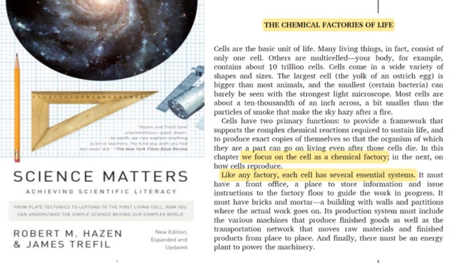
Robert M.Hazen Science matters (2009): 
Pg.239 Cells act as chemical factories, taking in materials from the environment, processing them, and producing “finished goods” to be used for the cell’s own maintenance and for that of the larger organism of which they may be part. In a complex cell, materials are taken in through specialized receptors (“loading docks”), processed by chemical reactions governed by a central information system (“the front once”), carried around to various locations (“assembly lines”) as the work progresses, and finally sent back via those same receptors into the larger organism. The cell is a highly organized, busy place, whose many different parts must work together to keep the whole functioning. While proteins supervise the cell’s chemical factories, carbohydrates provide each factory’s fuel supply.
Pg. 242 Nucleic acids. These molecules (DNA and RNA) carry the blueprint that runs the cell’s chemical factories, and also are the vehicle for inheritance
Pg. 243 Carbohydrates. While proteins supervise the cell’s chemical factories, carbohydrates provide each factory’s fuel supply. The basic building blocks of carbohydrates are sugars—small ring-
Pg. 245 Like any factory, each cell has several essential systems. It must have a front office, a place to store information, and issue instructions to the factory door to guide the work in progress. It must have bricks and mortar—a building with walls and partitions where the actual work goes on. Its production system must include the various machines that produce finished goods as well as the transportation network that moves raw materials and finished products from place to place. And finally, there must be an energy plant to power the machinery.
Pg. 246 Cellular factories consist of walls, partitions, and loading docks.
Pg. 249 Every living thing is composed of one or more cells, each of which has a complex anatomy. A “generic” cell contains many structures and organelles—tiny chemical factories.
Pg. 263 The sequence of the bases along the double helix of DNA contains the genetic code—all the information a cell needs to reproduce itself and run its chemical factories, all the characteristics and quirks that make you unique. 
Pg. 309 Shortly thereafter, the glucose is processed in cellular chemical factories to form part of the cellulose fibers that support each grass blade. The carbon atom has become an integral part of the structure of grass.7

Ben L. Feringa (2020): The miniaturization of complex physical and chemical systems is a key aspect of contemporary materials science. The bottom-up formation of dynamic structures with unusual properties has now been extended from the microscale to the nanoscale. Such extended dynamic structures are complemented by an increasing number of molecular species capable of transforming a physical or chemical stimulus into directional motion. These so-called artificial molecular machines (AMMs) are often regarded as molecular renderings of the macroscopic machines we experience in our daily lives — rotors, gears and cranks, for example. However, the inspiration for many AMMs is not from macroscopic man-made machines but, rather, from proteins or multi-protein complexes in biology that are capable of transforming energy into continuous, complex, structural motion. The process of vision, muscle contraction and bacterial flagellar movement are amazing examples of biological responsive systems. Biological molecular machines (BMMs) such as ATP synthase, ribosomes or myosin are structurally far more complex than any artificial molecular machines AMM made so far, and are an essential part of living systems. Embedded or immobilized within skeleton structures such as bilayer lipid membranes or larger protein complexes, BMMs are part of a cellular confinement in which their work is continuously synchronized with other machines of identical or different nature. Their functions are driven by chemical fuels such as ATP or electrochemical gradients and controlled by chemical or physical stimuli. Their main tasks involve intracellular, transmembrane and intercellular transport of reagents, as well as transformation of small, molecular building blocks into larger functional structures. A cell might thus be viewed as a complex molecular factory in which many different components are assembled, transformed, transported and disassembled. The dynamics of these processes at the molecular level are amplified by self-organization, cooperativity and synchronization, resulting in the living, moving organisms observed at the macroscopic scale. A modular building concept, periodical alignment and synchronization of individual dynamic components on a temporal and spatial domain are essential aspects of the performance of the whole system. Such organizational principles can also be found in macroscopic factories regardless of the difference in size, and they are considered fundamental principles in the design of cooperative dynamic systems of any size and composition. Nevertheless, biological systems strongly differ from man-made factories in certain aspects. BMMs and their complex assemblies are very versatile and selective in continuously producing a variety of complex molecules currently unobtainable by any man-made system. 8 

Von Neumann's universal constructor: We cannot replicate the cell's self-reproduction technology

Imagine a hypothetical human-made truly autonomous self-replicating factory analogous to living cells. It would have to be capable of replicating itself and constructing a copy of itself, without external help. Able to detect raw materials in its surroundings, in the environment,  that it needs, and prepare them to be transformed into the right form, so that import gates and mechanisms could import these materials into the factory inside. The daughter factory would require to get the entire information stored in the mother cell inherited. It would rely on conventional large-scale technology and automation. 

M. Sipper (1998): We would need to be able to understand the fundamental information-processing principles and algorithms involved in self-replication, even independent of their physical realization.9

Replicators have been called "von Neumann machines" after John von Neumann, who first rigorously studied the idea. Von Neumann himself used the term universal constructor to describe such a self-replicating machine. For a factory or machine to make a duplicate copy it must employ a description of itself. This description, being a part of the original factory, must itself be prescribed by something else that is not itself. That is, it must come from the outside. Why? In order to describe something, one needs to be a conscious agent, able to do so. If the factory itself was not the conscious agent, being able to observe and describe itself, it must have been something else. I, as a human being, conscious, can observe and describe myself. A non-conscious "something" has never been seen as having these necessary cognitive and intelligent capabilities. That's why the origin of biological information is an unsolvable problem for naturalists. That's why the origin of the information to make the first living self-replicating cell cannot be solved unless there was a creator. Another salient point: Parts, subunits, or an agglomeration of building blocks do not comprehend how they could join to become part of a functional interlocked complex system. So in order to construct a self-replicating system composed of many interlocking parts, foresight is required, otherwise, the parts could either remain non-assembled, disintegrate, or, eventually, driven by random external forces, interact and assemble into a basically infinite number of nonfunctional chaotic aggregation states.

R. A. Freitas (2004): Von Neumann thus hit upon a deceptively simple architecture for machine replication. The machine would have four parts:   

1. a constructor “A” that can build a machine “X” when fed explicit blueprints of that machine; 
2. a blueprint copier “B”; 
3. a controller “C” that controls the actions of the constructor and the copier, actuating them alternately; and finally 
4. a set of blueprints φ(A + B + C) explicitly describing how to build a constructor, a controller, and a copier. 

The entire replicator may therefore be described as (A + B + C) + φ(A + B + C.

Observers have noted that von Neumann’s early schema was later confirmed by subsequent research on the molecular biology of cellular reproduction, with von Neumann’s component “A” represented by the ribosomes and supporting cellular mechanisms, component “B” represented by DNA polymerase enzymes, component “C” represented by repressor and derepressor molecules and associated expression-control machinery in the cell, and finally component “φ(A + B + C)” represented by the genetic material DNA that carries the organism’s genome. (The correspondence is not complete: cells include additional complexities.) More importantly, the dual use of information — both interpreted and uninterpreted, as in von Neumann’s machine schema — was also found to be true for the information contained in DNA.10  

M. Sipper (1998): A noteworthy distinction apparent in von Neumann’s model of self-replication is the double-faceted use of the information stored in the artificial genome: It first serves as instructions to be interpreted so as to construct a new universal constructor, after which this same genome is copied unmodified, to be attached to the new offspring constructor—so that it may replicate in its turn. This aspect is quite interesting in that it bears strong resemblance to the genetic mechanisms of transcription (copying) and translation (interpretation) employed by biological life—which was discovered during the decade following von Neumann’s work. Von Neumann’s model employs a complex transition rule, with the total number of cells composing the universal constructor estimated at between 50,000 and 200,000 (the literature seems to disagree on the exact number). In the years that followed its introduction a number of researchers had worked toward simplifying this system. In the late 1960s Codd reduced the number of states required for a self-replicating universal constructor-computer from 29 to 8. His self-replicating structure comprised about 100,000,000 cells. A few years later Devore simplified Codd’s system, devising a self-replicating automaton comprising about 100,000 cells. 

Despite the complexity of von Neumann’s self-replicating universal constructor, a number of researchers have considered its implementation (or simulation) over the years. Signorini concentrated on the 29-state transition rule, discussing its implementation on a SIMD (single-instruction multiple-data) computer. Von Neumann’s constructor is divided into many functional blocks known as organs. In addition to implementing the transition rule, Signorini also presented the implementation of three such organs: a pulser, a decoder, and a periodic pulser. To date, Pesavento’s more recent work comes closest to a full simulation of von Neumann’s model. A computer simulation of the universal constructor—running on a standard workstation—even this comes short of realizing the full model: Self-replication is not demonstrated because the tape required to describe the constructor (i.e., the genome) is too large to simulate. 11

R. A. Freitas (2004): Penrose, quoting Kemeny, complained that the body of the von Neumann kinematic machine “would be a box containing a minimum of 32,000 constituent parts (likely to include rolls of tape, pencils, erasers, vacuum tubes, dials, photoelectric cells, motors, batteries, and other devices) and the ‘tail’ would comprise 150,000 [bits] of information.” Macroscale kinematic replicators will require a great deal of effort to design and to build, which may explain why so few working devices have been constructed to date,* despite popular interest. 12

Comment: A Von Neumann self-replicating machine has never been constructed because it is too complicated. Man, with all its intelligence, has failed. But, if abiogenesis is true, the emergence of self-replicating cells with a minimum of one million bits of information happened from randomly distributed, nonreplicating components by entirely non-intelligent unguided means.

A Self-Replicating Box

G. SEWELL (2021): To understand why human-engineered self-replicating machines are so far beyond current human technology, let’s imagine trying to design something as “simple” as a self-replicating cardboard box. Let’s place an empty cardboard box (A) on the floor, and to the right of it let’s construct a box (B) with a box-building factory inside it. I’m not sure exactly what the new box would need to build an empty box, but I assume it would at least have to have some metal parts to cut and fold the cardboard and a motor with a battery to power these parts. In reality, to be really self-replicating like living things, it would have to go get its own cardboard, so maybe it would need wheels and an axe to cut down trees and a small sawmill to make cardboard out of wood. But let’s be generous and assume humans are still around to supply the cardboard. Well, of course box B is not a self-replicating machine, because it only produces an empty box A.

So, to the right of this box, let’s build another box C which contains a fully automated factory that can produce box B’s. This is a much more complicated box, because this one must manufacture the metal parts for the machinery in box B and its motor and battery and assemble the parts into the factory inside B. In reality it needs to go mine some ore and smelt it to produce these metal parts, but again let’s be very generous and provide it all the metals and other raw materials it needs.

But box C would still not be a self-replicating machine, because it only produces the much simpler box B. So back to work, now we need to build a box D to its right with a fully automated factory capable of building box C’s with their box B factories. Well, you get the idea, and one begins to wonder if it is even theoretically possible to build a truly self-replicating machine. When we add technology to such a machine to bring it closer to the goal of reproduction, we only move the goalposts, because now we have a more complicated machine to reproduce. Yet we see such machines all around us in the living world.

If we keep adding boxes to the right, each with a fully automated factory that can produce the box to its left, it seems to me that the boxes would grow exponentially in complexity. But maybe I am wrong. Maybe they could be designed to converge eventually to a self-replicating box Z, although I can’t imagine how. 13

The self-assembly of a factory starting with unorganized raw materials has never been observed

When discussing the assembly of complex structures from raw materials "just laying around," we have to consider that solely the laws of physics and chemistry govern the behavior of these materials. The spontaneous self-assembly of complex factories or structures solely from raw materials without any external intervention is currently not well-documented or understood. In other words, it has never been demonstrated to be possible. The spontaneous assembly of a complex factory or structure through unguided means, without any external intelligence or intervention, has not been demonstrated or observed in scientific experiments or natural processes. While self-assembly and self-organization are observed in various systems, they often involve specific conditions, preexisting structures, or programmed interactions between components. When we talk about self-assembly and self-organization, it's important to understand that these processes typically occur within certain contexts and conditions. For example:

In living organisms, self-assembly processes are observed in various structures, such as the formation of cellular membranes, the assembly of protein complexes, or the organization of DNA into chromosomes. However, these processes rely on preexisting biological components, such as proteins, lipids, or nucleic acids, which have specific molecular interactions and are governed by biological mechanisms. The assembly and organization of these structures are guided by genetic information and cellular processes, involving complex networks of chemical reactions and molecular interactions.

In the field of nanotechnology, scientists have developed self-assembling systems at the molecular scale. These systems often involve specially designed molecules or nanoparticles that possess specific properties or functional groups. Through these properties, the components can interact and align in a way that facilitates self-assembly. The process may require specific environmental conditions, such as a particular solvent or temperature range, to trigger the self-assembly. Therefore, while self-organization is observed, it still relies on the design and manipulation of the components and their surrounding environment.

In synthetic systems, researchers have explored self-assembly processes using engineered components. For example, in robotics, researchers have developed small robotic units that can autonomously assemble into larger structures or perform collective tasks. However, these systems typically involve programmed interactions and behaviors. The individual units may have sensors, communication capabilities, or predefined rules that govern their assembly and coordination. They are designed with specific capabilities and functionalities to enable self-assembly under controlled conditions.

Observe the keywords here: guided by genetic information, and the involvement of programmed interactions. Generating information and programmed interactions typically require the involvement of a programmer or an intelligent agent. In the context of self-assembly and self-organization, the patterns, behaviors, and interactions observed in complex systems often stem from the information encoded within the system or introduced by an external intelligence. In biological systems, genetic information encoded in DNA serves as the blueprint for the assembly and functioning of organisms. This information cannot be the result of evolutionary processes, because since Darwinian evolution started with the first living self-replicating cells.  The discussion of evolutionary processes in the context of genetic information assumes the existence of life and the subsequent diversification and adaptation of organisms over time. Similarly, in synthetic systems or engineered materials, a programmer or designer imparts specific instructions, rules, or algorithms to guide the self-assembly or behavior of the components. Information, in the form of genetic code, algorithms, or predefined rules, plays a crucial role in shaping the behavior and outcomes of self-assembly and self-organization processes. Without the input of intelligent design or programming, the emergence of complex structures or organized behaviors is unlikely, if not, or rather impossible to occur spontaneously. The presence of information or programmed interactions does necessarily imply the involvement of a conscious or deliberate programmer. 


The Last Universal Common Ancestor (LUCA): What was its nature?

Before we can start investigating the course of evolution, we need to know what the starting point was. A lot has been speculated regarding the first life form. What did it look like? Was it indeed a Last Universal Common Ancestor (LUCA), or did life start polyphyletic? I have dedicated an entire chapter to my previous book: On the Origin of Life and Virus World by means of an Intelligent Designer,  attempting to get closer to answering what could serve as a model organism. This is a surprisingly difficult question to answer.  

The last universal common ancestor represents the primordial cellular organism from which diversified life was derived. It has been considered as the branching point on which Bacteria, Archaea and Eukaryotes have diverged.10

Carl R. Woese (2002): The central question posed by the universal tree is the nature of the entity (or state) represented by its root, the fount of all extant life. Herein lies the door to the murky realm of cellular evolution. Experience teaches that the complex tends to arise from the simple, and biologists have assumed it so in the case of modern cells. But this assumption is usually accompanied by another not-so-self-evident one: namely that the ‘‘organism’’ represented by the root of the universal tree was equivalent metabolically and in terms of its information processing to a modern cell, in effect was a modern cell. Such an assumption pushes the real evolution of modern cells back into an earlier era, which makes the problem not directly addressable through genomics. That is not a scientifically acceptable assumption. Unless or until facts dictate otherwise, the possibility must be entertained that some part of cellular evolution could have occurred during the period encompassed by the universal phylogenetic tree. There is evidence, good evidence, to suggest that the basic organization of the cell had not yet completed its evolution at the stage represented by the root of the universal tree. The best of this evidence comes from the three main cellular information processing systems. Translation was highly developed by that stage: rRNAs, tRNAs, and the (large) elongation factors were by then all basically in near-modern form; hence, their universal distributions. Almost all of the tRNA charging systems were in modern form as well. But, whereas the majority of ribosomal proteins are universal in distribution, a minority of them is not. A relatively small cadre is specific to the bacteria, a somewhat larger set common and confined to the archaea and eukaryotes, and a few others are uniquely eukaryotic. Almost all of the universal translational proteins (as well as those in transcription) show what is called the canonical pattern, i.e., the bacterial and archaeal versions of the protein are remarkably different from one another, so much so that their difference is distinguished as one of ‘‘genre’’. Except for the aminoacyl-tRNA synthetases the corresponding eukaryotic versions are virtually all of the archaeal genre. Why canonical pattern exists is a major unanswered question. In the overall it would seem that translation, although highly developed at the root of the universal tree, subsequently underwent idiosyncratic modifications in each of the three major cell types. Transcription seems to have been rather less developed at the root of the universal tree. The two largest (the catalytic) subunits of the DNA-dependent RNA polymerase are universal in distribution.

The cell is the essence of biology. At least that is how 20th-century molecular biology saw it, and the great goal was to understand how cells were organized and worked. This goal, it was assumed, could be accomplished by cataloging (and characterizing) all of the parts of the mechanism, with the tacit assumption that given such a parts list the overall organization of the cell would become apparent. Today, such lists exist for several organisms. Yet an understanding of the whole remains as elusive a goal as ever (34). The fault here lies with the reductionist perspective of molecular biology. The problem of cellular design cannot be fit into this rigid, procrustean framework. It should be obvious from the foregoing discussion that biological cell design is not a static, temporal, or local problem.

The Dilemma of Cellular Evolution. 

Evolving the cell requires evolutionary invention of unprecedented novelty and variety, the likes of which cannot be generated by any familiar evolutionary dynamic. The task can be accomplished only by a collective evolution in which many diverse cell designs evolve simultaneously and share their novelties with one another; which means that 

(i) HGT (and a genetic lingua franca) is a necessary condition for the evolution of cell designs, and 
(ii) a cell design cannot evolve in isolation; others will necessarily accompany it. 

Comment: That sounds suspiciously like a special creation. Once Woese admits that many diverse cells evolved simultaneously, he departs from the concept of universal common ancestry and resorts to polyphyly, that is the proposition, that at the beginning, there was a population of diverse cell designs, each one different from one another, that began to interact through horizontal gene transfer. 

Woese continues: There is an inherent contradiction in this situation. Although HGT is essential for sharing novelty among the various evolving cell designs, it is at the same time a homogenizing force, working to reduce diversity. Thus, what needs explaining is not why the major cell designs are so similar, but why they are so different. This apparent contradiction can be resolved by assuming that the highly diverse cell designs that exist today are the result of a common evolution in which each of them began under (significantly) different starting conditions. [Initial conditions do not necessarily damp out for complex dynamic processes; indeed, they can lead to vastly different outcomes.  14

E. V. Koonin (2020): The last universal cellular ancestor (LUCA) is the most recent population of organisms from which all cellular life on Earth descends. 

Comment:  Koonin goes with the same line of argumentation. He hypothesizes LUCA as a population of organisms. Where did it descend from? A population has to originate from self-replication, which produces offsprings. 

Berkley University's website on evolution claims:  The ability to copy the molecules that encode genetic information is a key step in the origin of life — without it, life could not exist. This ability probably first evolved in the form of an RNA self-replicator — an RNA molecule that could copy itself. Self-replication opened the door for natural selection. Once a self-replicating molecule formed, some variants of these early replicators would have done a better job of copying themselves than others, producing more “offspring.” 15

Comment: By giving careful examination, such assertions cannot be taken seriously.  This is pseudo-scientific storytelling. The evidence does not justify saying that probably, it happened. A self-replicating molecule has never been seen. But also if it existed, it would be helpless to create a living cell. If molecule A self-reproduces n-times we would have AAAAAAA....That is ridiculously trivial and has nothing to do with what we see in a cell. A cell is a cybernetic ultra-complex system, where, thanks to countless concurrent software-driven chemical and physical processes using languages and codes, the material is stored, managed, moved, assembled, converted, and positioned such that the cell survives and self-reproduces. To believe, as proponents of naturalistic mechanisms do, that AAAAAAA... leads to a cell, is like to think that by simply duplicating bricks BBBBBBB... we get a functioning complete self-replicating chemical factory.

Martina Preiner (2020): Many found the metaphor appealing: a world with a jack-of-all-trades RNA molecule, catalyzing the formation of indispensable cellular scaffolds, from which somehow then cells emerged. Others were quick to notice several difficulties with that scenario. These included the lack of templates enabling the polymerization of RNA in the prebiotic complex mixture and RNA’s extreme lability at moderate to high temperatures and susceptibility to base-catalyzed hydrolysis. 16

N. Sankaran (2017): Today, thirty years after the RNA World was first proposed, no one has seen an actual living system that is completely based in RNA. Nevertheless, the hypothesis lives on in the origins of life research community, albeit in a hotly debated, highly contentious atmosphere. Although there are strong opponents, many researchers agree that although far from complete, it remains one of the best theories we have to understand “the backstory to contemporary biology.” Gilbert himself expressed some disappointment that “a self-replicating RNA has not yet been synthesized or discovered” in the years since he predicted his hypothesis, but he remains optimistic that it will emerge eventually. 17

Koonin continues:  The reconstruction of the genome and phenotype of the LUCA is a major challenge in evolutionary biology. Given that all life forms are associated with viruses and/or other mobile genetic elements, there is no doubt that the LUCA was a host to viruses.

E. V. Koonin (2017): The entire history of life is the story of virus–host coevolution. Therefore the origins and evolution of viruses are an essential component of this process. A signature feature of the virus state is the capsid, the proteinaceous shell that encases the viral genome. Although homologous capsid proteins are encoded by highly diverse viruses, there are at least 20 unrelated varieties of these proteins.  A comprehensive sequence and structure analysis of major virion proteins indicates that they evolved on about 20 independent occasions. 18

Viruses and the tree of life (2009): Viruses are polyphyletic: In a phylogenetic tree, the characteristics of members of taxa are inherited from previous ancestors. Viruses cannot be included in the tree of life because they do not share characteristics with cells, and no single gene is shared by all viruses or viral lineages. While cellular life has a single, common origin, viruses are polyphyletic – they have many evolutionary origins. Viruses don’t have a structure derived from a common ancestor.  Cells obtain membranes from other cells during cell division. According to this concept of ‘membrane heredity’, today’s cells have inherited membranes from the first cells.  Viruses have no such inherited structure.  They play an important role by regulating population and biodiversity. 6 

Comment: Since viruses are polyphyletic, and, according to Woese, many diverse cell designs evolved simultaneously, which clarifies the picture: Life arose multiple times independently, and so did viruses. The hypothesis of universal common ancestry is not supported by the evidence. Separate origins of different life forms, and viruses, are.

There is no scientific consensus about LUCA's nature

D. C. Gagler et.al., (2021): Life emerges from the interplay of hundreds of chemical compounds interconverted in complex reaction networks. Some of these compounds and reactions are found across all characterized organisms, informing concepts of universal biochemistry and allowing rooting of phylogenetic relationships in the properties of a last universal common ancestor (LUCA). Thus, universality, as we have come to understand it in biochemistry, is a direct result of the observation that all known examples of life share common details in their component compounds and reactions. 20 

Eugene V. Koonin (2020): Considerable efforts have been undertaken over the years to deduce the genetic composition and biological features of the LUCA from comparative genome analyses combined with biological reasoning. These inferences are challenged by the complex evolutionary histories of most genes (with partial exception for the core components of the translation and transcription systems) that involved extensive horizontal transfer and non-orthologous gene displacement. Nevertheless, on the strength of combined evidence, it appears likely that the LUCA was a prokaryote-like organism (that is, like bacteria or archaea) of considerable genomic and organizational complexity. 21

J. D. Sutherland (2017): The latest list of genes thought to be present in LUCA is a long one. The presence of membranes, proteins, RNA and DNA, the ability to perform replication, transcription, and translation, as well as harboring an extensive metabolism driven by energy harvested from ion gradients using ATP synthase, reveal that there must have been a vast amount of evolutionary innovation between the origin of life and the appearance of LUCA. Many of the inferred proteins in LUCA use FeS clusters and other transition-metal-ion-based co-factors. 22

Life started complex

Life had to start complex because the earliest known cells, including a supposed last universal common ancestor (LUCA), were already functionally and genetically complex. The simplest cells available for study have a teleonomic apparatus so powerful that no vestiges of truly primitive structures are discernible. The LUCA was sophisticated, with a complex structure recognizable as a cell, and had representatives in practically all the essential functional niches currently present in extant organisms. Even the simplest known cellular life forms possess several hundred genes that encode the components of a fully-fledged membrane, the replication, transcription, and translation machinery, a complex cell-division apparatus, and at least some central metabolic pathways. Therefore, life did not start as a primitive or simple organism, but rather as a complex entity capable of metabolism, genetic replication, and maintaining a boundary that separates the cell from its environment.

J.Monod (1972): The simplest cells available to us for study have nothing "primitive" about them. They have a teleonomic apparatus so powerful that no vestiges of truly primitive structures are discernible. 23 Elsewhere, Monod stated: ‘We have no idea what the structure of a primitive cell might have been. The simplest living system known to us, the bacterial cell… in its overall chemical plan is the same as that of all other living beings. It employs the same genetic code and the same mechanism of translation as do, for example, human cells. Thus the simplest cells available to us for study have nothing “primitive” about them… no vestiges of truly primitive structures are discernible.’ Thus the cells themselves exhibit a similar kind of ‘stasis’  in connection with the fossil record.

J. A. G. Ranea (2006): We know that the LUCA, or the primitive community that constituted this entity, was functionally and genetically complex. Life achieved its modern cellular status long before the separation of the three kingdoms. we can affirm that the LUCA held representatives in practically all the essential functional niches currently present in extant organisms, with a metabolic complexity similar to translation in terms of domain variety. 24


Last edited by Otangelo on Fri Jul 07, 2023 8:44 pm; edited 11 times in total

https://reasonandscience.catsboard.com

Otangelo


Admin

D.YATES (2011): New evidence suggests that LUCA was a sophisticated organism after all, with a complex structure recognizable as a cell, researchers report. Their study appears in the journal Biology Direct. The study lends support to a hypothesis that LUCA may have been more complex even than the simplest organisms alive today, said James Whitfield, a professor of entomology at Illinois and a co-author on the study. 25

G. Caetano-Anollés (2011): corresponding authorLife was born complex and the LUCA displayed that heritage. Recent comparative genomic studies support the latter model and propose that the urancestor was similar to modern organisms in terms of gene content 26

E. V. Koonin (2012): All known cells are complex and elaborately organized. The simplest known cellular life forms, the bacterial (and the only known archaeal) parasites and symbionts, clearly evolved by degradation of more complex organisms; however, even these possess several hundred genes that encode the components of a fully fledged membrane; the replication, transcription, and translation machineries; a complex cell-division apparatus; and at least some central metabolic pathways. As we have already discussed, the simplest free-living cells are considerably more complex than this, with at least 1,300 genes 27

J. C. Xavier (2014): The cell is the most complex structure in the micrometer size range known to humans. At present, the minimal cell can be defined only on a semiabstract level as a living cell with a minimal and sufficient number of components and having three main features:

1. Some form of metabolism to provide molecular building blocks and energy necessary for synthesizing the cellular components,
2. Genetic replication from a template or an equivalent information processing and transfer machinery, and
3. A boundary (membrane) that separates the cell from its environment.
4. The necessity of coordination between boundary fission and the full segregation of the previously generated twin genetic templates could be added to this definition.
5. The essential feature of a minimal cell is the ability to evolve, which is a universal characteristic among all known living cells 28

F. El Baidouri (2021): Along with two robust prokaryotic phylogenetic trees we are able to infer that the last universal common ancestor of all living organisms was likely to have been a complex cell with at least 22 reconstructed phenotypic traits probably as intricate as those of many modern bacteria and archaea. Our results depict LUCA as likely to be a far more complex cell than has previously been proposed, challenging the evolutionary model of increased complexity through time in prokaryotes.29


Defining the LUCA: What might be a Cell’s minimal requirement of parts?

I have gone in-depth to elucidate this question in my previous book: On the Origin of Life and Virus World by means of an Intelligent Designer. I wrote: Science remains largely in the dark when it comes to pinpointing what exactly the first life form looked like. Speculation abounds. Whatever science paper about the topic one reads, confusion becomes apparent. Patrick Forterre wrote  in a science paper in 2015: The universal tree of Life: an update, confessed: 

There is no protein or groups of proteins that can give the real species tree, i.e., allow us to recapitulate safely the exact path of life evolution.

As such, whatever architecture one comes up with, remains in the realm of speculation. Is it therefore futile, to trace a borderline, and list a number of features, that most likely were present? No. Even if we can come up only with a hypothetical organism, it will nonetheless give us insight into the complexity involved, and bring us closer to deciding, what mechanisms most likely were involved and if intelligence was required to set up the first life forms.  

Andrew J. Crapitto (2022): The availability of genomic and proteomic data from across the Tree of life has made it possible to infer features of the genome and proteome of the last universal common ancestor (LUCA). Several studies have done so, all using a unique set of methods and bioinformatics databases. No individual study shares a high or even moderate degree of similarity with any other individual study. Studies of the genome or proteome of the LUCA do not uniformly agree with one another. The set of consensus LUCA protein family predictions between all of these studies portrays a LUCA genome that, at minimum, encoded functions related to protein synthesis, amino acid metabolism, nucleotide metabolism, and the use of common, nucleotide-derived organic cofactors.

The translation process is well known to be ancient and many of the proteins involved in translation machinery appear to predate the LUCA. A corollary to the influential RNA world hypothesis is that the translation system evolved within the context of an RNA-based genetic system. Most universal Clusters of Orthologous ( Orthologous are homologous genes where a gene diverges after a speciation event, but the gene and its main function are conserved) Groups of proteins (COGs) COGs encode proteins that physically associate with the ribosome and those that do not are often involved with the translation process in some other way. Similarly, nearly all universal, vertically inherited functional RNAs (save the SRP RNA) are involved in the translation system. Translation-related genes or proteins are prevalent in the predictions of seven of the eight previously published LUCA genome or proteome studies analyzed here. We identified 366 eggNOG clusters that were predicted by four or more studies to have been present in the genome of the LUCA (Appendix S2). 30

William Martin and colleagues from the University Düsseldorf’s Institute of Molecular Evolution give us also an interesting number: The metabolism of cells contains evidence reflecting the process by which they arose. Here, we have identified the ancient core of autotrophic metabolism encompassing 404 reactions that comprise the reaction network from H2, CO2, and ammonia (NH3) to amino acids, nucleic acid monomers, and the 19 cofactors required for their synthesis. Water is the most common reactant in the autotrophic core, indicating that the core arose in an aqueous environment. Seventy-seven core reactions involve the hydrolysis of high-energy phosphate bonds, furthermore suggesting the presence of a non-enzymatic and highly exergonic chemical reaction capable of continuously synthesizing activated phosphate bonds. CO2 is the most common carbon-containing compound in the core. An abundance of NADH and NADPH-dependent redox reactions in the autotrophic core, the central role of CO2, and the circumstance that the core’s main products are far more reduced than CO2 indicate that the core arose in a highly reducing environment. The chemical reactions of the autotrophic core suggest that it arose from H2, inorganic carbon, and NH3 in an aqueous environment marked by highly reducing and continuously far from equilibrium conditions. Supplementary Table 1. in the paper lists all 402 metabolic reactions   31, 32

John I. Glass (2006): Mycoplasma genitalium has the smallest genome of any organism that can be grown in pure culture. It has a minimal metabolism. Consequently, its genome is expected to be a close approximation to the minimal set of genes needed to sustain bacterial life. 33

The RNA-DNA Nexus: Unveiling the Molecular Machinery of Life, and the Intelligent Design Paradigm Genita10
Metabolic pathways and substrate transport mechanisms encoded by M. genitalium. Metabolic products are colored red, and mycoplasma proteins are black. White letters on black boxes mark nonessential functions or proteins based on our current gene disruption study. Question marks denote enzymes or transporters not identified that would be necessary to complete pathways, and those missing enzyme and transporter names are colored green. Transporters are colored according to their substrates: yellow, cations; green, anions and amino acids; orange, carbohydrates; purple, multidrug and metabolic end product efflux. The arrows indicate the predicted direction of substrate transport. The ABC type transporters are drawn as follows: rectangle, substrate-binding protein; diamonds, membrane-spanning permeases; circles, ATP-binding subunits.

J. A. G. Ranea (2006): In our view, the LUCA was faced with two important challenges associated with the source of amino acids and purine/pyrimidine bases or nucleosides. Most of these compounds need complex pathways to be synthesized and our analyses suggest that these are not present in the LUCA. Based on that, we are more in favor of amino acids and nitrogenous bases being present in a possible primitive soup rather than being synthesized by the LUCA.34

From a LUCA to the last bacterial common ancestor (LBCA)

Even though the existence of LUCA is supported by the universal presence of conserved biomolecules and a vast amount of genetic data, its characteristics and identity remain unknown. LBCA, on the other hand, stands for "Last Bacterial Common Ancestor," which refers to the hypothetical ancestor of all modern bacteria. Although the characteristics of LBCA are still uncertain, recent studies suggest that it might have been a monoderm bacterium with a complete 17-gene dcw cluster, which is two genes more than in the model E. coli cluster. 

The 17-gene dcw (division and cell wall) cluster is a group of bacterial genes that are involved in the regulation of cell division and the synthesis of the cell wall during the cell cycle. These genes encode proteins that are responsible for the assembly and contraction of the bacterial cell wall and septum, which eventually leads to the separation of the daughter cells. The dcw cluster includes genes that are involved in peptidoglycan synthesis, cell wall assembly, and septation, among others. These genes are found in many bacterial species and are thought to be essential for bacterial growth and survival. Understanding the composition and regulation of the dcw cluster can provide insights into bacterial cell division and the evolution of bacterial morphology.

Phylogenomic inference also reveals that the Clostridia, a class of Firmicutes, are the least diverged of the modern genomes, suggesting that the first lineage to diverge from the predicted LBCA was similar to the modern Clostridia.

In 2004, Rosario Gil proposed a minimal gene set composed of 206 genes that would sustain the main vital functions of a hypothetical simplest bacterial cell. These functions include DNA replication, transcription, translation, protein processing, folding, secretion and degradation, cell division, energetic metabolism, pentose pathway, nucleotide biosynthesis, and lipid biosynthesis. The minimal cell does not include biosynthetic pathways for amino acids or most cofactor precursors, as they can be obtained from the environment.

While some amino acids can be obtained from the environment, not all of them are readily available or in sufficient quantities to support the growth of a minimal cell. In addition, the amino acids that are available in the environment may not be in the correct proportions or forms required by the cell. Therefore, some minimal cells may require biosynthetic pathways for certain amino acids to ensure their survival and growth.

R. R. Léonard (2022): The nature of the LBCA is unknown, especially the architecture of its cell wall. The lack of reliably affiliated bacterial fossils outside Cyanobacteria makes it elusive to decide the very nature of the LBCA. Nevertheless, phylogenomic inference leads to informative results, and our analysis of the cell-wall characteristics of extant bacteria, combined with ancestral state reconstruction and distribution of key genes, opens interesting possibilities: the LBCA might have been a monoderm bacterium featuring a complete 17-gene dcw cluster, two genes more than in the model E. coli cluster. This result was also supported by a recent study, in which the authors found 146 protein families that formed a predicted core for the metabolic network of the LBCA. From these families, phylogenetic trees were produced and the divergence of the modern genomes from the root to the tips was analysed. It appears that the Clostridia (a class of Firmicutes) are the least diverged of the modern genomes and thus the first lineage to diverge from the predicted LBCA were similar to the modern Clostridia. Based on these results, the authors suggested that the LBCA could have been a monoderm bacteria. (Having a single membrane, especially a thick layer of peptidoglycan) 35

P. C. Morales et.al., (2019) reconstructed the phylogenetic tree of Clostridium species. They set Clostridium difficile at the root of the tree. 36 The genome of C. difficile strain 630 consists of a circular chromosome of 4,290,252 bp 37

Taking Rosario Gil's model organism as the basis for our forthcoming investigation

Rosario Gil (2004): Based on the conjoint analysis of several computational and experimental strategies designed to define the minimal set of protein-coding genes that are necessary to maintain a functional bacterial cell, we propose a minimal gene set composed of 206 genes. Such a gene set will be able to sustain the main vital functions of a hypothetical simplest bacterial cell with the following features.

1. A virtually complete DNA replication machinery, composed of one nucleoid DNA binding protein, SSB, DNA helicase, primase, gyrase, polymerase III, and ligase. No initiation and recruiting proteins seem to be essential, and the DNA gyrase is the only topoisomerase included, which should perform both replication and chromosome segregation functions.

2. A very rudimentary system for DNA repair, including only one endonuclease, one exonuclease, and a uracyl-DNA glycosylase.

3. A virtually complete transcriptional machinery, including the three subunits of the RNA polymerase, a σ factor, an RNA helicase, and four transcriptional factors (with elongation, antitermination, and transcription-translation coupling functions). Regulation of transcription does not appear to be essential in bacteria with reduced genomes, and therefore the minimal gene set does not contain any transcriptional regulators.

4. A nearly complete translational system. It contains the 20 aminoacyl-tRNA synthases, a methionyl-tRNA formyltransferase, five enzymes involved in tRNA maturation and modification, 50 ribosomal proteins (31 proteins for the large ribosomal subunit and 19 proteins for the small one), six proteins necessary for ribosome function and maturation (four of which are GTP binding proteins whose specific function is not well known), 12 translation factors, and 2 RNases involved in RNA degradation.

5. Protein-processing, -folding, secretion, and degradation functions are performed by at least three proteins for posttranslational modification, two molecular chaperone systems (GroEL/S and DnaK/DnaJ/GrpE), six components of the translocase machinery (including the signal recognition particle, its receptor, the three essential components of the translocase channel, and a signal peptidase), one endopeptidase, and two proteases.

6. Cell division can be driven by FtsZ only, considering that, in a protected environment, the cell wall might not be necessary for cellular structure.

7. A basic substrate transport machinery cannot be clearly defined, based on our current knowledge. Although it appears that several cation and ABC transporters are always present in all analyzed bacteria, we have included in the minimal set only a PTS for glucose transport and a phosphate transporter. Further analysis should be performed to define a more complete set of transporters.

8. The energetic metabolism is based on ATP synthesis by glycolytic substrate-level phosphorylation.

9. The nonoxidative branch of the pentose pathway contains three enzymes (ribulose-phosphate epimerase, ribose-phosphate isomerase, and transketolase), allowing the synthesis of pentoses (PRPP) from trioses or hexoses.

10. No biosynthetic pathways for amino acids, since we suppose that they can be provided by the environment.

11. Lipid biosynthesis is reduced to the biosynthesis of phosphatidylethanolamine from the glycolytic intermediate dihydroxyacetone phosphate and activated fatty acids provided by the environment.

12. Nucleotide biosynthesis proceeds through the salvage pathways, from PRPP and the free bases adenine, guanine, and uracil, which are obtained from the environment.

13. Most cofactor precursors (i.e., vitamins) are provided by the environment. Our proposed minimal cell performs only the steps for the syntheses of the strictly necessary coenzymes tetrahydrofolate, NAD+, flavin aderine dinucleotide, thiamine diphosphate, pyridoxal phosphate, and CoA. 38

Comment: That would require LUCA to have complex import and transport mechanisms of nucleotides and amino acids, and membrane import channel proteins able to distinguish and select those building blocks for life that are used in life, from those that aren't. As I have outlined in my book, On the Origin of Life and Virus World by means of an Intelligent Designer, Origin of Life researchers have failed throughout to demonstrate the possible abiotic route to synthesize the basic building blocks of life non-enzymatically.  But even IF that would be the case, that would still not explain how LUCA made the transition from external incorporation to acquire the complex metabolic and catabolic pathways to synthesize nucleotides and amino acids which constitutes a huge, often overlooked gap. Mycoplasma genitalium is held as the smallest possible living self-replicating cell. It is, however, a pathogen, an endosymbiont that only lives and survives within the body or cells of another organism ( humans ).  As such, it IMPORTS many nutrients from the host organism. The host provides most of the nutrients such bacteria require, hence the bacteria do not need the genes for producing such compounds themselves. As such, it does not require the same complexity of biosynthesis pathways to manufacture all nutrients as a free-living bacterium. Amino Acids were no readily available on the early earth. In the Miller Urey experiment, eight of the 20 amino acids were never produced. Neither in 1953 nor in the subsequent experiments.


2


LUCAs information system

Currently, there is no known form of life that exists without DNA and RNA. DNA is a fundamental component of all known life on Earth and serves as the genetic blueprint that encodes the information necessary for the development, function, and reproduction of living organisms. Some claim that it is possible that alternative forms of genetic material or information storage may exist in other environments beyond our current understanding. This is however an argument from ignorance. It is a fallacy that occurs when someone asserts a claim based on the absence of evidence to the contrary. It is important to base claims on positive evidence rather than on the absence of evidence. It is therefore warranted to start with the presumption that DNA was present when life started. And as such, as well the biosynthesis pathways necessary to synthesize deoxynucleotides, the monomer building blocks of DNA. 

The Central Dogma

The term Central Dogma was coined by Francis Crick, who discovered the double-helix structure of DNA together with Rosalind Franklin, James Watson, and Maurice Wilkins. DNA is “the Blueprint of Life.” It contains part of the data needed to make every single protein that life can't go on without. ( Epigenetic data based on epigenetic languages is also involved). No DNA, no proteins, no life. RNA has a limited coding capacity because it is unstable.

The RNA-DNA Nexus: Unveiling the Molecular Machinery of Life, and the Intelligent Design Paradigm Crick_10
James Watson, left, with Francis Crick and their model of part of a DNA molecule SCIENCE PHOTO LIBRARY

YourGenome.org: The ‘Central Dogma’ is the process by which the instructions in DNA are converted into a functional product. It was first proposed in 1958 by Francis Crick, discoverer of the structure of DNA. The central dogma suggests that DNA contains the information needed to make all of our proteins, and that RNA is a messenger that carries this information to the ribosomes. The ribosomes serve as factories in the cell where the information is ‘translated’ from a code into a functional product. The process by which the DNA instructions are converted into the functional product is called gene expression. Gene expression has two key stages – transcription and translation. In transcription, the information in the DNA of every cell is converted into small, portable RNA messages. During translation, these messages travel from where the DNA is in the cell nucleus to the ribosomes where they are ‘read’ to make specific proteins.1

DNA and RNA: The only possible information storage molecules?
Steven A. Benner (2005): Starting in the 1980s, some synthetic biologists began to wonder whether DNA and RNA were the only molecular structures that could support genetics on Earth or elsewhere.   This knowledge, and the fact that the Watson–Crick model proposed no particular role for the phosphates in molecular recognition, encouraged the inference that the backbone could be changed without affecting pairing rules. The effort to synthesize non-ionic backbones changed the established view of nucleic acid structure. Nearly 100 linkers were synthesized to replace the 2′-deoxyribose sugar. Nearly all analogues that lacked the REPEATING CHARGE showed worse rule-based molecular recognition. Even with the most successful uncharged analogues (such as the polyamide-linked nucleic-acid analogues (PNA)) molecules longer than 15 or 20 building units generally failed to support rule-based duplex formation. In other uncharged systems, the breakdown occurs earlier. The repeating charge in the DNA backbone could no longer be viewed as a dispensable inconvenience. The same is true for the ribose backbone of RNA: The backbone is not simply scaffolding to hold the nucleobases in place; it has an important role in the molecular recognition that is central to genetics.  2

Lack of natural selection
The idea that nucleotides were readily laying around on the early earth, just waiting to be picked up, and concentrated on the building site of life, was mocked by Leslie Orgel as 'the Molecular Biologist's Dream. This is maybe the most stringent problem of prebiotic nucleotide synthesis: The materials on prebiotic earth were a mess of mixtures of lifeless chemicals, and nothing restricts the possibility of a great diversity of nucleotides with differing sugar moieties. There was no natural selection. Many science papers simply ignore this and resort nonetheless to little magic of selective pressure. It's like from Frankenstein to man. Some patchwork here and there, and chance does the rest and figures things out.  Szostak and colleagues were well aware of the problem. They wrote:

There are many nucleobase variations such as 8-oxo-purine, inosine, and the 2-thio-pyrimidines, as well as sugar variants including arabino-, 2′- deoxyribo-, and threonucleotides. The likely presence of byproducts leads to a significant problem with regard to the emergence of the RNA world, since the initially synthesized oligonucleotides would be expected to be quite heterogeneous in composition. How could such a heterogeneous mixture of oligonucleotides give rise to the relatively homogeneous RNAs that are thought to be required for the evolution of functional RNAs such as ribozymes? 3

So, in 2020, they presented a model, ignoring the fact made by Benner and others, that molecules simply disintegrate and randomize, they proposed that "  many versions of nucleotides merged to form patchwork molecules with bits of both modern RNA and DNA, as well as largely defunct genetic molecules, such as ANAThese chimeras, like the monstrous hybrid lion, eagle and serpent creatures of Greek mythology, may have been the first steps toward today's RNA and DNA." 4 Rather than focussing "on the consequences of coexisting activated arabino- and 2′-deoxy-nucleotides for nonenzymatic template-directed primer extension", the authors need to provide a plausible trajectory for how natural selection pressures provided the separation of non-canonical nucleotides to achieve a homogeneous state of affairs, where only RNA's and DNAs used in life polymerize. Often, the key questions in the mids of the often confusing technical jargon get lost. 

Fast decomposition rate
Adenine deaminates at 37°C with a half-life of 80 years (half-life = time that a substance takes to decompose, and loses half of its physiologic activity). At 100°C its half-live is 1 year. For guanine, at 100°C its half-live is 10 months, uracil is 12 years, and thymine 56 years.  For the decomposition of a nucleobase, this is very short. For nucleobases to accumulate in prebiotic environments, they must be synthesized at rates that exceed their decomposition. Therefore, adenine and the other nucleobases would never accumulate in any kind of "prebiotic soup." 5

A paper published in 2015 points out that: 

Nucleotide formation and stability are sensitive to temperature. Phosphorylation of nucleosides in the laboratory is slower at low temperatures, taking a few weeks at 65 ◦C compared with a couple of hours at 100 ◦C. The stability of nucleotides, on the other hand, is favored in warm conditions over high temperatures. If a WLP is too hot (>80 ◦C), any newly formed nucleotides within it will hydrolyze in several days to a few years. At temperatures of 5 ◦C to 35 ◦C that either characterize more-temperate latitudes or a post snowball Earth, nucleotides can survive for thousand-to-million-year timescales. However, at such temperatures, nucleotide formation would be very slow.  6

That means, in hot environments, nucleotides might form, but they decompose fast. On the other hand, in cold environments, they might not degrade that fast, but take a long time to form. Nucleotides would have to be generated by prebiotic environmental synthesis processes at a far higher rate than they are decomposed and destroyed, and accumulated and concentrated at one specific construction site. Putting that into perspective, P.Ubique, the smallest known free-living cell, has a genome size of 1,3 million nucleotides. The best-studied mechanism relevant to the prebiotic synthesis of ribose is the formose reaction. Several problems have been recognized in ribose synthesis via the formose reaction, which reaction is very complex. It depends on the presence of a suitable inorganic catalyst. Ribose is merely an intermediate product among a broad suite of compounds including sugars with more or fewer carbons. There would have been no way to activate phosphate somehow, in order to promote the energy dispendious reaction.

Extraterrestrial nucleobase sources
In april 2022, nature magazine announced the identification of nucleobases in carbonaceous meteorites.  Guanine and adenine were detected in murchison meteorite extracts, and now various pyrimidine nucleobases such as cytosine, uracil, and thymine, and their structural isomers such as isocytosine, imidazole-4-carboxylic acid, and 6-methyluracil, respectively. They came to the conclusion that "a diversity of meteoritic nucleobases could serve as building blocks of DNA and RNA on the early Earth".7 An article of NASA echoed the authors conclusion: "This discovery demonstrates that these genetic parts are available for delivery and could have contributed to the development of the instructional molecules on early Earth."8 The fatal blow is the fact that the nucleobases relevant for life come always mixed together with isomers that are irrelevant. There was no prebiotic selection to sort out and concentrate exclusively those relevant for life. 

Selecting the nucleobases used in life
Maybe you are familiar with the concept of "sequence space". It relates to the fact that there is a huge combinatorial space (or possibilities) to put an amino acid strand together, but only a very limited number of sequences bear function, or eventually fold into 3D forms, and become functional proteins. That makes it very remotely possible, that random chance joined functional sequences together on the early earth. Analogously, the same goes for "Structure space" of the four macromolecular "bricks" or building blocks used in life. Adenine, for example, one of the five nucleobases used in RNA and DNA,  are purines, made of carbon, hydrogen, and nitrogen atoms. They have a six-membered nitrogen ring, fused to a five-membered nitrogen ring. The thymine nucleobase is a pyrimidine, and has just a one-ring structure, using carbon, hydrogen, and nitrogen atoms. There is no physical law, that restricts these molecules to have this isomeric ring structure and atomic composition. But in structure space, only a very small set or arrangement of nucleobases, with a specified chemical arrangement, bears function. How was the functional nucleobase quintet selected prebiotically? 

H. James Cleaves 2nd (2015): ‘‘Structure space’’ represents the number of molecular structures that could exist given specific defining parameters. For example, the total organic structure space, the druglike structure space, the amino acid structure space, and so on. Many of these chemical spaces are very large. For example, the total number of possible stable drug-like organic molecules may be on the order of 10^33 to 10^180. , The number of known naturally occurring or synthetic molecules is much smaller. As of July 2009, there were 49,037,297 unique organic and inorganic chemical substances registered with the Chemical Abstracts Service As a final comparison, a recent exploration of the organic contents of methanol extracts of the Murchison meteorite using high-resolution mass spectrometry revealed a complex though a relatively small set of compounds ranging from 100,000 to perhaps 10,000,000. Clearly, nature is constrained in its exploration of the vastness of chemical space by the reaction mechanisms available to it at any given point in time and the physicochemical stability of the resulting structures in their environmental context.

The number of molecules that could fulfill the minimal requirements of being ‘‘nucleic acid-like’’ is remarkably large and in principle limitless, though reasonable arguments could probably be made as to why monomers cannot contain more than some given number of carbon atoms.

A variety of structural isomers of RNA could potentially function as genetic platforms. Ribonucleosides may have competed with a multitude of alternative structures whose potential proto-biochemical roles and abiotic syntheses remain to be explored. The rules of organic chemistry, though the set of possible molecules could be very large. If there were alternative molecules that could better fulfill these criteria, then extant genetic systems could be considered suboptimal. It is of interest to understand whether biology’s solution to these various problems is optimal, suboptimal, or arbitrary. To date, no one-pot reaction has yielded either the purine or pyrimidine ribonucleosides directly from likely prevalent prebiotic starting materials. Enumeration of the riboside BC5H9O4 space gives some appreciation of the size and dimensionality of nucleic acid-like molecule space and allows some consideration of the optimality or arbitrariness of biology’s choice of this particular isomer.

With respect to the atom choice explored here (using only carbon, hydrogen, and oxygen), we note first that C, H, and O are among the most cosmo- and geochemically abundant elements and that CHO isomers are in principle derivable from formose-type chemistry, which allows an obvious linkage to abiotic geochemistry. The evaluation of the BC5H9O4 isomer space must thus be viewed as a first practical example of an exploration of what is a much larger chemical space. Limiting the search to structural isomers with the molecular formula of the core sugar of RNA (BC5H9O4, where B= a nitrogenous base), the range and variety of possible structures is enumerated precisely with structure generation software. This gives a glimpse of what abiotic chemistry could produce.

The structural space explored here is restricted to the molecular formula of the core RNA riboside but nonetheless includes a large number of possible isomers. In the formula range from BC3H7O2 to BC5H9O4 (RNA’s) there are likely scores of valid formulas. These could collectively produce many thousands of structurally sound isomers. In turn, each of these isomers could yield many stereo- and macromolecular linkage isomers, leading ultimately to perhaps billions of nucleic acid polymer types potentially capable of supporting base-pairing. It is likely that only a subset of these structural and stereoisomers would lead to stable base-pairing systems 9

Andro C. Rios (2014): The native bases of RNA and DNA are prominent examples of the narrow selection of organic molecules upon which life is based. How did nature “decide” upon these specific heterocycles? Evidence suggests that many types of heterocycles could have been present on the early Earth. The prebiotic formation of polymeric nucleic acids employing the native bases remains a challenging problem. Hypotheses have proposed that the emerging RNA world may have included many types of nucleobases. This is supported by the extensive utilization of non-canonical nucleobases in extant RNA and the resemblance of many of the modified bases to heterocycles generated in simulated prebiotic chemistry experiments. Nucleobase modification is a ubiquitous post-transcriptional activity found across all domains of life. These transformations are vital to cellular function since they modulate genetic expression 10

If we consider that basically any of the basic compounds and atoms extant on early earth or in meteorites could have been incorporated to make macromolecules, and a wide array of different ring structures and isomeric conformations to make nucleobases, for example, could be formed, then it becomes clear, that the structure space becomes basically limitless. 

Premise 1: On the early Earth, a vast number of different molecules could have been generated through natural processes, leading to a limitless array of chemical structures.
Premise 2: Life utilizes a specific set of complex macromolecules, including nucleic acids, proteins, carbohydrates, and lipids, which are synthesized in modern cells through intricate metabolic pathways that were not present in prebiotic conditions.
Conclusion 1: The exclusive utilization of a quartet of specified complex macromolecules in life indicates a selective preference for these molecules.


Premise 3: Selecting a specific set of complex macromolecules from the unlimited "structure space" through unguided means is theoretically possible but practically unfeasible due to the astronomical number of possible molecules and the need for precise combinations and functional properties.
Conclusion 2: The selection and utilization of the quartet of complex macromolecules in life is highly unlikely to have occurred by random, unguided processes.
Conclusion 3: The specific set of complex macromolecules used in life, despite the vast "structure space" available, suggests the involvement of design or intentional selection rather than purely natural, unguided mechanisms.


Extension: The intricate interplay and functional integration of nucleic acids, proteins, carbohydrates, and lipids in living systems suggest a sophisticated and purposeful arrangement, supporting the idea of an intelligent design or guiding force behind the emergence of life's molecular complexity.

Biochemical fine-tuning - essential for life

An intriguing question arises: why did life specifically choose the ATGC quartet of nucleotide bases? The choice of a four-character alphabet for DNA's coding units, which are three characters long, indicates careful planning in the chemical architecture of DNA. Some scientists are exploring genetic variations with additional characters or longer units, which is fascinating research. However, DNA strives to be as efficient as possible, and for its longevity, it must possess high chemical stability. The four bases used in DNA precisely fulfill these requirements, as they are highly stable and can form strong covalent N-O bonds with ribose, ensuring secure attachment. Each member of the "Fantastic Four" bases can form perfect matches and exhibit precise molecular recognition through supramolecular hydrogen bonding. The G≡C base pair aligns precisely, establishing three strong hydrogen bonds, while the A=T pair forms two hydrogen bonds. Other combinations, such as G≡G, C≡C, A=A, or T=T, do not work. Although these pairs could potentially form two or three hydrogen bonds, the spacing between the two strands of the double helix (approximately 25 Å) cannot accommodate the pairing of the large bicyclic bases (A and G), and the small monocyclic bases (T and C) would be too far apart to form stable hydrogen bonds. Therefore, a stable double helix necessitates the perfect combination of a phosphate-ribose polymeric wire with internal space that can accommodate either A=T or G≡C base pairings, capable of forming two or three hydrogen bonds. Fortunately, this precise arrangement is what we observe in DNA, which is essential for the coding of life's information.

Graham Cairns-Smith: Fine-tuning in living systems: early evolution and the unity of biochemistry   11 November 2003
We return to questions of fine-tuning, accuracy, and specificity. Any competent organic synthesis hinges on such things. In the laboratory, the right materials must be taken from the right bottles and mixed and treated in an appropriate sequence of operations. In the living cell, there must be teams of enzymes with specificity built into them. A protein enzyme is a particularly well-tuned device. It is made to fit beautifully the transition state of the reaction it has to catalyze. Something must have performed the fine-tuning necessary to allow such sophisticated molecules as nucleotides to be cleanly and consistently made in the first place.
https://www.cambridge.org/core/journals/international-journal-of-astrobiology/article/abs/finetuning-in-living-systems-early-evolution-and-the-unity-of-biochemistry/193313763244F9E6D085A3F062110389

Yitzhak Tor: On the Origin of the Canonical Nucleobases: An Assessment of Selection Pressures across Chemical and Early Biological Evolution 2013 Jun; 5
How did nature “decide” upon these specific heterocycles? Evidence suggests that many types of heterocycles could have been present on the early Earth. It is therefore likely that the contemporary composition of nucleobases is a result of multiple selection pressures that operated during early chemical and biological evolution. The persistence of the fittest heterocycles in the prebiotic environment towards, for example, hydrolytic and photochemical assaults, may have given some nucleobases a selective advantage for incorporation into the first informational polymers.

The prebiotic formation of polymeric nucleic acids employing the native bases remains, however, a challenging problem to reconcile. Two such selection pressures may have been related to genetic fidelity and duplex stability. Considering these possible selection criteria, the native bases along with other related heterocycles seem to exhibit a certain level of fitness. We end by discussing the strength of the N-glycosidic bond as a potential fitness parameter in the early DNA world, which may have played a part in the refinement of the alphabetic bases. Even minute structural changes can have substantial consequences, impacting the intermolecular, intramolecular and macromolecular “chemical physiology” of nucleic acids 11

Libretext: In the context of DNA, hydrogen bonding is what makes DNA extremely stable and therefore well suited as a long-term storage medium for genetic information. 12

Amazing fine-tuning to get the right hydrogen bond strengths for Watson–Crick base-pairing

The nucleobases found in DNA and RNA have specific isomeric configurations that enable them to participate in base pairing and carry out their functions in genetic information storage and transfer. Considering the various possibilities for double bonds and substituents, it is safe to say that there could be numerous potential isomeric configurations for each nucleobase. Combining these possibilities for the four nucleobases found in DNA (or five in RNA) would result in an enormous number of potential configurations., Finding the correct Watson-Crick base pair forming configuration among a vast number of potential isomeric configurations would be an enormous task. The specificity and stability of base pairing in DNA and RNA rely on the complementary hydrogen bonding between the nucleobases: adenine (A) pairs with thymine (T) in DNA or uracil (U) in RNA, and cytosine (C) pairs with guanine (G).
The correct base pairing is crucial for maintaining the integrity and fidelity of genetic information. Each base has a specific pattern of hydrogen bonding that allows it to pair selectively with its complementary partner. The formation of these specific hydrogen bonds is essential for the structural stability and proper functioning of DNA and RNA. Given the potential vast number of isomeric configurations, the task of finding the correct Watson-Crick base pair forming configuration would indeed be challenging. 

The hydrogen bond strength between nucleotides in DNA base pairing is finely tuned and plays a crucial role in the stability and specificity of DNA double helix formation. In DNA, the base pairs consist of adenine (A) with thymine (T) and guanine (G) with cytosine (C). The base pairing is driven by hydrogen bonds between complementary nucleotides: A forms two hydrogen bonds with T, and G forms three hydrogen bonds with C. These hydrogen bonds are relatively weak individually but collectively provide the stability needed for the DNA structure. The strength of hydrogen bonds in DNA base pairing is carefully balanced to ensure the stability of the double helix while allowing for selective base pairing. The hydrogen bonds must be strong enough to maintain the integrity of the DNA molecule but not so strong that they become difficult to break during processes such as DNA replication and transcription. The specificity of DNA base pairing is determined by the complementary shapes and hydrogen bonding patterns between the nucleotide bases. Adenine forms hydrogen bonds with thymine specifically, and guanine with cytosine specifically, due to the specific geometry and arrangement of functional groups on the bases. This precise tuning of hydrogen bond strength and complementary base pairing is essential for the accurate replication and transmission of genetic information in DNA. Any significant deviation in hydrogen bond strength or base pairing specificity could result in errors in DNA replication and potentially disrupt the functioning of genetic processes.  

The RNA-DNA Nexus: Unveiling the Molecular Machinery of Life, and the Intelligent Design Paradigm 2114
The Watson-Crick base pairs, A-T (adenine-thymine) and G-C (guanine-cytosine), play a fundamental role in the structure and function of DNA. The arrangement of these base pairs contributes to the unique characteristics of DNA, including its double-helical structure and the presence of pseudo-twofold symmetry axes. In the Watson-Crick base pairing, adenine (A) always pairs with thymine (T) through two hydrogen bonds, while guanine (G) forms three hydrogen bonds with cytosine (C). This specific pairing pattern ensures complementary base pairing, where A-T and G-C base pairs fit together with precise geometric matching. One striking feature of the Watson-Crick base pairs is the equal length of the line joining the C1' atoms (atoms involved in the sugar-phosphate backbone) in both A-T and G-C base pairs. This equality in distance is essential for maintaining the structural stability and integrity of the DNA molecule. Additionally, the line joining the C1' atoms in the base pairs forms equal angles with the glycosidic bonds that connect the bases to the sugar moiety. This geometric arrangement results in a series of pseudo-twofold symmetry axes within the DNA molecule. These axes pass through the center of each base pair along the helical axis and are perpendicular to it. The presence of these symmetry axes contributes to the overall symmetry and stability of the DNA double helix. The pseudo-twofold symmetry axes provide DNA with an inherent structural regularity. This symmetry is significant in several aspects, including the packing of DNA within the cell, the recognition of DNA by enzymes and proteins, and the accurate replication and transmission of genetic information during cell division. The precise geometric matching, equal line lengths, and pseudo-twofold symmetry of the Watson-Crick base pairs are crucial for the stability, functionality, and overall architecture of DNA. These characteristics contribute to the ability of DNA to store and transmit genetic information, as well as the recognition and interaction with other molecules within the cellular environment.

Premise 1: The equal distance between the C1' atoms in the A-T and G-C Watson-Crick base pairs is essential for maintaining the structural stability and integrity of the DNA molecule.
Premise 2: Achieving the precise and equal distance in the Watson-Crick base pairs requires a specific arrangement of atoms and molecular interactions that are highly sensitive to changes in distance.
Conclusion: The right distance in the Watson-Crick base pairs is best explained by the setup of an intelligent designer.

Explanation: The equal distance between the C1' atoms in the A-T and G-C Watson-Crick base pairs is a critical aspect of the structural stability and integrity of DNA. This equal distance allows for optimal stacking of the base pairs along the double helix and contributes to the overall stability and integrity of the DNA molecule. Achieving such precise and equal distances requires an intricate arrangement of atoms and precise molecular interactions. The specific geometric matching and molecular forces involved in maintaining this distance are highly sensitive to changes. Even slight alterations in the distance could disrupt the stability and function of DNA. The level of precision required to achieve the equal distance in the Watson-Crick base pairs suggests a purposeful arrangement by an intelligent designer. The fine-tuning necessary to establish and maintain this distance implies a deliberate and intentional design process. While alternative explanations may attempt to account for the equal distance through naturalistic mechanisms, they would need to address the specific complexity and sensitivity involved. The intricate design and precise arrangement of atoms required to achieve the right distance in the Watson-Crick base pairs provide a more compelling explanation for the involvement of an intelligent designer.

The right bond strength in DNA base pairing depends not only on the hydrogen bonds themselves but also on the proper tautomer configuration of the nucleotide bases involved. Tautomeric forms of nucleotide bases refer to different arrangements of atoms within the base structure, which can lead to variations in hydrogen bonding patterns. Tautomerism involves the migration of a hydrogen atom and the rearrangement of double bonds within the molecule. The different tautomeric forms of nucleotide bases can exhibit different hydrogen bonding capabilities. In the context of DNA base pairing, the correct tautomeric form of each nucleotide base is essential for achieving stable and specific hydrogen bonding. The hydrogen bonds between A-T and G-C pairs rely on the proper tautomeric configurations of the bases to form the appropriate number of hydrogen bonds and maintain the structural integrity of the DNA molecule. For example, in the case of adenine, it can exist in two tautomeric forms known as amino and imino. Only the amino tautomer of adenine can form two hydrogen bonds with thymine, allowing for the stable A-T base pair. Similarly, guanine can exist in two tautomeric forms, keto and enol, and only the keto form can form three hydrogen bonds with cytosine, leading to the stable G-C base pair. The proper tautomeric configurations and hydrogen bonding patterns between nucleotide bases are crucial for the specificity and stability of DNA base pairing, which, in turn, is fundamental for the accurate replication and transmission of genetic information.

There are many possible analog atom compositions and structural variations for nucleotide bases, including different ring structures. The fundamental components of nucleotide bases are heterocyclic aromatic rings, which can have various compositions and arrangements of atoms. For example, purine bases, such as adenine and guanine, have a double-ring structure, while pyrimidine bases, such as cytosine, thymine, and uracil, have a single-ring structure. These bases can have different substituents, functional groups, or modifications, leading to a wide range of possible variations. In addition, analogs and derivatives of nucleotide bases can be synthesized or occur naturally, further expanding the potential variations. These analogs can have modified atoms, altered functional groups, or different positions of substituents within the base structure. Considering all the possible combinations of atoms, functional groups, and modifications, the number of potential nucleotide base compositions and structures can indeed be considered vast, if not infinite. However, it is important to note that within biological systems, only specific nucleotide bases are found in DNA and RNA, as they provide the necessary chemical properties and base pairing specificity for genetic information storage and transmission.

The selection of the right hydrogen bond strengths, as well as other critical aspects, play a significant role in configuring functional building blocks of life. Several factors need to be considered for the right selection to occur:

Tautomers and Isomers: Tautomers are structural isomers that exist in dynamic equilibrium, differing in the placement of protons and double bonds. Isomers, on the other hand, are molecules with the same molecular formula but different structural arrangements. The selection of the appropriate tautomers and isomers would be crucial as it affects the chemical reactivity, stability, and functional properties of the molecules involved.

Atom Analogues: The selection of the right atom analogues is important in the context of prebiotic chemistry. For example, in organic chemistry, carbon is the primary element, as it possesses unique bonding capabilities. However, other elements like nitrogen, oxygen, and phosphorus also play essential roles in the formation of organic molecules and biochemical processes.

Number of Atoms and Ring Structures: The number of atoms and the arrangement of these atoms within a molecule can significantly influence its stability and reactivity. Moreover, the formation of ring structures can introduce additional complexity and functional diversity. The selection of the appropriate number of atoms and the arrangement of ring structures would contribute to the suitability and functionality of the building blocks.

Overall Arrangement: The overall arrangement or spatial configuration of molecules is crucial for their interactions and functional properties. Stereochemistry, which deals with the three-dimensional arrangement of atoms, plays a vital role in determining the biological activity and compatibility of molecules.

Premise 1: The selection of the right tautomers, isomers, atom analogs, number of atoms, ring structures, and the overall arrangement is crucial for configuring functional building blocks of life.
Premise 2: Achieving the precise combination of these factors, such as the right hydrogen bond strengths and Watson-Crick base pairing, requires an intricate level of specificity and fine-tuning.
Conclusion: An intelligent designer is the best explanation for functional nucleobases that provide the right hydrogen bond strengths and Watson-Crick base pairing.

Explanation: The selection of the appropriate tautomers, isomers, atom analogs, number of atoms, ring structures, and the overall arrangement is essential for the formation of functional building blocks of life. Achieving the necessary level of precision and specificity in these factors, especially when considering the right hydrogen bond strengths and Watson-Crick base pairing, points to the involvement of an intelligent designer. The complexity and interdependence of these factors suggest that a random, naturalistic process alone would have difficulty accounting for the precise combination required for functional nucleobases. The intricate design and fine-tuning necessary to achieve the desired outcomes, which are crucial for the functioning of genetic information, strongly support the idea of an intelligent designer guiding the process. While naturalistic explanations can account for some aspects of chemical interactions and molecular properties, the specific configuration required for functional nucleobases and their ability to exhibit the right hydrogen bond strengths and Watson-Crick base pairing provides a more compelling explanation for the involvement of an intelligent designer.

Premise 1: Natural selection relies on the variation and differential reproductive success of individuals within a population.
Premise 2: The prebiotic Earth lacked the presence of life forms, including self-replicating organisms or cells.
Conclusion: The absence of natural selection on the prebiotic Earth makes naturalistic explanations for the selection of the right building blocks of life basically impossible.

Explanation: Natural selection operates through the mechanism of variation in traits within a population and the subsequent reproductive success of individuals with advantageous traits. However, in the absence of life on the prebiotic Earth, there were no organisms or cells with traits that could undergo selection. Without the presence of replicating entities, there would be no variation or differential reproductive success to drive natural selection.
Therefore, it becomes challenging to explain the selection of the right building blocks of life through naturalistic means alone on the prebiotic Earth. Other mechanisms, such as chemical reactions, environmental factors, or random chance, are also not a plausible explanation, and could not have played a role in the formation and selection of the building blocks of life.

Creationsafari (2004) DNA: as good as it gets?  Benner spent some time discussing how perfect DNA and RNA are for information storage.  The upshot: don’t expect to find non-DNA-based life elsewhere.  Alien life might have more than 4 base pairs in its genetic code, but the physical chemistry of DNA and RNA are hard to beat.  Part of the reason is that the electrochemical charges on the backbone keep the molecule rigid so it doesn’t fold up on itself, and keep the base pairs facing each other.  The entire molecule maximizes the potential for hydrogen bonding, which is counter-intuitive since it would seem to a chemist that the worst environment to exploit hydrogen bonding would be in water.  Yet DNA twists into its double helix in water just fine, keeping its base pairs optimized for hydrogen bonds, because of the particular structures of its sugars, phosphates, and nucleotides.  The oft-touted substitute named PNA falls apart with more than 20 bases.  Other proposed alternatives have their own serious failings. 13

Assignmentpoint: The existence of Watson–Crick base-pairing in DNA and RNA is crucially dependent on the position of the chemical equilibria between tautomeric forms of the nucleobases.  Tautomers are structural isomers (constitutional isomers) of chemical compounds that readily interconvert. The chemical reaction interconverting the two is called tautomerization. This conversion commonly results from the relocation of a hydrogen atom within the compound. Tautomerism is for example relevant to the behavior of amino acids and nucleic acids, two of the fundamental building blocks of life. 14

Bogdan I. Fedeles: Structural Insights Into Tautomeric Dynamics in Nucleic Acids and in Antiviral Nucleoside Analogs 25 January 2022
For nucleobases, tautomers refer to structural isomers ( In chemistry, a structural isomer of a compound is another compound whose molecule has the same number of atoms of each element, but with logically distinct bonds between them.) that differ from one another by the position of protons.


Last edited by Otangelo on Thu Jul 06, 2023 7:29 pm; edited 26 times in total

https://reasonandscience.catsboard.com

Otangelo


Admin

Pavel Hobza: Structure, Energetics, and Dynamics of the Nucleic Acid Base Pairs: Nonempirical Ab Initio Calculations June 29, 1999
There are nucleobases such as uracil or thymine for which there is a very large energy gap between the major form and minor tautomers. For some other bases (guanine, cytosine) there are several energetically acceptable tautomers. However, the major tautomer forms are still the only ones that appear in nucleic acids under normal circumstances. Many rare tautomers are destabilized by solvent effects, or they do not lead to a pairing compatible with the nucleic acids architecture. 15

Barrow, FITNESS OF THE COSMOS FOR LIFE,  Biochemistry and Fine-Tuning, page 154
These equilibria in both purines and pyrimidines lie sharply on the side of amide- and imide-forms containing the (exocyclic) oxygen atoms in the form of carbonyl groups (C=O) and (exocyclic) nitrogen in the form of amino groups (NH2). The positions of these equilibria in a given environment are an intrinsic property of these molecules, determined by their physico-chemical parameters (and thus, ultimately, by the fundamental physical constants of this universe). The chemist masters the Herculean task of grasping and classifying the boundless diversity of the constitution of organic molecules by using the concept of the “chemical bond.” He pragmatically deals with the differences in the thermodynamic stability of molecules by using individual energy parameters, which he empirically assigns to the various types of bonds in such a way that he can simply add up the number and kind of bonds present in the chemical formula of a molecule and use their associated average bond energies to estimate the relative energy content of essentially any given organic molecule. As it happens, the average bond energy of a carbon-oxygen double bond is about 30 kcal per mol higher than that of a carbon–carbon or carbon–nitrogen double bond, a difference that reflects the fact that ketones normally exist as ketones and not as their enol-tautomers. If (in the sense of a “counterfactual variation”) the difference between the average bond energy of a carbon–oxygen double bond and that of a carbon–carbon and carbon–nitrogen double bond were smaller by a few kcal per mol, then the nucleobases guanine, cytosine, and thymine would exist as “enols” and not as “ketones,” and Watson–Crick base-pairing would not exist – nor would the kind of life we know. It looks as though this is providing a glimpse of what might appear (to those inclined) as biochemical fine-tuning of life. However, I agree with Paul Davies’ comment at the workshop: in order for the proposed change of the bond energy of a carbon–oxygen double bond to be a proper counterfactual variation of a physicochemical parameter, we concomitantly would have to change the bond energies of all other bonds occurring in the chemical formulae of the nucleobases in such a way that we would remain internally consistent within the frame of molecular physics. To do this in a theory-based way is not feasible because the average energies assigned to (isolated) chemical bonds are empirical parameters that have no direct equivalents in quantum-mechanical models of organic molecules. Without the possibility of calculating bond energies from first principles, average bond energies cannot be meaningfully used as a parameter for counterfactual variation.

On the other hand, calculating the position of tautomeric equilibria in nucleobases is certainly within the grasp of contemporary quantum chemistry, and semi-empirical physico-chemical parameters on which the positions of these equilibria might most sensitively depend could presumably be identified. Whether in this special case it would be feasible and conceptually proper to attempt an internally consistent variation of Physico-chemical parameters followed by calculation of associated properties for resulting virtual nucleobases is a question to be answered by a quantum chemist rather than an experimentalist. It nevertheless would seem that Watson–Crick pairing is a promising target (for those so inclined) in a theory-consistent search for a biochemical example of fine-tuning of chemical matter toward life. It represents an example of a question referring to existence that might be reduced to a question of the position of chemical equilibrium between tautomers. Irrespective of the outcome of such a search, the cascade of coincidences embodied in nature’s canonical nucleobases will remain, from a chemical point of view, an extraordinary case of evolutionary contingency on the molecular level (even to those unconcerned about the question of a biocentric universe). The generational simplicity of these bases when compared with their relative constitutional complexity,  their capacity to communicate with one another in specific pairs through hydrogen bonding within oligonucleotides, and, finally, the role they were to take over at the dawn of life and to play at the heart of biology ever since is extraordinary. I have little doubt that Henderson – could he have known it – would have added these coincidences to his list of facts that were, to him, convincing evidence for the environment’s fitness to life.
 Let us then assume, for the sake of argument, that the equilibria between the tautomers of the nucleobases prevented Watson–Crick base-pairing of the kind we know. Would there be an alternative higher form of life? If we were to answer in the affirmative – aware of the immense diversity of the structures and properties of organic molecules and conscious of the creative powers of evolution – could we have any idea of what such a life form might look like, chemically? The helplessness that overwhelms us as chemists in being confronted with such a question can give rise to two different reactions. Some of us would seek comfort in declaring that such questions do not belong to science, and others would simply be painfully reminded of how little we really know and comprehend of the potential of chemical matter to become and to be alive. Our insight into the creativity of biological evolution on the molecular level is far too narrow for us to judge by biochemical reasoning what would have happened to the origin and the evolution of life if they had had to occur and operate in a world of (slightly) different physico-chemical parameters. 

I shall return to this point below. Statements about fine-tuning toward life in cosmology referring to criteria such as the potential of a universe to form heavy elements and planets are in a category fundamentally different from statements about fine-tuning of physico-chemical parameters toward life at the level of biochemistry. Whatever biological phenomena appear fine-tuned can be interpreted in principle as the result of life having fine-tuned itself to the properties of matter through natural selection. Indeed, to interpret in this way what we observe in the living world is mainstream thinking within contemporary biology and biological chemistry. 

My comment: It strikes me how un-imaginative these folks are. They cannot imagine anything else besides NATURAL SELECTION. So the hero on the block strikes again. The multi-versatile mechanism propagated by Darwin explains and solves practically any issue and arising question of origins. Can't explain a phenomenon in question? natural selection must be the hero on the block. It did it.....  huh...

The biosynthesis of nucleotides

In the complex process of synthesizing RNA and DNA, a remarkable array of enzymes and proteins work together with precision. The synthesis involves multiple steps, including the formation of nucleobases, the assembly of the sugar backbone, and the precise joining of nucleobases with phosphate groups.  The synthesis of RNA precedes that of DNA in the origin of life. RNA molecules play essential roles in various biological processes and are believed to have been the first genetic material. One of the critical steps in the transition from RNA to DNA is the conversion of ribonucleotides to deoxyribonucleotides, which are the building blocks of DNA. This conversion is facilitated by a remarkable molecular machine known as Ribonucleotide Reductase. This enzyme catalyzes the reduction of ribonucleotides to deoxyribonucleotides, enabling the subsequent synthesis of DNA. RNA and DNA both consist of four nucleobases, which form the alphabet of life. These nucleobases include adenine, guanine, cytosine, and either uracil in RNA or thymine in DNA. These bases are divided into two groups: purines (adenine and guanine) and pyrimidines (cytosine, uracil, and thymine). The synthesis of purines requires an intricate series of eleven sophisticated enzymes, while pyrimidines involve the action of seven enzymes. The synthesis of purines and pyrimidines involves complex biochemical reactions and intricate enzymatic processes. Each step requires specific enzymes with precise functions and remarkable catalytic capabilities. These enzymes ensure the accuracy and efficiency of nucleobase synthesis and the proper assembly of RNA and DNA molecules. The complexity and sophistication of the enzymatic machinery involved in nucleobase synthesis and the assembly of RNA and DNA are awe-inspiring. The intricate coordination of numerous enzymes, their specific substrates, and the regulation of their activities highlight the extraordinary nature of these biological processes. Scientists continue to explore and uncover the details of these processes, deepening our understanding of the origins of life and the origin of nucleic acids.

Purines and pyrimidines are derived largely from amino acids.  The amino acids glycine  and aspartate  are the scaffolds on which the ring systems present in nucleotides are assembled. Furthermore, aspartate and the side chain of glutamine serve as sources of NH2 groups in the formation of nucleotides. In de novo (from scratch) pathways, the nucleotide bases are assembled from simpler compounds. The framework for a pyrimidine base is assembled first and then attached to ribose. In contrast, the framework for a purine base is synthesized piece by piece directly onto a ribose-based structure. These pathways each comprise a small number of elementary reactions that are repeated with variations to generate different nucleotides. 13

De novo pathways lead to the synthesis of ribonucleotides. However, DNA is built from deoxyribonucleotides. Consistent with the notion that RNA preceded DNA, all deoxyribonucleotides are synthesized from the corresponding ribonucleotides. The deoxyribose sugar is generated by the reduction of ribose within a fully formed nucleotide. Furthermore, the methyl group f which distinguishes the thymine of DNA from the uracil of RNA is added at the last step in the pathway. A nucleoside is a purine or pyrimidine base linked to a sugar and a nucleotide is a phosphate ester of a nucleoside 

Geoffrey Zubay: Origins of Life on the Earth and in the Cosmos SECOND EDITION page 249 
The most striking difference in the pathways to the purines and pyrimidines is the timing of ribose involvement. In de novo purine synthesis the purine ring is built on the ribose in a stepwise fashion. In pyrimidine synthesis, the nitrogen base is synthesized prior to attachment of the ribose. In both instances, the ribose-5-phosphate is first activated by addition of a pyrophosphate group (Fig. 4) to the C'-1 of the sugar to form phosphoribosyl pyrophosphate (PRPP). This activation facilitates the formation of the linkage between the C'-1 carbon of the ribose and the nitrogen of the purine and pyrimidine bases.


The RNA-DNA Nexus: Unveiling the Molecular Machinery of Life, and the Intelligent Design Paradigm GI1roKJ
A nucleoside is a purine or pyrimidine base linked to a sugar and that a nucleotide is a phosphate ester of a nucleoside. 

dATP (desoxiAdenosine Trifosfate)
dCTP (desoxiCitidine Trifosfate)
dGTP (desoxiGuanose Trifosfate)
dTTP (desoxiTimidine Trifosfate)


Specialized biosynthetic pathways are responsible for synthesizing the nucleotides required for RNA and DNA production. These pathways utilize the abundant amino acids glutamine, aspartic acid, and glycine to provide the nitrogen atoms found in both purine and pyrimidine bases, as well as some of the carbon atoms. Additionally, the ribose and deoxyribose sugars needed for nucleotide synthesis are derived from glucose. The de novo biosynthesis of nucleotides is essential in cells because nucleotides serve as the building blocks of nucleic acids, which are critical for many fundamental cellular processes. Here are some key reasons why de novo nucleotide biosynthesis is essential in cells:


The RNA-DNA Nexus: Unveiling the Molecular Machinery of Life, and the Intelligent Design Paradigm 1912
A nucleotide is composed of three main components: a nitrogenous base, a pentose sugar, and one or more phosphate groups. The pentose sugar, which is either ribose (in RNA) or deoxyribose (in DNA), contains carbon residues numbered 1' through 5'. The prime symbol (') is used to differentiate these residues from the numbering of the bases. The nitrogenous base is attached to the 1' position of the pentose sugar, while the phosphate group(s) is connected to the 5' position. Nucleotides join together to form a polynucleotide chain through a process where the 5' phosphate of the incoming nucleotide links with the 3' hydroxyl group at the end of the growing chain.
There are two main types of pentose sugars found in nucleotides. Ribose, present in RNA, and deoxyribose, found in DNA. The difference between the two lies in the presence or absence of an oxygen atom at the 2' position of the sugar. In deoxyribose, this oxygen is replaced by a hydrogen atom. Bases can be categorized into two groups: purines and pyrimidines. Purines have a double-ring structure, while pyrimidines have a single-ring structure. Examples of purines include adenine and guanine, found in both DNA and RNA. Pyrimidines include cytosine, thymine (found only in DNA), and uracil (found only in RNA).

Understanding the composition and structure of nucleotides is essential for grasping the fundamental building blocks of DNA and RNA, and their role in genetic information storage and protein synthesis.

DNA and RNA synthesis: Nucleotides are the monomeric units that make up DNA and RNA, the two types of nucleic acids that carry genetic information in cells. De novo nucleotide biosynthesis provides the necessary raw materials for the synthesis of DNA and RNA, which are essential for cellular replication, growth, and inheritance of genetic information.

Energy storage and transfer: Nucleotides, particularly ATP (adenosine triphosphate), serve as a universal currency for energy transfer and storage in cells. ATP is used as an energy source to power numerous cellular processes, such as biosynthesis, transport of molecules across cell membranes, and cellular signaling. De novo nucleotide biosynthesis provides the precursors for the synthesis of ATP and other nucleotide-based energy molecules, which are critical for cellular energy metabolism.

Coenzymes and signaling molecules: Nucleotides also serve as important coenzymes and signaling molecules in cellular metabolism and signaling pathways. For example, NAD+ (nicotinamide adenine dinucleotide) and FAD (flavin adenine dinucleotide) are nucleotide-based coenzymes that play crucial roles in cellular redox reactions and energy metabolism. Additionally, cyclic AMP (cAMP) and GTP (guanosine triphosphate) are nucleotide-based signaling molecules that regulate various cellular processes, including cell growth, differentiation, and response to external stimuli.

Regulation of cellular processes: Nucleotides play regulatory roles in various cellular processes, such as gene expression, cell cycle progression, and immune response. For example, nucleotide-dependent enzymes, such as protein kinases and GTPases, control the activity of other proteins by phosphorylation or other post-translational modifications. Nucleotides also participate in feedback inhibition of de novo nucleotide biosynthesis, helping to regulate the cellular pool of nucleotides and maintain proper cellular nucleotide balance.

The stepwise synthesis process of nucleotides involves several key reactions and steps. Here is a general overview of the synthesis process:

1. Sugar moiety synthesis: The first step is the synthesis of the sugar moiety, which typically involves the formation of ribose or deoxyribose, the two common sugar molecules found in nucleotides. This can be achieved through various chemical reactions, such as the formose reaction or the Wohl degradation, which generate the desired sugar molecule.

2. Base synthesis: The second step is the synthesis of the nucleotide base. Bases such as adenine, guanine, cytosine, thymine, and uracil are commonly found in nucleotides. These bases can be synthesized through a variety of chemical reactions, such as the Pictet-Spengler reaction, the Fischer indole synthesis, or the Vorbrüggen glycosylation, which yields the desired base molecule.

3. Phosphate group addition: The third step is the addition of the phosphate group to the sugar molecule. This is typically achieved through phosphorylation reactions using phosphate donors, such as phosphoric acid, phosphorus oxychloride, or phosphorimidazolide. The phosphate group can be added to different positions on the sugar molecule, resulting in nucleotides with different properties and functions.

4. Nucleotide condensation: The next step is the condensation of the sugar moiety with the base and the phosphate group to form the nucleotide. This is typically achieved through chemical reactions, such as nucleophilic substitution or esterification, which result in the formation of the phosphodiester bond between the sugar and phosphate groups, with the base attached to the sugar molecule.

5. Protecting group manipulation: Throughout the synthesis process, protecting groups may be used to temporarily protect certain functional groups or prevent unwanted reactions. These protecting groups can be selectively removed or modified at specific steps using chemical reactions, allowing for the desired modifications and functionalizations of the nucleotide molecule.

6. Purification and characterization: Once the nucleotide is synthesized, it needs to be purified to remove any impurities or side products. This can be achieved through various methods, such as chromatography or crystallization. The purified nucleotide can then be characterized using techniques such as nuclear magnetic resonance (NMR) spectroscopy, mass spectrometry, or X-ray crystallography to confirm its structure and purity.

7. Further modifications: Finally, the synthesized nucleotide can be further modified or functionalized to obtain specific derivatives or analogs with desired properties or functions. This can involve additional chemical reactions, such as acylation, alkylation, or oxidation, to introduce specific functional groups or modifications to the nucleotide molecule.

The synthesis of nucleotides in living organisms occurs in a highly regulated and ordered fashion. The steps involved in nucleotide synthesis are tightly controlled to ensure the accurate production of nucleotides for DNA and RNA synthesis, as well as for other essential cellular processes. The enzymes involved in nucleotide synthesis are regulated through various mechanisms, including feedback inhibition and allosteric regulation. Feedback inhibition is a common regulatory mechanism in which the end product of a metabolic pathway acts as an inhibitor of an early enzyme in the pathway. This helps maintain a balance in nucleotide levels and prevents excessive accumulation of nucleotides. In the synthesis of purine nucleotides, the end products, AMP and GMP, act as feedback inhibitors of enzymes involved in the earlier steps of the pathway. When the levels of AMP and GMP are sufficient, they bind to specific sites on the enzymes, inhibiting their activity and reducing further production of purine nucleotides. Nucleotide synthesis is tightly linked to the availability of precursor molecules. For instance, the synthesis of the sugar moiety of nucleotides requires intermediates from the glycolysis pathway. The availability of these intermediates, such as glucose-6-phosphate or ribose-5-phosphate, influences the rate of nucleotide synthesis. The localization of enzymes involved in nucleotide synthesis is often compartmentalized within specific cellular compartments. This compartmentalization allows for efficient channeling of intermediates between enzymes and prevents interference from other metabolic processes.

If feedback mechanisms and regulation were not in place for nucleotide synthesis, it could lead to several potential consequences:

Excessive nucleotide production: Feedback mechanisms play a crucial role in maintaining the balance of nucleotide levels in cells. Without feedback inhibition, the enzymes involved in nucleotide synthesis would continue producing nucleotides unabated, leading to an excessive accumulation of nucleotides. This imbalance could disrupt cellular processes and potentially result in metabolic disorders.

Wasteful energy expenditure: Nucleotide synthesis requires energy in the form of ATP and other high-energy molecules. Without proper regulation, the continuous production of nucleotides could lead to wasteful energy expenditure. This would divert cellular resources that could be used for other essential processes.

Imbalanced nucleotide pools: Different nucleotides, such as ATP, GTP, CTP, and UTP, are required in varying amounts for various cellular processes. Feedback mechanisms help maintain balanced nucleotide pools by adjusting the synthesis rates of different nucleotides based on cellular needs. Without regulation, there could be an imbalanced distribution of nucleotides, potentially affecting DNA and RNA synthesis, energy metabolism, and other nucleotide-dependent processes.

Toxic buildup of intermediates: Nucleotide synthesis involves multiple intermediate molecules, and their accumulation could be toxic to cells if not properly regulated. Feedback inhibition helps prevent the excessive buildup of intermediates by regulating the activity of enzymes involved in the pathway. Without regulation, toxic intermediates may accumulate, disrupting cellular homeostasis and causing cellular damage.

Disrupted cellular processes: Nucleotides are crucial for DNA and RNA synthesis, energy metabolism, signaling pathways, and many other cellular processes. Without proper regulation of nucleotide synthesis, these processes could be impaired, leading to cellular dysfunction and potential adverse effects on growth, development, and overall cellular health.

Synthesis regulation and causal circularity

Synthesis regulation is essential for the cell's survival and proper functioning. Without regulation, the cell's biochemical processes would be uncontrolled and inefficient, leading to imbalances, wasteful energy expenditure, and potentially harmful consequences.

Nucleotide monomer synthesis and regulation show causal circularity 

Nucleotide monomer synthesis requires regulation and involves the control mechanisms that govern the rate and balance of nucleotide synthesis. The interdependence between nucleotide monomer synthesis and regulation can be seen in several aspects: We can describe the situation as causally circular. It is a common phenomenon in cellular biology where the output of a process or pathway influences its own regulation. Circular causality refers to a situation where two or more factors or processes influence each other in a cyclical or interconnected manner, creating a feedback loop. In the context of nucleotide monomer synthesis and regulation, circular causality can be observed in the following way:

Precursor availability: The rate and efficiency of nucleotide monomer synthesis depend on the availability of specific precursors, such as sugars, amino acids, and metabolites. Regulation mechanisms ensure that these precursors are present in sufficient amounts for nucleotide synthesis. However, the synthesis of these precursors is often influenced by the availability and demand of nucleotides. For example, nucleotide depletion can trigger signaling pathways that stimulate the synthesis of precursors required for nucleotide production. On the other hand, excess nucleotides can lead to feedback inhibition, reducing the synthesis of precursors. This interdependence creates a circular causality where precursor availability affects nucleotide synthesis, while nucleotide levels influence the synthesis of precursors.

Feedback inhibition: Feedback inhibition is an important regulatory mechanism in nucleotide synthesis pathways. The end products of the pathway, such as AMP and GMP, act as feedback inhibitors of enzymes involved in the earlier steps of nucleotide synthesis. This inhibition helps maintain balanced nucleotide levels and prevents excessive accumulation. However, the levels of AMP and GMP are influenced by the rate of nucleotide monomer synthesis. If the synthesis of AMP and GMP is low, their concentrations will decrease, relieving feedback inhibition and allowing for increased nucleotide synthesis. Conversely, if the synthesis of AMP and GMP is high, their concentrations will rise, resulting in stronger feedback inhibition and reducing nucleotide synthesis. This reciprocal relationship between nucleotide synthesis and feedback inhibition forms a circular causality.

Coordinated synthesis: Regulation mechanisms coordinate the synthesis of different nucleotide monomers to maintain balanced nucleotide pools. The synthesis rates of AMP, GMP, CMP, and UMP need to be adjusted to meet the requirements of DNA and RNA synthesis, energy metabolism, and other cellular processes. This coordination ensures that nucleotide production is balanced and responsive to cellular needs. However, the synthesis rates of different nucleotides are influenced by the availability of precursors and the levels of other nucleotides. For instance, if the synthesis of AMP is low, regulatory mechanisms can enhance the synthesis of precursors or decrease the synthesis of other nucleotides to maintain the balance. This reciprocal relationship between nucleotide synthesis rates and cellular requirements establishes a circular causality.

Nucleotide monomer synthesis and regulation exhibit circular causality due to the interdependence and reciprocal influences between precursor availability, feedback inhibition, and coordinated synthesis. This circular causality ensures that nucleotide synthesis is tightly regulated, responsive to cellular demands, and maintains balanced nucleotide pools.

The step-wise emergence of complex systems with causal circularity, such as the interdependent processes of nucleotide monomer synthesis and regulation, is unlikely to occur through natural means. The action of an intelligent agent is a better explanation for the origin and complexity of such systems rather than unguided natural mechanisms.  Causal circularity exhibits irreducible complexity. Irreducible complexity refers to systems that require multiple components or interactions to be present simultaneously for the system to function. The step-wise evolution of such systems would be improbable because the intermediate stages would lack functionality and would not provide any advantage, making it unlikely for natural events to drive their emergence. The intricate information content within biological systems, including the genetic code and regulatory networks, suggests the involvement of an intelligent source. The coordination of multiple processes points to the presence of an intelligent designer rather than purely natural processes. The probability of the step-wise emergence of complex systems with causal circularity, especially within a relatively short timeframe, is exceedingly low. The intricate interdependence and precise regulation observed in biological systems require an unlikely series of chance events. The complexity and specificity of such systems are best explained by the intentional action of an intelligent agent.

Salvage pathways: In addition to de novo synthesis, cells can also recycle nucleotides through salvage pathways. Salvage pathways recover nucleotide monomers from the breakdown of DNA and RNA or from external sources such as dietary nucleotides. The regulation of salvage pathways is intertwined with de novo synthesis regulation, as the cell needs to balance the use of salvaged nucleotides with the synthesis of new nucleotide monomers.

We will give a closer look at what it takes to synthesize RNA and DNA. We will start with the nucleobases. 

Synthesis of the RNA and DNA nucleobases

The biosynthesis of nucleobases, which are the building blocks of nucleotides, involves complex metabolic pathways that are essential for the synthesis of RNA and DNA, the two types of nucleic acids that carry genetic information in cells.

The RNA-DNA Nexus: Unveiling the Molecular Machinery of Life, and the Intelligent Design Paradigm 127

De novo nucleobase biosynthesis: Cells can synthesize nucleobases de novo, which means starting from simple precursors and synthesizing the nucleobases from scratch. De novo nucleobase biosynthesis pathways differ for RNA and DNA, although there are some similarities. The de novo biosynthesis of nucleobases generally involves a series of enzymatic reactions that convert simple precursors into complex nucleobases through multiple intermediate steps.

Purine nucleobase synthesis: Purine nucleobases, adenine (A) and guanine (G), are synthesized de novo from simpler precursors such as amino acids, bicarbonate, and phosphoribosyl pyrophosphate (PRPP). The biosynthesis of purine nucleobases involves several enzymatic steps, including ring construction, functional group modifications, and ring closure reactions, catalyzed by various enzymes such as amidotransferases, synthetases, and dehydrogenases.

Pyrimidine nucleobase synthesis: Pyrimidine nucleobases, cytosine (C), uracil (U), and thymine (T), are synthesized de novo from simpler precursors such as aspartate, bicarbonate, and PRPP. The biosynthesis of pyrimidine nucleobases also involves several enzymatic steps, including ring construction, functional group modifications, and ring closure reactions, catalyzed by various enzymes such as carbamoyl phosphate synthetase II (CPSII), dihydroorotase (DHOase), and orotate phosphoribosyltransferase (OPRT).

Salvage pathways: In addition to de novo biosynthesis, cells can also salvage nucleobases from the degradation of nucleic acids or from external sources, such as dietary intake. Salvage pathways involve the uptake of pre-formed nucleobases from the extracellular environment or the recycling of nucleobases from intracellular nucleotide degradation. Salvage pathways can provide an alternative source of nucleobases for nucleotide synthesis, and they are important for cellular nucleotide metabolism and conservation of resources. Salvage pathways, which involve the recycling or uptake of pre-formed nucleobases from the degradation of nucleic acids or from external sources, are not considered essential for life, as there are organisms that can survive without functional salvage pathways. However, salvage pathways play important roles in cellular nucleotide metabolism and can be advantageous for conserving resources and maintaining nucleotide pools under certain conditions.

The biosynthesis of nucleobases for RNA and DNA involves complex metabolic pathways that are essential for the synthesis of nucleotides, which are critical for the replication, transcription, and translation of genetic information in cells. De novo nucleobase biosynthesis, along with salvage pathways, ensures the availability of nucleobases for nucleotide synthesis, and proper regulation of these pathways is crucial for maintaining cellular nucleotide balance and function.

Here is a simplified overview of the minimum number of enzymes typically involved in the de novo biosynthesis of the four nucleobases used in genes (adenine, cytosine, guanine, and uracil) in most organisms:

Adenine (A) biosynthesis: The shortest pathway involves 5 enzymes: glutamine phosphoribosylpyrophosphate amidotransferase (GPAT), phosphoribosylaminoimidazole carboxamide formyltransferase (AICAR Tfase), phosphoribosylaminoimidazole succinocarboxamide synthetase (SAICAR synthetase), adenylosuccinate synthetase (ADSS), and adenylosuccinate lyase (ADSL).

Cytosine (C) biosynthesis: The shortest pathway involves 3 enzymes: carbamoyl phosphate synthetase II (CPSII), aspartate transcarbamylase (ATCase), and dihydroorotase (DHOase).
Guanine (G) biosynthesis: The shortest pathway involves 4 enzymes: inosine monophosphate (IMP) dehydrogenase (IMPDH), GMP synthase (GMPS), xanthosine monophosphate (XMP) aminase, and GMP reductase.
Uracil (U) biosynthesis: The shortest pathway involves 3 enzymes: carbamoyl phosphate synthetase II (CPSII), dihydroorotase (DHOase), and uracil phosphoribosyltransferase (UPRT).

These are simplified pathways and the actual biosynthesis of nucleobases in living organisms can be more complex, involving regulation, feedback mechanisms, and additional enzymes or intermediates. The specific enzymes and pathways for nucleobase biosynthesis can also vary depending on the organism, as different organisms may have different metabolic pathways for nucleotide biosynthesis. Regulation, feedback mechanisms, and additional enzymes or intermediates play important roles in nucleotide synthesis, as they help to maintain proper control and balance in the production of nucleotides in living organisms. While they may not be absolutely essential for nucleotide synthesis to occur, they are crucial for ensuring that nucleotide production is regulated and optimized for the needs of the cell or organism. Here's a brief overview:

Regulation: Nucleotide synthesis is typically regulated at multiple levels to maintain proper control over the production of nucleotides. Enzymes involved in nucleotide synthesis are often regulated through feedback inhibition, where the end products of nucleotide metabolism (i.e., nucleotides or their derivatives) act as feedback inhibitors, binding to specific enzymes in the synthesis pathway and inhibiting their activity. This helps to prevent overproduction of nucleotides and maintain a balanced pool of nucleotides in the cell.

Feedback mechanisms: Feedback mechanisms involve the sensing of intracellular nucleotide levels and subsequent regulation of nucleotide synthesis. For example, if the cell has sufficient nucleotide levels, feedback mechanisms may downregulate the activity of enzymes involved in nucleotide synthesis to prevent overproduction. Conversely, if nucleotide levels are low, feedback mechanisms may upregulate the activity of enzymes involved in nucleotide synthesis to meet the cellular demand.

Additional enzymes or intermediates: Nucleotide synthesis pathways often require multiple enzymes and intermediates to catalyze the various chemical reactions involved. These enzymes and intermediates may be essential for the proper progression of the synthesis pathway and the efficient production of nucleotides. For example, enzymes such as kinases, phosphatases, and ligases may be required for the addition or removal of phosphate groups during nucleotide synthesis, while intermediates such as PRPP (5-phosphoribosyl-1-pyrophosphate) may serve as critical precursors for nucleotide biosynthesis.

Here are some examples of enzymes that are involved in the regulation, feedback mechanisms, and additional intermediates of nucleotide synthesis, and are essential for the survival of the cell:

Ribonucleotide reductase: Ribonucleotide reductase is a key enzyme involved in the synthesis of deoxyribonucleotides, which are the building blocks of DNA. It catalyzes the conversion of ribonucleotides to deoxyribonucleotides, a crucial step in DNA synthesis. Ribonucleotide reductase is tightly regulated through allosteric feedback inhibition by the end products of the deoxyribonucleotide pathway, such as dATP, dGTP, dCTP, and dTTP, which bind to specific regulatory sites on the enzyme and inhibit its activity. This feedback inhibition helps to prevent overproduction of deoxyribonucleotides and maintains a balanced pool of nucleotides for DNA synthesis.

Purine and pyrimidine biosynthetic enzymes: Enzymes involved in the de novo biosynthesis of purine and pyrimidine nucleotides, such as phosphoribosyl pyrophosphate (PRPP) synthetase, adenylosuccinate synthase, and dihydroorotate dehydrogenase, are essential for nucleotide synthesis. These enzymes are regulated through feedback inhibition by the end products of the respective pathways, such as AMP, GMP, CMP, and UMP, which act as feedback inhibitors and help to maintain proper control over purine and pyrimidine nucleotide production.

Salvage pathway enzymes: Cells also have salvage pathways for recycling and salvaging nucleotides from cellular waste or exogenous sources. Enzymes involved in salvage pathways, such as hypoxanthine-guanine phosphoribosyltransferase (HGPRT) and thymidine kinase, are essential for salvaging and recycling nucleotides, as they help to replenish the cellular nucleotide pool and prevent nucleotide depletion. These salvage pathway enzymes are also regulated through feedback inhibition by the end products of nucleotide metabolism, which helps to regulate their activity and maintain nucleotide homeostasis.

Phosphatases and kinases: Enzymes such as nucleoside diphosphate kinases (NDPK), nucleotide monophosphate kinases (NMPK), and nucleotide diphosphatases (NDPases) are involved in the interconversion of nucleotide monophosphates, diphosphates, and triphosphates, and are essential for maintaining the proper balance of nucleotide pools in the cell. These enzymes are also regulated through feedback mechanisms and are important for regulating the cellular levels of nucleotide phosphates.

Enzymes involved in protecting group manipulations: Protecting groups are often used in nucleotide synthesis to temporarily protect specific functional groups or prevent unwanted reactions. Enzymes such as esterases or deprotecting enzymes are often used to selectively remove protecting groups at specific steps in the synthesis process, allowing for the desired modifications and functionalizations of the nucleotide molecule.

These are just a few examples of enzymes that are involved in the regulation, feedback mechanisms, and additional intermediates of nucleotide synthesis, and are essential for the survival of the cell. The specific enzymes and mechanisms involved may vary depending on the organism and the type of nucleotide being synthesized, but overall, these regulatory processes and enzymes are critical for maintaining proper control, balance, and efficiency in nucleotide synthesis, which is essential for cellular function and survival.

The biosynthesis of nucleobases is a complex process involving multiple distinct biosynthetic pathways. In total, six different biosynthetic pathways are involved in the de novo synthesis of the five nucleobases that make up DNA and RNA. Adenine and guanine are derived from the purine biosynthetic pathway, which involves 10 enzymatic steps. This pathway starts with simple precursors such as glycine, glutamine, aspartate, and CO2, and involves multiple intermediate compounds such as IMP, AMP, and GMP.

Uracil, thymine, and cytosine, on the other hand, are derived from the pyrimidine biosynthetic pathway, which involves six enzymatic steps. This pathway starts with simple precursors such as aspartate and carbamoyl phosphate, and involves intermediate compounds such as UMP, TMP, and CMP.

It's worth noting that some organisms have salvage pathways that can recycle pre-existing nucleobases to avoid the de novo synthesis of nucleobases altogether. However, the de novo synthesis of nucleobases remains a crucial process in many organisms.

The precursors for nucleotides are largely derived from amino acids, specifically glycine and aspartate, which serve as the scaffolds for the ring systems present in nucleotides. In addition, aspartate and glutamine serve as sources of NH2 groups in nucleotide formation. In de novo pathways, pyrimidine bases are assembled first from simpler compounds and then attached to ribose.

What does de novo mean?

In biochemistry, a de novo pathway is a metabolic pathway that synthesizes complex molecules from simple precursors. In other words, it is a process of creating new molecules from scratch rather than from pre-existing molecules.
De novo pathways are important for the synthesis of essential biomolecules such as nucleotides, amino acids, and fatty acids. For example, the de novo synthesis of purines and pyrimidines, the building blocks of DNA and RNA, are crucial for cell growth and replication. The term "de novo" comes from the Latin phrase "from the beginning," which reflects the fact that these pathways start with simple precursors and build up to more complex molecules through a series of biochemical reactions.

Purine bases, on the other hand, are synthesized piece by piece directly onto a ribose-based structure. These pathways consist of a small number of elementary reactions that are repeated with variations to generate different nucleotides. The simpler compounds used in the de novo pathways for nucleotide biosynthesis include carbon dioxide, amino acids (such as glycine, aspartate, and glutamine), tetrahydrofolate derivatives, ATP, and various cofactors such as NAD, NADP, and pyridoxal phosphate.  The derivatives of tetrahydrofolate (THF) that are involved as cofactors in various reactions include 

N10-formyl-THF
N5
N10-methylene-THF,
N5-formimino-THF,
and N5-methyl-THF.

These THF derivatives play crucial roles in providing one-carbon units for the synthesis of nucleotide bases. 

One-carbon units 

One-carbon units are necessary for the construction of nucleotides because they are used as building blocks for the synthesis of the nitrogen-containing bases that make up the nucleotides. The nitrogen-containing bases of nucleotides, such as purines and pyrimidines, are synthesized through a series of enzymatic reactions that involve the transfer of one-carbon units, such as  formyl, methyl, methylene, and formimino groups. Formyl is a functional group consisting of a carbon atom double-bonded to an oxygen atom and single-bonded to a hydrogen atom, and its formula is -CHO. Methyl is a one-carbon unit (-CH3) used in nucleotide biosynthesis and other metabolic processes. Methylene is a functional group consisting of a carbon atom with two hydrogen atoms attached to it (-CH2-), which is present in many important compounds and is a building block in the synthesis of many organic compounds. Formimino is a functional group consisting of a nitrogen atom attached to a carbon atom double-bonded to an oxygen atom, and it is an important intermediate in various biochemical reactions, including the metabolism of amino acids and the biosynthesis of some neurotransmitters.

These one-carbon units are derived from various sources, including amino acids, carbon dioxide, and folate derivatives, and are incorporated into the nitrogen-containing rings of the nucleotide bases. For example, in the de novo synthesis of purine nucleotides, the carbon atoms for the C4, C5, and N7 atoms of the purine ring are derived from N10-formyl-THF, N5, N10-methylene-THF, and N5-formimino-THF, respectively.

The RNA-DNA Nexus: Unveiling the Molecular Machinery of Life, and the Intelligent Design Paradigm 2812
De novo pathway for purine nucleotide synthesis. The origins of the atoms in the purine ring are indicated.

The nitrogen atom at position N1 of purines is derived from the amino group of aspartate, an amino acid. Carbon atoms C2 and C8 in the purine ring originate from formate, a small organic molecule. Nitrogen atoms N3 and N9 are contributed by the amide group of glutamine, another amino acid. Carbon atoms C4, C5, and nitrogen atom N7 are derived from glycine, indicating that glycine is fully incorporated into the purine ring structure. Lastly, carbon atom C6 comes from bicarbonate (HCO3-), a form of carbon dioxide. The different atoms in the purine ring structure have specific sources: aspartate contributes N1, formate contributes C2 and C8, glutamine contributes N3 and N9, glycine contributes C4, C5, and N7, and bicarbonate provides C6. This combination of various precursor molecules contributes to the formation of the complete purine structure.

In the de novo synthesis of thymidine nucleotides, the carbon atoms for the methyl group of thymine are derived from N5, N10-methylene-THF. The biosynthesis of nucleotides is therefore closely linked to the metabolism of folate, and deficiencies in folate intake or metabolism can lead to impaired nucleotide synthesis and various pathologies.

These compounds are assembled and converted into the nucleotide bases through a series of enzymatic reactions. For example, in the de novo pathway for pyrimidine biosynthesis, carbamoyl phosphate and aspartate are condensed to form the pyrimidine ring, which is then further modified to yield uridine monophosphate (UMP). In the de novo pathway for purine biosynthesis, the purine ring is assembled stepwise onto the ribose scaffold through a series of enzyme-catalyzed reactions that utilize a variety of simpler compounds as substrates.

L. Stryer (2002): Purines and pyrimidines are derived largely from amino acids.  The amino acids glycine and aspartate are the scaffolds on which the ring systems present in nucleotides are assembled. Furthermore, aspartate and the side chain of glutamine serve as sources of NH2 groups in the formation of nucleotides. In de novo (from scratch) pathways, the nucleotide bases are assembled from simpler compounds. The framework for a pyrimidine base is assembled first and then attached to ribose. In contrast, the framework for a purine base is synthesized piece by piece directly onto a ribose-based structure. These pathways each comprise a small number of elementary reactions that are repeated with variations to generate different nucleotides.53

The biosynthesis of glycine, one of the two amino acids required to assemble the ring systems of nucleotides, can occur through the serine hydroxymethyltransferase (SHMT) pathway or the glycine cleavage system. The biosynthesis of aspartate, the other amino acid required to assemble the ring systems of nucleotides, can occur through the transamination of oxaloacetate.

The biosynthesis of amino acids requires a series of enzymatic reactions that convert simple molecules such as glucose or other central metabolites into the final amino acid product. These pathways are highly regulated and often require energy input from ATP or other high-energy molecules.

The serine hydroxymethyltransferase (SHMT) pathway is a biosynthetic pathway that involves the interconversion of serine and glycine, two amino acids that are important building blocks for proteins and nucleotides. In this pathway, serine is converted into glycine through the action of the enzyme serine hydroxymethyltransferase (SHMT). This enzyme transfers a methyl group from serine to tetrahydrofolate (THF), a cofactor derived from folate, and produces glycine and 5,10-methylene-THF. The SHMT pathway is important for the biosynthesis of nucleotides, which are the building blocks of DNA and RNA. In this context, the glycine produced by the SHMT pathway can be used to synthesize purines, one of the two types of nucleotide bases. Additionally, the 5,10-methylene-THF produced by the pathway can be used to produce thymidylate, a precursor for the other type of nucleotide base, pyrimidines.

The starting molecules or substrates involved in the biosynthesis pathway of the serine hydroxymethyltransferase (SHMT) pathway are serine and tetrahydrofolate (THF). 

Tetrahydrofolate (THF) plays a crucial role in purine biosynthesis by providing one-carbon units necessary for the synthesis of purine nucleotides. Purines are essential components of DNA, RNA, and ATP, and their biosynthesis requires the contribution of THF.

THF acts as a carrier of one-carbon units in various biochemical reactions, including purine biosynthesis. The one-carbon units are attached to the pteridine ring structure of THF, which can be in different oxidation states, ranging from fully reduced (tetrahydrofolate) to partially oxidized forms.

In the de novo synthesis of purines, THF participates in the synthesis of inosine monophosphate (IMP), which serves as a precursor for other purine nucleotides. The one-carbon units carried by THF are transferred and incorporated into the growing purine ring during the biosynthesis of IMP. These one-carbon units are involved in the addition of specific atoms to the purine structure, such as carbon and nitrogen atoms.

The RNA-DNA Nexus: Unveiling the Molecular Machinery of Life, and the Intelligent Design Paradigm The-structure-of-tetrahydrofolate-In-natural-folates-the-pterin-ring-exists-in
The structure of tetrahydrofolate. In natural folates, the pterin ring exists in tetrahydro form (as shown) or in 7,8-dihydro form (as in DHF). The ring is fully oxidized in folic acid, which is not a natural folate. Folates usually have a γ-linked polyglutamyl tail of up to about eight residues attached to the first glutamate. One-carbon units (formyl, methyl, etc.) can be coupled to the N5 and/or N10 positions.

Serine is an amino acid that is used in the biosynthesis of proteins. It has a hydroxyl group (-OH) attached to its side chain and is one of the 20 common amino acids found in proteins. In addition to its role in protein synthesis, serine is also involved in the biosynthesis of other molecules such as purines, pyrimidines, and phospholipids. The biosynthesis of serine involves three enzymatic steps, which are catalyzed by 3-phosphoglycerate dehydrogenasephosphoserine phosphatase, and phosphoserine aminotransferase. The biosynthesis pathways for nucleotides, including the synthesis of serine, do require enzymes. And in turn, these enzymes are encoded by genes that are themselves made of DNA. So, in a sense, DNA is required to make the enzymes that are necessary for its own biosynthesis. This is one example of how the various components of a living system are interdependent and interconnected. This interdependence between biosynthetic pathways means that the cell must maintain a delicate balance of metabolic processes to function properly. The cell achieves this balance through a complex network of biochemical reactions and regulatory mechanisms. These reactions are finely tuned to ensure that the concentrations of various molecules are maintained within a narrow range, and that they are produced and consumed at the appropriate rates. Regulatory mechanisms, such as feedback inhibition and gene regulation, help to maintain this balance by controlling the expression and activity of enzymes involved in these pathways. Additionally, the cell has mechanisms for recycling and salvaging molecules, which helps to minimize waste and ensure that essential molecules are available for biosynthesis. Overall, the cell is able to achieve a dynamic balance through the integration of these complex biochemical and regulatory mechanisms. Maintaining the balance of biochemical reactions within the cell is essential for its survival. If the balance is disrupted or unregulated, it can lead to cell death or disease. Therefore, the cell has various mechanisms in place to regulate and control the balance of its biochemical reactions. These mechanisms can involve feedback loops, enzyme regulation, and cellular signaling pathways, among others. 
Some claim that the first life forms had simpler mechanisms of regulation and that more complex regulatory systems evolved over time, but there is no concrete supportive evidence for these claims. 

There are several enzymes involved in the biosynthesis of tetrahydrofolate (THF), a coenzyme that plays a critical role in nucleotide synthesis and other metabolic pathways. The pathway can vary depending on the organism, but in general, it involves at least five enzymes: GTP cyclohydrolase I (GCH1), 6-pyruvoyltetrahydropterin synthase (PTPS), dihydropteroate synthase (DHPS), dihydrofolate reductase (DHFR), and serine hydroxymethyltransferase (SHMT). These enzymes catalyze a series of reactions that convert GTP to THF, using various cofactors and substrates along the way. Tetrahydrofolate (THF) is an essential co-factor in many biological processes, including DNA synthesis, amino acid metabolism, and nucleotide biosynthesis. Cells cannot survive without it because THF is required for the synthesis of purines, pyrimidines, and certain amino acids that are essential for cell growth and division.

As mentioned above, aspartate depends on the transamination of oxaloacetate. Transamination is a metabolic process in which an amino group (-NH2) is transferred from an amino acid to a keto acid, resulting in the formation of a new amino acid and a new keto acid. The transfer of the amino group is catalyzed by enzymes known as transaminases or aminotransferases.

In the case of the transamination of oxaloacetate, the amino group is transferred from an amino acid (usually glutamate) to oxaloacetate, resulting in the formation of aspartate and alpha-ketoglutarate. This reaction is catalyzed by the enzyme aspartate aminotransferase. This transamination reaction is an important step in several metabolic pathways, including the biosynthesis and degradation of amino acids. For example, aspartate is a precursor for the synthesis of several other amino acids, including methionine and threonine, and alpha-ketoglutarate can enter the citric acid cycle and be used as a source of energy for the cell. 

Oxaloacetate

The RNA-DNA Nexus: Unveiling the Molecular Machinery of Life, and the Intelligent Design Paradigm Metabo10

Oxaloacetate is a four-carbon dicarboxylic acid that is an important intermediate in many metabolic pathways. It is synthesized from pyruvate or other intermediates through a series of enzymatic reactions in the mitochondrial matrix of eukaryotic cells or the cytoplasm of prokaryotic cells. One pathway for the synthesis of oxaloacetate involves the carboxylation of pyruvate, which is catalyzed by the enzyme pyruvate carboxylase. This reaction requires ATP and bicarbonate as cofactors, and results in the formation of oxaloacetate.


Last edited by Otangelo on Thu Jul 06, 2023 7:31 pm; edited 33 times in total

https://reasonandscience.catsboard.com

Otangelo


Admin

The complex metabolic pathways involved in the biosynthesis of the precursors to start the synthesis of nucleotides from simpler compounds demonstrate the intricate interdependence and regulation of various biochemical processes within the cell. Providing the precursors for the biosynthesis of amino acids, co-factors, and nucleotides requires a series of enzymatic reactions that are highly regulated and often require energy input from ATP or other high-energy molecules. Moreover, the biosynthesis of one molecule often depends on the availability of another molecule, resulting in a delicate balance of metabolic processes that must be maintained for the cell to function properly. This indicates that the setup is extremely unlikely to be achievable in a step-wise fashion, and an "all or nothing" approach is required, which only an intelligent designer is capable of instantiating.  

The gap between the prebiotic, non-enzymatic synthesis of organic compounds and the complex metabolic pathways found in living cells is significant and multifaceted.

Prebiotic chemistry is concerned with the chemical processes that took place on Earth before the emergence of life. It is hypothesized that the basic building blocks of life, such as amino acids, nucleotides, and sugars, were formed through a series of chemical reactions that occurred spontaneously in the early Earth's environment. These reactions would have been driven by energy sources such as lightning, volcanic activity, and UV radiation.

However, the formation of these simple organic molecules would not immediately lead to the formation of complex metabolic pathways. The formation of simple organic molecules, such as amino acids and sugars, is a crucial step in the origin of life. However, the existence of these molecules alone does not lead to the formation of complex metabolic pathways. This is because the formation of metabolic pathways requires a precise interconnection of multiple enzymes, each of which performs a specific function in the pathway. Enzymes are complex protein molecules that catalyze specific chemical reactions within a cell. For a metabolic pathway to function properly, the enzymes involved in the pathway must be present in the correct sequence, with each enzyme catalyzing the correct reaction to produce the desired end product. This interconnection of enzymes is critical to the function of the pathway and requires a high degree of specificity and precision. Furthermore, the formation of enzymes is a complex process that requires a specific sequence of amino acids to fold into the correct three-dimensional structure, which is essential for its function. The probability of a random sequence of amino acids folding into a functional enzyme is extremely low, making the spontaneous formation of a functional enzyme highly unlikely. Moreover, metabolic pathways require energy to function, which must come from an external source. In modern cells, energy is provided by the breakdown of nutrients through metabolic pathways, but in the absence of such pathways, the origin of life required an external energy source. Hypothesized is the provision by geothermal energy, lightning, or radiation, among other sources. The problem here is however, these sources are very unspecific in their delivery of energy. In contrast, ATP (adenosine triphosphate) is a highly specific energy carrier that can be precisely funneled to the site of an enzyme where it is needed for a specific chemical reaction to occur.

ATP is a small molecule that is synthesized by cells through metabolic pathways, and it is used to power many cellular processes, including muscle contraction, nerve impulses, and the synthesis of molecules. ATP stores energy in its high-energy phosphate bonds, which can be released through hydrolysis to drive endergonic reactions. The specificity of ATP lies in its ability to interact with enzymes in a highly specific manner. Enzymes can bind ATP at specific sites, called active sites, which are precisely shaped to fit the ATP molecule. Once ATP is bound to an enzyme, the high-energy phosphate bond can be cleaved, releasing energy that can be used to power specific chemical reactions.
The precise delivery of ATP to the site of an enzyme is critical for its function in metabolic pathways. This is because the energy required for a specific reaction may be different from that required for another reaction in the same pathway. Therefore, the ability to funnel ATP precisely to the site where it is needed ensures that the energy is used efficiently and only where it is required.

The hypothesis of the origin of life by unguided means faces significant challenges in explaining how metabolic pathways, which rely on the highly specific energy carrier ATP, arose in the absence of modern cellular machinery. One proposed solution to this challenge is the concept of proto-metabolic pathways, which are thought to have arisen through a series of chemical reactions that were catalyzed by minerals or simple organic molecules on the early Earth. Over time, these pathways would have become more complex and interconnected, eventually leading to the emergence of metabolic pathways as we know them today.

One of the major challenges in bridging the gap between prebiotic chemistry and living organisms is the complexity of metabolic pathways found in living cells. These pathways involve a series of enzyme-catalyzed reactions that convert simple organic molecules into more complex molecules and generate the energy required for cellular functions. The origin of these pathways is claimed to have occurred over billions of years, through a process of trial and error.

This is similar to saying that: On the one side you have an intelligent agency-based system of irreducible complexity of tightly integrated, information-rich functional systems which have ready on hand energy directed for such, that routinely generate the sort of phenomenon being observed.  And on the other side imagine a golfer, who has played a golf ball through a 12-hole course. Can you imagine that the ball could also play itself around the course in his absence? Of course, we could not discard, that natural forces, like wind, tornadoes, or rains or storms could produce the same result, given enough time.  the chances against it, however, are so immense, that the suggestion implies that the non-living world had an innate desire to get through the 12-hole course.

The analogy of the golf ball playing itself around a course can also be applied to metabolic pathways. Metabolic pathways are complex sequences of chemical reactions that occur within cells and are responsible for the production of energy and the synthesis of various cellular components. These pathways are highly integrated, with each step depending on the previous one, and require energy to function.

Metabolic pathways require all of their parts to be present and functioning together to work. For metabolic pathways to work, all of their parts must be present and functioning together. This is because each step in the pathway is catalyzed by a specific enzyme, which is a protein that facilitates the reaction. Enzymes are highly specific in their function, meaning that each enzyme is designed to work on a specific substrate, or molecule, and produce a specific product. For example, in the process of cellular respiration, glucose is broken down into smaller molecules through a series of reactions that occur in different parts of the cell. The breakdown of glucose occurs in several stages, each catalyzed by a specific enzyme. If any one of these enzymes is missing or not functioning properly, the entire pathway is disrupted and the cell cannot produce energy efficiently. Moreover, metabolic pathways are regulated by feedback mechanisms that ensure that the rate of the pathway matches the needs of the cell. If any part of the pathway is disrupted, it can lead to a buildup of intermediate molecules that can be toxic to the cell. This highlights the importance of all the components being present and functioning together for the pathway to work correctly. Therefore, the presence and functioning of all the components of a metabolic pathway are essential for the proper functioning of the pathway. Any disruption or absence of any one of the components can lead to the breakdown of the entire pathway, emphasizing the requirement for a highly specific and integrated system to function properly.

The origin of ATP remains a significant challenge for the proto-metabolic pathway hypothesis, as the molecule is not readily available on the prebiotic Earth. One proposed solution to this challenge is that ATP would have been produced through abiotic reactions, such as the phosphorylation of ADP (adenosine diphosphate) in the presence of mineral catalysts. Other proposed mechanisms include the production of ATP through the metabolism of simpler molecules, such as acetyl-CoA. Acetyl-CoA however is not naturally found in the environment. It is synthesized within living organisms through various metabolic pathways. Another proposed solution is that ATP would have been produced through the use of alternative energy carriers, such as pyrophosphate, which is a less efficient but more readily available molecule that can be used to drive chemical reactions. While these proposed solutions are still subject to ongoing investigation and debate, it is clear that the origin of metabolic pathways and the production of highly specific energy carriers such as ATP remain significant, in my view, unsurmountable challenges for proposals of the origin of life by unguided means. Continued research in this field will probably shed even more evidence and light on the impossibility of the claim that life could have arisen on Earth by stochastic, non-designed events. 

G. Zubay (2000): The most striking difference in the pathways to the purines and pyrimidines is the timing of ribose involvement. In de novo purine synthesis the purine ring is built on the ribose in a stepwise fashion. In pyrimidine synthesis, the nitrogen base is synthesized prior to the attachment of the ribose. In both instances, the ribose-5-phosphate is first activated by the addition of a pyrophosphate group to the C'-1 of the sugar to form phosphoribosyl pyrophosphate (PRPP). This activation facilitates the formation of the linkage between the C'-1 carbon of the ribose and the nitrogen of the purine and pyrimidine bases.54

D. Penny (1999): An interesting picture of the LUCA is emerging. It was a fully DNA and protein-based organism with extensive processing of RNA transcripts. 37 A. Hiyoshi (2011): All the self-reproducing cellular organisms so far examined have DNA as the genome.

E. V. Koonin (2012): All the difficulties and uncertainties of evolutionary reconstructions notwithstanding, parsimony analysis combined with less formal efforts on the reconstruction of the deep past of particular functional systems leaves no serious doubts that LUCA already possessed at least several hundred genes. In addition to the aforementioned “golden 100” genes involved in expression, this diverse gene complement consists of numerous metabolic enzymes, including pathways of the central energy metabolism and the biosynthesis of nucleotides, amino acids, and some coenzymes, as well as some crucial membrane proteins, such as the subunits of the signal recognition particle (SRP) and the H+- ATPase. 36

Ribose 5-phosphate (R5P)

Ribose 5-phosphate (R5P)  plays a crucial role in nucleotide biosynthesis as the starting material for the synthesis of purines and pyrimidines, which are essential components of DNA and RNA. R5P is produced in the pentose phosphate pathway, which is a metabolic pathway that generates both NADPH and pentose sugars. The pentose phosphate pathway consists of two phases: the oxidative phase and the non-oxidative phase. In the oxidative phase, glucose 6-phosphate is oxidized to generate NADPH and ribulose 5-phosphate. The key enzymes involved in this phase are glucose 6-phosphate dehydrogenase and 6-phosphogluconolactonase. The final product of the oxidative phase, ribulose 5-phosphate, is an intermediate in the biosynthesis of R5P. Ribulose 5-phosphate is converted to R5P through a series of enzymatic reactions. The enzyme ribulose 5-phosphate 3-epimerase catalyzes the conversion of ribulose 5-phosphate to xylulose 5-phosphate. Then, the enzyme transketolase transfers a two-carbon unit from xylulose 5-phosphate to another sugar phosphate called ribose 5-phosphate to form sedoheptulose 7-phosphate. Finally, the enzyme phosphopentose isomerase converts sedoheptulose 7-phosphate back to ribose 5-phosphate. This completes the biosynthesis of R5P, which can now be utilized for the synthesis of nucleotides. Once R5P is formed, it can undergo activation by the enzyme ribose-phosphate diphosphokinase (PRPS1) to form phosphoribosyl pyrophosphate (PRPP). PRPP is an essential molecule for both de novo synthesis of purines and the purine salvage pathway. In purine synthesis, PRPP serves as the substrate for the enzyme phosphoribosyl pyrophosphate amidotransferase (PRPPAT), which catalyzes the first committed step in the pathway. PRPPAT combines PRPP with glutamine to form 5-phosphoribosylamine, a key intermediate in the synthesis of purine nucleotides.  PRPP is involved in the salvage pathway, which recycles purine bases released during DNA and RNA turnover. In the salvage pathway, PRPP serves as a substrate for various enzymes that convert the free purine bases into their corresponding nucleotides without the need for de novo synthesis.


The RNA-DNA Nexus: Unveiling the Molecular Machinery of Life, and the Intelligent Design Paradigm 6q8fzd7

Oxidative reactions refer to chemical reactions in which a substance loses electrons or undergoes an increase in oxidation state. These reactions typically involve the transfer of electrons from one molecule to another.
One common type of oxidative reaction is oxidation-reduction (redox) reaction. In a redox reaction, one molecule is oxidized (loses electrons) while another molecule is reduced (gains electrons). The molecule that loses electrons is called the reducing agent or reductant, while the molecule that gains electrons is called the oxidizing agent or oxidant.

When there is a higher demand for R5P compared to NADPH, R5P can be formed from glycolytic intermediates, providing an alternative pathway for its production. During nucleotide biosynthesis, R5P is activated by an enzyme called ribose-phosphate diphosphokinase (PRPS1). This activation leads to the formation of phosphoribosyl pyrophosphate (PRPP), a key molecule in both de novo purine synthesis and the purine salvage pathway. PRPP serves as a precursor for the synthesis of purine nucleotides, which are essential for DNA and RNA synthesis and various cellular processes.


3


Purines 

The RNA-DNA Nexus: Unveiling the Molecular Machinery of Life, and the Intelligent Design Paradigm CAh3lJw
D. Voet et.al. (2016): Widely divergent organisms such as E. coli, yeast, pigeons, and humans have virtually identical pathways for the biosynthesis of purine nucleotides 49

What are purines?

Purines are a class of nitrogenous bases that serve as one of the two types of nucleobases in nucleic acids, namely DNA and RNA. Purines are aromatic compounds.

In chemistry, aromatic compounds refer to a class of organic compounds that possess a unique stability and characteristic odor. Aromaticity is a property associated with the presence of a conjugated ring system with delocalized electrons. In the context of purines, being aromatic means that the purine bases, such as adenine and guanine, exhibit a high degree of stability due to the presence of a conjugated ring system. The rings in purines contain alternating single and double bonds, which allow for the delocalization of electrons across the entire ring system. Aromatic compounds, including purines, have several distinctive properties:

Stability: The conjugated ring system and the delocalization of electrons provide a high level of stability to the molecule. This stability is due to the resonance stabilization resulting from the delocalization of pi electrons.

Planarity: Aromatic compounds tend to be planar, with the atoms in the ring system lying in the same plane. This planarity is a result of the conjugation and electron delocalization, which leads to a more efficient overlap of the orbitals.

Aromaticity rules: Aromatic compounds follow certain rules, such as Huckel's rule, which states that a compound must have a planar, cyclic, and fully conjugated ring system with 4n + 2 pi electrons (where n is an integer) to exhibit aromaticity.

Aromaticity is an important concept in chemistry and has implications for the reactivity, stability, and properties of aromatic compounds. In the case of purines, the aromatic nature of their ring systems contributes to their stability and functionality in DNA, RNA, and various cellular processes.

Purines are composed of a two-ring structure consisting of a pyrimidine ring fused with an imidazole ring.  An imidazole ring is a five-membered heterocyclic ring structure containing three carbon atoms and two nitrogen atoms. A heterocyclic ring structure refers to a cyclic (ring-shaped) structure in a molecule that contains atoms from at least two different elements. Specifically, in heterocyclic compounds, the ring consists of carbon atoms as well as atoms of other elements, such as nitrogen (N), oxygen (O), sulfur (S), or other elements.

The two major purine bases found in nucleotides are adenine (A) and guanine (G). Adenine is characterized by a double-ring structure composed of a pyrimidine ring fused with an imidazole ring containing two nitrogen atoms. Guanine also possesses a double-ring structure with a pyrimidine ring fused with an imidazole ring containing four nitrogen atoms.  Purine bases play crucial roles in cellular processes, including DNA replication, transcription, translation, and various signaling pathways. They are involved in the storage and transfer of genetic information, energy metabolism, and regulation of cellular functions.

Biochem (2022): Nucleotides serve numerous functions in different reaction pathways. For example, nucleotides are the activated precursors required for DNA and RNA synthesis. Nucleotides form the structural moieties of many coenzymes (examples include reduced nicotinamide adenine dinucleotide [NADH], flavin adenine dinucleotide [FAD], and coenzyme A). Nucleotides are critical elements in energy metabolism (adenosine triphosphate [ATP], guanosine triphosphate [GTP]). Nucleotide derivatives are frequently activated intermediates in many biosynthetic pathways. In addition, nucleotides act as second messengers in intracellular signaling (e.g., cyclic adenosine monophosphate [cAMP], cyclic guanosine monophosphate [cGMP]). Finally, nucleotides and nucleosides act as metabolic allosteric regulators. Think about all of the enzymes that have been studied that are regulated by levels of ATP, ADP, and AMP. Because of the minimal dietary uptake of these important molecules, de novo synthesis of purines and pyrimidines is required.

The RNA-DNA Nexus: Unveiling the Molecular Machinery of Life, and the Intelligent Design Paradigm Gi1rok10
A nucleoside is a purine or pyrimidine base linked to a ribose sugar and a nucleotide is a phosphate ester bonded to a nucleoside.

Differences between Guanine and adenine

Guanine and adenine are two different nucleobases that are part of the genetic code and play important roles in nucleic acids such as DNA and RNA. Guanine and adenine have different chemical structures. Guanine has a double-ring structure, consisting of a fused pyrimidine ring and an imidazole ring. Adenine also has a double-ring structure containing a fused pyrimidine ring and a six-membered imidazole ring.  Guanine and adenine have specific patterns of hydrogen bonding with their complementary nucleobases. In DNA, guanine forms hydrogen bonds with cytosine (C), while adenine forms hydrogen bonds with thymine (T) in DNA or uracil (U) in RNA. These base-pairing interactions are fundamental for maintaining the double-stranded structure of DNA and RNA.  Guanine has a slightly higher molecular weight than adenine. The molecular weight of guanine is approximately 151.13 grams per mole, while adenine has a molecular weight of approximately 135.13 grams per mole.  Guanine and adenine are involved in encoding genetic information and are part of the genetic code. They provide the sequence information necessary for the synthesis of proteins and the transmission of hereditary traits. They also participate in various cellular processes, such as signal transduction and energy metabolism. While guanine and adenine have distinct chemical structures and hydrogen bonding patterns, they are both essential components of nucleic acids and contribute to the diversity and functionality of genetic information.

The creation of purines, including guanine and adenine, can be seen as part of a purposeful plan by an intelligent designer.  Purines, such as guanine and adenine, are essential components of the genetic code, carrying information that is crucial for the synthesis of proteins and the transmission of hereditary traits. The specific molecular structures and properties of purines evidence to be intentionally designed to encode and store this vast amount of genetic information.  Guanine and adenine form base pairs (guanine-cytosine and adenine-thymine/uracil) in DNA and RNA, respectively. This complementary pairing allows for the faithful replication and accurate transmission of genetic information during cell division and protein synthesis. The precise and specific arrangement of purines is evidence of intelligent design, ensuring the stability and functionality of genetic material.  Purines have intricate chemical structures, involving multiple rings and specific functional groups. The complexity and specificity observed in the arrangement of atoms within purines are considered highly improbable to arise by chance alone. Such complexity points to purposeful design and the involvement of an intelligent designer.

De novo purine biosynthesis

Pathway overview
The first reaction in purine biosynthesis is catalyzed by an enzyme called PRPP synthetase (EC 2.7.6.1). In this reaction, two phosphoryl groups from ATP are transferred to carbon 1 of ribose 5-phosphate, resulting in the formation of phosphoribosyl pyrophosphate (PRPP). PRPP is an important molecule in the pathway and serves as the precursor for the synthesis of purine nucleotides. After PRPP is formed, a series of ten enzyme-catalyzed reactions occur, ultimately leading to the production of inosine monophosphate (IMP). IMP serves as the precursor for the synthesis of two important purine nucleotides: adenosine monophosphate (AMP) and guanosine monophosphate (GMP). Once IMP is synthesized, separate branches in the pathway lead to the production of AMP and GMP. Additional phosphoryl groups are transferred from ATP to convert AMP and GMP into adenosine diphosphate (ADP) and guanosine diphosphate (GDP), respectively. To convert GDP to guanosine triphosphate (GTP), a second phosphoryl group is transferred from ATP. On the other hand, the conversion of ADP to adenosine triphosphate (ATP) is primarily achieved through a process called oxidative phosphorylation, which occurs in the mitochondria and involves the transfer of electrons in the respiratory chain. The initial reaction in purine biosynthesis leads to the formation of PRPP, which serves as the precursor for the synthesis of purine nucleotides. The subsequent reactions generate IMP, which is further converted into AMP and GMP. Additional phosphoryl transfers from ATP are involved in the conversions between different nucleotide forms, such as ADP, GDP, GTP, and ATP.

The RNA-DNA Nexus: Unveiling the Molecular Machinery of Life, and the Intelligent Design Paradigm 225

To ensure an adequate supply of both AMP (Adenosine monophosphate) and GMP (Guanosine monophosphate) while minimizing the buildup of intermediates (A-I) when AMP and GMP levels are sufficient, a regulatory strategy employing feedback inhibition is employed for the pathway of purine nucleotide synthesis from ribose 5-phosphate (R5P). The regulatory strategy can involve feedback inhibition by AMP and GMP on specific enzymes in the pathway. When AMP and GMP levels are abundant, they can act as feedback inhibitors to regulate the synthesis of purine nucleotides. Specifically, AMP can inhibit enzymes involved in the synthesis of purine intermediates upstream of AMP, while GMP can inhibit enzymes involved in the synthesis of intermediates upstream of GMP. This feedback inhibition mechanism ensures that the synthesis of AMP and GMP is regulated in response to the levels of these nucleotides. When AMP and GMP concentrations are sufficient, the feedback inhibition will reduce the activity of the enzymes involved in their synthesis, slowing down the production of these nucleotides. As a result, the buildup of intermediates (A-I) will be minimized because the rate of synthesis of AMP and GMP is reduced. By employing this regulatory strategy, the pathway can maintain an appropriate balance between the production of AMP and GMP while preventing the accumulation of intermediates. When AMP and GMP levels are low, the feedback inhibition is relieved, allowing for increased synthesis of these nucleotides. This regulatory strategy involving feedback inhibition by AMP and GMP helps ensure the adequate supply of both nucleotides and prevents excessive accumulation of intermediates in the purine nucleotide synthesis pathway.

Biochem (2022):  Purines and pyrimidines are required for synthesizing nucleotides and nucleic acids. These molecules can be synthesized either from scratch, de novo, or salvaged from existing bases. The de novo pathway of purine synthesis is complex, consisting of 11 steps and requiring six molecules of adenosine triphosphate (ATP) for every purine synthesized. The precursors that donate components to produce purine nucleotides include glycine, ribose 5-phosphate, glutamine, aspartate, carbon dioxide, and N10-formyltetrahydrofolate (N10-formyl-FH4) (Figure below).


The RNA-DNA Nexus: Unveiling the Molecular Machinery of Life, and the Intelligent Design Paradigm Origin10
Origin of the atoms of the purine base. 
FH4, tetrahydrofolate; RP, ribose 5′-phosphate. FH4, tetrahydrofolate; RP, ribose 5′-phosphate.

Purines are synthesized as ribonucleotides, with the initial purine synthesized being inosine monophosphate (IMP). Adenosine monophosphate (AMP) and guanosine monophosphate (GMP) are each derived from IMP in two-step reaction pathways. The de novo pathway requires at least six high-energy bonds per purine produced.46

Comment: Every company that manufactures things, requires in many cases a purchasing department that is exclusively involved in acquiring and importing the goods, the basic materials used in the factory. That is already a complex process, requiring many different steps where communication plays a decisive role. Not any raw material can be used, but it must be the right materials, in the right quantities, in the right form, in purity, in concentrations, in sizes, etc. Once the raw materials are inside the factory of the company, the processing procedures can begin. Often these raw materials require specific processing before they can be used in the assembly process of the end product. In our case,  six(!) different atoms have to be recruited as precursors, to begin with nucleotide base synthesis. How did the LUCA get its know-how of the right atoms to make purines?   

Graham Cairns-Smith (2003): We return to questions of fine-tuning, accuracy, and specificity. Any competent organic synthesis hinges on such things. In the laboratory, the right materials must be taken from the right bottles and mixed and treated in an appropriate sequence of operations. In the living cell, there must be teams of enzymes with specificity built into them. A protein enzyme is a particularly well-tuned device. It is made to fit beautifully the transition state of the reaction it has to catalyze. Something ( or someone?) must have performed the fine-tuning necessary to allow such sophisticated molecules as nucleotides to be cleanly and consistently made in the first place.47

Yitzhak Tor (2013):  How did nature “decide” upon these specific heterocycles? Evidence suggests that many types of heterocycles could have been present on early Earth. It is therefore likely that the contemporary composition of nucleobases is a result of multiple selection pressures that operated during early chemical and biological evolution. The persistence of the fittest heterocycles in the prebiotic environment towards, for example, hydrolytic and photochemical assaults, may have given some nucleobases a selective advantage for incorporation into the first informational polymers. The prebiotic formation of polymeric nucleic acids employing the native bases remains, however, a challenging problem to reconcile. Two such selection pressures may have been related to genetic fidelity and duplex stability. Considering these possible selection criteria, the native bases along with other related heterocycles seem to exhibit a certain level of fitness. We end by discussing the strength of the N-glycosidic bond as a potential fitness parameter in the early DNA world, which may have played a part in the refinement of the alphabetic bases. Even minute structural changes can have substantial consequences, impacting the intermolecular, intramolecular and macromolecular “chemical physiology” of nucleic acids 48

Studies have revealed that organisms as diverse as E. coli (a bacterium), yeast (a eukaryotic microorganism), pigeons (a bird species), and humans (a mammalian species) have remarkably similar pathways for the biosynthesis of purine nucleotides. This finding highlights the fundamental importance of purine nucleotides for various biological processes across different organisms. Despite the vast differences between these organisms, the conservation of the purine biosynthesis pathway suggests its essential role in cellular metabolism. The high degree of similarity in the purine biosynthesis pathway across these organisms implies the presence of design constraints. The conservation of this pathway likely reflects its functional importance to maintain its efficiency and accuracy. The purine nucleotides synthesized through this pathway are vital components involved in DNA and RNA synthesis, energy metabolism (as ATP and GTP), signaling molecules (such as cyclic AMP), and coenzymes (such as NAD and FAD). These nucleotides play critical roles in cellular processes such as gene expression, protein synthesis, energy transfer, and cell signaling. The fact that such diverse organisms share virtually identical pathways for purine biosynthesis suggests that the basic requirements for these molecules and their functions have been conserved. It underscores the fundamental nature of purine metabolism and its significance in supporting life processes across different species.

Question: How could/would these selection pressures operate, if there was no intent for these molecules to become part of living cells in a distant future?
Answer:  This is an important question. When we discuss how nature "decided" upon specific heterocycles or any molecular structures, it's essential to remember that this phrasing is metaphorical. Nature does not have intent or decision-making capabilities. The urging problem is, the presence of certain heterocycles in biological systems cannot be understood through the principles of chemistry, physical necessity, or evolution. Early Earth provided a variety of conditions conducive to chemical reactions, including the presence of diverse elements and energy sources such as lightning, volcanic activity, and UV radiation. Under these conditions, various chemical reactions occurred, leading to the formation of a wide range of molecules, including heterocycles. But there was no natural selection, selecting molecules that would have a selective advantage of any kind, since there was no higher order system to be preserved, or having to survive, giving to the systems they were part of a higher chance of being preserved and propagated. There was no benefit of a complex system to be favored and to perpetuate, while others would have decayed or been out-competed.  The selection of specific molecules to become part of an information-storing molecule, such as DNA and RNA, cannot be attributed solely to natural processes and chance. The intricate design and complexity observed in these molecules suggest the involvement of an intelligent creator. The remarkable properties of nucleobases, such as their ability to store and transmit genetic information, participate in base-pairing interactions, and enable the emergence of self-replicating systems, are best explained by an intentional design. While scientific investigations continue to fail to uncover unguided mechanisms that would explain the selection of life-permitting molecules, an intelligent design perspective acknowledges that the complexity and purposeful arrangement of molecules, including nucleobases, are best understood as the result of intentional design by a higher intelligence.


Last edited by Otangelo on Fri Jun 30, 2023 10:53 am; edited 26 times in total

https://reasonandscience.catsboard.com

Otangelo


Admin

The metabolic pathway for the de novo biosynthesis of IMP

The synthesis of the purine ring occurs through a series of enzymatic reactions known as the de novo purine synthesis pathway. In this pathway, the purine ring is synthesized step by step from small molecules and chemical groups derived from various sources. The initial component for purine synthesis is the amino acid glycine, which provides the carbon and nitrogen atoms required for the formation of the purine ring. Glycine contributes two carbon atoms and one nitrogen atom to the purine structure.

Question: Is there any physical constraint or necessity, that dictates, that the ring needs to be synthesized in a specific way and order?
Answer: The sequence upon which the purine ring could be constructed is not limited to a specific order. There are multiple possible pathways, combinations, arrangements, ring structures, and selection possibilities, in which the atoms could be assembled to form the purine ring. Molecules do not have an inherent urge to self-organize themselves. The term "self-organizing processes" refers to the phenomenon where certain systems or components would exhibit emergent properties or patterns through interactions and dynamics, even without external guidance or direction. Why would unguided, random, prebiotic processes constrain themselves to create assembly-line processes, that would construct highly specified nucleobases, by selecting specific atoms, and isomeric arrangements?  Complex molecular systems would never have arisen through the interplay of simple molecules and their inherent chemical properties. These interactions can never give rise to patterns, structures, and even catalytic properties that contribute to the emergence of early biochemical pathways on their own. No scientific evidence has ever corroborated the possibility of such events. And, let's suppose for a moment, that such an assembly-line-like process with multiple enzymes in a joint venture would arise prebiotically, producing these nucleobases: What good would there be for the products of such processes, like the nucleobases to be constructed, with all their specification? None. Prebiotic chemistry has no goals.  

It is known that there are many other existing nucleobase arrangements, that have been discovered in nature. These non-canonical bases exhibit variations in their structure while still maintaining the ability to participate in base-pairing interactions, for example, Inosine (I),  5-Methylcytosine (m5C) 5-Hydroxymethylcytosine (hm5C) 7-Deazaguanine,  7-Deazaguanine, which are just a few examples of non-canonical nucleobases that have been discovered. 
There are also nucleobase analogs or modified nucleobases that do not participate in base pairing interactions due to significant alterations in their structure. These modified bases often have different functional properties. Here are a few examples: Hypoxanthine,  Xanthine, 5-Bromouracil, 2,6-Diaminopurine.

Minds, specifically conscious beings with the capacity for foresight and intentionality, are known to possess the ability to select and use specific materials for particular purposes. Conscious beings can evaluate specific requirements to accomplish specific goals, anticipate future outcomes, and make intentional choices based on specific goals and objectives. The ability to select and use materials for specific purposes is a characteristic commonly associated with conscious, intelligent action. Humans possess the cognitive abilities to identify materials with desired properties, engineer them to suit specific needs, and apply them in various contexts. In contrast, natural processes and non-conscious entities do not possess intentionality, foresight, or the capacity to purposefully select specific materials for particular purposes. Natural processes, such as those involved in the formation of biomolecules or the functioning of biological systems, operate based on physical and chemical principles and are not driven by conscious decision-making, and lack, therefore, a decisive ingredient to select things.  

When it comes to the selection of nucleobases and nucleotides for specific purposes, such as in the context of DNA or RNA, intelligence is a powerful mechanism. The intelligence of conscious beings can understand the properties and characteristics of nucleobases and nucleotides, as well as their functional roles in complex biological systems. Through knowledge and intentional design, conscious beings can select and engineer specific nucleobases and nucleotides to suit their desired purposes. On the other hand, natural processes, such as chemical reactions and the forces of natural selection, would not have played a role in shaping the properties and characteristics of nucleobases over long periods of time. They would have rather disintegrated them. 

The atoms of the purine ring are derived from different sources. One carbon atom is contributed by the formyl group of 10-formyl-tetrahydrofolate (THF), a derivative of folic acid. This formyl group is transferred to the purine ring during the enzymatic reactions.  Isotopic studies have played a crucial role in establishing the origin of each atom within the purine ring. By using isotopes of specific elements, researchers can track the movement of these atoms during the purine synthesis process. For example, isotopes of carbon and nitrogen can be labeled and traced to determine their incorporation into the purine ring. During the synthesis of purine, the purine ring is assembled through a series of reactions. Throughout this process, ribose-5-phosphate, derived from glucose via the pentose phosphate pathway, plays a crucial role.
Purine synthesis involves the assembly of molecular fragments to form the purine ring, which is an important component of nucleotides. The process starts with the transfer of the amide group from glutamine to a molecule called phosphoribosylpyrophosphate (PRPP) catalyzed by an enzyme called glutamine: PRPP amidotransferase. This forms 5-phosphoribosylamine, where nitrogen occupies position 9 in the purine ring. The carbon 1 of the ribose in PRPP has a beta configuration, which is commonly observed in natural nucleotides. Immediate hydrolysis of the released pyrophosphate (PPi) makes this step irreversible. The concentration of PRPP in the cell is regulated and can affect the activity of the transferase enzyme. 5-Phosphoribosylamine then reacts with glycine and ATP, catalyzed by the enzyme phosphoribosylglycinamide synthetase, to form phosphoribosylglycinamide. Glycine provides carbons 4, 5, and nitrogen 7 of the purine ring. The remaining atoms are added in successive stages to form a ribonucleotide, with ribose-5-phosphate remaining attached to nitrogen 9 throughout the process. The resulting compound, inosinic acid or inosine monophosphate (IMP), contains the nitrogenous base hypoxanthine. Carbon 6 of hypoxanthine in IMP is aminated by transferring the α-amino group of aspartate, forming AMP. The energy for this reaction comes from the hydrolysis of a phosphate bond from GTP. An alternative route involves the oxidation of hypoxanthine to xanthine, followed by amination at C2 through the transfer of a glutamine amide group, resulting in guanylic acid or guanosine monophosphate (GMP). The transfer of the dNH2 group is facilitated by the hydrolysis of ATP to AMP and pyrophosphate (PPi). Purine synthesis is regulated through feedback mechanisms. Phosphoribosylpyrophosphate synthetase is inhibited by the end products of the pathway (IMP, AMP, and GMP), reducing the production of PRPP. Glutamine PRPP amidotransferase is negatively affected by AMP, GMP, and IMP, as well as by ATP, ADP, GTP, GDP, ITP, and IDP. AMP inhibits the formation of GMP, and GMP inhibits the formation of AMP. The energy sources (GTP and ATP) also regulate the production of AMP and GMP, favoring one over the other based on their relative levels.

Comment:  This process can be likened to a complex production line in a factory. It involves a series of sequential reactions and enzymatic steps, each contributing to the assembly of the purine ring and the production of nucleotides. Just like in a factory production line, different components (molecular fragments) are brought together, modified, and combined in specific ways to create the final product (purine nucleotides). Each step in the process is carefully regulated and coordinated to ensure the proper formation of the purine ring and the generation of the necessary nucleotides. Analogous to a factory, the enzymes involved in purine synthesis act as catalysts, facilitating the chemical transformations required at each stage. The availability of specific substrates, such as ribose-5-phosphate, glutamine, glycine, and energy sources like ATP and GTP, is akin to the raw materials used in manufacturing processes. The regulation of the pathway through feedback mechanisms and the control of enzyme activities resemble quality control measures implemented in a production line to ensure efficient and balanced production. So, thinking of purine synthesis as a complex production line can provide a helpful analogy to understand the coordinated and regulated nature of the biochemical processes involved in nucleotide biosynthesis. If one of the enzymes in the purine synthesis pathway is missing or deficient, it can have significant consequences on nucleotide production and cellular processes that rely on nucleotides. The specific impact would depend on which enzyme is affected. Here are a few possible outcomes:

Inhibition of the enzyme that converts ribose-5-phosphate to PRPP: This would limit the availability of PRPP, which is a key precursor molecule for nucleotide synthesis. Reduced PRPP levels could lead to a decrease in the production of purine nucleotides, ultimately affecting DNA and RNA synthesis. Deficiency of the enzyme involved in the transfer of the amide group from glutamine to PRPP: This step is crucial for the formation of 5-phosphoribosylamine, a key intermediate in purine synthesis. Without this enzyme, the pathway would be interrupted, resulting in impaired purine nucleotide production. Lack of enzymes responsible for specific reactions in the pathway: Each enzymatic step in purine synthesis contributes to the construction of the purine ring and the formation of nucleotides. If any of these enzymes are missing, the corresponding reaction would not occur, leading to an incomplete or stalled pathway. This would result in a deficiency of specific purine nucleotides and disrupt cellular processes that rely on their availability.  The absence or complete deficiency of an enzyme in the purine synthesis pathway could lead to cell death. This is because purine nucleotides are essential for various cellular processes, including DNA and RNA synthesis, energy metabolism, and signaling. Without an adequate supply of purine nucleotides, cells may not be able to perform these vital functions.

The precursor molecule for all nucleotides is called PRPP, which stands for phospho-ribose pyrophosphate. PRPP is derived from ribose 5-phosphate through the action of an enzyme called PRPP synthetase. It is important to note that PRPP has a pyrophosphate group attached to the 1' position of the ribose ring, making it different from simple ribose 5-phosphate. PRPP serves as the starting point for both purine and pyrimidine synthesis. In the context of purine synthesis, PRPP provides the 5-carbon sugar backbone for the formation of purine nucleotides, such as ATP and GTP. In the future steps of nucleotide synthesis, the 2' hydroxyl group of the ribose ring will be removed to form deoxyribose, which is a component of DNA. Ribose 5-phosphate, the precursor for PRPP, is generated through the pentose phosphate pathway, a metabolic pathway that runs parallel to glycolysis. The cell can divert ribose 5-phosphate from the pentose phosphate pathway to produce PRPP for nucleotide synthesis. It is worth noting that PRPP synthetase is an allosteric enzyme, meaning its activity can be regulated by various factors. The major difference between purine and pyrimidine synthesis lies in how the nitrogenous base is constructed. In purine synthesis, the purine ring is built from scratch on the ribose ring. This means that the nitrogenous base is synthesized directly on the ribose 5-phosphate. On the other hand, in pyrimidine synthesis, the pyrimidine ring is made separately and then attached to the ribose ring in a later step.

Why are there different pathways between purine, and pýrimidine biosynthesis? 

The difference in the construction of the nitrogenous base between purine and pyrimidine synthesis arises due to their distinct chemical structures and pathways. Purine and pyrimidine are two different types of nitrogenous bases that form the building blocks of nucleotides, which are the units composing DNA and RNA. Purine molecules, such as adenine and guanine, have a double-ring structure consisting of a pyrimidine ring fused with an imidazole ring. To synthesize purines, the purine ring is assembled directly on the ribose 5-phosphate molecule. This involves the step-by-step addition of atoms and functional groups to the ribose ring, leading to the formation of the purine structure. The purine synthesis pathway is more complex and requires multiple enzymatic reactions and the utilization of various molecules from the cell. On the other hand, pyrimidine molecules, such as cytosine, thymine, and uracil, have a single-ring structure. Pyrimidine synthesis follows a different pathway where the pyrimidine ring is synthesized separately and then attached to the ribose ring in a subsequent step. The pyrimidine ring is formed through the condensation of smaller molecules and the subsequent addition of atoms and functional groups. This process occurs independently of the ribose molecule and is then linked to the ribose in nucleotide synthesis. The structural differences between purine and pyrimidine molecules contribute to the variation in their synthesis pathways. Purine synthesis requires the direct construction of the complex purine ring on the ribose, while pyrimidine synthesis involves the formation of the pyrimidine ring before its attachment to the ribose. These variations in synthesis pathways are fundamental to the distinct characteristics and functions of purine and pyrimidine nucleotides in cellular processes.

Observation:  The synthesis of nucleotides, including purines and pyrimidines, requires energy in the form of ATP and complex enzymatic reactions. The emergence of these processes could indeed be seen as a catch-22 situation, as they rely on components (such as enzymes and nucleic acids) that themselves require nucleotides for their formation. This raises the question of how such complex systems could have arisen in a prebiotic environment. Purine synthesis is energetically costly, relying on four ATP molecules to produce one inosine monophosphate (IMP). Additionally, activated tetrahydrofolate and glutamine are required for the construction of the purine ring. Various molecules from around the cell, such as glycine and aspartate, are used in different steps of the pathway.

It's worth noting that histidine synthesis, although not occurring in humans, is related to purine synthesis. A molecule called aminoimidazole ribonucleotide (AIR), which is an intermediate in histidine synthesis, can be channeled into the purine synthesis pathway. In purine synthesis, once IMP (also known as IYMP) is formed, it can be further processed into adenylate or guanylate. Enzymes such as adenylosuccinate synthetase and adenylosuccinate lyase convert IMP to AMP, while IMP dehydrogenase and xanthosine monophosphate glutamine amidotransferase convert IMP to GMP. These conversions involve the use of high-energy phosphates, with GTP used for AMP synthesis and ATP used for GMP synthesis. The direction of the synthesis, whether it leads to AMP or GMP, is tightly regulated. The utilization of GTP for AMP synthesis and ATP for GMP synthesis has important implications for the regulation of adenylate and guanylate synthesis.

The de novo pathway for purine synthesis. IMP (inosine monophosphate or inosinic acid) serves as a precursor to AMP and GMP.

The de novo biosynthesis of IMP (inosine monophosphate), which is a precursor for the production of adenine and guanine nucleotides, involves a series of 11 reactions. It starts with the activation of ribose-5-phosphate, a sugar molecule. During biosynthesis, the imidazole ring, which is a part of the purine structure, is formed first in the initial steps 1 to 6. Subsequently, the pyrimidine ring, which is another part of the purine structure, is formed in the later steps 7 to 11. The biosynthesis process requires the consumption of several molecules of ATP (adenosine triphosphate) along the way. ATP provides the necessary energy for the reactions to occur and drive the synthesis of IMP.
 Interestingly, several enzymes involved in these reactions have multiple functions and rely on a process called channeling for efficiency. Channeling refers to the direct transfer of intermediates between enzymes without their release into the surrounding solution. This mechanism allows for efficient coordination and regulation of the reactions, minimizing the loss of intermediates and maximizing the overall efficiency of the biosynthesis pathway.  The de novo biosynthesis of IMP is a complex process that involves multiple reactions, starting from the activation of ribose-5-phosphate and leading to the formation of the purine structure. It requires the consumption of ATP molecules and relies on the efficient channeling of intermediates between enzymes for optimal efficiency.

The RNA-DNA Nexus: Unveiling the Molecular Machinery of Life, and the Intelligent Design Paradigm 610

The metabolic pathway for the de novo biosynthesis of IMP (inosine monophosphate) involves a series of 11 enzyme-catalyzed reactions. Each step is represented by an arrow, and the X-ray structures of the enzymes involved are shown on the outside of the corresponding arrows. The enzymes are represented as peptide chains, and their structures are color-coded from blue (N-terminus) to red (C-terminus). Some enzymes are shown as multimeric complexes, consisting of identical polypeptide chains, with each chain colored differently. The structures of the enzymes were determined using X-ray crystallography, and the resulting models are depicted in the figure. In the figure, the bound ligands, which are the small molecules or substrates that the enzymes interact with, are shown as space-filling models. Carbon atoms in the ligands are shown in green, nitrogen atoms in blue, oxygen atoms in red, and phosphorus atoms in orange. These colors help to visually represent the different elements in the ligands. The figure includes the corresponding Protein Data Bank (PDB) identification codes for each enzyme structure. These codes can be used to access more detailed information about the enzymes and their three-dimensional structures in publicly available databases. 

Here, is another diagram: 


1. Ribose phosphate pyrophosphokinase activates the ribose by reacting it with ATP to form 5-phosphoribosyl-pyrophosphate (PRPP).
The RNA-DNA Nexus: Unveiling the Molecular Machinery of Life, and the Intelligent Design Paradigm 3012
2. Displacement of pyrophosphate by ammonia, rather than by a preassembled base, to produce 5-phosphoribosyl- 1-amine, with the amine in the b configuration.
The RNA-DNA Nexus: Unveiling the Molecular Machinery of Life, and the Intelligent Design Paradigm 2912

3. Glycine coupling: This step involves the coupling of glycine, an amino acid, to the amino group of phosphoribosylamine. This reaction forms an amino acid derivative called N-glycylglycine.
4. Formylation: N10-Formyltetrahydrofolate (THF), a coenzyme derived from folate, donates a formyl group to the amino group of the glycine residue. This step results in the formation of N-formylglycinamide ribonucleotide (FGAR).
5. Amidine formation: The inner amide group of FGAR is phosphorylated and converted into an amidine by the addition of ammonia derived from glutamine. This step introduces an amino group into the molecule.
6. Imidazole ring formation: An intramolecular coupling reaction occurs, leading to the formation of a five-membered imidazole ring. This ring is an essential structural component of purine nucleotides.
7. Bicarbonate addition: Bicarbonate, a carbon dioxide derivative, adds first to the exocyclic amino group and then to a carbon atom of the imidazole ring. These additions contribute to the overall structure and stability of the purin  intermediate.
8. Acquisition of N1. Purine atom N1 is contributed by aspartate in an amide-forming condensation reaction yielding 5-aminoimidazole-4- (N-succinylocarboxamide) ribotide (SACAIR). This reaction, which is driven by the hydrolysis of ATP, chemically resembles Reaction 3.
9. Phosphorylation and displacement: The imidazole carboxylate is phosphorylated, adding a phosphate group to the molecule. Subsequently, the phosphate group is displaced by the amino group of aspartate, another amino acid. This step forms a new bond between the imidazole ring and the aspartate residue.
10. Fumarate release: As a byproduct of the previous reaction, fumarate, a small organic molecule, is released from the intermediate compound.
11. Second formylation: Another formyl group is donated from N10-formyltetrahydrofolate (THF). This second formylation reaction adds the formyl group to the molecule, creating a key intermediate called formylglycinamidine ribonucleotide (FGAM).
12.Cyclization and completion: The final step involves the cyclization of FGAM, resulting in the synthesis of inosinate, also known as inosinic acid. Inosinate is a purine nucleotide and serves as a precursor for the synthesis of other purine nucleotides, including adenosine and guanosine.

The RNA-DNA Nexus: Unveiling the Molecular Machinery of Life, and the Intelligent Design Paradigm Image114

The de novo synthesis of purine nucleotides involves the construction of the purine ring, specifically focusing on the formation of inosinate (IMP), which is an essential intermediate in purine metabolism. Let's break down the steps involved in this process, explaining them in more detail as if to an undergraduate:

Step 1: The first committed step in purine synthesis is the formation of 5-phosphoribosylamine. This step involves the transfer of a phosphoribosyl group onto an amino group, resulting in the formation of 5-phosphoribosylamine.
Step 2: In step 2, the 5-phosphoribosylamine molecule undergoes a series of transformations, leading to the construction of the purine ring. 
Step 3: Following the construction of the purine ring, additional modifications occur to complete the synthesis of inosinate (IMP). These modifications involve the addition of various functional groups and chemical transformations to achieve the final structure of IMP.
Step 4: It is important to note that during this process, the remnant of ATP released during histidine biosynthesis, called AICAR, serves as an intermediate in the formation of IMP. AICAR plays a role in the transfer of chemical groups necessary for the synthesis of IMP.
Step 5: Throughout the synthesis, various abbreviations are used to represent intermediates, simplifying the naming of enzymes involved in the process.
Step 6a: Additionally, there is an alternative pathway from AIR (an intermediate) to CAIR (another intermediate) that occurs in higher eukaryotes. This pathway represents a variation in the route from one intermediate to another.

The synthesis of purine nucleotides starts with the first committed step, where an amino group donated by glutamine is attached at C-1 of phosphoribosyl pyrophosphate (PRPP). This reaction forms a highly unstable intermediate called 5-phosphoribosylamine. It's important to note that this intermediate has a short half-life of about 30 seconds at pH 7.5.

The second step involves the addition of three atoms from glycine to the 5-phosphoribosylamine. This reaction requires the consumption of ATP to activate the glycine carboxyl group, forming an acyl phosphate. Subsequently, the amino group of glycine is formylated by N10-formyltetrahydrofolate, and a nitrogen atom is contributed by glutamine. These steps lead to the formation of 5-aminoimidazole ribonucleotide (AIR), which serves as the precursor for the purine ring.

At this stage, three out of the six atoms required for the second ring in the purine structure are present. To complete the process, a carboxyl group is added. Unlike the biotin-dependent carboxylation reactions, this step utilizes bicarbonate present in the surrounding aqueous solution. A rearrangement then transfers the carboxylate group from the exocyclic amino group to position 4 of the imidazole ring. It's important to note that these steps are specific to bacteria and fungi. In higher eukaryotes, including humans, the 5-aminoimidazole ribonucleotide directly undergoes carboxylation to form carboxyaminoimidazole ribonucleotide in a single step. This reaction is catalyzed by AIR carboxylase.

The next contribution comes from aspartate, which donates its amino group in two steps (step 8 and 9). First, an amide bond is formed, and then the carbon skeleton of aspartate is eliminated as fumarate. It is worth mentioning that aspartate also plays a similar role in two steps of the urea cycle.

The final carbon atom required for the purine ring is contributed by N10-formyltetrahydrofolate in step 10. This is followed by a second ring closure, resulting in the formation of the second fused ring of the purine nucleus in step 11. The intermediate with a complete purine ring is known as inosinate (IMP), which is a purine nucleotide.

This step-by-step process involves the addition of various atoms and functional groups to gradually build up the purine ring structure, ultimately leading to the formation of inosinate. The pathway is conserved across organisms, although there are slight variations in specific steps between higher eukaryotes and bacteria/fungi.

Premise 1: The de novo synthesis of purine nucleotides follows a sequential production line process, where specific reactions and steps occur in a highly ordered and coordinated manner.
Premise 2: Production line processes are typically designed and implemented by intelligent agents to achieve efficient and precise outcomes.
Conclusion: Therefore, the de novo synthesis of purine nucleotides, resembling a production line process, is best explained by intelligent design.

Explanation: Production line processes, such as those seen in manufacturing and industrial settings, are designed by intelligent agents to optimize efficiency, accuracy, and productivity. They involve the careful arrangement of steps and the coordination of various components to achieve a specific outcome. Similarly, the de novo synthesis of purine nucleotides demonstrates a high level of order, coordination, and precision. The sequential addition of atoms and functional groups, the involvement of specific enzymes, and the precise timing of reactions all indicate a well-designed process. Therefore, it is reasonable to infer that an intelligent designer is the best explanation for the organized and purposeful nature of the purine synthesis pathway.

Question: What are multimeric complexes? 
Answer: Multimeric complexes refer to protein structures composed of multiple subunits or identical polypeptide chains. In other words, these complexes are formed when two or more copies of the same protein unit come together to create a functional entity. Multimeric complexes can have different arrangements and can be classified based on the number of subunits involved. Some common terms used to describe multimeric complexes include dimers (two subunits), trimers (three subunits), tetramers (four subunits), and so on. These complexes are formed through interactions between the individual subunits, often involving noncovalent bonding such as hydrogen bonds, electrostatic interactions, or hydrophobic interactions. The binding of the subunits in a specific arrangement contributes to the overall stability and function of the complex. Multimeric complexes can have several advantages compared to single-subunit proteins. They can provide increased stability, cooperativity, and functional diversity by allowing different subunits to contribute unique properties or perform specialized functions. They can also allow for efficient regulation and signal integration within a cellular context. Examples of multimeric complexes include hemoglobin, which is a tetrameric protein composed of two α-globin subunits and two β-globin subunits, and DNA polymerase, which often functions as a dimer or higher-order oligomer.

IMP refers to Inosine Monophosphate. It is a nucleotide, which is a building block of nucleic acids like DNA and RNA. IMP consists of a sugar molecule (ribose), a phosphate group, and a nitrogenous base called inosine. IMP is an important intermediate in various biochemical pathways. It plays a crucial role in the synthesis of purine nucleotides, which are essential for DNA and RNA synthesis. IMP serves as a precursor for the production of other nucleotides such as adenosine monophosphate (AMP) and guanosine monophosphate (GMP). IMP is involved in energy metabolism as well. It participates in the formation of adenosine triphosphate (ATP), the primary energy currency of cells, through the purine nucleotide cycle. The breakdown of ATP generates IMP as a byproduct, which can be further utilized for nucleotide synthesis or energy production. IMP holds significance in biochemistry due to its role in nucleotide synthesis, energy metabolism, and as a precursor for other important molecules in cellular processes.

An intelligent designer is required to set up the Metabolic Networks used in life

Observation: The existence of metabolic pathways is crucial for molecular and cellular function.  Although bacterial genomes differ vastly in their sizes and gene repertoires, no matter how small, they must contain all the information to allow the cell to perform many essential (housekeeping) functions that give the cell the ability to maintain metabolic homeostasis, reproduce, and evolve, the three main properties of living cells. Gil et al. (2004)  In fact, metabolism is one of the most conserved cellular processes. By integrating data from comparative genomics and large-scale deletion studies, the paper "Structural analyses of a hypothetical minimal metabolism" propose a minimal gene set comprising 206 protein-coding genes for a hypothetical minimal cell. The paper lists 50 enzymes/proteins required to create a metabolic network implemented by a hypothetical minimal genome for the hypothetical minimal cell. The  50 enzymes/proteins, and the metabolic network, must be fully implemented to permit a cell to keep its basic functions.
  
Hypothesis (Prediction): The origin of biological irreducible metabolic pathways that also require regulation and which are structured like a cascade, similar to electronic circuit boards,  are best explained by the creative action of an intelligent agent.

Experiment: Experimental investigations of metabolic networks indicate that they are full of nodes with enzymes/proteins, detectors, on/off switches, dimmer switches, relay switches, feedback loops etc. that require for their synthesis information-rich, language-based codes stored in DNA. Hierarchical structures have been proved to be best suited for capturing most of the features of metabolic networks (Ravasz et al, 2002). It has been found that metabolites can only be synthesized if carbon, nitrogen, phosphor, and sulfur and the basic building blocks generated from them in central metabolism are available.


This implies that regulatory networks gear metabolic activities to the availability of these basic resources.  So one metabolic circuit depends on the product of other products, coming from other, central metabolic pathways, one depending on the other, like in a cascade.  Further noteworthy is that Feedback loops have been found to be required to regulate metabolic flux and the activities of many or all of the enzymes in a pathway.  In many cases, metabolic pathways are highly branched, in which case it is often necessary to alter fluxes through part of the network while leaving them unaltered or decreasing them in other parts of the network (Curien et al., 2009). These are interconnected in a functional way, resulting in a living cell. The biological metabolic networks are exquisitely integrated, so the significant alterations in inevitably damage or destroys the function. Changes in flux often require changes in the activities of multiple enzymes in a metabolic sequence. Synthesis of one metabolite typically requires the operation of many pathways.

Conclusion: Regardless of its initial complexity, self-maintaining chemical-based metabolic life could not have emerged in the absence of a genetic replicating mechanism ensuring the maintenance, stability, and diversification of its components. In the absence of any hereditary mechanisms, autotrophic reaction chains would have come and gone without leaving any direct descendants able to resurrect the process. Life as we know it consists of both chemistry and information.   If metabolic life ever did exist on the early Earth, to convert it to life as we know it would have required the emergence of some type of information system under conditions that are favorable for the survival and maintenance of genetic informational molecules. ( Ribas de Pouplana, Ph.D.)
 
Biological systems are functionally organized, integrated into an interdependent network, and complex, like human-made machines and factories. The wiring or circuit board of an electrical device equals the metabolic pathways of a biological cell. For the assembly of a biological system of multiple parts, not only the origin of the genome information to produce all proteins/enzymes with their respective subunits and assembly cofactors must be explained, but also parts availability (The right materials must be transported to the building site). Often these materials in their raw form are unusable. Other complex machines come into play to transform the raw materials into a usable form.  (All this requires specific information. ) synchronization, ( these parts must be ready on hand at the building site )  manufacturing and assembly coordination ( which required information on how to assemble every single part correctly, at the right place, at the right moment, and in the right position), and interface compatibility (the parts must fit together correctly, like a lock and key). Unless the origin of all these steps is properly explained, functional complexity as existing in biological systems has not been addressed adequately. How could the whole process have started " off the hooks " from zero without planning intelligence? Why would natural, unguided mechanisms produce a series of enzymes that only generate useless intermediates until all of the enzymes needed for the end product exist, are in place, and do their job?

S.Lovtrup (1987):  "...the reasons for rejecting Darwin's proposal were many, but first of all that many innovations cannot possibly come into existence through accumulation of many small steps, and even if they can, natural selection cannot accomplish it, because incipient and intermediate stages are not advantageous." 16

On the one side, you have an intelligent agency-based system of irreducible complexity of tight integrated, information-rich functional systems which have ready on-hand energy directed for such, that routinely generate the sort of phenomenon being observed.  And on the other side imagine a golfer, who has played a golf ball through a 12-hole course. Can you imagine that the ball could also play itself around the course in his absence? Of course, we could not discard, that natural forces, like wind, tornadoes, or rains or storms could produce the same result, given enough time.  the chances against it, however, are so immense, that the suggestion implies that the non-living world had an innate desire to get through the 12-hole course.

D. Armenta-Medina (2014): Nucleotide metabolism is central in all living systems, due to its role in transferring genetic information and energy. Indeed, it has been described as one of the ancient metabolisms in evolution.  In addition, many of the intermediates associated with this metabolic module have been intimately associated with prebiotic chemistry and the origin of life. In this regard, we adopted a multigenomic strategy for the reconstruction and analysis of the metabolism of nucleotides, evaluating the contribution of the origin and diversification of de novo and salvage pathways for nucleotides in the evolution of organisms. In addition, these analyses allow the identification of a metabolic link between the LCA and the first steps in the structure of biological networks. Our strategy reveals some general rules concerning the adaptation of the first predominant chemical reactions to enzymatic steps in the LCA and allows us to infer environmental issues in the early stages of the emergence of life.39


The enzymes of de novo purine synthesis

Donald Voet et.al. (2016): Many of the intermediates in the de novo purine biosynthesis pathway degrade rapidly in water. Their instability in water suggests that the product of one enzyme must be channeled directly to the next enzyme along the pathway. Recent evidence shows that the enzymes do indeed form complexes when purine synthesis is required.

Comment: This is remarkable and shows how foreplanning is required to get the end product without it being destroyed along the synthesis pathway. There is no natural urge or need for these intermediates to be preserved.

The de novo pathway is like building purine molecules from basic building blocks. IMP is an intermediate molecule that is formed during this process, and it acts as a starting point for the production of AMP and GMP. These nucleotides play crucial roles in various cellular processes, including the synthesis of DNA and RNA, energy transfer, and cell signaling.

The purine ring system is assembled on ribose-phosphate

De novo purine biosynthesis, like pyrimidine biosynthesis, requires Phosphoribosyl pyrophosphate PRPP but, for purines, PRPP provides the foundation on which the bases are constructed step by step.

Bjarne Hove-Jensen (2016): Phosphoribosyl-pyrophosphate synthetase (Prs) catalyzes the synthesis of phosphoribosyl pyrophosphate (PRPP), an intermediate in nucleotide metabolism and the biosynthesis of the amino acids histidine and tryptophan. PRPP is required for the synthesis of purine and pyrimidine nucleotides, the pyridine nucleotide cofactor NAD(P), and the amino acids histidine and tryptophan. In nucleotide synthesis, PRPP is used both for de novo synthesis and for the salvage pathway, by which bases are metabolized to nucleotides.  Prs is thus a central enzyme in the metabolism of nitrogen-containing compounds. 17

Donald Voet et.al., (2016): IMP is synthesized in a pathway composed of 11 reactions

The shortest purine biosynthetic pathway, also known as the de novo purine biosynthesis pathway, involves the synthesis of inosine monophosphate (IMP), which is a precursor for both adenine and guanine, two of the four purine nucleotide bases found in DNA and RNA. The de novo purine biosynthesis pathway typically involves a series of enzymatic reactions that convert simple precursors into IMP.

In general, the de novo purine biosynthesis pathway consists of 10 enzymatic reactions, which are catalyzed by a series of enzymes. These enzymes, in sequential order, are:

1. Ribose-phosphate diphosphokinase Catalyzes the synthesis of PRPP from ribose-5-phosphate and ATP.
2. amidophosphoribosyl transferase(GPAT): Catalyzes the transfer of an amide group from glutamine to PRPP, forming 5-phosphoribosylamine (PRA).
3. Glycinamide ribotide (GAR) transformylase (GART): Catalyzes the synthesis of formylglycinamidine ribonucleotide (FGAR) from PRA and glycine.
4. 
Formylglycinamide ribotide (FGAR) amidotransferase (GART): Catalyzes the transfer of a formyl group from N10-formyltetrahydrofolate to FGAR, forming formylglycinamidine ribonucleotide (FGAM).
5. 
Formylglycinamidine ribotide (FGAM) synthetase (GART): Catalyzes the synthesis of formylglycinamidine ribonucleotide (FGAR) from FGAM.
6. 
5-aminoimidazole ribotide (AIR) carboxylase (PurK): Catalyzes the conversion of FGAM to 5-aminoimidazole ribotide (AIR).
7. 
5-aminoimidazole-4-(N-succinylocarboxamide) ribotide (SACAIR)synthetase (PurE): Catalyzes the synthesis of 5-aminoimidazole-4-(N-succinylocarboxamide) ribotide (SACAIR) from AIR.
8. 
Carboxyaminoimidazole ribotide (CAIR) mutase (PurK): Catalyzes the conversion of SACAIR to carboxyaminoimidazole ribotide (CAIR).
9. 
5-aminoimidazole-4-carboxamide ribotide (AICAR)transformylase (PurN): Catalyzes the conversion of CAIR to 5-aminoimidazole-4-carboxamide ribotide (AICAR).
10. 
5-formaminoimidazole-4- carboxamide ribotide (FAICAR) cyclase (PurM): Catalyzes the conversion of AICAR to 5-formaminoimidazole-4-carboxamide ribotide (FAICAR).
11. 
IMP cyclohydrolase (PurH): Catalyzes the conversion of FAICAR to inosine monophosphate (IMP).

IMP Is Converted to Adenine and Guanine Ribonucleotides using the following enzymes: 

12. Phosphoribosylaminoimidazole carboxylase (PurE)
13. Phosphoribosylaminoimidazole succinocarboxamide synthetase (PurC)
14. Adenylosuccinate synthetase (PurA)
15. Adenylosuccinate lyase (PurB)

The sequence of molecular machines in the purine biosynthetic pathway represents a finely tuned and coordinated series of reactions that lead to the production of the final product, inosine monophosphate (IMP). Each enzyme in the pathway has a specific function and interacts with the products and intermediates of the preceding reactions, ensuring a logical progression toward the desired outcome. Each enzyme plays a crucial role in catalyzing a specific reaction and producing an intermediate or final product. If any of these enzymes are missing or non-functional, it can disrupt the entire pathway, resulting in the inability to produce the desired end product, inosine monophosphate (IMP), which is necessary for the synthesis of other purine nucleotides. The absence or deficiency of any of these enzymes can lead to a metabolic disorder known as a purine biosynthesis disorder. These disorders are typically characterized by the accumulation of precursor molecules and the deficiency of downstream purine products, which can have detrimental effects on cellular processes and in the end, cell death. The production of a functional product, in this case, inosine monophosphate (IMP),  follows a carefully designed process. The pathway consists of a series of enzymatic reactions, each catalyzed by specific enzymes, that work in a coordinated manner to produce the desired end product. The enzymes are finely tuned to interact with one another, forming a logical sequence of molecular machines. Each enzyme performs a specific chemical transformation, and their sequential arrangement ensures the conversion of simple precursor molecules into complex intermediates and ultimately into IMP. If any of the enzymes in the pathway is missing or non-functional, it can disrupt the entire process, leading to a lack of useful products. Each enzyme serves a crucial role in the conversion of one intermediate to the next, and the absence of any enzyme can halt the progression of the pathway, resulting in a deficiency of IMP. The sequential arrangement of the enzymes in the purine synthesis pathway ensures that the products of one reaction serve as substrates for the subsequent reactions. The enzymes are designed to recognize and bind specific molecules, catalyze specific chemical reactions, and facilitate the transfer of functional groups. The interactions between enzymes and their substrates are precisely orchestrated to facilitate the efficient flow of molecules through the pathway. This designed process ensures that the intermediates and enzymes are present in the right concentrations and at the right times to drive the pathway forward. It also prevents the accumulation of unwanted byproducts or the diversion of intermediates into alternative pathways. The precise regulation and coordination of the enzymes in the pathway guarantee the production of IMP, the functional end product of the purine synthesis pathway.

The purine synthesis pathway, as well as any complex biological process, provides a compelling example of why chance alone is not an adequate explanation for its origin. The pathway involves a series of highly specific enzymatic reactions that must occur in a precise sequence and with precise coordination to produce the desired end product. The complexity and integrated nature of this pathway strongly suggest the involvement of intelligent agency rather than mere chance. Chance events, such as random chemical reactions or the occurrence of natural forces like wind, tornadoes, rains, or storms, are highly unlikely to produce the purine synthesis pathway in its functional form. The chances of random events leading to the precise arrangement and functioning of the numerous enzymes, substrates, and regulatory mechanisms required for the pathway are astronomically low. The purine synthesis pathway is characterized by irreducible complexity, meaning that it relies on multiple interdependent components that must be present and functioning together for the pathway to work. If any of the enzymes or intermediates are missing or non-functional, the pathway would not produce the necessary end product. It is highly improbable for all the required components and their interactions to emerge simultaneously through random chance. Moreover, the purine synthesis pathway exhibits a high degree of information-rich functionality. The enzymes within the pathway possess specific amino acid sequences that are encoded by genetic information in DNA. This information guides the precise folding of the enzymes and determines their catalytic activities. The origin of such complex and specific information required for the pathway through random chance is statistically implausible. Furthermore, the efficiency and regulation of the purine synthesis pathway also point towards intelligent design. The pathway operates with a high level of efficiency, ensuring the production of IMP while minimizing the generation of byproducts. The enzymes are precisely regulated to maintain the appropriate balance of intermediate molecules and to respond to the cellular demands for purine synthesis. Such precise regulation and optimization are unlikely to be achieved by random chance alone.

The RNA-DNA Nexus: Unveiling the Molecular Machinery of Life, and the Intelligent Design Paradigm 3811

The RNA-DNA Nexus: Unveiling the Molecular Machinery of Life, and the Intelligent Design Paradigm 3711
Structural Biology of the Purine Biosynthetic Pathway

The last four enzymes in the list are involved in the conversion of precursor molecules to adenine and guanine ribonucleotides, which are essential building blocks for DNA and RNA synthesis. Here is a brief explanation of their roles:

Phosphoribosylaminoimidazole carboxylase (PurE): Catalyzes the conversion of 5-aminoimidazole ribonucleotide (AIR) to carboxyaminoimidazole ribonucleotide (CAIR), which is a precursor in the synthesis of both adenine and guanine ribonucleotides.

Phosphoribosylaminoimidazole succinocarboxamide synthetase (PurC): Catalyzes the conversion of CAIR to 5-aminoimidazole-4-(N-succinylcarboxamide) ribonucleotide (SAICAR), which is an intermediate in the pathway leading to the synthesis of both adenine and guanine ribonucleotides.

Adenylosuccinate synthetase (PurA): Catalyzes the synthesis of adenylosuccinate from inosine monophosphate (IMP) and aspartate. Adenylosuccinate is a precursor molecule in the pathway leading to the synthesis of adenine ribonucleotides.

Adenylosuccinate lyase (PurB): Catalyzes the cleavage of adenylosuccinate, producing AMP (adenosine monophosphate) and fumarate. AMP is one of the final products in the pathway for the synthesis of adenine ribonucleotides.

In addition to these enzymatic reactions, the de novo purine biosynthesis pathway is also regulated at various steps to maintain cellular homeostasis and prevent excessive purine synthesis. Regulation can occur at the transcriptional, translational, and post-translational levels, involving feedback inhibition, allosteric regulation, and enzyme degradation, among other mechanisms.

Regulation of the de novo purine biosynthesis pathway 

The regulation of the de novo purine biosynthesis pathway is essential to maintain cellular homeostasis and prevent excessive purine synthesis. Purines are vital components of DNA, RNA, ATP, GTP, and other important molecules involved in cellular metabolism, energy production, and signaling. However, excessive purine synthesis can lead to an imbalance in cellular nucleotide pools, disrupt cellular metabolism, and result in various pathological conditions. Purine homeostasis ensures that cells have adequate levels of purine nucleotides for their normal functions while avoiding excessive accumulation or wasteful overproduction of these molecules. Cells need to carefully regulate purine nucleotide synthesis, salvage, and degradation pathways to maintain optimal intracellular levels of purine nucleotides, as imbalances can lead to cellular dysfunction and disease.

In bacteria, the regulation of purine nucleotide biosynthesis, including the PurR-mediated regulation of the purine operon, is an important mechanism to maintain purine homeostasis. This allows bacteria to modulate the expression of purine biosynthesis genes in response to changing cellular purine nucleotide levels, ensuring that they can efficiently utilize resources and adapt to different environments.

In higher organisms, including humans, purine homeostasis is also critical for normal cellular functions. Disruptions in purine metabolism or regulation can lead to various diseases, including metabolic disorders, immune system dysfunction, and cancer. For example, deficiencies in enzymes involved in purine metabolism can result in severe immunodeficiency disorders such as severe combined immunodeficiency (SCID) or Lesch-Nyhan syndrome, which are life-threatening conditions.

Here are some key points highlighting the importance of regulation in maintaining cellular homeostasis and preventing excessive purine synthesis:

Preventing Energy Waste: The de novo purine biosynthesis pathway requires multiple ATP and GTP molecules as substrates and energy sources. Uncontrolled and excessive purine synthesis could lead to the depletion of cellular ATP and GTP pools, resulting in energy waste and compromising cellular functions.

Maintaining Nucleotide Balance: Purine nucleotides are essential for DNA and RNA synthesis, and their balance is crucial for maintaining proper nucleotide pools. Unregulated purine synthesis can result in an excessive accumulation of purine nucleotides, leading to imbalances in nucleotide pools and disrupting cellular metabolism, DNA replication, and RNA transcription.

Preventing Toxic Intermediates: The de novo purine biosynthesis pathway involves multiple enzymatic steps and intermediate metabolites. Accumulation of toxic intermediates, such as adenosine monophosphate (AMP), can have detrimental effects on cellular health and function. Regulation of the pathway prevents the excessive buildup of toxic intermediates and protects cells from potential damage.

Preventing Cell Proliferation Disorders: Purine nucleotides are essential for cell proliferation, and uncontrolled purine synthesis can lead to uncontrolled cell growth and proliferation, which is associated with cancer and other cell proliferation disorders. Proper regulation of the de novo purine biosynthesis pathway helps prevent uncontrolled cell proliferation and maintain normal cellular growth and division.

Responding to Metabolic Demands: Cells need to adjust their purine nucleotide synthesis based on their metabolic demands, growth rate, and environmental conditions. Regulation of the pathway allows cells to modulate the expression of key enzymes involved in purine biosynthesis in response to changing cellular and environmental conditions, ensuring that purine synthesis is tailored to meet the metabolic demands of the cell.

It's important to note that the specific enzymes and regulatory mechanisms involved in the de novo purine biosynthesis pathway may vary slightly among different organisms, as there can be some variation in the pathway across different species. However, the overall general outline of the pathway and the number of enzymes involved are consistent with the typical de novo purine biosynthesis pathway.


De novo purine biosynthesis pathway regulation can occur at the transcriptional, translational, and post-translational levels, involving feedback inhibition, allosteric regulation, and enzyme degradation, among other mechanisms. 

At the transcriptional level

At the transcriptional level, the simplest form of regulation of the de novo purine biosynthesis pathway involves the control of gene expression through the binding of specific regulatory proteins to the promoter regions of the genes encoding the enzymes involved in the pathway. One well-studied example of transcriptional regulation of purine synthesis in bacteria is the purine repressor (PurR) system found in Escherichia coli (E. coli) and related species. The PurR protein acts as a transcriptional regulator that can bind to the promoter region of genes involved in purine synthesis, controlling their transcription.

In the absence of sufficient intracellular levels of purines, PurR binds to the purine operator sites located in the promoter regions of target genes, preventing RNA polymerase from binding and initiating transcription. This results in repression of purine synthesis gene expression, reducing the production of purine nucleotides when they are not needed. When intracellular levels of purines increase, they bind to the PurR protein, causing a conformational change that prevents PurR from binding to the operator sites. As a result, RNA polymerase can bind to the promoter regions and initiate transcription of the genes involved in purine synthesis, leading to increased production of purine nucleotides. The PurR system in bacteria is an example of negative transcriptional regulation, where the binding of a repressor protein prevents transcription of target genes. This is a simple but effective mechanism by which bacteria can control the production of purine nucleotides based on the availability of intracellular purine levels. It's important to note that while the PurR system is one example of transcriptional regulation of purine synthesis in bacteria, other bacteria may employ different mechanisms or additional regulatory proteins depending on their specific metabolic pathways and environmental conditions. Regulation of purine synthesis can also occur at other levels, such as post-transcriptional or post-translational regulation, in more complex life forms.

The purine operon regulatory system

The purine operon regulatory system is a mechanism found in bacteria that controls the expression of genes involved in the biosynthesis of purine nucleotides. The regulatory system is typically composed of two main components: the PurR protein, which acts as a transcriptional repressor, and the purine-responsive element (PRE), which is the DNA sequence that interacts with PurR.

In the presence of sufficient intracellular purine nucleotides, PurR protein binds to the PRE in the promoter region of the purine operon genes, thereby preventing RNA polymerase from initiating transcription. This results in the downregulation or repression of the purine biosynthesis genes, leading to a decrease in the production of purine nucleotides. The mechanism by which PurR protein binds to the PRE in the promoter region of the purine operon genes and prevents RNA polymerase from initiating transcription is as follows:

PurR protein is typically present in an inactive form when intracellular purine nucleotide levels are sufficient. In this state, PurR protein is bound to purine nucleotides, which induces a conformational change that allows PurR to bind to the PRE. The PRE is a specific DNA sequence located in the promoter region of the purine operon genes. When bound to the PRE, PurR protein acts as a transcriptional repressor by physically blocking the binding of RNA polymerase to the promoter. This prevents RNA polymerase from initiating transcription of the downstream genes involved in purine biosynthesis. The binding of PurR protein to the PRE is mediated by the DNA-binding domain (DBD) of PurR, which contains a winged helix-turn-helix (HTH) motif that recognizes and binds to the specific DNA sequence in the PRE. The binding of PurR protein to the PRE is stabilized by the formation of a PurR-PRE complex, which involves multiple protein-DNA interactions. The specific interactions between PurR and the PRE prevent RNA polymerase from accessing the promoter region, leading to the repression of purine biosynthesis genes.


Last edited by Otangelo on Sat Jul 08, 2023 2:33 pm; edited 40 times in total

https://reasonandscience.catsboard.com

Otangelo


Admin

The binding of PurR protein to the PRE is stabilized by multiple protein-DNA interactions, which involve specific molecular contacts between PurR and the DNA in the PRE. These interactions typically occur between amino acid residues in the DNA-binding domain (DBD) of PurR and the nucleotide bases in the PRE. The precise details of these interactions may vary depending on the bacterial species and the specific sequence of the PRE, but the general principles are as follows:

Hydrogen bonding: The amino acid residues in the DBD of PurR form hydrogen bonds with the nucleotide bases in the PRE. For example, amino acid residues like arginine (Arg) and lysine (Lys) can form hydrogen bonds with the purine or pyrimidine bases in the PRE. These hydrogen bonds help to stabilize the PurR-PRE complex by creating specific molecular contacts between PurR and the DNA.

Van der Waals interactions: Van der Waals interactions, which are weak attractive forces between atoms, also contribute to the stability of the PurR-PRE complex. Amino acid residues in the DBD of PurR and the nucleotide bases in the PRE come into close proximity, allowing for van der Waals interactions between their atoms. These interactions help to hold the PurR protein in place on the DNA, enhancing the stability of the complex.

Electrostatic interactions: Electrostatic interactions, which are attractive forces between charged atoms or molecules, also play a role in stabilizing the PurR-PRE complex. Amino acid residues in the DBD of PurR may carry positive or negative charges, while the phosphate backbone of the DNA in the PRE is negatively charged. This results in electrostatic interactions between PurR and the DNA, contributing to the overall stability of the complex.

Shape complementarity: The DBD of PurR and the PRE in the DNA also exhibit shape complementarity, where the shape of the protein fits precisely into the major and minor grooves of the DNA. This shape complementarity allows for optimal molecular contacts between PurR and the DNA, enhancing the stability of the PurR-PRE complex.

When intracellular levels of purine nucleotides are low, purine biosynthesis needs to be upregulated to meet cellular demands. In this case, the concentration of unbound purine nucleotides increases, and some of these molecules bind to PurR protein, causing a conformational change that reduces its affinity for the PRE. As a result, PurR is released from the PRE, allowing RNA polymerase to bind to the promoter and initiate transcription of the purine operon genes, leading to an increase in purine nucleotide biosynthesis. The purine operon regulatory system provides a feedback mechanism that helps maintain appropriate levels of purine nucleotides in the cell, ensuring that the cell has enough purines for vital cellular processes while preventing excessive accumulation of purines, which can be toxic. It allows bacteria to tightly regulate the expression of purine biosynthesis genes in response to intracellular purine levels, helping to maintain cellular homeostasis.

The promoter regions are regions of DNA that are located upstream of the coding regions of genes and contain specific DNA sequences that are recognized by regulatory proteins, also known as transcription factors.


The PurR protein

PurR protein is a transcriptional repressor enzyme found in bacteria that regulates the expression of genes involved in the biosynthesis of purine nucleotides. It is part of the purine operon regulatory system, which controls the production of enzymes required for the synthesis of purine nucleotides. The smallest version of PurR protein is typically referred to as the "core" PurR protein, which consists of the DNA-binding domain (DBD) and the helical dimerization domain (HDD). The DBD is responsible for binding to specific DNA sequences in the purine operon promoter region, while the HDD facilitates dimerization of PurR protein. The size of the smallest version of PurR protein varies among different bacterial species, but it typically contains around 90-100 amino acids. For example, in Escherichia coli (E. coli), the core PurR protein is 89 amino acids in length.

Post-transcriptional regulation of purine biosynthesis

Post-transcriptional regulation of purine biosynthesis refers to the regulatory mechanisms that occur after transcription, the process of synthesizing RNA from DNA, in the pathway responsible for producing purine nucleotides. These mechanisms play a crucial role in fine-tuning the expression of genes involved in purine biosynthesis, allowing cells to efficiently modulate purine nucleotide production in response to changing cellular conditions.

There are several post-transcriptional regulatory mechanisms involved in purine biosynthesis, including:

RNA degradation: The stability of mRNA molecules, which carry the genetic information from DNA to synthesize proteins, can be regulated by various factors, including RNA-binding proteins and small regulatory RNAs. These factors can bind to specific regions of mRNA molecules involved in purine biosynthesis and either promote their degradation or protect them from degradation, thus controlling their abundance in the cell.

Alternative splicing: In some cases, the same mRNA molecule can give rise to multiple protein isoforms through a process called alternative splicing. Alternative splicing involves the selective inclusion or exclusion of specific exons, which are the coding regions of genes, in the final mRNA molecule. This can result in the production of different protein isoforms with distinct functions or regulatory properties. Alternative splicing can occur in genes involved in purine biosynthesis, leading to the production of different protein isoforms that may have differential activity or stability.

RNA editing: RNA molecules can also undergo post-transcriptional modifications through a process called RNA editing. RNA editing involves the alteration of specific nucleotide residues in the mRNA molecule, resulting in changes in the encoded protein's amino acid sequence. RNA editing can affect genes involved in purine biosynthesis, leading to changes in the function or activity of the encoded proteins.

Riboswitches: Riboswitches are regulatory elements found in the untranslated regions (UTRs) of mRNA molecules that can undergo conformational changes in response to binding of specific metabolites or ligands. These conformational changes can affect mRNA stability, translation efficiency, or splicing, thus regulating gene expression. Riboswitches have been identified in some genes involved in purine biosynthesis, and they play a role in regulating their expression in response to cellular purine nucleotide levels.

These post-transcriptional regulatory mechanisms work in concert with transcriptional regulation, including the PurR-mediated regulation of the purine operon, to tightly control purine biosynthesis and maintain purine homeostasis in cells. They allow cells to fine-tune the expression of genes involved in purine biosynthesis in response to changing cellular conditions, ensuring efficient production of purine nucleotides for cellular processes while avoiding excessive accumulation or wasteful utilization of resources. The post-transcriptional regulation of purine biosynthesis, along with transcriptional regulation, is coordinated through information exchange within the cell. Different regulatory elements, such as RNA-binding proteins, small regulatory RNAs, riboswitches, and other factors, interact with specific regions of mRNA molecules involved in purine biosynthesis, and these interactions convey regulatory information that determines the fate of the mRNA molecules. For example, RNA-binding proteins and small regulatory RNAs can bind to specific regions of mRNA molecules and influence their stability, translation efficiency, or splicing, depending on the cellular conditions. This information exchange allows the cell to modulate the abundance of mRNA molecules and, consequently, the levels of the encoded proteins involved in purine biosynthesis. Similarly, riboswitches, which are regulatory elements located in the UTRs of mRNA molecules, can undergo conformational changes in response to binding of specific metabolites or ligands. These conformational changes convey information about the cellular purine nucleotide levels and can affect mRNA stability, translation efficiency, or splicing, ultimately regulating gene expression. In coordination with transcriptional regulation, these post-transcriptional regulatory mechanisms allow cells to fine-tune the expression of genes involved in purine biosynthesis in response to changing cellular conditions. This information exchange ensures that the production of purine nucleotides is tightly controlled and optimized for cellular needs, helping to maintain purine homeostasis in the cell.

The "code" involved in the information exchange in post-transcriptional regulation of purine biosynthesis is mediated by specific sequences and structures in the mRNA molecules and regulatory factors, such as RNA-binding proteins, small regulatory RNAs, and riboswitches, which determine the outcome of regulation and convey information about the cellular conditions that influence purine homeostasis. Here's an overview of how these actors interact in the post-transcriptional regulation of purine biosynthesis:


RNA-binding proteins: RNA-binding proteins are proteins that specifically recognize and bind to specific RNA sequences or structures in mRNA molecules. In the context of purine biosynthesis regulation, RNA-binding proteins may bind to specific mRNA molecules involved in purine biosynthesis and affect their stability, translation efficiency, or splicing. For example, RNA-binding proteins may bind to the 5' or 3' untranslated regions (UTRs) of purine biosynthesis mRNA molecules, which can affect their stability and translation efficiency. The binding of RNA-binding proteins can be influenced by the cellular levels of purine nucleotides, which serves as a form of communication between the purine nucleotide levels and gene expression.

Small regulatory RNAs: Small regulatory RNAs are short RNA molecules that can specifically base pair with complementary regions in mRNA molecules, leading to gene regulation. In the context of purine biosynthesis regulation, small regulatory RNAs may base pair with specific mRNA molecules involved in purine biosynthesis and affect their translational efficiency or stability. The small regulatory RNAs can be produced in response to changes in cellular purine nucleotide levels or other signaling cues, and their base pairing with target mRNA molecules conveys information about the cellular conditions and regulates gene expression accordingly.

Riboswitches: Riboswitches are specific RNA sequences and structures that can change conformation in response to binding of specific metabolites or ligands. In the context of purine biosynthesis regulation, riboswitches may be present in the 5' UTR of mRNA molecules involved in purine biosynthesis and can change conformation upon binding of purine nucleotides. This conformational change can affect mRNA stability, translation efficiency, or splicing, and serves as a form of communication between the cellular purine nucleotide levels and gene expression.

Interdependence of the complex regulatory network

The various actors involved in the post-transcriptional regulation of purine biosynthesis, including RNA-binding proteins, small regulatory RNAs, and riboswitches, are interdependent and form a complex regulatory network that is irreducible, meaning that the removal of any one of these actors would disrupt the regulatory system. Here's an outline of how these actors are interdependent and irreducible in the context of purine biosynthesis regulation:

RNA-binding proteins: RNA-binding proteins specifically bind to mRNA molecules involved in purine biosynthesis and can affect their stability, translation efficiency, or splicing. The binding of RNA-binding proteins is often influenced by the cellular levels of purine nucleotides or other signaling cues. Removal of RNA-binding proteins would result in loss of their regulatory function and disruption of the post-transcriptional regulation of purine biosynthesis.

Small regulatory RNAs: Small regulatory RNAs can specifically base pair with complementary regions in mRNA molecules and affect their translational efficiency or stability. These small regulatory RNAs are often produced in response to changes in cellular purine nucleotide levels or other signaling cues. Removal of small regulatory RNAs would result in loss of their base pairing and regulatory function, disrupting the post-transcriptional regulation of purine biosynthesis.

Riboswitches: Riboswitches are specific RNA sequences and structures that can change conformation in response to binding of specific metabolites or ligands, such as purine nucleotides. This conformational change can affect mRNA stability, translation efficiency, or splicing. Removal of riboswitches would result in loss of their conformational switching ability and regulatory function, disrupting the post-transcriptional regulation of purine biosynthesis.

The various actors involved in the post-transcriptional regulation of purine biosynthesis, including RNA-binding proteins, small regulatory RNAs, and riboswitches, are interdependent and form a complex regulatory network. Each of these actors plays a crucial role in the regulation of purine biosynthesis, and their removal would disrupt the regulatory system, making it irreducible. This highlights the importance of the interplay between these actors in coordinating the regulation of purine biosynthesis at the post-transcriptional level.  The individual players involved in the post-transcriptional regulation of purine biosynthesis, such as RNA-binding proteins, small regulatory RNAs, and riboswitches, typically do not function effectively on their own. Their regulatory functions are typically dependent on their interactions with other molecules and components within the cellular environment.

For example, RNA-binding proteins require specific binding sites on mRNA molecules and other factors for their regulatory function. Small regulatory RNAs typically require complementary base pairing with target mRNA molecules to exert their regulatory effects. Riboswitches require binding of specific metabolites or ligands to undergo conformational changes and regulate mRNA stability, translation, or splicing. These interactions and dependencies allow these regulatory molecules to function effectively in coordinating the post-transcriptional regulation of purine biosynthesis. Without the appropriate interactions and dependencies, these individual players may not be able to effectively regulate purine biosynthesis or perform their regulatory functions. Therefore, the interdependence of these regulatory molecules is essential for the proper functioning of the post-transcriptional regulation of purine biosynthesis in the cell. The emergence of an integrated system for post-transcriptional regulation of purine biosynthesis through unguided means, such as evolution, could indeed pose challenges in terms of intermediate steps that may not confer a functional advantage. It is a complex process that likely requires multiple components that would have to evolve in a coordinated manner to confer a selective advantage.

Purine biosynthesis regulation at the translational level

Purine biosynthesis regulation at the translational level involves mechanisms that control the translation of mRNA molecules encoding enzymes involved in purine biosynthesis. These mechanisms can impact the production of these enzymes and thereby regulate the overall rate of purine biosynthesis in a cell. One common mechanism of translational regulation in purine biosynthesis involves the binding of small regulatory RNAs or RNA-binding proteins to the mRNA molecules encoding the enzymes involved in purine biosynthesis. These regulatory RNAs or proteins can interact with specific regions of the mRNA molecules, such as the 5' untranslated region (UTR) or the coding sequence, and modulate translation initiation or elongation, leading to changes in protein production. For example, some small regulatory RNAs called riboswitches can directly bind to mRNA molecules and undergo conformational changes in response to changing intracellular purine levels. These conformational changes can either promote or inhibit translation initiation, depending on the specific riboswitch and the intracellular purine levels. This allows the cell to tightly regulate the production of purine biosynthesis enzymes based on the cellular purine levels. RNA-binding proteins can also play a role in translational regulation of purine biosynthesis. They can bind to specific regions of the mRNA molecules and either enhance or inhibit translation initiation or elongation, depending on the binding protein and its regulatory role.

Purine biosynthesis regulation at the post-translational level

Purine biosynthesis regulation at the post-translational level involves mechanisms that control the activity or stability of enzymes involved in purine biosynthesis after they have been translated and synthesized into functional proteins. These mechanisms can impact the function or abundance of these enzymes, leading to changes in purine biosynthesis rates. One common mechanism of post-translational regulation in purine biosynthesis involves protein modification, such as phosphorylation, acetylation, or ubiquitination. These modifications can occur on specific amino acid residues of the enzymes involved in purine biosynthesis and can alter their activity, stability, or protein-protein interactions. For example, phosphorylation is a common post-translational modification that can regulate the activity of enzymes involved in purine biosynthesis. Phosphorylation can either activate or inhibit the activity of these enzymes, depending on the specific enzyme and the site of phosphorylation. Protein kinases are enzymes that add phosphate groups to specific amino acids, and protein phosphatases are enzymes that remove phosphate groups, thus controlling the phosphorylation status of proteins involved in purine biosynthesis.

Another example is protein degradation, which can regulate the stability of enzymes involved in purine biosynthesis. Ubiquitination is a common post-translational modification that targets proteins for degradation by the proteasome, cellular proteolytic machinery. Ubiquitin ligases are enzymes that add ubiquitin moieties to proteins, marking them for degradation, while deubiquitinases are enzymes that remove ubiquitin moieties. Ubiquitination can affect the stability and turnover rate of enzymes involved in purine biosynthesis, thereby regulating their abundance and activity. In addition, post-translational regulation of purine biosynthesis can also involve protein-protein interactions or protein conformational changes that modulate the activity or localization of the enzymes. For example, the formation of protein complexes or the binding of regulatory proteins to enzymes involved in purine biosynthesis can influence their activity or localization, and thereby regulate purine biosynthesis. Overall, post-translational regulation of purine biosynthesis involves a complex interplay of protein modifications, protein-protein interactions, and conformational changes that modulate the activity, stability, and localization of enzymes involved in purine biosynthesis, leading to fine-tuning of purine homeostasis in the cell.


Interdependence of the complex regulatory network

The various actors involved in the post-transcriptional regulation of purine biosynthesis, including RNA-binding proteins, small regulatory RNAs, and riboswitches, are interdependent and form a complex regulatory network that is irreducible, meaning that the removal of any one of these actors would disrupt the regulatory system. Here's an outline of how these actors are interdependent and irreducible in the context of purine biosynthesis regulation:

RNA-binding proteins: RNA-binding proteins specifically bind to mRNA molecules involved in purine biosynthesis and can affect their stability, translation efficiency, or splicing. The binding of RNA-binding proteins is often influenced by the cellular levels of purine nucleotides or other signaling cues. Removal of RNA-binding proteins would result in loss of their regulatory function and disruption of the post-transcriptional regulation of purine biosynthesis.

Small regulatory RNAs: Small regulatory RNAs can specifically base pair with complementary regions in mRNA molecules and affect their translational efficiency or stability. These small regulatory RNAs are often produced in response to changes in cellular purine nucleotide levels or other signaling cues. Removal of small regulatory RNAs would result in loss of their base pairing and regulatory function, disrupting the post-transcriptional regulation of purine biosynthesis.

Riboswitches: Riboswitches are specific RNA sequences and structures that can change conformation in response to binding of specific metabolites or ligands, such as purine nucleotides. This conformational change can affect mRNA stability, translation efficiency, or splicing. Removal of riboswitches would result in loss of their conformational switching ability and regulatory function, disrupting the post-transcriptional regulation of purine biosynthesis.

Overall, the various actors involved in the post-transcriptional regulation of purine biosynthesis, including RNA-binding proteins, small regulatory RNAs, and riboswitches, are interdependent and form a complex regulatory network. Each of these actors plays a crucial role in the regulation of purine biosynthesis, and their removal would disrupt the regulatory system, making it irreducible. This highlights the importance of the interplay between these actors in coordinating the regulation of purine biosynthesis at the post-transcriptional level.  The individual players involved in the post-transcriptional regulation of purine biosynthesis, such as RNA-binding proteins, small regulatory RNAs, and riboswitches, typically do not function effectively on their own. Their regulatory functions are typically dependent on their interactions with other molecules and components within the cellular environment.

For example, RNA-binding proteins require specific binding sites on mRNA molecules and other factors for their regulatory function. Small regulatory RNAs typically require complementary base pairing with target mRNA molecules to exert their regulatory effects. Riboswitches require binding of specific metabolites or ligands to undergo conformational changes and regulate mRNA stability, translation, or splicing. These interactions and dependencies allow these regulatory molecules to function effectively in coordinating the post-transcriptional regulation of purine biosynthesis. Without the appropriate interactions and dependencies, these individual players may not be able to effectively regulate purine biosynthesis or perform their regulatory functions. Therefore, the interdependence of these regulatory molecules is essential for the proper functioning of the post-transcriptional regulation of purine biosynthesis in the cell. The emergence of an integrated system for post-transcriptional regulation of purine biosynthesis through unguided means, such as evolution, could indeed pose challenges in terms of intermediate steps that may not confer a functional advantage. It is a complex process that likely requires multiple components that would have to evolve in a coordinated manner to confer a selective advantage.

Purine biosynthesis regulation at the translational level

Purine biosynthesis regulation at the translational level involves mechanisms that control the translation of mRNA molecules encoding enzymes involved in purine biosynthesis. These mechanisms can impact the production of these enzymes and thereby regulate the overall rate of purine biosynthesis in a cell. One common mechanism of translational regulation in purine biosynthesis involves the binding of small regulatory RNAs or RNA-binding proteins to the mRNA molecules encoding the enzymes involved in purine biosynthesis. These regulatory RNAs or proteins can interact with specific regions of the mRNA molecules, such as the 5' untranslated region (UTR) or the coding sequence, and modulate translation initiation or elongation, leading to changes in protein production. For example, some small regulatory RNAs called riboswitches can directly bind to mRNA molecules and undergo conformational changes in response to changing intracellular purine levels. These conformational changes can either promote or inhibit translation initiation, depending on the specific riboswitch and the intracellular purine levels. This allows the cell to tightly regulate the production of purine biosynthesis enzymes based on the cellular purine levels. RNA-binding proteins can also play a role in translational regulation of purine biosynthesis. They can bind to specific regions of the mRNA molecules and either enhance or inhibit translation initiation or elongation, depending on the binding protein and its regulatory role.

Purine biosynthesis regulation at the post-translational level

Purine biosynthesis regulation at the post-translational level involves mechanisms that control the activity or stability of enzymes involved in purine biosynthesis after they have been translated and synthesized into functional proteins. These mechanisms can impact the function or abundance of these enzymes, leading to changes in purine biosynthesis rates. One common mechanism of post-translational regulation in purine biosynthesis involves protein modification, such as phosphorylation, acetylation, or ubiquitination. These modifications can occur on specific amino acid residues of the enzymes involved in purine biosynthesis and can alter their activity, stability, or protein-protein interactions. For example, phosphorylation is a common post-translational modification that can regulate the activity of enzymes involved in purine biosynthesis. Phosphorylation can either activate or inhibit the activity of these enzymes, depending on the specific enzyme and the site of phosphorylation. Protein kinases are enzymes that add phosphate groups to specific amino acids, and protein phosphatases are enzymes that remove phosphate groups, thus controlling the phosphorylation status of proteins involved in purine biosynthesis.

Another example is protein degradation, which can regulate the stability of enzymes involved in purine biosynthesis. Ubiquitination is a common post-translational modification that targets proteins for degradation by the proteasome, cellular proteolytic machinery. Ubiquitin ligases are enzymes that add ubiquitin moieties to proteins, marking them for degradation, while deubiquitinases are enzymes that remove ubiquitin moieties. Ubiquitination can affect the stability and turnover rate of enzymes involved in purine biosynthesis, thereby regulating their abundance and activity. In addition, post-translational regulation of purine biosynthesis can also involve protein-protein interactions or protein conformational changes that modulate the activity or localization of the enzymes. For example, the formation of protein complexes or the binding of regulatory proteins to enzymes involved in purine biosynthesis can influence their activity or localization, and thereby regulate purine biosynthesis. Overall, post-translational regulation of purine biosynthesis involves a complex interplay of protein modifications, protein-protein interactions, and conformational changes that modulate the activity, stability, and localization of enzymes involved in purine biosynthesis, leading to fine-tuning of purine homeostasis in the cell.

Protein Kinases

Protein kinases are enzymes that catalyze the transfer of phosphate groups from ATP (adenosine triphosphate) or other phosphate donors to specific amino acid residues on target proteins, including enzymes involved in purine biosynthesis. Phosphorylation of these enzymes can regulate their activity, stability, localization, and protein-protein interactions, thereby influencing purine biosynthesis. In purine biosynthesis, protein kinases can phosphorylate enzymes at specific amino acid residues to either activate or inhibit their activity. For example, in the de novo purine biosynthesis pathway, the enzyme phosphoribosyl pyrophosphate (PRPP) synthetase, which catalyzes the first committed step in purine biosynthesis, can be phosphorylated by protein kinases such as AMP-activated protein kinase (AMPK) or protein kinase C (PKC) at specific serine or threonine residues. Phosphorylation of PRPP synthetase can regulate its enzymatic activity, influencing the rate of PRPP production, which in turn affects the rate of purine nucleotide synthesis. Similarly, other enzymes involved in purine biosynthesis, such as adenylosuccinate synthetase, adenylosuccinate lyase, and IMP dehydrogenase, can also be phosphorylated by protein kinases, which can modulate their activity, stability, or interactions with other proteins. The specific effects of phosphorylation on these enzymes can vary depending on the enzyme and the site of phosphorylation, and can either stimulate or inhibit their enzymatic activity. Protein kinases involved in phosphorylation of enzymes in purine biosynthesis are regulated themselves through various mechanisms, including changes in cellular energy status, cellular stress, or signaling pathways. For example, AMPK, which phosphorylates PRPP synthetase, is activated by an increase in cellular AMP-to-ATP ratio, indicating low cellular energy status. Other protein kinases involved in purine biosynthesis regulation may be activated by specific signaling pathways or cellular cues that are relevant to the metabolic state or physiological conditions of the cell. Overall, phosphorylation by protein kinases is an important post-translational mechanism that can regulate the activity of enzymes involved in purine biosynthesis, contributing to the fine-tuning of purine homeostasis in the cell.

How important is fine-tuning of cellular homeostasis? 

Homeostasis, or the ability of a cell or organism to maintain a stable internal environment despite external fluctuations, is crucial for the proper functioning of biological systems. Fine-tuning of homeostasis, including purine biosynthesis homeostasis, is essential for maintaining cellular health and function.

Purine nucleotides are essential building blocks for DNA, RNA, and ATP, which are critical for various cellular processes such as DNA replication, RNA transcription, protein synthesis, and energy metabolism. Proper regulation of purine biosynthesis is necessary to ensure an adequate supply of purine nucleotides for cellular processes while avoiding an excess that could lead to toxicity or imbalance in nucleotide pools.  Fine-tuning of purine biosynthesis ensures that the cell can respond to changing metabolic demands, energy status, and other physiological cues to maintain optimal purine nucleotide levels for cellular function. Imbalances in purine homeostasis can have detrimental effects on cellular function and contribute to various diseases. The fine-tuning of purine homeostasis through various regulatory mechanisms, including post-translational regulation, is critical for maintaining cellular health and function.

The enzymes for Adenine synthesis

1. Adenylosuccinate synthase 
2. adenylosuccinase (adenylosuccinate lyase)

The enzymes for Guanine synthesis

1. IMP dehydrogenase
2. GMP synthase


Comment: How did these multifunctional enzymes, avoid leaking the intermediate products to their surrounding, emerge in a gradualistic manner, if leaking would lead to degradation, and eventually the death of the "protocell"? Let's not forget, these metabolic pathways are life-essential and had to emerge somehow prior to life to start. What I see here, is evidence of exquisite design that was planned with foresight and intentions.

Several transport proteins are involved in delivering cofactors and other essential molecules to the enzymes involved in purine biosynthesis

ATP-binding cassette (ABC) transporters: These transporters utilize ATP hydrolysis to transport various molecules across cellular membranes. They play a crucial role in supplying ATP, which is a cofactor required for many enzymatic reactions, including those involved in purine biosynthesis.

Magnesium transporters: Magnesium ions (Mg2+) serve as cofactors for numerous enzymes, including those involved in purine biosynthesis. Specific transport proteins facilitate the uptake of magnesium ions into cells and their delivery to the enzymes that require them.

Amino acid transporters: Glutamine, one of the essential substrates and cofactors for enzymes in purine biosynthesis, is an amino acid. Amino acid transporters are responsible for the uptake of glutamine from the extracellular environment into cells, ensuring its availability for the enzymes involved in the pathway.

Nucleotide transporters: Once purine nucleotides, such as IMP, are synthesized, they need to be transported to various cellular compartments for further utilization. Nucleotide transporters facilitate the transport of these nucleotides across cellular membranes, ensuring their availability for DNA and RNA synthesis.

Adenine phosphoribosyltransferase (APRT): This enzyme transports adenine, a precursor for purine nucleotide synthesis, by catalyzing its conversion to adenine monophosphate (AMP).

Hypoxanthine-guanine phosphoribosyltransferase (HGPRT): HGPRT transports hypoxanthine and guanine, which are precursors for purine nucleotide synthesis, by catalyzing their conversion to inosine monophosphate (IMP) and guanosine monophosphate (GMP), respectively.

Nucleoside transporters: These transporters facilitate the uptake of nucleosides, such as adenosine and guanosine, which can be used as building blocks for purine nucleotide synthesis.

Glutamine transporters: Glutamine is an essential substrate and cofactor for enzymes in purine biosynthesis. Glutamine transporters facilitate the uptake of extracellular glutamine into cells, ensuring its availability for the pathway.

Phosphate transporters: Phosphate is an important component of nucleotides, including purine nucleotides. Phosphate transporters facilitate the uptake of extracellular phosphate into cells, ensuring an adequate supply for nucleotide synthesis.

S-adenosylmethionine (SAM) transporters: SAM is involved in various methylation reactions, including those that modify nucleotides. SAM transporters facilitate the uptake of SAM into cells, ensuring its availability for nucleotide methylation.

Folate transporters: Folate is a cofactor involved in one-carbon metabolism, which is essential for the synthesis of nucleotides. Folate transporters facilitate the uptake of extracellular folate into cells, ensuring its availability for nucleotide synthesis.

Ribose transporters: Ribose is a sugar component required for the synthesis of nucleotides. Ribose transporters facilitate the uptake of extracellular ribose into cells, ensuring its availability for nucleotide biosynthesis.

Organic cation/carnitine transporters: These transporters facilitate the uptake of various organic cations, including some nucleobases and nucleosides, into cells, which can be utilized in purine nucleotide synthesis.

The coordinated activity of transporters is crucial for the proper functioning of enzymes in the purine biosynthesis pathway. Without the complete set of transporters, the necessary cofactors, substrates, and precursors would not be delivered to the enzymes, hindering their activity and compromising the synthesis of purine nucleotides. The delivery of molecules is coordinated through several mechanisms:  Each transporter has a specific substrate or a group of substrates that it recognizes and transports. This specificity ensures that the right molecules are delivered to the appropriate enzymes in the pathway. Transporters are typically localized in specific cellular compartments or membranes where they are needed. For example, transporters involved in delivering molecules from the extracellular environment to the cytoplasm are located on the plasma membrane, while transporters involved in intracellular transport are present on specific organelle membranes. This localization ensures that molecules are delivered to the correct cellular locations where the enzymes reside for several reasons:  Transporters are localized in close proximity to the enzymes they serve. This proximity reduces the diffusion distance for molecules between the transporter and the enzyme, allowing for more efficient and rapid delivery. It ensures that the molecules have a higher probability of reaching the enzyme's active site without being diluted or diffusing to unintended locations. Cellular compartments are often separated by membranes that act as barriers. These membranes help maintain distinct environments within different compartments and prevent the unrestricted movement of molecules between compartments. Membranes that act as barriers and maintain distinct environments between compartments exist in prokaryotes as well. While prokaryotes lack membrane-bound organelles found in eukaryotic cells, they have a different type of membrane structure that serves similar functions. In prokaryotes, the primary membrane that separates the cell from its external environment is the plasma membrane. It is a phospholipid bilayer that encloses the cytoplasm of the cell. The plasma membrane controls the movement of molecules into and out of the cell, allowing for selective transport and maintaining internal homeostasis. In addition to the plasma membrane, some prokaryotes possess internal membrane structures that divide the cytoplasm into distinct compartments. These structures, called intracellular membranes or intracytoplasmic membranes, can take various forms, such as invaginations of the plasma membrane, membrane stacks, or extensive infoldings. Prokaryotes can have specialized intracellular membranes for specific functions. For example, photosynthetic bacteria may possess intracytoplasmic membranes known as thylakoid membranes, where photosynthesis occurs. These membranes contain pigments and proteins necessary for capturing light energy and generating chemical energy. Other prokaryotes may have mesosomes, which are invaginations of the plasma membrane that are involved in various cellular processes like DNA replication and cell division. While the precise functions and significance of mesosomes are still a topic of debate in prokaryotic biology, they indicate the presence of internal membrane structures within the cell. These intracellular membranes in prokaryotes help create distinct compartments, segregate cellular processes, and provide localized environments for specific functions. While they may not be as elaborate as the membrane-bound organelles in eukaryotic cells, they serve a similar purpose by maintaining compartmentalization and allowing for the organization and coordination of cellular activities in prokaryotes. By localizing transporters on specific membranes, molecules can be selectively transported across these barriers to reach the appropriate compartment where the enzymes reside. This localization ensures that molecules are not dispersed randomly throughout the cell but are directed to the specific site where they are needed.  Transporters exhibit substrate specificity, meaning they recognize and transport specific molecules or groups of molecules. By localizing transporters near the enzymes, only the relevant molecules recognized by the transporters are delivered to the enzymes. This specificity prevents the delivery of non-specific or potentially harmful molecules to the enzymes, ensuring that only the required substrates, cofactors, or precursors are delivered for the enzymatic reactions. The localization of transporters can be regulated in response to the cellular demand for specific molecules. For example, the expression or activity of transporters can be modulated to increase or decrease the delivery of certain molecules based on the metabolic needs of the cell. This regulation ensures that the molecules are delivered to the right place at the right time, optimizing the efficiency of the enzymatic reactions.

Signaling plays a crucial role in directing the supply of molecules to the enzymes used in metabolic pathways. Signaling pathways allow cells to communicate and coordinate their activities, including the delivery of molecules to specific enzymes involved in metabolic processes.  Signaling molecules, such as hormones or growth factors, can bind to specific receptors on the cell surface or inside the cell. This binding triggers a signaling cascade that ultimately leads to changes in gene expression or enzymatic activity. In the context of metabolic pathways, these signaling pathways can modulate the expression or activity of transporters involved in delivering molecules to enzymes. For example, insulin signaling regulates glucose transporters, ensuring a sufficient supply of glucose for metabolic processes. Some signaling pathways involve the generation of second messengers, which are small molecules or ions that relay signals within the cell. Second messengers, such as cyclic AMP (cAMP) or calcium ions (Ca2+), can regulate the activity of transporters or enzymes directly or indirectly. They can affect the synthesis, degradation, or localization of transporters, thereby influencing the supply of molecules to enzymes in metabolic pathways.  Metabolic pathways often involve feedback regulation, where the end products of the pathway can act as signaling molecules to regulate their own synthesis or the activity of enzymes involved in the pathway. For instance, if the concentration of a particular product becomes too high, it can inhibit the activity of enzymes earlier in the pathway or regulate the expression of transporters responsible for supplying substrates.  Cells can sense the availability of substrates or cofactors required for metabolic pathways and adjust their signaling accordingly. For example, when the concentration of a particular substrate is low, cells can activate signaling pathways that enhance the expression or activity of transporters responsible for importing the substrate from the extracellular environment.

Transporter activity can be regulated to match the demand for molecules in the pathway. For example, the expression or activity of transporters may be upregulated when there is a need for increased delivery of specific cofactors or substrates. This regulation ensures that the enzymes have an adequate supply of molecules for efficient purine biosynthesis.  Transporters may directly interact with the enzymes they serve, allowing for efficient transfer of molecules. For example, certain transporters may physically associate with specific enzymes in the purine biosynthesis pathway, enabling direct delivery of cofactors or substrates to the active sites of the enzymes.  The activity of transporters can be influenced by feedback mechanisms. Feedback inhibition, for example, may occur when the end products of the purine biosynthesis pathway accumulate and inhibit the transporters involved in delivering precursors or cofactors. This feedback helps regulate the overall flux of molecules in the pathway.

The transporters and enzymes involved in purine biosynthesis form an interdependent system

Each component relies on the others for efficient functioning, and the pathway would not work properly without the coordinated activity of both transporters and enzymes. Transporters deliver the necessary molecules to the enzymes, ensuring that they have an adequate supply of substrates, cofactors, and precursors for their catalytic activity. Without the transporters, the enzymes would not receive the required molecules, hindering their function and impeding the synthesis of purine nucleotides.  The enzymes involved in purine biosynthesis are localized within specific cellular compartments or membranes. Transporters play a crucial role in delivering molecules to these compartments, ensuring that the enzymes receive the necessary substrates and cofactors in the correct cellular locations. Without transporters, the molecules would be dispersed randomly throughout the cell, reducing the efficiency of enzymatic reactions and compromising the synthesis of purine nucleotides. Transporters are specific to particular molecules or groups of molecules. They recognize and transport only the relevant substrates, cofactors, or precursors required for the enzymatic reactions in purine biosynthesis. This specificity prevents the delivery of non-specific or potentially harmful molecules to the enzymes, ensuring that only the required molecules are delivered. The enzymes, in turn, rely on the transporters' specificity to receive the appropriate molecules for their catalytic activity.  Transporters and enzymes are subject to regulation and feedback mechanisms that ensure the proper functioning of the pathway. Signaling pathways and feedback inhibition mechanisms can modulate the expression, activity, or localization of both transporters and enzymes based on the cellular demand for specific molecules. This regulation helps maintain the balance of molecules and prevents the overproduction or depletion of purine nucleotides.

We can draw a comparison between the purine biosynthesis pathway, including transporters and enzymes, and a production line in a factory along with its supply chain.  In a factory production line, different stations or workstations are responsible for specific tasks in the manufacturing process. Similarly, in the purine biosynthesis pathway, enzymes function as the stations or workstations, each carrying out a specific reaction or conversion step.  Just like conveyor belts in a factory, transporters in the purine biosynthesis pathway act as the means of transportation. They carry molecules, substrates, cofactors, and precursors from one station (enzyme) to another, ensuring their efficient delivery. The supply chain in a factory involves the procurement, transportation, and delivery of raw materials and components to the production line. Similarly, in the purine biosynthesis pathway, the supply chain involves the transporters responsible for delivering necessary molecules (raw materials) to the enzymes (production line) for the synthesis of purine nucleotides (final product).  In both systems, specificity is essential. In a factory, specific raw materials and components are required for each stage of the production line. Similarly, transporters in the purine biosynthesis pathway exhibit substrate specificity, ensuring that only the appropriate molecules are delivered to the enzymes.  Both the production line in a factory and the purine biosynthesis pathway require proper coordination and timing. In a factory, the timing of material delivery and production stages must be synchronized to avoid bottlenecks or delays. Likewise, in the purine biosynthesis pathway, the transporters must deliver molecules to the enzymes at the right time to maintain the efficiency of the pathway.  Quality control is crucial in a factory to ensure that the final product meets the desired specifications. Similarly, in the purine biosynthesis pathway, the interdependent system of transporters and enzymes ensures that only the required molecules reach the enzymes, preventing the introduction of non-specific or harmful substances. Both systems incorporate regulation and feedback mechanisms. In a factory, production levels may be adjusted based on demand, and feedback mechanisms monitor and control the quality of the output. In the purine biosynthesis pathway, regulation and feedback mechanisms modulate the activity of transporters and enzymes, ensuring the appropriate supply of molecules based on the cellular demand.

Premise 1: In the purine biosynthesis pathway, the transporters and enzymes exhibit interdependence, with the transporters delivering the necessary molecules to the enzymes for the efficient synthesis of purine nucleotides.
Premise 2: Interdependent systems, where the components rely on each other for proper functioning, are commonly associated with design and intentionality.
Conclusion: The interdependence observed between the transporters and enzymes in the purine biosynthesis pathway points to a designed setup with foresight and intentionality.

Explanation: In the purine biosynthesis pathway, the transporters and enzymes rely on each other for the successful synthesis of purine nucleotides. This interdependence suggests a well-coordinated system designed to ensure the efficient delivery of specific molecules to the enzymes at the right time and location. Such interdependent systems, where multiple components work together towards a specific goal, are often associated with intentional design rather than random chance. The precise specificity of the transporters, the localization of enzymes within specific cellular compartments, and the presence of regulation and feedback mechanisms all indicate a deliberate setup that optimizes the synthesis of purine nucleotides.


Last edited by Otangelo on Thu Jun 15, 2023 1:44 pm; edited 35 times in total

https://reasonandscience.catsboard.com

Otangelo


Admin

1. Activation of ribose-5-phosphate

Ribose phosphate pyrophosphokinase activates the ribose by reacting it with ATP to form 5-phosphoribosyl-pyrophosphate (PRPP).

The starting material for purine biosynthesis is α-D-ribose-5-phosphate, a product of the pentose phosphate pathway. In the first step of purine biosynthesis, ribose phosphate pyrophosphokinase activates the ribose by reacting it with ATP to form 5-phosphoribosyl-pyrophosphate (PRPP). This compound is also a precursor in the biosynthesis of pyrimidine nucleotides and the amino acids histidine and tryptophan. As is expected for an enzyme at such an important biosynthetic crossroads, the activity of ribose phosphate pyrophosphokinase is precisely regulated.

In the first step of purine de novo biosynthesis, ribose-5-phosphate pyrophosphokinase (also known as PRPP synthetase) plays a crucial role. This enzyme activates ribose-5-phosphate, which is a sugar molecule, by transferring a pyrophosphoryl group from ATP (adenosine triphosphate) to carbon-1 of the ribose. 

The RNA-DNA Nexus: Unveiling the Molecular Machinery of Life, and the Intelligent Design Paradigm Ribose_deoxyribose

This results in the formation of  5-phosphoribosyl-alpha-pyrophosphate (PRPP). 

The RNA-DNA Nexus: Unveiling the Molecular Machinery of Life, and the Intelligent Design Paradigm 3-s2.0-B9780120954612000254-u25-01-9780120954612
In this reaction, the pyrophosphate group of ATP is transferred to ribose 5-phosphate

The RNA-DNA Nexus: Unveiling the Molecular Machinery of Life, and the Intelligent Design Paradigm 1119
ATP

To put it simply, PRPP synthetase adds a phosphate group to ribose-5-phosphate using energy from ATP.  Ribose-5-phosphate remains attached to the growing purine molecule, and ultimately, a nucleotide is produced.

As an interesting side note: The vitamins thiamine and cobalamin, and the amino acid tryptophan also contain fragments derived from PRPP. All known life forms on Earth, from bacteria to plants to animals, depend on DNA (deoxyribonucleic acid) as the genetic material that carries the instructions for their development, growth, and functioning. Cobalamin, also known as vitamin B12, is essential for the synthesis of DNA. Cobalamin plays a crucial role in the metabolism of cells and is required for the proper functioning of enzymes involved in DNA synthesis. It is a cofactor for the enzyme called methionine synthase, which is involved in the conversion of homocysteine to methionine. Methionine is an amino acid that is necessary for DNA synthesis.

Without sufficient levels of cobalamin, the synthesis of DNA can be impaired. When we say that DNA can be impaired, it means that the normal structure or function of DNA is compromised or altered in some way. This impairment can occur due to various factors such as mutations, damage, or deficiencies in the necessary components for DNA synthesis. The lack of Cobalamin (vitamin B12) would indeed mean cell death. Since DNA synthesis is a vital process for cell division and growth, the impaired DNA synthesis resulting from cobalamin deficiency can disrupt the normal functioning of cells. If DNA is life-essential, and the synthesis of DNA depends on proteins, that require cobalamin, then cobalamin and its origin is an origin of life problems. The availability of essential molecules like cobalamin and the processes that enable its synthesis had to play a role in the emergence of life. The synthesis of cobalamin involves complex biosynthetic pathways that require specific enzymes and precursor molecules. These pathways and enzymes were crucial for the development of life-sustaining molecules. 

The synthesis of cobalamin itself requires a complex biosynthetic pathway and specific enzymes. However, the production of these enzymes is dependent on the genetic information encoded in DNA. This creates a circular dependency where DNA is required for the synthesis of enzymes involved in cobalamin biosynthesis, and cobalamin is required for proper DNA synthesis. This type of interdependence between molecules and processes is not unique to cobalamin and DNA but can be seen in various biological systems.

Activation of ribose-5-phosphate

The starting material for purine biosynthesis is Ribose 5-phosphate, a product of the pentose phosphate pathwayThat means the synthesis of ribonucleosides depends on the pentose phosphate pathwayTo be used in nucleotide biosynthesis, ribose-5-phosphate needs to be activated. In the first step of purine biosynthesis,  Ribose-phosphate diphosphokinase ( PRPP synthetase) activates the ribose by reacting it with ATP to form 5-Phosphoribosyl-1-Pyrophosphate (PRPP). This compound is also a precursor in the biosynthesis of pyrimidine nucleotides and the amino acids histidine and tryptophan. As is expected for an enzyme at such an important biosynthetic crossroads, the activity of ribose-phosphate pyrophosphokinase is precisely regulated. The two major purine nucleoside diphosphates, ADP and GDP, are negative effectors of ribose-5-phosphate pyrophosphokinase. That raises the question which emerged first: ADP and GDP which are the product of the pathway of which Ribose-phosphate diphosphokinase makes part or the enzyme per se. When we refer to the activation, it means that ribose-5-phosphate undergoes a chemical modification that allows it to participate in the synthesis of nucleotides. In its unactivated form, ribose-5-phosphate is not directly suitable for nucleotide synthesis. It needs to undergo a specific enzymatic reaction to convert it into a more reactive and specialized form called 5-phosphoribosyl-1-pyrophosphate (PRPP). This activation step involves the transfer of a pyrophosphate (PPi) group from ATP (adenosine triphosphate) to the carbon 1 position of the ribose-5-phosphate molecule.
The transfer of the pyrophosphate group is catalyzed by phosphoribosylpyrophosphate synthetase. This enzyme specifically transfers the PPi group from ATP to carbon 1 of ribose-5-phosphate. As a result, PRPP is formed.
The activation of ribose-5-phosphate into PRPP is a critical step in nucleotide biosynthesis because PRPP serves as a key precursor molecule for the synthesis of both purine and pyrimidine nucleotides. PRPP provides the activated ribose sugar moiety necessary for nucleotide formation, and it also contains the necessary phosphate groups that will be incorporated into the growing nucleotide structure. It is noteworthy that this enzyme transfers PPi instead of a phosphate group. As a result of this reaction, a compound called 5-phosphoribosyl-1-pyrophosphate (PRPP) is formed. PRPP has an alpha configuration at carbon 1.  PPi and phosphate group are related terms but refer to different entities.

Phosphate Group: A phosphate group is a chemical functional group consisting of a phosphorus atom bonded to four oxygen atoms. It has the formula PO₄³⁻ and carries a negative charge due to the presence of three oxygen atoms with lone pairs. Phosphate groups are essential components of various biological molecules, such as nucleotides, DNA, RNA, and ATP (adenosine triphosphate). In these molecules, phosphate groups play a crucial role in energy storage and transfer, as well as in the structure and function of nucleic acids.

PPi (Pyrophosphate): PPi, also known as pyrophosphate, is a molecule composed of two phosphate groups linked together. It has the chemical formula P₂O₇²⁻. Pyrophosphate is formed when a phosphate group (PO₄³⁻) loses a water molecule (H₂O), resulting in the formation of a high-energy bond between the two phosphates. This bond is referred to as a pyrophosphate bond. Pyrophosphate is involved in various biochemical processes, such as DNA replication and synthesis, as well as in the energy-releasing reactions of ATP hydrolysis.

PRPP serves as a key molecule in various metabolic processes, not just in purine synthesis. The availability of PRPP is a limiting factor in the overall purine biosynthesis pathway. The levels of two purine nucleoside diphosphates, namely ADP and GDP, can negatively influence the activity of ribose-5-phosphate pyrophosphokinase. In other words, when ADP and GDP are present in high amounts, they can inhibit the activity of PRPP synthetase, reducing the production of PRPP. Despite this regulatory mechanism, the next reaction in the pathway is actually considered the committed step. This means that the subsequent reaction, which involves the enzyme glutamine-PRPP amidotransferase, is the point where the pathway becomes dedicated to purine synthesis.

Ribose-phosphate diphosphokinase (RPK)

Overall description of the enzyme

The overall structure of ribose-phosphate diphosphokinase is typically a homodimer, meaning it consists of two identical subunits. Each subunit has its own active site where the enzyme's catalytic activity takes place. The minimal bacterial isoform of ribose-phosphate diphosphokinase is a small protein with a size of approximately 150-200 amino acids, although the exact size may vary depending on the specific bacterial species. The atomic count of 2,351 in E.Coli indicates the total number of atoms present in the protein structure of PRPP synthetase. This count includes atoms of carbon, hydrogen, oxygen, nitrogen, phosphorus, and potentially other elements present in the amino acid residues that make up the protein. The specific arrangement of these atoms within the protein is crucial for the proper folding and functioning of PRPP synthetase.

It is composed of a single polypeptide chain folded into a three-dimensional structure, with specific regions or domains that are responsible for its catalytic activity and substrate binding. The amino acid sequence of ribose-phosphate diphosphokinase can vary among different bacterial species, but it typically contains conserved regions that are important for its function. These regions may include ATP binding sites, R5P binding sites, and catalytic residues that are involved in the enzymatic reaction. Ribose-phosphate diphosphokinase is an important enzyme in nucleotide metabolism and is found in both prokaryotic and eukaryotic organisms. It plays a crucial role in the biosynthesis of nucleotides, which are essential for DNA and RNA synthesis, energy metabolism, and various cellular processes. The specific structure and function of ribose-phosphate diphosphokinase may vary among different organisms, but its overall role in nucleotide metabolism is conserved across species. If a cell lacks RPK or has impaired RPK activity, it can have severe consequences for cellular function and viability. If a cell lacks RPK or has reduced RPK activity, it can lead to a deficiency of PRPP, which in turn can result in impaired nucleotide biosynthesis and other metabolic pathways that depend on PRPP as a precursor. This can disrupt cellular processes that require nucleotides, such as DNA and RNA synthesis, and can ultimately lead to cell death or severe cellular dysfunction. Additionally, RPK has been found to be important for the regulation of cellular metabolism, cell proliferation, and response to stress and other environmental cues. Dysfunction or absence of RPK can have far-reaching effects on cellular metabolism and physiology, beyond nucleotide biosynthesis.

The GPAT enzyme consists of two distinct domains. The first domain is similar to the phosphoribosyltransferases found in purine salvage pathways. This domain is responsible for transferring a phosphoribosyl group from phosphoribosyl pyrophosphate (PRPP) to an acceptor molecule, initiating the synthesis of purine nucleotides. An acceptor molecule, in the context of enzymatic reactions, refers to a molecule that accepts a chemical group or moiety from another molecule during a reaction. It acts as a recipient or a recipient site for the transferred group. In the case of GPAT, the acceptor molecule is involved in the transfer of a phosphoribosyl group from phosphoribosyl pyrophosphate (PRPP) to initiate the synthesis of purine nucleotides.

The second domain of GPAT is responsible for hydrolyzing glutamine and producing ammonia. Hydrolyzing glutamine refers to the enzymatic process of breaking down or cleaving the molecule of glutamine into its constituent components through the addition of a water molecule (H2O). Glutamine is an amino acid that consists of a glutamic acid molecule linked to an amine group derived from ammonia. When glutamine is hydrolyzed, the enzyme involved (in this case, glutamine phosphoribosyl amidotransferase or GPAT) facilitates the cleavage of the peptide bond between the glutamic acid and the amine group. This results in the separation of glutamic acid and the release of ammonia (NH3) as a byproduct.

This domain is different from the domain found in carbamoyl phosphate synthetase II (CPS II), even though both enzymes perform a similar function of generating ammonia from glutamine. In GPAT, a cysteine residue located at the amino terminus of the enzyme facilitates the hydrolysis of glutamine. The term "amino terminus" refers to one end of a protein or enzyme molecule. Proteins are composed of a chain of amino acids linked together, and they have distinct ends known as the amino terminus (N-terminus) and carboxyl terminus (C-terminus). The amino terminus, or N-terminus, is the starting point or the beginning of the protein chain. It is where the first amino acid of the protein is located. The N-terminus of a protein is typically the end that contains an amino group (-NH2). In the case of enzymes like glutamine phosphoribosyl amidotransferase (GPAT), the amino terminus refers to the specific end of the enzyme where a cysteine residue is located. The cysteine residue at the amino terminus of GPAT plays a role in facilitating the hydrolysis of glutamine. It is involved in the enzymatic reaction that breaks the peptide bond within the glutamine molecule, resulting in the release of ammonia. This cysteine residue contributes to the catalytic activity of GPAT and is essential for the proper functioning of the enzyme.

The presence of the cysteine residue at the amino terminus of the enzyme is indeed critical for its function 

This residue plays a specific role in facilitating the hydrolysis of glutamine, which is a crucial step in the catalytic activity of glutamine phosphoribosyl amidotransferase (GPAT). If the cysteine residue were absent or mutated, it is likely that the enzyme would lose its ability to hydrolyze glutamine effectively. This could result in a loss or significant reduction in the enzymatic activity of GPAT. Without the proper hydrolysis of glutamine, the subsequent steps in purine metabolism, which rely on the production of ammonia, would be disrupted. The presence of specific amino acid residues, like cysteine in this case, is essential for the proper folding, structure, and function of enzymes. These residues contribute to the catalytic activity of the enzyme and are often involved in substrate binding, reaction mechanisms, or molecular interactions necessary for the enzymatic function. Therefore, the cysteine residue at the amino terminus of GPAT is crucial for the enzyme to confer its specific function of hydrolyzing glutamine and participating in the regulation of purine metabolism. Any alterations or deficiencies in this residue could potentially impair the enzyme's activity and disrupt its role in the cellular metabolic processes. Understanding the locations of the amino terminus and carboxyl terminus is important in studying the structure and function of proteins and enzymes. These termini can have specific roles in enzymatic reactions, protein-protein interactions, and overall protein folding and stability.

The odds of unguided events randomly "finding out" the exact spot to place the cysteine residue in an enzyme are extremely low. The functional positioning of amino acid residues within proteins, including the cysteine residue in the case of glutamine phosphoribosyl amidotransferase (GPAT), is highly specific and crucial for the proper functioning of the enzyme.

Calculating the precise odds of finding the right residue in an enzyme with a specific sequence of amino acids is a complex task. It involves considering the total number of possible amino acid sequences and configurations, the specific requirements for functional positioning, and the probabilities associated with random events. To give a rough idea of the vastness of the possibilities, let's consider the simplest case where each amino acid residue can be any of the 20 standard amino acids. For an enzyme with 200 amino acids, each position can have 20 different options. The total number of possible sequences for an enzyme with 200 amino acids would be 20^200, which is an astronomically large number (approximately 1.61 x 10^260). This represents the number of all possible combinations without considering functional constraints. However, the functional constraints significantly reduce the number of viable sequences. The specific positioning of amino acids within an enzyme is determined by factors such as protein folding, active site formation, and catalytic functionality. The functional requirements narrow down the potential sequences to a much smaller subset. Estimating the precise odds of finding the right residue would require detailed knowledge of the specific functional requirements and constraints for that enzyme. It would involve considering the complex interplay of protein structure, stability, and function. 

The estimated number of atoms in the observable universe is still approximately 10^80. Taking the logarithm base 10 of both numbers: log10(10^80) = 80 log10(1.61 x 10^260) ≈ 260 Calculating the difference: 260 - 80 = 180
Therefore, the number of possible sequences for a 200-amino-acid enzyme is approximately 10^180 orders of magnitude larger than the number of atoms in the observable universe. This enormous difference in magnitude further emphasizes the vastness of the sequence space and the incredibly unlikely probability of randomly finding the right arrangement of amino acid residues for a specific enzyme. The sheer number of possible sequences for complex biomolecules like proteins makes it highly unlikely to stumble upon a specific functional arrangement through random processes alone  even if we have 13,7 billion years, the supposed age of the universe. 

Premise 1: The high degree of specificity in the positioning of amino acid residues within proteins, such as the cysteine residue in RPK enzymes, is critical for their proper structure and function.
Premise 2: Random and unguided events have extremely low odds of achieving such specific positioning.
Conclusion: Therefore, the best explanation for the specificity of amino acid residues is intelligent design.

Explanation: In this syllogism, we begin with the premise that the specificity in the positioning of amino acid residues is crucial for the proper structure and function of proteins. This premise is supported by the understanding that proteins rely on precise interactions between their constituent amino acids to fold into their functional three-dimensional structures and perform their specific roles in biological processes. The second premise states that random and unguided events have extremely low odds of achieving the required level of specificity in positioning amino acid residues. This is based on the understanding that the probability of random events precisely "finding out" the right spot to place a specific amino acid residue within a protein is astronomically low, considering the vast number of possible amino acid sequences and configurations. From these premises, the conclusion is drawn that the best explanation for the specificity observed in the positioning of amino acid residues is intelligent design. The intricate and finely-tuned arrangements of amino acids within proteins suggest the involvement of a purposeful and knowledgeable designer, as it is highly unlikely to be achieved solely through random chance.


To prevent wasteful hydrolysis of either substrate (PRPP or glutamine), the GPAT enzyme assumes an active configuration only when both PRPP and glutamine are bound to it simultaneously. This ensures that the reaction proceeds efficiently and avoids unnecessary consumption of substrates.

In the active configuration of GPAT, the ammonia generated during glutamine hydrolysis needs to reach PRPP without being released into the surrounding solution. This is achieved through a channel or pathway within the enzyme. Similar to CPS II, GPAT has a channel that allows the ammonia to pass directly from the glutamine-hydrolysis active site to PRPP. This mechanism ensures that the ammonia is efficiently channeled to its next destination in the purine synthesis pathway, without being lost or dispersed in the cellular environment.

From a functional perspective, this arrangement can indeed be interpreted as goal-oriented. The specific configuration of the enzyme, including the presence of the channel, indicates a purposeful design that allows for the efficient transfer of ammonia between active sites. The existence of this channel suggests that the enzyme is designed to optimize the flow of reactants and products, preventing wasteful diffusion of ammonia in the cellular environment.
Intelligence and foresight are concepts typically associated with goal-oriented design, as they imply a deliberate plan or intention behind the arrangement of components. In this case, the organization of the enzyme and the presence of the channel appear to serve a specific purpose: to ensure the efficient transfer of ammonia during the purine synthesis pathway.

Premise 1: The enzyme GPAT exhibits a specific configuration with a channel that allows ammonia generated during glutamine hydrolysis to efficiently reach PRPP without being released into the surrounding solution.
Premise 2: This channel ensures the ammonia is channeled directly to its next destination in the purine synthesis pathway, optimizing its utilization.
Conclusion: The presence of the channel in GPAT indicates a purposeful design and organization of the enzyme to achieve efficient ammonia transfer, implying an intelligent and goal-oriented design behind its structure.

The RNA-DNA Nexus: Unveiling the Molecular Machinery of Life, and the Intelligent Design Paradigm 1419

Ribose-phosphate diphosphokinase

Mechanism description

The mechanism of RPK involves several steps, including substrate binding, phosphoryl transfer, and product release. Here is a general overview of the RPK mechanism:

The RNA-DNA Nexus: Unveiling the Molecular Machinery of Life, and the Intelligent Design Paradigm Cells-10
The reaction is catalyzed by PRPP synthetase and metabolic pathways utilizing its product, PRPP.

Substrate binding: RPK binds both R5P and ATP as substrates. R5P binds first to the active site of RPK, followed by ATP binding to a separate site on the enzyme. The binding of ATP induces a conformational change in RPK that positions the two substrates for phosphoryl transfer.

Phosphoryl transfer: RPK catalyzes the transfer of a pyrophosphate (PPi) group from ATP to R5P. The phosphoryl group from ATP is transferred to the C1 position of R5P, forming PRPP and releasing ADP as a byproduct. The reaction involves a nucleophilic attack by the C1 hydroxyl group of R5P on the γ ( gamma) phosphate of ATP, resulting in the formation of a phosphodiester bond between R5P and the transferred phosphoryl group.

Product release: After phosphoryl transfer, PRPP is released from the active site of RPK, and the enzyme is ready for another catalytic cycle. The released ADP can be further metabolized or recycled by other cellular processes.

The mechanism of RPK is complex and involves multiple steps, including substrate binding, phosphoryl transfer, and product release. The enzyme's active site and conformational changes play a crucial role in facilitating the catalytic reaction and ensuring efficient PRPP synthesis, which is essential for nucleotide biosynthesis and other cellular processes. RPK, like other enzymes, possesses an active site that is structurally and chemically complementary to its substrate. This complementarity ensures proper binding and positioning of the substrate for catalysis. The active site's specific shape, charge distribution, and interactions with the substrate help prevent the binding or catalysis of molecules that do not closely match the intended substrate. RPK has a high substrate specificity, meaning it is designed to specifically recognize and bind ribose-5-phosphate. The enzyme's active site and surrounding residues are tailored to accommodate this particular substrate, reducing the likelihood of binding other molecules. Enzymes often stabilize the transition state of the reaction, which is the intermediate state between the substrate and the product. By lowering the energy barrier for the reaction's transition state, enzymes increase the reaction rate and improve the accuracy of catalysis. RPK likely employs mechanisms to stabilize the transition state during the phosphoryl transfer reaction from ATP to ribose-5-phosphate, enhancing the fidelity of the reaction. Enzymes can undergo conformational changes during catalysis to ensure proper substrate binding and catalytic efficiency. These changes can facilitate the correct alignment of reactants, exclude incorrect substrates, or optimize interactions with co-factors or other residues. Such conformational changes contribute to the fidelity of the enzyme's operation.

On what factors does its activity depend?

The activity of ribose-phosphate diphosphokinases, like other enzymes, depends on several factors, including:

Co-factors or co-enzymes: Ribose-phosphate diphosphokinase requires specific co-factors or co-enzymes for its activity. These are small molecules that are necessary for the enzyme to function properly. For example, ribose-phosphate diphosphokinase may require magnesium ions (Mg2+) as a co-factor for its enzymatic activity.

Protein-protein interactions: Ribose-phosphate diphosphokinase may interact with other proteins or enzymes in the cellular pathway or metabolic network in which it operates. These interactions can modulate its activity or regulation.

Post-translational modifications: Ribose-phosphate diphosphokinase or its isoforms may undergo post-translational modifications, such as phosphorylation, acetylation, or methylation, which can affect its activity, stability, or localization.

Genetic regulation: The expression and activity of ribose-phosphate diphosphokinase can be regulated at the genetic level. Transcription factors, regulatory proteins, or other cellular processes can modulate the enzyme's expression or activity.

Ribose-phosphate diphosphokinase requires two inorganic cofactors for its activity:

Magnesium ions (Mg2+): Magnesium ions are essential for the catalytic activity of ribose-phosphate diphosphokinase. They play a critical role in stabilizing the enzyme's active site and facilitating the transfer of phosphate groups between ATP and R5P during the enzymatic reaction.

Inorganic pyrophosphate (PPi): Inorganic pyrophosphate (PPi) is a high-energy phosphate molecule that serves as a donor of pyrophosphate group in the synthesis of PRPP from ATP and R5P. PPi is hydrolyzed during the enzymatic reaction, providing the energy necessary to drive the formation of PRPP.

When we say that PPi (pyrophosphate) is hydrolyzed during an enzymatic reaction, it means that a chemical reaction is taking place where water is used to break down the pyrophosphate molecule into its constituent parts. Hydrolysis refers to the process of breaking a chemical bond by adding water. In the case of PPi hydrolysis, the pyrophosphate molecule (P₂O₇²⁻) is cleaved into two phosphate ions (PO₄³⁻) by the addition of water. 
The hydrolysis of PPi is an important process in various biological reactions, such as DNA synthesis, RNA synthesis, and energy metabolism. 

Both magnesium ions and inorganic pyrophosphate are required for the proper functioning of ribose-phosphate diphosphokinase, and their presence is critical for the enzyme's catalytic activity. The ribose-phosphate diphosphokinase enzyme can obtain magnesium ions from the cellular environment or from specific binding sites within the enzyme itself. Inorganic pyrophosphate, as a byproduct of other cellular reactions, is also available within the intracellular environment for the enzyme to utilize as a substrate. These cofactors play an essential role in stabilizing the enzyme's structure, facilitating substrate binding, and promoting the chemical reactions involved in the conversion of R5P and ATP to PRPP. It's important to note that the availability of cofactors, including magnesium ions and inorganic pyrophosphate, in the cellular environment can be regulated by cellular homeostasis and metabolic pathways. Cells tightly regulate the concentrations of cofactors to maintain optimal enzyme activity and cellular function. Additionally, the specific mechanisms by which ribose-phosphate diphosphokinase acquires these cofactors may vary depending on the organism, cellular context, and environmental conditions.

Regulating the availability of cofactors

The cell regulates the availability of cofactors, including magnesium ions and inorganic pyrophosphate, through various mechanisms to maintain optimal enzyme activity and cellular function. Here are some examples of how cells regulate cofactor levels:

Cellular transporters: Cells can have specific transporters that actively import or export cofactors, including magnesium ions, to regulate their intracellular concentrations. These transporters can be regulated by various factors, such as cellular signaling, energy status, and cofactor availability, to maintain appropriate levels of cofactors in the cell.

Chelation and sequestration: Cells can use chelating molecules or proteins to tightly bind and sequester cofactors, such as magnesium ions, in specific cellular compartments or organelles. This can help regulate the availability and distribution of cofactors within the cell, ensuring that they are available for the appropriate enzymes or metabolic pathways.

Enzymatic synthesis and degradation: Cells can synthesize and degrade cofactors as needed to regulate their intracellular concentrations. For example, inorganic pyrophosphate (PPi), which is a byproduct of ribose-phosphate diphosphokinase activity, can be further metabolized or regenerated by other enzymes or pathways in the cell.

Feedback regulation: The activity of enzymes involved in cofactor metabolism or utilization can be regulated by feedback mechanisms. For example, high intracellular concentrations of certain cofactors, such as magnesium ions or ATP, can allosterically inhibit or activate enzymes involved in cofactor biosynthesis or utilization to maintain appropriate levels of cofactors in the cell.

Gene expression regulation: Cells can regulate the expression of genes encoding enzymes involved in cofactor metabolism or utilization to control the levels of cofactors. This can be achieved through transcriptional regulation, where specific transcription factors or regulatory proteins control the expression of these genes in response to cellular signals or environmental cues.

The regulation of cofactor levels in the cell is a tightly controlled process that involves various mechanisms, including cellular transporters, chelation and sequestration, enzymatic synthesis and degradation, feedback regulation, and gene expression regulation. These mechanisms work together to maintain optimal cofactor concentrations for the proper functioning of enzymes and metabolic pathways in the cell.

How is Ribose-phosphate diphosphokinase regulated?

The activity of RPK can be regulated at multiple levels, including transcriptional control, post-translational modifications, and allosteric regulation. Here are some key regulatory mechanisms:

The RNA-DNA Nexus: Unveiling the Molecular Machinery of Life, and the Intelligent Design Paradigm 1318

Transcriptional regulation: The expression of the RPK gene can be regulated by transcription factors or signaling pathways in response to cellular needs. Changes in the availability of nucleotides or growth conditions can influence the expression of RPK, thereby controlling its levels and activity.

Post-translational modifications: RPK can undergo various modifications that can affect its activity. For example, phosphorylation or dephosphorylation of specific residues can modulate the enzyme's catalytic activity or its interaction with other proteins.

Allosteric regulation: Allosteric regulation occurs when molecules bind to specific sites on the enzyme, away from the active site, and induce conformational changes that either enhance or inhibit the enzyme's activity. In the case of RPK, ATP, ADP, and PRPP are known to act as allosteric regulators. The binding of ATP or ADP can either activate or inhibit RPK, depending on the cellular energy status and nucleotide levels. PRPP, the product of RPK, can also act as a feedback inhibitor, regulating its own production by inhibiting RPK activity when sufficient PRPP levels are reached.

Metabolic regulation: RPK activity can be influenced by the availability of substrates and metabolites in the nucleotide biosynthesis pathway. The levels of ribose-5-phosphate and ATP, which are the substrates for RPK, can affect its activity. Furthermore, the overall metabolic state of the cell, including the levels of other nucleotides and energy molecules, can influence RPK regulation.

Question: Why random chance alone is not a plausible explanation for its origin? 
Answer:  Enzymes like RPK exhibit high levels of complexity and functional specificity. They have intricate three-dimensional structures and precise interactions with substrates, cofactors, and other molecules. The probability of such complex and specific structures emerging purely by random chance is extremely low. The odds of the required amino acid sequences coming together in the right order and configuration are astronomically small.  Random chance alone is unlikely to produce the specific arrangements and interactions required for the functional and regulatory properties of enzymes. The number of possible sequences and structures vastly exceeds the number of functional configurations. The chances of random mutations or combinations generating functional enzymes in a single step are statistically negligible within the timeframe of life's existence.

Ribose-phosphate diphosphokinase (RPK) functions as a highly sophisticated and complex machine within the intricate network of biochemical reactions in living organisms. Its precise and finely tuned functions are essential for the regulation of nucleotide biosynthesis and energy metabolism. RPK is specific to its substrates, requiring ribose-5-phosphate and ATP to catalyze the formation of phosphoribosyl pyrophosphate (PRPP). This specificity ensures that the enzyme acts precisely on the intended substrates, avoiding unwanted reactions and maintaining metabolic fidelity.  RPK possesses an active site where the substrates bind and the chemical reaction occurs. The active site provides a unique environment that facilitates the transfer of the pyrophosphate group from ATP to ribose-5-phosphate. The enzyme stabilizes the transition state of the reaction, lowering the activation energy required for the conversion. RPK activity is often regulated to maintain proper nucleotide balance in the cell. Feedback inhibition, allosteric regulation, and post-translational modifications are some of the mechanisms that modulate RPK activity. This fine-tuning allows the enzyme to respond to cellular needs and maintain homeostasis. RPK undergoes conformational changes during its catalytic cycle. These changes are crucial for substrate binding, catalysis, and product release. The enzyme adopts different conformations at various stages of the reaction, ensuring efficient and accurate conversion of substrates into products.  RPK exhibits complex kinetics, including factors like substrate concentration, temperature, and pH dependence. These kinetics describe how the enzyme's activity changes under different conditions and provide insights into its function and regulation.  RPK's highly elaborated structure is a result of optimal function and stability. The enzyme's amino acid sequence determines its three-dimensional structure, and specific regions of the protein contribute to its catalytic and regulatory properties.  RPK is an integral component of nucleotide biosynthesis pathways, connecting various metabolic processes. It acts as a key regulatory point, ensuring the availability of PRPP, which serves as a precursor for nucleotide synthesis and other important cellular functions. The various aspects of RPK's structure and function demonstrate a remarkable level of precision and optimization. The specific interactions between the enzyme and its substrates, the regulatory mechanisms governing its activity, the conformational changes that occur during catalysis, and the kinetic properties all contribute to the finely tuned nature of RPK. These characteristics collectively ensure that RPK operates with high specificity, efficiency, and accuracy, allowing it to fulfill its essential role in nucleotide biosynthesis and energy metabolism. (RPK) exhibits an extraordinary level of complexity and precision in its structure and function, which is justified to be interpreted as evidence for intelligent design. The intricate mechanisms and finely tuned characteristics of RPK suggest a purposeful design to fulfill its specific role in nucleotide biosynthesis and energy metabolism.


Last edited by Otangelo on Sun Jul 02, 2023 3:56 pm; edited 11 times in total

https://reasonandscience.catsboard.com

Otangelo


Admin

2. Acquisition of the nitrogen atom on position N9 of the purine ring

Biosynthesis of phosphoribosylamine (PRA) 

Displacement of pyrophosphate by ammonia, rather than by a preassembled base, to produce 5-phosphoribosyl- 1-amine, with the amine in the b configuration.

This step in this pathway involves the displacement of PRPP's pyrophosphate group by the amide nitrogen of glutamine, resulting in the formation of PRA. This step is catalyzed by an enzyme called amidophosphoribosyltransferase. The displacement of the pyrophosphate group by the amide nitrogen of glutamine is a nucleophilic attack, leading to the formation of a phosphoribosyl-amine intermediate. 

A nucleophilic attack is a chemical reaction in which a nucleophile, an electron-rich species, donates a pair of electrons to an electron-deficient atom or center, known as an electrophile. This electron transfer results in the formation of a new covalent bond. In the context of organic chemistry, nucleophiles can be negatively charged species like hydroxide ions (OH-), alkoxides (RO-), or carbanions (R-), as well as neutral molecules with a lone pair of electrons, such as ammonia (NH3) or water (H2O). Nucleophiles are attracted to electrophilic sites, which are atoms or functional groups with a partial positive charge or an electron-deficient area. During a nucleophilic attack, the nucleophile approaches the electrophilic center, and its lone pair of electrons interacts with the electrophile's vacant orbital. The nucleophilic attack can result in the formation of a new bond, often accompanied by the breaking of an existing bond. The nature of the reaction and the specific mechanism involved depend on the identity of the nucleophile and electrophile, as well as the reaction conditions. Nucleophilic attacks are fundamental to various organic reactions, such as substitution, addition, and nucleophilic acyl substitution reactions. These reactions play a significant role in organic synthesis, enabling the construction of complex molecules by selectively modifying functional groups or creating new carbon-carbon or carbon-heteroatom bonds.

In the context of the original statement, the displacement of the pyrophosphate group by the amide nitrogen of glutamine in the reaction catalyzed by amidophosphoribosyltransferase involves a nucleophilic attack. The amide nitrogen of glutamine acts as a nucleophile, donating its lone pair of electrons to the electrophilic phosphorus atom in PRPP, leading to the formation of a new bond and resulting in the formation of 5-phospho-β-ribosylamine (PRA).

This reaction generates flux in the purine synthesis pathway because inorganic pyrophosphate (PPi) is released as a byproduct. The release of PPi drives the reaction forward by relieving the thermodynamic equilibrium.

The release of inorganic pyrophosphate (PPi) is an essential aspect of the reaction catalyzed by amidophosphoribosyltransferase (ATase) in de novo purine synthesis. The release of PPi helps drive the reaction forward by relieving the thermodynamic equilibrium. In chemical reactions, equilibrium refers to the point where the rates of the forward and reverse reactions are equal, and the concentrations of reactants and products reach a steady state. 
By releasing PPi, the reaction is effectively shifting the equilibrium towards the formation of PRA. According to Le Chatelier's principle, if a product is removed from the reaction mixture, the reaction will favor the forward direction to replenish the removed product. In this case, the removal of PPi helps push the reaction forward by increasing the concentration of PRA. This process is essential for de novo purine synthesis because it ensures the efficient production of purine nucleotides, which are vital for various cellular processes such as DNA and RNA synthesis, energy metabolism, and signaling. Without the release of PPi, the equilibrium position would favor the hydrolysis of PRPP rather than its conversion to PRA, leading to a decreased flux through the purine synthesis pathway and reduced production of purine nucleotides.

Therefore, the release of PPi is a crucial factor in driving the reaction forward and maintaining the flux in de novo purine synthesis. It helps overcome the thermodynamic barrier and ensures the continuous production of PRA, which serves as the starting point for the subsequent steps in the purine synthesis pathway.

The interdependent nature of this process points to the involvement of an intelligent creator. The precise coordination and timing required for the release of PPi, along with the other components and steps involved in purine synthesis, cannot be explained by gradual, unguided processes. The binding of substrates, and the release of PPi, all in a coordinated manner, suggests the presence of purposeful design. The release of PPi at the right time and in the right quantities is crucial for driving the reaction forward and maintaining the flow of purine synthesis. Naturalistic explanations for the emergence of such a precise and efficient system are inadequate because random events do not create specificity as described here. The simultaneous appearance and integration of the necessary components and processes required for the release of PPi require an intricate level of coordination and information, which is best explained by the purposeful action of an intelligent creator.

The amidophosphoribosyltransferase enzyme plays a crucial role in regulating the rate of purine synthesis. It is subject to feedback inhibition by purine nucleotides, which are the end products of the purine synthesis pathway. When the concentration of purine nucleotides is high, they bind to specific allosteric sites on the amidophosphoribosyltransferase enzyme, leading to its inhibition. Feedback inhibition ensures that purine synthesis is tightly regulated to maintain cellular homeostasis. When the concentration of purine nucleotides is sufficient, the inhibition of amidophosphoribosyltransferase prevents the excessive production of purines. This mechanism helps conserve cellular resources and prevents the accumulation of purine intermediates, which could be metabolically costly. However, when the concentration of purine nucleotides is low, the feedback inhibition is relieved, and the amidophosphoribosyltransferase enzyme becomes active. This allows the continuation of purine synthesis to meet the cellular demand for these essential biomolecules.

The displacement of PRPP's pyrophosphate group by the amide nitrogen of glutamine to yield 5-phospho-β-ribosylamine (PRA) is the first committed step in de novo purine synthesis. This step generates flux by releasing inorganic pyrophosphate (PPi). The enzyme responsible for this reaction, amidophosphoribosyltransferase, is regulated by feedback inhibition through binding of purine nucleotides. This regulatory mechanism ensures balanced purine synthesis in response to cellular demands and prevents excessive production or accumulation of purine intermediates.


The RNA-DNA Nexus: Unveiling the Molecular Machinery of Life, and the Intelligent Design Paradigm 520

In the first reaction unique to purine biosynthesis, Amidophosphoribosyl transferase (GPAT) catalyzes the displacement of PRPP’s pyrophosphate group by glutamine’s amide nitrogen. The reaction occurs with the inversion of the α configuration at the Carbon 1 (C1) of PRPP, thereby forming-5-phosphoribosylamine and establishing the anomeric form of the future nucleotide. 

The RNA-DNA Nexus: Unveiling the Molecular Machinery of Life, and the Intelligent Design Paradigm 1617

In nucleotides, the anomeric form refers to the configuration of the sugar moiety (ribose or deoxyribose) in relation to the nitrogenous base and the phosphate group. Nucleotides can exist in two anomeric forms: the α-anomer and the β-anomer. The α-anomer of a nucleotide is when the hydroxyl group attached to the anomeric carbon (C1' carbon) is in the opposite direction (trans) to the nitrogenous base or the phosphate group. The β-anomer, on the other hand, is when the hydroxyl group attached to the anomeric carbon is in the same direction (cis) as the nitrogenous base or the phosphate group.

The reaction, which is driven to completion by the subsequent hydrolysis of the released PPi, is the pathway’s flux-controlling step.

An amide group refers to a functional group that consists of a carbonyl group [carbon = oxygen (C=O)] and an amino group [ An amino group is a functional group that consists of a single nitrogen atom bonded to two hydrogen atoms (NH2)]  connected to the same carbon atom. Amines are compounds and functional groups that contain a basic nitrogen atom with a lone pair. 

In an amine group, the lone pair refers to a pair of valence electrons on the nitrogen atom (N) that are not involved in bonding. Valence electrons are the electrons located in the outermost energy level or shell of an atom. These electrons are involved in the formation of chemical bonds and determine the atom's reactivity and bonding behavior. 

The RNA-DNA Nexus: Unveiling the Molecular Machinery of Life, and the Intelligent Design Paradigm 1717

The lone pair is often represented as a pair of dots (: ) or a line (— ) near the nitrogen atom. Nitrogen is an atom that typically has five valence electrons. In an amine group, three of these valence electrons are used to form covalent bonds with other atoms or functional groups, while the remaining two electrons form a lone pair. The lone pair is located in the electron cloud around the nitrogen atom and is available for chemical interactions. The presence of a lone pair on the nitrogen atom is a key characteristic of amines and contributes to their reactivity and chemical properties. The lone pair can act as a Lewis base, capable of donating its electrons to an electron-deficient species, such as a proton (H+), metal ion, or electron-deficient carbon atom. For example, in basic conditions, the lone pair of an amine can accept a proton (H+) to form an ammonium ion (NH4+). This protonation reaction leads to the formation of a positively charged ammonium group. Furthermore, the lone pair can participate in various reactions, such as nucleophilic substitution or coordination with metal ions. It can also engage in hydrogen bonding interactions, where the lone pair of one amine can form a weak electrostatic interaction with a hydrogen atom bonded to an electronegative atom, such as oxygen or nitrogen. In summary, the lone pair of an amine group refers to a pair of valence electrons on the nitrogen atom that are not involved in bonding. The presence of a lone pair contributes to the reactivity, basicity, and ability of amines to engage in various chemical interactions, including protonation, nucleophilic reactions, and hydrogen bonding.

GPAT catalyzes a reaction where the anomeric carbon atom of the substrate PRPP (phosphoribosyl pyrophosphate) forms a bond with the nitrogen atom of glutamine. This results in the formation of a nine-membered purine ring, with the nitrogen atom from glutamine becoming N-9 of the purine ring. The configuration of the anomeric carbon in PRPP is in the α-configuration, but the resulting product is a β-glycoside. In biological nucleotides, such as those found in DNA and RNA, the nucleobases are linked to the sugar molecule in the β-configuration.

Amidophosphoribosyl transferase(GPAT)

Amidophosphoribosyl transferase (GPAT), also known as PRPP amidotransferase, is an enzyme involved in the purine biosynthesis pathway. The enzyme has a total structure weight of approximately 42.2 kilodaltons (kDa). 
It contains a total of about 3,200 atoms. These atoms would consist of carbon (C), hydrogen (H), oxygen (O), nitrogen (N), and potentially other elements such as sulfur (S) or phosphorus (P), depending on the specific amino acids present in the enzyme's structure.

 It plays a crucial role in the conversion of phosphoribosyl pyrophosphate (PRPP) into phosphoribosylamine (PRA), which is a key step in the de novo synthesis of purine nucleotides. It is a multimeric protein, meaning it is composed of multiple subunits that come together to form a functional enzyme complex. The exact structure and composition of GPAT can vary among different organisms, but it typically consists of several subunits encoded by different genes. GPAT contains an active site that catalyzes the transfer of the amidophosphoribosyl group from glutamine to PRPP. This reaction results in the formation of PRA, which serves as a precursor for the synthesis of both adenine and guanine nucleotides. The enzyme GPAT requires several cofactors and substrates for its activity. It utilizes PRPP as the ribose-phosphate donor and glutamine as the nitrogen donor. In addition, it relies on metal ions, such as magnesium or manganese, for optimal catalytic activity. The regulation of GPAT is complex and tightly controlled to ensure proper balance in the synthesis of purine nucleotides. It is subject to feedback inhibition by the end products of the purine pathway, such as AMP and GMP, which help regulate the overall rate of purine synthesis in the cell. 

The RNA-DNA Nexus: Unveiling the Molecular Machinery of Life, and the Intelligent Design Paradigm 1517

Amidophosphoribosyl transferase (also known as PRAT or APRT)  catalyzes the transfer of an amide group from glutamine to phosphoribosyl pyrophosphate (PRPP), resulting in the formation of phosphoribosylamine.

Glutamine phosphoribosyl pyrophosphate amidotransferase is regulated through feedback inhibition by various nucleotides. The G series of nucleotides (GMP, GDP, and GTP) bind to a specific site on the enzyme, while the adenine nucleotides (AMP, ADP, and ATP) bind to a separate site. The presence of these nucleotides at their respective sites inhibits the activity of the enzyme. This regulation ensures that sufficient amounts of both adenine and guanine nucleotides are synthesized before the enzyme activity is fully inhibited. Additionally, glutamine phosphoribosyl pyrophosphate amidotransferase is sensitive to inhibition by a compound called azaserine, which is an analog of glutamine. Azaserine irreversibly inactivates enzymes that depend on glutamine by binding to the glutamine-binding site and reacting with nucleophilic groups. In the purine biosynthetic pathway, two enzymes, including the one at Step 2, are susceptible to inhibition by azaserine. The regulation of glutamine phosphoribosyl pyrophosphate amidotransferase ensures that the synthesis of purine nucleotides is carefully controlled. Feedback inhibition by nucleotides and sensitivity to azaserine help maintain a balance in the production of adenine and guanine nucleotides, which are crucial for various cellular processes and the synthesis of DNA and RNA.

The process of GPAT enzyme activity can be likened to a machine-like process with a clear goal-oriented logic, from substrate binding to product release, and resetting of the active site for subsequent catalysis.

Substrate binding: GPAT first binds its substrates, PRPP as the donor molecule and an acceptor molecule, such as a nucleotide base or an amino acid, at its active site. The active site is a specific region of the enzyme that allows for substrate recognition and catalysis. The binding of the substrates is highly specific and precise, ensuring that only the correct substrates are bound and processed by the enzyme.

Catalysis: Once the substrates are bound, GPAT catalyzes the transfer of the amidophosphoribosyl (PRPP) group from PRPP to the acceptor molecule. This transfer results in the formation of a new bond between the PRPP group and the acceptor molecule, which is an essential step in the biosynthesis of purine nucleotides. During this process, GPAT facilitates the chemical reaction required for the transfer of the PRPP group, ensuring that the reaction occurs efficiently and effectively.

Product release: After the transfer reaction is complete, GPAT releases the newly formed product, which now contains the PRPP group, from its active site. This allows the product to be further utilized in downstream metabolic pathways for the biosynthesis of purine nucleotides, which are important for cellular processes such as DNA and RNA synthesis.

Resetting the active site: GPAT may undergo conformational changes to reset its active site for another round of catalysis. This may involve the release of any remaining pyrophosphate (PPi) or other cofactors, and the enzyme may return to its original conformation to await the binding of new substrates. This resetting process ensures that GPAT is ready to bind and process new substrates for subsequent rounds of catalysis, maintaining its efficiency and effectiveness in synthesizing purine nucleotides.

The GPAT enzyme operates with clear goal-oriented logic, akin to a machine-like process, where it binds substrates at its active site, catalyzes the transfer of a PRPP group to the acceptor molecule, releases the product, and resets its active site for subsequent rounds of catalysis. This efficient and precise process allows for the de novo synthesis of purine nucleotides, a critical cellular function.

The GPAT enzyme, like many other enzymes, follows a specific sequence of events from substrate binding to product release, and resetting of the active site for subsequent catalysis. Each step in this process is highly orchestrated and relies on precise molecular interactions to occur in a sequential and coordinated manner. If any of the intermediate stages in the GPAT enzyme process, such as substrate binding, catalysis, product release, or resetting of the active site, were not pre-programmed to occur in a clear and logical sequence, it could disrupt the proper functioning of the enzyme. Enzymes are finely-tuned biological machines that require specific molecular interactions and conformational changes to perform their functions effectively.

For example, if the substrate binding step is disrupted, the enzyme may not be able to properly recognize and bind the substrates, leading to a loss of catalytic activity. If the catalysis step is compromised, the enzyme may not be able to facilitate the chemical reaction required for the transfer of the PRPP group, leading to a failure in product formation. Similarly, if the product release or active site resetting steps are impaired, it could result in a buildup of intermediate products or a failure to prepare the enzyme for subsequent rounds of catalysis.

Any disruptions or deviations from the normal sequence of events in the GPAT enzyme process could potentially result in a breakdown of the enzyme's function, leading to a loss or reduction in its catalytic activity, and ultimately affecting the biosynthesis of purine nucleotides, which are important for cellular processes. Therefore, a clear and sequential functioning of the enzyme is crucial for its proper activity and overall biological function.

In enzyme-catalyzed reactions, each step in the process, including substrate binding, catalysis, product release, and resetting of the active site, is interconnected and serves a specific purpose in the overall enzymatic pathway. These steps are coordinated and integrated to ensure efficient and effective enzymatic activity.

Substrate binding is necessary to ensure that only the correct substrates are recognized and processed by the enzyme, and it is a crucial step for the subsequent catalytic reaction. Catalysis is the central step where the enzyme facilitates the chemical reaction required for the conversion of substrates into products. Product release allows the newly formed product to be released from the active site and utilized in downstream metabolic pathways. Resetting the active site prepares the enzyme for subsequent rounds of catalysis and maintains its efficiency.

All these steps work together in a coherent and sequential manner to achieve the desired enzymatic function. If any of these steps were missing or disrupted, it could compromise the overall effectiveness and efficiency of the enzyme, and the process may not proceed as intended.

Enzymes have to perform their functions through a tightly regulated and integrated series of steps. Each step contributes to the overall process and is advantageous when integrated into the whole process. The coordinated interplay of these steps allows enzymes to carry out their specific functions with high specificity, efficiency, and accuracy, enabling the intricate biochemical pathways that occur in living organisms.

The GPAT enzyme operates with a clear goal-oriented logic, akin to a machine-like process, where it binds substrates at its active site, catalyzes the transfer of a PRPP group to the acceptor molecule, releases the product, and resets its active site for subsequent rounds of catalysis. This efficient and precise process allows for the de novo synthesis of purine nucleotides, a critical cellular function.

Goal-orientedness is a hallmark of intelligent setup and design. It refers to the intentional and systematic alignment of actions, processes, and resources toward achieving a specific objective or purpose. Whether it is designing a physical product, developing a software application, or organizing a complex system, goal-orientedness ensures that efforts are directed towards a well-defined end goal, which increases the chances of success.

One of the key aspects of goal-orientedness is the clarity of the objective. A well-defined and specific goal provides a clear sense of direction and purpose, enabling  to focus their efforts and resources effectively. A goal acts as a guiding star that helps in making informed decisions and prioritizing tasks. Without a clear goal, efforts may be scattered, resources may be misallocated, and progress may be hindered.

Another important aspect of goal-orientedness is the ability to adapt and adjust as circumstances change. Intelligent setup and design require flexibility to respond to changing requirements, constraints, or opportunities. This means constantly reviewing and aligning actions with the changing context to ensure that the goal remains relevant and achievable. This adaptability allows for optimization and improvement, and it ensures that the design remains effective and efficient in achieving the intended purpose.

Amidophosphoribosyl transferase (GPAT), it is an enzyme that is designed to be tightly regulated in cells to maintain cellular purine levels and balance. GPAT is subject to feedback inhibition, where the end product of the purine biosynthesis pathway, inosine monophosphate (IMP), can bind to and inhibit GPAT, regulating its activity. This feedback inhibition mechanism helps to prevent the overproduction of purine nucleotides, ensuring that cellular purine levels are maintained within appropriate ranges. Additionally, the expression and activity of GPAT can also be influenced by various cellular factors, including changes in substrate availability, cellular energy status, and other environmental conditions. For example, GPAT activity has been shown to be regulated by the availability of substrates, such as phosphoribosyl pyrophosphate (PRPP) and glutamine, which are required for the biosynthesis of purine nucleotides. Changes in cellular energy status, such as alterations in ATP levels, can also impact the activity of GPAT.

Overall, the activity of Amidophosphoribosyl transferase (GPAT) is regulated through complex mechanisms to maintain cellular purine levels and adapt to changing cellular conditions. Further research is needed to fully understand the intricacies of GPAT regulation and its adaptability in different cellular contexts.

The regulation of enzyme activity, including that of Amidophosphoribosyl transferase (GPAT), can be likened to a tightly regulated process in a factory where production is carefully controlled. Enzymes are biological catalysts that facilitate specific chemical reactions in cells, and their activity needs to be precisely regulated to maintain cellular homeostasis and ensure proper cellular function. In a factory setting, production processes are typically designed and controlled to achieve specific goals, such as optimizing efficiency, maintaining quality standards, and minimizing waste. Similarly, in cells, the activity of enzymes, including GPAT, is regulated through various mechanisms to achieve specific cellular goals, such as maintaining proper purine levels, preventing overproduction, and responding to changes in cellular conditions. The regulation of GPAT activity involves complex feedback mechanisms, where the end product of the purine biosynthesis pathway, IMP, can inhibit GPAT to prevent excessive purine production. Additionally, other cellular factors, such as substrate availability and cellular energy status, can also impact GPAT activity. These regulatory mechanisms ensure that GPAT and other enzymes function optimally within the cellular context and respond to changing conditions as needed.

Goal-orientedness also promotes accountability and measurement. When a specific goal is set, it becomes easier to measure progress and success. It allows for tracking and evaluating performance against the desired outcomes. This measurement provides valuable feedback and insights that can be used to refine and improve the setup or design. It also helps in identifying any deviations or inefficiencies, enabling timely corrective actions.

The end-products of nucleotide biosynthesis, such as purine and pyrimidine nucleotides, can feedback inhibit the activity of GPAT, thereby regulating its activity and controlling the production of nucleotides. This feedback inhibition helps to prevent the overproduction of nucleotides and maintain the appropriate balance of nucleotide pools in the cell. The feedback mechanisms that regulate GPAT, and other enzymes, are an example of how biological systems must have been conceptualized and designed from the get-go with these complex and sophisticated regulatory mechanisms to ensure the proper functioning and adapt to changing cellular conditions. GPAT must have been present in the emergence of life on Earth, as these enzymes are essential for the chemical reactions that sustain life. Their origin can, therefore, not be explained by invoking evolutionary mechanisms.This is clear evidence that implies a designed manufacturing process.

Question: Is science clueless about how enzymes like GPAT could have emerged prebiotically ? 
Answer: The specific details of the emergence of enzymes like GPAT at the origin of life are not yet fully understood, scientists explore plausible scenarios and conduct experiments to gain insights into the potential chemical and physical processes involved. In other words, those working in the field, are clueless.

GPA'Pase, also known as glutamine phosphoribosyl amidotransferase (GPATase), achieves its diverse functions through a combination of conserved and variable structural elements. The active sites of the catalytic domains in GPATases are highly conserved among a variety of sequences. Similarly, the glutaminase active site remains invariant across the Ntn amidotransferase family, to which GPATase belongs. This conservation suggests the critical role of these regions in catalytic activity. However, while the principles of catalysis are conserved, the specific details vary among the larger Nm hydrolase and type I PRTase families. This divergence may be attributed to both the greater diversity of the Nm hydrolase family and the range of substrates it interacts with. In the case of type I PRTases, the variation in catalysis may stem from the contribution of the high-energy substrate itself. In GPATases, the NH3 channel, which plays a crucial role in substrate transfer, is conserved. On the other hand, interdomain signaling functions are performed by peptides that exhibit a mix of conservation and variability in their sequences. This is particularly evident in the glutamine loop, an 11-residue segment that shows near-invariant conservation in its first half but variability in the latter half. The conserved region of the glutamine loop interacts with conserved residues in the flexible loop, while the variable region contacts the highly variable C-terminal helix. The C-terminal helix is located on the surface of GPATase that likely interacts with GARS, another enzyme involved in the pathway. Feedback regulation mechanisms have independently evolved in each GPATase. Nucleotide feedback inhibitors, which structurally resemble the PRPP substrate, bind to conserved residues. However, other parts of the nucleotide inhibitors interact with nonconserved residues, resulting in different selectivity and synergistic effects among GPATases. The general principles of feedback regulation are conserved, but the specific details differ among the enzymes. In summary, GPATase exhibits a combination of conservation and divergence in its structure and function. Structural elements involved in catalytic functions, such as NH3 channeling, are more conserved than those responsible for regulatory functions like catalytic coupling and feedback inhibition. Understanding the interplay between conserved and variable elements in GPATase is essential to comprehend the diverse roles it plays in cellular metabolism.

In conclusion, the coordinated nature of the biosynthesis pathway, the precise timing of reactions, and the regulation of enzyme activity indicate a highly organized and purposeful design. The emergence of such a complex and efficient system cannot be adequately explained by gradual, unguided processes. Instead, it suggests the involvement of an intelligent creator.

Premise 1: Complex biological systems exhibit precise and integrated functionality, similar to purposefully designed machines.
Premise 2: Intelligent design is the most reasonable explanation for the existence of intricate and goal-oriented systems.  Enzymes, such as Amidophosphoribosyl transferase (GPAT), operate with a clear goal-oriented logic, where they bind substrates, catalyze chemical reactions, release products, and reset for subsequent rounds of catalysis. This efficient and precise process enables the de novo synthesis of purine nucleotides, a crucial cellular function.
Conclusion: Therefore, the complex and goal-oriented nature of biological systems, including the enzymatic process of GPAT, provides strong evidence for intelligent design as the most plausible explanation for their existence.


Last edited by Otangelo on Sun Jul 02, 2023 4:02 pm; edited 16 times in total

https://reasonandscience.catsboard.com

Otangelo


Admin

3. Acquisition of purine atoms C4, C5, and N7

Glycine coupling: This step involves the coupling of glycine, an amino acid, to the amino group of phosphoribosylamine. This reaction forms an amino acid derivative called N-glycylglycine.

In the third step of purine de novo biosynthesis, an enzyme called glycineamide ribonucleotide synthetase (GAR synthetase) is involved.  Glycine is initially coupled with 5-phosphoribosyl-1-pyrophosphate (PRPP) to form aminoimidazole ribonucleotide (AIR).  Glycine’s carboxyl group forms an amide with the amino group of phosphoribosylamine, yielding glycinamide ribotide (GAR). It is the only step of the purine biosynthetic pathway in which more than one purine ring atom is acquired. To explain it in simpler terms, the process can be broken down into two parts.

In the first stage, the carboxyl group of glycine, which contains carbon atoms numbered 4 and 5, is activated by attaching a phosphate group from ATP (adenosine triphosphate). This activation step ensures that the glycine molecule is ready to participate in the subsequent reactions.

In the second stage, the activated glycine is bonded to the b-amine group of a molecule called 5-phosphoribosyl-b-amine. This forms an amide bond between the activated carboxyl group of glycine and the b-amine group of 5-phosphoribosyl-b-amine. The glycine contributes carbon atoms numbered 4 and 5, as well as nitrogen atom 7, to the growing purine structure.

Question: How would or could unguided events on prebiotic earth select, among a basically infinite number of possibilities, precisely these three atoms of the periodic table, that would convey a selective advantage, and be placed in the right order of the nucleobase, having later on functional DNA watson crick base-pairing capabilities, and convey the right, precise hydrogen bond strengths between the bases to convey stability of the DNA ladder? Unguided nature has no foresight, no goals. Furthermore, there is a huge gap between nucleobases, that would be selected prebiotically, and transition to the complex biosynthesis pathway, described here, using several enzymes with high specificity and complexity. 
Answer: This is an important point about the challenges of the origin of life and the emergence of functional DNA. The process by which complex biomolecules, such as DNA, arise from simple precursor molecules in a prebiotic Earth is an enduring mystery of scientific investigation and remains a subject of ongoing research and debate. Unguided natural processes, such as those occurring on a prebiotic Earth, do not possess foresight or goals. The emergence of functional DNA with its specific base pairing and coding capabilities is a complex problem that scientists are still working to understand. Various prebiotic scenarios have been proposed, such as the "RNA World" hypothesis, where RNA played a crucial role as both a genetic material and a catalyst in the early stages of life. Simple chemical reactions have never been shown to lead all on themselves, spontaneously, to replication, mutation, and selection. There is no advantage, for certain tautomeric compositions of nucleotides or analogs over others, because there was no replication, requiring stable DNA.  The specific path by which functional DNA and the precise base pairing of Watson-Crick emerged is not understood.  None of the ongoing research and experimental investigations aiming to shed light on the plausibility of various prebiotic scenarios have been fruitful. These studies explore how the chemical and physical properties of early Earth environments could have facilitated the emergence of biomolecules with the capacity for heredity and information storage, like DNA. The problem is not that there has not been enough scientific investigation to get clarifying answers. The problem is conceptual. Only intelligent agents select and create complex building materials with specific shapes, having distant goals in mind. The current scientific approach focuses on understanding natural phenomena through natural processes. And that arbitrary restriction is precisely what limits the inference of an intelligent designer as the best explanation.  

This step involves the addition of a molecule of glycine to generate N1-(5-Phospho-β-D-ribosyl)glycinamide, also known as glycinamide ribotide (GAR). This reaction is catalyzed by the enzyme GAR synthetase.
GAR synthetase facilitates the condensation of PRA and glycine, resulting in the formation of GAR. This reaction is reversible, meaning that GAR can be converted back to PRA and glycine under certain conditions. The reversible nature of this reaction is important for the regulation and control of purine synthesis. During the reaction, the hydrolysis of ATP (adenosine triphosphate) to ADP (adenosine diphosphate) and inorganic phosphate (Pi) provides the energy required for the condensation reaction. The energy released from the ATP hydrolysis helps drive the synthesis of GAR by providing the necessary activation energy.

The addition of glycine to PRA in the presence of ATP results in the formation of a new bond between the carboxyl group of glycine and the ribose phosphate moiety of PRA. This bond formation leads to the synthesis of GAR, which is an important intermediate in the de novo purine synthesis pathway. GAR serves as a precursor for the subsequent steps in purine synthesis. The reversible nature of the GAR synthetase reaction allows for fine-tuning and regulation of the purine synthesis pathway. Depending on the cellular requirements and availability of substrates, the equilibrium of the reaction can be shifted to favor either the formation of GAR or the hydrolysis of GAR back to PRA and glycine. This helps maintain a balance in purine nucleotide levels and prevents excessive accumulation of intermediates.

Glycinamide ribotide (GAR) transformylase (GART)

Glycinamide ribotide (GAR) transformylase, also known as GART, catalyzes the transfer of a formyl group from N10-formyltetrahydrofolate to GAR, forming formylglycinamidine ribonucleotide (FGAR) as an intermediate in the pathway. The overall structure of GART typically consists of a single polypeptide chain folded into a globular shape, composed of multiple alpha helices and beta sheets.  The (GART) enzyme has a total structure weight of approximately 47.96 kilodaltons (kDa). This weight represents the combined mass of all the atoms that make up the enzyme. Furthermore, the enzyme contains a total of about 3,500 atoms. 

Globular shape

In proteins, the term "globular shape" refers to a specific three-dimensional conformation or structure that many proteins adopt. A globular protein is characterized by its compact, roughly spherical shape. This shape is primarily the result of the protein folding into a specific arrangement driven by various forces, such as hydrophobic interactions, hydrogen bonding, and electrostatic interactions. The primary structure of a protein is its linear sequence of amino acids. Through the process of protein folding, the linear chain of amino acids forms secondary structures, such as alpha helices and beta sheets, which then fold further to create the overall globular shape. Globular proteins typically have a hydrophilic exterior and a hydrophobic interior. The hydrophilic exterior allows the protein to interact with water and other molecules in its environment, while the hydrophobic interior shields hydrophobic residues from water. This arrangement is energetically favorable as it minimizes the exposure of hydrophobic residues to the aqueous environment. The globular shape of a protein is critical for its function. It allows proteins to have specific binding sites, active sites, and regions that interact with other molecules, such as substrates, ligands, or other proteins. The compact structure also helps proteins to be stable and resistant to denaturation. Examples of globular proteins include enzymes, antibodies, and many signaling proteins. These proteins often have well-defined structures that enable them to perform specific functions within the cell or organism.

GART is classified as a member of the amidotransferase family of enzymes, and it requires ATP as a cofactor for its activity. 

The amidotransferase family

The amidotransferase family of enzymes refers to a group of enzymes that catalyze the transfer of an amide group (NH2) from one molecule to another. These enzymes are involved in various biochemical processes, including the biosynthesis of amino acids, nucleotides, and other important compounds. Amidotransferases typically act on substrates that contain an amide group, such as glutamine or asparagine, and transfer the amide group to an acceptor molecule. The acceptor molecule can be another amino acid, a nucleotide, or a small molecule, depending on the specific enzyme and its biological function. These enzymes play crucial roles in nitrogen metabolism and the biosynthesis of important biomolecules. They help in the synthesis of amino acids by transferring amide groups to amino acid precursors, thus contributing to the diversification of the amino acid pool within cells. Amidotransferases are also involved in the synthesis of nucleotides, where they transfer amide groups to nucleotide precursors to form the final nucleotide products.

The minimal bacterial isoform of GART, commonly found in bacteria such as Escherichia coli, is known as PurN. PurN is a monomeric enzyme with a size of approximately 30-35 kDa (kilodaltons) and typically consists of around 260-290 amino acid residues. It plays a critical role in bacterial purine nucleotide biosynthesis and is essential for the survival and growth of bacteria.

The RNA-DNA Nexus: Unveiling the Molecular Machinery of Life, and the Intelligent Design Paradigm 1814

The mechanism of GART

The mechanism of GART involves several steps: Substrate binding: GAR and N10-formylTHF bind to the active site of GART, which is typically located in a pocket or cleft within the protein structure. This binding brings the substrates in close proximity for the formylation reaction to occur.

Formyl transfer: The formyl group from N10-formylTHF is transferred to the amino group of GAR, resulting in the formation of FGAR. This transfer is facilitated by the catalytic residues within the active site of GART, which may include amino acid residues with specific functional groups that participate in the transfer reaction.

Product release: 
FGAR is released from the active site of GART, making it available for further downstream reactions in the purine biosynthesis pathway.

Cofactor regeneration: N10-formylTHF, which acts as a cofactor in the formylation reaction, may be regenerated through other enzymatic reactions in the folate metabolic pathway, allowing it to be reused in subsequent rounds of GART catalysis.

Question: Is N10-formylTHF essential for the action of  Glycinamide ribotide (GAR) transformylase (GART) ?
Response:  Yes, N10-formyltetrahydrofolate (N10-formylTHF) is essential for the action of Glycinamide ribotide (GAR) transformylase (GART). N10-formylTHF serves as the formyl donor in this reaction. It transfers a formyl group (-CHO) to the amino group of GAR, catalyzed by GART, resulting in the formation of FGAR. The formyl group is derived from N10-formylTHF, which is a derivative of tetrahydrofolate (THF) and is involved in various one-carbon transfer reactions in the cell. The formylation of GAR by GART is a crucial step in the purine biosynthesis pathway, as it generates FGAR, which is subsequently converted into other intermediates leading to the production of purine nucleotides. Formylation refers to the addition or transfer of a formyl group (-CHO) to a molecule. The formyl group is a functional group consisting of a carbonyl group (C=O) attached to a hydrogen atom (-H). When a molecule undergoes formylation, a formyl group is added to it, typically at a specific functional group or position. Without N10-formylTHF, GART would lack the necessary formyl group donor, and the reaction would not proceed, impeding the synthesis of FGAR and subsequently affecting the overall synthesis of purine nucleotides. Therefore, N10-formylTHF is indeed essential for the action of GART in the de novo synthesis of purine nucleotides.  The interdependence between N10-formylTHF and GART is critical for the proper functioning of the purine biosynthesis pathway. 

The exact details of GART's mechanism may vary depending on the specific organism or isoform, and may involve additional cofactors or regulatory factors. GART plays a critical role in the biosynthesis of purine nucleotides, providing the formyl group necessary for the construction of purine bases, which are essential building blocks of DNA and RNA in living organisms.

The regeneration of N10-formyltetrahydrofolate (N10-formylTHF) in the folate metabolic pathway typically involves several enzymatic reactions. Here is a detailed description of the process:

Formyl transfer from N10-formylTHF: N10-formylTHF serves as a formyl donor in the formylation reaction catalyzed by enzymes like Glycinamide ribotide transformylase (GART). During this reaction, N10-formylTHF donates its formyl group to the amino group of Glycinamide ribotide (GAR), resulting in the formation of Formylglycinamide ribotide (FGAR).

Formate release: After donating its formyl group, N10-formylTHF is converted into dihydrofolate (DHF) through the release of formate, a one-carbon unit. This reaction is typically catalyzed by the enzyme formate-tetrahydrofolate ligase (FTL), which transfers the formate group to another molecule, usually tetrahydrofolate (THF), forming N10-formylTHF again.

Dihydrofolate reduction: Dihydrofolate (DHF) formed in the previous step is then converted back to tetrahydrofolate (THF) through a reduction reaction. This reaction is typically catalyzed by the enzyme dihydrofolate reductase (DHFR), which uses NADPH as a cofactor to transfer electrons and reduce DHF to THF.

Methyl group addition: Tetrahydrofolate (THF) can then be converted to N5,N10-methylenetetrahydrofolate (N5,N10-methyleneTHF) through a series of enzymatic reactions involving methylene-tetrahydrofolate dehydrogenase (MTHFD) and methylene-tetrahydrofolate reductase (MTHFR). This involves the addition of a methyl group to THF, forming N5-methyltetrahydrofolate (N5-methylTHF), which is then converted to N5,N10-methylenetetrahydrofolate (N5,N10-methyleneTHF).

Formyl group addition: N5,N10-methylenetetrahydrofolate (N5,N10-methyleneTHF) can be converted back to N10-formylTHF through a series of enzymatic reactions involving formyl-tetrahydrofolate synthetase (FTHFS). This involves the addition of a formyl group to N5,N10-methylenetetrahydrofolate (N5,N10-methyleneTHF), forming N10-formylTHF, which can then be used as a cofactor in subsequent rounds of formylation reactions catalyzed by enzymes like GART.

The regeneration of N10-formylTHF in the folate metabolic pathway involves a series of enzymatic reactions that convert dihydrofolate (DHF) back to N10-formylTHF through reduction, addition of methyl and formyl groups, and release of formate. This allows N10-formylTHF to be recycled and reused as a cofactor in multiple rounds of formylation reactions, including the formylation of GAR by GART in the biosynthesis of purine nucleotides.

The folate metabolic pathway is a life-essential pathway

Folate is a critical coenzyme involved in various biochemical reactions, including the synthesis of nucleotides (DNA, RNA), amino acids, and other important cellular components. The folate pathway is essential for the production of nucleotides, which are the building blocks of DNA and RNA. It provides the necessary precursors and cofactors for the synthesis of purine and pyrimidine nucleotides, which are vital for DNA replication, RNA synthesis, and cellular proliferation. Moreover, the folate pathway is involved in the remethylation of homocysteine to methionine, a process crucial for the synthesis of S-adenosylmethionine (SAM). SAM serves as a methyl donor for various methylation reactions, including the methylation of DNA, RNA, proteins, and other molecules. These methylation reactions are essential for gene regulation, epigenetic modifications, and many cellular processes. Additionally, folate is important for the synthesis of certain amino acids, such as serine and glycine, which serve as building blocks for protein synthesis and various metabolic pathways. Given the central role of the folate metabolic pathway in nucleotide synthesis, methylation reactions, and amino acid metabolism, it is indeed essential for cellular function and the overall viability of organisms. Therefore, the proper functioning and regeneration of N10-formylTHF in the folate metabolic pathway are crucial for sustaining fundamental cellular processes and maintaining overall health and well-being.

Concluding, Glycine’s carboxyl group forms an amide with the amino group of phosphoribosylamine, yielding glycinamide ribotide (GAR). It is the only step of the purine biosynthetic pathway in which more than one purine ring atom is acquired. The complexity and specificity of this step suggest the set up by an intelligent designer. The intricate molecular interactions, the precise positioning of reactants, and the high degree of coordination necessary for this process to occur effectively and accurately strongly imply the existence of an intelligent agency guiding the intricate design of these molecular mechanisms.


Last edited by Otangelo on Sun Jul 02, 2023 4:08 pm; edited 4 times in total

https://reasonandscience.catsboard.com

Otangelo


Admin

4. Acquisition of purine atom C8

Formylation: N10-Formyltetrahydrofolate (THF), a coenzyme derived from folate, donates a formyl group to the amino group of the glycine residue. This step results in the formation of N-formylglycinamide ribonucleotide (FGAR).

The RNA-DNA Nexus: Unveiling the Molecular Machinery of Life, and the Intelligent Design Paradigm 520

GAR’s free α-amino group is formylated to yield formylglycinamide ribotide (FGAR). The formyl donor in the reaction is N10-formyltetrahydrofolate (N10-formyl-THF), a coenzyme that transfers C1 units (THF cofactors). The X-ray structure of the enzyme catalyzing the reaction, GAR transformylase, in complex with GAR and the THF analog 5-deazatetrahydrofolate (5dTHF) was determined by Robert Almassy. Note the proximity of the GAR amino group to N10 of 5dTHF. This supports enzymatic studies suggesting that the GAR transformylase reaction proceeds via the nucleophilic attack of the GAR amine group on the formyl carbon of N10-formyl-THF to yield a tetrahedral intermediate.

Step 4 is the first of two THF-dependent reactions in the purine pathway of eukaryotes. (In E. coli and related organisms, formate, not N 10-formyl-THF, is the source of formyl groups, both here in and in step 10. In prokaryotes, these reactions depend on ATP for formate activation.) GAR transformylase transfers the N 10-formyl group of N10-formyl-THF to the free amino group of GAR to yield a-N-formylglycinamide ribonucleotide (FGAR). Thus, C-8 of the purine is “formyl-ly” introduced. Although all of the atoms of the imidazole portion of the purine ring are now present, the ring is not closed until Reaction 6.

The formylation of the amino group of GAR is catalyzed by the enzyme GAR transformylase, also known as phosphoribosyl glycinamide formyltransferase. GAR transformylase transfers a formyl group from N10-formyl tetrahydrofolate (N10-formyl-THF) to the amino group of GAR. N10-formyl-THF serves as the formyl donor in this reaction, providing the formyl group required for the formylation of GAR. The resulting product of this reaction is N2-formyl-N1-(5-phospho-β-D-ribosyl)glycinamide, also known as formyl glycinamide ribotide (FGAR). 

GAR transformylase

GAR transformylase, also known as glycinamide ribonucleotide transformylase, is an enzyme involved in the de novo purine biosynthesis pathway. E. coli Glycinamide ribotide (GAR) transformylase (GART) enzyme has a total structure weight of approximately 47.44 kilodaltons (kDa). This weight represents the combined mass of all the atoms that make up the enzyme. Furthermore, the enzyme contains a total of 3,539 atoms. These atoms would consist of carbon (C), hydrogen (H), oxygen (O), nitrogen (N), and potentially other elements such as sulfur (S) or phosphorus (P), depending on the specific amino acids and cofactors present in the enzyme's structure.

Its primary function is to catalyze the transfer of a formyl group from N10-formyltetrahydrofolate (THF) to the amino group of glycinamide ribonucleotide (GAR). This formylation reaction plays a crucial role in the synthesis of purine nucleotides. Structurally, GAR transformylase is composed of a single polypeptide chain folded into a three-dimensional structure. The enzyme consists of multiple domains that work together to facilitate its catalytic function. These domains are responsible for binding the substrates, coordinating metal ions, and facilitating the transfer of the formyl group.  GAR transformylase typically forms a complex with its cofactor, N10-formyltetrahydrofolate (THF). The cofactor is essential for providing the formyl group required for the enzymatic reaction. The binding of N10-formyltetrahydrofolate to the enzyme helps to position the formyl group in close proximity to the amino group of glycinamide ribonucleotide, promoting the transfer of the formyl group.
The active site of GAR transformylase consists of specific amino acid residues that directly interact with the substrates and cofactors, facilitating the catalytic reaction. These residues are carefully positioned to enable the precise transfer of the formyl group and ensure the specificity of the enzyme for its substrates. The structure of GAR transformylase is optimized for its function in the de novo purine biosynthesis pathway. Its three-dimensional arrangement and specific active site residues allow for the efficient catalysis of the formylation reaction, contributing to the synthesis of purine nucleotides essential for various cellular processes.

The cofactor involved in the catalytic activity is N10-formyltetrahydrofolate (THF). N10-formyltetrahydrofolate serves as a carrier of the formyl group that is transferred to glycinamide ribonucleotide (GAR) during the enzymatic reaction.

The mechanism of GAR (glycinamide ribonucleotide) transformylase involves the transfer of a formyl group from N10-formyltetrahydrofolate (THF) to the amino group of glycinamide ribonucleotide (GAR), resulting in the formation of formylglycinamidine ribonucleotide (FGAM).

The mechanism of GAR transformylase

The enzyme first binds to both N10-formyltetrahydrofolate (THF) and glycinamide ribonucleotide (GAR) at the active site. The formyl group from N10-formyltetrahydrofolate is transferred to the amino group of glycinamide ribonucleotide. This transfer involves the breakage of the bond between the formyl group and THF and the formation of a new bond between the formyl group and the amino group of GAR. As a result, the formylglycinamidine ribonucleotide (FGAM) is formed.  After the formyl transfer, the product FGAM is released from the active site of the enzyme, completing the catalytic cycle. The transfer of the formyl group is facilitated by the specific arrangement of active site residues in GAR transformylase. These residues interact with the substrates and stabilize the transition state during the formyl transfer reaction.


The RNA-DNA Nexus: Unveiling the Molecular Machinery of Life, and the Intelligent Design Paradigm 2216

The RNA-DNA Nexus: Unveiling the Molecular Machinery of Life, and the Intelligent Design Paradigm 4711

The RNA-DNA Nexus: Unveiling the Molecular Machinery of Life, and the Intelligent Design Paradigm 2314


In the X-ray structure of E. coli GAR transformylase in complex with GAR and 5dTHF, several key components are highlighted. The protein itself is represented in a rainbow color scheme, starting from blue at the N-terminus and transitioning to red at the C-terminus. Within the structure, two important molecules, GAR and 5dTHF, are depicted in stick form. GAR is shown in green, with its carbon atoms represented as green sticks, while 5dTHF is illustrated with magenta-colored carbon atoms. Other elements are also indicated, with nitrogen atoms shown in blue, oxygen atoms in red, and phosphorus atoms in orange. A significant feature observed in this structure is the close proximity between the amino group of GAR and the N10 atom of 5dTHF. The distance between these two groups measures approximately 3.3 Å, indicating a tight and specific interaction between them. This spatial arrangement is critical for the function of E. coli GAR transformylase. The close approach of the GAR amino group to the N10 atom of 5dTHF suggests the occurrence of a chemical reaction or binding event that facilitates the enzymatic activity of GAR transformylase. The X-ray structure provides valuable insight into the molecular interactions and structural features of E. coli GAR transformylase in complex with GAR and 5dTHF. This information contributes to our understanding of the protein's function and its role in the metabolic processes of E. coli.

GAR transformylase is a complex enzyme with multiple functional domains and precise interactions between substrates and active site residues. Such complexity implies that the enzyme would not function unless all its components are present and properly integrated from the beginning. This complexity is better explained by the action of an intelligent designer rather than an unguided prebiotic processes. GAR transformylase has a specific function in the de novo purine biosynthesis pathway, transferring a formyl group to glycinamide ribonucleotide. The precise functional specificity of the enzyme, along with the specificity of the substrates and active site residues, implies intentional design. Naturalistic mechanisms alone would be highly unlikely to produce such precise functional specificity. GAR transformylase, like all enzymes, relies on the information encoded in the genetic material (DNA) to guide its structure and function. The information content present in biological systems, including the specific sequence and arrangement of amino acids in GAR transformylase, points to the involvement of an intelligent agent capable of generating complex information.


Last edited by Otangelo on Sun Jul 02, 2023 4:13 pm; edited 13 times in total

https://reasonandscience.catsboard.com

Otangelo


Admin

5. Acquisition of purine atom N3

Amidine formation: The inner amide group of FGAR is phosphorylated and converted into an amidine by the addition of ammonia derived from glutamine. This step introduces an amino group into the molecule.

The amide amino group of a second glutamine is transferred to the growing purine ring to form formylglycinamidine ribotide (FGAM). This reaction is driven by the coupled hydrolysis of ATP to ADP + Pi. Step 5 is catalyzed by  FGAR amidotransferase (also known as FGAM synthetase). ATP-dependent transfer of the glutamine amido group to the C-4-carbonyl of FGAR yields formylglycinamidine ribonucleotide (FGAM). The imino-N becomes N-3 of the purine.

The next step after the formylation of glycinamide ribotide (FGAR) is the donation of an amino group from glutamine to the growing purine ring. This reaction is catalyzed by the enzyme phosphoribosyl formyl glycinamide synthetase (FGAM synthetase). FGAM synthetase facilitates the transfer of an amino group from glutamine to the formyl group of FGAR. This results in the formation of 2-(formamido)-N1-(5-phospho-β-D-ribosyl)acetamidine, also known as formyl glycinamidine ribonucleotide (FGAM). The reaction requires the hydrolysis of ATP (adenosine triphosphate) to provide the necessary energy for the amino group transfer. ATP is cleaved to ADP (adenosine diphosphate) and inorganic phosphate (Pi), releasing energy that drives the reaction forward. The addition of the amino group from glutamine to FGAR leads to the formation of FGAM, which is an important intermediate in the de novo purine synthesis pathway. FGAM serves as a precursor for the subsequent steps leading to the synthesis of adenylosuccinate, which further undergoes transformations to produce AMP (adenosine monophosphate). The reaction catalyzed by FGAM synthetase is an essential step in the synthesis of purine nucleotides. It adds complexity and diversity to the growing purine ring structure, contributing to the formation of the final purine nucleotide products. The hydrolysis of ATP coupled with the amino group transfer from glutamine ensures that the reaction is energetically favorable and progresses in the desired direction. The utilization of ATP as an energy source underscores the high-energy requirements of the de novo purine synthesis pathway.

FGAM synthetase

The RNA-DNA Nexus: Unveiling the Molecular Machinery of Life, and the Intelligent Design Paradigm 3212

The enzyme FGAR amidotransferase ( or FGAM synthetase) is a multimeric protein composed of multiple subunits. A multimeric protein refers to a protein that consists of multiple subunits that come together to form a functional complex. Each subunit is a distinct protein chain that contributes to the overall structure and function of the multimeric protein. The total structure weight of FGAM synthetase is 150.14 kDa, and it contains 11,716 atoms. Specific atoms or groups of atoms are vital for its proper functioning. These include active site residues and cofactors that directly participate in the enzyme's catalytic activity.

Multimeric proteins

The subunits in a multimeric protein can be identical or different from one another. When the subunits are identical, the protein is referred to as homomultimeric, whereas if the subunits are different, it is called a heteromultimeric protein. The assembly of subunits in a multimeric protein can occur through noncovalent interactions, such as hydrogen bonding, hydrophobic interactions, and electrostatic interactions. In some cases, multimeric proteins may also contain covalent bonds between subunits, such as disulfide bonds. The subunits in a multimeric protein work together cooperatively to form a functional protein complex. Each subunit contributes specific structural elements, functional domains, or catalytic sites, allowing the multimeric protein to carry out complex functions that may not be achievable by individual subunits alone. The assembly of multiple subunits into a single complex increases the diversity and complexity of protein structures and functions in biological systems. Examples of multimeric proteins include enzymes, ion channels, receptors, and structural proteins. These proteins often exhibit distinct properties and functions that emerge from the interaction of their individual subunits.

If one or more subunits are missing in a multimeric protein, it can lead to a loss of function or impaired functionality. The presence and proper assembly of all subunits are often essential for the multimeric protein to achieve its functional structure and perform its biological role effectively. The subunits in a multimeric protein contribute to the overall stability, structural integrity, and functional properties of the protein complex. Each subunit may contain specific functional domains or catalytic sites that contribute to the overall activity of the protein. Additionally, the interactions between subunits play a crucial role in maintaining the correct conformation and stability of the complex.
If a subunit is missing, it can disrupt the assembly of the multimeric protein or result in an incomplete or unstable complex. This can lead to a loss or alteration of the protein's function. Without the necessary subunits, the multimeric protein may not be able to properly bind to its target molecules, catalyze biochemical reactions, transmit signals, or carry out other functions it is designed for. It's important to note that the specific consequences of subunit loss in a multimeric protein can vary depending on the protein and its biological context. In some cases, the absence of one subunit may completely abolish the function of the complex, while in other cases, it may lead to reduced activity or altered properties. 

The concept of irreducible complexity posits that certain biological systems or structures are too complex to have evolved gradually because they require all their components to be present in order to function.
FGAR is a crucial intermediate in the biosynthesis of purine nucleotides, and its conversion requires the enzymatic activity of FGAR amidotransferase, a multimeric enzyme complex. The enzyme complex consists of multiple subunits, each playing a specific role in the catalytic process. If any of these subunits were missing, the proper assembly and function of the enzyme complex would be compromised. The subunits of FGAR amidotransferase exhibit a high degree of specificity and coordination, enabling the precise catalytic activity required for the conversion of FGAR. The interaction between subunits ensures the correct alignment of active sites, substrates, and cofactors necessary for the enzymatic reaction. The absence of any subunit would disrupt this coordination and render the enzyme non-functional.  FGAR amidotransferase is involved in a highly regulated and intricate biosynthetic pathway for purine nucleotides. The stepwise conversion of FGAR to FGAM requires precise control and coordination, with multiple enzymatic reactions and intermediates involved. The complexity of this pathway suggests that the components, including FGAR amidotransferase, evolved in a coordinated manner, with each component contributing to the overall function of the pathway. Removing any subunit from FGAR amidotransferase would break the functional chain, rendering the pathway incomplete and non-functional. FGAR amidotransferase is subject to feedback inhibition by downstream products of the purine biosynthesis pathway, such as AMP and GMP. This regulatory mechanism ensures that the production of purine nucleotides is tightly controlled. If any subunit were absent, the feedback inhibition mechanism would be disrupted, leading to uncontrolled purine synthesis and potentially harmful consequences for the cell.
 The irreducible complexity of FGAR amidotransferase is also based on the absence of any plausible intermediate forms or functions. It is challenging to conceive of a partial assembly or incomplete subunits having independent functions. The presence of all subunits appears to be necessary for the enzyme's proper assembly and function.

In bacteria, it is commonly found as a heterodimeric complex consisting of two subunits: PurL and PurQ. The PurL subunit, also known as FGAR amidotransferase A or PurL-A, is responsible for the amidotransferase activity. The PurQ subunit, also known as FGAR amidotransferase B or PurL-B, acts as a stimulatory protein, aiding in the activity of PurL. In eukaryotes, FGAR amidotransferase is a larger complex consisting of several subunits, including PurL, PurQ, and additional regulatory subunits. The exact composition and organization of the enzyme complex can vary across different organisms. FGAR amidotransferase catalyzes the transfer of an ammonia molecule to FGAR, resulting in the formation of FGAM. The ammonia required for the reaction is derived from glutamine, which serves as the nitrogen donor. The enzymatic reaction is tightly regulated and requires energy sources to proceed. The activity of FGAR amidotransferase is tightly controlled to maintain the balance of purine nucleotides in the cell. It is subject to feedback inhibition by downstream products of the purine biosynthesis pathway, such as AMP and GMP. When the concentration of these nucleotides is high, they bind to the enzyme and inhibit its activity, preventing excessive purine production. The formation of FGAM by FGAR amidotransferase represents a critical step in purine biosynthesis, as FGAM serves as a precursor for subsequent reactions leading to the synthesis of IMP. IMP is further metabolized to produce other important purine nucleotides, including adenine and guanine.

FGAM synthetase possesses several remarkable features that distinguish its reaction from other enzymes

FGAM synthetase belongs to the family of ATP-dependent amidotransferases. This means that it utilizes ATP as an energy source to drive the amidotransferase reaction. The energy released from the hydrolysis of ATP is coupled to the formation of formylglycinamidine ribonucleotide (FGAM), making it an energetically favorable process. FGAM synthetase couples the amidotransferase reaction with the hydrolysis of ATP. This means that ATP is not only used as an energy source but also plays a role in the chemical transformation of the substrate. The hydrolysis of ATP to ADP and inorganic phosphate (Pi) provides the necessary energy for the conversion of formylglycinamide ribotide (FGAR) to FGAM. FGAM synthetase exhibits high specificity for its substrates. It recognizes and binds to FGAR and glutamine with high affinity, ensuring that the correct substrates are utilized for the amidotransferase reaction. This specificity is crucial for the accurate and efficient synthesis of FGAM in the de novo purine synthesis pathway. FGAM synthetase catalyzes a key step in the de novo purine synthesis pathway. The formation of FGAM is an important intermediate in the pathway, leading to the subsequent synthesis of adenylosuccinate, AMP, and ultimately various purine nucleotides.  FGAM synthetase activity is regulated by feedback inhibition. The enzyme is sensitive to the levels of purine nucleotides, particularly AMP and GMP. Elevated levels of these nucleotides can inhibit FGAM synthetase activity, preventing excessive synthesis of purine nucleotides when they are already abundant. This feedback inhibition helps maintain balanced purine nucleotide levels in the cell. The remarkable features of FGAM synthetase include its dependence on ATP for both energy and chemical transformation, its specific substrate recognition, its essential role in purine synthesis, and its regulation through feedback inhibition. These features contribute to the efficiency and precision of purine nucleotide synthesis in cells.

Premise 1: FGAM synthetase is a multimeric protein consisting of multiple subunits, which work together cooperatively to form a functional enzyme complex involved in the biosynthesis of purine nucleotides.
Premise 2: The remarkable features of FGAM synthetase, such as its ATP-dependent amidotransferase activity, specific substrate recognition, essential role in purine synthesis, and regulation through feedback inhibition, indicate a high level of complexity and precision that is best explained by intelligent design.
Conclusion: Therefore, the intricate architecture and functionality of FGAM synthetase provide evidence of intelligent design as the best explanation for its origins and the complex biosynthetic pathway of purine nucleotides.


Last edited by Otangelo on Mon Jul 03, 2023 5:48 am; edited 10 times in total

https://reasonandscience.catsboard.com

Otangelo


Admin

6. Formation of the purine imidazole ring

Imidazole ring formation: An intramolecular coupling reaction occurs, leading to the formation of a five-membered imidazole ring. This ring is an essential structural component of purine nucleotides.


During the biosynthesis of the purine imidazole ring, the closure of the ring occurs in an intramolecular condensation reaction that requires ATP. This condensation reaction leads to the formation of 5-aminoimidazole ribotide (AIR), an important intermediate in the purine synthesis pathway. The condensation reaction involves the tautomeric shift of the reactant from its imine form to its enamine form. Tautomers are isomers that exist in dynamic equilibrium, interconverting through the migration of a hydrogen atom and a double bond. In this case, the reactant undergoes a tautomeric shift, changing its chemical structure from an imine to an enamine. The ATP molecule serves as an energy source for this intramolecular condensation reaction. The energy from ATP is used to facilitate the bond formation and ring closure, resulting in the formation of the imidazole ring in AIR. The exact mechanism and details of this reaction may vary depending on the specific enzyme and organism involved in purine biosynthesis. This step in the purine imidazole ring biosynthesis is crucial for the subsequent synthesis of various purine nucleotides, which play essential roles in cellular processes such as DNA and RNA synthesis, energy metabolism, and signaling. The intramolecular condensation reaction, coupled with the tautomeric shift and ATP utilization, ensures the efficient formation of the purine imidazole ring, contributing to the overall production of purine nucleotides in the cell.

The enzymes involved can vary across different domains. 

Bacteria

In bacteria, the biosynthesis of the purine imidazole ring involves two enzymes, PurK and PurE, that are responsible for the interconversion between carboxyaminoimidazole ribonucleotide (CAIR) and N-carboxyaminoimidazole ribonucleotide (NCAIR).

Conversion of CAIR to NCAIR by PurK:
The enzyme PurK, also known as N-carboxyaminoimidazole ribonucleotide synthase, catalyzes the conversion of CAIR to NCAIR. This conversion occurs in an ATP-dependent manner and involves the ligation of bicarbonate (HCO3-) and the N(5) amino group of CAIR. The ATP molecule provides the necessary energy for this reaction.
The mechanism of this reaction involves the following steps:

Bicarbonate activation: ATP reacts with bicarbonate, leading to the formation of carboxyphosphate, which is an activated bicarbonate intermediate.
Activation of CAIR: The N(5) amino group of CAIR reacts with carboxyphosphate, resulting in the formation of a carbamate intermediate.
Ligation: The carbamate intermediate is ligated with the activated bicarbonate, leading to the formation of NCAIR.
This enzymatic reaction catalyzed by PurK plays a crucial role in the biosynthesis of purine nucleotides by providing the necessary precursor, NCAIR, for further steps in the pathway.

Conversion of NCAIR to CAIR by PurE:
The enzyme PurE, also known as N-carboxyaminoimidazole ribonucleotide mutase, catalyzes the conversion of NCAIR back to CAIR in an unusual mutase reaction.
The mechanism of this reaction involves an intramolecular rearrangement of the NCAIR molecule. It proceeds through a series of intermediate steps, including tautomerization and rearrangement of chemical bonds, resulting in the conversion of NCAIR to CAIR.

The exact details and mechanism of the mutase reaction catalyzed by PurE are not fully understood. However, it is known that this reaction is unique and differs from typical mutase reactions observed in other biochemical pathways.

In bacteria, these two enzymes, PurK and PurE, work in concert to maintain the balance between CAIR and NCAIR during the biosynthesis of the purine imidazole ring. Their coordinated action ensures the proper synthesis of purine nucleotides and the efficient utilization of cellular resources.

The specific enzymes and their mechanisms may vary in different organisms and even within bacterial species. The described process represents a general understanding of the steps involved in the interconversion between CAIR and NCAIR in bacteria.

Eukaryotes

In eukaryotes, the formation of the purine imidazole ring involves a series of enzymatic reactions. The key enzymes include:
PAICS: Phosphoribosylaminoimidazole carboxylase (AICAR transformylase) catalyzes the conversion of AICAR to 5-formamidoimidazole-4-carboxamide ribonucleotide (FAICAR) in the de novo purine biosynthesis pathway. This enzyme plays a vital role in the closure of the imidazole ring.

PUR7: This enzyme, also known as ADE17, is involved in the subsequent steps of purine biosynthesis. It catalyzes the conversion of FAICAR to 5-aminoimidazole-4-carboxamide ribotide (AICAR).

Archaea

In archaea, the specific enzymes involved in the formation of the purine imidazole ring may vary among different species. However, the overall pathway and key steps are likely to be similar to those observed in prokaryotes, as archaea share common ancestral roots with bacteria.

PurE II (AIR carboxylase): The enzyme 5-aminoimidazole ribotide carboxylase, also known as PurE II, is sometimes referred to as AIR synthetase.  This enzyme is present in both prokaryotes and eukaryotes. It catalyzes the conversion of 5-aminoimidazole ribotide (AIR) to 5-carboxyaminoimidazole ribotide (CAIR).
PurK (NCAIR synthetase): This enzyme is found in prokaryotes, eukaryotes, and archea. It catalyzes the synthesis of N-carbamoyl-5-aminoimidazole ribotide (NCAIR) from CAIR.
PurE I (NCAIR mutase): This enzyme is predominantly found in prokaryotes. It catalyzes the rearrangement of NCAIR to 4-carboxy-5-aminoimidazole ribotide (CAIR).

PurK (NCAIR synthetase)

The term "AIR synthetase" is an abbreviation for aminoimidazole ribonucleotide (AIR) synthetase. AIR synthetase is an enzyme that catalyzes a crucial step in the purine biosynthetic pathway. The total structure weight you provided, 87.27 kDa, refers to the molecular weight of the PurK enzyme. The term kDa stands for kilodalton, which is a unit of measurement used to express the molecular weight of proteins and other large biological molecules.
It contains 6,258 atoms. It represents the total count of all the individual atoms, including carbon, hydrogen, oxygen, nitrogen, sulfur, and any other elements that make up the molecule. . Its primary function is to convert aminoimidazole ribotide (AIR) to aminoimidazole carboxamide ribotide (CAIR) through the addition of a formyl group. This conversion is an essential step in the synthesis of AMP, which is a key component of DNA, RNA, and energy-rich molecules like ATP. Structurally, AIR synthetase is typically a homodimeric enzyme, meaning it is composed of two identical subunits. Each subunit consists of several domains responsible for binding substrates, catalyzing reactions, and regulating enzymatic activity. The enzyme usually requires ATP as a cofactor and utilizes it to activate the AIR molecule and perform the formylation reaction. The exact structure and characteristics of AIR synthetase can vary between different organisms. It is an evolutionarily conserved enzyme found in various organisms, including bacteria, archaea, and eukaryotes. 

Evolutionary conservation

When an enzyme is described as "evolutionarily conserved," it means that the enzyme's structure, function, or both have remained relatively unchanged throughout the course of time across different species. In other words, the enzyme is highly similar or identical in different organisms, suggesting that it plays a crucial role in a fundamental biological process.  The conservation of essential enzymes across different species can be attributed to common design, and the creator's deliberate choice to use similar solution for performing critical biological functions. The shared characteristics of these enzymes reflect the underlying design principles implemented by the designer.  The conservation of essential enzymes supports the idea that life was designed with a specific purpose and functionality in mind. The precise mechanisms by which these enzymes emerged may be seen as part of the intricate design plan implemented by the creator. 

The conservation of essential enzymes poses a challenge for evolutionary explanations. Evolutionary theory relies on random mutations and natural selection, genetic drift, and flow, as the driving force for the diversification and adaptation of life forms. However, the conservation of essential enzymes, which are crucial for basic biological processes, raises questions about the likelihood of these enzymes evolving through selecting alleles that emerged by random chance alone. The complexity and precise functionality of essential enzymes suggests a purposeful design rather than the result of random processes. The intricate mechanisms and specific arrangements of amino acids within these enzymes indicate a level of sophistication that is difficult if not impossible to explain solely through gradual, step-by-step changes over long periods of time. Additionally, the conservation of essential enzymes across diverse species implies a high degree of functional constraint. According to evolutionary theory, organisms are expected to undergo continuous change and adaptation in response to their environments. However, the conservation of these enzymes suggests that their functions have remained relatively unchanged throughout time, indicating a stability and consistency that contradicts the notion of constant adaptation and innovation. If essential enzymes have evolved through a gradual process, there should be evidence of transitional forms with partially developed functions. Yet, the conservation of these enzymes implies that their structures and functions are finely tuned and optimized, making it challenging to envision how the necessary intermediate steps could have provided any survival advantage. The conservation of essential enzymes raises doubts about the feasibility of explaining their origin and functionality solely through evolutionary processes. The complexity, specificity, and stability of these enzymes are seen as evidence of intelligent design rather than the result of random mutations and natural selection.

Aminoimidazole ribonucleotide (AIR) synthetase, also known as phosphoribosylaminoimidazole synthetase (PRS), is an enzyme involved in the de novo biosynthesis of purine nucleotides. It catalyzes the conversion of 5-aminoimidazole ribonucleotide (AIR) to 5-aminoimidazole ribotide (AIR) by attaching a phosphoribosyl group to the aminoimidazole moiety.

AIR synthetase is responsible for the activation and utilization of AIR, which serves as an intermediate in the synthesis of purine nucleotides, including adenosine monophosphate (AMP) and guanosine monophosphate (GMP). AIR synthetase catalyzes a two-step reaction. First, it activates AIR by phosphorylating it using adenosine triphosphate (ATP) as a phosphate donor, resulting in the formation of 5-aminoimidazole ribonucleotide (AIR) adenylate intermediate. In the second step, it transfers the activated phosphoribosyl group from ATP to AIR, forming 5-aminoimidazole ribotide (AIR). AIR synthetase specifically recognizes and binds AIR as its substrate. It has high selectivity for AIR and can distinguish it from other molecules present in the cellular environment. This substrate specificity ensures the accurate channeling of AIR into the purine biosynthesis pathway. The activity of AIR synthetase is tightly regulated to maintain the balance of purine nucleotide synthesis within the cell. The enzyme's expression and activity can be regulated at multiple levels, including transcriptional regulation and post-translational modifications. AIR synthetase is typically composed of multiple subunits, forming a complex quaternary structure. The exact structure and composition can vary between different organisms. The enzyme's active site contains specific amino acid residues and regions that interact with AIR and ATP, facilitating the catalytic reaction. AIR synthetase is an essential enzyme in most organisms, as it plays a vital role in purine nucleotide synthesis. Disruptions or mutations in the gene encoding AIR synthetase can lead to defects in purine metabolism, affecting cellular processes that rely on purine nucleotides.

The RNA-DNA Nexus: Unveiling the Molecular Machinery of Life, and the Intelligent Design Paradigm 3510

The mechanism of aminoimidazole ribonucleotide (AIR) synthetase involves a two-step reaction that converts 5-aminoimidazole ribonucleotide (AIR) into 5-aminoimidazole ribotide (AIR), which is an important intermediate in the biosynthesis of purine nucleotides. The enzyme utilizes adenosine triphosphate (ATP) as a source of energy and a phosphoribosyl group for the conversion.

The reaction can be described as follows:

Step 1: Phosphorylation of AIR
In the first step, AIR synthetase catalyzes the phosphorylation of AIR using ATP as a phosphate donor. This results in the formation of a phosphorylated intermediate called 5-aminoimidazole ribonucleotide (AIR) adenylate. The phosphate group from ATP is transferred to the aminoimidazole ribonucleotide, activating it for the subsequent step.

Step 2: Transfer of the phosphoribosyl group
In the second step, the activated AIR adenylate intermediate undergoes a nucleotidyl transfer reaction. The phosphoribosyl group from ATP is transferred to the aminoimidazole ribonucleotide, forming 5-aminoimidazole ribotide (AIR). This step involves the displacement of AMP (adenosine monophosphate) from the adenylate intermediate.

AIR synthetase facilitates the conversion of AIR into AIR by adding a phosphoribosyl group to the aminoimidazole moiety. This reaction is crucial for the biosynthesis of purine nucleotides, as AIR serves as a precursor in the pathway leading to the production of adenosine monophosphate (AMP) and guanosine monophosphate (GMP). The specific details of the enzyme's mechanism, including the active site residues involved and the exact sequence of events, may vary depending on the organism and the specific form of AIR synthetase. Experimental studies, such as X-ray crystallography and kinetic analyses, have provided insights into the detailed mechanism of AIR synthetase in different organisms.

Premise 1: AIR synthetase is a conserved enzyme found in various organisms, including bacteria, archaea, and eukaryotes.
Premise 2: Evolutionary conservation suggests that the enzyme's structure, function, or both have remained relatively unchanged throughout different species over time, indicating its crucial role in a fundamental biological process.  The conservation of essential enzymes across different species supports the idea of a common design and a deliberate choice by the creator to use similar solutions for performing critical biological functions. The conservation of essential enzymes implies a high degree of functional constraint, as these enzymes have maintained their functions relatively unchanged throughout time, contradicting the notion of constant adaptation and innovation.
Conclusion: The conservation of essential enzymes, such as AIR synthetase, raises doubts about the feasibility of explaining their origin and functionality solely through evolutionary processes. The complexity, specificity, and stability of these enzymes are seen as evidence of intelligent design rather than the result of random mutations and natural selection.

PurK (NCAIR synthetase)

PurK, or NCAIR synthetase, is employed in multiple domains of life, including prokaryotes, eukaryotes, and archaea.  PurK catalyzes the synthesis of N-carbamoyl-5-aminoimidazole ribotide (NCAIR) from 5-carboxyaminoimidazole ribotide (CAIR) by adding a carbamoyl group. This step is crucial in the production of purine nucleotides. PurK can exist as a monomeric or multimeric enzyme, depending on the organism. In some cases, it forms homodimers or higher-order oligomers. The enzyme has a complex three-dimensional structure composed of amino acids, including domains and active sites that are responsible for substrate binding and catalytic activity.  PurK specifically recognizes and binds to CAIR as its substrate to perform the carbamoylation reaction. The enzyme's active site accommodates the substrate, allowing for the precise interaction and catalysis.    PurK, like other enzymes in the purine biosynthesis pathway, requires various cofactors and metabolites for its proper function. These cofactors and metabolites are synthesized through interconnected metabolic pathways and are essential for maintaining the activity of PurK and the overall purine biosynthesis process. Some of the key cofactors and metabolites required for PurK include: ATP (Adenosine Triphosphate), Bicarbonate (HCO3-) which acts as a substrate in the ligation reaction with the N(5) amino group of CAIR, forming an intermediate in the conversion to NCAIR. Bicarbonate is derived from metabolic processes and is involved in various cellular processes, including carbon metabolism. N(5)-Phosphoribosylglycinamide (PRGAR), which is an intermediate in the purine biosynthesis pathway and is derived from the conversion of phosphoribosylamine and glycine. It serves as a precursor for the synthesis of CAIR, which is subsequently converted to NCAIR by PurK. Phosphoribosylamine (PRA), which is another intermediate in the purine biosynthesis pathway, formed by the transfer of an amide group from glutamine to 5-phosphoribosylamine. It is involved in the formation of PRGAR and is necessary for the subsequent steps leading to the synthesis of CAIR. Glycine, which donates an amide group that is incorporated into PRGAR, leading to the formation of CAIR. These cofactors and metabolites are interconnected within the purine biosynthesis pathway and are synthesized through a series of enzymatic reactions involving various enzymes. Their availability and proper regulation are crucial for the activity of PurK and the successful progression of purine biosynthesis.

PurE I (NCAIR mutase)

PurE I (NCAIR mutase) is primarily employed in prokaryotes, specifically in bacteria.  PurE I is a monomeric enzyme, meaning it consists of a single polypeptide chain. The overall structure of PurE I may vary among different organisms, but it generally adopts a globular protein fold. The enzyme does not contain a metal co-factor in its reaction pocket.  PurE I requires various cofactors and metabolites for its activity. One of the essential cofactors is 5,10-methylenetetrahydrofolate (CH2-H4folate), which serves as a methyl donor during the mutase reaction.  PurE I catalyzes an unusual mutase reaction, converting N-5-carboxyaminoimidazole ribotide (NCAIR) to carboxyaminoimidazole ribotide (CAIR). This reaction involves the rearrangement of a carboxyl group within the molecule. The exact mechanism of the mutase reaction catalyzed by PurE I may vary, but it typically involves the transfer of the carboxyl group to a different position within the molecule.  The biosynthesis of PurE I, as well as its associated cofactors, is mediated through various metabolic pathways. The synthesis of PurE I is regulated at the genetic level and involves the transcription and translation of the corresponding gene. The synthesis of the cofactors, such as CH2-H4folate, involves complex biochemical pathways that require several enzymatic reactions.  The activity of PurE I can be regulated at multiple levels. It may be subject to feedback inhibition by downstream products of the purine biosynthesis pathway, which can control the overall flux of the pathway. Additionally, the expression of the gene encoding PurE I can be regulated in response to environmental cues or cellular demands.  PurE I may undergo repair mechanisms to ensure its proper function. If the enzyme becomes damaged or denatured, cellular repair systems may assist in restoring its structural integrity or facilitate the degradation and replacement of the damaged protein. The activity of PurE I depends on various factors, including the availability of substrates and cofactors, the pH and temperature of the cellular environment, and the presence of any regulatory molecules or effectors that modulate its activity. Changes in these factors can influence the catalytic efficiency and overall activity of PurE I.


Last edited by Otangelo on Mon Jul 03, 2023 6:48 am; edited 15 times in total

https://reasonandscience.catsboard.com

Otangelo


Admin

7. Acquisition of C6

In the seventh step of Purine synthesis, Purine C6 is introduced as HCO− 3 (CO2) in a reaction catalyzed by AIR carboxylase that yields carboxyaminoimidazole ribotide (CAIR). In yeast, plants, and most prokaryotes (including E. coli), AIR carboxylase consists of two proteins called PurE and PurK. Although PurE alone can catalyze the carboxylation reaction, its KM for HCO− 3 is ∼110 mM, so the reaction would require an unphysiologically high HCO− 3 concentration (∼100 mM) to proceed. PurK decreases the HCO− 3 concentration required for the PurE reaction by >1000-fold but at the expense of ATP hydrolysis.

AIR carboxylase catalyzes the carboxylation of aminoimidazole ribotide (AIR) using bicarbonate (HCO−3/CO2) as the source of the carboxyl group.  Carboxylation refers to the addition of a carboxyl group (-COOH) to a molecule. In the context of biochemical reactions, carboxylation typically involves the addition of a carbon dioxide molecule (CO2) to a substrate, resulting in the formation of a carboxyl group. Carboxylation reactions are common in various metabolic pathways and have important roles in the synthesis of many essential biomolecules. The addition of a carboxyl group can introduce new chemical functionalities, alter the charge or polarity of a molecule, or create binding sites for other molecules or enzymes.

The enzyme AIR carboxylase catalyzes the carboxylation of aminoimidazole ribotide (AIR) using bicarbonate (HCO−3/CO2) as the source of the carboxyl group. This carboxylation reaction leads to the formation of  carboxyaminoimidazole ribotide (CAIR), which serves as an intermediate in the synthesis of purine nucleotides.  The carboxylation reaction catalyzed by PurE alone in the purine biosynthesis pathway has a relatively high KM value for bicarbonate (HCO−3), which means that it has a low affinity for the substrate. The KM value represents the concentration of substrate at which the enzyme operates at half of its maximum velocity. KM, or the Michaelis-Menten constant, is a parameter used to describe the affinity of an enzyme for its substrate. It is named after Leonor Michaelis and Maud Menten, who developed the Michaelis-Menten equation to describe enzyme kinetics. The KM value represents the substrate concentration at which an enzyme operates at half of its maximum velocity (Vmax). In other words, it quantifies the concentration of substrate required for an enzyme to achieve half of its catalytic efficiency. Enzymes with a low KM value have a high affinity for their substrate, meaning they can effectively bind and catalyze the reaction even at low substrate concentrations. On the other hand, enzymes with a high KM value have a lower affinity for their substrate and require higher substrate concentrations to achieve the same catalytic efficiency. The KM value is influenced by several factors, including the strength of the enzyme-substrate interaction, the stability of the enzyme-substrate complex, and the rate at which the enzyme converts the substrate into a product. In the context of the carboxylation reaction catalyzed by PurE, the relatively high KM value for bicarbonate indicates that PurE has a lower affinity for bicarbonate. It means that PurE requires a higher concentration of bicarbonate to efficiently catalyze the carboxylation reaction compared to an enzyme with a lower KM value.

In the case of PurE, its high KM value for bicarbonate indicates that it requires a relatively high concentration of bicarbonate to effectively catalyze the carboxylation reaction. The reported KM value of approximately 110 mM suggests that the enzyme would need bicarbonate concentrations around 100 mM to proceed at a reasonable rate. In biochemistry, mM stands for millimolar, which is a unit of concentration. It represents the number of millimoles of a substance per liter of solution. A mole (mol) is a unit used to measure the amount of a substance, and millimole (mmol) is one-thousandth of a mole. Concentrations expressed in millimolar (mM) are commonly used to describe the concentration of solutes in biological systems.  Such a high bicarbonate concentration is considered unphysiological because the intracellular concentration of bicarbonate in cells is typically much lower, ranging from a few millimolar to tens of millimolar. Therefore, if PurE were the sole enzyme responsible for the carboxylation reaction, it would require an excessive amount of bicarbonate that is not typically present in the cellular environment. To overcome this limitation and ensure efficient carboxylation, the purine biosynthesis pathway in organisms like yeast, plants, and most prokaryotes, including E. coli, utilizes a two-protein system consisting of PurE and PurK. PurK acts as a helper protein and interacts with PurE to enhance the efficiency of the carboxylation reaction.

If the helper protein PurK is not present in the purine biosynthesis pathway, and only the enzyme PurE is active, it would have several consequences: PurE alone would still be able to catalyze the carboxylation reaction, but with a lower efficiency. This is because PurK enhances the efficiency of the reaction by reducing the concentration of bicarbonate (HCO−3) required for PurE to function optimally. PurK acts as a helper protein and interacts with PurE to enhance the efficiency of the carboxylation reaction. It reduces the required concentration of bicarbonate by more than 1000-fold, making the reaction more feasible under physiological conditions. Without PurK, PurE would have a higher KM value for bicarbonate, meaning it would require a higher concentration of bicarbonate to achieve the same catalytic efficiency.  This higher bicarbonate requirement would be unphysiological, as it would exceed the typical bicarbonate concentrations found in cells.  Bicarbonate can be produced as a byproduct of various metabolic reactions within the cell. For example, during the breakdown of certain molecules, such as amino acids or carbohydrates, bicarbonate can be generated as part of the metabolic pathway. Consequently, the pathway might be less efficient in converting substrates to products.  The absence of PurK could potentially disrupt the metabolic flux through the purine biosynthesis pathway. The decreased efficiency and higher bicarbonate requirement of PurE alone may lead to a reduction in the production of downstream intermediates and final purine products. This could result in a decreased availability of purines for essential cellular processes such as DNA and RNA synthesis.

The carboxylation reaction involves the conversion of a substrate molecule to a carboxylated product using bicarbonate as a source of carbon dioxide. The precise mechanism by which PurK enhances the carboxylation reaction is not fully understood. However, it is believed that PurK plays a role in stabilizing the transition state of the reaction, lowering the activation energy required for the carboxylation to occur. In biochemical reactions, the transition state is an intermediate stage that occurs during the conversion of reactants into products. It represents a high-energy state where the bonds in the reactants are breaking, and new bonds in the products are forming. PurK, as a helper protein, facilitates the carboxylation reaction by stabilizing this transition state. The carboxylation reaction is a biochemical process that involves the addition of a carboxyl group (COOH) to a molecule. This process is typically facilitated by enzymes called carboxylases. In biological systems, the carboxylation reaction is crucial for various metabolic pathways. It often serves as a means of activating or modifying a molecule, providing it with a carboxyl group that can participate in further reactions. It does so by binding to PurE, the enzyme responsible for catalyzing the carboxylation reaction, and altering its conformation or structure. This conformational change induced by PurK can have several effects that contribute to lowering the activation energy required for the reaction:  PurK can interact with the transition state of the carboxylation reaction, forming specific molecular interactions. These interactions can stabilize the transition state, making it more energetically favorable and easier to proceed towards the formation of the carboxylated product.  PurK's binding to PurE can also optimize the active site of the enzyme. The active site is the region of the enzyme where the reaction takes place. By interacting with PurE, PurK may help position the reactants (substrate and bicarbonate) in an optimal orientation for the carboxylation reaction to occur efficiently. This optimization reduces the strain on the bonds involved in the reaction, making it easier for the transition state to form.  PurK may also contribute to lowering the activation energy by modulating the electrostatic environment around the active site of PurE. By altering the distribution of charges and electric fields, PurK can influence the attraction and repulsion between the reactants and the active site, facilitating their proper positioning for the carboxylation reaction to proceed.

This stabilization allows the reaction to proceed more readily and at a faster rate. One way PurK achieves this is by binding to PurE and altering its conformation or structure. This conformational change in PurE may facilitate the binding of bicarbonate and its subsequent carboxylation. Additionally, PurK may also help in positioning bicarbonate in close proximity to the active site of PurE, further enhancing the efficiency of the reaction. By reducing the required concentration of bicarbonate by more than 1000-fold, PurK makes the carboxylation reaction more feasible under physiological conditions. This is crucial because physiological conditions typically involve lower bicarbonate concentrations compared to the concentrations required for the carboxylation reaction to occur spontaneously. PurK's interaction with PurE helps overcome this limitation, making the reaction more favorable and allowing it to proceed efficiently at physiological bicarbonate levels.

There are organisms that have alternative pathways for purine biosynthesis that do not require the helper protein PurK. One example is the archaeon Methanocaldococcus jannaschii, which lacks the PurK protein but still synthesizes purines. The absence of PurK in certain organisms can be attributed to adaptations and the development of alternative enzymatic reactions. These organisms have different mechanisms to achieve the same end result, which is the production of purines. In the case of Methanocaldococcus jannaschii, it has been found that it utilizes a distinct enzyme, known as PurP, to catalyze the carboxylation reaction instead of relying on PurK. PurP is capable of directly carboxylating the purine precursor in a manner that is independent of ATP hydrolysis. The presence or absence of PurK in different organisms likely reflects diversification and adaptation to specific environmental conditions. Organisms lacking PurK may have acquired alternative pathways to optimize their purine biosynthesis based on their unique ecological niches or metabolic requirements.

Methanocaldococcus jannaschii using PurP, and an entirely different enzyme for the same biosynthesis step in purine synthesis

Methanocaldococcus jannaschii, a methanogenic archaeon, uses PurP instead of the traditional PurE and PurK enzymes found in other organisms. The specific reason for this adaptation in Methanocaldococcus jannaschii is related to its unique ecological niche and metabolic requirements. Methanogens are microorganisms that produce methane as a byproduct of their metabolism. They thrive in anaerobic environments, such as deep-sea hydrothermal vents, where they utilize carbon dioxide (CO2) and hydrogen (H2) to produce methane (CH4). This unique metabolic pathway requires efficient utilization of CO2 as a carbon source. PurP, found in Methanocaldococcus jannaschii, has a distinct ability to use CO2 and formate as substrates for the carboxylation step in purine synthesis. This adaptation may be advantageous for Methanocaldococcus jannaschii in environments where CO2 is more abundant or as a means to efficiently incorporate CO2 into essential cellular components, such as purine nucleotides.

Despite catalyzing the same step in purine synthesis, PurP is indeed an entirely different enzyme compared to PurE and PurK. While PurE and PurK are structurally and functionally related enzymes that act as a two-protein system, PurP has and exhibits distinct characteristics. PurP has a different protein structure. It possesses unique catalytic properties, such as utilizing CO2 and formate as substrates instead of bicarbonate. Therefore, while PurP and PurE/K catalyze the same step in purine synthesis, PurP can be considered an enzyme that has emerged independently to fulfill a similar function in different organisms, with structural and functional differences that reflect its unique trajectory of origins. PurP, PurE, and PurK have distinct structural and functional characteristics that allow them to be distinguished from one another. 

PurP has a distinct protein structure compared to PurE and PurK. The amino acid sequence and overall folding of PurP are different, resulting in a unique three-dimensional architecture. While PurE and PurK primarily use bicarbonate as a substrate, PurP has a broader substrate specificity. PurP can utilize CO2 and formate as substrates for the carboxylation reaction, distinguishing it from PurE and PurK. The catalytic mechanisms of PurP, PurE, and PurK may differ due to their structural variations and specific active site configurations. These differences may affect how they interact with substrates and carry out the carboxylation reaction. PurP, PurE, and PurK exhibit different levels of catalytic efficiency. Each enzyme has unique kinetic properties, such as turnover rate (kcat) and substrate affinity (KM), which influence their overall efficiency in the purine synthesis pathway.  The genes encoding PurP, PurE, and PurK may have distinct DNA sequences and regulatory elements. Differences in gene expression patterns, transcriptional regulation, and protein synthesis contribute to the differential production and presence of these enzymes in different organisms.  PurP, PurE, and PurK likely have different origins. While PurE and PurK are structurally and functionally related enzymes that form a two-protein system, PurP represents a distinct enzyme that has emerged independently in certain organisms.

The distinct protein structures and catalytic properties of PurP, PurE, and PurK provide compelling evidence that these enzymes were separately designed for their specific functions in different organisms. The remarkable complexity and specificity of these enzymes make it highly unlikely that they could have arisen through a gradual step-by-step evolutionary process. The unique features exhibited by PurP, PurE, and PurK are finely tuned to perform their specific roles in purine synthesis. Any significant changes in their amino acid sequences or protein structures would likely disrupt their functionality, rendering them ineffective or even non-functional. To transition from one species to another, these enzymes would require substantial modifications. However, the probability of random mutations producing the precise sequence and structural changes necessary for functional enzymes in different organisms is astronomically low. The intricate interplay between the amino acid residues and the overall three-dimensional structure of these enzymes is finely balanced and optimized for their respective catalytic activities. Such complex, finely-tuned design and functionality could only be the result of deliberate and purposeful design by an intelligent agent. These enzymes serve their specific functions in different organisms, and had to be designed from scratch, rather than emerging through an undirected, gradual process of evolution, starting with a common ancestor. 

Remarkably, these enzymes produce the same products but have as input different raw materials, and use distinct machinery for the process.  Such enzymes are commonly referred to as convergent enzymes. The term "convergent" reflects the fact that these enzymes perform similar functions, despite using different substrates and employing distinct molecular machinery. Convergent enzymes typically have different protein structures and catalytic mechanisms but share a common end product. The existence of convergent enzymes that produce the same products from different raw materials and utilize distinct machinery raises compelling questions about the origin and purpose of such systems. Convergent enzymes exhibit a remarkable functional consistency in producing the same end products, despite their differences in inputs and molecular machinery. If these enzymes had emerged through unguided, gradual changes, it would be highly improbable for them to independently evolve to perform identical functions using different substrates and machinery. Convergent enzymes demonstrate a high level of specificity and optimization in their catalytic activities. Their unique protein structures and catalytic mechanisms are finely tuned to efficiently process distinct raw materials and produce the desired end products.  The development of convergent enzymes requires the precise arrangement of amino acids and the intricate coordination of multiple components. The arrangement of these enzymes' active sites, binding pockets, and catalytic residues are highly specific. Convergent enzymes must overcome functional constraints to accommodate different raw materials and utilize distinct machinery. Random mutations and gradual modifications alone would be highly unlikely to produce such functional adaptations. The emergence of convergent enzymes through independent evolutionary paths, resulting in similar functionalities, poses significant challenges for an evolutionary explanation. The probability of random mutations producing the necessary changes in protein structure, catalytic mechanisms, and substrate specificity in multiple instances is astronomically low. The coordinated development of convergent enzymes suggests a more plausible explanation of intentional design by an intelligent agent.

Premise 1: Convergent enzymes exhibit complex and specified information content, finely-tuned characteristics, and specific functional adaptations.
Premise 2: Random and undirected processes of evolution have low probability of producing complex and specified information, fine-tuning, and specific functional adaptations.
Conclusion 1: Therefore, the presence of convergent enzymes suggests that they were intentionally designed by an intelligent agent, as opposed to arising through random and undirected processes of evolution.
Conclusion 2: Intelligent design provides a superior explanation to account for the complex and specified information content, fine-tuning, and specific functional adaptations of convergent enzymes.

Suppose we have a factory that produces calculators, and we want to examine whether it is possible for the factory to evolve into a computer factory through gradual changes. In this scenario, manufacturing errors occasionally introduce variations in the calculators. If one of these variations happens to improve the calculator's functionality, it gains popularity among users, and the factory incorporates the change permanently. However, the transition from a calculator factory to a computer factory presents substantial challenges. A calculator is a relatively simple device that performs basic arithmetic operations and has a limited number of buttons for numerical input. On the other hand, a computer requires complex processing capabilities, storage, input/output devices, an operating system, and various software applications. Suppose a manufacturing error occurs, resulting in a calculator with slightly more memory or a larger display. While these changes may enhance the calculator's functionality, they would not be sufficient for it to become a computer. Additional components such as a keyboard, storage units, a monitor, and interfaces for peripherals would be required. However, these components cannot be easily modified or duplicated from existing calculator parts. Even if, by chance, a neighboring factory accidentally supplies a computer's motherboard to the calculator factory, numerous specific modifications would still be necessary to integrate it with the existing calculator components. The calculator's buttons would need to be reconfigured as keys, the display would have to be upgraded to a monitor, and various new interfaces and connections would need to be developed from scratch. The transition from a calculator to a computer also involves significant changes in the manufacturing processes and production flow. Computer manufacturing requires advanced techniques such as printed circuit board assembly, soldering, and chip integration, which differ substantially from the processes used in calculator production. The factory would need to acquire new machinery, retrain its workforce, and establish new quality control measures specific to computer production. Moreover, the transition would require the introduction of entirely different raw materials and supply chains. Computer components like integrated circuits, processors, memory modules, and hard drives would need to be sourced and integrated into the production process. This would require establishing relationships with new suppliers, implementing specialized import mechanisms, and incorporating additional testing and validation procedures. Additionally, the factory would need to adapt its production lines and infrastructure to accommodate the assembly of computers. The manufacturing process would become more complex, involving the installation of different components, the integration of software systems, and the testing and quality assurance of the final product. The transition from a calculator factory to a computer factory involves far more than just gradual modifications or adaptations. It requires the integration of specialized components, the development of complex interactions and systems, the acquisition of new machinery, the implementation of advanced manufacturing techniques, the sourcing of different raw materials, and the establishment of new supply chains and quality control measures. While biological evolution through gradual accumulation of unguided errors is a valid concept, applying it directly to the complex transition from a calculator to a computer poses significant challenges that go beyond simple modifications and adaptations within the existing production process.

AIR carboxylase

The RNA-DNA Nexus: Unveiling the Molecular Machinery of Life, and the Intelligent Design Paradigm 3612

AIR carboxylase

E. coli PurE and PurK are not subunits of an AIR carboxylase. These proteins function independently. This led to the identification of two new enzymatic activities and a chemically unstable intermediate called N5-CAIR.
PurK belongs to the ATP-grasp superfamily, which includes enzymes like biotin carboxylase and carbamoyl phosphate synthetase that catalyze similar chemical reactions. The total structure weight of AIR carboxylase is 138.09 kilodaltons (kDa). It contains a total of 9,724 atoms.

ATP-grasp enzymes are a class of enzymes that play crucial roles in various biochemical pathways. These enzymes are involved in the synthesis of important biomolecules by utilizing adenosine triphosphate (ATP) as a source of energy and substrate. The name "ATP-grasp" comes from the fact that these enzymes contain a conserved structural motif known as the ATP-grasp domain. This domain binds to and "grasps" ATP molecules, facilitating their hydrolysis and transfer of the released energy to drive specific chemical reactions. The ATP-grasp domain consists of two regions: a nucleotide-binding region and a substrate-binding region. The nucleotide-binding region interacts with ATP, allowing the enzyme to harness the energy stored in the ATP molecule. The substrate-binding region, on the other hand, binds to the specific substrate molecule involved in the enzymatic reaction. The enzymatic reactions catalyzed by ATP-grasp enzymes involve the formation of a high-energy intermediate. Typically, the ATP molecule is first hydrolyzed to adenosine monophosphate (AMP) and inorganic pyrophosphate (PPi), releasing energy. The AMP molecule remains bound to the enzyme, forming an acyl-AMP intermediate. Next, the substrate molecule binds to the enzyme, and the high-energy acyl-AMP intermediate reacts with the substrate, transferring the acyl group or other chemical moieties to the substrate. This results in the formation of a new product and the release of AMP. ATP-grasp enzymes are involved in various essential metabolic pathways, including amino acid biosynthesis, nucleotide biosynthesis, and lipid metabolism. They catalyze reactions such as amino acid activation, peptide bond formation, and the synthesis of nucleotide precursors. The ATP-grasp enzymes exhibit structural diversity, and different members of this enzyme family have unique substrate specificities and catalytic mechanisms tailored for their respective metabolic pathways.

Interestingly, this superfamily also includes two other enzymes involved in E. coli purine biosynthesis: glycinamide ribonucleotide synthetase (PurD) and formate-dependent GAR formyltransferase (PurT). These enzymes are proposed to involve phosphoanhydride intermediates. In the case of PurK, carboxyphosphate is proposed as the intermediate.  E. coli PurE catalyzes the conversion of N5-CAIR to CAIR, which involves the direct transfer of the carbamate's CO2 to the C4 position without exchange with bicarbonate/CO2 from the surrounding solution. The analysis of conserved residues among various PurE sequences and the location of the active site indicate that the mononucleotide substrate binds to an N-terminal strand-loop-helix motif known as the P-loop. The active site and mononucleotide binding have been confirmed by co-crystallization of CAIR with PurE, allowing speculation about the decarboxylation and recarboxylation mechanism catalyzed by PurE. The structure of PurE reveals that it is an octamer composed of identical subunits, with each monomer consisting of a central domain and a C-terminal α helix extending away from it. The central domain adopts a fold similar to the dinucleotide-binding domain found in many nucleotide-binding enzymes. PurE has a unique quaternary structure. PurE is an enzyme that forms an octamer consisting of identical subunits. It has fourfold symmetry along the top and bottom surfaces and twofold symmetry along the four sides. The overall shape of the octamer resembles a square box with measurements of 75 Å along an edge and 39 Å in thickness.

Specific atoms within the enzyme's active site contribute to its catalytic activity. In AIR carboxylase, the carboxylate group of a glutamate residue (Glu-107) is essential for the carboxylation reaction. This carboxylate group acts as a nucleophile, attacking the carbonyl carbon of the AIR substrate and initiating the carboxylation process. The active site of AIR carboxylase also involves the participation of other atoms in substrate binding and catalysis. For example, the amino group of a lysine residue (Lys-41) forms hydrogen bonds with the phosphate group of the AIR substrate, aiding in substrate recognition and binding. Additionally, the active site may contain metal ions or cofactors, such as magnesium or manganese, which can assist in stabilizing the transition state of the reaction and enhancing catalytic efficiency. Proton transfer reactions are also crucial in the catalytic mechanism of AIR carboxylase. Specific amino acid residues, such as histidine (His) or acidic residues (e.g., aspartate or glutamate), can participate in proton transfer steps. These residues are positioned within the active site to accept or donate protons during the reaction, facilitating the conversion of AIR to CAIR. The precise arrangement of atoms within AIR carboxylase contributes to its overall stability and structural integrity. Hydrogen bonds, electrostatic interactions, and hydrophobic contacts between atoms in the enzyme and the substrate help to ensure proper binding and catalysis. Disruptions or deviations in the positioning of critical atoms can lead to reduced catalytic efficiency or even loss of activity.
In terms of rotation angles, specific amino acids within AIR carboxylase require fine-tuning for optimal catalytic activity. Changes in the rotation angles of amino acid side chains can affect the positioning and interactions of atoms within the active site, potentially impacting catalytic efficiency. Mutations or alterations in amino acid residues can disrupt rotation angles and, in turn, affect the enzyme's catalytic activity. Aside from rotation angles, other factors contribute to the catalytic activity of AIR carboxylase, including the presence of necessary cofactors or metal ions, the proper positioning of critical atoms, and the overall structural integrity of the enzyme.

The precise arrangement and fine-tuning of rotation angles in enzymes like AIR carboxylase suggest the involvement of an intelligent designer. The intricate and specific molecular systems, such as the optimal arrangement of atoms and functional groups within the enzyme's active site, are best explained by the presence of an intelligent agent capable of designing and orchestrating these complex systems. The fine-tuning required for the rotation angles in enzymes involves a high level of functional complexity and specificity, which is unlikely to arise solely from random chance or natural processes. The informational content and precise arrangements observed in biological systems strongly point towards the involvement of an intelligent designer capable of encoding and implementing such complexity.

Channeling occurs between PurE and PurK

Channeling is known to occur between PurE and PurK. Channeling refers to the direct transfer of intermediates between sequential enzymes without their release into the bulk solvent. The channel between PurE and PurK is a physical pathway or tunnel that facilitates the direct transfer of intermediates between the two enzymes. It allows for the efficient and coordinated flow of substrates without their release into the bulk solvent. The channel is a specialized feature that ensures the intermediates produced by PurE are swiftly and directly transferred to the active site of PurK for further processing. The channel is formed by specific structural elements and spatial arrangements within the proteins. It typically consists of a pathway lined with amino acid residues that create a favorable environment for the intermediate molecules to pass through. The precise dimensions and shape of the channel are designed to accommodate the size and chemical properties of the intermediates, ensuring their efficient transport. In the case of PurE and PurK, the channel connects the active site of PurE, where the intermediate molecule is generated, to the active site of PurK, where it undergoes further enzymatic reactions. The channel allows for the rapid and direct transfer of the intermediate, preventing its diffusion into the surrounding solvent and minimizing the loss to competing reactions or cellular processes. The channeling mechanism ensures the intermediates are effectively channeled from one enzyme to another, promoting the sequential reactions required for the synthesis of specific molecules. It enhances the overall efficiency of the pathway by reducing the loss of intermediates and maintaining their concentration within the enzymatic cascade.

The channeling of intermediates between PurE and PurK allows for efficient and coordinated enzymatic reactions. Instead of diffusing freely in the solution, the intermediate molecules produced by PurE are transferred directly to the active site of PurK, minimizing the loss of intermediates and enhancing the overall efficiency of the pathway. This channeling mechanism ensures that the intermediates are efficiently passed from one enzyme to another, preventing their diffusion and potential loss to competing reactions or cellular processes. The physical proximity and specific structural features of PurE and PurK contribute to the efficient channeling of intermediates. The active site of PurE is connected to the active site of PurK through a molecular channel or tunnel, which facilitates the transfer of the intermediate FGAR to PurK for further processing. This channeling mechanism allows for rapid and direct transfer of the intermediate, promoting the sequential enzymatic reactions required for the synthesis of 5-aminoimidazole ribotide (AIR).

If the channel between PurE and PurK was not fully developed from the beginning, and the substrate molecules leaked into the surrounding environment instead of being efficiently transferred, it would have several consequences:
The efficiency of the pathway would be significantly reduced. Diffusion of the substrate molecules in the bulk solvent would slow down the overall rate of the enzymatic reactions. Without direct transfer through the channel, the substrate molecules would need to diffuse and randomly encounter the active site of PurK, which could be time-consuming and inefficient. Substrate molecules that leak into the surrounding environment would be prone to various competing reactions or interactions with other cellular components. They could be degraded by enzymes or react with molecules unrelated to the biosynthetic pathway. This would result in the loss of intermediates and disrupt the continuity of the pathway. Leakage of the substrate molecules into the surrounding environment could potentially interfere with other cellular processes. These molecules might interact with other enzymes or cellular components, leading to unintended reactions or perturbations in cellular homeostasis. This interference could have negative consequences for overall cellular function. The channeling mechanism ensures the fidelity of the biosynthetic pathway by directing intermediates specifically between PurE and PurK. Without the fully developed channel, there would be a higher chance of intermediates being diverted into alternative pathways or reacting with unintended molecules. This could lead to the production of incorrect or undesired end products.

The efficient channeling mechanism is a complex and highly specialized feature. From an evolutionary perspective, it is challenging to explain the gradual development of such a precise and coordinated system. The channeling mechanism between PurE and PurK relies on specific structural features and precise spatial arrangements to ensure the efficient transfer of intermediates. The system requires both PurE and PurK to possess complementary binding sites and compatible active site architectures. It is unlikely that such precise and interdependent features would arise gradually, as any intermediate stages lacking the necessary functionality would not confer a selective advantage and would be selected against. The development of the channeling mechanism would require multiple simultaneous mutations in both PurE and PurK to establish the necessary structural elements and intermolecular interactions. The probability of these mutations occurring simultaneously in the correct positions and in a coordinated manner is exceedingly low, making the gradual acquisition of this mechanism through random mutations highly implausible. The channeling mechanism relies on the coordinated evolution of both PurE and PurK. Any intermediate stages in the development of the channel that do not provide a functional advantage on their own would not be positively selected. For the channeling mechanism to be beneficial, both enzymes must possess compatible features, and any changes that occur in one enzyme would need to be matched by corresponding changes in the other enzyme. Achieving this level of interdependence through gradual, step-by-step mutations is highly unlikely. The development of the channeling mechanism requires the genetic information for both PurE and PurK to undergo specific and coordinated changes. The information content necessary for the development of this complex mechanism would be substantial and highly specific. The generation of this information through random mutations and natural selection would require an implausible number of genetic changes over a relatively short period.

Premise 1: The channel between PurE and PurK allows for the efficient and coordinated transfer of intermediates without their release into the bulk solvent.
Premise 2: The fully developed channeling mechanism enhances the overall efficiency of the biosynthetic pathway and prevents the loss of intermediates.
Conclusion: If the channel between PurE and PurK was not fully developed from the beginning and the substrate molecules leaked into the surrounding environment instead of being efficiently transferred, it would result in reduced pathway efficiency, potential loss of intermediates, disruption of the pathway continuity, interference with other cellular processes, and a higher chance of incorrect or undesired end products. The development of the channeling mechanism requires specific structural features and precise spatial arrangements, making the gradual acquisition through random mutations highly implausible.


Last edited by Otangelo on Mon Jul 03, 2023 10:23 am; edited 19 times in total

https://reasonandscience.catsboard.com

Otangelo


Admin

8. Acquisition of N1

Purine atom N1 is contributed by aspartate in an amide-forming condensation reaction yielding 5-aminoimidazole-4-(N-succinylocarboxamide) ribotide (SACAIR). This reaction, which is driven by the hydrolysis of ATP, chemically resembles Reaction 3.

The atom N1 of the purine ring is contributed by aspartate through an amide-forming condensation reaction. This reaction results in the formation of 5-aminoimidazole-4-(N-succinylocarboxamide) ribotide (SACAIR), an intermediate in the purine biosynthesis pathway. The reaction is facilitated by enzymes known as amidotransferase, specifically in this case, the enzyme involved is typically known as aspartate transcarbamoylase (ATCase). This enzyme catalyzes the transfer of the amido group from the amino acid aspartate to the C1 carbon of 5-phosphoribosyl-1-pyrophosphate (PRPP), which is derived from ribose-5-phosphate and ATP. The amide-forming condensation reaction between aspartate and PRPP is energetically favorable and is coupled with the hydrolysis of ATP. The energy released from ATP hydrolysis drives the overall reaction forward. The resulting product, SACAIR, is a key intermediate in the pathway and serves as a precursor for the subsequent steps leading to the synthesis of purine nucleotides. The chemical resemblance to Reaction 3 refers to a similar reaction mechanism involving the transfer of an amide group in purine biosynthesis. While the specific details of Reaction 3 are not provided in the context you provided, the underlying principle of amide formation through condensation is shared between the reactions. The contribution of aspartate in the amide-forming condensation reaction plays a crucial role in incorporating the N1 atom into the purine ring and advancing the synthesis of purine nucleotides in the cell.

The RNA-DNA Nexus: Unveiling the Molecular Machinery of Life, and the Intelligent Design Paradigm 3911

SAICAR synthetase

SAICAR synthetase, also known as phosphoribosylaminoimidazolecarboxamide formyltransferase (ATIC), is an enzyme involved in the de novo biosynthesis of purine nucleotides, which are essential components of DNA, RNA, and ATP. SAICAR synthetase catalyzes the conversion of succinylaminoimidazolecarboxamide ribotide (SAICAR) to aminoimidazolecarboxamide ribotide (AICAR) in the purine biosynthesis pathway.

The enzyme SAICAR synthetase is composed of a single polypeptide chain and is found in both prokaryotes and eukaryotes. 


It plays a crucial role in linking the early and late steps of purine biosynthesis. The reaction catalyzed by SAICAR synthetase involves the transfer of a formyl group from the donor molecule N10-formyltetrahydrofolate (N10-formyl-THF) to SAICAR (succinylaminoimidazolecarboxamide ribotide), resulting in the production of AICAR (aminoimidazolecarboxamide ribotide). This formylation reaction is important for the subsequent steps of purine biosynthesis.

The mechanism of SAICAR synthetase involves several steps:

SAICAR synthetase binds SAICAR and N10-formyl-THF at distinct binding sites on the enzyme. The formyl group from N10-formyl-THF is transferred to the amino group of SAICAR, resulting in the formation of AICAR. This transfer is facilitated by the enzyme's active site, which provides the necessary environment for the reaction to occur.  Once the formyl group is transferred, AICAR is released from the enzyme, along with the byproduct, 5,10-methenyltetrahydrofolate (5,10-methylene-THF). These products can then continue to participate in subsequent reactions in the purine biosynthesis pathway. 

The reaction performed by the enzyme SAICAR synthetase (ATIC) and its architecture possess several unique and special features: SAICAR synthetase acts as a multifunctional enzyme, catalyzing both the formylation of SAICAR and the subsequent transformation of AICAR. This dual functionality allows for the efficient conversion of SAICAR into AICAR, an essential step in purine biosynthesis.  SAICAR synthetase specifically recognizes SAICAR and N10-formyl-THF as substrates. The enzyme's active site provides a precise environment that facilitates the transfer of the formyl group from N10-formyl-THF to SAICAR, leading to the synthesis of AICAR. SAICAR synthetase is composed of a single polypeptide chain with a well-defined three-dimensional structure. It consists of distinct domains or regions that facilitate the binding and interaction with substrates, cofactors, and other molecules involved in the reaction. SAICAR synthetase plays a regulatory role in purine nucleotide biosynthesis. The enzyme's activity is modulated by feedback inhibition, where the end products of the pathway, such as AMP and GMP, inhibit the enzyme, helping maintain a balance of purine nucleotides in the cell.  SAICAR synthetase is highly conserved across different organisms, including bacteria, archaea, and eukaryotes. This conservation highlights its importance in cellular processes and suggests its fundamental role in purine metabolism throughout evolution.

SAICAR synthetase exhibits a high level of specificity in recognizing and binding its substrate, aspartic acid (ASP), while selectively avoiding other relatively abundant dicarboxylic acids. This level of precision suggests a purposeful designed implementation, where the enzyme is tailored to interact specifically with ASP and not other similar molecules. SAICAR synthetase demonstrates resistance to inhibition by succinate and malate, despite their structural similarities to ASP. This indicates a specific design that allows the enzyme to function optimally without being hindered by similar compounds that could potentially interfere with its activity.  The differences in inhibition patterns between SAICAR synthetase and AMPSase, as well as the distinct interactions with inhibitors like hadacidin, suggest that SAICAR synthetase has its own unique strategies for substrate recognition and stabilization. This differentiation in function and response to inhibitors implies that the enzyme has been designed with a specific purpose in mind.  The intricate interplay of specific molecular interactions, such as hydrogen bonding and steric clashes, in the recognition and binding of ASP by SAICAR synthetase points to a sophisticated design. The precise positioning and coordination of functional groups within the enzyme's active site suggest an intentional arrangement to facilitate its catalytic activity.

Regulation

When the concentration of SAMP increases, it binds to SAICAR synthetase, leading to allosteric inhibition. This binding induces a conformational change in the enzyme, reducing its catalytic activity and slowing down the synthesis of aminoimidazole carboxamide ribonucleotide (AICAR). This negative feedback mechanism helps maintain cellular homeostasis by preventing an excessive accumulation of purine nucleotides. Additionally, the activity of SAICAR synthetase can be modulated by post-translational modifications such as phosphorylation. Phosphorylation can either activate or inhibit the enzyme, depending on the specific regulatory context and signaling pathways involved. These modifications can fine-tune the activity of SAICAR synthetase in response to various cellular signals and metabolic demands. The precise details of SAICAR synthetase regulation may vary depending on the organism and specific cellular conditions. The regulation of the enzyme is a complex process that involves multiple factors and interactions. The regulation involves multiple factors and interactions.  The end product of the pathway, adenylosuccinate (SAMP), acts as a feedback inhibitor. When SAMP concentrations increase, it binds to SAICAR synthetase, causing allosteric inhibition and reducing the enzyme's catalytic activity. The availability of substrates required for SAICAR synthetase activity, such as aspartic acid (ASP) and 5-aminoimidazole ribonucleotide (AIR), can influence the enzyme's regulation. Changes in the concentrations of these substrates impact the rate of SAICAR synthetase activity. Various metabolic signals and cellular conditions affect the regulation of SAICAR synthetase. For example, the levels of ATP and AMP, which are involved in energy metabolism, can modulate the enzyme's activity. High ATP levels may inhibit SAICAR synthetase, while low ATP levels and elevated AMP levels may stimulate its activity.  SAICAR synthetase can undergo post-translational modifications, such as phosphorylation, which can modulate its activity. Phosphorylation can be catalyzed by specific kinases or phosphatases, leading to either activation or inhibition of the enzyme.  The expression of SAICAR synthetase can be regulated at the transcriptional level. Transcription factors and signaling pathways can influence the synthesis and degradation of the enzyme, thereby controlling its overall abundance and activity.  SAICAR synthetase is part of a metabolic pathway, and its regulation is interconnected with other enzymes involved in purine nucleotide biosynthesis. The activity of upstream and downstream enzymes, as well as the availability of intermediates, can impact the regulation of SAICAR synthetase.

The presence of a feedback inhibition mechanism, where the end product SAMP acts as an allosteric inhibitor, reveals a sophisticated control system. This mechanism allows the enzyme to sense and respond to changes in purine nucleotide levels, preventing excessive accumulation and maintaining a balanced state. The enzyme exhibits specificity towards its substrates ASP and AIR, favoring their selection over other similar compounds. This coordination enables the enzyme to respond dynamically to energy demands, ensuring optimal purine nucleotide production.  The ability of SAICAR synthetase to undergo post-translational modifications, such as phosphorylation, indicates a regulatory mechanism that can rapidly modulate the enzyme's activity in response to specific cellular signals. This adaptability suggests purposeful design to enable precise control and adaptation to varying conditions.  The interconnected regulation of SAICAR synthetase with other enzymes in the purine nucleotide biosynthesis pathway indicates a well-coordinated system. The interplay between SAICAR synthetase, IMP dehydrogenase, adenylosuccinate lyase, and other enzymes suggests an orchestrated design that ensures balanced purine nucleotide synthesis and avoids wasteful processes. The regulation of SAICAR synthetase at the transcriptional level highlights a design feature that allows for the modulation of enzyme abundance in response to specific cellular needs. This regulation ensures the enzyme's presence is finely tuned to support optimal purine nucleotide biosynthesis.

Premise 1: The intricate interplay of specific molecular interactions, such as hydrogen bonding and steric clashes, in the recognition and binding of aspartic acid (ASP) by SAICAR synthetase suggests a sophisticated design and intentional arrangement to facilitate its catalytic activity.
Premise 2: The presence of a feedback inhibition mechanism, post-translational modifications, and interconnected regulation with other enzymes in the purine nucleotide biosynthesis pathway indicates a well-coordinated system with precise control and adaptation to varying cellular conditions.
Conclusion: The complex molecular interactions and regulatory mechanisms exhibited by SAICAR synthetase strongly suggest intentional design and purposeful implementation rather than arising solely through undirected natural processes. The enzyme's specific recognition and binding of ASP, its sensitivity to feedback inhibition, and its interconnected regulation with other enzymes point to a well-orchestrated system for efficient purine nucleotide biosynthesis.


Last edited by Otangelo on Thu Jun 15, 2023 8:15 am; edited 8 times in total

https://reasonandscience.catsboard.com

Otangelo


Admin

9. Elimination of fumarate


SACAIR is cleaved with the release of fumarate, yielding 5-aminoimidazole-4-carboxamide ribotide (AICAR). Reactions 8 and 9 chemically resemble the reactions in the urea cycle in which citrulline is aminated to form arginine. In both pathways, aspartate’s amino group is transferred to an acceptor through an ATP-driven coupling reaction followed by the elimination of the aspartate carbon skeleton as fumarate.

The process starts with the cleavage of SACAIR, an enzyme involved in purine biosynthesis. The cleavage reaction results in the release of fumarate, a molecule derived from the Krebs cycle (also known as the citric acid cycle). This cleavage reaction is catalyzed by the SACAIR enzyme, breaking down SACAIR into two separate molecules: fumarate and AICAR. The resemblance to the urea cycle lies in the subsequent reactions, specifically reactions 8 and 9. In both pathways (the AICAR synthesis pathway and the urea cycle), the amino group from aspartate is transferred to an acceptor molecule through an ATP-driven coupling reaction. This means that the amino group from aspartate is transferred to another molecule with the help of ATP, providing the necessary energy for the reaction. After the amino group transfer, the aspartate carbon skeleton is eliminated as fumarate. In other words, the remaining carbon structure of aspartate, without the amino group, is converted into fumarate, which was released in the initial cleavage reaction. Overall, these reactions involving SACAIR and AICAR synthesis share certain similarities with specific steps in the urea cycle. The transfer of the amino group from aspartate, driven by ATP, and the subsequent elimination of the aspartate carbon skeleton as fumarate are common features in both pathways. These similarities highlight the interconnectedness and shared metabolic strategies within biological systems.

Adenylosuccinate lyase

Adenylosuccinate lyase plays a crucial role in catalyzing the breakdown of adenylosuccinate into AMP (adenosine monophosphate) and fumarate.


The RNA-DNA Nexus: Unveiling the Molecular Machinery of Life, and the Intelligent Design Paradigm 4011

Adenylosuccinate lyase (ADSL) belongs to the lyase class of enzymes, specifically the adenylosuccinate lyase family. Lyases are a class of enzymes that catalyze the cleavage or formation of chemical bonds within a molecule, leading to the formation of new products.  Lyases act on a variety of substrates, including but not limited to carbohydrates, nucleic acids, and amino acids. They participate in various metabolic pathways and contribute to essential cellular processes. The total structure weight of Escherichia coli adenylosuccinate lyase is approximately 52.86 kDa. It consists of 3,971 atoms. ADSL is a homotetrameric enzyme (Homotetramers have four identical subunits), meaning it is composed of four identical subunits. Each subunit consists of approximately 440-480 amino acid residues. ADSL contains multiple α-helices and β-strands, forming a complex three-dimensional structure. The enzyme has binding sites for both adenylosuccinate and fumarate, the substrates involved in the catalytic reaction. Adenylosuccinate lyase does not require any cofactors for its catalytic activity. It functions using only its protein structure.  The catalytic mechanism of adenylosuccinate lyase involves the cleavage of adenylosuccinate into AMP (adenosine monophosphate) and fumarate. This reaction proceeds via a two-step mechanism. Initially, a water molecule attacks the carbonyl group of adenylosuccinate, forming a transient intermediate. A transient intermediate refers to a short-lived chemical species that forms during a reaction but is not stable enough to be considered a final product or a reactant. It is an intermediate stage in a reaction pathway, existing only temporarily before undergoing further chemical transformations to yield the desired products. This intermediate then undergoes intramolecular rearrangement, resulting in the breakdown of the bond between adenosine and succinate, producing AMP and fumarate. Adenylosuccinate lyase is not involved in the biosynthesis of its substrates or cofactors.  Adenylosuccinate lyase is subject to regulation at various levels. The expression of the ADSL gene can be regulated by factors such as hormones, transcription factors, or signaling pathways. Additionally, ADSL activity can be modulated through post-translational modifications, such as phosphorylation or allosteric regulation.  If adenylosuccinate lyase is damaged or becomes non-functional due to mutations or other factors, cells have mechanisms in place to repair or replace the enzyme. Repair mechanisms can involve DNA repair pathways or protein quality control systems within cells.  The origin of adenylosuccinate lyase, like all enzymes and proteins, is attributed to naturalistic mechanisms, specifically biological evolution. Enzymes and proteins evolve through genetic mutations, genetic recombination, and natural selection acting upon the variations present in populations over time. The activity of adenylosuccinate lyase can depend on various factors. These factors include the availability of substrates, such as adenylosuccinate, the presence of necessary cellular components, and the absence of inhibitory molecules that may interfere with the enzyme's function. Additionally, regulatory factors, as mentioned earlier, can modulate the activity of adenylosuccinate lyase.

The intricate and functional complexity of enzymes like adenylosuccinate lyase exceeds what can reasonably be explained by random chance or undirected natural processes. The precise arrangement of amino acids, the formation of complex three-dimensional structures, and the existence of specific binding sites all point to a designed setup.  The homotetrameric structure of adenylosuccinate lyase suggests that the simultaneous emergence of all necessary components required for a functional enzyme is highly unlikely through unguided natural processes alone.  The homotetrameric structure of adenylosuccinate lyase is functional when it is fully assembled. The enzyme's catalytic activity relies on the presence of all four identical subunits coming together to form the active tetramer. Each subunit contributes to the overall function of the enzyme. The subunits need to come together and form the fully assembled homotetrameric structure for the enzyme to exhibit catalytic activity. The assembly of the subunits into the functional tetramer allows for cooperative interactions and the formation of critical binding sites and active sites. These sites are necessary for the enzyme to bind to its substrates and carry out the catalytic reaction. In the absence of any of the subunits, the tetrameric structure may not form properly, leading to a loss of enzymatic activity. If the enzyme is not fully assembled, it may not possess the necessary binding sites, conformational changes, or cooperative interactions required for its function.

The specific details of the assembly process depend on various factors:  The genetic information encoded in the DNA of the organism contains the instructions for producing adenylosuccinate lyase. The DNA sequence includes the coding region for the enzyme, specifying the amino acid sequence of each subunit. The gene encoding adenylosuccinate lyase is transcribed into messenger RNA (mRNA) by the cellular machinery. The mRNA is then translated by ribosomes, which link together the amino acids in the order dictated by the mRNA sequence, producing individual subunits of adenylosuccinate lyase. Inside the cell, molecular chaperones play a role in guiding the folding and assembly of newly synthesized protein subunits. Chaperones help prevent misfolding and assist in the correct folding of the subunits into their functional conformation.  Once the subunits are properly folded, they can associate with each other to form the tetrameric structure. The association may be facilitated by specific protein-protein interactions between the subunits. These interactions can involve complementary binding surfaces or specific regions on the subunits that promote their assembly into a stable tetramer. 

The enzyme has specific binding sites of the substrate. ADSL has two specific binding sites for its substrates. The enzyme can bind to both adenylosuccinate and fumarate, which are the substrates involved in the catalytic reaction.
The binding sites in ADSL allow for the recognition and binding of these substrates, facilitating the enzymatic reaction. The active site of ADSL accommodates adenylosuccinate, enabling the cleavage of the bond between adenosine and succinate. Additionally, there is a separate binding site for fumarate, which is involved in the rearrangement of the intermediate formed during the catalytic process. By binding to these substrates, ADSL positions them in the correct orientation and creates an environment conducive to the catalytic reaction. The specific binding sites in ADSL play a crucial role in facilitating the enzymatic activity and the conversion of adenylosuccinate into AMP (adenosine monophosphate) and fumarate. The precise coordination of multiple amino acids and structural elements within the binding sites suggests the implementation by a deliberate and purposeful agent, capable of designing and constructing functional systems. The odds of randomly assembling the complex and precise binding sites necessary for enzyme activity are exceedingly low. The precise arrangement and complementary interactions required to ensure efficient substrate binding and catalysis are seen as highly unlikely to arise through chance alone. The presence of functional binding sites in enzymes is necessary for the overall functionality and viability of biological systems. The simultaneous emergence of multiple components, such as the enzyme structure, substrate specificity, and catalytic activity, is best explained by the intentional design of an intelligent agent. An intelligent designer carefully implemented the architecture of binding sites to fulfill specific biological functions.

The ID perspective is often discussed in the context of ongoing debates surrounding the nature of life's origins and the mechanisms driving biological complexity. The scientific consensus, based on evidence from various fields such as genetics, biochemistry, and evolutionary biology, supports the view that the complexity and functionality of enzymes and their binding sites can be explained through naturalistic mechanisms, such as genetic variation, mutation, natural selection, and gradual evolution over time. The naturalistic hypothesis is that the basic building blocks of life, such as amino acids and nucleotides, were formed through chemical reactions in the early Earth's environment. These building blocks would have been synthesized through natural processes like prebiotic chemistry, involving sources such as volcanic activity, hydrothermal vents, or impact events. Over time, these building blocks would have accumulated and undergone further chemical reactions, leading to the formation of more complex molecules. The formation of early enzymes and their binding sites would have been facilitated by the inherent chemical properties and interactions of these molecules. Simple catalytic activities would have emerged through self-assembly or the interaction of molecules with catalytic properties, leading to the emergence of rudimentary enzyme-like structures. The catalytic mechanism of adenylosuccinate lyase involves the formation and subsequent rearrangement of a transient intermediate. The complexity and specificity of this mechanism imply the involvement of intelligent setup rather than undirected natural processes. The scientific investigations into prebiotic chemistry and the origin of life have remained clueless of how such precise implementation of specific biological functions could have arisen prebiotically, even after more than half a century of intensive investigation. The many details remain uncertain, despite researchers actively working to unravel the processes that would have contributed to the emergence of enzymes and their binding sites by unguided means. But so far, without the slightest success. Suggested chemical evolution, self-organization, self-replication, and the gradual accumulation of complexity through natural selection have remained hypotheses without empirical confirmation. Despite the lack of evidence, the scientific consensus has remained that naturalistic mechanisms, rather than invoking an intelligent designer, provide the most promising explanations for the origin and development of life on Earth.

Premise 1: The specific arrangement and complementary interactions required for the precise binding sites in enzymes, such as adenylosuccinate lyase, are highly unlikely to arise through chance alone. The precise coordination of multiple amino acids and structural elements within the binding sites suggests the implementation by a deliberate and purposeful agent capable of designing and constructing functional systems. The presence of functional binding sites in enzymes is necessary for the overall functionality and viability of biological systems.
Premise 2: The simultaneous emergence of multiple components, such as enzyme structure, substrate specificity, and catalytic activity, is best explained by the intentional design of an intelligent agent.
Conclusion: The complex and precise binding sites in enzymes, including adenylosuccinate lyase, strongly indicate the involvement of an intelligent designer rather than undirected natural processes. The arrangement and functionality of these binding sites suggest purposeful implementation by an intentional agent capable of designing and constructing complex biological systems.


Last edited by Otangelo on Mon Jul 03, 2023 11:13 am; edited 10 times in total

https://reasonandscience.catsboard.com

Otangelo


Admin

10. Acquisition of C2


The final purine ring atom is acquired through formylation by N10-formyl-THF, yielding 5-formaminoimidazole-4- carboxamide ribotide (FAICAR). This reaction and Reaction 4 of purine biosynthesis are inhibited indirectly by sulfonamides, structural analogs of the p-aminobenzoic acid constituent of THF

The biosynthesis of purines, including the formation of 5-formaminoimidazole-4-carboxamide ribotide (FAICAR), involves several remarkable and noteworthy features: The addition of a formyl group to the purine ring is a critical step in purine biosynthesis. This reaction is catalyzed by  N10-formyltetrahydrofolate (N10-formyl-THF) which is a folate derivative that serves as a coenzyme in many one-carbon transfer reactions.  The formyl group is derived from N10-formyl-THF, which acts as a one-carbon donor in the reaction. The formylation of the purine ring is essential for the subsequent steps in purine biosynthesis. In the context of purine biosynthesis, it donates a formyl group to the developing purine ring, specifically at the N10 position. The availability and proper functioning of N10-formyl-THF are crucial for the synthesis of FAICAR and other purine intermediates. Sulfonamides are structural analogs of p-aminobenzoic acid (PABA), which is a constituent of tetrahydrofolate (THF), including N10-formyl-THF. Sulfonamides competitively inhibit enzymes involved in the synthesis of folic acid, which is a precursor to THF. This inhibition indirectly affects the availability of N10-formyl-THF, disrupting the formylation reaction in purine biosynthesis. Multiple feedback mechanisms and regulatory enzymes are involved in modulating the activity of various enzymes in the pathway. For example, enzymes like adenylosuccinate synthase and adenylosuccinate lyase are subject to feedback inhibition by end products of the pathway, ensuring that purine synthesis is adjusted according to cellular needs. The biosynthesis of purines requires a significant input of energy in the form of ATP and GTP. Several ATP and GTP molecules are consumed during the various enzymatic reactions involved in the pathway. This highlights the metabolic investment required for the synthesis of purines, underscoring their importance in cellular processes such as DNA and RNA synthesis.

AICAR transformylase

AICAR transformylase catalyzes the transfer of a formyl group from 10-formyltetrahydrofolate to AICAR, resulting in the production of FAICAR and tetrahydrofolate. This reaction is a critical step in the conversion of AICAR to IMP, an intermediate in purine nucleotide biosynthesis. The total structure weight of AICAR transformylase is approximately 262.03 kDa. It consists of 18,271 atoms. This count includes atoms of different elements such as carbon, hydrogen, oxygen, nitrogen, and possibly others depending on the presence of cofactors or metal ions.

The RNA-DNA Nexus: Unveiling the Molecular Machinery of Life, and the Intelligent Design Paradigm 4312

AICAR transformylase specifically acts on two substrates: 10-formyltetrahydrofolate and AICAR. It recognizes and binds to these molecules to facilitate the transfer of the formyl group. AICAR transformylase, in its catalytic function, recognizes and binds to its two substrates, 10-formyltetrahydrofolate and AICAR, to facilitate the transfer of a formyl group (-CHO) from 10-formyltetrahydrofolate to AICAR.  AICAR transformylase exhibits high specificity for its substrates, meaning it selectively acts on 10-formyltetrahydrofolate and AICAR while ignoring other molecules. This specificity is crucial for the enzyme to participate in the purine biosynthesis pathway. It achieves substrate specificity through a combination of structural and chemical factors. The enzyme's active site, the region where substrates bind and catalysis occurs, is specifically designed to accommodate and interact with the substrates, 10-formyltetrahydrofolate and AICAR. Here's how AICAR transformylase achieves its specificity:  The active site of AICAR transformylase has a specific shape and size that matches the substrates, 10-formyltetrahydrofolate and AICAR. The active site possesses grooves, pockets, and other structural features that allow the substrates to fit snugly. This complementarity ensures that only the correct substrates can bind effectively while preventing other molecules from fitting properly. The active site of AICAR transformylase contains amino acid residues with specific charges. These charges interact with charged regions on the substrates, forming electrostatic interactions. Complementary charges between the active site and substrates contribute to their specific recognition and binding.

Fine-tuning of the amino acids charges of the enzyme to bind the substrate

The charges within the active site of AICAR transformylase are fine-tuned to enable specific recognition and binding of the substrates. The precise arrangement and distribution of charged amino acid residues within the active site play a crucial role in establishing complementary electrostatic interactions with specific regions on the substrates. The fine-tuning of charges within the active site allows for optimal attraction and stabilization of the substrates, while simultaneously repelling or excluding other molecules that do not possess the appropriate charge distribution or complementarity. It's important to note that achieving optimal charge complementarity is just one aspect of substrate specificity. Other factors such as shape complementarity, hydrogen bonding, and hydrophobic interactions also contribute to the overall fine-tuning of the active site to ensure specific substrate recognition and binding.  AICAR transformylase's active site also has amino acid residues that can form hydrogen bonds with specific functional groups on the substrates. Hydrogen bonding helps to stabilize the binding of substrates and facilitates proper alignment for the catalytic reaction. Hydrophobic regions within the active site can interact with hydrophobic portions of the substrates. These hydrophobic interactions contribute to the overall binding affinity and specificity of the enzyme for its substrates. Collectively, the combination of shape complementarity, electrostatic interactions, hydrogen bonding, and hydrophobic interactions ensures that AICAR transformylase selectively recognizes and binds to 10-formyltetrahydrofolate and AICAR, while excluding other molecules that lack the appropriate structural and chemical features. The specificity of AICAR transformylase is crucial for its role in the purine biosynthesis pathway. By selectively acting on its specific substrates, the enzyme ensures that the formyl group is transferred accurately and efficiently, contributing to the overall regulation of purine metabolism in the cell.

The RNA-DNA Nexus: Unveiling the Molecular Machinery of Life, and the Intelligent Design Paradigm 4210
Schematic diagram of the active site with the substrate bound

With the substrates bound in the active site, AICAR transformylase facilitates the transfer of the formyl group from 10-formyltetrahydrofolate to AICAR. This transfer involves a series of enzymatic reactions and chemical transformations that occur within the enzyme's active site. During the transfer, specific amino acid residues within the active site play crucial roles in stabilizing and activating the substrates. They assist in the breakage and formation of chemical bonds, ensuring the smooth transfer of the formyl group between the substrates. Once the formyl group is transferred, the products, tetrahydrofolate and FAICAR, are released from the active site, completing the catalytic cycle of AICAR transformylase. By specifically recognizing and binding to its substrates, AICAR transformylase enables the efficient and specific transfer of the formyl group, playing a critical role in the regulation of purine biosynthesis. The regulation of AICAR transformylase activity is not explicitly mentioned in the provided information. Enzymes can be regulated by various mechanisms, such as feedback inhibition, covalent modifications, or binding of allosteric effectors. Additional research would be needed to determine the specific regulatory mechanisms of AICAR transformylase.

The intricate coordination of charges, shape, and other molecular features required for proper functioning is clear indication of purposeful design. Explaining the features of enzymes like AICAR transformylase from a naturalistic, unguided perspective can be challenging due to several reasons:  AICAR transformylase exhibits an incredibly high level of complexity and specificity in its structure and function. The precise arrangement of amino acids, the formation of active sites, and the fine-tuning of interactions between the enzyme and its substrates require a high degree of coordination and specificity. Naturalistic explanations struggle to account for the origin of such intricate molecular systems through random, unguided processes alone. AICAR transformylase is part of complex biological system that involves multiple interconnected components. The function of AICAR transformylase is dependent on interactions with the other enzymes in the purine biosynthesis pathway. Explaining the coordinated origin and their functional integration solely through naturalistic means is challenging. AICAR transformylase exhibits irreducible complexity, meaning that its function relies on the precise arrangement and interaction of multiple components. Removing or altering any of these components would render the enzyme non-functional. Explaining the system through gradual, unguided processes poses significant challenges.

Premise 1: AICAR transformylase exhibits a high level of complexity and specificity in its structure and function, including the precise arrangement of amino acids, the formation of active sites, and the fine-tuning of interactions with substrates. The intricate coordination of charges, shape, and other molecular features within the active site of AICAR transformylase is essential for its specific recognition and binding of substrates, facilitating the transfer of the formyl group.  Achieving optimal charge complementarity, shape complementarity, hydrogen bonding, and hydrophobic interactions within the active site is crucial for the substrate specificity and catalytic efficiency of AICAR transformylase.
Premise 2: AICAR transformylase is part of a larger biological system, such as the purine biosynthesis pathway, where its function is interconnected with other enzymes and components.
Conclusion: The precise coordination of complex and specific features within AICAR transformylase's active site indicates a purposeful design to achieve optimal substrate recognition, binding, and catalytic function. Naturalistic explanations struggle to account for the origin of such complex and specific features through random, unguided processes alone.  The features of AICAR transformylase, including its complexity, specificity, and irreducible complexity, are better explained by intelligent design rather than naturalistic, unguided processes.


Last edited by Otangelo on Mon Jul 03, 2023 11:50 am; edited 6 times in total

https://reasonandscience.catsboard.com

Otangelo


Admin

11. Cyclization to form IMP

The final reaction in the purine biosynthetic pathway involves the ring closure to form inosine monophosphate (IMP) and occurs through the elimination of water. This reaction is distinct from the previous step, which is the cyclization that forms the imidazole ring and does not require ATP hydrolysis. In the penultimate step of the pathway, the imidazole ring is formed through a complex series of enzymatic reactions that involve ATP hydrolysis and the rearrangement of atoms within the molecule. This cyclization reaction is energy-intensive and requires the expenditure of ATP molecules as a source of energy to drive the process forward. However, the final reaction in the purine biosynthetic pathway, the ring closure to form IMP, operates differently. It does not rely on ATP hydrolysis for its catalysis. Instead, the reaction proceeds through the elimination of water, a process known as a dehydration reaction or condensation reaction. This type of reaction involves the removal of a water molecule from the reactants, resulting in the formation of a new bond between the remaining atoms. The ring closure reaction to form IMP is catalyzed by the enzyme inosine monophosphate synthase (IMPS), also known as GMP synthase. This enzyme facilitates the elimination of a water molecule from the precursor molecule, which leads to the formation of the desired product, IMP. The absence of ATP hydrolysis in this final step suggests a distinct mechanism and highlights the diversity of enzymatic strategies employed in biological processes. The elimination of water to drive the ring closure reaction demonstrates the versatility of enzymes in utilizing different chemical reactions to achieve specific outcomes.

Monophosphate synthase (IMPS)

Inosine monophosphate synthase (IMPS) catalyzes the conversion of 5-aminoimidazole-4-carboxamide ribonucleotide (AICAR) to inosine monophosphate (IMP). The total structure weight of IMPS is 62.69 kDa (kilodaltons), and it consists of 4,846 atoms. IMP is an essential precursor for the synthesis of adenosine and guanosine nucleotides, which are building blocks of DNA and RNA.  IMPS is a multidomain enzyme composed of multiple subunits. It typically consists of two domains: the glutaminase domain and the synthetase domain. The glutaminase domain hydrolyzes glutamine to produce ammonia, which is used in the subsequent steps of the reaction. The synthetase domain then transfers the amide group from glutamine to AICAR, leading to the formation of IMP.  IMPS requires several cofactors for its catalytic activity. One of the key cofactors is magnesium ions (Mg2+), which help stabilize the enzyme-substrate complex and facilitate the reaction. Additionally, other cofactors such as ATP and glutamine are involved in the enzymatic reaction and provide necessary energy and functional groups. Another important cofactor for IMPS is glutamine. Glutamine serves as a source of amide group during the reaction, transferring the amide to the precursor molecule. This step is catalyzed by amidophosphoribosyl transferase (GPAT), which acts upstream of IMPS in the purine synthesis pathway. The amide group transfer facilitated by glutamine is essential for the formation of 5-phosphoribosylamine (PRA), which serves as a substrate for IMPS.  The mechanism of IMPS involves a series of chemical transformations. It starts with the hydrolysis of glutamine to produce glutamate and ammonia in the glutaminase domain. The glutaminase domain refers to a specific region or segment within a protein that possesses glutaminase activity. Glutaminase is an enzyme that catalyzes the hydrolysis of the amino acid glutamine into glutamate and ammonia. The presence of a glutaminase domain within a protein indicates that it contains a functional region capable of performing this enzymatic activity. The glutaminase domain typically exhibits conserved structural motifs and amino acid residues that are essential for its catalytic function. These residues are responsible for binding and cleaving the glutamine molecule, resulting in the formation of glutamate and ammonia. The precise arrangement and configuration of these residues within the glutaminase domain contribute to its catalytic efficiency and substrate specificity. The ammonia generated is then transferred to AICAR in the synthetase domain, resulting in the formation of IMP. This reaction involves the transfer of the amide group from glutamine to AICAR, leading to the rearrangement of chemical bonds and the formation of IMP.

The biosynthesis of IMPS involves the synthesis of its constituent parts, including the individual subunits and cofactors. The subunits are typically encoded by specific genes and synthesized through transcription and translation processes. The cofactors, such as magnesium ions, ATP, and glutamine, are synthesized through various metabolic pathways within the cell.  The activity of IMPS can be regulated at multiple levels. One of the regulatory mechanisms involves feedback inhibition, where the end product of the purine biosynthesis pathway, IMP, can inhibit the enzyme's activity, preventing excessive production. Other regulatory mechanisms may involve post-translational modifications, enzyme activation or inhibition by specific molecules, and gene expression regulation. 


The RNA-DNA Nexus: Unveiling the Molecular Machinery of Life, and the Intelligent Design Paradigm 4411
 the origin of Inosine Monophosphate Synthase (IMPS) is believed to be best explained by design rather than naturalistic mechanisms. Here are some arguments supporting this viewpoint:

IMPS is a complex enzyme composed of multiple subunits and domains working together to catalyze the conversion of AICAR to IMP. Each domain, such as the glutaminase domain and synthetase domain, plays a specific role in the enzymatic reaction. The precise coordination and interaction of these domains are essential for the enzyme's function. The intricate arrangement of these components suggests the involvement of a deliberate design process.  IMPS exhibits high substrate specificity, recognizing and binding to AICAR and glutamine with great precision. The enzyme's active site and catalytic residues are finely tuned to accommodate and facilitate the specific chemical transformations required for the reaction. The remarkable specificity and precision observed in IMPS point towards intentional design, as chance-based processes are unlikely to generate such intricate molecular recognition and catalytic capabilities.  Various cofactors contribute to the stability of the enzyme-substrate complex, provide the necessary energy, and supply functional groups for enzymatic reactions. The coordinated presence of these cofactors is essential for the enzyme's proper functioning. Here are a few ways in which the presence of cofactors is coordinated: The cell regulates the biosynthesis and metabolism of cofactors to maintain their appropriate concentrations. Cofactors like magnesium ions (Mg2+), ATP, and glutamine are synthesized through specific metabolic pathways. Enzymes involved in these pathways are regulated to produce cofactors in response to cellular needs. The cell monitors the levels of cofactors and adjusts their synthesis and degradation processes accordingly to maintain a balanced pool.  Cofactors may need to be transported from their site of synthesis or uptake to the specific cellular location where the enzyme operates. Transport proteins or ion channels facilitate the movement of cofactors across cellular membranes or within subcellular compartments. This ensures that cofactors are delivered to the appropriate location where the enzyme requiring them is present.

In some cases, enzymes rely on protein-protein interactions to coordinate the presence of cofactors. Auxiliary proteins or subunits may interact with the enzyme to facilitate the binding or activation of cofactors. These interactions help stabilize the cofactors in their proper positions within the enzyme and enhance their catalytic efficiency.  Allosteric regulation refers to the modulation of an enzyme's activity through the binding of specific molecules at sites other than the active site. Some enzymes, including IMPS, can be regulated allosterically by cofactors or other molecules. The binding of a particular cofactor or ligand can induce conformational changes in the enzyme, leading to either activation or inhibition of its catalytic activity. This allosteric regulation ensures that the presence of cofactors is coordinated with the enzyme's activity. The specific utilization of these cofactors further suggests a designed system where all necessary components are integrated to achieve a specific purpose.  The genes encoding IMPS and its associated subunits contain complex and information-rich sequences. The precise arrangement of nucleotides within the genes is responsible for the production of functional protein subunits and the correct assembly of the enzyme. The presence of encoded information within the DNA sequences, directing the synthesis of the enzyme with its specific structure and function, points towards intentional design rather than random processes.  Naturalistic mechanisms, such as random mutation and natural selection, face significant challenges in explaining the origin of complex biochemical systems like IMPS. The coordinated assembly of multiple subunits, the precise arrangement of catalytic residues, and the intricate molecular interactions involved in IMPS's function pose significant hurdles for gradual, step-by-step evolutionary processes. The lack of plausible naturalistic explanations strengthens the argument for intelligent design as a better explanation for the origin of IMPS.


Last edited by Otangelo on Mon Jul 03, 2023 11:59 am; edited 3 times in total

https://reasonandscience.catsboard.com

Otangelo


Admin

Inosine monophosphate (IMP) Is Converted to Adenine and Guanine Ribonucleotides

In the process of nucleotide synthesis, IMP (inosine monophosphate) serves as a precursor for the synthesis of both AMP (adenosine monophosphate) and GMP (guanosine monophosphate). AMP, or adenosine monophosphate, is a nucleotide molecule that plays a vital role in various biological processes. It consists of three main components: a nitrogenous base called adenine, a ribose sugar, and a single phosphate group. GMP, or guanosine monophosphate, is another nucleotide molecule that is similar in structure to AMP. It consists of a nitrogenous base called guanine, a ribose sugar, and a single phosphate group. Like AMP, GMP is an essential component of nucleic acids, participating in the formation of DNA and RNA.

AMP and GMP were selected to be part of the RNA and DNA quartet due to their structural and functional properties. The presence of AMP and GMP in the nucleic acid quartet (alongside their respective counterparts, T and C) allows for the diversity of base pairings and enhances the informational content of DNA and RNA.  The specific selection of AMP and GMP, among other nucleotides, is a result of their ability to form stable base pairs and their compatibility with the overall structure and function of DNA and RNA. Through the precise arrangement and interactions of these nucleotides, the quartet of A, T/U, G, and C enables the remarkable genetic diversity and information storage capacity observed in living organisms. 

AMP synthesis

The conversion of IMP to AMP and GMP occurs through separate pathways, each involving two key reactions. The pathway for AMP synthesis begins with the formation of adenylosuccinate which is catalyzed by the enzyme adenylosuccinate synthase (also known as adenylosuccinase).. In this reaction, the amino group of aspartate is transferred to IMP, resulting in the linkage of aspartate's amino group to IMP. This reaction is energetically driven by the hydrolysis of GTP to GDP and inorganic phosphate (Pi). Hydrolysis of GTP (guanosine triphosphate) to GDP (guanosine diphosphate) and inorganic phosphate (Pi) is a biochemical reaction that involves the breaking of chemical bonds through the addition of a water molecule. GTP is a nucleotide molecule that consists of a guanine base, a ribose sugar, and three phosphate groups. It serves as an energy carrier in cells and plays a crucial role in various cellular processes, including protein synthesis, signal transduction, and energy metabolism. When GTP undergoes hydrolysis, a water molecule (H2O) is added to the molecule, resulting in the breaking of a high-energy phosphate bond.  The high-energy phosphate bond between the last two phosphate groups in GTP is cleaved, leading to the formation of GDP and inorganic phosphate (Pi). GDP (guanosine diphosphate) is formed by removing one phosphate group from GTP, while inorganic phosphate (Pi) is released as a separate molecule. This hydrolysis reaction is typically catalyzed by enzymes called GTPases, which facilitate the conversion of GTP to GDP and Pi. GTPases play crucial roles in cellular processes such as G protein signaling, regulation of protein synthesis, and molecular motors involved in intracellular transport. The hydrolysis of GTP to GDP and Pi releases energy that can be utilized by the cell for various energy-demanding processes. The energy released during the hydrolysis of GTP is often harnessed for driving cellular reactions and powering biological work.

The resulting adenylosuccinate molecule then undergoes a second reaction catalyzed by adenylosuccinate lyase. This enzyme removes the fumarate group from adenylosuccinate, leading to the formation of AMP. Notably, adenylosuccinate lyase also catalyzes Reaction 9 of the IMP pathway, where it eliminates fumarate from another intermediate. Both reactions involved in AMP synthesis add a nitrogen atom to the molecule, and fumarate is released as a byproduct.

GMP synthesis

On the other hand, GMP synthesis from IMP follows a different pathway. The first step involves the dehydrogenation of IMP, which leads to the reduction of NAD+ and the formation of xanthosine monophosphate (XMP), which is the ribonucleotide of the base xanthine. This reaction is catalyzed by an enzyme called IMP dehydrogenase. Subsequently, XMP is converted to GMP in a reaction that involves the transfer of the glutamine amide nitrogen by the enzyme GMP synthetase. The hydrolysis of ATP to AMP and pyrophosphate (PPi) provides the energy required for this transfer reaction. The resulting GMP molecule can then be utilized for various cellular processes.

The RNA-DNA Nexus: Unveiling the Molecular Machinery of Life, and the Intelligent Design Paradigm 4811

Adenylosuccinate synthase (ADSS)

Adenylosuccinate synthase catalyzes the formation of adenylosuccinate, an intermediate in the synthesis of adenosine monophosphate (AMP). The total structure weight of ADSS is 48.27 kDa (kilodaltons), and it consists of 3,553 atoms. This enzyme is essential for the production of AMP, which plays a crucial role in various cellular processes. It typically requires divalent metal ions, such as magnesium (Mg2+), as cofactors to facilitate its catalytic activity. The acquisition of divalent metal ions, such as magnesium (Mg2+), by the simplest cells can occur through various mechanisms:  Divalent metal ions, including magnesium, can passively diffuse across the cell membrane if there is a concentration gradient. This process depends on the difference in ion concentrations between the external environment and the cell's cytoplasm. When there is a higher concentration of divalent metal ions outside the cell compared to the inside, the ions can passively diffuse across the cell membrane, moving from an area of higher concentration (outside) to an area of lower concentration (inside). This diffusion continues until the concentration of the ions inside and outside the cell reaches equilibrium, where there is an equal concentration on both sides of the membrane. Passive diffusion does not require the input of energy or the involvement of specialized transport proteins. Instead, it relies on the random movement of particles driven by the concentration gradient. This process occurs passively and spontaneously, as long as there is a difference in concentration across the cell membrane. It's important to note that passive diffusion is only effective for small, uncharged molecules or ions that can freely move through the lipid bilayer of the cell membrane. For larger molecules or charged ions, specific channels, transporters, or pumps are required for their transport across the membrane.

In the context of the simplest cells, such as bacteria or archaea, the concentration difference of divalent metal ions like magnesium (Mg2+) between the external environment and the cell's cytoplasm can be obtained through various mechanisms:  The concentration difference may arise from the difference in Mg2+ ion concentrations between the external environment and the surrounding medium in which the cell resides. The extracellular environment may contain higher concentrations of Mg2+ ions compared to the cytoplasm of the cell.  Some cells have metabolic processes that can actively accumulate or deplete Mg2+ ions within the cytoplasm. For example, certain enzymes or transporters involved in cellular metabolism may utilize Mg2+ ions as cofactors, resulting in their consumption and establishment of a concentration gradient.  Simple cells may possess transport systems, such as ion channels or transporters, that selectively facilitate the movement of Mg2+ ions across the cell membrane. These transport systems can actively transport Mg2+ ions into the cell or export them out, contributing to the establishment of concentration differences. The cell's membrane potential, which is the difference in electrical charge across the cell membrane, can also influence ion concentrations. The electrical potential across the membrane can affect the movement of charged ions like Mg2+, creating concentration differences between the extracellular environment and the cytoplasm.  Cells have regulatory mechanisms that control the concentrations of various ions, including Mg2+, to maintain cellular homeostasis. These mechanisms involve ion channels, transporters, and regulatory proteins that help establish and maintain concentration differences by selectively allowing or blocking the movement of ions across the membrane.

Adenylosuccinate synthase belongs to the class of ligases, specifically the family of transferases that form carbon-nitrogen bonds. The enzyme catalyzes a multistep reaction.  It involves the combination of substrates inosine monophosphate (IMP) and aspartate to form adenylosuccinate. This reaction is crucial in the de novo synthesis of adenosine monophosphate (AMP). Here is a detailed description of the individual steps involved:

Step 1: Phosphorylation of IMP
The first step of the reaction involves the phosphorylation of IMP, where a phosphate group is transferred from a molecule of guanosine triphosphate (GTP) to IMP. This step is catalyzed by the enzyme, and it results in the formation of adenylosuccinate 6-phosphate. The phosphate group transfer is facilitated by the active site of adenylosuccinate synthase. This step is crucial for the subsequent formation of adenylosuccinate.  In the active site of adenylosuccinate synthase, IMP and GTP bind to specific binding sites on the enzyme. Placing the residues in the right place within the binding pocket of a protein depends on several factors. The residues in the binding pocket interact with the ligand or substrate through various types of molecular interactions, such as hydrogen bonding, electrostatic interactions, hydrophobic interactions, and van der Waals forces. These interactions help orient the residues in a specific configuration to achieve optimal binding.  The binding pocket is often complementary in shape and size to the ligand or substrate it binds. The specific arrangement of residues in the pocket allows for precise fitting and interaction with the ligand. This shape complementarity ensures a tight and specific binding interaction. The charges and sizes of the residues within the binding pocket can impose constraints on the positioning of other residues. Electrostatic repulsion or attraction between charged residues and steric hindrance between bulky residues can influence the arrangement of neighboring residues within the pocket.  The overall folding and stability of the protein can also impact the positioning of residues in the binding pocket. The folding pattern of the protein determines the three-dimensional structure of the pocket and the accessibility of specific residues for binding interactions.  In some cases, binding of a ligand or substrate may induce conformational changes in the protein, leading to the rearrangement of residues within the binding pocket.

Implementing conformational change functions in the protein

Conformational changes can optimize the binding interaction and enhance the specificity and affinity of the binding. Conformational changes induced by ligand binding can add significant complexity to the process.  When a ligand or substrate binds to a protein's binding pocket, it can trigger conformational changes in the protein's structure. These changes can involve shifts in the positions of individual amino acid residues or larger-scale rearrangements of protein domains. The conformational changes are driven by the interaction between the ligand and specific residues within the binding pocket. Ligand-induced conformational changes can propagate through the protein, affecting regions distant from the binding site. This phenomenon is known as allosteric regulation. Allosteric effects can modify the protein's activity, stability, or interactions with other molecules, beyond the immediate binding site. The communication between the binding site and other regions of the protein can be essential for regulating cellular processes.

Long distant signaling through allosteric networks points to a designed setup

A well-known example of long-range communication in enzymes is observed in allosteric enzymes. Allosteric enzymes have multiple distinct binding sites: an active site for substrate binding and an allosteric site that is often located at a significant distance from the active site. The binding of a molecule at the allosteric site can induce conformational changes in the enzyme that modulate its activity. One example of an allosteric enzyme with long-range communication is the enzyme adenylosuccinate synthase, discussed here. Adenylosuccinate synthase undergoes conformational changes in response to ligand binding at an allosteric site, which regulates its catalytic activity.
The communication between the allosteric site and the active site occurs through a network of interactions, including changes in protein conformation, the propagation of structural changes through flexible regions or protein domains, and transmission of the signal via specific amino acid residues or protein segments.  Proteins have networks of interconnected residues or protein segments that facilitate the transmission of signals. These networks, known as communication pathways or allosteric networks, consist of residues that are in close spatial proximity or connected through a series of interacting residues. When a signal is generated at one site in the protein, it can travel through this network, affecting the behavior or properties of distant regions. The transmission can occur via changes in hydrogen bonding, electrostatic interactions, or steric effects.  Certain amino acid residues play crucial roles in signal transmission. For example, residues involved in ligand binding or catalysis can serve as key intermediaries in transmitting the signal. These residues may directly interact with the ligand or undergo conformational changes that propagate the signal. Additionally, residues with unique properties, such as highly conserved residues or residues involved in important structural motifs, can contribute to signal transmission by maintaining the structural integrity or functional properties of the protein. Protein segments or domains can act as conduits for signal transmission. These regions may possess unique structural features or dynamics that allow them to relay the signal. They can serve as bridges connecting different functional sites within the protein or act as hinges facilitating conformational changes. The exact mechanism of signal transmission depends on the specific protein and the nature of the signal being transmitted. It is often a combination of multiple factors, including conformational changes, communication networks, and the involvement of specific amino acid residues or protein segments. The interplay of these mechanisms enables proteins to respond to external cues and regulate their activity and function. In some cases, long-range communication can also occur through protein dynamics, where the transmission of signals involves the collective motions of protein domains or subunits. These dynamics can facilitate the transfer of information across significant distances within the enzyme structure. It's important to note that the specific mechanism and distance of communication can vary among different enzymes. Some enzymes may have shorter-range communication, while others may exhibit long-range communication over larger distances. The exact details of the communication mechanism depend on the specific enzyme's structure, the nature of its allosteric regulation, and the requirements of its biological function.

The origin and implementation of long-range communication mechanisms in enzymes, such as allosteric enzymes, is evidence of deliberate design. Long-range communication in enzymes involves intricate networks of interconnected residues, specific amino acid interactions, and structural dynamics. The precise arrangement and coordination of these elements suggest a high level of complexity and precision, which are characteristics of intelligent design. The intricate design of these communication pathways points to the deliberate arrangement of components to achieve specific functional outcomes.  Long-range communication mechanisms in enzymes are crucial for regulating their activity and coordinating multiple binding sites. The integration of multiple sites and the ability to transmit signals over significant distances require intricate coordination and functional integration. This level of integration and coordination implies purposeful design, as it would be highly unlikely for these mechanisms to arise randomly or through unguided processes. The transmission of signals through specific amino acid residues, protein segments, and communication networks involves the transmission of information within the protein structure. Information-rich systems, such as these communication pathways, are often considered products of intelligent design. The transmission of signals through specific interactions and the ability to relay information to distant regions of the protein suggest the presence of pre-existing information necessary for proper function.  Long-range communication mechanisms in enzymes contribute to the optimization of enzyme function. By allosterically modulating enzyme activity, these mechanisms allow for fine-tuning and regulation of enzymatic processes, enhancing efficiency and adaptability. The ability to optimize function through long-range communication implies intentional design aimed at achieving specific objectives. 

Proteins often exhibit structural plasticity, meaning they can adopt different conformations or undergo dynamic fluctuations. Ligand binding can stabilize specific conformations or shift the equilibrium between different protein states. This flexibility allows proteins to accommodate different ligands or undergo conformational changes necessary for their function.  The conformational changes induced by ligand binding involve changes in the protein's energy landscape. Proteins typically have a range of conformational states with different energy levels. Ligand binding can stabilize or destabilize specific conformations by altering the energy balance. The protein undergoes a transition from a pre-existing conformational ensemble to a ligand-bound conformation with lower energy.  Determining the precise conformational changes induced by ligand binding is a challenging task. Experimentally, techniques such as X-ray crystallography, nuclear magnetic resonance (NMR) spectroscopy, or cryo-electron microscopy (cryo-EM) can provide insights into the structural changes. However, capturing the complete conformational landscape can be technically demanding and may require sophisticated computational modeling techniques. The complexity arises from the need to precisely orchestrate the conformational changes to achieve the desired functional outcome. Ligand-induced conformational changes can optimize binding affinity, selectivity, and catalytic activity. The precise positioning of residues within the binding pocket and the alignment of functional groups are crucial for efficient ligand recognition and enzymatic activity. Overall, the complexity of ligand-induced conformational changes arises from the need to integrate multiple factors, including the inherent flexibility of proteins, energetic considerations, and functional requirements. Achieving the specific conformational changes necessary for proper protein function adds a layer of complexity to the understanding and engineering of protein-ligand interactions. Water molecules can play a crucial role in mediating interactions between the residues in the binding pocket and the ligand. Water molecules can form hydrogen bonds with both the protein and the ligand, helping to stabilize the binding interaction and contribute to the overall binding affinity. These factors work together to ensure the proper placement of residues within the binding pocket, allowing for specific and efficient binding interactions between proteins and their ligands. The intricate interplay of these factors is crucial for achieving the functional specificity observed in biological systems.

The enzyme positions the phosphate group of GTP in close proximity to the 6th carbon atom of the sugar ring in IMP. A nucleophilic attack occurs, where the phosphate group from GTP attacks the 6th carbon atom of IMP. As a result of the nucleophilic attack, the phosphate group is transferred from GTP to IMP, leading to the formation of adenylosuccinate 6-phosphate.  In this reaction, the phosphate group is transferred from the terminal phosphate of GTP to the 6th carbon of the sugar ring in IMP. The enzyme provides the appropriate environment and active site to facilitate the transfer of the phosphate group. This phosphorylation of IMP is a key step in the overall synthesis of adenylosuccinate and subsequently AMP. It's important to note that adenylosuccinate synthase catalyzes other steps in the overall reaction, as described in the previous response, which involve additional substrate binding, cleavage of GTP, activation of aspartate, and the condensation of activated aspartate with adenylosuccinate 6-phosphate. These steps collectively lead to the formation of adenylosuccinate, an essential intermediate in the biosynthesis of AMP.

Step 2: Cleavage of GTP
In the second step, the GTP molecule that donated the phosphate group in the previous step undergoes cleavage. The cleavage of GTP yields guanosine diphosphate (GDP) and inorganic phosphate (Pi). This step is essential for the overall reaction and allows for the recycling of GDP for future reactions.

Step 3: Aspartate Activation
Next, the enzyme catalyzes the activation of aspartate by adding a phosphate group to it. This step requires ATP as a cofactor. The phosphate group is transferred from ATP to aspartate, resulting in the formation of phosphoribosylaminoimidazole carboxylate (AICAR) and pyrophosphate (PPi).

Step 4: Formation of Adenylosuccinate
In the final step, the activated aspartate (AICAR) is condensed with adenylosuccinate 6-phosphate produced in the first step. This condensation reaction leads to the formation of adenylosuccinate. The enzyme catalyzes the formation of a carbon-nitrogen bond between the carboxyl group of aspartate and the amino group of adenylosuccinate 6-phosphate.

The multistep reaction of adenylosuccinate synthase involves the phosphorylation of IMP, cleavage of GTP, activation of aspartate, and the subsequent condensation of the activated aspartate with adenylosuccinate 6-phosphate. These steps result in the formation of adenylosuccinate, an important intermediate in the biosynthesis of AMP. The enzyme facilitates the transfer of phosphate groups and the formation of carbon-nitrogen bonds, contributing to the synthesis of nucleotides in cells. The activity of adenylosuccinate synthase can be regulated at various levels. Feedback inhibition by AMP and other nucleotides can control the enzyme's activity to maintain balanced nucleotide levels in the cell. Additionally, gene expression regulation and post-translational modifications may also play a role in modulating its activity.  The activity of adenylosuccinate synthase can depend on various factors, including the availability of substrates (IMP and aspartate), the presence of necessary cofactors (such as Mg2+), the regulation by feedback inhibition, and any potential modifications or mutations that may affect its structure or function.
Adenylosuccinate synthase, like many enzymes, displays remarkable features in its catalytic ability and substrate specificity. Its role in nucleotide biosynthesis and the precise formation of adenylosuccinate highlight its importance in cellular metabolism and genetic processes. The enzyme's evolutionary conservation across diverse organisms further emphasizes its significance in maintaining cellular function and survival.

The prebiotic origin of adenylosuccinate synthase (ADSS) presents several challenges.  ADSS is a highly complex enzyme with specific amino acid sequences that fold into a precise three-dimensional structure, allowing it to perform its catalytic function. Such intricate complexity, including the precise arrangement of amino acids and the specific interactions required for enzyme activity, is better explained by intelligent design rather than blind chemical processes.  Enzymes like ADSS contain specific sequences of amino acids that encode the information necessary for their structure and function. The high information content, or specified complexity, observed in enzymes cannot be attributed solely to random chemical reactions. The intricate information present within ADSS points to the involvement of an intelligent agent.  ADSS, like many other enzymes, exhibits irreducible complexity, meaning that it requires multiple interacting components to function properly. The simultaneous appearance of all necessary components for ADSS to function is highly improbable through gradual step-by-step processes.  Enzymes possess a specific shape, active site, and catalytic properties that allow them to interact precisely with their substrates and perform their functions with efficiency. The precision and fine-tuning observed in ADSS, suggest intentional design rather than random chance. The limitations of chemical processes need to be considered when explaining the origin of complex enzymes. The probability of enzymes like ADSS arising through random unguided events is exceedingly low, given the specific functional requirements and complex interactions involved. An intelligent designer provides a more plausible explanation for the origins of such complex biochemical systems.

Adenylosuccinate lyase (ASL)

Adenylosuccinate lyase (also known as adenylosuccinase) belongs to the class of lyases and is classified under EC number 4.3.2.2. The total structure weight of ASL is 52.86 kDa (kilodaltons), and it consists of 3,971 atoms.
Adenylosuccinate lyase (ASL) is an enzyme that plays a crucial role in the purine nucleotide biosynthesis pathway. Its main function is to catalyze the cleavage of adenylosuccinate, a key intermediate molecule, into AMP (adenosine monophosphate) and fumarate. This reaction occurs in the second step of the de novo synthesis of AMP, which takes place in the cytoplasm of cells. ASL cleaves the bond between the adenosine and the succinate groups in adenylosuccinate, resulting in the formation of AMP and fumarate. By catalyzing this reaction, ASL enables the production of AMP, an important nucleotide involved in various cellular processes, including energy metabolism, RNA synthesis, and signaling pathways. Adenylosuccinate lyase is generally monomeric, consisting of a single polypeptide chain. The enzyme adopts a TIM barrel fold, which is a common structural motif found in many enzymes. The TIM barrel fold, also known as the triosephosphate isomerase (TIM) barrel, is a common protein structural motif found in a wide range of enzymes. It is named after the triosephosphate isomerase enzyme, which was the first protein discovered to adopt this fold. The TIM barrel fold consists of a repeated arrangement of alpha helices and beta strands that form a barrel-like structure. It typically contains eight parallel strands of beta sheet connected by alpha helices. The strands and helices alternate around the central axis of the barrel. The topology of the TIM barrel fold follows a specific pattern: the first strand is adjacent to the last strand, and each strand is connected to its neighboring strands by loops or short helices. The N- and C-termini of the protein are usually located close to each other, forming the ends of the barrel. The interior of the TIM barrel fold is often highly conserved and forms the active site of the enzyme. This interior region is typically hydrophobic, allowing it to accommodate and interact with specific substrates or ligands. The TIM barrel fold is known for its versatility and is found in enzymes with diverse functions, including catalysis, isomerization, and metabolic pathways. It provides a stable and rigid framework for the enzyme's active site, facilitating efficient catalytic reactions.

The RNA-DNA Nexus: Unveiling the Molecular Machinery of Life, and the Intelligent Design Paradigm 2pts_assembly-1

It contains an active site where the catalytic reaction takes place. Adenylosuccinate lyase specifically acts on adenylosuccinate as its substrate, cleaving it into AMP and fumarate. The enzyme binds to adenylosuccinate through interactions with specific amino acid residues within its active site. It relies on the positioning of specific amino acid residues within its active site to carry out the enzymatic reaction. The activity of adenylosuccinate lyase can be regulated at the transcriptional level. The expression of the gene encoding the enzyme can be influenced by various factors, including the availability of purine nucleotides in the cell. Additionally, post-translational modifications and interactions with other proteins may also modulate the enzyme's activity.  The monomeric form is essential for its proper function. The amino acid sequence and structural arrangement of the enzyme allow it to perform the catalytic reaction efficiently.


Last edited by Otangelo on Thu Jul 13, 2023 12:49 pm; edited 15 times in total

https://reasonandscience.catsboard.com

Otangelo


Admin

IMP dehydrogenase (IMPDH)

IMP dehydrogenase is an enzyme involved in the de novo synthesis of guanine nucleotides (GMP) from inosine monophosphate (IMP). the total structure weight of IMPDH is 47.75 kDa (kilodaltons), and it consists of 3,656 atoms. It catalyzes the oxidation of IMP to xanthosine monophosphate (XMP) using NAD+ as a cofactor.  The catalytic activity of IMPDH is dependent on specific amino acids and their arrangement within the active site. The active site of IMPDH consists of a conserved motif called the Rossmann_fold that binds the cofactor NAD^+. The amino acids within this motif are crucial for coordinating the cofactor and facilitating the transfer of electrons during the catalytic reaction. Key amino acids involved in this coordination include aspartate, glutamate, histidine, and lysine residues. Additionally, residues like arginine and serine participate in the precise arrangement of atoms and groups within the active site. These residues can form hydrogen bonds, electrostatic interactions, and other molecular contacts with the substrate and cofactor, aiding in their specific recognition and binding. The fine-tuning of interactions within the active site is essential for the enzyme's specificity and catalytic efficiency. The active site's precise arrangement of charges, shape, and other molecular features allows for optimal recognition and binding of substrates. Subtle changes in the active site can significantly impact the enzyme's catalytic activity and specificity. The precise rotation angle of atoms in some amino acids within the active site can also be crucial for catalytic activity. Enzymes often rely on specific conformational changes to facilitate substrate binding and catalysis. Rotations of atoms in amino acids can alter the shape and orientation of the active site, optimizing interactions with substrates and facilitating the catalytic reaction.

NAD+ (nicotinamide adenine dinucleotide) 

It is a coenzyme that serves as a vital cofactor in numerous enzymatic reactions in cells. NAD+ is a non-protein molecule derived from vitamin B3 (niacin). Vitamin B3, also known as niacin, is a water-soluble vitamin that belongs to the group of B-complex vitamins. It plays a vital role in various metabolic processes within the body. A vitamin is an essential organic compound that the body requires in small amounts for proper growth, development, and functioning. Vitamins are necessary for a wide range of physiological processes and play critical roles as coenzymes, regulators of gene expression, antioxidants, and facilitators of metabolic reactions. They are involved in energy production, cell division, immune function, vision, bone health, and many other functions. Vitamins are classified into two main groups: Fat-soluble vitamins: These vitamins (A, D, E, and K) are soluble in fats and oils. They are absorbed with dietary fats and can be stored in the body's fatty tissues. Fat-soluble vitamins tend to accumulate in the body, and excessive intake can lead to toxicity. Water-soluble vitamins: These vitamins (B-complex vitamins and vitamin C) are soluble in water. They are not stored in large amounts in the body, and any excess is typically excreted in the urine. Water-soluble vitamins need to be replenished regularly through the diet.

Vitamin B3 (Niacin) is a heterocyclic aromatic compound. It consists of a pyridine ring fused with a carboxyl group. Niacin exists in two forms: nicotinic acid and nicotinamide (also called niacinamide). Both forms have similar vitamin activity but differ in their chemical structures. Niacin is synthesized within the body. Niacin is synthesized in the body from the amino acid tryptophan, which is found in protein-rich foods. Vitamin B3 (niacin) was not present in the prebiotic environment, meaning it did not exist before the origin of life on Earth. Prebiotic environments are the conditions on Earth before the emergence of life, where simple organic molecules were formed through chemical reactions. Niacin is a complex organic molecule that requires specific biosynthetic pathways for its synthesis. It is not considered a building block of life or a molecule that could have spontaneously formed under prebiotic conditions. Instead, niacin is synthesized by living organisms, including plants, animals, and certain microorganisms.

Vitamin B3 is synthesized in organisms from the amino acid tryptophan, which is synthesized in the cell. However, tryptophan itself is a complex organic molecule that also did not exist in the prebiotic environment. The synthesis of tryptophan requires several enzymatic steps and complex biosynthetic pathways that involve multiple enzymes. These enzymes, in turn, require cofactors and other molecules for their proper functioning. Therefore, the synthesis of niacin relies on the availability of tryptophan, which itself requires a pre-existing biosynthetic pathway. This catch-22 situation highlights the complexity of the origins of organic molecules and the challenges involved in explaining their existence in the prebiotic environment.  Niacin plays a crucial role as a precursor for the coenzymes nicotinamide adenine dinucleotide (NAD+) and nicotinamide adenine dinucleotide phosphate (NADP+). These coenzymes are involved in various metabolic reactions, particularly in redox reactions and energy metabolism.  NAD+ and NADP+ act as electron carriers in numerous enzymatic reactions. They participate in the transfer of electrons and hydrogen atoms during cellular respiration, which is essential for the production of ATP (adenosine triphosphate), the energy currency of cells. Niacin also plays a role in DNA repair and synthesis, lipid metabolism.

It functions as a cofactor by accepting and donating electrons during redox reactions, playing a critical role in cellular energy metabolism.  NAD+ is an organic cofactor and belongs to the coenzyme family. It acts as an electron carrier and participates in oxidation-reduction reactions.  NAD+ acts as an electron carrier, shuttling electrons between enzymes in redox reactions. It functions as an oxidizing agent (NAD+) and a reducing agent (NADH) by accepting and donating electrons, respectively. This electron transfer is crucial for the conversion of energy-rich molecules, such as glucose, into cellular energy in the form of ATP.  NAD+ activates enzymes by accepting electrons from substrates during oxidation reactions. The binding of NAD+ to an enzyme can induce conformational changes that allow for proper catalysis and facilitate the transfer of electrons. NAD+ binds tightly to enzymes through non-covalent interactions at specific binding sites. The binding occurs at the active site or separate binding domains within the enzyme structure, facilitating electron transfer and catalytic activity. NAD+ is essential for cellular energy metabolism and is involved in numerous enzymatic reactions critical for cell function and survival. It is a key player in processes like glycolysis, the citric acid cycle, and oxidative phosphorylation, which generate ATP, the universal energy currency of cells.

The intricate complexity and functionality observed in biological systems, including the synthesis of molecules like niacin, cannot be adequately explained by naturalistic processes alone. The presence of complex, specified information, and finely-tuned systems points to the work of an intelligent designer. The existence of niacin and its synthesis in organisms is evidence of a purposeful, goal-directed process. The intricate molecular control networks and cooperative systems within living organisms are powerful indications of a higher level of organization and design. The precise arrangement and interplay of biomolecules are best attributed to the foresight and planning involved in the origin of life. The complexity and interdependence of various biological components, such as the biosynthetic pathways leading to niacin, necessitate that all parts be present and functioning simultaneously. This "all or nothing" aspect, referred to as a catch-22 situation, implies that the components of life had to be created together in a fully functional state. The intricate design details observed in molecular machines and the exquisite balance in chemical reactions are hallmarks of an intelligent designer. The incredible information storage capacity of DNA, the specified complexity of genetic patterns, and the finely tuned ballet-like coordination of molecular processes are indicative of an intelligent agent's involvement. The possibility of purely blind chemical forces or chance processes accomplishing the creation and functionality of complex organic molecules is nil. That challenges the view of spontaneous emergence without the guidance of an intelligent designer.

IMP dehydrogenase is a multimeric enzyme, typically existing as a tetramer composed of four identical or similar subunits. Each subunit consists of multiple domains, including an N-terminal domain involved in catalysis and a C-terminal domain involved in binding the substrate and cofactor.  The average size of IMP dehydrogenase subunits is around 500 to 550 amino acids. IMP dehydrogenase does not contain a metal cofactor in the reaction pocket.
IMP dehydrogenase catalyzes the conversion of IMP to XMP through a two-step reaction. In the first step, the enzyme oxidizes IMP, transferring two electrons to NAD+ and producing NADH. This reaction forms an intermediate called E-XMP* (enzyme-XMP). In the second step, the intermediate is hydrolyzed, releasing the enzyme and resulting in the formation of XMP.   The activity of IMP dehydrogenase is tightly regulated at multiple levels. It is subject to feedback inhibition by GMP, which acts as an end-product inhibitor. Additionally, the enzyme's expression and activity can be regulated by factors such as phosphorylation, gene expression, and allosteric modulation.

 GMP synthetase

GMP synthetase catalyzes the final step in the de novo biosynthesis of GMP. It combines the phosphate group from ATP (adenosine triphosphate) with guanosine monophosphate to form GMP, releasing pyrophosphate (PPi) in the process. GMP synthetase is typically a multimeric enzyme, composed of multiple subunits. Its structure consists of various domains, including catalytic domains responsible for substrate binding and catalysis.  GMP synthetase specifically acts on guanosine monophosphate and ATP, recognizing and binding these molecules in its active site to facilitate the synthesis of GMP. The activity of GMP synthetase can be regulated through various mechanisms, including feedback inhibition by GMP or other regulatory molecules. For example, elevated levels of GMP can inhibit GMP synthetase activity to prevent excessive GMP production.  GMP synthetase is typically composed of multiple subunits, and all subunits are essential for its proper function. Each subunit contributes to the overall structure and activity of the enzyme.

Purine Nucleotide Biosynthesis Is Regulated at Several Steps

Purine nucleotide biosynthesis, which involves the synthesis of IMP (inosine monophosphate), ATP (adenosine triphosphate), and GTP (guanosine triphosphate), is regulated at multiple steps to maintain control over the overall levels of purine nucleotides available for nucleic acid synthesis and to ensure the appropriate balance between ATP and GTP. The regulation of the IMP pathway begins with the first two reactions involved in its synthesis: the formation of PRPP (5-phosphoribosyl-1-pyrophosphate) and 5-phosphoribosylamine. The enzyme ribose phosphate pyrophosphokinase catalyzes the first reaction, and it is inhibited by both ADP and GDP. This means that when the levels of ADP or GDP increase, the enzyme's activity is suppressed, resulting in a decrease in the production of PRPP. The second reaction in the IMP pathway is catalyzed by the enzyme amidophosphoribosyl transferase, which carries out the first committed step. This enzyme is subject to feedback inhibition. It has two inhibitory sites where it can bind different nucleotides. One site can bind ATP, ADP, and AMP, while the other site can bind GTP, GDP, and GMP. The binding of these nucleotides inhibits the activity of the enzyme, thereby regulating the rate of IMP production. The levels of adenine nucleotides (ATP, ADP, and AMP) and guanine nucleotides (GTP, GDP, and GMP) independently and synergistically control the rate of IMP synthesis. Additionally, amidophosphoribosyl transferase is allosterically stimulated by PRPP, which is a molecule involved in the pathway. This is known as feedforward activation, where the presence of PRPP enhances the enzyme's activity, promoting the synthesis of IMP. A second level of regulation occurs below the branch point where IMP can be converted to AMP and GMP. Both AMP and GMP act as competitive inhibitors of IMP, meaning they can bind to the enzymes involved in their synthesis and inhibit their own production. This prevents the excessive accumulation of AMP and GMP within the pathway. Furthermore, the rates of adenine and guanine nucleotide synthesis are coordinated to maintain the balance between AMP and GMP production. GTP is required for the synthesis of AMP from IMP, while ATP is required for the synthesis of GMP from IMP. This reciprocal relationship ensures that the production of AMP and GMP, which are needed in approximately equal amounts for nucleic acid biosynthesis, is balanced. The rate of GMP synthesis increases with higher levels of ATP, while the rate of AMP synthesis increases with higher levels of GTP.

Purine Nucleotide Biosynthesis Is Regulated at Several Steps

In every factory that has a certain volume of production, the supply of the basic materials and building blocks has to be guaranteed to be in the right amount. Too much, and it will be a waste of resources and space, too little, and the demand might not be attended to as required. Optimal supply depends on regulation and control. The cell factory ( how could it be different), has such sophisticated monitoring and control mechanisms implemented.  

The RNA-DNA Nexus: Unveiling the Molecular Machinery of Life, and the Intelligent Design Paradigm Purine12
Control of the purine biosynthesis pathway.
Red octagons and green circles indicate control points. Feedback inhibition is indicated by dashed red arrows, and feedforward activation is represented by a dashed green arrow. 

Voet et.al., (2016): The pathways synthesizing IMP, ATP, and GTP are individually regulated in most cells so as to control the total amounts of purine nucleotides available for nucleic acid synthesis, as well as the relative amounts of ATP and GTP.

Andrew N Lane (2015): Nucleotides are required for a wide variety of biological processes and are constantly synthesized de novo in all cells. When cells proliferate, increased nucleotide synthesis is necessary for DNA replication and for RNA production to support protein synthesis at different stages of the cell cycle, during which these events are regulated at multiple levels. Therefore the synthesis of the precursor nucleotides is also strongly regulated at multiple levels. Nucleotide synthesis is an energy-intensive process that uses multiple metabolic pathways across different cell compartments and several sources of carbon and nitrogen. The processes are regulated at the transcription level by a set of master transcription factors but also at the enzyme level by allosteric regulation and feedback inhibition.

Although the basic nucleotide synthesis pathways are known and their energy demand can be estimated, the specific requirement for nutrient precursors/energy and the regulatory networks for modulating nucleotide biosynthesis in dividing cells remain unclear, particularly in terms of dependence on cell type and pathological conditions. This is in part due to the lack of powerful tools for elucidating the actual paths from nutrient precursors through various feeder pathways to denovo synthesized nucleotides. 52

Comment: These are very sophisticated, complex regulatory networks, which have to be there, and implemented fro the get go, when life started, and in parallel, at the same time, when the biosynthesis pathways were created. Otherwise, how could a homeostatic balance be guaranteed, and the optimal supply, which is life-essential?

Purines Can Be Salvaged

Purine salvage pathways play a crucial role in maintaining the balance of nucleotide metabolism in cells. When nucleic acids, particularly certain types of RNA, undergo turnover, they release free purines such as adenine, guanine, and hypoxanthine. Instead of being lost, these purines can be recycled and converted back into their corresponding nucleotides through salvage pathways. While the de novo synthesis of purine nucleotides is similar across cells, salvage pathways exhibit diverse characteristics and distribution. In mammals, two primary enzymes are involved in purine salvage: adenine phosphoribosyltransferase (APRT) and
hypoxanthine-guanine phosphoribosyltransferase (HGPRT). APRT facilitates the formation of AMP using PRPP (phosphoribosyl pyrophosphate), while HGPRT catalyzes a similar reaction for both hypoxanthine and guanine. These salvage pathways are essential for recycling purines and conserving energy that would otherwise be required for de novo purine biosynthesis.

Purine salvage pathways in cells demonstrate a finely engineered and ingenious process that points to a designed setup with the goal of energy effectiveness. The existence of these pathways is an indication of the planning involved and the incredible bioengineering at work. The coordination and interdependence of enzymes like adenine phosphoribosyltransferase (APRT) and hypoxanthine-guanine phosphoribosyltransferase (HGPRT) highlight the highly intricate network and the ingenious solutions found in nature. The purine salvage pathways are part of a larger molecular wonder that had to be in place at the same time. They are a make-or-break feature for cells, as without them, no cell would be able to effectively recycle purines and conserve energy. These pathways can be considered large multimolecular machines, perfectly suited for their job of recycling purines. The fact that these pathways are found in cells and have been conserved throughout evolution indicates their importance and the mastery involved in their design. They represent a wonderful array of engineering finesse that allows for the efficient recycling of purines. The existence and functionality of purine salvage pathways raise questions about how such a perfect, molecular wonder formed without anything telling it to. The incredible process involved, the just-in-time delivery of solutions, and the link to construct these pathways suggest a level of foresight and sound engineering. It is an example of molecular architecture and miniaturized technology. The stability control provided by these pathways for both DNA and RNA had to be anticipated ahead of time, with the solution ready to go in the very first organism. The coordination and specificity required for making, finding, and specifically selecting this particular and life-essential process are remarkable.

In most cells, the turnover of nucleic acids, particularly some types of RNA, releases adenine, guanine, and hypoxanthine. These free purines are reconverted to their corresponding nucleotides through salvage pathways. 

Recycling or reuse of used material, organized decomposition into basic building blocks, separation, and organized reuse is an exclusive activity performed by an intelligence, namely by us, humans, who have figured out how to do it. And the more we practice it, the more sustainable and less destructive our activities are for the planet where we live in.

R. H. Garrett (2016): In biological cells, recycling is a highly orchestrated, complex, and coordinated process. It is called catabolism. While in anabolism, metabolic networks construct molecules from smaller units, while in catabolism, a set of metabolic pathways breaks down molecules into smaller units that are either oxidized to release energy or used in other anabolic reactions. Interestingly, anabolism and catabolism occur simultaneously in the cell. The conflicting demands of concomitant catabolism and anabolism are managed by cells in two ways. First, the cell maintains tight and separate regulation of both catabolism and anabolism, so metabolic needs are served in an immediate and orderly fashion. Second, competing metabolic pathways are often localized within different cellular compartments. Isolating opposing activities within distinct compartments, such as separate organelles, avoids interference between them. 50

Comment: Cells "know" how to minimize energy costs. Salvage pathways also serve to keep homeostasis of the nucleotide pool. A rather limited collection of simple precursor molecules is sufficient to provide for the biosynthesis of virtually any cellular constituent, be it protein, nucleic acid, lipid, or polysaccharide. Certain of the central pathways of intermediary metabolisms, such as the citric acid cycle, and many metabolites of other pathways have dual purposes—they serve in both catabolism and anabolism. Remarkably, the opposite metabolic direction is that such pathways must be independently regulated. How could such regulation have emerged in a stepwise, slow, gradual manner of trial and error? Could and would both independent regulation implementations not have had to emerge simultaneously, if considered, that the reverse cycle is slightly different and differently adjusted in order to work properly? If catabolism and anabolism passed along the same set of metabolic tracks, equilibrium considerations would dictate that slowing the traffic in one direction by inhibiting a particular enzymatic reaction would necessarily slow traffic in the opposite direction. Independent regulation of anabolism and catabolism can be accomplished only if these two contrasting processes move along different routes or, in the case of shared pathways, the rate-limiting steps serving as the points of regulation are catalyzed by enzymes that are unique to each opposing sequence. It is evident that in order to implement such a system that works both ways, there must be foresight and the setting of specific goals, and teleology, which is what naturalism must try to avoid in order to be warranted.

The spatial compartmentalization of metabolic pathways within cells provides important advantages, one of which is isolating competing pathways from one another. Cells and organisms also exhibit temporal compartmentalization of their metabolic pathways. That is, metabolic pathways may be turned on and off in a time-dependent and/or cyclic fashion. For example, the metabolism of many organisms—microbes, animals, and plants—is regulated in synchrony with the 24-hour cycle of day and night, a pattern called circadian rhythmicity and often referred to as the biological clock.

D. Armenta-Medina (2014): PRPP is a key precursor for biosynthesis in the de novo and salvage pathways for purines and pyrimidines; however, this intermediary is unstable and susceptible to hydrolysis.39

ANTONIO LAZCANO (1996): The purine nucleotide salvage pathways were assembled by a patchwork process that probably took place before the divergence of the three cell domains (Bacteria, Archaea, and Eucarya).43

Merriam-Webster defines patchwork as Something composed of miscellaneous or incongruous parts. It is made up of many different parts, and pieces. In other words, it is a process that relies on chance, luck, and fortunate accidents. 

Reply: Yitzhak Tor (2013):  Even minute structural changes can have substantial consequences, impacting the intermolecular, intramolecular, and macromolecular “chemical physiology” of nucleic acids.48

Is it plausible to imagine that one would yield a functional product, by putting together an assembly line, where the robots and machines to be lined up and interconnected, would be selected randomly, by a patchwork process?! That is what Lazcano suggests. Any robotic production line in a factory is a highly specialized complex sophisticated system, where every part, machine, and/or ingredient must be carefully planned and put together in the right way, with each machine lined up in the right place and order. Such things are not constructed by chance. Foresight is needed to know in advance what one wants to achieve. It is a process of elaborating on a project first, and implementing afterward, based on the instructional blueprints coming from the engineering department. 

Geng-Min Lin (2019): Cells are the envy of chemist as they are able to build complex chemicals at high yields under ambient conditions. They excel in dictating patterns of stereochemistry and their products are impossibly functionalized. 

Stadler, R., T. Change; The Stairway To Life (2020): In all living systems, homochirality is produced and maintained by enzymes, which are themselves composed of homochiral amino acids that were specified through homochiral DNA and produced via homochiral messenger RNA, homochiral ribosomal RNA, and homochiral transfer RNA. No one has ever found a plausible abiotic explanation for how life could have become exclusively homochiral.44

Comment: The synthesis of homochiral molecules, nucleotides, amino acids, and glycerols, which are components of the heads of phospholipids, requires sophisticated enzymes, like Aspartate aminotransferases which can produce chiral amines.   

Andrzej Łyskowski (2014):ω-Transaminases are able to directly synthesize enantiopure chiral amines by catalyzing the transfer of an amino group from a primary amino donor to a carbonyl acceptor with pyridoxal 5′-phosphate (PLP) as a cofactor. In nature, (S)-selective (left) amine transaminases are more abundant than the (R)-selective (right) enzymes 45

Geng-Min Lin continues: With a view of the complex chemicals built by the natural world, it is clear that it would be revolutionary to be able to harness these processes to build unnatural molecules of such complexity by design. Their high specificity makes it more difficult to mix-and-match them between pathways as part of retrosynthetic effort.

Comment: That means, a random assembly by a patchwork process suggested by Lazcano and coworkers should be a plausible process that supposedly originated these complex biosynthesis pathways, despite the fact, that intelligent chemists have been unable to do the same!! That could be called a "patchwork of the gaps" argument. Chance did it. The proposition is fraught with considerable problems. That takes a lot of faith since we've never observed chance or patchwork assembling a functional assembly line. Saying that something self-organizes and does it by chance is self-refuting. This is a giant leap of faith. It appears that those who point to chance, are simply putting a different name on their god (of the gaps). They make observations of the occurrence but cannot tell us how it occurred. If they think that one day in the future this will be revealed to them, and it will be a random process, they are simply exercising faith. They are also exercising the logical fallacy of Appealing to the Future. No, what one thinks might happen in the future is not proof or even evidence for your present notions. Order, information, and complexity never arise spontaneously. but are simply always the product of a conscious intelligent agent.

Natural selection can only select an allele variation that happened by chance. We get rid of the intelligent designer, of the creator,  and then we are left, in the end, with chance. In its etymology, the word chance means mathematical probability. What is meant, however, when used related to biology, are the odds for an unpredictable event to happen. Let's suppose that the problem is just adding one enzyme to an extant metabolic pathway. That's as if I take any random robotic machine and add it to an extant assembly line. Do you get how irrational that is. Obviously, one needs to know precisely what manufacturing step that machine has to perform, what substrate it has to process, what exactly it has to do, how to hand it over to the next machine, etc.  This sounds so ridiculous and yet this is the alternative to design. It is completely incoherent and irrational.  It is logical to infer design when you see a complex assembly line, integrated and working in a factory. Claiming: "No, no, evolution made it." is rational suicide, that's incoherent and irrational. Logic abandoned leaves one with urban myths. The opposite of mythology is empirical scientific data, observable facts, and logical, plausible inferences. So in order to stay with the evolutionary narrative, one needs to reject inferences that are based on background data and experience, and stick to wishful thinking.

Prebiotic synthesis of Purines

The only alternative to these biochemical processes would be, that the basic building blocks were readily available on the prebiotic earth. Glycine for instance is an indispensable substrate for purine nucleotide synthesis, and so - DNA - in cells. It requires at least 5 biosynthetic steps and the respective enzymes to be synthesized. In prebiotic earth, the only alternative would have been that glycine came from comets.

Phys.org (2016): An important amino acid called glycine has been detected in a comet for the first time, supporting the theory that these cosmic bodies delivered the ingredients for life on Earth, researchers said Friday.
In addition to the simple amino acid glycine, the instrument also found phosphorus. The two are key components of DNA and cell membranes. "Demonstrating that comets are reservoirs of primitive material in the Solar System, and vessels that could have transported these vital ingredients to Earth, is one of the key goals of the Rosetta mission, and we are delighted with this result."55

Comment: Chemistry happens, and interesting molecules form in space; so what?  It’s not going to help the believers in the naturalistic origin of life.  So they found glycine, the simplest and only non-chiral amino acid.  The biologists told the astronomers to look for life’s building blocks in space, because they were having such a hard time producing them on Earth.  They would need megatons of amino acids and nucleic acid bases to rain down on the Earth for any hope of getting successful concentrations, but then the precious cargo would be subject to rapid degradation by water, oxygen, UV light, and harmful cross-reactions.  Even then, they would be mixtures of left and right-handed forms, with no desire nor power to organize themselves into astronomers who could invent weird science like this.

The presence of numerous regulatory steps throughout the biosynthetic pathway indicates a high level of engineering cleverness. Each step is precisely controlled to ensure the proper levels of purine nucleotides required for nucleic acid synthesis. The fact that these regulations exist in diverse organisms further emphasizes the ingenuity behind their design. The feedback inhibition of ribose phosphate pyrophosphokinase by ADP and GDP showcases the designer's attention to detail. By inhibiting this enzyme in response to increased ADP or GDP levels, the production of PRPP is suppressed, preventing an overabundance of purine nucleotides. This level of regulatory control requires an understanding of the system's requirements and the ability to implement specific inhibitory mechanisms. The allosteric inhibition of amidophosphoribosyl transferase by ATP, ADP, AMP, GTP, GDP, and GMP further illustrates the design brilliance. Binding of these nucleotides to the enzyme's inhibitory sites modulates its activity, allowing for fine-tuning of IMP production. This level of specificity and control suggests careful planning and engineering to achieve the desired outcome. The reciprocal relationship between ATP and GTP in the synthesis of AMP and GMP is yet another example of the design's ingenuity. By coordinating the rates of synthesis based on the availability of these nucleotides, the designer ensures the balance between AMP and GMP, which is crucial for nucleic acid biosynthesis. This precise coordination points to a purposeful arrangement of molecular components. Moreover, the regulation below the branch point, where AMP and GMP inhibit their own synthesis, is a testament to the designer's foresight. By inhibiting their production, the designer prevents an excessive accumulation of AMP and GMP, which could disrupt cellular processes. This shows a deep understanding of the system's requirements and the implementation of safeguards to maintain its stability. The overall regulation of purine nucleotide biosynthesis demonstrates an extraordinary level of engineering finesse. The intricate molecular control networks involved, along with the interplay of enzymes, metabolites, and feedback mechanisms, indicate the presence of a designer who organized everything with meticulous precision.


Last edited by Otangelo on Tue Jul 11, 2023 2:20 pm; edited 13 times in total

https://reasonandscience.catsboard.com

Otangelo


Admin

4


Pyrimidines

In nucleic acids, three types of nucleobases are pyrimidine derivatives: cytosine (C), thymine (T), and uracil (U).

The RNA-DNA Nexus: Unveiling the Molecular Machinery of Life, and the Intelligent Design Paradigm PRHoJ2X

What are pyrimidines?

Pyrimidines are vital components of DNA and RNA, and their synthesis and incorporation into nucleic acids are essential for proper genetic information storage and transmission in living organisms. Pyrimidines are a class of organic compounds that serve as the building blocks of nucleic acids, specifically DNA and RNA. They are heterocyclic molecules composed of a six-membered ring containing two nitrogen atoms at positions 1 and 3. The ring structure of pyrimidines consists of four carbon atoms and two nitrogen atoms. In DNA and RNA, pyrimidines are essential for the formation of the genetic code. There are three main pyrimidine bases found in nucleic acids: cytosine (C), thymine (T, found only in DNA), and uracil (U, found only in RNA). These bases pair with their complementary purine bases (guanine in both DNA and RNA, and adenine in DNA) through hydrogen bonding, forming the rungs of the DNA double helix or the base pairs in RNA. Cytosine (C) pairs with guanine (G) via three hydrogen bonds in DNA, and with guanine (G) via two hydrogen bonds in RNA. Thymine (T) pairs with adenine (A) via two hydrogen bonds specifically in DNA. Uracil (U) pairs with adenine (A) via two hydrogen bonds in RNA. Pyrimidines are synthesized de novo through complex pathways involving various enzymes and intermediates. The biosynthesis of pyrimidines involves the assembly of the pyrimidine ring, followed by the addition of ribose 5-phosphate to form the corresponding nucleotide. In contrast to purine biosynthesis, pyrimidine ring formation occurs before the attachment of the ribose 5-phosphate moiety. Once the pyrimidine base is synthesized, it can be coupled with ribose 5-phosphate to form the corresponding nucleotide, such as cytidine, thymidine, or uridine.

The Remarkable Size of Nucleobases in DNA: Evidence of purposeful design

The size of nucleobases plays a crucial role in determining the complementarity and stability of nucleotide-base pairing in DNA. Complementarity refers to the specific pairing between nucleobases that allows DNA strands to align and form a stable double helical structure. In DNA, two strands are held together by hydrogen bonds formed between the nucleobases. The four nucleobases present in DNA are adenine (A), thymine (T), cytosine (C), and guanine (G). Adenine and thymine form a base pair through two hydrogen bonds, while cytosine and guanine form a base pair through three hydrogen bonds. This specific pairing scheme is often referred to as Watson-Crick base pairing.
The size of nucleobases is important because it influences the geometry and stability of base pairing. Adenine and thymine (or uracil in RNA) have similar sizes and can form two hydrogen bonds with each other. The two hydrogen bonds provide sufficient stability for A-T (or A-U) base pairing. Similarly, cytosine and guanine have comparable sizes and can form three hydrogen bonds with each other, creating a stronger interaction for C-G base pairing.
The precise fit of nucleobases is crucial for proper base pairing. The size of the nucleobases ensures that they can come together in a complementary manner, with adenine pairing specifically with thymine (or uracil) and cytosine pairing specifically with guanine. The hydrogen bonds formed between the nucleobases contribute to the overall stability of the DNA double helix. If the size of the nucleobases were significantly different, it would hinder their ability to form proper base pairs. For example, if a nucleobase larger than thymine were to pair with adenine, the hydrogen bonding and geometry would be distorted, leading to instability in the DNA structure. Similarly, if a nucleobase smaller than cytosine were to pair with guanine, the base pairing would be energetically unfavorable and less stable. The precise size and shape complementarity of the nucleobases allow DNA to maintain its characteristic double helical structure, providing stability and fidelity during DNA replication, transcription, and other cellular processes that rely on accurate base pairing.

There is a vast structure space for the formation of analogs, tautomeric, and isomeric alternatives of nucleobases. Quantifying the number of alternative configurations is a complex task due to the vast number of potential modifications and variations that can be made to the natural nucleobases. The precise fit of base pairing and the right nucleobase size in DNA cannot be adequately explained by unguided and undirected physical forces alone. The specific complementarity and stability observed in DNA's base-pairing system, where adenine pairs with thymine (or uracil) and cytosine pairs with guanine, indicate a level of purposeful design and engineering. The intricate matching of nucleobase sizes is crucial for the proper formation of hydrogen bonds and the maintenance of the DNA double helix structure. If the nucleobases were significantly different in size, the stability and geometry of base pairing would be compromised, leading to structural instability. This precise arrangement suggests the involvement of an intelligent creator with foresight and purposeful goals. The vast number of potential modifications and variations that can be made to nucleobases further highlights their incomparable versatility. The fact that the natural nucleobases align so perfectly for proper base pairing, amidst this immense space of possibilities, underscores the unlikely occurrence of such a precise fit through unguided physical processes alone.  An intelligent creator with foresight and purposeful goals can deliberately engineer the precise size and geometry of nucleobases to achieve precise complementarity and stability in DNA. The specific fit and complementarity between adenine and thymine, as well as cytosine and guanine, are a remarkable demonstration of purpose-driven craftsmanship. The fact that nucleobases come together in such a flawless and unmatched manner for proper base pairing is an impressive manifestation of genius. Furthermore, the sheer number of potential modifications and variations that can be made to nucleobases highlights incomparable versatility and versatility.

Base substitutions 

Early nucleic acids could have used different bases. A variety of purine and pyrimidine derivatives have been found in prebiotic experiments. However, some of these may not be compatible with inclusion in a polymer, as some would lose their aromaticity when attached to a backbone. Some of these are rather poor at base-pairing

The RNA-DNA Nexus: Unveiling the Molecular Machinery of Life, and the Intelligent Design Paradigm B0MfqjE

Alternative bases could have been involved in the original informational polymer.

Considerable research has been dedicated to studying base-pairing in nucleic acids from a theoretical perspective. Scientists have explored various possibilities regarding alternative base compositions, different types of hydrogen bonding, and alternative points of attachment within the nucleotide structure. One area of investigation has focused on the exploration of alternative pyrimidines or purines, as well as other heterocycles that could potentially serve as building blocks for nucleic acids. The goal behind considering alternative bases is to identify molecules that might have been more easily synthesized, more stable, or more prone to engage in self-organizational chemistry than the bases found in modern nucleic acids. By studying theoretical models and conducting experiments, scientists have aimed to understand the properties and potential advantages of different base compositions. Additionally, the concept of bases capable of forming more than three hydrogen bonds has been explored. While the Watson-Crick base-pairing between adenine (A) and thymine (T) or uracil (U), and between cytosine (C) and guanine (G) is based on two or three hydrogen bonds, respectively, the idea of base pairs with stronger interactions has been considered. Bases capable of forming additional hydrogen bonds might have conferred advantages in terms of stability and fidelity of base pairing. However, the actual advantage would depend on their availability in the prebiotic environment, as well as their interactions with the nucleic acid backbone and the environmental conditions influencing base pairing, such as ionic strength, pH, and temperature. The strength of the base pairs, such as the GC base pair having three hydrogen bonds, compared to the AU or AT base pairs with two hydrogen bonds each, suggests that alternative base pairing could have had different properties and implications for nucleic acid stability and function.

Other variables that must be finely adjusted to achieve Watson-Crick base pairing

In addition to hydrogen bond strength, atom isomer configuration, and base-pair sizes, there are several other variables that must be finely adjusted to achieve Watson-Crick base pairing.   The geometry of hydrogen bonds is crucial for proper base pairing.  Base stacking refers to the non-covalent interactions between adjacent base pairs in a DNA or RNA double helix. These interactions arise from the overlap of the aromatic rings of the bases, contributing to the stability of the helical structure. Proper base stacking is important for maintaining the integrity and stability of the double helix.  The overall structural constraints of the DNA or RNA molecule play a role in proper base pairing. The sugar-phosphate backbone provides rigidity to the helical structure and helps align the bases for optimal hydrogen bonding.  Metal ions, such as magnesium (Mg2+), can interact with DNA or RNA and influence base pairing. These ions can stabilize the double helix structure by neutralizing the negative charges on the phosphate backbone and promoting proper base stacking.  The specific sequence context of a nucleotide base within a DNA or RNA strand can influence its ability to participate in base pairing. Adjacent bases and neighboring structural elements can affect the local stability and dynamics of base pairing interactions. These variables collectively contribute to the fidelity and stability of Watson-Crick base pairing. Fine-tuning each of these factors is necessary to achieve the precise and specific base pairing required for accurate replication, gene expression, and other fundamental biological processes.

Premise 1: The precise fit and complementarity between nucleobases in DNA, such as adenine with thymine (or uracil) and cytosine with guanine, are crucial for proper base pairing and the stability of the DNA double helix structure. 
Premise 2: The specific size and shape complementarity of nucleobases allows for the formation of hydrogen bonds, contributing to the overall stability of the DNA double helix.
Conclusion: The precise fit, complementarity, and versatility of nucleobases in DNA, which enable accurate base pairing and the maintenance of the DNA double helix structure, suggest the involvement of an intelligent creator with foresight and purposeful goals in the design and engineering of RNA and DNA.

The RNA-DNA Nexus: Unveiling the Molecular Machinery of Life, and the Intelligent Design Paradigm JItvFpo

The RNA-DNA Nexus: Unveiling the Molecular Machinery of Life, and the Intelligent Design Paradigm 5512

Differences between Cytosine, Uracil, and Thymine

Cytosine, uracil, and thymine are three nitrogenous bases that share some similarities, but there are distinct differences between them. Cytosine is present in both DNA and RNA. Uracil is found exclusively in RNA. Thymine is specific to DNA and is not present in RNA. In DNA, cytosine forms a base pair with guanine (G) through three hydrogen bonds. Uracil pairs with adenine (A) in RNA through two hydrogen bonds. In DNA, thymine pairs with adenine (A) via two hydrogen bonds, similar to uracil in RNA. Cytosine consists of a six-membered ring containing two nitrogen atoms. Uracil has also a six-membered ring structure with two nitrogen atoms, similar to cytosine. Thymine is a modified version of uracil and contains a methyl group in place of one of the hydrogen atoms in the ring, making it a methylated pyrimidine. Cytosine plays a crucial role in DNA replication and gene expression.  Uracil's primary function is as one of the four RNA bases. It replaces thymine in RNA and is responsible for coding and decoding genetic information during protein synthesis. Thymine is exclusively found in DNA and is important for maintaining the stability and integrity of the genetic material.

De novo pyrimidine biosynthesis

Pathway overview


The RNA-DNA Nexus: Unveiling the Molecular Machinery of Life, and the Intelligent Design Paradigm 4912

The biosynthesis of pyrimidines, compared to purines follows a simpler pathway. N1: The nitrogen atom at position 1 of the pyrimidine ring is derived from an amino group in aspartic acid. The carbon atoms at positions 4, 5, and 6  are also derived from aspartic acid. The carbon backbone of aspartic acid provides the carbon atoms for these positions in the pyrimidine ring. The nitrogen atom at position 3 of the pyrimidine ring is contributed by glutamine. Glutamine is an amino acid that donates its amide group to form this nitrogen atom in the pyrimidine ring.  The carbon atom at position 2 of the pyrimidine ring arises from carbamoyl phosphate. Carbamoyl phosphate is an important intermediate in the biosynthesis of pyrimidines, providing the carbon atom for position 2.  Aspartic acid contributes to the carbon atoms at positions 4, 5, and 6, as well as the nitrogen atom at position 1. Glutamine contributes the nitrogen atom at position 3, while carbamoyl phosphate provides the carbon atom at position 2. These findings highlight the interplay between various metabolic pathways and the intricate processes involved in the synthesis of biological molecules. Pyrimidines are synthesized differently from purines. The pyrimidine ring system is constructed first before attaching a ribose-5-phosphate (ribose sugar) moiety. 

The RNA-DNA Nexus: Unveiling the Molecular Machinery of Life, and the Intelligent Design Paradigm 5011

Unlike purines, pyrimidines are derived from only two precursors: carbamoyl phosphate and aspartate. In mammals, carbamoyl phosphate for pyrimidine biosynthesis is produced by an enzyme called carbamoyl phosphate synthetase II (CPS-II), which is located in the cytosol. CPS-II utilizes bicarbonate (HCO3-), water (H2O), glutamine, and two ATP molecules as substrates. The amide group of glutamine contributes to the nitrogen atom to form carbamoyl phosphate through a series of steps involving the activation of CO2, displacement of phosphate, and phosphorylation. CPS-II is considered the committed step in the pyrimidine de novo pathway in mammals. Bacteria and plants, on the other hand, have a single carbamoyl phosphate synthetase (CPS) that produces carbamoyl phosphate for both pyrimidine biosynthesis and arginine biosynthesis. The next crucial step in bacterial pyrimidine synthesis is catalyzed by aspartate transcarbamoylase (ATCase). ATCase combines carbamoyl phosphate and aspartate to form carbamoyl aspartate. Unlike CPS-II, this step does not require ATP since carbamoyl phosphate serves as an "activated" carbamoyl group. In the third step, the dihydroorotase enzyme facilitates ring closure and dehydration by linking the ONH2 group from carbamoyl phosphate with the former β-COO2 of aspartate. This reaction leads to the formation of dihydroorotate (DHO), which is a six-membered ring compound. Dihydroorotate is not a true pyrimidine, but it can be oxidized to yield orotate, which is a pyrimidine. The oxidation of dihydroorotate to orotate is catalyzed by dihydroorotate dehydrogenase. Bacterial dihydroorotate dehydrogenases are flavoproteins that utilize NAD+ as a cofactor. They also contain additional redox prosthetic groups like nonheme Fe-S centers. In eukaryotes, dihydroorotate dehydrogenase is a protein component of the inner mitochondrial membrane. It interacts with a quinone as its immediate electron acceptor, and the oxidation of the reduced quinone by the mitochondrial electron transport chain drives ATP synthesis through oxidative phosphorylation. In the next step, ribose-5-phosphate is joined to the N-1 position of orotate in a specific N-β-glycosidic configuration. This reaction is catalyzed by orotate phosphoribosyltransferase, resulting in the formation of orotidine-5'-monophosphate (OMP), which is a pyrimidine nucleotide. The final step is catalyzed by OMP decarboxylase, which removes the carboxyl group from OMP, leading to the formation of uridine-5'-monophosphate (UMP), also known as uridylic acid. UMP is one of the two common pyrimidine ribonucleotides.

Enzymes used in pyrimidine synthesis:

1. Carbamoyl phosphate synthase II
2. Aspartate carbamoyltransferase
3. Dihydroorotase
4. Dihydro Orotate Dehydrogenase
5. Orotate Phosphoribosyl transferase
6. Orotidine 5'-phosphate decarboxylase
7. Nucleoside-phosphate kinase  & Nucleoside-diphosphate kinase
The RNA-DNA Nexus: Unveiling the Molecular Machinery of Life, and the Intelligent Design Paradigm 5110

In the de novo pathway for pyrimidine nucleotide synthesis, the carbon atom at position 2 (C-2) and the nitrogen atom at position 3 (N-3) in the pyrimidine ring is derived from carbamoyl phosphate, while the other atoms of the ring come from aspartate. The synthesis of pyrimidine nucleotides starts with the generation of carbamoyl phosphate. Carbamoyl phosphate is produced by the enzyme carbamoyl phosphate synthetase II (CPS-II) using bicarbonate (HCO3-), water (H2O), glutamine, and ATP as substrates. The amide group of glutamine provides the nitrogen atom for carbamoyl phosphate. CPS-II is a cytosolic enzyme in mammals and serves as the committed step in the de novo pathway of pyrimidine biosynthesis. Once carbamoyl phosphate is formed, it provides the C-2 and N-3 atoms of the pyrimidine ring. The other atoms of the ring, including carbon atoms 4, 5, and 6 (C-4, C-5, and C-6), are derived from aspartate. Aspartate is an amino acid that serves as a precursor for pyrimidine biosynthesis by contributing carbon atoms to the pyrimidine ring. The next step involves the condensation of carbamoyl phosphate and aspartate. This reaction is catalyzed by the enzyme aspartate transcarbamoylase (ATCase), resulting in the formation of carbamoyl aspartate or N-carbamoylaspartate. ATCase links the carbamoyl group from carbamoyl phosphate with the amino group of aspartate, creating the carbamoyl aspartate intermediate. The subsequent enzymatic reactions in the pathway are responsible for the conversion of carbamoyl aspartate into orotate, a precursor for pyrimidine nucleotide synthesis.  After the formation of orotate, the pathway continues with the attachment of ribose-5-phosphate (derived from the pentose phosphate pathway) to the nitrogen atom at position 1 (N-1) of orotate. This reaction is catalyzed by the enzyme orotate phosphoribosyltransferase, resulting in the formation of orotidine-5'-monophosphate (OMP), also known as orotidylic acid. OMP serves as an intermediate in pyrimidine biosynthesis and undergoes further enzymatic reactions to produce the other pyrimidine nucleotides, such as uridine monophosphate (UMP), cytidine monophosphate (CMP), and thymidine monophosphate (TMP).


Last edited by Otangelo on Fri Jun 30, 2023 10:54 am; edited 3 times in total

https://reasonandscience.catsboard.com

Otangelo


Admin

1. Synthesis of carbamoyl phosphate CPS

In the de novo pathway of pyrimidine biosynthesis, the first reaction involves the synthesis of carbamoyl phosphate, which is essential for the subsequent steps in pyrimidine nucleotide synthesis. This reaction is catalyzed by the cytosolic enzyme carbamoyl phosphate synthetase II (CPS-II). Carbamoyl phosphate is synthesized from bicarbonate (HCO3-) and the amide nitrogen of glutamine. Glutamine is an amino acid that provides the amide group necessary for the formation of carbamoyl phosphate.  The first ATP molecule is hydrolyzed, providing the energy required to activate bicarbonate. This process involves the transfer of a phosphate group from ATP to bicarbonate, resulting in the formation of carboxy phosphate. Carboxy phosphate is an activated form of carbon dioxide (CO2).  Glutamine, an amino acid, acts as the source of the amide nitrogen. The amide group of glutamine displaces the phosphate group from carboxy phosphate, leading to the formation of carbamate. The second ATP molecule is hydrolyzed, transferring a phosphate group to carbamate. This phosphorylation reaction results in the formation of carbamoyl phosphate. Carbamoyl phosphate synthetase II is specific to the de novo pyrimidine biosynthesis pathway and is distinct from the mitochondrial enzyme carbamoyl phosphate synthetase I (CPS-I). CPS-I is involved in the urea cycle, which is responsible for detoxifying ammonia in the liver. In the urea cycle, carbamoyl phosphate is synthesized using ammonia as the nitrogen source. CPS-I catalyzes the reaction within the mitochondria, using bicarbonate and ammonia as substrates. The ammonia utilized by CPS-I is a byproduct of amino acid metabolism and is crucial for the elimination of excess nitrogen as urea. Although both CPS-I and CPS-II are involved in the synthesis of carbamoyl phosphate, they are localized in different cellular compartments and serve distinct metabolic pathways. CPS-I functions in the urea cycle and ammonia detoxification, while CPS-II is dedicated to the de novo pyrimidine biosynthesis pathway. By generating carbamoyl phosphate, CPS-II initiates the de novo pathway for pyrimidine nucleotide synthesis, providing the necessary precursor for the subsequent steps leading to the formation of pyrimidine bases, nucleosides, and nucleotides.

Carbamoyl Phosphate Synthetase II (CPS-II)

The primary function of CPS-II is to catalyze the synthesis of carbamoyl phosphate, a crucial precursor in the de novo pathway of pyrimidine nucleotide synthesis. CPS-II is composed of multiple subunits, and its total structure weight is approximately 647.38 kDa. It consists of a complex arrangement of atoms, with a total atom count of 49,731.  CPS-II initiates the pathway by incorporating the amide nitrogen from glutamine into the carbamoyl group, leading to the formation of carbamoyl phosphate. Carbamoyl Phosphate Synthetase II is a multifunctional enzyme complex. It is typically composed of multiple subunits, making it a multimeric enzyme. The exact composition and organization of the subunits can vary among different species. CPS-II generally consists of several catalytic and regulatory subunits that work together to carry out its function.
The average size of CPS-II can vary, but it is typically a large enzyme complex, consisting of several hundred to over a thousand amino acids.  

The RNA-DNA Nexus: Unveiling the Molecular Machinery of Life, and the Intelligent Design Paradigm 5210

CPS-II requires ATP and Glutamine substrates to perform its catalytic activity.  The catalytic mechanism of CPS-II involves multiple steps. Initially, ATP is utilized to activate bicarbonate, generating carboxy phosphate. Glutamine then donates its amide nitrogen, displacing the phosphate group and forming carbamate. Subsequently, another ATP molecule is hydrolyzed, leading to the phosphorylation of carbamate and the production of carbamoyl phosphate.  The biosynthesis of CPS-II involves the expression of specific genes encoding the various subunits of the enzyme. The synthesis of the subunits occurs through the standard processes of transcription and translation. Post-translational modifications and assembly of the subunits into the active enzyme complex are also critical for its proper functioning.  Post-translational modifications (PTMs) play a crucial role in the proper functioning of Carbamoyl Phosphate Synthetase II (CPS-II) by modifying and fine-tuning the activity, stability, and localization of the enzyme complex. Phosphorylation is a common post-translational modification that can regulate the activity of CPS-II. The addition of phosphate groups to specific amino acid residues within the enzyme complex can modulate its catalytic activity or its interaction with other proteins or cofactors. Acetylation is another PTM that can occur in CPS-II. Acetyl groups are added to lysine residues in the enzyme complex, affecting its stability, protein-protein interactions, or enzymatic activity.  Glycosylation involves the addition of sugar molecules to specific amino acid residues. While glycosylation is not extensively reported in CPS-II, it is a common PTM that can impact protein folding, stability, and function.  CPS-II is a multimeric enzyme complex composed of multiple subunits. The proper assembly of these subunits is critical for the formation of the active enzyme complex. Chaperone proteins often assist in the correct folding and assembly of the subunits to ensure their functional integrity.  CPS-II can undergo targeting or localization signals to direct its subunits to the appropriate subcellular compartments, such as the cytosol. Specific signal sequences within the subunits guide their trafficking and assembly into the active enzyme complex. Post-translational modifications and assembly processes are essential for maintaining the structural integrity, stability, and functionality of CPS-II. These processes contribute to the precise regulation of its activity, subcellular localization, and interaction with other cellular components, ensuring the efficient and accurate synthesis of carbamoyl phosphate in the de novo pyrimidine biosynthesis pathway.  The activity of CPS-II can be regulated at multiple levels to ensure the tight control of pyrimidine nucleotide synthesis. The regulation involves feedback inhibition by the end products of the pathway, allosteric regulation by nucleotides and other metabolites, and post-translational modifications of the enzyme subunits. These mechanisms help maintain cellular homeostasis and prevent overproduction of pyrimidine nucleotides. The biosynthesis of CPS-II involves error-checking mechanisms to ensure the fidelity of the synthesized enzyme subunits. Quality control processes, such as chaperones and proteases, help in the correct folding and assembly of the subunits. In cases of misfolding or errors in assembly, proteolytic degradation or refolding mechanisms can be activated to maintain the integrity of the enzyme.

The RNA-DNA Nexus: Unveiling the Molecular Machinery of Life, and the Intelligent Design Paradigm 5310

Crystal structure of Carbamoyl Phosphate Synthetase (CPS) from Escherichia coli (E. coli). There are two key aspects: the tetrameric (αβ)4 form and the functional αβ entity, along with the active sites, substrate channel, and allosteric domain.

A) The tetrameric (αβ)4 form: CPS is composed of four subunits, with each subunit consisting of an α and a β chain. The crystal structure reveals the arrangement of these subunits, forming a tetrameric structure. This organization is important for the overall functionality of CPS, as it allows for the coordination and interaction of the individual subunits.

B) The functional αβ entity: Within each αβ subunit, there are specific features highlighted in the crystal structure. The small subunit contains a single active site, emphasized by the presence of a catalytic cysteine at position 269. The large subunit, on the other hand, possesses two active sites: one within the carboxyphosphate domain and another within the carbamoylphosphate domain. These active sites are highlighted by the presence of bound adenosine diphosphate (ADP), which is likely involved in the catalytic process.

The crystal structure also reveals a substrate channel that connects the three active sites within CPS. This channel serves as a pathway for the transfer of reaction intermediates between the active sites, facilitating efficient catalysis. The substrate channel is colored gray. Additionally, the crystal structure highlights the presence of an allosteric domain, which is associated with the binding of ornithine. Allosteric regulation plays a role in modulating CPS activity, and the presence of bound ornithine suggests its involvement in the allosteric regulation of CPS.

The RNA-DNA Nexus: Unveiling the Molecular Machinery of Life, and the Intelligent Design Paradigm 5410
Reactions Catalyzed by CPS

Carbamoyl Phosphate Synthetase II (CPS-II) is unique and special for several reasons: CPS-II is a multifunctional enzyme complex consisting of three distinct catalytic activities within a single polypeptide chain. These activities are carbamoyl phosphate synthetase (CPS), aspartate transcarbamoylase (ATC), and dihydroorotase (DHOase). It is warranted to infer, that the precise coordination and integration of the three activities within CPS-II are beyond the reach of purely natural processes. The simultaneous presence of these activities within a single polypeptide chain requires a high degree of coordination and integration. The complexity and specificity of CPS-II's catalytic sites are indicative of intentional design, as it would be highly unlikely for such intricate functionality to arise through random unguided events. CPS-II, cannot function unless all its component parts are present and properly integrated.  CPS-II's three catalytic activities would require multiple coordinated implementations occurring simultaneously, which is statistically unlikely.  The specific arrangement and functional integration of the three catalytic activities in CPS-II contain specified information that points to intelligent design. The presence of functional complexity, combined with the precise arrangement of the catalytic sites, implies the involvement of an intelligent agent who intentionally designed CPS-II to fulfill a specific purpose. This organization allows for efficient channeling of the intermediates between the enzymatic reactions, enhancing the overall efficiency of pyrimidine biosynthesis. 

The catalytic activity of CPS-II is dependent on specific amino acids and their arrangement within the enzyme's active site. Several amino acids play crucial roles in CPS-II's catalytic activity. For instance, in the CPS domain, cysteine and histidine residues are involved in binding and activating ATP, while aspartate and glutamate residues participate in coordinating the binding of carbamoyl phosphate. Other amino acids, such as arginine and lysine, are involved in stabilizing substrates and facilitating the transfer of chemical groups. The precise arrangement of amino acids within the active site of CPS-II is essential for the formation of active sites and fine-tuning of interactions with substrates. The active site is a region within the enzyme where the catalytic reaction occurs. It often involves the precise coordination of charges, shape, and other molecular features to facilitate the specific recognition and binding of substrates. In CPS-II, the active site provides a complementary shape and charge distribution that allows for the specific recognition and binding of substrates involved in the pyrimidine biosynthesis pathway. The interactions within the active site, including hydrogen bonding, electrostatic interactions, and hydrophobic interactions, help stabilize the substrates and promote the catalytic reaction. The precise rotation angle of atoms in some amino acids within CPS-II can indeed be crucial for its catalytic activity. Enzymes often undergo conformational changes during catalysis, where specific atoms and groups within the active site rotate or move to facilitate the reaction. These conformational changes can optimize the positioning of substrates and catalytic residues, leading to enhanced catalytic efficiency. Cysteine (Cys) and Histidine (His) residues are found in the CPS domain and are involved in binding and activating ATP, an essential step in the synthesis of carbamoyl phosphate. Cysteine residues can form disulfide bridges important for stabilizing the enzyme's structure. Aspartate (Asp) and Glutamate (Glu) residues in the CPS domain are crucial for coordinating the binding of carbamoyl phosphate, ensuring its proper positioning for the subsequent reactions. Arginine (Arg) and Lysine (Lys) are positively charged residues that are often involved in stabilizing substrates and facilitating the transfer of chemical groups within the active site. They can form hydrogen bonds, salt bridges, and other electrostatic interactions to assist in substrate binding and catalysis. Serine (Ser) can participate in hydrogen bonding and other interactions within the active site, contributing to the fine-tuning of substrate recognition and binding. The precise rotation angles of these amino acids within CPS-II are crucial for catalytic activity. Conformational changes involving the rotation of these residues can optimize the positioning of functional groups and catalytic residues, enabling efficient substrate binding and catalysis. These conformational changes may occur during substrate binding, transition state formation, or product release.

The fine-tuning of amino acids, their precise arrangement within the active site, and the optimization of rotation angles in enzymes like Carbamoyl Phosphate Synthetase II (CPS-II) are  evidence of purposeful design by an intelligent creator. The intricate coordination of charges, shape, and other molecular features within the active site of CPS-II, which allow for specific recognition and binding of substrates, is best explained as the result of intentional design. The precise arrangement of amino acids and the formation of active sites are indications of purposeful engineering to ensure optimal catalytic efficiency and specificity. Moreover, the precise rotation angles of atoms within CPS-II and other enzymes are essential for their catalytic activity. These precise rotations, which facilitate conformational changes and optimize the positioning of substrates and catalytic residues, could not have emerged through random chance or unguided processes. The fine-tuning of rotation angles in enzymes seems best to be explained by the deliberate planning and engineering of an intelligent agent. The intricate coordination of atoms and the optimization of rotation angles are evidence of a purposeful design that reflects the foresight and intelligence of a creator.

Substrate channeling

CPS-II employs substrate channeling, wherein the intermediates produced in one catalytic site are directly transferred to the neighboring active site without their release into the bulk solution. This mechanism ensures the efficient transfer of unstable intermediates, minimizes side reactions, and prevents the loss of reactive intermediates.   The efficient coupling of these partial reactions and the rapid decomposition of certain intermediates suggest the need for spatially adjacent active sites or diffusional channeling of reaction intermediates. The presence of physical tunnels or pathways that facilitate the transfer of intermediates between the catalytic domains of CPS-II. The reactive intermediates are channeled directly between active sites to enhance the efficiency of the overall reaction. This mechanism involves the direct transfer of reaction intermediates from one active site to another without their complete diffusion into the bulk solution. Channeling provides several advantages: It allows the reactive intermediates to be shielded from the solvent and other molecules in the bulk solution, reducing the potential for side reactions or premature decomposition. This protection ensures that the intermediates remain in close proximity to the enzymes responsible for subsequent reactions. By keeping the intermediates within the proximity of the active sites, channeling increases the effective local concentration of the intermediates. This higher concentration promotes more efficient and rapid reaction kinetics, as the reactants are more likely to collide and react with each other.  Channeling bypasses the need for the diffusion of intermediates through the bulk solution, which can be relatively slow and hindered by solvent viscosity. Instead, the intermediates are efficiently shuttled between active sites through a dedicated pathway, allowing for faster reaction rates.

CPS-II activity is tightly regulated to maintain the balance between the production of pyrimidine nucleotides and the cellular requirements. The enzyme is allosterically regulated by several metabolites, including ATP, UTP, PRPP (phosphoribosyl pyrophosphate), and CTP. These regulators modulate the activity of CPS-II, allowing the enzyme to respond to changes in nucleotide pools and metabolic demands. This spatial organization ensures that pyrimidine biosynthesis occurs close to the sites where nucleotides are required, facilitating efficient utilization of the synthesized building blocks. CPS-II plays a crucial role in providing an adequate supply of pyrimidine nucleotides for DNA and RNA synthesis during cell division. Inhibition of CPS-II activity can disrupt nucleotide biosynthesis and cause cell death. The complex structure, precise catalytic mechanisms, and regulation of CPS-II suggest a highly coordinated and purposeful design. The intricate organization of multiple subunits, the requirement for specific cofactors, and the interplay of different enzymatic activities within CPS-II demonstrate a level of complexity that is unlikely to arise through natural, unguided means. Additionally, the error-checking and repair mechanisms involved in the biosynthesis of CPS-II further highlight the intricate design and purposeful maintenance of the enzyme's functionality.

Elaborated Tunnel Architectures in Enzyme Systems point to a designed setup

RNA and DNA belong to the four basic building blocks of life. They are complex macromolecules made of three constituents: the base, the backbone, which is the ribose five-carbon sugar, and phosphate, the moiety which permits DNA polymerization and catenation of monomers, to become polymers. The nucleobases are divided into pyrimidine and purines. These bases must be made in complex biosynthesis pathways in the cell, requiring several molecular machines, and enzymes, that perform the gradual, stepwise operations to yield the nucleobases, which, in the end, are handed over for further processing. Pyrimidines, one of the two classes, require 7 enzymes, of which Carbamoyl phosphate synthase II is the first in the production line. 

In bacteria, a single enzyme supplies carbamoyl phosphate for the synthesis of arginine and pyrimidines. The bacterial enzyme has three separate active sites, spaced along a tunnel nearly 100 Å long. Bacterial carbamoyl phosphate synthetase provides a vivid illustration of the channeling of unstable reaction intermediates between active sitesThis reaction consumes two molecules of ATP: One provides a phosphate group and the other energizes the reaction.  The need for this channel exists to efficiently translocate reactive gaseous molecules that can either be toxic to the cell or are reactive intermediates that need to be delivered to complete a coupled reaction.

Comment: Consider that no lifeform exists that does not use DNA and RNA. Therefore, the synthesis of these molecules is a prerequisite for life. The origin of this metabolic pathway can therefore not be explained through evolution. Either it was design or random nonguided fortunate events.  

Tunnel Architectures in Enzyme Systems that Transport Gaseous Substrates

Derinkuyu Underground City in Cappadocia, Turkey, is one of the deepest and most fascinating multilevel subterranean cities, excavated in tunnel systems. Specifically constructed, elaborated Air ducts ensure fresh oxygen supply, and the oxygen ratio inside never changes no matter at what level one is in. Such systems are always engineering marvels, and must be precisely calculated, and constructed. Remarkably, some proteins act similarly and exist in molecular biological systems.  

Ruchi Anand (2021): Tunnels connect the protein surface to the active site or one active site with the others and serve as conduits for the convenient delivery of molecules. Tunnels transferring small molecules such as N2, CH4, C2H6, O2, CO, NH3, H2, C2H2, NO, and CO2 are termed gaseous tunnels. Conduits that have a surface-accessible connection and can accept gases from the surroundings are named external gaseous (EG) tunnels. Whereas, buried gaseous tunnels that do not emerge to the surface are named internal gaseous (IG) tunnels. In some cases, the tunnels can be performed, permanently visible within the protein structure such that the natural breathing motions in proteins do not alter the tunnel dimensions to the extent that the radius of the gaseous tunnel falls below the minimum threshold diameter, e.g., carbamoyl phosphate synthetase (CPS) has a preformed tunnel. In contrast, it can be transient such that the tunnel diameter is not sufficiently wide enough to allow the incoming molecule to pass through it or certain constrictions in the tunnel block its delivery. This could be either to control the frequency of molecules traveling across or to coordinate and facilitate coupled reaction rates. Another possible scenario of transient tunnel formation is one in which the tunnel is nonexistent in the apo state, and only upon significant conformational change, under appropriate cues, is the tunnel formed. In several cases transient tunnels require intermediate/substrate-induced conformational changes in the tunnel residues to open up for the transport of the incoming molecule, within the respective enzyme. These tunnels undergo enormous fluctuations and switch between open and close states. It is remarkable that the presence of these conduits, which are as long as 20−30 Å and even longer like 96 Å in CPS,6a run inside the protein body, forming pores that serve as highways for transport of these gaseous molecules. In several cases, an added level of tuning into the tunnel architecture is introduced by incorporating gating mechanisms into the EG and IG tunnel architectures.

Gates serve as checkpoints and vary from system to system; some are as simple as an amino acid blocking the path which moves out upon receiving appropriate cues such as the swinging door type in cytidine triphosphate synthase (CTP) and in others more complex arrangement of amino acids come together to form control units such as aperture gates, drawbridge, and shell type gates. These tunnels and their gates are connected via an active communication network that spans between distal centers and hence introduces both conformation and dynamic allostery into the protein systems. It is not uncommon to observe long-distance allosteric networks that can be dynamic in nature and transiently formed via the motion of loop elements, secondary structural rearrangements, or of entire domains.

EXTERNAL GASEOUS (EG) TUNNEL ARCHITECTURES 
EG tunnels connect the bulk solvent with the active site of an enzyme. These tunnels are found in several enzymes that accept gaseous substrates to facilitate their delivery to the buried active site. A class of predominant gaseous substrates are alkanes such as methane and ethane gases that are oxidized aerobically or via anaerobic pathways. Recently,   the crystal structure of the enzyme that anaerobically oxidizes ethane to ethylCoM from Candidatus Ethanoperedens thermophilum was determined, and named it ethylCoM reductase. The enzyme belongs to the broad methylCoM reductase superfamily, which oxidizes methane. The ethylCoM reductase has a 33 Å tunnel that runs across the length of the protein. Interestingly, the EG tunnel present in ethylCoM reductase has some very unique features. At the end of the tunnel, near the Ni-cofactor F430 active site, there are several residues that are post-translationally modified. Methylated amino acids, such as S-methylcysteine, 3-methylisoleucine, 2(S)-methylglutamine, and N2 -methylhistidine line the tunnel. It is likely that these residues tune the enzyme to select for ethane by creating a very hydrophobic environment and prevent similar-sized hydrophilic molecules such as methanol from reaching the active center. The larger hydrophobic alkanes are selected out via optimization of the tunnel diameter, which is fit to accommodate ethane. Another example of an alkane transporting tunnel exists in soluble methane monooxygenase (sMMO) that performs C− H functionalization by breaking the strongest C−H bond, among saturated hydrocarbons, in methane and aerobically oxidizes it to form methanol. In methanotrophs, these enzymes are tightly regulated, and the complex formation between the two proteins, hydroxylase MMOH and regulatory protein MMOB, is required for function. The EG tunnel formed in this system is very hydrophobic, and the diameter is such that it only allows for smaller gases such as methane and O2 to percolate into the di-Fe cluster harboring active site. In Methylosinus trichosporium OB3b, half of the tunnel is at the interface of the MMOH/MMOB complex, and another half of the tunnel is buried within MMOH, where the oxidation reaction is catalyzed. As an added control feature, the complex has multiple gates to regulate its function. Residues W308 and P215 guard the entrance of the substrate molecules and block the formation of the EG tunnel in the absence of the complex between MMOH and MMOB.

Comment: This demonstrates and exemplifies how in many cases, single monomers have important functions, and changing them through mutations can remove the function of the entirety of the enzyme.  

Upon complexation, a conformational change is triggered, and these residues move out of the path, opening the passage for the entire tunnel. When the upper gating residues move upon MMOB/MMOH complex formation, another residue F282 right near the active site also concomitantly undergoes a shift, allowing methane and oxygen to access the di-Fe center. MMOH also has an alternative secondary hydrophilic passage, accessible only when MMOB/MMOH complex dissociates which allows the polar methanol product to be released through it. The gating residues, F282 in the hydrophobic EG tunnel and E240 in the hydrophilic passage, switch between open and close states alternately upon binding/unbinding of MMOB and hence opens one of the two tunnels at a time. This regulates the flow of substrates and products and avoids overoxidation of methanol by releasing it through the hydrophilic passage prior to the entry of substrates in the active site via the hydrophobic EG tunnel.

One of the most common gaseous substrates for which several examples of tunneling enzymes exist is oxygen (O2). It is used in several important oxidation reactions for the generation of essential pathway intermediates and also is a key transport gas in cells. Interestingly in several cases, oxygen is transported to the desired site via molecular tunnels, perhaps to modulate its flow. There are two types of tunnel architectures that are prevalent: first, where there is a main tunnel connected to several subsidiary tunnels, and second, those with fewer tunnels but with stringent gating controls. For instance, soybean lipoxygenase-1 is an example of a multitunnel system that has eight EG tunnels, out of which the one that is formed by hydrophobic residues, such as L496, I553, I547, and V564, has the highest throughput and is identified as the main gaseous tunnel for delivering O2 to the reaction center. It catalyzes the stereospecific peroxidation of linoleic acid via forming a pentadienyl radical intermediate. Under oxygen-deficient conditions, the intermediate escapes from the active site to the bulk and forms four products, i.e., 13S-, 13R-, 9S-, and 9R-hydroperoxy-octadecadienoic acid, in equal distributions. However, under ambient O2 conditions, the EG tunnel delivers O2 efficiently into the active site which has a properly positioned and oriented radical intermediate. Here, O2 is delivered by the EG tunnel such that it stereo- and regiospecifically attacks the radical intermediate to yield 13S-hydroperoxy-octadecadienoic acid as a major product with ∼90% yield. It has also been shown that when the EG tunnel residue L496 is mutated to a bulky tryptophan, it opens up a new gaseous tunnel for O2 delivery, where it attacks at the different side of the pentadienyl intermediate, preferring the formation of 9S- and 9Rproducts. This example showed the importance of the gaseous tunnel in determining the stereo- and regiospecificity for product formation

INTERNAL GASEOUS (IG) TUNNEL ARCHITECTURES 

While the EG tunnels transport gases and have pores that are accessible to the surface, there is another class of tunnels formed within the core of the enzyme system, buried in the body of the protein, called the IG tunnels. 

Question: How could these tunnels be the product of evolutionary pressures, requiring long periods of time, if, in case the tunnel that protects the toxic intermediates is not instantiated from the beginning, the products would leak, and eventually kill the cell? This is an all-or-nothing business, where these tunnels had to be created right from the start, fully set up and developed. 

These systems generally have the tunnel connecting two reactive centers, and the product of one reaction is transported to the second active site. In some cases, an IG tunnel network, instead of leading to another active site, can also lead to the lipid membrane so as to directly access the active site of membrane-bound enzymes. The substrate is generated within one of the active centers and is in the limiting amount as well as it could be toxic or unstable in the presented environment. Therefore, to ensure it reaches the destination reaction center, nature has devised strategies by constructing IG tunnels which, in several instances, are transient tunnels that only form upon entry of substates and have much more controlled and complex gating architectures. 57

Comment: This is truly fascinating evidence of intended design for important functions: To direct gases to where they are needed to perform a reaction.

Image description: The structure of carbamoyl phosphate synthetase 
The small subunit that contains the active site for the hydrolysis of glutamine is shown in green. The N-terminal domain of the large subunit that contains the active site for the synthesis of carboxy phosphate and carbamate is shown in red. The C-terminal domain of the large subunit that contains the active site for the synthesis of carbamoyl phosphate is shown in blue. The two molecular tunnels for the translocation of ammonia and carbamate are shown in yellow dotted lines 56

Nucleotide metabolism: By evolution? 

G. Caetano-Anollés (2013): The origin of metabolism has been linked to abiotic chemistries that existed in our planet at the beginning of life. While plausible chemical pathways have been proposed, including the synthesis of nucleobases, ribose and ribonucleotides, the cooption of these reactions by modern enzymes remains shrouded in mystery. Pathways of nucleotide biosynthesis, catabolism, and salvage originated ∼300 million years later by concerted enzymatic recruitments and gradual replacement of abiotic chemistries. The simultaneous appearance of purine biosynthesis and the ribosome probably fulfilled the expanding matter-energy and processing needs of genomic information. 59

Comment: These are assertions, clearly not based on scientific data and observations, but ad-hoc conclusions that lack evidence. 

The RNA-DNA Nexus: Unveiling the Molecular Machinery of Life, and the Intelligent Design Paradigm F_carb10


Last edited by Otangelo on Mon Jul 03, 2023 12:54 pm; edited 6 times in total

https://reasonandscience.catsboard.com

Otangelo


Admin

2. Synthesis of carbamoyl aspartate

The condensation reaction between carbamoyl phosphate and aspartate to form carbamoyl aspartate is catalyzed by the enzyme aspartate transcarbamoylase (ATCase). Unlike some other biochemical reactions, this particular reaction proceeds without the need for ATP hydrolysis. This is due to the fact that carbamoyl phosphate is already "activated" in terms of its chemical reactivity. Carbamoyl phosphate is a high-energy compound that contains a reactive carbonyl group. It is formed through the action of the enzyme carbamoyl phosphate synthetase, which utilizes ATP to phosphorylate bicarbonate and incorporate ammonia. The resulting carbamoyl phosphate is an energetically favorable molecule, possessing a high potential for chemical reactivity. In the context of the ATCase-catalyzed reaction, the activation state of carbamoyl phosphate means that it is already in a chemically reactive form, ready to participate in the condensation reaction with aspartate. This is in contrast to reactions that require ATP hydrolysis to activate a substrate, providing the necessary energy for the reaction to proceed. ATCase facilitates the condensation reaction by bringing together carbamoyl phosphate and aspartate in its active site. The enzyme's active site provides an environment that promotes the formation of carbamoyl aspartate by bringing the reactants into close proximity and facilitating the transfer of functional groups between them.The absence of ATP hydrolysis in this particular reaction makes it more energetically efficient. ATP hydrolysis is typically associated with the transfer of phosphate groups and the release of energy, which is not required in the ATCase-catalyzed reaction. Instead, the reactivity of carbamoyl phosphate is already sufficient to drive the condensation reaction forward, resulting in the formation of carbamoyl aspartate.

Aspartate Carbamoyltransferase

Enzymes are essential for carrying out chemical reactions in cells. However, not all enzymes can be active at all times because it would lead to an uncontrolled rush of chemical changes. Imagine enzymes involved in building nucleotides or amino acids being active simultaneously with enzymes responsible for their degradation. This would result in a wasteful cycle of synthesis and destruction. To prevent this, key enzymes are carefully regulated. They are activated only when their products are needed and turned off when not required.

The RNA-DNA Nexus: Unveiling the Molecular Machinery of Life, and the Intelligent Design Paradigm 5611


Aspartate transcarbamoylase (ATCase) is a multimeric enzyme. It is an enzyme that catalyzes the condensation of aspartate and carbamoyl phosphate to form N-carbamoyl aspartate, a key step in the pyrimidine biosynthesis pathway.  The primary function of ATCase is to catalyze the condensation reaction between aspartate and carbamoyl phosphate, producing N-carbamoylaspartate and orthophosphate.  ATCase is a multimeric enzyme, composed of multiple subunits. In Escherichia coli (E. coli), for example, it consists of two catalytic subunits (C chains) and two regulatory subunits (R chains), forming a tetrameric structure (C2R2). Each catalytic subunit (C chain) possesses the active site responsible for catalyzing the condensation reaction between aspartate and carbamoyl phosphate. The regulatory subunits (R chains) are involved in binding allosteric effectors, such as nucleotides, to modulate the enzyme's activity. The tetrameric assembly of ATCase allows for both catalytic and regulatory subunits to interact with each other, leading to the regulation of enzymatic activity. The C2R2 arrangement of subunits in ATCase is crucial for its proper function and regulation. The catalytic subunits provide the active sites necessary for the enzymatic reaction, while the regulatory subunits bind allosteric effectors to modulate the enzyme's activity based on the cellular requirements. The tetrameric structure ensures the coordinated and controlled functioning of the enzyme in the pyrimidine biosynthesis pathway.

If the structure of aspartate transcarbamoylase (ATCase) was not tetrameric and did not consist of the C2R2 arrangement, it would have significant implications for the enzyme's function and regulation.  The catalytic subunits (C chains) of ATCase are responsible for carrying out the condensation reaction between aspartate and carbamoyl phosphate. If the tetrameric structure is disrupted, it leads to the loss of catalytic activity. Without the proper arrangement and interaction of catalytic subunits, the enzyme would not be able to perform its intended function efficiently. The regulatory subunits (R chains) of ATCase play a crucial role in allosteric regulation, binding to nucleotides to modulate the enzyme's activity. If the tetrameric structure is disrupted, the regulatory subunits would not be able to interact properly with the catalytic subunits or bind allosteric effectors correctly. This would lead to a loss or alteration of the enzyme's response to regulatory signals, resulting in dysregulated activity. The tetrameric structure provides essential quaternary interactions between the catalytic and regulatory subunits. These interactions stabilize the overall structure and contribute to the proper functioning of the enzyme. If the tetrameric structure is disrupted, the loss of these interactions could destabilize the enzyme, affecting it's folding, stability, and overall integrity. The specific arrangement of subunits in the tetrameric structure of ATCase contributes to its substrate specificity. Disruption of this structure would lead to changes in the enzyme's substrate-binding properties. This could result in altered substrate specificity or diminished affinity for the substrates, affecting the enzyme's ability to catalyze the intended reaction.

The requirement for a tetrameric structure in aspartate transcarbamoylase (ATCase) indicates that all its subunits, both catalytic and regulatory, are necessary for proper function. This has implications for the origin of the enzyme because a stepwise process, where each subunit would be added independently and later come together, is highly unlikely.   In ATCase, the catalytic and regulatory subunits interact and rely on each other for proper function. The catalytic subunits carry out the enzymatic reaction, while the regulatory subunits modulate the enzyme's activity. If any of the subunits were missing or non-functional during the evolutionary process, the resulting enzyme would lack essential catalytic or regulatory capabilities, rendering it non-functional or significantly impaired. The tetrameric structure provides stability and proper folding to ATCase. If the subunits were added separately, it would be challenging for them to acquire the necessary conformation and stability required for the formation of a functional tetramer. The absence of any subunit or incorrect folding would likely result in an unstable or non-functional enzyme.
 The allosteric regulation of ATCase is a crucial aspect of its function. The regulatory subunits bind allosteric effectors, such as nucleotides, to modulate the enzyme's activity. Without both the catalytic and regulatory subunits present and properly interacting, the enzyme would lack the ability to respond to regulatory signals effectively, compromising its regulation and control. The specific arrangement of subunits in the tetrameric structure of ATCase contributes to its substrate specificity. Disruption of this structure would likely lead to changes in substrate-binding properties. In the absence of a proper tetrameric assembly, the enzyme may not recognize or bind its substrates correctly, impairing its catalytic function.

The RNA-DNA Nexus: Unveiling the Molecular Machinery of Life, and the Intelligent Design Paradigm OtXn1mr
Aspartate transcarbamoylase catalyzes the formation of N-carbamoyl aspartate from carbamoyl phosphate and aspartate.
Both of its substrates bind cooperatively to the enzyme. Moreover, ATCase is allosterically inhibited by cytidine triphosphate (CTP), a pyrimidine nucleotide, and is allosterically activated by adenosine triphosphate (ATP), a purine nucleotide.

The catalytic subunits possess the active sites responsible for the condensation reaction, while the regulatory subunits bind allosteric effectors to modulate the enzyme's activity. The average size of the ATCase enzyme is around 1500-2000 amino acids, depending on the organism. The individual catalytic and regulatory subunits contribute to the overall size of the enzyme.  ATCase does not require metal cofactors for its catalytic activity. However, it does rely on the binding of regulatory nucleotides, such as ATP and CTP, to the regulatory subunits for allosteric regulation.  ATCase is allosterically regulated by the binding of nucleotides to the regulatory subunits. ATP acts as an activator, while CTP acts as an inhibitor. The binding of CTP induces the enzyme to adopt a tense (T) conformation, reducing its catalytic activity. Conversely, ATP binding promotes the relaxed (R) conformation and enhances catalytic activity. The complex structure and intricate regulation of ATCase suggest a design that allows for precise control over pyrimidine biosynthesis. The coordinated assembly of multiple subunits and the specific binding of allosteric effectors highlight the sophisticated nature of the enzyme. The probability of such complex systems arising solely through natural, unguided processes is considered highly unlikely.

The RNA-DNA Nexus: Unveiling the Molecular Machinery of Life, and the Intelligent Design Paradigm XEvlskf
Aspartate carbamoyltransferase is a multi-subunit protein complex composed of 12 subunits

Enzyme regulation is a fundamental process that ensures the proper timing and control of biochemical reactions within cells. It involves a variety of mechanisms that allow enzymes to be activated or inhibited based on the cellular needs and environmental conditions. Here are some additional details on the regulation of enzymes:  One common regulatory mechanism is feedback inhibition or end-product inhibition. In this process, the final product of a metabolic pathway acts as an inhibitor of one of the enzymes earlier in the pathway. When the concentration of the product becomes sufficiently high, it binds to the allosteric site of the enzyme, altering its conformation and reducing its activity. This feedback inhibition helps maintain the balance of metabolites by preventing excessive synthesis.  Many enzymes possess allosteric sites, which are distinct from their active sites. Allosteric regulation involves the binding of small molecules, known as allosteric effectors, to these sites, resulting in a conformational change in the enzyme. This conformational change can either activate or inhibit the enzyme's activity. Allosteric regulation allows for rapid and reversible control of enzymatic activity in response to changes in the concentration of specific molecules. Enzymes can be regulated through covalent modification, which involves the addition or removal of a chemical group to or from the enzyme molecule. Phosphorylation, for example, is a common covalent modification that can either activate or inhibit an enzyme, depending on the specific enzyme and the site of phosphorylation. Enzyme phosphorylation is often controlled by protein kinases and phosphatases, which add or remove phosphate groups, respectively. Hormones play a crucial role in regulating enzyme activity in various physiological processes. Hormones bind to specific receptors on the cell surface, triggering a cascade of intracellular events that ultimately lead to the activation or inhibition of specific enzymes. For example, insulin stimulates the uptake of glucose into cells by activating enzymes involved in glucose metabolism.  Enzyme activity can also be regulated at the level of gene expression. Transcription factors, which are proteins that bind to specific DNA sequences, can enhance or repress the transcription of genes encoding enzymes. This regulation allows cells to adjust the synthesis of enzymes based on the metabolic demands or environmental cues. Enzymes involved in different metabolic pathways are often compartmentalized within specific organelles or cellular compartments. This physical separation allows for spatial regulation of enzyme activity. By isolating enzymes within specific compartments, cells can prevent unwanted interactions and ensure that reactions occur in the appropriate locations.

The synthesis of DNA bases cytosine and thymine involves the action of several enzymes. The initial step is a condensation reaction, where two shorter molecules are connected to form a longer chain. Aspartate carbamoyltransferase is an enzyme that performs this crucial step. It controls the entire pathway in bacteria and is involved in determining when thymine and cytosine will be synthesized. Aspartate carbamoyltransferase is an allosteric enzyme, meaning it can change its shape in response to specific signals. It consists of six large catalytic subunits and six smaller regulatory subunits. The active site of the enzyme is located where two catalytic subunits come into contact. The positioning of these subunits is critical for the enzyme's activity. When the subunits are tightly in contact, an amino acid from one subunit blocks the active site of the other, preventing its action. However, when the subunits are slightly pulled apart, the active sites are exposed, allowing molecules to bind and the reaction to occur. The regulatory subunits play a role in pulling the catalytic subunits apart, activating the enzyme, or allowing them to stick together, deactivating the enzyme. The regulation of aspartate carbamoyltransferase involves the binding of specific molecules. When the necessary raw materials for synthesis are abundant, they bind to the active sites and induce the enzyme to adopt an active conformation. On the other hand, if the end product (CTP) is in excess, it binds to a regulatory domain, causing the enzyme to close and turning off each active site. The binding of molecules at one or two sites can shut down the entire enzyme, effectively closing all six active sites. This precise regulation ensures that the synthesis of nucleotides occurs only when needed, preventing wasteful energy expenditure. Aspartate carbamoyltransferase is a highly complex enzyme, consisting of over 40,000 atoms. Each atom within the enzyme plays a vital role, with a handful of atoms directly involved in catalyzing the chemical reaction. Every atom contributes to the overall structure and function of the enzyme. The atoms lining the surfaces between subunits are carefully chosen to complement each other, facilitating regulatory motions. The atoms on the enzyme's surface interact optimally with water, ensuring its individuality and functionality as a floating factory. Additionally, the thousands of interior atoms fit together like a jigsaw puzzle, forming a sturdy framework. Enzymes like aspartate carbamoyltransferase are finely tuned to perform their specific functions. The complexity and precision of these enzymes reflect the incredible design and efficiency found in biological systems.

The regulation of aspartate transcarbamoylase (ATCase) plays a crucial role in controlling the production of pyrimidine nucleotides in the cell. ATCase is an allosteric enzyme, meaning it has distinct regulatory sites separate from its active site. These regulatory sites bind to specific signaling molecules or ligands, inducing conformational changes in the enzyme that affect its catalytic activity. In the case of ATCase, the binding of regulatory nucleotides, such as CTP, to the regulatory sites modulates the enzyme's activity.  CTP acts as an allosteric inhibitor of ATCase. As the final product of the pathway initiated by ATCase, CTP binds to the regulatory sites of the enzyme, causing a decrease in its catalytic activity. This feedback inhibition ensures that the production of pyrimidines is tightly regulated. When CTP levels are high, the pathway slows down, preventing the unnecessary accumulation of pyrimidines. The reaction catalyzed by ATCase, the condensation of aspartate and carbamoyl phosphate, is referred to as the committed step in the pyrimidine biosynthesis pathway. By regulating ATCase, the cell can control the rate of the entire pathway. When the cell requires more pyrimidines, the inhibition by CTP is relieved, allowing ATCase to become active and initiate the pathway.  Three-dimensional structural studies have provided valuable insights into how ATCase functions. These studies have helped elucidate the molecular details of the enzyme's catalytic mechanism, its transition between the tense (T) and relaxed (R) conformations, and the long-range effects of nucleotide binding on enzyme activity. Understanding these structural aspects contributes to our knowledge of how ATCase is regulated and how it coordinates the synthesis of pyrimidines.  In ATCase, the catalytic sites and regulatory sites are located on separate polypeptide chains. This unique feature allows for independent regulation of enzyme activity. The binding of allosteric inhibitors or activators to the regulatory chains modulates the conformation of the catalytic chains, influencing their catalytic activity. This organization provides an additional layer of control over ATCase's regulation.

The intricate and interdependent nature of the structure and function of aspartate transcarbamoylase (ATCase) is truly awe-inspiring. This enzyme exhibits a remarkable and extraordinary design that is exceptional in its complexity and functionality. The tetrameric structure, with its unparalleled arrangement of catalytic and regulatory subunits, is nothing short of impressive and genius. ATCase stands as a flawless masterpiece of design, with each subunit perfectly integrated to fulfill its specific role. The ingenious coordination between the catalytic and regulatory subunits, allows for the enzyme's outstanding catalytic activity and precise allosteric regulation. The stability and precise folding of ATCase's tetrameric structure indicate a deliberate construction that surpasses the capabilities of a gradual and unguided process. It is an engineering feat in its intricacy and design. The substrate specificity and allosteric regulation achieved by the specific arrangement of subunits are nothing short of sublime. The enzyme's performance is unrivaled, demonstrating its prodigious capabilities and efficiency. It is a testament to intelligent creation. The orchestrated arrangement of its components, guided formation, and directed craftsmanship showcase the conscious shaping of a truly phenomenal enzyme. It is an exemplary and unsurpassable example of purpose-driven craftsmanship.

https://reasonandscience.catsboard.com

Otangelo


Admin

3. Ring closure to form dihydroorotate

In the process of pyrimidine synthesis, step 3 involves an important reaction mediated by the enzyme dihydroorotase. This reaction leads to the ring closure and dehydration of a compound by linking the ONH2 group introduced by carbamoyl phosphate with the former β-COO2 group of aspartate. The product of this reaction is dihydroorotate (DHO), which is a six-membered ring compound. This reaction plays a crucial role in the biosynthesis of pyrimidine nucleotides, specifically in the de novo synthesis of uridine monophosphate (UMP). Dihydroorotase is an enzyme that facilitates the conversion of carbamoyl aspartate, an intermediate in the pathway, into dihydroorotate. The reaction occurs within the same molecule, making it an intramolecular condensation. Dihydroorotase catalyzes the transfer of a carbamoyl group from carbamoyl aspartate to a specific site on the same molecule, forming a cyclic intermediate. The condensation reaction catalyzed by dihydroorotase is essential for the subsequent steps in pyrimidine biosynthesis. Dihydroorotate serves as a precursor molecule for the synthesis of orotate, which is then converted into orotidine monophosphate (OMP) and eventually UMP. UMP is a building block for the synthesis of other pyrimidine nucleotides, such as cytidine monophosphate (CMP) and thymidine monophosphate (TMP), which are critical for DNA and RNA synthesis. The activity of dihydroorotase is tightly regulated to ensure the proper balance of pyrimidine nucleotide synthesis in the cell. The regulation of dihydroorotase is often influenced by feedback inhibition, where the end product of the pathway, UMP or its derivatives, acts as an allosteric inhibitor. This feedback inhibition helps maintain the appropriate levels of pyrimidine nucleotides in the cell, preventing overproduction or depletion.

Dihydroorotate is not considered a true pyrimidine because it lacks the characteristic carbonyl group at the C2 position that is present in pyrimidines. However, dihydroorotate serves as a crucial intermediate in pyrimidine biosynthesis. It undergoes oxidation to yield orotate, which is a true pyrimidine. The oxidation of dihydroorotate to orotate is an essential step in pyrimidine metabolism. This reaction is catalyzed by the enzyme dihydroorotate dehydrogenase, which utilizes a flavin coenzyme (usually flavin mononucleotide, FMN) as a cofactor. The oxidation reaction involves the removal of two hydrogen atoms from dihydroorotate, leading to the formation of a double bond and the conversion to orotate. Orotate serves as a precursor for the subsequent steps in pyrimidine biosynthesis. It undergoes further modifications, including the addition of phosphate groups and the formation of the characteristic pyrimidine ring structure. Eventually, orotate is converted into uridine monophosphate (UMP) or cytidine monophosphate (CMP), which are essential components of RNA and DNA, respectively. The conversion of dihydroorotate to orotate represents an important branching point in pyrimidine metabolism. The oxidation of dihydroorotate to orotate is a highly regulated step, as the levels of pyrimidine nucleotides need to be tightly controlled in the cell. Imbalances in pyrimidine metabolism can have significant implications for cellular functions and can lead to disorders such as orotic aciduria, a rare metabolic disorder characterized by the accumulation of orotic acid. The enzymatic reactions involved in pyrimidine synthesis, including the conversion of dihydroorotate to orotate, are intricately regulated to ensure the proper balance of nucleotides for cellular processes. Understanding these processes at a molecular level is crucial for unraveling the complexities of nucleotide metabolism and developing targeted therapies for related disorders.

Dihydroorotase

Dihydroorotase is a monomeric enzyme, meaning it consists of a single polypeptide chain. The primary function of dihydroorotase is to catalyze the reversible conversion of carbamoyl aspartate to dihydroorotate in the fourth step of the de novo pyrimidine biosynthesis pathway. This reaction is crucial for the synthesis of uridine monophosphate (UMP), which is a building block for RNA and DNA. The average size of dihydroorotase is approximately 35-40 kilodaltons, corresponding to around 300-350 amino acids. 

The RNA-DNA Nexus: Unveiling the Molecular Machinery of Life, and the Intelligent Design Paradigm 5710

It does not contain a metal co-factor in its active site. Dihydroorotase does not require any cofactors for its catalytic activity. It functions through a mechanism known as direct hydrolysis, where it facilitates the addition of water to carbamoyl aspartate to form dihydroorotate. The origin of dihydroorotase is better explained by intelligent design.  In terms of regulation, dihydroorotase activity can be modulated by feedback inhibition. The end product of the pyrimidine biosynthesis pathway, UMP, can allosterically inhibit dihydroorotase, thus preventing the overproduction of pyrimidine nucleotides. The biosynthesis pathway of dihydroorotase is error-checked and repaired at both transcriptional and translational levels to maintain the integrity and functionality of the enzyme.

https://reasonandscience.catsboard.com

Otangelo


Admin

4. Oxidation of dihydroorotate. Dihydroorotate is irreversibly oxidized to orotate by dihydroorotate dehydrogenase

The oxidation of dihydroorotate to orotate is a critical step in pyrimidine nucleotide biosynthesis. This process is catalyzed by the enzyme dihydroorotate dehydrogenase. In eukaryotic cells, the enzyme is located on the outer surface of the inner mitochondrial membrane. Dihydroorotate dehydrogenase contains flavin mononucleotide (FMN) as a cofactor, which plays a crucial role in the oxidation reaction. 

Dihydroorotate dehydrogenase (DHODH)

DHODH requires two cofactors for its function. One is flavin mononucleotide (FMN), which serves as the electron acceptor/donor during the catalytic cycle. The other is a coenzyme Q10 (CoQ10), which shuttles the electrons between the enzyme and the electron transport chain. Without CoQ10, DHODH would not be able to carry out its enzymatic function effectively. That raises questions about its origin in the context of the origin of life. 

Flavin mononucleotide (FMN)

Flavin mononucleotide (FMN) is synthesized in the cell through a biosynthetic pathway known as the riboflavin biosynthesis pathway. Riboflavin, also known as vitamin B2, serves as the precursor for the synthesis of FMN.
The initial step of the pathway involves the conversion of GTP (guanosine triphosphate) to 5-amino-6-ribitylamino-2,4(1H,3H)-pyrimidinedione (DHBP)  (also known as 5-amino-6-ribitylamino-2,4-dihydroxypyrimidine or DHBP) through a series of enzymatic reactions. The conversion of GTP (guanosine triphosphate) to 5-amino-6-ribitylamino-2,4(1H,3H)-pyrimidinedione (DHBP) involves a series of enzymatic reactions in the riboflavin biosynthesis pathway. 

The riboflavin biosynthesis pathway

Considering a generalized pathway, the riboflavin biosynthesis pathway typically involves at least six enzymes.

1. Conversion of GTP to 2,5-diamino-6-ribosylamino-4(3H)-pyrimidinone 5'-phosphate (known as HTP or 2,5-diamino-4-oxo-6-ribosylamino-pyrimidine 5'-phosphate). This reaction is catalyzed by the enzyme GTP cyclohydrolase II (GCH II). GTP is hydrolyzed to form HTP, releasing a molecule of pyrophosphate (PPi).

2. HTP is subsequently converted to 3,4-dihydroxy-2-butanone 4-phosphate (DHBP), which is the immediate precursor to FMN. This conversion involves several enzymatic steps:

a. The enzyme HTP kinase phosphorylates HTP to form 2,5-diamino-6-ribitylamino-4(3H)-pyrimidinone 5'-phosphate (DARP).
b. DARP undergoes a rearrangement reaction catalyzed by the enzyme DARP deaminase, resulting in the formation of 2,5-diamino-6-ribitylamino-4(3H)-pyrimidinone (known as pyrimidinone or P).
c. The enzyme pyrimidinone-phosphate (P-P) phosphatase dephosphorylates P-P to yield DHBP.

DHBP serves as an intermediate in the synthesis of FMN. It can be converted to FMN through the action of the enzyme FMN synthase, as mentioned previously. DHBP is then converted to 5-amino-6-(D-ribitylamino)uracil (also known as 5-amino-6-ribitylamino-2,4(1H,3H)-pyrimidinedione 5'-phosphate or AIR) by the enzyme DHBP synthase. The enzyme FMN synthase catalyzes the final step in FMN synthesis. It converts AIR into FMN by adding a riboflavin mononucleotide moiety to AIR. The riboflavin biosynthesis pathway can vary across different organisms. Some organisms, including bacteria and fungi, are capable of synthesizing riboflavin and FMN de novo. In humans and other animals, riboflavin must be obtained from dietary sources. The riboflavin biosynthesis pathway ensures the production of FMN, which is a crucial cofactor involved in various biological processes, including redox reactions and enzymatic activities. FMN is utilized by several enzymes, including dihydroorotate dehydrogenase, to carry out important cellular functions.

Additionally, the enzyme also requires a nonheme iron (Fe) cofactor for its activity. The oxidizing power needed for the reaction is supplied by quinones, which are molecules present in the mitochondrial membrane. In bacteria that lack mitochondria, the riboflavin biosynthesis pathway takes place in the cytoplasm or in specific compartments such as the bacterial nucleoid. These bacteria have adapted their riboflavin biosynthesis pathway to function independently of mitochondria.


The RNA-DNA Nexus: Unveiling the Molecular Machinery of Life, and the Intelligent Design Paradigm 5811

Dihydroorotate dehydrogenase can exist in both monomeric and multimeric forms, depending on the organism. In humans, it is a homodimer, meaning it consists of two identical subunits. The primary function of DHODH is to catalyze the fourth step in the de novo pyrimidine biosynthesis pathway. It converts dihydroorotate into orotate by transferring electrons from dihydroorotate to a coenzyme called ubiquinone, which is a mobile electron carrier in the electron transport chain.   ( The electron transport chain (ETC) does not exist in all cells. The electron transport chain is a series of protein complexes and electron carriers located in the inner mitochondrial membrane of eukaryotic cells. It is responsible for generating adenosine triphosphate (ATP), the main energy currency of cells, through oxidative phosphorylation.)

The biosynthesis pathway of Coenzyme Q10 

The biosynthesis pathway of Coenzyme Q10 (CoQ10), also known as ubiquinone, involves a series of enzymatic reactions.  The overall pathway is commonly referred to as the mevalonate pathway or the methyl-erythritol phosphate pathway, as it utilizes precursor molecules derived from two main metabolic pathways: the mevalonate pathway and the methyl-erythritol phosphate (MEP) pathway. These pathways provide the necessary building blocks for the synthesis of CoQ10. In the simplest version of the mevalonate pathway found in some bacteria,  seven key enzymes are involved. These enzymes act in different steps of the pathway to convert precursors into the building blocks required for Coenzyme Q10 (CoQ10) synthesis. In the MEP pathway, another seven key enzymes are typically involved in the enzymatic reactions. These enzymes play essential roles in converting precursor molecules into the isoprenoid precursors required for Coenzyme Q10 (CoQ10) synthesis. When considering both the mevalonate pathway and the MEP pathway, approximately 14 key enzymes are typically involved in the enzymatic reactions required to synthesize the precursor molecules for Coenzyme Q10 (CoQ10) production. These enzymes work together in a coordinated manner to convert precursor molecules into the necessary building blocks, ultimately leading to the synthesis of CoQ10.

The RNA-DNA Nexus: Unveiling the Molecular Machinery of Life, and the Intelligent Design Paradigm Lossy-page1-500px-Wiki_pathway_hi_def_tiff.tif
Mevalonate pathway diagram showing the conversion of acetyl-CoA into isopentenyl pyrophosphate, the essential building block of all isoprenoids. The eukaryotic variant is shown in black. Archaeal variants are shown in red and blue.

The most simplified enzymatic pathway for CoQ10 biosynthesis involves the following steps:

1. The conversion of tyrosine to p-hydroxybenzoate: This step is catalyzed by the enzyme tyrosine aminotransferase (TAT) and involves the conversion of tyrosine to p-hydroxyphenylpyruvate.
2. The conversion of p-hydroxybenzoate to 4-hydroxybenzoate: This step is catalyzed by the enzyme 4-hydroxybenzoate polyprenyltransferase (COQ2). It involves the attachment of a prenyl side chain (derived from the isoprenoid pathway) to p-hydroxybenzoate to form 4-hydroxybenzoate.
3. The conversion of 4-hydroxybenzoate to demethyl-4-hydroxybenzoate: This step is catalyzed by the enzyme polyprenyl-4-hydroxybenzoate carboxylase (COQ6). It involves the addition of a methyl group to 4-hydroxybenzoate.
4. The conversion of demethyl-4-hydroxybenzoate to decaprenyl-4-hydroxybenzoate: This step is catalyzed by the enzyme polyprenyl-4-hydroxybenzoate decarboxylase (COQ7). It involves the decarboxylation of demethyl-4-hydroxybenzoate and attachment of a prenyl side chain to form decaprenyl-4-hydroxybenzoate.
5. The conversion of decaprenyl-4-hydroxybenzoate to Coenzyme Q10: This step involves several enzymatic reactions, including the conversion of decaprenyl-4-hydroxybenzoate to decaprenyl-4-hydroxy-3-methylbenzoate and subsequent modifications. There are another five enzymes involved. 
6. The final step is the reduction of the molecule to CoQ10. Various enzymes are involved in these reactions, including decaprenyl-4-hydroxybenzoate methyltransferase (COQ3), demethyldecaprenyl-4-hydroxybenzoate methyltransferase (COQ5), and Coenzyme Q10 monooxygenase (COQ6). In bacteria, the final step of Coenzyme Q10 (CoQ10) synthesis, which involves the reduction of the molecule, is typically carried out by enzymes involved in the electron transport chain. The specific enzymes involved can vary among bacterial species, but some common enzymes implicated in this step include: Coenzyme Q-cytochrome C reductase (also known as Complex III): This enzyme complex transfers electrons from CoQ10 to cytochrome C, allowing for the reduction of CoQ10. NADH:ubiquinone oxidoreductase (also known as Complex I): This enzyme complex transfers electrons from NADH to CoQ10, resulting in the reduction of CoQ10. These enzymes, along with other components of the electron transport chain, play a crucial role in the reduction of CoQ10 in bacteria. It is important to note that the exact enzymes involved and the organization of the electron transport chain may vary among different bacterial species.

The biosynthesis of these last two enzymes involves a complex network of metabolic pathways, protein synthesis machinery, and transport systems. The synthesis of the enzymes themselves requires the cellular machinery responsible for protein synthesis, including ribosomes, tRNA molecules, amino acids, and translation factors. These components work together to translate the genetic information encoded in mRNA into the corresponding amino acid sequences of the enzymes.  The biosynthesis of the enzymes often relies on precursor molecules derived from various metabolic pathways. For example, the synthesis of heme, a cofactor involved in the formation of Complex III, requires precursors derived from the heme biosynthesis pathway. Similarly, the synthesis of flavin adenine dinucleotide (FAD) and nicotinamide adenine dinucleotide (NAD+), cofactors involved in Complex I, requires precursors derived from the metabolic pathways of riboflavin and niacin, respectively. These metabolic pathways involve multiple enzymes and cofactors.  The synthesis of the cofactors necessary for the function of Complex III and Complex I also involves specific biosynthetic pathways. Efficient delivery of substrates and cofactors to the site of enzyme synthesis is facilitated by various transport systems within the cell. These transport systems ensure that the necessary molecules and precursors are transported to the appropriate cellular compartments or organelles where the enzymes are synthesized. The precise number of enzymes and cofactors involved in the delivery of substrates and molecules for the synthesis of Complex III and Complex I can vary depending on the organism and the specific cellular context. It is a complex and tightly regulated process that requires the coordination of various cellular components and metabolic pathways.

The biosynthesis of Coenzyme Q10 (CoQ10) involves a complex network of metabolic pathways, protein synthesis machinery, and transport systems. The synthesis of the enzymes and cofactors necessary for CoQ10 biosynthesis relies on precursor molecules derived from various metabolic pathways. These pathways involve multiple enzymes and cofactors, and they are interconnected, meaning that the synthesis of one molecule often depends on the availability of precursors from other pathways. The biosynthesis of CoQ10 requires the coordination of various cellular components and metabolic pathways to provide the necessary substrates, cofactors, and precursors. The availability and regulation of these components are essential for initiating and sustaining the biosynthetic process. For example, the synthesis of Complex III and Complex I, which are involved in the final step of CoQ10 biosynthesis, requires precursor molecules derived from the heme biosynthesis pathway, riboflavin pathway, and niacin pathway. These pathways provide the necessary components for the formation of cofactors involved in the function of these enzyme complexes. Additionally, the cellular machinery responsible for protein synthesis, such as ribosomes, tRNA molecules, amino acids, and translation factors, is needed to synthesize the enzymes themselves. Moreover, efficient delivery of substrates, cofactors, and precursors to the site of enzyme synthesis is facilitated by transport systems within the cell. These transport systems ensure that the necessary molecules are transported to the appropriate cellular compartments or organelles where the enzymes are synthesized.

Protein translocation systems transport proteins across cellular membranes. These systems include the general secretory pathway (Sec pathway) and the twin-arginine translocation system (Tat system). The Sec pathway transports proteins into or across the cytoplasmic membrane, while the Tat system exports proteins across the cytoplasmic membrane in a folded state.  While membrane-bound ABC transporters may also possess intracellular ABC transporters. These transporters are involved in the translocation of a wide range of molecules, including ions, amino acids, peptides, sugars, and vitamins, within the cell's compartments. Vesicular transport processes move molecules between different cellular compartments. Vesicular transport involves the formation of membrane-bound vesicles that transport cargo molecules from one compartment to another. These vesicles can fuse with target membranes, delivering their contents.  To assist in protein folding and localization, Pelagibacter ubique likely utilizes chaperones and protein escorts. Chaperones help newly synthesized proteins achieve their proper three-dimensional structure, while protein escorts guide proteins to their correct subcellular destinations. The biosynthesis of CoQ10 involves a highly interconnected and coordinated network of metabolic pathways, protein synthesis machinery, and transport systems. Each component relies on the availability and proper functioning of other components, indicating a mutual dependence within the system.

The complexity and intricacy of enzymes like Coenzyme Q-cytochrome C reductase (Complex III) and NADH:ubiquinone oxidoreductase (Complex I) highlight the remarkable molecular machinery involved in cellular processes. These enzymes, along with the entire electron transport chain, are highly sophisticated and finely tuned to carry out their specific functions in energy production. The complexity and integrated functionality of enzymes like Complex III and Complex I pose challenges to the idea of their spontaneous emergence through unguided natural processes. These enzymes consist of intricate arrangements of proteins, cofactors, and prosthetic groups that work together in a coordinated manner to catalyze specific reactions with high efficiency. The probability of these enzymes and their precise arrangement arising by chance alone is exceedingly low. The specific amino acid sequences, three-dimensional structures, and active sites of these enzymes need to be precisely encoded and assembled to ensure their proper functioning. The likelihood of such complexity emerging by random processes alone, without the guidance of any intelligent agency, remains highly improbable. Furthermore, these enzymes often require the presence of specific co-factors, prosthetic groups, or metal ions to perform their functions effectively. The biosynthesis and incorporation of these co-factors into the enzyme structure add an additional layer of complexity, suggesting intricate design and organization. The intelligent design perspective posits that the complexity and functionality observed in enzymes like Complex III and Complex I are more plausibly explained by the involvement of an intelligent agent or designer. The precise arrangement of these enzymes, their ability to carry out specific reactions, and their integration within complex cellular processes point towards intentional design rather than unguided, random processes.

When considering both the mevalonate pathway and the methyl-erythritol phosphate (MEP) pathway, along with the enzymatic reactions involved in the final steps of Coenzyme Q10 (CoQ10) synthesis, it is estimated that at least 20 complex enzymes are involved in the biosynthesis of CoQ10. These enzymes work together in a coordinated manner, catalyzing various reactions and converting precursor molecules into the necessary building blocks for CoQ10 production. The complexity and interplay of these enzymes highlight the intricacy of the biosynthetic process and the sophisticated molecular machinery involved in CoQ10 synthesis. 

The complexity and intricacy of the biosynthesis pathway of Coenzyme Q10 (CoQ10) is truly awe-inspiring. The process involves a network of metabolic pathways, protein synthesis machinery, and transport systems, all working together in a highly coordinated manner. The enzymes and cofactors involved in the pathway are remarkable in their design and functionality, carrying out specific reactions with extraordinary efficiency. The biosynthesis of CoQ10 requires precise arrangements of proteins, cofactors, and prosthetic groups, which is an exceptional feat of molecular engineering. The probability of such complexity emerging by chance alone is stunningly low. The intricate design and organization of enzymes like Complex III and Complex I, as well as the entire biosynthetic pathway, strongly suggest the involvement of an intelligent designer. The origins of the biosynthesis pathway of CoQ10 point towards intentional design rather than unguided, random processes. It is an unparalleled example of the genius and brilliance of the designer's craftsmanship, leaving us in awe of its unmatched complexity.

https://reasonandscience.catsboard.com

Otangelo


Admin

5. Acquisition of the ribose phosphate moiety

Orotate phosphoribosyl transferase (OPRT) is an enzyme that plays a crucial role in the biosynthesis of pyrimidine nucleotides, specifically in the conversion of orotate to orotidine-5'-monophosphate (OMP). This reaction is an important step in the de novo synthesis of pyrimidine nucleotides and also serves as a salvage pathway for the conversion of other pyrimidine bases, such as uracil and cytosine, into their respective nucleotides. The reaction catalyzed by OPRT involves the transfer of a phosphoribosyl group from phosphoribosyl pyrophosphate (PRPP) to orotate. PRPP is a common precursor in nucleotide biosynthesis and acts as a donor of the ribose-5-phosphate moiety. The reaction takes place through a nucleophilic attack of the nitrogen atom of the pyrimidine ring in orotate by the C1' carbon of the ribose-5-phosphate group in PRPP. The result of the reaction is the formation of orotidine-5'-monophosphate (OMP) and the release of pyrophosphate (PPi). OMP is an intermediate in the biosynthesis of UMP (uridine monophosphate) and CMP (cytidine monophosphate), which are essential building blocks for RNA and DNA synthesis. Notably, the reaction catalyzed by OPRT is driven by the hydrolysis of the pyrophosphate (PPi) molecule. The hydrolysis of PPi provides thermodynamic favorability to the forward reaction, ensuring the conversion of orotate to OMP. In addition to orotate, OPRT can also catalyze the conversion of uracil and cytosine to their corresponding nucleotides, UMP and CMP, respectively. This salvage pathway allows cells to recycle and utilize preformed pyrimidine bases, minimizing the need for de novo synthesis under certain conditions.

The RNA-DNA Nexus: Unveiling the Molecular Machinery of Life, and the Intelligent Design Paradigm 5910

Orotate phosphoribosyl transferase

Orotate phosphoribosyl transferase (OPRT) is a monomeric enzyme involved in the biosynthesis and salvage of pyrimidine nucleotides. OPRT is a monomeric enzyme, meaning it consists of a single polypeptide chain. The total structure weight of OPRT is 47.34 kDa (kilodaltons), and it consists of 3,431 atoms in E.Coli. Its average size can vary among organisms, but typically it ranges from around 200 to 400 amino acids. OPRT has a defined three-dimensional structure, including active site residues that participate in catalysis. The primary function of OPRT is to catalyze the transfer of a phosphoribosyl group from phosphoribosyl pyrophosphate (PRPP) to orotate, resulting in the formation of orotidine-5'-monophosphate (OMP). This reaction is a key step in pyrimidine nucleotide biosynthesis, both in the de novo pathway and the salvage pathway. OPRT is also involved in salvaging other pyrimidine bases, such as uracil and cytosine, by converting them to their corresponding nucleotides. OPRT facilitates the transfer of the phosphoribosyl group by forming a covalent intermediate between orotate and the enzyme-bound PRPP. The reaction is driven by the hydrolysis of pyrophosphate (PPi), which provides thermodynamic favorability to the formation of OMP. This step fixes the anomeric configuration of the pyrimidine nucleotide in the β (5') position.  OPRT does not typically require metal cofactors for its catalytic activity. Its function relies primarily on the binding and utilization of PRPP and orotate as substrates. The activity of OPRT can be regulated at various levels, including gene expression, post-translational modifications, and allosteric regulation. Regulatory mechanisms may vary among organisms and can be influenced by factors such as the availability of pyrimidine nucleotides or the cellular metabolic state.

The RNA-DNA Nexus: Unveiling the Molecular Machinery of Life, and the Intelligent Design Paradigm 6tai_assembly-1

Several crucial amino acids in OPRT contribute to its catalytic activity. Aspartate (Asp) residues play a crucial role in OPRT by coordinating metal ions, such as magnesium, which are necessary for catalysis. Arginine (Arg) residues are involved in stabilizing the negatively charged substrates and facilitating the transfer of phosphate groups during the catalytic reaction. Histidine (His) residues are often present in the active sites of enzymes and can act as acid-base catalysts, helping to facilitate proton transfers during the reaction. Cysteine (Cys) residues can participate in the formation of disulfide bonds or in the coordination of metal ions, contributing to the stability and activity of the enzyme. The precise arrangement of these amino acids within the active site of OPRT is critical for the formation of active sites and the fine-tuning of interactions with substrates. The active site is a region within the enzyme where the catalytic reaction takes place, and it is often shaped to allow for specific recognition and binding of substrates. The intricate coordination of charges, shape, and other molecular features within the active site of OPRT is essential for the specific recognition and binding of substrates. The active site provides a complementary shape and charge distribution that allows for the precise recognition and binding of substrates involved in pyrimidine biosynthesis. Interactions such as hydrogen bonding, electrostatic interactions, and hydrophobic interactions within the active site stabilize the substrates and promote the catalytic reaction. The precise rotation angle of atoms within some amino acids of OPRT can be crucial for its catalytic activity. Enzymes often undergo conformational changes during catalysis, where specific atoms and groups within the active site rotate or move to facilitate the reaction. These conformational changes can optimize the positioning of substrates and catalytic residues, leading to enhanced catalytic efficiency. The fine-tuning of rotation angles in enzymes like OPRT is best explained by the result of purposeful design by an intelligent creator. The precise arrangement and optimization of rotation angles within the active site seem to have been intentionally engineered to ensure the enzyme's catalytic function.


Last edited by Otangelo on Mon Jul 03, 2023 1:19 pm; edited 1 time in total

https://reasonandscience.catsboard.com

Sponsored content


Back to top  Message [Page 1 of 2]

Go to page : 1, 2  Next

Permissions in this forum:
You cannot reply to topics in this forum