ElShamah - Reason & Science: Defending ID and the Christian Worldview
Would you like to react to this message? Create an account in a few clicks or log in to continue.
ElShamah - Reason & Science: Defending ID and the Christian Worldview

Otangelo Grasso: This is my library, where I collect information and present arguments developed by myself that lead, in my view, to the Christian faith, creationism, and Intelligent Design as the best explanation for the origin of the physical world.


You are not connected. Please login or register

Mechanobiology - how it points to design

Go down  Message [Page 1 of 1]

1Mechanobiology - how it points to design Empty Mechanobiology - how it points to design Mon Sep 14, 2020 12:57 pm

Otangelo


Admin

Mechanobiology - how it points to design

At the molecular level, mechanobiology specifies how mechano-molecular players are recruited and interconnected together to activate a specific biological function.These phenomena are the consequence of two main events, referred to as (i) mechanosensing, or the capacity of cells to sense physical cues and mechanical forces from the surrounding microenvironment and (ii) mechanotransduction, or the capacity of the cells to transduce either external forces into biochemical signals to elicit selected cell functions or to intracellular molecular interaction into forces that influence the architecture and properties of the microenvironment.

Mechanobiology - how it points to design Mechan10
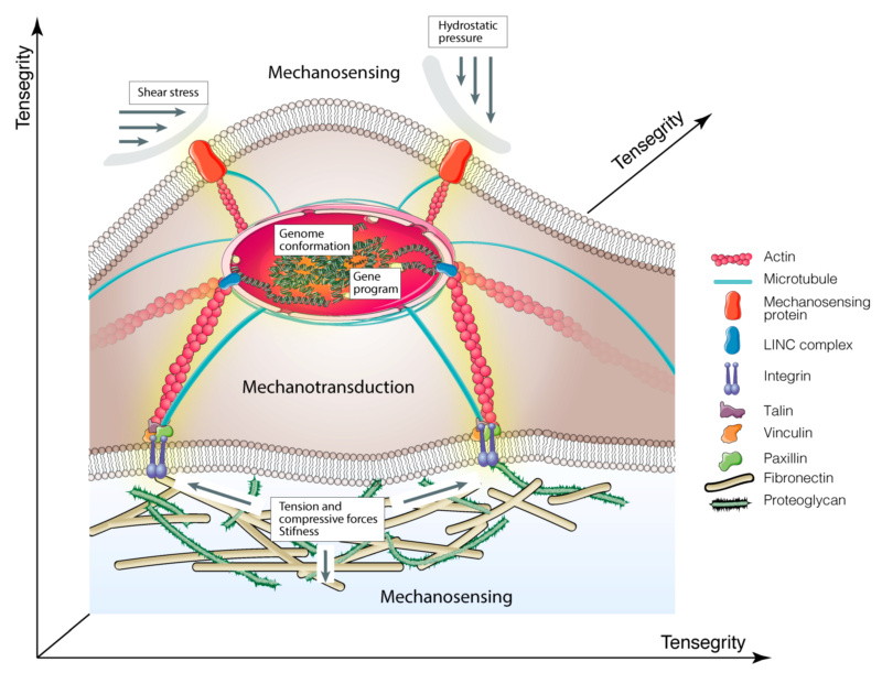
Schematic illustration of molecular basis of mechanobiology.
Cartoon shows how mechanical cues are transmitted to the nucleus via integrins > focal adhesion complex > cytoskeletal components > nucleoskeleton. The yellow shadow indicates mechanotransduction signals.

Tensegrity
Currently, the theoretical explanation of mechanobiology is based on the discovery that, in all cells, the cytoskeleton acts as a dynamic machine that collects the external forces applied to the cell from the microenvironment and responds by generating traction/compression forces transmitted to other molecular components inside or outside the cells. This model is based on the concept of “tensegrity” (tensional integrity), by which living cells organize their cytoskeleton as a hard-wired that immediately responds to external mechanical stresses stabilizing its form.
Tensegrity is a building principle, being originally described by the architect R.B. Fuller and pictured by the sculptor K. Snelson. While Fuller defined a tensegrity system “as structures that stabilize their shape by continuous tension or ‘tensional integrity’, rather than by continuous compression”, Snelson demonstrated that network structures may mechanically stabilize themselves through the use of tensile pre-stress forces. In 1993, D. Ingber applied the term “tensegrity” to living organisms, suggesting a mechanical model where the cytoskeleton structure acts as a dynamic load-bearing pillar. This model, which is capable of recapitulating the events leading to cytoskeletal mechanics, cell shape, and movement, allowed for explaining how cells sense and respond to mechanical forces and, above all, how these two events are connected. Hence, tensegrity predicts that cells respond straightaway to external mechanical stresses applied to the cells’ surface, through proteins that are physically connected to the cytoskeleton. Additionally, in this mechanical model, molecules that are activated by changes in cytoskeletal architecture function in the “solid-state” and transduce mechanical stresses into biochemical signals and gene expression program within single living cells. Therefore, all living organisms use “tensegrity” to mechanically stabilize their shape and integrate and balance their structure and function at all size scales, from the molecular level to organs. This is a consequence of cytoskeleton tension that is transduced into an equilibrium of opposing forces that are dispersed through the network of cytoskeletal filaments. Generally, tension is generated within the actomyosin contractile microfilaments and is counteracted by microtubules, which are able to resist the compression forces. The cytoskeletal components are nonlinear cell mechanical supports, introduced the concept of “initial imperfections” in the original tensegrity model. This scheme provided a new intuitive method for understanding the load-bearing capacity and distribution of force into the cytoskeleton.

Mechanosensing
As mentioned above, all organisms have structures, enabling them to recognize and respond to mechanical forces. This cross-talk takes place at the macroscale level (e.g., in organs and tissues), at the microscale level (e.g., in single cells), and also at the nanoscale level (e.g., in molecular complexes or single proteins). At present, we know that the different types of forces orchestrate the control of all biological functions, including stem cells’ commitment, determination, development, and maintenance of cells and tissues homeostasis. Table 1 summarizes the different mechanical properties and the proteins serving as transmitters in mediating these processes.


Mechanical properties and biological mediators

TensionTensile forces refer to the external stimuli that tend to stretch cells, acting in opposite directions, thus causing their elongation. Cellular responses to stretching depend largely on the type and amount of load as well as on the composition of the extracellular matrix.Myosin II, integrins, FAK, F-actin, Ifs, ZO-1, E-cadherin, Lmn A/C, Arp2/3, formin, coronin 1B, a-catenin, vinculin, collagens, elastin, fibrillin, fibulin, tenascin-C, pacsin-2, F-actin, microtubules[26,27,28,29,30]
CompressionContrary to tension, compressive forces applied from the outside towards the centre of cells result in cells contraction and shortening.Collagen, vimentin, F-actin ROCK, myosin regulatory light chain, Wnt/β-catenin[29]
Shear StressWhen two opposite forces are tangentially applied to cells surface, they generate shear stress, which cause changes in morphology and adhesion properties.PECAM1, VEGFR, ERK1/2, PGTS2, IER3, EGR1, IGF1, IGFBP1, Integrin, TGF-β, β-catenin, MAPK, laminin-5, F-actin, PI3K[31,32]
Hydrostatic PressureHydrostatic pressure is the force exercised by the surrounding fluid to cells membranes. Due to its nondirectional nature, it is mainly non-deforming but has an important thermodynamic effect on the cytoskeleton influencing microtubule stability.Shc1, integrins, collagen, TGF-β, F-actin[32]
StiffnessThe term stiffness, which generally is used to describe the ability of an object to resist deformation after the application of a force, is also a measure of the rigidity of the extracellular matrix or the cells were those forces are applied.Integrin (α2), fibronectin, collagens, α-actinin, Rho signaling cascade, talin vinculin, FAK, BMP receptor, F-Actins, vimentin Ifs, microtubules, filamin, lamin-A/C, emerin, Yap1[28,33,34]
ElasticityElasticity is the property of the object to complement its original shape and size after removal of the applied force. In biology is the resistance of cells to the extracellular matrix deformation.Collagen VI, tenascins, titin, elastin, fibrillins, integrins, F-Actins, microtubules, Myosin II[33]
ViscoelasticityIt indicates the elastic and viscous properties by which an object contrasts the deformation.Collagens, Elastin, ICAM-1, F-Actins[35,36]

https://reasonandscience.catsboard.com

Otangelo


Admin

Extracellular Matrix

Chemical, mechanical, and topographical cues of ECM control cell adhesion, shape, and migration, as well as the activation of signal transduction pathways orchestrating gene expression and dictating proliferation and stem cells’ fate. The ECM is a structural macromolecular network that creates a scaffold for cells interactions and support. It is composed of (i) solid components, consisting of fibrous proteins, (e.g., collagen, elastin, laminin, fibronectin), glycosaminoglycans (GAGs; e.g., hyaluronic acid), proteoglycans (PGs; e.g., chondroitin sulfate, heparan sulfate, keratan sulfate), and syndecans; 

Solid Components Proteins
Collagens are the main structural glycoproteins of ECM. They interact with other ECM components and cellular integrins and exist as fibrils of 10-300 nm in diameter (e.g., types I, II, III) and reticular forms (e.g., type IV). Fibrils transmit tensile strength originated by mechanical stresses, tension, pressure and shear while type IV collagen is bound to the other ECM structural components such as laminin and fibronectin (to form the basal lamina of basement membranes).
Fibronectin is the major dimeric fibrillar glycoprotein of ECM. It interacts with other ECM proteins, cellular membrane integrins, glycosaminoglycans, and other fibronectin molecules.
Elastin/Tropelastin is a hydrophobic protein rich in glycine and proline. The soluble precursor tropoelastin is secreted into the extracellular space where then polymerize into insoluble elastic fibers or sheets. Elastic fibers guarantee flexibility to the structures, which can go towards withdrawal after a temporary stretch. Elastin interacts with the cellular integrins and with several ECM components (e.g., collagens, laminin, fibrillin, proteoglycans, glycosaminoglycans).
Laminins are high-molecular-weight heterotrimeric glycoproteins formed by α, β andγ subunits that combine to form 15 different types of heterotrimers. They represent the main non-collagenous components of the basal membrane.
Other proteins: vitronectin, tenascins, nidogens, fibulins, trombospondins.
Glycosaminoglycans (GAGs)
Hyaluronic acid is a polysaccharide consisting of alternating residues of D-glucuronic acid and N-acetylglucosamine. It is absent in proteoglycan. It confers the ability to resist compression through swelling by absorbing water. Hyaluronic acid regulates cell during embryonic development, inflammation, healing processes and tumor development.
Proteoglycans
Chondroitin sulphate is involved in compression of ECM. It contributes to the tensile strength of cartilage, tendons, ligaments, and affects neuroplasticity.
Heparin/Heparan sulphate is involved in cell adhesion, migration and proliferation, developmental processes, angiogenesis, blood coagulation and tumor metastasis. It serves as a cellular receptor for a number of viruses.
Dermatan sulphate interacts with different cell receptors and with other ECM components (e.g., collagen, tenascin, fibronectin, GAGs, and other proteoglycans).
Keratan sulphate regulates the diameter of the fibrils in ECM and regulates interfibrillar spacing. It interacts with many proteins of the neural tissues and with collagen, glycosaminoglycans, and proteoglycans.
Syndecans
The syndecan protein family has four members that have a single transmembrane domain that act as coreceptors. These core proteins contain three to five heparan- and chondroitin-sulfate chains, which allow the interaction with different growth factors, fibronectin and antithrombin-1.
[43,71,72,73,74,75,76,77,78,79,80,81,82,83,84,85,86]
Soluble components
Cytokines: TNF-a, IL-7, IL-2, CCL5, MIP-1b
Growth factors: VEGF, FGFR1, PDGF, TGF-α, TGF-β, bFGF, IGF-1 ecc.
Matrix metalloproteinases and proteases: adamalysins, serralysins, astacins and metzincin superfamily.
[87,88,89]
Integrins
Integrins are the main transmembrane proteins that established cell-ECM interaction. They are heterodimers of α and β subunits. In mammals there are 18 α and 8 β subunits that associate to form 24 integrins that have affinity for different ligands. They have a large extracellular domain that links to ECM proteins and a cytoplasmic tail that bind to the cytoskeleton proteins.[90,91]
Focal Adhesion (FA)Proteins
Vinculin is the main protein of the FA complex. It is involved in the connexion of integrins with F-actin. Vinculin is involved in the association of cell-cell and cell-matrix junctions and is also critical in controlling the cell spreading, cytoskeletal mechanics, and lamellipodia formation. Therefore, vinculin has an essential role in controlling focal adhesions structure and function.
Paxillin binds tubulin and targets focal adhesions through its C-terminal region, which is composed of double zinc finger LIM domains organized in four tandems.
Talin interact with vinculin and paxillin and exists in two isoforms, talin1, ubiquitously expressed, and talin2 (striated muscle and brain). The N-terminal FERM domain have three subdomains: F1, F2, and F3. The latter contains the binding site for integrin β tails and is enough to activate integrins.
Focal adhesion kinase (FAK). The C-terminal region contains the FAT (focal adhesion targeting) domain for the binding with proteins of the focal adhesion complex. The N-terminal domain interacts with the β1 subunit of integrins and is involved in the transduction of signals from ECM.
Other proteins: p130Cas, zyxin, tensin, tindlins, Ena/VASP family, Arp2/3 complex.
[62,92,93,94]
Adherens Junctions (AJs)
Cadherins (N-cadherin, E-cadherin, P-cadherin, T-cadherin, V-cadherin). Cadherins or “calcium-dependent adhesion” proteins belong to the cell adhesion molecule (CAM) family and are involved in the formation of AJs and mediate cell-to-cell contact. During development, they are essential for the proper positioning of the cells. This includes the separation of the different tissue layers and cell migration. The transmembrane domain contains five repetitions in tandem that allow the binding of Ca2+ ions while the extracellular domain mediates the connexion between adjacent cells. In fact, a cadherin interacts with another cadherin of the same type on the adjacent cell in an anti-parallel conformation, creating a linear adhesive “zipper” between cells. The C-terminal cytoplasmic ends, mediate the binding to catenins, which in turn interact with the actin cytoskeleton.
β-catenin (Catenin beta-1) is a multifunctional protein involved in the transduction of Wnt signals and in the intercellular processes of adhesion by linking the cytoplasmic domain of cadherin.
α-catenin binds cadherins and F-actin. Moreover, α-catenin recruit vinculin.
Other proteins: l-afadin, p120, EPLIN (also known as Lima-1), ZO-1, nectins.
[95,96,97,98,99,100]
Cytoskeleton
Microtubules are polymers of α-tubulin and β-tubulin dimers that form protofilaments, which are then associated laterally (13 protofilaments) to form a hollow tube diameter of about 25 nm. Microtubules are essential for determining cell shape and movement, intracellular transport of organelles and the formation of mitotic spindle. The dynamic activity of microtubules is under the control of microtubule-associate proteins, which increase their stability or disassembling, separation and increasing the rate of tubulin depolymerization.
F-Actin microfilaments are polymers of G-actin monomers. F-actin fibers (diameter of about 7 nm) generate networks that regulate cellular shape and are directly involved in the generation of forces, cell migration and division. Actin filaments end at the plasma membrane, where they form a network of philopodia and lamellipodia that provide mechanical support to cells. Moreover, the activity of F-actin is strictly assisted by many actin-binding proteins.
Intermediate filaments have a diameter of about 10 nm, have a structural role and provide mechanical strength to cells. They organize and participate to the three-dimensional structure of the cell and nucleus, and serves as anchor to organelles. Moreover, they contribute to some cell-to-cell and cell-to-matrix junctions. Intermediate filaments belong to vimentins, keratin, neurofilaments, lamins and desmin families.
Actin-Linking- Proteins
Myosin II is a motor protein that associate with F-actin generating both extensile and compressive forces that push and pull actin filaments by hydrolysis of ATP.
α-actinin is a member of the spectrin superfamily. It forms an anti-parallel rod-shaped dimer by which binds both actin- domain at each end and bundles actin filaments at rod-end.
Filamins family serve as scaffolds for more than 90 partners (e.g., channels, receptors, transcription factors) through its immunoglobulin-like domains. Filamin binds all actin isoforms (e.g., F-actin, G-actin). It forms a flexible bridge between two actin filaments generating an actin network with movable or gel-like qualities with increased elastic stiffness depending of the critical concentration of filamin.
Cofilin protein has emerged as a key regulator of actin dynamics. In particular, it regulates the actin filament assembly/disassembly by binding to actin monomers and filaments.
Other proteins: Arp2/3, fascin, spectrin, profilin, fimbrin (also known as is plastin 1), formins, villin
[101,102,103,104,105,106,107,108,109,110]
Nucleoskeleton
LINC complex.
SUN1 and SUN2 are transmembrane proteins of the inner nuclear membrane with a conserved C-terminal SUN domain that localize to the perinuclear space.
Nesprins contain the conserved KASH domain at transmembrane C-terminal tail by which bind SUN proteins. KASH–SUN bridges interact with the cytoskeleton and therefore respond to the forces generated by the cytoskeleton.
Lamins
Lamin A/C are intermediate filaments that ensure the nuclear architecture. They have a role in nuclear assembly, genome organization and telomere dynamics. Lamin A responds to the cytoskeletal tension and interacts with numerous proteins involved in transduction pathways. Lamin A/C expression is lower in stem cells and increases in differentiated stem cells.
Lamin B1/B2 are components of the nuclear lamina, form an outer rim and interact with chromatin. Lamin B is expressed in all cells.
Other proteins LAP2, BAF.


(ii) soluble components, such as cytokines, growth factors, and several classes of proteases, like a metalloproteinases (see Table above), all of which serve as mediators between ECM and cells. Based on composition and structural organization, it is possible to distinguish two extensive ECM structures: the basement membranes, providing a two-dimensional support for the cells (mainly composed of laminin, collagen IV, nidogen and heparan sulphate) and the connective tissues that provide a fibrous three-dimensional scaffold to the cells that is mainly composed of fibrillar collagens, PGs, and GAGs.

https://reasonandscience.catsboard.com

Otangelo


Admin

The overall components confer topography, viscosity, and mechanical properties to ECM. In particular elastic fibers, fibrillar collagens, GAGs, and the related PGs provide the mechanical properties of ECM, while fibrous proteins provide tensile strength (collagens, elastin). Therefore, based on the composition, ECM has the characteristics of a “soft material”, easily deformable at low stresses, or of an “hard material”, which require greater stresses to generate deformation. Interestingly, it seems that the resulting architecture provides a sort of ‘mechanical memory’, correlating with stem cells’ differentiation toward selected lineages.

According to its composition, ECM might also acquire a peculiar geometrical conformation providing topographic and mechanical stimuli, which are critical in modulating stem cells’ phenotype.
Notably, between ECM and stem cells exists a dynamic cross-talk, as stem cells may change the ECM composition and remodel the architecture either by the secretion of ECM structural components and matrix metalloproteinases, or by exerting mechanical forces through the cytoskeleton fibers. The challenge is to create a suitable cell microenvironment that generates mechanosensing/mechanotransduction signals and guide stem cells’ functions.
It is not surprising that alterations in specific ECM components or in regulatory players could have an impact on biochemical and physical properties of ECM, which leads to a disorganized network and, eventually, to organ dysfunctions. In particular, abnormal ECM composition has consequences on its mechanical properties and on the onset and progression of numerous diseases, such as cancer and fibrosis.

Development
The developing of embryos consists of stem cell aggregation and organization into increasingly more complex structures and internal and external mechanical forces determine these events. Furthermore, the correct positioning of stem cells during morphogenesis is guaranteed by the appropriate establishment of mechanical interactions among them and with their microenvironment ECM. The first evidence of the role that mechanics play in developmental processes came from non-mammalians, such as Drosophila, avians, amphibians, and fish, in which became clear how eggs fertilization and maturation strongly depend on osmotic pressure gradients that influence cells shape. On the other hand, significantly less is known about the influence of mechanical forces in the development of human embryos inside uterus because of the limited amount of material available for experimentations on animal models used for recapitulating human physiology and because of ethical restrictions regarding human embryos manipulation. Therefore, much of our knowledge on this topic relies on observations on close primate species or on archival material. Human embryonic mechano-signalling have been recently characterized. These include i) key regulator genes; ii) body axes establishment and local strains; iii) geometrical influence on cell populations sorting; and, iv) embryonic architecture and early signalling gradients. Hydrostatic pressure (HP) is one of the mechanical forces involved in embryogenesis (both in early and late phases of development). At the blastocyst stage, the internal HP dictates the right definition of cell fate and embryonic size, while in later stages the pressure applied by the amniotic fluid appears to guide notochord extension by stimulating the underlying mesoderm.At present, in-vitro models of embryogenesis seem to be the only tool for effectively understanding the processes regulating patterning, morphogenesis, and mechanobiology in the peri-implantation human embryo, as far as progresses in the possibility of working with human embryos are made. Nevertheless, it will be necessary to wait more precise characterization of the embryos that they are expected to model, especially given that benchmarks based on mouse biology may not hold true in human, in order to understand if these models accurately recapitulate the molecular events happening in-vivo.

https://reasonandscience.catsboard.com

Sponsored content


Back to top  Message [Page 1 of 1]

Permissions in this forum:
You cannot reply to topics in this forum