ElShamah - Reason & Science: Defending ID and the Christian Worldview
Would you like to react to this message? Create an account in a few clicks or log in to continue.
ElShamah - Reason & Science: Defending ID and the Christian Worldview

Welcome to my library—a curated collection of research and original arguments exploring why I believe Christianity, creationism, and Intelligent Design offer the most compelling explanations for our origins. Otangelo Grasso


You are not connected. Please login or register

Centriole/Centrosome: The centriole spindle  the most complex machine known in nature

Go down  Message [Page 1 of 1]

Otangelo


Admin

Centriole/Centrosome: The centriole spindle  the most complex machine known in nature

Centrosome 1
Centriole/Centrosome: The centriole spindle  the most complex machine known in nature Centro11

In cell biology, the centrosome (Latin centrum 'center' + Greek sōma 'body') is an organelle that serves as the main microtubule organizing center (MTOC) of the animal cell as well as a regulator of cell-cycle progression.  The centrosome is thought to have evolved only in the metazoan lineage of eukaryotic cells. Fungi and plants lack centrosomes and therefore use other MTOC structures to organize their microtubules. Although the centrosome has a key role in efficient mitosis in animal cells, it is not essential.
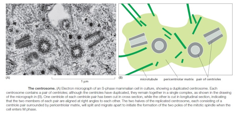
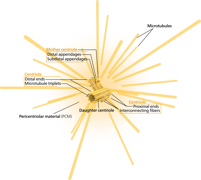
Centrosomes are composed of two orthogonally arranged centrioles surrounded by an amorphous mass of protein termed the pericentriolar material (PCM). The PCM contains proteins responsible for microtubule nucleation and anchoring including γ-tubulin, pericentrin and ninein. In general, each centriole of the centrosome is based on a nine triplet microtubule assembled in a cartwheel structure, and contains centrin, cenexin and tektin.

Functions

Role of the centrosome in cell cycle progression
Centrosomes are associated with the nuclear membrane during prophase of the cell cycle. In mitosis the nuclear membrane breaks down and the centrosome nucleated microtubules (parts of the cytoskeleton) can interact with the chromosomes to build the mitotic spindle.
The mother centriole, the older of the two in the centriole pair, also has a central role in making cilia and flagella.

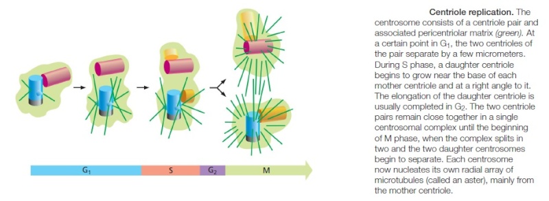
The centrosome is copied only once per cell cycle so that each daughter cell inherits one centrosome, containing two structures called centrioles

Centriole/Centrosome: The centriole spindle  the most complex machine known in nature Centrosome_Cycle

Centrosome Duplication

Cell Cycle Regulation of Centrosome Duplication

Centrosome duplication is heavily regulated by cell cycle controls. This link between the cell cycle and the centrosome cycle is mediated by cyclin-dependent kinase 2 (Cdk2). There has been ample evidence that Cdk2 is necessary for both DNA replication and centrosome duplication, which are both key events in S phase. It has also been shown  that Cdk2 complexes with both cyclin A and cyclin E and this complex is critical for centrosome duplication. Three Cdk2 substrates have been proposed to be responsible for regulation of centriole duplication. These include: nucleophosmin (NPM/B23), CP110, and MPS1.[3] Nucleophosmin is only found in unreplicated centrosomes and it’s phosphorylation by Cdk2/cyclin E removes NPM from the centrosomes, initiating procentriole formation. CP110 is an important centrosomal protein that is phosphorylated by both mitotic and interphase Cdk/cyclin complexes and is thought to influence centrosome duplication in S phase.  MPS1 is a protein kinase that is essential to the spindle assembly checkpoint, and may remodel an SAS6-cored intermediate between severed mother and daughter centrioles into a pair of cartwheel protein complexes onto which procentrioles assemble.

http://www.ncbi.nlm.nih.gov/pmc/articles/PMC3947860/


Last edited by Admin on Wed Aug 29, 2018 2:27 am; edited 2 times in total

https://reasonandscience.catsboard.com

Otangelo


Admin

Microtubules emanate from the centrosome in Animal cells

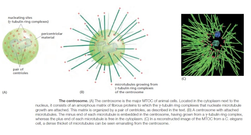
Microtubules Emanate from the Centrosome in Animal Cells. Many animal cells have a single, well-defined MTOC called the centrosome, which is located near the nucleus and from which microtubules are nucleated at their minus ends, so the plus ends point outward and continuously grow and shrink, probing the entire three-dimensional volume of the cell. A centrosome typically recruits more than fifty copies of γ-TuRC. In addition, γ-TuRC molecules are found in the cytoplasm, and centrosomes are not absolutely required for microtubule nucleation, since destroying them with a laser pulse does not prevent microtubule nucleation elsewhere in the cell. A variety of proteins have been identified that anchor γ-TuRC to the centrosome, but mechanisms that activate microtubule nucleation at MTOCs and at other sites in the cell are poorly understood.
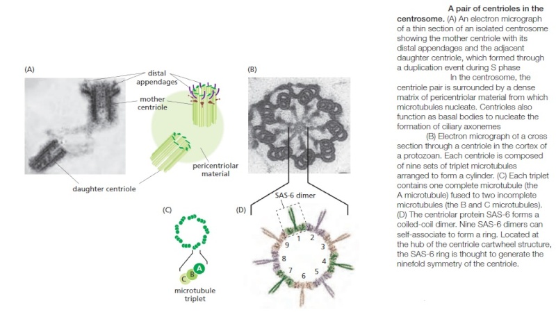
Embedded in the centrosome are the centrioles, a pair of cylindrical structures arranged at right angles to each other in an L-shaped configuration

Centriole/Centrosome: The centriole spindle  the most complex machine known in nature Sfsfsd10

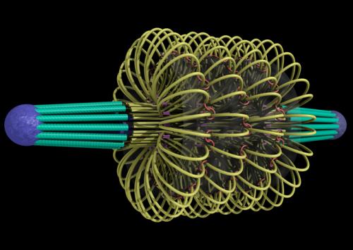
A centriole consists of a cylindrical array of short, modified microtubules arranged into a barrel shape with striking ninefold symmetry

Centriole/Centrosome: The centriole spindle  the most complex machine known in nature Sfsfsd11

Together with a large number of accessory proteins, the centrioles organize the pericentriolar material, where microtubule nucleation takes place. the centrosome duplicates and splits into two parts before mitosis, each containing a duplicated centriole pair. The two centrosomes move to opposite sides of the nucleus when mitosis begins, and they form the two poles of the mitotic spindle  Microtubule organization varies widely among different species and cell types. In budding yeast, microtubules are nucleated at an MTOC that is embedded in the nuclear envelope as a small, multilayered structure called the spindle pole body, also found in other fungi and diatoms. Higher-plant cells appear to nucleate microtubules at sites distributed all around the nuclear envelope and at the cell cortex. Neither fungi nor most plant cells contain centrioles. Despite these differences, all these cells seem to use γ-tubulin to nucleate their microtubules. In cultured animal cells, the aster-like configuration of microtubules is robust, with dynamic plus ends pointing outward toward the cell periphery and stable minus ends collected near the nucleus. The system of microtubules radiating from the centrosome acts as a device to survey the outlying regions of the cell and to position the centrosome at its center. Even in an isolated cell fragment lacking the centrosome, dynamic microtubules arrange themselves into a star-shaped array with the microtubule minus ends clustered at the center by minus-end-binding proteins .This ability of the microtubule cytoskeleton to find the center of the cell establishes a general coordinate system, which is then used to position many organelles within the cell. Highly differentiated cells with complex morphologies such as neurons, muscles, and epithelial cells must use additional measuring mechanisms to establish their more elaborate internal coordinate systems. Thus, for example, when an epithelial cell forms cell–cell junctions and becomes highly polarized, the microtubule minus ends move to a region near the apical plasma membrane. From this asymmetrical location, a microtubule array extends along the long axis of the cell, with plus ends directed toward the basal surface

Centriole/Centrosome: The centriole spindle  the most complex machine known in nature Ssfsds11

The Mitotic Spindle Is a Microtubule-Based Machine

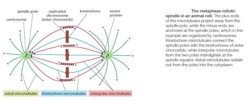
The central event of mitosis—chromosome segregation—depends in all eukaryotes on a complex and beautiful machine called the mitotic spindle. The spindle is a bipolar array of microtubules, which pulls sister chromatids apart in anaphase, thereby segregating the two sets of chromosomes to opposite ends of the cell, where they are packaged into daughter nuclei. M-Cdk triggers the assembly of the spindle early in mitosis, in parallel with the chromosome restructuring just described. Before we consider how the spindle assembles and how its microtubules attach to sister chromatids, we briefly review the basic features of spindle structure. The core of the mitotic spindle is a bipolar array of microtubules, the minus ends of which are focused at the two spindle poles, and the plus ends of which radiate outward from the poles.

Centriole/Centrosome: The centriole spindle  the most complex machine known in nature Centro15

The plus ends of some microtubules called the interpolar microtubules—overlap with the plus ends of microtubules from the other pole, resulting in an antiparallel array in the spindle midzone. The plus ends of other microtubules—the kinetochore microtubules—are attached to sister-chromatid pairs at large protein structures called kinetochores, which are located at the centromere of each sister chromatid.

Finally, many spindles also contain astral microtubules that radiate outward from the poles and contact the cell cortex, helping to position the spindle in the cell. In most somatic animal cells, each spindle pole is focused at a protein organelle called the centrosome . Each centrosome consists of a cloud of amorphous material (called the pericentriolar matrix) that surrounds a pair of centrioles . The pericentriolar matrix nucleates a radial array of microtubules, with their fast-growing plus ends projecting outward and their minus ends associated with the centrosome. The matrix contains a variety of proteins, including microtubule-dependent motor proteins, coiled-coil proteins that link the motors to the centrosome, structural proteins, and components of the cell-cycle control system. Most important, it contains γ-tubulin ring complexes, which are the components mainly responsible for nucleating microtubules. Some cells—notably the cells of higher plants and the oocytes of many vertebrates—do not have centrosomes, and microtubule-dependent motor proteins and other proteins associate with microtubule minus ends to organize and focus the spindle poles.


Centriole/Centrosome: The centriole spindle  the most complex machine known in nature Sfsfsd12

Centriole/Centrosome: The centriole spindle  the most complex machine known in nature Sfsfsd13

1) https://en.wikipedia.org/wiki/Centrosome
2) http://www2.lbl.gov/Science-Articles/Archive/sabl/2007/Oct/onering.html

further readings:

http://jcb.rupress.org/content/195/6/993.full

https://reasonandscience.catsboard.com

Otangelo


Admin

The centriole spindle  the most complex machine known in nature 1

Centriole and PI

Centriole/Centrosome: The centriole spindle  the most complex machine known in nature Mitoti12

The centriole spindle has been described as the most complex machine known in nature. It orchestrates the dividing of the cell and is made up of nine triplets of microtubules similar to the structure of the base of the PI.  Recently, many relationships have been found between the centriole and the PI.

The mitotic spindle isolates and separates chromosomes during cell division. In the human being it consists of 4000 different microtubules with orchestrated movements. It is the most complex process known in biology. It involves many different motors and pumps.

Recently, it was discovered that a membrane made of protein from the PI is placed in a sac and travels with the centriole spindle as a small vesicle when the cell divides. The sac that has the piece from the PI is passed onto one cell and it then has the same molecules to create the exact form and structure of the mother PI in the new cell. Could the PI microtubule structure that is passed on to the next generation have information somehow stored in the microtubules.

Centriole/Centrosome: The centriole spindle  the most complex machine known in nature Metoch10

Another strange connection is that the same molecules that are at the site upon which cilia microtubules grow are also in the centriole. Both the cilium base and the spindle use the same microtubule structure of nine triplets. The same process of cilia microtubules is part of the process of cell division. Also, receptors from the PI membrane, serotonin and somatostatin, occurs in both.

As several other biologists have shown, the  centrioles that compose the centrosomes replicate independently of DNA replication: daughter centrioles receive their form from the overall structure of the mother centriole, not from the individual  gene products that constitute them


1) http://jonlieffmd.com/blog/is-the-primary-cilium-a-cells-antenna-or-its-brain

more readings:

file:///E:/Downloads/information-03-00344.pdf


Last edited by Admin on Tue Apr 04, 2017 8:45 pm; edited 1 time in total

https://reasonandscience.catsboard.com

Otangelo


Admin

An Evolutionary Origin of the Centrosome?

Jonathan Wells May 7, 2008 12:46 PM | Permalink

According to today's ScienceDaily, "New Evidence Suggests a Symbiogenetic Origin for the Centrosome."

But the evidence suggests no such thing. Instead, it points to the willingness of evolutionary biologists to believe just-so stories, and to the ideological corruption of the National Institutes of Health and the Proceedings of the National Academy of Sciences USA.
Centrosomes ("central bodies") are fascinating organelles. In undividing eukaryotic cells one is located next to the nucleus, where it serves as the organizing center for microtubules that make up the "cytoskeleton." The cytoskeleton gives the cell its shape and serves as the highway system along which many intracellular components are transported to their proper locations. (See the cell animation sequence in the movie "Expelled.") Just before cell division the centrosome duplicates, and the two centrosomes then form the poles of the cell division "spindle," a very complex apparatus composed of microtubules that emanate from the centrosomes. The spindle distributes chromosomes to the daughter cells, each of which also inherits one centrosome.

Even though centrosomes control many features of the cell (such as its morphology and the process of division), they are of much less interest to neo-Darwinists than cell nuclei, because centrosomes contain no DNA [1], so they cannot provide the DNA mutations that are assumed to be the raw materials for evolution. Thus many aspects of centrosomes, including their chemical composition, structure, function, and mode of replication--not to mention their origin--are (in the jargon of we-now-know-almost-everything Darwinists) "poorly understood."

Along come Mark and Mary Anne Alliegro of the Marine Biological Laboratory in Woods Hole, Massachusetts. In the abstract of an article published online May 5 by the Proceedings of the National Academy of Sciences USA (the complete article is not yet available, except to PNAS subscribers), the Alliegros report that some RNAs they extracted from surf clams are "centrosome-associated transcripts representing a structurally unique intron-poor collection of nuclear genes." Since a subset of these RNAs "contain functional domains that are highly conserved across distant taxa," the Alliegros conclude that they may "serve as cytological progenitors of the centrosome and may support a symbiogenetic model for its evolution."

Wait a minute. As the ScienceDaily press release explains, "only two cellular components--the mitochondria and the chloroplasts--are generally accepted by evolutionary biologists as having a symbiogenetic origin." This is because only mitochondria and chloroplasts (as far as we know) contain DNA that is inherited independently of the nuclear DNA. Since these tiny organelles contain DNA and look a bit like bacteria, the hypothesis of symbiogenesis asserts that they were once free-living prokaryotes that were engulfed by other prokaryotes to form eukaryotic cells. The hypothesis has lots of evidentiary problems, but whatever plausibility it seems to have with regard to mitochondria and chloroplasts completely evaporates in the case of centrosomes.

The problem is not just that centrosomes contain no DNA. The most solidly established function of centrosomes is that they serve as the organizing centers for microtubules, but microtubules occur only in eukaryotes, not in prokaryotes. Furthermore, centrosomes in animal cells contain two turbine-like centrioles oriented at a right angle to each other. Nobody knows for sure what centrioles do, nor why they occur in orthogonal pairs, and their origin is a complete mystery. There is no evidence that centrosomes or centrioles ever existed -- or ever could have existed -- in anything other than eukaryotic cells. The idea that free-living centrosome-like organisms were once engulfed by other primitive organisms is about as implausible as you can get, and the implausibility is not lessened by the presence in centrosomes of RNAs "that are highly conserved across distant taxa."

In this case, evolutionary jargon has taken the place of clear thinking. Yet the National Institutes of Health supported this medically useless speculation with our tax dollars, and the Proceedings of the National Academy of Sciences USA elevated it to the status of "peer-reviewed" science--part of the "overwhelming evidence" for evolution.


Centrioles: Elegant Cartwheels Come Into Focus 4

Centrioles are organelles involved in cell division and the construction of cilia and flagella. How they work is unknown, but using new imaging techniques, a research team has unveiled their complex structure in more detail than ever before.

You can't look at an electron micrograph of a centriole in cross-section and not be struck by its beautiful radial symmetry -- a highly ordered structure that really stands out from the background. Constructed of rod-like microtubules, most centrioles have a nine-fold pattern, nine triplets or doublets evenly spaced at the rim, giving it a "cartwheel" appearance in cross-section. The side view shows it to be a cylinder, about 500 nm long and 250 nm wide. In the centrosome, two centrioles reside at right angles to each other, connected at one end by fibers.

These architecturally perfect structures are essential in many animal cells and plants (though not in flowering plants or fungi, or in prokaryotes). They help organize the centrosomes, whose spindles of microtubules during cell division reach out to the lined-up chromosomes and pull them into the daughter cells. They also form the basal bodies of cilia and eukaryotic flagella. They help establish the left-right axis during embryonic development in animals. Interestingly, they undergo their own division process apart from DNA replication. Centrioles are "highly conserved" from protists to humans. Mutations in centrioles are implicated in some serious diseases, if the embryo survives at all.


The basic cross-sectional structure of a centriole has been known since the late 1800s, but new features continue to come to light. Now, a team from Switzerland and Japan, reporting in Current Biology, has revealed this wonderful structure in unprecedented detail using cryotomography. It's like going from seeing an out-of-focus motorcycle wheel from a distance to seeing it up close and noticing all kinds of new structures. The pictures are worth a thousand words -- that is one intricate device!

They found new structures and gave names to them: the pinhead, the pinfeet, the A-C linker, the Cartwheel Inner Densities (CID), and more. One of their findings was that the central hub has the same 9-fold radial symmetry as the spokes that link to the rim triplets. Another is that the entire structure has some polarity or chirality; if you turned the centriole over, the inside wouldn't orient the same way. Some of the linkers are at angles along the radial axis.

The inner hub and spoke structure is really elegant. Stacks of 9-fold rings, each with their own linkers and "pinheads," merge as they extend out to the rim. The triplets at the end of each radial spoke appear just as precise, as if crafted in a machine shop.

It wasn't the goal of the authors to uncover the mechanism by which these machines operate. That's still somewhat of a mystery. How are they assembled? How do they duplicate themselves? How do they work? Discovery Institute's Dr. Jonathan Wells hypothesized years ago that centrioles operate like winches, pulling the chromosomes apart with force . This new paper did not address that question, but surely something this finely crafted has a great scientific story to tell. As for evolution, the only times the authors mentioned it, they referred to the "evolutionary conservation" of the overall architecture and individual parts.

Since the centriole appears fully formed in the simplest eukaryotic cells, and is "highly conserved" (not evolved) from microbes to humans, a design-theoretic approach to understanding its operation would surely prove more fruitful than the assumption that this marvel of craftsmanship "arose" through unguided processes.


Centrosome Attack: 1 Mitosis, or cell division, has been studied for many decades, but now another essential player has been identified.  Scientists from Japan and Pennsylvania describe what happened when they played “take off what you don’t need” with a centrosome protein named Su48:

The centrosome functions as the major microtubule-organizing center and plays a vital role in guiding chromosome segregation during mitosis.  Centrosome abnormalities are frequently seen in a variety of cancers, suggesting that dysfunction of this organelle may contribute to malignant transformation.  In our efforts to identify the protein components of the centrosome and to understand the structure features involved in the assembly and functions of this organelle, we cloned and characterized a centrosome-associated protein called Su48.  We found that a coiled coil-containing subdomain of Su48 was both sufficient and required for its centrosome localization.  In addition, this structure also modulates Su48 dimerization.  Moreover, ectopic expression of Su48 causes abnormal mitosis, and a mutant form of Su48 disrupts the localization of gamma-tubulin to the centrosome.  Finally, by microinjection of an anti-Su48 antibody, we found that disruption of normal Su48 functions leads to mitotic failure, possibly due to centrosome defects or incomplete cytokinesis.  Thus, Su48 represents a previously unrecognized centrosome protein that is essential for cell division.  We speculate that Su48 abnormalities may cause aberrant chromosome segregation and may contribute to aneuploidy and malignant transformation. [url=Characterization of Su48, a centrosome protein essential for cell division]2[/url]

Consider the problem this poses for neo-Darwinism.  Natural Selection depends on unfailing cell division – and not just any splitting of a cell into parts somewhere and somehow, but on the formation of highly accurate daughter copies of germline cells.  This is because (according to theory) only the daughter cells can preserve any beneficial variations produced by accident in the parent cell.  Otherwise, evolution comes to a sudden stop.
    As revealed in the last century, cell division is a highly complex process with numerous players, all of which have vital functions.  Scientists apparently did not even know about Su48, but without it, cell division doesn’t work.  So here is another extra in the play, like a nameless stage hand, without whom it’s curtains for the Darwin show.

1) http://creationsafaris.com/crev200604.htm
2) http://www.ncbi.nlm.nih.gov/pmc/articles/PMC1458915/
3) http://www.evolutionnews.org/2008/05/an_evolutionary_origin_of_the006309.html
4) http://www.evolutionnews.org/2013/08/centrioles_eleg075431.html

https://reasonandscience.catsboard.com

Otangelo


Admin

The Centrosome

http://creationsafaris.com/crev200611.htm

Centriole olé: Tiny devices called centrioles are vital to all life, because they duplicate each cell division and are intimately involved in it: “Centrioles are necessary for flagella and cilia formation, cytokinesis, cell-cycle control and centrosome organization/spindle assembly,” wrote 5 biologists in Nature 11/30.6 How the little machines duplicate themselves has been unclear. “Here we show using electron tomography of staged C. elegans [roundworm] one-cell embryos that daughter centriole assembly begins with the formation and elongation of a central tube followed by the peripheral assembly of nine singlet microtubules,” they announced. Various other proteins trigger, regulate, signal and terminate the process.
Their models of the centrioles resemble cylinders lined by equally-spaced rods on the outside. The shape can be discerned in the photographs. “The structure of centrioles is conserved [i.e., unevolved] from ancient eukaryotes to mammals,” they noted, saying also at the end of the paper, “It is therefore likely that some of the assembly intermediates uncovered here in C. elegans are conserved in mammals and other eukaryotes.”
As they reproduce, the daughter centrioles grow at a perpendicular angle to the mother. How this all happens is mysterious, but you can watch movies of these geometric structures emerging out of the cell matrix in the supplementary materials of the paper. The authors superimpose models of the centrioles to aid the visualization of a mechanical process just now coming into focus. To watch machinery 400 billionths of a meter in size assembling itself in a living cell is a harbinger of exciting days ahead for cell biology. For more on the lab roundworm C. elegans, visit our 06/25/2006 entry, and try counting the number of times “information” is used.


http://creationsafaris.com/crev200712.htm

A nine in time saves stitch: Centrosomes control the orientation of chromosomes before the split. They create a spindle of microtubules that line the pairs up at the midplane, then pull them apart. Within the centrosomes are two motors called centrioles, oriented perpendicular to one another, that look for all the world like turbines. The blades of the turbine are microtubules with spokes, forming a cylinder that looks like a pie with exactly nine slices. Why nine, and only nine?
Wallace Marshall (UC San Francisco) reviewed experiments into the mechanical basis for nine-ness in centrioles, and published a report in Current Biology.1 Experiments with mutants show that the number is controlled by the length of the spokes that emanate from each slice. This sets the overall diameter of the centriole, and thus the number of pie slices that will fit in the cylinder.
“This study provides an interesting geometrical mechanism by which a length can control a number,” Marshall said. Why was the research worthwhile? “Understanding centriole assembly is likely to reveal many more engineering-design principles that cells use to build complex structures.”

An Evolutionary Origin of the Centrosome?

According to today's ScienceDaily, "New Evidence Suggests a Symbiogenetic Origin for the Centrosome."

But the evidence suggests no such thing. Instead, it points to the willingness of evolutionary biologists to believe just-so stories, and to the ideological corruption of the National Institutes of Health and the Proceedings of the National Academy of Sciences USA.
Centrosomes ("central bodies") are fascinating organelles. In undividing eukaryotic cells one is located next to the nucleus, where it serves as the organizing center for microtubules that make up the "cytoskeleton." The cytoskeleton gives the cell its shape and serves as the highway system along which many intracellular components are transported to their proper locations. (See the cell animation sequence in the movie "Expelled.") Just before cell division the centrosome duplicates, and the two centrosomes then form the poles of the cell division "spindle," a very complex apparatus composed of microtubules that emanate from the centrosomes. The spindle distributes chromosomes to the daughter cells, each of which also inherits one centrosome.

Even though centrosomes control many features of the cell (such as its morphology and the process of division), they are of much less interest to neo-Darwinists than cell nuclei, because centrosomes contain no DNA [1], so they cannot provide the DNA mutations that are assumed to be the raw materials for evolution. Thus many aspects of centrosomes, including their chemical composition, structure, function, and mode of replication--not to mention their origin--are (in the jargon of we-now-know-almost-everything Darwinists) "poorly understood."

Along come Mark and Mary Anne Alliegro of the Marine Biological Laboratory in Woods Hole, Massachusetts. In the abstract of an article published online May 5 by the Proceedings of the National Academy of Sciences USA (the complete article is not yet available, except to PNAS subscribers), the Alliegros report that some RNAs they extracted from surf clams are "centrosome-associated transcripts representing a structurally unique intron-poor collection of nuclear genes." Since a subset of these RNAs "contain functional domains that are highly conserved across distant taxa," the Alliegros conclude that they may "serve as cytological progenitors of the centrosome and may support a symbiogenetic model for its evolution."

Wait a minute. As the ScienceDaily press release explains, "only two cellular components--the mitochondria and the chloroplasts--are generally accepted by evolutionary biologists as having a symbiogenetic origin." This is because only mitochondria and chloroplasts (as far as we know) contain DNA that is inherited independently of the nuclear DNA. Since these tiny organelles contain DNA and look a bit like bacteria, the hypothesis of symbiogenesis asserts that they were once free-living prokaryotes that were engulfed by other prokaryotes to form eukaryotic cells. The hypothesis has lots of evidentiary problems, but whatever plausibility it seems to have with regard to mitochondria and chloroplasts completely evaporates in the case of centrosomes.

The problem is not just that centrosomes contain no DNA. The most solidly established function of centrosomes is that they serve as the organizing centers for microtubules, but microtubules occur only in eukaryotes, not in prokaryotes. Furthermore, centrosomes in animal cells contain two turbine-like centrioles oriented at a right angle to each other. Nobody knows for sure what centrioles do, nor why they occur in orthogonal pairs, and their origin is a complete mystery. There is no evidence that centrosomes or centrioles ever existed -- or ever could have existed -- in anything other than eukaryotic cells. The idea that free-living centrosome-like organisms were once engulfed by other primitive organisms is about as implausible as you can get, and the implausibility is not lessened by the presence in centrosomes of RNAs "that are highly conserved across distant taxa."

In this case, evolutionary jargon has taken the place of clear thinking. Yet the National Institutes of Health supported this medically useless speculation with our tax dollars, and the Proceedings of the National Academy of Sciences USA elevated it to the status of "peer-reviewed" science--part of the "overwhelming evidence" for evolution.

http://www.discovery.org/scripts/viewDB/filesDB-download.php?command=download&id=490

http://www.nature.com/nature/journal/v444/n7119/full/nature05318.html
http://www.sdbonline.org/sites/fly/genebrief/sas-4.htm
http://www.ncbi.nlm.nih.gov/pmc/articles/PMC2115052/

further readings: http://www.ncbi.nlm.nih.gov/pmc/articles/PMC2835302/

https://reasonandscience.catsboard.com

Sponsored content


Back to top  Message [Page 1 of 1]

Permissions in this forum:
You cannot reply to topics in this forum