ElShamah - Reason & Science: Defending ID and the Christian Worldview
Would you like to react to this message? Create an account in a few clicks or log in to continue.
ElShamah - Reason & Science: Defending ID and the Christian Worldview

Otangelo Grasso: This is my personal virtual library, where i collect information, which leads in my view to the Christian faith, creationism, and Intelligent Design as the best explanation of the origin of the physical Universe, life, biodiversity


You are not connected. Please login or register

Cell Junctions and the Extracellular Matrix

Go down  Message [Page 1 of 1]

Otangelo


Admin

Cell Junctions and the Extracellular Matrix

http://reasonandscience.heavenforum.org/t2187-cell-junctions-and-the-extracellular-matrix

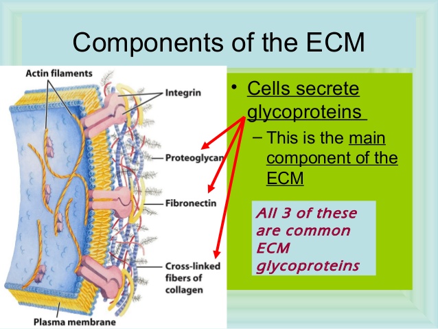
Of all the social interactions between cells in a multicellular organism, the most fundamental are those that hold the cells together. Cells may be linked by direct interactions, or they may be held together within the extracellular matrix, a complex network of proteins and polysaccharide chains that the cells secrete. By one means or another, cells must cohere if they are to form an organized multicellular structure that can withstand and respond to the various external forces that try to pull it apart. The mechanisms of cohesion govern the architecture of the body—its shape, its strength, and the arrangement of its different cell types. The making and breaking of the attachments between cells and the modeling of the extracellular matrix govern the way cells move within the organism, guiding them as the body grows, develops, and repairs itself. Attachments to other cells and to extracellular matrix control the orientation and behavior of the cell’s cytoskeleton, thereby allowing cells to sense and respond to changes in the mechanical features of their environment. Thus, the apparatus of cell junctions and the extracellular matrix is critical for every aspect of the organization, function, and dynamics of multicellular structures. Defects in this apparatus underlie an enormous variety of diseases. The key features of cell junctions and the extracellular matrix are best illustrated by considering two broad categories of tissues that are found in all animals.

Cell Junctions and the Extracellular Matrix Two_ma10

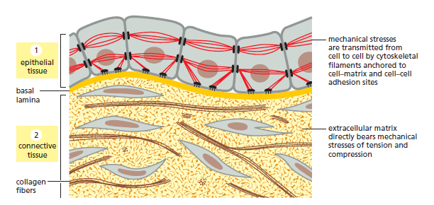
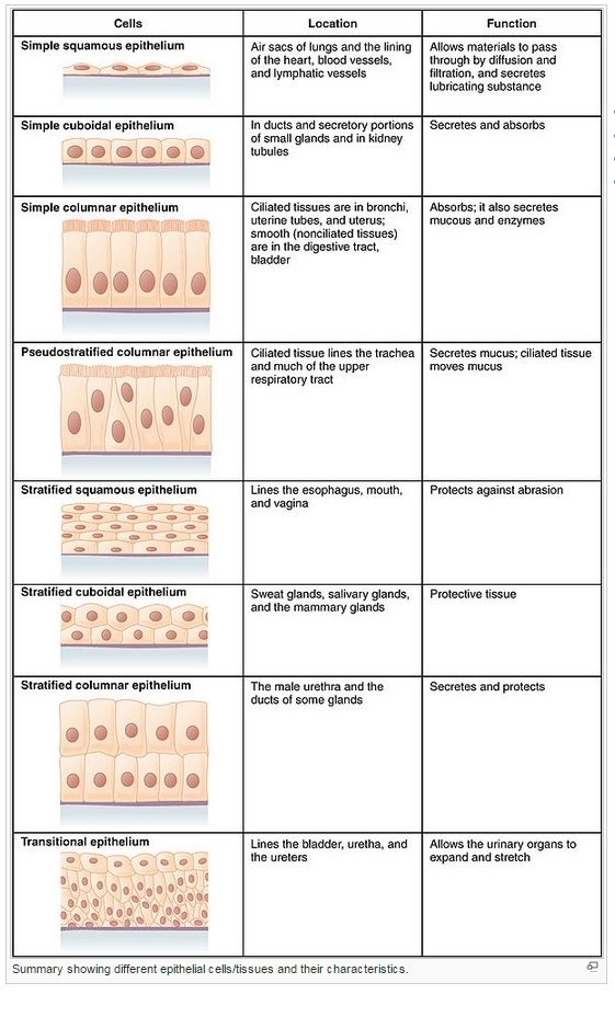
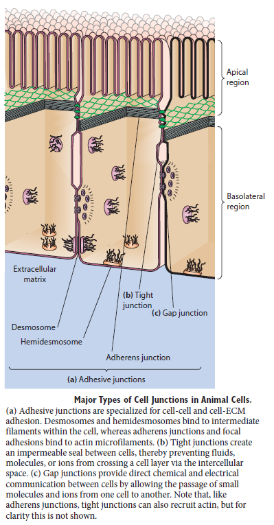
Connective tissues, such as bone or tendon, are formed from an extracellular matrix produced by cells that are distributed sparsely in the matrix. It is the matrix—rather than the cells—that bears most of the mechanical stress to which the tissue is subjected. Direct attachments between one cell and another are relatively rare, but the cells have important attachments to the matrix. These cell–matrix junctions link the cytoskeleton to the matrix, allowing the cells to move through the matrix and monitor changes in its mechanical properties. In epithelial tissues, such as the lining of the gut or the epidermal covering of the skin, cells are tightly bound together into sheets called epithelia. The extracellular matrix is less pronounced, consisting mainly of a thin mat called the basal lamina (or basement membrane) underlying the sheet. Within the epithelium, cells are attached to each other directly by cell–cell junctions, where cytoskeletal filaments are anchored, transmitting stresses across the interiors of the cells, from adhesion site to adhesion site. The cytoskeleton of epithelial cells is also linked to the basal lamina through cell–matrix junctions

Cell Junctions and the Extracellular Matrix Cell_j10

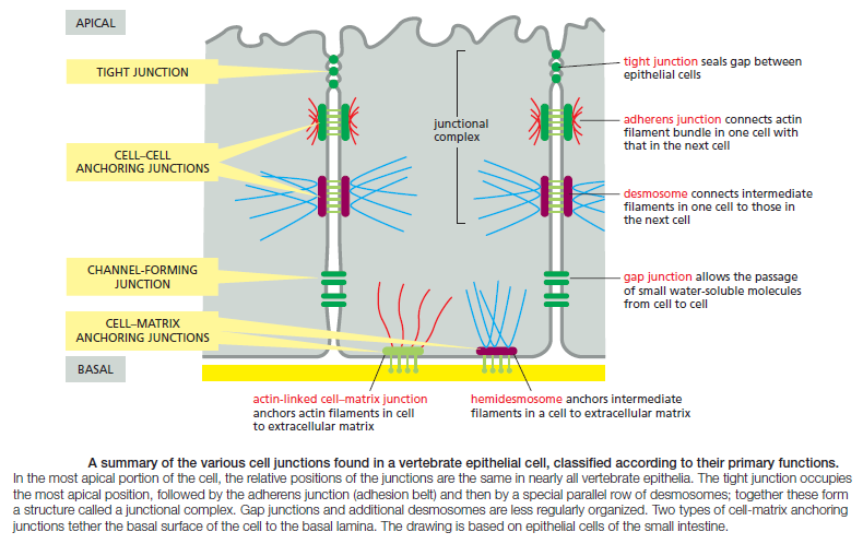
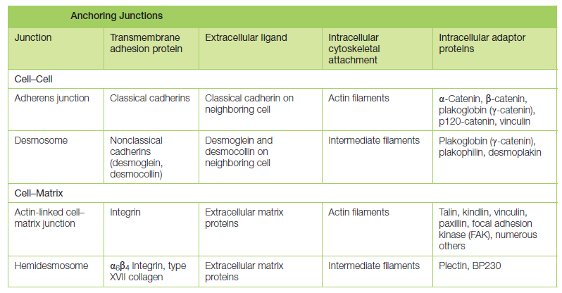
Figure above provides a closer view of epithelial cells to illustrate the major types of cell–cell and cell–matrix junctions. The diagram shows the typical arrangement of junctions in a simple columnar epithelium such as the lining of the small intestine of a vertebrate. Here, a single layer of tall cells stands on a basal lamina, with the cells’ uppermost surface, or apex, free and exposed to the extracellular medium. On their sides, or lateral surfaces, the cells make junctions with one another. Two types of anchoring junctions link the cytoskeletons of adjacent cells: adherens junctions are anchorage sites for actin filaments; desmosomes are anchorage sites for intermediate filaments. Two additional types of anchoring junctions link the cytoskeleton of the epithelial cells to the basal lamina: actin-linked cell–matrix junctions anchor actin filaments to the matrix, while hemidesmosomes anchor intermediate filaments to it.

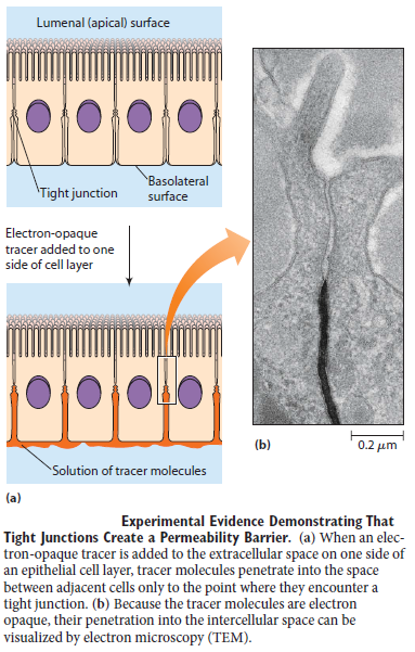
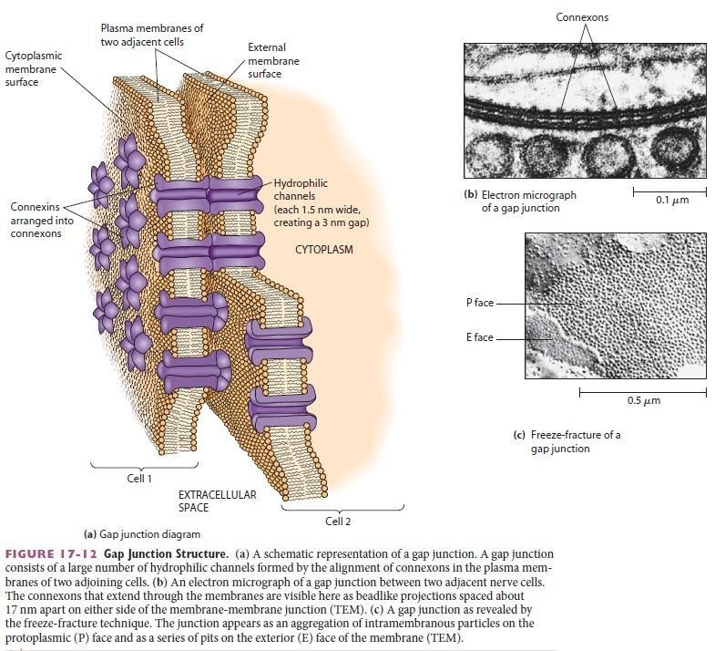
Two other types of cell–cell junction are shown in Figure above. Tight junctions hold the cells closely together near the apex, sealing the gap between the cells and thereby preventing molecules from leaking across the epithelium. Near the basal end of the cells are channel-forming junctions, called gap junctions, that create passageways linking the cytoplasms of adjacent cells. Each of the four major anchoring junction types depends on transmembrane adhesion proteins that span the plasma membrane, with one end linking to the cytoskeleton inside the cell and the other end linking to other structures outside it

Cell Junctions and the Extracellular Matrix Transm10

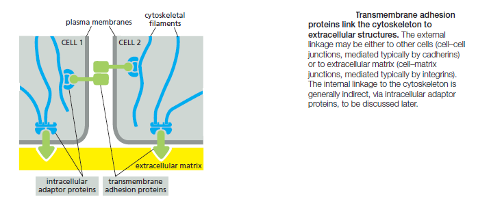
These cytoskeleton-linked transmembrane proteins fall neatly into two superfamilies, corresponding to the two basic kinds of external attachment. Proteins of the cadherin superfamily chiefly mediate attachment of cell to cell


Proteins of the integrin superfamily chiefly mediate attachment of cells to matrix. There is specialization within each family: some cadherins link to actin and form adherens junctions, while others link to intermediate filaments and form desmosomes; likewise, some integrins link to actin and form actinlinked cell–matrix junctions, while others link to intermediate filaments and form hemidesmosomes

Cell Junctions and the Extracellular Matrix Anchor11

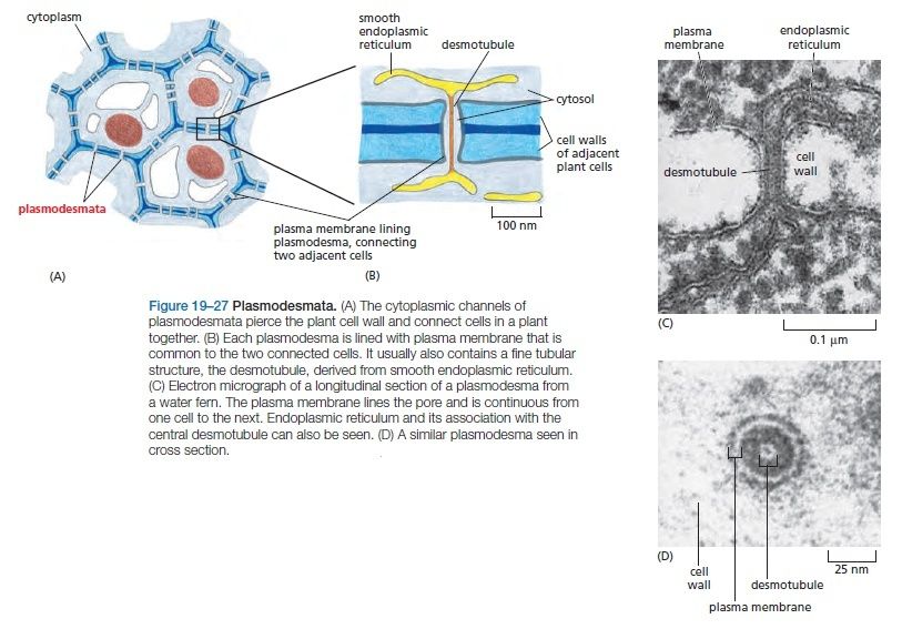
There are some exceptions to these rules. Some integrins, for example, mediate cell–cell rather than cell–matrix attachment. Moreover, there are other types of cell adhesion molecules that can provide transient cell–cell attachments more flimsy than anchoring junctions, but sufficient to stick cells together in special circumstances. We begin the chapter with a discussion of the major forms of cell–cell junctions. We then consider in turn the extracellular matrix of animals, the structure and function of integrin-mediated cell–matrix junctions, and, finally, the plant cell wall, a special form of extracellular matrix (ECM)

Cell Junctions and the Extracellular Matrix Ap-bio10

CELL–CELL JUNCTIONS

Cell–cell junctions come in many forms and can be regulated by a variety of mechanisms. The best understood and most common are the two types of cell– cell anchoring junctions, which employ cadherins to link the cytoskeleton of one cell with that of its neighbor. Their primary function is to resist the external forces that pull cells apart. The epithelial cells of your skin, for example, must remain tightly linked when they are stretched, pinched, or poked. Cell–cell anchoring junctions must also be dynamic and adaptable, so that they can be altered or rearranged when tissues are remodeled or repaired, or when there are changes in the forces acting on them. In this section, we focus primarily on the cadherin-based anchoring junctions. We then briefly describe tight junctions and gap junctions. Finally, we consider the more transient cell–cell adhesion mechanisms employed by some cells in the bloodstream.

Cadherins Form a Diverse Family of Adhesion Molecules

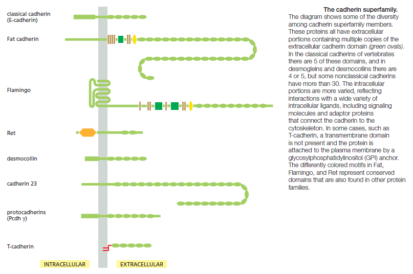
Cadherins are present in all multicellular animals whose genomes have been analyzed. They are also present in the choanoflagellates, which can exist either as free-living unicellular organisms or as multicellular colonies and are thought to be representatives of the group of protists . Other eukaryotes, including fungi and plants, lack cadherins, and they are also absent from bacteria and archaea. Cadherins therefore seem to be part of the essence of what it is to be an animal. The cadherins take their name from their dependence on Ca2+ ions: removing Ca2+ from the extracellular medium causes adhesions mediated by cadherins to come apart. The first three cadherins to be discovered were named according to the main tissues in which they were found: E-cadherin is present on many types of epithelial cells; N-cadherin on nerve, muscle, and lens cells; and P-cadherin on cells in the placenta and epidermis. All are also found in other tissues. These and other classical cadherins are closely related in sequence throughout their extracellular and intracellular domains. There are also a large number of nonclassical cadherins that are more distantly related in sequence, with more than 50 expressed in the brain alone. The nonclassical cadherins include proteins with known adhesive function, such as the diverse protocadherins found in the brain, and the desmocollins and desmogleins that form desmosomes . Other family members are involved primarily in signaling. Together, the classical and nonclassical cadherin proteins constitute the cadherin superfamily , with more than 180 members
in humans.

Cell Junctions and the Extracellular Matrix Cadher10

Cadherins Mediate Homophilic Adhesion

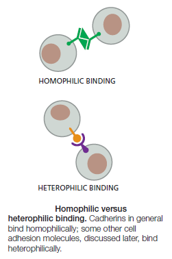
Anchoring junctions between cells are usually symmetrical: if the linkage is to actin in the cell on one side of the junction, it will be to actin in the cell on the other side. In fact, the binding between cadherins is generally homophilic (like to- like ): cadherin molecules of a specific subtype on one cell bind to cadherin molecules of the same or closely related subtype on adjacent cells.

Cell Junctions and the Extracellular Matrix Cadher11

The spacing between the cell membranes at an anchoring junction is precisely defined and depends on the structure of the participating cadherin molecules. All the members of the superfamily, by definition, have an extracellular portion consisting of several copies of the extracellular cadherin (EC) domain. Homophilic binding occurs at the N-terminal tips of the cadherin molecules—the cadherin domains that lie furthest from the membrane. These terminal domains each form a knob and a nearby pocket, and the cadherin molecules protruding from opposite cell membranes bind by insertion of the knob of one domain into the pocket of the other (Figure A below).

Cell Junctions and the Extracellular Matrix Cadher12

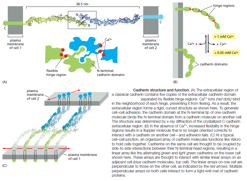
Each cadherin domain forms a more-or-less rigid unit, joined to the next cadherin domain by a hinge. Ca2+ ions bind to sites near each hinge and prevent it from flexing, so that the whole string of cadherin domains behaves as a rigid and slightly curved rod. When Ca2+ is removed, the hinges can flex, and the structure becomes floppy (Figure B above ). At the same time, the conformation at the N-terminus
is thought to change slightly, weakening the binding affinity for the matching cadherin molecule on the opposite cell. Unlike receptors for soluble signal molecules, which bind their specific ligand with high affinity, cadherins (and most other cell–cell adhesion proteins) typically bind to their partners with relatively low affinity. Strong attachments result from the formation of many such weak bonds in parallel. When binding to oppositely oriented partners on another cell, cadherin molecules are often clustered side-to-side with many other cadherin molecules on the same cell (Figure C). The strength of this junction is far greater than that of any individual intermolecular bond, and yet regulatory mechanisms can easily disassemble the junction by separating the molecules sequentially, just as two pieces of fabric can be joined strongly by Velcro and yet easily peeled apart from the sides. A similar “Velcro principle” also operates at cell–cell and cell–matrix adhesions formed by other types of transmembrane adhesion proteins.

Cadherin-Dependent Cell–Cell Adhesion Guides the Organization of Developing Tissues


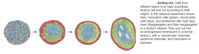
Cadherins form specific homophilic attachments, explaining why there are so many different family members. Cadherins are not like glue, making cell surfaces generally sticky. Rather, they mediate highly selective recognition, enabling cells of a similar type to stick together and to stay segregated from other types of cells. Selectivity in the way that animal cells consort with one another was first demonstrated
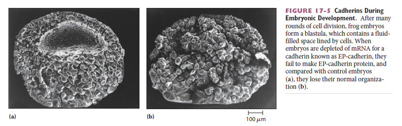
in the 1950s, long before the discovery of cadherins, in experiments in which amphibian embryos were dissociated into single cells. These cells were then mixed up and allowed to reassociate. Remarkably, the dissociated cells often reassembled into structures resembling those of the original embryo.These experiments, together with numerous more recent experiments, reveal that selective cell–cell recognition systems make cells of the same differentiated tissue preferentially adhere to one another

Cell Junctions and the Extracellular Matrix Sem_ty10

Cadherins play a crucial part in these cell-sorting processes during development. The appearance and disappearance of specific cadherins correlate with steps in embryonic development where cells regroup and change their contacts to create new tissue structures. In the vertebrate embryo, for example, changes in cadherin expression are seen when the neural tube forms and pinches off from the overlying ectoderm: neural tube cells lose E-cadherin and acquire other cadherins, including N-cadherin, while the cells in the overlying ectoderm continue to express E-cadherin (Figure A and B below).

Cell Junctions and the Extracellular Matrix Cadher10

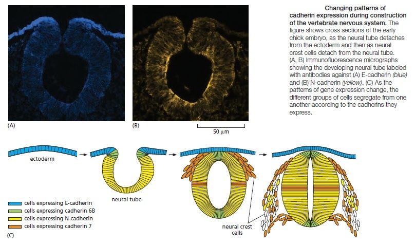
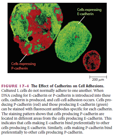
Then, when the neural crest cells migrate away from the neural tube, these cadherins become scarcely detectable, and another cadherin (cadherin 7) appears that helps hold the migrating cells together as loosely associated cell groups (Figure C). Finally, when the cells aggregate to form a ganglion, they switch on expression of N-cadherin again. If N-cadherin is artificially overexpressed in the emerging neural crest cells, the cells fail to escape from the neural tube. Studies with cultured cells further support the idea that the homophilic binding of cadherins controls these processes of tissue segregation. In a line of cultured fibroblasts called L cells, for example, cadherins are not expressed and the cells do not adhere to one another. When these cells are transfected with DNA encoding E-cadherin, E-cadherins on one cell bind to E-cadherins on another, resulting in cell–cell adhesion. If L cells expressing different cadherins are mixed together, they sort out and aggregate separately, indicating that different cadherins preferentially bind to their own type (Figure A below)

Cell Junctions and the Extracellular Matrix Cadher13

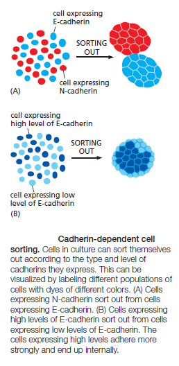
mimicking what happens when cells derived from tissues that express different cadherins are mixed together. A similar segregation of cells occurs if L cells expressing different amounts of the same cadherin are mixed together (Figure B above). It therefore seems likely that both qualitative and quantitative differences in the expression of cadherins have a role in organizing tissues.

Epithelial–Mesenchymal Transitions Depend on Control of Cadherins

The assembly of cells into an epithelium is a reversible process. By switching on expression of adhesion molecules, dispersed unattached mesenchymal cells, such as fibroblasts, can come together to form an epithelium. Conversely, epithelial cells can change their character, disassemble, and migrate away from their parent epithelium as separate cells. Such epithelial–mesenchymal transitions play an important part in normal embryonic development; the origin of the neural crest is one example. These transitions depend in part on transcription regulatory proteins called Slug, Snail, and Twist. Increased expression of Twist, for example, converts epithelial cells to a mesenchymal character, and switching it off does the opposite. Twist exerts its effects, in part, by inhibiting expression of cadherins, including E-cadherin, that hold epithelial cells together. Epithelial–mesenchymal transitions also occur as pathological events during adult life, in cancer. Most cancers originate in epithelia, but become dangerously prone to spread—that is, malignant—only when the cancer cells escape from the epithelium of origin and invade other tissues. Experiments with malignant breast cancer cells in culture show that blocking expression of Twist can convert the cells back toward a nonmalignant character. Conversely, by forcing Twist expression, one can make normal epithelial cells undergo an epithelial–mesenchymal transition and behave like malignant cells. Mutations that disrupt the production or function of E-cadherin are often found in cancer cells and are thought to help make them malignant.

Catenins Link Classical Cadherins to the Actin Cytoskeleton

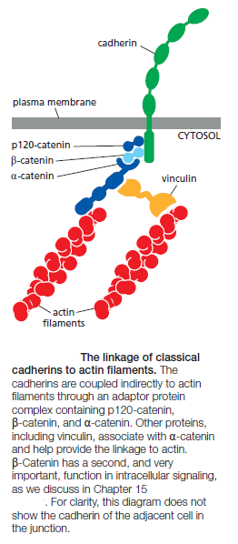
The extracellular domains of cadherins mediate homophilic binding at adherens junctions. The intracellular domains of typical cadherins, including all classical and some nonclassical ones, interact with filaments of the cytoskeleton: actin at adherens junctions and intermediate filaments at desmosomes. These cytoskeletal linkages are essential for efficient cell–cell adhesion, as cadherins that lack their cytoplasmic domains cannot stably hold cells together. The linkage of cadherins to the cytoskeleton is indirect and depends on adaptor proteins that assemble on the cytoplasmic tail of the cadherin. At adherens junctions, the cadherin tail binds two such proteins: β-catenin and a distant relative called p120-catenin; a third protein called α-catenin interacts with β-catenin and recruits a variety of other proteins to provide a dynamic linkage to actin filaments

Cell Junctions and the Extracellular Matrix The_li10

At desmosomes, cadherins are linked to intermediate filaments through other adaptor proteins, including a β-catenin-related protein called plakoglobin. In their mature form, adherens junctions are enormous protein complexes containing hundreds to thousands of cadherin molecules, packed into dense, regular arrays that are linked on the extracellular side by lateral interactions between cadherin domains, On the cytoplasmic side, a complex network of catenins, actin regulators, and contractile actin bundles holds the cluster of cadherins together and links it to the actin cytoskeleton. Assembling a structure of this complexity is not a simple task, and it involves a complex sequence of events controlled by the actin-regulatory proteins. The general features of the assembly process are summarized below:

Cell Junctions and the Extracellular Matrix Assemb10
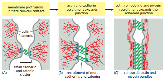
Assembly of an adherens junction.
(A) Assembly begins when two unattached epithelial cell precursors explore their surroundings with membrane protrusions, generated by local nucleation of actin networks. When the cells make contact, small cadherin and catenin clusters take shape at the contact sites and associate with actin, leading to activation of the small monomeric GTPase Rac (not shown), an important actin regulator.
(B) Rac promotes additional actin protrusions in the vicinity, expanding the size of the contact zone and thereby promoting further recruitment of cadherins and their associated catenin proteins.
(C) Eventually, Rac is inactivated and replaced by the related GTPase Rho (not shown), which shifts actin remodeling toward the assembly of linear, contractile filament bundles. Rho also promotes
the assembly of myosin II filaments that associate with bundles of actin filaments to generate contractile activity. This contractile activity generates tension that stimulates further actin recruitment and expansion of the junction, in part through the mechanisms illustrated in Figure below.


Cell Junctions and the Extracellular Matrix Mechan10

Adherens Junctions Respond to Forces Generated by the Actin Cytoskeleton

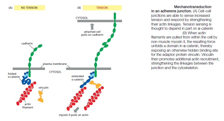
Most adherens junctions are linked to contractile bundles of actin filaments and non-muscle myosin II. These junctions are therefore subjected to pulling forces generated by the attached actin. The pulling forces are important for junction assembly and maintenance: disruption of myosin activity, for example, results in the disassembly of many adherens junctions. Furthermore, the contractile forces acting on a junction in one cell are balanced by contractile forces at the junction of the opposite cell, so that no cell pulls others toward it and thereby disrupts the uniform distribution of cells in the tissue. We do not understand the mechanisms responsible for maintaining this balance. Adherens junctions seem to sense the forces acting on them and modify local actin and myosin behavior to balance the forces on both sides of the junction. Evidence for these mechanisms comes from studies of pairs of cultured mammalian cells connected by adherens junctions. If contractile activity in one cell is increased experimentally, the adherens junctions linking the two cells increase in size, and the contractile activity of the second cell increases to match that of the first—resulting in a balance of forces across the junction. These and other experiments suggest that adherens junctions are not simply passive sites of protein–protein binding but are dynamic tension sensors that regulate their behavior in response to changing mechanical conditions. This ability to transduce a mechanical signal into a change in junctional behavior is an example of mechanotransduction. We will see later that it is also important at cell–matrix junctions. The mechanotransduction at cell–cell junctions is thought to depend, at least in part, on proteins in the cadherin complex that alter their shape when stretched by tension. The protein α-catenin, for example, is stretched from a folded to an extended conformation when contractile activity increases at the junction. The unfolding exposes a cryptic binding site for another protein, vinculin, which promotes the recruitment of more actin to the junction (Figure above).

By mechanisms such as this, pulling on a junction makes it stronger. Furthermore, as noted above, pulling on a junction in one cell will increase the contractile force generated in the attached cell. In some cell types, actin contractility reduces cell–cell adhesion, particularly if large forces are involved. Large actin-based contractile forces might, in some tissues, pull sufficiently hard on the edges of cell–cell adhesions to peel them apart, particularly if contraction is coupled to additional regulatory mechanisms that weaken the adhesion. This mechanism might be important in certain forms of tissue remodeling during development.


Last edited by Admin on Mon Apr 24, 2017 9:54 pm; edited 12 times in total

https://reasonandscience.catsboard.com

Otangelo


Admin

Tissue Remodeling Depends on the Coordination of Actin- Mediated Contraction With Cell–Cell Adhesion

Adherens junctions are an essential part of the machinery for modeling the shapes of multicellular structures in the animal body. By indirectly linking the actin filaments in one cell to those in its neighbors, they enable the cells in the tissue to use their actin cytoskeletons in a coordinated way. Adherens junctions occur in various forms. In many nonepithelial tissues, they appear as small punctate or linear attachments that connect the cortical actin filaments beneath the plasma membranes of two interacting cells. In heart muscle, they anchor the actin bundles of the contractile apparatus and act in parallel with desmosomes to link the contractile cells end-to-end. But the prototypical examples of adherens junctions occur in epithelia, where they often form a continuous adhesion belt (or zonula adherens) just beneath the apical face of the epithelium, encircling each of the interacting cells in the sheet (Figure below).

Cell Junctions and the Extracellular Matrix Adhere10

Within each cell, a contractile bundle of actin filaments and myosin II lies adjacent to the adhesion belt, oriented parallel to the plasma membrane and tethered to it by the cadherins and their associated intracellular adaptor proteins. The actin–myosin bundles are thus linked, via the cadherins, into an extensive transcellular network. Coordinated contraction of this network provides the motile force for
a fundamental process in animal morphogenesis—the folding of epithelial cell sheets into tubes, vesicles, and other related structures (Figure below).

Cell Junctions and the Extracellular Matrix Foldin10

Desmosomes Give Epithelia Mechanical Strength

Desmosomes are structurally similar to adherens junctions but contain specialized cadherins that link to intermediate filaments instead of actin filaments. Their main function is to provide mechanical strength. Desmosomes are important in vertebrates but are not found, for example, in Drosophila. They are present in most mature vertebrate epithelia and are particularly plentiful in tissues that are subject to high levels of mechanical stress, such as heart muscle and the epidermis, the epithelium that forms the outer layer of the skin.

Cell Junctions and the Extracellular Matrix Desmos10

Figure A shows the general structure of a desmosome, and Figure B shows some of the proteins that form it. Desmosomes typically appear as buttonlike spots of adhesion, riveting the cells together (Figure C). Inside the cell, the bundles of ropelike intermediate filaments that are anchored to the desmosomes form a structural framework of great tensile strength (Figure D), with linkage to similar bundles in adjacent cells, creating a network that extends throughout the tissue

Cell Junctions and the Extracellular Matrix Hemide10

The particular type of intermediate filaments attached to the desmosomes depends on the cell type: they are keratin filaments in most epithelial cells, for example, and desmin filaments in heart muscle cells. The importance of desmosomes is demonstrated by some forms of the potentially fatal skin disease pemphigus. Affected individuals make antibodies against one of their own desmosomal cadherin proteins. These antibodies bind to and disrupt the desmosomes that hold their epidermal cells (keratinocytes) together. This results in a severe blistering of the skin, with leakage of body fluids into the loosened epithelium.

Epithelium (epi- + thele + -ium) is one of the four basic types of animal tissue. The other three types are connective tissue, muscle tissue and nervous tissue. Epithelial tissues line the cavities and surfaces of blood vessels and organs throughout the body. There are three principal shapes of epithelial cells: squamous, columnar, and cuboidal. These can be arranged in a single layer of cells as simple epithelium, either squamous, columnar or cuboidal, or in layers of two or more cells deep as stratified (layered), either squamous, columnar or cuboidal. All glands are made up of epithelial cells. Functions of epithelial cells includesecretion, selective absorption, protection, transcellular transport, and sensing.


Cell Junctions and the Extracellular Matrix Epithe10


Tight Junctions Form a Seal Between Cells and a Fence Between Plasma Membrane Domains

Sheets of epithelial cells enclose and partition the animal body, lining all its surfaces and cavities, and creating internal compartments where specialized processes occur. The epithelial sheet seems to be one of the inventions that lie at the origin of animals, diversifying in a huge variety of ways but retaining an organization based on a set of conserved molecular mechanisms. Essentially all epithelia are anchored to other tissue on one side—the basal side—and free of such attachment on their opposite side—the apical side. A basal lamina lies at the interface with the underlying tissue, mediating the attachment, while the apical surface of the epithelium is generally bathed by extracellular fluid. Thus, all epithelia are structurally polarized, and so are their individual cells: the basal end of a cell, adherent to the basal lamina below, differs from the apical end, exposed to the medium above. Correspondingly, all epithelia have at least one function in common: they serve as selective permeability barriers, separating the fluid that permeates the tissue on their basal side from fluid with a different chemical composition on their apical side. This barrier function requires that the adjacent cells be sealed together by tight junctions, so that molecules cannot leak freely across the cell sheet. The epithelium of the small intestine provides a good illustration of tight-junction structure and function. This epithelium has a simple columnar structure; that is, it consists of a single layer of tall (columnar) cells. These are of several differentiated types, but the majority are absorptive cells, specialized for uptake of nutrients from the internal cavity, or lumen, of the gut. The absorptive cells have to transport selected nutrients across the epithelium from the lumen into the extracellular fluid on the other side. From there, these nutrients diffuse into small blood vessels to provide nourishment to the organism. This transcellular transport depends on two sets of transport proteins in the plasma membrane of the absorptive cell. One set is confined to the apical surface of the cell (facing the lumen) and actively transports selected molecules into the cell from the gut. The other set is confined to the basolateral (basal and lateral) surfaces of the cell, and it allows the same molecules to leave the cell by passive transport into the extracellular fluid on the other side of the epithelium. For this transport activity to be effective, the spaces between the epithelial cells must be tightly sealed, so that the transported molecules cannot leak back into the gut lumen through these spaces (Figure below).

Cell Junctions and the Extracellular Matrix Transc10

Moreover, the transport proteins must be correctly distributed in the plasma membranes: the apical transporters must be delivered to the cell apex and must not be allowed to drift to the basolateral membrane, and the basolateral transporters must be delivered to and remain in the basolateral membrane.

Cell Junctions and the Extracellular Matrix Figure11

Tight junctions, besides sealing the gaps between the cells, also function as “fences” that help prevent apical or basolateral proteins from diffusing into the wrong region. The sealing function of tight junctions is easy to demonstrate experimentally: a low-molecular-weight tracer added to one side of an epithelium will generally not pass beyond the tight junction

Cell Junctions and the Extracellular Matrix Epithe10

This seal is not absolute, however. Although all tight junctions are impermeable to macromolecules, their permeability to ions and other small molecules varies. Tight junctions in the epithelium lining the small intestine, for example, are 10,000 times more permeable to inorganic ions, such as Na+, than the tight junctions in the epithelium lining the urinary bladder. The movement of ions and other molecules between epithelial cells is called paracellular transport, and tissue-specific differences in transport rates generally result from differences in the proteins that form tight junctions.

Tight Junctions Contain Strands of Transmembrane Adhesion Proteins

When tight junctions are visualized by freeze-fracture electron microscopy, they are seen as a branching network of sealing strands that completely encircles the apical end of each cell in the epithelial sheet (Figure A and B).

Cell Junctions and the Extracellular Matrix Epithe11

In conventional electron micrographs, the outer leaflets of the two interacting plasma membranes are tightly apposed where sealing strands are present (Figure C). Each sealing strand is composed of a long row of transmembrane homophilic adhesion proteins embedded in each of the two interacting plasma membranes. The extracellular domains of these proteins adhere directly to one another to occlude the intercellular space

Cell Junctions and the Extracellular Matrix Tight_10

 The main transmembrane proteins forming these strands are the claudins, which are essential for tight-junction formation and function. Mice that lack the claudin-1 gene, for example, fail to make tight junctions between the cells in the epidermal layer of the skin; as a result, the baby mice lose water rapidly by evaporation through the skin and die within a day after birth. Conversely, if nonepithelial cells such as fibroblasts are artificially caused to express claudin genes, they will form tight-junctional connections with one another. Normal tight junctions also contain a second major transmembrane protein called occludin, which is not essential for the assembly or structure of the tight junction but is important for limiting junctional permeability. A third transmembrane protein, tricellulin, is required to seal cell membranes together and prevent transepithelial leakage at the points where three cells meet. The claudin protein family has many members (24 in humans), and these are expressed in different combinations in different epithelia to confer particular permeability properties on the epithelial sheet. They are thought to form paracellular pores—selective channels allowing specific ions to cross the tight-junctional barrier, from one extracellular space to another. A specific claudin found in kidney epithelial cells, for example, is needed to let Mg2+ pass between the cells of the kidney tubules so that this ion can be resorbed from the urine into the blood. A mutation in the gene encoding this claudin results in excessive loss of Mg2+ in the urine.

Scaffold Proteins Organize Junctional Protein Complexes

Like the cadherin molecules of an adherens junction, the claudins and occludins of a tight junction interact with each other on their extracellular sides to promote junction assembly. Also as in adherens junctions, the organization of adhesion proteins in a tight junction depends on additional proteins that bind the cytoplasmic side of the adhesion proteins. The key organizational proteins at tight junctions are the zonula occludens (ZO) proteins. The three major members of the ZO family—ZO-1, ZO-2, and ZO-3—are large scaffold proteins that provide a structural
support on which the tight junction is built. These intracellular molecules consist of strings of protein-binding domains, typically including several PDZ domains—segments about 80 amino acids long that can recognize and bind the C-terminal tails of specific partner proteins

Cell Junctions and the Extracellular Matrix Scaffo11

One domain of these scaffold proteins can attach to a claudin protein, while others can attach to occludin or the actin cytoskeleton. Moreover, one molecule of scaffold protein can bind to another. In this way, the cell can assemble a mat of intracellular proteins that organizes and positions the sealing strands of the tight junction. The tight-junctional network of sealing strands usually lies just apical to adherens and desmosome junctions that bond the cells together mechanically; the whole assembly is called a junctional complex. The parts of this junctional complex depend on each other for their formation. For example, anticadherin antibodies that block the formation of adherens junctions also block the formation of tight junctions.

Gap Junctions Couple Cells Both Electrically and Metabolically

Tight junctions block the passageways through the gaps between epithelial cells, preventing extracellular molecules from leaking from one side of an epithelium to the other. Another type of junctional structure has a radically different function: it bridges gaps between adjacent cells so as to create direct channels from the cytoplasm of one to that of the other. These channels are called gap junctions. Gap junctions are present in most animal tissues, including connective tissues as well as epithelia and heart muscle. Each gap junction appears in conventional electron micrographs as a patch where the membranes of two adjacent cells are separated by a uniform narrow gap of about 2–4 nm

Cell Junctions and the Extracellular Matrix Gap_ju10

The gap is spanned by channel-forming proteins, of which there are two distinct families, called the connexins and the innexins. Connexins are the predominant gap-junction proteins in vertebrates, with 21 isoforms in humans. Innexins are found in the gap junctions of invertebrates. Gap junctions have a pore size of about 1.4 nm, which allows the exchange of inorganic ions and other small water-soluble molecules, but not of macromolecules such as proteins or nucleic acids

Cell Junctions and the Extracellular Matrix Gap_ju10

An electric current injected into one cell through a microelectrode causes an electrical disturbance in the neighboring cell, due to the flow of ions carrying electric charge through gap junctions. This electrical coupling via gap junctions serves an obvious purpose in tissues containing electrically excitable cells: action potentials can spread rapidly from cell to cell, without the delay that occurs at chemical synapses. In vertebrates, for example, electrical coupling through gap junctions synchronizes the contractions of heart muscle cells as well as those of the smooth muscle cells responsible for the peristaltic movements of the intestine. Gap junctions also occur in many tissues whose cells are not electrically excitable. In principle, the sharing of small metabolites and ions provides a mechanism for coordinating the activities of individual cells in such tissues and for smoothing out random fluctuations in small-molecule concentrations in different cells.

A Gap-Junction Connexon Is Made of Six Transmembrane Connexin Subunits

Connexins are four-pass transmembrane proteins, six of which assemble to form a hemichannel, or connexon. When the connexons in the plasma membranes of two cells in contact are aligned, they form a continuous aqueous channel that connects the two cell interiors

Cell Junctions and the Extracellular Matrix Gap_ju11

A gap junction consists of many such connexon pairs in parallel, forming a sort of molecular sieve. Not only does this sieve provide a communication channel between cells, but it also provides a form of cell–cell adhesion that supplements the cadherin- and claudin-mediated adhesions. Gap junctions in different tissues can have different properties because they are formed from different combinations of connexins, creating channels that differ in permeability and regulation. Most cell types express more than one type of connexin, and two different connexin proteins can assemble into a heteromeric connexon, with its own distinct properties. Moreover, adjacent cells expressing different connexins can form intercellular channels in which the two aligned halfchannels are different (see Figure B). Like conventional ion channels, individual gapjunction channels do not remain open all the time; instead, they flip between open and closed states. These changes are triggered by a variety of stimuli, including the voltage difference between the two connected cells, the membrane potential of each cell, and various chemical properties of the cytoplasm, including the pH and concentration of free Ca2+. Some subtypes of gap junctions can also be regulated by extracellular signals such as neurotransmitters. We are only just beginning to understand the physiological functions and structural basis of these various gating mechanisms. Each gap-junctional plaque is a dynamic structure that can readily assemble, disassemble, or be remodeled, and it can contain a cluster of a few to many thousands of connexons. Studies with fluorescently labeled connexins in living cells show that new connexons are continually added around the periphery of an existing junctional plaque, while old connexons are removed from the middle of it and destroyed

Cell Junctions and the Extracellular Matrix Gap_ju12

This turnover is rapid: the connexin molecules have a half-life of only a few hours. The mechanism of removal of old connexons from the middle of the plaque is not known, but the route of delivery of new connexons to its periphery seems clear: they are inserted into the plasma membrane by exocytosis, like other integral membrane proteins, and then diffuse in the plane of the membrane until they bump into the periphery of a connexon plaque and become trapped. This has a corollary: the plasma membrane away from the gap junction should contain connexons—hemichannels—that have not yet paired with their counterparts on another cell. It is thought that these unpaired hemichannels are normally held in a closed conformation, preventing the cell from losing its small molecules by leakage through them. But there is also evidence that in some circumstances they can open and serve as channels for the release of small signal molecules.

In Plants, Plasmodesmata Perform Many of the Same Functions as Gap Junctions

The tissues of a plant are organized on different principles from those of an animal. This is because plant cells are imprisoned within tough cell walls composed of an extracellular matrix rich in cellulose and other polysaccharides, as we discuss later. The cell walls of adjacent cells are firmly cemented to those of their neighbors, which eliminates the need for anchoring junctions to hold the cells in place. But a need for direct cell–cell communication remains. Thus, plant cells have only one class of intercellular junctions, plasmodesmata. Like gap junctions, they directly connect the cytoplasms of adjacent cells. In plants, the cell wall between a typical pair of adjacent cells is at least 0.1 μm thick, and so a structure very different from a gap junction is required to mediate communication across it. Plasmodesmata solve the problem. With a few specialized exceptions, every living cell in a higher plant is connected to its living neighbors by these structures, which form fine cytoplasmic channels through the intervening cell walls. As shown in Figure A

Cell Junctions and the Extracellular Matrix Plasmo10

the plasma membrane of one cell is continuous with that of its neighbor at each plasmodesma, which connects the cytoplasms of the two cells by a roughly cylindrical channel with a diameter of 20–40 nm. Running through the center of the channel in most plasmodesmata is a narrower cylindrical structure, the desmotubule, which is continuous with elements of the smooth endoplasmic reticulum (ER) in each of the connected cells (Figure B–D). Between the outside of the desmotubule and the inner face of the cylindrical channel formed by plasma membrane is an annulus of cytosol through which small molecules can pass from cell to cell. As each new cell wall is assembled during the cytokinesis phase of cell division, plasmodesmata are created within it. They form around elements of smooth ER that become trapped across the developing cell plate . They can also be inserted de novo through preexisting cell walls, where they are commonly found in dense clusters called pit fields. When no longer required, plasmodesmata can be removed. In spite of the radical difference in structure between plasmodesmata and gap junctions, they seem to function in remarkably similar ways. Evidence obtained by injecting tracer molecules of different sizes suggests that plasmodesmata allow the passage of molecules with a molecular weight of less than about 800, which is similar to the molecular-weight cutoff for gap junctions. As with gap junctions, transport through plasmodesmata is regulated. Dye-injection experiments, for example, show that there can be barriers to the movement of even low-molecularweight molecules between certain cells, or groups of cells, that are connected by apparently normal plasmodesmata; the mechanisms that restrict communication in these cases are not understood.

Selectins Mediate Transient Cell–Cell Adhesions in the Bloodstream

We now complete our overview of cell–cell junctions and adhesion by briefly describing some of the more specialized adhesion mechanisms used in some tissues. In addition to those we have already discussed, at least three other superfamilies of cell–cell adhesion proteins are important: the integrins, the selectins, and the adhesive immunoglobulin (Ig) superfamily members. We shall discuss integrins in more detail later: their main function is in cell–matrix adhesion, but a few of them mediate cell–cell adhesion in specialized circumstances. Ca2+ dependence provides one simple way to distinguish among these classes of adhesion proteins experimentally. Selectins, like cadherins and integrins, require Ca2+ for their adhesive function; Ig superfamily members do not. Selectins are cell-surface carbohydrate-binding proteins (lectins) that mediate a variety of transient cell–cell adhesion interactions in the bloodstream. Their main role, in vertebrates at least, is in governing the traffic of white blood cells into normal lymphoid organs and any inflamed tissues. White blood cells lead a nomadic life, roving between the bloodstream and the tissues, and this necessitates special adhesive behavior. The selectins control the binding of white blood cells to the endothelial cells lining blood vessels, thereby enabling the blood cells to migrate out of the bloodstream into a tissue. Each selectin is a transmembrane protein with a conserved lectin domain that binds to a specific oligosaccharide on another cell (Figure A) below:

Cell Junctions and the Extracellular Matrix Select10

The structure and function of selectins. (A) The structure of P-selectin. The selectin attaches to the actin cytoskeleton through adaptor proteins that are still poorly characterized. (B) How selectins and integrins mediate the cell–cell adhesions required for a white blood cell to migrate out of the bloodstream into a tissue. First, selectins on endothelial cells bind to oligosaccharides on the white blood cell, so that it becomes loosely attached and rolls along the vessel wall. Then the white blood cell activates a cellsurface integrin called LFA1, which binds to a protein called ICAM1 (belonging to the Ig superfamily) on the membrane of the endothelial cell. The white blood cell adheres to the vessel wall and then crawls out of the vessel by a process that requires another immunoglobulin superfamily member called PECAM1 (or CD31), not shown . EGF, epidermal growth factor.

There are at least three types: L-selectin on white blood cells, P-selectin on blood platelets and on endothelial cells that have been locally activated by an inflammatory response, and E-selectin on activated endothelial cells. In a lymphoid organ, such as a lymph node or the spleen, the endothelial cells express oligosaccharides that are recognized by L-selectin on lymphocytes, causing the lymphocytes to loiter and become trapped. At sites of inflammation, the roles are reversed: the endothelial cells switch on expression of selectins that recognize the oligosaccharides on white blood cells and platelets, flagging the cells down to help deal with the local emergency. Selectins do not act alone, however; they collaborate with integrins, which strengthen the binding of the blood cells to the endothelium. The cell–cell adhesions mediated by both selectins and integrins are heterophilic—that is, the binding is to a molecule of a different type: selectins bind to specific oligosaccharides
on glycoproteins and glycolipids, while integrins bind to specific Ig-family proteins. Selectins and integrins act in sequence to let white blood cells leave the bloodstream and enter tissues (Figure B). The selectins mediate a weak adhesion because the binding of the lectin domain of the selectin to its carbohydrate ligand is of low affinity. This allows the white blood cell to adhere weakly and reversibly to the endothelium, rolling along the surface of the blood vessel, propelled by the flow of blood. The rolling continues until the blood cell activates its integrins. As we discuss later, these transmembrane molecules can be switched into an adhesive conformation that enables them to latch onto specific macromolecules external to the cell—in the present case, proteins on the surfaces of the endothelial cells. Once it has attached in this way, the white blood cell escapes from the bloodstream into the tissue by crawling out of the blood vessel between adjacent endothelial cells.

Members of the Immunoglobulin Superfamily Mediate Ca2+-Independent Cell–Cell Adhesion

The chief endothelial cell proteins that are recognized by the white blood cell integrins are called ICAMs (intercellular cell adhesion molecules) or VCAMs (vascular cell adhesion molecules). They are members of another large and ancient family of cell-surface molecules—the immunoglobulin (Ig) superfamily. These contain one or more extracellular Ig-like domains that are characteristic of antibody molecules. They have many functions outside the immune system that are unrelated to immune defenses. While ICAMs and VCAMs on endothelial cells both mediate heterophilic
binding to integrins, many other Ig superfamily members appear to mediate homophilic binding. An example is the neural cell adhesion molecule (NCAM), which is expressed by various cell types, including most nerve cells, and can take different forms, generated by alternative splicing of an RNA transcript produced from a single gene (Figure below).

Cell Junctions and the Extracellular Matrix Ncam10

Some forms of NCAM carry an unusually large quantity of sialic acid (with chains containing hundreds of repeating sialic acid units). By virtue of their negative charge, the long polysialic acid chains can interfere with cell adhesion (because like charges repel one another); thus, these forms of NCAM can serve to inhibit adhesion, rather than cause it. A cell of a given type generally uses an assortment of different adhesion proteins to interact with other cells, just as each cell uses an assortment of different receptors to respond to the many soluble extracellular signal molecules in its
environment. Although cadherins and Ig superfamily members are frequently expressed on the same cells, the adhesions mediated by cadherins are much stronger, and they are largely responsible for holding cells together, segregating cell collectives into discrete tissues, and maintaining tissue integrity. Molecules such as NCAM seem to contribute more to the fine-tuning of these adhesive interactions during development and regeneration, playing a part in various specialized adhesive phenomena, such as that discussed for blood cells and endothelial cells. Thus, while mutant mice that lack N-cadherin die early in development, those that lack NCAM develop relatively normally but show some mild abnormalities in the development of certain specific tissues, including parts of the nervous system.


Last edited by Admin on Wed Sep 30, 2015 2:11 pm; edited 5 times in total

https://reasonandscience.catsboard.com

3Cell Junctions and the Extracellular Matrix Empty Cell adhesions, Cell junctions Wed Sep 30, 2015 1:39 pm

Otangelo


Admin

Cell adhesions, Cell junctions

However, most organisms are multicellular: They consist of many—sometimes trillions—of cells. Many kinds of cells—including most of those in your own body—spend all their lives linked to neighboring cells. It is the organization of cells into tissues that allows multicellular organisms to adopt complex structures. These tissues, in turn, are arranged in precise ways to generate the form of an organism. How do these tissues achieve their structures? Consider the two types of animal tissues shown in Figure below:

Cell Junctions and the Extracellular Matrix Differ10

One is a sheet of cells known as an epithelium, such as the cells that line the small intestine. Such cells are clearly polarized—that is, they contain discrete functional domains at opposite ends of the cell. The end of the cell in contact with the external environment (for example, the lumen of the small intestine) is often called the apical side of the cell. The parts on the other side of the tight junction, including the surfaces in contact with the basal lamina, are called the basolateral side of the cell. The other cell type in Figure above is from a more loosely organized connective tissue, such as might be found in the dermis of the skin. In each case, cells must be attached to one another, to a mechanically rigid scaffolding, or to both. Thus, in order to understand how multicellular organisms are constructed, we will need to consider both connections between cells, or cell-cell adhesions, and the extracellular structures to which cells attach. Cells use a variety of elaborate molecular complexes, or junctions, to attach to one another, and most of these involve transmembrane proteins that link the cell surface to the cytoskeleton . Cells use other types of molecular complexes to attach to extracellular structures, and these also involve specific linkages between the cell surface and the cytoskeleton. The extracellular structures themselves consist mainly of macromolecules that are secreted by the cell. Animal cells have an extracellular matrix that takes on a variety of forms and plays important roles in cellular processes as diverse as division, motility, differentiation, and adhesion. Epithelial cells, such as the ones shown in Figure above, produce a specialized extracellular matrix called a basal lamina. Connective tissues, on the other hand, produce a more loosely organized matrix. In plants, fungi, algae, and prokaryotes, the extracellular structure is a cell wall— although its chemical composition differs considerably among these organisms. Cell walls confer rigidity on the cells they encase, serve as permeability barriers, and protect cells from physical damage and from attack by viruses and infectious organisms. Most of our attention in this chapter is devoted to the adhesions animal cells make with one another, the junctions that characterize these adhesions, and how animal cells interact with the extracellular matrix. Then we will turn to the walls that surround plant cells and the specialized structures that allow direct cell-to-cell communication between plant cells, despite the presence of a cell wall.

Cell-Cell Recognition and Adhesion

The ability of individual cells to associate in precise patterns to form tissues, organs, and organ systems requires that individual cells be able to recognize, adhere to, and communicate with each other. We discuss cell-cell contact in this section and consider intercellular junctions later in the chapter.

Transmembrane Proteins Mediate Cell-Cell Adhesion

Animal cells use specialized adhesion receptors to attach to one another. Many of these adhesion proteins are transmembrane proteins, which means the extracellular portion of these proteins can interact with the extracellular portion of similar proteins on the surface of a neighboring cell. Although diagrams of adhesive structures may suggest that they are static once assembled, they are anything but. Cells can dynamically assemble and disassemble adhesions in response to a variety of events. 

This seems to be a essential requirement for function right from the beginning of multicellularity. 

Many adhesion proteins are continuously recycled: Protein at the cell surface is internalized by  endocytosis, and new protein is deposited at the surface via exocytosis. In addition, adhesion proteins serve as key sites for 


1.assembly of signaling complexes in cells and for 
2.dynamically assembling cytoskeletal structures at sites of cell adhesion.


 In this way, cell adhesion is coordinated with other major processes, including 


1.cell signaling, 
2.cell movement, 
3.cell proliferation, and 
4.cell survival. 

We now know that cell-cell adhesion receptors fall into a relatively small number of classes. They include 

1.immunoglobulin superfamily (IgSF) proteins, 
2.cadherins, 
3.selectins, and, in a few cases, 
4.integrins


This is relevant for the hypothesis of macro evolution. In a naturalistic evolutionary view, ther transitions were  from LUCA, the last common universal ancestor, to the congregation  to yield the first prokaryotic cells, the associations of prokaryotic cells to create eukaryotic cells with organelles such as chloroplasts and mitochondria, and the establishment of cooperative societies composed of discrete multicellular individuals.


Cell Junctions and the Extracellular Matrix Differ11

In each case, the adhesion protein on the surface of one cell binds to the appropriate ligand on the surface of a neighboring cell. In some cases, such as many cadherins and immunoglobulin superfamily members, cells interact with identical molecules on the surface of the cell that they adhere to. Such interactions are said to be homophilic interactions (from the Greek homo, meaning “like,” and
philia, meaning “friendship”). In other cases, such as the selectins and integrins, a cell adhesion receptor on one cell interacts with a different molecule on the surface of the cell to which it attaches. Such interactions are said to be heterophilic interactions (from the Greek hetero, meaning “different”). Many transmembrane adhesion receptors attach to the cytoskeleton via linker proteins, which differ
depending on the class of molecule and its location within the cell. In the next few sections, we consider examples of each of these major classes of cell-cell adhesion molecules.

Cell Adhesion Molecules (CAMs).

CAMs are members of the immunoglobulin superfamily (IgSF). The founding member of this family, called neural cell adhesion molecule (N-CAM), was identified using an antibody that disrupted cell-cell adhesion between isolated neuronal cells. Proteins in this large superfamily are so named because they contain domains, characterized by well-organized loops, that are similar to those in the immunoglobulin subunits that
constitute antibodies. CAMs, such as N-CAM, on one cell interact homophilically with CAMs on an adjacent cell via these domains. Other IgSF members interact heterophilically with their ligands. IgSF members participate in a wide range of adhesion events. In the embryonic nervous system, CAMs, such as N-CAM and L1-CAM, are involved in the outgrowth and bundling of neurons. Humans with mutations in the L1-CAM gene show defects in the corpus callosum (a region that interconnects the two hemispheres of the brain), mental retardation, and other defects.

Cadherins

The use of antibodies that block cell adhesion also led to the discovery of the cadherins, an important group of adhesive glycoproteins found in the plasma membranes of most animal cells. Like CAMs, cadherins
play a crucial role in cell-cell recognition and adhesion. The two groups of proteins can be distinguished from each other because cadherins, but not CAMs, require calcium to function. binds to and stabilizes the
conformation of cadherins that allows them to mediate cell-cell adhesion. Cadherins are characterized by a series of structurally similar domains (or “repeats”) in their extracellular domain. Members of the cadherin superfamily have widely varying numbers of these repeats, and they vary in the structure of their cytosolic ends. The best-characterized cadherin, E-cadherin (E is for “epithelial”), has five such repeat domains. E-cadherin molecules associate in pairs in the plasma membrane. Their extracellular domains have a structure that allows them to “zip” together in a homophilic fashion, as cadherins from one cell interlock with those from a neighboring cell

Cell Junctions and the Extracellular Matrix Cadher14

At their cytosolic ends, cadherins associate with the cytoskeleton, thereby linking the cell surface to the cytoskeleton.

How could this linking have evolved naturally ? Trial and error ? 

 These linkages differ for different types of cadherins. Different cadherins are expressed in specific tissues. Their regulated expression is a particularly striking feature of embryonic development and contributes to the ability of different tissues to separate from one another as embryos change their shape.

How could this regulated expression have evolved ? Trial and error ? 

The role of different cadherins in cell-cell adhesion has been investigated in cultured fibroblasts called L cells, which bind poorly to one another and contain little cadherin. When purified DNA encoding E-cadherin or P-cadherin (P is for “placental,” where this cadherin was first described) is introduced into L cells, the cells begin to produce cadherins and to bind more tightly to one another. Moreover, L cells that produce E-cadherin bind preferentially to other cells producing E-cadherin. Similarly, cells that produce P-cadherin bind selectively to other cells that are also producing P-cadherin. Such observations suggest that the amount and types of cadherin molecules on cell surfaces help to segregate cells into specific tissues.

Cell Junctions and the Extracellular Matrix The_ef11

Cadherins have especially important roles during embryonic development, where the dynamic assembly and disassembly of cadherin-based adhesions is tightly regulated. For example, when frog embryos are depleted of mRNA for the main type of cadherin found in the early embryo, they lose their normal organization

Cell Junctions and the Extracellular Matrix Cadher15

Other cadherins are involved in helping neuronal cells to form bundles and to establish synaptic connections. One particularly important event that occurs frequently in embryos is the breakdown of an epithelium into loosely organized, migratory cells called mesenchymal cells. This epithelial-mesenchymal transition (EMT) is accompanied by changes in cadherin expression. Changes in cadherin expression also occur in cancer cells. Cancer cells often stop expressing cadherins, such as E-cadherin, on their surfaces. As a result, they undergo an EMT that healthy cells would not, and these cells spread to other parts of the body by a process known as metastasis

Carbohydrate Groups Are Important in Cell-Cell Recognition and Adhesion

Like other glycoproteins, the carbohydrate side chains of CAMs and cadherins likely affect their adhesion properties. In addition, there are several well-studied examples of how carbohydrates on the cell surface affect cell adhesion.

Lectins

A role for carbohydrate groups in cell adhesion is also suggested by the fact that many animal and plant cells secrete carbohydrate-binding proteins called lectins, which promote cell-cell adhesion by binding to a specific sugar or sequence of sugars exposed at the outer cell surface. Because lectins usually have more than one carbohydrate- binding site, they can bind to carbohydrate groups on two different cells, thereby linking the cells together.

Selectins and Leukocyte Adhesion

Carbohydrate recognition also plays an important role during the interactions of leukocytes with endothelial cells lining blood vessels or with platelets. Cell surface glycoproteins called selectins mediate these interactions. A different selectin is expressed by each cell type (L-selectin on leukocytes, E-selectin on the endothelial cells of blood vessels, and P-selectin on platelets and endothelial cells). Leukocytes roll along the walls of blood vessels. During inflammation, they attach to the wall of a blood vessel in the vicinity of the inflammation and then migrate through the blood vessel to the inflammation site. The initial attachment of leukocytes is mediated by binding of selectins on the leukocyte to carbohydrates on the surface of the endothelial cells and vice versa. When leukocytes stop rolling and begin to invade the blood vessel, they make more stable
adhesions, which are mediated by a specific integrin on the surface of the leukocyte and immunoglobulin superfamily proteins, called ICAMs, on the surfaces of endothelial cells

Cell Junctions and the Extracellular Matrix Leukoc10

Carbohydrates and the Survival of Erythrocytes

One especially well-known example of the importance of carbohydrates at the cell surface is the determination of the human blood types A, B, AB, and O by a specific carbohydrate side chain present on a glycolipid of the erythrocyte plasma membrane ( Red blood cells (RBCs), also called erythrocytes ) . The ABO blood group involves differences in carbohydrate side chains on the surface of red blood cells that can be detected by antibodies present in the blood, leading to clumping of red blood cells and likely to the patient’s death if the wrong blood type is used in transfusion. The four blood types in the ABO system depend on genetically determined differences in the structure of a branched-chain carbohydrate attached to a specific glycolipid in the erythrocyte plasma membrane. Individuals with blood type A have the amino sugar N-acetylgalactosamine (GalNAc) at the ends of this carbohydrate, whereas individuals with blood type B have galactose (Gal) instead. Individuals with blood type AB have both N-acetylgalactosamine and galactose present; and, in individuals with type O blood, these terminal sugars are missing entirely. These minor differences have major effects on the compatibility of blood transfusions because people with blood types A, B, or O have antibodies in their bloodstream that recognize and bind to the respective terminal sugars of this specific glycolipid. An individual whose blood contains antibodies against one or both sugars (GalNAc or Gal) cannot accept blood containing the glycolipid with that terminal sugar. Individuals with type A blood have antibodies against carbohydrate chains ending in galactose, which occur in type B and type AB blood. They therefore cannot be transfused with type B or AB blood, but they can accept blood from type A or O donors. Conversely, individuals with type B blood have antibodies against carbohydrate chains ending in GalNAc, which occur in blood of types A and AB. They therefore cannot be transfused with type A or AB blood but can accept blood from B or O donors. Individuals with type O blood are called universal donors because their erythrocytes do not generate an immune response when transfused into individuals of any blood type.

Cell-Cell Junctions

By definition, unicellular organisms have no permanent associations between cells (although they can form temporary associations, such as during bacterial swarming or the aggregation of slime mold amoebae). Whereas a single cell is an entity unto itself, multicellular organisms have specific means of joining cells in long-term associations to form tissues and organs. Such associations usually involve specialized modifications of the plasma membrane at the point where two cells come together. These specialized structures are called cell-cell junctions. In animals, the three most common kinds of cell junctions are adhesive junctions, tight junctions, and gap junctions. In plants, the presence of a cell wall between the plasma membranes of adjacent cells precludes the kinds of cell junctions that link animal cells. However, the cell wall and special structures called plasmodesmata carry out similar functions.

Cell Junctions and the Extracellular Matrix Major_10

Polarity Proteins Regulate the Positioning of Cell-Cell Junctions

Both migrating single cells and sheets of epithelial cells display polarity: They have defined regions that have obviously different activities. Epithelial cells in particular have a striking polarity that separates apical from basolateral regions of the cell surface

Cell Junctions and the Extracellular Matrix Differ12

Numerous studies, including key work in the nematode worm, C. elegans, and the fruit fly, Drosophila, have identified proteins that help to establish and maintain epithelial polarity. One important protein complex, localized at the apical ends of epithelial cells, is the Par3/Par6/atypical protein kinase C (aPKC) complex. The Par proteins get their name from mutations in C. elegans that result in defective partitioning of materials in the one-cell embryo. In conjunction with the Rho family proteins Cdc42 or Rac, the Par3/Par6//aPKC complex is important for recruiting other protein complexes to the apical ends of epithelial cells. Par proteins also play important roles in non-epithelial cells, which are less well understood.

Adhesive Junctions Link Adjoining Cells to Each Other

One of the three main kinds of junctions in animal cells is the adhesive (or anchoring) junction .

Cell Junctions and the Extracellular Matrix Juncti10

Adhesive junctions link cells together into tissues, thereby enabling the cells to function as a unit. All junctions in this category anchor the cytoskeleton to the cell surface. The resulting interconnected cytoskeletal network
helps to maintain tissue integrity and to withstand mechanical stress. The two main kinds of cell-cell adhesive junctions are adherens junctions and desmosomes. Despite structural and functional differences, adhesive junctions all contain two distinct kinds of proteins: intracellular attachment proteins, which link the junction to the appropriate cytoskeletal filaments on the inside of the plasma membrane, and cadherins, which protrude on the outer surface of the membrane and bind cells to each other.

Adherens Junctions

Cadherin-mediated adhesive junctions that interact with actin are called adherens junctions (Table above). At adherens junctions, the space between the adjacent membranes is about 20–25 nm. Adherens junctions are especially prominent in epithelial cells. In these cells adherens junctions form a continuous belt that encircles the cell near the apical end of the lateral membrane.
Adherens junctions are points of attachment between the cell surface and the cytoskeleton (see Figure cathedrine structure, above). A protein known as b-catenin binds to the cytosolic tail of
the cadherin. b-catenin plays multiple roles in the cell: In addition to its role in cell adhesion, it also functions in the Wnt pathway, a cell signaling pathway that is important in cancer. b-catenin in turn is bound by a second protein called a-catenin, which can recruit actin to the junction. Although normally thought of as a desmosomal protein , in some adherens junctions another b-catenin family member, plakoglobin, is present alongside b-catenin. A final core component of adherens junctions is p120catenin (p120ctn). This protein binds to the cytoplasmic tail of cadherins near the plasma membrane; p120ctn regulates stability of cadherin at the surface, as well as the activity of Rho, an actin regulator. We have seen that cell-cell and cell–extracellular matrix adhesions are important for the normal functions of cells and tissues in the body. A surprising finding of modern microbiology is that many pathogens, such as those responsible for several types of food
poisoning, infect the body by using these very same adhesion systems to gain entry into healthy cells.

Desmosomes

Desmosomes are button-like points of strong adhesion between adjacent cells in a tissue. Desmosomes give the tissue structural integrity, enabling cells to function as a unit and to resist stress. Desmosomes are found in many tissues but are especially abundant in skin, heart muscle, and the neck of the uterus. The structure of a typical desmosome is shown in Figure below.

Cell Junctions and the Extracellular Matrix Desmos11

The plasma membranes of the two adjacent cells are aligned in parallel, separated by a space of about 25–35 nm. The extracellular space between the two membranes is called the desmosome core. The desmosome core consists of the desmosomal cadherins, desmocollin and desmoglein . Unlike E-cadherin, desmocollins and desmogleins probably interact heterophilically across the intercellular space. Like other cadherins, linker proteins bind to their cytosolic tail and link them to the cytoskeleton. The b-catenin family protein plakoglobin binds to desmocollin. Plakoglobin in turn binds to a protein called desmoplakin. Desmoplakin in turn attaches to tonofilaments, which are composed of intermediate filaments such as vimentin, desmin, or keratin. A thick plaque containing these linker proteins who develop autoimmune reactions against components of their desmosomes develop blistering diseases of the skin known as pemphigus. Some patients develop antibodies against desmogleins, while others generate antibodies against linker proteins, such as desmoplakin. Similar blistering disorders arise when human patients have mutations in the genes encoding desmosomal proteins

Tight Junctions Prevent the Movement of Molecules Across Cell Layers

A key feature of epithelial tissues is that they form barriers between the internal cells of the body and the outside world. For example, intestinal cells must seal off the fluids that pass through the digestive tract from the internal fluids of the body. Epithelial cells, therefore, need specialized structures that serve to seal them tightly together. Tight junctions serve this function. As their name implies, they leave no space at all between the plasma membranes of adjacent cells

Cell Junctions and the Extracellular Matrix Tight_11

Tight Junction Structure. (a) A schematic representation of several adjoining epithelial cells connected by tight junctions. (b) Transmembrane junctional proteins in the plasma membranes of two adjacent cells are clustered along the points of contact, forming ridges of protein particles that join the two plasma membranes together tightly. Tight junctions prevent the passage of extracellular molecules through the spaces between cells (red arrows) and also block lateral movement of transmembrane proteins. (c) This electron micrograph illustrates tight junctions between cells in a frog bladder, as revealed by the freeze-fracture technique. Tight junctions appear as raised ridges on the protoplasmic (P) face of the membrane. The lumen is the cavity of the bladder (TEM).

The tight junctions between adjacent cells in an epithelium lining an organ or body cavity form a continuous belt around the apical ends of the lateral surfaces of each cell, just apical to the adherens junction. These belts together form a formidable barrier (Figure a above), so that molecules must typically cross the cell layer by passing through the cells themselves. Tight junctions are especially prominent in intestinal epithelial cells. Tight junctions are also abundant in the ducts and cavities of glands that connect with the digestive tract, such as the liver and pancreas, as well as in the urinary bladder, where they ensure that the urine stored in the bladder does not seep out between cells. Tight junctions seal the membranes of adjacent cells together very effectively. However, the membranes are not actually in close contact over broad areas. Rather, they are connected along sharply defined ridges (Figure b). Tight junctions can be seen especially well by freezefracture microscopy, which reveals the inner faces of membranes. Each junction appears as a series of ridges that form an interconnected network extending across the junction (Figure c). Each ridge consists of a continuous row of tightly packed transmembrane junctional proteins about 3–4 nm in diameter. The result is rather like placing two pieces of corrugated metal together so that their ridges are aligned and then fusing the two pieces lengthwise along each ridge of contact. The fused ridges eliminate the intercellular space and effectively seal the junction, creating a barrier that prevents the passage of extracellular fluid through the spaces between adjacent cells. Not surprisingly, the number of such ridges across a junction correlates well with the tightness of the seal made by the junction. In addition to these close membrane appositions, scaffolding proteins at tight junctions recruit cytoskeletal proteins, such as F-actin, to tight junctions. The properties of tight junctions have been studied by incubating tissues in the presence of electron-opaque tracer molecules and then using electron microscopy to observe the movement of the tracer through the extracellular space.

Cell Junctions and the Extracellular Matrix Tight_12

Role of Tight Junctions in Blocking Lateral Movement of Membrane Proteins.

Tight junctions act like “gates,” preventing the movement of fluids, ions, and molecules between cells. In addition, tight junctions act like “fences,” blocking the lateral movement of lipids and proteins within the membrane. Lipid movement is blocked in the outer monolayer only, but the movement of integral membrane proteins is blocked entirely. As a result, different kinds of integral membrane proteins can be maintained in the portions of a plasma membrane on opposite sides of a tight junction belt.

Claudins Form a Seal at Tight Junctions

Tight junctions contain several major transmembrane proteins. These include a transmembrane protein known as occludin and immunoglobulin superfamily proteins known as junctional adhesion molecules (JAMs). In addition, tight junctions contain claudins. Claudins have four membrane-spanning domains; the largest extracellular loop contains charged amino acids that are thought to allow passage of specific ions

Cell Junctions and the Extracellular Matrix Claudi10

Claudins in the plasma membrane of adjacent cells are thought to interlock to form a tight seal. Charged amino acids in the large extracellular loop of claudins are thought to form ion-selective pores that allow passage of specific ions through the epithelium. Because in this case ions move between cells, rather than through them, this type of transport is termed paracellular transport . When cells that do not make tight junctions are forced to express claudins, they form junctions that look very similar to tight junctions. In addition to their functions in assembling tight junctions, claudins appear to regulate paracellular transport of ions across epithelia. Different claudins are expressed in different epithelial tissues and are thought to confer on these tissues different permeability properties. Mutations in one claudin (claudin-16) result in familial hypomagnesemia with hypercalciuria and nephrocalcinosis (FHHNC), an autosomal recessive disease characterized by severe and imbalance.

Gap Junctions Allow Direct Electrical and Chemical Communication Between Cells

A gap junction is a region where the plasma membranes of two cells are aligned and brought into intimate contact, with a gap of only 2–3 nm in between, spanned by small molecular “pipelines.” The gap junction thus provides a point of cytoplasmic contact between two adjacent cells through which ions and small molecules can pass. It allows adjacent cells to be in direct electrical and chemical
communication with each other. The structure of gap junctions is illustrated below:

Cell Junctions and the Extracellular Matrix Gap_ju13

At a gap junction, the two plasma membrane from adjacent cells are joined by tightly packed, hollow cylinders called connexons. A single gap junction may consist of just a few or as many as thousands of clustered connexons. In vertebrates, each connexon is  circular assembly of six subunits of the protein connexin Invertebrates do not have connexins. Instead, they produc proteins called innexins that appear to serve the same function Many different connexins (more than a dozen types are found in different tissues, but each one functions similarly in forming connexons. The assembly spans the membrane and protrudes into the space, or gap, between the two cells (Figure 17-12a). Each connexon has a diameter of about 7 nm and a hollow center that forms a very thin hydrophilic channel through the membrane. The channel is about 3 nm in diameter at its narrowest point—just large enough to allow the passage of ion and small molecules but too small to allow proteins, nucleic acids, and organelles through. Included in this range are single sugars, amino acids, and nucleotides— most of the molecules involved in cellular metabolism. By injecting fluorescent molecules into cells connected by gap junctions, researchers have shown that gap junctions allow the passage of solutes with molecular weights up to about 1200. Although they are formed in a closed state initially, once connexons from adjacent cells meet, the cylinders in the two membranes join end to end. They form direct channels of communication between the two cells that can be seen with an electron microscope (Figure 17-12, parts b and c). Once fully formed, conditions inside of the cell, including electrical potential, concentration of second


Last edited by Admin on Fri Oct 02, 2015 7:11 pm; edited 19 times in total

https://reasonandscience.catsboard.com

Otangelo


Admin

Extracellular Structures 

1

The cell does not end at the cell membrane. 
Many animal cells are intrinsically linked to other cells and to the extracellular matrix (ECM). 
Bone and cartilage are mostly ECM plus a very few cells. 
Connective tissue, that surrounds glands and blood vessels, is a gelatinous matrix containing many fibroblast cells. 
The ECM contains three classes of molecules: 
1) structural proteins (collagens and elastins); 
2) protein-polysachharide complexes to embed the structural proteins (proteoglycans); 
3) adhesive glycoproteins to attach cells to matrix (fibronectins and laminins),
N-CAMs and cadherins mediate cell-cell recognition and cell-cell adhesion. 
Neural cell adhesion molecules (N-CAMs) are large plasma membrane glycoproteins that all share a similar extacellular domain that contains a binding site involved in cell-cell adhesion. 
When embryonic tissue is exposed to antibodies that interact with N-CAMs, the cells do not bind to each other and neural tissue is not formed. 
Cadherins are a group of adhesive glycoproteins that are similar to the N-CAMs that require extracellular Calcium ions to function and are required for development. 
E-cadherin (epithethial tissue), N-cadherin (nervous tissues) and P-cadherin (placental tissue) act to drive the adhesion of cells of particular tissue type.

The carbohydrate groups of N-CAMs and caherins determine the strength and specificity of cell-cell recognition and adhesion. 
N-CAMs have repeating chains of negatively charged sialic acid which changes during development. 
Vesicles with N-CAMs having little sialic acid bind tighter than those with large amounts. 
The loss of sialic acid groups from glycophorin may target old erythrocytes for destruction in the spleen. 
The enzyme neuraminidase can cleave the terminal sialic acid groups as a mechanism to identify old red blood cells for retirement. 
During inflamation, leukocytes initiate attachment to the endothelial cell surface through the selectins then stabilize the adhesion through the interaction of an integrin and an ICAM.


Cell Junctions


The cell junction are the structures where long term association between neighbouring cells are established. 
In animals, the three most common kinds of cell junctions are adhesive junctions, tight junctions and gap junctions. 
Adhesive junctions (desmosomes, hemidesmosomes and adherens junctions) link adjoining cell to each other and to the ECM. 
Although adhesive junction types are similar in structure and function, they contain distinct 1) intracellular attachment proteins and 2) transmembrane linker proteins. 
The intracellular attachment proteins form a thick layer of fibrous material on the cytoplasmic side of the plasma membrane called a plaque which binds actin microfilaments in adherens junctions and intermediate filaments in desmosomes and hemidesmosomes. 
The transmembrane linker proteins is anchored to the plaque by the cytoplasmic domain and binds the ECM or to the same proteins on other cells. 
Desmosomes form strong points of adhesion between cells in a tissue such that two adjoining cells are separated by a thin space of 25-35 nm, the desmosome core, in which cadherin molecules mediate cell-cell adhesion. 
The plaques on the inner surfaces of cells joined by desmosomes have a mixture of intracellular attachment proteins (desmoplakins and plakoglobin) which interact with the tonofilament intermediate filaments. 
Hemidesmosomes connect a cell, through a plaque, to the basal lamina (ECM) by integrins. 
As in desmosomes, hemidesmosomes interact with tonofilament intermediate filaments.
Adherens junctions resemble desmosomes except two adjoining cells are separated by a thin space of 20-25 nm and connect to actin microfilaments in the cytoplasm. 
Some of the transmembrane glycoproteins are cadherins. 
Adherens junctions called focal contacts can join a cell to the ECM, primarily through fibronectin receptors.

Tight junctions leave no space between plasma membranes of adjacent cells to prevent the movement of molecules across cell layers. 
The structure of tight junctions consists of fused ridges of tightly packed transmembrane junctional proteins. 
Tight junctions  block lateral movement of lipids and membrane proteins to keep a cell polarized. 
In intestinal epithelial cells transport of glucose from the intestinal lumen through the cell to the blood stream requires the uptake of glucose through apical surface sodium/glucose symport proteins and export by glucose transport proteins on the basalateral surface and tight junctions prevent the lateral movement of these transport proteins.

Gap junctions separate cells by 2-3 nm and allow direct electrical and chemical communication. 
Connexons are tightly packed 7 nm wide hollow cylinders in two adjacent cell membranes that form a 3 nm thin hydrophilic channel that allows the passage small molecules and ions.


Collagens & Elastins


Collagens, the most abundant proteins in the ECM (25-30% of total protein in vertebrates), are secreted by cells, such as fibroblasts. 
Collagens are responsible for the strength of the ECM and form high tensile strength fibres and are prominent in tendons and ligaments. 
Collagens occur in a triple helix of three polypeptide chains and are high in glycine, hydroxylysine and hydroxyproline. 
Collagen fibres are bundles of collagen fibrils which are, in turn, bundles of collagen molecules which consist of three alpha chains of collagen polypeptides. 
Procollagen forms many types of tissue-specific collagens.
Many tissues require flexibility and strength (lung tissues, arteries, skin and intestines) constantly change shape. 
The elastins impart elasticity and flexibility to the ECM and can stretch several times their length. 
Elastins are rich in glycine and proline and are crosslinked by covalent bonds between lysines. 
The crosslinks allow elastin fibres to recoil back to original shape after extension. 
During aging, collagens become more crosslinked and elastins are lost resulting in bones, joints and skin losing flexibility.

Proteoglycans

A matrix of proteoglycans (many glycosaminoglycans attached to each protein) embed collagen and elastin fibres 
Glycosaminoglycans (GAGs), the major carbohydrate part of proteoglycans, consist of repeating disaccharide subunits. 
One of the two sugars in the disaccharide is often an amino sugar (N-acetylglucosamine or N-acetylgalactosamine; usually with an attached sulfate group) and the other is a sugar or sugar acid (galactose or glucuronate). 
GAGs, which are hydrophilic due to the negative sulfate and carboxyl groups, attract water and cations to form the hydrated gelatinous matrix. 
Chondroitin sulfate, keratan sulfate and hyaluronate are the most common GAGs. 
Most GAGs in the ECM are bound to proteins to form proteoglycans (mucoproteins). 
Numberous GAGs (1-200 per molecule, average length of 800 monosaccharide units) are attached to a core protein and different kinds of proteoglycans can be made by varing the combination of core proteins and GAGs.
These large proteoglycans (MW of~ 1 million) can be individual or attached to long hyaluronate molecules to form complexes (as in cartilage). 
Proteoglycans trap water (up to 50 times their weight) to act as extracellular sponges resistant to physical forces in cartilage and joints. 
Proteoglycans can be embedded in the plasma membrane or covalently linked to membrane phospholipids or bound to receptor proteins. 
Proteoglycans and collagen may bind to receptor proteins (often integrins) which are reinforced by adhesive glycoproteins, such as fibronectins and laminins, to anchor cells to the ECM.


Fibronectins and Laminins


Fibronectins, a family of closely related glycoproteins, are soluble in body fluids (blood), insoluble in the ECM and partially soluble at the cell surface. 
The fibronectins bind cells to the matrix and guide cellular movement. 
The RGD (arginine-glycine-aspartate) sequence binds to the integrin fibronectin receptor. 
The fibronectins bind cells to the ECM by bridging cell-surface receptors to the ECM. 
The intracellular cytoskeleton will align with the extracellular fibronectin to detemine cell shape. 
In many kinds of cancer, cells unable to make fibronectins loose shape and detach from the ECM to become malignant. 
During cell movement (as during embryogenesis), pathways of fibronectins guide cells to their destinations. 
Soluble plasma fibronectin promotes blood clotting by direct binding of fibrin. 
Fibronectins guide immune cells to wounded areas and thus promote wound healing.
Laminins bind cells to the basal lamina. 
Laminins are found mostly in the basal laminae, the ~50 nm thick ECM layer between epithelial cells and connective tissue, and surrounding muscle cells, fat cells, and Schwann cells. 
The basal lamina serves as a structural support for tissues and as a permeability barrier to regulate movement of both cell and molecules. 
Laminin is a very large protein comprised of three proteins that form a cross. 
The domains of laminin bind type IV collagen, heparin, heparin sulfate, entactin and laminin receptor proteins in overlying cells to allow bridging between the cells and the ECM.


Integrins, N-CAMs & Cadherins


Integrins are cell surface receptors that bind the ECM. 
Fibronectins, laminins and other ECM components bind specific receptor glycoproteins on the cells� surfaces known as the integrins. 
The fibronectin receptor is the best characterized integrin. 
Integrins act to integrate the cytoskeleton and the extracellular matrix. 
Integrins consist of two large non-covalently bound transmembrane proteins (alpha and beta subunits). 
A number of both alpha and beta subunits combine to produce a large variety of heterodimeric integrins. 
On the outer surface, the subunits interact to form a binding site for the adhesive glycoprotein, the RGD sequence of the ECM glycoprotein. 
Most of the binding specificity depend upon the alpha subunit. 
On the cytosolic side, the receptor binds components of the cytoskeleton to enable the ECM to communicate through the plasma membrane to the cytoskeleton.

1) http://www.mun.ca/biology/desmid/brian/BIOL2060/BIOL2060-17/CB17.html

https://reasonandscience.catsboard.com

5Cell Junctions and the Extracellular Matrix Empty What Keeps Skin Strong? Velcro! Wed Sep 30, 2015 3:06 pm

Otangelo


Admin

What Keeps Skin Strong? Velcro!    1

Skin would fall to pieces were it not for velcro-like molecules that bind its cells together.  These molecules, called cadherins, make skin strong but also supple.  Their secret was explained by Ashraf Al-Amoudi of the European Molecular Biology Laboratory, quoted in Live Science.  “The trick is that each cadherin binds twice: once to a molecule from the juxtaposed cell and once to its next-door neighbor.  The system works a bit like specialized Velcroand establishes very tight contacts between cells.
    Al-Amoudi, Achilleas Frangakis and two others achieved a first: an image of skin cells using cryo-electron tomography.  By taking images at different angles, this technique allows construction of a 3-D image of the subject.  A color-coded reconstruction of a skin cell and its tightly-packed organelles accompanies the article.  A similar report on Science Daily shows an image of how the cadherin molecules interact.
    The original paper in Nature provides more details about how cadherins link up.1  In skin, the proteins are parts of structures called desmosomes, which not only provide adhesion, but “encode instructions to drive tissuemorphogenesis [structure formation] and to regulate tissue homeostasis [dynamic equilibrium].”  When seen for the first time via cry-electron tomography, the structure of the desmosomes, previously thought to be rather chaotic, came to light: “Our results indicate that the molecules interact at the midline, forming building blocks of alternate cis [same-side] and trans [alternate-side] dimers [proteins consisting of two domains], and thus resulting in a highly packed regular organization.”  They called this interaction a “zipper-like organization” of the cadherin molecules.  “Despite the quasi-periodicity of the cadherin arrangement, the cadherins retain a significant flexibility without losing their alternating interaction pattern.
    Even more fascinating than the structure is how it is assembled in an orderly sequence:

Even though the images are static, our results support the hypothesis that desmosomal cadherins on the cell surface are first clustered into small groups interacting through specific residues in the EC1 domains to formcis homodimers.  The opposing cell membranes are then brought in close proximity to enable the formation of the trans homodimers, which relies on Trp 2 and the hydrophobic pocket together with residues involved in molecular specificity.  Once the initial recognition is established, more molecules are brought to the contact zone, thus compacting the junction.  This compaction process is regularized by building blocks of alternate cis and trans dimers so that the strength of cell-cell contact is homogeneous.  These processes are repeated to extend the junction and finally form the fully mature desmosome.


There’s more going on in your outer layer than you can possibly imagine.  The beauty of biological organization is not just skin deep.  Skin deep is only where it starts.  And realize that the skin tissue itself is the easy part; skin is also filled with sensors, communication channels and other wonders that bear witness to wise planning and intelligent organization.  That’s why these scientists, like many others reporting on real lab work, seemed to have no need or use for evolutionary explanations.


1) http://creationsafaris.com/crev200712.htm


Last edited by Admin on Fri Oct 02, 2015 1:32 pm; edited 1 time in total

https://reasonandscience.catsboard.com

Otangelo


Admin

Why Skin Is Strong: Cells Stick Like Velcro 1

Cell Junctions and the Extracellular Matrix 07120510

Scientists have gotten their best look ever at interactions inside human skin cells, finding a Velcro-like setup that links them and makes skin strong while also supple.

The cell-interior images, made with a new a technique called cryo-electron tomography, show the proteins responsible for cell-cell contacts for the first time.

"This is a real breakthrough in two respects," said Achilleas Frangakis of the European Molecular Biology Laboratory. "Never before has it been possible to look in three dimensions at a tissue so close to its native state at such a high resolution. We can now see details at the scale of a few millionths of a millimeter. In this way we have gained a new view on the interactions of molecules that underlie cell adhesion in tissues—a mechanism that has been disputed over decades."

The results are detailed in the Dec. 6 issue of the journal Nature.

So far, the only information available about a protein’s position and interactions in a cell was based on either light microscopy images at poor resolution or techniques that remove proteins from their natural context. Electron microscopy normally requires tissue to be treated with chemicals or coated in metal, a procedure that disturbs the natural state of a sample.

Frangakis and his group developed a technique that instantly freezes cells in their natural state prior to imaging with an electron microscope. With cyro-electron tomography, images of the untreated sample are taken from different directions and assembled into an accurate 3-D image by a computer.

The researchers applied this technique to observe proteins that are crucial for the integrity of tissues and organs such as the skin and heart, but also play an important role in cell proliferation. These proteins, called cadherins, are anchored in cell membranes and interact with each other to bring cells close together and interlink them tightly.

"We could see the interaction between two cadherins directly, and this revealed where the strength of human skin comes from," says Ashraf Al-Amoudi, who carried out the work in Frangakis’ lab. "The trick is that each cadherin binds twice: once to a molecule from the juxtaposed cell and once to its next-door neighbor. The system works a bit like specialized Velcro and establishes very tight contacts between cells."


1) http://www.livescience.com/2072-skin-strong-cells-stick-velcro.html

https://reasonandscience.catsboard.com

Otangelo


Admin

The molecular architecture of cadherins in native epidermal desmosomes 1


1) Al-Amoudi, Diez, Betts and Frangakis, “The molecular architecture of cadherins in native epidermal desmosomes,” Nature 450, 832-837 (6 December 2007) | doi:10.1038/nature05994.

https://reasonandscience.catsboard.com

Otangelo


Admin

Evolution of the Cadherin–Catenin Complex 1

Many questions regarding the evolution of cadherins and catenins are emerging and remain to be answered. Unicellular lineages exist between the metazoans and slime molds, but it is difficult to reconstruct evolutionary transitions between unicellular and multicellular life

1) file:///E:/Downloads/9789400741850-c1.pdf

https://reasonandscience.catsboard.com

Otangelo


Admin

Morphogenesis and Cell Adhesion

A body is more than a collection of randomly distributed cell types. Development involves not only the differentiation of cells, but also their organization into multicellular arrangements such as tissues and organs. When we observe the detailed anatomy of a tissue such as the neural retina of the eye, we see an intricate and precise arrangement of many types of cells. How can matter organize itself so as to create a complex structure such as a limb or an eye?

There are five major questions for embryologists who study morphogenesis:

1. How are tissues formed from populations of cells? For example, how do neural retina cells stick to other neural retina cells and not become integrated into the pigmented retina or iris cells next to them? How are the various cell types within the retina (the three distinct layers of photoreceptors, bipolar neurons, and ganglion cells) arranged so that the retina is functional?

2. How are organs constructed from tissues? The retina of the eye forms at a precise distance behind the cornea and the lens. The retina would be useless if it developed behind a bone or in the middle of the kidney. Moreover, neurons from the retina must enter the brain to innervate the regions of the brain cortex that analyze visual information. All these connections must be precisely ordered.

3. How do organs form in particular locations, and how do migrating cells reach their destinations? Eyes develop only in the head and nowhere else. What stops an eye from forming in some other area of the body? Some cells for instance, the precursors of our pigment cells, germ cells, and blood cells must travel long distances to reach their final destinations. How are cells instructed to travel along certain routes in our embryonic bodies, and how are they told to stop once they have reached their appropriate destinations?

4. How do organs and their cells grow, and how is their growth coordinated throughout development? The cells of all the tissues in the eye must grow in a coordinated fashion if one is to see. Some cells, including most neurons, do not divide after birth. In contrast, the intestine is constantly shedding cells, and new intestinal cells are regenerated each day. The mitotic rate of this tissue must be carefully regulated. If the intestine generated more cells than it sloughed off, it could produce tumorous outgrowths. If it produced fewer cells than it sloughed off, it would soon become nonfunctional. What controls the rate of mitosis in the intestine?

5. How do organs achieve polarity? If one were to look at a cross section of the fingers, one would see a certain organized collection of tissues bone, cartilage, muscle, fat, dermis, epidermis, blood, and neurons. Looking at a cross section of the forearm, one would find the same collection of tissues. But they are arranged very differently in different parts of the arm. How is it that the same cell types can be arranged in different ways in different parts of the same structure? All these questions concern aspects of cell behavior. There are two major types of cell arrangements in the embryo: epithelial cells, which are tightly connected to one another in sheets or tubes, and mesenchymal cells, which are unconnected to one another and which operate as independent units. Morphogenesis is brought about through a limited repertoire of variations in cellular processes within these two types of arrangements:

(1) the direction and number of cell divisions;
(2) cell shape changes;
(3) cell movement;
(4) cell growth;
(5) cell death;
(6) changes in the composition of the cell membrane or secreted products. We will discuss the last of
these considerations here.

Differential cell affinity

Many of the answers to our questions about morphogenesis involve the properties of the cell surface. The cell surface looks pretty much the same in all cell types, and many early investigators thought that the cell surface was not even a living part of the cell. We now know that each type of cell has a different set of proteins in its surfaces, and that some of these differences are responsible for forming the structure of the tissues and organs during development. Observations of fertilization and early embryonic development made by E. E. Just (1939) suggested that the cell membrane differed among cell types, but the modern analysis of morphogenesis began with the experiments of Townes and Holtfreter in 1955. Taking advantage of the discovery that amphibian tissues become dissociated into single cells when placed in alkaline solutions, they prepared single-cell suspensions from each of the three germ layers of amphibian embryos soon after the neural tube had formed. Two or more of these single-cell suspensions could be combined in various ways, and when the pH was normalized, the cells adhered to one another, forming aggregates on agar-coated petri dishes. By using embryos from species having cells of different sizes and colors, Townes and Holtfreter were able to follow the behavior of the recombined cells

https://reasonandscience.catsboard.com

Otangelo


Admin

The Organization of Cells in Tissue- The Extracellular Matrix, Cell Junctions and Cell Adhesion 1


1) https://wikispaces.psu.edu/pages/viewpage.action?pageId=112526382&navigatingVersions=true

https://reasonandscience.catsboard.com

Sponsored content


Back to top  Message [Page 1 of 1]

Permissions in this forum:
You cannot reply to topics in this forum