ElShamah - Reason & Science: Defending ID and the Christian Worldview
Would you like to react to this message? Create an account in a few clicks or log in to continue.
ElShamah - Reason & Science: Defending ID and the Christian Worldview

Otangelo Grasso: This is my personal virtual library, where i collect information, which leads in my view to the Christian faith, creationism, and Intelligent Design as the best explanation of the origin of the physical Universe, life, biodiversity


You are not connected. Please login or register

Chaperones

Go down  Message [Page 1 of 1]

1Chaperones  Empty Chaperones Thu Jan 16, 2014 6:57 pm

Otangelo


Admin

Chaperones

https://reasonandscience.catsboard.com/t1437-chaperones

Molecular Chaperones Help Guide the Folding of Most Proteins
https://reasonandscience.catsboard.com/t1437-chaperones

Imagine machines, that fix machines, which help to fix other machines.  All fully set up to operate in an independent, fully automated, robot-like manner. Evidently, such a process/system requires the highest sort of engineering artistry and foreknowledge of what could be wrong and how to fix the errors. That is what we see in life.

https://www.youtube.com/watch?v=--NcNeLc1mo


Proteins,marvelous pieces of chemical nanoengineering, in order to become functional, must fold from linear, to specific 3-D forms. Properly folded proteins are essential for life because they conduct most of the necessary functions in a cell. If it folds into the wrong shape, a protein is useless. If the first proteins fell into these death valleys, life on Earth would never have appeared. Spontaneous folding is quite rapid (milliseconds to seconds) for many proteins, but many large, critical proteins fail to find by themselves the right shape and, without help, would become only so much molecular waste.So when a protein misfolds, other proteins, named chaperonins, help proteins fold into the right shape. They have been shown to interact with up to 30% of the cell’s proteins, so their importance is real. Now, amazingly, even these very own proteins, named GroEL, which help misfolded proteins to fold properly, can also misfold. And it has been  shown that GroES, a co-chaperone assists in folding GroEL. Amazing. Machines, that help other machines to assemble properly, are by themselves subject to errors, and life has inbuilt mechanisms to fix as well these machines that help fixing other machines !!

Most proteins probably do not fold correctly during their synthesis and require a special class of proteins called molecular chaperones to do so. Molecular chaperones are useful for cells because there are many different folding paths available to an unfolded or partially folded protein. Without chaperones, some of these pathways would not lead to the correctly folded (and most stable) form because the protein would become “kinetically trapped” in structures that are off-pathway. Some of these off-pathway configurations would aggregate and be left as irreversible dead ends of nonfunctional (and potentially dangerous) structures.

Molecular chaperones specifically recognize incorrect, off-pathway configurations by their exposure of hydrophobic surfaces, which in correctly folded proteins are typically buried in the interior. The binding of these exposed hydrophobic surfaces to each other is what causes off-pathway conformations to irreversibly aggregate.  In some cases of inherited human diseases, aggregates do form and can cause severe symptoms and even death. Chaperones prevent this from happening in normal proteins by binding to the exposed hydrophobic surfaces using hydrophobic surfaces of their own. There are several types of chaperones; once bound to an incorrectly folded protein, they ultimately release it in a way that gives the protein another chance to fold correctly.

Cells Utilize Several Types of Chaperones

Many molecular chaperones are called heat-shock proteins (designated hsp), because they are synthesized in dramatically increased amounts after a brief exposure of cells to an elevated temperature (for example, 42°C for cells that normally live at 37°C). This reflects the operation of a feedback system that responds to an increase in misfolded proteins (such as those produced by elevated temperatures) by boosting the synthesis of the chaperones that help these proteins refold. There are several major families of molecular chaperones, including the hsp60 and hsp70 proteins. Different members of these families function in different organelles. Thus, mitochondria contain their own hsp60 and hsp70 molecules that are distinct from those that function in the cytosol; and a special hsp70 (called BIP) helps to fold proteins in the endoplasmic reticulum. The hsp60 and hsp70 proteins each work with their own small set of associated proteins when they help other proteins to fold. These hsps share an affinity for the exposed hydrophobic patches on incompletely folded proteins, and they hydrolyze ATP, often binding and releasing their protein substrate with each cycle of ATP hydrolysis. In other respects, the two types of hsp proteins function differently. The hsp70 machinery acts early in the life of many proteins (often before the protein leaves the ribosome), with each monomer of hsp70 binding to a string of about four or five hydrophobic amino acids ( see figure below )

Chaperones  Iuioui10

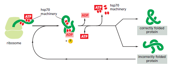
The hsp70 family of molecular chaperones. These proteins act early, recognizing a small stretch of hydrophobic amino acids on a protein’s surface. Aided by a set of smaller hsp40 proteins (not shown), ATP-bound hsp70 molecules grasp their target protein and then hydrolyze ATP to ADP, undergoing conformational changes that cause the hsp70 molecules to associate even more tightly with the target. After the hsp40 dissociates, the rapid rebinding of ATP induces the dissociation of the hsp70 protein after ADP release. Repeated cycles of hsp binding and release help the target protein to refold.

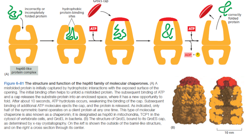
On binding ATP, hsp70 releases the protein into solution allowing it a chance to re-fold. In contrast, hsp60-like proteins form a large barrel-shaped structure that acts after a protein has been fully synthesized. This type of chaperone, sometimes called a chaperonin, forms an “isolation chamber” for the folding process (Figure below).

Chaperones  Ert10

Starting at 41.13, this video showes nicely the amazing movement and functioning of this machine :


To enter a chamber, a substrate protein is first captured via the hydrophobic entrance to the chamber. The protein is then released into the interior of the chamber, which is lined with hydrophilic surfaces, and the chamber is sealed with a lid, a step requiring ATP. Here, the substrate is allowed to fold into its finalconformation in isolation, where there are no other proteins with which to aggregate. When ATP is hydrolyzed, the lid pops off, and the substrate protein, whether folded or not, is released from the chamber.  Chaperones  often need many cycles of ATP hydrolysis to fold a single polypeptide chain correctly. This energy is used to perform mechanical movements of the hsp60 and hsp70 “machines,” converting them from binding forms to releasing forms. Just as we saw for transcription, splicing, and translation, the expenditure of free energy can be used by cells to improve the accuracy of a biological process. In the case of protein folding, ATP hydrolysis allows chaperones to recognize a wide variety of misfolded structures, to halt any further misfolding, and to recommence the folding of a protein in an orderly way.

Question: Could this recognition be a mechanism that arose by natural means? If so, why would chance, or evolution or whatever natural mechanism be supposed, produce such a device, that performs in such an accurate way production control and repair and optimization? Is that not a goal-driven event, that has to be programmed and invented? Or does mindless matter have any goal-driven purposes? That's hard to fathom.

Although our discussion focuses on only two types of chaperones, the cell has a variety of others. The enormous diversity of proteins in cells presumably requires
a wide range of chaperones with versatile surveillance and correction capabilities

Exposed Hydrophobic Regions Provide Critical Signals for Protein Quality Control

If radioactive amino acids are added to cells for a brief period, the newly synthesized proteins can be followed as they mature into their final functional forms.
This type of experiment demonstrates that the hsp70 proteins act first, beginning when a protein is still being synthesized on a ribosome, and the hsp60-like proteins act only later to help fold completed proteins. We have seen that the cell distinguishes misfolded proteins, which require additional rounds of ATP-catalyzed refolding, from those with correct structures through the recognition of hydrophobic surfaces. Usually, if a protein has a sizable exposed patch of hydrophobic amino acids on its surface, it is abnormal: it has either failed to fold correctly after leaving the ribosome, suffered an accident that partly unfolded it at a later time, or failed to find its normal partner subunit in a larger protein complex. Such a protein is not merely useless to the cell, it can be dangerous.
Proteins that rapidly fold correctly on their own do not display such patterns and generally bypass the chaperones. For the others, the chaperones can carry out
“protein repair” by giving them additional chances to fold while, at the same time, preventing their aggregation.

Chaperones  Rytryt10

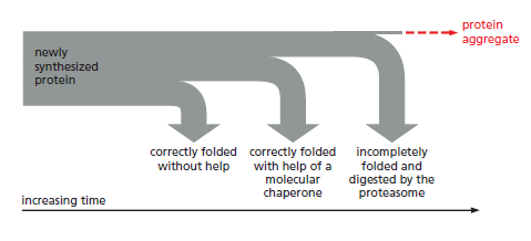
The processes that monitor protein quality following protein synthesis. A newly synthesized protein sometimes folds correctly and assembles on its own with its partner proteins, in which case the quality control mechanisms leave it alone. Incompletely folded proteins are helped to properly fold by molecular chaperones: first by a family of hsp70 proteins, and then, in some cases, by hsp60-like proteins. For both types of chaperones, the substrate proteins are recognized by an abnormally exposed patch of hydrophobic amino acids on their surface. These “protein-rescue” processes compete with another mechanism that, upon recognizing an abnormally exposed hydrophobic patch, marks the protein for destruction by the proteasome. The combined activity of all of these processes is needed to prevent massive protein aggregation in a cell, which can occur when many hydrophobic regions on proteins clump together nonspecifically.

The figure above outlines all of the quality-control choices that a cell makes for a difficult-to-fold, newly synthesized protein. As indicated, when attempts to refold
a protein fail, an additional mechanism is called into play that completely destroys the protein by proteolysis. This proteolytic pathway begins with the recognition of an abnormal hydrophobic patch on a protein’s surface, and it ends with the delivery of the entire protein to a protein-destruction machine, a complex protease
known as the proteasome. As described next, this process depends on an elaborate protein-marking system that also carries out other central functions in the cell by destroying selected normal proteins.


Chaperones are a marvel of molecular engineering:

Molecular chaperones caught on film 1

'The movements we see in the videos suggest how the subunits can first bind the protein inside the cavity, potentially stretch it to undo incorrectly folded molecules, and then release it to allow it to find its own structure in the aqueous environment inside the cavity, while at the same time binding the GroES lid to prevent its premature escape,' Saibil explains. 'If the protein still displays hydrophobic patches after it is released from the chaperone, it will bind to a new GroEL ring and run through the cycle again. Once it is correctly folded it will be stable in the cell environment and will carry out its biological function.'


1) http://www.rsc.org/chemistryworld/News/2012/March/groel-groes-chaperone-caught-on-film.asp

more:

http://www.cryst.bbk.ac.uk/PPS2/course/section12/ellis.html
http://www.stanford.edu/group/frydman/web/
http://www4.utsouthwestern.edu/chuang_lab/project3.html


Last edited by Otangelo on Wed Sep 28, 2022 12:43 pm; edited 20 times in total

https://reasonandscience.catsboard.com

2Chaperones  Empty Hsp70 chaperones Sat Mar 08, 2014 7:28 pm

Otangelo


Admin

Chaperone-assisted protein folding 5

Protein folding is the process by which newly synthesized polypeptide chains acquire the three-dimensional structures necessary for biological function. For many years, protein folding was believed to occur spontaneously, on the basis of the pioneering experiments of Christian Anfinsen, who showed in the late 1950s that purified proteins can fold on theirown after removal from denaturant. Anfinsen had discovered the fundamental principle that the linear amino acid sequence holds all the information necessary to specify a protein’s three-dimensional structure. But it soon became apparent that test-tube folding experiments work mostly for small, single-domain proteins, often only in conditions far removed from those encountered in a cell. Large proteins frequently fail to reach native state under these experimental conditions, forming nonfunctional aggregates instead.

That raises interesting questions : How should and could natural non intelligent natural mechanisms forsee the necessity of chaperones in order to get a specific goal and result, that is functional proteins to make living organisms ? Non living matter has no natural " drive " or purpose or goal to become living. The make of proteins to create life however is a multistep process of many parallel acting complex metabolic pathways and production-line like processes to make proteins and other life essential products like lipids , carbohydrates etc. The right folding of proteins is just one of several other essential processes in order to get a functional protein. But a functional protein by its own has no function, unless correctly embedded through the right order of assembly at the right place.

Despite these problems, protein folding was of little interest to cell biologists until the mid- and late 1980s, when the chaperone story began to unfold. As a result, we now know that in cells, many (perhaps most) proteins require molecular chaperones and metabolic energy to fold efficiently and at a biologically relevant rate. Here I describe, from a personal perspective, the developments leading to this new view.

GroEL binds its substrates in a loosely folded, ‘molten globule’–like conformation, exposing hydrophobic surfaces. As proteins in such states tend to aggregate, their binding by GroEL explained how aggregation is prevented. We also obtained evidence that at least partial folding occurred in association with GroEL and that this process was dependent
on the presence of GroES, suggesting an encapsulation mechanism.  GroEL complex consists of two stacked, heptameric rings. The new images revealed that GroEL binds the
unfolded protein in the ring center. GroES, a heptameric ring of ~10 kDa subunits, binds like a lid over the central GroEL cavity, causing major conformational changes in the interacting GroEL subunits . The  GroEL-GroES complex is asymmetrical and highly dynamic, with GroES binding and unbinding in a mechanism regulated by the GroEL ATPase.  GroEL and GroES essentially function as a folding cage. Nature’s The creator's  Wink solution to the problem of protein folding in the crowded cellular environment seemed extremely impressive in its simplicity and elegance: a single protein molecule folding in a macromolecular cage would be unable to aggregate. However, our evidence to support this model was still indirect, and many researchers had difficulty accepting the idea of a proteinaceous folding cage. the crystal structure of GroEL, solved in 1994 by the late Paul Sigler in collaboration with Art Horwich, seemed to support the folding-in-solution model: the central cavity of GroEL was simply not wide enough for
even a relatively small protein such as rhodanese to fit. However, this interpretation of the structure failed to take into account that GroEL cooperates with GroES. Interestingly,
a month before the crystal structure was published, Helen Saibil and her colleagues had shown by advanced electron cryomicroscopy that GroES binding causes a dramatic conformational change in GroEL, resulting in the formation of a large space capped by GroES. ATP-dependent GroES binding results in protein displacement and encapsulation in the central cavity (the so called cis complex). The cage opens again after ~10 s in a reaction timed by the allosterically regulated GroEL ATPase: When the seven ATP molecules in the GroES-bound GroEL ring have been hydrolyzed, ATP binds to the trans ring, triggering the signal that causes  GroES to unbind Using quantitative proteomics in collaboration with Matthias Mann, we showed in 2005 that at least 250 different proteins interact with GroEL upon synthesis (~10% of cytosolic proteins).

Huh... How could natural mechanisms have achieved that ??  Rolling Eyes

Of these, 60–80 proteins are absolutely GroELGroES–dependent, including a number of essential proteins. They are generally below ~60 kDa in size and can be accommodated by the chaperonin cage. Interestingly, many GroEL-dependent proteins have complex fold topologies comprising a mixture of a-helices and b-sheets, such as the TIM barrel, and are known for their tendency to populate kinetically trapped states during folding. We suggested that the confining environment of the chaperonin cage not only prevents aggregation but also can smooth rugged folding energy landscapes, allowing folding to occur within a biologically relevant time frame.

Cells contain at least one other type of ATP-dependent chaperone, Hsp70, which also interacts with newly synthesized proteins. But what was the relationship between the Hsp70 and chaperonin systems, and did they cooperate in protein folding? There was evidence that Hsp70 binds hydrophobic peptides44 and can associate with nascent polypeptide chains emerging from ribosomes, that is, at a stage when the polypeptide is structurally incomplete and not yet capable of folding. Taking this into consideration, we envisioned a coherent pathway in which Hsp70 would interact with the (growing) polypeptide chain, preventing premature misfolding and aggregation (the negative principle), and then GroEL-GroES would mediate folding of the completed protein to the native state (the positive principle)

Chaperone machines for protein folding, unfolding and disaggregation  1

Chaperones  W3423410

a | Overview of unliganded (apo) GroEL (left) and the GroEL–GroES complex  (right). The overall shapes are shown as blue surfaces, with three subunits coloured by domain in red, green and yellow in apo GroEL. One subunit of GroEL and one of GroES (cyan) are highlighted in the GroEL–GroES complex.

b | Conformation of a GroEL subunit in the apo form (left) and the GroES-bound form (right), with GroEL key sites indicated (GroES is not shown).

c | Cartoons of complexes with folding proteins. Hydrophobic surfaces and residues are shown in yellow and polar residues in green.

d | Cut open view of the cryo-electron microscopy structure  of GroEL in complex with bacteriophage  capsid protein , with a non-native gp23 bound to both rings64. The pink density in the folding chamber corresponds to newly folded gp23, and the yellow density in the open ring is part of a non-native gp23 subunit. The corresponding atomic structures are shown embedded in the electron microscopy density map, except for the non-native substrate, which is unknown and only partially visualized owing to disorder. The open ring with its hydrophobic lining is the acceptor state for non-native polypeptides, and binding to multiple sites may facilitate unfolding. ATP and GroES binding to the chaperonin create a protected chamber with a hydrophilic lining that allows the encapsulated protein to fold.


Such assemblies, called chaperonins, also exist in other cellular compartments and are essential components, mediating protein folding under both heat shock and normal conditions. Ever since 1987, we've been studying these fascinating molecules both in vivo and in vitro, with particular emphasis on the Hsp60 homologue in E. coli known as GroEL.  We and others found early on that a chaperonin-mediated folding reaction can be reconstituted in a test tube, and that has enabled structural and functional studies that have begun to explain how chaperonins work. In particular, a combination of crystallographic studies, with the late Paul Sigler's group here at Yale, and functional studies, using dynamic studies of a variety of mutant chaperonins, have begun to reveal how these chaperonins work.  The schematic diagram below summarizes our current view of the chaperonin-mediated protein folding pathway. 2

Chaperones  Chaprx10


Chaperones  Ssfsd10

(a,b) GroEL alone.


(c) GroEL-unfolded rhodanese.


(d) GroEL-unfolded rhodanese-GroES. 


(e) GroEL-GroES complexes


End-on views are shown in ac and d and side views in b and e. In e, GroES sits like a lid on the GroEL cavity, causing a conformational change in the outer domains of the interacting GroEL subunits.

Hsp70 chaperones: Cellular functions and molecular mechanism 3

Hsp70 proteins are central components of the cellular network of molecular chaperones and folding catalysts. They assist a large variety of protein folding processes in the cell by transient association of their substrate binding domain with short hydrophobic peptide segments within their substrate proteins. The substrate binding and release cycle is driven by the switching of Hsp70 between the low-affinity ATP bound state and the high-affinity ADP bound state. Thus, ATP binding and hydrolysis are essential in vitro and in vivo for the chaperone activity of Hsp70 proteins. This ATPase cycle is controlled by co-chaperones of the family of J-domain proteins, which target Hsp70s to their substrates, and by nucleotide exchange factors, which determine the lifetime of the Hsp70-substrate complex. Additional co-chaperones fine-tune this chaperone cycle. For specific tasks the Hsp70 cycle is coupled to the action of other chaperones, such as Hsp90 and Hsp100.


Chaperones  Sdfsds12 4

a | In the ADP-bound or nucleotide-free state, the nucleotide-binding domain (green; Protein Data Bank (PDB) code: 3HSC)16 of heat shock protein 70 (HSP70) is connected by a flexible linker to the substrate-binding domain (blue; PDB code: 1DKZ), with the lid domain (red) locking a peptide substrate (yellow) into the binding pocket18. A side view of the substrate domain is shown on the right. A cartoon depicting the two-domain complex is shown below. The bound nucleotide is shown in space filling format.

b | In the ATP-bound state, the lid opens, and both the lid and the substrate-binding domain dock to the nucleotide-binding domain (PDB code: 4B9Q)20. The corresponding cartoon of this conformation is shown below. When ATP binds, the cleft closes, triggering a change on the outside of the nucleotide-binding domain that creates a binding site for the linker region. Linker binding causes the substrate-binding domain and the lid domain to bind different sites on the nucleotide-binding domain, resulting in a widely opened substrate-binding site that enables rapid exchange of polypeptide substrates. After hydrolysis, the domains separate and the lid closes over the bound substrate. Such binding and release of extended regions of polypeptide chain are thought to unfold and stabilize non-native proteins either for correct folding or degradation.

Chaperones  Oppoop10

The nascent chain is stabilized in a folding-competent state during translation by the Hsp70 chaperone system (DnaK, DnaJ) (1 and 2).

These chaperones bind hydrophobic segments exposed by the extended chain that will later be buried within the folded structure. Upon completion of translation, the protein is unable to fold using the Hsp70 chaperone system and must be transferred into the central cavity of GroEL. This step requires GrpE, the nucleotide exchange factor of DnaK (3).

After binding of the protein in a molten globule–like conformation into the open ring of GroEL (4)

the protein is encapsulated by GroES in the folding cage (5).

Folded protein emerges from the cage as GroES unbinds (6)

The model was later extended to include the cooperation of DnaK with the ribosome-bound chaperone trigger factor and the finding that the Hsp70 system mediates the folding of proteins that do not require the physical environment of the chaperonin cage


Chaperones  Sdfsdf17

Substrate binding to GroEL (upon transfer from the upstream chaperone Hsp70) may result in local unfolding. ATP binding then triggers a conformational rearrangement of the GroEL apical domains. This is followed by the binding of GroES (forming the cis complex) and substrate encapsulation for folding. At the same time, ADP and GroES dissociate from the opposite (trans) GroEL ring, allowing the release of substrate that had been enclosed in the former cis complex (omitted for simplicity). The substrate remains encapsulated, free to fold, for the time needed to hydrolyze the seven ATP molecules in the newly formed cis complex (~10 s). Binding of ATP and GroES to the trans ring causes the opening of the cis complex.

Biochemists trap a chaperone machine in action 6

Molecular chaperones have emerged as exciting new potential drug targets, because scientists want to learn how to stop cancer cells, for example, from using chaperones to enable their uncontrolled growth. Now a team of biochemists at the University of Massachusetts Amherst led by Lila Gierasch have deciphered key steps in the mechanism of the Hsp70 molecular machine by "trapping" this chaperone in action, providing a dynamic snapshot of its mechanism.

She and colleagues describe this work in the current issue of Cell. Gierasch's research on Hsp70 chaperones is supported by a long-running grant to her lab from NIH's National Institute for General Medical Sciences.

Molecular chaperones like the Hsp70s facilitate the origami-like folding of proteins, made in the cell's nanofactories or ribosomes, from where they emerge unstructured like noodles. Proteins only function when folded into their proper structures, but the process is so difficult under cellular conditions that molecular chaperone helpers are needed.

The newly discovered information about chaperone action is important because all rapidly dividing cells use a lot of Hsp70, Gierasch points out. "The saying is that cancer cells are addicted to Hsp70 because they rely on this chaperone for explosive new cell growth. Cancer shifts our body's production of Hsp70 into high gear. If we can figure out a way to take that away from cancer cells, maybe we can stop the out-of-control tumor growth. To find a molecular way to inhibit Hsp70, you've got to know how it works and what it needs to function, so you can identify its vulnerabilities."

Chaperone proteins in cells, from bacteria to humans, act like midwives or bodyguards, protecting newborn proteins from misfolding and existing proteins against loss of structure caused by stress such as heat or a fever. In fact, the heat shock protein (Hsp) group includes a variety of chaperones active in both these situations.

As Gierasch explains, "New proteins emerge into a challenging environment. It's very crowded in the cell and it would be easy for them to get their sticky amino acid chains tangled and clumped together. Chaperones bind to them and help to avoid this aggregation, which is implicated in many pathologies such as neurodegenerative diseases. This role of chaperones has also heightened interest in using them therapeutically."

However, chaperones must not bind too tightly or a protein can't move on to do its job. To avoid this, chaperones rapidly cycle between tight and loose binding states, determined by whether ATP or ADP is bound. In the loose state, a protein client is free to fold or to be picked up by another chaperone that will help it fold to do its cellular work. In effect, Gierasch says, Hsp70s create a "holding pattern" to keep the protein substrate viable and ready for use, but also protected.

She and colleagues knew the Hsp70's structure in both tight and loose binding affinity states, but not what happened between, which is essential to understanding the mechanism of chaperone action. Using the analogy of a high jump, they had a snapshot of the takeoff and landing, but not the top of the jump. "Knowing the end points doesn't tell us how it works. There is a shape change in there that we wanted to see," Gierasch says.

To address this, she and her colleagues postdoctoral fellows Anastasia Zhuravleva and Eugenia Clerico obtained "fingerprints" of the structure of Hsp70 in different states by using state-of-the-art nuclear magnetic resonance (NMR) methods that allowed them to map how chemical environments of individual amino acids of the protein change in different sample conditions. Working with an Hsp70 known as DnaK from E. coli bacteria, Zhuravleva and Clerico assigned its NMR spectra. In other words, they determined which peaks came from which amino acids in this large molecule.

The UMass Amherst team then mutated the Hsp70 so that cycling between tight and loose binding states stopped. As Gierasch explains, "Anastasia and Eugenia were able to stop the cycle part-way through the high jump, so to speak, and obtain the molecular fingerprint of a transient intermediate." She calls this accomplishment "brilliant."

Now that the researchers have a picture of this critical allosteric state, that is, one in which events at one site control events in another, Gierasch says many insights emerge. For example, it appears nature uses this energetically tense state to "tune" alternate versions of Hsp70 to perform different cellular functions. "Tuning means there may be evolutionary changes that let the chaperone work with its partners optimally," she notes.

"And if you want to make a drug that controls the amount of Hsp70 available to a cell, our work points the way toward figuring out how to tickle the molecule so you can control its shape and its ability to bind to its client. We're not done, but we made a big leap," Gierasch adds. "We now have a idea of what the Hsp70 structure is when it is doing its job, which is extraordinarily important."

1) http://www.nature.com/nrm/journal/v14/n10/full/nrm3658.html
2) https://tools.medicine.yale.edu/horwich/www/
3) http://www.ncbi.nlm.nih.gov/pmc/articles/PMC2773841/
4) http://www.nature.com/nrm/journal/v14/n10/fig_tab/nrm3658_F1.html
5) http://www.nature.com/nm/journal/v17/n10/pdf/nm.2467.pdf
6) http://www.eurekalert.org/pub_releases/2012-12/uoma-bta120412.php


Last edited by Otangelo on Wed Sep 28, 2022 12:42 pm; edited 19 times in total

https://reasonandscience.catsboard.com

3Chaperones  Empty Molecular chaperones Sun Aug 16, 2015 12:05 pm

Otangelo


Admin

Molecular chaperones 1

Cells provide a sophisticated machinery of proteins which play a fundamental role in preventing the aggregation and in assisting the folding and assembly of proteins: the molecular chaperones. A variety of molecular chaperones have been found and characterized up to date. Based on differences regarding size, structure, function, mechanism or cellular compartmentalization, different families of chaperones can be classified: Hsp100/Clp, Hsp90, Hsp70, chaperonins (Hsp60), small Hsps and calnexin/calreticulin . The chaperones stabilize these non-native proteins, they can prevent them from misfolding or aggregation and direct them into productive folding and assembly pathways. If chaperones assist folding, they are not themselves components of the final structure and they do not impose structural information directing the folding of substrates. Their potential to prevent aggregation and to mediate protein folding is particularly relevant for de novo protein biosynthesis, but also for the reduction of damage under stress conditions, such as elevated temperatures.


Chaperones involved in de novo protein folding

Cells provide a complex network of molecular chaperones that assist proteins to adopt their functional three-dimensional structures. In general, there are two major classes of cytosolic chaperones mediating the folding of newly synthesized polypeptides in a sequential manner


1) http://edoc.ub.uni-muenchen.de/7577/1/Saschenbrecker_Sandra.pdf


Last edited by Admin on Mon Aug 17, 2015 6:50 pm; edited 1 time in total

https://reasonandscience.catsboard.com

Otangelo


Admin

Protein Dressing Room Has Electronic Walls   1
 
Properly folded proteins are essential to all of life.  When a polypeptide, or chain of amino acids, emerges from the ribosome translation factory on its way to becoming a protein, it looks like a useless, shapeless piece of string.  It cannot perform its function till folded into a precise, compact shape particular for its job.  Some short polypeptides will spontaneously fold into their “native” state, ready for work, but many of the bigger ones need help.  Fortunately, the cell provides a private dressing room called the GroEL-GroES chaperonin that not only gives them privacy, away from the bustle of colliding molecules in the cytoplasm but actually helps them get correctly folded.  This chaperone or “helper” machine thus not only gets the actor ready for the stage faster, but prevents misfolding that could clutter the cell with useless or harmful aggregates of protein.

The inside walls of the GroEL barrel and the inside walls of the GroES lid contain protrusions that generate electrostatic and hydrophobic forces on the interior space.  When the unfolded protein enters, therefore, it is subjected to gentle pressures that coax it to fold.  These forces are nonspecific enough to work on hundreds of different substrates that use this general-purpose machine.   The forces change during the entry of the nascent protein.  The interior is not barrel shaped when the actor approaches the door; the GroES lid, with the help of the energy molecule ATP, guides the protein in, and then the barrel pops into its shape, providing a safe haven for folding.  The electronic walls turn on to provide that gentle nudge to get the polypeptide over its energy barriers and into the right folding pathway.  When the protein has properly completed its folding after about 10 seconds, the door opens and the protein pops out, ready for operation.

How finely tuned is this machine?  The authors did some experiments on mutating the chaperone to make the barrel looser and tighter.  They found that volume changes as small as 2-5% slowed down the folding considerably. The barrel volume needs to be within certain narrow limits, yet general enough to handle a variety of small, medium and large proteins.

The GroEL/GroES nano-cage allows a single protein molecule to fold in isolation.  This reaction has been compared to spontaneous folding at infinite dilution.  However, recent experimental and theoretical studies indicate that the physical environment of the chaperonin cage can alter the folding energy landscape, resulting in accelerated folding for some proteins.  By performing an extensive mutational analysis of GroEL, we have identified three structural features of the chaperonin cage as major contributors to this capacity: 


1. geometric confinement exerted on the folding protein inside the limited volume of the cage; 
2. a mildly hydrophobic, interactive surface at the bottom of the cage; 
3. Clusters of negatively charged amino acid residues exposed on the cavity wall. These features in combination provide a physical environment that has been optimized to catalyze the structural annealing of proteins with kinetically complex folding pathways.  Thus, the chaperonin system and its mutant versions may prove as useful tools in understanding how proteins navigate their energy landscape of folding.

1Tang et al., “Structural Features of the GroEL-GroES Nano-Cage Required for Rapid Folding of Encapsulated Protein,” Cell

What we see here are molecular machines working with precision, efficiency, control – and design. Protein folding is a complex affair, wherein several domains of the polypeptide fold sequentially or simultaneously following an energy landscape (like a pinball negotiating obstacles) that leads to the completed protein. Some domains fold into a helix or sheet, or several, which then combine into larger structures.  Even then, after the protein exits the chaperone, there can be subsequent modifications: multiple proteins, for instance, might be joined into complexes, with metal ions inserted (as in hemoglobin or chlorophyll), and these proteins usually become part of networks.  Add to that now the exciting discovery that the walls of the chaperone barrel are interactive, coaxing the proteins to fold properly. At every stage, there is coordinated, synchronized, elegant design. Think about how these molecules operate in the blind.  They do not have eyes and brains telling them where to go – yet they succeed.  There is no analog in human technology; the closest, perhaps, is computer programming, but in life, at scales smaller than most of us can imagine, nano-factories operate with physical entities moving through space and time.  How fortunate we are to see these marvels unfold.  Our ancestors might have wondered at the mysteries of biological life, but could they in their wildest dreams have imagined the city-like organization at work at the molecular level?

1) http://creationsafaris.com/crev200606.htm


Last edited by Otangelo on Wed Sep 28, 2022 12:42 pm; edited 4 times in total

https://reasonandscience.catsboard.com

5Chaperones  Empty What Chaperone Proteins Know Sun Aug 16, 2015 3:11 pm

Otangelo


Admin

What Chaperone Proteins Know 1

Here's a riddle for you: Proteins are used to make proteins, so if we assume a purely naturalistic origin of life, where did the first proteins come from?

If a cell is a factory, proteins are the factory workers. Proteins conduct most of the necessary functions in a cell. Proteins are made up of amino acid building blocks. A chain of amino acids must fold into the appropriate three-dimensional structure so that the protein can function properly. Within cells are proteins known as chaperones that help fold the amino acid chain into its proper three-dimensional structure. If the amino acid chain folds improperly, then this could wreak havoc on the cell and potentially the entire organism. The chaperone works to prevent folding defects and is a key player in the final steps of protein synthesis.

However, as important as chaperones are, there are still many questions as to how exactly they work. For example, do the chaperones fold the amino acid chain while it is still being constructed (during translation), or is the amino acid chain first put together, and then the folding beings? Or, is it some combination of both? Studies indicate that it is indeed a combination of both. There are two different kinds of chaperone proteins within the cell, one for translation and one for post-translation. With these two different kinds of chaperones, where and how does regulation happen to prevent misfolds?

Recent research on bacterial cells sheds light on the chaperones' important function. One chaperone in particular, Trigger Factor, plays a key role in correcting misfolds that may occur early on in the translational process. Trigger Factor can slow down improper amino acid folding, and it can even unfold amino acid chains that have already folded up incorrectly.

Here are some of the neat features of Trigger Factor:

Trigger Factor actually constrains protein folding more than the ribosome does. It doesn't just "get in the way" like the ribosome. It also regulates the folding.
Trigger Factor's function is specific to the particular region of the amino acid chain. It does not just perform one function no matter what the composition of the amino acid chain. It changes based on the region of the chain it is working with.
Trigger Factor also changes its activity based on where the protein is in the translation process.
Trigger Factor's process depends on how the amino acid chain is bound to the ribosome, and can even unfold parts of the chain that were misfolded in the translation process.
An additional factor that regulates when amino acid chains fold into proteins is its distance from the ribosome (the place where the amino acid chain is made). The closer the chain is to the ribosome, the less room it has to fold into a three-dimensional protein. Trigger Factor works with this spatial hindrance, making an interesting and complex regulation system.

Trigger Factor is only called into the game once the amino acid chain is a certain length (around 100 amino acids long) and when the chain has certain features, such as hydrophobicity. As the authors state it, Trigger Factor keeps the protein from folding into its three-dimensional structure until the amino acid chain has all of the information it needs to fold properly:

In summary, we show that the ribosome and TF each uniquely affect the folding landscape of nascent polypeptides to prevent or reverse early misfolds as long as important folding information is still missing and the nascent chain is not released from the ribosome.
So we have a protein that is able to perform various functions that inhibit or slow protein folding until the amino acid has the right chemical information for folding to occur.
This does not solve the riddle about proteins being made from proteins (otherwise known as the chicken-and-the-egg problem). It actually adds another twist to the riddle: How does one protein know how much information a completely different protein needs to fold into a three dimensional structure? How does a protein evolve the ability to "know" how to respond to specific translational circumstances as Trigger Factor does?


1) http://www.evolutionnews.org/2012/09/what_chaperone063951.html


Last edited by Admin on Wed Sep 12, 2018 1:21 pm; edited 1 time in total

https://reasonandscience.catsboard.com

Otangelo


Admin

New research on protein folding demonstrates intelligent design 1


The journal Nature has just published a detailed and fascinating review about the way proteins in our bodies are helped by other proteins, known as chaperones, to become functional.

Proteins are the most complex molecules in our bodies and are involved in virtually all biological processes. Our cells typically manufacture over 10,000 different proteins, synthesised on ribosomes as chains of up to several thousand amino acids.

For a protein to function it must fold to its ‘native state’ which is a complex three-dimensional structure.  If a protein fails to fold into its functional structure then it is not only without function but in many cases is actually toxic to the cell. It is thought that as we age, the systems for helping proper folding work less well, which is one of the reasons for the symptoms of ageing and some diseases.

The number of possible shapes that a protein can fold into is very high and folding reactions are very complex, involving the co-operation of many weak, non-covalent interactions. A high percentage of proteins do not fold automatically into the required shape and are at risk of aberrant folding and aggregation. As the abstract to this paper states: “To avoid these dangers, cells invest in a complex network of molecular chaperones, which are ingenious mechanisms to prevent aggregation and promote efficient folding.”

Not only do proteins require other proteins (chaperones) to fold properly, they also require chaperones constantly, after correct folding, to maintain their functional states. This is known as proteostasis.

There is also another whole system in the cell (involving more proteins) called the ‘ubiquitin-proteasome system’ which breaks down irreversibly misfolded and aggregated proteins for safe excretion.

This is not the whole story either. Proteins that are still not properly folded are transported ‘downstream’ to another system of proteins: the chaperonins. These are large double-ring complexes that enclose one protein at a time in a sort of cage structure. Within this structure the protein folding is completed before the protein is released.

The review in the journal Nature does not discuss the origins of these systems but we need to ask a question: how does all this fit with current evolutionary theory? One might think that such complex systems are confined to mammals or at least the higher orders of animals. This would be a mistake however, because chaperones and chaperonins are in bacteria and archaea also. Indeed it would seem that for any cell to function there needs to be not just proteins but, at the same time, these chaperone systems, which are absolutely essential for proper folding and maintenance of proteins. Without such systems, in place already, the cell will not function.

Now, as explained, these chaperone systems are themselves made of proteins which also require the assistance of chaperones to correctly fold and to maintain integrity once folded. Chaperones for chaperones in fact. The very simplest of cells that we know of have these systems in place.


Darwinian evolution requires step by step changes in molecular systems, with one step leading to another in a manner that is statistically reasonable to expect from selection of mutant strains. There is no Darwinian explanation however for the evolution of proteins which already have chaperone systems in place to ensure proper function.

This points very strongly to an intelligent origin of these ‘ingenious’ systems found in all of life.

1) http://www.c4id.org.uk/index.php?option=com_content&view=article&id=239:new-research-on-protein-folding-demonstrates-intelligent-design&catid=52:frontpage&Itemid=1

https://reasonandscience.catsboard.com

7Chaperones  Empty Re: Chaperones Wed Aug 26, 2015 4:56 am

Otangelo


Admin

chaperones are required to fold other proteins. Some proteins do not fold correctly without them. One has no use without the other. How did chaperones " know " about the requirement of the other proteins and the need to help them to fold correctly ? what happened during the time that chaperones did not adquire the right shape and function to exercise their job properly ?

Chaperones are ancient protein structures, which were highly conserved throughout all the known parts of evolution, and are repeatedly emerging as parts of the minimal genomes of various organisms suggesting their presence in the hypothetical Last Universal Common Ancestor (LUCA) 1

1) http://www.linkgroup.hu/docs/pcs/03theochem.pdf

further readings: http://www.ncbi.nlm.nih.gov/pmc/articles/PMC4339202/

https://reasonandscience.catsboard.com

Sponsored content


Back to top  Message [Page 1 of 1]

Permissions in this forum:
You cannot reply to topics in this forum